
COVID-19 Rush Journal Club - Rush University
256
COVID-19 Rush Journal Club NOVEL CORONAVIRUS SARS-COV-2. Transmission electron micrograph of SARS-CoV-2 virus particles, iso- lated from a patient. Image captured and color-enhanced at the NIAID Integrated Research Facility (IRF) in Fort Detrick, Maryland. Credit: NIAID Available at: https://www.flickr.com/photos/niaid/49597768397/in/al- bum-72157712914621487/. Accessed April 19, 2020.
-
Upload
khangminh22 -
Category
Documents
-
view
1 -
download
0
Transcript of COVID-19 Rush Journal Club - Rush University

COVID-19 Rush Journal Club
NOVEL CORONAVIRUS SARS-COV-2. Transmission electron micrograph of SARS-CoV-2 virus particles, iso-lated from a patient. Image captured and color-enhanced at the NIAID Integrated Research Facility (IRF) in
Fort Detrick, Maryland. Credit: NIAID Available at: https://www.flickr.com/photos/niaid/49597768397/in/al-bum-72157712914621487/. Accessed April 19, 2020.

This document is a collection of efforts from students of Rush University. It provides brief reviews of research articles regarding COVID-19. We hope that this will be helpful to clini-
cians, students, community leaders, and the general public. This document, however, does not act as a replacement of the original source documents. Please use the DOI on each
page to read more.
Literature SelectionChristi Brown, MS2
Lindsay Friedman, MS3Danesha Lewis, PhD Student
Travis Tran, MS2
Student Editors in ChiefJoseph deBettencourt, MS3
Alyssa Coleman, MS3Lindsay Friedman, MS3Leah Greenfield, MS2
Beth Hall, MDMorgan Sturgis, MS2
Travis Tran, MS2Ashley Wehrheim, MS2Kaitlyn Wehrheim, MS2
Bijan Zarrabi, MS2
Faculty Editors and MentorsFrank Cockerill, MD - Adjunct Professor of Medicine
Mete Altintas, PhD - Director, Translational and Preclinical StudiesRachel Miller, PhD - Assistant Professor of Internal Medicine
Michael Bradaric, PhD - Director of Pharmacology, Rush Medical CollegeBrinda Bradaric, PhD - Assistant Professor, Dept of Health Sciences
Jitesh Pratap, PhD - Assistant Professor, Dept of Cell and Molecular MedicineSteve Mangos, PhD - Director, Internal Medicine Drug Discovery and Imaging Core
Graphic designSam Auger, MD Beth Hall, MD
John Levinson, MS2Luke McCormack, MS2Diana Parker, MS2Elena Perkins, MS3Hannah Raff, MS3Ayesan Rewane, MSCRAudrey Sung, MS3
Is there a study you’d like us to review? Do you have questions or feedback?Please email: [email protected]
Blake Beehler, MDAlice Burgess, MS2
Alyssa Coleman, MS3Shyam Desai, MD
Joseph Dodson, MS1Joshua Doppelt, MD
Emily Hejna, MS2
Danesha Lewis, PhD student
Student Editors
COVID-19Rush Journal Club
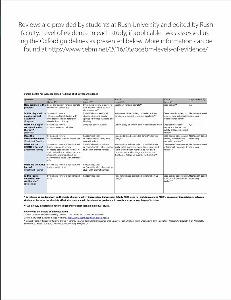
Oxford Centre for Evidence-Based Medicine 2011 Levels of Evidence
* Level may be graded down on the basis of study quality, imprecision, indirectness (study PICO does not match questions PICO), because of inconsistency between
studies, or because the absolute effect size is very small; Level may be graded up if there is a large or very large effect size.
** As always, a systematic review is generally better than an individual study.
How to cite the Levels of Evidence Table
OCEBM Levels of Evidence Working Group*. "The Oxford 2011 Levels of Evidence".
Oxford Centre for Evidence-Based Medicine. http://www.cebm.net/index.aspx?o=5653
* OCEBM Table of Evidence Working Group = Jeremy Howick, Iain Chalmers (James Lind Library), Paul Glasziou, Trish Greenhalgh, Carl Heneghan, Alessandro Liberati, Ivan Moschetti,
Bob Phillips, Hazel Thornton, Olive Goddard and Mary Hodgkinson
Question Step 1
(Level 1*)
Step 2
(Level 2*)
Step 3
(Level 3*)
Step 4
(Level 4*)
Step 5 (Level 5)
How common is the
problem?
Local and current random sample
surveys (or censuses)
Systematic review of surveys
that allow matching to local
circumstances**
Local non-random sample** Case-series** n/a
Is this diagnostic or
monitoring test
accurate?
(Diagnosis)
Systematic review
of cross sectional studies with
consistently applied reference
standard and blinding
Individual cross sectional
studies with consistently
applied reference standard and
blinding
Non-consecutive studies, or studies without
consistently applied reference standards**
Case-control studies, or
“poor or non-independent
reference standard**
Mechanism-based
reasoning
What will happen if
we do not add a
therapy?
(Prognosis)
Systematic review
of inception cohort studies
Inception cohort studies Cohort study or control arm of randomized trial* Case-series or case-
control studies, or poor
quality prognostic cohort
study**
n/a
Does this
intervention help?
(Treatment Benefits)
Systematic review
of randomized trials or n-of-1 trials
Randomized trial
or observational study with
dramatic effect
Non-randomized controlled cohort/follow-up
study**
Case-series, case-control
studies, or historically
controlled studies**
Mechanism-based
reasoning
What are the
COMMON harms?
(Treatment Harms)
Systematic review of randomized
trials, systematic review
of nested case-control studies, n-
of-1 trial with the patient you are
raising the question about, or
observational study with dramatic
effect
Individual randomized trial
or (exceptionally) observational
study with dramatic effect
Non-randomized controlled cohort/follow-up
study (post-marketing surveillance) provided
there are sufficient numbers to rule out a
common harm. (For long-term harms the
duration of follow-up must be sufficient.)**
Case-series, case-control,
or historically controlled
studies**
Mechanism-based
reasoning
What are the RARE
harms?
(Treatment Harms)
Systematic review of randomized
trials or n-of-1 trial
Randomized trial
or (exceptionally) observational
study with dramatic effect
Non-randomized controlled cohort/follow-up
study (post-marketing surveillance) provided
there are sufficient numbers to rule out a
common harm. (For long-term harms the
duration of follow-up must be sufficient.)**
Case-series, case-control,
or historically controlled
studies**
Mechanism-based
reasoning
Is this (early
detection) test
worthwhile?
(Screening)
Systematic review of randomized
trials
Randomized trial Non -randomized controlled cohort/follow-up
study**
Case-series, case-control,
or historically controlled
studies**
Mechanism-based
reasoning
Reviews are provided by students at Rush University and edited by Rush faculty. Level of evidence in each study, if applicable, was assessed us-
ing the Oxford guidelines as presented below. More information can be found at http://www.cebm.net/2016/05/ocebm-levels-of-evidence/
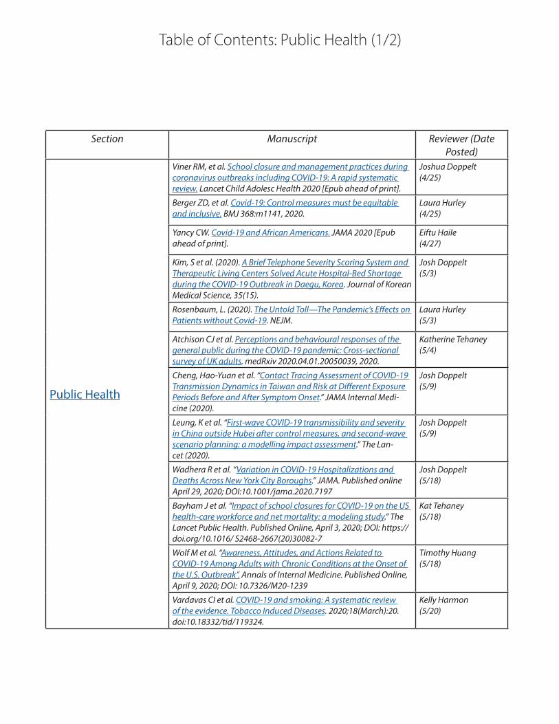
Table of Contents: Public Health (1/2)
Section Manuscript Reviewer (Date Posted)
Public Health
Viner RM, et al. School closure and management practices during coronavirus outbreaks including COVID-19: A rapid systematic review. Lancet Child Adolesc Health 2020 [Epub ahead of print].
Joshua Doppelt(4/25)
Berger ZD, et al. Covid-19: Control measures must be equitable and inclusive. BMJ 368:m1141, 2020.
Laura Hurley(4/25)
Yancy CW. Covid-19 and African Americans. JAMA 2020 [Epub ahead of print].
Eiftu Haile(4/27)
Kim, S et al. (2020). A Brief Telephone Severity Scoring System and Therapeutic Living Centers Solved Acute Hospital-Bed Shortage during the COVID-19 Outbreak in Daegu, Korea. Journal of Korean Medical Science, 35(15).
Josh Doppelt(5/3)
Rosenbaum, L. (2020). The Untold Toll—The Pandemic’s Effects on Patients without Covid-19. NEJM.
Laura Hurley(5/3)
Atchison CJ et al. Perceptions and behavioural responses of the general public during the COVID-19 pandemic: Cross-sectional survey of UK adults. medRxiv 2020.04.01.20050039, 2020.
Katherine Tehaney(5/4)
Cheng, Hao-Yuan et al. “Contact Tracing Assessment of COVID-19 Transmission Dynamics in Taiwan and Risk at Different Exposure Periods Before and After Symptom Onset.” JAMA Internal Medi-cine (2020).
Josh Doppelt(5/9)
Leung, K et al. “First-wave COVID-19 transmissibility and severity in China outside Hubei after control measures, and second-wave scenario planning: a modelling impact assessment.” The Lan-cet (2020).
Josh Doppelt(5/9)
Wadhera R et al. “Variation in COVID-19 Hospitalizations and Deaths Across New York City Boroughs.” JAMA. Published online April 29, 2020; DOI:10.1001/jama.2020.7197
Josh Doppelt(5/18)
Bayham J et al. “Impact of school closures for COVID-19 on the US health-care workforce and net mortality: a modeling study.” The Lancet Public Health. Published Online, April 3, 2020; DOI: https://doi.org/10.1016/ S2468-2667(20)30082-7
Kat Tehaney(5/18)
Wolf M et al. “Awareness, Attitudes, and Actions Related to COVID-19 Among Adults with Chronic Conditions at the Onset of the U.S. Outbreak”. Annals of Internal Medicine. Published Online, April 9, 2020; DOI: 10.7326/M20-1239
Timothy Huang(5/18)
Vardavas CI et al. COVID-19 and smoking: A systematic review of the evidence. Tobacco Induced Diseases. 2020;18(March):20. doi:10.18332/tid/119324.
Kelly Harmon(5/20)

Table of Contents: Public Health (2/2)
Section Manuscript Reviewer (Date Posted)
Public Health cont.
Zheng Z et al. Risk factors of critical & mortal COVID-19 cases: A systematic literature review and meta-analysis. J Infect 2020 [Epub ahead of print].
Kelly Harmon(5/20)
Wang B et al. Does comorbidity increase the risk of patients with COVID-19: evidence from meta-analysis. Aging (Albany NY). 2020;12(7):6049‐6057. doi:10.18632/aging.103000
Kelly Harmon(5/20)
Roberton T et al. Early estimates of the indirect effects of the COVID-19 pandemic on maternal and child mortality in low-in-come and middle-income countries: a modelling study. Lancet Glob Health 2020 [Epub ahead of print].
Kat Tehaney(5/20)
Xie X et al. Mental Health Status Among Children in Home Confinement During the Coronavirus Disease 2019 Outbreak in Hubei Province, China. JAMA Pediatr. Published online April 24, 2020. doi:10.1001/jamapediatrics.2020.1619
Kat Tehaney(5/21)
Bialek S et al. Geographic Differences in COVID-19 Cases, Deaths, and Incidence - United States, February 12-April 7, 2020. MMWR Wkly Rep 69(15):465-471, 2020.
Eiftu Haile(5/21)
Hawks L, Woolhandler S, McCormick D. COVID-19 in Prisons and Jails in the United States. JAMA Intern Med.Published online April 28, 2020. doi:10.1001/jamainternmed.2020.1856
Alice Burgess(5/23)
Nussbaumer-Streit B et al., Quarantine alone or in combination with other public health measures to control COVID‐19: a rapid review. Cochrane Database of Systematic Reviews 2020, Issue 4. Art. No.: CD013574. DOI: 10.1002/14651858.CD013574.
Kelly Harmon(6/1)
Liu M et al., Internet searches for unproven COVID-19 therapies in the United States. JAMA Intern Med 2020 [Epub ahead of print].
Kat Tehaney(6/1)
Yan L et al., An interpretable mortality prediction model for COVID-19 patients. Nat Mach Intell 2, 283–288 (2020). https://doi.org/10.1038/s42256-020-0180-7
Kat Tehaney(6/2)
Joensen LE et al., Diabetes and COVID-19: psychosocial con-sequences of the COVID-19 pandemic in people with diabetes in Denmark-what characterizes people with high levels of COVID-19-related worries? [published online ahead of print, 2020 May 11]. Diabet Med. 2020;10.1111/dme.14319.
Kat Tehaney(6/2)
Lai J et al., Factors Associated With Mental Health Outcomes Among Health Care Workers Exposed to Coronavirus Disease 2019. JAMA Netw Open. 2020;3(3):e203976. doi:10.1001/jama-networkopen.2020.3976
Kat Tehaney(6/3)
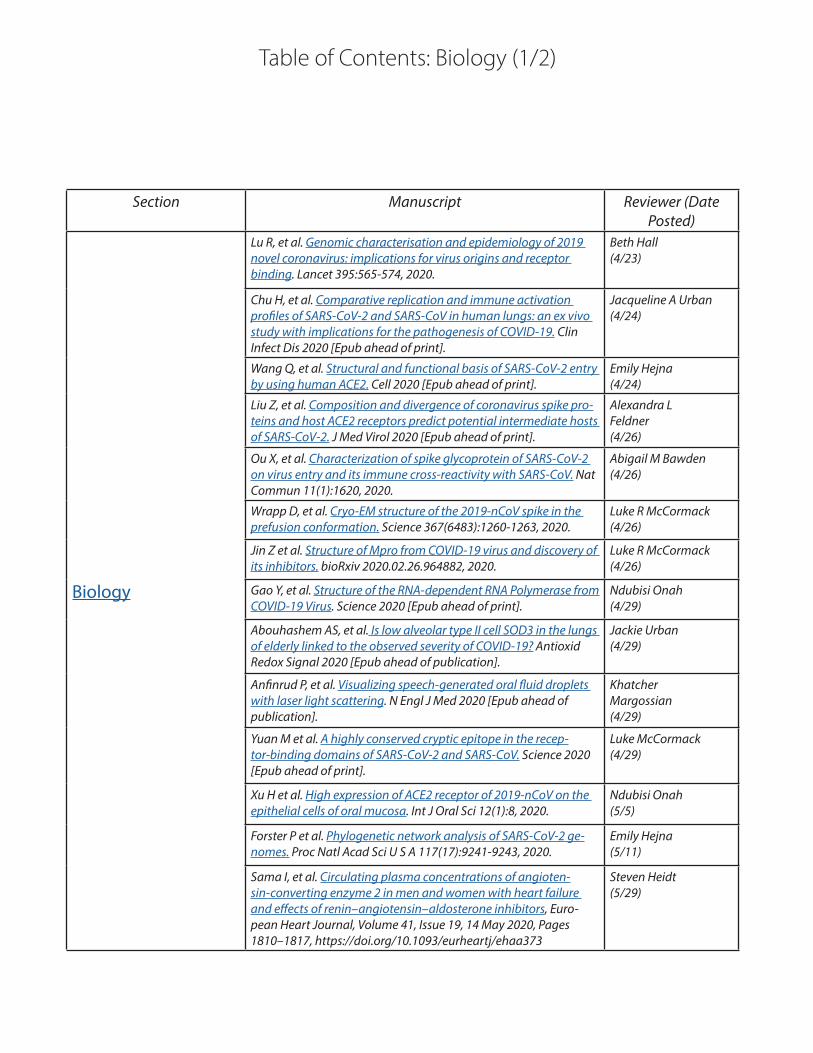
Table of Contents: Biology (1/2)
Section Manuscript Reviewer (Date Posted)
Biology
Lu R, et al. Genomic characterisation and epidemiology of 2019 novel coronavirus: implications for virus origins and receptor binding. Lancet 395:565-574, 2020.
Beth Hall(4/23)
Chu H, et al. Comparative replication and immune activation profiles of SARS-CoV-2 and SARS-CoV in human lungs: an ex vivo study with implications for the pathogenesis of COVID-19. Clin Infect Dis 2020 [Epub ahead of print].
Jacqueline A Urban(4/24)
Wang Q, et al. Structural and functional basis of SARS-CoV-2 entry by using human ACE2. Cell 2020 [Epub ahead of print].
Emily Hejna(4/24)
Liu Z, et al. Composition and divergence of coronavirus spike pro-teins and host ACE2 receptors predict potential intermediate hosts of SARS‐CoV‐2. J Med Virol 2020 [Epub ahead of print].
Alexandra L Feldner(4/26)
Ou X, et al. Characterization of spike glycoprotein of SARS-CoV-2 on virus entry and its immune cross-reactivity with SARS-CoV. Nat Commun 11(1):1620, 2020.
Abigail M Bawden(4/26)
Wrapp D, et al. Cryo-EM structure of the 2019-nCoV spike in the prefusion conformation. Science 367(6483):1260-1263, 2020.
Luke R McCormack(4/26)
Jin Z et al. Structure of Mpro from COVID-19 virus and discovery of its inhibitors. bioRxiv 2020.02.26.964882, 2020.
Luke R McCormack(4/26)
Gao Y, et al. Structure of the RNA-dependent RNA Polymerase from COVID-19 Virus. Science 2020 [Epub ahead of print].
Ndubisi Onah(4/29)
Abouhashem AS, et al. Is low alveolar type II cell SOD3 in the lungs of elderly linked to the observed severity of COVID-19? Antioxid Redox Signal 2020 [Epub ahead of publication].
Jackie Urban(4/29)
Anfinrud P, et al. Visualizing speech-generated oral fluid droplets with laser light scattering. N Engl J Med 2020 [Epub ahead of publication].
Khatcher Margossian(4/29)
Yuan M et al. A highly conserved cryptic epitope in the recep-tor-binding domains of SARS-CoV-2 and SARS-CoV. Science 2020 [Epub ahead of print].
Luke McCormack(4/29)
Xu H et al. High expression of ACE2 receptor of 2019-nCoV on the epithelial cells of oral mucosa. Int J Oral Sci 12(1):8, 2020.
Ndubisi Onah(5/5)
Forster P et al. Phylogenetic network analysis of SARS-CoV-2 ge-nomes. Proc Natl Acad Sci U S A 117(17):9241-9243, 2020.
Emily Hejna(5/11)
Sama I, et al. Circulating plasma concentrations of angioten-sin-converting enzyme 2 in men and women with heart failure and effects of renin–angiotensin–aldosterone inhibitors, Euro-pean Heart Journal, Volume 41, Issue 19, 14 May 2020, Pages 1810–1817, https://doi.org/10.1093/eurheartj/ehaa373
Steven Heidt(5/29)

Table of Contents: Biology (2/2)
Section Manuscript Reviewer (Date Posted)
Biology cont.Hoffman, M. et al. A multi-basic cleavage site in the spike protein of SARS-CoV-2 is essential for infection of human lung cells. Molecular Cell. 1 May 2020
Luke McCormack(6/1)

Table of Contents: Epidemiology (1/3)
Section Manuscript Reviewer (Date Posted)
Epidemiology
Choi SH, et al. Epidemiology and clinical features of coronavirus disease 2019 in children. Clin Exp Pediatr 63:125-132, 2020.
Alice Burgess(4/23)
Tang X, et al. Comparison of hospitalized patients with acute respiratory distress syndrome caused by COVID-19 and H1N1. Chest 2020 [Epub ahead of print].
Bryant Yu(4/24)
Liu Y, et al. The reproductive number of COVID-19 is higher com-pared to SARS coronavirus. J Travel Med 27:taaa021, 2020.
Conor Flavin(4/24)
Fang L, et al. Are patients with hypertension and diabetes melli-tus at increased risk for COVID-19 infection? Lancet Respir Med 8:e21, 2020.
Joseph Dodson(4/24)
Leung K, et al. First-wave COVID-19 transmissibility and severity in China outside Hubei after control measures, and second-wave scenario planning: A modelling impact assessment. Lancet 2020 [Epub ahead of print].
Connor J Wakefield(4/24)
Fan J, et al. Epidemiology of 2019 novel coronavirus disease-19 in Gansu Province, China, 2020. Emerg Infect Dis 26(6), 2020.
Antonios Skondras(4/25)
Gudbjartsson DF, et al. Spread of SARS-CoV-2 in the Icelandic population. New Eng J Med 2020 [E-pub ahead of print].
Gary Wu(4/25)
He X, et al. Temporal dynamics in viral shedding and transmissi-bility of COIVD-19. Nature: Medicine 2020. [Epub ahead of print].
Steven Heidt(4/28)
Garg S, et al. Hospitalization Rates and Characteristics of Patients Hospitalized with Laboratory-Confirmed Coronavirus Disease 2019 – COVID-NET, 14 States, March 1-30, 2020. MMWR Morb Mortal Wkly Rep 69:458–464, 2020.
Elena Perkins(4/28)
Dong Y, et al. Epidemiology of COVID-19 among children in China. Pediatrics e20200702, 2020.
Grace Alexander,Mike Seidman(4/28)
Rothan, H & Byrareddy S. The epidemiology and pathogenesis of coronavirus disease (COVID-19) outbreak. Journal of Autoimmu-nity. 109:102433, 2020
Alice Burgess(4/28)
Liang K. Mathematical model of infection kinetics and its anal-ysis for COVID-19, SARS and MERS. Infect Genet Evol 82:104306, 2020.
Travis Tran(4/28)
Sutton, D et al. Universal Screening for SARS-CoV-2 in Women Admitted for Delivery. NEJM 2020 [Epub ahead of print].
Alice Burgess (5/1)
Du X et al. Duration for carrying SARS-CoV-2 in COVID-19 pa-tients. J Infect 2020 [Epub ahead of print].
Bryant Yu(5/1)
Zhang J et al.Evolving epidemiology and transmission dynamics of coronavirus disease 2019 outside Hubei province, China: A descriptive and modelling study. Lancet Infect Dis 2020 [Epub ahead of print].
Steven Heidt(5/1)

Table of Contents: Epidemiology (2/3)
Section Manuscript Reviewer (Date Posted)
Epidemiology
Butler MJ, Barrientos RM. The impact of nutrition on COVID-19 susceptibility and long-term consequences. Brain Behav Im-mun 2020 [Epub ahead of print]
Alice Burgess(5/4)
Luo Y et al. Asymptomatic SARS-CoV-2 infection in household contacts of a healthcare provider, Wuhan, China. Emerg Infect Dis 26(8), 2020.
Antonios Skondras(5/4)
Sanche S et al. High contagiousness and rapid spread of severe acute respiratory syndrome coronavirus 2. Emerg Infect Dis 26(7), 2020.
Grace Alexander(5/4)
Park M et al. A systematic review of COVID-19 epidemiology based on current evidence. J Clin Med 9(4), 2020.
Kelly Harmon(5/5)
Hellewell J et al. Feasibility of controlling COVID-19 out-breaks by isolation of cases and contacts. Lancet Glob Health 8(4):e488-e496, 2020.
Steven Heidt(5/5)
Yang P et al. Clinical characteristics and risk assessment of new-borns born to mothers with COVID-19. J Clin Virol 127:104356, 2020.
Gary Wu(5/5)
Xu K et al. Factors associated with prolonged viral RNA shedding in patients with COVID-19. Clin Infect Dis 2020 [Epub ahead of print].
Elena Perkins(5/6)
Mehra MR et al. Cardiovascular disease, drug therapy, and mor-tality in Covid-19. N Engl J Med. DOI: 10.1056/NEJMoa2007621.
Kavya Timmireddy & Nick Sytsma(5/8)
Chen L et al. Clinical characteristics of pregnant women with Covid-19 in Wuhan, China. N Engl J Med 2020 [Epub ahead of print].
Bryant Yu(5/9)
Pan A et al. Association of Public Health Interventions with the Epidemiology of the COVID-19 Outbreak in Wuhan, China. JAMA 2020 [Epub ahead of print].
Steve Heidt(5/10)
Yang Z et al. Modified SEIR and AI prediction of the epidemics trend of COVID-19 in China under public health interventions. J Thorac Dis 12(3):165-174, 2020.
Susan Mari(5/11)
Wu JT et al. Estimating clinical severity of COVID-19 from the transmission dynamics in Wuhan, China. Nat Med 26(4):506-510, 2020.
Gary Wu5/12
Paret M et al. SARS-CoV-2 infection (COVID-19) in febrile infants without respiratory distress. Clin Infect Dis 2020[Epub ahead of print].
Bryant Yu(5/12)
Dowd, JB et al. Demographic science aids in understanding the spread and fatality rates of COVID-19. PNAS, May 2020, 117 (18) 9696-9698; DOI: 10.1073/pnas.2004911117.
Steven Heidt(5/12)

Table of Contents: Epidemiology (3/3)
Section Manuscript Reviewer (Date Posted)
Epidemiology
Frieden TR et al. Identifying and interrupting superspreading events—implications for control of severe acute respiratory syndrome coronavirus 2. Emerg Infect Dis. 2020 Jun [5/12/2020]. https://doi.org/10.3201/eid2606.200495
Natalie Maltby(5/12)
Richardson S et al. Presenting Characteristics, Comorbidi-ties, and Outcomes Among 5700 Patients Hospitalized With COVID-19 in the New York City Area. JAMA. Published online April 22, 2020. doi:10.1001/jama.2020.6775
Elena Perkins(5/12)
Ghinai I et al. Community transmission of SARS-CoV-2 at two family gatherings - Chicago, Illinois, February-March 2020. MMWR Morb Mortal Wkly Rep 69(15):446-450, 2020.
Natalie Maltby(5/16)
Wynants L et al. Prediction models for diagnosis and prognosis of covid-19 infection: systematic review and critical appraisal. BMJ 369:m1328, 2020.
Steven Heidt(5/16)
Lau H et al. The association between international and domestic air traffic and the coronavirus (COVID-19) outbreak. J Microbiol Immunol Infect 2020 [Epub ahead of print].
Elena Perkins(5/16)
Randhawa AK et al. “Changes in SARS-CoV-2 positivity rate in outpatients in Seattle and Washington state, March 1-April 16, 2020.” JAMA 2020 [Epub ahead of print].
Antonios Skondras(5/19)
Haffajee, Rebecca L., and Michelle M. Mello. “Thinking Globally, Acting Locally—The US Response to Covid-19.” New England Journal of Medicine (2020).
Ritika Dhawan(5/19)
Park SY et al. “Coronavirus disease outbreak in call center, South Korea.” Emerg Infect Dis 2020.
Antonios Skondras(5/19)
Pongpirul WA et al. Clinical characteristics of patients hospital-ized with coronavirus disease, Thailand. Emerg Infect Dis. 2020. https://doi.org/10.3201/eid2607.200598
Natalie Maltby(5/21)
Lyu W et al. Comparison of Estimated Rates of Coronavirus Disease 2019 (COVID-19) in Border Counties in Iowa Without a Stay-at-Home Order and Border Counties in Illinois With a Stay-at-Home Order. JAMA Netw Open. 2020;3(5):e2011102. doi:10.1001/jamanetworkopen.2020.11102
Antonios Skondras(5/30)
Anirban Basu. Estimating the Infection Fatality Rate Among Symptomatic COVID-19 Cases in the United States. Health Affairs May 7, 2020. https://doi.org/10.1377/hlthaff.2020.00455
Alexandria Taphorn(6/8)

Table of Contents: Pathogenesis (1/4)
Section Manuscript Reviewer (Date Posted)
Pathogenesis
Mason RJ. Pathogenesis of COVID-19 from a cell biology per-spective. Eur Respir J 55:2000607, 2020
John Bretzman(4/24)
Li X, et al. Molecular immune pathogenesis and diagnosis of COVID-19. J Pharm Anal 2020 [Epub ahead of print].
Rob DeStefano(4/26)
Lin L, et al. Hypothesis for potential pathogenesis of SARS-CoV-2 infection - A review of immune changes in patients with viral pneumonia. Emerg Microbes Infect 9(1):727-732, 2020.
Kaitlyn Wehrheim(4/26)
Xiao F, et al. Evidence for gastrointestinal infection of SARS-CoV-2. Gastroenterology 2020 [Epub ahead of print].
Al Hornung(4/27)
Kim D, et al. Rates of co-infection between SARS-CoV-2 and other respiratory pathogens. JAMA 2020 [Epub ahead of print].
Kevin Grudzinski(4/27)
Zhang C, et al. Liver injury in COVID-19: Management and chal-lenges. Lancet Gastroenterol Hepatol 5(5):428-430, 2020.
Dallas Kramer(4/27)
Hendren NS, et al. Description and proposed management of the acute COVID-19 cardiovascular syndrome. Circulation 2020 [Epub ahead of print].
Adithya Sivakumar(4/29)
Xu Z et al. Pathological findings of COVID-19 associated with acute respiratory distress syndrome. Lancet Respir Med 8(4):420-422, 2020
Al Hornung(4/30)
Rockx B et al. Comparative pathogenesis of COVID-19, MERS, and SARS in a nonhuman primate model. Science 2020 [Epub ahead of print].
John Levinson(4/30)
Li J, Fan JG. Characteristics and mechanism of liver injury in 2019 coronavirus disease. J Clin Transl Hepatol 8(1):13–17, 2020.
Sameera Khan(4/30
Nikolich-Zugich J et al. SARS-CoV-2 and COVID-19 in older adults: what we may expect regarding pathogenesis, immune responses, and outcomes. GeroScience 2020 [Epub ahead of print].
Danesha Lewis(5/1)
Magro C et al. Complement associated microvascular injury and thrombosis in the pathogenesis of severe COVID-19 infection: A report of five cases. Transl Res 2020 [Epub ahead of print].
Alex Hornung(5/1)
Hoffmann M et al. SARS-CoV-2 Cell Entry Depends on ACE2 and TMPRSS2 and Is Blocked by a Clinically Proven Protease Inhibitor. Cell. 2020;181(2):271–280.e8. doi:10.1016/j.cell.2020.02.052
John Bretzman(5/2)
Nguyen, A. Human leukocyte antigensusceptibility map for SARS-CoV-2. Journal of Virology Apr 2020, JVI.00510-20; DOI: 10.1128/JVI.00510-20
Sameera Khan(5/2)
Jin Y et al. Virology, epidemiology, pathogenesis, and control of COVID-19. Viruses 12(4), 2020
Mira Marchioretto(5/2)
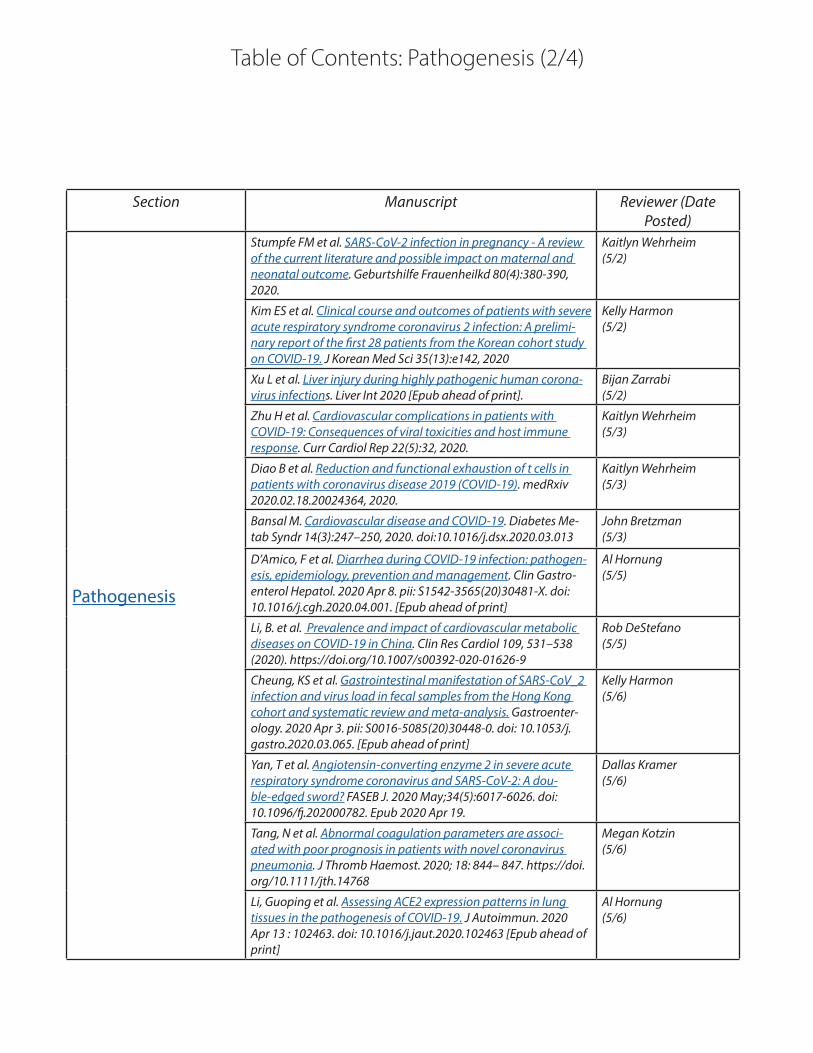
Table of Contents: Pathogenesis (2/4)
Section Manuscript Reviewer (Date Posted)
Pathogenesis
Stumpfe FM et al. SARS-CoV-2 infection in pregnancy - A review of the current literature and possible impact on maternal and neonatal outcome. Geburtshilfe Frauenheilkd 80(4):380-390, 2020.
Kaitlyn Wehrheim(5/2)
Kim ES et al. Clinical course and outcomes of patients with severe acute respiratory syndrome coronavirus 2 infection: A prelimi-nary report of the first 28 patients from the Korean cohort study on COVID-19. J Korean Med Sci 35(13):e142, 2020
Kelly Harmon(5/2)
Xu L et al. Liver injury during highly pathogenic human corona-virus infections. Liver Int 2020 [Epub ahead of print].
Bijan Zarrabi(5/2)
Zhu H et al. Cardiovascular complications in patients with COVID-19: Consequences of viral toxicities and host immune response. Curr Cardiol Rep 22(5):32, 2020.
Kaitlyn Wehrheim(5/3)
Diao B et al. Reduction and functional exhaustion of t cells in patients with coronavirus disease 2019 (COVID-19). medRxiv 2020.02.18.20024364, 2020.
Kaitlyn Wehrheim(5/3)
Bansal M. Cardiovascular disease and COVID-19. Diabetes Me-tab Syndr 14(3):247–250, 2020. doi:10.1016/j.dsx.2020.03.013
John Bretzman(5/3)
D’Amico, F et al. Diarrhea during COVID-19 infection: pathogen-esis, epidemiology, prevention and management. Clin Gastro-enterol Hepatol. 2020 Apr 8. pii: S1542-3565(20)30481-X. doi: 10.1016/j.cgh.2020.04.001. [Epub ahead of print]
Al Hornung(5/5)
Li, B. et al. Prevalence and impact of cardiovascular metabolic diseases on COVID-19 in China. Clin Res Cardiol 109, 531–538 (2020). https://doi.org/10.1007/s00392-020-01626-9
Rob DeStefano(5/5)
Cheung, KS et al. Gastrointestinal manifestation of SARS-CoV_2 infection and virus load in fecal samples from the Hong Kong cohort and systematic review and meta-analysis. Gastroenter-ology. 2020 Apr 3. pii: S0016-5085(20)30448-0. doi: 10.1053/j.gastro.2020.03.065. [Epub ahead of print]
Kelly Harmon(5/6)
Yan, T et al. Angiotensin-converting enzyme 2 in severe acute respiratory syndrome coronavirus and SARS-CoV-2: A dou-ble-edged sword? FASEB J. 2020 May;34(5):6017-6026. doi: 10.1096/fj.202000782. Epub 2020 Apr 19.
Dallas Kramer(5/6)
Tang, N et al. Abnormal coagulation parameters are associ-ated with poor prognosis in patients with novel coronavirus pneumonia. J Thromb Haemost. 2020; 18: 844– 847. https://doi.org/10.1111/jth.14768
Megan Kotzin(5/6)
Li, Guoping et al. Assessing ACE2 expression patterns in lung tissues in the pathogenesis of COVID-19. J Autoimmun. 2020 Apr 13 : 102463. doi: 10.1016/j.jaut.2020.102463 [Epub ahead of print]
Al Hornung(5/6)

Table of Contents: Pathogenesis (3/4)
Section Manuscript Reviewer (Date Posted)
Pathogenesis
Giamarellos-Bourboulis EJ et al. Complex immune dysregulation in COVID-19 patients with severe respiratory failure. Cell Host Microbe 2020 [Epub ahead of print].
Al Hornung(5/6)
Fanelli V et al. Acute kidney injury in SARS-CoV-2 infected pa-tients. Crit Care 24(1):155, 2020.
Megan Kotzin(5/7)
Zhang Y et al. Interferon-induced transmembrane protein-3 genetic variant rs12252-C is associated with disease severity in COVID-19. J Infect Dis 2020 [Epub ahead of print].
John Levinson(5/7)
Spinato G et al. Alterations in Smell or Taste in Mildly Symp-tomatic Outpatients with SARS-CoV-2 Infection. JAMA 2020 [Epub ahead of print].
Kevin Grudzinski(5/7)
Huang Z et al. Inhibitors of the renin–angiotensin system: The potential role in the pathogenesis of COVID-19. Cardiol J 2020 [Epub ahead of print].
Clara Ledsky(5/8)
Toscano G et al. Guillain-Barre syndrome associated with SARS-CoV-2. N Engl J Med 2020 [Epub ahead of print].
Mira Marchioretto(5/8)
Fogarty H et al. COVID-19 Coagulopathy in Caucasian patients. Br J Haematol 2020 [Epub ahead of print].
Al Hornung(5/8)
Aziz M et al. Elevated interleukin-6 and severe COVID-19: A me-ta-analysis. J Med Virol 2020 [Epub ahead of print].
Dallas Kramer(5/11)
Guo T et al. Cardiovascular Implications of Fatal Outcomes of Patients With Coronavirus Disease 2019 (COVID-19). JAMA Cardiol. Published online March 27, 2020. doi:10.1001/jamacar-dio.2020.1017
Dallas Kramer(5/11)
Li, Hui et al. SARS-CoV-2 and Viral Sepsis: Observations and Hypothesis. Lancet 2020. [Epub ahead of print].
Kaitlyn Wehrheim(5/11)
Reynolds HR et al. Renin-angiotensin-aldosterone system inhibitors and risk of Covid-19. N Engl J Med 2020 [Epub ahead of print].
Rob DeStefano(5/11)
Qin C et al. Dysregulation of immune response in patients with COVID-19 in Wuhan, China. Clin Infect Dis 2020 [Epub ahead of print].
Kaitlyn Wehrheim(5/14)
Arentz M et al. Characteristics and outcomes of 21 critically ill patients with COVID-19 in Washington state. JAMA 2020 [Epub ahead of print].
Samantha Betman(5/14)
Mancia G et al. Renin-angiotensin-aldosterone system block-ers and the risk of Covid-19. N Engl J Med 2020 [Epub ahead of print].
Dallas Kramer(5/14)
Ong EZ et al. A dynamic immune response shapes COVID-19 progression. Cell Host Microbe 2020 [Epub ahead of print].
Al Hornung(5/14)

Table of Contents: Pathogenesis (4/4)
Section Manuscript Reviewer (Date Posted)
Pathogenesis
Ye Q et al. The pathogenesis and treatment of the `Cytokine Storm’ in COVID-19. J Infect 2020 [Epub ahead of print].
Adithya Sivakumar(5/15)
Kim NY et al. Acute Hyperglycemic crises with coronavirus dis-ease-19: Case reports. Diabetes Metab J 44(2):349-353, 2020.
Al Hornung(5/15)
Castagnoli R et al. Severe acute respiratory syndrome corona-virus 2 (SARS-CoV-2) infection in children and adolescents: A systematic review. JAMA Pediatr 2020 [Epub ahead of print].
Samantha Betman(5/15)
Bao L et al. The pathogenicity of SARS-CoV-2 in hACE2 transgen-ic mice. Nature 2020 [Epub ahead of print].
Rob DeStefano(5/20)
Li D et al. Immune dysfunction leads to mortality and organ injury in patients with COVID-19 in China: insights from ERS-COVID-19 study. Signal Transduct Target Ther 5(1):62, 2020.
Kaitlyn Wehrheim(5/20)
Zhang L et al. D-dimer levels on admission to predict in-hospital mortality in patients with Covid-19 [published online ahead of print, 2020 Apr 19]. J Thromb Haemost. 2020;10.1111/jth.14859. doi:10.1111/jth.14859
Samantha Betman(5/20)
Varga Z et al. Endothelial cell infection and endotheliitis in COVID-19. Lancet. 2020. 395(10234): 1417–1418.
Kevin Grudzinski(5/20)
Wölfel, R. et al. Virological assessment of hospitalized patients with COVID-2019. Nature (2020). https://doi.org/10.1038/s41586-020-2196-x
Rob DeStefano(5/22)
Poissy J et al. Lille ICU Haemostasis COVID-19 group. Pulmonary embolism in COVID-19 patients: Awareness of an increased prevalence. Circulation 2020 [Epub ahead of print].
Samantha Betman(6/2)
Pei G et al. Renal Involvement and Early Prognosis in Patients with COVID-19 Pneumonia [published online ahead of print, 2020 Apr 28]. J Am Soc Nephrol. 2020;ASN.2020030276.
Kevin Grudzinski(6/3)
Zhang X et al. Viral and host factors related to the clinical out-come of COVID-19. Nature 2020 [Epub ahead of print].
Kelly Harmon(6/4)
Wang Z. et al. High Fluorescent Lymphocytes Are Increased in COVID-19 Patients. Br J Haematol May 20, 2020 [Epub ahead of print].
Kelly Harmon(6/5)
Schaller T. et al. Postmortem Examination of Patients With COVID-19. JAMA. Published online May 21, 2020. doi:10.1001/jama.2020.8907
Kelly Harmon(6/15)

Table of Contents: Diagnosis (1/1)
Section Manuscript Reviewer (Date Posted)
Diagnosis
Li H, et al. Serum Amyloid A is a biomarker to distinguish the severity and prognosis of coronavirus disease 2019 (COVID-19). J Infect 2020 [Epub ahead of print].
Kelsey T Danley(4/23)
Long C, et al. Diagnosis of the Coronavirus disease (COVID-19): rRT-PCR or CT? Eur J Radiol 126:108961, 2020.
Paul R Parker(4/24)
Breslin N, et al. COVID-19 infection among asymptomatic and symptomatic pregnant women: Two weeks of confirmed pre-sentations to an affiliated pair of New York City hospitals. Am J Obstet Gynecol MFM 2020 [Epub ahead of print].
Wendy Tian(4/27)
Guo L, et al. Profiling early humoral response to diagnose novel coronavirus disease (COVID-19). Clin Infect Dis 2020 [Epub ahead of print].
Hannah Raff(4/27)
Guan WJ, et al. Clinical Characteristics of Coronavirus Disease 2019 in China. New Eng J Med 2020 [Epub ahead of print].
Andy Wu(4/30)
Zhao W et al. Relation between chest CT findings and clinical conditions of coronavirus disease (COVID-19) pneumonia: A multicenter study. American Journal of Roentgenology 2020 214:5, 1072-1077
Ahmet Sakiri(4/30)
Lin C. et al. Comparison of throat swabs and sputum specimens for viral nucleic acid detection in 52 cases of novel coronavirus (SARS-Cov-2)-infected pneumonia (COVID-19). Clin Chem Lab Med 2020 [Epub ahead of print].
Kelsey Danley(5/4)
Liu, W et al. Evaluation of Nucleocapsid and spike protein-based ELISAs for detecting antibodies against SARS-CoV-2. J Clin Micro-biol 2020 [Epub ahead of print].
Paul Parker(5/4)
Cao Y et al. Imaging and clinical features of patients with 2019 novel coronavirus SARS-CoV-2: A systematic review and me-ta-analysis. J Med Virol 2020 [Epub ahead of print].
Emily He(5/10)
Huang Y et al. A preliminary study on the ultrasonic manifesta-tions of peripulmonary lesions of non-critical novel coronavirus pneumonia (COVID-19). SSRN (Published online) 2020.
Nick Sytsma(5/10)
Farkash, EA et al. “Ultrastructural evidence for direct renal infec-tion with SARS-CoV-2.” J Am Soc Nephrol. 2020 [Epub ahead of print]
Hannah Raff(5/19)
Jin Y et al. Diagnostic value and dynamic variance of serum antibody in coronavirus disease 2019 [published online ahead of print, 2020 Apr 3]. Int J Infect Dis. 2020;94:49‐52. doi:10.1016/j.ijid.2020.03.065
Kelsey Danley(5/12)
Table of Contents: Critical Care (1/1)
Section Manuscript Reviewer (Date Posted)
Critical Care
Wu CN, et al. High-flow nasal-oxygenation-assisted fiberoptic tracheal intubation in critically ill patients with COVID-19 pneu-monia: a prospective randomized controlled trial. Br J Anaesth 2020 [Epub ahead of print].
Shyam Desai(4/25)
Greenland, J. R. et al. COVID-19 Infection Implications for Periop-erative and Critical Care Physicians. Anesthesiology 2020 [Epub ahead of print].
Beth Hall(4/27)
L Meng et al. Intubation and ventilation amid the COVID-19 out-break: Wuhan’s experience. Anesthesiology 2020 [Epub ahead of print].
Nick Sytsma(4/28)
S Lie et al. Practical considerations for performing regional anesthesia: lessons learned from the COVID-19 pandemic. Can J Anaesth 2020 [Epub ahead of print].
John Sweeney(4/28)
Peng, Qian-Yi, et al. “Findings of lung ultrasonography of novel corona virus pneumonia during the 2019–2020 epidemic.” Inten-sive care medicine (2020): 1.
Nick Sytsma(5/5)
Liang W et al. Development and Validation of a Clinical Risk Score to Predict the Occurrence of Critical Illness in Hospitalized Patients With COVID-19. JAMA Intern Med. Published online May 12, 2020. doi:10.1001/jamainternmed.2020.2033
Nick Sytsma(5/21)
Sommer, P et al., Initial Clinical Impressions of the Critical Care of COVID-19 Patients in Seattle, New York City, and Chicago., Anesthesia & Analgesia: March 25, 2020 - Volume Publish Ahead of Print - Issue - doi: 10.1213/ANE.0000000000004830
Nick Sytsma(6/10)

Table of Contents: Treatment (1/5)(Divided by Therapy)
Section Manuscript Reviewer (Date Posted)
Treatment- Combination andReview articles
Gautret P, et al. Clinical and microbiological effects of a combi-nation of hydroxychloroquine and azithromycin in 80 COVID-19 patients with at least a six-day follow up: A pilot observational study. Travel Med Infect Dis 101663, 2020.
Christina Brown(4/24)
Marini, J. J., & Gattinoni, L. (2020). Management of COVID-19 Respiratory Distress. JAMA.
Eric Moyer(4/30)
Gautret, P. et al. Hydroxychloroquine and azithromycin as a treatment of COVID-19: results of an open-label non-random-ized clinical trial. International journal of antimicrobial agents, 105949.
Eric Moyer(4/30)
Sanders JM et al. Pharmacologic treatments for coronavirus disease 2019 (COVID-19): A review. JAMA 2020 [Epub ahead of print].
Sarah Sun(5/4)
Wang, M et al. Remdesivir and chloroquine effectively inhibit the recently emerged novel coronavirus (2019-nCoV) in vitro. Cell Res 30, 269–271 (2020). https://doi.org/10.1038/s41422-020-0282-0
Maria Amir(5/7)
Some Drugs for COVID-19. 2020, April 6. Retrieved from https://secure.medicalletter.org/sites/default/files/freedocs/w1595a.pdf
Demetrios Geanon(4/30)
Shi X, et al. Evaluation of antiviral therapies for coronavirus disease 2019 (COVID-19) pneumonia in Shanghai, China. J Med Virol 2020 [Epub ahead of print].
Athena Jane Manatis-Lornel(4/27)
van Rensburg R et al. “Current evidence for directed and support-ive investigational therapies against COVID-19.” Afr J Thoracic Crit Care Med 26(2), 2020. DOI: 10.7196/AJTCCM.2020.v26i2.072
Christi Brown(5/17)
Hung, IF et al. Triple combination of interferon beta-1b, lopina-vir–ritonavir, and ribavirin in the treatment of patients admitted to hospital with COVID-19: an open-label, randomised, phase 2 trial. The Lancet, May 2020 [Epub ahead of print]. DOI:https://doi.org/10.1016/S0140-6736(20)31042-4
Ashley Wehrheim(5/19)
Bhimraj A et al., Infectious Diseases Society of America Guide-lines on the Treatment and Management of Patients with COVID-19, Clinical Infectious Diseases, ciaa478, https://doi.org/10.1093/cid/ciaa478
Emily Chi(5/30)

Table of Contents:Treatment (2/5)
Section Manuscript Reviewer (Date Posted)
Treatment-Hydroxychloroquine
Chen Z, et al. Efficacy of hydroxychloroquine in patients with COVID-19: results of a randomized clinical trial. medRxiv 2020.03.22.20040758, 2020.
Joseph BdeBettencourt(4/23)
Ferner, Robin E., and Jeffrey K. Aronson. “Chloroquine and hy-droxychloroquine in covid-19.” (2020).
Amanda Narkis(5/5)
Borba M, et. al. Effect of High vs Low Doses of Chloroquine Di-phosphate as Adjunctive Therapy for Patients Hospitalized With Severe Acute Respiratory Syndrome Coronavirus 2 (SARS-CoV-2) Infection. JAMA Netw Open. 2020 Apr 24;3(4):e208857. doi: 10.1001/jamanetworkopen.2020.8857.
Joseph BdeBettencourt(4/30)
Chen Jun LD. A pilot study of hydroxychloroquine in treatment of patients with common coronavirus disease-19 (COVID-19). J Zhejiang Univ Med Sci. 2020;49(1):0-0.
Joseph BdeBettencourt(4/27)
Geleris J, Sun Y, Platt J, et al. Observational study of hydroxychlo-roquine in hospitalized patients with COVID-19. N Engl J Med 2020. doi:10.1056/NEJMoa2012410
Kavya Timmireddy(5/23)
Mehra MR et al., Hydroxychloroquine or chloroquine with or without a macrolide for treatment of COVID-19: a multinational registry analysis. Lancet, Published online May 22, 2020; https://doi.org/10.1016/S0140-6736(20)31180-6
Joseph BdeBettencourt(5/30)
Bessière F et al., Assessment of QT Intervals in a Case Series of Patients With Coronavirus Disease 2019 (COVID-19) Infection Treated With Hydroxychloroquine Alone or in Combination With Azithromycin in an Intensive Care Unit. JAMA Cardiol. Published online May 01, 2020. doi:10.1001/jamacardio.2020.1787
Steven Heidt(6/3)
Mercuro NJ et al., Risk of QT Interval Prolongation Associated With Use of Hydroxychloroquine With or Without Concomitant Azithromycin Among Hospitalized Patients Testing Positive for Coronavirus Disease 2019 (COVID-19). JAMA Cardiol. Published online May 01, 2020. doi:10.1001/jamacardio.2020.1834
Steven Heidt(6/4)
Rosenberg ES et al., Association of Treatment With Hydroxychlo-roquine or Azithromycin With In-Hospital Mortality in Patients With COVID-19 in New York State. JAMA. Published online May 11, 2020. doi:10.1001/jama.2020.8630
Steven Heidt & Hannah Raff(6/4)

Table of Contents: Treatment (3/5)
Section Manuscript Reviewer (Date Posted)
Treatment- Lopinavir/Ritonavir
Li, Yi et al. “Efficacy and safety of lopinavir/ritonavir or arbidol in adult patients with mild/moderate COVID-19: an exploratory randomized controlled trial.” medRxiv 2020.03.19.20038984; doi: https://doi.org/10.1101/2020.03.19.20038984..
Demetrio Geanon(5/13)
Zhu, Z et al. (2020). Arbidol monotherapy is superior to lopinavir/ritonavir in treating COVID-19. Journal of Infection.
Demetrios Geanon(5/3)
Cao B, et al. A Trial of Lopinavir-Ritonavir in Adults Hospitalized with Severe Covid-19. N Engl J Med 2020 [Epub ahead of print].
Ashley N Wehrheim(4/26)
Ye XT et al. Clinical efficacy of lopinavir/ritonavir in the treat-ment of Coronavirus disease 2019. Eur Rev Med Pharmacol Sci 24(6):3390-3396, 2020.
Manvita Tatavarthy(5/20)
Treatment- Plasma Therapies
Shen C, et al. Treatment of 5 critically ill patients with COVID-19 with convalescent plasma. JAMA 2020 [Epub ahead of print].
Amanda Narkis(4/24)
Duan K, et al. The feasibility of convalescent plasma ther-apy in severe COVID- 19 patients: A pilot study. medRxiv 2020.03.16.20036145, 2020.
Manvita Tatavarthy(4/26)
Cao W, et al. High-Dose Intravenous Immunoglobulin as a Therapeutic Option for Deteriorating Patients With Coronavirus Disease 2019. Open Forum Infect Dis 7(3):ofaa102, 2020.
Karina Oelerich (4/26)
Ahn, J. et al. Use of Convalescent Plasma Therapy in Two COVID-19 Patients with Acute Respiratory Distress Syndrome in Korea. Journal of Korean Medical Science, 35(14).
Christina Brown(4/30)
Duan K et al. Effectiveness of convalescent plasma therapy in severe COVID-19 patients. Proc Natl Acad Sci USA 2020 [Epub ahead of print].
Ashley Wehrheim(4/30)
Zhang B et al. Treatment With Convalescent Plasma for Critically Ill Patients With SARS-CoV-2 Infection. Chest 2020 [Epub ahead of print].
Sarah Sun(4/30)
Rajendran K et al. Convalescent plasma transfusion for the treat-ment of COVID-19: Systematic review. J Med Virol 2020 [Epub ahead of print]. DOI: 10.1002/jmv.25961
Maria Amir(5/17)
Treatment- Remdesivir
Wang, Yeming et al. “Remdesivir in adults with severe COVID-19: a randomised, double-blind, placebo-controlled, multicentre trial.” The Lancet (2020).
Ashley Wehrheim(5/9)
Grein J et al. Compassionate use of remdesivir for patients with severe Covid-19. New Eng J Med 2020 [E-pub ahead of print].
Ayesan Rewane(4/28)
Treatment- Tocillizumab
Luo O, et al. Tocilizumab treatment in COVID‐19: A single center experience. J Medical Virol, 2020 [Epub ahead of print].
Amanda Narkis(4/26)
Xu, X et al. Effective treatment of severe COVID-19 patients with tocilizumab. ChinaXiv, 202003(00026), v1.
Ashley Wehrheim(4/30)

Table of Contents: Treatment (4/5)
Section Manuscript Reviewer (Date Posted)
Treatment- Tocillizumab (cont.)
Di Giambenedetto S, et al. Off-label use of Tocilizumab in pa-tients with SARS-CoV-2 infection. J Med Virol 2020 [Epub ahead of print]
Maria Amir(5/8)
Morrison, Austin et al. “Letter to the Editor: Acute hypertriglyceri-demia in patients with COVID‐19 receiving tocilizumab.” Journal of Medical Virology (2020).
Amanda Narkis(5/9)
Alattar, R et al. “Tocilizumab for the Treatment of Severe COVID‐19.” Journal of Medical Virology (2020).
Amanda Narkis(5/13)
Colaneri M et al., Tocilizumab for treatment of severe COVID-19 patients: Preliminary results from SMAtteo COvid19 REgis-try (SMACORE). Microorganisms 2020, 8(5), 695; https://doi.org/10.3390/microorganisms8050695
Kavya Timmireddy(5/30)
Treatment- Other
Zha L et al. Corticosteroid treatment of patients with coronavirus disease 2019 (COVID-19). Med J Aust 2020 [Epub ahead of print].
Joseph deBettencourt(5/10)
Dean NE et al. Creating a framework for conducting random-ized clinical trials during disease outbreaks. N Engl J Med 382(14):1366-1369, 2020
Ayesan Rewane(5/4)
Favalli EG et al. COVID-19 infection and rheumatoid arthritis: Faraway, so close! Autoimmun Rev 19(5):102523, 2020.
Danesha Lewis(5/15)
Qing, G et al. “Traditional Chinese and Western Medicines Jointly Beat COVID-19 Pandemic.” Chinese Journal of Integrative Medi-cine (2020).
Sarah Sun(5/13)
Li J et al. Association of Renin-Angiotensin System Inhibitors with Severity or Risk of Death in Patients with Hypertension Hospitalized for Coronavirus Disease 2019 (COVID-19) Infection in Wuhan, China. JAMA Cardiol 2020. Published online April 23, 2020. doi:10.1001/jamacardio.2020.1624
Eric Moyer(4/30)
Li SR, et al. Searching therapeutic strategy of new coronavirus pneumonia from angiotensin-converting enzyme 2: the target of COVID-19 and SARS-CoV. Eur J Clin Microbiol Infect Dis 2020 [Epub ahead of print].
Caleb J Bailie(4/26)
Suba, Z. (2020). Prevention and therapy of COVID-19 via exog-enous estrogen treatment for both male and female patients. Journal of Pharmacy & Pharmaceutical Sciences, 23, 75-85.
Ashley Wehrheim(4/30)
Wang Z et al. Clinical characteristics and therapeutic procedure for four cases with 2019 novel coronavirus pneumonia receiv-ing combined Chinese and Western medicine treatment. Biosci Trends 14(1):64-68, 2020.
Maria Amir(5/6)
Mullard, Asher. “Flooded by the torrent: the COVID-19 drug pipe-line.” The Lancet 395.10232 (2020): 1245-1246.
Joseph deBettencourt(5/5)

Table of Contents: Treatment (5/5)
Section Manuscript Reviewer (Date Posted)
Treatment- Other cont.
Cai, Q. et al. Experimental treatment with favipiravir for COVID-19: an open-label control study. Engineering.
Karina Oelerich(5/1)
Gordon, David E. et al. “A SARS-CoV-2 protein interaction map reveals targets for drug repurposing.” Nature (2020): 1-13.
Ashley Wehrheim(5/6)
Vaduganathan, M et al. “Renin–Angiotensin–Aldosterone System Inhibitors in Patients with Covid-19.” N Engl J Med, 2020, 382:1653-1659; DOI: 10.1056/NEJMsr2005760
Joseph B deBettencourt(5/19)
Cavalli, G et al. “Interleukin-1 blockade with high-dose anakin-ra in patients with COVID-19, acute respiratory distress syn-drome, and hyperinflammation: a retrospective cohort study. “The Lancet Rheumatology, C,May 2020 [Epub ahead of print]. DOI:https://doi.org/10.1016/S2665-9913(20)30127-2
Ashley Wehrheim(5/19)
Adams KK et al., Myth Busters: Dietary Supplements and COVID-19 [published online ahead of print, 2020 May 12]. Ann Pharmacother. 2020;1060028020928052. doi:10.1177/1060028020928052
Joseph B deBettencourt(5/30)

Table of Contents: Vaccine Development (1/2)
Section Manuscript Reviewer (Date Posted)
Vaccine development
Sun C, et al. SARS-CoV-2 and SARS-CoV spike-RBD structure and receptor binding comparison and potential implica-tions on neutralizing antibody vaccine development. bioRxiv 2020.02.16.951723, 2020.
John Sweeney(4/23)
Pang J, et al. Potential rapid diagnostics, vaccine and therapeu-tics for 2019 novel coronavirus (2019-nCoV): A systematic review. J Clin Med 9(3), 2020.
Leah R Greenfield(4/23)
Thanh Le T, et al. The COVID-19 vaccine development landscape. Nat Rev Drug Discov 2020 [Epub ahead of print].
Leah R Greenfield(4/24)
Herst CV, et al. An effective CTL peptide vaccine for Ebola Zaire based on survivors’ CD8+ targeting of a particular nucleocapsid protein epitope with potential implications for COVID-19 vaccine design. bioRxiv 2020.02.25.963546, 2020.
Emily M Beltran(4/26)
Dhama K, et al. COVID-19, an emerging coronavirus infection: advances and prospects in designing and developing vaccines, immunotherapeutics, and therapeutics. Hum Vaccin Immunoth-er 2020 [Epub ahead of print].
Ahmad Gill(4/26)
Prompetchara, E.,et al. Immune responses in COVID-19 and po-tential vaccines: Lessons learned from SARS and MERS epidemic. Asian Pac J Allergy Immunol, 38(1), 1-9, 2020.
Morgan Sturgis(4/27)
Feng, Y. et al. Multi-epitope vaccine design using an immunoin-formatics approach for 2019 novel coronavirus in China (SARS-CoV-2). bioRxiv 2020.03.03.962332, 2020.
Audrey Sung(4/27)
Mckay, P et al. Self-amplifying RNA SARS-CoV-2 lipid nanopar-ticle vaccine induces equivalent preclinical antibody titers and viral neutralization to recovered COVID-19 patients. bioRxiv 2020.04.22.055608, 2020.
Leah Greenfield(5/1)
Behbahani, M et al. In silico Design of novel Multi-epitope recombinant 1 Vaccine based on Coronavirus surface glycopro-tein. COVID-19 preprints 2020.03.10.985499, 2020.
Ahmad Gill(5/1)
Poh, CM et al. Potent neutralizing antibodies in the sera of convalescent COVID-19 patients are directed against con-served linear epitopes on the SARS-CoV-2 spike protein. bioRxiv 2020.03.30.015461, 2020.
Yereida Gallardo(5/1)
Khamsi, R. (2020). If a coronavirus vaccine arrives, can the world make enough?. Nature.
Morgan Sturgis(5/3)
Robson B. Computers and viral diseases. Preliminary bioin-formatics studies on the design of a synthetic vaccine and a preventative peptidomimetic antagonist against the SARS-CoV-2 (2019-nCoV, COVID-19) coronavirus. Comput Biol Med 119:103670, 2020
Johanna Balas & Diana Q Vazquez Parker(5/4)

Table of Contents: Vaccine Development (2/2)
Section Manuscript Reviewer (Date Posted)
Vaccine development
Ramaiah, A et al. “Insights into cross-species evolution of novel human coronavirus 2019-nCoV and defining immune determi-nants for vaccine development.” bioRxiv (2020).
Yereida Gallardo(5/5)
Ahmed, SF et al. “Preliminary identification of potential vaccine targets for the COVID-19 coronavirus (SARS-CoV-2) based on SARS-CoV immunological studies.” Viruses 12, no. 3 (2020): 254.
Pranita Kaginele(5/6)
Basu, A et al. “Strategies for vaccine design for corona virus using Immunoinformatics techniques.” bioRxiv (2020).
Audrey Sung(5/6)
Li L et al. Epitope-based peptide vaccine design and target site characterization against novel coronavirus disease caused by SARS-CoV-2. bioRxiv 2020.02.25.965434, 2020.
Sharice Hall(5/7)
Padron-Regalado, Eriko. “Vaccines for SARS-CoV-2: Lessons from Other Coronavirus Strains.” Infectious diseases and therapy (2020): 1-20.
Ahmad Gill(5/12)
Tian X et al. “Potent binding of 2019 novel coronavirus spike protein by a SARS coronavirus-specific human monoclonal antibody.” Emerg Microbes Infect 9(1):382-385, 2020. DOI: 10.1080/22221751.2020.1729069
Yereida Gallardo(5/17)

Table of Contents: Infection Control/Prevention (1/1)
Section Manuscript Reviewer (Date Posted)
Infection Control/Pre-vention
Qiu, Lin et al. “SARS-CoV-2 is not detectable in the vaginal fluid of women with severe COVID-19 infection.” Clinical Infectious Diseases (2020).
Samantha Betman(5/7)

COVID-19Rush Journal Club
PUBLIC HEALTH
Table of Contents

COVID-19Rush Journal Club
School closure and management practices during coronavirus out-breaks including COVID-19: a rapid systematic reviewRussell Viner et al.The Lancet Child & Adolescent HealthApril 6, 2020DOI: https://doi.org/10.1016/S2352-4642(20)30095-X
Purpose To understand the effectiveness of school closure and other school-based social distancing practices in affecting infection rates and transmission of coronaviruses during disease outbreaks.
Study design Systematic Review (n=16 studies included)Level of evidence
Level 1
Methods Authors performed a systematic review of pre-prints and papers available in PubMed, the WHO Global Database on COVID-19, and medRxiv to assess the effects of school closure during coronavirus outbreaks with disease transmis-sion.
Findings Sixteen studies included from a total of 618 that predominantly covered coronavirus outbreaks in Asian countries during the early 21st century. School closures on their own may be insufficient to mitigate coronavi-rus spread, versus the influenza virus where school closures show to be effective as primary mitigation tactic. Authors acknowledged a UK study, which estimates that school closures may reduce total COVID-19 deaths by only 2-4% and highlights the need for school dismissal to prevent a serious outbreak.
ClinicalImplications
School closures drastically reduce influenza transmission in the general pop-ulation, however not with coronavirus. Data on coronavirus infections has shown school closures to be much less impactful as a primary mitigation factor in decreasing transmission in the general public.
Limitations Seven of sixteen included studies have not been peer reviewed. Only one modeling study (not peer reviewed) compared school closures with other mitigating factors for COVID-19.
PUBLIC HEALTH
Review by Josh Doppelt, MS4 Table of Contents

COVID-19Rush Journal Club
Covid-19: control measures must be equitable and inclusive
Zackary Berger et al.The BMJApril 21, 2020DOI: https://doi.org/10.1136/bmj.m1141
Purpose Highlight necessary efforts within COVID-19 response, notably free testing and employment rights, to most inclusively address needs of all individuals, including vulnerable populations
Study design EditorialLevel of evidence
Not applicable
Methods Not applicableFindings Provision of free testing for COVID-19, and healthcare for COVID-19-related con-
cerns and pre-existing conditions, for all individuals is vital. This will support com-munity mitigation efforts and relieve burden on emergency departments and walk-in clinics. Support should be provided for organizations addressing lack of housing, food and medication, especially with forced closures of supporting institutions (ex. schools). Employees should be provided financial support, in the event they require home quarantine or sick days, in order to best optimize community mitigation ef-forts. Healthcare workers and first responders should be provided the appropriate protective equipment and mental health resources, to address both physical and mental wellbeing.
ClinicalImplications
Healthcare, for COVID-19-related concerns and pre-existing conditions, should be provided to everyone, including the homeless , udocumented immigrants , and others with poor healthcare access. Employment rights and protection should be provided. This includes allowing employees to prioritize individual and communi-ty health without fear of financial loss, as well as providing appropriate protective equipment and mental health resources for healthcare workers.
Limitations The article is limited in its study design; as an editorial piece, it has strong referenc-es yet lacks higher level of evidence that could limit applicability. It highlights key considerations for city and state officials to address in their COVID-19 responses, but may require further analyses of their unique populations to better inform the details of most inclusive interventions.
PUBLIC HEALTH
Review by Laura Hurley, MS3Table of Contents

COVID-19Rush Journal Club
COVID-19 and African Americans
Clyde W. YancyJAMAApril 15, 2020DOI: 10.1001/jama.2020.6548
Purpose Evaluate particular risk factors and social determinants of health negatively affecting health outcomes for African American patients with COVID-19.
Study design Perspective/viewpoint articleLevel of evidence
N/A
Methods Review of available evidence to support the central claim: underserved minorities are developing COVID-19 more frequently and dying dispropor-tionately (a 6-fold increase in the rate of death for African-American patients infected with coronavirus).
Findings In Chicago, greater than 50% of COVID-19 cases and approximately 70% of COVID-19 deaths involve black individuals, even though blacks only make up 30% of the population. Additionally, these deaths are more concentrated in 5 neighborhoods in Chicago’s South Side. This spread is similar in both Louisiana, Michigan, and New York City. There is a higher prev-alence of known risk factors for COVID-19 complications (hypertension, dia-betes, obesity, cardiovascular disease) in black patients. Social determinents of health further exacerbate risk factors (i.e. higher housing density impairing social distancing, poor access to healthy foods affecting immunity).
ClinicalImplications
This viewpoint/perspective lends a historical and epidemiological dimension to the factual evidence that outcomes for African-Americans suffering with COVID-19 are worse. Author addresses this severe COVID-19 infection and death in black individuals to low socio-economic status and health care dis-parity.
Limitations This is a viewpoint article and thus may have inherent bias from the author and also uses epidemiological studies which are prone to effects from con-founding variables.
PUBLIC HEALTH
Review by Eiftu Haile, MS4 Table of Contents

COVID-19Rush Journal ClubA brief telephone severity scoring system and therapeutic living cen-ters solved acute hospital-bed shortage during the COVID-19 out-break in Daegu, KoreaShin-Woo Kim et al.J Korean Med SciApril 20, 2020DOI: https://doi.org/10.3346/jkms.2020.35.e152
Purpose To study the efficacy of telephone-based screening to determine the level of care coronavirus-positive individuals received during a hospital bed shortage in the COVID-19 pandemic.
Study design Case series (n=6610)Level of evidence
4
Methods Physicians in Daegu (Korea) developed a remote screening tool for COVID-posi-tive individuals during a hospital-bed shortage. This telephone screener utilized a point system for disease symptom severity, age, pre-existing conditions, and social factors. Based on point totals, individuals were sent to a tertiary hospital, public hospital, or therapeutic living center to quarantine. This protocol was put in place on February 29th presumably through March 29th, 2020.
Findings Only 81/3033 (2.67%) patients admitted to therapeutic living centers for quarantining were transferred to the hospital for higher level care. Only three patients out of 6,610 cumulative cases, between February 18th and March 29th, died at home while awaiting a hospital bed.
ClinicalImplications
In the event of hospital bed shortage, health systems will need to quickly deter-mine which level of care each patient needs in order to be efficient with resourc-es. While this study has numerous limitations, remote screening tools may be an effective way to triage patients with known COVID-19 prior to hospital arrival. The US does not have sufficient community screening for most individuals to know their COVID-19 status, which hinders this protocol from being effective in the US. However, if community testing increases, remote screening like this may warrant further study.
Limitations Authors did not specify where the COVID-positive patients are tested (communi-ty vs in hospital setting). They did not have a clear end date listed for the study, so we do not know how long this protocol was studied for. The data also does not clearly indicate that remote screening decreased the number of persons waiting for hospital beds in the setting of a decrease in new COVID-19 cases in Daegu.
PUBLIC HEALTH
Review by Josh Doppelt, MS4Table of Contents
COVID-19Rush Journal Club
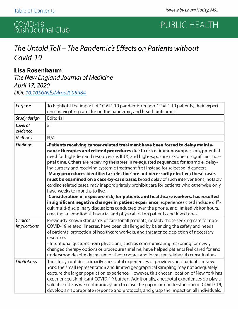
The Untold Toll – The Pandemic’s Effects on Patients without Covid-19
Lisa RosenbaumThe New England Journal of MedicineApril 17, 2020DOI: 10.1056/NEJMms2009984
Purpose To highlight the impact of COVID-19 pandemic on non-COVID-19 patients, their experi-ence navigating care during the pandemic, and health outcomes.
Study design EditorialLevel of evidence
5
Methods N/AFindings -Patients receiving cancer-related treatment have been forced to delay mainte-
nance therapies and related procedures due to risk of immunosuppression, potential need for high-demand resources (ie. ICU), and high-exposure risk due to significant hos-pital time. Others are receiving therapies in re-adjusted sequences; for example, delay-ing surgery and receiving systemic treatment first instead for select solid cancers. -Many procedures identified as ‘elective’ are not necessarily elective; these cases must be examined on a case-by-case basis; broad delay of such interventions, notably cardiac-related cases, may inappropriately prohibit care for patients who otherwise only have weeks to months to live.-Consideration of exposure risk, for patients and healthcare workers, has resulted in significant negative changes in patient experience; experiences cited include diffi-cult multi-disciplinary discussions conducted over the phone, and limited visitor hours, creating an emotional, financial and physical toll on patients and loved ones.
ClinicalImplications
Previously known standards of care for all patients, notably those seeking care for non-COVID-19 related illnesses, have been challenged by balancing the safety and needs of patients, protection of healthcare workers, and threatened depletion of necessary resources.- Intentional gestures from physicians, such as communicating reasoning for newly changed therapy options or procedure timeline, have helped patients feel cared for and understood despite decreased patient contact and increased telehealth consultations.
Limitations The study contains primarily anecdotal experiences of providers and patients in New York; the small representation and limited geographical sampling may not adequately capture the larger population experience. However, this chosen location of New York has experienced significant COVID-19 burden. Additionally, anecdotal experiences do play a valuable role as we continuously aim to close the gap in our understanding of COVID-19, develop an appropriate response and protocols, and grasp the impact on all individuals.
PUBLIC HEALTH
Review by Laura Hurley, MS3Table of Contents

COVID-19Rush Journal Club
Perceptions and behavioural responses of the general public during the COVID-19 pandemic: Cross-sectional survey of UK adults
Christina J Atchison et al.medRxiv preprintApril 3, 2020DOI: https://doi.org/10.1101/2020.04.01.20050039
Purpose To examine risk perceptions, behavioral responses, and intention to comply with non-pharmaceutical interventions (NPIs) of the UK adult population during the early phase of the COVID-19 pandemic in the UK.
Study design Cross-sectional survey (n=2108)Level of evidence
Level 3
Methods A survey was emailed to a nationally representative sample of UK adults and adminis-tered by YouGov (a market research company) between March 17-18, 2020. The ques-tionnaire had four components: socio-demographic characteristics, risk perceptions towards COVID-19, preventative behaviors, and willingness and ability to self-isolate. Data was collected and sent to Imperial College London research team for analysis.
Findings 94.2% (n=1,992) of respondents reported at least one preventative measure: 85.8% washed their hands more frequently, 56.5% avoided crowded areas and 54.5% avoided social events. Adoption of social distancing was higher in those aged over 70 com-pared to ages 18-34. Those with the lowest household income were 6 times less likely to be able to work from home (adjusted odds ratio, aOR: 0.16) and 3 times less likely to be able to self-isolate (aOR: 0.31). Ability to self-isolate was also lower in black and minority ethnic groups (aOR: 0.47). Willingness to self-isolate was high across all re-spondents.
ClinicalImplications
· This study highlights the barriers that those in lower socio-economic groups face and predicts that the impact of this pandemic will be felt unequally in our society. The study recommends governments implement appropriate social and economic policies to mitigate this.· Incorporating differences in non-pharmaceutical intervention adherence among so-cio-economic subpopulations can improve mathematical models of transmission and outcomes.
Limitations The study was limited by the online survey, which responses of those without in-ternet access were under-represented. Second, the survey tool used predominantly closed-ended questions, limiting the ability explore responses in more depth. Final-ly, using self-report data has limitations including honesty, introspective ability, and question interpretation. This study still has to be peer-reviewed.
PUBLIC HEALTH
Review by Katherine Tehaney, MS1Table of Contents
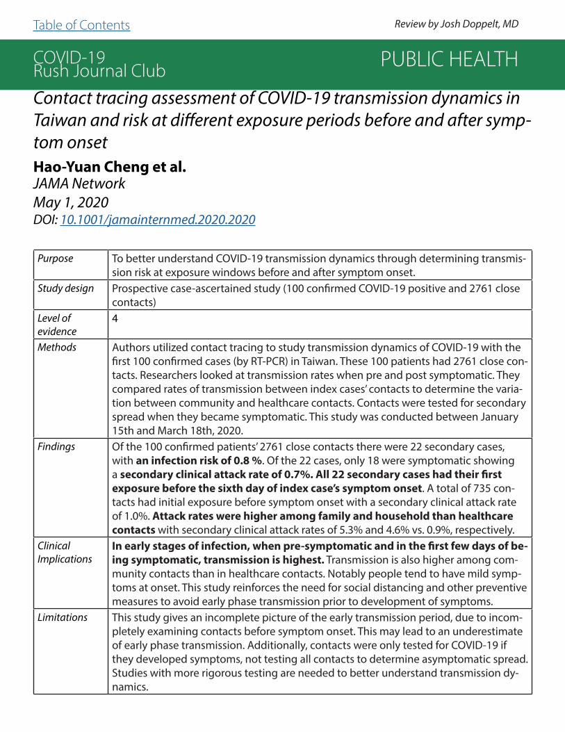
COVID-19Rush Journal ClubContact tracing assessment of COVID-19 transmission dynamics in Taiwan and risk at different exposure periods before and after symp-tom onsetHao-Yuan Cheng et al.JAMA NetworkMay 1, 2020DOI: 10.1001/jamainternmed.2020.2020
Purpose To better understand COVID-19 transmission dynamics through determining transmis-sion risk at exposure windows before and after symptom onset.
Study design Prospective case-ascertained study (100 confirmed COVID-19 positive and 2761 close contacts)
Level of evidence
4
Methods Authors utilized contact tracing to study transmission dynamics of COVID-19 with the first 100 confirmed cases (by RT-PCR) in Taiwan. These 100 patients had 2761 close con-tacts. Researchers looked at transmission rates when pre and post symptomatic. They compared rates of transmission between index cases’ contacts to determine the varia-tion between community and healthcare contacts. Contacts were tested for secondary spread when they became symptomatic. This study was conducted between January 15th and March 18th, 2020.
Findings Of the 100 confirmed patients’ 2761 close contacts there were 22 secondary cases, with an infection risk of 0.8 %. Of the 22 cases, only 18 were symptomatic showing a secondary clinical attack rate of 0.7%. All 22 secondary cases had their first exposure before the sixth day of index case’s symptom onset. A total of 735 con-tacts had initial exposure before symptom onset with a secondary clinical attack rate of 1.0%. Attack rates were higher among family and household than healthcare contacts with secondary clinical attack rates of 5.3% and 4.6% vs. 0.9%, respectively.
ClinicalImplications
In early stages of infection, when pre-symptomatic and in the first few days of be-ing symptomatic, transmission is highest. Transmission is also higher among com-munity contacts than in healthcare contacts. Notably people tend to have mild symp-toms at onset. This study reinforces the need for social distancing and other preventive measures to avoid early phase transmission prior to development of symptoms.
Limitations This study gives an incomplete picture of the early transmission period, due to incom-pletely examining contacts before symptom onset. This may lead to an underestimate of early phase transmission. Additionally, contacts were only tested for COVID-19 if they developed symptoms, not testing all contacts to determine asymptomatic spread. Studies with more rigorous testing are needed to better understand transmission dy-namics.
PUBLIC HEALTH
Review by Josh Doppelt, MDTable of Contents

COVID-19Rush Journal ClubFirst-wave COVID-19 transmissibility and severity in China outside Hubei after control measures, and second-wave scenario planning: A modelling impact assessmentKathy Leung et al.The LancetApril 8, 2020DOI: https://doi.org/10.1016/S0140-6736(20)30746-7
Purpose To assess transmissibility and fatalities in response to non-pharmaceutical intervention versus relaxation during the first wave of COVID-19 in China, outside of Hubei (epicenter) and to esti-mate a model for a second wave upon reopening the economy and relaxing social restrictions.
Study design Modeling study (health records for 4 cities and 10 provinces are included)
Level of evidence
5
Methods Authors utilized publicly available health information to chart an epidemic curve for Shanghai, Beijing, Wenzhou, and Shenzhen between January 20th and February 20th, 2020. They collect-ed patient and hospital data for travel and contact with people from Hubei province (epicen-ter) as well as time from first symptoms to healthcare setting presentation or death to assess instantaneous reproduction number (Rt) and confirmed case-fatality risk (cCFR) in relation to change in policy (i.e., beginning social distancing, stopping school, and reopening portions of the economy). Rt was defined as the average number of secondary cases generated by one primary case with symptom onset on day t.
Findings - Non-pharmaceutical interventions, including social distancing and population behav-ioral change, decreased fatalities and Rt below 1.- Opening the economy quickly led to a spike, with Rt above 1.0 and increased cCFR. At this point, the duration of intervention needed to reduce Rt was greater than the eco-nomic opening period.- Intervention measures to counteract the rise in Rt took a much longer time to bring Rt down below 1.0 than the duration of relaxed economic policies.
ClinicalImplications
- As Rt increases above 1.0 it takes exponentially longer to get those levels back below 1.0, making tracking those levels essential to developing policies to intervene.- Relaxing control measures leads to an exponential rise in Rt.- Maintaining an Rt less than or equal to 1.0 is likely the optimal strategy to reduce a surge in cases until a vaccine is developed.- Through increased COVID-19 screening it may be possible to tightly monitor the Rt and en-able policy makers to balance economic needs with risk of a worsened second wave.
Limitations Data collection was not universal across cities and provinces, and as such categories of data collected varied, as did the quality of data. We must question reporting quality and accuracy of Rt and cCFR, especially as one province reported a cCFR of 0.00, which is highly unrealistic considering the scope and impact of this pandemic. This study also only shows correlations between specific policy changes with Rt and cCRT, not direct causation.
PUBLIC HEALTH
Review by Josh Doppelt, MDTable of Contents

COVID-19Rush Journal Club
Variation in COVID-19 Hospitalizations and Deaths Across New York City BoroughsRishi Wadhera et al.Journal of the American Medical AssociationApril 29, 2020DOI: 10.1001/jama.2020.7197
Purpose To understand patterns in COVID-19 hospitalizations and deaths in five New York City boroughs (Bronx, Brooklyn, Manhattan, Queens, and Staten Island) in relation to race and socioeconomic status.
Study design Research Letter (n = 8,398,748)
Level of evidence
Level 5
Methods The following available data bases were used to outline patterns of COVID-19 out-comes through April 25, 2020: American Community Survey to understand popula-tion characteristics; American Hospital Association 2016 file and manual search for hospitals and their capacity; New York City Department of Health and Mental Hy-giene for number of hospitalizations and deaths per borough. Both lab confirmed and probable COVID-19 cases were included.
Findings The Bronx had the highest deaths and hospitalization (634 and 224/100,000 persons respectively) of all boroughs and consisted of the highest percent Afri-can Americans (38.3%), lowest household median income ($38,467), and lowest proportion of bachelor’s degree holders (20.7%). Manhattan was least affected with 331 hospitalizations and 122 deaths per 100,000, demonstrating the highest median household income ($85,066) and percentage of bachelor’s degree holders (61.4%). No notable association found between lack of hospital beds, percentage of elderly adults, number of hospitals or hospital beds and hospitalization or death.
ClinicalImplications
These findings help illustrate disproportionate morbidity and mortality due to COVID-19 in African American neighborhoods. This study shows an association of poor outcomes for COVID-19 infection in New York City with African American race, low household income, and lower educational level. Understanding these patterns may inform preventive polices to mitigate ongoing spread.
Limitations This study’s ecological design only shows correlation. Additionally, rate of test-ing for COVID-19 was not evaluated, which would have been important to un-derstand given the variability in testing.
PUBLIC HEALTH
Review by Josh Doppelt, MD Table of Contents

COVID-19Rush Journal Club
Impact of school closures for COVID-19 on the US health-care work-force and net mortality: a modelling studyJude Bayham et al.The Lancet Public HealthApril 3, 2020DOI: https://doi.org/10.1016/S2468-2667(20)30082-7
Purpose To measure child-care obligations for US health-care workers arising from school closures and assess the contribution of health-care workers in reducing mortality to calculate the net mortality reduction.
Study design Modelling analysis
Level of evidence
Level 5
Methods Data from the US Current Population Survey was utilized to identify areas of the health-care workforce most impacted by school closures. The goal of the modeling was to identify the conditions where school closures would no longer help to save lives due to health-care workers having to stay home because of child-care obliga-tions. The following were assumed: 15% case reduction from school closures, 2.0% baseline mortality rate for COVID-19.
Findings 28.8% (95% CI 28.5-29.1) of the health-care workforce requires child-care for chil-dren aged 3-12 years and 15% (95% CI 14.8-15.2) do not have a non-working adult or a child 13 years or older to provide care at home. States with the greatest child-care obligations include Utah (35.4%, 95% CI 32.9-37.9), Louisiana (35.0%, 33.1-36.8), and Missouri (34.0%, 31.5-36.5). The infection mortality rate of COVID-19 increases from 2.0% to 2.35% when the health-care workforce declines by 15.0% with school closures.
ClinicalImplications
This study highlights the obstacles faced by those in lower socio-economic groups and predicts the unequal impact of this epidemic on varying groups in society. It recommends that governments implement appropriate social and economic poli-cies to mitigate this disproportionate impact.
Limitations The analysis did not model family members outside of the household, such as neighbors or friends, that may help care for children in a primary caregivers ab-sence. The information about social distancing policies is based on models of influ-enza, in which children are a vulnerable group for morbidity, whereas children do not appear to be a sensitive group to COVID-19.
PUBLIC HEALTH
Review by Kat Tehaney, MS1 Table of Contents

COVID-19Rush Journal Club
Awareness, Attitudes, and Actions Related to COVID-19 Among Adults with Chronic Conditions at the Onset of the U.S. OutbreakMichael Wolf et al.Annals of Internal MedicineApril 9, 2020DOI: https://doi.org/10.7326/M20-1239
Purpose To assess COVID-19 awareness, knowledge, attitudes, and related behaviors among vulnerable U.S. adults.
Study design Cross-sectional survey linked to the Chicago COVID-19 Comorbidities (C3) Survey. (n=630).
Level of evidence
Level 2
Methods Authors gathered data from the ongoing Chicago COVID-19 Comorbidities Survey to interview high risk, older adults with at least 1 or more chronic conditions who would be at greater risk for COVID-19. During the period between March 13th to March 20th, participants were asked to answer items from a questionnaire used to study prior outbreaks.
Findings 28.3% of the participants could not correctly identify symptoms and 30.2% did not know methods to prevent infection. 24.6% of adults believed that they were “not at all likely” to get the virus, and 21.9% reported that COVID-19 did not impact their daily routine. Women, African American, and Hispanic persons, individuals with Limited English Proficiency (LEP), living below the poverty level, lower health literacy, and unmarried were more likely to believe that it was “not at all likely” they would contract COVID-19. African American participants, individuals living below the poverty level, and those with low health literacy were more likely to be less worried about COVID-19, to not believe they would become infected, and to feel less prepared for an outbreak.
ClinicalImplications
Existing efforts are not adequate in reaching vulnerable populations and more public health efforts need to be taken to help disseminate critical information about COVID-19.
Limitations This is a cross-sectional study of adults with underlying health conditions in 1 American city during the initial week of the COVID-19 outbreak and what was reported has likely changed considerably.
PUBLIC HEALTH
Review by Timothy Huang, MS1 Table of Contents

COVID-19Rush Journal Club
COVID-19 and smoking: A systematic review of the evidence
Constantine Vardavas et al.Tobacco Induced DiseasesMarch 20, 2020DOI: https://doi.org/10.18332/tid/119324
Purpose To evaluate the association between smoking and COVID-19 outcomes including severity of disease, need for mechanical ventilation, need for ICU hospitalization, and death.
Study design Literature Review (n=41-1099)
Level of evidence
Level 4
Methods The literature search was conducted on March 17, 2020, using two databases (PubMed, ScienceDirect), with the following search terms: [‘smoking’ OR ‘tobacco’ OR ‘risk factors’ OR ‘smoker*’] AND [‘COVID-19’ OR ‘COVID 19’ OR ‘novel coronavirus’ OR ‘sars cov-2’ OR ‘sars cov 2’]. Studies published in 2019 and 2020 were included. A total of 71 studies were retrieved, of which 66 were excluded after full-text screening, leaving five studies that were includ-ed. Retrospective and prospective methods were used. The time frame of all five studies included the first two months of the COVID-19 pandemic (December 2019, January 2020). All studies were conducted in China, four in Wuhan and one across multiple provinces in mainland China. Sample sizes of the included studies range from 41 to 1099 patients.
Findings Guan et al. (n=1099) found that smokers were 2.4 times more likely to be admitted to an ICU, require mechanical ventilation, or die when compared to non-smokers (relative risk, RR: 2.4). Liu et al. (n=78) reported that in those with a history of smoking, there was in-creased likelihood of adverse outcomes and risk of disease progression (p=0.018). Zhou et. al. (n=191) reported a no statistically significant difference in mortality between smokers and non-smokers (p=0.21). Huang et al. (n=41) found no statistically significant difference in likelihood of ICU admission between smokers and non-smokers (p=0.31). Zhang et al. (n=140) also showed no statistically significant difference in severity of infection among smokers vs. non-smokers (odds ratio, OR: 2.23; p=0.2).
ClinicalImplications
Although the data presented in this review require confirmation and adjustment for other risk factors, it should be noted that smoking may increase the rate of adverse events and outcome, such as ICU admission, mechanical ventilation, or death; therefore, smokers should strictly adhere to social distancing guidelines.
Limitations The reviewed studies reported conflicting results; therefore, minimal conclusions may be drawn from this systematic review. All the studies included were conducted in China, which has a higher rate of smoking than countries like the United States (25.6% vs. 21.8%), limiting generalizability. In addition, some of the studies had a relatively small number of participants, thereby limiting their application to larger populations.
PUBLIC HEALTH
Review by Kelly Harmon, MS1 Table of Contents

COVID-19Rush Journal Club
Risk factors of critical & mortal COVID-19 cases: A systematic litera-ture review and meta-analysisZhaohai Zheng et al.Journal of InfectionApril 23, 2020DOI: 10.1016/j.jinf.2020.04.021
Purpose To find risk factors for the progression of COVID-19 to help reduce the risk of critical illness and death.
Study design Meta-analysis (n=3027)
Level of evidence
Level 1
Methods Studies published between January 1, 2020 and March 20, 2020, by searching Pubmed, Embase, Web of Science, and CNKI were selected. The search terms and relative vari-ants included: “severe acute respiratory syndrome coronavirus 2”, “Wuhan coronavirus”, “Wuhan seafood market pneumonia virus”, “COVID-19”, “COVID19”, “coronavirus disease 2019 virus”, “SARS-CoV-2”, “SARS2”, “2019-nCoV” or “2019 novel coronavirus” and “Mor-talities”, “Mortality”, “Fatality”, “Death”, “acute respiratory distress syndrome (ARDS)” or “ICU”. Authors also reviewed the references of included articles to guarantee the com-prehensiveness and accuracy of research. Thirteen studies were included with a total of 3027 SARS-CoV-2 positive patients. Inclusion criteria consisted of study sample size greater than 20, confirmed infection by 2019 novel coronavirus, and presence of criti-cal illness (defined as death) and non-critical illness. The following study designs were included: randomized controlled trials, nonrandomized controlled trials, case‐control studies, cohort studies, cross‐sectional studies, and case reports.
Findings Disease progression was associated with male gender (p<0.00001), age >65 years (p<0.00001), and current smoking (p=0.0006). Critical patients were more likely to have underlying disease such as diabetes (p< 0.00001), hypertension (p=0.0002), car-diovascular disease (p<0.00001), or respiratory disease (p<0.00001).
ClinicalImplications
The study provides guidance on vulnerable populations with COVID-19. The following individuals should adhere to strict social distancing guidelines given increased risk for disease progression: male, older than 65, or smokers. Individuals with the following co-morbidities should adhere to strict social distancing guidelines due to increased risk of critical illness: hypertension, diabetes, cardiovascular disease, and respiratory disease.
Limitations Many of the articles included were cross-sectional studies, limiting the study’s casual association. Much of the patient population was Chinese, which may limit the studies broader application amongst other races and nationalities.
PUBLIC HEALTH
Review by Kelly Harmon, MS1 Table of Contents

COVID-19Rush Journal Club
Does comorbidity increase the risk of patients with COVID-19: evi-dence from meta-analysisBolin Wang et al.Aging (Albany NY)April 8, 2020DOI: https://doi.org/10.18632/aging.103000
Purpose To assess the prevalence of comorbidities in COVID-19 patients and the risk of underlying disease in these infected patients.
Study design Meta-analysis (n=1558)
Level of evidence
Level 1
Methods Meta-analysis was performed according to the Preferred Reporting Items for Systematic re-views and Meta-analysis (PRISMA) statement. Relevant literature was identified using PubMed (Medline), EMBASE, Springer, Web of Science, and Cochrane Library databases up to March 1, 2020, using the following terms: “2019-nCoV” or “Coronavirus” or “COVID-19” or “SARS-CoV-2” or “2019-nCoV” or “Wuhan Coronavirus.” Two participants conducted separate literature screenings, data extraction, and literature quality evaluations, with resolution of differenc-es through discussion or third analyst. Researchers chose studies reporting the relationship between comorbid health conditions and outcomes related to COVID-19. Inclusion criteria consisted of the following comorbidities: hypertension, diabetes, chronic obstructive pulmo-nary disease (COPD), liver disease, malignancy, renal disease, cardiovascular disease, and cere-brovascular disease. Studies were excluded if the design was that of a case report, review, or discussion summary. Studies with insufficient data and those that did not stratify patients by degree of severity were also excluded. Six retrospective studies from China met the inclusion criteria with a total of 1558 patients.
Findings COVID-19 patients with hypertension, diabetes, or COPD demonstrated increased risk of dis-ease exacerbation (p<0.001). Cardiovascular disease was found to be a significant risk factor for infection with COVID-19 (p<0.001). Concurrent cerebrovascular disease was associated with severe COVID-19 (p=0.002). Previous history of liver disease and presence of a malig-nant tumor or kidney disease did not significantly increase the risk of disease progression (p=0.070).
ClinicalImplications
Comorbidities are risk factors for COVID-19 patients. Individuals diagnosed with hypertension, diabetes, COPD, cardiovascular disease, and cerebrovascular disease should adhere to strict social distancing guidelines given their increased risk for COVID-19 infection and disease pro-gression.
Limitations Criteria for severe vs. non-severe patients was not uniform amongst the studies included. In addition, some studies included patients diagnosed with more than one comorbidity, thereby interfering with the ability to correlate COVID-19 infection and severity to one specific diagno-sis. All the studies were comprised entirely of Chinese participants, which may limit the gener-alizability of study findings.
PUBLIC HEALTH
Review by Kelly Harmon, MS1 Table of Contents

COVID-19Rush Journal Club
Early estimates of the indirect effects of the COVID-19 pandemic on ma-ternal and child mortality in low-income and middle-income countries: a modelling studyTimothy Roberton et al.The Lancet Global Healthdate publishedDOI: https://doi.org/10.1016/S2214-109X(20)30229-1
Purpose To estimate the additional maternal and under-5 child deaths resulting from the potential disruption of health systems and decreased access to food.
Study design Modelling analysis
Level of evidence
Level 5
Methods Three scenarios were modelled with varying provisions of health services and uti-lizations of health services, with small, moderate, and severe reductions. The Lives Saved Tool (LiST) was used to estimate the additional maternal and under-5 child deaths under each scenario in 118 low- and middle-income countries for 1-month period and extrapolated for 3, 6, and 12 months.
Findings Results from small (scenario 1) to severe reductions (3) showed an additional 9.8-44.7% increase in under-5 child deaths and 8.3-38.6% increase in maternal deaths per month. The largest contribution to maternal death across all scenarios was due to disruption in parental administration of uterotonics, antibiotics, anticonvulsants, and clean birth environments, accounting for approximately 60% of the addition-al maternal deaths. The largest contributor to child death across all scenarios was reduced coverage of antibiotics for pneumonia and neonatal sepsis, and oral rehy-dration solution for diarrhea, accounting for ~41% of additional child deaths. The increase in wasting prevalence accounted for 18-23% of additional child deaths.
ClinicalImplications
This study primarily demonstrates that government responses, whether intentional or unintentional, will have an impact on maternal and child health. It also shows that not all health interventions are equally susceptible or will have equal effects, therefore some interventions should be prioritized. Finally, it highlights the need for interventions to mitigate increases in wasting.
Limitations Analysis was limited by the LiST program being constrained to a defined set of health-sector interventions, thereby the effects of income, agriculture, or food markets on wasting were directly modified instead of estimated. Also, LiST was not able to capture individual infectious disease dynamics or predict potential effects of secondary outbreaks. These limitations, however, would result in an overly con-servative estimate and actual increases in mortality would be greater.
PUBLIC HEALTH
Review by Kat Tehaney, MS1 Table of Contents

COVID-19Rush Journal Club
Mental Health Status Among Children in Home Confinement During the Coronavirus Disease 2019 Outbreak in Hubei Province, ChinaXinyan Xie et al.Journal of the American Medical Association PediatricsApril 24, 2020DOI: 10.1001/jamapediatrics.2020.1619
Purpose To investigate presence of anxiety and depressive symptoms among students in Hubei prov-ince, China following nationwide school closures.
Study design Cross-sectional survey
Level of evidence
Level 3
Methods A survey was sent to guardians of 2330 students from two schools in Hubei province in grades 2-6. The survey was completed between February 28, 2020 to March 5, 2020, ~1 month follow-ing school closures. In total, 1784 participants (56.7% boys) completed the survey. The Chil-dren’s Depression Inventory-Short Form (CDI-S) and the Screen for Child Anxiety Related Emo-tional Disorders were used to assess symptoms of depression and anxiety. Generalized linear regressions were used for continuous variables and logistic regressions for binary variables.
Findings 22.6% and 18.9% of students reported symptoms of depression and anxiety respectively. Students living in Wuhan had significantly higher CDI-S scores compared to those in Huangshi (β, 0.092), with a greater risk of depressive symptoms (odds ratio, OR: 1.426). No significant association was found between demographic characteristics and symptoms of anxiety. Those who reported feeling slightly or not worried about infection with COVID-19 had significantly lower CDI-S scores (β, −0.184) and a decreased risk of depressive symptoms (OR: 0.521) than individuals feeling quite worried. In addition, students who were not optimistic about the epidemic were found to have significantly higher CDI-S scores (β, 0.367) and an increased risk of depressive symptoms (OR: 2.262) than those who were quite optimistic.
ClinicalImplications
This study demonstrates that symptoms of depression and anxiety are experienced to a great-er degree by students worried about COVID-19 infection and by those lacking optimism about the epidemic. This study suggests that pandemics have the capacity to negatively influ-ence a child’s mental health, particularly when closure of schools is required, resulting in home confinement and social isolation. Governments, schools, and social services need to allocate resources to adequately respond to the mental health needs of children.
Limitations The survey was sent to each student’s guardian electronically leaving potential for someone other than the intended student to complete the survey. Also, the design of the study did not allow us to conclude with certainty whether these emotional symptoms should be interpreted as short-term, normal psychological responses, or early signs of long-term psychiatric prob-lems.
PUBLIC HEALTH
Review by Kat Tehaney, MS1 Table of Contents
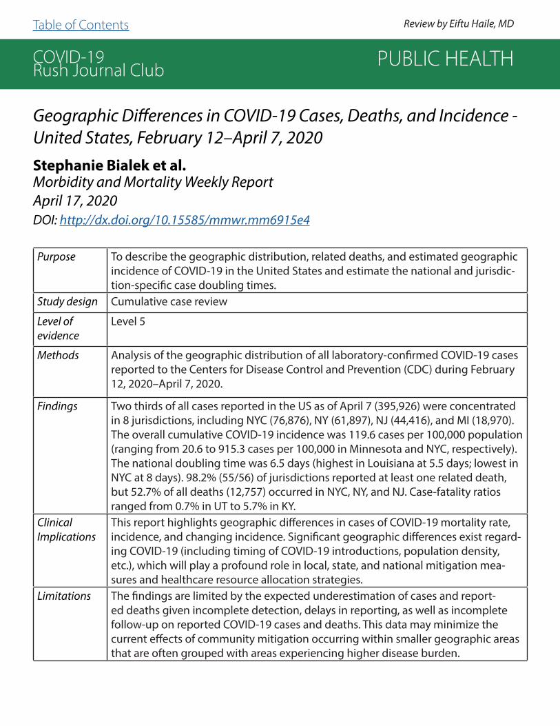
COVID-19Rush Journal Club
Geographic Differences in COVID-19 Cases, Deaths, and Incidence - United States, February 12–April 7, 2020Stephanie Bialek et al.Morbidity and Mortality Weekly ReportApril 17, 2020 DOI: http://dx.doi.org/10.15585/mmwr.mm6915e4
Purpose To describe the geographic distribution, related deaths, and estimated geographic incidence of COVID-19 in the United States and estimate the national and jurisdic-tion-specific case doubling times.
Study design Cumulative case review
Level of evidence
Level 5
Methods Analysis of the geographic distribution of all laboratory-confirmed COVID-19 cases reported to the Centers for Disease Control and Prevention (CDC) during February 12, 2020–April 7, 2020.
Findings Two thirds of all cases reported in the US as of April 7 (395,926) were concentrated in 8 jurisdictions, including NYC (76,876), NY (61,897), NJ (44,416), and MI (18,970). The overall cumulative COVID-19 incidence was 119.6 cases per 100,000 population (ranging from 20.6 to 915.3 cases per 100,000 in Minnesota and NYC, respectively). The national doubling time was 6.5 days (highest in Louisiana at 5.5 days; lowest in NYC at 8 days). 98.2% (55/56) of jurisdictions reported at least one related death, but 52.7% of all deaths (12,757) occurred in NYC, NY, and NJ. Case-fatality ratios ranged from 0.7% in UT to 5.7% in KY.
ClinicalImplications
This report highlights geographic differences in cases of COVID-19 mortality rate, incidence, and changing incidence. Significant geographic differences exist regard-ing COVID-19 (including timing of COVID-19 introductions, population density, etc.), which will play a profound role in local, state, and national mitigation mea-sures and healthcare resource allocation strategies.
Limitations The findings are limited by the expected underestimation of cases and report-ed deaths given incomplete detection, delays in reporting, as well as incomplete follow-up on reported COVID-19 cases and deaths. This data may minimize the current effects of community mitigation occurring within smaller geographic areas that are often grouped with areas experiencing higher disease burden.
PUBLIC HEALTH
Review by Eiftu Haile, MD Table of Contents

COVID-19Rush Journal Club
COVID-19 in Prisons and Jails in the United States
Laura Hawks et al.Journal of the American Medical Association Internal Medicine April 28, 2020DOI: 10.1001/jamainternmed.2020.1856
Purpose To outline the public health risk of COVID-19 spreading through American prisons/jails, and to propose strategies to mitigate this risk.
Study design Expert opinion
Level of evidence
Level 5
Methods N/A
Findings Prisoners are at an increased risk for COVID-19 infection due to the inability to enact adequate social distancing policies, a prison population with a large percentage of individuals in high risk categories, and poor access to appropriate health care. Sug-gested strategies to ameliorate disease burden on the incarcerated population and prison staff include low, medium, and high-risk strategies. Low impact recommenda-tions include increasing availability of personal protective equipment (PPE), increased testing, eliminate copayments/policies that deter inmates from seeking care. Medium impact recommendations include reducing unnecessary jail-time, expedite prison re-lease when appropriate, and release pretrial detainees. High impact recommendations include releasing those in high risk categories (over 55 years of age, underlying health conditions, etc.), those convicted of nonviolent crimes, and those with less than 2 years of their sentence remaining. These steps would pose little risk to public safety.
ClinicalImplications
The impact of COVID-19 infection spreading through prisons has important impli-cations for Chicago hospitals, and the West Side Rush Medical Center community in particular: Cook County jail has reported that over 350 incarcerated persons and staff members have tested positive for SARS-CoV-2 as of early April 2020. This is the highest total caseload of any single site in the country. These inmates receive care at Stroger, increasing the risk of virus spread to more patients and hospital staff. Doctors must be public health advocates who support bold policy changes to minimize the catastrophe brewing in prisons and jails. Furthermore, both congressional legislation and adminis-trative action are needed as quickly as possible to mandate nationwide plans to mini-mize the toll of the pandemic.
Limitations This article is an opinion piece, rather than a formal research study, limiting its impact.
PUBLIC HEALTH
Review by Alice Burgess, MS2 Table of Contents

COVID-19Rush Journal Club
Quarantine Alone or in Combination With Other Public Health Mea-sures to Control COVID-19: A Rapid ReviewBarbara Nussbaumer-Streit et al.Cochrane Database of Systematic Reviews April 8, 2020 DOI: 10.1002/14651858.CD013574
Purpose To assess the effects of quarantine alone or in combination with other measures on the spread of COVID-19.
Study design Literature Review (n=29 studies)
Level of evidence
Level 4
Methods This rapid review included literature published between January 1, 2002 and March 12, 2020. Studies that assessed the effect of quarantine alone or in combination with other measures (isolation, social distancing, school closures, hand hygiene) were included. As COVID-19 is relatively new, authors included studies on similar viruses (SARS and MERS) to incorporate more evidence. 30% of abstracts in this study were dually screened and 70% were screened by one reviewer. Case reports and systematic reviews were excluded. 229 studies (10 focused on COVID-19, 15 on related evidence on SARS, 2 on SARS and other viruses, and 2 on MERS) met the inclusion criteria.
Findings Ferguson et. al. found that case isolation, voluntary quarantine, and social distancing of indi-viduals greater than 70 years of age could have prevented 49% of deaths and reduced critical care bed usage by 67% in the UK. Choi et. al. modeled that prevention and control measures reduced the transmission rate by 90-99% in South Korea. Zhao et. al. noted that without any prevention or control measures, China would have had greater than 800 million COVID-19 cases with an epidemic duration of 477 days. Geng et. al. reported that community quarantine and school closures in China reduced the peak of transmissions by 45.7% and 29.9%, respec-tively. Regarding the 2003 SARS outbreak in South Korea, Hsieh et. al. reported that quarantin-ing travelers from high risk regions prevented 511 cases and 70 deaths. If quarantine measures had been implemented sooner, 280 cases and 48 deaths could have been prevented.
ClinicalImplications
Given the asymptomatic period of COVID-19 infection, health officials may struggle to man-date isolation of asymptomatic patients and those with known sick contacts. Individuals should adhere to social distancing guidelines as quarantine practices have demonstrated suc-cess in limiting the strain on healthcare systems and infection-related morbidity and mortality. While quarantine efforts have been shown to be effective in the short-term, they may result in a more robust delayed epidemic due to the lack of herd immunity.
Limitations Studies included were limited to those in English and Chinese, which may have excluded sev-eral studies. Time of assumed infectivity varied between the studies, which may have altered modeling and led to uncertainties when predicting the time to relaxation of quarantine and other public health measures. Comparison between SARS and COVID-19 are limited in that models assume SARS and MERS begin with symptomatic transmission, but not COVID-19. There was no dual independent assessment for risk of bias and rating of quality of evidence.
PUBLIC HEALTH
Review by Kelly Harmon, MS1 Table of Contents

COVID-19Rush Journal Club
Internet Searches for Unproven COVID-19 Therapies in the United StatesMichael Liu et al.Journal of the American Medical Association Internal MedicineApril 29, 2020DOI: 10.1001/jamainternmed.2020.1764
Purpose To better understand the scope of demand for chloroquine and hydroxychloroquine to pre-vent or treat COVID-19 infection by individuals not under supervision by a licensed physician through examination of internet searches indicative of attempts to purchase these drugs.
Study design Retrospective
Level of evidence
Level 4
Methods Researchers examined daily Google searches from February 1, 2020 to March 29, 2020, splitting the time period into three sections: February 1, 2020 to March 16, 2020, all days after March 16, 2020, the date Elon Musk endorsed chloroquine and hydroxychloroquine as the standard of care (i.e., when knowledge of the drugs was widespread), and all days after March 22nd, when news outlets began reporting chloroquine related poisonings. Authors then compared volumes of Google searches originating from the US including the terms: buy, order, Amazon, eBay, or Walmart in combination with chloroquine or hydroxychloro-quine. The proportion of such searches per 10 million total searches were examined using Google Trends. Expected volumes were calculated using forecasting approaches such as Hyndman and Khandakar’s algorithm.
Findings Queries for purchasing chloroquine and hydroxychloroquine were increased by 442% and 1389% respectively following high-profile claims that these drugs were effective COVID-19 therapies. The first spike in searches corresponded with entrepreneur Elon Musk’s tweet on March 16th, and the largest spike in searches corresponded with US President Donald Trump’s first televised endorsements on March 19th. After news reports of the first fatal poi-soning on March 22nd, searches remained above expected levels with chloroquine at 212% and hydroxychloroquine at 1167%.
ClinicalImplications
High-profile figures endorsing drug therapies not supported by adequate evidence can lead to negative consequences, particularly when the drugs are commercially available within non-medical products (i.e., chloroquine phosphate, an aquarium cleaner). Public health agencies should take steps to warn the public against unapproved therapies unless pre-scribed, especially surrounding endorsement by high-profile figures.
Limitations This study was limited by the design which only used Google searches and restricted on-line markets to Amazon, eBay, and Walmart (top 3 e-commerce companies). The scope of searches could have been improved by using additional terms, e-commerce companies, and languages other than English. It is likely that these limitations led to underestimation of the demand for chloroquine and hydroxychloroquine through online markets.
PUBLIC HEALTH
Review by Kat Tehaney, MS1 Table of Contents

COVID-19Rush Journal Club
An interpretable mortality prediction model for COVID-19 patients
Li Yan et al.Nature Machine IntelligenceMay 14, 2020DOI: https://doi.org/10.1038/s42256-020-0180-7
Purpose To identify the most crucial biomarkers associated with patient mortality to distinguish those at imminent risk, thereby decreasing clinical burden and potentially reducing the COVID-19 mortality rate.
Study design Retrospective case report (n=485)
Level of evidence
Level 4
Methods The medical records of 375 patients from Tongji Hospital in Wuhan, China were col-lected between January 10-February 18, 2020 with an additional 110 patient records collected between February 19-24, 2020 to serve as an external test dataset. The Multi-tree XGBoost algorithm was used to assess the contribution of individual patient parameters (e.g., basic information and symptoms) in addition to laboratory data (e.g., blood samples, liver function, kidney function, coagulation function, electrolytes and inflammatory factors). The data were also used to create a simplified and clinically op-erable decision model.
Findings Lactate dehydrogenase (LDH), lymphocytes, and high-sensitivity C-reactive protein (hs-CRP) were identified as important biomarkers in determining patient mortality. A decision rule using LDH <365 U/L, hs-CRP <41.2 mg/L, and lymphocytes >14.7% values had a 1.00 precision on predicting survival and 0.81 precision on predicting death.
ClinicalImplications
Being able to predict COVID-19 patient’s risk of mortality more than 10 days in ad-vance and with greater than 90% accuracy is vital. Authors designed a mathematical modelling approach based on machine learning algorithms, enabling detection and early intervention with the potential to reduce the mortality rate of COVID-19 infected patients. The authors identified three plasma indicators (LDH, lymphocyte, and hs-CRP levels) in conjunction with a clinical decision model to assist in COVID-19 prognostic prediction. Their model provided a simple and interpretable test to precisely quantify the risk of death.
Limitations All data originated from a single region and hospital. Moreover, the model only used data from the patients’ final sample. Since the machine learning method is purely da-ta-driven, it may vary with a different dataset. More accurate models may be obtained by following the same procedure but incorporating a larger sample size and multi-cen-ter data.
PUBLIC HEALTH
Review by Kat Tehaney, MS1 Table of Contents

COVID-19Rush Journal Club
Diabetes and COVID‐19: psychosocial consequences of the COVID‐19 pandemic in people with diabetes in Denmark—what characterizes people with high levels of COVID‐19‐related worries?Lene Eide Joensen et al.Diabetic Medicine May 11, 2020DOI: https://doi.org/10.1111/dme.14319
Purpose To map COVID-19 specific concerns and overall psychosocial health among diabetic in-dividuals during the initial phase of the COVID-19 pandemic in Denmark and to explore characteristics of diabetic individuals and those with high levels of worry related to the pandemic.
Study design Cross-sectional survey (n=2430)
Level of evidence
Level 3
Methods Online questionnaires were distributed to 2430 adults from user panels at Steno Dia-betes Center Copenhagen and The Danish Diabetes Association. The surveys included items addressing COVID-19 specific worries, concerns related to diabetes, sociodemo-graphic and health status, social relations, diabetes-specific social support, and changes in diabetes-specific behaviors. Responses were analyzed with descriptive statistics and logistic regressions.
Findings Participants were most frequently worried about being ‘overly affected due to diabetes if infected with COVID-19’ (56%), ‘people with diabetes are characterized as a risk group’ (39%), and ‘not being able to manage diabetes if infected with COVID-19’ (28%). Logistic regressions showed that being female, having type 1 diabetes, diabetes complications, diabetes distress, feelings of isolation and loneliness, and having altered diabetes behav-iors were all associated with being more worried about COVID-19 and diabetes. There was no association between level of social support and COVID-19 specific worries.
ClinicalImplications
Diabetic individuals have COVID-19 specific worries related to diabetes which is associat-ed with poorer psychosocial health. These concerns could be addressed through sup-port targeting specific questions and needs of diabetic individuals, as well as frequent updates on new knowledge regarding COVID-19’s impact on diabetic patients. These findings will assist in improving support for patients with diabetes and management of their anxieties, but further studies are needed to explore if and how COVID‐19 worries change during the pandemic.
Limitations This study was limited by the convenience sample of participants, who may be generally healthier than the average individual with diabetes, as shown by the lower prevalence of diabetic complications. Additionally, as all measures are self-reported, there may be uncertainty regarding the presence of diabetic complications and inappropriate glucose levels. The date of the survey was not disclosed.
PUBLIC HEALTH
Review by Kat Tehaney, MS1 Table of Contents

COVID-19Rush Journal Club
Factors Associated With Mental Health Outcomes Among Health Care Workers Exposed to Coronavirus Disease 2019Jianbo Lai et al.Journal of the American Medical Association Network OpenMarch 23, 2020DOI: 10.1001/jamanetworkopen.2020.3976
Purpose To assess the magnitude of mental health outcomes and associated factors among health care workers treating patients exposed to COVID-19 in China.
Study design Cross-sectional survey (n=1257)
Level of evidence
Level 3
Methods Researchers collected demographic data and mental health measurements from 1257 health care workers (493 physicians and 764 nurses) in 34 hospitals in China from January 29-February 3, 2020. The degree of symptoms of depression, anxiety, insomnia, and dis-tress were assessed by Chinese versions of the 9-item Patient Health Questionnaire (PHQ-9), 7-item Generalized Anxiety Disorder (GAD-7) scale, 7-item Insomnia Severity Index (ISI), and 22-item Impact of Event Scale-Revised (IES-R). Multivariable logistic regression analy-sis was used to identify factors associated with mental health outcomes.
Findings A considerable number of participants reported negative impacts of working on the front-lines during the coronavirus pandemic: 50.4% reported depressive symptoms, 44.6% re-ported anxiety symptoms, 34.0% reported insomnia, and 71.5% reported distress. Nurses (vs physicians, p=0.01), women (vs men, p=0.001), and those working in Wuhan (vs Hubei outside Wuhan and outside Hubei, p<0.01) reported more severe degrees of all mental health symptoms. Frontline health care workers engaged in direct diagnosis, treatment, and care of COVID-19 patients were associated with a higher risk of depressive symptoms (odds ratio, OR: 1.52), anxiety (OR: 1.57), insomnia (OR: 2.97), and distress (OR: 1.60).
ClinicalImplications
Health care workers responding to the spread of COVID-19 reported high rates of depres-sive symptoms, anxiety, insomnia, and distress. Increased psychological interventions are necessary to promote mental well-being and self-care of health care workers caring for COVID-19 patients. Health care workers requiring particular attention are women, nurses, and frontline workers.
Limitations Most participants (81.2%) were from Hubei province, limiting generalization to less effect-ed regions. The timeline of the study, carried out over 6 days, lacks longitudinal follow-up and thus long-term psychological impact warrants further investigation. Finally, response bias may be present as there was a 68.7% response rate, the majority (764) of those being nurses (vs 493 physicians).
PUBLIC HEALTH
Review by Kat Tehaney, MS1 Table of Contents
COVID-19Rush Journal Club
BIOLOGY
Table of Contents

COVID-19Rush Journal Club
Genomic characterisation and epidemiology of 2019 novel corona-virus: implications for virus origins and receptor bindingRoujian Lu et al.LancetJanuary 20, 2020DOI: https://doi.org/10.1016/S0140-6736(20)30251-8
Purpose To sequence and characterize the genome of 2019 novel coronavirus (2019-nCov).
Study design Case Series (n = 9 patients) Level of evidence
Not applicable
Methods Isolates collected from nine inpatients of three area hospitals in Wuhan, China pre-senting with viral pneumonia of unknown origin. Researchers studied eight com-plete and two partial genomes obtained from the nine study patients.
Findings Novel coronavirus 19 belongs to subgenus Sarbecovirus. The virus is more similar to two bat-derived coronavirus strains (88% identity) than to known human-in-fecting coronaviruses (SARS-CoV: 79% identity, MERS-CoV: 50% identity). Howev-er, there was greater similarity in S1 domain of the spike protein of the novel coronavirus and SARS-CoV, possibly suggesting the novel coronavirus might also use angiotensin-converting enzyme 2 (ACE-2) as a cell receptor (as SARS-CoV is known to do). There is likely an intermediate host, currently unknown, between bats and humans for novel coronavirus.
Clinical Implications
The genomes of the virus across all patient samples were remarkably similar, indicating a common source of infection. Not all patients had visited the Hua-nan wet market, also supporting hypothesized human-to-human transmission via droplets. Identification of the 2019-nCov intermediate host could facilitate further understanding of disease control during this pandemic. Like SARS-CoV, the 2019-nCoV uses ACE2 as a receptor, although there were key variances in the recep-tor-binding domains of SARS-CoV and 2019-nCoV. Further research is necessary to determine the significance of this similarity, and how it could affect the diagnosis and treatment of the virus, as well as vaccine development.
Limitations While likely due to the urgency surrounding the health crisis of this novel coronavi-rus, this study only analyzed sequences from isolates of a small number of patients. Future research is needed to map the changes in genome sequencing of this virus as it spreads throughout the world.
BIOLOGY
Review by Beth Hall, MS4 Table of Contents

COVID-19Rush Journal Club
Comparative replication and immune activation profiles of SARS-CoV-2 and SARS-CoV in human lungs: an ex vivo study with implica-tions for the pathogenesis of COVID-19.Hin Chu et al.Clinical Infectious DiseasesApril 9, 2020DOI: https://doi.org/10.1093/cid/ciaa410
Purpose To compare viral kinetics, cell tropism, and immune response profiles of SARS-CoV-2 and SARS-CoV to understand the mechanism behind transmission and pre-sentation of COVID-19.
Study design Case Series (n = 6 patients)Level of evidence
Level 4
Methods Six patients undergoing wedge resection or lobectomy for lung tumor donated tissue to be inoculated with SARS-CoV-2 and SARS-CoV. Virus titers measured at 2, 24, and 48 hours, and tissues underwent panel of representative IFNs and pro-in-flammatory cytokines/chemokines.
Findings SARS-CoV-2 antigens detected in significantly higher amount and in more areas of the lung tissues than SARS-CoV antigens. SARS-CoV-2 generated 3.20 folds more infectious virus particles in 48 hours than SARS-CoV (P<0.024). Both SARS-CoV-2 and SARS-CoV infected type I pneumocytes, type II pneumocytes, and alveo-lar macrophages. SARS-CoV-2 infection did not significantly trigger the expression of any IFN, compared to SARS-CoV. SARS-CoV-2 infection only significantly upregu-lated 5 of 13 inflammatory mediators, compared to 8 of 13 in SARS-CoV.
ClinicalImplications
The high degree of replication and viral particles of SARS-CoV-2 may explain high viral loads in COVID-19 patients presenting early in the disease course, and possibly during intubation. The SARS-CoV-2 triggers fewer pro-inflamma-tory markers than SARS-CoV, potentially explaining why many patients remain asymptomatic or with mild symptoms throughout their disease course. Future re-search should explore how the mechanism of how SARS-CoV-2 suppresses the IFN and cytokine/chemokine response.
Limitations Ex-vivo human lung tissue explant culture doesn’t represent the effect of host systemic inflammatory response and the adaptive immune response. Human tissue supply is limited, and is not a viable option to investigate the characteristics of SARS-CoV-2. This study used a small sample size, and all lung tissue donators were diagnosed with lung cancer, as such the results might not be generalizable to the larger population.
BIOLOGY
Review by Jackie Urban, MS4 Table of Contents

COVID-19Rush Journal Club
Structural and Functional Basis of SARS-CoV-2 Entry by Using Hu-man ACE2Qihui Wang et al.CellApril 7, 2020DOI: https://doi.org/10.1016/j.cell.2020.03.045
Purpose To analyze the human angiotensin-converting enzyme 2 (hACE2) entry receptor as a binding site for SARS-CoV-2 compared to SARS-CoV and MERS-CoV.
Study design Descriptive StudyLevel of evidence
Not applicable
Methods Immunostaining and flow cytometry were used to identify the spike (S) glycopro-tein subunits of SARS-CoV-2 which interact with the hACE2 receptor. SARS-CoV-2/hACE2 complexes were analyzed in-vitro via size exclusion chromatography to study virus-receptor interaction and complex formation. Surface plasmon reso-nance (SPR) assay was used to demonstrate virus-receptor binding affinity of SARS-CoV-2 compared to SARS-CoV and MERS-CoV. The epitope features of SARS-CoV-2 were assessed using murine monoclonal antibodies against SARS-CoV and MERS-CoV S proteins.
Findings Entry of coronaviruses into host cells is mediated by the S glycoprotein. Immu-nostaining and flow cytometry with SARS-CoV-2 S protein preparations revealed a strong affinity for hACE2 binding, and the complex was largely similar to the structure of the SARS-CoV/hACE2 complex. SARS-CoV-2-CTD binding interface displayed significantly stronger interactions with hACE2 compared to SARS-CoV, with more amino acid residues that directly bind the hACE2 receptor, more hydro-gen bonds, and larger buried surface areas resulting in overall increased atomic interactions. SARS-CoV-2 demonstrated a 4-fold higher binding affinity for the hACE2 receptor when compared with SARS-CoV and MERS-CoV.
ClinicalImplications
Monoclonal and polyclonal antibodies directed against both SARS-CoV and MERS-CoV were unable to bind the SARS-CoV-2 S protein, despite the shared character-istic of these three coronaviruses to engage the hACE2 receptor for entry into host cells. This lack of monoclonal antibody binding indicates distinct antigenic features of the novel coronavirus, SARS-CoV-2, that reduce the effectiveness of potential vaccine therapies utilized in previous coronavirus outbreaks.
Limitations Additional research is needed to determine the efficacy of vaccines and their role in targeting S proteins for SARS-CoV-2 as a means of prophylaxis.
BIOLOGY
Review by Emily Hejna, MS2 Table of Contents

COVID-19Rush Journal Club
Composition and divergence of coronavirus spike proteins and host ACE2 receptors predict potential intermediate hosts of SARS‐CoV‐2Zhixin Liu et al.Journal of Medical VirologyFebruary 26, 2020DOI: https://doi.org/10.1002/jmv.25726
Purpose To identify potential intermediate hosts transmitting SARS-CoV-2 to humans by characterizing various species’ coronavirus spike protein and its interaction with angiotensin‐converting enzyme 2 (ACE2).
Study design Observational study
Level of evidence
Not applicable
Methods Protein sequences of ACE2 and spike glycoproteins of SARS-CoV-2, SARS-CoV, and bat SARS-like CoV were obtained in addition to the bat SARS-like CoV RaTG13 sequence and the pangolin metagenome. Protein sequence alignment and phylo-genetic analysis was accomplished using Molecular Evolutionary Genetics Analysis (MEGA-X) software, and multiple comparisons were done by ClustalW. Structure and binding models of the spike receptor were accomplished using I-TASSER, PRISM 2.0, and PyMOL softwares.
Findings SARS-CoV-2 receptor‐binding domain (RBD) sequence has 93% similarity when compared to pangolin SARS-like CoV SRR10168377, which has 89% similarity with bat SARS-like CoV RaTG13. SARS-CoV and CoV-2 enter the respiratory tract by the receptor ACE2 and were found to have good alignment. An interaction model from the PRISM 2.0 database indicated that SARS-CoV-2 spike protein may bind to ACE2 through Leu455, Phe486, Gln493, Asn501, and Tyr505. CoV spike – ACE2 binding in turtles and pangolins more resemble humans than bats. ACE2 site 41 residue is tyrosine in pangolin, turtle, and human, but histidine in bat; tyrosine may possess higher affinity for RBD than histidine.
ClinicalImplications
Studying the evolutionary relationship of the RBD of the spike protein of SARS-CoV-2 is useful in determining possible intermediate hosts. This method may facilitate finding reservoirs of SARS-CoV2 and future viral pandemics.
Limitations The authors noted that the spike protein crystallization of SARS-CoV-2 was ana-lyzed urgently. Further studies need to confirm its structure, and the interactions between SARS-CoV-2 spike protein RBD and ACE2 in other possible intermediate hosts.
BIOLOGY
Review by Alexandra Feldner, MS3Table of Contents

COVID-19Rush Journal Club
Characterization of spike glycoprotein of SARS-CoV-2 on virus entry and its immune cross-reactivity with SARS-CoVXiuyuan Ou et al.Nature CommunicationsMarch 27, 2020DOI: https://doi.org/10.1038/s41467-020-15562-9
Purpose To determine cell type susceptibility, entry receptor, entry pathway, protease priming mechanisms, and serological specificity of SARS-CoV-2.
Study design Observational studyLevel of evidence
Not applicable
Methods Pseudovirions produced by co-transfection 293T cells with plasmids encoding SARS-CoV-2 S. HEK 293 cells stably expressing hACE2 (293/hACE2) were pretreated with lysosomotropic agents or cathepsin inhibitors, then inoculated with pseudovirions. Cells were lysed, and luciferase activity measured to determine entry into the cell. SARS-CoV S, SARS-CoV-2 S, and VSV G pseudovirons (control) were pre-incubated with rabbit anti-SARS S1 antibodies T62 or patient sera. Mixture was added onto 293/hACD2 cells. Cells were lysed and pseudovirus transduction was measured.
Findings 293/hACE2 cells were highly transduced by SARS-CoV-2 S pseudovirions, indicating that hACE2 is the receptor for SARS-CoV-2. Phosphatidylinositol 3-phosphate 5-kinase (PIKfyve) inhibitors, apili-mod, and YM201636 inhibited entry of SARS-CoV-2 S pseudovirions via endocytosis to 293/hACE2 cells. Cathepsin L inhibitors decreased entry of SARS-CoV-2 S pseudovirions by over 76%, suggest-ing it is essential for SARS-CoV-2 S priming. Type II membrane serine proteases-mediated cleavage activated fusion potential of SARS-CoV-2 S protein in 293/hACE2 cells. Trypsin could activate SARS-CoV-2 S protein efficiently but was not necessary for SARS-CoV-2 S protein to trigger syncytium formation. SARS-CoV-2 S protein was less stable than SARS-CoV-S protein, requiring shorter time and lower temperature to be inactivated. Anti-SARS S1 antibodies T62 recognized SARS-CoV-2 S protein. A low-level binding of SARS-CoV-2 S protein to rabbit anti-SARS S1 antibody T62 was de-tected. Substitution of SARS-CoV-2 receptor binding domain (RBD) with SARS-CoV RBD increased the affinity of S protein to polyclonal antibodies T62 suggesting differences between SARS-CoV and SARS-CoV-2 RBDs. Serum from recovered SARS patient demonstrated strong inhibition on transduction by SARS-CoV S pseudovirions and modest neutralization activity against SARS-CoV-2 S pseudovirions. Sera from all five COVID-19 patients neutralized SARS-CoV-2 S pseudovirions but had no effect on transduction by SARS-CoV S pseudovirions.
ClinicalImplications
Entry of SARS-CoV-2 S pseudovirions into 293/hACE2 cells was reduced by preincubation of soluble hACE2; soluble hACE2 may be a viable therapeutic inhibitor against SARS-CoV-2 infection. Inhibition of SARS-CoV-2 S pseudovirion entry by lysosomotropic agents suggested that PIKfyve should be considered as a potential drug target. Due to only moderate cross-neutral-ization between covalescent sera of SARS and COVID-19 patients, those previously recovered from SARS-CoV infection may not be protected against SARS-CoV-2 infection.
Limitations Involvement of additional endogenous proteases in HEK293T cell syncytium formation and mechanism of entry into other cell types requires further research. Further characterization of the differences between the SARS-CoV-2 RBD and SARS-CoV RBD should be pursued.
BIOLOGY
Review by Abigail Bawden, MS1 Table of Contents

COVID-19Rush Journal Club
Cryo-EM structure of the 2019-nCoV spike in the prefusion confor-mation.Daniel Wrapp et al.ScienceMarch 13, 2020DOI: 10.1126/science.abb2507
Purpose To facilitate vaccine formulation and identification of drug targets against COVID-19 (2019-nCoV), the CoV spike (S) glycoprotein was characterized and com-pared to the S glycoprotein of various other coronaviruses including SARS-CoV.
Study design Observational study
Level of evidence
Not applicable
Methods 2019-nCoV S protein was compared to several other coronaviruses, including SARS-CoV S, in protein sequence, orientation of receptor binding domains (RBD) by cryo-electron microscopy (cryo-EM), binding affinity for angiotensin-converting enzyme 2 (ACE2), and cross reactivity of four published anti-SARS-CoV S protein antibodies against 2019-nCoV S protein by biolayer interferometry (BLI).
Findings Cryo-Em demonstrated that structure, orientation, and binding movement of 2019-nCoV S protein overlapped strongly with SARS and MERS-CoV S protein. Amino acid sequence of 2019-nCoV S protein was 98% shared between bat coronavirus RaTG13 (SARS-CoV S protein shared sequence was not reported). Of note, a gain of function mutation in 2019-nCoV S protein leads to RRAR furin recognition site, allowing protease cleavage and potentially enhanced virulence. Similar mutations are seen in hemagglutinin of highly virulent strains of influenza. Binding orienta-tion of S protein to ACE2 was preserved across the viruses and revealed a 10-20-fold higher affinity in 2019-nCoV S protein compared to SARS. To investigate if im-munity to SARS-CoV could contribute to immunity to COVID-19, authors assessed binding of four published anti-SARS S protein antibodies to 2019-nCoV S protein. No binding was detected between anti-SARS antibodies and COVID-19 antigens.
ClinicalImplications
Enhanced affinity for fusion and entry may contribute to COVID-19 virus’s capaci-ty to spread rapidly and make S protein-ACE2 binding a potential pharmacologic target. Previous infection with SARS-CoV does not necessarily confer immunity to Covid-19.
Limitations Analysis of additional antigens and population-level epidemiological studies are needed to assess the shared immunity of various coronaviruses, such as SARS and MERS, with COVID-19.
BIOLOGY
Review by Luke McCormack, MS2 Table of Contents
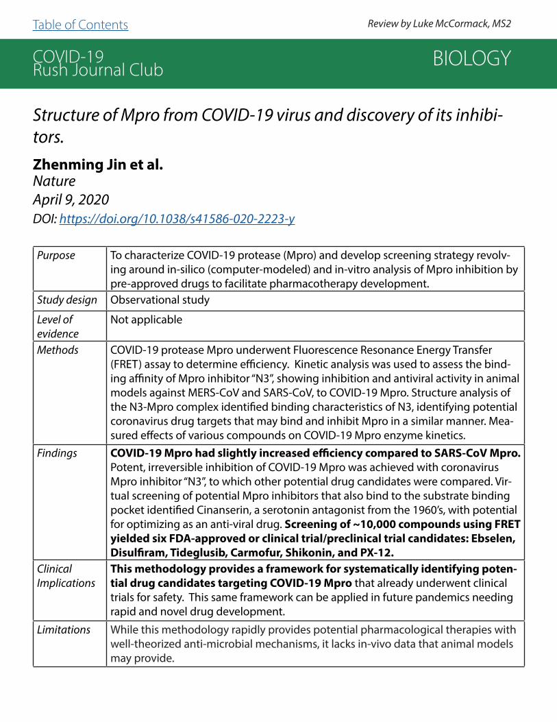
COVID-19Rush Journal Club
Structure of Mpro from COVID-19 virus and discovery of its inhibi-tors.Zhenming Jin et al.NatureApril 9, 2020DOI: https://doi.org/10.1038/s41586-020-2223-y
Purpose To characterize COVID-19 protease (Mpro) and develop screening strategy revolv-ing around in-silico (computer-modeled) and in-vitro analysis of Mpro inhibition by pre-approved drugs to facilitate pharmacotherapy development.
Study design Observational study
Level of evidence
Not applicable
Methods COVID-19 protease Mpro underwent Fluorescence Resonance Energy Transfer (FRET) assay to determine efficiency. Kinetic analysis was used to assess the bind-ing affinity of Mpro inhibitor “N3”, showing inhibition and antiviral activity in animal models against MERS-CoV and SARS-CoV, to COVID-19 Mpro. Structure analysis of the N3-Mpro complex identified binding characteristics of N3, identifying potential coronavirus drug targets that may bind and inhibit Mpro in a similar manner. Mea-sured effects of various compounds on COVID-19 Mpro enzyme kinetics.
Findings COVID-19 Mpro had slightly increased efficiency compared to SARS-CoV Mpro. Potent, irreversible inhibition of COVID-19 Mpro was achieved with coronavirus Mpro inhibitor “N3”, to which other potential drug candidates were compared. Vir-tual screening of potential Mpro inhibitors that also bind to the substrate binding pocket identified Cinanserin, a serotonin antagonist from the 1960’s, with potential for optimizing as an anti-viral drug. Screening of ~10,000 compounds using FRET yielded six FDA-approved or clinical trial/preclinical trial candidates: Ebselen, Disulfiram, Tideglusib, Carmofur, Shikonin, and PX-12.
ClinicalImplications
This methodology provides a framework for systematically identifying poten-tial drug candidates targeting COVID-19 Mpro that already underwent clinical trials for safety. This same framework can be applied in future pandemics needing rapid and novel drug development.
Limitations While this methodology rapidly provides potential pharmacological therapies with well-theorized anti-microbial mechanisms, it lacks in-vivo data that animal models may provide.
BIOLOGY
Review by Luke McCormack, MS2 Table of Contents
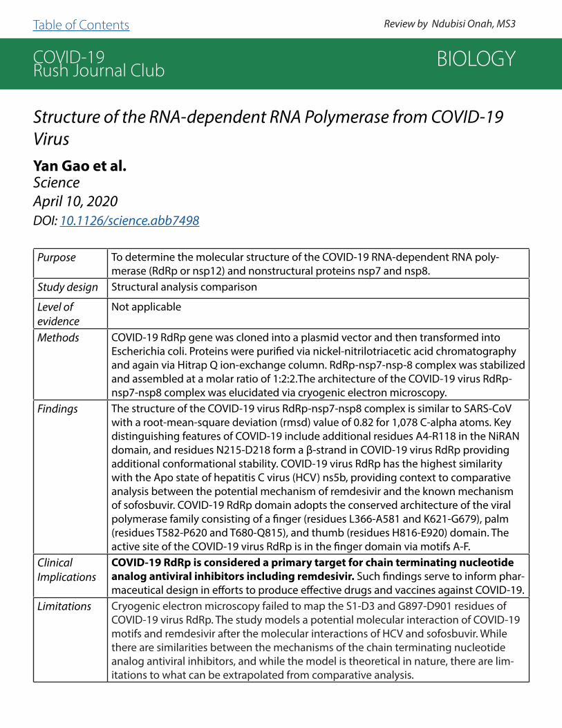
COVID-19Rush Journal Club
Structure of the RNA-dependent RNA Polymerase from COVID-19 VirusYan Gao et al.ScienceApril 10, 2020DOI: 10.1126/science.abb7498
Purpose To determine the molecular structure of the COVID-19 RNA-dependent RNA poly-merase (RdRp or nsp12) and nonstructural proteins nsp7 and nsp8.
Study design Structural analysis comparison
Level of evidence
Not applicable
Methods COVID-19 RdRp gene was cloned into a plasmid vector and then transformed into Escherichia coli. Proteins were purified via nickel-nitrilotriacetic acid chromatography and again via Hitrap Q ion-exchange column. RdRp-nsp7-nsp-8 complex was stabilized and assembled at a molar ratio of 1:2:2.The architecture of the COVID-19 virus RdRp-nsp7-nsp8 complex was elucidated via cryogenic electron microscopy.
Findings The structure of the COVID-19 virus RdRp-nsp7-nsp8 complex is similar to SARS-CoV with a root-mean-square deviation (rmsd) value of 0.82 for 1,078 C-alpha atoms. Key distinguishing features of COVID-19 include additional residues A4-R118 in the NiRAN domain, and residues N215-D218 form a β-strand in COVID-19 virus RdRp providing additional conformational stability. COVID-19 virus RdRp has the highest similarity with the Apo state of hepatitis C virus (HCV) ns5b, providing context to comparative analysis between the potential mechanism of remdesivir and the known mechanism of sofosbuvir. COVID-19 RdRp domain adopts the conserved architecture of the viral polymerase family consisting of a finger (residues L366-A581 and K621-G679), palm (residues T582-P620 and T680-Q815), and thumb (residues H816-E920) domain. The active site of the COVID-19 virus RdRp is in the finger domain via motifs A-F.
ClinicalImplications
COVID-19 RdRp is considered a primary target for chain terminating nucleotide analog antiviral inhibitors including remdesivir. Such findings serve to inform phar-maceutical design in efforts to produce effective drugs and vaccines against COVID-19.
Limitations Cryogenic electron microscopy failed to map the S1-D3 and G897-D901 residues of COVID-19 virus RdRp. The study models a potential molecular interaction of COVID-19 motifs and remdesivir after the molecular interactions of HCV and sofosbuvir. While there are similarities between the mechanisms of the chain terminating nucleotide analog antiviral inhibitors, and while the model is theoretical in nature, there are lim-itations to what can be extrapolated from comparative analysis.
BIOLOGY
Review by Ndubisi Onah, MS3Table of Contents

COVID-19Rush Journal Club
Is low alveolar type II cell SOD3 in the lungs of elderly linked to the observed severity of COVID-19? Ahmed Abouhashem et al.Antioxidants and Redox SignalingApril 23, 2020DOI: https://doi.org/10.1089/ars.2020.8111
Purpose To compare type II alveolar cell RNA sequencing data between elderly and young healthy subjects to further understand age differences in COVID-19 presentations.
Study design Case series (n = 4)
Level of evidence
Level 4
Methods Human lung single cell RNA sequencing (scRNA-seq) data from 4 healthy donors were divided into the “old-age group” (57 and 63 years-old) and the “young-age group” (22 and 29-years-old). Using expression atlas of human primary cells, alve-olar type II pneumocytes were isolated from the mixed cell populations. Genes of this alveolar type II cell cluster were assessed for differential expression as a func-tion of aging.
Findings Expression of angiotensin-converting enzyme 2 (ACE2) and transmembrane serine protease 2 (TMPRSS2), colocalization of which enables SARS-CoV-2 to enter cells, in the alveolar type II cells were comparable between old and young donors. Super-oxide dismutase 3 (SOD3) was the most downregulated in the old-age group, along with downregulation of other redox-based genes including activating transcription factor 4 (ATF4), metallothionein 2A (M2TA) and glutathione peroxidase 1 (GPX1). 21 of the 24 ATF4 downstream factors were significantly downregulated in alve-olar type II cells of the elderly. Gene regulator database analysis suggested older patients’ ability to respond to heme deficiency and the ATF4-dependent ability to respond to ER oxidative stress is significantly compromised.
ClinicalImplications
COVID-19 produces oxidative stress within the lungs, and older patients displayed reduced redox gene expression relative to two younger patients. This data, in combination with evidence that superoxide dismutase (SOD) can decrease severity of respiratory illnesses, warrants further investigation of therapies focused on reducing the oxidative stress on type II pneumocytes both in vitro and in vivo.
Limitations A small sample, low powered case series with 4 COVID19-free participants.
BIOLOGY
Review by Jackie Urban, MS4 Table of Contents
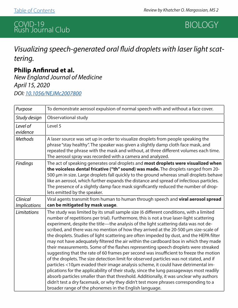
COVID-19Rush Journal Club
Visualizing speech-generated oral fluid droplets with laser light scat-tering. Philip Anfinrud et al.New England Journal of MedicineApril 15, 2020DOI: 10.1056/NEJMc2007800
Purpose To demonstrate aerosol expulsion of normal speech with and without a face cover.
Study design Observational study
Level of evidence
Level 5
Methods A laser source was set up in order to visualize droplets from people speaking the phrase “stay healthy”. The speaker was given a slightly damp cloth face mask, and repeated the phrase with the mask and without, at three different volumes each time. The aerosol spray was recorded with a camera and analyzed.
Findings The act of speaking generates oral droplets and most droplets were visualized when the voiceless dental fricative (“th” sound) was made. The droplets ranged from 20-500 µm in size. Large droplets fall quickly to the ground whereas small droplets behave like an aerosol, which further expands the distance and spread of infectious particles. The presence of a slightly damp face mask significantly reduced the number of drop-lets emitted by the speaker.
ClinicalImplications
Viral agents transmit from human to human through speech and viral aerosol spread can be mitigated by mask usage.
Limitations The study was limited by its small sample size (6 different conditions, with a limited number of repetitions per trial). Furthermore, this is not a true laser-light scattering experiment, despite the title—the analysis of the light scattering data was not de-scribed, and there was no mention of how they arrived at the 20-500 µm size-scale of the droplets. Studies of light scattering are often impeded by dust, and the HEPA filter may not have adequately filtered the air within the cardboard box in which they made their measurements. Some of the flashes representing speech droplets were streaked suggesting that the rate of 60 frames per second was insufficient to freeze the motion of the droplets. The size detection limit for observed particles was not stated, and if particles <10µm evaded their image analysis scheme, it could have detrimental im-plications for the applicability of their study, since the lung passageways most readily absorb particles smaller than that threshold. Additionally, it was unclear why authors didn’t test a dry facemask, or why they didn’t test more phrases corresponding to a broader range of the phonemes in the English language.
BIOLOGY
Review by Khatcher O. Margossian, MS 2Table of Contents

COVID-19Rush Journal Club
A highly conserved cryptic epitope in the receptor-binding domains of SARS-CoV-2 and SARS-CoVMen Yuan et al.ScienceApril 3, 2020DOI: 10.1126/science.abb7269
Purpose To analyze the ACE2 receptor binding domain (RBD) of SARS-CoV and SARS-CoV-2 S-spike protein, and the cross-reactivity of an anti- SARS-CoV S-protein antibody with SARS-CoV-2.
Study design Original molecular biology investigation
Level of evidence
Level 5
Methods CR3022, a neutralizing antibody targeting RBD of SARS-CoV, was exposed to SARS-CoV-2 S-protein RBD. Authors assessed this complex’s structure and binding affinity to find out conserved vs divergent RBD sequences affecting binding of CR3022 on these S-proteins. Ability of CR3022 to neutralize SARS-CoV-2 was assessed via in vitro by microneutralization assay. ELISA assessed interaction of CR3022 and m396, another SARS-CoV antibody, with SARS-CoV-2 RBD.
Findings As revealed by structural studies, CR3022 targets a highly conserved epitope that enables cross-reactive binding between SARS-CoV-2 and SARS-CoV. Despite cross-re-activity of CR3022 and CoV-2 and 86% conserved residues between CoV and CoV-2 RBDs, CR3022 binds to CoV RBD (Kd=1 nM) with significantly more affinity than CoV-2 RBD (Kd=115 nM). Authors attributed this to an additional N-glycosylation site on CoV-2 RBD. While CR3022 does not block ACE2 binding of RBD, its epitope is only exposed when RBDs are in their “up” conformation allowing them to bind to ACE2. This suggests a different mechanism of suppression besides blocking cell entry via ACE2. In-vitro microneutralization assay failed to show any neutralization of CoV-2 at the highest concentration CR3022 tested (400 μg/ml). ELISA confirmed CR3022 does interact with CoV-2 and demonstrated a higher binding signal compared to m396 antibody.
ClinicalImplications
Despite binding both CoV and CoV-2, immunity conferred by anti-SARS-CoV RBD antibodies is not robust. Other targets should be considered for vaccine and drug development against coronaviruses broadly and COVID-19 specifically.
Limitations This study did not assess possible synergistic effects of CR3022 with other known anti-RBD antibodies in-vitro, nor did they investigate in-vivo effects against CoV-2. Authors cite numerous examples of antibodies against coronaviruses, influenza A, herpesvirus, cytomegalovirus, dengue virus and others which confer in-vivo protection while failing in-vitro neutralization. Additionally, further investigation of other con-served CoV epitopes are needed for coronavirus vaccine development.
BIOLOGY
Review by Luke McCormack, MS2 Table of Contents
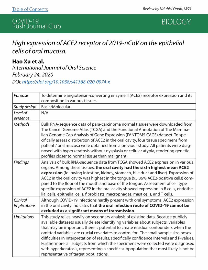
COVID-19Rush Journal Club
High expression of ACE2 receptor of 2019-nCoV on the epithelial cells of oral mucosa.Hao Xu et al.International Journal of Oral ScienceFebruary 24, 2020DOI: https://doi.org/10.1038/s41368-020-0074-x
Purpose To determine angiotensin-converting enzyme II (ACE2) receptor expression and its composition in various tissues.
Study design Basic/MolecularLevel of evidence
N/A
Methods Bulk RNA-sequence data of para-carcinoma normal tissues were downloaded from The Cancer Genome Atlas (TCGA) and the Functional Annotation of The Mamma-lian Genome Cap Analysis of Gene Expression (FANTOM5 CAGE) dataset. To spe-cifically assess distribution of ACE2 in the oral cavity, four tissue specimens from patients’ oral mucosa were obtained from a previous study. All patients were diag-nosed with hyperkeratosis without dysplasia or cellular atypia, rendering genetic profiles closer to normal tissue than malignant.
Findings Analysis of bulk RNA-sequence data from TCGA showed ACE2 expression in various organs. Among these tissues, the oral cavity had the sixth highest mean ACE2 expression (following intestine, kidney, stomach, bile duct and liver). Expression of ACE2 in the oral cavity was highest in the tongue (95.86% ACE2-positive cells) com-pared to the floor of the mouth and base of the tongue. Assessment of cell type specific expression of ACE2 in the oral cavity showed expression in B cells, endothe-lial cells, epithelial cells, fibroblasts, macrophages, mast cells, and T cells.
ClinicalImplications
Although COVID-19 infections hardly present with oral symptoms, ACE2 expression in the oral cavity indicates that the oral infection route of COVID-19 cannot be excluded as a significant means of transmission.
Limitations This study relies heavily on secondary analysis of existing data. Because publicly available datasets usually delete identifying variables about subjects, variables that may be important, there is potential to create residual confounders when the omitted variables are crucial covariates to control for. The small sample size poses difficulties in interpretation of results, specifically confidence intervals and P-values. Furthermore, all subjects from which the specimens were collected were diagnosed with hyperkeratosis, representing a specific subpopulation that most likely is not be representative of target populations.
BIOLOGY
Review by Ndubisi Onah, MS3 Table of Contents

COVID-19Rush Journal Club
Phylogenetic network analysis of SARS-CoV-2 genomes.
Peter Forster et al.PNASApril 8, 2020DOI: https://doi.org/10.1073/pnas.2004999117
Purpose To understand the evolution of the coronavirus within humans by analyzing 160 complete viral genomes to be sequenced from human patients in the midst of the current pandemic.
Study design Basic/experimental research study, genetic analysis
Level of evidence
N/A
Methods Phylogenetic network analysis was utilized to study 160 complete SARS-CoV-2 genomes contributed by researchers around the world since December 2019. Viral genomes were provided by the Global Initiative on Sharing Avian Influenza Data (GISAID), a database for global collaboration since 2006.
Findings A bat coronavirus (BatCoVRaTG13) from Yunnan Province (China) with 96.2% se-quence similarity to that found in humans, was used as an outgroup to determine the root of the phylogeny within the network. Phylogenetic network analysis iden-tified three major variants of SARS-CoV-2: A, B, and C, types distinguished by amino acid changes. Type A was most closely related to the virus found in both bats and pangolins and represents the ancestral type. Types A and C genomes are primarily found outside of East Asia, particularly in Europe and the Americas. Type C differs from type B by a G26144T mutation which changes a glycine to a valine. This vari-ant is the major genome identified in Europe, California, and Brazil. Type B was found to be derived from type A by two mutations. The ancestral B-type genome is clustered in East Asia. All B-type genomes found outside of East Asia have mutated.
ClinicalImplications
Identification of viral genome mutations by genetic networking techniques allows for the reconstruction of infection paths. An understanding of the SARS-CoV-2 mutational variants poses significant implications for the development of a vaccine and may play a role in the range of clinical presentations and spread of the disease.
Limitations Given the pandemic-level spread of SARS-CoV-2, phylogenetic network analysis is less useful as significant migration and mutation of the virus has taken place. The first viral genome sampled in late December 2019 was found to be evolutionarily distant from the root type in comparison to the bat coronavirus outgroup.
BIOLOGY
Review by Emily Hejna, MS2 Table of Contents

COVID-19Rush Journal Club
Circulating plasma concentrations of angiotensin-converting enzyme 2 in men and women with heart failure and effects of renin-angiotensin-aldosterone inhibitorsIziah E Sama et al.European Heart JournalMay 10, 2020DOI: https://doi.org/10.1093/eurheartj/ehaa373
Purpose To examine plasma angiotensin-converting enzyme 2 (ACE-2) levels in heart failure (HF) patients and impact of renin-angiotensin-aldosterone system (RAAS) inhibitors on ACE-2 levels.
Study design Non-randomized cohort study with index (n = 2022; 1485 male, 537 female) and validation cohorts (n = 1698; 1123 male, 575 female)
Level of evidence
Level 3
Methods Data were obtained from the BIOlogy Study to Tailored Treatment in Chronic Heart Failure (BIOSTAT-CHF) which focused on improvement of HF management. Data were drawn from an index cohort of patients from 11 European countries and a validation cohort of patients from Scotland. Included patients had LVEF <40%, BNP > 400 pg/mL, or pro-BNP > 2000 pg/mL. Per study protocol, patients were treated with furosemide though use of goal-directed medical therapy for HF including beta-blockers, angiotensin conversion enzyme inhibitors (ACE-Is) or angiotensin receptor blockers (ARBs), and mineralocorticoid receptor antagonists (MRAs), was not standardized. ACE-2 levels were measured via immunoassay. Data for BIOSTAT-CHF were collected prior to the onset of coronavirus disease 19 (COVID-19).
Findings In both index and validation cohorts, mean concentration of ACE-2 was significantly higher in men than women (5.38 vs. 5.09, p<0.001 and 5.46 vs. 5.16, p<0.001, respectively). In the index cohort, ACE-2 levels were not elevated to a statistically significant level in patients on ACE-Is, ARBs, or MRAs. In the validation cohort, use of ACE-Is or ARBs was associated with lower ACE-2 concentrations (p=0.002 and p=0.03, respectively) while use of MRAs was associated with elevated ACE-2 concentrations (p=0.04). These data do not definitively illustrate that ACE-2 levels are significantly altered in HF patients using RAAS inhib-itors.
ClinicalImplications
Elevated ACE-2 levels are associated with male sex, though ACE-2 levels are not consistently elevated in patients using ACE-Is, ARBs, or MRAs. Therefore, this study does not support stopping use of RAAS inhibitors in patients undergoing treatment of COVID-19.
Limitations Data were not collected from CHF patients with COVID-19, so no direct inference can be made to ACE-2 levels in such a patient population. ACE-2 concentrations were measured in the plasma and therefore do not account for membrane bound ACE-2. The authors speculate that these levels are similar.
BIOLOGY
Review by Steven Heidt, MD Table of Contents

COVID-19Rush Journal Club
A multi-basic cleavage site in the spike protein of SARS-CoV-2 is es-sential for infection of human lung cellsMarkus Hoffmann et al.Molecular Cell March 5, 2020DOI: https://doi.org/10.1016/j.molcel.2020.04.022
Purpose To assess the interaction of proteases with arginine multi-basic cleavage site of spike (S) protein of SARS-CoV-2. Authors manipulated this epitope to investigate its contribution to infectivity and virulence.
Study design Original Biomolecular Investigation.
Level of evidence
Level 4
Methods Authors used a pseudotype particle system to generate multiple viruses harboring different spike protein variations with alterations in the multi-basic S1/ S2 cleavage site. These variants included analogous cleavage sites from SARS and RaTG (bat-born coronavirus), the addition of basic arginine residues, and the deletion of basic residues. The cleavage of the spike protein from SARS-Cov, SARS-Cov-2 and RatG viruses were directly compared. This study assessed the contribution of the S1/S2 site to TMPRSS2-dependent cleavage of the spike protein (known mechanism of SARS-2 entry into pneumocytes) and the efficacy of furin protease inhibitor on spike protein cleavage. Viral entry and infectivity by cell-cell fusion/spread (syncytia forma-tion) was assessed using cultured cell lines from human and non-human primate sources.
Findings Cleavage of the SARS-Cov-2 spike protein was enhanced by and dependent on the multi-basic S1/S2 motif but was not enhanced by additional arginine resides. Treatment with furin prote-ase inhibitor resulted in a dose-dependent reduction in cleavage at the SARS-2-S and MERS-S S1/S2 site. No cleavage was seen in SARS-S. Vero cell syncytia formation was also dependent on the multi-basic SARS-2 S1/ S2 site. Synctia formation was enhanced with induced TMPRSS2 expression, and insertion of additional arginine residues. Mutations of SARS-2-S multi-basic site prevented entry into TMPRSS2+ cells, but these mutants were able to enter TMPRSS2- cells, likely via an alternative cleavage pathway.
ClinicalImplications
The multi-basic SARS-2-S S1/S2 site appears to facilitate efficient SARS-2-S cleavage, entry into cells and cell-to-cell transmission. This proteolytic activation is not preserved across all corona-virus strains. Pharmacotherapy development may find targets in this pathway (furin, TMPRSS2) to reduce virulence of SARS-2 specifically. Mutations of monobasic cleavage sites in SARS-CoV-2-related zoonotic viruses may be key in their ability to infect humans.
Limitations This study assessed recombinant protein as a proof of principle but investigating this S1/S2 motif with patient-derived SARS-CoV-2, and primary human pneumocytes would bolster these findings and open potential targets for pharmacology.
BIOLOGY
Review by Luke McCormack, MS2 Table of Contents

COVID-19Rush Journal Club
EPIDEMIOLOGY
Table of Contents

COVID-19Rush Journal Club
Epidemiology and clinical features of coronavirus disease 2019 in childrenSoo-Han Choi et al.Clinical and Experimental PediatricsApril 6, 2020DOI: https://doi.org/10.3345/cep.2020.00535
Purpose To summarize the incidence and most common presenting symptoms in children/adolescents with COVID-19, and to discuss the potentialof 2019-nCoV transmission in pregnant mothers and newborns.
Study design Systematic Review
Level of evidence
Level 2
Methods This paper reviewed selected pediatric cases of 2019-nCoV worldwide, summariz-ing findings from prospective and retrospective studies of pediatric cohorts.
Findings The limited data on the infection rate of 2019-nCoV in children, due to the lack of pediatric testing, shows that children’s symptoms are often mild, but some cases may progress to severe disease. Of all COVID-19 cases in the studied popu-lations, pediatric cases comprise 0.6-5.2%. The clinical manifestations of 2019-nCoV in children are most commonly fever, cough, and fatigue along with rhinor-rhea, diarrhea, and headache. In studies of 31 COVID-19 positive pregnant wom-en, 29 had C-sections, 11 infants experienced fetal distress, 1 infant was stillborn, and 1 infant died after birth. To date, there have been no reports of the vertical transmission of COVID-19. However, a small number of neonatal COVID-19 diag-noses have been reported independent of maternal infections.
Clinical Implications
Transmission of 2019-nCoV in children primarily occurs through contact with adult patients, mainly through household exposure. Prolonged detection of viral RNA in throat swabs and feces suggests that children may transmit the virus to others in the community. While infected neonates had mild symptoms, special precautions including hand hygiene must be taken by individuals in close contact with newborns.
Limitations This study only included data through March 12th. Due to lack of testing capability, it is unclear how long infected children are contagious. Studies of pregnant moth-ers and newborns had small sample sizes, only observed third trimester mothers, and were disproportionately C-section cases. Further research is needed to assess the possibility of vertical transmission.
EPIDEMIOLOGY
Review by Alice Burgess, MS2 Table of Contents

COVID-19Rush Journal Club
Comparison of Hospitalized Patients with Acute Respiratory Distress Syndrome Caused by COVID-19 and H1N1Xiao Tang et al.ChestMarch 26, 2020DOI: https://doi.org/10.1016/j.chest.2020.03.032
Purpose To compare clinical presentation of acute respiratory distress syndrome (ARDS) in COVID-19 and H1N1 patients
Study design Retrospective case-control ( n = 148)Level of evidence
Level 3
Methods Retrospective case-control study of COVID-19 positive patients with ARDS (n=73, Wuhan Pulmonary Hospital, 12/2019-02/2020) compared with H1N1 (n=75, Beijing Chao-Yang Hospital, 03/2016-12/2019).
Findings All differences in patient characteristics listed below were statistically significant.• Median age is higher in COVID-19 (67 years) vs H1N1 (52 years)• Septic shock more prevalent in COVID-19 (31.5%) vs H1N1 (13.3%)• Lower Sequential Organ Failure Assessment (SOFA) score and Acute Physiology And Chronic Health Evaluation II (APACHE II) score in COVID-19 (2, 11) vs H1N1 (5, 14)• Median PaO2/FIO2 in COVID-19 was higher (198.2 mm Hg) vs H1N1 (107.0 mm Hg)• Greater proportion of patients with PMHx of cardiovascular disease in COVID-19 (31.5%) vs H1N1 (10.7%)• SOFA adjusted mortality greater in H1N1 than COVID-19 (rate ratio=2.009)
Clinical implications
COVID-19 patients were more likely to exhibit constitutional symptoms such as fa-tigue and diarrhea. Ground-glass opacities on imaging were more common in COVID-19 patients compared to consolidation in H1N1 patients. ARDS is accompanied by fibromyx-oid exudates in COVID-19, whereas H1N1 is accompanied by necrotizing bronchiolitis and extensive hemorrhage. Prior data suggest that glucocorticoid use in treating MERS-CoV and SARS-CoV has increased morbidity and mortality. Steroid use in COVID-19 patients should be carefully considered.
Limitations Retrospective study including data from two independent single-center cohorts may introduce bias. The patients of the H1N1 cohort were more clinically ill than the COVID-19 cohort. A large proportion of the COVID-19 cohort (35.6%) were still hospitalized at time of manuscript writing, possibly leading to underestimation of COVID-19 mortality rate. Data from H1N1 cohort was gathered from a longer time period than COVID-19 cohort, which may have affected results and analysis. Therefore, continued follow-up on this cohort and studies conducting longer follow-up are necessary.
EPIDEMIOLOGY
Review by Bryant Yu, MS2Table of Contents
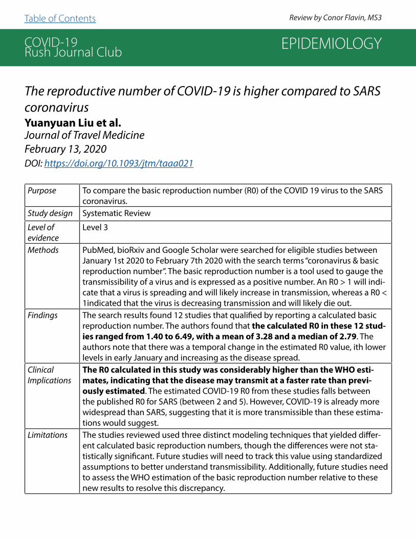
COVID-19Rush Journal Club
The reproductive number of COVID-19 is higher compared to SARS coronavirusYuanyuan Liu et al.Journal of Travel MedicineFebruary 13, 2020DOI: https://doi.org/10.1093/jtm/taaa021
Purpose To compare the basic reproduction number (R0) of the COVID 19 virus to the SARS coronavirus.
Study design Systematic Review
Level of evidence
Level 3
Methods PubMed, bioRxiv and Google Scholar were searched for eligible studies between January 1st 2020 to February 7th 2020 with the search terms “coronavirus & basic reproduction number”. The basic reproduction number is a tool used to gauge the transmissibility of a virus and is expressed as a positive number. An R0 > 1 will indi-cate that a virus is spreading and will likely increase in transmission, whereas a R0 < 1indicated that the virus is decreasing transmission and will likely die out.
Findings The search results found 12 studies that qualified by reporting a calculated basic reproduction number. The authors found that the calculated R0 in these 12 stud-ies ranged from 1.40 to 6.49, with a mean of 3.28 and a median of 2.79. The authors note that there was a temporal change in the estimated R0 value, ith lower levels in early January and increasing as the disease spread.
ClinicalImplications
The R0 calculated in this study was considerably higher than the WHO esti-mates, indicating that the disease may transmit at a faster rate than previ-ously estimated. The estimated COVID-19 R0 from these studies falls between the published R0 for SARS (between 2 and 5). However, COVID-19 is already more widespread than SARS, suggesting that it is more transmissible than these estima-tions would suggest.
Limitations The studies reviewed used three distinct modeling techniques that yielded differ-ent calculated basic reproduction numbers, though the differences were not sta-tistically significant. Future studies will need to track this value using standardized assumptions to better understand transmissibility. Additionally, future studies need to assess the WHO estimation of the basic reproduction number relative to these new results to resolve this discrepancy.
EPIDEMIOLOGY
Review by Conor Flavin, MS3 Table of Contents

COVID-19Rush Journal Club
Are patients with hypertension and diabetes mellitus at increased risk for COVID-19 infection?Lei Fang et al.Lancet: Respiratory MedicineMarch 11, 2020DOI: https://doi.org/10.1016/S2213-2600(20)30116-8
Purpose To explore the theory that comorbidities, specifically hypertension and diabetes, put individuals at greater risk of severe COVID-19 infection.
Study design Systematic Review
Level of evidence
Level 4
Methods The authors reviewed three studies demonstrating increased incidence among severe COVID-19 disease in patients with hypertension (HTN) and diabetes mellitus (DM).
Findings The first study found that of 32 non-survivors from a group of 52 intensive care unit patients, 22% had cerebrovascular disease and 22% had diabetes (RR 1.34 and 1.78, respectively). In the second study, 173 patients with severe disease were found to have comorbidities of HTN (23.7%), DM (16.2%), coronary heart diseases (5.8%), and cerebrovascular disease (2.3%). By contrast, the third study observed comorbidities in all hospitalized patients (total of 140), regardless of COVID severity, with HTN and DM being the most prevalent at 30% and 12% respectively.
ClinicalImplications
Human pathogenic coronaviruses bind to membrane bound angiotensin-con-verting enzyme 2 (ACE2) on endothelial cells expressed in epithelial cells of the lung, intestine, kidney, and blood vessels. So far, evidence suggests COVID-19 uses the same mechanism. Individuals with hypertension and diabe-tes are commonly treated with ACE-inhibitors and angiotensin receptor blockers (ARBs), which lead to the upregulation of ACE2 receptors. ACE2 receptor polymor-phisms have been linked to diabetes and hypertension. With this understanding, the article proposes a hypothesis for the increase risk of severe disease in this patient population.
Limitations Only three studies were reviewed. It was not noted whether patients with severe disease and comorbid hypertension or diabetes were being treated with ACE-in-hibitors or ARBs. Future research could address patient medications in addition to comorbid conditions, as well as genetic testing to determine the existence of a possible link between polymorphisms of ACE2 receptor and severe disease.
EPIDEMIOLOGY
Review by Joseph Dodson, MS1 Table of Contents

COVID-19Rush Journal Club
First-wave COVID-19 transmissibility and severity in China outside Hubei after control measures, and second-wave scenario planning: A modelling impact assessment.Kathy Leung et al.LancetApril 8, 2020DOI: https://doi.org/10.1016/S0140-6736(20)30746-7
Purpose To review the impact and transmissibility of COVID-19 during the early cases of Hubei province (original epicenter) in China.
Study design Retrospective analysis
Level of evidence
Level 4
Methods The authors used publicly available data from four major cities (Beijing, Shanghai, Shenzhem, and Wenzhou) to determine reproduction number (Rt) and confirmed case fatality rate (cCFR). Furthermore, they also incorporated susceptible-infec-tious-recovery models to assess what role relaxing intervention (social distancing, community shutdowns, etc.) had on spread of virus.
Findings Estimates of instantaneous reproduction number (Rt) on weekly intervals were reported between early January and later February 2020. In most provinces the mean Rt decreased after aggressive control measures were implemented, suggesting that reduction in control measures could lead to a resurgence of case numbers. The confirmed case-fatality risk (cCFR) was estimated to be 0.98% (0.82-1.16) among the provinces/cities outside Hubei. In comparison the cCFR in Hubei province was 5.91% and Wuhan was 1.4%.
ClinicalImplications
Relaxation of social control measures increased the cumulative case count exponentially proportional to the duration of relaxation. Furthermore, the dura-tion of aggressive control measures must be longer than the duration of attempted relaxation in order to get the Rt below 1.5. Stated differently, allowing Rt to rise when no herd immunity is present will incur health and economic loss even if future aggressive control measures push prevalence of infection back to the previ-ous level during original aggressive control measures.
Limitations This study used public data only from provinces that actively reported cases. Cases were likely under-reported due lack of testing and resources. Asymptomatic cases of COVID-19 that go undiagnosed would alter the projections of Rt and cCFR. The data obtained did not include specifics regarding exposure or travel history, which could be important to further understanding virus transmission.
EPIDEMIOLOGY
Review by Connor Wakefield, MS3 Table of Contents
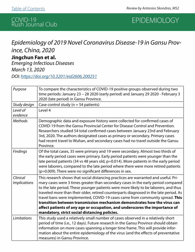
COVID-19Rush Journal Club
Epidemiology of 2019 Novel Coronavirus Disease-19 in Gansu Prov-ince, China, 2020Jingchun Fan et al.Emerging Infectious DiseasesMarch 13, 2020DOI: https://doi.org/10.3201/eid2606.200251
Purpose To compare the characteristics of COVID-19 positive groups observed during two time periods: January 23 – 28 2020 (early period) and January 29 2020 - February 3 2020 (late period) in Gansu Province.
Study design Case control study (n = 54 patients)Level of evidence
Level 4
Methods Demographic data and exposure history were collected for confirmed cases of COVID-19 from the Gansu Provincial Center for Disease Control and Prevention. Researchers studied 54 total confirmed cases between January 23rd and February 3rd, 2020. The authors designated cases as primary or secondary. Primary cases had recent travel to Wuhan, and secondary cases had no travel outside the Gansu Province.
Findings Of the total cases, 35 were primary and 19 were secondary. Almost two thirds of the early period cases were primary. Early period patients were younger than the late period patients (34 vs 48 years old, p=0.014). More patients in the early period were laborers, compared to the late period where there were more retired patients (p=0.009). There were no significant differences in sex.
ClinicalImplications
This research shows that social distancing practices are warranted and useful. Pri-mary cases were 3 times greater than secondary cases in the early period compared to the late period. These younger patients were more likely to be laborers, and thus traveled more than their older, retired counterparts diagnosed in the late period. As travel bans were implemented, COVID-19 cases came from community spread. This transition between transmission mechanism demonstrates how the virus can affect patients of any age or occupation, and underscores the importance of mandatory, strict social distancing policies.
Limitations This study used a relatively small number of cases observed in a relatively short period of time (i.e., 12 days). Future research in the Gansu Province should obtain information on more cases spanning a longer time frame. This will provide infor-mation about the entire epidemiology of the virus (and the effects of preventative measures) in Gansu Province.
EPIDEMIOLOGY
Review by Antonios Skondras, MS2 Table of Contents
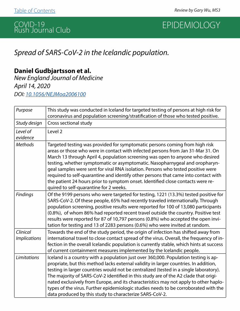
COVID-19Rush Journal Club
Spread of SARS-CoV-2 in the Icelandic population.
Daniel Gudbjartsson et al.New England Journal of MedicineApril 14, 2020DOI: 10.1056/NEJMoa2006100
Purpose This study was conducted in Iceland for targeted testing of persons at high risk for coronavirus and population screening/stratification of those who tested positive.
Study design Cross sectional study
Level of evidence
Level 2
Methods Targeted testing was provided for symptomatic persons coming from high risk areas or those who were in contact with infected persons from Jan 31-Mar 31. On March 13 through April 4, population screening was open to anyone who desired testing, whether symptomatic or asymptomatic. Nasopharnygeal and oropharyn-geal samples were sent for viral RNA isolation. Persons who tested positive were required to self-quarantine and identify other persons that came into contact with the patient 24 hours prior to symptom onset. Identified close contacts were re-quired to self-quarantine for 2 weeks.
Findings Of the 9199 persons who were targeted for testing, 1221 (13.3%) tested positive for SARS-CoV-2. Of these people, 65% had recently traveled internationally. Through population screening, positive results were reported for 100 of 13,080 participants (0.8%), of whom 86% had reported recent travel outside the country. Positive test results were reported for 87 of 10,797 persons (0.8%) who accepted the open invi-tation for testing and 13 of 2283 persons (0.6%) who were invited at random.
ClinicalImplications
Towards the end of the study period, the origin of infection has shifted away from international travel to close contact spread of the virus. Overall, the frequency of in-fection in the overall Icelandic population is currently stable, which hints at success of current containment measures implemented by the Icelandic people.
Limitations Iceland is a country with a population just over 360,000. Population testing is ap-propriate, but this method lacks external validity in larger countries. In addition, testing in larger countries would not be centralized (tested in a single laboratory). The majority of SARS-CoV-2 identified in this study are of the A2 clade that origi-nated exclusively from Europe, and its characteristics may not apply to other haplo-types of the virus. Further epidemiologic studies needs to be corroborated with the data produced by this study to characterize SARS-CoV-2.
EPIDEMIOLOGY
Review by Gary Wu, MS3 Table of Contents

COVID-19Rush Journal Club
Temporal dynamics in viral shedding and transmissibility of COVID-19.Xi He et al.Nature: MedicineApril 15, 2020DOI: https://doi.org/10.1038/s41591-020-0869-5
Purpose To evaluate the viral shedding pattern of COVID-19 and study the periods in which COVID-19 is most transmissible.
Study design Observational, retrospective studyLevel of evidence
Level 4
Methods Two-part study with separate clinical and observational groups. In the clinical group, 94 COVID-19 positive patients from Guangzhou Hospital in Guangdong, China received a nasopharyngeal swab during each day of symptoms to evaluate temporal changes in viral load. Patients had laboratory confirmed COVID-19 by RT-PCR. In the epidemiologic group, 77 pairs of patients who developed COVID-19 were identified to determine the serial interval (time between symptom presenta-tion in infector to symptom presentation in infectee). Case information was gath-ered from publicly available sources in several countries. All infector-infectee pairs were evaluated to ensure that neither party had other contacts who tested positive for COVID-19 or traveled to areas known to have a high number of COVID-19 cases.
Findings Viral load was highest on day one of hospitalization and decreased in a linear fash-ion. Mean serial interval was noted to be 5.8 days, and when taken with a noted incubation period of 5.2 days, COVID-19 infectiousness was noted to begin 2.3 days prior to symptom presentation and peak 0.7 days prior to symptom pre-sentation. Authors estimate that 44% of patients were infected with COVID-19 by a carrier who was asymptomatic at the time of transmission
ClinicalImplications
Viral load was highest on day of COVID-19 diagnosis and decreased linearly. 44% of patients were suspected of contracting COVID-19 from an asymptom-atic carrier.
Limitations Patient pair data in the epidemiologic group was collected by governmental agen-cy reports or via media, this data is possibly inaccurate or incomplete. Cases in this group were not stratified by age, gender, country, or disease severity. Treatments for the clinical group were given according to national health guidelines, and could have affected viral load testing by RT-PCR. Recall bias was likely present in the epi-demiologic group and may have altered incubation period length.
EPIDEMIOLOGY
Review by Steven Heidt, MS4Table of Contents

COVID-19Rush Journal Club
Hospitalization Rates and Characteristics of Patients Hospitalized with Laboratory-Confirmed Coronavirus Disease 2019 – COVID-NET, 14 States, March 1-30, 2020Shikha Garg et al.CDC: Mordibity and Mortality Weekly ReportApril 17, 2020DOI: http://dx.doi.org/10.15585/mmwr.mm6915e3
Purpose To conduct a population-based surveillance for laboratory-confirmed COVID-19-as-sociated hospitalizations in the US.
Study design Retrospective cross-sectional study (n = 1482 patients)
Level of evidence
Level 3
Methods Data included from 1482 hospitalized patients with laboratory confirmed COVID-19, representing 14 states and 90 counties. Study included only the resi-dents of a designated COVID-NET catchment area and hospitalized within 14 days of a positive SARS-CoV-2 test.
Findings Overall, a high proportion of US patients hospitalized with COVID are older (74.5% were aged ≥50 years) and have underlying medical conditions includ-ing hypertension (49.7%), obesity (48.3%), chronic lung disease (34.6%), diabetes mellitus (28.3%), and cardiovascular disease (27.8%). It is therefore imperative to implement preventative measures to especially protect elderly population and those with underlying medical conditions. Among 580 COVID-19 patients with race/ethnicity data, 45% were non-Hispanic white, 33.1% were non-Hispanic blacks, 8.1% were Hispanic, 5.5 were Asian, 0.3% were American Indian/Alaskan Native, 7.9% were unknown. No statistical analysis between groups were performed, but of note, the COVID-NET catchment general population are 59% non-Hispanic white and 18% non-Hispanic black, suggesting the non-Hispanic black population is disproportionally affected by SARS-CoV-2.
ClinicalImplications
Elderly patients with underlying medical conditions are most susceptible to COVID-19 disease and have the worst outcomes
Limitations This study included preliminary results from the first month of US surveillance of COVID-19 and are subject to change as more patients are diagnosed and evaluated. Additionally, patients in this study represented a wide variety of care facilities. With-out standardization of diagnostic testing practices for SARS-CoV-2, under-identifi-cation of COVID-19 cases is likely.
EPIDEMIOLOGY
Review by Elena Perkins, MS3Table of Contents

COVID-19Rush Journal Club
Epidemiology of COVID-19 among children in China
Yuanyuan Dong et al.PediatricsMarch 13, 2020DOI: https://doi.org/10.1542/peds.2020-0702
Purpose To evaluate the epidemiological characteristics and transmission trends of pediatric patients with 2019 novel coronavirus disease (COVID-19) in China.
Study design Observational studyLevel of evidence
Level 3
Methods Researchers collected data on reported COVID-19 pediatric cases from January 16, 2020 to February 8, 2020. Cases were defined as having high, medium, or low risk based on patient exposure to a COVID-19 patient and proximity to endemic areas. Sus-pected cases were identified if a child at high risk had 2 of the following: clinical symp-toms, elevated white blood cell count or CRP, or abnormal chest radiograph. Suspected cases were confirmed by positive RT-PCR nasal swab or blood samples, or by genetic sequencing of respiratory tract or blood samples.
Findings Among 2135 patients, 728 (34.1%) were confirmed cases and 1407 (65.9%) were sus-pected cases. The median age was 7, and complete age range was 1 day to 18 years. Nearly half of total cases were from Hubei province (46.0%), and another 18.5% from surrounding provinces. Of both suspected and confirmed cases, 94 (4.4%), 1088 (51.0%), 826 (38.7%), and 125 (5.8%) cases were diagnosed as asymptomatic, mild, moderate, or severe/critical, respectively. One child died. Compared to adults, there were less severe and critical cases in children (5.8% vs 18.5%). The proportions of severe and critical cases in the pediatric population was highest in infants (10.6%).
ClinicalImplications
Children might be less affected by COVID-19 because the maturity and functioning of ACE-2 in children is lower than adults. They may experience coronavirus infections more frequently and develop cross reacting antibodies, or their immune systems are less developed and therefore have less severe immune responses. The highest pro-portion of critical and severe pediatric COVID-19 cases was in infants less than 1 year old.
Limitations The study was unable to assess clinical characteristics or incubation period due to lack of data at the time of analysis. There were more severe and critical cases in the suspected than confirmed group, suggesting that some suspected cases may be due to other respiratory infections besides COVID-19. Further research containing more detailed patient information and clinical outcomes is needed.
EPIDEMIOLOGY
Review by Grace Alexander, MS2 and Mike Seidman, MS4Table of Contents

COVID-19Rush Journal Club
The epidemiology and pathogenesis of coronavirus disease (COVID-19) outbreakHussin Rothan & Siddappa ByrareddyJournal of AutoimmunityFebruary 26, 2020DOI: https://doi.org/10.1016/j.jaut.2020.102433
Purpose To summarize recent findings regarding the symptoms, epidemiology, transmis-sion, pathogenesis, and future directions of treating/preventing COVID-19.
Study design Systematic review
Level of evidence
Level 3
Methods Reviewed information from COVID-19 case reports and cohort studies.
Findings Viruses in the Coronaviridae family generally presents with fever, dry cough, and dyspnea. Distinct symptoms for COVID-19 include rhinorrhea, sneezing, sore throat, and diarrhea. Elevated C-reactive protein, erythrocyte sedimentation rate, D-dimer, and cytokines and chemokines are generally observed, with severe cases correlating with higher levels of pro-inflammatory cytokines. Transmission occurs primarily via direct contact of virus particles with mucus membranes. Lung epithe-lial cells appear to be the primary target of the virus, and binding occurs between the receptor-binding domain of the spike protein and the angiotensin-converting enzyme II (ACE2) receptor. Of note, the sequence of the receptor-binding domain of COVID-19 spike protein is similar to that of SARS-CoV, which also binds to the ACE2 receptor. Pre-existing drugs including Oseltamivir, Lopinavir, Ritonavir, and Ganciclovir could prove useful in treating COVID-19 patients, as well as Remdesivir and Chloroquine. The EIDD-2801 compound (an isopropylester prodrug of a ribo-nucleoside analog that has shown anti-influenza virus activity in cultured cells and mice), has potential to target seasonal and pandemic viruses.
ClinicalImplications
Correlation between cytokines and disease severity can be used to predict progno-sis, and hopefully prevent adverse outcomes. There is further need for investigation of alternative (fecal oral) routes of transmission, and investigation of pre-existing antiviral drugs as treatment modalities for COVID-19.
Limitations The authors discuss the need to make more testing available in order to further understand the pathogenesis and spread; to study pediatric populations, who have been under-diagnosed; to investigate the vast range of clinical presentations, from virtually asymptomatic ranging to critical condition; and to further research poten-tial viral targets for pharmacologic therapies.
EPIDEMIOLOGY
Review by Alice Burgess, MS2 Table of Contents

COVID-19Rush Journal Club
Mathematical Model Of Infection Kinetics And Its Analysis For COVID-19, SARS And MERS.Kaihao LiangInfection, Genetics and EvolutionApril 8, 2020DOI: https://doi.org/10.1016/j.meegid.2020.104306
Purpose To discuss the spread rules of the three coronavirus epidemics and pandemic: COVID-19, SARS and MERS. A propagation growth model was established using growth rate and inhibition constant of infectious diseases with the parameters of the three coronavirus transmission growth models obtained by nonlinear fitting.
Study design Statistical analysisLevel of evidence
Not applicable
Methods Data on COVID-19 cases were obtained from Hubei, Guangdong, Zhejiang, Henan. Data on SARS cases collected using China’s National data, as well as data from Guangdong, Beijing, Hong Kong. Data from MERS cases analyzed from Saudi Arabia database on 4 distinct cycles of MERS outbreaks. Model hypothesis can be simpli-fied as: r(N) = r0 – s*N, where N = # of infected, r(N) = the growth rate as a function of # of people, r0 = constant indicating growth in the case of no preventative mea-sures, s = infection inhibition coefficient (reflecting control/prevention measures).
Findings The multiplication cycles of SARS and MERS are similar, ranging from 5 to 10 days. The multiplication cycle of COVID-19 is only two to three days, and the number of cases of COVID-19 will increase rapidly under the effect of exponential growth. In Hubei, the infection inhibition constant of COVID-19 is 3.58 × 1e-6 , two orders of magnitude lower than in Guangdong, Zhejiang, and Henan.
ClinicalImplications
The growth rate of COVID-19 is about twice that of SARS and MERS, and the COVID-19 doubling cycle is two to three days, suggesting that the number of COVID-19 patients would double in two to three days without human inter-vention. COVID-19 spread rapidly in Hubei due to the large number of patients in the early stage, as medical institutions dealt with the shortage of hospital beds, equipment, medicine, masks, and protective clothing, resulting in the inability to effectively isolate patients. This led to a lower infection inhibition constant in Hubei.
Limitations This type of modeling seems to have the most use in retrospectively analyzing the pandemic response based on region but may be of more limited use in prospective modeling.
EPIDEMIOLOGY
Review by Travis Tran, MS2Table of Contents

COVID-19Rush Journal Club
Universal Screening for SARS-CoV-2 in Women Admitted for Delivery
Desmond Sutton et al.New England Journal of MedicineApril 13, 2020DOI: 10.1056/NEJMc2009316
Purpose To investigate the prevalence of SARS-CoV-2 amongst pregnant patients upon de-livery, as a vast majority of patients are asymptomatic.
Study design Cross-sectional study (n = 215)
Level of evidence
Level 3
Methods Between March 22 - April 4, 2020, a total of 215 pregnant women delivered infants at the New York–Presbyterian Allen Hospital and Columbia University Irving Medi-cal Center, and all were screened on admission for COVID-19 with quantitative PCR.
Findings Four women (1.9%) had fever or other symptoms on admission, and all 4 women tested positive for SARS-CoV-2. Of the 211 women without symptoms, all were afebrile on admission. Nasopharyngeal swabs were obtained from 210 of the 211 women (99.5%) who did not have symptoms of Covid-19; of these women, 29 (13.7%) were positive for SARS-CoV-2. Thus, 29 of the 33 (87.9%) COVID-19-posi-tive patients were asymptomatic at presentation.
ClinicalImplications
The use of universal SARS-CoV-2 testing in all pregnant patients presenting for delivery revealed that at this point in the pandemic in New York City, most of the patients who were positive for SARS-CoV-2 at delivery were asymptomatic. More than one of every eight asymptomatic patients who were admitted to the labor and delivery unit were positive for SARS-CoV-2. Although this prevalence may not apply to geographic regions with lower rates of infection, it highlights the hid-den prevalence of COVID-19 among asymptomatic obstetrical patients. Universal screening for the pregnant population gives the health system the opportunity to protect mothers, babies, and health care teams as the pandemic continues.
Limitations The true prevalence of SARS-CoV-2 infection may also be underreported because of false negative results of COVID-19 tests. Given the test’s relatively weak sensitivity, future research is needed to understand how patients’ COVID-19 status from RT-PCR should best be used to determine hospital isolation practices and bed assign-ments, inform neonatal care, and guide the use of PPE.
EPIDEMIOLOGY
Review by Alice Burgess, MS2 Table of Contents

COVID-19Rush Journal Club
Duration for carrying SARS-CoV-2 in COVID-19 patients.
Xinwei Du et al.Journal of InfectionApril 10, 2020DOI: https://doi.org/10.1016/j.jinf.2020.03.053
Purpose To evaluate the duration of carrier status for SARS-CoV-2.
Study design Case series (n = 161)
Level of evidence
Level 4
Methods From January 20, 2020 to March 1, 2020, inpatients in Henan province (China) with a specific SARS-CoV-2 epidemiological history and a positive nucleic acid test were identified for data collection. The duration of carrier status was defined as the time from a close contact with the source of infection to the last positive test for nucleic acid. Then, duration of carrying SARS-CoV-2 in different ages, gender, and disease condition were compared.
Findings Patients were contagious at all stages of disease with median carrying duration of 26 days and longest up to 50 days. There was statistically significantly longer carrying duration based on condition and age, with longer duration seen in older age and with severe COVID-19 cases. Duration for carrying SARS-CoV-2 has nothing to do with gender. However, it is related to the age of patients. The median duration in the ≥ 60 years-old group was 28 days vs 20 days in the 0–59 years-old group (P<0.01).
ClinicalImplications
Persistent infection in elderly patients may be the initiating factor that causes or-gan damage, especially persistent inflammation of the alveoli, and disease progres-sion. The long infectious duration of SARS-CoV-2 in patients means that early, purposeful isolation and monitoring are necessary to prevent further spread.
Limitations Limited by sample size, as well as by single location study. Additionally, carrying duration definition could be influenced by recall bias.
EPIDEMIOLOGY
Review by Bryant Yu, MS2 Table of Contents

COVID-19Rush Journal Club
Evolving epidemiology and transmission dynamics of coronavirus disease 2019 outside Hubei province, China: a descriptive and mod-elling study Juanjuan Zhang et al.The Lancet: Infectious DiseasesApril 2, 2020DOI: https://doi.org/10.1016/S1473-3099(20)30230-9
Purpose Characterization of epidemiologic qualities of COVID-19 outside of Hubei province, China fifty days after first noted case outside of the region.
Study design Retrospective, observational study (n = 8579)Level of evidence
Level 4
Methods Information on laboratory positive COVID-19 patients outside of Hubei province, China was collected from national, provincial, and municipal databases as well as from media reports. Cases were divided into two groups based on date of symptom onset: December 24-January 27 and January 28-February 17. Case grouping was based on fourth revision of COVID-19 case definition. In comparing the two groups, authors analyzed differences in case demographics and time-to-event intervals, such as time to hospitalization. Incubation period and serial interval were identified from subsets of the study sample.
Findings As the epidemic progressed, time to hospitalization and time to first healthcare consultation decreased; specifically, time to hospitalization fell from 4.4 days in the first period to 2.6 days in the second period (P<0.0001). Incubation period was estimated at 5.2 days from 49 individuals via contact tracing. Serial interval was es-timated at 5.1 days from 28 index cases and 35 secondary cases. Net reproduction number differed between provinces and cities, ranging from 1.08 to 1.71 in areas under study. Reproduction number in all studied areas fell below 1 at the end of January 2020, indicating that disease did not spread in the community anymore.
ClinicalImplications
Isolation measures have an effect on healthcare ascertainment, and epidemiologic characteristics of COVID-19 are varied outside of Hubei province.
Limitations Data were collected from governmental databases and media reports, though the authors did validate their cases by examining the official line lists for cities and provinces for which reproduction number was calculated. Study data were collect-ed during an infectious disease outbreak and therefore are subject to non-homog-enous sampling and case ascertainment bias.
EPIDEMIOLOGY
Review by Steven Heidt, MS4 Table of Contents
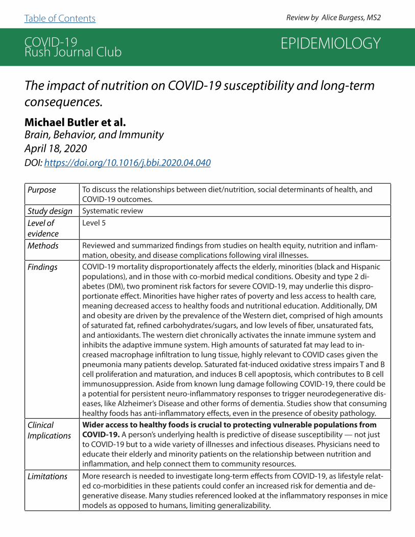
COVID-19Rush Journal Club
The impact of nutrition on COVID-19 susceptibility and long-term consequences.Michael Butler et al.Brain, Behavior, and ImmunityApril 18, 2020DOI: https://doi.org/10.1016/j.bbi.2020.04.040
Purpose To discuss the relationships between diet/nutrition, social determinants of health, and COVID-19 outcomes.
Study design Systematic review
Level of evidence
Level 5
Methods Reviewed and summarized findings from studies on health equity, nutrition and inflam-mation, obesity, and disease complications following viral illnesses.
Findings COVID-19 mortality disproportionately affects the elderly, minorities (black and Hispanic populations), and in those with co-morbid medical conditions. Obesity and type 2 di-abetes (DM), two prominent risk factors for severe COVID-19, may underlie this dispro-portionate effect. Minorities have higher rates of poverty and less access to health care, meaning decreased access to healthy foods and nutritional education. Additionally, DM and obesity are driven by the prevalence of the Western diet, comprised of high amounts of saturated fat, refined carbohydrates/sugars, and low levels of fiber, unsaturated fats, and antioxidants. The western diet chronically activates the innate immune system and inhibits the adaptive immune system. High amounts of saturated fat may lead to in-creased macrophage infiltration to lung tissue, highly relevant to COVID cases given the pneumonia many patients develop. Saturated fat-induced oxidative stress impairs T and B cell proliferation and maturation, and induces B cell apoptosis, which contributes to B cell immunosuppression. Aside from known lung damage following COVID-19, there could be a potential for persistent neuro-inflammatory responses to trigger neurodegenerative dis-eases, like Alzheimer’s Disease and other forms of dementia. Studies show that consuming healthy foods has anti-inflammatory effects, even in the presence of obesity pathology.
ClinicalImplications
Wider access to healthy foods is crucial to protecting vulnerable populations from COVID-19. A person’s underlying health is predictive of disease susceptibility — not just to COVID-19 but to a wide variety of illnesses and infectious diseases. Physicians need to educate their elderly and minority patients on the relationship between nutrition and inflammation, and help connect them to community resources.
Limitations More research is needed to investigate long-term effects from COVID-19, as lifestyle relat-ed co-morbidities in these patients could confer an increased risk for dementia and de-generative disease. Many studies referenced looked at the inflammatory responses in mice models as opposed to humans, limiting generalizability.
EPIDEMIOLOGY
Review by Alice Burgess, MS2Table of Contents

COVID-19Rush Journal Club
Asymptomatic SARS-CoV-2 infection in household contacts of a healthcare provider, Wuhan, China.Yi Luo et al.Emerging Infectious DiseasesApril 24, 2020DOI: https://doi.org/10.3201/eid2608.200282
Purpose To evaluate the case of a physician with mildly symptomatic COVID-19 and their household contacts in Wuhan, China.
Study design Case series (n = 6)Level of evidence
Level 3
Methods The patient (patient 1) was a 39-year-old nephrologist at the Central Hospital of Wuhan. The patient’s five household members included were his wife (contact 1), a 37-year-old laboratory scientist, 7-year-old fraternal twins (contacts 2 & 3), a 62-year-old retired grandfather who was a current smoker (contact 4), and a retired 64-year-old grandmother in good health (contact 5). All household contacts underwent CT scans, and then daily throat swabs for qRT-PCR test for SARS-CoV-2 during the the obser-vation period: February 11th-March 1st, 2020. Blood was taken for laboratory tests: C-reactive protein, leukocyte count, lymphocyte ratio, CD19+ absolute count, alanine aminotransferase (ALT), aspartate aminotransferase (AST), and D-Dimer.
Findings All household contacts of patient 1 had qRT-PCR confirmed SARS-CoV-2 and were asymptomatic throughout the observation period. Stool specimens for contacts 1, 2, and 3 were positive for SARS-CoV-2. Contact 2 tested negative on 4 consecutive throat swab PCRs but had a positive stool specimen for SARS-CoV-2. Contact 1 was positive for SARS-CoV-2 on qRT-PCR tests of multiple serial throat swabs but negative on IgM and IgG tests. Contact 1 underwent 11 serial throat swabs and on 2 separate occasions, she had 2 consecutive negative results for SARS-CoV-2 only to revert to having a throat swab specimen positive for SARS-CoV-2.
ClinicalImplications
Data about the incidence of asymptomatic infection in families of healthcare workers can help to construct a more informed public health response during the pandemic. There may be underestimation of positive cases, as this study illustrated negative qRT-PCR tests that subsequently tested positive. Studies on serial testing on as-ymptomatic patients with a negative test should be conducted to see if they are truly negative. More research on fecal-oral transmission of SARS-CoV-2 is necessary, as three household contacts in this study had positive stool specimens.
Limitations This study was limited by its small sample size, as it focused on asymptomatic trans-mission amongst household contacts of a healthcare provider.
EPIDEMIOLOGY
Review by Antonios Skondras, MS2 Table of Contents

COVID-19Rush Journal Club
High contagiousness and rapid spread of severe acute respiratory syndrome coronavirus 2.Steven Sanche et al.Emerging Infectious DiseasesApril 7, 2020DOI: https://doi.org/10.3201/eid2607.200282
Purpose To estimate key epidemiological parameters of the early outbreak of 2019 novel coronavi-rus disease (COVID-19) in Wuhan.
Study design Case series (n= 140)
Level of evidence
Level 4
Methods 140 case reports from across China were collected from the Chinese Centers for Disease Control and Prevention (China CDC) and provincial health commissions between January 15-30, 2020. Two models were developed to estimate the growth rate of the outbreak in Wuhan. First arrival model: domestic travel data from Baidu Migration server was used to compute the likelihood of arrival times of first known cases in provinces outside of Hubei, as a function of the rate of epidemic growth in Wuhan. Case count model: a hybrid deter-ministic-stochastic susceptible-exposed-infectious-recovered (SEIR) model was used to account for additional persons infected in Wuhan but diagnosed in other provinces. Dates for this model were restricted to January 19-26, 2020. Both models calculated growth rate (r), and theoretical time (T0), using travel data, specifically the earliest times that an infect-ed person arrived in each of the 26 provinces.
Findings The incubation period was estimated to be 4.2 days. The time from symptom onset to hospitalization decreased from 5.5 days before January 18 2020, to 1.5 days after January 18 2020. The change coincides with report of human to human transmission and upgrade of the emergency response to level 1 by the Chinese CDC. The time from initial hospital admittance to discharge was 11.5 days, initial hospital admittance to death was 11.2 days, and time from symptom onset to death was 16.1 days. The growth rate in the first arriv-al model was 0.29/day, corresponding to a doubling time of 2.4 days. This estimate was much higher than previous estimates of 0.10-0.14/day. The case count model and first arrival model both estimated consistent exponential growth rates and theoretical times: 0.29 vs 0.30/day, and December 20 vs December 16 2019.
ClinicalImplications
Results suggest that surveillance, quarantine, and strong social distancing efforts are essential for slowing down or stopping the spread of this virus.
Limitations Because the reports collected were from the first few persons detected in each province, the estimations may be biased toward cases with more severe symptoms. In both models, researchers assumed perfect detection of infected cases outside of Hubei Province - how-ever in reality, this may not be the case due to changing surveillance intensity.
EPIDEMIOLOGY
Review by Grace Alexander, MS2 Table of Contents

COVID-19Rush Journal Club
A systematic review of COVID-19 epidemiology based on current evidenceMinah Park et al.Journal of Clinical MedicineMarch 31, 2020DOI: https://doi.org/10.3390/jcm9040967
Purpose To summarize epidemiological characteristics of SARS-CoV-2 and the effectiveness of control measures.
Study design Systematic reviewLevel of evidence
Level 5
Methods Authors used the following terms in PubMed and preprint archives to find research articles published up to February 21, 2020: “COVID-19”, “SARS-CoV-2”, “2019-nCoV”, “n-CoV”, and “coronavirus.” Of 317 research articles generated from the initial search, 41 met the inclusion criteria.
Findings Basic reproduction number (R0; expected number of cases generated from 1 case) ranged from 1.9 to 6.5 based on eight published and eight preprint papers. The estimates of R0 for SARS-CoV in the early phase of the outbreak in Hong Kong was 2.7 and in Singapore as 2.2–3.6. In the studies of SARS-CoV-2, the final attack rate would lie between 75% and 100% in a completely susceptible population assum-ing no intervention. Median incubation period of the included studies ranged from 4 to 6 days. This is comparable to SARS-CoV (4.4 days) and MERS-CoV (5.5 days). Serial interval (time between the start of symptoms in the infector and start of symptoms in the infectee) range from 4 to 8 days. Pre-symptomatic infection is possible, given that the estimated serial interval is shorter than the incuba-tion period. An analysis of 468 infector–infectee pairs confirmed in China reported a mean serial interval of 3.96 days. The study also noted that 59 of 468 pairs (12.6%) had negative-valued serial intervals, suggesting pre-symptomatic transmission.
ClinicalImplications
Infected patients may not display symptoms for 4-6 days and have the po-tential to spread COVID before they demonstrate symptoms, therefore both symptomatic and asymptomatic patients should strictly adhere to social distancing guidelines.
Limitations Not all studies have been peer-reviewed. Additionally, the studies included did not use the same method of calculating the basic reproduction number (R0). Most of the studies included in this review are based on data collected during the ear-ly phase of the outbreak. For some of that time, there were no social distancing guidelines in effect.
EPIDEMIOLOGY
Review by Kelly Harmon, MS1 Table of Contents
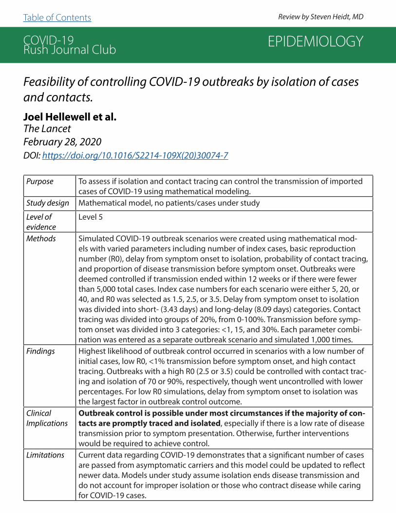
COVID-19Rush Journal Club
Feasibility of controlling COVID-19 outbreaks by isolation of cases and contacts.Joel Hellewell et al.The LancetFebruary 28, 2020DOI: https://doi.org/10.1016/S2214-109X(20)30074-7
Purpose To assess if isolation and contact tracing can control the transmission of imported cases of COVID-19 using mathematical modeling.
Study design Mathematical model, no patients/cases under study
Level of evidence
Level 5
Methods Simulated COVID-19 outbreak scenarios were created using mathematical mod-els with varied parameters including number of index cases, basic reproduction number (R0), delay from symptom onset to isolation, probability of contact tracing, and proportion of disease transmission before symptom onset. Outbreaks were deemed controlled if transmission ended within 12 weeks or if there were fewer than 5,000 total cases. Index case numbers for each scenario were either 5, 20, or 40, and R0 was selected as 1.5, 2.5, or 3.5. Delay from symptom onset to isolation was divided into short- (3.43 days) and long-delay (8.09 days) categories. Contact tracing was divided into groups of 20%, from 0-100%. Transmission before symp-tom onset was divided into 3 categories: <1, 15, and 30%. Each parameter combi-nation was entered as a separate outbreak scenario and simulated 1,000 times.
Findings Highest likelihood of outbreak control occurred in scenarios with a low number of initial cases, low R0, <1% transmission before symptom onset, and high contact tracing. Outbreaks with a high R0 (2.5 or 3.5) could be controlled with contact trac-ing and isolation of 70 or 90%, respectively, though went uncontrolled with lower percentages. For low R0 simulations, delay from symptom onset to isolation was the largest factor in outbreak control outcome.
ClinicalImplications
Outbreak control is possible under most circumstances if the majority of con-tacts are promptly traced and isolated, especially if there is a low rate of disease transmission prior to symptom presentation. Otherwise, further interventions would be required to achieve control.
Limitations Current data regarding COVID-19 demonstrates that a significant number of cases are passed from asymptomatic carriers and this model could be updated to reflect newer data. Models under study assume isolation ends disease transmission and do not account for improper isolation or those who contract disease while caring for COVID-19 cases.
EPIDEMIOLOGY
Review by Steven Heidt, MD Table of Contents

COVID-19Rush Journal Club
Clinical characteristics and risk assessment of newborns born to mothers with COVID-19.Pu Yang et al.Journal of Clinical VirologyApril 5, 2020DOI: https://doi.org/10.1016/j.jcv.2020.104356
Purpose To report clinical outcomes of newborns delivered by SARS-CoV-2 positive preg-nant women, and to determine the risk of vertical transmission of SARS-CoV-2.
Study design Prospective cohort studyLevel of evidence
Level 2
Methods Single institutional study of newborns (n=7) delivered between January 20-29 2020 by SARS-Cov-2 infected pregnant women. Delivery history including APGAR scores, clinical features, blood tests, nuclei acid detection of amniotic fluid, umbilical cord blood, and neonatal pharyngeal swabs were collected for risk assessment analysis.
Findings Six of 7 SARS-CoV-2 positive mothers were symptomatic 1-6 days before delivery. Symptoms included fever, cough, and other respiratory symptoms followed by abdominal pain, diarrhea and other gastrointestinal symptoms. All 7 women de-livered via Cesarean section: 2 were emergency cesarean section for severe pre-eclampsia, 1 was for severely elevated aspartate aminotransferase (AST), 4 were for risk reduction of potential SARS-CoV-2 vertical transmission from mother to baby. Four newborns were delivered late preterm (36-37 weeks) and 3 newborns were delivered full term. Of 5 neonates admitted to the neonatal intensive care unit (NICU), 3 underwent chest x-ray. Eventually all 7 newborns were discharged for isolated home care within 7 days of birth. All 7 newborns tested negative for SARS-CoV-2 via qRT-PCR in amniotic fluid, cord blood, and pharyngeal swabs.
ClinicalImplications
Current data does not demonstrate that pregnant women infected with SARS-CoV-2 pose a risk of vertical transmission or other severe adverse events to their newborns. However, newborns are still at high risk for infection once deliv-ered, thus it is necessary to separate newborns from their mothers to avoid poten-tial complications of infection.
Limitations The sample size of this study was small. Not all newborns received a chest x-ray, which may have missed non-symptomatic presentations on imaging. The short length of observation in the hospital may have missed subsequent manifestations of COVID-19. All patients delivered by cesarean section, and it is unknown at this time whether elective cesarean section is required to prevent vertical transmission of SARS-CoV-2 from mother to baby.
EPIDEMIOLOGY
Review by Gary Wu, MS3 Table of Contents

COVID-19Rush Journal Club
Factors associated with prolonged viral RNA shedding in patients with COVID-19.Kaijin Xu et al.Clinical Infectious DiseasesApril 9, 2020DOI: https://doi.org/10.1093/cid/ciaa351
Purpose To assess viral RNA clearance within 21 days after illness onset and to determine factors associ-ated with prolonged viral RNA shedding in SARS-CoV-2 patients.
Study design Retrospective cohort (n = 113)
Level of evidence
Level 3
Methods Authors analyzed 113 patients with confirmed SARS-CoV-2 infection across 2 Wuhan hospi-tals who met one of three inclusion criteria: 1) disease duration >21 days without viral RNA clearance; 2) viral RNA clearance occurred within 21 days; 3) death occurred within 21 days. Duration of viral RNA shedding was considered the number of days from symptom onset viral RNA clearance using RT-PCR assay. Viral RNA clearance was defined as 3 consecutive negative respiratory tract specimens.
Findings 74.3% (84/113) of patients had viral RNA clearance within 21 days after illness onset, with median duration of viral RNA detection being 17 days. The 84 patients with viral RNA clear-ance within 21 days were further divided into persistent negative viral detection results <15 days after illness onset (n=37) and prolonged viral RNA shedding ≥15 days after illness on-set (n=79). Prolonged viral RNA shedding was associated with male sex (P=0.009), older age (median age of group with prolonged shedding was 54.5 years old compared to 48 years old, P=0.033), and concomitant hypertension (P=0.009). Treatment with corticosteroid (P=0.025) and invasive mechanical ventilation (P=0.006) were also related to prolonged shedding. Sever-ity of disease was related to prolonged shedding with 34.2% of patients with severe disease at admission (defined as patients with severe pneumonia, ARDS, or sepsis) having prolonged shedding compared to 16.2% of severe patients who did not (P=0.049). Multivariate analysis of the variables with statistical significance (P<0.05) showed that time from illness onset to hospital admission (odds ratio, OR, 1.30, P=0.002) and male sex (OR, 3.24, P=0.011) were independent risk factors for prolonged viral RNA shedding.
ClinicalImplications
Further investigation into the sex-related dimorphism of COVID-19 is needed. This study recommends that symptomatic patients should be admitted to the hospital as early as possible if SARS-CoV-2 infection is confirmed as time from illness onset to hospital admis-sion was found to be an independent risk factor for prolonged viral RNA shedding.
Limitations This study was limited by small sample size. Additionally, viral RNA shedding is not the same as viral shedding. It is unclear how shedding of viral RNA correlates with shedding of infec-tious virus. For patients with invasive mechanical ventilation, lower respiratory tract specimens (endotracheal aspirate or bronchoalveolar lavage) were collected and viral RNA shedding may not be equivocal to specimens collected in sputum.
EPIDEMIOLOGY
Review by Elena Perkins, MS3 Table of Contents
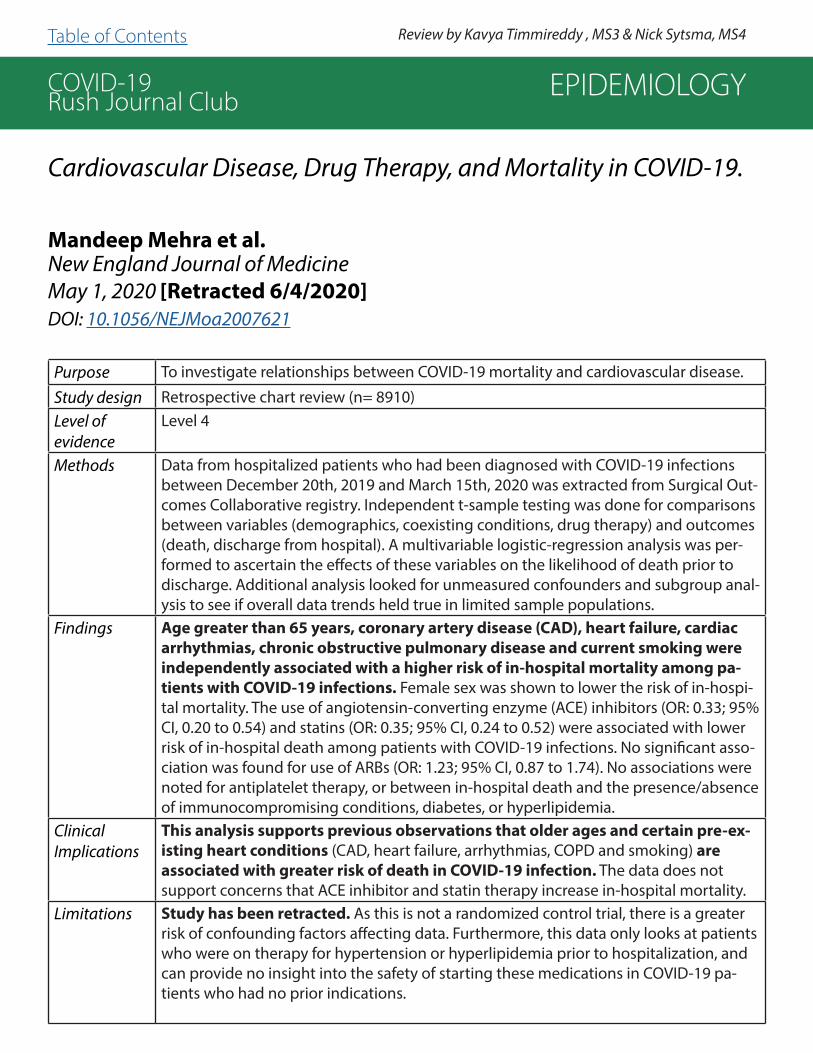
COVID-19Rush Journal Club
Cardiovascular Disease, Drug Therapy, and Mortality in COVID-19.
Mandeep Mehra et al.New England Journal of MedicineMay 1, 2020 [Retracted 6/4/2020]DOI: 10.1056/NEJMoa2007621
Purpose To investigate relationships between COVID-19 mortality and cardiovascular disease.
Study design Retrospective chart review (n= 8910)Level of evidence
Level 4
Methods Data from hospitalized patients who had been diagnosed with COVID-19 infections between December 20th, 2019 and March 15th, 2020 was extracted from Surgical Out-comes Collaborative registry. Independent t-sample testing was done for comparisons between variables (demographics, coexisting conditions, drug therapy) and outcomes (death, discharge from hospital). A multivariable logistic-regression analysis was per-formed to ascertain the effects of these variables on the likelihood of death prior to discharge. Additional analysis looked for unmeasured confounders and subgroup anal-ysis to see if overall data trends held true in limited sample populations.
Findings Age greater than 65 years, coronary artery disease (CAD), heart failure, cardiac arrhythmias, chronic obstructive pulmonary disease and current smoking were independently associated with a higher risk of in-hospital mortality among pa-tients with COVID-19 infections. Female sex was shown to lower the risk of in-hospi-tal mortality. The use of angiotensin-converting enzyme (ACE) inhibitors (OR: 0.33; 95% CI, 0.20 to 0.54) and statins (OR: 0.35; 95% CI, 0.24 to 0.52) were associated with lower risk of in-hospital death among patients with COVID-19 infections. No significant asso-ciation was found for use of ARBs (OR: 1.23; 95% CI, 0.87 to 1.74). No associations were noted for antiplatelet therapy, or between in-hospital death and the presence/absence of immunocompromising conditions, diabetes, or hyperlipidemia.
ClinicalImplications
This analysis supports previous observations that older ages and certain pre-ex-isting heart conditions (CAD, heart failure, arrhythmias, COPD and smoking) are associated with greater risk of death in COVID-19 infection. The data does not support concerns that ACE inhibitor and statin therapy increase in-hospital mortality.
Limitations Study has been retracted. As this is not a randomized control trial, there is a greater risk of confounding factors affecting data. Furthermore, this data only looks at patients who were on therapy for hypertension or hyperlipidemia prior to hospitalization, and can provide no insight into the safety of starting these medications in COVID-19 pa-tients who had no prior indications.
EPIDEMIOLOGY
Review by Kavya Timmireddy , MS3 & Nick Sytsma, MS4Table of Contents

COVID-19Rush Journal Club
Clinical characteristics of pregnant women with Covid-19 in Wuhan, China.Lian Chen et al.New England Journal of MedicineApril 17, 2020DOI: 10.1056/NEJMc2009226
Purpose To analyze clinical characteristics of pregnant women with COVID-19.
Study design Case series (n=118)
Level of evidence
Level 3
Methods Data collected from December 8, 2019 to March 20, 2020,on pregnant women in Wuhan, China that met the criteria of the Chinese Clinical Guidance for Covid-19 Pneumonia Diagnosis and Treatment was analyzed. The women were grouped as to having severe or nonsevere disease and parameters regarding symptoms and pregnancy outcome were compared . Diagnosis was based on positive PCR testing (71%) or from suggestive CT findings (29%).
Findings In this cohort of pregnant women from Wuhan (China), 92% had non-severe dis-ease, and 8% had severe disease with hypoxemia. 95% were symptomatic, with fever (75%) and cough (73%) being most common. Of those who delivered (58%), 93% delivered via Cesarean section due to obstetrical indications (39%) or con-cerns about COVID-19 (61%). 35% of patients still had ongoing pregnancy at the end of the study period, and 8% of mothers had an abortion. Median APGAR score at 1 minute was 9. No deaths were recorded amongst mothers or neonates during study period. Testing for SARS-CoV-2 performed on 8 neonatal throat swabs and 3 breast milk samples were negative.
ClinicalImplications
Unlike influenza (H1N1), where pregnant women are more likely to experience se-vere symptoms and complications (Creanga AA, et al., Obstet Gynecol 115:717-26, 2010), SARS-CoV-2 infection is not associated with an increased risk of severe disease among pregnant women nor adverse neonatal outcomes.
Limitations This analysis was limited by sample size, as well as by single location study. Future studies looking at long-term follow-up for neonates should be encouraged to eval-uate any effect of maternal COVID-19 infection on the infant’s development.
EPIDEMIOLOGY
Review by Bryant Yu, MS3 Table of Contents
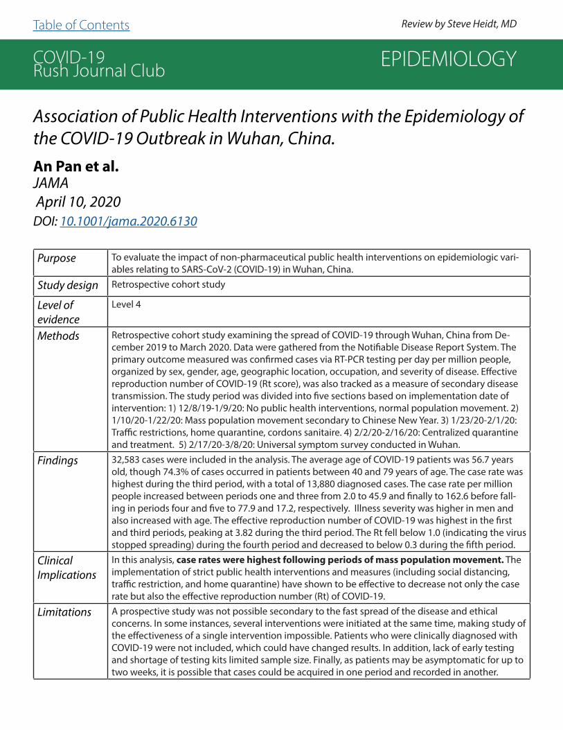
COVID-19Rush Journal Club
Association of Public Health Interventions with the Epidemiology of the COVID-19 Outbreak in Wuhan, China.An Pan et al.JAMA April 10, 2020DOI: 10.1001/jama.2020.6130
Purpose To evaluate the impact of non-pharmaceutical public health interventions on epidemiologic vari-ables relating to SARS-CoV-2 (COVID-19) in Wuhan, China.
Study design Retrospective cohort study
Level of evidence
Level 4
Methods Retrospective cohort study examining the spread of COVID-19 through Wuhan, China from De-cember 2019 to March 2020. Data were gathered from the Notifiable Disease Report System. The primary outcome measured was confirmed cases via RT-PCR testing per day per million people, organized by sex, gender, age, geographic location, occupation, and severity of disease. Effective reproduction number of COVID-19 (Rt score), was also tracked as a measure of secondary disease transmission. The study period was divided into five sections based on implementation date of intervention: 1) 12/8/19-1/9/20: No public health interventions, normal population movement. 2) 1/10/20-1/22/20: Mass population movement secondary to Chinese New Year. 3) 1/23/20-2/1/20: Traffic restrictions, home quarantine, cordons sanitaire. 4) 2/2/20-2/16/20: Centralized quarantine and treatment. 5) 2/17/20-3/8/20: Universal symptom survey conducted in Wuhan.
Findings 32,583 cases were included in the analysis. The average age of COVID-19 patients was 56.7 years old, though 74.3% of cases occurred in patients between 40 and 79 years of age. The case rate was highest during the third period, with a total of 13,880 diagnosed cases. The case rate per million people increased between periods one and three from 2.0 to 45.9 and finally to 162.6 before fall-ing in periods four and five to 77.9 and 17.2, respectively. Illness severity was higher in men and also increased with age. The effective reproduction number of COVID-19 was highest in the first and third periods, peaking at 3.82 during the third period. The Rt fell below 1.0 (indicating the virus stopped spreading) during the fourth period and decreased to below 0.3 during the fifth period.
ClinicalImplications
In this analysis, case rates were highest following periods of mass population movement. The implementation of strict public health interventions and measures (including social distancing, traffic restriction, and home quarantine) have shown to be effective to decrease not only the case rate but also the effective reproduction number (Rt) of COVID-19.
Limitations A prospective study was not possible secondary to the fast spread of the disease and ethical concerns. In some instances, several interventions were initiated at the same time, making study of the effectiveness of a single intervention impossible. Patients who were clinically diagnosed with COVID-19 were not included, which could have changed results. In addition, lack of early testing and shortage of testing kits limited sample size. Finally, as patients may be asymptomatic for up to two weeks, it is possible that cases could be acquired in one period and recorded in another.
EPIDEMIOLOGY
Review by Steve Heidt, MD Table of Contents
COVID-19Rush Journal Club
Modified SEIR and AI prediction of the epidemics trend of COVID-19 in China under public health interventions.Zifeng Yang et al.Journal of Thoracic DiseaseFebruary 28, 2020DOI: 10.21037/jtd.2020.02.64
Purpose To evaluate effectiveness of control measures implemented in China on January 23, 2020 (including quarantines, travel limitations, closure of public spaces, and tem-perature monitoring) in reducing the spread of the COVID-19 epidemic.
Study design Modified Susceptible-Exposed-Infected-Removed (SEIR) ModelLevel of evidence
Level 5
Methods Epidemiological data retrieved from the National Health Commission of China was combined with population migration data before and after January 23, 2020 (in-bound/outbound rail, air, and road traffic) obtained from a web-based program. These data were applied to the SEIR model with an estimated incubation period of 7 days and estimated values for the probability of transmission, recovery, and death based on epidemic data from Hubei Provence. The predictions from this model were corroborated by artificial intelligence (AI) trained on data obtained from the 2003 SARS outbreak.
Findings The model predicted that a five-day delay in the implementation of China’s control measures would have resulted in a three-fold increase in the number of coronavirus cases, suggesting that the measures put in place by the Chinese government did in fact have a positive impact on the course of the pandemic. Additionally, modeling predicted that relaxing the control measures at the time of publication would lead to a second wave of infections in Hubei Provence.
ClinicalImplications
This study adds to existing evidence that social distancing, travel restrictions, and mandatory quarantines are actively working to reduce the spread of the novel coronavirus. Most importantly, it predicts that ending restrictions too soon could result in a resurgence of the virus within the population.
Limitations The study was limited by its reliance on data gathered by the government, as the true number of COVID-positive individuals in the population most likely exceed-ed what was reported based on lack of available diagnostic testing. Moreover, the model used early estimates of incubation time for the virus of 7 days, while more recent data suggests a median incubation time before symptom onset is 3 days. Also, the study did not take phase-adjusted preventive measures and time-varying parameters into consideration, which may affect the accuracy of predictions.
EPIDEMIOLOGY
Review by Susan Mari, MS2Table of Contents

COVID-19Rush Journal Club
Estimating clinical severity of COVID-19 from the transmission dy-namics in Wuhan, China.Joseph Wu et al.Nature: MedicineMarch 19, 2020DOI: https://doi.org/10.1038/s41591-020-0822-7
Purpose To synthesize confirmed and published coronavirus case data from mainland China to estimate the clinical severity of COVID-19 using a transmission dynamics model.
Study design Retrospective observational study (n = 48,557)
Level of evidence
Level 3
Methods Collected case data from multiple Chinese studies. The prevalence of infection in travelers on flights before January 19th and on charter flights from January 19th-February 4th were used to estimate infection prevalence. Authors used the first 425 cases in Wuhan, China to estimate the growth rate of the epidemic. Clinical severity was measured by infection fatality risk (IFR; defines a case as a person who would, if tested, be counted as infected and rendered immune), symptomat-ic case fatality risk (sCFR; defines a case as someone who is infected and shows certain symptoms), and hospitalization fatality risk (HFR; defines a case as someone who is infected and hospitalized).
Findings Age-specific sCFRs and susceptibility to symptomatic infection both increased substantially with age. Assuming that the probability of developing symptoms after an infection (Psym) is 0.5, the sCFR values are 0.3% in those age <30, 0.5% in those age 30-59, and 2.6% in those age >59. The overall sCFR was 1.4%, which was lower than the corresponding confirmed case fatality risk of 4.5% (2,169 deaths/48,557 confirmed cases in Wuhan). Those aged <30 or >59 years are 0.16 and 2.0 times more susceptible to symptomatic infection than their middle aged counterparts. Using the Psym=0.5 parameter, the mean reproductive number (the number of new cases generated by a single case) is 1.94. The mean time from symptom onset to death is 20 days, with a standard deviation of 10 days. The epidemic doubling time was 5.2 days before Wuhan was quarantined. In comparison to the SARS virus, whose IFR and sCFR are essentially the same as the HFR, the sCFR is substantially lower than the HFR for COVID-19. Despite a lower sCFR, COVID-19 is still likely to be the cause of more deaths than SARS and MERS due to the larger number of people infected by COVID-19.
ClinicalImplications
For COVID-19, sCFR is highest in the >59 age group. Unlike previously reported pandemics (SARS and MERS) or influenza, the risk of symptomatic infection also increases with age.
Limitations The study uses an arbitrary Psym of 0.5. If the Psym were higher, for example 0.75 or 0.95, that would yield an overall sCFR of 1.3% and 1.2%, respectively. Although age-specific sCFRs are not susceptible to changes in Psym, susceptibility to symptomatic infection is unknown. The study uses population case data collected in all of mainland China prior to Wuhan undergoing quaran-tine. First, the data might not be generalizable to different locations outside of China. Secondly, it is unknown whether quarantine measures, social distancing, and other precautions of infection control are effective in changing the predicted outcome measure of sCFR, which should be ex-plored in future studies.
EPIDEMIOLOGY
Review by Gary Wu, MS3 Table of Contents
COVID-19Rush Journal Club
SARS-CoV-2 infection (COVID-19) in febrile infants without respira-tory distress.Michal Paret et al.Clinical Infectious DiseasesApril 17, 2020DOI: https://doi.org/10.1093/cid/ciaa452
Purpose To analyze SARS-CoV-2 infection in infants.Study design Case series (n= 2)
Level of evidence
Level 4
Methods A real-time RT-PCR assay performed to detect SARS-CoV-2 RNA in infants’ nasopha-ryngeal (NP) samples. Samples of blood, urine, and CSF were obtained for labora-tory evaluation including white blood cell, platelet, neutrophil, lymphocyte and monocyte counts, hemoglobin and C-reactive protein concentrations.
Findings SARS-CoV-2 has been detected in two young infants with fever as the only mani-festation: First infant was a 25-day old full-term male. He had fever, and erythema-tous, papular facial rash. Empiric therapy with parenteral ampicillin and cefepime was started on admission and continued until blood, urine, and CSF cultures were negative for > 48 hours. Second infant was a 56-day old full-term male. He was presented to the hospital with fever. Empiric therapy with parenteral ceftriaxone continued until blood and urine cultures were negative for > 36 hours. Both infants were discharged home in stable condition.
ClinicalImplications
In young infants, SARS-CoV-2 can cause fever without any other manifesta-tions, including respiratory symptoms and signs. This suggest that children are either less likely to have been exposed to the virus (due to an un-identified recep-tors or co-receptors, which are differently distributed in adults and infants and binds SARS-CoV-2 more efficiently in adults) or that there is a different mechanism in child’s body, which responds to the virus in a less-dramatic way (e.g., children’s immune system might not be mature yet to start a cytokine storm similar to the one observed in adults).
Limitations Limited by sample size, as well as by single center study.
EPIDEMIOLOGY
Review by Bryant Yu, MS3 Table of Contents
COVID-19Rush Journal Club
Demographic science aids in understanding the spread and fatality rates of COVID-19Jennifer Beam Dowd et al.PNASApril 16, 2020DOI: https://doi.org/10.1073/pnas.2004911117
Purpose To explain how average country population age influences COVID-19 national case fatality rate (CFR).
Study design Disease projections from publicly available data. No patients under study.
Level of evidence
Level 5
Methods Authors created models of population size and expected population deaths in Italy, South Korea, Brazil, Nigeria, and the United States. Direct comparison models were created for all countries aside from the USA. Head-to-head comparisons were made between Italy and South Korea as well as between Brazil and Nigeria. Projec-tions assumed a 10% population infection rate and the age-sex specific CFR seen in Italy. Separate projections of deaths per 10-year age group were made using three countries of differing average age (Italy, USA, and Nigeria) and two separate infec-tion rates (10 and 40%).
Findings Head-to-head comparison of population age and expected deaths, countries with a higher proportion of older citizens had a far higher number of expected deaths. Brazil, which has a higher population percentage over 80 years of age when com-pared to Nigeria (2.0% vs 0.2%), also has a dramatically more COVID-19 deaths than Nigeria (452,694 vs 142,056). In evaluating deaths by ten-year age group, deaths were highest in the Italian population (highest proportion of elderly) and lowest in the Nigerian population (lowest proportion of elderly).
ClinicalImplications
Demographic evaluation and population analysis aids in understanding the COVID-19 fatality rates. COVID-19 CFR is likely to be highest in countries and re-gions with a higher percentage of older citizens. Social distancing guidelines, particularly avoidance of intergenerational contact, may need to be more strictly enacted in these areas.
Limitations Models were created using CFRs specific to Italy as of the date of publication. Ex-trapolation of data may inappropriately estimate the number of expected deaths related to COVID-19, as the true CFR will not be known until the end of the out-break. Patient characteristics, such as comorbidities or social history, nor isolation characteristics, such as level of social distancing, were not included in these models and therefore expected death rate may be incorrect.
EPIDEMIOLOGY
Review by Steven Heidt, MDTable of Contents

COVID-19Rush Journal Club
Identifying and Interrupting Superspreading Events—Implications for Control of Severe Acute Respiratory Syndrome Coronavirus 2 Thomas Frieden et al.Emerging Infectious DiseasesMarch 18, 2020DOI: 10.3201/eid2606.200495
Purpose To identify causes of superspreading events and how to both prevent and reduce their impact.
Study design Narrative review
Level of evidence
Level 5
Methods Narrative review of historical pandemics and current COVID-19 research pertaining to superspreading events (SSEs). The primary endpoint of the paper was to review the factors that contribute to superspreading events and identify how they may be prevented.
Findings Strategies exist to reduce both the number and impact of superspreading events. These strategies include preventing infection through the healthcare system and non-pharmaceutical interventions (NPIs). To prevent SSEs, it is necessary to under-stand the pathogen, host, environmental, and behavioral drivers of SSEs. Whole genome sequencing and acquiring more information on viral binding sites, per-sistence, virulence and infectious dose of SARS-CoV-2 will determine if different variants of the virus are more readily transmitted. A better understaning of host factors such as a) duration, location and burden, and symptomatology of infection, b) transmission prior to being symptomatic and c) those associated with increased infectivity are needed. As SARS-CoV-2 is found in stool samples, it necessitates ad-equate sanitation especially in public spaces. Important behavioral factors include perception of risk, social customs, health seeking behaviors, and adherence to public health guidance. Public health campaigns should focus on targeting these behaviors to reduce SSEs. Rapid identification of infected individuals is critical as delay of diagnosis is the most common cause of SSEs.
ClinicalImplications
Identifying superspreading events is important, as they are associated with early growth and continued infection, prolonging the duration of epidemics. There have been numerous examples of how a single index patient can be the source of multiple generations of infection, thus measures need to be taken to slow the spread of COVID-19.
Limitations The method of selecting studies was unknown. The authors did not address bias or the strength of the studies cited. The conclusions drawn are qualitative.
EPIDEMIOLOGY
Review by Natalie Maltby, MS1 Table of Contents

COVID-19Rush Journal Club
Presenting Characteristics, Comorbidities, and Outcomes Among 5700 Patients Hospitalized With COVID-19 in the New York City Area Safiya Richardson et al.JAMAApril 22, 2020DOI: 10.1001/jama.2020.6775
Purpose To describe clinical characteristics and outcomes of patients with COVID-19 hospi-talized in the US health care system.
Study design Cohort study (n= 5700)Level of evidence
Level 4
Methods Subjects included all SARS-CoV-2 positive patients from hospitals in Northwell Health from March 1, 2020 to April 4, 2020. Charlson Comorbidity Index, which pre-dicts 10-year survival in patients with multiple comorbidities, was used as a mea-sure of total comorbidity burden. Outcomes were reported for patients who were discharged or had died at study end point.
Findings 5700 patients were included, median age 63 (range 0-107 years; 39.7% female). Most common comorbidities were hypertension (3026, 56.6%), obesity (1737, 41.7%), and diabetes (1808, 33.8%). Median Charlson Comorbidity Index was 4 (IQR, 2-6), which represents an estimated 53% 10-year survival. 2634 patients were discharged or had died at study end point. 373 (14.2%) were treated in the ICU, 320 (12.2%) received invasive mechanical ventilation, 81 (3.2%) received dialysis, 553 (21%) died. Mortality for patients on mechanical ventilation was 88.1%. 436 (16.6%) were younger than age 50 with a score of 0 on the Charlson Comorbidity Index. Mortality for this group was 2%.
ClinicalImplications
This study was the largest cohort of sequentially hospitalized patients with con-firmed COVID-19 in the US. It showed high mortality of mechanically ventilated pa-tients, as well as a large percentage of hospitalized COVID-19 patients with medical comorbidities, including hypertension, obesity, and diabetes.
Limitations Only patients within the NY metropolitan area were included. Mortality rates were calculated only for patients who were discharged alive or dead by the study end point, which biases rates toward including more patients who died early in their hospital course. Supplementary indices provided outcomes stratified by age, risk factors, and angiotensin-converting enzyme inhbitor (ACE-I) or angiotensin-II receptor blocker (ARB) use, but no statistical analysis was performed and data was not adjusted for known confounders.
EPIDEMIOLOGY
Review by Elena Perkins, MS3 Table of Contents
COVID-19Rush Journal Club
Community transmission of SARS-CoV-2 at two family gatherings - Chicago, Illinois, February-March 2020.Isaac Ghinai et al.Morbidity and Mortality Weekly ReportApril 8, 2020DOI: http://dx.doi.org/10.15585/mmwr.mm6915e1
Purpose To better understand non-household community transmission of COVID-19 by investi-gating a multifamily cluster.
Study design Case reportLevel of evidence
Level 4
Methods The Chicago Department of Public Health (CDPH) investigated a multifamily cluster of COVID-19 cases. The CDPH performed contact tracing interviews using a structured questionnaire of confirmed COVID-19 positive patients and their contacts. The close contacts were then enrolled in active symptom monitoring using Research Electronic Data Capture software (REDCap). Patients were classified as COVID-19 positive if SARS-CoV-2 was detected by laboratory testing or as being probable COVID-19 positive if they developed fever and respiratory symptoms within 14 days of close contact. The primary endpoint of the study was to show how COVID-19 can be spread in the com-munity among non-household contacts.
Findings This study found 16 cases or probable COVID-19 cases (7 confirmed and 9 probable) traced back to one individual. Of the 16 cases, 3 died. The locations where transmission occurred included a funeral, birthday party, church, and while caring for a family mem-ber sick with COVID-19.
ClinicalImplications
This study illustrates the importance of following CDC and state recommendations for social distancing after 16 potential COVID-19 cases and 3 deaths were linked back to one person. There is implication for concern about superspreading events which can lead to considerable morbidity, mortality, and prolong the duration of the pandemic.
Limitations As this was a single case report, it only investigated one incidence of COVID-19 trans-mission; therefore, it may not be representative of how most cases are being trans-mitted. Of the 16 cases only 7 were confirmed. It is possible that they were sick with a different respiratory illness which would reduce the secondary attack rate. Only those who experienced symptoms were considered cases; however, COVID-19 has been found to produce asymptomatic illness in some individuals. Contact tracing requires accurate recall which could introduce recall bias. Some of the patients may not have been able to remember all the potential contacts such as those which were associated with transmission in a church. It is possible that for some of the cases, it was not the index patient who transmitted but rather from another source within the community.
EPIDEMIOLOGY
Review by Natalie Maltby, MS1 Table of Contents

COVID-19Rush Journal Club
Prediction models for diagnosis and prognosis of covid-19 infection: systematic review and critical appraisal.Laure Wynants et al.BMJApril 7, 2020DOI: https://doi.org/10.1136/bmj.m1328
Purpose To evaluate currently available diagnostic and prognostic prediction models for COVID-19 infection.
Study design Systematic review of prediction models
Level of evidence
Level 1
Methods Articles for evaluation were collected by searching PubMed, Embase, Ovid, bioRxiv, medRxiv, and arXiv for COVID-19 research published after January 3, 2020. Studies which developed or validated a multivariable model or scoring system using individual patient level data were included. Epidemiologic studies, such as those evaluating case fatality, were excluded. Model discrimination was assessed via C-index, with 1 representing per-fect discrimination between outcomes in participants. Calibration was assessed using cal-ibration intercept and slope to determine if risks were appropriately estimated and if risks were appropriately experienced by the participant, respectively. Bias was assessed using the prediction model risk of bias assessment tool (PROBAST).
Findings Of 2696 screened titles, 27 studies with 31 total prediction models were evaluated. Three models predicted hospital admission, 18 were diagnostic models based on symptoms or CT imaging, and 10 were prognostic for length of stay, mortality, or disease progression. Most studies (25) developed models using Chinese data, though Italian (1) and interna-tional (1) data were also used in some cases. C-indices were high in diagnostic (0.81 to 0.99), prognostic (0.85 to 0.98), and admission prediction (0.73-0.81) models, though cal-ibration was rarely completed. Bias was classified as high for all prediction models under PROBAST criteria secondary to non-representative control patient selection, limited study size, and improper patient classification.
ClinicalImplications
Current prediction models regarding hospital admission, diagnosis, and prognosis of COVID-19 show promise, particularly in terms of appropriate patient discrimination, but suffer heavily from selection bias and lack of external validation. Therefore, care must be taken before making critical decisions based on these disease models, which have not been properly tested.
Limitations Some prediction models in this study were classified as pre-prints or under peer review at the time of this publication, and it is possible that peer review could improve discrimina-tion, calibration, and overall instrument validity. Literature was evaluated last on March 24, 2020 and additional prediction models have likely since been created and validated.
EPIDEMIOLOGY
Review by Steven Heidt, MD Table of Contents

COVID-19Rush Journal Club
The association between international and domestic air traffic and the coronavirus (COVID-19) outbreak.Hien Lau et al.Journal of Microbiology, Immunology and InfectionMarch 28, 2020DOI: https://doi.org/10.1016/j.jmii.2020.03.026
Purpose To investigate the relation between regular international flight connections with China and the spread of COVID-19 cases.
Study design Systematic review
Level of evidence
Level 5
Methods Data on current domestic and international passenger volume and flight routes was compared to the distribution of COVID-19 cases. Information was collect-ed from various databases, including the Civil Aviation Administration of China (CAAC), Official Aviation Guide (OAG), and the World Health Organization (WHO). All correlation analysis was evaluated using linear regression and the goodness of fit of the model was assessed by r2 value.
Findings There is a strong, but not statistically significant, linear correlation between do-mestic COVID-19 cases and passenger volume for regions within China (r2=0.92, P=0.19). There is a strong and significant correlation between international COVID-19 cases and passenger volume (r2=0.98, P<0.01).
ClinicalImplications
Air travel is a major facilitator in the international distribution of COVID-19 cases. The number of international cases is directly correlated to the number of air traffic routes and passenger volume. As new epicenters develop, flight services from these secondary epicenters will play a major role in COVID-19 spread.
Limitations The CAAC has partially restricted access to information on passenger volume, desti-nation, and location, making acquiring exact data difficult. Domestic transportation via train and car was not considered during data analysis and likely affected the relation between domestic COVID-19 cases and domestic flight.
EPIDEMIOLOGY
Review by Elena Perkins, MS3 Table of Contents

COVID-19Rush Journal Club
Changes in SARS-CoV-2 positivity rate in outpatients in Seattle and Washington state, March 1-April 16, 2020April Kaur Randhawa et al.JAMAMay 8, 2020DOI: 10.1001/jama.2020.8097
Purpose To explore the effect of social distancing measures implemented by Seattle and the state of Washington on COVID-19 positivity rates in outpatients from March 1-April 16, 2020.
Study design Case Series
Level of evidence
Level 4
Methods 17,232 samples were collected from patients at 127 outpatient clinics and from 1,932 patients at 3 emergency departments in Seattle between March 1-April 16, 2020. Diagnostic tests for COVID-19 were done by rt-PCR of nasopharyngeal swabs. COVID-19 positivity rates were analyzed during the study period and compared across two regions (Washington State vs Seattle-area) and between two populations (ER patients vs non-ER outpatients).
Findings COVID-19 positivity rates peaked between March 28-29 for Washington State and Seattle area outpatients and then declined for both populations. The positivity rate at peak period was 17.6% for outpatient clinics and 14.3% in the emergency departments and declined to 3.8% at outpatient clinics and 9.8% at emergency departments by the end of the study period. Over the course of the entire study, positivity rates for COVID-19 were 8.2% in the Washington State outpatient clinics, 8.4% in Seattle-area outpatient clinics, and 14.4% iSeattle emergency departments (p<0.001 for outpatient clinics vs emergency departments).
ClinicalImplications
The trajectory of COVID-19 positivity rate (peaking in late March and declining the remainder of the study) was aligned with local physical distancing guidelines including closure of bars and limiting social gatherings. These results suggest that social distancing regulations, enacted in a timely manner by Washington state, changed the course of COVID-19 infections and should be considered by other states/countries based on the trends seen in COVID-19 positivity rate.
Limitations Samples were not collected across the entire state of Washington and were collected in specific areas of Seattle, so this study is not fully representative. A potential confounding variable which was not assessed was symptom severity (or presence of symptoms at all) of tested patients. These variables may have changed throughout the study period and skewed results.
EPIDEMIOLOGY
Review by Antonios Skondras, MS2 Table of Contents
COVID-19Rush Journal Club
Thinking Globally, Acting Locally—The US Response to Covid-19
Rebecca Haffajee et al.New England Journal of MedicineApril 2, 2020DOI: 10.1056/NEJMp2006740
Purpose To highlight weaknesses in the United States’“federalist system of public health governance, which divides powers among the federal, state and local govern- ments”.
Study design Opinion Article
Level of evidence
N/A
Methods N/A
Findings States & federal government can activate emergency powers to expand their ability to act to protect human life & health – such is the case now that all 50 states & the federal government have declared a state of emergency for COVID. Typicalconcerns with activating emergency powers is that both state and federal government can exploit their power without checks. However, the article states that during the COVID-19 pandemic, the federal government has done too little. Limited response could be a function of initial misleading information about severity of threat, negative public sentiment about worsening economy & stock market, and pressure to create sense of calm.
ClinicalImplications
The authors feel we lost the chance to contain COVID through unified action – mirroring what’s happened in Italy. The article states that federal measures must strengthen, not relax over these next few weeks, with social distancing extensions, relief packages contigent on state adherence, travel restrictions, and use of the Defence Production Act.
Limitations This article provided a nice overview of how our state and federal government structures are set up to respond to a public health crises, flaws of this system, and recommendations for how to improve. This article could have expanded a bit more on ways to improve the disconnect between the executive branch and the scientific community.
EPIDEMIOLOGY
Review by Ritika Dhawan, MD Table of Contents

COVID-19Rush Journal Club
Coronavirus Disease Outbreak in Call Center, South Korea
Shin Young Park et al.Emerging Infectious DiseasesApril 23, 2020DOI: https://doi.org/10.3201/eid2608.201274
Purpose To describe the epidemiology of a COVID-19 outbreak in a call center in South Korea.
Study design Case Series
Level of evidence
Level 4
Methods The study defines a patient under investigation (PUI) as someone who worked at, lived at, or visited a 19-story building in Seoul during February 21-March 8, 2020. The study also definedthe diagnosis of COVID-19 as positive through rt-PCR. A symptomatic PUI was defined as a confirmed patient with symptoms at the time of positive testing, a pre-symptomatic PUI as confirmed patient who was asymptomatic at the time of positive testing but later developed symptoms, and an asymptomatic PUI as confirmed patients without symptoms at the time of positive testing and remained asymptomatic during the 14 day period after. Information on demographic characteristics and presence of symptoms was conducted through face-to-face interviews with case patients using standardized epidemiologic investigation forms. Negative case patients were retested and followed over the 14 days as well.
Findings Out of the 1,143 PUIs tested for COVID-19, 97 were identified as confirmed case-patients for COVID-19 (8.5% of the total). Most of the confirmed cases 94 out of the 97 (96.9%) were working on the 11th floor call center. The call center had 216 employees which translated to an attack rate of 43.5%. The household secondary attack rate among symptomatic case-patients was 16.2%. Of the 97 PUIs with confirmed COVID-19, only 4 (1.9%) remained asymptomatic within 14 days of quarantine. Of those that remained asymptomatic, none of their household contacts acquired secondary infections.
ClinicalImplications
Extensive contact tracing, testing all contacts, and early quarantine can block further transmission and might be effective at containing rapid outbreaks in work settings. This can be accomplished by continuing social distancing measures once people start returning to work. Further research can be done in close contacts to patients who are asymptomatic as this study showed that there was no secondhand transmission in the patients who were asymptomatic.
Limitations This study was not able to track these cases to another cluster, which made it difficult to iden- tify the actual index case-patient. Not all clinical information was available for all confirmed cases, prohibiting detail description of clinical symptoms.
EPIDEMIOLOGY
Review by Antonios Skondras, MS2 Table of Contents
COVID-19Rush Journal Club
Clinical characteristics of patients hospitalized with coronavirus disease, Thailand.Wannarat A. Pongpirul et al.Emerging Infectious Diseases April 8, 2020DOI: https://doi.org/10.3201/eid2607.200598
Purpose To characterize the clinical presentation, management, and laboratory findings of 11 SARS-CoV-2 positive patients in Thailand.
Study design Case series (n = 11)
Level of evidence
Level 4
Methods In January 2020, persons in Thailand were tested for SARS-CoV-2 if they met criteria of fever ( >38o C) or respiratory illness and recent travel to Wuhan, China. Contact tracing of patients with a positive test was performed and those contacts were also tested. The 11 cases were hospitalized, had daily nasopharyngeal and oropharyngeal testing, and their clinical course was monitored.
Findings The median age of the patients was 61 years old (28-74 years). Clinical features included symptoms of cough, malaise, and sore throat. All had radiologic evidence of pneu-monia. Viral co-infections including adenovirus and influenza A were detected in 2 patients. Most patients only required supportive care, none needed mechanical ventilation. One patient remained asymptomatic despite having specimens with detectable SARS-CoV-2 and having a chest radiograph with signs of unilateral pneumonia at admission. In patients with fever, resolution took a median of 6 days (range, 4-11.5 days). Clinical resolution took a median of 12 (range, 9-13.5) days. SARS-CoV-2 RNA was detectable for median of 14 (range, 9-26) days after symptom onset.
ClinicalImplications
Presence of another viral infection should not completely deter providers from testing for SARS-CoV-2, as this study identified the presence of viral co-infections. Screening measures may not catch all patients who present later in their clinical course as this study found fevers resolved eight days prior to resolution of viral shedding. Asymptomatic patients are a concern for continual spreading of the virus. Duration of detectable SARS-CoV-2 RNA in specimens (median 14 days) underscores the lengthy duration of infectivity and risks to healthcare pro-viders. The length of clinical resolution (median 12 days) highlights the burden on the health-care system.
Limitations The study had a very small sample size (n=11) limiting its generalizability. Thailand has a small-er population and current number of infected persons compared to other countries. Their methods of screening and contact tracing are less practical for countries with a larger popu-lation and larger disease burden limiting the reproducibility of the study. The study relied on accurate recall of when symptoms began to characterize the median duration of illness. Recall bias may alter the accuracy of the results.
EPIDEMIOLOGY
Review by Natalie Maltby, MS1 Table of Contents

COVID-19Rush Journal Club
Comparison of estimated rates of coronavirus disease 2019 (COVID-19) in border counties in Iowa without a stay-at-home order and border counties in Illinois with a stay-at-home orderWei Lyu et al.Journal of the American Medical AssociationMay 15, 2020DOI: 10.1001/jamanetworkopen.2020.11102
Purpose To investigate the effect of stay-at-home orders on the spread of COVID-19 by comparing cases in counties of Iowa, which did not issue the order, and Illinois, which did issue the order.
Study design Cross-Sectional Study (n=734,740)
Level of evidence
Level 3
Methods Researchers used a difference-in-difference design to compare changes in COVID-19 cases per 10,000 residents of 8 Iowa counties and 7 Illinois counties bordering the Iowa-Illinois state line. The rates per 10,000 people were recorded before Illinois implemented stay-at-home orders (between March 15th-21st) and then 10, 20, and 30 days after the orders were made.
Findings After stay-at-home orders were placed, the cases increased more quickly in Iowa compared to Illinois. The difference in average daily cases from 10, 20, and 30 days after the stay-at-home order in Illinois showed a rate reduction ratio of -0.51 per 10,000 residents (p<0.01), -1.15 per 10,000 residents (p=0.02), and -4.71 per 10,000 residents (p=0.02) for the 10-day increments, respectively
ClinicalImplications
The results suggest that issuing a stay-at-home order in Iowa may have helped limit the spread of COVID-19. These findings provide evidence to support the use of stay-at-home orders in counties that see future spikes in cases.
Limitations Compared to Iowa, Illinois had a greater increase in overall COVID-19 tests follow-ing the stay-at-home order which may be considered a confounding variable. The study also did not mention whether or not the testing was standardized across all counties. This should be considered given variable sensitivities and specificities of testing methods. Lastly, population density and poverty rates were not identified in the study and may have played a confounding role.
EPIDEMIOLOGY
Review by Antonios Skondras, MS2 Table of Contents

COVID-19Rush Journal Club
Estimating the Infection Fatality Rate Among Symptomatic COVID-19 Cases in the United StatesAnirban BasuHealth AffairsMay 7, 2020DOI: https://doi.org/10.1377/hlthaff.2020.00455
Purpose To estimate the infection fatality rate (IFR) among symptomatic COVID-19 cases (IFR-S) in the United States.
Study design Observational (ecological)
Level of evidence
N/A
Methods The authors hold three assumptions: 1) errors in the numerator and the denominator of the infection fatality rate (IFR) lead to underreporting of true COVID-19 deaths and cases, with error in deaths being smaller than cases 2) Both these errors are declining over time 3) Errors in the denominator are declining faster than errors in the numerator. A detailed mathematical formula was created, considering these assumptions, which allowed the authors to predict the IFR. Publicly reported data in GitHub from the Johns Hopkins Repository and the New York Times on the total number of cumulative deaths and detected cases by day for each county in the US was used. Each counties’ analysis began from the day of the first peak in rate variable (cumulative number of deaths divided by cumulative total detected cases), and was carried out through April 20, 2020. The model fit was assessed using posterior predictions from the model against four consecutive follow-up days per county.
Findings Overall, 40,835 confirmed cases and 1,620 confirmed deaths until April 20 were used over 116 counties and 33 states. The posterior mean of the IFR-S was estimated to be 1.3% (medi-an: 1.3%, Std. Dev: 0.4%) with a 95% central credible interval of 0.6% to 2.1%. The 95% central credible intervals from the posterior predictive distribution from the model for the four fol-low-up days were able to capture the true CFR rates for all counties over the four days. Bayes-ian posterior predictive two-sided p-values were less than 0.05 for none of the 116 counties for any of the four days.
ClinicalImplications
The infection fatality rate in symptomatic individuals (IFR-S) of 1.3% is higher than the approximate IFR-S of seasonal influenza (0.1%). This model has potential to be used by health officials and policy makers to make accurate predictions for the epidemiology of the disease and the impact of alternative policies to contain the pandemic.
Limitations The IFR-S of this study may be higher than the true overall IFR because the study relies on cases which are all symptomatic. The true IFR may be hard to ascertain as the number of truly asymptomatic individuals remains unknown. The IFR-S of this study may also be lower than the true overall IFR because the study doesn’t take into account the number of cases who may die in the future. Data was not broken down to account for age or comorbidities. Conclusions are specific to the United States, where the policies and social distancing practices differ from those seen in other countries.
EPIDEMIOLOGY
Review by Alexandria Taphorn, MS3 Table of Contents

COVID-19Rush Journal Club
PATHOGENESIS
Table of Contents

COVID-19Rush Journal Club
Pathogenesis of COVID-19 from a cell biology perspective
Robert MasonEuropean Respiratory JournalApril 8, 2020DOI: 10.1183/13993003.00607-2020
Purpose To correlate the clinical stages of COVID-19 with viral activity at the cellular and molecular level.
Study design Systematic ReviewLevel of evidence
Level 3
Methods 21 reference articles were reviewed, including retrospective and in-vitro studies. The author used this data to categorize 3 major stages of disease based on the clinical course of the patients and the pathogenic mechanisms taking place at the cellular/molecular level.
Findings Stage 1: Asymptomatic stage. Inhaled virus enters ciliated epithelial cells in the nasal cavity. There is local spread of the virus, and very limited response by the innate immune system. Stage 2: Upper airway response. Viral particles infect the conducting airways, triggering a robust innate immune system response. CXCL10 (interferon responsive gene) activity has been correlated with cell response to SARS-CoV and influenza, therefore CXCL10 levels may predict prognosis COVID-19 patients. Stage 3: Hypoxia and progression to ARDS. The virus infects gas exchanging regions of the lung, causing apoptosis and release of viral particles, with infection of other pneumocytes and diffuse alveolar damage.
Clinical Implications
Patients are infectious but not symptomatic during Stage 1. RT-PCR could pre-dict viral load and infectivity. Approximately 80% of patients will have disease lim-ited to the upper airways - these Stage 2 patients can predominantly be treated symptomatically at home. Treating with epithelial growth factors such as KGF might increase ACE2 expressing cells, worsening the condition. The Elderly are at high risk for severe, Stage 3 disease due to decreased mucociliary clearance, diminished immune response, and limited regenerative capacity.
Limitations The mechanisms of pathogenesis in SARS-CoV-2 are largely unknown. Due to limit-ed data, the author is assuming that viral entry for SARS-CoV-2 is the same as SARS-CoV. The conclusions drawn in this article are based on scientific studies but are ultimately opinions of the author. Further research is needed to prove these claims.
PATHOGENESIS
Review by John Bretzman, MS4 Table of Contents

COVID-19Rush Journal Club
Molecular immune pathogenesis and diagnosis of COVID-19
Xiaowei Li et al.Journal of Pharmaceutical AnalysisMarch 5, 2020DOI: https://doi.org/10.1016/j.jpha.2020.03.001
Purpose To provide possible explanations for the immune pathogenesis of SARS-CoV-2 based on prior findings in SARS-CoV and MERS-CoV.
Study design Systematic Review
Level of evidence
Level 5
Methods Review of 73 published references including animal models, comparative analysis, epidemiologic and longitudinal studies examining the SARS and MERS epidemics.
Findings SARS-CoV-2 shows 88% identity to bat-derived coronaviruses and 50% similarity to sequences of MERS-CoV. Prior research during the SARS-CoV epidemic showed that antigen detection by means of human leukocyte antigen (HLA) presentation plays a role in pathogenesis. Various HLA polymorphisms correlated to susceptibility of SARS-CoV (e.g., HLA-B*4601, HLA-B*0703) infection while other genetic variances had a relatively protective effect (e.g., HLA-A*0201). Both SARS-CoV and MERS-CoV demonstrated a critical proteolytic cleavage of its structural spike (S) protein to mediate membrane fusion and cell entry. In a longitudinal study, 14 of 23 SARS-CoV patients were found to have persistent specific T-cell memory responses to the S protein 6-years post-recovery. SARS-CoV and MERS-CoV have adapted strategies to evade immune detection, hiding pattern recognition receptors through specialized double-membrane vesicles.
ClinicalImplications
Polymorphisms in HLA molecules played a role in susceptibility to SARS-CoV and MERS-CoV; this genetic variation could possibly influence the diverse immune reaction to SARS-CoV-2. By studying SARS-CoV and MERS-CoV mechanism of entry, inhibiting S protein cleavage may serve as a possible target for therapy.
Limitations This review describes molecular pathogenesis for SARS-CoV and MERS-CoV and only infers molecular pathogenesis for SARS-CoV-2.
PATHOGENESIS
Review by Rob DeStefano, MS4 Table of Contents
COVID-19Rush Journal Club
Hypothesis for potential pathogenesis of SARS-CoV-2 infection - a review of immune changes in patients with viral pneumonia.
Ling Lin et al.Emerging Microbes and InfectionsMarch 20, 2020DOI: https://doi.org/10.1080/22221751.2020.1746199
Purpose To review the immunological changes of coronaviruses like SARS, MERS and other viral pneumonia similar to SARS-CoV-2 and to infer potential pathogenesis.
Study design Systematic reviewLevel of evidence
Level 5
Methods A total of 43 published literature regarding the pathogenesis, immunological changes, and clinical observations seen in the SARS-CoV, MERS-CoV, Influenza (H1N1), and COVID-19 viral infections were reviewed.
Findings Review of SARS-CoV, MERS-CoV and H1N1 pathogenesis suggested several com-mon immunological changes: increased inflammatory factors (such as IL6, IL8, TNF-α) and lymphopenia. Histopathology shows diffuse alveolar damage, edem-atous lung lesions and pneumonia. SARS-CoV-2 virus passes through mucous membranes and enters the lungs through the respiratory tract, then the peripheral blood stream causing viremia. The virus targets organs that express angioten-sin-converting enzyme 2 (ACE2) such as the lungs, heart, kidney and gastrointes-tinal tract. The clinical phase of SARS-CoV-2 is divided into three phases based on symptoms: Viremia phase, acute phase (pneumonia phase), and recovery period. The following scenarios are described: If the immune function is effective in the acute phase, the virus can be suppressed and enter recovery phase. Risk factors for severe or critical disease include older age, immuno-incompetence, hypertension, and diabetes.
ClinicalImplications
Understanding the pathogenesis and immune related changes that take place during the COVID-19 infectious course can help build the treatment protocol for disease and improve prognosis. Clinical course can inform optimal treatment and potentially improve prognosis.
Limitations The study was limited by the lack of immune-related research directed solely to-wards the immuno-pathogenesis of the SARS-CoV-2 virus. Additionally, the clinical pathogenesis and host immunity patterns of SARS-CoV, MERS-CoV, and H1N1 virus are incompletely characterized by the reviewed literature limiting the hypothesized pathogenesis of SARS-CoV-2.
PATHOGENESIS
Review by Kaitlyn Wehrheim, MS2Table of Contents
COVID-19Rush Journal Club
Evidence for Gastrointestinal Infection of SARS-CoV-2.
Fei Xiao et al.GastroenterologyMarch 3, 2020DOI: https://doi.org/10.1053/j.gastro.2020.02.055
Purpose To further investigate which cells SARS-CoV-2 infects by examining the level of RNA in feces, viral RNA and nucleocapsid protein in gastrointestinal tissue.
Study design Case series (n = 73)Level of evidence
Level 4
Methods Clinical specimens including serum, nasopharyngeal, and oropharyngeal swabs, urine, stool from 73 hospitalized patients infected with SARS-CoV-2 were obtained and tested for SARS-CoV-2 RNA using the China Disease Control and Prevention’s standardized qPCR. Esophageal, gastric, duodenal, and rectal tissues were obtained from one patient. Histologic staining as well as viral receptor angiotensin-convert-ing enzyme 2 (ACE2) and viral nucleocapsid staining were performed on the tissue.
Findings In 20% of patients with SARS-CoV-2, fecal samples continued to test positive for SARS-CoV-2 despite negative respiratory samples. Additionally, immunofluores-cence showed that ACE2 protein, a cell receptor for SARS-CoV-2, is abundantly expressed in the glandular cells of gastric, duodenal, and rectal epithelia, which supports the existent hypothesis of SARS-CoV-2’s entry into host cells.
ClinicalImplications
This study recommends rRT-PCR testing of fecal samples for SARS-CoV-2 to deter-mine viral load and to ensure isolation precautions for hospitalized patients contin-ue if positive. Furthermore, the authors argue that the low levels of SARS-CoV-2 nu-cleocapsid within the esophageal mucosa is due to their composition of squamous cells. The positive detection of viral RNA in feces implies that infectious virions are secreted from infected gastrointestinal cells. This supports the hypothesis of fe-cal-oral transmission as another route for viral spread and reiterates the importance of developing prevention practices addressing the fecal-oral transmission route.
Limitations The sample size of this study was limited, particularly in regard to the tissue sam-ples retrieved. Future research is needed to deliberate the likelihood of fecal oral transmission after viral clearance in the respiratory tract. Additionally, further histo-logic evidence of viral nucleocapsid in the four kinds of mucosa examined (esoph-ageal, gastric, duodenal, and rectal epithelia) is necessary to build upon the afore-mentioned findings. Viral cultures were not performed on any of the specimens in this study. Some positive PCR results could represent non viable virus.
PATHOGENESIS
Review by Al Hornung, MS1 Table of Contents

COVID-19Rush Journal Club
Rates of co-infection between SARS-CoV-2 and other respiratory pathogensDavid Kim et al.JAMAApril 15, 2020DOI: 10.1001/jama.2020.6266
Purpose To report co-infection rates between SARS-CoV-2 and other respiratory pathogens seen in Northern California.
Study design Cross sectional study (n = 1217)
Level of evidence
Level 2
Methods Nasopharyngeal swabs of 1217 specimens, from 1206 symptomatic patients, were collected and underwent reverse transcriptase-polymerase chain reaction (RT-PCR). Additionally, samples were tested for influenza A/B, respiratory syncytial virus, non-SARS-CoV-2 Coronaviridae, adenovirus, parainfluenza 1-4, human metapneumovi-rus, rhinovirus/enterovirus, Chlamydia pneumoniae, and Mycoplasma pneumoniae. Samples were then stratified based on results for SARS-CoV-2 status and non-SARS-CoV-2 pathogen status.
Findings A total of 116 (9.5%) samples tested positive for SARS-CoV-2 and 318 (26.1%) tested positive for non-SARS-CoV-2 respiratory pathogens. Of the 116 SARS-CoV-2 positive specimens, 24 (20.7%) were also positive for 1 or more additional non-SARS-CoV-2 respiratory pathogens. Of 1101 negative samples for SARS-CoV-2, 294 (26.7%) were positive for 1 or more additional non-SARS-CoV-2 respiratory pathogens. Co-in-fected patients did not differ significantly in age from only SARS-CoV-2 infected patients. This study found higher rates of co-infection occurring than previously reported from China.
ClinicalImplications
Detecting the presence of a non-SARS-CoV-2 respiratory pathogen may not be sufficient to rule out that a patient does not also have SARS-CoV-2. In this study, co-infection with influenza, RSV, Parainfluenza, Metapneumovirus, Rhinovi-rus/enterovirus, and other corona viruses, albeit at low frequencies, occurred.
Limitations This study includes samples from Northern California, which regionally limits the patient population as well as the viral epidemiology. Some viruses are seasonal or sporadic in occurrence and these variations could not assessed due to the short time period of the study.
PATHOGENESIS
Review by Kevin Grudzinski, MS2 Table of Contents

COVID-19Rush Journal Club
Liver injury in COVID-19: Management and challenges
Chao Zhang et al.Lancet: Gastroenterology and HepatologyMarch 4, 2020DOI: https://doi.org/10.1016/S2468-1253(20)30057-1
Purpose To assess how the liver is affected by COVID-19 using the available case studies and data from The Fifth Medical Center of PLS General Hospital in Beijing, China.
Study design Case series (n = 56)
Level of evidence
Level 4
Methods Review of 7 case studies to date reporting the clinical features of patients with COVID-19. The authors report on laboratory studies of 56 cases of COVID-19 pa-tients who were hospitalized at their institution.
Findings Seven large-scale case studies reported on clinical features of patients with COVID-19, indicating 2-11% of patients had liver comorbidities. During disease progression, 14-53% of patients had elevated alanine (ALT) and aspartate amino-transferase (AST). Those with severe disease, defined as necessitating care in the ICU, had higher rates of liver dysfunction. Confirmed COVID-19 cases in the sub-clinical phase (i.e., before symptom onset) had significantly lower AST abnormal-ity than did patients diagnosed after symptom onset. SARS-CoV-2 RNA has been detected in stool and blood samples, and 2-10% of patients present with diarrhea, implicating the possibility of viral exposure in the liver. SARS-CoV-2 and SARS-CoV both bind to angiotensin-converting enzyme 2 (ACE2) receptors to gain cell en-try. A preliminary study has suggested that cholangiocytes are rich with the ACE2 receptor, providing a direct means of liver damage. Authors reported elevated gamma-glutamyl transferase (GGT) in 30 (54%) of patients with COVID-19 during hospitalization. Additionally, they found elevated alkaline phosphatase levels in a single (1.8%) patient.
ClinicalImplications
Cholangiocyte expression of ACE2 receptors may provide a means of virus inocu-lation. Patients with severe disease (exhibiting massive alveolar damage and progressive respiratory failure) are more likely to have liver dysfunction.
Limitations While likely due to the urgency surrounding the health crisis of this novel coronavi-rus, this study has analyzed a small number of patients with limited follow-up. They cite preliminary studies which have yet to undergo rigorous peer-review, again due to the urgency of the present situation.
PATHOGENESIS
Review by Dallas Kramer, MS3 Table of Contents
COVID-19Rush Journal Club
Description and proposed management of the acute COVID-19 car-diovascular syndrome.Nicholas Hendren et al.CirculationApril 16, 2020DOI: https://doi.org/10.1161/CIRCULATIONAHA.120.047349
Purpose To outline the available information about the epidemiology, pathogenesis, diagnosis, and treatment of Acute COVID-19 Cardiovascular Syndrome (ACovCS).
Study design Systematic review
Level of evidence
Level 4
Methods The authors evaluated data from many sources, including clinical studies, previous MERS and SARS-CoV-1 cases, and case reports from COVID-19-positive patients.
Findings In one analysis, 15 of 52 (28.8%) critically ill COVID-19 patients in China had associ-ated myocardial injury. Another retrospective study revealed that 32 of 54 (59.3%) COVID-19 patients who had died had myocardial injury. Only one of the 95 (1.05%) sur-viving patients had myocardial injury. In a small case series where three patients died from COVID-19, no SARS-CoV-2 was detected in cardiac tissue but the observed mor-phological changes suggested a secondary mechanism of injury, such as a cytokine storm. The contagious nature of COVID-19 is a barrier to endomyocardial biopsy. In lieu of biopsy, patients who have abnormal troponin and can be excluded from a myocar-dial infarction diagnosis on clinical grounds can be treated with classic management. The paper also states echocardiogram, angiography, or clinical imaging should not be done on COVID-19 patients, unless the patient is deteriorating. A cardiac ultrasound can be recommended if the physician can reduce exposure. ACE2 entry is a known mechanism of SARS-CoV-2, but its effect on cardiac cells specifically is theoretical as of now; SARS-CoV-1 was found to have affected cardiacmyocytes among other ACE-2 targets in patients who succumbed to that virus.
ClinicalImplications
Treatment of ACovCS is not well understood, and therapies such as hydroxychloro-quine and antivirals may increase risk for cardiac arrhythmia, further exacerbating ACovCS. IL-6 inhibitors are being studied to reduce the cytokine release thought to contribute to myocardial injury. In those with refractory shock or ventricular arrhyth-mias due to ACovCS, mechanical support can be used; case reports have shown rescue in these patients by using veno-arterial and veno-arterial-veno ECMO.
Limitations Limited ability to conduct biopsies common in other cases may play a significant role in our ability to determine the effects of COVID-19 on the heart itself. Many compo-nents of this review use prior research done with SARS-CoV-1, which although in the same family, may present with a different set of symptoms and pathogenesis.
PATHOGENESIS
Review by Adithya Sivakumar, MS1 Table of Contents

COVID-19Rush Journal Club
Pathological findings of COVID-19 associated with acute respiratory distress syndrome.Zhe Xu et al.Lancet Respiratory MedicineFebruary 18, 2020DOI: https://doi.org/10.1016/S2213-2600(20)30076-X
Purpose To investigate the clinical characteristics of a patient who died from severe acute respiratory syndrome (ARDS) secondary to a severe coronavirus (SARS-CoV-2) infec-tion.
Study design Case report
Level of evidence
Level 5
Methods One participant positive with SARS-CoV-2, as confirmed by RT-PCR, was subjected to scrutiny of patient records, radiographs and histological analysis of post-mortem biopsy samples (lung, liver and heart).
Findings Histological examination showed bilateral diffuse alveolar damage with cellular fibromyxoid exudates. Both lungs showed evidence of hyaline membrane forma-tion, suggestive of ARDS. Additionally, CD8+ T cells within the samples displayed high concentrations of cytotoxic granules (31.6% cells were perforin positive, 64.2% cells were granulysin positive, and 30.5% cells were both granulysin and perforin double-positive). Of note, serial bilateral radiographs indicated rapid progression of pneumonia. Liver biopsy showed moderate, microvesicular steatosis and mild lobular activity, although there was no irrefutable evidence to support SARS-CoV-2 infection as the cause.
ClinicalImplications
The study results suggest that an overactivation of T cells, increased Th17 and high-ly cytotoxic CD8 T cells may account for the severity of injury in this patient.
Limitations As with many studies during this time frame, the sample size was limited (n=1). Ad-ditionally, the study design itself decreases the applicability of these results. None-theless, the authors findings do provide a scaffolding for future studies which could clarify the pathogenesis of SARS-CoV-2-related pneumonia and also when thera-peutics should be administered to in similar severe patients to reduce mortality.
PATHOGENESIS
Review by Al Hornung, MS1 Table of Contents

COVID-19Rush Journal Club
Comparative pathogenesis of COVID-19, MERS, and SARS in a non-human primate model.Barry Rockx et al.ScienceApril 17, 2020DOI: 10.1126/science.abb7314
Purpose To identify key pathways in the pathogenesis of SARS-CoV, MERS-CoV and SARS-CoV-2 by using cynomolgus macaques as a model system, which is critical in the evaluation of therapeutic strategies against COVID-19 infection for use in humans.
Study design Animal model studyLevel of evidence
Level 5
Methods One group of four young macaques (4-5 years) and a group of four old macaques (15-20 years) were inoculated with COVID-19. Nasal, throat, and rectal swabs were taken daily for up to 21 days and were tested for the virus by RT-qPCR and virus culture. Environmental sampling was done to determine if the surfaces where the macaques habituated were contaminated. On day 4, autopsies were completed on 4 macquaes with RT-qPCR was done on samples from their respiratory, digestive, urinary, and car-diovascular systems in addition to various lymphoid tissues. This process was repeated with 6 macaques who were inoculated with MERS-CoV. Histopathological findings of MERS-CoV and SARS-CoV-2 were compared to previous studies of SARS-CoV.
Findings Histopathologic examination of both young and old macaques inoculated with either SARS-CoV-2 or MERS showed evidence of diffuse alveolar damage. There was syncytia and hyaline membrane formation in SARS-CoV-2 macquaes, similar to previous studies on SARS-CoV. However, there were scant findings of syncytia formation and hyaline membranes in MERS-CoV macaques. SARS-CoV-2 antigens were found in type I and II pneumocytes, similar to SARS-CoV as reported in previous studies. No MERS-CoV an-tigens were found in type I pneumocytes, and were only occasionally found in type II pneumocytes. SARS-CoV-2 shedding peaked early in the course of infection, similar to observations in symptomatic patients. Increased age did not affect disease outcome, but there was prolonged viral shedding in the upper respiratory tract of aged animals. Prolonged shedding was also reported in SARS-CoV and SARS-CoV-2 patients.
ClinicalImplications
SARS-CoV-2’s ability to infect type II pneumocytes may contribute to respiratory dis-tress syndrome (ARDS). Authors sugest that the virus can spread early in the infection.
Limitations The study was limited by its small sample size. Previous studies of SARS-CoV were used to compare histological findings to MERS-CoV and SARS-CoV-2. This study was done on macaques and findings may not be generalizable to humans.
PATHOGENESIS
Review by John Levinson, MS2Table of Contents

COVID-19Rush Journal Club
Characteristics and mechanism of liver injury in 2019 coronavirus disease.Jie Li & Jian-Gao FanJournal of Clinical and Translational HepatologyMarch 28, 2020DOI: 10.14218/JCTH.2020.00019
Purpose To highlight the clinical, pathological, and laboratory characteristics of liver injury in COVID-19.
Study design Retrospective analysis
Level of evidence
Level 4
Methods Authors looked at studies originating from mainland China and in hospital settings, evaluating the clinical features and outcomes of SARS-CoV-2. They also evaluated prior studies of MERS and SARS-CoV to hypothesize possible pathophysiology and etiology of liver injury in COVID-19 and relied on prior understanding of secondary mechanisms of liver injury in patients suffering from infectious disease.
Findings COVID-19-positive patients have had transient elevations in serum levels of alanine aminotransferase (ALT), aspartate aminotransferase (AST), lactic dehydrogenase (LDH), and bilirubin. Some pathological changes, including microthrombi, in the liver have been observed in autopsies, but no specific features of liver failure nor bile duct inju-ries were noted. Prior research of SARS-CoV in mouse models has suggested upregula-tion of ACE2 in the liver is caused by compensatory proliferation of cholangiocyte-de-rived hepatocytes during acute liver injury during infection. Liver injury by SARS-CoV-2 may be through ACE-2-mediated damage to hepatocytes. Additionally, the use of ac-etaminophen and hepatoxic anti-viral drugs such as oseltamivir, abidol, and lopinavir/ritonavir in COVID-19 patients could be contributing to elevated liver enzymes. SARS-CoV-2 infection and related immune changes might also be regarded as a “second-hit” to existing chronic liver diseases and needs to be further investigated.
ClinicalImplications
There is insufficient evidence of SARS-CoV-2 infecting hepatocytes or causing direct liver injury. However, this review argues that the liver is the second most frequently af-fected organ outside of the lungs in COVID-19 patients, so more research on its effects on the liver is necessary.
Limitations Many of the proposed etiologies of liver injury in COVID-19 in this study are specula-tive and based on prior studies conducted on SARS-CoV. Further follow up is needed on SARS-CoV-2-specific manifestation and pathogenesis of liver injury as research on this topic continues to evolve. Liver biopsies are rarely performed unless post mor-tem, so future histopathological studies may be limited. Additionally, this review only looked at Chinese studies.
PATHOGENESIS
Review by Sameera Khan, MS1 Table of Contents

COVID-19Rush Journal Club
SARS-CoV-2 and COVID-19 in older adults: what we may expect re-garding pathogenesis, immune responses, and outcomes.Janko Nikolich-Zugich et al.GeroScienceApril 10, 2020DOI: https://doi.org/10.1007/s11357-020-00186-0
Purpose To compare the presentation of SARS-CoV-2 (cause of COVID-19 disease) in older adults to MERS and SARS-CoV.
Study design Systematic Review
Level of evidence
Level 4
Methods Reviewed 67 publications on SARS-CoV-2, MERS-CoV, and SARS-CoV-1.
Findings SARS-CoV-2 infectivity peaks with high titers of viral load even before symptom-atic infection compared to SARS-CoV-1 and MERS-CoV. Progression to ARDS after COVID-19 infection has been marked by upregulation of plasma proinflammatory mediator. Typical clinical features of COVID-19, similar to SARS-CoV-1, include fever, cough, and myalgias. Unique to COVID-19, diarrhea and nausea may precede fever and respiratory symptoms. Coronaviruses are recognized by TLR7, RIG-I/MDA, and cGAS/STING innate immune sensors; all result in early IFN-I responses necessary for control of infection. Neutralizing antibodies directed at the spike (S) protein binding site for the ACE2 receptor can possibly serve as protection but can be a challenge since SARS-2 S protein for ACE2 receptors have a higher affinity when compared to SARS-1.
ClinicalImplications
COVID-19 patients show similar clinical symptoms to SARS-1 and although charac-terized as non-specific, the symptoms resemble influenza more than the common cold. Older adults are more susceptible to infections due to their immune respons-es being slower, less coordinated, and less efficient. Older men and men with co-morbidities are at highest risk for severe disease if infected with COVID-19.
Limitations This study was limited in sample size and diversity of presentation and based on case studies. Further research is needed to know just how disproportionately older adults are affected by SARS-CoV-2 and COVID-19.
PATHOGENESIS
Review by Danesha Lewis, PhD student Table of Contents

COVID-19Rush Journal Club
Complement associated microvascular injury and thrombosis in the pathogenesis of severe COVID-19 infection: A report of five cases. Cynthia Magro et al.Translational ResearchApril 9, 2020DOI: https://doi.org/10.1016/j.trsl.2020.04.007
Purpose To define the role of complement activation and microvascular thrombosis in cases of severe COVID-19.
Study design Case series (n = 5)
Level of evidence
Level 4
Methods Five patients were enrolled over a two-week period based on a positive SARS-CoV-2 result (confirmed by RT-PCR on respiratory tract samples). Two patients died and were autopsied. The remaining three individuals were examined for development of an extensive skin rash. Patient records as well as tissue samples were subjected to immunohistological assessments for a variety of complement proteins.
Findings This study found patterns of damage in both the lungs and skin consistent with complement-mediated microvascular injury in all five cases of severe COVID-19. Of note, there were depositions of C5b-9, C4d, and MASP2, which provides evi-dence for the authors’ hypothesis surrounding an overactivation of the alternative and lectin-based pathways. The authors speculated that saturation of angiotensin converting enzyme 2 (ACE2), which normally leads to the deactivation of reactive oxygen species (ROS), may lead to amplified complement activation thus increas-ing the likelihood of vascular injury. Some samples had pronounced C5b-9 deposi-tion, a key feature of many microthrombotic syndromes, indicating individuals with severe infection may benefit from the addition of anti-complement therapies.
ClinicalImplications
Labs that could indicate complement-mediated microvascular injury and thrombosis should be considered in COVID-19 positive patients, including: d-dimer, factor VIII, fibrinogen, antiphospholipid antibodies, and circulating com-plement proteins.
Limitations Case series are subject to selection bias. Additionally, this case series had a small sample size, limiting the generalizability of the findings. Further examination of the proposed hypotheses, drug recommendations and pathways as well as confirma-tion of the pauci-inflammatory response exhibited in this cohort is necessary.
PATHOGENESIS
Review by Al Hornung, MS1 Table of Contents
COVID-19Rush Journal Club
SARS-CoV-2 Cell Entry Depends on ACE2 and TMPRSS2 and Is Blocked by a Clinically Proven Protease Inhibitor.Markus Hoffman et al.CellApril 16, 2020DOI: https://doi.org/10.1016/j.cell.2020.02.052
Purpose 1) To study the hypothesis that SARS-CoV-2 enters host cells through the binding viral spike (S) proteins to ACE2. 2) To evaluate whether S proteins undergo priming by the protease TMPRSS2, and if a protease inhibitor successfully blocks viral entry. 3) To evaluate whether neutralizing anti-bodies against SARS-CoV have cross reactivity with SARS-CoV-2.
Study design In-vitro laboratory studies
Level of evidence
Not applicable
Methods Part 1: Human cells (cell line 293T) were transfected to express SARS-2-S. A replication defective vesicular stomatitis virus (VSV) was used (one batch bearing SARS-2-S and one bearing SARS-S), in-fecting multiple human and animal cell lines, and genetic analysis was performed. Part 2: Authors tested whether SARS-2-S is primed by cathepsin B and L (CatB/L) by using ammonium chloride to block CatB/L activity. The serine protease inhibitor camostat mesylate (active against TMPRSS2) was used to partially block SARS-2-S. Part 3: Sera from 3 convalescent SARS patients was used to test efficacy against SARS-CoV-2.
Findings Part 1: Western blot confirmed efficiency of human cells of this line in proteolytic processing of SARS-2-S. SARS-2-S and SARS-S-expressing viruses gained entry to an identical spectrum of cell lines, suggesting that they use similar receptors to gain entry. Genetic analysis showed similar amino acid residues between SARS-S and SARS-2-S in the receptor binding motif required for interaction with ACE2. SARS-CoV-2 infected the same cells lines as the SARS-CoV-related viruses that use ACE2 as their receptor. The amino acid sequence thought to be crucial for binding to ACE2 found in SARS-CoV-2 was not present in the SARS-CoV-related viruses that were unable to bind cells expressing ACE2. The SARS-2-S expressing virus was not able to enter cells when an antibody against ACE2 was present. Lastly, BHK-21 cells transfected to express ACE2 were easily infected, but parental cells with no ACE2 expression were not infected. Part 2: Both SARS-2-S and SARS-S viruses were inhibited by ammonium chloride, suggesting CatB/L dependence. Full inhibition was at-tained when camostat mesylate and E-64d (a CatB/L inhibitor) were used together. When tested in human lung cells, this caused reduced viral entry. Part 3: SARS-S expressing viruses had a concen-tration dependent inhibition from SARS patient sera. SARS-2-S expressing virus was also inhibited, but with lower efficiency compared to SARS-S.
ClinicalImplications
SARS-CoV-2 enters cells via interaction between S protein and ACE2. Viral entry was dependent on proteolytic cleavage by TMPRSS2 as well as CatB/L. When a TMPRSS2 inhibitor was introduced, viral entry was inhibited. This may be a potential target for treatment. Antibodies against SARS-CoV from convalescent donors partially inhibited the infectivity of SARS-CoV-2. This may be a viable option for treatment/prophylaxis against the virus.
Limitations This in vitro work requires validation in humans especially as it relates to possible drug targets for viral entry including a TMPRSS2 inhibitor.
PATHOGENESIS
Review by John Bretzman, MD Table of Contents
COVID-19Rush Journal Club
Human leukocyte antigen susceptibility map for SARS-CoV-2
Austin Nguyen et al.Journal of VirologyApril 17, 2020DOI: 10.1128/JVI.00510-20
Purpose To explore the role of individual genetic variability in the immune response against SARS-CoV-2.
Study design Observational analysis
Level of evidence
Level 5
Methods Protein sequences of 34 representative alpha and betacoronaviruses, including all human coronaviruses were obtained. MHC class I-peptide binding affinity using 145 different HLA alleles was assessed against highly conserved sequences. These peptides were than cross-referenced with known SARS-CoV epitopes. Population allele and haplotype frequency data was aggregated by country, from which global haplotype frequency maps were generated.
Findings This study identified HLA-B*46:01 as the HLA allele with the fewest predict-ed binding peptides for SARS-CoV-2. This was in keeping with previous clinical data associating this allele with severe disease in SARS-CoV. The top presenters of conserved peptides with high predicted binding affinities were identified as HLA-A*02:02, HLA-B*15:03, and HLA-C*12:03. 56 different HLA alleles did not demonstrate significant binding affinity suggesting lack of potential for cross-pro-tective immunity from other human coronaviruses. There was no global correlation between conservation of SARS-CoV-2 and its predicted MHC-binding affinity was observed, suggesting lack of selective pressure to present coronavirus epitopes. Peptide presentation appears to be independent of estimated time of peptide pro-duction during the viral life cycle, with no differences in presentation of early and late peptides.
ClinicalImplications
This study has the potential to inform pairing HLA typing with COVID-19 testing to rapidly develop predictors of viral severity in the population and to tailor future vaccine strategies to genotypically at-risk populations
Limitations This in silico study does not evaluate individual-level HLA typing and clinical out-comes data for any real-world COVID-19 populations. Relative risk of HLA type versus other disease modifying risk factors was not assessed. Peptide-MHC binding affinity is used as a predictor for T-cell responses.
PATHOGENESIS
Review by Sameera Khan, MS1 Table of Contents

COVID-19Rush Journal Club
Virology, Epidemiology, Pathogenesis, and Control of COVID-19
Yuefei Jin et al.VirusesMarch 27, 2020DOI: https://doi.org/10.3390/v12040372
Purpose To provide a summary to public health authorities of COVID-19, the current state of treatment and vaccine development.
Study design Systematic review
Level of evidence
Level 5
Methods Review of 86 published studies on the virology, epidemiology, diagnosis, patho-genesis, potential therapeutics, and vaccine development of SARS-CoV, MERS-CoV, and SARS-CoV-2.
Findings SARS-CoV-2 appears similar to previous SARS and MERS outbreaks with bats be-ing a likely important reservoir. Main mode of transmission is through inhalation of respiratory droplets, indirect or direct contact. COVID-19 and SARS have similar pathogenesis, with upper respiratory tract infection leading to lower respiratory tract infection, either resulting in mild viremia, viral replication in target organs, or over-activation of T cells leading to immune dysfunction, cytokine storm and acute respiratory distress syndrome (ARDS). Antibody-dependent enhancement (ADE) can promote viral cellular uptake of infectious virus-antibody complexes resulting in enhanced invasion of target cells. The interaction of FcγR with the virus-an-ti-S-protein neutralizing antibodies may facilitate both inflammatory responses and persistent viral replication in the lungs of patients.
ClinicalImplications
Finding consistency in the S protein of SARS-CoV-2, which binds to human angiotensin-converting enzyme 2 (ACE2), may prove helpful for vaccination efforts. The most urgent task is to develop more intervention to allow for effective control of infection.
Limitations This study offered no new clinical information and was simply a review of existing literature to guide public health officials in their response. The study focused main-ly on response and research performed in China and lacked global perspective.
PATHOGENESIS
Review by Mira Marchioretto, MS2Table of Contents

COVID-19Rush Journal Club
SARS-CoV-2 Infection in Pregnancy – A Review of the Current Litera-ture and Possible Impact on Maternal and Neonatal Outcome Florian Stumpfe et al.Geburtshilfe und FrauenheilkundeMarch 10, 2020DOI: https://doi.org/10.1055/a-1134-5951
Purpose To provide an overview of severe acute respiratory syndrome coronavirus 2 (SARS-CoV-2) and to outline the potential risks and complications for pregnant patients.
Study design Systematic reviewLevel of evidence
Level 4
Methods Literature from cases on infection in pregnancy during the SARS and MERS epidem-ics as well as recent publications on cases infected with SARS-CoV-2 in pregnancy were reviewed.
Findings Clinical symptoms of an infection of SARS-CoV-2 are identical to those cases in-volving non – pregnant female patients. Clinical course of COVID-19 in pregnancy can be associated with a higher mortality. Based on current literature published on SARS-CoV-2 in pregnancy, it does not appear that there is intrauterine transmission of SARS-CoV-2 onto the fetus. No pathogen has been isolated in maternal milk in the studies carried out to date, thus transmission via breastfeeding is currently regarded as improbable. Neonatal outcomes of patients born to SARS-CoV-2 infect-ed mothers had unremarkable outcomes as compared to their gestational age and weight-matched counterparts.
ClinicalImplications
No treatment regarded as appropriate for non-pregnant women should be with-held in pregnant women unless contraindicated. Empiric antibiotic treatment for secondary bacterial infections is indicated. There is no recommendation for mode of delivery. It is assumed that transmission from mother to child in utero is un-likely and breastfeeding is possible once infection has been excluded.
Limitations This study was limited by the number of studies available on disease course and outcome of pregnant women infected with SARS-CoV-2. Additionally, methodol-ogy for testing vertical transmission from mother to fetus differed among studies, with several only testing throat swabs from neonates rather than including placen-ta, umbilical cord and amniotic fluid analysis.
PATHOGENESIS
Review by Kaitlyn Wehrheim, MS2 Table of Contents
COVID-19Rush Journal Club
Clinical Course and Outcomes of Patients with Severe Acute Respira-tory Syndrome Coronavirus 2 Infection: a Preliminary Report of the First 28 Patients from the Korean Cohort Study on COVID-19 Eu Suk Kim et al.Journal of Korean Medical ScienceApril 6, 2020DOI: https://doi.org/10.3346/jkms.2020.35.e142
Purpose To investigate the clinical course and outcomes of novel coronavirus disease 2019 (COVID-19) from early cases in Republic of Korea.
Study design Cohort study (n = 28)
Level of evidence
Level 4
Methods The first 28 patients in the Republic of Korea were included. All cases were con-firmed by real time polymerase chain reaction (RT-PCR). Clinical data were collected and analyzed for changes in clinical severity including laboratory, radiological, and virologic dynamics during the progression of illness.
Findings Based off viral kinetics by serial RT-PCR of respiratory specimens from 9 patients from the early course of illness, viral shedding was high from the prodromal phase of the illness to the first 5 days of illness. Viral shedding decreased after day 7 of ill-ness. Viral shedding was higher in the upper respiratory tract (URT) than the lower respiratory tract (LRT).
ClinicalImplications
Transmission of SARS-CoV-2 may begin from the prodromal phase of illness, just like common cold or influenza viruses. The median time from symptom on-set to isolation of the patients was 3 days, and that high titers of virus shedding began from day 1 of illness with the peak around day 3–5 of illness, early de-tection and isolation strategy may be relatively less effective in containing the virus in COVID-19.
Limitations Only 28 patients were included in this study and only 9 had viral kinetics by serial RT-PCR of respiratory specimens evaluated. The proportion of elderly patients and frequency of underlying conditions were small, and therefore the first 28 patients had relatively favorable outcomes. Evaluation for coinfection of other respiratory viruses such as influenza was not conducted.
PATHOGENESIS
Review by Kelly Harmon, MS1 Table of Contents

COVID-19Rush Journal Club
Liver injury during highly pathogenic human coronavirus infections
Ling Xu et al.Liver InternationalMarch 14, 2020DOI: https://doi.org/10.1111/liv.14435
Purpose To summarize the characteristics and mechanism of liver injury caused by the highly pathogenic human coronaviruses: SARS-CoV, MERS-CoV, and the new 2019 coronavirus (SARS-CoV-2).
Study design Systematic review
Level of evidence
Level 1
Methods Systematic review of case series studies on SARS-CoV, MERS-CoV, and SARS-CoV-2 cases in which liver injury was measured. The number of analyzed cases for each virus was 1907, 447, and 2264, respectively.
Findings Recent studies on COVID-19 have shown that the incidence of liver injury ranged from 14.8% to 53.0%, mainly indicated by abnormal alanine transaminase/ aspartate aminotransferase (ALT/AST) levels with slight elevations in bilirubin. Albumin was decreased in severe cases, around 26.3-30.9 g/L. There was a larger proportion of those who developed liver injury in more severe COVID-19 cases compared to more mild cases. In those who died of COVID-19, the incidence of liver injury was between 58% and 78%. The mechanism of injury is still unclear, but since bile duct epithelial cells express angiotensin‐converting enzyme (ACE2) in much higher numbers than hepatocytes, this suggests that COVID-19 induced liver injury may be due to bile duct cell damage, rather than hepatocellular dam-age. However, drug-induced liver damage cannot be ruled out, given many of these patients are being treated with lopinavir/litonavir, which have hepato-toxicity as an adverse effect.
ClinicalImplications
While treating the primary disease caused by SARS-CoV-2, clinicians should also monitor for liver injury and be mindful when applying drugs that can induce liver damage. This paper recommends that patients with liver damage can be treated with drugs that both protect liver function and inhibit inflammatory re-sponse, such as ammonium glycyrrhizinate.
Limitations Studies in this review were case series rather than controlled trials, so it is difficult to draw conclusions on whether the liver injury was due to COVID-19 itself or other variables such as medication adverse effects or medical comorbidities.
PATHOGENESIS
Review by Bijan Zarrabi, MS2 Table of Contents

COVID-19Rush Journal Club
Cardiovascular Complications in Patients with COVID-19: Conse-quences of Viral Toxicities and Host Immune ResponseHan Zhu et al.Current Cardiology ReportsApril 21, 2020DOI: https://doi.org/10.1007/s11886-020-01292-3
Purpose To review current knowledge of the biology of SARS-CoV-2 and the potential mecha-nisms of myocardial injury.
Study design Systematic review
Level of evidence
Level 5
Methods 87 published studies regarding the biology, immunology, pathogenesis, and cardio-vascular complications seen in the SARS-CoV-2 infection were reviewed.
Findings SARS-CoV-2 can present with severe cardiac injury manifesting with elevated troponin and heart failure. Cardiotoxicity, pre-exiting cardio-metabolic disease, and increased frequency of adverse cardiovascular events after disease resolution is associated with increased mortality. Increased incidences of cardiac injury among those with inflam-matory response syndromes and shock in the setting of SARS-CoV-2 infection suggests an important relationship between the immune response and the cardiovascular sys-tem. The mechanism of cardiac injury is unclear but likely involves a combination of direct viral damage and immune mediated damage by inflammatory cytokines and chemokines, and cytotoxic immune cell responses. Literature suggests that di-rect cell cytotoxicity through viral entry into vascular tissues via angiotensin convert-ing enzyme 2 (ACE2) may induce endothelial shedding and dysfunction that contrib-utes to vascular damage, local inflammation, and production of procoagulant factors predisposing to thrombosis, and myocardial infarction. Literature also suggests that there is no evidence of direct lymphocytic infiltration in the myocardium, but that the dysfunction of T cells can contribute to the cytokine storm and multiorgan dam-age seen in the setting of SARS-CoV-2 infection.
ClinicalImplications
Understanding the pathogenesis of cardiotoxicity in the SARS-CoV-2 infection can aid in the use and development of treatments that can minimize the permanent damage to the cardiovascular system and decrease the cardiovascular – associated mortality of SARS-CoV-2.
Limitations There are relatively few studies addressing the mechanism and pathogenesis of cardio-toxicity in the SARS-CoV-2 infection. More studies are needed on cardiovascular pro-tection during treatment for COVID-19.
PATHOGENESIS
Review by Kaitlyn Wehrheim, MS2 Table of Contents

COVID-19Rush Journal Club
Reduction and Functional Exhaustion of T Cells in Patients with Coronavirus Disease 2019 (COVID-19)Bo Diao et al.medRxivFebruary 20, 2020DOI: https://doi.org/10.1101/2020.02.18.20024364
Purpose To investigate the etiology of T-cell count reduction and activation status in COVID-19 patients.
Study design Case control (n = 562)
Level of evidence
Level 4
Methods Retrospective review of data from 522 patients with laboratory-confirmed COVID-19 admitted to two hospitals in Wuhan from December 2019 to January 2020, and 40 healthy controls who came to the hospitals for routine physical exam. Lab data included total T-cell count, CD4+, CD8+ T-cell subsets, and serum cytokine concentration. The expression of T cell exhaustion markers PD-1 and Tim-3 were measured by flow cytometry in the peripheral blood of 14 COVID-19 cases.
Findings The number of total T cells, CD4 + and CD8 + T cells were dramatically reduced in COVID-19 patients, especially among elderly patients (≥60 years of age) and in patients requiring Intensive Care Unit (ICU) stay. Counts of total T cells, CD8+ T cells or CD4+ T cells lower than 800/μL, 300/μL, or 400/μL, respectively, are negatively correlated with patient survival. T cell numbers are negatively correlated to serum IL-6, IL-10 and TNF-α concentration, with patients in recovery showing reduced IL-6, IL-10 and TNF-α concentrations and restored T cell counts. T cells from COVID-19 patients have significantly higher levels of the exhausted marker PD-1 as compared to healthy controls.
ClinicalImplications
T cell counts are reduced significantly in COVID-19 patients, and the sur-viving T cells appear to be functionally exhausted. Cytokines such as IL-10, IL-6 and TNF-α might directly mediate T cell reduction. Thus, new treatments fo-cused on mitigating these findings can be further investigated for use in ICU pa-tients. Additionally, the study suggests that treatment may even be necessary early on to preempt disease progression in higher-risk patients with low T cell counts.
Limitations The study was limited by including only subjects admitted into two hospitals in Wuhan, China and by the limited number of healthy controls. Additionally, the co-morbidities and prior health status of the study participants were not included.
PATHOGENESIS
Review by Kaitlyn Wehrheim, MS2 Table of Contents
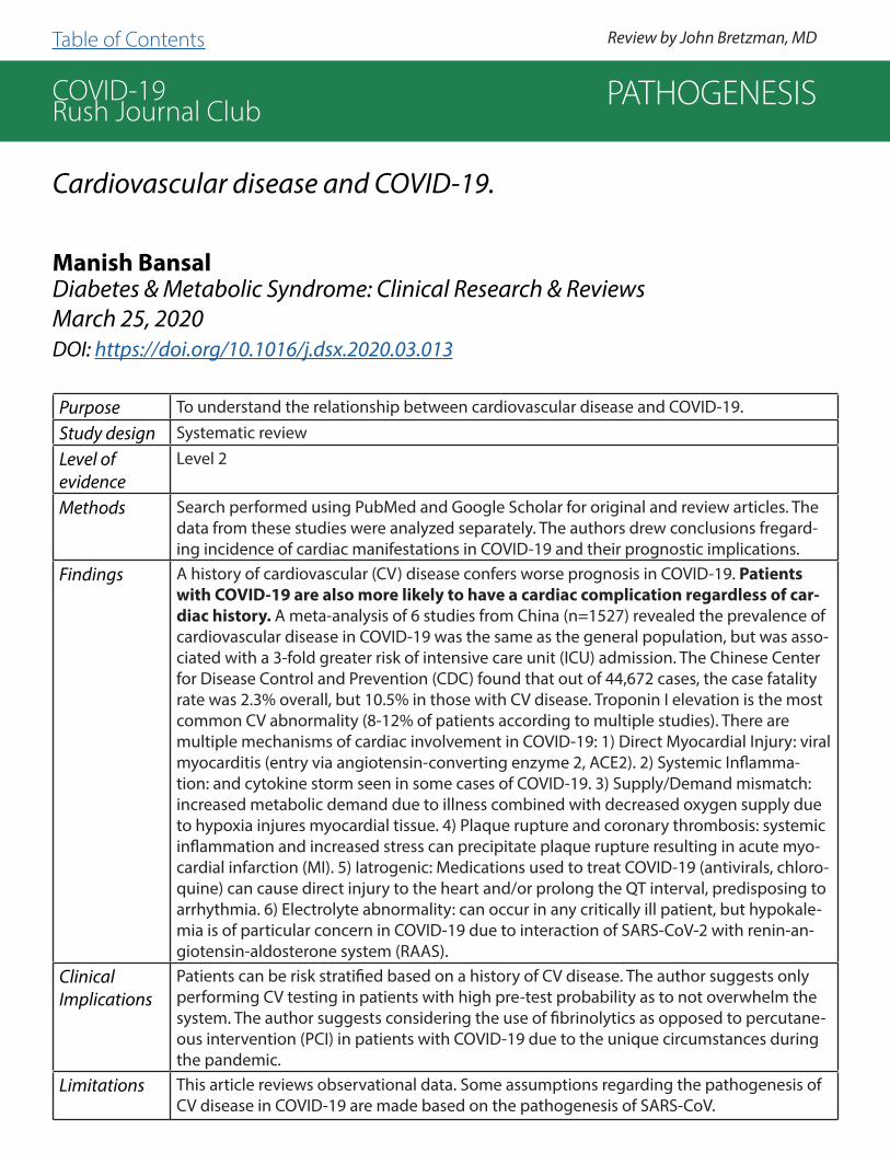
COVID-19Rush Journal Club
Cardiovascular disease and COVID-19.
Manish BansalDiabetes & Metabolic Syndrome: Clinical Research & ReviewsMarch 25, 2020DOI: https://doi.org/10.1016/j.dsx.2020.03.013
Purpose To understand the relationship between cardiovascular disease and COVID-19.
Study design Systematic review
Level of evidence
Level 2
Methods Search performed using PubMed and Google Scholar for original and review articles. The data from these studies were analyzed separately. The authors drew conclusions fregard-ing incidence of cardiac manifestations in COVID-19 and their prognostic implications.
Findings A history of cardiovascular (CV) disease confers worse prognosis in COVID-19. Patients with COVID-19 are also more likely to have a cardiac complication regardless of car-diac history. A meta-analysis of 6 studies from China (n=1527) revealed the prevalence of cardiovascular disease in COVID-19 was the same as the general population, but was asso-ciated with a 3-fold greater risk of intensive care unit (ICU) admission. The Chinese Center for Disease Control and Prevention (CDC) found that out of 44,672 cases, the case fatality rate was 2.3% overall, but 10.5% in those with CV disease. Troponin I elevation is the most common CV abnormality (8-12% of patients according to multiple studies). There are multiple mechanisms of cardiac involvement in COVID-19: 1) Direct Myocardial Injury: viral myocarditis (entry via angiotensin-converting enzyme 2, ACE2). 2) Systemic Inflamma-tion: and cytokine storm seen in some cases of COVID-19. 3) Supply/Demand mismatch: increased metabolic demand due to illness combined with decreased oxygen supply due to hypoxia injures myocardial tissue. 4) Plaque rupture and coronary thrombosis: systemic inflammation and increased stress can precipitate plaque rupture resulting in acute myo-cardial infarction (MI). 5) Iatrogenic: Medications used to treat COVID-19 (antivirals, chloro-quine) can cause direct injury to the heart and/or prolong the QT interval, predisposing to arrhythmia. 6) Electrolyte abnormality: can occur in any critically ill patient, but hypokale-mia is of particular concern in COVID-19 due to interaction of SARS-CoV-2 with renin-an-giotensin-aldosterone system (RAAS).
ClinicalImplications
Patients can be risk stratified based on a history of CV disease. The author suggests only performing CV testing in patients with high pre-test probability as to not overwhelm the system. The author suggests considering the use of fibrinolytics as opposed to percutane-ous intervention (PCI) in patients with COVID-19 due to the unique circumstances during the pandemic.
Limitations This article reviews observational data. Some assumptions regarding the pathogenesis of CV disease in COVID-19 are made based on the pathogenesis of SARS-CoV.
PATHOGENESIS
Review by John Bretzman, MD Table of Contents
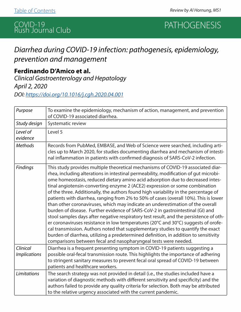
COVID-19Rush Journal Club
Diarrhea during COVID-19 infection: pathogenesis, epidemiology, prevention and managementFerdinando D’Amico et al.Clinical Gastroenterology and HepatologyApril 2, 2020DOI: https://doi.org/10.1016/j.cgh.2020.04.001
Purpose To examine the epidemiology, mechanism of action, management, and prevention of COVID-19 associated diarrhea.
Study design Systematic review
Level of evidence
Level 5
Methods Records from PubMed, EMBASE, and Web of Science were searched, including arti-cles up to March 2020, for studies documenting diarrhea and mechanism of intesti-nal inflammation in patients with confirmed diagnosis of SARS-CoV-2 infection.
Findings This study provides multiple theoretical mechanisms of COVID-19 associated diar-rhea, including alterations in intestinal permeability, modification of gut microbi-ome homeostasis, reduced dietary amino acid adsorption due to decreased intes-tinal angiotensin-converting enzyme 2 (ACE2) expression or some combination of the three. Additionally, the authors found high variability in the percentage of patients with diarrhea, ranging from 2% to 50% of cases (overall 10%). This is lower than other coronaviruses, which may indicate an underestimation of the overall burden of disease. Further evidence of SARS-CoV-2 in gastrointestinal (GI) and stool samples days after negative respiratory test result, and the persistence of oth-er coronaviruses resistance in low temperatures (20°C and 30°C) suggests of orofe-cal transmission. Authors noted that supplementary studies to quantify the exact burden of diarrhea, utilizing a predetermined definition, in addition to sensitivity comparisons between fecal and nasopharyngeal tests were needed.
ClinicalImplications
Diarrhea is a frequent presenting symptom in COVID-19 patients suggesting a possible oral-fecal transmission route. This highlights the importance of adhering to stringent sanitary measures to prevent fecal oral spread of COVID-19 between patients and healthcare workers.
Limitations The search strategy was not provided in detail (i.e., the studies included have a variation of diagnostic methods with different sensitivity and specificity) and the authors failed to provide any quality criteria for selection. Both may be attributed to the relative urgency associated with the current pandemic.
PATHOGENESIS
Review by Al Hornung, MS1 Table of Contents

COVID-19Rush Journal Club
Prevalence and impact of cardiovascular metabolic diseases on COVID-19 in ChinaBo Li et al.Clinical Research in CardiologyMarch 11, 2020DOI: https://doi.org/10.1007/s00392-020-01626-9
Purpose To better understand the prevalence of cardiovascular and metabolic disease in patients with SARS-CoV-2 and the subsequent severity of COVID-19.
Study design Meta-Analysis (n = 1,527)
Level of evidence
Level 1
Methods Studies demonstrating the prevalence and severity of COVID-19 were identified through EMBASE and PubMed. Between December 2019 and February 2020, six studies for a total of 1527 patients were included. Key criteria included confirma-tion of infection, study populations greater than 10 participants, and documented comorbidities.
Findings Analysis demonstrated the following prevalence of comorbidities among COVID-19 patients: hypertension (17.1%), cardio/cerebrovascular disease (16.4%), diabetes (9.7%). Hypertension as a comorbidity was present in 28.8% of COVID-19 ICU ad-missions and 14.1% of non-ICU patients. Similarly, cardio/cerebrovascular disease accounted for 16.7% and 6.2%, respectively. Patients with hypertension had a sta-tistically significant increased risk for ICU admission (relative risk (RR) = 2.03). Those with cardio/cerebrovascular history had a RR = 3.30. Effect of diabetes on COVID-19 severity as judged by ICU admission requirement did not achieve statistical signifi-cance. There was a statistically significant increased incidence of myocardial injury (elevated Troponin I/T) among ICU patients (RR = 13.48).
ClinicalImplications
The data suggests those with hypertension or cardio/cerebrovascular disease have a 2-3x greater likelihood of requiring ICU admission. In these cases, car-diac damage is more likely. Possible pathogenetic explanations for these results include direct viral damage to the cardiomyocytes as seen in SARS-CoV, hypox-ia-induced damage, inflammation from cytokine storm, and repeated floods of catecholamines due to anxiety and medications. When compared to the general population, the prevalence of cardio/cerebrovascular disease among patients with COVID-19 was higher.
Limitations This study’s analysis of cardiac injury used only 2 of the 6 studies (n = 179). Popula-tion settings (inpatient vs outpatient) and disease criteria were vaguely defined.
PATHOGENESIS
Review by Rob DeStefano, MDTable of Contents

COVID-19Rush Journal Club
Gastrointestinal Manifestations of SARS-CoV-2 Infection and Virus Load in Fecal Samples from the Hong Kong Cohort and Systematic Review and Meta-analysisKa Shing Cheung et al.GastroenterologyApril 3, 2020DOI: https://doi.org/10.1053/j.gastro.2020.03.065
Purpose To summarize epidemiological characteristics of SARS-CoV-2 and the effectiveness of control measures to inform management guidelines.
Study design Case series (n=59) and meta-analysis (n=4,243)
Level of evidence
Level 4 (case series), Level 2 (meta-analysis)
Methods Case Series: Data were collected from a COVID-19 positive cohort of patients in Hong Kong (n=59; diagnosis from February 2 through Feb 29, 2020). Excluded patients had no virologic proof of SARS-CoV2 infection or asymptomatic SARS-CoV-2 infection. Meta-analysis: Sixty studies were included for a total of 4,243 COVID-19 patients. Fifty-three (88.3%) studies were from China.
Findings Case series: Thirty-six (61.0%) patients did not have respiratory symptoms of cough or dyspnea on presentation. Among 15 (25.4%) patients who had gastrointestinal symptoms (vomiting: 1 [1.7%], diarrhea: 13 [22.0%], and abdominal pain/discomfort:7 [11.9%]), all had fever but 8 (53.5%) did not have cough or dyspnea. The proportion of patients with detectable stool viral RNA was higher among those with diarrhea than those without diarrhea (38.5% vs 8.7%; p=0.019). Of the 44 patients without gastrointestinal symptoms, 4 (9.1%) had positive stool viral RNA. There was a trend for higher stool viral load in patients with diarrhea (median: 5.1 [IQR: 4.8–5.6] vs 3.9 [IQR: 3.5–4.4] log10cpm; p=0.06). Meta-analysis: 68 of 138 patients (pooled prevalence: 48.1%, 95% CI: 38.3–57.9) tested positive for both respiratory and stool specimens (“R+S+”) after hospitalization. Positive stool viral RNA samples persist despite negative respi-ratory samples. In nine studies with serial viral RNA test results of “R+S+” patients, 87 of 124 patients (pooled prevalence: 70.3%, 95% CI: 49.6–85.1) had persistent positive stool viral RNA despite negative respiratory samples (“R-S+”). Steroid use may be one factor that contributes to a longer fecal carry time, as stool viral clearance was longer in patients with steroid use compared to those without steroid use (20 vs. 11 days; p<0.001).
ClinicalImplications
Fecal spread of virus may be possible even after negative respiratory test, potentially exac-erbated by use of steroids. Healthcare workers should exercise caution when collecting fecal samples or performing endoscopic procedures in patients with recent history of COVID-19.
Limitations A small number of patients were included in the case series. In the meta-analysis, the gastro-intestinal symptoms may be under-reported in some of the studies, which may lead to a lower pooled prevalence rate. In addition, the majority of the studies were conducted on Chinese participants, therefore the study may not be generalizable to other ethnic groups. A positive result for RT-PCR may not indicate infectivity. Viral cultures, which were not performed in this study, are required to support active viral replication and therefore, contagion.
PATHOGENESIS
Review by Kelly Harmon, MS1 Table of Contents
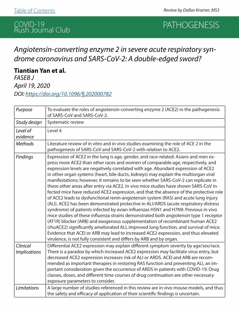
COVID-19Rush Journal Club
Angiotensin-converting enzyme 2 in severe acute respiratory syn-drome coronavirus and SARS-CoV-2: A double-edged sword? Tiantian Yan et al.FASEB JApril 19, 2020DOI: https://doi.org/10.1096/fj.202000782
Purpose To evaluate the roles of angiotensin-converting enzyme 2 (ACE2) in the pathogenesis of SARS-CoV and SARS-CoV-2.
Study design Systematic review
Level of evidence
Level 4
Methods Literature review of in vitro and in vivo studies examining the role of ACE 2 in the pathogenesis of SARS-CoV and SARS-CoV-2 with relation to ACE2.
Findings Expression of ACE2 in the lung is age, gender, and race-related: Asians and men ex-press more ACE2 than other races and women of comparable age, respectively, and expression levels are negatively correlated with age. Abundant expression of ACE2 in other organ systems (heart, bile ducts, kidneys) may explain the multiorgan viral manifestations; however, it remains to be seen whether SARS-CoV-2 can replicate in these other areas after entry via ACE2. In vivo mice studies have shown SARS-CoV in-fected mice have reduced ACE2 expression, and that the absence of the protective role of ACE2 leads to dysfunctional renin-angiotensin system (RAS) and acute lung injury (ALI). ACE2 has been demonstrated protective in ALI/ARDS (acute respiratory distress syndrome) of patients infected by avian influenzas H5N1 and H7N9. Previous in vivo mice studies of these influenza strains demonstrated both angiotensin type 1 receptor (AT1R) blocker (ARB) and exogenous supplementation of recombinant human ACE2 (rhuACE2) significantly ameliorated ALI, improved lung function, and survival of mice. Evidence that ACEI or ARB may lead to increased ACE2 expression, and thus elevated virulence, is not fully consistent and differs by ARB and by organ.
ClinicalImplications
Differential ACE2 expression may explain different symptom severity by age/sex/race. There is a paradox by which increased ACE2 expression may facilitate virus entry, but decreased ACE2 expression increases risk of ALI or ARDS. ACEI and ARB are recom-mended as important therapies in restoring RAS function and preventing ALI, an im-portant consideration given the occurrence of ARDS in patients with COVID-19. Drug classes, doses, and different time courses of drug continuation are other necessary exposure parameters to consider.
Limitations A large number of studies referenced in this review are in vivo mouse models, and thus the safety and efficacy of application of their scientific findings is uncertain.
PATHOGENESIS
Review by Dallas Kramer, MS3 Table of Contents
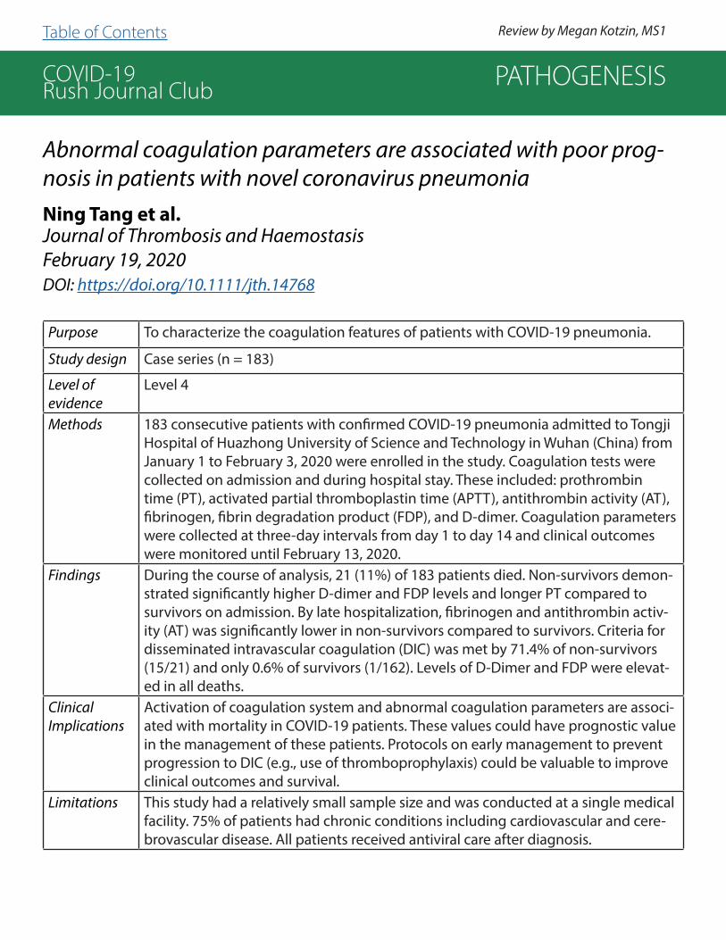
COVID-19Rush Journal Club
Abnormal coagulation parameters are associated with poor prog-nosis in patients with novel coronavirus pneumoniaNing Tang et al.Journal of Thrombosis and HaemostasisFebruary 19, 2020DOI: https://doi.org/10.1111/jth.14768
Purpose To characterize the coagulation features of patients with COVID-19 pneumonia.
Study design Case series (n = 183)
Level of evidence
Level 4
Methods 183 consecutive patients with confirmed COVID-19 pneumonia admitted to Tongji Hospital of Huazhong University of Science and Technology in Wuhan (China) from January 1 to February 3, 2020 were enrolled in the study. Coagulation tests were collected on admission and during hospital stay. These included: prothrombin time (PT), activated partial thromboplastin time (APTT), antithrombin activity (AT), fibrinogen, fibrin degradation product (FDP), and D-dimer. Coagulation parameters were collected at three-day intervals from day 1 to day 14 and clinical outcomes were monitored until February 13, 2020.
Findings During the course of analysis, 21 (11%) of 183 patients died. Non-survivors demon-strated significantly higher D-dimer and FDP levels and longer PT compared to survivors on admission. By late hospitalization, fibrinogen and antithrombin activ-ity (AT) was significantly lower in non-survivors compared to survivors. Criteria for disseminated intravascular coagulation (DIC) was met by 71.4% of non-survivors (15/21) and only 0.6% of survivors (1/162). Levels of D-Dimer and FDP were elevat-ed in all deaths.
ClinicalImplications
Activation of coagulation system and abnormal coagulation parameters are associ-ated with mortality in COVID-19 patients. These values could have prognostic value in the management of these patients. Protocols on early management to prevent progression to DIC (e.g., use of thromboprophylaxis) could be valuable to improve clinical outcomes and survival.
Limitations This study had a relatively small sample size and was conducted at a single medical facility. 75% of patients had chronic conditions including cardiovascular and cere-brovascular disease. All patients received antiviral care after diagnosis.
PATHOGENESIS
Review by Megan Kotzin, MS1 Table of Contents

COVID-19Rush Journal Club
Assessing ACE2 expression patterns in lung tissues in the pathogen-esis of COVID-19Guoping Li et al.Journal of AutoimmunityApril 13, 2020DOI: https://doi.org/10.1016/j.jaut.2020.102463
Purpose To determine whether patients with underlying diseases are more susceptible to SARS-CoV than the healthy population, and to examine the role and regulation of angioten-sin-converting enzyme 2 (ACE2) during coronavirus infection.
Study design Comparative analysis of ACE-2 expression in various conditions including SARS-CoV (n=623)
Level of evidence
Level 5
Methods Gene expression data from samples of lung tissue, bronchoalveolar lavage samples, bron-chial epithelial cells, small airway epithelial cells, and SARS infected cells were extracted from 6 known databases. Gene Set Enrichment Analysis (GSEA) was performed to analyze potential biological processes related to ACE2 in healthy people using the cluster Profiler package. Additionally, all proteins involved in the viral-related biological process and cyto-kine secretion-biological process were extracted from the gmt file and subjected to topo-logical evaluation. The generated interaction network was screened by calculating Degree Centrality (DC) and hub proteins were determined with Cytoscape plugin CytoHubba.
Findings This study showed that ACE2 was not substantially different between healthy populations and those with chronic airway disease, with the exception of elevated expression of ACE2 in cigarette smokers, which is divergent from current COVID-19 hypothesizes that those with certain chronic respiratory diseases may be more susceptible to the disease. Addi-tionally, this study indicated high expression of ACE2 was related to elevated innate im-mune and adaptive immune responses as indicated by alterations in B cell regulation and cytokine secretion (such as IL-1, IL-10, IL-6, and IL-8). The author’s posited that the immune system dysfunction involved in the high expression of ACE2 may be related to the symp-toms of severe inflammatory response and potentially cytokine storm. Finally, this study suggested ADAM-17 as a potential target of treatment due to its role in decreasing the membrane bound form of ACE2 resulting in a downregulation of the immune system.
ClinicalImplications
These findings provide a scaffolding for the pathogenesis of COVID-19, while at the same time indicate potential therapeutic strategies or targets for disruption of the severe in-flammatory response potentially associated with SARS-CoV-2 infection.
Limitations It is important to note that all results in this study are based on previous data on SARS-CoV and were used as means of extrapolating SARS-CoV-2 pathogenetic char-acteristics. Further examination of the proposed mechanisms and targets are necessary to determine their role in the current SARS-CoV-2 epidemic.
PATHOGENESIS
Review by Al Hornung, MS1 Table of Contents

COVID-19Rush Journal Club
Complex immune dysregulation in COVID-19 patients with severe respiratory failureEvangelos Giamarellos-Bourboulis et al.Cell Host and MicrobeApril 21, 2020DOI: https://doi.org/10.1016/j.chom.2020.04.009
Purpose To investigate whether a previously established classification of immune responses in critically ill, septic patients provides a pathogenetic scaffolding for patients with severe respiratory failure caused by SARS-CoV-2.
Study design Retrospective Cohort (n=179)Level of evidence
Level 3
Methods Data from three patient cohorts were compared: 104 patients with sepsis caused by bacterial community-acquired pneumonia (CAP); 21 patients from the 2009 H1N1 influenza outbreak; and 54 patients with CAP caused by SARS-CoV-2. Each cohort was split into patients who developed severe respiratory failure (SRF) requiring mechanical breathing assistance and those who did not.
Findings Three main features became evident in the comparison of sepsis caused by bacteri-al CAP vs SRF caused by SARS-CoV-2: 1) Lower sequential organ failure assessment (SOFA) and acute physiology and chronic health evaluation (APACHE) II in SARS-CoV-2 versus bacterial CAP; 2) Lower co-morbidities in COVID-19 patients vs. bacterial CAP (higher comorbidity index levels in COVID-19 patients with SRF than those without SRF were observed); 3) Significantly higher Glasgow Coma Scale scores of patients with COVID-19 (14.71) vs bacterial CAP (8.80), which may explain the rapid worsening following admission in COVID-19 patients. Patients with SRF displayed hyper-inflam-matory responses with features of either immune dysregulation (driven by IL-6) or macrophage-activation syndrome (driven by IL-1β). Furthermore, the SRF observed with COVID-19 may be a result of immune dysregulation resulting in IL-6 induced de-pression of HLA-DR levels on CD14 monocytes, an inverse relationship between HLA-DR expression and IL-6, as well as restoration of HLA-DR expression with an increase in absolute lymphocyte count after Tocilizumab administration.
ClinicalImplications
Immune dysregulation in severe COVID-19 is characterized by IL-6-mediated low HLA-DR expression and lymphopenia, over-production of cytokines and hyper-inflamma-tion which may be alleviated by Tocilizumab administration.
Limitations Despite the overall strength of this study, it lacks randomization of Tocilizimab admin-istration. Further examination of the IL-6 inhibitor is necessary to determine not only its clinical efficacy, but overall safety in the COVID-19 patient population.
PATHOGENESIS
Review by Al Hornung, MS1 Table of Contents

COVID-19Rush Journal Club
Acute kidney injury in SARS-CoV-2 infected patients
Vito Fanelli et al.Critical CareApril 24, 2020DOI: https://doi.org/10.1186/s13054-020-02872-z
Purpose To discuss possible mechanisms of COVID-19 induced acute kidney injury (AKI) and strategies for risk stratification.
Study design Systematic review
Level of evidence
Level 5
Methods Literature review of COVID-19 patients with AKI
Findings About 25% of patients with acute respiratory distress syndrome (ARDS) have AKI as reported by two studies in Wuhan, China and by Italian public health institution ISS (Istituto Superiore di Sanità). Age, severity of illness, presence of diabetes are risk factors for AKI development. Severity of AKI is associated with BMI and histo-ry of heart failure. ARDS patient with AKI are at high risk for severe outcomes and mortality. Possible mechanisms for AKI in COVID-19 patients include impairment of gas exchange, hemodynamic alterations, fluid overload, injury due to mechanic ventilation, secondary infection, harmful mediators released as a result of immune/inflammatory response, and ability of SARS-CoV-2 to use ACE-2 to enter kidney tu-bular cells. Classic measurement of AKI is using serum creatinine and urine output, but these do not catch early evidence of kidney damage. Tissue inhibitor of metal-loproteinase 2 (TIMP-2) and Insulin-like growth factor binding protein (IGFBPY-7) and their product ([TIMP-2]*[IGFBP-7]) can be used as markers of acute tubular stress/damage and can show abnormalities sooner than serum creatinine and urine output.
ClinicalImplications
Early detection of AKI using novel biomarkers could lead to earlier nephrol-ogy intervention, closer monitoring of kidney function and optimization of volume and hemodynamic status. The use of iodine contrast and nephrotoxic drugs should be limited in this population. Ultimately this could improve mortali-ty in COVID-19 patients who are at high risk for developing AKI.
Limitations Even though the exact mechanism of kidney involvement in COVID-19 infection is unclear, the level of ACE-2 expression in kidney tubules can be regarded as the risk factor for developing AKI in SARS-CoV-2 infected patients. However, authors did not cite any study reporting the expression and distribution of ACE2 in kidney cells.
PATHOGENESIS
Review by Megan Kotzin, MS1Table of Contents

COVID-19Rush Journal Club
Interferon-induced transmembrane protein-3 genetic variant rs12252-C is associated with disease severity in COVID-19.Yonghong Zhang et al.Journal of Infectious DiseasesApril 29, 2020DOI: https://doi.org/10.1093/infdis/jiaa224
Purpose To determine if homozygosity of the C allele of rs12252 in the interferon-in-duced transmembrane protein 3 (IFITM3) is associated with more severe cases of COVID-19.
Study design Case control (n= 80)
Level of evidence
Level 4
Methods 80 patients who were admitted to Beijing Youan Hospital due to COVID-19 from January to February had their genomes sequenced to see what they genotype was for IFITM3. Patients were split into two groups: mild disease (fever, respiratory symptoms, and pneumonia from imaging, n=56) and severe disease (symptoms listed previously and developed significant tachypnea, hypoxia, or respiratory, or another organ failure, n=24). Then, the cohort was genotyped by sequencing a 300bp locus spanning rs12252 to test whether the homozygous C-allele carriers associate with the severity of COVID-19.
Findings The IFITM3 gene encodes an immune effector protein that is vital for viral restric-tion and inhibits membrane fusion. This variant has been associated with increased influenza severity.There was a significant difference between mild and severe cases adjusting for age on regression analysis, for homozygosity, and for C-allele (CC) with an odds ratio (OR) of 6.37 (P=0.0009). Therefore, this genotype is associated with more severe disease in an age dependent manner. Overall, 50.0% of the severe group and 28.6% of the mild group had the CC genotype. The frequency of the CC genotype in the Beijing population is 26.2%.
ClinicalImplications
The study suggests a significant association between a minor IFITM3 allele (a sin-gle-nucleotide polymorphism, rs12252-C) with severe COVID-19 susceptibility. Homozygosity for the C allele of rs12252 in the IFITM3 can be identified in certain patients particularly those of Asian descent, who need early targeted intervention.
Limitations There was a small sample size of people at one hospital. This mutation is more com-mon in people of Asian descent so it may not be as significant for others. This was only a case-control study so further studies need to be done to confirm the genetic association with COVID-19 severity.
PATHOGENESIS
Review by John Levinson, MS2Table of Contents

COVID-19Rush Journal Club
Alterations in Smell or Taste in Mildly Symptomatic Outpatients with SARS-CoV-2 Infection.Giacomo Spinato et al.JAMAApril 22, 2020DOI: 10.1001/jama.2020.6771
Purpose To evaluate the prevalence, intensity, and timing of anosmia in patients with SARS-CoV-2.
Study design Cross-sectional survey (n = 202)
Level of evidence
Level 4
Methods Adults (≥18 years) that tested positive for SARS-CoV-2 via RT-PCR and deemed suitable for home management were contacted 5-6 days after nasal swab testing. Patients underwent a phone interview utilizing the Acute Respiratory Tract Infec-tion Questionnaire (ARTIQ) and asked whether they experienced a sudden onset of an altered sense of smell or taste within the 2 weeks preceding nasal swag testing. Symptoms were rated on the sino-nasal outcomes test 22 (SNOT-22), which grades symptom severity on a scale from 0-5 (0 represents no symptoms and 5 represents symptoms that are “as bad as it can be”). Prevalence was expressed as the percent-age of total patients.
Findings An altered sense of taste or smell was reported in 130 of 202 patients (64.4%) that completed the telephone interview. The median SNOT-22 score was 4, with 27 patients (13.4%) reporting 4 and 48 patients (23.8%) reporting 5. Symptom time-line presentation analysis revealed that an altered sense of smell or taste presented before other symptoms in 24 patients (11.9%); at the same time as other symptoms in 46 patients (22.8%); and after other symptoms in 54 patients (26.7%). Gender comparison revealed that an altered sense of smell or taste was reported more frequently among the 105 women (72.4%) than among the 97 men (55.7%, P=0.02).
ClinicalImplications
This cross-sectional survey suggests that clinicians should consider isolating pa-tients that present with new onset altered sense of taste or smell. Those are distinct symptoms of COVID-19, which do not appear in other respiratory illnesses.
Limitations The study population was small and geographically limited to the Treviso Regional Hospital (Italy). Data was self-reported and lacked symptom evaluation on a longi-tudinal scale. The SNOT-22 test has been validated, but patients may have difficulty quantifying olfactory symptoms.
PATHOGENESIS
Review by Kevin Grudzinski, MS2 Table of Contents

COVID-19Rush Journal Club
Inhibitors of the renin–angiotensin system: The potential role in the pathogenesis of COVID-19.Ziyin Huang et al.Cardiology JournalApril 14, 2020DOI: 10.5603/CJ.a2020.0056
Purpose To explore the role angiotensin converting enzyme 2 (ACE2) plays in the pathogen-esis of COVID-19 and its implications for the use of ACE inhibitors (ACEIs) and an-giotensin II (AngII) receptor blockers (ARBs) in COVID-19 patients.
Study design Systematic reviewLevel of evidence
Level 5
Methods Authors reviewed 24 research articles about the viral entry of SARS-CoV, dysregula-tion of ACE2 on lung injury and cardiotoxicity, and modeling of SARS-CoV2 viral en-try. The authors utilized this data to propose a mechanism of cellular injury caused by SARS-CoV-2 and suggest a direction for therapeutic investigation.
Findings ACE2 and its downstream products Ang(1-7) counter-regulate the pro-inflamma-tory, pro-fibrotic, pro-hyperresponsive ACE/AngII pathway. Higher ACE and AngII levels have been found to be poor prognostic factors for severe pneumonia. Ex-periments in ACE2 knockout (KO) mice had higher levels of lung damage than wild type (WT) mice when treated with acid. Further, injection of recombinant ACE2 res-cued lung function in both ACE2 KO and controls. WT mice treated with the Spike-Fc protein from SARS-CoV were found to have diminished levels of ACE2 and higher levels of AngII. When treated with an AngII receptor type 1 (AT1R) inhibitor, severity of lung injury and pulmonary edema diminished in these mice. Models of SARS-CoV-2 indicate that it binds to ACE2 even more tightly than SARS-CoV, suggesting a similar mechanism of entry and downstream effects. ACEIs and ARBs increase ACE2 levels in chronically treated mice and rats. Those agents are well tolerated and widely used worldwide. Paradoxically, increased levels of ACE2 could provide more receptors to which SARS-CoV-2 could bind and enter cells.
ClinicalImplications
ACEIs and ARBs could help restore the balance between ACE/AngII and ACE2/Ang(1-7) and reducing the pulmonary inflammatory response in patients with COVID-19. Prospective studies and randomized trials are needed to prove if it has beneficial clinical effects in patients with COVID-19.
Limitations There have been minimal studies about SARS-CoV-2 and its effects on ACE2 and resultant tissue damage. Further, the studies in this review primarily used animal models, limiting the generalizability of the findings.
PATHOGENESIS
Review by Clara Ledsky, MS2 Table of Contents

COVID-19Rush Journal Club
Guillain-Barre syndrome associated with SARS-CoV-2.
Gianpolo Toscano et al.New England Journal of MedicineApril 17, 2020DOI: 10.1056/NEJMc2009191
Purpose To summarize the clinical course of five patients in northern Italy who contracted Guillain-Barre Syndrome (GBS) after the onset of COVID-19.
Study design Case series
Level of evidence
Level 4
Methods Five patients in northern Italy were found to have GBS symptoms after COVID-19 onset. Onset of GBS symptoms, signs and symptoms, CSF findings, antiganglioside antibodies, MRI results, treatment and outcomes at week 4 were recorded.
Findings From February 28th to March 21st, 2020, 1000 to 1200 patients with COVID-19 were admitted to hospitals in northern Italy and five had GBS after COVID-19 onset. Four patients had positive nasopharyngeal swab at GBS onset, one had negative swab and bronchoalveolar lavage but positive serologic testing. Interval between onset of symptoms of COVID-19 and first symptom of GBS ranged from 5-10 days. Cere-brospinal fluid (CSF) in all patients was negative for SARS-CoV-2 via RT-PCR assay. All patients were given intravenous immune globulin (IVIG) and one had plasma exchange. After 4 weeks of treatment, two patients remained in the ICU, two were receiving physical therapy because of flaccid paraplegia and had minimal upper limb movement, and one was discharged and able to walk.
ClinicalImplications
Respiratory viruses penetrating the blood brain barrier is rare, but with in-creasing prevalence of COVID-19 cases worldwide, neurological disease is being observed in severe cases. It is important to aware of neurological manifes-tations of SARS-CoV-2 infection and distinguish GBS with COVID-19 from critical illness neuropathy and myopathy which tends to occur later than GBS.
Limitations Small sample size is the main limitation. Also, with the current setting of the study, it is not possible to determine whether severe deficits and axonal involvement are typical features of COVID-19 GBS. The authors were not able to differentiate the effect of reduced vital capacity due to neuromuscular failure from GBS. The authors did not correlate severity of COVID-19 symptoms with severity of GBS symptoms.
PATHOGENESIS
Review by Mira Marchioretto, MS2 Table of Contents

COVID-19Rush Journal Club
COVID-19 Coagulopathy in Caucasian patients.
Helen Fogarty et al.British Journal of HaematologyApril 24, 2020DOI: https://doi.org/10.1111/bjh.16749
Purpose To evaluate whether there is a correlation between coagulopathic features and COVID-19 disease severity in infected Caucasian patients.
Study design Case Series (n = 83)Level of evidence
Level 4
Methods PCR-confirmed, COVID-19+ adult patients at St. James’s Hospital (Dublin, Ireland) between March 13th and April 10th (2020) were included. Hospitalized study pa-tients received standard of care along with weight and renally-appropriate doses of low molecular weight heparin (LMWH) thromboprophylaxis, unless contra-indi-cated. The hospital’s electronic patient record was used to collect epidemiological, demographic, treatment and outcome data. The utility of D-dimer levels as a prog-nostic marker -when low molecular weight heparin (LMWH) thromboprophylaxis is utilized- was also examined.
Findings Severe coagulopathy was positively correlated with disease severity in Caucasian individuals. Additionally, patients transferred to the ICU or those that died were more likely to have underlying co-morbidities. The progression to overt disseminat-ed intravascular coagulation (DIC) in COVID-19 patients maintained on prophylactic dose LMWH was rare despite significantly increased D-dimers levels. The authors suggest that the COVID-19 induced diffuse bilateral pulmonary inflammation is a novel and distinct form of DIC, recently named pulmonary intravascular coagulopa-thy (PIC), due to its apparent pulmonary-specific nature.
ClinicalImplications
Examination of individuals thrombotic risk may serve as a means of identifying those with increased mortality risk from COVID-19.
Limitations There are several limitations associated with this study, including limited sample size and potential for selection bias. In order to fully corroborate the findings of this study, randomized controlled studies are necessary. Despite reports in the literature describing dissimilarities in thrombotic risk in individuals of different races and the authors’ findings regarding the susceptibility of Caucasians to COVID-19 mortality, as it is entirely possible that the findings of this study may be explained by an alter-native factor that the authors did not consider.
PATHOGENESIS
Review by Al Hornung, MS1 Table of Contents
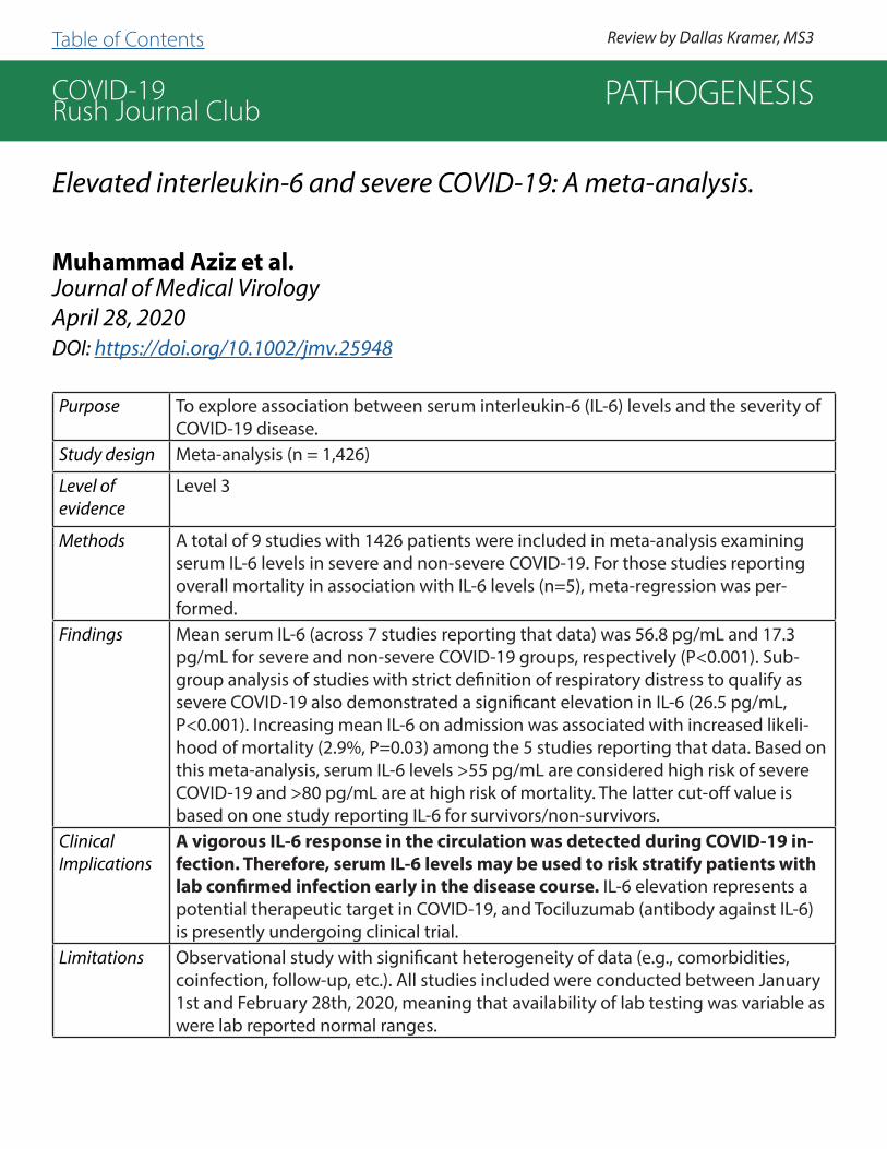
COVID-19Rush Journal Club
Elevated interleukin-6 and severe COVID-19: A meta-analysis.
Muhammad Aziz et al.Journal of Medical VirologyApril 28, 2020DOI: https://doi.org/10.1002/jmv.25948
Purpose To explore association between serum interleukin-6 (IL-6) levels and the severity of COVID-19 disease.
Study design Meta-analysis (n = 1,426)
Level of evidence
Level 3
Methods A total of 9 studies with 1426 patients were included in meta-analysis examining serum IL-6 levels in severe and non-severe COVID-19. For those studies reporting overall mortality in association with IL-6 levels (n=5), meta-regression was per-formed.
Findings Mean serum IL-6 (across 7 studies reporting that data) was 56.8 pg/mL and 17.3 pg/mL for severe and non-severe COVID-19 groups, respectively (P<0.001). Sub-group analysis of studies with strict definition of respiratory distress to qualify as severe COVID-19 also demonstrated a significant elevation in IL-6 (26.5 pg/mL, P<0.001). Increasing mean IL-6 on admission was associated with increased likeli-hood of mortality (2.9%, P=0.03) among the 5 studies reporting that data. Based on this meta-analysis, serum IL-6 levels >55 pg/mL are considered high risk of severe COVID-19 and >80 pg/mL are at high risk of mortality. The latter cut-off value is based on one study reporting IL-6 for survivors/non-survivors.
ClinicalImplications
A vigorous IL-6 response in the circulation was detected during COVID-19 in-fection. Therefore, serum IL-6 levels may be used to risk stratify patients with lab confirmed infection early in the disease course. IL-6 elevation represents a potential therapeutic target in COVID-19, and Tociluzumab (antibody against IL-6) is presently undergoing clinical trial.
Limitations Observational study with significant heterogeneity of data (e.g., comorbidities, coinfection, follow-up, etc.). All studies included were conducted between January 1st and February 28th, 2020, meaning that availability of lab testing was variable as were lab reported normal ranges.
PATHOGENESIS
Review by Dallas Kramer, MS3 Table of Contents

COVID-19Rush Journal Club
Cardiovascular Implications of Fatal Outcomes of Patients with Coronavirus Disease 2019 (COVID-19) Tao Guo et al.JAMA CardiologyMarch 27, 2020DOI: 10.1001/jamacardio.2020.1017
Purpose To evaluate association of underlying cardiovascular disease (CVD) and myocardial injury with fatal outcomes in patients with COVID-19.
Study design Retrospective case series (n=187)Level of evidence
Level 4
Methods Retrospective single-institution case series in Wuhan, China, analyzing comorbidities, treatments, troponin T (TnT) levels in association with fatal outcomes. CVD included hypertension, coronary artery disease, and cardiomyopathy.
Findings Of the 187 patients included in this study, 27.8% had myocardial injury resulting in cardiac dysfunction and arrhythmias. Mortality rate was greatest in those with under-lying CVD and elevated TnT levels (69.4%) and underlying CVD with normal TnT levels (37.5%). The mortality rate in patients without underlying CVD and with elevated Tnt levels was (13.3%) and without (7.6%) TnT elevation exhibited lower mortality. TnT levels demonstrated a significant positive liner correlation with C-reactive protein (p<0.001) and N-terminal pro-brain natriuretic peptide (NT-proBNP) levels (p<0.001). Plasma TnT (p=0.001) and NT-proBNP (p<0.001) levels increased significantly from admission in those patients who died. No significant changes were observed in those who were discharged following hospitalization. Patients with elevated TnT had a high-er frequency of malignant arrythmia, and were more likely to received glucocorticoid therapy (71.2% vs. 51.1%) and mechanical ventilation (59.6% vs. 10.4%) than those with normal TnT levels. The use of angiotensin-converting enzyme (ACE) inhibitors and angiotensin receptor blockers (ARBs)did not show a significant change in outcome.
ClinicalImplications
Myocardial injury is significantly associated with fatal outcome of COVID-19, and patients with underlying CVD +/- elevated TnT levels on admission have a poorer prognosis with COVID-19.
Limitations Retrospective nature with absence of echocardiogram and interleukin-6 data owing to the urgency and isolation of patients. Relatively small cohort (n=187) size. There is also limited data and follow-up to capture post-hospitalization cardiac complications and mortality. Those patients should be triage-tagged as necessary to assure more aggres-sive treatment strategies in an effort to increase the chance of survival.
PATHOGENESIS
Review by Dallas Kramer, MS3 Table of Contents

COVID-19Rush Journal Club
SARS-CoV-2 and Viral Sepsis: Observations and Hypothesis
Hui Li et al.LancetApril 17, 2020DOI: https://doi.org/10.1016/S0140-6736(20)30920-X
Purpose To discuss SARS-CoV-2 pathogenesis and its relation to the development of sepsis.Study design Systematic reviewLevel of evidence
Level 5
Methods 30 studies were reviewed in order to meet the primary end point of this study: to put forward a hypothesis about the pathogenesis of SARS-CoV-2.
Findings Based on the authors’ review of published studies, including autopsy studies and basic research, the following hypothesis for pathogenesis of SARS-CoV-2 is proposed. Mild cases: Resident macrophages initiating lung inflammatory responses are able to contain the virus after SARS-CoV-2 infection; both innate and adaptive immune re-sponses are efficiently established to curb the viral replication. Severe or critical cases: In response to the infection of SARS-CoV-2, alveolar macrophages or epithelial cells produce proinflammatory cytokines and chemokines. Monocytes and neutrophils are then chemotactic to the infection resulting in uncontrolled inflammation. There is sub-stantial reduction and dysfunction of lymphocytes and the adaptive immune response cannot be effectively initiated. Additionally, there is a direct attack on other organs by disseminated SARS-CoV-2, immune pathogenesis caused by systemic cytokine storm, and microcirculation dysfunction that together leads to viral sepsis. Therefore, the pro-cess of viral sepsis is crucial to the disease mechanism of COVID-19.
ClinicalImplications
This study investigates the viral infection immune response and pathogenesis of SARS-CoV-2 and describes the process of viral sepsis and abnormal coagulation in patients infected with SARS-CoV-2. A better understanding of the mechanism of viral sepsis in SARS-CoV-2 infection is warranted for exploring enhanced clinical care for patients infected with SARS-CoV-2.
Limitations This study was limited by the lack of basic science research available exploring the kinetics of the cytokine storm and the immunologic response of the SARS -CoV-2 infection. Additionally, further research is needed to explore the effect of SARS-CoV-2 on coagulation, virus dissemination and its effect on the innate and adaptive immune response. The authors’ hypothesize that damage to organs outside of the lungs is due to direct effects of the virus; however, as of yet, replicating virus has not been isolated from these extra-pulmonary tissues.
PATHOGENESIS
Review by Kaitlyn Wehrheim, MS2 Table of Contents

COVID-19Rush Journal Club
Renin-angiotensin-aldosterone system inhibitors and risk of Covid-19.Harmony Reynolds et al.New England Journal of MedicineMay 1, 2020DOI: 10.1056/NEJMoa2008975
Purpose To explore the relationship between medications inhibiting the renin–angiotensin–aldo-sterone system (RAAS) pathway and severe COVID-19 illness.
Study design Observational study (n=12,594)
Level of evidence
Level 4
Methods Data on 12,594 patients with a COVID-19 test result in New York University (NYU) Langone Health system was extracted between March 1 and April 15, 2020. Positive cases were cat-egorized as severe if resulting in ICU admission, mechanical ventilation, or death. Charted use of angiotensin-converting enzyme (ACE) inhibitors, angiotensin-receptor blockers (ARBs), beta-blockers, calcium-channel blockers, and thiazide diuretics were included. The primary goal was to rule out a difference (significance defined as 10%) in likelihood of pos-itive or negative COVID-19 tests and likelihood of developing severe COVID-19 infection in the patients who took these medications.
Findings A total of 5,894 patients were Covid-19 positive, among whom 1,002 had severe illness. There was no statistically significant difference in likelihood of testing positive among those taking a RAAS inhibiting medication compared to those unexposed. Of note, be-ta-blocker use was associated with a decreased likelihood of a positive test [912 (54.1%) vs 976 (57.9%) for a median difference of -3.8%], though this was not statistically relevant based on the study’s criteria. Exposure to these medications did not have a statistically significant impact on severity of disease among Covid-19 infected patients. When a sub-group of patients with documented hypertension was analyzed, the use of calcium-chan-nel blockers had an increased association with severe Covid-19 cases [253 (26.6%) vs 207 (22.3%) for a median difference of +4.4%], though this too was not statistically significant.
ClinicalImplications
Given the importance of ACE2 in the mechanism of entry for SARS-CoV-2 and subsequent pathogenesis, there has been abundant speculation regarding the impact of RAAS inhib-iting medications. This study suggests these classes of medications (including ACE in-hibitors and ARBs) do not play a statistically significant role in developing COVID-19 nor do they largely affect the severity of the disease. The effect of beta-blockers and calcium-channel blockers noted may be due to ACE2 expression, dampening sympathetic effects, or residual confounding variables.
Limitations It is important to emphasize the cut-off for statistically significant difference was defined as 10 percentage points, and thus there may be clinically relevant findings here otherwise excluded from this review. Additionally, medication adherence could not be assessed.
PATHOGENESIS
Review by Robert DeStefano, MD Table of Contents

COVID-19Rush Journal Club
Dysregulation of immune response in patients with COVID-19 in Wuhan, China.Chuan Qin et al.Clinical Infectious DiseasesMarch 12, 2020DOI: https://doi.org/10.1093/cid/ciaa248
Purpose To analyze then compare the expression of infection-related biomarkers, inflammatory cytokines and lymphocyte subsets between confirmed cases of severe and non-severe COVID-19.
Study design Case Control Study involving 452 patients with COVID-19 (n=286 severe and 168 non-se-vere)
Level of evidence
Level 4
Methods Retrospective recruitment of 452 patients with RT-PCR confirmed COVID-19 admitted to Tongji Hospital in Wuhan, China, from January 10th to February 12th, 2020. The primary endpoint was the laboratory quantification of lymphocyte subsets, infection–related bio-markers, inflammatory cytokines, immunoglobulins and complement proteins.
Findings Severe cases tend to have lower lymphocyte counts (0.8 vs 1.0.109; P<0.001), higher leuko-cyte counts (5.6 vs 4.9.109; P<0.001), and a higher neutrophil-lymphocyte ratio (5.5 vs 3.2; P<0.001). Additionally, these cases demonstrated lower percentages of monocytes (6.6 vs 8.4%; P<0.001), eosinophils (0.0 vs 0.2%; P<0.001), and basophils (0.1 vs 0.2%; P=0.015). Severe cases had elevated levels of infection-related biomarkers such as procalcitonin (0.1 vs 0.05 ng/mL; P<0.001), serum ferritin (800.4 vs 523.7 ng/mL; P<0.001), and C-re-active protein (57.9 vs 33.2 mg/L; P<0.001), and inflammatory cytokines including IL-2R (757.0 vs 663.5 U/mL; P=0.001), IL-6 (25.2 vs 13.3 pg/mL; P<0.001), IL-8 (18.4 vs 13.7 pg/mL; P<0.001), IL-10 (6.6 vs 5.0 pg/mL; P<0.001), and TNF-α (8.7 vs 8.4 pg/mL; P=0.037). Helper T cells and suppressor T cells in COVID-19 patients were below normal levels, and the decline in Th cells was more pronounced in severe cases (285.1 vs 420.5/μL; P=0.027). The percentage of naive Th cells increased (44.5 vs 35.0%; P=0.035) and memory Th cells decreased (55.5 vs 65.0%; P=0.035) in severe cases when compared with non-severe cases.
ClinicalImplications
Severe COVID-19 is complicated by overwhelming immunological reactions. The virus might act on lymphocytes, especially T lymphocytes, suggesting that hyper-inflammatory responses might be responsible for lung pathology during COVID-19. Therefore, surveil-lance of neutrophil-lymphocyte-ratio (NLR) and lymphocyte subsets may helpful in the early screening, diagnosis. and treatment of COVID-19.
Limitations This study was a retrospective, small-sample study of patients admitted to a single hos-pital, limiting its use to generally assess the temporal change of immune response after infection with COVID-19. Additionally, co-infection with bacteria or the presence of a superinfection might affect the results of the immune response in patients with COVID-19.
PATHOGENESIS
Review by Kaitlyn Wehrheim, MS2 Table of Contents

COVID-19Rush Journal Club
Characteristics and outcomes of 21 critically ill patients with COVID-19 in Washington state.Matt Arentz et al.JAMAMarch 19, 2020DOI: 10.1001/jama.2020.4326
Purpose To examine the clinical presentations and outcomes of patients with severe COVID-19 infections hospitalized in the ICU at Evergreen Hospital in Seattle, Wash-ington.
Study design Case series (n=21)
Level of evidence
Level 4
Methods This case series included all patients hospitalized in the ICU at Evergreen Hospital who tested positive for SARS-CoV-2 via PCR of a nasopharyngeal swab between February 20, 2020 and March 5, 2020. Patient data, including age, gender, medical history, presenting symptoms, lab results, imaging, and hospital course were col-lected and analyzed. The primary endpoints were presenting symptoms and out-comes for each patient included in the study.
Findings A total of 21 cases were included (mean age: 70 years, 52% male). On admission, 16 patients (76%) presented with shortness of breath, 11 patients (52%) presented with fever, and 10 patients (48%) presented with cough. An abnormal chest radio-graph was observed in 20 patients (95%), while 7 patients (33%) had an elevated leukocyte count. All 15 patients (71%) requiring mechanical ventilation subse-quently developed acute respiratory distress syndrome (ARDS). Cardiomyopathy developed in 7 patients (33%). By the end of the study (March 17, 2020), 14 patients (67%) lost their lives to COVID-19, 5 patients (24%) remained stable in the ICU, and 2 patients (9.5%) were discharged from the hospital.
ClinicalImplications
This is the first study to examine both the initial presentation and outcomes of patients hospitalized in the ICU with COVID-19 in the United States. This study has reproduced similar results as studies in China that have detailed the severity of COVID-19 infection in hospitalized patients, including cardiomyopathy, ARDS, and death. Cardiac ultrasound can be used in the ICU to increase the effectiveness for the management of COVID-19 patients developing postviral cardiomyopathy. The high occurrence of cardiomyopathy is becoming more apparent in severely ill pa-tients with COVID-19 infection in other studies.
Limitations This study had a small sample size and was only conducted in one hospital in Wash-ington, making it less applicable to patients in other geographical areas.
PATHOGENESIS
Review by Samantha Betman, MS3 Table of Contents

COVID-19Rush Journal Club
Renin-angiotensin-aldosterone system blockers and the risk of Covid-19. Giuseppe Mancia et al.New England Journal of MedicineMay 1, 2020DOI: 10.1056/NEJMoa2006923
Purpose To evaluate association of underlying cardiovascular disease (CVD) and myocardial injury with fatal outcomes in patients with COVID-19.
Study design Population-based case-control study
Level of evidence
Level 4
Methods A total of 6272 patients with confirmed SARS-CoV-2 in Italy between February 21 and March 11, 2020 were matched to 30,759 controls from the Regional Health Service according to sex, age, and region.
Findings Use of angiotensin-converting–enzyme (ACE) inhibitors, angiotensin-receptor blockers (ARBS), other antihypertensive and non-antihypertensive drugs was more common in COVID-19 patients than controls. Among overall patients, there was no association with COVID-19 and the use of ACE inhibitors (adjusted odds ratio: 0.96) or ARBs (0.95). Among fatal case patients, there was no association with COVID-19 and the use of ACE inhibitors (adjusted odds ratio: 0.91) or ARBs (0.83). There was no association among case patients between the use of ACE inhibitors and ARBs with other variables including sex, age at diagnosis, and disease severity. Overall, the study population mean age was 68 years, and 37% were women.
ClinicalImplications
Patients with COVID-19 have a higher prevalence of cardiovascular disease as compared to controls, as evidenced by more frequent use of ACE inhibitors and ARBs. Use of ACE inhibitors and ARBs do not appear to affect the risk of COVID-19 and is unlikely to be harmful in patients with COVID-19.
Limitations Information on drug use and doses were limited to those captured in the National Health Service system and did not include those prescribed privately. Additionally, these results were obtained from a predominantly white population and cannot be generalized to other races.
PATHOGENESIS
Review by Dallas Kramer, MS3 Table of Contents
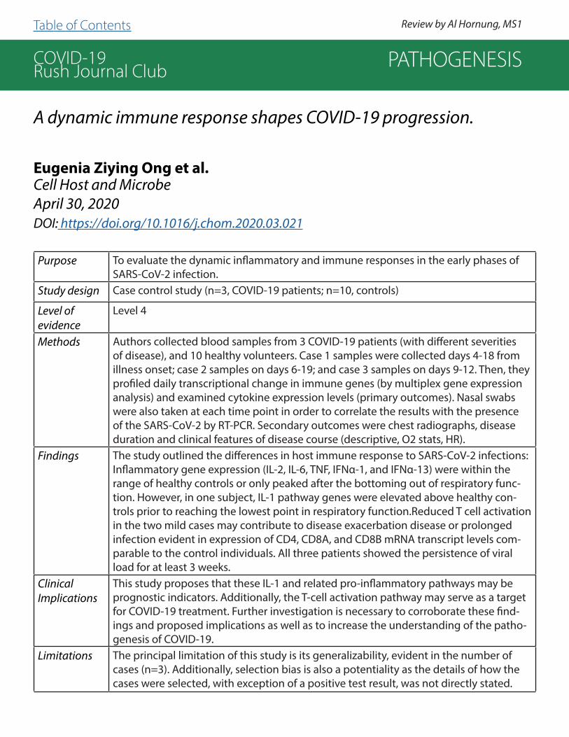
COVID-19Rush Journal Club
A dynamic immune response shapes COVID-19 progression.
Eugenia Ziying Ong et al.Cell Host and MicrobeApril 30, 2020DOI: https://doi.org/10.1016/j.chom.2020.03.021
Purpose To evaluate the dynamic inflammatory and immune responses in the early phases of SARS-CoV-2 infection.
Study design Case control study (n=3, COVID-19 patients; n=10, controls)
Level of evidence
Level 4
Methods Authors collected blood samples from 3 COVID-19 patients (with different severities of disease), and 10 healthy volunteers. Case 1 samples were collected days 4-18 from illness onset; case 2 samples on days 6-19; and case 3 samples on days 9-12. Then, they profiled daily transcriptional change in immune genes (by multiplex gene expression analysis) and examined cytokine expression levels (primary outcomes). Nasal swabs were also taken at each time point in order to correlate the results with the presence of the SARS-CoV-2 by RT-PCR. Secondary outcomes were chest radiographs, disease duration and clinical features of disease course (descriptive, O2 stats, HR).
Findings The study outlined the differences in host immune response to SARS-CoV-2 infec tions: Inflammatory gene expression (IL-2, IL-6, TNF, IFNα-1, and IFNα-13) were within the range of healthy controls or only peaked after the bottoming out of respiratory func-tion. However, in one subject, IL-1 pathway genes were elevated above healthy con-trols prior to reaching the lowest point in respiratory function.Reduced T cell activation in the two mild cases may contribute to disease exacerbation disease or prolonged infection evident in expression of CD4, CD8A, and CD8B mRNA transcript levels com-parable to the control individuals. All three patients showed the persistence of viral load for at least 3 weeks.
ClinicalImplications
This study proposes that these IL-1 and related pro-inflammatory pathways may be prognostic indicators. Additionally, the T-cell activation pathway may serve as a target for COVID-19 treatment. Further investigation is necessary to corroborate these find-ings and proposed implications as well as to increase the understanding of the patho-genesis of COVID-19.
Limitations The principal limitation of this study is its generalizability, evident in the number of cases (n=3). Additionally, selection bias is also a potentiality as the details of how the cases were selected, with exception of a positive test result, was not directly stated.
PATHOGENESIS
Review by Al Hornung, MS1 Table of Contents

COVID-19Rush Journal Club
The pathogenesis and treatment of the `Cytokine Storm’ in COVID-19.Qing Ye et al.Journal of InfectionApril 10, 2020DOI: https://doi.org/10.1016/j.jinf.2020.03.037
Purpose To describe mechanisms by which human coronaviruses (HCoV) can induce cyto-kine storms, and potential treatments for this disease process.
Study design Systematic review
Level of evidence
Level 5
Methods The authors reviewed 15 studies on how the cytokine storm can affect SARS-CoV, SARS-CoV-2, and MERS-CoV.
Findings This study first outlines how COVID-19 induces a cytokine storm in patients. At first, according to in vitro experiments, this may include delayed release of cytokines and chemokines in respiratory epithelial cells, dendritic cells (DCs), and macro-phages. Later, cells released low levels of antiviral factors interferons (IFNs) and high levels of proinflammatory cytokines IL-1B, IL-6, TNF, and chemokines. Serum levels of IL-2R and IL-6 are positively correlated with the severity of COVID-19 in patients. In addition, acute respiratory distress syndrome (ARDS), the leading cause of death with patients infected with SARS-CoV, is linked with cytokine storm. The amount of cytokine increase is positively correlated with mortality rate. The pathogenesis of the cytokine storm can also explain signs of extrapulmonary organ failure.
ClinicalImplications
Cytokine storm is a dangerous mechanism of how COVID-19 can increase mortal-ity in the patients it infects, especially through routes such as ARDS. The authors believed that IFN- λ (early stages), corticosteroids, (later stages), IFN- αβ (later stages), ulinastatin, chloroquine, oxidized phospholipids (OxPL) inhibitors, sphin-gosine-1-phosphate receptor 1 (S1P1) agonists, mesenchymal stem cells, blood purification treatments, C-C chemokine receptor type 2 (CCR2) inhibitors, and TLR7 antagonists may help treat SARS-CoV-2. However, more studies need to be done to determine their effectiveness.
Limitations With respect to some of the treatment options, there were only a few studies con-ducted on COVID-19 or similar diseases to test the efficacy of the treatment. Also, the reviewed studies were not high-quality.
PATHOGENESIS
Review by Adithya Sivakumar, MS1 Table of Contents

COVID-19Rush Journal Club
Acute Hyperglycemic crises with coronavirus disease-19: Case re-ports.Na-young Kim et al.Diabetes and Metabolism JournalApril 23, 2020DOI: https://doi.org/10.4093/dmj.2020.0091
Purpose To describe two COVID-19 cases complicated by acute hyperglycemic events.Study design Case series (n = 2)Level of evidence
Level 4
Methods Authors described two COVID-19 patients admitted to Yeungnam University Col-lege of Medicine in Daegu, South Korea complicated by acute hyperglycemic crises - diabetic ketoacidosis (DKA) and hyperosmolar hyperglycemic state (HHS). Both cases were COVID-19 confirmed by RT-PCR and cross-checked by two independent physicians before inclusion in the study. Vital signs, imaging, blood chemistry, he-moglobin A1c (HbA1c), and arterial blood gas were analyzed for each of the cases. Serial chest radiographs were taken (at the day of admission and during hospital-ization) to assess the severity of infection and predict the clinical outcome.
Findings The study provides evidence of two individuals, with COVID-19 that progressed to an acute hyperglycemic event, either DKA or HHS. In both cases, diabetes was poorly controlled. This was highlighted by recent discontinuations of oral glucose management for health or monetary concerns. Chest radiographic findings includ-ed peri bronchial ground-glass opacities (GGOs) in both lungs (of the patient with DKA) and multifocal patchy consolidation at both lungs (of the patient with HHS). Both patients had adverse outcomes including dependence on ventilator support and death. This is the first case report of COVID-19 combined with DKA and HHS.
ClinicalImplications
This study provides two instances of acute hyperglycemic events, DKA or HHS, in patients with poorly controlled diabetes. Their states may have been precipitated by COVID-19 suggesting that other factors (such as health or monetary concerns) could lead to diabetic crises in this patient subset.
Limitations The principal limitation of this study is its generalizability, evident in the number of cases (n=2). Their states may have been precipitated by COVID-19 and results in catastrophic outcomes. Therefore, intensive monitoring and aggressive support-ive care should be needed to inadequately controlled patients with diabetes and COVID-19 infection.
PATHOGENESIS
Review by Al Hornung, MS1 Table of Contents

COVID-19Rush Journal Club
Severe acute respiratory syndrome coronavirus 2 (SARS-CoV-2) in-fection in children and adolescents: A systematic review.Riccardo Castagnoli et al.JAMA PediatricsApril 22, 2020DOI: 10.1001/jamapediatrics.2020.1467
Purpose To examine the presenting symptoms, potential treatment options, and prognosis of the SARS-CoV-2 infection in pediatric patients.
Study design Systematic Review (n=1065)Level of evidence
Level 5
Methods Reviewed articles published between December 1, 2019 and March 3, 2020 that included participants aged 19 or younger with the SARS-CoV-2 infection. Study types included cross-sectional, case control, case series, case reports, bulletins, and national reports. Eighteen articles with a total of 1065 participants were ultimately included in this analysis. The primary endpoints included the age, presenting symp-toms, diagnostic method, treatment, and prognosis of pediatric patients diagnosed with SARS-CoV-2 infection.
Findings 444 (44.5%) cases were present in children under 10 years old and 553 (55.5%) cases were present in children aged 10-19. The most common symptoms experi-enced by these pediatric patients were fever, cough, fatigue, nasal congestion, and rhinorrhea. Pediatric patients were found to have a good prognosis, as the majority of pediatric patients presented with mild symptoms, were treated with supportive measures, and fully recovered in 1-2 weeks. There was only one death reported in this study.
ClinicalImplications
This is the first systematic review that has examined the effects of the SARS-CoV-2 virus in a pediatric population. This study demonstrates that children appear to have a better prognosis compared to adults and that these children make a full re-covery with supportive measures and without the use of other medications. These case reports highlight the need for further well-designed studies focusing on the incidence of asymptomatic and symptomatic coronavirus infection in children.
Limitations There is no indication of the proportion of symptomatic vs asymptomatic infection or the rates of infection in children. 17 (94.4%) articles included in this study came from China, except for one (5.6%) article that came from Singapore, so these results may not be generalizable to the United States pediatric population. Additionally, all 18 articles included in this study were observational studies, which makes it diffi-cult to draw significant conclusions.
PATHOGENESIS
Review by Samantha Betman, MS3 Table of Contents

COVID-19Rush Journal Club
The pathogenicity of SARS-CoV-2 in hACE2 transgenic mice
Linlin Bao et al.NatureFebruary 28, 2020DOI: https://doi.org/10.1038/s41586-020-2312-y
Purpose To better understand the pathogenicity of SARS-CoV-2 by developing a relevant animal model of the human disease and to develop a platform to pre-clinically test potential therapeutics.
Study design Basic science: animal experimentation
Level of evidence
N/A
Methods Transgenic mice expressing human angiotensin-converting enzyme 2 (hACE2) were exposed to SARS-Cov-2. Results were compared to SARS-Cov-2 infected mice expressing endogenous murine ACE2 (wild type mice - WT) and mock-virus infected mice. Viral replication and pathologic changes were tracked in each group at regular intervals (1, 3, 5, and 7 days post inoculation, dpi). Viral loads were measured by qRT-PCR and autopsies were performed to study gross pathology, histopathology, and immunohistochemistry.
Findings Only SARS-Cov-2 treated hACE2 transgenic mice showed signs of disease, including initial weight loss and a qRT-PCR detected viral load in lungs. No SARS-CoV-2 viral mRNA was detected in other organs including myocardium, liver, spleen, kidney, cerebrum, intestine, and testis. Of note, viral loads were detectable in the intestine at 1 dpi, which was presumed to be swallowed inoculation, as no viral particles could be isolated from the tissue. Grossly, SARS-Cov-2 infected hACE2 transgenic mice exhibited enlarged lungs with multifocal palpable pulmonary nodules by 5 dpi . Histopathology in these mice demonstrated interstitial pneumonia with alveolar septa thickening, mild mucus production, and progressive inflammatory cell accumulation. At day 5, a small amount of fibrous collagen development was noted in the alveolar interstitium, as well as denatured and detached bronchiolar epithelium. Perivascular inflammatory cell infiltration was observed around affect-ed areas. Viral antigens were isolated both within affected as well as non-lesional areas of the lungs. Flourescent immunohistochemistry on mouse lung tissue provides evidence for co-lo-calization of viral and hACE2 proteins, suggesting a similar method of cell entry for the virus.
ClinicalImplications
Results from these animal models support the hypothesis that the ACE2 receptor plays an integral role for SARS-CoV-2 entry into human cells. This study illustrates that SARS-Cov-2 infection localizes to respiratory epithelium without additional organ involvement. This observation is dissimilar to SARS-CoV infection, which demonstrated extrapulmonary damage in similar studies.
Limitations Though transgenic mice were used with appropriate controls, there may be limited translatability to humans. Tissue sampling in a more extensive time frame (greater than 7 days) would be beneficial to explore sub-acute and chronic effects of the virus.
PATHOGENESIS
Review by Rob DeStefano, MD Table of Contents
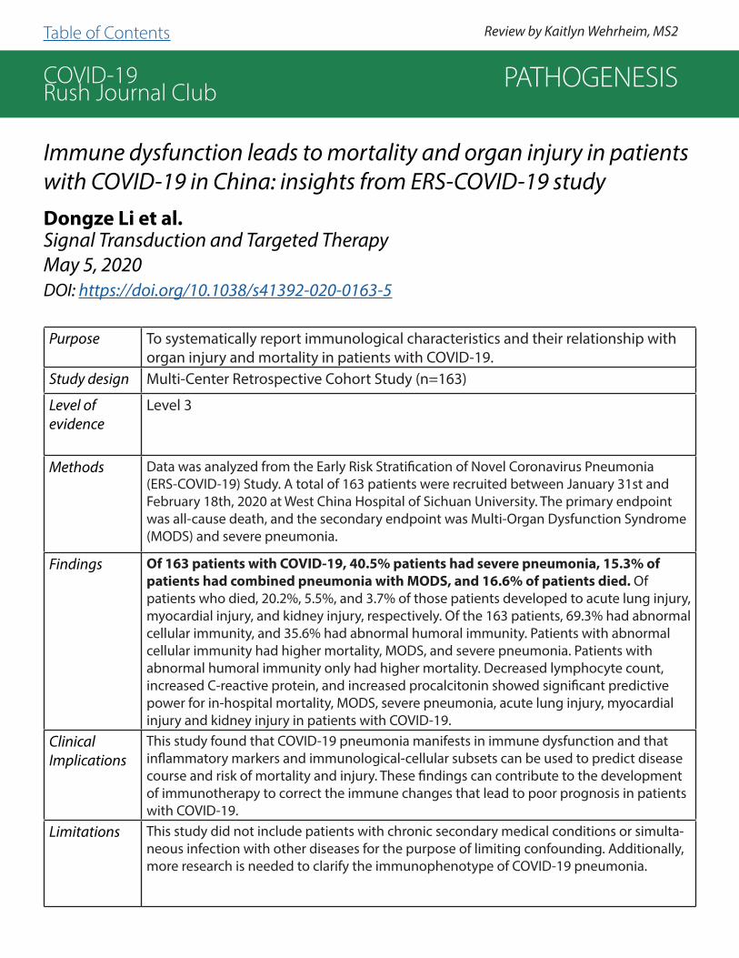
COVID-19Rush Journal Club
Immune dysfunction leads to mortality and organ injury in patients with COVID-19 in China: insights from ERS-COVID-19 studyDongze Li et al.Signal Transduction and Targeted TherapyMay 5, 2020DOI: https://doi.org/10.1038/s41392-020-0163-5
Purpose To systematically report immunological characteristics and their relationship with organ injury and mortality in patients with COVID-19.
Study design Multi-Center Retrospective Cohort Study (n=163)
Level of evidence
Level 3
Methods Data was analyzed from the Early Risk Stratification of Novel Coronavirus Pneumonia (ERS-COVID-19) Study. A total of 163 patients were recruited between January 31st and February 18th, 2020 at West China Hospital of Sichuan University. The primary endpoint was all-cause death, and the secondary endpoint was Multi-Organ Dysfunction Syndrome (MODS) and severe pneumonia.
Findings Of 163 patients with COVID-19, 40.5% patients had severe pneumonia, 15.3% of patients had combined pneumonia with MODS, and 16.6% of patients died. Of patients who died, 20.2%, 5.5%, and 3.7% of those patients developed to acute lung injury, myocardial injury, and kidney injury, respectively. Of the 163 patients, 69.3% had abnormal cellular immunity, and 35.6% had abnormal humoral immunity. Patients with abnormal cellular immunity had higher mortality, MODS, and severe pneumonia. Patients with abnormal humoral immunity only had higher mortality. Decreased lymphocyte count, increased C-reactive protein, and increased procalcitonin showed significant predictive power for in-hospital mortality, MODS, severe pneumonia, acute lung injury, myocardial injury and kidney injury in patients with COVID-19.
ClinicalImplications
This study found that COVID-19 pneumonia manifests in immune dysfunction and that inflammatory markers and immunological-cellular subsets can be used to predict disease course and risk of mortality and injury. These findings can contribute to the development of immunotherapy to correct the immune changes that lead to poor prognosis in patients with COVID-19.
Limitations This study did not include patients with chronic secondary medical conditions or simulta-neous infection with other diseases for the purpose of limiting confounding. Additionally, more research is needed to clarify the immunophenotype of COVID-19 pneumonia.
PATHOGENESIS
Review by Kaitlyn Wehrheim, MS2 Table of Contents

COVID-19Rush Journal Club
D-Dimer Levels on Admission to Predict In-Hospital Mortality in Patients with Covid-19Litao Zhang et al.Journal of Thrombosis and HaemostasisApril 19, 2020DOI: https://doi.org/10.1111/jth.14859
Purpose To determine if elevated D-dimer levels on hospital admission could predict mor-tality in patients hospitalized with COVID-19 in Wuhan, China.
Study design Retrospective case series (n=343)
Level of evidence
Level 4
Methods All patients hospitalized in Wuhan Asia General Hospital who tested positive for the COVID-19 virus between January 12, 2020 and March 15, 2020 and had a D-dimer level on admission were eligible to participate. Data was collected and analyzed from the electron-ic medical record. The primary outcomes were the patient’s D-dimer level on admission and whether the patient survived or passed away from the COVID-19 virus.
Findings The median age of this study population was 62 years, 37.6% of patients were older than 65 years, and 50.3% of patients were female. The cutoff value for the D-dimer level to pre-dict mortality in patients with COVID-19 was 2.0 mg/mL which resulted in a sensitivity of 92.3% and specificity of 83.3%. 276 (80.5%) patients in this study had a D-dimer level less than 2.0 mg/mL and 67 (19.5%) patients in this study had a D-dimer level greater than 2.0 mg/mL. 13 (3.8%) patients in this study died from COVID-19 and 12 (92.3%) of those patients had D-dimer levels above 2.0 mg/mL on admission. Elevated D-dimer levels (above 2.0 mg/mL) were found to significantly predict mortality in hospitalized patients (p<0.001).
ClinicalImplications
This study suggests that an elevated D-dimer level in patients diagnosed with COVID-19 can predict mortality in hospitalized patients, and patients with elevated D-dimer levels had higher rates of underlying disease. This gives physicians an objective lab value that is easily ordered on hospital admission and can guide medical decision making in terms of which patients may develop a more severe hospital course and are at a higher risk of death from the COVID-19 virus.
Limitations This study was conducted at one hospital in China, making it less generalizable to patients in the United States. Additionally, this was a retrospective study that only included pa-tients with a D-dimer level on admission, so there may have been a bias in patient selec-tion based upon which patients had D-dimer levels initially ordered on admission. Finally, the patients in this study waited different amounts of time between the onset of COVID-19 symptoms and presenting to the hospital, which may have influenced their D-dimer levels.
PATHOGENESIS
Review by Samantha Betman, MS3 Table of Contents

COVID-19Rush Journal Club
Endothelial cell infection and endotheliitis in COVID-19
Zsuzsanna Varga et al.The LancetApril 20, 2020DOI: https://doi.org/10.1016/S0140-6736(20)30937-5
Purpose To present evidence of viral endothelial cell involvement in a series of patients with COVID-19.
Study design Case Series (n=3)
Level of evidence
Level 4
Methods SARS-CoV-2 disproportionately infects patients with underlying cardiovascular disease via pathogenic mechanisms that are incompletely understood. Three COVID-19 patients, with underlying cardiovascular comorbidities who experienced respiratory failure, underwent pathological sampling, across multiple organs, for histologic and immunohistochemical evaluation.
Findings Samples demonstrated evidence of direct viral infection of the endothelial cells and diffuse endothelial inflammation. SARS-CoV-2 facilitates endotheliitis across several organs, via direct viral infection and the host inflammatory response, possibly resulting in the induction of apoptosis and pyroptosis.
ClinicalImplications
This case-series study had a small sample size (n=3) that lacked explicit inclusion and exclusion criteria. There was no control group. Additionally, the study is prone to selection bias by the researchers.
Limitations This case-series provides support for the rationale of concomitant use of therapies that promote endothelial stabilization in addition to anti-viral, anti-inflammatory, and ACE inhibitor therapies. This strategy may be of particular use for patients with underlying endothelial dysfunction. As a correspondence, this article has not been peer-reviewed.
PATHOGENESIS
Review by Kevin Grudzinski, MS2 Table of Contents
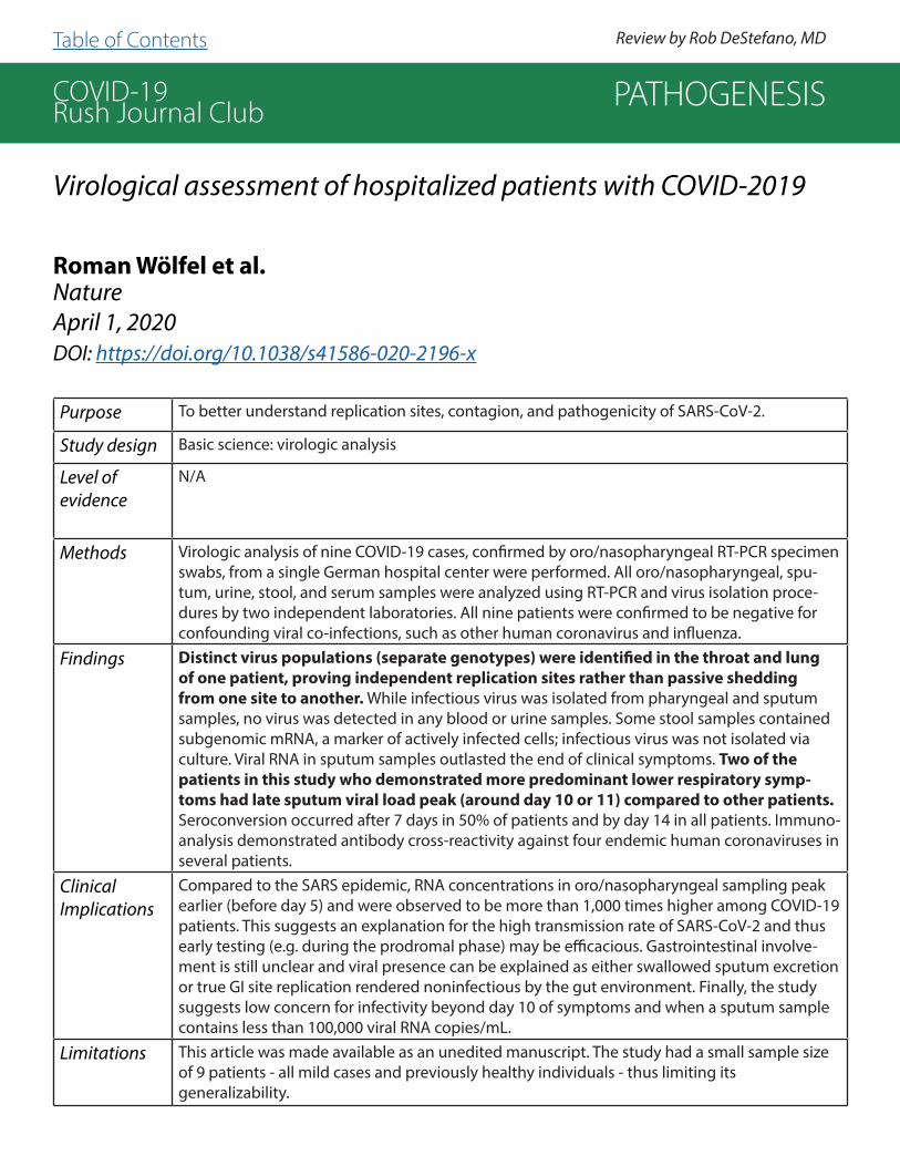
COVID-19Rush Journal Club
Virological assessment of hospitalized patients with COVID-2019
Roman Wölfel et al.NatureApril 1, 2020DOI: https://doi.org/10.1038/s41586-020-2196-x
Purpose To better understand replication sites, contagion, and pathogenicity of SARS-CoV-2.
Study design Basic science: virologic analysis
Level of evidence
N/A
Methods Virologic analysis of nine COVID-19 cases, confirmed by oro/nasopharyngeal RT-PCR specimen swabs, from a single German hospital center were performed. All oro/nasopharyngeal, spu-tum, urine, stool, and serum samples were analyzed using RT-PCR and virus isolation proce-dures by two independent laboratories. All nine patients were confirmed to be negative for confounding viral co-infections, such as other human coronavirus and influenza.
Findings Distinct virus populations (separate genotypes) were identified in the throat and lung of one patient, proving independent replication sites rather than passive shedding from one site to another. While infectious virus was isolated from pharyngeal and sputum samples, no virus was detected in any blood or urine samples. Some stool samples contained subgenomic mRNA, a marker of actively infected cells; infectious virus was not isolated via culture. Viral RNA in sputum samples outlasted the end of clinical symptoms. Two of the patients in this study who demonstrated more predominant lower respiratory symp-toms had late sputum viral load peak (around day 10 or 11) compared to other patients. Seroconversion occurred after 7 days in 50% of patients and by day 14 in all patients. Immuno-analysis demonstrated antibody cross-reactivity against four endemic human coronaviruses in several patients.
ClinicalImplications
Compared to the SARS epidemic, RNA concentrations in oro/nasopharyngeal sampling peak earlier (before day 5) and were observed to be more than 1,000 times higher among COVID-19 patients. This suggests an explanation for the high transmission rate of SARS-CoV-2 and thus early testing (e.g. during the prodromal phase) may be efficacious. Gastrointestinal involve-ment is still unclear and viral presence can be explained as either swallowed sputum excretion or true GI site replication rendered noninfectious by the gut environment. Finally, the study suggests low concern for infectivity beyond day 10 of symptoms and when a sputum sample contains less than 100,000 viral RNA copies/mL.
Limitations This article was made available as an unedited manuscript. The study had a small sample size of 9 patients - all mild cases and previously healthy individuals - thus limiting its generalizability.
PATHOGENESIS
Review by Rob DeStefano, MD Table of Contents

COVID-19Rush Journal Club
Pulmonary embolism in COVID-19 patients: Awareness of an increased prevalence.Julien Poissy et al.CirculationApril 24, 2020DOI: https://doi.org/10.1161/CIRCULATIONAHA.120.047430
Purpose To determine if there is an increased prevalence of the development of a pulmonary embo-lism in patients hospitalized in the ICU with pneumonia caused by the COVID-19 virus at Lille University Hospital in France.
Study design Case series (n=107)
Level of evidence
Level 4
Methods All patients with confirmed COVID-19 pneumonia hospitalized in the ICU at Lille University Hospital from February 27th, 2020 through March 31st, 2020 were examined for the devel-opment of a pulmonary embolism (PE) by computed tomography pulmonary angiography (CTPA) (n=107). Additionally, chart reviews were conducted to determine the number of patients who developed a PE while hospitalized in the ICU for any indication from February 27th, 2019 through March 31st, 2019 (n=196), and the number of patients who developed a PE while hospitalized in the ICU for confirmed influenza pneumonia from January 1st, 2019 through December 31st, 2019 (n=40).
Findings 22 patients out of 107 with COVID-19 pneumonia (20.6%) had a CTPA-confirmed diagnosis of PE (a median of 6 days after admission) during their ICU stay between February 27th, 2020 and March 31st, 2020. 20 patients out of 22 with COVID-19 pneumonia who devel-oped a PE (90.9%) were taking prophylactic antithrombotic treatment with either unfraction-ated heparin or low-molecular-weight heparin. In contrast, only 12 patients out of 196 patients (6.1%) hospitalized in ICU during the same time interval in 2019 developed a PE during their ICU stay. 3 patients out of 40 with influenza pneumonia (7.5%) developed a PE during their ICU stay between January 1st, 2019 and December 31st, 2019.
ClinicalImplications
This study suggests that the development of a PE is more common in patients hospitalized in the ICU with pneumonia caused by COVID-19, as compared to patients hospitalized in the ICU with other illnesses, including pneumonia caused by influenza (20.6% vs 6.1%). This suggests that thrombotic complications could be a hallmark feature of COVID-19. Furthermore, 90.9% of the patients who developed a PE in this study were on anticoagulation, so more research is needed to determine why prophylactic anticoagulation is not sufficient in preventing the development of a PE and what is the optimal anticoagulation strategy.
Limitations This study was conducted at one hospital in France, making it less generalizable to patients in the United States. Additionally, its small sample size warrants further investigation.
PATHOGENESIS
Review by Samantha Betman, MS3Table of Contents
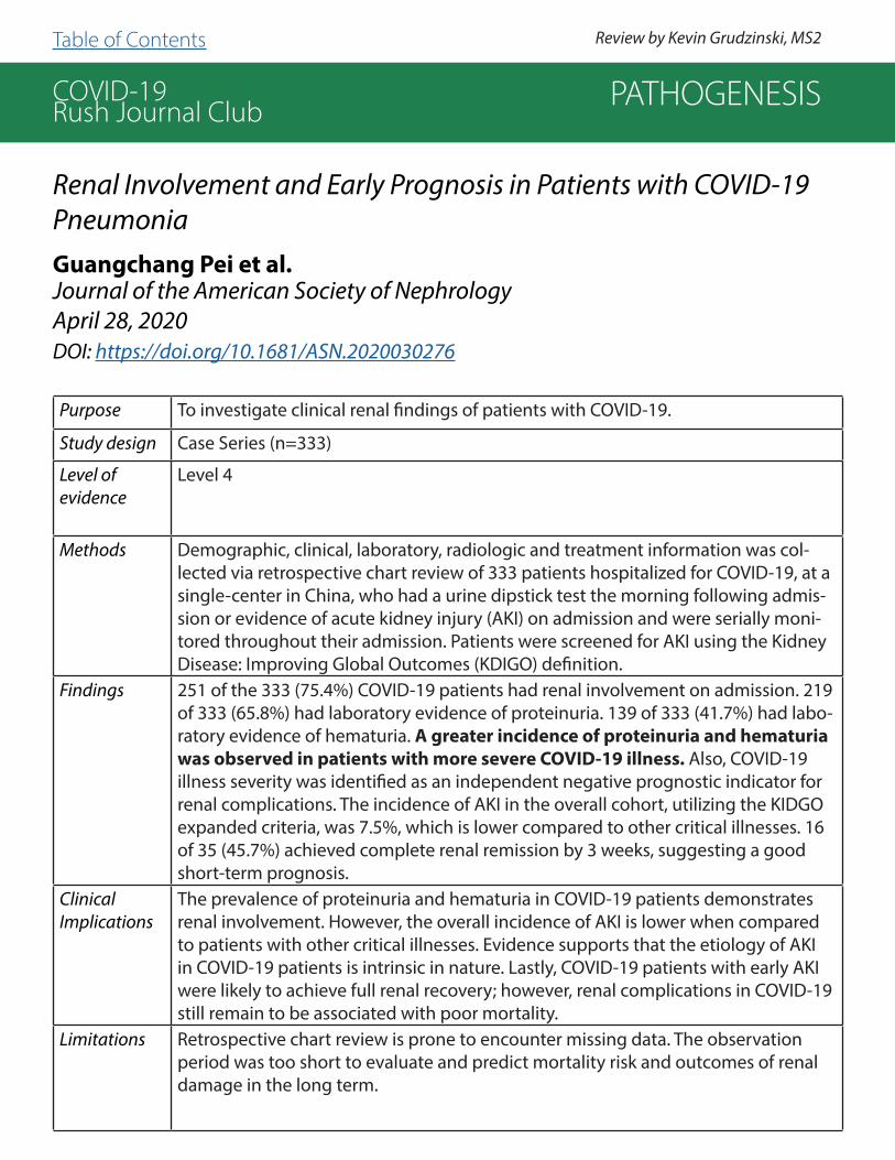
COVID-19Rush Journal Club
Renal Involvement and Early Prognosis in Patients with COVID-19 PneumoniaGuangchang Pei et al.Journal of the American Society of NephrologyApril 28, 2020DOI: https://doi.org/10.1681/ASN.2020030276
Purpose To investigate clinical renal findings of patients with COVID-19.
Study design Case Series (n=333)
Level of evidence
Level 4
Methods Demographic, clinical, laboratory, radiologic and treatment information was col-lected via retrospective chart review of 333 patients hospitalized for COVID-19, at a single-center in China, who had a urine dipstick test the morning following admis-sion or evidence of acute kidney injury (AKI) on admission and were serially moni-tored throughout their admission. Patients were screened for AKI using the Kidney Disease: Improving Global Outcomes (KDIGO) definition.
Findings 251 of the 333 (75.4%) COVID-19 patients had renal involvement on admission. 219 of 333 (65.8%) had laboratory evidence of proteinuria. 139 of 333 (41.7%) had labo-ratory evidence of hematuria. A greater incidence of proteinuria and hematuria was observed in patients with more severe COVID-19 illness. Also, COVID-19 illness severity was identified as an independent negative prognostic indicator for renal complications. The incidence of AKI in the overall cohort, utilizing the KIDGO expanded criteria, was 7.5%, which is lower compared to other critical illnesses. 16 of 35 (45.7%) achieved complete renal remission by 3 weeks, suggesting a good short-term prognosis.
ClinicalImplications
The prevalence of proteinuria and hematuria in COVID-19 patients demonstrates renal involvement. However, the overall incidence of AKI is lower when compared to patients with other critical illnesses. Evidence supports that the etiology of AKI in COVID-19 patients is intrinsic in nature. Lastly, COVID-19 patients with early AKI were likely to achieve full renal recovery; however, renal complications in COVID-19 still remain to be associated with poor mortality.
Limitations Retrospective chart review is prone to encounter missing data. The observation period was too short to evaluate and predict mortality risk and outcomes of renal damage in the long term.
PATHOGENESIS
Review by Kevin Grudzinski, MS2 Table of Contents

COVID-19Rush Journal Club
Viral and host factors related to the clinical outcome of COVID-19
Xiaonon Zhang et al. NatureMay 20, 2020DOI: https://doi.org/10.1038/s41586-020-2355-0
Purpose To analyze strains of COVID-19 and to identify inflammatory biomarkers and their association with disease severity.
Study design Cohort (n=326)
Level of evidence
Level 4
Methods The study population consists of 326 COVID-19 patients in Shanghai from January 20 to Feb-ruary 25, 2020. Patients were grouped based upon severity of disease: asymptomatic, mild, severe, and critical, with severe exhibiting 1 or more of the following criteria: respiratory rate ≥ 30 per minute, O2 saturation ≤ 93%, PaO2/FIO2 ≤ 300mmHg, or pulmonary imaging with le-sion increase by 50%+ within 48 hours. Sequencing data from 112 patients was collected from sputum via an oropharyngeal swab and analyzed for nucleotide variation. Sequences around nucleotide number (nt) 8,782 and nt28,144 were compared with the closely related bat coro-navirus Bat-SARS-CoV-RaTG13. Phylogeny analysis was also conducted.
Findings Two major clades were identified. Clade I subclades: ORF3a: p.251G>V (subclade V), S: p.614D>G (subclade G). The 6 cases with a clear interaction with the Huanan Seafood Whole-sale Market (HSWM), theorized to be the source of the COVID-19 outbreak, were in Clade I. Clades II subclades: ORF8: p.84L>S (28144T>C) and ORF1ab: p.2839S (8782C>T). 3 cases without contact history to HSWM were in Clade II. Non-HSWM sequences were identical to Bat-SARS-CoV-RaTG13 at the sequences around nt8,782 and nt28,144. There were no significant differences in pathogenicity between the 2 clades. COVID-19 patients, regard-less of disease severity, demonstrated lymphocytopenia. Severe and critical patients exhibited progressive lymphocytopenia. CD3+ T cells were the most affected, followed by CD8+. The level of CD19+ B cells were lower in critical cases only. CD3+ T cells demonstrated gradual decline. Age, number of lymphocytes at admission, gender, and comorbidities were associ-ated with worse disease severity, although the comorbidities group was older. Age and lym-phocytopenia were independently associated with worse disease severity. IL-6 and IL-8 were increased. The critical patient group comprised a greater percentage of the highest IL-6 and IL-8 levels measured.
ClinicalImplications
Lymphocytopenia and inflammatory markers may be targets for future treatment trials. Lim-ited variation between the 2 clades suggest a stable virus, which will prove beneficial in the development of targeted treatments and vaccines.
Limitations The study contained a limited number of participants (n=326) from Shanghai, which may limit its application to other populations, races, or ethnicities.
PATHOGENESIS
Review by Kelly Harmon, MS1 Table of Contents

COVID-19Rush Journal Club
High Fluorescent Lymphocytes Are Increased in COVID-19 Patients
Zhao Wang et al.British Jounral of HaemotologyMay 20, 2020 DOI: https://doi.org/10.1111/bjh.16867
Purpose To assess the association between high fluorescent lymphocytes and severity of COVID-19.
Study design Retrospective analysis (n=111)
Level of evidence
Level 4
Methods High fluorescent lymphocytes have been associated with activated B cells and plasma cells, indicating an immune response. This study retrospectively analyzed complete blood count results of COVID-19 cases admitted to Wuhan Union Hospital in China from Janu-ary 29 to March 8, 2020. Cases were classified as mild or severe, with severe exhibiting 1 or more of the following criteria: respiratory rate ≥ 30 per minute, O2 saturation ≤ 93%, PaO2/FIO2 ≤ 300, or pulmonary imaging with lesion increase by 50%+ in 24-48 hours. Complete blood count results were performed with the Sysmex XE-5000 automated cytometry. Healthy controls were enrolled in November 2019 before the COVID-19 out-break. The primary endpoint of the trial was between high fluorescent lymphocytes versus severity of COVID-19.
Findings The median age was 48.6 years. Patients in the severe group were older than those in the mild group (median age: 66 years vs. 42 years, p<0.001). Of the 19 COVID-19 pa-tients (17.1%) in the severe group, 5 died. Lymphocytes were decreased in mild and severe COVID-19 patients versus healthy controls (Mild: 1400 x 106/L, severe: 820 x 106/L, healthy: 2100 x 106/L; p<0.0001). Lymphocytes were more decreased in the severe COVID-19 patients versus the mild (p=0.080). High fluorescent lymphocytes (HFL) were higher in mild and severe COVID-19 patients versus healthy controls (Mild: 11.8 x 10⁶/L, severe: 20.4 x 10⁶/L, healthy: 0.0 x 10⁶/L; p<0.0001). 2 patients with the highest ratio of HFL to lymphocytes died.
ClinicalImplications
While young, middle aged, and older adults are affected, older patients may be at height-ened risk for severe disease. As such, older individuals should more strictly adhere to social distancing guidelines. Decreased lymphocytes and increased high fluorescent lympho-cytes may be a potential target in future treatment trials.
Limitations This study comprises a small population (n=111), which may limit its broader application. This study also was conducted at 1 hospital in China, which may limit its broader applica-tion to other races and ethnicities.
PATHOGENESIS
Review by Kelly Harmon, MS1 Table of Contents

COVID-19Rush Journal Club
Postmortem Examination of Patients With COVID-19
Tina Schaller et al. Journal of the American Medical AssociationMay 21, 2020DOI: https://doi.org/10.1001/jama.2020.8907
Purpose To examine COVID-19 patients postmortem to determine specific factors contributing to death.
Study design Cohort (n=10)
Level of evidence
Level 4
Methods Postmortem examinations were conducted according to published best practice in 10 pa-tients (3 females, 7 males; median age 79 years, range 64-90) with confirmed SARS-CoV-2 who died at the University Medical Center Augsburg in Germany between April 4 and April 19, 2020.
Findings The median duration from hospital admission to death was 7.5 days (1-26 days). SARS-CoV-2 was detected in respiratory tracts and pleural effusions of all patients at autopsy. The patients had a median of 4 comorbidities (0-6); cardiovascular comorbidities were most frequently reported. 2 patients (20%) had preexisting structural lung damage (emphysema). Chest x-ray demonstrated 9 patients (90%) with ground glass opacities in the middle and lower lung fields. 6 patients (60%) did not receive invasive ventilation and demonstrated disseminated diffuse alveolar damage (DAD), particularly in the middle and lower lung fields. Exudative ear-ly-phase acute DAD presented with hyaline membrane, intra-alveolar edema, and thickened alveolar septa with perivascular lymphocyte-plasmocytic infiltration. Organizing DAD present-ed as fibroblastic proliferation, partial fibrosis, pneumocyte hyperplasia, interstitial thickening, collapsed alveoli, patchy lymphocyte infiltration, reactive osseous, and squamous metaplasia. 1 patient (10%) demonstrated full fibrosis with total destruction of pulmonary parenchyma. 5 patients (50%) demonstrated minor neutrophil infiltration, indicating secondary infection. 4 patients (40%) exhibited mild lymphocytic myocarditis and 2 patients (20%) epicarditis. Peri-portal lymphoplasmacellular infiltration and fibrosis was found in liver histology.
ClinicalImplications
Acute and organizing diffuse DAD and viral RNA persistence represented the leading cause of death in patients with and without invasive ventilation. While there is no specific treatment for DAD, supportive measures such as low tidal volume ventilation have been used. However, the use of low tidal volume has failed to achieve adequate oxygenation in SARS-CoV-2 patients. Future trials should examine the utility of low tidal volume versus prone ventilation. In addi-tion, future trials should examine whether cardiac inflammation represented systemic inflam-mation or early myocarditis
Limitations The study contained a limited number of participants (n=10), which may limit its application to the broader SARS-CoV-2 patient population. The study comprised of a small, older (median age 79 years, range 64-90) cohort from Germany, which may limit its reproducibility in other races, ethnicities, and age groups.
PATHOGENESIS
Review by Kelly Harmon, MS1 Table of Contents

COVID-19Rush Journal Club
DIAGNOSIS
Table of Contents
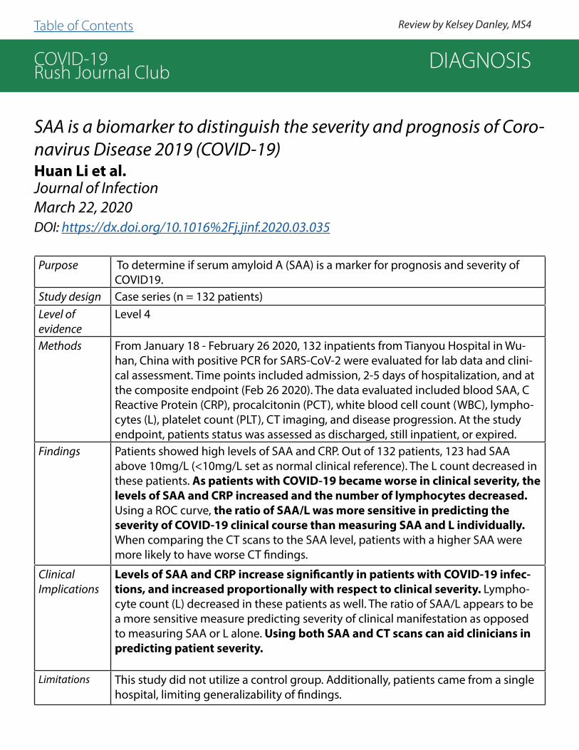
COVID-19Rush Journal Club
SAA is a biomarker to distinguish the severity and prognosis of Coro-navirus Disease 2019 (COVID-19)Huan Li et al.Journal of InfectionMarch 22, 2020DOI: https://dx.doi.org/10.1016%2Fj.jinf.2020.03.035
Purpose To determine if serum amyloid A (SAA) is a marker for prognosis and severity of COVID19.
Study design Case series (n = 132 patients)Level of evidence
Level 4
Methods From January 18 - February 26 2020, 132 inpatients from Tianyou Hospital in Wu-han, China with positive PCR for SARS-CoV-2 were evaluated for lab data and clini-cal assessment. Time points included admission, 2-5 days of hospitalization, and at the composite endpoint (Feb 26 2020). The data evaluated included blood SAA, C Reactive Protein (CRP), procalcitonin (PCT), white blood cell count (WBC), lympho-cytes (L), platelet count (PLT), CT imaging, and disease progression. At the study endpoint, patients status was assessed as discharged, still inpatient, or expired.
Findings Patients showed high levels of SAA and CRP. Out of 132 patients, 123 had SAA above 10mg/L (<10mg/L set as normal clinical reference). The L count decreased in these patients. As patients with COVID-19 became worse in clinical severity, the levels of SAA and CRP increased and the number of lymphocytes decreased. Using a ROC curve, the ratio of SAA/L was more sensitive in predicting the severity of COVID-19 clinical course than measuring SAA and L individually. When comparing the CT scans to the SAA level, patients with a higher SAA were more likely to have worse CT findings.
ClinicalImplications
Levels of SAA and CRP increase significantly in patients with COVID-19 infec-tions, and increased proportionally with respect to clinical severity. Lympho-cyte count (L) decreased in these patients as well. The ratio of SAA/L appears to be a more sensitive measure predicting severity of clinical manifestation as opposed to measuring SAA or L alone. Using both SAA and CT scans can aid clinicians in predicting patient severity.
Limitations This study did not utilize a control group. Additionally, patients came from a single hospital, limiting generalizability of findings.
DIAGNOSIS
Review by Kelsey Danley, MS4 Table of Contents

COVID-19Rush Journal Club
Diagnosis of the Coronavirus disease (COVID-19): rRT-PCR or CT?
Chunqin Long et al.European Journal of RadiologyMarch 25, 2020DOI: https://doi.org/10.1016/j.ejrad.2020.108961
Purpose To evaluate the diagnostic value of computed tomography (CT) and real-time re-verse-transcriptase-polymerase chain reaction (rRT-PCR) for COVID-19 pneumonia.
Study design Retrospective chart review (n = 87 patients)Level of evidence
Level 3
Methods Nasopharyngeal isolates and CT imaging were obtained on 87 patients among 204 patients suspected of having COVID-19. Nasopharyngeal swabs were collected and sent for reverse transcriptase polymerase chain reaction (rRT-PCR), and CT scans were examined retrospectively by two radiologists with 10 and 15 years of expe-rience in chest imaging, respectively. In cases of disagreement, a consensus was reached. CT evaluations included the lobar location and pattern of the lesion.
Findings On CT imaging, peripheral distributions were observed more in the diseased group (72.2%) vs more focal consolidation (52.0%) in the control group (p<0.05). Moreover, ground-glass opacities were observed more frequently in the dis-ease group. Only one patient with COVID-19 had a normal chest CT. This yielded a sensitivity of 97.2% (35/36). In terms of the rRT-PCR testing, the test was initial-ly positive in 30 patients, for a sensitivity of 83.3% (30/36). However, of the six false-negative patients, three were positive with a second round of testing, and the final three were positive with a third round of testing.
ClinicalImplications
CT imaging proved to be more sensitive than rRT-PCR in the initial diagnosis of COVID-19 in a cohort of patients in China. This study recommends that pa-tients with positive CT findings but negative rRT-PCR results still be placed in isolation and have rRT-PCR testing repeated in the subsequent days of admission.
Limitations The study has a small sample size, likely because it was conducted during the height of the pandemic in China. The number of rRT-PCR tests conducted were limited by supply, and only those with fever and a positive CT test were tested with rRT-PCR.
DIAGNOSIS
Review by Paul Parker, MS4Table of Contents
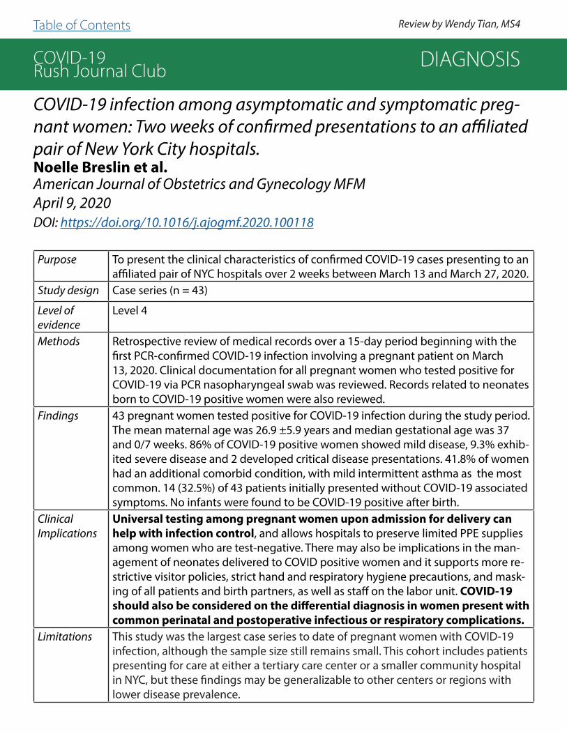
COVID-19Rush Journal Club
COVID-19 infection among asymptomatic and symptomatic preg-nant women: Two weeks of confirmed presentations to an affiliated pair of New York City hospitals.Noelle Breslin et al.American Journal of Obstetrics and Gynecology MFMApril 9, 2020DOI: https://doi.org/10.1016/j.ajogmf.2020.100118
Purpose To present the clinical characteristics of confirmed COVID-19 cases presenting to an affiliated pair of NYC hospitals over 2 weeks between March 13 and March 27, 2020.
Study design Case series (n = 43)
Level of evidence
Level 4
Methods Retrospective review of medical records over a 15-day period beginning with the first PCR-confirmed COVID-19 infection involving a pregnant patient on March 13, 2020. Clinical documentation for all pregnant women who tested positive for COVID-19 via PCR nasopharyngeal swab was reviewed. Records related to neonates born to COVID-19 positive women were also reviewed.
Findings 43 pregnant women tested positive for COVID-19 infection during the study period. The mean maternal age was 26.9 ±5.9 years and median gestational age was 37 and 0/7 weeks. 86% of COVID-19 positive women showed mild disease, 9.3% exhib-ited severe disease and 2 developed critical disease presentations. 41.8% of women had an additional comorbid condition, with mild intermittent asthma as the most common. 14 (32.5%) of 43 patients initially presented without COVID-19 associated symptoms. No infants were found to be COVID-19 positive after birth.
ClinicalImplications
Universal testing among pregnant women upon admission for delivery can help with infection control, and allows hospitals to preserve limited PPE supplies among women who are test-negative. There may also be implications in the man-agement of neonates delivered to COVID positive women and it supports more re-strictive visitor policies, strict hand and respiratory hygiene precautions, and mask-ing of all patients and birth partners, as well as staff on the labor unit. COVID-19 should also be considered on the differential diagnosis in women present with common perinatal and postoperative infectious or respiratory complications.
Limitations This study was the largest case series to date of pregnant women with COVID-19 infection, although the sample size still remains small. This cohort includes patients presenting for care at either a tertiary care center or a smaller community hospital in NYC, but these findings may be generalizable to other centers or regions with lower disease prevalence.
DIAGNOSIS
Review by Wendy Tian, MS4 Table of Contents

COVID-19Rush Journal Club
Profiling early humoral response to diagnose novel coronavirus dis-ease (COVID-19)Li Guo et al.Clinical Infectious DiseasesMarch 21, 2020DOI: https://doi.org/10.1093/cid/ciaa310
Purpose To describe the timeline of antibody production against SARS-CoV-2 and to evaluate the efficacy of diagnostic testing for COVID-19.
Study design Cross-sectional studyLevel of evidence
Level 3
Methods Paired plasma samples and throat swabs were collected from 82 confirmed (via qPCR or deep sequencing) and 58 probable (qPCR and deep sequencing negative, but typi-cal symptoms) COVID-19 cases from Beijing and Wuhan hospitals. 135 plasma samples collected from adults in 2018 with acute lower respiratory tract infections and 150 plas-ma samples collected from healthy adults in 2018 and 2019 were used as controls. Host response against SARS-CoV-2 was examined using ELISA on recombinant viral nucleocap-sid protein. Cross-reactivity of anti-SARS-CoV-2 antibodies against nucleocapsid genes of CoV-229E, -NL63, -OC43, HKU1, SARS-CoV-1, and MERS-CoV were tested by Western Blotting. CLustalW program was utilized to align nucleocapsid gene sequences.
Findings IgM, IgA, and IgG antibodies against SARS-CoV-2 were positively detected, respectively, in 90.4%, 93.3%, and 77.9% of the plasma samples collected from confirmed and suspected cases of COVID-19. The median time to IgM and IgA detection was 5 days after symptom onset, peaking between 8-14 days. The median time to IgG detection was 14 days after symptom onset, peaking between 15-21 days. The detection rate for a single PCR test is 51.9% with >90% positive detection 1-3 days after symptom onset. By day 5, PCR detec-tion decreases to <80%, then <50% at 14 days. When comparing PCR to anti-SARS-CoV-2 IgM antibody detection, the detection rate was higher by qPCR within the first 5.5 days after symptom onset, and higher by IgM ELISA after 5.5 days of symptom onset. Perform-ing IgM ELISA on qPCR negative samples, however, increased the detection rate to 98.6%.
ClinicalImplications
Testing for the presence of IgM on PCR negative samples in cases of suspected COVID-19 could improve sensitivity of testing and aid in early detection of subclini-cal patients, helping to prevent spread of the virus with early and accurate diagnosis.
Limitations This study used a cross-sectional sample. Due to individual variation in production of antibody development, longitudinal studies likely would have yielded more reliable data. A strong cross reactivity was found between SARS-CoV-2 recombinant N proteins with human plasma positive for IgG antibodies against SARS-CoV-1, but not NL63, 229E, OC43, and HKU1. It is unlikely, however, that these patients were pre-infected with SARS-CoV-1 during the last epidemic in 2002.
DIAGNOSIS
Review by Hannah Raff, MS3 Table of Contents

COVID-19Rush Journal Club
Clinical Characteristics of Coronavirus Disease 2019 in China
W Guan et al.New England Journal of MedicineFebruary 28, 2020DOI: 10.1056/NEJMoa2002032
Purpose To identify the clinical characteristics of COVID-19 patients in China.Study design Case series (n = 1,099)Level of evidence
Level 4
Methods The study included 1,099 COVID-19 patients from 552 hospitals across 30 provinces in mainland China from December 11, 2019 to January 29, 2020. Data included exposure history, clinical symptoms, and laboratory and radiologic findings. Cases were defined as severe vs. non-severe using the American Thoracic Society guidelines for communi-ty-acquired pneumonia. Incubation period was defined as time between earliest date of exposure and symptom onset. The primary composite end point was admission to ICU, mechanical ventilation, or death.
Findings Most patients (43.9%) were Wuhan residents, and 72.3% of nonresidents reported contact with Wuhan residents. However, 25.9% of nonresidents denied recent travel or contact with Wuhan residents. Patient ages ranged from 0-14 (0.9%), 15-49 (55.1%), 50-64 (28.9%), and >65 (15.1%), with median age of 47 years. Most patients had nev-er smoked (85.4%), and 12.6% were current smokers. The most common presenting symptom was cough (67.8%), followed by fever (43.8%). Admission to ICU, mechanical ventilation, or death occurred in 24.9% of patients with severe disease and 6.1% of all patients. Pneumonia was diagnosed in 91.1% of patients and acute respiratory dis-tress syndrome (ARDS) in 3.4%. Most common CT findings were ground-glass opacity (56.4%) and bilateral patchy shadowing (51.8%). No radiographic or CT abnormality was found in 2.9% of severe patients. Notable laboratory findings of patients included lymphocytopenia (83.2%), thrombocytopenia (36.2%), and leukopenia (33.7%).
ClinicalImplications
The clinical characteristics and conventional routes of transmission of COVID-19 mim-ic those of SARS-CoV. Absence of fever occurs more frequently in COVID-19 (56% on presentaton, 11% after hospitalization) than in SARS-CoV-1 (1%) and MERS-CoV (2%). Using fever as a symptom for detection may not be reliable.
Limitations Some cases demonstrated incomplete documentation of exposure history and lab-oratory testing. The incubation period could only be estimated for 26.5% of patients, allowing recall bias to play a significant role. The studied population inevitably rep-resents a more severe presentation of COVID-19, as asymptomatic individuals or those with mild symptoms would likely remain home, not requiring additional medical care.
DIAGNOSIS
Review by Andy Wu, MS2 Table of Contents

COVID-19Rush Journal Club
Relation between chest CT findings and clinical conditions of coro-navirus disease (COVID-19) pneumonia: A multicenter study.Wei Zhao et al.American Journal of Roentgenologydate publishedDOI: 10.2214/AJR.20.22976
Purpose To explore the relationship between chest CT findings and COVID-19 pneumonia.
Study design Retrospective study (n = 101)
Level of evidence
Level 4
Methods Medical records were retrospectively collected for 101 patients with confirmed COVID-19 diagnosis in four cities of Hunan province in China. Patients were divided in two groups: non-emergent vs emergent type infections. All patients underwent CT upon admission and the extent of respiratory involvement was evaluated using a CT scoring system. Chest CT scans were blindly and independently reviewed by two radiologists with 5 and 15 years of experience, respectively.
Findings Out of 101 patients in the study, 87 (86.1%) were in the non-emergent group, 14 (13.9%) were in the emergency group. Eighty-seven patients had ground glass opacities (GGO) on CT, 65 had mixed GGO and consolidations, 72 had vascular enlargement in the lesion and 53 had traction bronchiectasis. Lesions on CT were more likely to have peripheral distribution (n=88), bilateral involvement (n=83), have lower lung predominance (n=55), and be multifocal (n=55). Patients in emer-gency group were older than those in non-emergent group but the difference in the rate of underlying disease was not significant.
ClinicalImplications
CT is considered routine imaging for care of COVID-19 patients. Combination of chest CT and PCR screening is necessary for early diagnosis. COVID-19 pres-ents with typical CT features can be helpful in screening suspected cases and evalu-ating extent of disease.
Limitations The study was limited by the number of patients included (n=101). Other viral in-fections and negative results were not included in the analysis limiting comprehen-sive exploration compared to other lung infections. Follow up CT findings were not evaluated limiting insight on progression.
DIAGNOSIS
Review by Ahmet Sakiri, MS2 Table of Contents
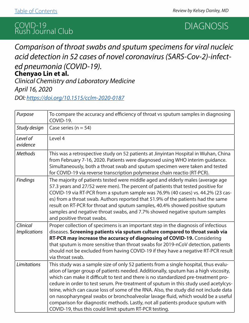
COVID-19Rush Journal Club
Comparison of throat swabs and sputum specimens for viral nucleic acid detection in 52 cases of novel coronavirus (SARS-Cov-2)-infect-ed pneumonia (COVID-19).Chenyao Lin et al.Clinical Chemistry and Laboratory MedicineApril 16, 2020DOI: https://doi.org/10.1515/cclm-2020-0187
Purpose To compare the accuracy and efficiency of throat vs sputum samples in diagnosing COVID-19.
Study design Case series (n = 54)
Level of evidence
Level 4
Methods This was a retrospective study on 52 patients at Jinyintan Hospital in Wuhan, China from February 7-16, 2020. Patients were diagnosed using WHO interim guidance. Simultaneously, both a throat swab and sputum specimen were taken and tested for COVID-19 via reverse transcription polymerase chain reactio (RT-PCR).
Findings The majority of patients tested were middle aged and elderly males (average age 57.3 years and 27/52 were men). The percent of patients that tested positive for COVID-19 via RT-PCR from a sputum sample was 76.9% (40 cases) vs. 44.2% (23 cas-es) from a throat swab. Authors reported that 51.9% of the patients had the same result on RT-PCR for throat and sputum samples, 40.4% showed positive sputum samples and negative throat swabs, and 7.7% showed negative sputum samples and positive throat swabs.
ClinicalImplications
Proper collection of specimens is an important step in the diagnosis of infectious diseases. Screening patients via sputum culture compared to throat swab via RT-PCR may increase the accuracy of diagnosing of COVID-19. Considering that sputum is more sensitive than throat swabs for 2019-nCoV detection, patients should not be excluded from having COVID-19 if they have a negative RT-PCR result via throat swab.
Limitations This study was a sample size of only 52 patients from a single hospital, thus evalu-ation of larger group of patients needed. Additionally, sputum has a high viscosity, which can make it difficult to test and there is no standardized pre-treatment pro-cedure in order to test serum. Pre-treatment of sputum in this study used acetylcys-teine, which can cause loss of some of the RNA. Also, the study did not include data on nasopharyngeal swabs or bronchoalveolar lavage fluid, which would be a useful comparison for diagnostic methods. Lastly, not all patients produce sputum with COVID-19, thus this could limit sputum RT-PCR testing.
DIAGNOSIS
Review by Kelsey Danley, MD Table of Contents

COVID-19Rush Journal Club
Evaluation of Nucleocapsid and Spike Protein-based ELISAs for de-tecting antibodies against SARS-CoV-2Wanbing Liu et al.Journal of Clinical MicrobiologyMarch 30, 2020DOI: 10.1128/JCM.00461-20
Purpose To look for the presence of IgM and IgG antibodies on two immunogenic portions of the SARS-CoV-2 virus in patients, the nucleocapsid protein (rN) and the spike protein (rS).
Study design Observational laboratory analysis
Level of evidence
Level 3
Methods Blood samples of 214 patients with laboratory confirmed COVID-19 (and 100 healthy con-trol samples) were run using enzyme-linked immunosorbent assay (ELISA) to detect both IgM and IgG antibodies against the rN and rS proteins of the SARS-CoV-2 viral structure. Patients were 0-55 days post onset (DPO) of disease, with median DPO of 15 days.
Findings Of the 214 patients samples tested for presence of anti-rN IgM and IgG antibodies, 68.2% and 70.1% of patients displayed positive IgM and IgG antibodies, respectively. For anti-rS antibody presence, 77.1% and 74.3% of patients displayed positive IgM and IgG anti-bodies, respectively. The presence of IgM and/or IgG was looked at for both rN and rS, and patients displayed one or both of the antibodies in 80.4% and 82.2%, respectively. Regarding time course of serologic response, positive rates of both IgM and IgG for rN and rS assays were low at <10 DPO, although IgM increased at 6-10 DPO. IgM and/or IgG for patients at 16 DPO was 88.9% and 90.7% for rN and rS, respectively. None of the 100 healthy controls displayed positive IgM or IgG to either rN or rS. The detection of IgM was significantly higher using both kits (81.3%) vs. the rN kit alone (68.2%), but not using the rS kit alone (77.1%). For IgG, 80.4% were positive using both kits, significantly higher than the rN kit alone (70.1%) but not so for the rS kit alone (74.3%). When testing for presence of IgM and/or IgG, no significant difference was seen between both kits (86.9%) and either rN (80.4%) or rS (82.2%).
ClinicalImplications
The rS-based IgM antibody test is more sensitive for the detection of SARS-CoV-2 due to the earlier immune response against the S protein. Antibodies increased in a time-dependent manner, with IgM decreasing at >35 DPO. Antibody tests should be considered as adjuvant testing to nasopharyngeal PCR swabs, especially in patients who are >10 DPO.
Limitations The study could not follow up with discharged patients to see the extent of the antibody decrease >35 DPO. Moreover, additional immunogenic proteins could have been tested using ELISA to observe antibody production to various parts of the protein.
DIAGNOSIS
Review by Paul Parker, MD Table of Contents

COVID-19Rush Journal Club
Imaging and clinical features of patients with 2019 novel coronavi-rus SARS-CoV-2: A systematic review and meta-analysisYinghao Cao et al.Journal of Medical VirologyMarch 30, 2020DOI: https://doi.org/10.1002/jmv.25822
Purpose To outline clinical characteristics of patients with novel coronavirus pneumonia (NCP). Study design Meta-analysis
Level of evidence
Level 5
Methods Authors executed a detailed search on PubMed, Cochrane Library, Embase, National Knowledge Infrastructure [CNKI], and China Biology Medicine disc [CBMdisc] using the keywords 2019 novel coronavirus pneumonia, COVID-19, Coronavirus, SARS-CoV-2, Wuhan Coronavirus, clinical features, 2019 novel coronavirus pneumonia, and imaging features. Inclusion criteria included cross-sectional studies, case studies, patients with confirmed NCP, and data on clinical characteristics, biochemical indicators, and imag-ing signs. Case reports were excluded. The quality of all included literature was as-sessed using the Institute of Health Economics (IHE) scale. After final review, 31 articles and 46,959 patients were included in the meta-analysis.
Findings Mean age of patients with SARS-CoV-2 infection was 46.4 years old and 55.6% were male. About 35.6% of patients had comorbidities, including 18.3% with hypertension, 11.2% with cardiovascular disease, 10.3% with diabetes, 3.9% with chronic obstructive pulmonary disease, and 3.0% with chronic hepatonephropathy. The main symptoms were fever (87.3%) and cough (58.1%). Other presentations included dyspnea (38.3%), myalgia or weakness (35.5%) and chest tightness (31.2%). Most patients with NCP re-quired hospitalizations, and 29.3% of those patients required intensive care. The main complications were respiratory failure, acute respiratory distress syndrome (ARDS), and multiple organ failure. In imaging results, 75.7% of patients had lesions involving both lungs, and 69.9% showed ground-glass shadows. A computerized tomography (CT) chest is highly sensitive to SARS-CoV-2 (97% in epidemic areas).
ClinicalImplications
This study illustrates that COVID-19 causes bilateral pneumonia and lung function rapidly deteriorates. Chest CT imaging has proved to be very sensitive to COVID-19 and serves as an important supplement to nucleic acid detection.
Limitations The study was limited to studies in England and China, but not from South Korea, Italy, Iran, and Japan where the pandemic is rapidly increasing and causing a catastrophic impact on the health of communities.
DIAGNOSIS
Review by Emily He, MS3Table of Contents
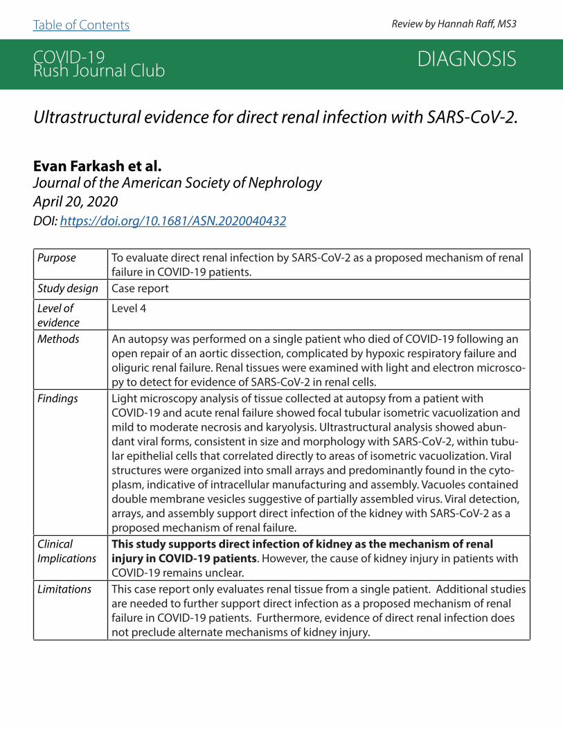
COVID-19Rush Journal Club
Ultrastructural evidence for direct renal infection with SARS-CoV-2.
Evan Farkash et al.Journal of the American Society of NephrologyApril 20, 2020DOI: https://doi.org/10.1681/ASN.2020040432
Purpose To evaluate direct renal infection by SARS-CoV-2 as a proposed mechanism of renal failure in COVID-19 patients.
Study design Case report
Level of evidence
Level 4
Methods An autopsy was performed on a single patient who died of COVID-19 following an open repair of an aortic dissection, complicated by hypoxic respiratory failure and oliguric renal failure. Renal tissues were examined with light and electron microsco-py to detect for evidence of SARS-CoV-2 in renal cells.
Findings Light microscopy analysis of tissue collected at autopsy from a patient with COVID-19 and acute renal failure showed focal tubular isometric vacuolization and mild to moderate necrosis and karyolysis. Ultrastructural analysis showed abun-dant viral forms, consistent in size and morphology with SARS-CoV-2, within tubu-lar epithelial cells that correlated directly to areas of isometric vacuolization. Viral structures were organized into small arrays and predominantly found in the cyto-plasm, indicative of intracellular manufacturing and assembly. Vacuoles contained double membrane vesicles suggestive of partially assembled virus. Viral detection, arrays, and assembly support direct infection of the kidney with SARS-CoV-2 as a proposed mechanism of renal failure.
ClinicalImplications
This study supports direct infection of kidney as the mechanism of renal injury in COVID-19 patients. However, the cause of kidney injury in patients with COVID-19 remains unclear.
Limitations This case report only evaluates renal tissue from a single patient. Additional studies are needed to further support direct infection as a proposed mechanism of renal failure in COVID-19 patients. Furthermore, evidence of direct renal infection does not preclude alternate mechanisms of kidney injury.
DIAGNOSIS
Review by Hannah Raff, MS3 Table of Contents

COVID-19Rush Journal Club
A preliminary study on the ultrasonic manifestations of peripul-monary lesions of non-critical novel coronavirus pneumonia (COVID-19).Yi Huang et al.SSRNFebruary 28, 2020DOI: http://dx.doi.org/10.2139/ssrn.3544750
Purpose To explore the ultrasonic manifestations of peripulmonary lesions of non-critical COVID-19 and provide reference for clinical diagnosis and efficacy evaluation.
Study design Retrospective analysis
Level of evidence
Level 4
Methods The authors reviewed pulmonary ultrasonography studies performed on 20 non-crit-ical patients with COVID-19. Each patient had direct ties to Wuhan in January and February, 2020. Studies included conventional two-dimensional ultrasound and color Doppler flow imaging ultrasonography (CDFI). Each study was reviewed independent-ly by two physicians using the six-zone method of each lung, where the sum of all zone’s scores represented the global lung ultrasound score.
Findings COVID-19 foci are mainly observed in the posterior fields in both lungs, especially in the posterior lower fields. Fused B-lines and waterfall signs are visible under the pleura. The B lines, which are good indicators of alveolar interstitial syndrome, are fused and in fixed position. The pleural line is unsmooth, discontinuous, and interrupted. The subpleural lesions show patchy, strip, and nodule consolidation. Air bronchogram sign or air bronchiologram sign can be seen in the consolidation. The involved interstitial tissues have localized thickening and edema, and there is localized pleural effusion around the lesions. CDFI ultrasound shows insufficient blood supply in the lesions. High frequency linear array probe is suggested to be used for minor subpleural lesions, as it can provide rich information and improve diagnostic accuracy.
ClinicalImplications
Chest CT is recommended for the diagnosis of COVID-19 since lung abnormalities may develop before clinical manifestations of SARS-CoV-2 infection. This study offers a brief overview of expected ultrasonic pulmonary findings in COVID-19 patients to under-stand the pathophysiology of the disease. Ultrasound cannot replace CT scans, but is superior to CT in detecting small, peripulmonary lesions and effusions. It can also produce real-time images and show dynamic changes such as blood flow. Ad-ditionally, ultrasound is available point-of care, radiation-free, and is repeatable, and machines are easily disinfected. Ultrasound may be a preferable imaging modality for some patients.
Limitations The study sample size is small. There are no control studies or interventions. The study did not follow patients over time nor re-scan with symptom resolution.
DIAGNOSIS
Review by Nick Sytsma, MDTable of Contents

COVID-19Rush Journal Club
Diagnostic value and dynamic variance of serum antibody in coro-navirus disease 2019.Yujiao Jin et al.International Journal of Infectious DiseasesApril 3, 2020DOI: 10.1016/j.ijid.2020.03.065
Purpose To evaluate diagnostic value of dynamic variance of serological testing in COVID-19.Study design Case controlLevel of evidence
Level 4
Methods A retrospective study comparing the IgM and IgG titers of COVID-19 nucleocapsid and spike protein from 43 laboratory confirmed COVID-19 patients to titers from 33 patients with suspected COVID-19 who tested negative from January 2020 to March 4 2020 at Xi Hospital of Hangzhou, China. A positive COVID-19 diagnosis was deter-mined using RT-PCR on patient sputum or oral swab samples. IgM and IgG titers were measured on patient serum samples using chemi¬luminescence immunoassay (CLIA) analysis and presented in arbitrary units/mL (AU/mL).
Findings Sensitivity of serum IgM and IgG antibodies was 48.1% and 88.9%, respectively. Specificity of serum IgM and IgG antibodies was 100% and 90.9%, respective-ly. No control patients were positive for IgM antibodies and only 3/33 (9.1%) control patients were positive for IgG antibodies, however, their titers were low (less than 15 AU/mL). Of the RT-PCR confirmed COVID-19 group, 27 were tested for viral antibodies before becoming virus negative. In this group, 13/27 (48.1%) had IgM antibodies and 24/27 (88.9%) had IgG antibodies. The IgM positive predictive value was 100% (13/13) while the IgM negative predictive value was 70.2% (33/47). The IgG positive predictive value was 88.9% (24/27) while the IgG negative predictive value was 90.9% (30/33). Over the span of 32 days after RT-PCR confirmation, patients with COVID-19 had IgM titers that initially increased then declined, while IgG titers increased and then became stable. Only IgG had a significantly different median titer after patients converted to vi-rus-negative with double the titer compared to when the patients were virus-positive.
ClinicalImplications
This study suggests that serological testing may be helpful in the diagnosis of COVID-19 patients.
Limitations Due to the small sample size used in this study, the reliability of the results should be questioned. Additionally, since serological testing kits were not readily available at the start of the study, the time between the onset of symp¬toms to serological testing was varied. Furthermore, this study does not attempt to correlate the serology results with severity of disease.
DIAGNOSIS
Review by Kelsey Danley, MD Table of Contents

COVID-19Rush Journal Club
CRITICAL CARE
Table of Contents

COVID-19Rush Journal Club
High-flow nasal-oxygenation-assisted fiberoptic tracheal intuba-tion in critically ill patients with COVID-19 pneumonia: a prospective randomized controlled trialCai-Neng Wu et al.British Journal of AnaesthesiaMarch 19, 2020DOI: https://doi.org/10.1016/j.bja.2020.02.020
Purpose Examine efficacy and safety of high-flow nasal oxygenation (HFNO) versus stan-dard bag-mask oxygenation (SMO) during fiberoptic bronchoscopic intubation in critically ill patients with COVID-19 pneumonia.
Study design Prospective Randomized Controlled Trial (n= 60)Level of evidence
Level 2
Methods 60 patients participated the study and randomly split into HFNO and SMO groups, each including 30 patients. Two patients from the HFNO ultimately dropped out of the study. Six anesthesiologists performed 10 consecutive intu-bations, five from each group. Patients were positioned supine with their heads up and were pre-oxygenated for a total of four minutes via HFNO or SMO prior to rapid sequence fiberoptic intubation. During intubation attempts, HFNO was maintained whereas no oxygen was administered to the SMO group.
Findings Intubation time was significantly shorter in the HFNO versus SMO group. HFNO group had both greater minimum SpO2% and lower occurrence of rescue face-mask ventilation during intubation as compared to the SMO group.There were no significant differences in 7-day mortality, incidence of SpO2<80%, or percentage of minimum SpO2 >95% during intubation.
ClinicalImplications
There was shorter intubation time and lower incidence of desaturation during intubations leading to rescue face-mask ventilation in patients who received preoxygenation with HFNO. The results of this study suggest a possible avenue for anesthesiologists to consider during intubation of COVID-19 patients to limit potential exposure to the virus.
Limitations Six anesthesiologists participated in the study; however, it was not reported in the manuscript whether outcomes were similar between the six anesthesiolo-gists or not. Smaller final sample size in HFNO vs SMO groups is also a concern. Authors did not mention breathing treatments or medications, such as steroids, prior to intubation, which could potentially impact ease of intubation and/or PaO2 percentage.
CRITICAL CARE
Review by Shyam Desai, MS4Table of Contents

COVID-19Rush Journal ClubCOVID-19 Infection: Implications for Perioperative and Critical Care Physicians
John R. Greenland et al.AnesthesiologyMarch 27, 2020DOI: 10.1097/ALN.0000000000003303
Purpose To summarize the currently available evidence to guide management of critically ill pa-tients with COVID-19.
Study design Systematic review
Level of evidence
4
Methods Literature review of 108 journals.
Findings Critically ill patients tend to have high SARS-CoV-2 viral loads, increasing transmission risk to healthcare providers. Studies conducted in Hubei Province, China showed 5-25% of COVID-19 positive patients were admitted to the ICU, and of those in the ICU, 60-70% progressed to acute respiratory distress syndrome (ARDS). Although high-flow nasal cannula does not significantly decrease intubation rates, it has been shown to lower 90-day mortality in ARDS. Noninvasive positive pressure ventilation in H1N1 and MERS had failure rates 50-90%. After intubation, management of COVID-19 is similar to management of ARDS. Guidelines recommend low tidal volume ventilation (4-8 ml/kg body weight) to maintain plateau pressures <30 cm H2O, with permissive hypercap-nia. Guidelines also recommend prone positioning >12 hr/day in severe ARDS if there are sufficient personnel to ensure lines and endotracheal tube are not displaced. High positive end-expiratory pressure (PEEP) is cautiously recommended to increase lung recruitment, but some studies have shown increased mortality with high vs low PEEP.
ClinicalImplications
While use of angiotensin-converting enzyme (ACE)-inhibitors and angiotensin-receptor blockers (ARBs) could lead to upregulation of ACE-2 receptors, major cardiology societies do not currently recommend altering therapy for patients on these medications. Impaired secretion clearance is thought to predispose respiratory failure, highlighting the impor-tance of consistent bronchial hygiene in COVID-19 patients. Use of noninvasive positive pressure ventilation with close monitoring should only be used as an initial management strategy for mild COVID-19-associated ARDS, or if chronic obstructive pulmonary disease (COPD) exacerbation or heart failure is contributing to respiratory distress, as it had high failure rates in the prior H1N1 and MERS epidemics.
Limitations The conclusions by the authors are drawn from review of clinical studies but need sub-stantiation from further research. Due to limited data, many of the management guide-lines illustrated in this paper are based off studies on MERS, SARS, and H1N1. Future research is also needed to address route of infection, and whether there are observed differences in clinical presentation based on transmission.
CRITICAL CARE
Review by Beth Hall, MS4 Table of Contents
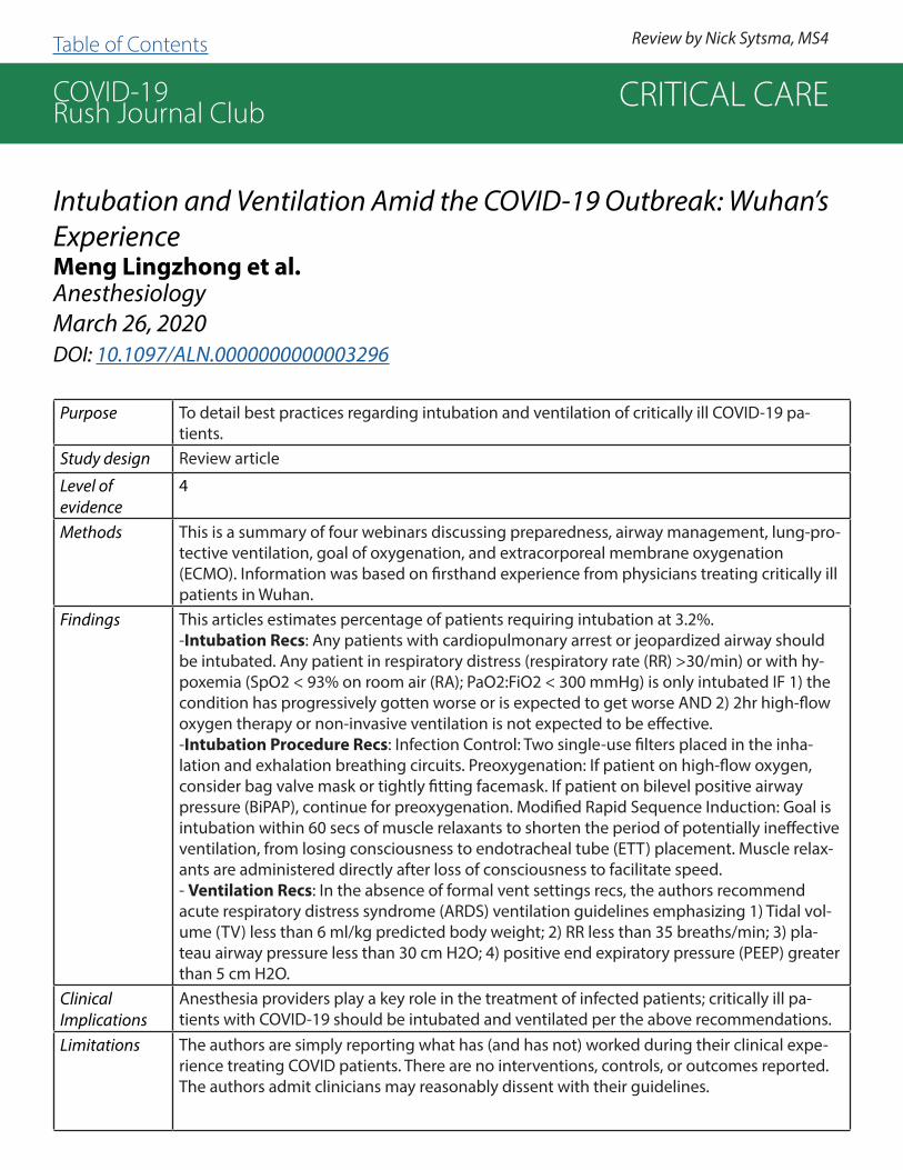
COVID-19Rush Journal Club
Intubation and Ventilation Amid the COVID-19 Outbreak: Wuhan’s ExperienceMeng Lingzhong et al.AnesthesiologyMarch 26, 2020DOI: 10.1097/ALN.0000000000003296
Purpose To detail best practices regarding intubation and ventilation of critically ill COVID-19 pa-tients.
Study design Review article
Level of evidence
4
Methods This is a summary of four webinars discussing preparedness, airway management, lung-pro-tective ventilation, goal of oxygenation, and extracorporeal membrane oxygenation (ECMO). Information was based on firsthand experience from physicians treating critically ill patients in Wuhan.
Findings This articles estimates percentage of patients requiring intubation at 3.2%.-Intubation Recs: Any patients with cardiopulmonary arrest or jeopardized airway should be intubated. Any patient in respiratory distress (respiratory rate (RR) >30/min) or with hy-poxemia (SpO2 < 93% on room air (RA); PaO2:FiO2 < 300 mmHg) is only intubated IF 1) the condition has progressively gotten worse or is expected to get worse AND 2) 2hr high-flow oxygen therapy or non-invasive ventilation is not expected to be effective.-Intubation Procedure Recs: Infection Control: Two single-use filters placed in the inha-lation and exhalation breathing circuits. Preoxygenation: If patient on high-flow oxygen, consider bag valve mask or tightly fitting facemask. If patient on bilevel positive airway pressure (BiPAP), continue for preoxygenation. Modified Rapid Sequence Induction: Goal is intubation within 60 secs of muscle relaxants to shorten the period of potentially ineffective ventilation, from losing consciousness to endotracheal tube (ETT) placement. Muscle relax-ants are administered directly after loss of consciousness to facilitate speed.- Ventilation Recs: In the absence of formal vent settings recs, the authors recommend acute respiratory distress syndrome (ARDS) ventilation guidelines emphasizing 1) Tidal vol-ume (TV) less than 6 ml/kg predicted body weight; 2) RR less than 35 breaths/min; 3) pla-teau airway pressure less than 30 cm H2O; 4) positive end expiratory pressure (PEEP) greater than 5 cm H2O.
ClinicalImplications
Anesthesia providers play a key role in the treatment of infected patients; critically ill pa-tients with COVID-19 should be intubated and ventilated per the above recommendations.
Limitations The authors are simply reporting what has (and has not) worked during their clinical expe-rience treating COVID patients. There are no interventions, controls, or outcomes reported. The authors admit clinicians may reasonably dissent with their guidelines.
CRITICAL CARE
Review by Nick Sytsma, MS4 Table of Contents
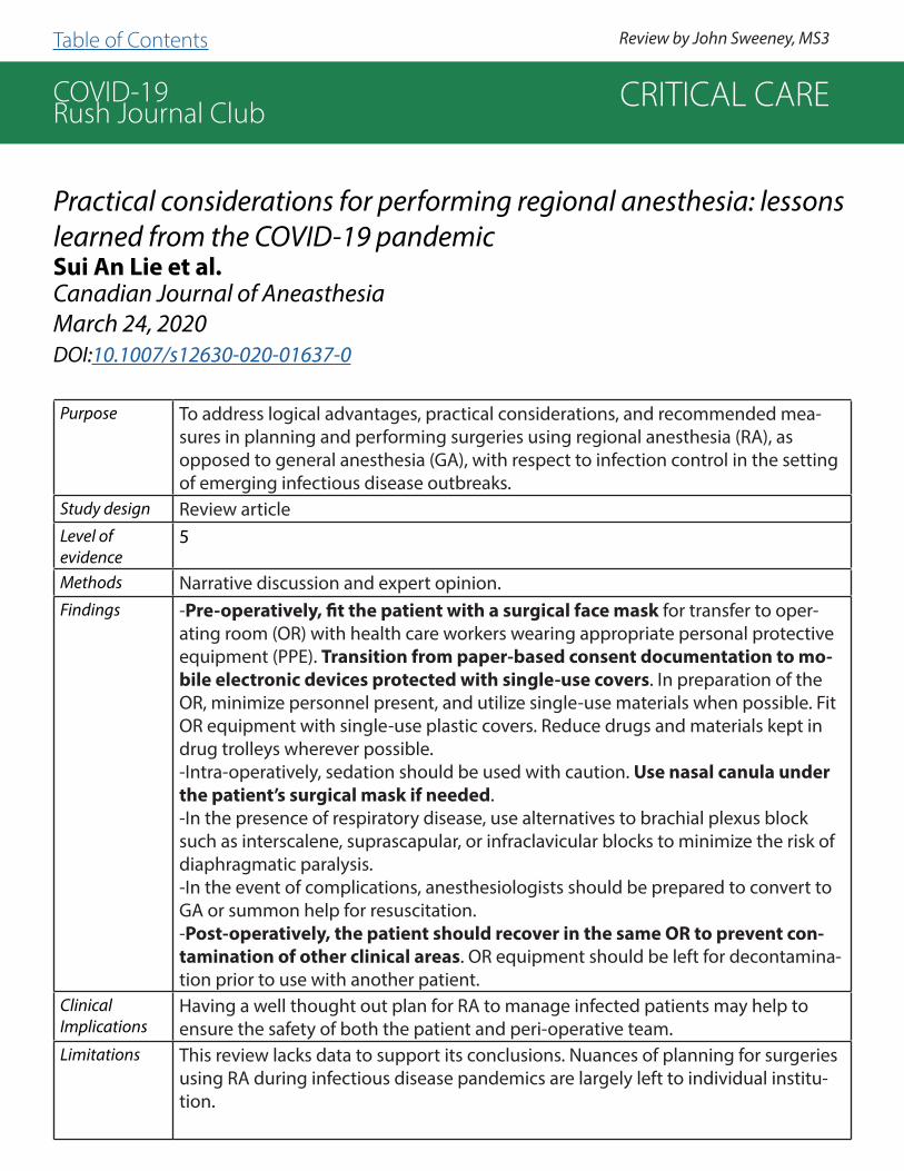
COVID-19Rush Journal Club
Practical considerations for performing regional anesthesia: lessons learned from the COVID-19 pandemicSui An Lie et al.Canadian Journal of AneasthesiaMarch 24, 2020DOI:10.1007/s12630-020-01637-0
Purpose To address logical advantages, practical considerations, and recommended mea-sures in planning and performing surgeries using regional anesthesia (RA), as opposed to general anesthesia (GA), with respect to infection control in the setting of emerging infectious disease outbreaks.
Study design Review articleLevel of evidence
5
Methods Narrative discussion and expert opinion.Findings -Pre-operatively, fit the patient with a surgical face mask for transfer to oper-
ating room (OR) with health care workers wearing appropriate personal protective equipment (PPE). Transition from paper-based consent documentation to mo-bile electronic devices protected with single-use covers. In preparation of the OR, minimize personnel present, and utilize single-use materials when possible. Fit OR equipment with single-use plastic covers. Reduce drugs and materials kept in drug trolleys wherever possible.-Intra-operatively, sedation should be used with caution. Use nasal canula under the patient’s surgical mask if needed. -In the presence of respiratory disease, use alternatives to brachial plexus block such as interscalene, suprascapular, or infraclavicular blocks to minimize the risk of diaphragmatic paralysis.-In the event of complications, anesthesiologists should be prepared to convert to GA or summon help for resuscitation.-Post-operatively, the patient should recover in the same OR to prevent con-tamination of other clinical areas. OR equipment should be left for decontamina-tion prior to use with another patient.
ClinicalImplications
Having a well thought out plan for RA to manage infected patients may help to ensure the safety of both the patient and peri-operative team.
Limitations This review lacks data to support its conclusions. Nuances of planning for surgeries using RA during infectious disease pandemics are largely left to individual institu-tion.
CRITICAL CARE
Review by John Sweeney, MS3 Table of Contents

COVID-19Rush Journal Club
Findings of lung ultrasonography of novel corona virus pneumonia during the 2019-2020 epidemic
Qian-Yi Peng et al.Intensive Care MedicineMarch 12, 2020DOI: https://doi.org/10.1007/s00134-020-05996-6
Purpose To summarize lung ultrasonography findings for evaluation of COVID-19.
Study design Case SeriesLevel of evidence
Level 4
Methods The authors performed lung ultrasonography on 20 patients with COVID-10 using a 12-zone method and aggregated typical pathological findings.
Findings -Some providers have recommended early CT for screening suspected patients, but lung ultrasound may not be practical for critically ill patients; additionally, lung ultra-sound has significant advantages in that there is ease of use at point of care, repeat-ability, absence of radiation exposure, and low cost.-Characteristic findings in 20 confirmed COVID-19 pts include 1) thickening of the pleural line with pleural line irregularity; 2) B-lines in a variety of patterns including focal, multi-focal, and confluent; 3) consolidations in a variety of patterns including multifocal small, non-translobar, and translobar with occasional mobile air broncho-grams; 4) appearance of A-lines during recovery phase; and 5) pleural effusions are uncommon.- Focal B-lines are the main feature in the early stage and in mild infection; alveolar interstitial syndrome is the main feature in the progressive stage and in critically ill patients; A-lines can be found in the convalescence.- Predominant pattern is of varying degrees of interstitial syndrome and alveolar con-solidation, correlated with the severity of the lung injury.
ClinicalImplications
This study offers a brief overview of expected lung ultrasound findings in COVID-19 patients. Lung ultrasound is helpful for diagnosing pneumonia/ARDS at presen-tation, tracking the evolution of disease, monitor lung recruitment maneuvers, managing ECMO and/or prone positioning, and making decisions related to weaning ventilator support.
Limitations The study only includes scans from twenty patients. No demographic information included; authors assume the ultrasound findings are universal. These ultrasound findings are consistent with many different lung processes/types of pneumonia; therefore can likely help diagnose pneumonia but not specifically COVID pneumonia.
CRITICAL CARE
Review by Nick Sytsma, MD

COVID-19Rush Journal Club
Development and Validation of a Clinical Risk Score to Predict the Occurrence of Critical Illness in Hospitalized Patients With COVID-19Wenhua Liang et al.Journal of the American Medical Association Internal MedicineMay 12, 2020DOI: 10.1001/jamainternmed.2020.2033
Purpose To develop and validate a clinical score assigned at hospital admission to assist in prediction of which patients with COVID-19 will develop critical illness.
Study design Retrospective cohort (developmental cohort n=1590; validation cohort n=710)
Level of evidence
Level 3
Methods Researchers established a cohort of COVID-19 positive patients from 575 hospitals across 31 Chinese provinces. Patients were considered critical upon ICU admission, intubation, or death. Epidemiological, clinical, laboratory, and imaging variables ascertained at hospital admission were screened using Least Absolute Shrinkage and Selection Operator (LASSO) and logistic regression to identify predictive factors of critical illness. Independent predictive factors were then combined to form a risk score calculator (COVID-GRAM). The scoring system’s accuracy was measured by the area under curve (AUC) method. The scoring system was subsequently validated using four additional Chinese cohorts.
Findings The development cohort included 1590 patients. Mean age was 48.9 years and 57.3% of pa-tients were male. The validation cohort included 710 patients with a mean age of 48.2 years. 53.8% of patients were male. From 72 potential predictors, 10 variables were independent predictive factors and included in the risk score: chest radiographic abnormality (odds ratio, OR: 3.39), age (OR: 1.03), hemoptysis (OR: 4.53), dyspnea (OR: 1.88), unconsciousness (OR: 4.71), number of comorbidities (OR: 1.60), history of cancer (OR: 4.07), neutrophil-to-lympho-cyte ratio (OR: 1.06), lactate dehydrogenase (OR: 1.002), and direct bilirubin (OR: 1.15). Mean AUC in the development cohort was 0.88 and the AUC in the validation cohort was 0.88. The score has been translated into an online risk calculator that is freely available to the public at http://118.126.104.170/.
ClinicalImplications
The authors have created an easily accessible, highly accurate scoring system (COVID-GRAM) for predicting a COVID-19 patient’s risk of developing critical illness, therefore allowing staff to better prioritize patient care and optimize the use of resources. Early returns indicate this scoring system (using ten significant variables and factors including chest X-ray abnormality, age, hemoptysis, dyspnea, and the number of comorbidities) is more accurate than the CURB-6 system commonly used by emergency physicians.
Limitations The populations used in both the developmental and validation cohorts were not enormous. All data originates from China, which may impact its generalizability to patients in other coun-tries as it has been well documented that countries have varying incidences of comorbidities.
CRITICAL CARE
Review by Nick Sytsma, MD Table of Contents

COVID-19Rush Journal Club
Initial Clinical Impressions of the Critical Care of COVID-19 Patients in Seattle, New York City, and ChicagoPhillip Sommer et al.Anesthesia & AnalgesiaMarch 25, 2020DOI: 10.1213/ANE.0000000000004830
Purpose To describe the clinical impressions of COVID-19 cases requiring intensive care.
Study design Observational/Expert Opinion
Level of evidence
Level 4
Methods This study was observational, with no reported interventions or population data. The study mentions aggregating more than 300 clinical cases, 100 of which required intubation, from America’s hardest hit cities: Seattle, New York City, and Chicago.
Findings Patients present diverse and nonspecific initial symptoms of fever, malaise, fatigue, and cough in addition to chest pain, headache, altered mental status, and gastrointestinal changes. Younger patients were not exempt from severe presentation. Lab findings were notable for rarely elevated white blood cell (WBC) counts with lymphopenia being a common prognostic sign. Procalcitonin levels were typically somewhat low. C-reactive protein (CRP) was frequently elevated with a positive correlation to severity of illness. Troponin and liver biomarkers were also sometimes elevated. Coagulation abnormalities, including D-dimer elevations, were frequently observed. Bilateral patchy pulmonary opacities on chest x-ray were almost always present and severity was usually congruent with disease severity. Hypotension was com-mon and treated with low-dose vasopressors. Concurrent myocardial infarction (MI) was also described. Severe cardiomyopathy presenting as respiratory symptoms was observed and appeared to dramatically increase mortality. Severe acute kidney injury (AKI) and refractory metabolic acidosis cases were not uncommon along with electrolyte abnormalities including hyperkalemia. Hypoxemia presented out of proportion to clinical presentation, frequently re-quiring noninvasive oxygenation which was associated with a high rate of intubation and low rate of successful extubation. Severe respiratory failure was reported to occur ~ 1 week after initial symptoms. Treatment of COVID-19 in the critical care setting was largely supportive. An-tiviral therapies, hydroxychloroquine, and aminoquinolines have generated research interest, but there is no consensus on their use or the utility of administering steroids.
ClinicalImplications
As rapidly-expanding COVID-19 infections continue to consume the US health care system’s finite resources, this study, which summarizes the COVID-19 landscape in three US metropol-itan areas, is useful for urban providers to preserve and augment their dwindling clinical and operational resources. It is also potentially useful for providers who have not encountered a COVID-19 surge to date, as it helps to establish clinical expectations.
Limitations This article is limited by a lack of objective data, lack of interventions, and urban patient pop-ulations and therefore, the findings may not be generalized to a wide array of clinical settings.
CRITICAL CARE
Review by Nick Sytsma, MDTable of Contents
COVID-19Rush Journal Club
TREATMENT
Table of Contents
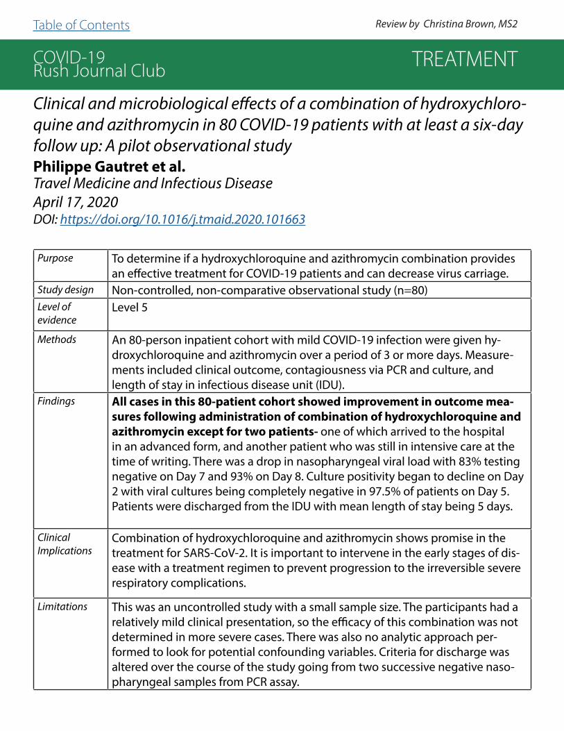
COVID-19Rush Journal Club
Clinical and microbiological effects of a combination of hydroxychloro-quine and azithromycin in 80 COVID-19 patients with at least a six-day follow up: A pilot observational studyPhilippe Gautret et al.Travel Medicine and Infectious DiseaseApril 17, 2020DOI: https://doi.org/10.1016/j.tmaid.2020.101663
Purpose To determine if a hydroxychloroquine and azithromycin combination provides an effective treatment for COVID-19 patients and can decrease virus carriage.
Study design Non-controlled, non-comparative observational study (n=80)Level of evidence
Level 5
Methods An 80-person inpatient cohort with mild COVID-19 infection were given hy-droxychloroquine and azithromycin over a period of 3 or more days. Measure-ments included clinical outcome, contagiousness via PCR and culture, and length of stay in infectious disease unit (IDU).
Findings All cases in this 80-patient cohort showed improvement in outcome mea-sures following administration of combination of hydroxychloroquine and azithromycin except for two patients- one of which arrived to the hospital in an advanced form, and another patient who was still in intensive care at the time of writing. There was a drop in nasopharyngeal viral load with 83% testing negative on Day 7 and 93% on Day 8. Culture positivity began to decline on Day 2 with viral cultures being completely negative in 97.5% of patients on Day 5. Patients were discharged from the IDU with mean length of stay being 5 days.
ClinicalImplications
Combination of hydroxychloroquine and azithromycin shows promise in the treatment for SARS-CoV-2. It is important to intervene in the early stages of dis-ease with a treatment regimen to prevent progression to the irreversible severe respiratory complications.
Limitations This was an uncontrolled study with a small sample size. The participants had a relatively mild clinical presentation, so the efficacy of this combination was not determined in more severe cases. There was also no analytic approach per-formed to look for potential confounding variables. Criteria for discharge was altered over the course of the study going from two successive negative naso-pharyngeal samples from PCR assay.
TREATMENT
Review by Christina Brown, MS2 Table of Contents

COVID-19Rush Journal Club
Management of COVID-19 Respiratory Distress
John J. Marini et al.JAMAApril 24, 2020DOI: 10.1001/jama.2020.6825
Purpose The purpose of this article is to summarize the current recommendations for ventilation support in COVID-19 patients with ARDS
Study design Literature Review
Level of evidence
5
Methods This paper reviewed current literature on the management of COVID-19 ventilation sup-port. The authors describe two patient phenotypes: Type L and Type H. -Typle L: Scattered ground-glass infiltrates, higher compliance (>50 mL/cm H2O), not PEEP responsive, less dyspnea-Type H: Extensive infiltrates of atelectasis and edema, lower compliance, PEEP responsive, overtly dyspneic
Findings -In the early stages of CARDS (COVID-19 with ARDS), the objective should be ade-quate gas exchange and avoidance of patient self-induced lung injury (P-SILI) from powerful respiratory effort causing lung and vascular stress. Options include sup-plemental O2, CPAP, noninvasive ventilation (NIV), high flow nasal cannula (HFNC), prone positioning and target nonvigorous breathing. Early intubation, effective sedation, and/or paralysis may interrupt this cycle. For Type L patients after intubation, the goal is to minimize pulmonary stress, optimize O2, and avoid VILI (ventilator-induced lung injury) vortex. Use lower PEEP (<10 cm H2O), use more liberal tidal volume (7-9 mL/kg) as needed, and consider prone positioning. If lung edema increases in the Type L patient (either because of the disease itself and/or P-SILI), Type H phenotype progressively develops. The goal in Type H patients after intubation is to reduce and evenly distrib-ute lung and vascular stresses, optimize O2, and avoid VILI. Use higher PEEP (<15 cm H2O), lower tidal volume (5-7 mL/kg), reduce O2 demand and implement prone position-ing. Despite the disease type, weaning should be undertaken cautiously. The goal for the weaning phase is to avoid reversion to previously worsened pulmonary state by causing VILI and worsening edema.
ClinicalImplications
The recommendations described above represent the most current ventilation recom-mendations in COVID-19 patients with ARDS and could improve outcomes.
Limitations This paper attempts to categorize COVID patients with respiratory distress into two groups; however, not all patients may conveniently fall into each of these two groups but rather fall along a spectrum of respiratory compromise.
TREATMENT
Review by Eric Moyer, MS4 Table of Contents
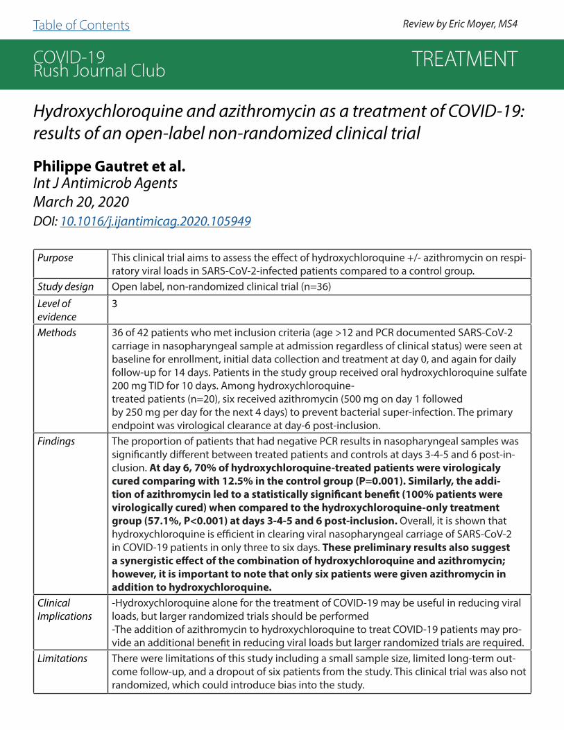
COVID-19Rush Journal Club
Hydroxychloroquine and azithromycin as a treatment of COVID-19: results of an open-label non-randomized clinical trial
Philippe Gautret et al.Int J Antimicrob AgentsMarch 20, 2020DOI: 10.1016/j.ijantimicag.2020.105949
Purpose This clinical trial aims to assess the effect of hydroxychloroquine +/- azithromycin on respi-ratory viral loads in SARS-CoV-2-infected patients compared to a control group.
Study design Open label, non-randomized clinical trial (n=36)
Level of evidence
3
Methods 36 of 42 patients who met inclusion criteria (age >12 and PCR documented SARS-CoV-2 carriage in nasopharyngeal sample at admission regardless of clinical status) were seen at baseline for enrollment, initial data collection and treatment at day 0, and again for daily follow-up for 14 days. Patients in the study group received oral hydroxychloroquine sulfate 200 mg TID for 10 days. Among hydroxychloroquine-treated patients (n=20), six received azithromycin (500 mg on day 1 followedby 250 mg per day for the next 4 days) to prevent bacterial super-infection. The primary endpoint was virological clearance at day-6 post-inclusion.
Findings The proportion of patients that had negative PCR results in nasopharyngeal samples was significantly different between treated patients and controls at days 3-4-5 and 6 post-in-clusion. At day 6, 70% of hydroxychloroquine-treated patients were virologicaly cured comparing with 12.5% in the control group (P=0.001). Similarly, the addi-tion of azithromycin led to a statistically significant benefit (100% patients were virologically cured) when compared to the hydroxychloroquine-only treatment group (57.1%, P<0.001) at days 3-4-5 and 6 post-inclusion. Overall, it is shown that hydroxychloroquine is efficient in clearing viral nasopharyngeal carriage of SARS-CoV-2 in COVID-19 patients in only three to six days. These preliminary results also suggest a synergistic effect of the combination of hydroxychloroquine and azithromycin; however, it is important to note that only six patients were given azithromycin in addition to hydroxychloroquine.
ClinicalImplications
-Hydroxychloroquine alone for the treatment of COVID-19 may be useful in reducing viral loads, but larger randomized trials should be performed -The addition of azithromycin to hydroxychloroquine to treat COVID-19 patients may pro-vide an additional benefit in reducing viral loads but larger randomized trials are required.
Limitations There were limitations of this study including a small sample size, limited long-term out-come follow-up, and a dropout of six patients from the study. This clinical trial was also not randomized, which could introduce bias into the study.
TREATMENT
Review by Eric Moyer, MS4Table of Contents

COVID-19Rush Journal Club
Pharmacologic treatments for coronavirus disease 2019 (COVID-19)
James M. SandersJAMA NetworkApril 13, 2020DOI: 10.1001/jama.2020.6019
Purpose To summarize current evidence regarding major proposed, repurposed or experi-mental treatments for COVID-19 and to provide a summary of current clinical expe-rience and treatment guidance for COVID-19.
Study design Literature ReviewLevel of evidence
N/A
Methods A literature review was performed using PubMed to identify relevant English-lan-guage articles published through March 25th, 2020. Search terms included ‘corona-virus’, ‘severe acute respiratory syndrome coronavirus 2’, ‘2019-nCoV’, ‘SARS-CoV-2’, ‘SARS-CoV’, ‘MERS-CoV’, and ‘COVID-19’ in combination with treatment and pharma-cology. Case reports, case series, and review articles were included due to the lack of randomized controlled trials. Currently active clinical trials were also included using the disease search term ‘coronavirus infection’ on ClinicalTrials.gov and the in-dex of studies of novel coronavirus pneumonia in the Chinese Clinical Trial Registry.
Findings Treatment recommendations based on clinical treatment experience, descriptive reports, and case series should be interpreted with caution due to lack of clinical trials. There are currently no medical therapies that have been definitively shown to improve outcomes in patients with COVID-19. Several drugs have demonstrated in vitro activity against SARS-CoV-2 virus including hydroxychloroquine, chloroquine, darunavir, ribavirin, baricitinib, imatinib, dasatinib, cyclosporine, nitazoxanide, rem-desivir, and favipiravir.
ClinicalImplications
There is currently no effective therapy for COVID-19, and therefore there is an ur-gent need for randomized clinical trials to test the effectiveness of proposed thera-pies.
Limitations To date, published data is limited to observational studies and small clinical trials with less than 250 patients. This review focused primarily on adult patients and lacks data on pediatric population infected with SARS-CoV-2. The amount of pub-lished literature is rapidly growing, and recommendations are constantly changing.
TREATMENT
Review by Sarah Sun, MS2Table of Contents

COVID-19Rush Journal Club
Remdesivir and chloroquine effectively inhibit the recently emerged novel coronavirus (2019-nCoV) in vitroManli Wang et al.Cell Research, Nature Publishing GroupFebruary 4, 2020DOI: https://doi.org/10.1038/s41422-020-0282-0
Purpose To determine which drug is the most effective in treating the 2019-nCov virus.
Study design Letter to the editor – in vitro lab studyLevel of evidence
Level 5
Methods Seven drugs, Ribavirin, Penciclovir, Nitazoxanide, Nafamostat, Chloroquine, Remdesivir and Favipiravir, were evaluated to determine their efficacy against 2019-nCov in vitro. Vero E6 cells were infected with the COVID virus at a multiplicity of infection of 0.05 with various degrees of concentration of the trial drugs for 48 hours. For the control group, DMSO was used instead. The primary endpoint was to determine the cytotoxici-ty of these drugs using CCK-8 assays, as well as the viral yield which was determined by quantifying the superna-tant using RT-PCR. This was later confirmed by using immu-nofluorescence to visualize the virus nucleoprotein expression. The results are listed below showcasing the half-maximal effective concentration used (EC50 in micromoles) and the selectivity index (SI) achieved during testing.
Findings - Ribavirin (EC50 = 109.50, SI > 3.65), Penciclovir (EC50 = 95.96, SI > 4.17) and Favipira-vir (EC50 = 61.88, SI > 6.46), the 3 nucleoside analogs, required high levels of concen-tration to reduce the viral infection.- Nafamostat (EC50 = 22.50, SI > 4.44) which prevents membrane fusion, was inhibitive against the Covid virus.- Nitazoxanide, (EC50 = 2.12, SI > 16.76) an antiprotozoal agent, was able to inhibit 2019 nCoV at low micro molar concentration.- Remdesevir (EC50 = 0.77, SI > 129.87) and Cloroquine (EC50 = 1.13, SI > 88.50) were able to block the virus infection at low micro molar concentration and demonstrated high selectivity index.
ClinicalImplications
Further in vivo studies are required to evaluate the true efficacy of these drugs, however, Remdesevir and Cloroquine appear promising. Their low micromolar concentration and high selectivity index to block virus infection sets them apart from other drugs.
Limitations The study was conducted in vitro using Vero E6 cells which are derived from the Afri-can green monkey and hence may not be translatable to human trials. As a letter to the editor, it is not clear if this study has been peer-reviewed.
TREATMENT
Review by Maria Amir, MS2Table of Contents

COVID-19Rush Journal Club
Some Drugs for COVID-19
The Medical Letter on Drugs and TherapeuticsApril 6, 2020Retrieved from: secure.medicalletter.org/w1595a
Purpose To review current data regarding the efficacy or lack thereof for the use of repurposed drugs in the treatment of COVID-19 in addition to the impact of commonly used daily medications on COVID-19 disease progression.
Study design Review
Level of evidence
Level 3
Methods The researchers summarized the clinical evidence related to repurposing of drugs for the treat-ment of COVID-19. They chose agents that have been widely reported on as potential treatments.
Findings -ACE inhibitors and ARBS: There is no clinical evidence to suggest these agents increase or decrease the severity of COVID-19. Patients who take these drugs and contract COVID-19 should continue their medications as prescribed. -NSAIDS: There is no clinical evidence that NSAIDs increase or decrease the severity of COVID-19, however continued fever suppression with NSAIDs can possibly decrease the immune system and increase the duration of viral shedding. Patients who are taking NSAIDs for other indications should not stop taking them.-Lopinavir/ritonavir (Kaletra): When compared to standard care in clinical trial of severely dis-eased COVID-19 patients, Kaletra was no more effective than the standard of care alone and Soci-ety of Critical Care Medicine does not recommend its use in critically ill patients. -Hydroxychloroquine with Azithromycin: Open label study in hospitalized COVID-19 patients in France suggests enhanced viral load reduction compared to treatment of hydroxychloroquine alone. These drugs can prolong the QT interval – clinical trials evaluating safety and efficacy are in progress.-IL-6 inhibitors (ie: tocilizumab, sarilumab): Insufficient data, clinical trials are in progress to see if these agents and reduce cytokine induced lung damage in patients with severe disease. -Convalescent sera: Passive antibody therapy using serum of recovered patients was both safe and reduced viral load in Chinese patients who were treated early in the course of their infection with COVID-19. There are ongoing studies to examine this effect in critically ill patients.
ClinicalImplications
The data summarized in this review helps guide clinicians in the treatment of COVID-19 and helps delineate between evidence-based practice and media publicization of available agents. The authors also advise that until clinical trials clearly establish the safety and efficacy of any drug used for COVID-19 treatment, current standard of practice is supportive treatment and management of COVID-19 complications.
Limitations This review was unable to adequately synthesize efficacy of the drugs highlighted in this review due to lack of high quality RCTs. Also, little data describing the effect of the drugs in patients with different disease severity; the review findings only commented on limited ranges of disease sever-ity.
TREATMENT
Review by Demetrios Geanon, MS3Table of Contents

COVID-19Rush Journal Club
Evaluation of Antiviral Therapies for Coronavirus Disease 2019 (COVID-19) Pneumonia in Shanghai, ChinaXiudong Shi et al.J Med Virol.April 16, 2020DOI: https://doi.org/10.1002/jmv.25893
Purpose Evaluate the therapeutic effect of antiviral drugs on COVID-19 pneumonia.
Study design Single-center, retrospective review (n=184)Level of evidence
4
Methods A total of 184 patients seen at the Shanghai Public Health Clinical Center that tested positive for COVID-19 were divided into 7 different groups according to their treatment, which was administered over a 5-day period. The groups were as follows: (1) symptomatic treatment only (2) Arbidol, (3) Lopinavir/Ritonavir, (4) Arbidol and Lopinavir/Ritonavir, (5) Interferon, (6) Interferon and Lopinavir/Ri-tonavir, and (7) Interferon and Darunavir. Chest CT scans at admission and at day 1 or 2 after treatment were reviewed and the Quantitative Evaluation System of CT for Pneumonia was utilized to calculate pulmonary inflammation volume (pneumonia volume).
Findings The average pneumonia volume in all groups increased, except in the In-terferon and Lopinavir/Ritonavir combination group. However, differences between groups (i.e. Lopinavir/Ritonavir treatment alone or in combination with Interferon-α2β or Arbidol) were not statistically significant. While there was also no significant difference in pneumonia resolution among the groups, the highest proportion of pneumonia resolution was in the Interferon and Lopinavir/Ritonavir combination group, followed by the Interferon and Daruna-vir combination group.
ClinicalImplications
This study did not find that the addition of antiviral drugs in therapeutic regi-mens reduced the volume of lung affected by pneumonia in COVID-19 patients, nor did it significantly shorten their hospital stay compared to symptomatic treatment alone. The treatment of COVID-19 pneumonia remains challenging, as there are no specific and effective drugs available.
Limitations This study set the treatment period of 5 days, which may not have been long enough to see results. Additionally, results of quantitative detection of viral load were not reported as part of this study and CT follow up findings were not in-cluded.
TREATMENT
Review by Athena Jane Manatis-Lornell, MS1 Table of Contents

COVID-19Rush Journal Club
Current evidence for directed and supportive investigational therapies against COVID-19R van Rensburg et al.African Journal of Thoracic and Critical Care MedicineApril 30, 2020DOI: https://doi.org/10.7196/AJTCCM.2020.v26i2.072
Purpose To investigate types of therapies currently being studied for treatment of COVID-19.
Study design Systematic Review
Level of evidence
Level 3
Methods Authors used current literature published in peer-reviewed scientific journals to determine current evidence regarding several types of therapeutic interventions for the treatment of COVID-19.
Findings Two groups of therapies were evaluated, directed therapies and supported therapies. Directed therapies included hydroxychloroquine and chloroquine, lopinavir/ritonavir, remdesivir, and favipiravir. Hydroxychloroquine and chloroquine are immunomodulato-ry drugs that show in vitro activity against COVID-19, theorized to be due to increasing endosomal pH to inhibit COVID-19 spike protein cleavage, preventing entry. Studies are showing conflicting evidence of hydroxychloroquine and chloroquine having no effect or being able to reduce viral shedding. Lopinavir/ritonavir are protease inhibitors that have shown in vitro activity against SARS-CoV. Case reports show successful man-agement with lopinavir/ritonavir combination. Remdesivir is a nucleotide analogue de-veloped against the Ebola virus which shows in vitro activity against COVID-19. Supported therapies included tocilizumab and corticosteroids. Tocilizumab is a monoclonal antibody against the IL-6 receptor that is approved for treating cytokine release syndrome. Trials have shown patients improving following inadequate response to standard care. Cortico-steroid trials are inconclusive or showed potential to cause harm.
ClinicalImplications
There is a lack of in vivo and human studies to determine which therapy, if any, is effective in treating COVID-19. Studies for hydroxychloroquine, chloroquine, lopinavir/ritonavir, remdesivir, favipiravir, tocilizumab show promise based on in vitro studies, but there is significant lack of data to conclude what is effective in treating COVID-19. Studies typically started later in the disease course, after organ damage had potentially occurred.
Limitations There are limited peer-reviewed publications out currently that can further knowledge of therapeutic studies. These studies also are started late in the disease course when it may be too late to prevent irreversible organ damage.
TREATMENT
Review by Christi Brown, MS2 Table of Contents

COVID-19Rush Journal Club
Triple Combination of Interferon beta-1b, Lopinavir–Ritonavir, and Ribavirin in the Treatment of Patients Admitted to Hospital with COVID-19: An Open-label, Randomised, Phase 2 TrialIvan Fan-Ngai Hung et al.The LancetMay 08, 2020DOI: https://doi.org/10.1016/S0140-6736(20)31042-4
Purpose To assess the efficacy and safety of a triple anti-viral therapy consisting of interferon beta-1b, lopinavir-ritonavir, and ribavirin for treatment of patients with COVID-19.
Study design Multicenter prospective, open-label, randomized, phase 2 trial
Level of evidence
Level 2
Methods Patients diagnosed with mild to moderate COVID-19 were randomly assigned (2:1) to a 14-day combination therapy consisting of lopinavir (400mg) and ritonavir (100mg) ev-ery 12 hours, ribavirin (400mg) every 12 hours, and three doses of 8 million IU of inter-feron beta-1b on alternate days (combination growth) or 14 days of lopinavir 400mg and ritonavir 100mg every 12 hours (control group). The primary endpoint was the time to RT-PCR negative nasopharyngeal swab for SARS-CoV-2. The secondary end-point was time to resolution of symptoms defined as a national early warning score of 0 maintained for 24 hours, and length of hospital stay.
Findings Patients receiving combination therapy demonstrated superior clinical improvement with shorter time to complete symptom resolution (4 days), significantly shorter time from initiation of treatment to negative nasopharyngeal swab (7 days), and shorter mean hospital stay (9 days) when compared to the control group (8, 12, and 14.5 days respectively). Treatment was also shown to be safe with minor and self-limited gastro-intestinal adverse events of diarrhea and vomiting with no difference in rate between those receiving combination therapy and the control group. In addition, serum levels of the inflammatory cytokine interleukin 6 (IL-6) were significantly lower in patients treated with combination therapy on treatment days 2, 6, and 8.
ClinicalImplications
Early treatment with triple antiviral therapy was safe and superior to lopinavir-ri-tonavir in alleviating symptoms and shortening the duration of viral shedding and hospitalization in patients with mild to moderate COVID-19. Triple antiviral therapy rapidly rendered viral load negative in all patients, thereby reducing infectiousness of the patient.
Limitations The trial was open label without inclusion of a placebo group. Results were confound-ed by the subgroup omitting interferon beta-1b within the combination group and were dependent on time of symptom onset. The absence of critically ill patients does not allow for the generalization of findings to severe cases.
TREATMENT
Review by Ashley Wehrheim, MS2 Table of Contents

COVID-19Rush Journal Club
Infectious Diseases Society of America Guidelines on the Treatment and Management of Patients with COVID-19 Adarsh Bhimraj et al.Clinical Infectious DiseasesApril 17, 2020DOI: https://doi.org/10.1093/cid/ciaa478
Purpose To develop evidence-based rapid guidelines intended to support patients, clinicians, and oth-er health-care professionals in their decision-making regarding treatment and management of patients with COVID-19.
Study design Systematic Review
Level of evidence
Level 3
Methods Infectious Diseases Society of America (IDSA) formed a multidisciplinary guideline panel of nine infectious disease clinicians, pharmacists, and methodologists with varied areas of expertise. Clinical questions in PICO format (Population, Intervention, Comparison, Outcomes) were developed and panel members prioritized questions with available evidence meeting minimum acceptable criteria (i.e., body of evidence reported on at least a case-series design, case reports excluded). A systematic review of the peer-reviewed and grey literature from Ovid Medline and Embase was then conducted. The Grading of Recommendations Assessment, Development and Evaluation (GRADE) approach was used to assess certainty of evidence and make recommendations. 435 viable references were identified, of which 13 informed the evi-dence base for the authors’ recommendations.
Findings The IDSA guideline panel recommends that treatment of COVID-19 infected patients with the following agents should be limited to the context of a clinical trial: hydroxychloroquine (HCQ), HCQ + azithromycin combination, lopinavir/ritonavir, tocilizumab, COVID-19 convalescent plasma transfusion, and corticosteroids (based on indirect findings from systematic review of SARS and MERS outbreaks). Of note, if a patient is receiving steroid therapy for another indica-tion (e.g., asthma), the steroid should not be discontinued.
ClinicalImplications
Given that the panel could not make a determination whether the benefits outweigh harms for HCQ, azithromycin, steroids, or IL-6 inhibitors, it would be ethical to enroll patients with COVID-19 in clinical trials, rather than use clinically unproven therapies. There are about 100 ongoing clinical trials on COVID-19 in the U.S. alone, which will allow us to understand more about the effects of these treatments and their potential therapeutic benefits within the com-ing months.
Limitations Due to the urgency in producing, synthesizing, and disseminating data during the current pandemic, an increase in “fast-tracked” study publication has resulted in issues including circumvention of usual research steps (delay of IRB approval, inclusion of same patients in several studies), a limited peer-review process, and increased potential for publication bias (in the interest of showing promising data). The extent and impact of these considerations were acknowledged in the development of these IDSA guidelines.
TREATMENT
Review by Emily Chi, MS3 Table of Contents

COVID-19Rush Journal Club
Efficacy of hydroxychloroquine in patients with COVID-19: results of a randomized clinical trialZhaowei Chen et al.medRxivApril 10, 2020DOI: https://doi.org/10.1101/2020.03.22.20040758
Purpose To investigate the efficacy of hydroxychloroquine in addition to standard treatment in patients hospitalized with confirmed COVID-19
Study design Randomized double-blind study (n = 62 patients)
Level of evidence
Level 2
Methods Patients were followed for 6 days. The treatment group received 400mg/day of oral hydroxychloroquine sulfate (HCQ) tablets on days 1-5 plus Standard Treatment (O2 therapy, antiviral agents, antibiotics, and immunoglobulin +/- corticosteroids). The control group was given Standard Treatment only.
Findings Statistically significant differences were observed favoring the treatment group vs the control group for improvement in Chest CT findings (p=0476), days for reso-lution in fever (p=0.0008) and days for resolution of cough (p=0016). Only 2 of 31 patients in the treatment group developed side effects that were not “severe”.
ClinicalImplications
Although this study suggests that hyoxychloroquine treatment for patients with mild COVID-19 disease is beneficial, auxiliary treatment modalities are unspeci-fied and may have confounded the results, the patients enrolled were relative-ly young and the observation time was limited.
Limitations The results may not be applicable to hospitalized patients in the US due to the ex-clusion criteria. Patients with cardiac conduction/arrhythmias were excluded which could limit the use of this medication in hospital patients who often are sicker at baseline and with significant cardiac comorbidities. Similarly, patients with renal and liver dysfunction at baseline were excluded, limiting application. Patients were of relatively young age (44.7 yrs), all patients had “mild disease”, the observation period was limited to 6 days and most significantly the standard therapy included confounding and unspecified treatments: antiviral agents, antibiotics and immu-noglobulin +/- corticosteroids. This standard care is worrisome as we do no know which patients received which antiviral agent or antibiotics and which patients received immuglobulin and in those who received immunoglobulin which also received corticosteroids.
TREATMENT
Review by Joseph deBettencourt, MS3 Table of Contents

COVID-19Rush Journal Club
Chloroquine and hydroxychloroquine in COVID-19
Robin E Ferner et al.BMJApril 8, 2020DOI: https://doi.org/10.1136/bmj.m1432
Purpose To advise on the potential premature use and potential harm of chloroquine and hy-droxychloroquine in COVID-19.
Study design EditorialLevel of evidence
N/A
Methods Authors report their opinions on how previous lab studies in combination with poor methods and reporting may show that the early use of 4-aminoquinolines, chloro-quine and hydroxychloroquine may lead to potential harm.
Findings In cell cultures and animal studies, the effects of 4-aminoquinolines on viruses from H5N1 to Zika have been variable. For example, in one study of chikungunya virus, chlo-roquine was active in laboratory studies but worsened the clinical course of infection in monkeys. The disparity between laboratory and clinical experiments may be due to the complex pharmokinetics of 4-aminoquinolines, making it hard to use the correct concentration in culture media to doses in humans. Many studies currently coming out about positive findings of hydroxychloroquine treatment of COVID-19 include poor methods as well as unreliable results. Although advocates have deemed hydroxychlo-roquine as safe and widely used, that cannot be guaranteed and can expose some patients to rare and potentially fatal reactions. There have been previous medications that have been withdrawn because of adverse reactions after showing clinical promise.
ClinicalImplications
· Many studies have shown that 4-aminoquinolines, such as hydroxychloroquine and chloroquine, are active against a range of viruses, but the translation to clini-cal use as treatment with multiple other viruses has not proven as useful.· There is to be more success in COVID-19 treatment via prevention by a vaccine or treatment with drugs that target specific structures in the virus rather than using old drugs that may work in the laboratory, but lack data supporting clinical use.· There needs to be better, properly powered, randomized controlled trials of chloro-quine or hydroxychloroquine in order to prove effectiveness. Until then, SARS-CoV-2 is “essentially untreatable” except for supportive measures.
Limitations The editorial only referred to two studies of 4-aminoquinolines in COVID-19 treatment that had poor study designs, while also citing previous studies in a wide range of virus-es, which are not directly compared to the pathogenesis of SARS-Cov-2.
TREATMENT
Review by Amanda Narkis, MS2Table of Contents

COVID-19Rush Journal ClubEffect of High vs Low Doses of Chloroquine Diphosphate as Adjunc-tive Therapy for Patients Hospitalized With Severe Acute Respiratory Syndrome Coronavirus 2 (SARS-CoV-2) InfectionMayla Gabriela Silva Borba et al.JAMA NetworkApril 22, 2020DOI: 10.1001/jamanetworkopen.2020.8857
Purpose To assess the safety and efficacy of high and low-dose chloroquine (CQ) for patients with severe COVID-19
Study design Parallel, double-masked, randomized, phase IIb clinical trial (n= 81)
Level of evidence
Level 2
Methods Primary outcome was lethality by day 28, secondary outcomes were lethality on day 13, patient clinical status, lab examinations, electrocardiogram (ECG) on days 13 and 28, daily clinical status, duration of mechanical ventilation, supplemental O2 use, and time from treatment to death. Of 131 patients admitted to the hospital with acute respiratory dis-tress syndrome (ARDS) older than 18 years of age, 81 were determined to be positive for COVID-19 by RT-PCR or have a high likelihood of having COVID-19 by epidemiologic data. 41 patients were placed on high dose CQ (600mg BID for 10 days) and 40 were placed on low dose CQ (450mg BID for one day then 450mg daily for 4 days; placebo tablets were used such that low dose patients took equal number of total tablets as high dose pa-tients). The study hypothesized that lethality would be decreased by 50% in the high-dos-age versus the low-dosage group.
Findings At day 13, lethality was 39.0% (16/41) in the high-dose group and 15.0% (6/40) in the low-dosage group, additionally 86.4% (19/22) of the deceased still had virologic con-firmation of SARS-CoV-2 infection antemortem. These findings showed the opposite of the study’s hypothesis and the safety review board recommended the immediate inter-ruption of the study. This study shows no evidence of benefit or increased viral clear-ance with use of chloroquine in patients presenting with SARS-CoV-2. High-dose chloroquine was associated with increased mortality over low-dose chloroquine dosing and over historical mortality data of similar patients with SARS-CoV-2.
ClinicalImplications
This trial suggests use of high-dose CQ (12g) given concurrently with azithromycin and oseltamivir is not safe, showed no evidence of benefit, and should not be used to treat patients with severe COVID-19.
Limitations The study was ended early due to concerns for increased lethality with high-dose regimen, leaving the study underpowered to detect efficacy of either dosages. It also only focuses on critically ill patients and results may not be generalizable to less severe disease. All patients received oseltamivir (for influenza) which is also known to increase the QTc inter-val. It is possible increased lethality of CQ may have resulted from synergistic cardiotoxic effects. No placebo group was used.
TREATMENT
Review by Joseph deBettencourt, MS3Table of Contents

COVID-19Rush Journal Club
A pilot study of hydroxychloroquine in treatment of patients with common coronavirus disease-19 (COVID-19)J Chen et al.Journal of Zhejiang University (Medical Sciences)March 6, 2020DOI: 10.3785/j.issn.1008-9292.2020.03.03
Purpose To study the efficacy and safety of hydroxychloroquine sulfate treatment in COVID-19
Study design Single-center, randomized study (n = 30 patients) Level of evidence
Level 2
Methods The experimental group received hydroxychloroquine sulfate 400mg once a day for 5 days plus conventional treatment. The control received conventional treatment alone. Conventional treatment included bed rest, oxygen support, symptomatic care, antiviral medications (nebulized IFN-alpha, oral lopinavir/ritonavir), and antibi-otics. The primary endpoint of the study was negative conversion rate of COVID-19 nucleic acid in respiratory pharyngeal swab on day 7 after randomization or death within 2 weeks. Secondary endpoints were serious adverse effects or deterioration of patient’s condition within 2 weeks.
Findings By day seven, 86.7% (13/15) of the trial group and 93.3% (14/15) of the control group tested negative for SARS-CoV-2 via pharyngeal swab. At two weeks all pa-tients tested negative, and were clinically improved.
ClinicalImplications
This study showed no improvement in clearance or change in mortality with the addition of hydroxychloroquine to the conventional treatment. This study suggests that hydroxychloroquine is not effective as an adjuvant medication in addition to current standards of care.
Limitations The study had a small sample size of only 30 patients. It excluded any patients with serious comorbidities. These exclusions make the results difficult to generalize to critically ill patients. The authors also noted that their study was underpowered to determine if HCQ was better or worse than standard care, by their estimates at least 784 subjects would be needed to appropriately power the study.
TREATMENT
Review by Joseph deBettencourt, MS3 Table of Contents

COVID-19Rush Journal Club
Observational Study of Hydroxychloroquine in Hospitalized Patients with Covid-19Joshua Geleris et al.The New England Journal of MedicineMay 7, 2020DOI: 10.1056/NEJMoa2012410
Purpose To investigate the relationship between hydroxychloroquine (HCQ) use and respiratory failure using a composite endpoint of intubation and/or death as major predictors of respiratory failure.
Study design Observational study (n=1376)
Level of evidence
Level 4
Methods De-identified data (age, sex, ethnicity, insurance, initial vital signs, arterial partial pressure of O2 to the fraction of inspired oxygen ratio [PaO2/FIO2] at admission, BMI, initial lab tests, smoking status, past medical history, medication administration, HCQ exposure, and outcomes [dis-charge, death, intubation]) from time of admission to death or discharge (prior to April 25, 2020) of COVID-19 positive patients hospitalized for a minimum of 24 hours between March 7-April 8, 2020 at Columbia University Irving Medical Center (CUIMC) were extracted and analyzed. Treat-ment with HCQ was at provider discretion and consistent with current hospital guidelines (600 mg twice on day 1, followed by 400 mg daily for 4 additional days). Bivariate frequencies were calculated to evaluate associations between pre-admission variables. Cox proportional hazard regression models were run to analyze association between HCQ exposure and outcomes of death or intubation. Additional models were used to account for demographic, clinical, and laboratory variables. To reduce risk of confounding, propensity scores for receipt of HCQ were calculated and used in inverse probability weighted analysis for Kaplan-Meier curves and Cox models. Additional analyses were completed for patients admitted at least 48 hours.
Findings 1376 patients were followed for a median of 22.5 days, 811 (58.9%) received HCQ. Among pa-tients recieving HCQ, administration began after 24 hours in 45.8% and after 48 hours in 85.9%. Patients receiving HCQ demonstrated lower PaO2:FIO2 at baseline than those who did not (median, 233 vs 360 mmHg). Death or intubation occurred in 346 (262 received HCQ, 84 did not). While crude analysis hazard ratios (HR) showed a significant association between HCQ use and death/intubation (HR: 2.37), no significant association was observed in the multivariable analysis with inverse probability weighting (HR: 1.04). No association was found between death/intuba-tion and azithromycin use (HR: 1.03)
ClinicalImplications
This study does not support the routine use of HCQ for COVID-19 patients as they found no association (neither harm nor benefit) between the drug’s use and patient outcomes of death or intubation. This study recommends that HCQ should only be used in clinical trials for efficacy. More research is needed to determine the best dose, when to administer (and for how long), and how the risks and benefits of these medications compare when treating COVID-19 patients.
Limitations This study was observational in nature and used relatively wide confidence intervals, there-fore these findings cannot rule out the harms or benefits of HCQ use in patients infected with COVID-19. As an observational study (i.e., the authors only looked at the end results without involving treatment), the analysis is subject to unmeasured confounding and bias, although attempts were made to limit their impact.
TREATMENT
Review by Kavya Timmireddy, MS3 Table of Contents
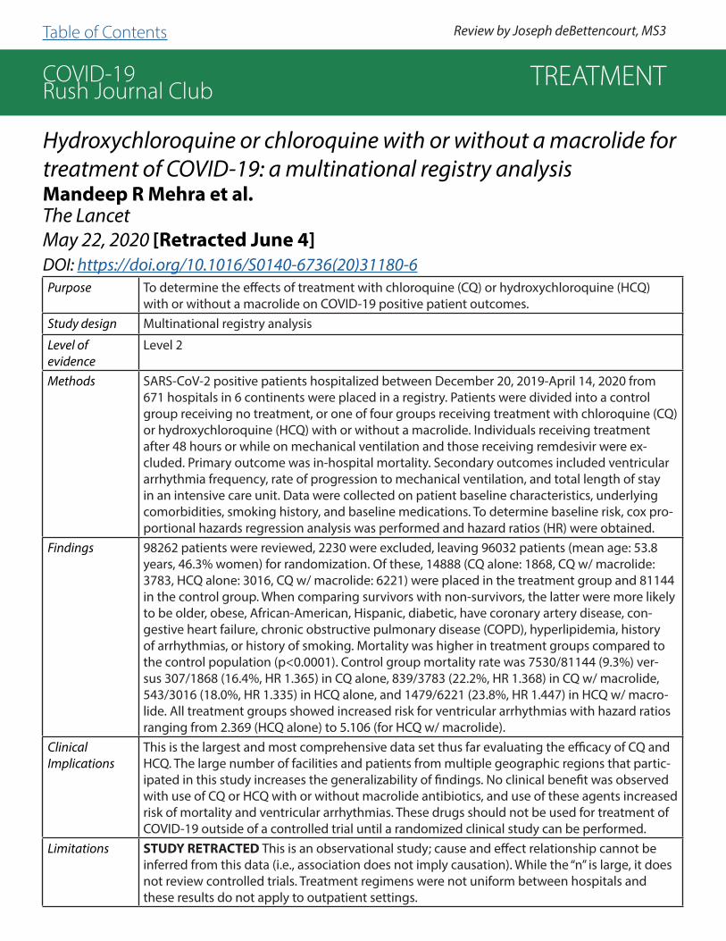
COVID-19Rush Journal Club
Hydroxychloroquine or chloroquine with or without a macrolide for treatment of COVID-19: a multinational registry analysisMandeep R Mehra et al.The LancetMay 22, 2020 [Retracted June 4]DOI: https://doi.org/10.1016/S0140-6736(20)31180-6Purpose To determine the effects of treatment with chloroquine (CQ) or hydroxychloroquine (HCQ)
with or without a macrolide on COVID-19 positive patient outcomes.Study design Multinational registry analysis
Level of evidence
Level 2
Methods SARS-CoV-2 positive patients hospitalized between December 20, 2019-April 14, 2020 from 671 hospitals in 6 continents were placed in a registry. Patients were divided into a control group receiving no treatment, or one of four groups receiving treatment with chloroquine (CQ) or hydroxychloroquine (HCQ) with or without a macrolide. Individuals receiving treatment after 48 hours or while on mechanical ventilation and those receiving remdesivir were ex-cluded. Primary outcome was in-hospital mortality. Secondary outcomes included ventricular arrhythmia frequency, rate of progression to mechanical ventilation, and total length of stay in an intensive care unit. Data were collected on patient baseline characteristics, underlying comorbidities, smoking history, and baseline medications. To determine baseline risk, cox pro-portional hazards regression analysis was performed and hazard ratios (HR) were obtained.
Findings 98262 patients were reviewed, 2230 were excluded, leaving 96032 patients (mean age: 53.8 years, 46.3% women) for randomization. Of these, 14888 (CQ alone: 1868, CQ w/ macrolide: 3783, HCQ alone: 3016, CQ w/ macrolide: 6221) were placed in the treatment group and 81144 in the control group. When comparing survivors with non-survivors, the latter were more likely to be older, obese, African-American, Hispanic, diabetic, have coronary artery disease, con-gestive heart failure, chronic obstructive pulmonary disease (COPD), hyperlipidemia, history of arrhythmias, or history of smoking. Mortality was higher in treatment groups compared to the control population (p<0.0001). Control group mortality rate was 7530/81144 (9.3%) ver-sus 307/1868 (16.4%, HR 1.365) in CQ alone, 839/3783 (22.2%, HR 1.368) in CQ w/ macrolide, 543/3016 (18.0%, HR 1.335) in HCQ alone, and 1479/6221 (23.8%, HR 1.447) in HCQ w/ macro-lide. All treatment groups showed increased risk for ventricular arrhythmias with hazard ratios ranging from 2.369 (HCQ alone) to 5.106 (for HCQ w/ macrolide).
ClinicalImplications
This is the largest and most comprehensive data set thus far evaluating the efficacy of CQ and HCQ. The large number of facilities and patients from multiple geographic regions that partic-ipated in this study increases the generalizability of findings. No clinical benefit was observed with use of CQ or HCQ with or without macrolide antibiotics, and use of these agents increased risk of mortality and ventricular arrhythmias. These drugs should not be used for treatment of COVID-19 outside of a controlled trial until a randomized clinical study can be performed.
Limitations STUDY RETRACTED This is an observational study; cause and effect relationship cannot be inferred from this data (i.e., association does not imply causation). While the “n” is large, it does not review controlled trials. Treatment regimens were not uniform between hospitals and these results do not apply to outpatient settings.
TREATMENT
Review by Joseph deBettencourt, MS3 Table of Contents

COVID-19Rush Journal Club
Assessment of QT Intervals in a Case Series of Patients With Coronavirus Disease 2019 (COVID-19) Infection Treated With Hydroxychloroquine Alone or in Combination With Azithromycin in an Intensive Care UnitFrancis Bessière et al.Journal of the American Medical Association CardiologyMay 1, 2020DOI: 10.1001/jamacardio.2020.1787
Purpose To evaluate changes in corrected QT (QTc) interval among critically ill COVID-19 positive pa-tients receiving therapy with hydroxychloroquine (HCQ) alone or in combination with azithro-mycin.
Study design Retrospective case series (n=40)
Level of evidence
Level 4
Methods COVID-19 positive patients (via PCR analysis of nasopharyngeal swab) admitted to the inten-sive care unit (ICU) between March 15 and March 29, 2020 were included in the analysis. All patients received HCQ monotherapy (200 mg, twice a day, for 10 days). A subset of patients also received azithromycin (250 mg daily, for 5 days). Patients were not included in the analy-sis if baseline QTc was greater than 460 ms. Patients underwent daily electrocardiogram (ECG) and continuous monitoring of the QTc interval. Prolonged QTc was the primary endpoint and was classified by one of two parameters: a QTc change (DQTc) of more than 60 ms or a pro-longed QTc interval of more than 500 ms.
Findings The outcomes of 40 COVID-19 patients who received HCQ alone (55%) or in combination with azithromycin (45%) were examined. Of these, the median age was 68 years, 32 (75%) were male, 30 (75%) required intubation, and 25 (63%) required vasoactive medications. Most patients (93%) had an increase in QTc after administration of therapy, regardless of treatment group. 10 patients (25%) demonstrated a DQTc of more than 60 ms and 7 patients (17.5%) demonstrated prolonged QTc intervals of more than 500 ms, with some patients meeting both criteria. By the authors’ definition, 14 total patients (36%) demonstrated prolonged QTc. A greater percentage of patients in the combination therapy group (33%), compared to those in the monotherapy group (5%, p=0.03), demonstrated a DQTc of more than 60 ms. No ventricu-lar arrhythmias were seen in either group, but therapy was stopped in 45% of patients due to ECG abnormalities or acute renal failure.
ClinicalImplications
HCQ and azithromycin use in COVID-19 positive patients may result in prolongation of the QTc interval, particularly when administered together. Authors suggest that this treatment should only be used when patients can be closely monitored because serious complications may de-velop. Further research conducted in a larger number of patients may help to clarify the risks and benefits of these therapies.
Limitations This was a small case series conducted at a single site. In addition to small sample size, gener-alizability is limited due to all patients receiving ICU level care. ICU patients may require and receive additional medications that contribute to prolongation of the QTc interval.
TREATMENT
Review by Steven Heidt, MDTable of Contents

COVID-19Rush Journal Club
Risk of QT Interval Prolongation Associated With Use of Hydroxychloro-quine With or Without Concomitant Azithromycin Among Hospitalized Patients Testing Positive for Coronavirus Disease 2019 (COVID-19)
Nicholas J. Mercuro et al.Journal of the American Medical Association CardiologyMay 1, 2020DOI: 10.1001/jamacardio.2020.1834
Purpose To evaluate QTc changes in patients diagnosed with COVID-19 pneumonia treated with hydroxychloroquine (HCQ) monotherapy or in combination with azithromycin.
Study design Observational, retrospective cohort study at a single center.
Level of evidence
Level 3
Methods Clinical data were obtained from COVID-19 positive patients diagnosed (via PCR analysis of nasopharyngeal swabs) between March 1 and April 7, 2020. All patients received at least one day of HCQ monotherapy or HCQ with concomitant azithromycin. Data were analyzed at the cohort level and comparisons were made between the monotherapy and combina-tion therapy groups. Primary endpoints of interest included DQTc from baseline, develop-ment of prolonged QTc greater than 500ms, and development of adverse drug events.
Findings The study was comprised of 90 patients with a mean age of 60.1 years, mean BMI of 31.5, and 48.9% female. At the cohort level, median baseline QTc was 455ms, 11% of patients had a DQTc of greater than 60ms, and 20% demonstrated post-treatment QTc of more than 500ms. In the monotherapy group, 3% of patients had a DQTc of more than 60ms and 19% demonstrated a prolonged QTc. In the combination therapy group, 13% had a DQTc of more than 60ms and 21% had a prolonged QTc. The likelihood of a prolonged QTc was greater in those taking loop diuretics (31% vs 12%, p=0.03) and those with a baseline QTc of greater than 450ms (30% vs 8%, p=0.008). Both findings remained independently associated after adjusting for at least two systemic inflammatory response syndrome (SIRS) criteria. One patient ultimately developed torsades de pointes and other ventricular ar-rhythmias.
ClinicalImplications
Patients receiving HCQ therapy alone or in combination with azithromycin for the treat-ment of COVID-19 pneumonia experience alterations in QTc which may progress to signif-icant arrhythmias. Larger scale research is required to further characterize the risk-benefit ratio of such medications in the treatment of COVID-19 patients.
Limitations This retrospective, non-randomized study consisted of only 90 patients at a single medical center and did not include a control arm to evaluate potential changes to QTc attributable to COVID-19. Patients in the study were not stratified by illness severity and it is possible that sicker patients may demonstrate more profound changes to QTc due to disease pro-gression and administration of additional medications.
TREATMENT
Review by Steven Heidt, MD Table of Contents

COVID-19Rush Journal Club
Association of Treatment With Hydroxychloroquine or Azithromycin With In-Hospital Mortality in Patients With COVID-19 in New York State
Eli S. Rosenberg et al.Journal of the American Medical AssociationMay 11, 2020DOI: 10.1001/jama.2020.8630
Purpose To evaluate in-patient mortality of COVID-19 positive patients treated with hydroxychlo-roquine (HCQ) with or without azithromycin as compared to patients treated with nei-ther drug.
Study design Retrospective cohort study (n=1438 patients)
Level of evidence
Level 3
Methods Lab confirmed COVID-19 patients admitted across 25 New York hospitals for at least 24 hours between March 15 and March 28, 2020 were randomly selected. Only hospitals with at least 45 COVID-19 discharges or deaths within that time frame were included. Medications, preexisting conditions, clinical measures on admission, outcomes, and ad-verse events were collected from medical records. The primary outcome was in-hospital mortality. Secondary outcomes included cardiac arrest and abnormal electrocardiogram (ECG) findings (e.g., arrhythmia or QT prolongation). The final date of follow up was April 24, 2020.
Findings Of 1438 patients included in the study, 735 (51.1%) received HCQ and azithromycin, 271 (18.8%) received HCQ alone, 211 (14.7%) received azithromycin alone, and 221 (15.4%) received neither medication. Adjusted Cox proportional hazard models demonstrated no significant difference in in-hospital mortality in the combination therapy group (haz-ard ratio, HR: 1.35), HCQ group (HR: 1.08), or azithromycin group (HR: 0.56) when com-pared to patients receiving neither medication. An unadjusted logistic model demon-strated an elevated risk of cardiac arrest or abnormal ECG findings in patients receiving combination therapy (odds ratio, OR: 2.13), but adjusted models found no significant differences between groups.
ClinicalImplications
There was no significant difference in in-patient mortality in COVID-19 positive patients treated with HCQ with or without azithromycin as compared to patients treated with neither drug. While there were no significant differences between groups regarding incidence of cardiac arrest or abnormal ECG findings, a risk-benefit calculation should be made when using these drugs.
Limitations This observational study did not standardize medication dose among patients under evaluation. Mortality data was limited to in-hospital deaths, which assumes that dis-charged patients were still alive during the study period.
TREATMENT
Review by Steven Heidt, MD & Hannah Raff, MS4 Table of Contents
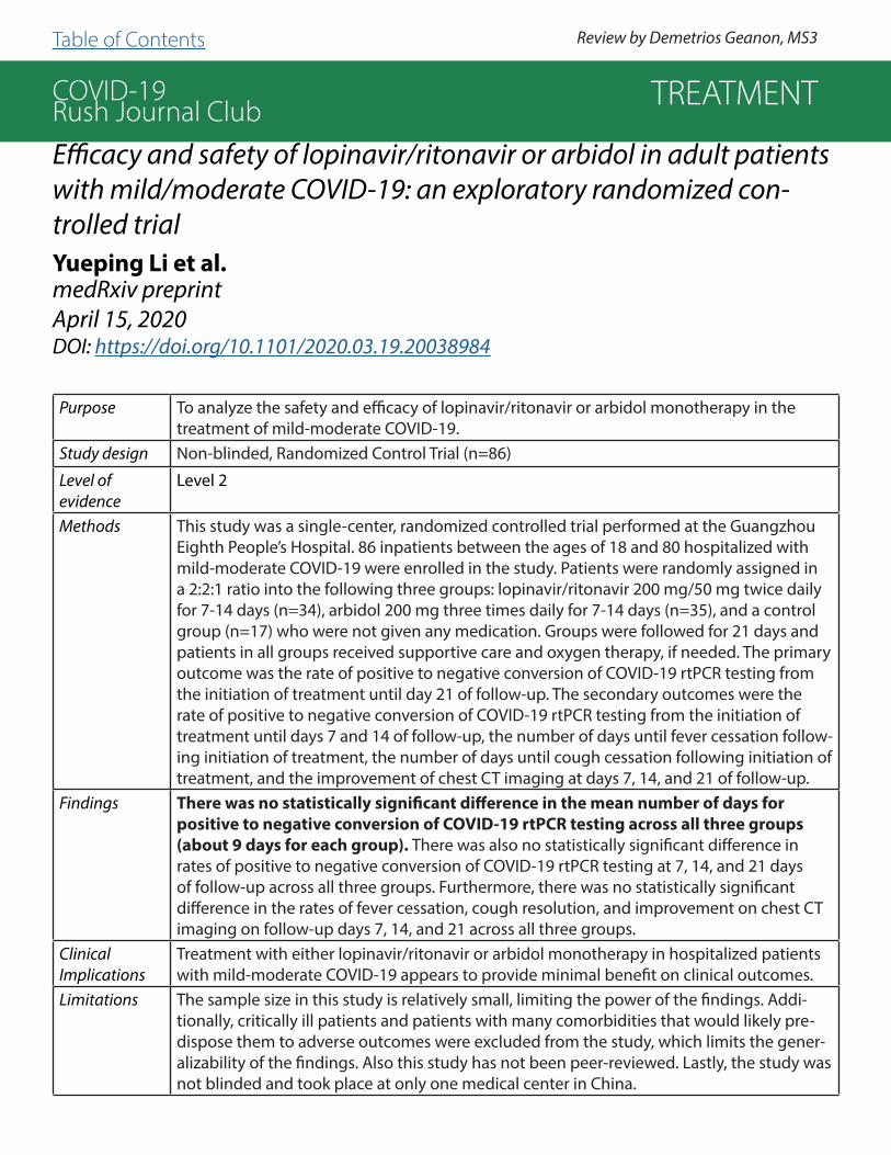
COVID-19Rush Journal ClubEfficacy and safety of lopinavir/ritonavir or arbidol in adult patients with mild/moderate COVID-19: an exploratory randomized con-trolled trialYueping Li et al.medRxiv preprintApril 15, 2020DOI: https://doi.org/10.1101/2020.03.19.20038984
Purpose To analyze the safety and efficacy of lopinavir/ritonavir or arbidol monotherapy in the treatment of mild-moderate COVID-19.
Study design Non-blinded, Randomized Control Trial (n=86)
Level of evidence
Level 2
Methods This study was a single-center, randomized controlled trial performed at the Guangzhou Eighth People’s Hospital. 86 inpatients between the ages of 18 and 80 hospitalized with mild-moderate COVID-19 were enrolled in the study. Patients were randomly assigned in a 2:2:1 ratio into the following three groups: lopinavir/ritonavir 200 mg/50 mg twice daily for 7-14 days (n=34), arbidol 200 mg three times daily for 7-14 days (n=35), and a control group (n=17) who were not given any medication. Groups were followed for 21 days and patients in all groups received supportive care and oxygen therapy, if needed. The primary outcome was the rate of positive to negative conversion of COVID-19 rtPCR testing from the initiation of treatment until day 21 of follow-up. The secondary outcomes were the rate of positive to negative conversion of COVID-19 rtPCR testing from the initiation of treatment until days 7 and 14 of follow-up, the number of days until fever cessation follow-ing initiation of treatment, the number of days until cough cessation following initiation of treatment, and the improvement of chest CT imaging at days 7, 14, and 21 of follow-up.
Findings There was no statistically significant difference in the mean number of days for positive to negative conversion of COVID-19 rtPCR testing across all three groups (about 9 days for each group). There was also no statistically significant difference in rates of positive to negative conversion of COVID-19 rtPCR testing at 7, 14, and 21 days of follow-up across all three groups. Furthermore, there was no statistically significant difference in the rates of fever cessation, cough resolution, and improvement on chest CT imaging on follow-up days 7, 14, and 21 across all three groups.
ClinicalImplications
Treatment with either lopinavir/ritonavir or arbidol monotherapy in hospitalized patients with mild-moderate COVID-19 appears to provide minimal benefit on clinical outcomes.
Limitations The sample size in this study is relatively small, limiting the power of the findings. Addi-tionally, critically ill patients and patients with many comorbidities that would likely pre-dispose them to adverse outcomes were excluded from the study, which limits the gener-alizability of the findings. Also this study has not been peer-reviewed. Lastly, the study was not blinded and took place at only one medical center in China.
TREATMENT
Review by Demetrios Geanon, MS3Table of Contents
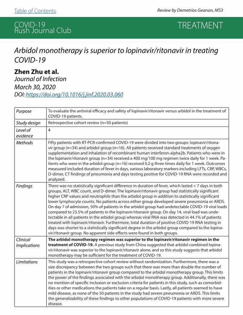
COVID-19Rush Journal Club
Arbidol monotherapy is superior to lopinavir/ritonavir in treating COVID-19Zhen Zhu et al.Journal of InfectionMarch 30, 2020DOI: https://doi.org/10.1016/j.jinf.2020.03.060
Purpose To evaluate the antiviral efficacy and safety of lopinavir/ritonavir versus arbidol in the treatment of COVID-19 patients.
Study design Retrospective cohort review (n=50 patients)
Level of evidence
4
Methods Fifty patients with RT-PCR-confirmed COVID-19 were divided into two groups: lopinavir/ritona-vir group (n=34) and arbidol group (n=16). All patients received standard treatments of oxygen supplementation and inhalation of recombinant human interferon-alpha2b. Patients who were in the lopinavir/ritonavir group (n=34) received a 400 mg/100 mg regimen twice daily for 1 week. Pa-tients who were in the arbidol group (n=16) received 0.2 g three times daily for 1 week. Outcomes measured included duration of fever in days, various laboratory markers including LFTs, CRP, WBCs, D-dimer, CT findings of pneumonia and days testing positive for COVID-19 RNA were recorded and analyzed.
Findings There was no statistically significant difference in duration of fever, which lasted < 7 days in both groups, ALT, WBC count, and D-dimer. The lopinavir/ritonavir group had statistically significant higher CRP values and neutrophils than the arbidol group in addition to statistically significant lower lymphocyte counts. No patients across either group developed severe pneumonia or ARDS. On day 7 of admission, 50% of patients in the arbidol group had undetectable COVID-19 viral loads compared to 23.5% of patients in the lopinavir/ritonavir group. On day 14, viral load was unde-tectable in all patients in the arbidol group whereas viral RNA was detected in 44.1% of patients treated with lopinavir/ritonavir. Furthermore, total duration of positive COVID-19 RNA testing in days was shorter to a statistically significant degree in the arbidol group compared to the lopina-vir/ritonavir group. No apparent side effects were found in both groups.
ClinicalImplications
The arbidol monotherapy regimen was superior to the lopinavir/ritonavir regimen in the treatment of COVID-19. A previous study from China suggested that arbidol combined lopina-vir/ritonavir was superior to the lopinavir/ritonavir alone, and so this study suggests that arbidol monotherapy may be sufficient for the treatment of COVID-19.
Limitations This study was a retrospective cohort review without randomization. Furthermore, there was a size discrepancy between the two groups such that there was more than double the number of patients in the lopinavir/ritonavir group compared to the arbidol monotherapy group. This limits the power of the findings associated with the arbidol monotherapy group. Additionally, there was no mention of specific inclusion or exclusion criteria for patients in this study, such as comorbid-ities or other medications the patients take on a regular basis. Lastly, all patients seemed to have mild disease, as none of the 50 patients in the study had severe pneumonia or ARDS. This limits the generalizability of these findings to other populations of COVID-19 patients with more severe disease.
TREATMENT
Review by Demetrios Geanon, MS3Table of Contents

COVID-19Rush Journal Club
A Trial of Lopinavir-Ritonavir in Adults Hospitalized with Severe Covid-19Bin Cao et al.New England Journal of MedicineMarch 18, 2020DOI: 10.1056/NEJMoa2001282
Purpose Determine the efficacy of Lopinavir-Ritonavir treatment in confirmed SARS-CoV2 infected patients in decreasing time to clinical improvement.
Study design Open label, randomized control trial. (N=199)Level of evidence
Level 2
Methods 189 patients were randomly assigned to receive either Lopinavir-Ritonavir (400mg and 100mg) orally or via a nasogastric tube plus standard care; or standard care alone for 14 days. The primary endpoint was time to clinical improvement.
Findings Patients assigned to receive Lopinavir-Ritonavir treatment did not have a time to clinical improvement different from that of standard-care group. The 28-day mortality was numerically lower in the Lopinavir-Ritonavir treatment group as compared to the standard-care group (19.2% vs 25% difference).The percentage of patients with clinical improvement on day 14 was higher in the Lopinavir-Ritonavir treatment group (45.5% vs 30%). Secondary findings included the percentage of patients with clinical improvement on day 14 was higher in the Lopinavir-Ritonavir treatment group (45.5% vs 30%) and detectable viral RNA at various time points was similar in both groups on subsequent sampling days.
ClinicalImplications
A 14-day Lopinavir-Ritonavir (400mg-100mg) therapy does not have a time to clinical improvement different from that of patients assigned to standard-of-care alone. Gastrointestinal adverse events (nausea, vomiting, diarrhea) were more common in the Lopinavir-Ritonavir treatment group. The side effect profile ob-served in this study increases concern that a lengthened course of treatment or a higher dose regimen to improve outcomes might not be feasible.
Limitations Based on the emergency nature of the trial, placebos were not prepared, thus limiting the ability to blind the participants and researchers. The characteristics of the patients were generally balanced but there was a slightly higher throat viral load in the lopinavir-ritonavir group, raising the possibility that this group has more viral replication, influencing clinical outcomes. Additionally, some patients received additional pharmacological interventions, such as glucocorticoid treat-ment, which might have acted as a cofounder.
TREATMENT
Review by Ashley Wehrheim, MS2 Table of Contents

COVID-19Rush Journal Club
Clinical Efficacy of Lopinavir/Ritonavir in the Treatment of Coronavi-rus Disease 2019Xiaoting Ye et al.European Review for Medical and Pharmacological SciencesMarch 2020DOI: 10.26355/eurrev_202003_20706
Purpose To investigate whether lopinavir/ritonavir (LPV/R) in combination with pneumonia-asso-ciated adjuvant drugs has therapeutic benefits when compared to pneumonia-associated adjuvant treatment alone in the context of COVID-19.
Study design Retrospective cohort study (n=47)
Level of evidence
Level 3
Methods 47 COVID-19 positive patients (via quantitative PCR) admitted to the same hospital in Rui’an, China were divided into a test group (n=42) or a control group (n=5). The test group consisted of patients treated with LPV/R in addition to pneumonia-associated ad-juvant drugs (therapeutic scheme including other antivirals and anti-inflammatory med-ications) during hospitalization. The control group included patients treated with pneu-monia-associated adjuvant drugs alone. Body temperature and laboratory values were measured three times over a ten-day period. The primary endpoint was improvement of clinical symptoms, as measured by fever. Secondary endpoints included improvement in laboratory findings, as measured by blood routine indexes, and the hepatic safety of LPV/R.
Findings Patients in the test group returned to normal body temperature in a shorter time than the control group (P=0.036). Patients in the test group were also found to have generally lower levels of abnormal proportions of WBC, lymphocytes, CRP, and PLT after three treatments than that in the control group. The number of patients with abnormal AST/ALT measure-ments in the test group was not significantly increased as compared to the control group, suggesting that LPV/R does not cause significant hepatotoxicity.
ClinicalImplications
These findings suggest that LPV/R is safe for clinical use and may demonstrate efficacy in treating COVID-19 when used with adjuvant drugs.
Limitations Large age range of patients from 5-68 years of age. The control group was small (n=5) and mostly female (n=4). Patients in both the test group and the control group received vari-ous pneumonia-associated adjuvant drugs. Therefore, results of this study demonstrating the efficacy of LPV/R may be dependent on simultaneously receiving the same therapeu-tic regimen used in these patients. Additionally, as this was a retrospective cohort study, further randomized double-blinded clinical trials are needed.
TREATMENT
Review by Manvita Tatavarthy, MS2 Table of Contents

COVID-19Rush Journal Club
Treatment of 5 Critically Ill Patients With COVID-19 With Convales-cent PlasmaChenguang Shen et al. JAMAMarch 27, 2020DOI: 10.1001/jama.2020.4783
Purpose Investigate the potential benefit of administration of convalescent plasma trans-fusion for treatment of critically ill patients with SARS-CoV-2 infection.
Study design Preliminary Uncontrolled Case Series (n= 5)
Level of evidence
Level 4
Methods Five laboratory-confirmed COVID-19 patients with acute respiratory distress syndrome (ARDS) who also met the following criteria of severe pneumonia with rapid progression and continuously high viral load despite antiviral treatment; PAO2/FIO2 <300; and mechanical ventilation were given a transfusion with con-valescent plasma with a SARS-CoV-2–specific antibody 10-22 days after admis-sion.
Findings Following plasma transfusion, body temperature normalized within 3 days in 4 of 5 patients, the Sequential Organ Failure Assessment (SOFA) score de-creased, and Pao2/Fio2 increased within 12 days. Viral loads decreased and became negative within 12 days after the transfusion, and SARS-CoV-2–specific ELISA and neutralizing antibody titers increased. ARDS resolved in 4 patients at 12 days after transfusion, and 3 patients were weaned from mechan-ical ventilation within 2 weeks of treatment. At the end of the study, 3 patients were discharged from the hospital and 2 were in stable condition.
ClinicalImplications
Administration of convalescent plasma with neutralizing antibody lead to im-provement in the patients’ clinical status. The limited sample size and study design do not allow for a conclusive statement about the potential effectiveness of plasma transfusion therapy.
Limitations The study was limited by its small sample size and study design that included no controls. It is unclear if patients would have had the same outcome without transfusion of plasma especially since they previously were treated with other agents. The transfusions were also given 10-22 days after admission, so the tim-ing and association of outcomes is unknown.
TREATMENT
Review by Amanda Narkis, MS2Table of Contents

COVID-19Rush Journal Club
The feasibility of convalescent plasma therapy in severe COVID- 19 patients: a pilot studyKai Duan et al.medRxivMarch 23, 2020DOI: https://doi.org/10.1101/2020.03.16.20036145
Purpose To examine the feasibility of providing convalescent plasma (CP) as a poten-tial therapy for patients who have tested positive for COVID-19.
Study design Safety trial (n=10)Level of evidence
Level 4
Methods Ten patients in three Chinese hospitals who had been diagnosed with COVID-19 (by rRT-PCT) and found to be in respiratory distress were identified. Convalescent plasma was donated by patients at the same three hospitals who met criteria for recovery from COVID-19. 200mL convalescent plasma was prepared and transfused according to WHO transfusion protocol. The first study endpoint was evaluating the safety of convalescent plasma trans-fusion. The second endpoint was improvement of clinical symptoms and laboratory parameters within three days of transfusion.
Findings There were no serious adverse events in all ten patients. All ten patients in the study experienced an improvement in clinical symptoms within one to three days of transfusion. Furthermore, all ten patients also demonstrated negative RT-PCR testing, increased oxygen saturation, and improved lympho-cyte counts, CRP and liver function. Neutralizing antibody levels, compared to those prior to transfusion, increased in five patients, remained the same in four patients and were not measured in one. All ten patients showed varia-tion in reduction of lesions on chest CT after transfusion.
ClinicalImplications
These findings suggest that CP appears safe in a small number of patients. Additionally, CP may improve clinical outcomes.
Limitations Prior to transfusion of CP, patients received varying therapeutic regimens. Some received antivirals, whereas others only supportive care. This lack of standardization of treatment protocol confounds the assessment of the po-tential benefits of CP alone. Furthermore, the small sample size and lack of a control group, limit the power and subsequent observations of this study.
TREATMENT
Review by Manvita Tatavarthy, M2 Table of Contents

COVID-19Rush Journal Club
High-Dose Intravenous Immunoglobulin as a Therapeutic Option for Deteriorating Paients With Coronavirus Disease 2019Wei Cao et al.Open Forum Infectious DiseaseMarch 21, 2020DOI: https://doi.org/10.1093/ofid/ofaa102
Purpose Evaluate the value of administration of high-dose IVIG for rapidly deteriorating patients with COVID-19 pneumonia.
Study design Case Observational Study (n= 3)Level of evidence
Level 4
Methods Three patients were diagnosed with COVID-19, severe type and administered high-dose IVIg (at 0.3-0.5 g/kg) for 5 days. Computed tomography (CT) scan was documented before and after treatment. All patients were treated at the early stage of clinical deterioration. Testing for COVID-19 was performed through PCR or oropharyngeal swab.
Findings Of the 3 patients, all demonstrated clinical improvement shortly after administration of high dose IVIg. Variables measured include stabilizing tem-perature within 1-2 days and breathing difficulty alleviated in 3-5 days. CT scans compared before and after treatment demonstrated partial to complete resolu-tion of lesions. Between 5-6 days after the first dose of treatment, all 3 pa-tients tested negative for COVID-19. No adverse outcomes were reported in any of the 3 patients.
ClinicalImplications
IVIg used early in a patients course after diagnosis with the severe type of COVID-19 could provide clinical use in shortening the duration of symptoms of COVID-19 pneumonia. However, the clinical results require confirmation from a randomized controlled trial (RCT). The study describes 3 clinically rele-vant phases of COVID-19 including an initial phase, an accelerating phase with potential for an overall inflammatory storm (lab values indicating progressive lymphocytopenia and inflammatory markers) and a recovery phase.
Limitations The study was limited by its small sample size (n=3) and lack of control cases. Additionally, confounding factors between patients include the use of lopinavir/ritonavir in one patient and moxifloxacin in a one patient and a short course of steroids in the third patient.
TREATMENT
Review by Karina Oelerich, MS2 Table of Contents

COVID-19Rush Journal Club
Use of convalescent plasma therapy in two COVID-19 patients with acute respiratory distress syndrome in KoreaJin Young Ahn et al.J Korean Med SciApril 13, 2020DOI: 10.3346/jkms.2020.35.e149
Purpose To describe outcomes of convalescent plasma therapy in acute respiratory distress (ARDS) in two patients.
Study design Uncontrolled Case Series (n=2)
Level of evidence
Level 4
Methods Both patients received hydroxychloroquine and lopinavir/ritonavir after initial diagnosis of COVID-19, both progressed to ARDS, and required mechanical ventilation. The patients then received convalescent plasma from donors in their 20s, positive for ELISA IgG test for SARS-CoV-2, with previous presentation of bilateral pneumonia. The donors were determined to have completely recovered. Plasma was divided into two doses and administered at 12-hour intervals with each dose given over course of 1 hour. Both cases involved simultaneous infu-sion of corticosteroids. Corticosteroids were not given initially (due to the lack of evidence of its clinical efficacy on mortality reduction) but applied when the patients’ condition dete-riorated to ARDS (methylprednisolone was administered one day and two days before the plasma infusion to patient 1 and 2, respectively).
Findings Patient 1: Previously healthy 71-year-old male-Day 9 of hospitalization (Day 22 of symptom onset), received convalescent plasma.-Day 11, patient’s condition improved with fever subsiding, decreased oxygen demand and CRP. Radiograph showed resolution of lung infiltrate. Patient was weaned from ventilator.Patient 2: 67-year-old woman with history of hypertension-Day 4, patient was intubated due to increased oxygen demand with intravenous methyl-prednisolone added and was put into prone position to improve oxygen demand.-Day 6, convalescent plasma was provided.-Day 9, density of bilateral infiltration improved along with decreased CRP and IL-6 levels.-Day 20, negative rRT-PCR for SARS-CoV-2. -Day 24, patient was extubated and discharged.
ClinicalImplications
Transfusion of convalescent plasma shows promise for the treatment of severe COVID-19 patients. Following the infusion of convalescent plasma, viral loads rapidly decrease, inflam-matory makers decrease, and oxygenation improves.
Limitations This describes only two patients with similar clinical presentations of ARDS and similar treat-ment regimens, preventing generalizability to a diverse patient population. Additionally, the timing of administration of plasma varied greatly, and the antibodies provided in the plasma could not be determined if they had high neutralizing titers.
TREATMENT
Review by Christina Brown, MS2Table of Contents

COVID-19Rush Journal Club
Effectiveness of convalescent plasma therapy in severe COVID-19 patients
Kai Duan et al.Proc Natl Acad SciMarch 18, 2020DOI: https://doi.org/10.1073/pnas.2004168117
Purpose To determine the safety of convalescent plasma (CP) transfusion in patients with COVID-19 and to observe the improvement of clinical symptoms, radiologic findings and laboratory parameters within 3 days after CP transfusion.
Study design Prospective Cohort Study (n=10)Level of evidence
Level 3
Methods Ten COVID-19 patients (confirmed by real-time viral RNA test) in severe condition re-ceived one dose of 200 mL of CP derived from recently recovered donors (titers>1:640) transfused ~16.5 days after development of symptoms. CP was given in addition to maximal supportive care and antiviral treatments. Severe classification was defined as patients presenting with severe dyspnea, respiratory distress (tachypnea>30 breaths/min), or hypoxia (SpO2<90%). A historic control group was formed by random se-lection of 10 patients treated in the same hospital and matched by age, gender, and severity of the diseases.
Findings Chest CTs showed improvement in different degrees of absorption of pulmonary le-sions after CP transfusion in all patients. Lymphocytopenia and SaO2 increased where-as C-reactive protein (CRP), aspartate aminotransferase (AST), and alanine aminotrans-ferase (ALT) decreased. An increase in neutralizing antibody titers was found in 5/10 patients, viral load was decreased to an undetectable level in 3/10 patients. Clinical outcome in the CP group improved as compared to the control group. In the CP group, three cases discharged, while the remaining seven cases were given a “much improved status” and were ready for discharge. In the control group, there were three deaths, six cases were classified as “stabilized”, and one case classified as “much improved status”. No serious adverse reactions or safety events were recorded after CP transfusion.
ClinicalImplications
One dose of CP with high concentration of neutralizing antibodies can rapidly reduce the viral load and tends to improve clinical outcomes with minimal adverse reactions.
Limitations First, patients received additional treatment beyond CP, which may have contributed to clinical outcomes. Additionally, this study had a small sample size and was unblind-ed, increasing risk of bias. Finally, some patients received glucocorticoid therapy, which could interfere with the immune response and delay virus clearance, altering study findings.
TREATMENT
Review by Ashley Wehrheim, MS2Table of Contents
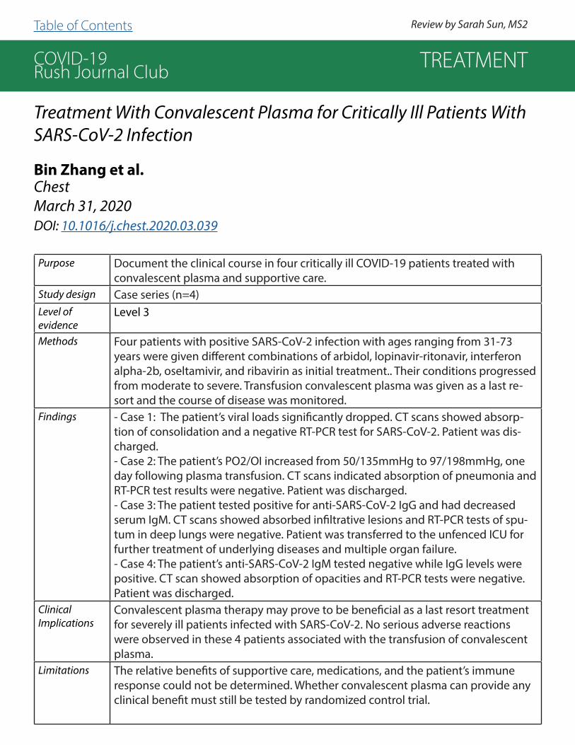
COVID-19Rush Journal Club
Treatment With Convalescent Plasma for Critically Ill Patients With SARS-CoV-2 Infection
Bin Zhang et al.ChestMarch 31, 2020DOI: 10.1016/j.chest.2020.03.039
Purpose Document the clinical course in four critically ill COVID-19 patients treated with convalescent plasma and supportive care.
Study design Case series (n=4)Level of evidence
Level 3
Methods Four patients with positive SARS-CoV-2 infection with ages ranging from 31-73 years were given different combinations of arbidol, lopinavir-ritonavir, interferon alpha-2b, oseltamivir, and ribavirin as initial treatment.. Their conditions progressed from moderate to severe. Transfusion convalescent plasma was given as a last re-sort and the course of disease was monitored.
Findings - Case 1: The patient’s viral loads significantly dropped. CT scans showed absorp-tion of consolidation and a negative RT-PCR test for SARS-CoV-2. Patient was dis-charged.- Case 2: The patient’s PO2/OI increased from 50/135mmHg to 97/198mmHg, one day following plasma transfusion. CT scans indicated absorption of pneumonia and RT-PCR test results were negative. Patient was discharged.- Case 3: The patient tested positive for anti-SARS-CoV-2 IgG and had decreased serum IgM. CT scans showed absorbed infiltrative lesions and RT-PCR tests of spu-tum in deep lungs were negative. Patient was transferred to the unfenced ICU for further treatment of underlying diseases and multiple organ failure.- Case 4: The patient’s anti-SARS-CoV-2 IgM tested negative while IgG levels were positive. CT scan showed absorption of opacities and RT-PCR tests were negative. Patient was discharged.
ClinicalImplications
Convalescent plasma therapy may prove to be beneficial as a last resort treatment for severely ill patients infected with SARS-CoV-2. No serious adverse reactions were observed in these 4 patients associated with the transfusion of convalescent plasma.
Limitations The relative benefits of supportive care, medications, and the patient’s immune response could not be determined. Whether convalescent plasma can provide any clinical benefit must still be tested by randomized control trial.
TREATMENT
Review by Sarah Sun, MS2Table of Contents

COVID-19Rush Journal Club
Convalescent plasma transfusion for the treatment of COVID-19: Systematic reviewKarthick Rajendran et al.Journal of Medical VirologyMay 1, 2020DOI: https://doi.org/10.1002/jmv.25961
Purpose To determine the efficacy of convalescent plasma transfusion (CPT) therapy on COVID-19 patients.
Study design Systematic Review
Level of evidence
Level 3
Methods A systematic search was conducted using electronic databases such as PubMed, EMBASE and Medline to identify case studies and clincial trials regarding treatment of COVID-19 patients using CPT therapy. Studies were published literature between December 1, 2019 to April 19, 2020. “Convalescent plasma AND COVID-19” was used as the search term and there were no restrictions placed on the comparator in each study. The primary endpoints were clinical effects, survival benefits, viral load & antibody titer status and adverse events. Two authors independently assessed the studies to reduce the risk of bias.
Findings Amongst all 8 of the studies reviewed, 5 were selected for further evaluation. These 5 stud-ies had a total of 27 patients enrolled that received the CPT therapy. All the patients re-ceived CPT between Day 6 and Day 50, however, it varied between the studies the dosage administered and the length of treatment. All 5 studies found zero mortality rate for patients that had CPT administrated, albeit it being in varying doses. Every study also found that CPT, in conjunction with other antiviral treatments, reduced the viral load and increased the level of neutralizing antibody titer. Every single patient had also received more than one antiviral drug in addition to the CPT. Almost all patients also showed im-provement of their symptoms such as their body temperature. Furthermore, CPT treat-ment was well tolerated by all the patients and no adverse reactions were noted.
ClinicalImplications
Although there is limited scientific data, it appears that COVID-19 patients treated with CPT treatment have positive outcomes. Further randomized controlled trials are urgently needed to determine the ideal dosage and treatment time for CPT therapy for optimal outcomes.
Limitations The included studies were predominantly case reports or case series and lacked proper control groups. It is also difficult to determine that CPT alone reduced the viral load and increased antibody titers because all patients were given multiple therapies. Additionally, the authors did not compare different doses of CPT with outcomes, making it difficult to understand what dose might be most effective.
TREATMENT
Review by Maria Amir, MS2 Table of Contents

COVID-19Rush Journal Club
Remdesivir in adults with severe COVID-19: a randomised, dou-ble-blind, placebo-controlled, Multicenter trial
Yeming Wang, et al.The LancetApril 29, 2020DOI: https://doi.org/10.1016/S0140-6736(20)31022-9
Purpose To determine the effect of Remdesivir on time to clinical improvement in patients with severe coronavirus disease 2019 (COVID-19).
Study design Randomized, double-blind, placebo-controlled, multicenter trial at ten hospitals in Hubei, China
Level of evidence
2
Methods Patients admitted with severe cases of confirmed COVID-10 were randomly assigned in a 2:1 ratio of IV Remdesivir (200mg on day 1 followed by 100mg on days 2-10 with a single daily infection) or the same volume of placebo infusions for 10 days. Patients received concomitant use of lopinavir-ritonavir, interferons and corticosteroids. The primary end-point of this study was time to clinical improvement within 28 days after randomization. This was defined as the time in days from randomization to a decline of two levels on a six-point ordinal scale (1= discharged, 6=death), or discharge alive from the hospital, which-ever came first.
Findings - Remdesivir use was not associated with a statistically significant difference in time to clinical improvement compared to the control population.- Although not statistically significant, patients receiving Remdesivir had a numerical-ly faster time to clinical improvement compared to those receiving placebo among patients with symptom duration of <10 days.- Adverse events were reported in (66%) Remdesivir patients and (64%) of patients who received the placebo. More patients in the group discontinued the study due to severe adverse events (12% in Remdesivir group vs 5% in placebo group)
ClinicalImplications
- In Adult patients hospitalized for severe COVID-19, Remdesivir was not associated with statically significant clinical benefits beyond those receiving the standard of care.- The higher rate of adverse events observed in the Remdesivir group increases concern that a lengthened course of treatment or a higher dose regimen to improve outcomes might not be feasible
Limitations The study did not reach its target enrollment, leading to insufficient power to detect dif-ference in clinical outcomes. Additionally, restrictions on hospital bed availability result-ed in most patients being enrolled later in the course of disease, so research could not adequately assess whether earlier Remdesivir treatment might have provided additional clinical benefit. Finally, concurrent treatment with lopinavir-ritonavir, interferons and corti-costeroids may have influenced clinical outcomes.
TREATMENT
Review by Ashley Wehrheim, MS2Table of Contents

COVID-19Rush Journal Club
Compassionate Use of Remdesivir for Patients with Severe COVID-19
Jonathan Grein et al. New England Journal of MedicineApril 10, 2020DOI: 10.1056/NEJMoa2007016
Purpose To determine the clinical outcome of compassionate use Remdesivir (inhibits viral RNA polymerases) in the treatment of Covid-19 infection, caused by the SARS-CoV-2.
Study design Open-label clinical trial (n = 53)Level of evidence
3
Methods It is a multicenter study conducted at sites in United States, Japan, Italy, Austria, France, Germany, Netherlands, Spain, and Canada. PCR-confirmed SARS-CoV-2 infection defined as oxygen saturation of 94% or less while on ambient air or ox-ygen support. Participants needed a creatinine clearance greater than 30 ml/min and liver enzymes (alanine transaminase, ALT and aspartate transaminase, AST) 5 times below the upper limit of normal. The duration of Remdesivir therapy is 10 days, consisting of 200 mg administered intravenously on day 1, followed by 100 mg daily for the remaining 9 days of treatment. The main outcomes were discharge or death.
Findings During a median follow-up of 18 days, 36 (68%) of the patients improved clinically. Seven (13%) died after at the end of Remdesivir treatment. A total of 32 patients reported adverse events mostly elevated liver function tests (LFTs), rash, diarrhea, hypotension, and renal impairment, worse with those on invasive ventila-tion.
ClinicalImplications
68% of patients diagnosed with severe Covid-19 improved with the use of Rem-desivir. Therefore, compassionate use of the medication is clinically valuable in the management of severe Covid-19 infection.
Limitations Most of the study participants (75%) were men ranging between 23 to 82 years, which may introduce gender bias. The small sample size and non-randomization of participants, missing data were limitations in the data interpretation and results. There were no clearly stated inclusion and exclusion criteria. Even though compas-sionate use of the medication should be investigated for efficacy, it is difficult to draw useful conclusions from uncontrolled studies like this.
TREATMENT
Review by Ayesan Rewane, MSCR Table of Contents
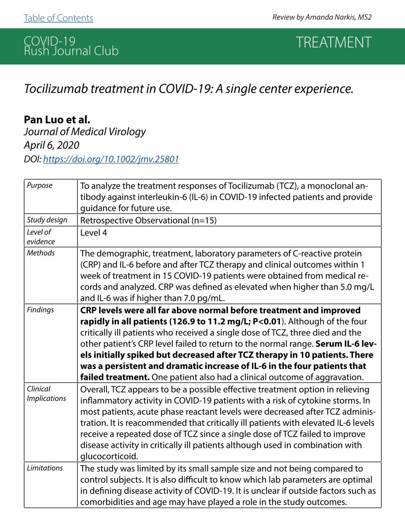
COVID-19Rush Journal Club
Tocilizumab treatment in COVID‐19: A single center experience.
Pan Luo et al.Journal of Medical VirologyApril 6, 2020DOI: https://doi.org/10.1002/jmv.25801
Purpose To analyze the treatment responses of Tocilizumab (TCZ), a monoclonal an-tibody against interleukin-6 (IL-6) in COVID-19 infected patients and provide guidance for future use.
Study design Retrospective Observational (n=15)Level of evidence
Level 4
Methods The demographic, treatment, laboratory parameters of C‐reactive protein (CRP) and IL‐6 before and after TCZ therapy and clinical outcomes within 1 week of treatment in 15 COVID‐19 patients were obtained from medical re-cords and analyzed. CRP was defined as elevated when higher than 5.0 mg/L and IL-6 was if higher than 7.0 pg/mL.
Findings CRP levels were all far above normal before treatment and improved rapidly in all patients (126.9 to 11.2 mg/L; P<0.01). Although of the four critically ill patients who received a single dose of TCZ, three died and the other patient’s CRP level failed to return to the normal range. Serum IL-6 lev-els initially spiked but decreased after TCZ therapy in 10 patients. There was a persistent and dramatic increase of IL-6 in the four patients that failed treatment. One patient also had a clinical outcome of aggravation.
ClinicalImplications
Overall, TCZ appears to be a possible effective treatment option in relieving inflammatory activity in COVID-19 patients with a risk of cytokine storms. In most patients, acute phase reactant levels were decreased after TCZ adminis-tration. It is reacommended that critically ill patients with elevated IL-6 levels receive a repeated dose of TCZ since a single dose of TCZ failed to improve disease activity in critically ill patients although used in combination with glucocorticoid.
Limitations The study was limited by its small sample size and not being compared to control subjects. It is also difficult to know which lab parameters are optimal in defining disease activity of COVID-19. It is unclear if outside factors such as comorbidities and age may have played a role in the study outcomes.
TREATMENT
Review by Amanda Narkis, MS2 Table of Contents

COVID-19Rush Journal Club
Effective Treatment of Severe COVID-19 Patients with Tocilizumab
Xiaoling Xu et al.ChinaXivApril 14, 2020DOI: 10.12074/202003.00026
Purpose To assess the efficacy of Tocilizumab in severe patients with COVID-19.
Study design Case Series (n=21)Level of evidence
Level 4
Methods Patients diagnosed as severe or critical COVID-19 were given tocilizumab (400 mg once through IV Drip) in addition to routine therapy between February 5, 2020 -February 14, 2020. Changes in clinical manifestations (Body temp., Oxygen saturations etc), CT scan images (performed on admission and 1 week after receiving therapy), and laboratory examinations were retrospectively analyzed.
Findings Within a few days, the fever returned to normal and all other symptoms im-proved remarkably. 75% of patients had lowered their oxygen intake and one patient no longer needed Oxygen therapy. CT scan manifested that the lung lesion opacity absorbed in 90.5% of patients. The percentage of lymphocytes in peripheral blood decreased in 85% of patients before treatment, returned to normal in 52.5% of pa-tients on Day 5 of treatment. Abnormally elevated C-reactive protein decreased sig-nificantly in 84.2% of patients. No obvious adverse reactions were observed during the duration of treatment. 90.5% of patients were discharged an average of 13.5 days after treatment with tocilizumab and the rest were recovering well at the end of the trial period.
ClinicalImplications
Clinical data showed that symptoms, hypoxemia and CT opacity changes were im-proved immediately after treatment with tocilizumab in most patients.- Tocilizumab is hypothesized to be an effective treatment in severe patients of COVID-19, which may provide a new therapeutic strategy for severe patients, but larg-er randomized trials must be performed.
Limitations The number of patients were rather limited and no control group was included. This was a single observation study and significant bias could possibly exist. The patients received standard of care treatment (lopinavir, methylprednisolone, other symptom relivers and oxygen therapy) in addition to Tocilizumab. As a result, the possibility that these agents could have contributed to positive clinical outcomes cannot be ruled out.
TREATMENT
Review by Ashley Wehrheim, MS2Table of Contents
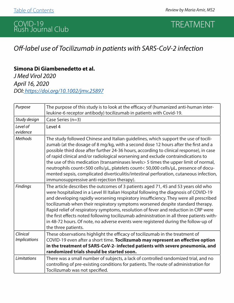
COVID-19Rush Journal Club
Off-label use of Tocilizumab in patients with SARS-CoV-2 infection
Simona Di Giambenedetto et al.J Med Virol 2020April 16, 2020DOI: https://doi.org/10.1002/jmv.25897
Purpose The purpose of this study is to look at the efficacy of (humanized anti-human inter-leukine-6 receptor antibody) tocilizumab in patients with Covid-19.
Study design Case Series (n=3)Level of evidence
Level 4
Methods The study followed Chinese and Italian guidelines, which support the use of tocili-zumab (at the dosage of 8 mg/kg, with a second dose 12 hours after the first and a possible third dose after further 24-36 hours, according to clinical response), in case of rapid clinical and/or radiological worsening and exclude contraindications to the use of this medication (transaminases levels> 5 times the upper limit of normal, neutrophils count<500 cells/µL, platelets count< 50,000 cells/µL, presence of docu-mented sepsis, complicated diverticulitis/intestinal perforation, cutaneous infection, immunosuppressive anti-rejection therapy).
Findings The article describes the outcomes of 3 patients aged 71, 45 and 53 years old who were hospitalized in a Level III Italian Hospital following the diagnosis of COVID-19 and developing rapidly worsening respiratory insufficiency. They were all prescribed tocilizumab when their respiratory symptoms worsened despite standard therapy. Rapid relief of respiratory symptoms, resolution of fever and reduction in CRP were the first effects noted following tocilizumab administration in all three patients with-in 48-72 hours. Of note, no adverse events were registered during the follow-up of the three patients.
ClinicalImplications
These observations highlight the efficacy of tocilizumab in the treatment of COVID-19 even after a short time. Tocilizumab may represent an effective option in the treatment of SARS-CoV-2- infected patients with severe pneumonia, and randomized trials should be started soon.
Limitations There was a small number of subjects, a lack of controlled randomized trial, and no controlling of pre-existing conditions for patients. The route of administration for Tocilizumab was not specified.
TREATMENT
Review by Maria Amir, MS2Table of Contents

COVID-19Rush Journal Club
Acute hypertriglyceridemia in patients with COVID‐19 receiving tocili-zumab
Austin R. Morrison et al.Journal of Medical VirologyApril 21, 2020DOI: https://doi.org/10.1002/jmv.25907
Purpose To report on two cases of acute hypertriglyceridemia in patients with COVID-19 treat-ed with tocilizumab leading to the recommendation of future monitoring.
Study design Letter to EditorLevel of evidence
N/A
Methods This article was written in response to the Luo et al. study of Tocilizumab (TCZ) for COVID-19 in a single center in China and due to no reports of acute adverse events with TCZ treatment in COVID-19 thus far. The authors discuss two cases of acute hy-pertriglyceridemia in patients with COVID-19 treated with TCZ with one developing acute pancreatitis.
Findings - Cases 1 and 2, a 65-year old-male and 43-year-old male respectively, were both in the ICU with respiratory failure and ARDS who received lopinavir/ritonavir, ribavirin, and hydroxychloroquine for COVID-19 treatment. Both patients received TCZ due to persistent fevers, severe ARDS, and elevated inflammatory markers. Following TCZ, both cases had a significant increase in TG levels, with case 1 developing acute pan-creatitis as defined by elevated amylase and lipase levels.- Both patients received propofol prior to treatment, but the effect of propofol on increased TGs is typically seen 2.25-7 days after starting therapy, with normalization occurring within 72 hours. Therefore, patients receiving TCZ and propofol may re-quire more frequent monitoring.- Membrane-bound soluble IL-6 receptor inhibition with TCZ administration may result in increased triglyceride levels by interfering with the metabolic pathways of IL-6, but exact mechanisms are unknown
ClinicalImplications
Clinicians should consider monitoring for hypertriglyceridemia and acute pancreatitis for those receiving TCZ treatment for severe COVID-19 as well as to remain vigilant for other acute adverse effects that are difficult to detect in small sample clinical trials.
Limitations The article only discusses two cases of hypertriglyceridemia, and both patients re-ceived other treatments for COVID-19 prior to TCZ. Further data collection is needed to determine the true relationship between increased TG levels and TCZ treatment in COVID-19. As a letter to the editor, it is unclear if this article has been peer-reviewed.
TREATMENT
Review by Amanda Narkis, MS2Table of Contents

COVID-19Rush Journal Club
Tocilizumab for the Treatment of Severe COVID‐19
Rand Alattar et al.Journal of Medical VirologyMay 5 2020DOI: https://doi.org/10.1002/jmv.25964
Purpose To report on the clinical outcomes and laboratory findings of patients with severe COVID-19 that were treated with Tocilizumab (TCZ), an interleukin 6-inhibitor.
Study design Retrospective Review (n=25)
Level of evidence
Level 4
Methods The study completed a retrospective chart review of patients in Qatar with laboratory con-firmed severe COVID-19 that received TCZ for 14 days and followed from day 1 through 14. The primary outcome was discharge from the ICU by day 14. Twenty-five patients were eligible; these patients had a median age of 58 years old, median BMI of 29 kg/m2, and the majority were male (92%).
Findings - The decline in temperature and serum CRP levels are likely a reflection of TCZ’s immune modulating effect. Median oral temperatures on day 1, day 3 and day 7 were 38.0°C, 37.3°C (P 0.043) and 37.0°C (P 0.064), respectively, while corresponding median CRP was 193 mg/L, 7.9 mg/L (P <0.0001) and <6 mg/L (P 0.0001).- At the time of TCZ administration, 84% of patients were on invasive ventilation, which declined to 60% on day 7 (P 0.031) and 28% on day 14 (p 0.001). There was radiological improvement on patient’s chest x-rays for 44% of patients by day 7 and 68% by day 14.- Nine patients (36%) were discharged alive from the ICU and three (12%) died. Since the median age of the patients was 58, it is possible this played a role in the low mortality, since older age has been found to be associated with poorer COVID-19 outcomes.- The majority (92%) of patients experienced at least one adverse event. However, patients were critically ill and received other investigational antiviral therapies, so it is difficult to conclude if any were specifically due to TCZ.
ClinicalImplications
Patients with severe COVID-19 that were treated with TCZ had a dramatic decline in inflammatory markers, radiological improvement, and reduced ventilatory support requirements.
Limitations Limitations in this study include it being retrospective, lack of a control comparison, and potential confounding effects from concomitant investigational antivirals. Further ran-domized controlled trials are necessary to conclude effectiveness of TCZ treatment.
TREATMENT
Review by Amanda Narkis, MS2Table of Contents
COVID-19Rush Journal Club
Tocilizumab for Treatment of Severe COVID-19 Patients: Preliminary Results from SMAtteo COvid19 REgistry (SMACORE)Marta Colaneri et al.MicroorganismsMay 9, 2020DOI: https://doi.org/10.3390/microorganisms8050695
Purpose To assess the effect of tocilizumab (TCZ) on ICU admission and mortality rates in COVID-19 infected patients.
Study design Retrospective analysis of matched cases (n=21 matched pairs)
Level of evidence
Level 4
Methods Patient data from the SMAtteo COvid19 REgistry (SMACORE) database was collected on 112 patients hospitalized between March 14-27, 2020 with confirmed SARS-CoV-2 pneumonia. Retrospective analysis via linear regression and generalized linear mixed models were per-formed on 21 matched pairs of patients receiving either standard of care (SOC) (hydroxychlo-roquine (HCQ): 200 mg BID, azithromycin: 500 mg once, low weight heparin, methylprednis-olone: tapered dose of 1 mg/kg up to a maximum of 80 mg for 10 days) or SOC + TCZ (8 mg/kg IV, capped at 800, repeated 12 hours later). Patients in the SOC + TCZ treatment group were required to meet certain laboratory criteria (C-reactive protein [CRP] >5 mg/dL, procalcitonin <0.5 ng/mL, arterial partial pressure of oxygen/fractional inspired oxygen [PF ratio] <300, and alanine aminotransferase [ALT] <500 U/L). Pairs were matched based on propensity scores calculated from demographic and clinical variables. Primary outcomes included 7 day mortali-ty and ICU admission. Secondary outcomes included CRP, international normalized ratio (INR), lymphocyte and neutrophil counts, platelets, lactate dehydrogenase (LDH), and ALT.
Findings Treatment with TCZ did not significantly affect likelihood of ICU admission (odds ratio, OR: 0.11) or 7 day mortality (OR: 0.78) when compared to patients receiving SOC. TCZ treatment was correlated with a significant down trend in CRP and INR, an uptrend in ALT, and stable platelet counts. TCZ was not associated with significant hepatotoxicity or secondary infection.
ClinicalImplications
Preliminary data suggest that TCZ does not have a profound impact on COVID-19 patient mor-tality or ICU admission when compared to SOC (combination of HCQ, heparin, azithromycin, and methylprednisolone). TCZ is associated with up trending ALT levels and should be used with caution in those with increased potential for severe liver injury. Further data is required to determine utility of TCZ in this clinical context.
Limitations This study was observational, involved a short follow up period, and included a relatively small sample size. As an observational study, analysis is subject to bias, although attempts were made to limit this through propensity score matching. In addition, both treatment groups received methylprednisolone, which could potentially confound anti-inflammatory effects of TCZ.
TREATMENT
Review by Kavya Timmireddy, MS3 Table of Contents

COVID-19Rush Journal Club
Corticosteroid treatment of patients with coronavirus disease 2019 (COVID‐19).Lei Zha et al.Medical Journal of AustraliaApril 8, 2020DOI: https://doi.org/10.5694/mja2.50577
Purpose To determine the efficacy of early corticosteroid treatment in patients with COVID-19 not presenting with acute respiratory distress.
Study design Observational comparison study
Level of evidence
Level 3
Methods 31 patients with COVID-19 were drawn from two designated hospitals in Wuhu, China. Re-cords were reviewed of patients admitted between January 24th and February 24th, 2020 with confirmed SARS-CoV-2 infection by local health agencies using RT-PCR. Patients who received at least one dose of corticosteroids within 24 hours of admittance were com-pared against those who did not receive any. The primary outcome was time to clearance of the virus, whereas secondary outcomes were duration of clinical recovery, and length of hospital stay. Patients were followed until February 29th, 2020. Those patients who received corticosteroids within 24 hours of arrival were compared against patients not-re-ceiving corticosteroids. All patients received standard therapies.
Findings Eleven patients received corticosteroids (40 mg methylprednisolone) once or twice per day for a median of 5 days. The patients receiving steroids had a higher maximum tem-perature on admission (38.8°C vs 37.8°C, P=0.002), symptoms including myalgia or fa-tigue (100% vs 40%, P=0.004) and cough (91% vs 40%, P=0.018), higher median CRP level (84.0 vs 18.7 mg/L, P=0.026), and lower median lymphocyte count (0.99 vs 1.54×1e9/L, P=0.012). However, no significant difference in virologic and clinical outcomes was seen between the corticosteroid and control groups. This suggests there is no additional bene-fit to use of corticosteroids in COVID-19 patients who are not in acute respiratory distress syndrome (ARDS). There was an unplanned association found between prolonged viral clearance and hepatitis B virus (HBV) infection (mean difference: 10.6 days, P<0.001). How-ever, this study is underpowered to detect a true association.
ClinicalImplications
Early dosing of corticosteroids may not be indicated in patients presenting with mild COVID-19 and may not improve health outcomes or hospital length-of-stay. Therefore, corticosteroids should be avoided unless indicated for other reasons.
Limitations The sample size was only 31 patients. It is not apparent what the inclusion and exclusion criteria were. Overall, the study population is younger (average age of 39) and all had a mild disease at presentation. The study is observational; trial and control groups were not randomized. The patients who received steroids did not all receive same daily dose or the same duration of treatment.
TREATMENT
Review by Joseph deBettencourt, MS3Table of Contents

COVID-19Rush Journal Club
Creating a framework for conducting randomized clinical trials during disease outbreaks
Natalie E. Dean et al.New England Journal of MedicineApril 2, 2020DOI: 10.1056/NEJMsb1905390
Purpose To address some of the challenges faced when conducting clinical trials during pandemics and proffer important recommendations.
Study design ReviewLevel of evidence
N/A
Methods This article closely observed the trends of two major randomized trials during the Ebola epidemic: The Partnership for Research on Ebola Virus in Liberia (PREVAIL) and Investigational Therapeutics for the Treatment of People with Ebola Virus Dis-ease (PALM) conducted in Liberia and Congo (DRC).
Findings Authors call for the implementation of a generalized protocol, which would serve as a reliable model across multiple infectious disease outbreaks. The details are as below: All clinical trials should be published regardless of the results or clinical out-comes as clearly stated in the Declaration of Helsinki. This serves to eliminate publi-cation bias. Inconclusive results are useful to support the evidence of the safety and efficacy of the agent under investigation. The PREVAIL study using ZMapp did not show a clear efficacy, but ZMapp was the control used in the PALM trials. A Core or Master Protocol is being proposed to regulate the conduct of clinical trials during pandemics. Specifically, this proposal focuses on diseases that occur irregularly, yet still relatively frequently, like Ebola. Efficacy data should not be released from trials that have not been completed due to insufficient enrollment. In these cases, an in-dependent monitoring team can use interim results to make recommendations on whether or not the trial should continue, but the investigators would not be made aware of the results of these analyses.
ClinicalImplications
Common challenges of conducting clinical trials during an outbreak include unpre-dictable sample size, duration of outbreaks and geographic location. Regardless of the outcome of these clinical trials, they all provide important information to deter-mine treatment and evaluate potential vaccines for emerging diseases only if they are designed under a “core protocol”, which meets the conventional standards for licensure and is applicable to various infectious disease outbreaks.
Limitations The conclusions of this review were drawn mostly from smaller scale disease out-breaks and failed to compare the conduction of clinical trials during pandemics.
TREATMENT
Review by Ayesan Rewane, MSCRTable of Contents

COVID-19Rush Journal Club
COVID-19 infection and rheumatoid arthritis: Faraway, so close!
Ennio Giulio Favalli et al.Autoimmunity ReviewsMarch 20, 2020DOI: https://doi.org/10.1016/j.autrev.2020.102523
Purpose To analyze the viral infectious risk in rheumatoid arthritis (RA) patients and the nega-tive or positive effects of anti-rheumatic drugs used to treat SARS-CoV-2.
Study design Systematic reviewLevel of evidence
Level 4
Methods 117 studies on the pathophysiology of COVID-19, the risk of viral infections in RA pa-tients, and the impact of anti-rheumatic drugs on viral infections were reviewed.
Findings Studies have shown that patients with RA have a significantly higher risk of serious (risk ratio, RR: 1.53) and hospitalized (RR: 1.88) infections. An analysis showed that each 0.6 unit increase in Disease Activity Score 28 (DAS28) correlates to a 25% increased rate of infections requiring hospitalization (incident rate ratio, IRR: 1.25, P=0.03) and 4% increased rate of outpatient infections (IRR:1.04, P=0.01). Risk of serious infections increased progressively in patients with low (adjusted IRR: 1.69) to moderate (adjusted IRR: 1.30) disease activity, showing that maintaining good disease control reduces infectious complications. Anti-rheumatic drugs such as Corti-costeroids/nonsteroidal anti-inflammatory drugs (CS/NSAIDs), csDMARDs, bDMARDs, and tDMARDs were studied, showing that CS has negative effects on infections like MERS-CoV and SARS-CoV. There are no clear benefits from CS in patients with COVID-19. The use of csDMARDs without CS showed a small decrease mild infection risk (adjusted RR: 0.90) and was not associated with increased serious infection risk (ad-justed RR: 0.92). Literature shows no increased risk of infection in patients receiving methotrexate (MTX) (RR: 1.14). RA patients taking bDMARDs have a slightly higher risk of infection (from 1.5- up to 2-fold) compared with csDMARDs. The risk of serious infec-tion in RA patients taking tDMARDs/JAK inhibitors is roughly the same as bDMARDs.
ClinicalImplications
The use of biologic disease-modifying drugs can be associated with potential increase of serious infections. Furthermore, poor control of RA disease activity in patients has an even greater infectious risk factor. RA patients are encouraged to continue treatment during the COVID-19 outbreak to prevent bridging therapy like corticosteroids, which may increase the risk of viral infection.
Limitations Further research is needed on the effects of RA on respiratory viral infections like SARS-CoV-2.
TREATMENT
Review by Danesha Lewis, PhD student Table of Contents

COVID-19Rush Journal Club
Traditional Chinese and Western medicines jointly beat COVID-19 pan-demic
Guang-chao Qing et al.Chinese Journal of Integrative MedicineMay 2, 2020DOI: 10.1007/s11655-020-3095-6
Purpose To review the efficacy and propose the use of Chinese medicine (CM) combined with and Western medicine (WM) in in the treatment of COVID-19.
Study design Retrospective observational reviewLevel of evidence
N/A
Methods Authors reviewed four studies that implemented a combination of CM and WM in the treatment of patients with COVID-19. The studies investigated the combination of WM drugs arbidol and tocilizumab with CM drugs Toujie Quwen granules and Xuebijing injections. Integrated CM and WM treatment has been approved for clin-ical use according to the Handbook of Prevention and Treatment of the Pneumonia Caused by the Novel Coronavirus, which was issued by the Chinese authorities.
Findings - Indexes such as serum amyloid A, lymphocyte percentage, creatine kinase isoen-zyme MB, alanine transaminase, aspartate transaminase, and blood urea nitrogen in patients treated with combination medicine therapy recovered faster than those receiving WM alone.- Combination administration of Toujie Quwen and arbidol up-regulated the ex-pression of peripheral blood CD4+/CD8+ and lymphocyte levels in 37 cases of mild COVID-19 while treatment with arbidol alone did not.- In a comparative study on 710 cases, patients treated with Xuebijing and WM reduced the mortality rate of severe pneumonic patients by 8.8% (p=0.006). It was also found that Xuebijing injection has certain antiviral and anti-inflammatory fac-tors in vitro.
ClinicalImplications
The outbreak of COVID-19 in China has, for the most part, been contained. The combined therapy of CM and WM may prove to be more effective and eco-nomical in combating COVD-19 than administration of either medicine alone, according to the four studies reviewed in this paper.
Limitations The mechanisms and pathways through which CM and WM function together are not well-defined and require further investigation. Side effects, dosage, and drug-drug interactions should also be evaluated. There is a lack of randomized clinical trials for the proposed CMs.
TREATMENT
Review by Sarah Sun, MS2Table of Contents

COVID-19Rush Journal ClubAssociation of Renin-Angiotensin System Inhibitors with Severity or Risk of Death in Patients with Hypertension Hospitalized for Corona-virus Disease 2019 (COVID-19) Infection in Wuhan, ChinaJuyi Li et al.JAMA CardiologyApril 23, 2020DOI: 10.1001/jamacardio.2020.1624
Purpose To determine whether patients with hypertension who are taking angiotensin-con-verting enzyme inhibitors (ACEIs) or angiotensin receptor blockers (ARBs) have increased severity or risk of mortality during hospitalization for COVID-19.
Study design Retrospective, single-center case series (n=1178; 362 with hypertension; 115 taking ACEI/ARBs)
Level of evidence
Level 4
Methods Patients with COVID-19 (confirmed by real-time reverse transcription polymerase chain reaction) admitted to Central Hospital of Wuhan (Hubei Province, China) from January 15, 2020 to March 15, 2020, were stratified based on the severity of COVID-19 pneumonia symptoms. Hypertension was defined as a history of blood pressure of 140/90 mmHg or greater or a history of antihypertensive medica-tion use. Statistical analysis was preformed on all 1178 patients hospitalized with COVID-19 to determine if there was a statistically significant difference in clinical severity or outcome for patients taking ACEIs/ARBs for their hypertension.
Findings The frequency of severity of illness, acute respiratory distress syndrome, and mor-tality did not differ with respect to ACEI/ARB therapy. Similarly, the percentage of patients with hypertension taking any drug or drug combination did not differ be-tween those with severe and nonsevere infections and nonsurvivors and survivors. The findings, however, confirm data that patients with hypertension have more severe illness and higher mortality rates than those without hypertension.
ClinicalImplications
This study shows data to support the continuation of hypertensive medication during hospitalization for COVID-19.
Limitations The study was limited by a small sample size of patients with hypertension on ACEI/ARB therapy who were hospitalized with COVID-19. In addition, the current find-ings may not be generalizable to all patient with hypertension and it is still possible that ACEIs/ARBs could affect the chance of hospitalization. It was also not certain whether ACEI/ARB treatment at baseline was maintained throughout the hospital-ization for all patients.
TREATMENT
Review by Eric Moyer, MS4Table of Contents

COVID-19Rush Journal ClubSearching therapeutic strategy of new coronavirus pneumonia from angiotensin-converting enzyme 2: the target of COVID-19 and SARS-CoVShu-ren Li et al.European Journal of Clinical Microbiology and Infectious DiseasesApril 13, 2020DOI: 10.1007/s10096-020-03883-y
Purpose To summarize the role of angiotensin-converting enzyme 2 (ACE2) in multiple organ damage caused by COVID-19 and SARS-CoV, targeted blocking drugs against ACE2, and drugs that inhibit inflammation to provide a basis for further research, diagnosis and treatment, and drug development.
Study design Review articleLevel of evidence
N/A
Methods This article is a summary of pre-existing literature.Findings ACE2 is responsible for the degradation of angiotensin II (AngII) and it is down-
regulated after viral infections. This is thought to contribute to the inflammatory response. An imbalance of the AngII signaling system is thought to play an important role in end organ damage. ACE2 is widely distributed and appears to be involved with the damage seen in various tissues (specifically cardiac, kidney, testicular, liver, and intestines). As such, cardiac damage is a concern in high-risk groups with COVID-19. Due to the potential pathophysiology in the gastrointestinal (GI) tract, there is the possibility for fecal-oral transmission of COVID-19. The use of ACE inhibitor in COVID-19 patients is still controversial and there are con-flicting theories surrounding whether ACEIs are beneficial or harmful in these patients. There are some small molecules on the market that target ACE2 and they have been shown to be effective at blocking SARS-CoV infection. However, the key amino acids in the SARS S protein that interact with ACE2 don’t seem to be conserved in COVID-19 and there is no data to show whether these molecules are effective for COVID-19. In a study done in spontaneously hypertensive rats, a de-crease in thrombotic ACE2 activity was associated with an increase in thrombosis.
ClinicalImplications
Be aware of the potential for fecal-oral transmission, cardiac damage in high risk groups, testicular and renal involvement and abnormal coagulation in COVID-19 patients
Limitations It is reasonable to believe that ACE2 plays a role in the pathophysiology of COVID-19, but the clinical significance remains to be determined.
TREATMENT
Review by Caleb Bailie, MS1 Table of Contents

COVID-19Rush Journal Club
Prevention and Therapy of COVID-19 via Exogenous Estrogen Treat-ment for Both Male and Female Patients; An Opinion PaperZsuzanna SubaJournal of Pharmacological SciencesApril 22, 2020DOI: 10.18433/jpps31069
Purpose To discuss the proposed efficacy of Exogenous estrogen therapy in COIVD-19.
Study design Opinion paperLevel of evidence
N/A
Methods Authors review the epidemiology of COVID-19 disease (caused by SARS-CoV-2) in humans and animal models of SARS-CoV that studied the effects of estrogen on pathogenesis and outcome of SARS-CoV disease.
Findings Patient demographics for COVID-19 disease (caused by SARS-Cov-2) at the time of this publication show that males more often experience disease and have higher mortality than women. Literature exists for SARS-CoV murine experiments that demonstrate an estrogen protective effect.
ClinicalImplications
The authors speculate that exogenous estrogen treatment may be beneficial for men experiencing COVID-19 disease.
Limitations The authors provide a somewhat unorthodox approach for treatment for COVID-19 disease in males. Nevertheless, prospective clinical studies might be warranted, i.e., determine the efficacy of exogenous estrogen treatment for COVID-19 disease in men.
TREATMENT
Review by Ashley Wehrheim, MS2 Table of Contents

COVID-19Rush Journal Club
Clinical characteristics and therapeutic procedure for four caseswith 2019 novel coronavirus pneumonia receiving combined Chi-nese and Western medicine treatment.Zhenwei Wang et al.BioScience TrendsMarch 16, 2020DOI: https://doi.org/10.5582/bst.2020.01030
Purpose To determine the efficacy of antiviral treatment, alongside traditional Chinese med-icine such as Shufeng Jiedu Capsule (SFJDC) on subjects with COVID-19 associatedpneumonia.
Study design Retrospective case series (n = 4)
Level of evidence
Level 4
Methods Baseline data were collected using medical records of 4 patients that were admit-ted to Shanghai Public Health Clinical Center in Shanghai, China. Throat swabs were obtained from the upper respiratory tract and RT-PCR was performed for COVID-19 diagnosis. All patients were also given CT or chest radiograph. Patients were diag-nosed as having COVID-19 associated pneumonia on admission and they were fol-lowed from January 21st-24th, 2020 to February 4th, 2020. All patients were treated with combined Lopinavir 400 mg/Ritonavir 100 mg, q12h, po (HIV medication), Arbidol 0.2 g, tid, po, (an antiviral treatment for influenza infection used in China and Russia) and Shufeng Jiedu Capsule 2.08 g, TID, PO (Chinese medicine used to treat influenza), for 6-15 days.
Findings All patients exhibited common symptomatology and exhibited ground-glass opac-ities and consolidation were the most common radiological findings. Using the combination medication, 3 of the 4 patients showed improvement. Two of those patients tested negative for COVID-19 following treatment and were discharged. The patient with severe pneumonia was given an intubated ventilator-assisted breathing therapy, as well as human seroalbumin and y-immunoglobulin.
ClinicalImplications
The study indicates a favorable outcome for the combined treatment; however,further verification of this method is warranted.
Limitations This study was limited by its small sample size, as well as a very short clinical followup period. The efficacy of antiviral treatment using these agents warrants furtherinvestigation. The MOA of Arbidol and SFJDC is unknown, so cannot determine drug interactions or pharmacologic implications.
TREATMENT
Review by Maria Amir, MS2Table of Contents

COVID-19Rush Journal Club
Flooded by the Torrent: The COVID-19 drug pipeline
Asher MullardLancetApril 18, 2020DOI: https://doi.org/10.1016/S0140-6736(20)30894-1
Purpose To discuss the current state of clinical trials for COVID-19 treatments.
Study design OpinionLevel of evidence
N/A
Methods This is an opinion piece written after interviews with researchers and medical executives. It looks at the current state of clinical trials for COVID-19 treatments and focuses on the new umbrella trial called Solidarity developed by the WHO. It also mentions other large-scale trials such as the RECOVERY trial in the UK. The key defining feature of these trials is their large scale, and multi-arm design allowing them to study multiple drugs at once across a wide population. The author posits that trials such as these or those that align their criteria and outcomes will be most beneficial in determining efficacy of new treatments.
Findings Participation in large multicenter, international umbrella clinical trials, such as the WHO’s Solidarity trial, should be the priority for testing treatments for COVID-19. Small case-reports and multiple individual studies with different criteria make it difficult to draw large scale conclusions about efficacy. Enrolling as many and as diverse of a population as possible with improve data collection. Organizations cre-ating their own trials should attempt to align their criteria and clinical determinants with other studies to improve generalizability and make merging of databases possible in the future. For the drug pipeline to work effectively there will also need to be coordination at the levels of manufacturing, regulation, supply and access.
ClinicalImplications
· Researchers considering starting their own trials should first determine if they can fit within currently active large-scale trials rather than on their own.· Researchers conducting their own trials should review other articles and at-tempt to align their criteria to other studies underway.· Small-scale studies and case reports may not be generalizable and may not help determine efficacy of new treatments.
Limitations This is an opinion piece written by one author. While it includes quotes from other sources their opinions may not be representative of the field. Author interviewed researchers directly involved in the studies the article supports, they are not un-bi-ased opinions.
TREATMENT
Review by Joseph deBettencourt, MS3Table of Contents

COVID-19Rush Journal Club
Experimental Treatment with Favipiravir for COVID-19: An Open-La-bel Control StudyQingxian Cai et al.EngineeringMarch 18, 2020DOI: 10.1016/j.eng.2020.03.007
Purpose Evaluate the effects of Favipiravir (FPV) compared to Lopinavir (LPV)/Ritonavir (RTV) for treatment of COVID-19 pneumonia.
Study design Open-label non-randomized control study (n= 80)
Level of evidence
3
Methods In the experimental arm of the study, 35 patients with laboratory-confirmed COVID-19 received oral FPV (Day 1: 1600 mg 2x/day; Days 2-14 600 mg 2x/day). In the control group, 45 patients with laboratory-confirmed COVID-19 were treated with LPV/RTV (Days 1-14: 400 mg/100mg 2x/day). Both groups were also treated with IFN-alpha by aerosol inhala-tion. Patients with severe clinical condition were excluded from the study. Treatment was continued with anti-viral therapy until viral clearance was attained or until 14 days had passed. The primary endpoints were time to viral clearance and the improvement rate of chest computed tomography (CT) scans on Day 14 after treatment.
Findings The median time of viral clearance (Kaplan-Meier survival curves) for patients treated with FPV was 4 days compared to patients treated with LPV/RTV, which was 11 days. After con-trolling for confounding factors, antiviral therapy when comparing FPV to LPV/RTV had a Hazard Ratio = 3.434 and 95% Confidence Interval = 1.162-10.148 demonstrating that FPV had a greater effect on viral clearance. Patients treated with FPV had a greater im-provement rate in CT on day 14 after treatment compared to the control arm of the study (91.4% compared to 62.2%, P=.004) In the FPV treated group, 4 patients (11.43%) expe-rienced adverse reactions (diarrhea, liver injury and poor diet). In the group treated with LPV/RTV, 25 patients (55.56%) experienced adverse reactions (diarrhea, vomiting, nausea, rash, liver injury, chest tightness and palpitations).
ClinicalImplications
-Both CT imaging and time to viral clearance showed greater improvement in pa-tients treated with RPV compared to LPV/RTV, demonstrating better treatment outcomes in the FPV group.-These clinical results should be confirmed with a Randomized Control Trial (RCT), to bet-ter understand both the adverse effects of each medication as well as their clinical efficacy.
Limitations The study was limited by a small sample size, inherent selection bias in patient recruitment and a lack of randomization. Because patients with severe clinical condition were exclud-ed from the study, the results may be less applicable to any patients with a more severe clinical course.
TREATMENT
Review by Karina Oelerich, MS2Table of Contents

COVID-19Rush Journal Club
A SARS-CoV2 Protein Interaction Map reveals targets for drug repur-posing
David E. Gordon et al.NatureApril 30, 2020DOI: https://doi.org/10.1038/s41586-020-2286-9
Purpose To identify potential human proteins or host factors associated with CoV-2 that can be target-ed by drug therapy.
Study design Laboratory study
Level of evidence
Level 5
Methods Researchers aimed to identify potential COVID-19 therapeutic targets by systematically ex-ploring the host dependencies of the SARS-CoV2 virus to identify other host proteins already targeted by existing drugs. This was done by systemically mapping the interaction landscape between SARS-CoV-2 proteins and human proteins by identifying PPI’s (SARS-COV-2 Human Protein-Protein Interactions). Two in vitro viral assays were used to test antiviral activity of the selected drugs.
Findings -Identified 332 high confidence SARS-Cov-2 human PPIs connected to multiple biologic pro-cesses, including protein tracking, translation, transcription and ubiquitination.-Against the 332 targets they identified 69 drugs (ranging from FDA approved drugs, drugs in clinical trails, and investigational drugs not yet currently in clinical trials) that can target SARS-COV2 PPI’s (Please see article for full proposed drug list)- Antiviral tests revealed two broad sets of active drugs and compounds that are proposed to have a high therapeutic benefit: those impairing translation and those modulating Sig-ma1 and Sigma2 receptors.- Approved drugs like Clemastine and Cloperastine, currently used as antihistamines and antitussives, do not have clear roles sustainable for antiviral therapy. Based on their side effect profile, the authors caution against their use in treatment outside of control studies.- Dextromethorphan has been shown to harbor proviral activity and increase viral titers possibly worsening disease course, thus, it’s use in treatment should merit caution.
ClinicalImplications
- Dozens of approved drugs are active against Sigma receptors; this has great promise for repurposing and the optimization of these new agents in the fight against COVID-19.- Host-directed intervention as an antiviral strategy overcomes problems associated with drug resistance and also can provide pan-viral therapies as we prepare for the next pandemic.
Limitations While the cells used in the study have been proved to be permissive to SARS-CoV2 infections, it does not represent the physiological site of infection - the lung tissue. As a result, there is a risk that some of the findings in the study may not apply as successfully clinically. Additionally, this study is limited simply in this mechanistic based reasoning. All hypotheses require further study to identify the clinical effectiveness of these proposed therapies.
TREATMENT
Review by Ashley Wehrheim, MS2Table of Contents
COVID-19Rush Journal Club
Renin–Angiotensin–Aldosterone System Inhibitors in Patients with Covid-19Muthiah Vaduganathan et al.The New England Journal of Medicine April 23, 2020 DOI: 10.1056/NEJMsr2005760
Purpose To highlight the current data on the use of renin-angiotensin-aldosterone system (RAAS) inhibitors in patients with Covid-19 and to discuss the possible harm of with-drawal of these agents during treatment for Covid-19.
Study design Special Report
Level of evidence
Level 5
Methods This special report reviewed the limited studies available describing the relationship between RAAS via angiotensin-converting enzyme 2 (ACE2) and severe acute respi-ratory syndrome coronaviruses. It then posits a possible benefit for continued use of RAAS inhibitors in patients with Covid-19, as well as describing the harms of withdraw-al of these agents.
Findings The principle cellular receptor for SARS-CoV-2 in lung alveolar epithelial cells is ACE2, an enzyme that counters RAAS activation. Preclinical studies suggest that RAAS inhib-itors increase ACE2 expression, thereby increasing risk of SARS-CoV-2 infection. This is further supported by the high proportion of patients with hypertension admitted with Covid-19 in China. Experimental animal models and small human studies have also suggested a possible benefit in disruption of the RAAS system in patients with Covid-19 by interrupting acute lung damage mediated by the ACE2 receptor and pro-viding cardioprotection with promotion of myocardial recovery after viral infection.
ClinicalImplications
No guidance currently exists for use of RAAS inhibitors in patients with Covid-19. Due to the possibility of adverse health outcomes, abrupt withdrawal of RAAS inhibitors in patients with high-risk conditions (including those who have heart failure or have had myocardial infarction) is not preferable. This report recommends maintaining stable patients on their previously prescribed RAAS inhibitors while being treated or evaluat-ed for Covid-19 since these medications likely do not alter Covid-19 risk.
Limitations Authors note that the data available in humans is too limited to currently support or refute either the use or discontinuation of RAAS inhibitors in patients with Covid-19 to maintain cardiovascular health.
TREATMENT
Review by Joseph deBettencourt, MS3 Table of Contents

COVID-19Rush Journal Club
Interleukin-1 Blockade with High-dose Anakinra in Patients with COVID-19, Acute Respiratory Distress Syndrome, and Hyperinflamma-tion: A Retrospective Cohort StudyGiulio Cavalli et al.The Lancet RheumatologyMay 7, 2020DOI: https://doi.org/10.1016/S2665-9913(20)30127-2
Purpose To determine the efficacy of treatment with Anakinra (recombinant Interluekin-1 receptor antagonist) in patients with moderate to severe COVID-19.
Study design Retrospective cohort study
Level of evidence
Level 3
Methods This study was conducted in patients with COVID-19 complicated by moderate-to-severe acute respiratory distress syndrome and hyperinflammation (defined as a serum C-reac-tive protein >100mg/L, Ferritin >900ng/mL, or both) managed with non-invasive venti-lation outside of the ICU. Patients received 200mg hydroxychloroquine twice a day orally and 400mg lopinavir with 100mg ritonavir twice a day orally (standard treatment group). These patients were compared to a cohort who received additional treatment with anak-inra (either 5mg/kg twice a day IV [high dose] or 100mg twice a day subcutaneously [low dose]). 29 patients received high dose anakinra with standard treatment, 7 received low dose anakinra with standard treatment, and 16 received stranded treatment alone. The primary endpoint was to compare survival, mechanical ventilation free survival, changes in CRP, respiratory function, and clinical status of each subgroup as assessed at 21 days.
Findings At 21 days, 72% of patients receiving high dose anakinra demonstrated a decrease in se-rum C-reactive protein and progressive improvement in respiratory function, survival rate was 90%, and mechanical ventilation-free survival rate was 72%. 50% of patients receiving standard therapy showed respiratory improvement and reduction in serum C-reactive protein at 21 days, with a survival rate of 56% and mechanical ventilation-free survival rate of 50%. Discontinuation of anakinra was not followed by inflammatory relapse.
ClinicalImplications
In patients with COVID-19 and ARDS managed with non-invasive ventilation outside of the ICU, treatment with high-dose anakinra in addition to standard therapy was safe and associated with superior clinical improvement when compared to standard therapy alone.
Limitations The retrospective nature and relatively small sample size of cohorts limited the interpre-tation of the results and precluded the ability to make definitive conclusions. Additionally, this study lacked a control group which requires caution before considering high-dose intravenous anakinra as an anti-inflammatory treatment for COVID-19.
TREATMENT
Review by Ashley Wehrheim, MS2 Table of Contents

COVID-19Rush Journal Club
Myth Busters: Dietary Supplements and COVID-19
Kathleen K. Adams et al.Annals of Pharmacotherapy May 12, 2020 DOI: https://doi.org/10.1177/1060028020928052
Purpose To review the theoretical mechanisms and current evidence of the efficacy and safety of select supplements in treatment and prevention of COVID-19 infection.
Study design Review article
Level of evidence
Level 5
Methods Authors reviewed recent evidence-based literature on supplements and COVID-19 infection. The study focused specifically on vitamin C, vitamin D, zinc, elderberry, and silver supple-ments, which were those most frequently mentioned in the news and social media. Studies were not reviewed systematically.
Findings Vitamin C (Vit C): Evidence does not support its use for prevention of viral infections and shows only limited benefits of intravenous (IV) administration for acute respiratory distress syndrome and shock. Chinese studies have reported shorter hospital stays for COVID-19 patients treated with high-dose IV Vit C, but these investigations are under powered and use much higher doses than that available over the counter (OTC). Vitamin D (Vit D): COVID-19 patient data has shown a high prevalence of hypovitaminosis D. Studies have demonstrat-ed that oral Vit D3 supplementation can reduce the risk of acute respiratory tract infection, particularly in those with low 25-hydroxyvitamin D levels (<25ng/mL). Patients should follow the recommended daily allowance of Vit D (800-4000IU). Zinc: Studies on supplementation are mixed with only moderate evidence supporting reduction in common cold symptoms. Although reports on zinc consumption for the management of COVID-19 are beginning to emerge, no literature is currently available on its supplementation in the context of COVID-19. Elderberry: Some studies support its use to reduce viral respiratory symptom duration, but they are underpowered and of poor quality. Elderberry may cause several serious adverse effects by interacting with other drugs and its unripe berries are toxic. There is no evidence to support its use in COVID-19. Silver: Colloidal silver has been claimed to be antibacterial and antiviral, however the safety and efficacy are poor. OTC products containing silver are not recognized as safe or effective due to the potential for harmful adverse effects, including neurotoxicity.
ClinicalImplications
Physicians and pharmacists should be aware that news and social media may influence a patient’s supplement use in the context of COVID-19 infection. None of the above-mentioned supplements are currently recommended for COVID-19 prophylaxis or treatment and some may cause serious adverse effects. Healthcare workers should inquire if a patient is using sup-plements to prevent or treat COVID-19 and be prepared to educate about risks.
TREATMENT
Review by Joseph deBettencourt, MS3 Table of Contents
COVID-19Rush Journal Club
VACCINE DEVELOPMENT
Table of Contents

COVID-19Rush Journal Club
SARS-CoV-2 and SARS-CoV Spike-RBD Structure and Receptor Bind-ing Comparison and Potential Implications on Neutralizing Anti-body Vaccine DevelopmentChunyun Sun et al.bioRxivFebruary 20, 2020DOI: https://doi.org/10.1101/2020.02.16.951723
Purpose To investigate whether SARS-CoV neutralizing antibodies (nAbs) possess cross-re-activity to SARS-CoV-2.
Study design Observational study
Level of evidence
Level 4
Methods Sequence, macro, and microstructures were evaluated with computer analysis soft-wares. Binding kinetics, antibody cross-reactivity, and neutralization efficiency were measured by ELISA. SARS-CoV nAbs were generated by immunizing rodents with SARS-CoV S1 or RBD protein; two SARS-CoV S-protein rabbit polyclonal antibod-ies and four monoclonal antibodies were analyzed for cross reactivity using ELISA. Non-Ace2-blocking antibodies were also screened for cross-reactivity using ELISA.
Findings Within the receptor binding domains (RBDs) of SARS-CoV and SARS-CoV-2, significant differences were found in the receptor binding motifs (RBMs). SARS-CoV nAbs demonstrated little cross-reactivity with SARS-CoV-2 S1 protein, indicating a low probability of identifying efficacious cross-reactive nABs to SARS-CoV-2 from SARS-CoV antibodies or antibody libraries. However, three ACE2-non-blocking monoclonal antibodies were found to cross-bind the SARS-CoV-2 S1 protein and cross neutralize the SARS-CoV-2 pseudovirus at high concen-trations. Based on the two viruses’ RBD structural similarities, targets for cross-reac-tive and neutralizing antibodies may be found.
ClinicalImplications
There is a potential trade-off between the efficacy and spectrum of therapeutic antibodies to different coronaviruses. This paper underscores the challenges in developing broadly protecting antibodies and vaccines against SARS-CoV-2 and its future mutants or SARs-CoV, should it re-emerge.
Limitations Types of antibodies obtained in this study were limited. Future research needs to explore mechanism of neutralization for non-blocking RBD antibodies.
VACCINE DEVELOPMENT
Review by John Sweeney, MS3 Table of Contents

COVID-19Rush Journal Club
Potential Rapid Diagnostics, Vaccine and Therapeutics for 2019 Novel Coronavirus (2019-nCoV): A Systematic ReviewJunxiong Pang et al.Journal of Clinical MedicineFebruary 26, 2020DOI: https://doi.org/10.3390/jcm9030623
Purpose To provide guidance to policymakers on the most effective distribution of resourc-es for research and development surrounding (2019-nCoV).
Study design Systematic Review
Level of evidence
Level 1
Methods Authors compiled the findings of 27 human studies examining the diagnostics, vac-cines, and therapeutic drugs for Severe Acute Respiratory Syndrome (SARS), Middle East Respiratory Syndrome (MERS) and the 2019 novel coronavirus (2019-nCoV).
Findings Nucleic acid based tests are useful for diagnosing active 2019-nCoV (a.k.a. SARS-CoV-2) infection and serological testing may be effective for determining the ex-tent of infection, including asymptomatic infection and attack rate in populations. Potential vaccines and antiviral treatment modalities for SARS-CoV-2 infection are presented based on data from trials for other corona viruses, including MERS-CoV and SARS-CoV.
Clinical Implications
Nucleic acid testing is useful for diagnosing SARS-CoV-2 infection At the time of publishing of this study, there was only one vaccine that has received emergen-cy approval, and is currently being used in clinical and surveillance centers in China. Other vaccine candidates will take roughly one year to start phase 1 clinical trials. Given the absence of a vaccine and a long interval before one is available, clinicians should consult literature for optimizing treatment protocols with pre-existing or available medications.
Limitations Inferences of vaccine effect on 2019-nCoV are largely based on the behavior of SARS and MERS. Most of the studies on vaccines for SARS and MERS were exclud-ed by reviewers for being performed in cell or animal models, so only four studies were ultimately included. No vaccine studies in any population type were available for 2019-nCoV specifically. In addition, no completed trials on 2019-nCoV therapeu-tics had been completed at the time of the review.
VACCINE DEVELOPMENT
Review by Leah Greenfield, MS1 Table of Contents
COVID-19Rush Journal Club
The COVID-19 vaccine development landscape
Tung Thanh Le et al.Nature Reviews, Drug DiscoveryApril 8, 2020DOI: 10.1038/d41573-020-00073-5
Purpose To contribute to the global efforts for the development of vaccines against COVID-19 by providing an overview of current vaccine development activity.
Study design Narrative Review
Level of evidence
Level 5
Methods Since February of 2020, reviewers have continually monitored and reported on the COVID-19 vaccine development landscape using sources such as the World Health Organization, clinical trial databases, publicly available literature, press releases, and information from product developers.
Findings There are currently 115 vaccine candidates for COVID-19 globally, 78 of which were confirmed to be in active development. Of these, 73 vaccines are in ex-ploratory stages and the remaining five are in clinical development. In clini-cal development, there are two vaccines using a non-replicating viral vector, one using recombinant protein, one using RNA, and one using DNA. In addition, at least 10 projects are investigating the use of adjuvants, which would make lower doses possible and allow more people to get vaccinated without compromising efficacy.
ClinicalImplications
The high number of vaccine technology platforms being tested to fight COVID-19 offer hope for a safe, effective, and quickly available vaccine. In addi-tion, it is possible that multiple platforms will be successful, which would allow different patient populations to receive different vaccines depending on their age and medical conditions.
Limitations Many of the technology platforms being used for development are not currently used in any licensed vaccines, raising questions about safety and efficacy in an already accelerated development landscape. In addition, these platforms may not be able to use existing production equipment to manufacture vaccines on the massive scale that may be necessary.
VACCINE DEVELOPMENT
Review by Leah Greenfield, MS1 Table of Contents

COVID-19Rush Journal ClubAn effective CTL peptide vaccine for Ebola Zaire based on survivors’ CD8+ targeting of a particular nucleocapsid protein epitope with potential implications for COVID-19 vaccine designCV Herst et al.BioRxIV/VaccineMarch 9, 2020DOI: https://doi.org/10.1101/2020.02.25.963546
Purpose Develop a CTL (cytotoxic T lymphocyte) peptide vaccine producing a T-cell response against EBOV (Zaire ebolavirus); determine if CTL expansion can be driven by NP43-53, an EBOV nucleoprotein (NP) peptide shown to provide pro-tective CTL-mediated immunity against EBOV in previous mouse studies; and see if this peptide is protective in an in-vivo EBOV murine challenge model. A similar approach to a CTL vaccine design may be possible for SARS-CoV-2.
Study design Basic science/translational mouse studyLevel of evidence
Level 5
Methods An established microsphere-based, synthetic vaccine platform was used to im-munize C57BL/6 mice with NP43-53 or 9mer sub-sequences of NP43-53. Spleno-cytes were harvested 14 days after immunization and analyzed for IFN-γ release after simulation with peptide used in vaccination. Mice were then vaccinated with NP44-52 (a subsequence of NP43-53) or a control and challenged 14 days later with mouse-adapted EBOV.
Findings For the splenocyte restimulation experiments, there was no significant difference in IFN-γ production between NP43-53 vaccinated mice and con-trol-treated mice. NP44-52-vaccination induced greater IFN-γ release than NP43-54 and was used for subsequent experiments. In the in vivo EBOV chal-lenge model, control-treated mice showed increasing mortality compared to vaccinated mice, which survived and showed no morbidity. Low levels of IL-6, MCP-1, IL-9, and GM-CSF and increased IFN-γ were found in surviving mice.
ClinicalImplications
As a single dose of the peptide vaccine protected mice from EBOV morbidity and mortality, a CTL-mediated peptide vaccine may be feasible and efficacious for SARS-CoV-2.
Limitations This is a murine study, so there is a limitation to how these findings can be ap-plied to humans. For example, the NP43-53 peptide is a mouse peptide, and a human HLA-restricted peptide would need to be determined for an effective vaccine.
VACCINE DEVELOPMENT
Review by Emily Beltran, MS1 Table of Contents

COVID-19Rush Journal ClubCOVID-19, an emerging coronavirus infection: advances and pros-pects in designing and developing vaccines, immunotherapeutics, and therapeutics.Kuldeep Dhama et al.Human Vaccines & ImmunotherapeuticsMarch 18, 2020DOI: https://doi.org/10.1080/21645515.2020.1735227
Purpose Highlight the ongoing advances in designing vaccines to counter COVID-19 (SARS-CoV-2), and focus on earlier efforts to develop a vaccine to fight hu-man coronavirus (CoV) infections, such as severe acute respiratory syndrome (SARS) and Middle East respiratory syndrome (MERS).
Study design Review articleLevel of evidence
Level 5
Methods This paper served as a brief review to highlight ongoing advances in COVID-19 vaccine development and compare them with prior efforts made in history.
Findings Most COVID-19 vaccine strategies target the surface-exposed spike (S) glyco-protein as the major inducer of neutralizing antibodies. The possibility of de-veloping a universal COVID-19 vaccine was assessed based on the similarity in T-cell epitopes of SARS-CoV and SARS-CoV-2. SARS-CoV-2 shares genetic similarity with SARS-CoV, making it possible that vaccines developed for SARS-CoV can exhibit cross-reactivity to SARS-CoV-2. Direct administra-tion of monoclonal antibodies can help play a role in COVID-19 control. Stud-ies have shown that patients recovering from SARS display potent neutraliz-ing antibody responses. Studies have also shown that passive immunization with neutralizing antibodies induces substantial protection in mice subjected to lethal MERS-CoV challenge.
ClinicalImplications
This study describes various designs of COVID-19 vaccine development: the S glycoprotein (recognizing the human ACE2 cellular receptor), the similarity in T-cell epitopes of SARS-CoV and SARS-CoV-2, and the direct administration of monoclonal antibodies.
Limitations This study was limited by its lack of including suitable animal models for ana-lyzing replication, transmission, and pathogenesis.
VACCINE DEVELOPMENT
Review by Ahmad Gill, MS3 Table of Contents

COVID-19Rush Journal ClubImmune responses in COVID-19 and potential vaccines: Lessons learned from SARS and MERS epidemic
Eakachai Prompetchara et al.Asian Pacific Journal of Allergy and Immunology March 2020DOI: 10.12932/AP-200220-0772
Purpose Utilize historical data from SARS-CoV and MERS-CoV to compare past with the present SARS-CoV-2 outbreak. Aim to understand viral pathogenesis, host im-mune responses, and evasion strategies in order to inform therapeutic vaccine strategies moving forward.
Study design Review articleLevel of evidence
N/A
Methods SARS-CoV, MERS-CoV, and SARS-CoV-2 were compared using viral comparisons using demographic data, characteristics, and immunopathogenesis as a foun-dation to inform discussion of various vaccine platforms. Delivery mechinisms range from DNA, viral vector, subunit, virus-like particles, inactivated and live-at-tenuated virus vaccines.
Findings SARS-CoV-2 shares 79% genomic similarity with SARS-CoV and 50% with MER-SCoV. SARS-CoV-2 and SARS-CoV also share the same entry receptor of ACE2, expressed on type 2 alveolar cells. Nucleic acid-based vaccines showed prom-ise in Phase I and II of trials for SARS-CoV and MERS-CoV (SARS-CoV-2 was not presented in this review); similar vaccines were trialed in less than one year after the Zika outbreak, depicting a hopeful timeline to therapeu-tic intervention. This platform yields advantages of rapid production, simple design, and induction of both B and T cell responses. Yet this delivery modality has disadvantages of efficient delivery system requirement and lower immune response induction compared to live vaccines.
ClinicalImplications
This paper highlights the robust and rapid response to SARS-CoV-2 vaccine de-velopment globally and suggests the pivotal role of international collaboration, preclinical studies performed in parallel with clinical trials to expediate therapu-tic timeline.
VACCINE DEVELOPMENT
Review by Morgan Sturgis, MS2Table of Contents

COVID-19Rush Journal ClubMultiple-epitope vaccine design using an immunoinformatics ap-proach for 2019 novel coronavirus in China (SARS-CoV-2)
Ye Feng et al.bioRxivMarch 3, 2020DOI: https://doi.org/10.1101/2020.03.03.962332
Purpose Identify surface-exposed peptides of SARS-CoV-2 to design multiple-epitope vaccines.
Study design Basic Science StudyLevel of evidence
N/A
Methods Based on the genome sequence of SARS-CoV-2 isolate Wuhan-Hu-1 retrieved from the NCBI database, candidate B-cell epitopes were analyzed from an online tool in the Immune-Epitope-Database And Analysis-Resource. Prediction of linear B-cell epitopes and surface accessible epitopes were performed through Bepipred soft-ware and Emini tool. Prediction of T-cell epitope binding affinity to specific HLA alleles were performed through netMHCpan and prediction software iNeo-Pred. Based on B-cell and T-cell epitopes, vaccine peptides were optimized for high epi-tope count and HLA score, then designed by an in-house tool iNeo-Design. In addi-tion, an online server swiss-model was used to the predict the 3D protein structures and HLA molecules.
Findings Based on 19 B-cell epitopes and 121 adjacent T-cell epitopes, 17 candidate vaccine peptides that contained both B-cell and T-cell epitopes were generat-ed. 499 core T-cell epitopes were analyzed to generate 13 T-cell epitopes-only vaccine peptides. All in all, a total of 30 peptide vaccine candidates were designed to potentially cause an immune response against SARS-CoV-2. 26 of them were from the spike protein, 2 were from the membrane pro-tein, and 2 were from the envelope protein. 5 peptides were located in the receptor binding domain region, suggesting that they were likely to induce production of neutralizing antibody.
ClinicalImplications
This study recommends multiple vaccine peptides involving B-cell and T-cell epitopes to use as potential vaccines against SARS-CoV-2
Limitations The study was limited in that these multiple-epitope vaccine designs require in vitro and in vivo trials to determine effectiveness of these vaccine pep-tides. In addition, this study is a preliminary report that has not been peer-re-viewed.
VACCINE DEVELOPMENT
Review by Audrey Sung, MS3 Table of Contents
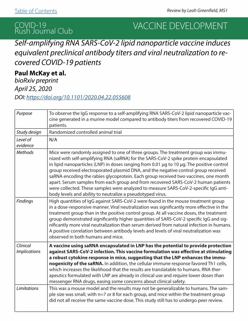
COVID-19Rush Journal ClubSelf-amplifying RNA SARS-CoV-2 lipid nanoparticle vaccine induces equivalent preclinical antibody titers and viral neutralization to re-covered COVID-19 patientsPaul McKay et al.bioRxiv preprintApril 25, 2020DOI: https://doi.org/10.1101/2020.04.22.055608
Purpose To observe the IgG response to a self-amplifying RNA SARS-CoV-2 lipid nanoparticle vac-cine generated in a murine model compared to antibody titers from recovered COVID-19 patients.
Study design Randomized controlled animal trialLevel of evidence
N/A
Methods Mice were randomly assigned to one of three groups. The treatment group was immu-nized with self-amplifying RNA (saRNA) for the SARS-CoV-2 spike protein encapsulated in lipid nanoparticles (LNP) in doses ranging from 0.01 μg to 10 μg. The positive control group received electroporated plasmid DNA, and the negative control group received saRNA encoding the rabies glycoprotein. Each group received two vaccines, one month apart. Serum samples from each group and from recovered SARS-CoV-2 human patients were collected. These samples were analyzed to measure SARS-CoV-2-specific IgG anti-body levels and ability to neutralize a pseudotyped virus.
Findings High quantities of IgG against SARS-CoV-2 were found in the mouse treatment group in a dose-responsive manner. Viral neutralization was significantly more effective in the treatment group than in the positive control group. At all vaccine doses, the treatment group demonstrated significantly higher quantities of SARS-CoV-2 specific IgG and sig-nificantly more viral neutralization than serum derived from natural infection in humans. A positive correlation between antibody levels and levels of viral neutralization was observed in both humans and mice.
ClinicalImplications
A vaccine using saRNA encapsulated in LNP has the potential to provide protection against SARS-CoV-2 infection. This vaccine formulation was effective at stimulating a robust cytokine response in mice, suggesting that the LNP enhances the immu-nogenicity of the saRNA. In addition, the cellular immune response favored Th1 cells, which increases the likelihood that the results are translatable to humans. RNA ther-apeutics formulated with LNP are already in clinical use and require lower doses than messenger RNA drugs, easing some concerns about clinical safety.
Limitations This was a mouse model and the results may not be generalizable to humans. The sam-ple size was small, with n=7 or 8 for each group, and mice within the treatment group did not all receive the same vaccine dose. This study still has to undergo peer review.
VACCINE DEVELOPMENT
Review by Leah Greenfield, MS1Table of Contents

COVID-19Rush Journal Club
In silico Design of novel Multi-epitope recombinant 1 Vaccine based on Coronavirus surface glycoprotein
Mandana BehbahanibioRxiv preprintApril 21, 2020DOI: https://doi.org/10.1101/2020.03.10.985499
Purpose To perform an in silico design a vaccine for Coronavirus based on surface glycoproteins.
Study design In SilicoLevel of evidence
N/A
Methods A multi-epitope vaccine based on surface glycoprotein was designedthrough application of bioinformatics methods. NCBI resources were used to gather relevant protein sequences and to determine sequence alignment. Antigenicity of the coronavirus surface glycoprotein was evaluated using Vax-iJen 2.0 server. B-cell epitopes were predicted using ElliPro and IEDB analysis resource. T-cell epitope prediction was performed using ProPred-1 server. 17 potent linear B-cell and T-cell binding epitopes from surface glycoprotein were predicted in silico. Then, the epitopes were joined via different linkers. The ability of selected epitopes to induce interferon-gamma was then evalu-ated using IFNepitope web server.
Findings A final vaccine was constructed, which composed of 398 amino acids and at-tached to 50S ribosomal protein L7/L12. Physicochemical properties and anti-genicity in the proposed vaccine demonstrated that the vaccine was stable. Molecular docking studies confirmed that the vaccine interacted with MHC-I and MHC-II molecules as expected. Interferon-gamma analysis showed that 16/17 epitopes had the potential to produce interferon-gamma.
ClinicalImplications
This study introduced a novel multi epitope vaccine design against Coronavi-rus. The multi-epitope vaccine with 50S ribosomal protein L7/L12 as adjuvant was a stable construct with high aliphatic content and high antigenicity.
Limitations This was an in silico study; the proposed virus must still be followed by in vi-tro and in vivo studies before proceeding. Also, this study still has to undergo peer review.
VACCINE DEVELOPMENT
Review by Ahmad Gill, MS3Table of Contents
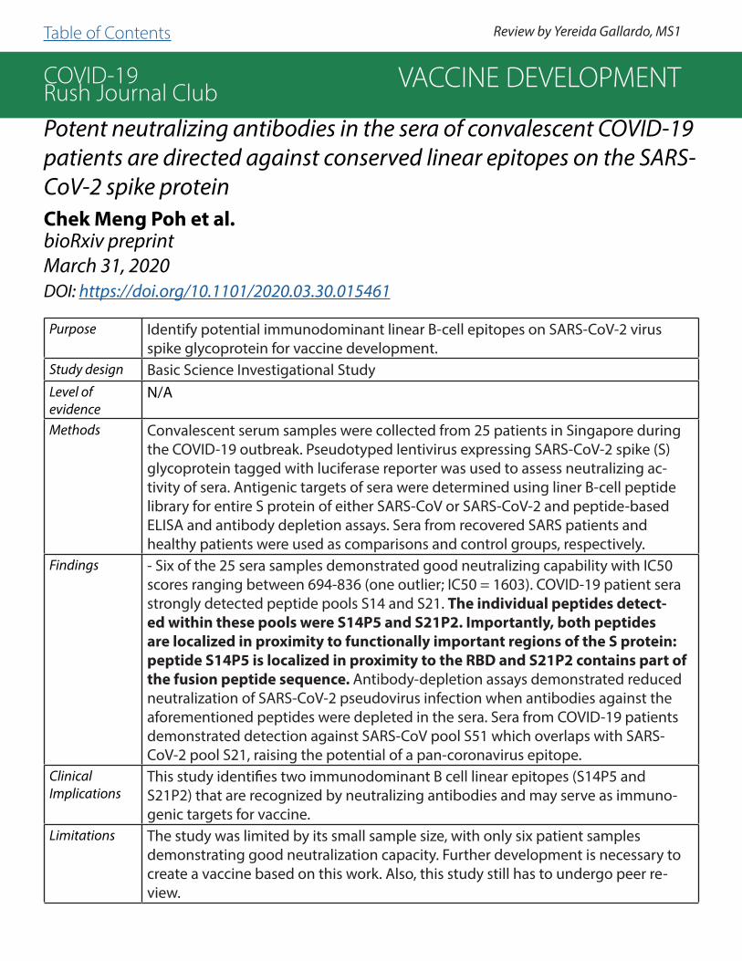
COVID-19Rush Journal ClubPotent neutralizing antibodies in the sera of convalescent COVID-19 patients are directed against conserved linear epitopes on the SARS-CoV-2 spike proteinChek Meng Poh et al.bioRxiv preprintMarch 31, 2020DOI: https://doi.org/10.1101/2020.03.30.015461
Purpose Identify potential immunodominant linear B-cell epitopes on SARS-CoV-2 virus spike glycoprotein for vaccine development.
Study design Basic Science Investigational StudyLevel of evidence
N/A
Methods Convalescent serum samples were collected from 25 patients in Singapore during the COVID-19 outbreak. Pseudotyped lentivirus expressing SARS-CoV-2 spike (S) glycoprotein tagged with luciferase reporter was used to assess neutralizing ac-tivity of sera. Antigenic targets of sera were determined using liner B-cell peptide library for entire S protein of either SARS-CoV or SARS-CoV-2 and peptide-based ELISA and antibody depletion assays. Sera from recovered SARS patients and healthy patients were used as comparisons and control groups, respectively.
Findings - Six of the 25 sera samples demonstrated good neutralizing capability with IC50 scores ranging between 694-836 (one outlier; IC50 = 1603). COVID-19 patient sera strongly detected peptide pools S14 and S21. The individual peptides detect-ed within these pools were S14P5 and S21P2. Importantly, both peptides are localized in proximity to functionally important regions of the S protein: peptide S14P5 is localized in proximity to the RBD and S21P2 contains part of the fusion peptide sequence. Antibody-depletion assays demonstrated reduced neutralization of SARS-CoV-2 pseudovirus infection when antibodies against the aforementioned peptides were depleted in the sera. Sera from COVID-19 patients demonstrated detection against SARS-CoV pool S51 which overlaps with SARS-CoV-2 pool S21, raising the potential of a pan-coronavirus epitope.
ClinicalImplications
This study identifies two immunodominant B cell linear epitopes (S14P5 and S21P2) that are recognized by neutralizing antibodies and may serve as immuno-genic targets for vaccine.
Limitations The study was limited by its small sample size, with only six patient samples demonstrating good neutralization capacity. Further development is necessary to create a vaccine based on this work. Also, this study still has to undergo peer re-view.
VACCINE DEVELOPMENT
Review by Yereida Gallardo, MS1Table of Contents

COVID-19Rush Journal Club
If a coronavirus vaccine arrives, can the world make enough?
Roxanne KhamsiNatureApril 9, 2020DOI: 10.1038/d41586-020-01063-8
Purpose Biomedical companies around the world are working on formulating a SARS-CoV-2 vac-cine. Most research to date has compared the numerous platforms available and their respective advantages and disadvantages. Optimistic timelines project a vaccine will be available within the next 12 to 18 months. This article seeks to address the question that follows: once a vaccine is formulated, how is the world to go about its production and equitable distribution?
Study design News, review article
Level of evidence
5
Methods Quantitative and narrative data collection from reputable sources including: World Health Organization (WHO), Coalition for Epidemic Preparedness Innovations (CEPI), US Pharma-copeia (USP), and several others.
Findings How vaccine distribution looks will depend heavily on the delivery mechanism, which range from inactivated form to subunit to RNA/DNA based, with each formu-lation bearing its own challenges and rewards. Regardless of the platform chosen, CEPI describes a gap that needs to be unpacked: a fair allocation system. In response to pan-demics of the past, the World Health Organization (WHO) adopted the Pandemic Influenza Preparedness (PIP) Framework to coordinate the supply-demand of vaccines, diagnostics, and drugs but due to its specificity for influenza, PIP does not apply to SARS-CoV-2. The article advocates for a rapid response mimicking this framework yet addresses time to devise as a limiting factor in addition to the retroactive obtainment of samples posing a logistical threat to its success.
ClinicalImplications
This article allows for greater understanding of the vast limitations of vaccine develop-ment beyond delivery type; touching on production costs, resource scarcity, politics, and upholding the justice pillar of medical ethics. It calls on global governments and private funders to be proactive in their response to this knowledge; the Coalition for Epidemic Preparedness Innovations (CEPI) estimates minimum $3 billion USD are needed to devel-op, trial, and manufacture, and distribute a vaccine for the world (not including estimated billions needed to fuel manufacturing plants).
Limitations The inevitable limitation in our current stage of the SARS-CoV-2 vaccine development lies in the careful balance of efficiency and efficacy. This article seeks to predict future chal-lenges in this realm based on past patterns and present predicaments.
VACCINE DEVELOPMENT
Review by Morgan Sturgis, MS2Table of Contents

COVID-19Rush Journal ClubComputers and viral diseases. Preliminary bioinformatics studies on the design of a synthetic vaccine and a preventative peptidomimetic antag-onist against the SARS-CoV-2 (2019-nCoV, COVID-19) coronavirus
Barry RobsonComputers in Biology and MedicineFebruary 26, 2020DOI: https://doi.org/10.1016/j.compbiomed.2020.103670
Purpose To find a short section or sections of SARS-CoV-2 viral protein sequence suitable for preliminary design proposal for a peptide synthetic vaccine and a peptidomimetic therapeutic, and to explore some design possibilities.
Study design BioinformaticsLevel of evidence
Level 5
Methods The use of Q-UEL (an involved automatic surfing of the world wide web to speed access to information) systems to access relevant emerging literature, and to interact with standard publicly available bioinformatics tools on the internet. Additionally, the use of MARPLE/HDNstudent with XTRACTOR allows for specific search queries that bypass “autosurfing” of the internet.
Findings The sequence of amino acids KRSFIEDLLFNKV was found to be particularly well con-served across many coronaviruses (including 2019-nCoV) and corresponds to the re-gion around one of the known cleavage sites of the SARS virus believed to be required for virus activation for cell entry. Many conventions in diagnostic and vaccine design are not significant in coronavirus matches, however an exposed loop in the SARS coro-navirus is an important target for creating antibodies and a carrier protein is necessary to promote immunogenicity. The addition of C-terminal and N-terminal linkage se-quences resulted in the proposed L-amino sequence of GPSKRSFIEDLLFNKVTLAC as a B-epitope to be synthesized and attached to a carrier.
ClinicalImplications
KRSFIEDLLFNKV protein subsequence is seen as a potential Achilles’ heel because it is exposed or potentially exposable, being required for proteolytic activation cleavage, and is also a well conserved feature on the virus. This motif seems a likely primary tar-get for synthetic vaccines and a basis for drug discovery.
Limitations One must be aware of coincidental matches that are not truly significant unless one can see that the proteins being compared are essentially of the same function or fam-ily, with the order of similar sections preserved, and that the correspondences make sense. This was an in silico study; the proposed sequence must still be followed by in vitro and in vivo studies before proceeding.
VACCINE DEVELOPMENT
Review by Johanna Balas, MS1 & Diana Vazquez ParkerTable of Contents

COVID-19Rush Journal ClubInsights into Cross-species Evolution of Novel Human Coronavirus 2019-nCoV and Defining Immune Determinants for Vaccine Devel-opmentArunachalam Ramaiah, et al.bioRxiv preprintFebruary 04, 2020DOI: https://doi.org/10.1101/2020.01.29.925867
Purpose To analyze the genomic evolution of 2019 novel coronavirus (2019-nCov) and identify poten-tial high binding affinity (HBA) CD4 T-cell epitopes (TCEs) for subunit vaccine development.
Study design Basic Science Investigational Study
Level of evidence
N/A
Methods Genome sequences of 2019-nCoV (n=48) were obtained on January 29th, 2020 from GSAID and GenBank, along with genomic sequences for SARS-CoV, SARS-like-CoV, and MERS-CoV strains (n=8,2,10, respectively). One alphacoronavirus sequence was used as an outgroup con-trol. To identify potential target peptides for vaccine design, TCEs were predicted using struc-tural protein sequences of highly conserved and representative 2019-nCoV strain Wuhan-Hu-1 (MN908947.3)..
Findings Phylogenic analysis showed 2019-nCoV to be genetically and evolutionary related to bat CoVs. A large clade was formed by these viruses with both 2019-nCoV and bat-CoV virus clusters sharing a common ancestor (92% bootstrap support). Additionally, there was a 96% sequence similarity between the 2019-nCoV Wuhan-Hu-1 and bat/Yunnan/RaTG13/2013 strains. 2019-nCoV and bat/Yunnan/RaTG13/2013 sequences differed in the structural proteins spike (S) and membrane (M), but envelope (E) and nucleocapsid (N) were highly conserved. The binding af-finity for all possible 15-mer peptides from the 2019-nCoV structural proteins against the pre-dominant HLA-DR alleles in the ethnic populations of China, Thailand, Japan, and Asia-Pacific Region were assessed. There were eight common epitopes recognized by all HLA-DR alleles across the ethnic populations distributed among S, E, and M proteins (n=2, 3,3, respectively).
ClinicalImplications
This study suggests the current 2019-nCoV likely evolved from bat CoVs through a series of recombinant events that enabled it to adapt to humans. Additionally, 8 epitopes recog-nized by HLA alleles common to different ethnic populations were identified; creating a sub-unit vaccine containing these eight epitopes may induce effective antiviral T-cell and antibody responses in different ethnic populations.
Limitations This study only identified high-binding affinity TCEs against HLA-DR alleles predominant in Asia and Asia Pacific Region. As the 2019-nCoV is now classified as a pandemic and has spread to various countries globally, this decision may limit the generality of the predicted epitopes. Additionally, further in vitro and in vivo studies are needed to validate the predicted epitopes and whether they can produce a robust immune response. Lastly, this study has not been peer-reviewed.
VACCINE DEVELOPMENT
Review by Yereida Gallardo, MS1Table of Contents

COVID-19Rush Journal ClubPreliminary Identification of Potential Vaccine Targets for COVID-19 Coronavirus (SARS-CoV-2) Based on SARS-CoV Immunological Stud-iesSyed Faraz Ahmed et al.VirusesMarch 2020DOI: https://doi.org/10.3390/v12030254
Purpose To identify identical epitopes between SARS-CoV-2, SARS-CoV (responsible for the 2003 endem-ic), and MERS-CoV (responsible for the 2012 outbreaks) to find potential targets for an effective vaccine.
Study design Laboratory Study
Level of evidence
N/A
Methods The investigators obtained reference protein sequences for SARS-CoV-2, SARS-CoV, and MERS-CoV, along with SARS-CoV B- and T-cell epitopes. They estimated a population coverage for the T cell epitopes to represent the population likely to elicit an immune response for at least 1 T-cell epitope. Lastly, the investigators constructed a phylogenetic tree of the structural protein of SARS-CoV, MERS-CoV, and SARS-CoV-2, as well as the Zaria Bat coronavirus strain.
Findings The M (membrane), N (nucleocapsid), and E (envelope) proteins of SARS-CoV and SARS-CoV-2 have over 90% genetic similarity while the S (spike) protein has 76% similarity. The similarity between SARS-CoV-2 and MERS-CoV is substantially lower. From previous studies, S and N proteins are known to induce immune responses. 27/115 T-cell epitopes are identical between SARS-CoV and SARS-CoV-2 (all in N or S proteins). MHC binding assays suggested 5 dis-tinct MHC alleles for 19 of these epitopes. The population coverage for these epitopes is 59.76% globally and 32.36% in the Chinese population. To identify potential T cell targets that would cover a larger percentage of the population, additional T cell epitopes were considered that have not yet been experimentally tested for SARS-CoV. Of these, 229 epitope sequences had an identical match in SARS-CoV-2 with MHC allele information available and 102 of these were S or N proteins. Population coverage estimates for these sequences showed 96.29% with 20 distinct MHC alleles. 49 B-cell epitopes from SARS-CoV had an identical match in SARSCoV-2 (45 from N or S protein).
ClinicalImplications
The study demonstrates similarities and differences between the novel coronavirus of 2020 and past coronavirus outbreaks. The study also determines important structural aspects of the SARS-CoV-2 epitope that may be used to develop an immune response. We may be able to extrapo-late the data from previous immune responses to try to find ways to create a positive immune response against SARS-CoV-2.
Limitations Despite the similarities between SARS-CoV and SARS-CoV-2, there is still significant genetic variation between the two and it is not obvious that immune responses will be elicited against both viruses. Additionally, as the virus continues to evolve, it is expected that more mutations will be observed, and as long as they occur outside of the epitope regions identified, they may not affect this analysis. Further T and B cell assays are important to identify the potential of the epitopes to induce a positive immune response against SARS-CoV-2.
VACCINE DEVELOPMENT
Review by Pranita Kaginele, MS2Table of Contents

COVID-19Rush Journal Club
Strategies for vaccine design for corona virus using Immunoinfor-matics techniques
Anamika Basu et al.bioRxiv preprintMarch 2, 2020DOI: https://doi.org/10.1101/2020.02.27.967422
Purpose To evaluate potential B cell and T cell epitopes present in non-structural protein 4 of beta coronavirus as a strategy for vaccine design against corona-virus.
Study design Genome sequencing and analysisLevel of evidence
N/A
Methods Based on the non-structural protein NS4 in Beta coronavirus HKU24 obtained from the InterPro database (accession number A0A0A7UXD8), potential B and T cell epitopes were predicted using various methods, such as the Ka-laskar and Tongaonkar antigenicity scale, Stabilized matrix method, and a consensus approach which combines NN-align, SMM-align, and combinato-rial library methods. Then, population coverage was assessed, and molecular docking studies were performed for identified T cell epitopes.
Findings -The peptide sequence IRNTTNPSAR and PTDTYTSVYLGKFRG were considered as the most potential B cell and T cell epitopes respective-ly. These epitopes may be considered as potential peptides for a pep-tide-based vaccine for coronavirus.-The predicted T cell epitopes PTDTYTSVY and PTDTYTSVYLGKFRG perfectly fitted into the epitope binding grooves of alpha helix of MHC I molecule and MHC II molecule. The epitope PTDTYTSVY was present in 58.87% of the Chi-nese population and 50.16% of the world population.
ClinicalImplications
This study recommends potential B-cell and T-cell epitopes to use in a pep-tide-based vaccine for coronavirus.
Limitations The study was limited in that this study requires in vitro and in vivo trials to determine effectiveness of these vaccine peptides. In addition, this study is a preliminary report that has not been peer-reviewed.
VACCINE DEVELOPMENT
Review by Audrey Sung, MS3Table of Contents
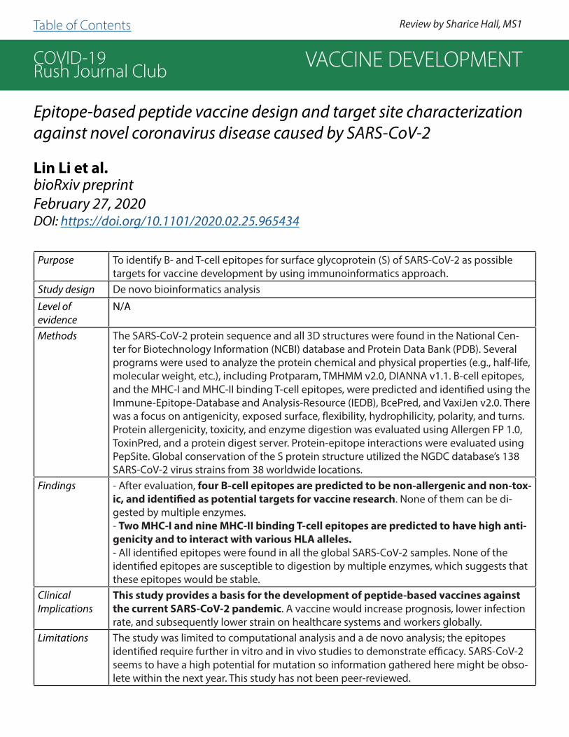
COVID-19Rush Journal Club
Epitope-based peptide vaccine design and target site characterization against novel coronavirus disease caused by SARS-CoV-2
Lin Li et al.bioRxiv preprintFebruary 27, 2020DOI: https://doi.org/10.1101/2020.02.25.965434
Purpose To identify B- and T-cell epitopes for surface glycoprotein (S) of SARS-CoV-2 as possible targets for vaccine development by using immunoinformatics approach.
Study design De novo bioinformatics analysis
Level of evidence
N/A
Methods The SARS-CoV-2 protein sequence and all 3D structures were found in the National Cen-ter for Biotechnology Information (NCBI) database and Protein Data Bank (PDB). Several programs were used to analyze the protein chemical and physical properties (e.g., half-life, molecular weight, etc.), including Protparam, TMHMM v2.0, DIANNA v1.1. B-cell epitopes, and the MHC-I and MHC-II binding T-cell epitopes, were predicted and identified using the Immune-Epitope-Database and Analysis-Resource (IEDB), BcePred, and VaxiJen v2.0. There was a focus on antigenicity, exposed surface, flexibility, hydrophilicity, polarity, and turns. Protein allergenicity, toxicity, and enzyme digestion was evaluated using Allergen FP 1.0, ToxinPred, and a protein digest server. Protein-epitope interactions were evaluated using PepSite. Global conservation of the S protein structure utilized the NGDC database’s 138 SARS-CoV-2 virus strains from 38 worldwide locations.
Findings - After evaluation, four B-cell epitopes are predicted to be non-allergenic and non-tox-ic, and identified as potential targets for vaccine research. None of them can be di-gested by multiple enzymes.- Two MHC-I and nine MHC-II binding T-cell epitopes are predicted to have high anti-genicity and to interact with various HLA alleles.- All identified epitopes were found in all the global SARS-CoV-2 samples. None of the identified epitopes are susceptible to digestion by multiple enzymes, which suggests that these epitopes would be stable.
ClinicalImplications
This study provides a basis for the development of peptide-based vaccines against the current SARS-CoV-2 pandemic. A vaccine would increase prognosis, lower infection rate, and subsequently lower strain on healthcare systems and workers globally.
Limitations The study was limited to computational analysis and a de novo analysis; the epitopes identified require further in vitro and in vivo studies to demonstrate efficacy. SARS-CoV-2 seems to have a high potential for mutation so information gathered here might be obso-lete within the next year. This study has not been peer-reviewed.
VACCINE DEVELOPMENT
Review by Sharice Hall, MS1Table of Contents

COVID-19Rush Journal Club
Vaccines for SARS-CoV-2: Lessons from other Coronavirus Strains
Eriko Padron-RegaladoInfectious Diseases and TherapyApril 23, 2020DOI: https://doi.org/10.1007/s40121-020-00300-x
Purpose To review the pertinent information about COVID-19 to help create a vaccine
Study design ReviewLevel of evidence
Level 5
Methods Review the past and ongoing vaccine development efforts for clinically rele-vant coronavirus strains in order to help develop safe and effective vaccines for COVID-19.
Findings - No vaccine is currently available for SARS-CoV-2.- While we want to develop safe and efficient vaccines, we should worry about adverse drug events as animal models and vaccination regimens have demonstrated the possibility of this occurring.- We need to address the possibility of short-term immunogenicity, which would happen if the virus induces production of neutralizing antibodies.- Exploiting T cell responses for coronavirus vaccination should also be con-sidered (along with B cell responses) as they are persistent and protective in animal models.- Employing the N protein of coronavirus for vaccination has the potential of providing long-term cross-protection.
ClinicalImplications
As of April 2020, no vaccine is commercially available for coronavirus. Moving forward, we can study the MERS and SARS vaccine development pro-cesses to learn more about SARS-CoV-2. While creating a vaccine is of utmost importance, we must also realize the potential side effects of vaccines and should not be oblivious to this. The N protein of coronavirus has the po-tential for providing long term cross-protection.
Limitations Coronavirus is highly infectious, which limits the progress of creating a vac-cine. Furthermore, it is difficult to find adequate animals or individuals to start vaccine trials.
VACCINE DEVELOPMENT
Review by Ahmad Gill, MS3Table of Contents

COVID-19Rush Journal Club
Potent binding of 2019 novel coronavirus spike protein by a SARS coronavirus-specific human monoclonal antibodyXiaolong Tian et al.Emerging Microbes & InfectionsFebruary 17, 2020DOI: https://doi.org/10.1080/22221751.2020.1729069
Purpose To experimentally determine the cross-reactivity of several anti SARS-CoV antibod-ies with 2019-nCoV spike protein.
Study design Basic Science Investigational Research
Level of evidence
N/A
Methods 2019-nCoV receptor binding domain (RBD) protein was expressed and purified. Its conforma-tion with its human receptor, angiotensin converting enzyme 2 (ACE2), was predicted. Biolayer interferometry binding (BLI) assay was utilized to determine the binding affinity of SARS-CoV spike protein and 2019-nCoV RBD to human ACE2. Lastly, ELISA (utilizing high binding assay plates coated with purified 2019-nCoV RBD) and BLI (utilizing streptavidin-coated biosen-sors) assays were employed to measure the binding affinities of a series of representative SARS-CoV-specific antibodies reported to target RBD and possess neutralizing activity: m396, CR3014, CR3022, and MERS-CoV-specific human monoclonal antibody m366. Anti-CD40 anti-body was used as a negative control.
Findings 2019-nCoV RBD bound potently to human ACE2 as determined by BLI, with an affini-ty comparable to that of SARS-CoV spike protein with human ACE2 (15.2 and 15.0 nM, respectively). Of the antibodies tested, only SARS-CoV specific antibody CR3022 was found to bind potently with 2019-nCoV RBD. No competition in binding for 2019-nCoV RBD was found between CR3022 and ACE2 as determined by BLI. This suggests CR3022 recognizes an epitope that does not overlap with the ACE2 binding site of 2019-nCoV. Protein sequence alignment of 2019-nCoV and SARS-CoV RBD demonstrates they differ at the C-terminus residues.
ClinicalImplications
This study points to CR3022 as a potential therapeutic for the treatment and prevention of 2019-nCoV. The lack of cross-reactivity from the other RBD targeting SARS-CoV-specific anti-bodies may have resulted as a consequence of the structural differences between 2019-nCoV and SARS-CoV RBD. This study raises the possibility that a vaccine developed from epitopes outside the ACE2 binding site may be a more promising in inducing cross-reactive neutralizing antibodies.
Limitations The study was limited by the small number of antibodies utilized and none of the tested antibodies showed cross-reactivity with the ACE2 binding site of 2019-nCoV, which is essential for the virus replication. Also, virus neutralization tests were not performed. More work will be needed for the development of novel monoclonal antibodies, which specifically binds to 2019-nCoV spike protein.
VACCINE DEVELOPMENT
Review by Yereida Gallardo, MS1 Table of Contents

COVID-19Rush Journal Club
INFECTION CONTROL/PREVENTION
Table of Contents
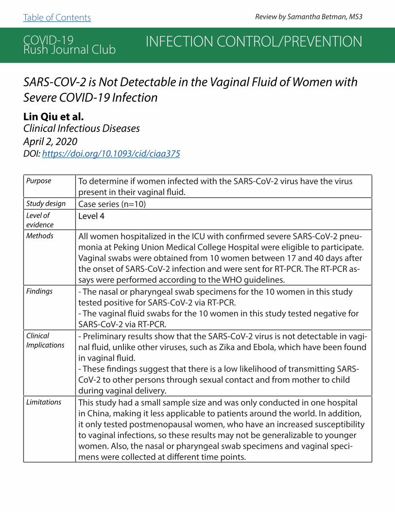
COVID-19Rush Journal Club
SARS-COV-2 is Not Detectable in the Vaginal Fluid of Women with Severe COVID-19 InfectionLin Qiu et al.Clinical Infectious DiseasesApril 2, 2020DOI: https://doi.org/10.1093/cid/ciaa375
Purpose To determine if women infected with the SARS-CoV-2 virus have the virus present in their vaginal fluid.
Study design Case series (n=10)Level of evidence
Level 4
Methods All women hospitalized in the ICU with confirmed severe SARS-CoV-2 pneu-monia at Peking Union Medical College Hospital were eligible to participate. Vaginal swabs were obtained from 10 women between 17 and 40 days after the onset of SARS-CoV-2 infection and were sent for RT-PCR. The RT-PCR as-says were performed according to the WHO guidelines.
Findings - The nasal or pharyngeal swab specimens for the 10 women in this study tested positive for SARS-CoV-2 via RT-PCR.- The vaginal fluid swabs for the 10 women in this study tested negative for SARS-CoV-2 via RT-PCR.
ClinicalImplications
- Preliminary results show that the SARS-CoV-2 virus is not detectable in vagi-nal fluid, unlike other viruses, such as Zika and Ebola, which have been found in vaginal fluid.- These findings suggest that there is a low likelihood of transmitting SARS-CoV-2 to other persons through sexual contact and from mother to child during vaginal delivery.
Limitations This study had a small sample size and was only conducted in one hospital in China, making it less applicable to patients around the world. In addition, it only tested postmenopausal women, who have an increased susceptibility to vaginal infections, so these results may not be generalizable to younger women. Also, the nasal or pharyngeal swab specimens and vaginal speci-mens were collected at different time points.
INFECTION CONTROL/PREVENTION
Review by Samantha Betman, MS3Table of Contents