Optimisation of Neuroendocrine Tumor Treatment
142
Stefan Pool Optimisation of Neuroendocrine Tumor Treatment: Locoregional administration, combination therapy and multimodal imaging
-
Upload
khangminh22 -
Category
Documents
-
view
6 -
download
0
Transcript of Optimisation of Neuroendocrine Tumor Treatment
Stefan Pool
Optimisation of Neuroendocrine Tumor Treatment:
Locoregional administration,combination therapy and
multimodal imaging
O
ptim
isation
of N
eu
roe
nd
ocrin
e Tu
mo
r Treatm
en
t S
tefa
n P
oo
l
Uitnodiging
Voor het bijwonenvan de openbareverdediging vanhet proefschrift
Optimisation ofNeuroendocrine
Tumor Treatment:Locoregional
administration,combination therapy
and multimodal imaging
doorStefan Pool
Dinsdag16 december 2014
15:30 uur QueridozaalErasmus MC
Westzeedijk 361Rotterdam
Receptie na afloop
Paranimfen:Nanno Ouwehand
&Stuart Koelewij n
E-mail: promotie.stefan@
gmail.com


OPTIMISATION OF NEUROENDOCRINETUMOR TREATMENT:
LOCOREGIONAL ADMINISTRATION,COMBINATION THERAPY AND
MULTIMODAL IMAGING
Stefan E. Pool
ISBN/EAN: 978-94-91487-20-0
Layout: Tekla F.H. Ouwehand
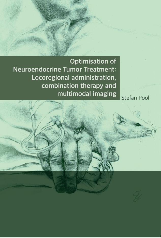
Illustration cover: Stuart J. Koelewijn
Printing: Ridderprint BV, Ridderkerk, the Netherlands
Publisher: Medix Publishers BV, Keizersgracht 317A, 1016 EE, Amsterdam, the Netherlands
Copyright © 2014 S.E. Pool, Rotterdam, the Netherlands. All rights reserved. No part of this thesis
may be reproduced or transmitted in any form or by any means, without the prior written permission
of the author.
De digitale versie van dit proefschrift is te vinden in de YourThesis app en kan gelezen worden op
een tablet of smartphone. De app kan gedownload worden in de App store en de Google Play store
of middels het scannen van de onderstaande QR-code.

Optimisation of NeuroendocrineTumor Treatment:
Locoregional administration,combination therapy and
multimodal imaging
Optimalisatie van de behandeling van neuro endocriene tumoren: locoregionale toediening, combinatietherapie en
multimodale visualisatie
Proefschrift
ter verkrijging van de graad van doctor aan de Erasmus Universiteit Rotterdam op gezag van de rector magnificus
Prof.dr. H.A.P. Pols
en volgens besluit van het College voor Promoties.
De openbare verdediging zal plaatsvinden op
dinsdag 16 december 2014 om 15.30 uur
door Stefan Ernest Pool
geboren te Hilversum

Promotiecommissie
Promotoren: Prof.dr.ir. M. de Jong Prof.dr. C.H.J. van Eijck
Overige leden: Prof.dr. E.P. Krenning Prof.dr. L.J. Hofland Prof.dr. O.C. Boerman
Copromotor: Dr. G.A. Koning
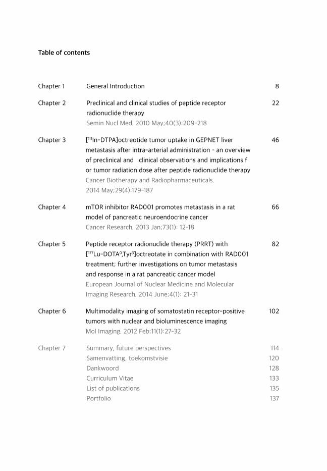
Table of contents
Chapter 1 General Introduction 8
Chapter 2 Preclinical and clinical studies of peptide receptor 22
radionuclide therapy
Semin Nucl Med. 2010 May;40(3):209-218
Chapter 3 [111In-DTPA]octreotide tumor uptake in GEPNET liver 46
metastasis after intra-arterial administration – an overview
of preclinical and clinical observations and implications f
or tumor radiation dose after peptide radionuclide therapy
Cancer Biotherapy and Radiopharmaceuticals.
2014 May;29(4):179-187
Chapter 4 mTOR inhibitor RAD001 promotes metastasis in a rat 66
model of pancreatic neuroendocrine cancer
Cancer Research. 2013 Jan;73(1): 12-18
Chapter 5 Peptide receptor radionuclide therapy (PRRT) with 82
[177Lu-DOTA0,Tyr3]octreotate in combination with RAD001
treatment; further investigations on tumor metastasis
and response in a rat pancreatic cancer model
European Journal of Nuclear Medicine and Molecular
Imaging Research. 2014 June;4(1): 21-31
Chapter 6 Multimodality imaging of somatostatin receptor-positive 102
tumors with nuclear and bioluminescence imaging
Mol Imaging. 2012 Feb;11(1):27-32
Chapter 7 Summary, future perspectives 114
Samenvatting, toekomstvisie 120
Dankwoord 128
Curriculum Vitae 133
List of publications 135
Portfolio 137

1

1I N T R O D U C T I O N
CH
AP
TE
R 1
Introduction
CHAPTER 1
8
General introduction
There are many treatment options for neuroendocrine tumors, including surgical
resection, chemotherapy, (radio) embolization, radiofrequency ablation (RFA), targeted
drug therapy and peptide receptor radionuclide therapy. This thesis focuses on attempts
to combine some of these methods in order to improve treatment outcome in patients
with neuroendocrine tumors (NETs).
In this Introduction the following topics will be discussed:
Neuroendocrine TumorsEpidemiologyDiagnosisDiagnostic ProceduresTreatmentChemotherapy Radiotherapy Surgery Liver-Directed Therapies Medical Therapy Molecular Targeted Agents Peptide Receptor Radionuclide Therapy Molecular Imaging Translational research: preclinical-animal models Animal NET modelPreclinical ImagingAims and ouline of this thesis
Neuroendocrine Tumors
Neuroendocrine tumors belong to a family of rare neoplasms that are mostly found in
gastro enteric, pancreatic, and pulmonary tissues, but that may be present virtually in any
organ of the body. Neuroendocrine tumors originate from cells of the neuroendocrine
system. Some of these neuroendocrine tumors produce hormones (functioning NETs)
resulting in distinct clinical symptoms like in Cushing’s syndrome (ectopic ACTH
production), Zollinger-Ellison syndrome (ectopic gastrin secretion), Verner-Morrison
syndrome (vasoactive intestinal peptide production) and Carcinoid syndrome (ectopic
serotonin production)1,2. in a large epidemiological study in the USA the majority of
NETs were located in the gastrointestinal tract (69.7%) (13,715 NET patients), whereas

CH
AP
TE
R 1
Introduction
CHAPTER 1
9
24.5% of the NETs were located in the tracheobronchopulmonary tract. Ovary, pancreas,
thymus and gallbladder were the remaining localizations.3 This study excluded patients
with functioning or nonfunctioning pancreatic NETs (pNETs) because the then common
indication ‘carcinoid’ was used as search term. These pNETs account for approximately
1.3-2% of all pancreatic cancers with regard to incidence4,5 but due to their slow-
growing nature they account for almost 10 % of pancreatic cancers in prevalence4.
Nonfunctioning pancreatic pNETs are more common compared to functioning pNETs.6
Most of NETs overexpress the somatostatin 2 receptor (sst2), a G-protein coupled
receptor that inhibits the secretion of a wide range of hormones upon activation by
binding of the hormone somatostatin. Five sst subtypes have been identified (sst2)
which all have different roles7. The sst2 overexpression on NETs enables imaging
with for example [111Indium-DTPA0]-octreotide (Octreoscan®) or [68Ga-DOTA0-
Tyr3]-octreotate and/or treatment by Peptide Receptor Radionuclide Therapy.
Epidemiology
The incidence of NETs (pancreatic NETs not included) in the Netherlands was 1.8 per
100,000 inhabitants for men and 1.9 per 100,000 inhabitants for women in the period
1989-19968. The highest incidence of NETs occurs in the seventh decade of life 8,9.
A rise in incidence of NETs has been reported by several authors3,4,9. This rise in incidence
could partly be explained by the increased use and improved techniques of diagnostic
modalities. Also the introduction of the World Health Organization classification for
NETs of the gastroenteropancreatic tract in the year 2000 may have resulted in an
increased awareness of the existence of these tumors and in more intelligibility for the
nomenclature and categorization of gastroenteropancreatic neuroendocrine tumors
(GEP-NETs).
Diagnosis
As mentioned before, part of the NETs is hormone producing, resulting in specific
hormone-related symptoms that are quite often the first signs of (functioning) NET
presence. Non-specific tumor-related symptoms are for example unexplained weight
loss, pain and anorexia. Because of the specific hormone-related symptoms, functioning
NETs are most of the time diagnosed earlier in the course of disease compared to non-
functioning NETs. The carcinoid syndrome, consisting of secretory diarrhea, flushing,
wheezing, and right-sided valvular heart disease, is caused by serotonin excretion by liver
CH
AP
TE
R 1
Introduction
CHAPTER 1
10
metastases. When the carcinoid syndrome is present, most of the times the tumor has
already metastasized to the liver or retroperitoneal lymph nodes with drainage through
the caval vein instead of the portal vein. Also localization of a primary tumor in the
testis or ovary may result in the carcinoid syndrome. In these cases there is no hepatic
breakdown of serotonin resulting in serotonin availability in the systemic circulation.
Diagnostic Procedures
Several laboratory tests can be used in de diagnostic process like chromogranine
A (CgA), neuron-specific enolase (NSE), specific hormones in case of functioning NETs,
general blood tests, such as liver function tests in the case of liver metastasis. Also the
5-HIAA (serotonin metabolite) urine test is commonly used.
Imaging can be performed by anatomical and functional imaging. Examples of
anatomical imaging of NETs are conventional radiography, ultrasonography, computed
tomography (CT), magnetic resonance imaging (MRI) and angiography.
Examples of functional imaging are somatostatin receptor scintigraphy using [111
Indium-DTPA0]-octreotide (Octreoscan®)10 and positron emission tomography-
computed tomography (PET-CT) with for example 68Ga-DOTA0-Tyr3-octreotide11 or 68Ga-DOTA-Tyr3-octreotate12. These imaging techniques can screen the total body
and provide information about the presence of stt on the tumor. Other functional
imaging techniques for the detection of NETs, not based on sst receptor targeting,
include PET imaging with 6-18F-fluoro-L-DOPA13 or 11C-5-hydroxytryptophan14. 18F-fluorodeoxyglucose (18F-FDG) PET imaging, reflecting glucose metabolism, may be
of value in NETs with a high proliferation index15.
By endoscopy of the gastrointestinal tract, e.g. upper gastrointestinal endoscopy or
colonoscopy, primary NETs can be diagnosed. Endoscopic ultrasonography can be used
for diagnosis of pancreatic NETs including assessment of tumor relation with surrounding
structures and the presence of pathological lymph nodes16.
For a conclusive diagnosis a pathological analysis must be performed on tumor tissue
derived by biopsy from the primary tumor or metastasis. Hematotoxylin and eosin
staining, immunostaining for chromogranine A and synaptophysin, assessment of the
CH
AP
TE
R 1
Introduction
CHAPTER 1
11
mitotic index and the Ki67 proliferative index should be performed17-19. Furthermore
immunohistochemical staining for sst2, insulin, gastrin, glucagon, or vasoactive intestinal
peptide is optional17-19.
Treatment
Chemotherapy
Several studies show a limited effect of chemotherapy in patients with well-
differentiated NETs of non-pancreatic origin. Some objective responses have been
reported on mostly poorly differentiated NETs with different chemotherapies like
streptozocin in combination with doxorubicin20, fluorouracil in combination with
doxorubicin and streptozocin21, cisplatin in combination with etoposide22, capecitabine
in combination with temozolomide23, 5-fluorouracil and leucovorin in combination with
irinotecan24, temozolomide in combination with thalidomide25 and temozolomide in
combination with bevacuzimab26. The use of most of these chemotherapeutic regimens
is hampered by the mostly short duration of response and significant (hematological)
toxicity.
Radiotherapy
Radiotherapy is mainly applied for local treatment in case of brain metastases,
spinal cord compression due to bone metastases or painful bone metastases. In case
of localized bronchial NETs radiotherapy is being applied, especially if surgery is not
an option anymore27. Partial responses and clinical benefit experienced by the
patients were reported by Saif et al. after chemoradiation on the primary pancreatic
neuroendocrine tumor (bed)28. Prospective studies to investigate the role of radiation
and chemoradiation in NET treatment are warranted.
Surgery
Resection of the primary tumor is currently the only curative treatment option in
NET patients, provided there is no advanced disease. In patients with locally advanced
CH
AP
TE
R 1
Introduction
CHAPTER 1
12
disease, neo-adjuvant treatment can sometimes be considered as an option to make
surgery feasible. Unfortunately most NET patients suffer from metastasized disease
already at the time of diagnosis, most often to the liver. However, even in patients with
liver metastasis surgery can play a role. Kleine et al. reported in a rectrospective non-
randomized study that extended surgery (partial liver resection/portal vein resection/
partial gastric resection/liver transplantation) is feasible in highly selected patients;
disease-specific survival of patients who had a liver resection was similar to patients
without liver metastasis29. Another group reported, although also in a retrospective,
non-randomized study, an encouraging 5-year survival rate of 82% after major surgery
in patients with liver metastases from carcinoids and gastrinomas30. In a different study
the same group reported a 5-year survival rate of 80% after extended (hepatic) surgery
in patients with pancreatic or duodenal NETs metastasized to the liver31. Also symptom-
control surgery can play a role as described in a study by Sarmiento et al.32. In this
study hepatic resection was associated with a partial or complete response with respect
to hormonal symptoms in 104/108 GEP-NET patients. The median time to recurrence
was 46 months. Resection of the primary tumor was associated with a better survival in
several non-randomized retrospective studies33,34. However selection bias could have
played a role here, as younger and healthier patients are probably more likely to get
surgery.
Liver-Directed Therapies
For patients with metastasized disease primarily localized in the liver, several non-
surgical liver-directed interventional therapies are available: hepatic artery/transarterial
(chemo-) embolization (H/TA(C)E), radiofrequency ablation (RFA), cryoablation and
laser-induced thermotherapy (LITT). Intrahepatic malignancies mainly depend on the
hepatic artery for their blood supply in contrast to normal liver parenchyma, which
mainly relies on the portal vein. Therefore these intrahepatic malignancies can be
selectively treated by H/TA(C)E. With HAE/TAE, objective responses were achieved in
40-75% of patients with a large variability in duration of the effect (3 – 88 months). With
HACE/TACE objective responses were reported in 8-100%, again with variable duration
(6-63 months, means 14-42 months)35,36. RFA has a high rate of local tumor control
with limited local recurrence. Unfortunately these interventions only affect large tumor
lesions within the liver with almost certain disease recurrence at other microscopic tumor
sites in the liver.
CH
AP
TE
R 1
Introduction
CHAPTER 1
13
Intra-arterial radio embolization with yttrium-90 microspheres is an increasingly
applied treatment option for patients with unresectable primary or secondary hepatic
malignancies refractory to systemic therapies37. The high-energy beta-radiation emitting
microspheres subsequently strand in the arterioles (mainly) of the tumor, and a tumoricidal
radiation absorbed dose is delivered. The clinical results of this form of internal radiation
therapy are promising38,39. Recently 166Ho-loaded poly(L-lactic acid) microspheres ( 166Ho-PLLAMS) have been developed which like 90Y-microspheres emit high-energy
beta particles to eradicate tumor cells whereas 166Ho in addition also emits low-energy
(81 keV) gamma photons which allows for nuclear imaging. This facilitates pre-therapeutic
administration of a small scout dose, predicting the distribution of the therapeutic dose.
Secondly post therapy imaging can be used for dosimetry calculations40.
Intra-arterial administration of sst targeted PRRT is used by several groups for increasing
the radionuclide tumor uptake and thereby the therapeutic radiation effect41-44.
Medical Therapy
Binding of somatostatin analogs such as octreotide and lanreotide to the sst2 can
reduce hormonal overproduction by a NET and may result in symptomatic relief in
most patients with metastasized disease45-47. The long-acting somatostatin analog
octreotide LAR (Sandostatin LAR®; Novartis Basel, Switzerland) also showed a positive
effect on time to tumor progression compared to placebo in patients with functionally
active and inactive metastatic midgut NETs48. This anti-tumor activity was suggested
to be enhanced by combination of a somatostatin analog with recombinant interferon
alpha in some retrospective studies49,50. However in a prospective, randomized clinical
trial, no significant difference in overall survival was found between patients with midgut
NETs treated with the combination of octreotide and interferon alpha versus octreotide
alone51.
Molecular Targeted Agents
Recently the results of two phase III trials investigating the efficacy of Everolimus and
Sunitinib, both recently developed targeted therapies, were presented52,53. Treatment
of pNET patients with Everolimus (Affinitor, RAD001; Novartis Pharmaceuticals; Basel;
Switzerland), an inhibitor of mammalian target of rapamycin (mTOR), resulted in a
longer median progression free survival (PFS) of 11.0 months compared to 4.6 months
with placebo53. Also in PNET patients, treatment with Sunitinib (Sutent; Pfizer Inc., New
CH
AP
TE
R 1
Introduction
CHAPTER 1
14
York, NY), a tyrosine kinase inhibitor, resulted in a progression free survival of 11.4 months
versus 5.5 months with placebo52. Everolimus combined with Octreotide LAR treatment
in progressive PNET patients also resulted in a longer median PFS of 16.4 months versus
11.3 months with placebo combined with Octreotide LAR54.
Peptide Receptor Radionuclide Therapy
Peptide Receptor Radionuclide Therapy (PRRT) is a promising targeted therapy for
NETs using radiolabeled somatostatin analogs and is reviewed in Chapter 2. In summary,
PRRT is and has been performed with several somatostatin analogs labeled with different
radionuclides, such as [111Indium-DTPA0]-octreotide (Octreoscan®), [90Y-DOTA0,Tyr3]
octreotide (90Y-DOTATOC) and [117Lu-DOTA0,Tyr3]octreotate (117Lu-DOTATATE).
PRRT is discussed in more detail in Chapter 2.
Molecular imaging & therapy
Molecular Imaging
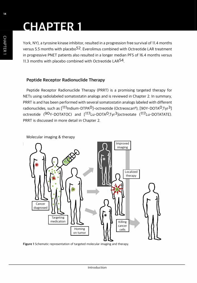
Figure 1 Schematic representation of targeted molecular imaging and therapy.
Cancer diagnosed
Targetingmedication
Homing on tumor
Killing cancer cells
Localized therapy
Improved imaging

CH
AP
TE
R 1
Introduction
CHAPTER 1
15
Molecular imaging is a multidisciplinary field, which has emerged as a discipline at
the intersection of molecular biology and in vivo imaging. It is used for non-invasively
visualizing cellular function/localization and the follow-up of the molecular process in
living organisms without the necessity to sacrifice them. Molecular imaging originates
from nuclear medicine since from early on this discipline has applied radiolabeled tracers
to show uptake or metabolism in specific organs/pathology. The most well known
example is visualization of well-differentiated thyroid carcinoma using radioactive iodine
(123I and 131I, both gamma ray emitters). Besides visualization of the tumor, treatment
is also possible with 131I, which next to gamma ray emission, used for imaging, emits
high-energy beta particles, inducing damage to the cancer cells. Based on the same
principle is the visualization of somatostatin receptors on GEPNETs using [111Indium-
DTPA0]-octreotide and treatment of these tumors by PRRT targeting the overexpressed
somatostatin receptor. One targeting moiety that can be applied for diagnosis, treatment
selection, and treatment is known as a theranostic.
Tracers used for molecular imaging can be labeled with different radionuclides to be
used for single photon emission computer tomography (SPECT) or positron emission
tomography (PET) imaging, with contrast agents with high relaxivity for magnetic
resonance imaging (MRI), and fluorophores for optical imaging. Especially preclinical
bioluminescence and fluorescence imaging is increasingly used from intracellular imaging
for example by confocal microscopy to in vivo imaging for tumor imaging/follow-up.
Translational research: preclinical-animal models
Animal NET model
In this thesis, most of the animal experiments have been performed in an established
tumor model in Lewis rats55. In these rats, CA20948 tumor cells have been inoculated
subcutaneously or subscapsularly in the liver. CA20948 cells are derived from a sst2-
positive pancreatic tumor of acinar origin that was originally induced by azaserine and
that is transplantable in syngeneic Lewis rats55. The CA20948 tumor has shown to
be very useful, both in culture as in vivo, as a model for peptide receptor radionuclide
scintigraphy and/or therapy55,57.
CH
AP
TE
R 1
Introduction
CHAPTER 1
16
Preclinical Imaging
Molecular imaging of the animals in the studies described in this thesis has been
performed using a dedicated camera platform for (small) animal imaging. (Micro)
SPECT/CT scanning was performed with a NanoSPECT/CT (Bioscan Inc., Washington,
DC), a multiple pinhole, helical SPECT/CT camera (Fig. 3) that can visualize and gamma
emitting radionuclides in vivo with high sensitivity and in sub-millimeter resolution.
Bioluminescence imaging (BLI) was performed with the IVIS camera system (Xenogen,
Hopkinton, MA).
Aims and outline of this thesis
The aims of the studies presented in this thesis are to improve PRRT by different
interventions:
1 Evaluate the effect of intra-arterial versus intravenous administration on [111Indium-
DTPA0]-octreotide tumor uptake in NET liver metastases in the rat model and in
patients.
2 Evaluate the effects of combining 177Lu-DOTATATE PRRT with the mTOR inhibitor
RAD001 (Everolimus, Affinitor®) in the rat tumor model.
3 Transfection of the sst2 overexpressing CA20948 rat tumor cell line to facilitate tumor
follow-up by bioluminescence imaging in preclinical studies.
Chapter 2 gives an overview of preclinical and clinical PRRT studies. In Chapter 3 the
effect of intra-arterial administration on [111Indium-DTPA0]-octreotide tumor uptake
in NET liver metastases is described in a pre-clinical rat model and in three GEPNET
patients. Based on the results derived from one patient, pharmacokinetic modeling and 177Lu dosimetry has been performed. Chapter 4 and 5 depict several animal experiments
in which 177Lu-DOTATATE PRRT has been combined with the mTOR inhibitor RAD001
(Affinitor®). The therapeutic effect of this combination therapy is described, but more
CH
AP
TE
R 1
Introduction
CHAPTER 1
17
importantly the unexpected development of metastasis in this tumor model after RAD001
treatment has been studied. In chapter 6 the development and in vivo application of the
luciferase-transfected CA20948-luc tumor cell line has been described. Chapter 7 and 8
provide a summary of the presented data and a general discussion.
References
1. Mignon M. Natural history of neuroendocrine enteropancreatic tumors. Digestion 62, suppl 1, 51-58 (2000). 2. Kulke, M. H. Clinical Presentation and Management of Carcinoid Tumors. Hematol. Oncol. Clin. Nort Am. 21, 433–455 (2007).3. Modlin, I. M., Lye, K. D. & Kidd, M. A 5-decade analysis of 13,715 carcinoid tumors. Cancer 97, 934–959 (2003).4. Kuiper, P. et al. Pathological Incidence of Duodenopancreatic Neuroendocrine Tumors in The Netherlands. (2010).5. Yao, J. C. et al. Population-Based Study of Islet Cell Carcinoma. Ann. Surg. Oncol. 14, 3492–3500 (2007).6. Halfdanarson, T. R., Rabe, K. G., Rubin, J. & Petersen, G. M. Pancreatic neuroendocrine tumors (PNETs): incidence, prognosis and recent trend toward improved survival. Ann. Oncol. 19, 1727–1733 (2008).7. Patel, Y. C. Somatostatin and its receptor family. Front. Neuroendocrinol. 20, 157–198 (1999).8. Quaedvlieg, P. F., Visser, O., Lamers, C. B., Janssen-Heijen, M. L. G. & Taal, B. G. Epidemiology and survival in patients with carcinoid disease in The Netherlands an epidemiological study with 2391 patients. Ann. Oncol. 12, 1295–1300 (2001).9. Yao, J. C. et al. One Hundred Years After ‘Carcinoid’: Epidemiology of and Prognostic Factors for Neuroendocrine Tumors in 35,825 Cases in the United States. J. Clin. Oncol. 26, 3063–3072 (2008).10. Krenning, E. P. et al. Somatostatin receptor scintigraphy with [111In-DTPA-D-Phe1]-and [123I-Tyr3]- octreotide: the Rotterdam experience with more than 1000 patients. Eur. J. Nucl. Med. 20, 716–731 (1993).11. Gabriel, M. et al. 68Ga-DOTA0-Tyr3-Octreotide PET in Neuroendocrine Tumors: Comparison with Somatostatin Receptor Scintigraphy and CT. J. Nucl. Med. 48, 508–518 (2007).12. Haug, A. R. et al. The Role of 68Ga-DOTATATE PET/CT in Suspected Neuroendocrine Tumors. J. Nucl. Med. 53, 1686–1692 (2012).13. Becherer, A. et al. Imaging of advanced neuroendocrine tumors with 18F-FDOPA PET. J. Nucl. Med. 45, 1161–1167 (2004).14. Koopmans, K. P. et al. Improved Staging of Patients With Carcinoid and Islet Cell Tumors With 18F-Dihydroxy-Phenyl-Alanine and 11C-5-Hydroxy-Tryptophan Positron Emission Tomography. J. Clin. Oncol. 26, 1489–1495 (2008).15. Binderup, T. et al. Functional Imaging of Neuroendocrine Tumors: A Head-to-Head Comparison of Somatostatin Receptor Scintigraphy, 123I-MIBG Scintigraphy, and 18F-FDG PET. J. Nucl. Med. 51, 704–712 (2010).16. Anderson, M. A. et al. Endoscopic ultrasound is highly accurate and directs management in patients with neuroendocrine tumors of the pancreas. Am. J. Gastroenterol. 95, 2271–2277 (2000).17. Falconi, M. et al. ENETS Consensus Guidelines for the Management of Patients with Digestive Neuroendocrine Neoplasms of the Digestive System: Well-Differentiated Pancreatic Non-Functioning Tumors. Neuroendocrinology 95, 120–134 (2012).18. Jensen, R. T. et al. ENETS Consensus Guidelines for the Management of Patients with Digestive Neuroendocrine Neoplasms: Functional Pancreatic Endocrine Tumor Syndromes. Neuroendocrinology 95, 98–119 (2012).19. Pape, U.-F. et al. ENETS Consensus Guidelines for the Management of Patients with Neuroendocrine Neoplasms from the Jejuno-Ileum and the Appendix Including Goblet Cell Carcinomas.
CH
AP
TE
R 1
Introduction
CHAPTER 1
18
Neuroendocrinology 95, 135–156 (2012).20. Moertel, C. G., Lefkopoulo, M., Lipsitz, S., Hahn, R. G. & Klaassen, D. Streptozocin–doxorubicin, streptozocin–fluorouracil, or chlorozotocin in the treatment of advanced islet-cell carcinoma. N. Engl. J. Med. 326, 519–523 (1992).21. Kouvaraki, M. A. Fluorouracil, Doxorubicin, and Streptozocin in the Treatment of Patients With Locally Advanced and Metastatic Pancreatic Endocrine Carcinomas. J. Clin. Oncol. 22, 4762–4771 (2004).22. Moertel, C. G., Kvols, L. K., O’Connell, M. J. & Rubin, J. Treatment of neuroendocrine carcinomas with combined etoposide and cisplatin. Evidence of major therapeutic activity in the anaplastic variants of these neoplasms. Cancer 68, 227–232 (1991).23. Fjällskog, M.-L. H. et al. Treatment with cisplatin and etoposide in patients with neuroendocrine tumors. Cancer 92, 1101–1107 (2001).24. Hentic, O. et al. FOLFIRI regimen: an effective second-line chemotherapy after failure of etoposide- platinum combination in patients with neuroendocrine carcinomas grade 3. Endocr. Relat. Cancer 19, 751–757 (2012).25. Kulke, M. H. Phase II Study of Temozolomide and Thalidomide in Patients With Metastatic Neuroendocrine Tumors. J. Clin. Oncol. 24, 401–406 (2006).26. Chan, J. A. et al. Prospective Study of Bevacizumab Plus Temozolomide in Patients With Advanced Neuroendocrine Tumors. J. Clin. Oncol. 30, 2963–2968 (2012).27. Oberg, K., Hellman, P., Kwekkeboom, D., Jelic, S. & On behalf of the ESMO Guidelines Working Group. Neuroendocrine bronchial and thymic tumours: ESMO Clinical Practice Guidelines for diagnosis, treatment and follow-up. Ann. Oncol. 21, v220–v222 (2010).28. Saif, M. W., Ng, J., Chang, B. & Russo, S. Is There a Role of Radiotherapy in the Management of Pancreatic Neuroendocrine Tumors (PNET)? JOP J. Pancreas 13, 174–176 (2012).29. Kleine, M. et al. Extended surgery for advanced pancreatic endocrine tumours. Br. J. Surg. 99, 88–94 (2012).30. Norton, J. A., Warren, R. S., Kelly, M. G., Zuraek, M. B. & Jensen, R. T. Aggressive surgery for metastatic liver neuroendocrine tumors. Surgery 134, 1057–1063 (2003).31. Norton Ja, K. M. MOrbidity and mortality of aggressive resection in patients with advanced neuroendocrine tumors. Arch. Surg. 138, 859–866 (2003).32. Sarmiento, J. M. et al. Surgical treatment of neuroendocrine metastases to the liver. J. Am. Coll. Surg. 197, 29–37 (2003).33. Tomassetti, P. Endocrine pancreatic tumors: factors correlated with survival. Ann. Oncol. 16, 1806–1810 (2005).34. Roland, C. L. et al. Survival impact of malignant pancreatic neuroendocrine and islet cell neoplasm phenotypes. J. Surg. Oncol. 105, 595–600 (2012).35. Vogl, T. J. et al. Liver metastases of neuroendocrine carcinomas: Interventional treatment via transarterial embolization, chemoembolization and thermal ablation. Eur. J. Radiol. 72, 517–528 (2009).36. Nazario, J. & Gupta, S. Transarterial Liver-Directed Therapies of Neuroendocrine Hepatic Metastases. Semin. Oncol. 37, 118–126 (2010).37. Smits, M. L. J. et al. Clinical and Laboratory Toxicity after Intra-Arterial Radioembolization with 90Y-Microspheres for Unresectable Liver Metastases. PLoS ONE 8, e69448 (2013).38. Vente, M. A., Hobbelink, M. G., van het Schip, A. D., Zonnenberg, B. A. & Nijsen, J. F. Radionuclide liver Med. Chem.-Anti-Cancer Agents 7, 441–459 (2007).39. Vente, M. A. D. et al. Yttrium-90 microsphere radioembolization for the treatment of liver malignancies: a structured meta-analysis. Eur. Radiol. 19, 951–959 (2008).40. Smits, M. L. et al. Research Holmium-166 radioembolization for the treatment of patients with liver metastases: design of the phase I HEPAR trial. (2010). at <http://www.biomedcentral.com/content/ pdf/1756-9966-29-70.pdf>41. Kratochwil, C. et al. Intraindividual Comparison of Selective Arterial versus Venous 68Ga-DOTATOC PET/CT in Patients with Gastroenteropancreatic Neuroendocrine Tumors. Clin. Cancer Res. 16, 2899– 2905 (2010).42. Kratochwil, C. et al. Hepatic arterial infusion enhances DOTATOC radiopeptide therapy in patients with neuroendocrine liver metastases. Endocr. Relat. Cancer 18, 595–602 (2011).43. Limouris, G. S. et al. Selective hepatic arterial infusion of In-111-DTPA-Phe1-octreotide in neuroendocrine
CH
AP
TE
R 1
Introduction
CHAPTER 1
19
liver metastases. Eur. J. Nucl. Med. Mol. Imaging 35, 1827–1837 (2008).44. Papakonstantinou, Karfis, Lyra, Paphiti, Stavraka, Voros, Smyrniotis, Gouliamos and Limouris, Super- Selective Hepatic Arterial Infusions of Y-90-DOTA-TOC and / or Lu-177-DOTA-TATE In Neuroendocrine Liver Metastases after Selective Catheterization of the Hepatic Artery and Permanent Port Installation, Previously Treated with High Doses of In-111-DTPA-Phe1-Octre-otide. Eur J Nucl Med Mol Imag 2010, suppl 2, OP391 S266.45. Arnold, R., Benning, R., Neuhaus, C., Rolwage, M. & Trautmann, M. E. Gastroenteropancreatic endocrine tumours: Effect of Sandostatin® on tumour growth. Digestion 54, 72–75 (1993).46. Janson, E. T. & Öberg, K. Long-Term Management of the Carcinoid Syndrome Treatment with octreotide alone and in combination with alpha-interferon. Acta Oncol. 32, 225–229 (1993).47. Ducreux, M. et al. The antitumoral effect of the long-acting somatostatin analog lanreotide in neuroendocrine tumors. Am. J. Gastroenterol. 95, 3276–3281 (2000).48. Rinke, A. et al. Placebo-Controlled, Double-Blind, Prospective, Randomized Study on the Effect of Octreotide LAR in the Control of Tumor Growth in Patients With Metastatic Neuroendocrine Midgut Tumors: A Report From the PROMID Study Group. J. Clin. Oncol. 27, 4656–4663 (2009).49. Janson, E. M. T., Ahlström, H., Andersson, T. & Öberg, K. E. Octreotide and interferon alfa: a new combination for the treatment of malignant carcinoid tumours. Eur. J. Cancer 28, 1647–1650 (1992).50. Joensuu, H., Kätkä, K. & Kujari, H. Dramatic response of a metastatic carcinoid tumour to a combination of interferon and octreotide. Acta Endocrinol. (Copenh.) 126, 184–185 (1992).51. Kolby, L., Persson, G., Franzen, S. & Ahren, B. Randomized clinical trial of the effect of interferon alpha on survival in patients with disseminated midgut carcinoid tumours. Br. J. Surg. 90, 687–693 (2003).52. Raymond, E. et al. Sunitinib malate for the treatment of pancreatic neuroendocrine tumors. N. Engl. J. Med. 364, 501–513 (2011).53. Yao, J. C. et al. Everolimus for advanced pancreatic neuroendocrine tumors. N. Engl. J. Med. 364, 514–523 (2011).54. Pavel, M. E. et al. Everolimus plus octreotide long-acting repeatable for the treatment of advanced neuroendocrine tumours associated with carcinoid syndrome (RADIANT-2): a randomised, placebo- controlled, phase 3 study. The Lancet 378, 2005–2012 (2011).55. Bernard, B. F. et al. Use of the rat pancreatic CA20948 cell line for the comparison of radiolabelled peptides for receptor-targeted scintigraphy and radionuclide therapy. Nucl. Med. Commun. 21, 1079–1085 (2000).56. Pool, S. E. et al. mTOR Inhibitor RAD001 Promotes Metastasis in a Rat Model of Pancreatic Neuroendocrine Cancer. Cancer Res. 73, 12–18 (2012).57. Lewis, J. S. et al. Toxicity and dosimetry of 177Lu-DOTA-Y3-octreotate in a rat model. Int. J. Cancer 94, 873–877 (2001).

2
2PRECLINICAL AND CLINICAL STUDIES OF PEPTIDE RECEPTOR RADIONUCLIDE THERAPY
Stefan E. Pool Eric P. Krenning Gerben A. Koning
Casper H.J. van Eijck Jaap J.M. Teunissen Boen L.R. Kam
Roelf Valkema Dik J. Kwekkeboom Marion de Jong
Seminars in Nuclear Medicine 2010; 3: 209-218
CH
AP
TE
R 2
Preclinical and Clinical Studies of Peptide Receptor Radionuclide Therapy
CHAPTER 2
22
Abstract
In the 1980s, the 111In-labeled somatostatin analog OctreoScan (Covidien,
Hazelwood, MO) was developed for imaging of somatostatin receptor subtype
2 (sst2) overexpressing tumors. On the basis of this success, peptide receptor
radionuclide therapy (PRRT) was developed using similar somatostatin
analogs with different therapeutic radionuclides. Clinical application of PRRT
demonstrated impressive results on tumor response, overall survival, and
quality of life in patients with gastroenteropancreatic neuroendocrine tumors.
The peptides 1,4,7,10-tetraazacyclododecane-1,4,7,10-tetraacetic acid (DOTA),
Tyr3-octreotate (DOTATATE) and DOTA, Tyr3-octreotide (DOTATOC) (brand name
Onalta), predominantly targeting sst2, have been granted Orphan Drug status
by the European Medicines Agency and the US Food and Drug Administration for
application in PRRT. Besides somatostatin receptor-targeting peptides, multiple
other radiopeptide analogs were developed targeting several other receptors
overexpressed on various tumors. Some of these peptide analogs, including
cholecystokinin, gastrin, gastrin-releasing peptide, arginine-glycine-aspartate
(RGD)-peptides, and glucagon-like peptide 1 analogs appeared very promising in
preclinical and clinical imaging and PRRT studies. Although the success of PRRT with
radiolabeled somatostatin analogs has been established, there is still room for
improvement. The therapeutic window of PRRT could be enlarged by the use of new
and improved targeting compounds, of which new antagonists with excellent tumor
to background ratios are very promising. Furthermore, locoregional administration,
improved healthy tissue protection, and combination treatment can be applied
to increase the effectiveness of PRRT. Combination treatment might include
cocktails of different peptide analogs of different therapeutic radionuclides and of
radiolabeled peptides with chemo-therapeutic or radiosensitizing agents. This
review summarizes results of PRRT and describes clinical and preclinical studies
regarding PRRT optimizing strategies.
CH
AP
TE
R 2
Preclinical and Clinical Studies of Peptide Receptor Radionuclide Therapy
CHAPTER 2
23
Introduction
Neuroendocrine cells are regulated by various hormones acting through
specific receptors on the membrane surface, mostly G protein-coupled receptors.
Tumors derived from neuroendocrine tissues usually express high levels of these
receptors on their cell surface, which can be used as a target for tumor cell-
specific therapy. The principle of high affinity targeting of such receptors for imaging
or radionuclide therapy purposes has led to the development of a multitude of
radiopharmaceuticals, most importantly radiopeptides 1,2
A well-known example is the development in the late 1980s of the somatostatin
analog 111In-diethylene triamine pentaacetic acid (DTPA)-octreotide
(OctreoScan, Covidien, Hazelwood, MO) for imaging of gastroenteropancreatic
neuroendocrine tumors (GEP-NETs).3 These tumors overexpress somatostatin
receptors, predominantly receptor subtype 2 (sst2), to which octreotide binds
with high affinity.4 In the past decade, several somatostatin analogs labeled
with therapeutic radionuclides, such as 111In, 90Y, and 177Lu, were developed
and have been applied in peptide receptor radionuclide therapy (PRRT) studies with
very impressive results,5-22 as summarized in Table 1 and reviewed in detail in
Reference 9.
Besides sst2 targeting with somatostatin analogs, many other peptides
have been developed for targeting of other peptide receptors, including
cholecystokinin-2/gastrin receptors (CCK-2r), gastrin-releasing peptide
receptors (GRP-r), vasoactive intestinal peptide receptors-1 (VPAC1-r),
melanocortin-1 receptors (MCR-1r), neurotensin receptors-1 (NTR-1),
neuropeptide Y-Y1 receptors (NP–Y Y1r), αvβ3 integrins, gonadotropin-releasing
hormone receptors (GnRHr-I), and glucagon-like peptide-1 receptors (GLP-
1r). These receptors are overexpressed on various tumor types and can be
targeted with peptide analogs with high affinity.1

CH
AP
TE
R 2
Preclinical and Clinical Studies of Peptide Receptor Radionuclide Therapy
CHAPTER 2
24
Despite the success of sst2-targeted PRRT, there is room for improvement. For
instance, development of more stable and/or antagonistic peptides, locoregional
administration, combination with (radiosensitizing) chemotherapeutics, and
healthy tissue protection are possible ways of enlarging the therapeutic window
of PRRT. This article, aiming to review the current status and future potential of
PRRT, consists of 3 parts:
1. current clinical PRRT studies using radiolabeled somatostatin analogs
2. promising new peptide candidates for PRRT
3. strategies to improve PRRT
Current Clinical PRRT Studies Using Radiolabeled Somatostatin Analogs
The most widely used and known ligands with therapeutic efficacy in GEP-NET
in clinical practice, including nuclear medicine, are somatostatin analogs. Five
somatostatin receptor subtypes have been characterized, the role of which is still
not completely elucidated. Agonist binding to somatostatin receptors induces
internalization of the ligand-receptor complex into endosomes and activation of
postreceptor mechanisms. After internalization, the receptor is either recycled
to the membrane surface or routed to a lysosomal degradation pathway.23
This process of internalization has been considered a crucial step in PRRT.
However, this paradigm has recently been challenged, as will be discussed in
the section “Promising New Peptide Candidates for PRRT, Somatostatin Analogs”
(sst2-antagonists).
Tumors overexpressing somatostatin receptors typically include
pituitary adenomas, gastrointestinal and pancreatic endocrine carcinomas,
paragangliomas, pheochromocytomas, small cell lung cancers, medullary
thyroid carcinomas, breast cancers, and malignant lymphomas. Most of the
tumors listed express multiple receptor subtypes simultaneously, with sst2
being the subtype most frequently detected.2 In the late 1980s, 111In-DTPA-
octreotide (OctreoScan) was developed, and was approved in 1994 by the
Food and Drug Administration for imaging of sst2-overexpressing tumors.
After the successful application of radiolabeled octreotide for diagnostic imaging of
CH
AP
TE
R 2
Preclinical and Clinical Studies of Peptide Receptor Radionuclide Therapy
CHAPTER 2
25
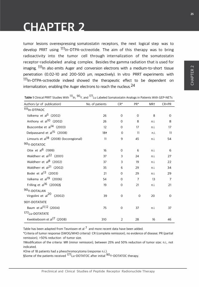
Table 1 Clinical PRRT Studies With 111
In, 90
Y, and 177
Lu Labeled Somatostatin Analogs in Patients With GEP-NETs
Table has been adapted from Teunissen et al 7 and more recent data have been added.
*Criteria of tumor response (SWOG/WHO criteria): CR (complete remission), no evidence of disease; PR (partial
remission), >50% reduction of tumor size.
†Modification of the criteria: MR (minor remission), between 25% and 50% reduction of tumor size; n.i., not
indicated.
‡One of 18 patients had a pheochromocytoma (response n.i.).
§Some of the patients received 177Lu-DOTATOC after initial 90Y-DOTATOC therapy.
Authors (yr of publication) No. of patients CR* PR* MR† CR+PR
111In-DTPAOC
Valkema et al5 (2002) 26 0 0 8 0
Anthony et al10 (2002) 26 0 8 n.i. 8
Buscombe et al14 (2003) 12 0 17 n.i. 17
Delpassand et al15 (2008) 18‡ 0 11 n.i. 11
Limouris et al18 (2008) (locoregional) 11 9 45 n.i. 54
90Y-DOTATOC
Otte et al6 (1999) 16 0 6 n.i. 6
Waldherr et al22 (2001) 37 3 24 n.i. 27
Waldherr et al8 (2002) 37 3 19 n.i. 22
Waldherr et al21 (2002) 35 6 29 n.i. 34
Bodei et al13 (2003) 21 0 29 n.i. 29
Valkema et al19 (2006) 54 0 7 13 7
Frilling et al16 (2006)§ 19 0 21 n.i. 21
90Y-DOTALAN
Virgolini et al20
(2002) 39 0 0 20 0
90Y-DOTATATE
Baum et al11,12 (2004) 75 0 37 n.i. 37
177Lu-DOTATATE
Kwekkeboom et al17 (2008) 310 2 28 16 46
tumor lesions overexpressing somatostatin receptors, the next logical step was to
develop PRRT using 111In-DTPA-octreotide. The aim of this therapy was to bring
radioactivity into the tumor cell through internalization of the somatostatin
receptor-radiolabeled analog complex. Besides the gamma radiation that is used for
imaging, 111In also emits Auger and conversion electrons with a medium-to-short tissue
penetration (0.02-10 and 200-500 μm, respectively). In vitro PRRT experiments with 111In-DTPA-octreotide indeed showed the therapeutic effect to be dependent on
internalization, enabling the Auger electrons to reach the nucleus.24
CH
AP
TE
R 2
Preclinical and Clinical Studies of Peptide Receptor Radionuclide Therapy
CHAPTER 2
26
Clinical PRRT trials with high doses of 111In-DTPA- octreotide showed
promising therapeutic effects (Table 1),5,10,14,15,18 but partial responses
were seldom achieved. Preclinical PRRT experiments with 111In-DTPA-octreotide
in rats bearing small (≤1 cm2) and larger (>8 cm2) sst2-overexpressing
subcutaneous tumors showed significantly more therapeutic effects in animals
bearing small tumors. The lower efficacy in larger tumors was most likely because
of the lack of crossfire of the Auger electrons.25 In this context the use of
radionuclides, such as 90Y and 177Lu that emit β-particles with higher energy and
longer particle ranges exceeding the tumor cell diameter, could have a greater
therapeutic potential. Indeed, preclinical and clinical studies with 90Y and 177Lu
labeled to the somatostatin analogs DOTATOC and DOTATATE showed more
impressive tumor response rates compared with those obtained with 111In-
labeled octreotide.5-11,13-20,22
DOTA-coupled analogs can be labeled with either therapeutic radiometals,
such as 90Y or 177Lu, or with positron or gamma radiation emitters, such as 68Ga for positron emission tomography (PET) and 111In for single-photon
emission computed tomography (SPECT) imaging. Peptide receptor imaging and
PRRT can therefore be performed by using the same peptide, which is named a
theranostic. To select patients who are likely to benefit from PRRT, a scan using
a peptide labeled with a diagnostic radionuclide can be made. Upon a positive
outcome, selected patients can then be treated using the same or a similar peptide
labeled with a therapeutic radionuclide. The development of such theranostics
could greatly advance the development of personalized treatments.
In addition to patient selection for PRRT, other imaging applications of targeted
radiopeptides include localization of primary tumors, detection of metastatic disease
(staging/restaging), dosimetry, prediction of response and radiotoxicity, and monitoring
effects of surgery, PRRT, or chemotherapy.
The radiopeptides for PRRT that have been studied most extensively are 90Y-DOTATOC
and 177Lu-DOTATATE. Published results of 90Y-DOTATOC and 177Lu-DOTATATE derived
from phase I–II trials, however, were not consistent with regard to patient selection,
inclusion criteria, treatment schemes, and dosages. Therefore, an inter-study comparison
is, in fact, not possible. Nevertheless, despite differences in the protocols applied
in various centers, complete, partial, and minor remissions were registered in a
CH
AP
TE
R 2
Preclinical and Clinical Studies of Peptide Receptor Radionuclide Therapy
CHAPTER 2
27
maximum of 46% of patients with GEP-NETs.17 Also, a clear survival benefit was
reported after both 90Y-DOTATOC19 and 177Lu- DOTATATE.17 Patient self-assessed global
health status improved significantly after therapy with 177Lu-DOTATATE PRRT.26,27
From preclinical studies it was concluded that DOTATATE is a more suitable
somatostatin analog than DOTATOC for PRRT, because of the higher affinity of
DOTATATE for the sst2 than that of DOTATOC, leading to a higher tumor uptake and
resulting in a significantly higher tumor radiation dose.28-31
To compare these 2 analogs in patients, Forrer et al32 used 111In as a
surrogate for 90Y and 177Lu and examined whether one of the 111In-labeled
DOTA-peptides had a more favorable biodistribution and tumor-targeting profile
using diagnostic peptide amounts. 111In-DOTATOC showed a higher tumor-
to-kidney absorbed dose ratio in 7 of 9 evaluated tumors. On the basis of these
results, the authors concluded that there were advantages for 111In-DOTATOC
over 111In-DOTATATE, and therefore continued to use 90Y-DOTATOC for PRRT.
By contrast, we compared the 2 analogs under PRRT conditions (with much
higher peptide amounts) in a group of GEP-NET patients.33 Comparing 177Lu-DOTATATE with 177Lu-DOTATOC, the mean residence time ratios of
TATE to TOC-peptide were 2.1 for tumor, 1.5 for spleen, and 1.4 for kidneys. 177Lu-DOTATATE had a longer tumor residence time than 177Lu-DOTATOC.
Therefore, we concluded that DOTATATE is the better peptide for use in PRRT.
The data on PRRT compare favorably with the limited number of alternative treatment
options, such as chemotherapy.9 Therefore, PRRT might become the therapy of first
choice in patients with metastasized or inoperable GEP-NETs. Also, the role of PRRT
in somatostatin receptor-expressing non-GEP-NETs, like metastasized paraganglioma/
pheochromocytoma and nonradioiodine avid differentiated thyroid carcinoma, might
become more important.34-36
Nevertheless, several research questions remain, including the optimal timing of
sequential PRRT treatments. Most PRRT results thus far derive from phase I–II studies and
many studies were carried out in patients with relatively advanced stages of disease,
whereas recent data indicated a possible higher efficacy of PRRT when applied in an
earlier phase of the disease. 17
CH
AP
TE
R 2
Preclinical and Clinical Studies of Peptide Receptor Radionuclide Therapy
CHAPTER 2
28
Promising New Peptide Candidates for PRRT
Somatostatin Analogs
Several new somatostatin analogs have been introduced for therapeutic and
diagnostic purposes, including the agonists DOTA-(1-NaI3)octreotide (DOTANOC) and
DOTA-(BzThi3)octreotide (DOTABOC).37,38 These compounds have a broader somato–
statin receptor affinity profile than DOTATATE and DOTATOC because of a higher affinity
for sst3 and sst5 in addition to their high affinity for sst2. This could increase the
number of tumors that could benefit from PRRT in the future. Promising preliminary PET
imaging results have been obtained in favor of DOTANOC vs DOTATATE.39 Recently,
peptides targeting all the sst receptors (pansomatostatins) were studied by Ginj
et al40 demonstrating high affinity of 90Y-DOTA-cyclo(D-diaminobutyric acid-Arg–Phe–
Phe-D-Trp–Lys–Thr–Phe) (90Y-KE88) for all 5 sst receptors. Surprisingly, sst2-dependent
internalization was demonstrated to be very low. Sst3-expressing tumors had high and
persistent uptake in mice biodistribution studies. The further development of these
pansomatostatins could improve the therapeutic potential of sst-targeted PRRT in
future.
So far, the studies in patients have been performed with somatostatin
receptor agonists because agonists are internalized in (tumor) cells after which
the radioactivity is retained in the cell. Most antagonists do not internalize41,42
and block the downstream intracellular cascade.
However, Ginj et al43 recently demonstrated in a preclinical study almost
twice as high tumor retention of a radiolabeled sst2 antagonist (111In-DOTA-
sst2-ANT) compared with the agonist 111In-DTPATATE, despite a somewhat
lower receptor affinity of the antagonist for the sst2. Tumor to kidney ratios 4
hours postinjection of the sst2 antagonist and sst2 agonist were 2.7 and 1.4,
respectively. The higher tumor uptake was thought to be caused by binding
of the antagonist to a larger variety of receptor conformations. This preclinical
finding of a superior tumor targeting by noninternalizing somatostatin receptor
antagonists is revolutionizing the current paradigm of the internalization
of the receptor-ligand complex as the basis for PRRT. If these findings can be
translated to the patient situation, antagonists can be applied to increase
CH
AP
TE
R 2
Preclinical and Clinical Studies of Peptide Receptor Radionuclide Therapy
CHAPTER 2
29
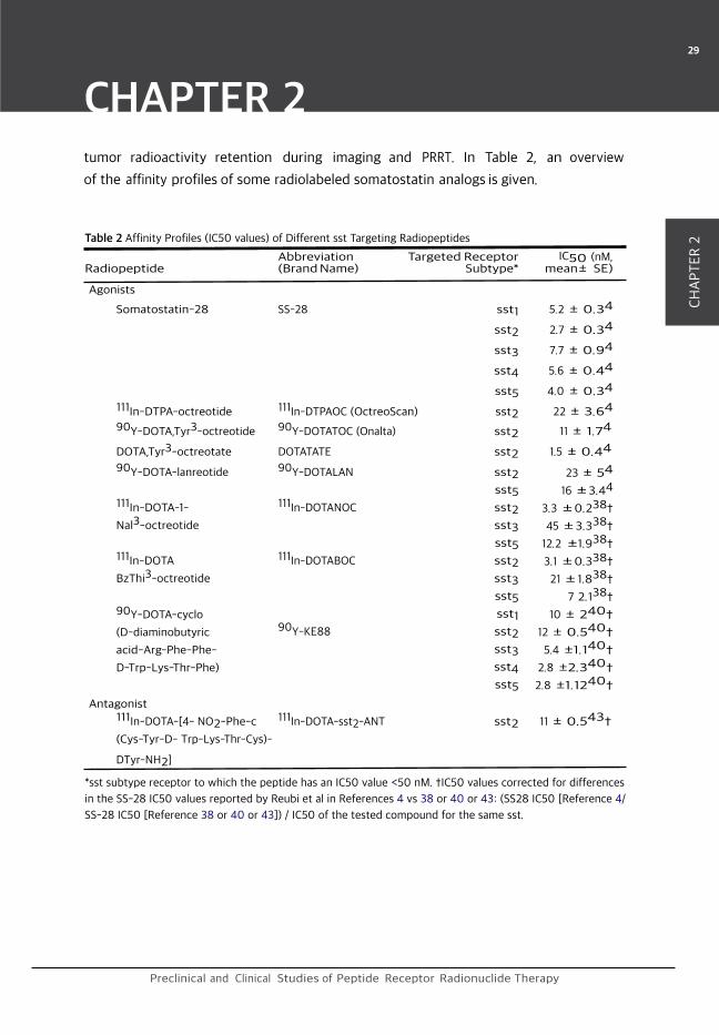
Abbreviation Targeted Receptor IC50 (nM, Radiopeptide (Brand Name) Subtype* mean ± SE)
Agonists
Somatostatin-28 SS-28 sst1 5.2 ± 0.34
sst2 2.7 ± 0.34
sst3 7.7 ± 0.94
sst4 5.6 ± 0.44
sst5 4.0 ± 0.34
111In-DTPA-octreotide 111In-DTPAOC (OctreoScan) sst2 22 ± 3.64
90Y-DOTA,Tyr3-octreotide 90Y-DOTATOC (Onalta) sst2 11 ± 1.74
DOTA,Tyr3-octreotate DOTATATE sst2 1.5 ± 0.44
90Y-DOTA-lanreotide 90Y-DOTALAN sst2 23 ± 54
sst5 16 ± 3.44
111In-DOTA-1- 111In-DOTANOC sst2 3.3 ± 0.238†
Nal3-octreotide sst3 45 ± 3.338†
sst5 12.2 ±1.938†111In-DOTA 111In-DOTABOC sst2 3.1 ± 0.338†
BzThi3-octreotide sst3 21 ± 1.838†
sst5 7 2.138†90Y-DOTA-cyclo sst1 10 ± 240†
(D-diaminobutyric 90Y-KE88 sst2 12 ± 0.540†
acid-Arg-Phe-Phe- sst3 5.4 ±1.140†
D-Trp-Lys-Thr-Phe) sst4 2.8 ±2.340†
sst5 2.8 ±1.1240†
Antagonist 111In-DOTA-[4- NO2-Phe-c 111In-DOTA-sst2-ANT sst2 11 ± 0.543†
(Cys-Tyr-D- Trp-Lys-Thr-Cys)-
DTyr-NH2]
Table 2 Affinity Profiles (IC50 values) of Different sst Targeting Radiopeptides
*sst subtype receptor to which the peptide has an IC50 value <50 nM. †IC50 values corrected for differences
in the SS-28 IC50 values reported by Reubi et al in References 4 vs 38 or 40 or 43: (SS28 IC50 [Reference 4/
SS-28 IC50 [Reference 38 or 40 or 43]) / IC50 of the tested compound for the same sst.
tumor radioactivity retention during imaging and PRRT. In Table 2, an overview
of the affinity profiles of some radiolabeled somatostatin analogs is given.
CH
AP
TE
R 2
Preclinical and Clinical Studies of Peptide Receptor Radionuclide Therapy
CHAPTER 2
30
Glucagon-like Peptide 1 Receptor Targeting Peptides
The GLP-1r is highly expressed in the majority of insulinomas,44 opening opportunities
for imaging and radionuclide therapy by radiolabeled exendin analogs that bind with
high affinity to GLP-1r. Also, pheochromocytomas45 and gastrinomas2 have been
shown to demonstrate elevated GLP-1r expression. Preclinical experiments in mice
bearing sub- cutaneous insulinomas showed specific uptake and internalization of 123I-labeled GLP-1 amide and the GLP-1r analog 123I-exendin-3,46 enabling tumor
scintigraphy. Preclinical experiments with the further optimized 111In-DTPA- conjugated
exendin analogs 111In-DTPA-Lys40-exendin-447 and (Lys40[Ahx-DTPA-111In]NH2)
exendin-448 showed high tumor to background ratios in animal tumor models.
PRRT with (Lys40[Ahx-DTPA-111In]NH2)exendin-4 showed to be feasible in repressing
insulinoma growth in mice.49 However, in these studies high radiopeptide renal uptake
resulted in long-term kidney toxicity after PRRT. The fact that coinfusion of albumin
fragments, lysine and/or gelofusin, reduces renal uptake of both targeting peptides
in animal experiments50 opens the way for PRRT studies with (Lys40[Ahx- DTPA-111In]NH2)exendin-4 in the clinical setting. Wild et al51 showed excellent antitumor
effects in mice using this exendin analog in a lower dose and combined with the
oral angiogenesis inhibitor PTK. Recently, a prospective clinical pilot study evaluating 111In-DOTA-exendin-4 in SPECT/CT (computed tomography) localization of insulinomas
showed impressive results demonstrating the high potential of targeting the GLP-1r 52
Gastrin-Releasing Peptide Receptor Targeting Peptides
On both prostate and breast tumors, which are among the major causes
of death worldwide, GRPr overexpression has been shown.53 Targeting of
the GRPr with radiolabeled bombesin analogs has been studied in the past
decade with many combinations of radionuclides and analogs.44,54-59 The
most promising bombesin analog studied in the preclinical setting is 99mTc-
Demobesin1, which demonstrated the highest absolute tumor uptake in animals
in combination with high stability in vivo and rapid clearance from the (GRPr-
positive) pancreas.44,60 To date, few radiolabeled bombesin analogs have been
tested in the clinic61 and only 1 analog was used for PRRT.62 Van de Wiele et al55
published imaging data on 99mTc-RP527 in 4 androgen-independent prostate
cancer (PC) patients with metastatic bone lesions. Another GRP analog, 99mTc-
CH
AP
TE
R 2
Preclinical and Clinical Studies of Peptide Receptor Radionuclide Therapy
CHAPTER 2
31
(Leu13)bombesin, was described in different studies.63-66 Scopinaro et al66
evaluated this analog in 8 PC patients and reported all 8 primary PCs to be
visualized by SPECT, whereas 2 patients with benign adenomas did not show
uptake. De Vincentis et al65 reported SPECT-detected PC in all 12 patients
with androgen-dependent PC, and locoregional lymph node visualization in 4
patients.
Only a very few PET studies have been reported for visualization and quantification
of GRPr expression in PC patients. A clinical study by Hofmann et al67 using 68Ga-DOTABOM for PET imaging delineated malignant PC lesions in 13 of 15 patients.
In a phase I PRRT study in hormone refractory PC patients using the 177Lu-
labeled bombesin agonist AMBA, SPECT imaging revealed lesions in 5 of 7
patients.62 Froberg et al68 reported high initial uptake of both the radiolabeled
agonist MP2248 and the antagonist Demobesin-1 in the pancreatic region of
4 PC patients, but retention of radioactivity in the pancreas after injection of
MP2248 was much longer than after Demobesin-1 injection. Because of this
slower decrease of pancreatic radioactivity after radio-agonist injection, a higher
radiation dose will be given to this organ during PRRT. In addition, side effects
can be expected at higher peptide amounts of the agonists, indicating the clear
advantage of antagonists over agonists for PRRT using bombesin analogs.
CCK2 Receptor Targeting
Medullary thyroid cancer (MTC) has a rather low sst2 expression compared with other
neuroendocrine tumors, and sst2 expression is even absent in clinically aggressive forms
of the disease.69,70 The CCK-2/ or gastrin receptor has been shown to be overexpressed
in more than 90% of MTCs, and in a high percentage of small-cell lung cancers, stromal
ovarian cancers, astrocytomas, and several other tumor types.71 111In- DTPA-CCK8 was
able to visualize advanced metastatic MTC in patients.72 Laverman et al73 reported
promising levels of tumor uptake and low levels of kidney uptake of the sulfated 111In-DOTA-sCCK8 in tumor-bearing mice, an ideal situation for radionuclide imaging
and therapy. Beside CCK analogs, radiolabeled analogs of gastrin and minigastrin also
showed suitable targeting affinity for the CCK2-receptor. Preclinical studies by Behr
et al74 showed promising tumor uptake and therapeutic efficacy using 131I-labeled
CH
AP
TE
R 2
Preclinical and Clinical Studies of Peptide Receptor Radionuclide Therapy
CHAPTER 2
32
gastrin-1. Tumor targeting of this compound was also shown in a metastatic MTC patient.
More recently, von Guggenberg et al75 were able to image tumors in mice using the
cyclized minigastrin analog 99mTc-EDDA-HYNIC-cyclo-MG1. In patients, 111In-DTPA-
minigastrin could visualize most tumor sites in MTC pa- tients.74,76 Nock et al77
synthesized 99mTc-labeled N4-derivatized analogs of minigastrin. N°-1, Gly0, (D)Glu1-
minigastrin (Demogastrin 2) was selected as the most promising after evaluation in
preclinical studies. In a clinical study, the qual- ity of Demogastrin 2 could be confirmed
by clearly delineating tumor deposits in metastatic MTC patients.78 Gotthardt et al79
compared the results of CCK2 gastrin receptor scintigra- phy (GRS) in metastatic MTC
patients, using 111In-(D)Glu1- minigastrin, with somatostatin receptor scintigraphy,
CT, and 18F-fluorodeoxyglucose PET. The combination of GRS with CT was the most
effective in detecting metastatic MTC. The authors concluded that GRS may become
the scintigraphic imaging modality of choice in MTC patients. Clinical CCK2-targeted PRRT
studies have not started yet, but its future is promising.
αvβ3 Integrin Targeting
The αvβ3 integrin is upregulated and accessible on proliferating endothelial cells,
whereas it is not on quiescent endothelial cells. For their growth, solid tumors depend
on angiogenesis, a process requiring endothelial cell proliferation. This makes αvβ3
integrins an interesting target for receptor-mediated tumor imaging and therapy for
a large number of different tumors. The arginine-glycine-aspartic acid (RGD) peptide
sequence was found to be responsible for extracellular matrix proteins binding to
the αvβ3 receptor.80 Cyclic RGD analogs conjugated to DOTA and DTPA have been
developed enabling SPECT and PET imaging and PRRT when labeled with 111In, 68Ga, 64Cu, 90Y, and 177Lu.81-83 18F-labeled cyclic RGD analogs were also developed
for PET imaging.82,84,85 18F-galacto-RGD could effectively demonstrate the level of
αvβ3 expression in human beings.86,87 Dijkgraaf et al89 synthesized DOTA-linked
mono- and multimeric RGD pep- tides88 as well as multimeric RGD peptides as dendrimers:
macromolecules consisting of multiple perfectly branched monomers. The tetrameric RGD
peptide and the tetrameric RGD dendrimer had the highest affinity and tumor uptake.
However, kidney retention was also increased. In another study, Dijkgraaf et al90 found
in an intraperitoneal (i.p.) tumor model that i.p. administration resulted in better
tumor-to- kidney ratios and a significant tumor growth inhibition during PRRT compared
with intravenous injection, showing the therapeutic potential of RGD peptides.
CH
AP
TE
R 2
Preclinical and Clinical Studies of Peptide Receptor Radionuclide Therapy
CHAPTER 2
33
Epidermal Growth Factor Receptor Targeting
Overexpression of the epidermal growth factor receptor (EGFr) has been found in a
variety of cancers like breast, bladder, gastric, and non–small-cell lung cancer, generally
indicating a more aggressive behavior compared with normal or low expression.91 A
preclinical study by Chen et al92 showed an antitumor effect of PRRT with 111In-
DTPA-EGF on EGFr-overexpressing breast carcinoma xenografts in mice. In a clinical
study in 9 squamous carcinoma patients, 131I-labeled EGF was able to visualize all
tumors. A drawback was formed by the frequently observed adverse effects probably
caused by the agonistic effect of EGF (nausea, vomiting, diarrhea, hypotension, fever,
and chills), recorded during the dose escalating studies.93 Radiolabeled antibodies that
target against the EGFr (family) could be an interesting alternative for imaging or PRRT.
However, a known disadvantage is the size of the antibodies (about 150 kDa), which
causes increased systemic retention and poor tissue penetration. To overcome this
problem, Tolmachev et al94 applied EGFr- mediated uptake and imaging using 111In-
labeled affibody molecules (6-7 kDa) in EGFr-expressing A431 xenografts. However,
the high uptake in healthy tissue expressing the EGFr, for example, the liver, remains a
problem in radionuclide imaging and PRRT targeting the EGFr.
Strategies to Improve PRRT
Tumor Mass and Choice of Radionuclide
Tumor radiation dose does not only depend on the administered dose of
radioactivity and the uptake vs time, but also on the tumor mass. Smaller masses
have higher chances of mass reduction, as confirmed by clinical data showing
that tumor remission was among other things positively correlated with a limited
number of liver metastases, whereas disease progression was significantly
more frequent in patients with a low performance status and a high tumor
load.95 Considering the use of 2 different radionuclides for PRRT using the 2
most commonly used radiopeptides, 90Y-DOTATOC and 177Lu- DOTATATE,
in a mathematical model showed that 177Lu would perform better in small
tumors (optimal diameter of 2 mm), whereas 90Y potentially had better tumor
responses in larger tumors (optimal diameter 34 mm).96 Very small tumors
will not absorb all the energy deposited in the tumor by 90Y, whereas larger

CH
AP
TE
R 2
Preclinical and Clinical Studies of Peptide Receptor Radionuclide Therapy
CHAPTER 2
34
tumors will suffer from the possible lack of homogeneous distribution of 177Lu
throughout the tumor. In addition, the longer physical half-life of 177Lu requires
a longer exposure to deliver the same radiation dose to a tumor in comparison
with the use of 90Y. Therefore, we hypothesized that a combination therapy
with 90Y and 177Lu labeled peptides, either given simultaneously or in separate
sessions, might overcome the difficulties of treatment of lesions with different
sizes in a single patient.97 Preclinical studies indeed showed 177Lu-DOTATATE
to be more effective in small tumors (<1 cm2), whereas 90Y-DOTATOC was
more effective in larger tumors (>1 cm2).98,99 A combination of 177Lu-
DOTATATE and 90Y-DOTATOC in rats bearing both smaller (<0.5 cm2) and large
(7-9 cm2) tumors led to a better survival compared with that after single dose of 177Lu-DOTATATE or 90Y-DOTATOC.97
Kunikowska et al100 showed higher overall survival in 36 patients with diffuse
neuroendocrine tumors treated with a combined regimen of 177Lu/90Y-DOTATATE
(mixed dose 1:1) compared non-randomly to 177Lu-DOTATATE alone. However, in
Figure 1 Anterior spot view images of the upper
abdomen, 24 hours after injection of 111In-
DTPA-octreotide (230 MBq/10 μg) in the same
patient with a 2-week interval; (A) i.v. injection,
(B) locoregional injection. catheter was placed
in the right hepatic artery. The encircled
metastasis, situated in the left part of the liver,
therefore did not receive 111In-DTPA-octreotide
locoregionally.

CH
AP
TE
R 2
Preclinical and Clinical Studies of Peptide Receptor Radionuclide Therapy
CHAPTER 2
35
the clinical setting randomized controlled trials comparing PRRT with 90Y, 177Lu or
combined regimens are still lacking. Of high interest for PRRT is also the application of
alpha emitters, as described in the article of Norenberg et al in this issue.
Locoregional Administration
To increase the therapeutic window of PRRT with radiolabeled somatostatin
analogs, several groups have studied the possibility of locoregional
administration for treatment of GEP-NET liver metastasis. McStay et al101
prospectively evaluated the safety and effectiveness of hepatic arterial injection
of the somatostatin analog 90Y-DOTA-lanreotide with or without embolization
as a treatment for patients with pro- gressive, large-volume GEP-NET liver
metastasis. This study showed hepatic arterial injection of 90Y-DOTA-lanreotide,
with or without embolization, to be safe and as effective as, or even more effective
than, the systemic administration in the Mauritius trial.20 The added value
of locoregional compared with systemic administration could not be shown,
probably because a limited number of patients23 were included, locoregional
administration was performed with or without embolization, and a (systemically
administered) control group was lacking. Limouris et al18 reported high tumor
to liver ratios after hepatic arterial infusion of 111In-DTPA-octreotide in patients
with inoperable liver-metastasized GEP-NETs. Also, a (systemically administered)
control group to compare intra- hepatic tumor uptake and treatment
effectiveness was lacking in this study. Seventeen patients underwent selective
hepatic artery catheterization (180 in total) up to 15 times with no severe side
effects. In another clinical study, 17 patients with primary or metastasized
NETs were treated by locoregional and systemic administration of 131I-MiBG.
Locoregional administration resulted in a 69% mean increase in tumor to
whole body ratio.102 These studies all show that locoregional administration
of radionuclide therapy can be regarded to be safe. However, in both studies
targeting the sst2 with radiolabeled somatostatin analogs, an intravenously
administered control group was lacking.
We recently demonstrated doubling of the 111In-DTPA- octreotide uptake in a liver
metastasis rat model after locoregional injection through the hepatic artery compared
CH
AP
TE
R 2
Preclinical and Clinical Studies of Peptide Receptor Radionuclide Therapy
CHAPTER 2
36
with systemic injection.103 Direct intratumoral injections of 111In-DTPA-octreotide
resulted in even >10 times higher tumor uptake compared with systemic injection in
preclinical mice experiments.104
Currently, we are performing a clinical pilot study to investigate GEP-NET liver
metastasis uptake of 111In-DTPA-octreotide after systemic and hepatic artery injection
in the same patient with a 2-week interval. Preliminary results from this study are
promising. In the first patient locoregional administration resulted in 2.4 times higher
tumor uptake in GEP-NET liver metastasis.103 In another patient, besides higher tumor
uptake, more lesions could be visualized (Fig. 1). In this patient, the catheter was placed
in the right hepatic artery resulting in locoregional administration in the right part of
the liver. One metastasis, situated in the left part of the liver (encircled in Fig. 1B),
therefore did not receive 111In-DTPA-octreotide locoregionally. Recently, Beauregard et
al105 described up to 4 times higher uptake in GEP-NET liver metastasis after hepatic
artery infusion of 177Lu-DOTATATE in patients. Further experiments will have to be
performed to be able to conclude on the benefit in terms of tumor uptake and therapy
efficacy after locoregional vs systemic administration in PRRT.
Combination Treatment of PRRT
In external radiation therapy, radiosensitizing agents are commonly used.
In several preclinical and clinical studies, combination of these agents with
PRRT have been evaluated. In preclinical studies with tumor-bearing mice
combinations of 177Lu-DOTATOC with doxorubicin or cisplatin during a
4-week period, respectively, showed to be 14% or 23% more effective than single
treatment.106 In 21 patients, the radio- sensitizing agent 5-fluorouracil (5-FU)
was combined with high-dose 111In-labeled octreotide. This PRRT combined
with 5-FU showed to be safe and therapeutic response rates obtained
were at least comparable to those reported for 111In-DTPA-octreotide
treatment alone.107 Two years ago our group started a multicenter,
2-armed, random- ized, prospective study investigating the combination of
CH
AP
TE
R 2
Preclinical and Clinical Studies of Peptide Receptor Radionuclide Therapy
CHAPTER 2
37
177Lu-DOTATATE PRRT with the oral prodrug of 5-FU, capecitabine, vs 177Lu-
DOTATATE PRRT alone, after evaluation of the feasibility of this combination treatment
in patients in a phase II trial.108 As mentioned earlier, Wild et al51 showed excellent
antitumor effects of (Lys40(Ahx-DTPA-111In)NH2)exendin-4 in a low dose combined
with the oral angiogenesis inhibitor PTK.
Kidney Protection
In PRRT with somatostatin analogs, the kidney is one of the dose-limiting organs as
was shown by renal toxicity after PRRT in clinical studies.109-111 Reducing the uptake
of radiopeptides in the kidney would therefore strongly contribute to enlarging the
therapeutic window of PRRT. In rats, radiolabeled somatostatin analogs were shown
to be filtered and reabsorbed in the proximal tubules of the kidneys.112 In the human
kidney, radioactivity was also found to be mostly located in the cortex.113 In preclinical
animal studies, megalin receptor-negative mice had 70%-85% less renal uptake com-
pared with wildtype mice. This showed that the megalin/cubulin system is essential
for a major part of the uptake of radiolabeled somatostatin analogs in the proximal
tubules.114
Sst2-specific uptake was shown to be responsible for 18% of renal uptake in human
beings.115 In our institution, renal uptake during PRRT in patients is reduced by a 4 hour
coadministration of lysine and arginine together with 177Lu- DOTATATE.17 In a preclinical
study in rats, we demonstrated that orally administered lysine also reduced renal uptake
by 40% comparable to that of i.v.-administered lysine.116 Further protection of the
kidneys during PRRT with different peptides can be achieved using gelofusin and
albumin-derived peptides.50 The cytoprotective drug amifostine also protected the
kidneys during PRRT with 177Lu-DOTATATE given in high doses to rats.117
Conclusions
Several different somatostatin receptor-binding analogs have now been described that
proved to be an excellent tool for PRRT of patients with GEP-NETs. PPRT showed few
serious adverse effects and important tumor responses, long progression-free survival
rates, and considerable improvement in quality of life. This field is rapidly growing; new
CH
AP
TE
R 2
Preclinical and Clinical Studies of Peptide Receptor Radionuclide Therapy
CHAPTER 2
38
agonist and antagonist peptides have been described that are or can soon be tested in
clinical trials.
As tumors can also overexpress other receptors that may bind peptide
analogs of naturally occurring hormones, for example, CCK-2, bombesin,
neuropeptide Y, or vasoactive intestinal peptide receptors, even simultaneously,
radioanalogs of these peptides will allow (multi)receptor PRRT in the future.
Future perspectives include studies exploring the effects of the combined use of PRRT
with other drugs, such as radiosensitizing chemotherapeutic agents and the effect of
locoregional administration of peptides, intra-arterially or even intratumorally injected,
on PRRT efficacy.
References
1. Schottelius M, Wester HJ: Molecular imaging targeting peptide receptors. Methods 48:161-177, 20092. Reubi JC: Peptide receptors as molecular targets for cancer diagnosis and therapy. Endocr Rev 24:389-
427, 20033. Krenning EP, Kwekkeboom DJ, Bakker WH, et al: Somatostatin receptor scintigraphy with [111In-DTPA-
D-Phe1]- and [123I-Tyr3]-octreotide: The Rotterdam experience with more than 1000 patients. Eur J Nucl Med 20:716-731, 1993
4. Reubi JC, Schar JC, Waser B, et al: Affinity profiles for human somatostatin receptor subtypes SST1-SST5 of somatostatin radiotracers selected for scintigraphic and radiotherapeutic use. Eur J Nucl Med 27:273-282, 2000
5. Valkema R, De Jong M, Bakker WH, et al: Phase I study of peptide receptor radionuclide therapy with [In-DTPA]octreotide: The Rotterdam experience. Semin Nucl Med 32:110-122, 2002
6. Otte A, Mueller-Brand J, Dellas S, et al: Yttrium-90-labelled somatostatin-analogue for cancer treatment. Lancet 351:417-418, 1998
7. Teunissen JJ, Kwekkeboom DJ, de Jong M, et al: Endocrine tumours of the gastrointestinal tract. Peptide receptor radionuclide therapy. Best Pract Res Clin Gastroenterol 19:595-616, 2005
8. Waldherr C, Pless M, Maecke HR, et al: Tumor response and clinical benefit in neuroendocrine tumors after 7.4 GBq (90)Y-DOTATOC. J Nucl Med 43:610-616, 2002
9. van Essen M, Krenning EP, Kam BL, et al: Peptide-receptor radionuclide therapy for endocrine tumors. Nat Rev Endocrinol 5:382-393, 2009.
10. Anthony LB, Woltering EA, Espenan GD, et al: Indium-111-pentetreotide prolongs survival in gastroenteropancreatic malignancies. Semin Nucl Med 32:123-132, 2002
11. Baum RP: Intravenous and intra-arterial peptide receptor radionuclide therapy (PRRT) using Y-90-DOTA-Tyr3-octreotate (Y-90- DOTA-TATE) in patients with metastatic neuroendocrine tumors. Eur J Nucl Med 31:S238, 2004
12. Baum RP: Peptidreceptorvermittelte Radiotherapie (PRRT) neuroendocriner tumoren klinischen Indikationen und erfahrung mit 90Yttrium-markierten Somatostatinanaloga der. Onkologe 10:1098-1110, 2004
13. Bodei L, Cremonesi M, Zoboli S, et al: Receptor-mediated radionuclide therapy with 90Y-DOTATOC in association with amino acid infusion: A phase I study. Eur J Nucl Med Mol Imaging 30:207-216, 2003
14. Buscombe JR, Caplin ME, Hilson AJ: Long-term efficacy of high-activity 111in-pentetreotide therapy in
CH
AP
TE
R 2
Preclinical and Clinical Studies of Peptide Receptor Radionuclide Therapy
CHAPTER 2
39
patients with disseminated neuroendocrine tumors. J Nucl Med 44:1-6, 200315. Delpassand ES, Sims-Mourtada J, Saso H, et al: Safety and efficacy of radionuclide therapy with high-
activity In-111 pentetreotide in patients with progressive neuroendocrine tumors. Cancer Biother Ra- diopharm 23:292-300, 2008
16. Frilling A, Weber F, Saner F, et al: Treatment with (90)Y- and (177)Lu-DOTATOC in patients with metastatic neuroendocrine tumors. Surgery 140:968-976, 2006; discussion 976-977
17. Kwekkeboom DJ, de Herder WW, Kam BL, et al: Treatment with the radiolabeled somatostatin analog [177Lu-DOTA0, Tyr3]octreotate: Toxicity, efficacy, and survival. J Clin Oncol 26:2124-2130, 2008
18. Limouris GS, Chatziioannou A, Kontogeorgakos D, et al: Selective hepatic arterial infusion of In-111-DTPA-Phe1-octreotide in neuroendocrine liver metastases. Eur J Nucl Med Mol Imaging 35:1827-1837,2008
19. Valkema R, Pauwels S, Kvols LK, et al: Survival and response after peptide receptor radionuclide therapy with [90Y-DOTA0,Tyr3]octreotide in patients with advanced gastroenteropancreatic neuroendocrine tumors. Semin Nucl Med 36:147-156, 2006
20. Virgolini I, Britton K, Buscombe J, et al: In- and Y-DOTA-lanreotide: Results and implications of the MAURITIUS trial. Semin Nucl Med 32:148-155, 2002
21. Waldherr C, Schumacher T, Maecke HR, et al: Does tumor response depend on the number of treatment sessions at constant injected dose using 90yttrium-DOTATOC in neuroendocrine tumors? Eur J Nucl Med 29:S100, 2002
22. Waldherr C, Pless M, Maecke HR, et al: The clinical value of [90Y- DOTA]-D-Phe1-Tyr3-octreotide (90Y-DOTATOC) in the treatment of neuroendocrine tumours: A clinical phase II study. Ann Oncol 12:941-945, 2001
23. Duncan JR, Stephenson MT, Wu HP, et al: Indium-111-diethylenetriaminepentaacetic acid-octreotide is delivered in vivo to pancreatic, tumor cell, renal, and hepatocyte lysosomes. Cancer Res 57:659-671, 1997
24. Capello A, Krenning EP, Breeman WA, et al: Peptide receptor radionuclide therapy in vitro using [111In-DTPA0]octreotide. J Nucl Med 44:98-104, 2003
25. Capello A, Krenning E, Bernard B, et al: 111In-labelled somatostatin analogues in a rat tumour model: Somatostatin receptor status and effects of peptide receptor radionuclide therapy. Eur J Nucl Med Mol Imaging 32:1288-1295, 2005
26. Khan S, Teunissen JJM, van Essen M, et al: Quality of life in 265 patients with gastroenteropancreatic tumours or bronchial carcinoids treated with [177Lu-DOTA0, Tyr3]octreotate. Eur J Nucl Med Mol Imaging 36:S219, 2009
27. Teunissen JJ, Kwekkeboom DJ, Krenning EP: Quality of life in patients with gastroenteropancreatic tumors treated with [177Lu- DOTA0,Tyr3]octreotate. J Clin Oncol 22:2724-2729, 2004
28. Capello A, Krenning EP, Breeman WA, et al: Tyr3-octreotide and Tyr3-octreotate radiolabeled with 177Lu or 90Y: Peptide receptor radionuclide therapy results in vitro. Cancer Biother Radiopharm 18: 761-768, 2003
29. de Jong M, Breeman WA, Bakker WH, et al: Comparison of (111)In- labeled somatostatin analogues for tumor scintigraphy and radionuclide therapy. Cancer Res 58:437-441, 1998
30. de Jong M, Breeman WA, Bernard BF, et al: Tumor response after [(90)Y-DOTA(0),Tyr(3)]octreotide radionuclide therapy in a transplantable rat tumor model is dependent on tumor size. J Nucl Med42:1841-1846, 2001
31. Lewis JS, Lewis MR, Srinivasan A, et al: Comparison of four 64Cu-labeled somatostatin analogues in vitro and in a tumor-bearing rat model: Evaluation of new derivatives for positron emission tomogra- phy imaging and targeted radiotherapy. J Med Chem 42:1341-1347,1999
32. Forrer F, Uusijarvi H, Waldherr C, et al: A comparison of (111)In-DOTATOC and (111)In-DOTATATE: Biodistribution and dosimetry in the same patients with metastatic neuroendocrine tumours. Eur J Nucl Med Mol Imaging 31:1257-1262, 2004
33. Esser JP, Krenning EP, Teunissen JJ, et al: Comparison of [(177)Lu- DOTA(0),Tyr(3)]octreotate and [(177)Lu-DOTA(0),Tyr(3)]octreotide: Which peptide is preferable for PRRT? Eur J Nucl Med Mol Imaging 33:1346-1351, 2006
34. van Essen M, Krenning EP, Bakker WH, et al: Peptide receptor radionuclide therapy with 177Lu-octreotate in patients with foregut carcinoid tumours of bronchial, gastric and thymic origin. Eur J Nucl Med Mol Imaging 34:1219-1227, 2007
CH
AP
TE
R 2
Preclinical and Clinical Studies of Peptide Receptor Radionuclide Therapy
CHAPTER 2
40
35. van Essen M, Krenning EP, Kooij PP, et al: Effects of therapy with [177Lu-DOTA0, Tyr3]octreotate in patients with paraganglioma, meningioma, small cell lung carcinoma, and melanoma. J Nucl Med 47:1599-1606, 2006
36. Forrer F, Riedweg I, Maecke HR, et al: Radiolabeled DOTATOC in patients with advanced paraganglioma and pheochromocytoma. Q J Nucl Med Mol Imaging 52:334-340, 200837. Wild D, Schmitt JS, Ginj M, et al: DOTA-NOC, a high-affinity ligand of somatostatin receptor subtypes 2, 3 and 5 for labelling with various radiometals. Eur J Nucl Med Mol Imaging 30:1338-1347, 2003
38. Ginj M, Schmitt JS, Chen J, et al: Design, synthesis, and biological evaluation of somatostatin-based radiopeptides. Chem Biol 13:1081-1090, 2006
39. Wild D, Bomanji JB, Reubi JC, et al: Comparison of 68Ga-DOTANOC and 68Ga-DOTATATE PET/CT in the detection of GEP NETs. Eur J Nucl Med Mol Imaging 36:S201, 2009
40. Ginj M, Zhang H, Eisenwiener KP, et al: New pansomatostatin ligands and their chelated versions: Affinity profile, agonist activity, internalization, and tumor targeting. Clin Cancer Res 14:2019-2027, 2008
41. Liu Q, Cescato R, Dewi DA, et al: Receptor signaling and endocytosis are differentially regulated by somatostatin analogs. Mol Pharmacol 68:90-101, 2005
42. Wynn PC, Suarez-Quian CA, Childs GV, et al: Pituitary binding and internalization of radioiodinated gonadotropin-releasing hormone agonist and antagonist ligands in vitro and in vivo. Endocrinology 119: 1852-1863, 1986
43. Ginj M, Zhang H, Waser B, et al: Radiolabeled somatostatin receptor antagonists are preferable to agonists for in vivo peptide receptor targeting of tumors. Proc Natl Acad Sci U S A 103:16436-16441, 2006
44. Nock B, Nikolopoulou A, Chiotellis E, et al: [99mTc]Demobesin 1, a novel potent bombesin analogue for GRP receptor-targeted tumour imaging. Eur J Nucl Med Mol Imaging 30:247-258, 2003
45. Korner M, Stockli M, Waser B, et al: GLP-1 receptor expression in human tumors and human normal tissues: Potential for in vivo targeting. J Nucl Med 48:736-743, 2007
46. Gotthardt M, Fischer M, Naeher I, et al: Use of the incretin hormone glucagon-like peptide-1 (GLP-1) for the detection of insulinomas: Initial experimental results. Eur J Nucl Med Mol Imaging 29:597-606, 2002
47. Gotthardt M, Lalyko G, van Eerd-Vismale J, et al: A new technique for in vivo imaging of specific GLP-1 binding sites: First results in small rodents. Regul Pept 137:162-167, 2006
48. Wild D, Behe M, Wicki A, et al: [Lys40(Ahx-DTPA-111In)NH2]exendin-4, a very promising ligand for glucagon-like peptide-1 (GLP-1) receptor targeting. J Nucl Med 47:2025-2033, 2006
49. Wicki A, Wild D, Storch D, et al: [Lys40(Ahx-DTPA-111In)NH2]-exendin-4 is a highly efficient radiotherapeutic for glucagon-like peptide-1 receptor-targeted therapy for insulinoma. Clin Cancer Res 13: 3696-3705, 2007
50. Rolleman EJ, Melis M, Valkema R, et al: Kidney protection during peptide receptor radionuclide therapy with somatostatin analogues. Eur J Nucl Med Mol Imaging (in press)
51. Wild D, Wicki A, Christofori G, et al: Combination therapy with [(Lys40(Ahx-[111In-DTPA])]-exendin-4 and VEGF-receptor tyrosine kinase inhibitor PTK in a glucagon-like-peptide-1 receptor-positive transgenic mouse tumor model. J Nucl Med 48:83p, 2007
52. Christ E, Wild D, Forrer F, et al: Glucagon-like peptide-1 receptor imaging for localization of insulinomas. J Clin Endocrinol Metab 94:4398-4405, 2009
53. Reubi JC, Wenger S, Schmuckli-Maurer J, et al: Bombesin receptor subtypes in human cancers: Detection with the universal radioligand (125)I-[D-TYR(6), beta-ALA(11), PHE(13), NLE(14)] bombesin(6-14). Clin Cancer Res 8:1139-1146, 2002
54. Nock BA, Nikolopoulou A, Galanis A, et al: Potent bombesin-like peptides for GRP-receptor targeting of tumors with 99mTc: A preclinical study. J Med Chem 48:100-110, 2005
55. Van de Wiele C, Dumont F, Vanden Broecke R, et al: Technetium-99m RP527, a GRP analogue for visualisation of GRP receptor-expressing malignancies: A feasibility study. Eur J Nucl Med 27:1694-1699, 2000
56. Hoffman TJ, Gali H, Smith CJ, et al: Novel series of 111In-labeled bombesin analogs as potential radiopharmaceuticals for specific targeting of gastrin-releasing peptide receptors expressed on human prostate cancer cells. J Nucl Med 44:823-831, 2003
57. Breeman WA, de Jong M, Erion JL, et al: Preclinical comparison of (111)In-labeled DTPA- or DOTA-

CH
AP
TE
R 2
Preclinical and Clinical Studies of Peptide Receptor Radionuclide Therapy
CHAPTER 2
41
bombesin analogs for receptor-targeted scintigraphy and radionuclide therapy. J Nucl Med 43:1650-1656, 2002
58. Ferro-Flores G, Arteaga de Murphy C, Rodriguez-Cortes J, et al: Preparation and evaluation of 99mTc-EDDA/HYNIC-[Lys 3]-bombesin for imaging gastrin-releasing peptide receptor-positive tumours. Nucl Med Commun 27:371-376, 2006
59. Garcia Garayoa E, Ruegg D, Blauenstein P, et al: Chemical and biological characterization of new Re(CO)3/[99mTc](CO)3 bombesin analogues. Nucl Med Biol 34:17-28, 2007
60. Schroeder R, Muller C, Reneman S, et al: Selection of the best bombesin analogue for use in phase I trial imaging prostate cancer with SPECT. (in press)
61. Schroeder RP, van Weerden WM, Bangma C, et al: Peptide receptor imaging of prostate cancer with radiolabelled bombesin analogues. Methods 48:200-204, 2009
62. Bodei L, Ferrari M, Nunn A, et al: 177Lu-AMBA bombesin analogue in hormone refractory prostate cancer patients: A phase I escalation study with single-cycle administrations. Eur J Nucl Med Mol Imaging 34:463, 2007
63. Varvarigou AD, Scopinaro F, Leondiadis L, et al: Synthesis, chemical, radiochemical and radiobiological evaluation of a new 99mTc-labelled bombesin-like peptide. Cancer Biother Radiopharm 17:317-326, 2002
64. De Vincentis G, Scopinaro F, Varvarigou A, et al: Phase I trial of technetium [Leu13] bombesin as cancer seeking agent: Possible scintigraphic guide for surgery? Tumori 88:S28-S30, 2002
65. De Vincentis G, Remediani S, Varvarigou AD, et al: Role of 99mTc-bombesin scan in diagnosis and staging of prostate cancer. Cancer Biother Radiopharm 19:81-84, 2004
66. Scopinaro F, De Vincentis G, Varvarigou AD, et al: 99mTc-bombesin detects prostate cancer and invasion of pelvic lymph nodes. Eur J Nucl Med Mol Imaging 30:1378-1382, 2003
67. Hofmann M, Machtens S, Stief C, et al: Feasibility of Ga-68-DOTABOM PET in prostate carcinoma patients. Eur J Nucl Med Mol Imaging 31:S253, 2004
68. Froberg AC, Visser M, Maina T, et al: Are GRP-receptors present in the human pancreas? J Nucl Med 47:429P, 2006
69. Reubi JC, Chayvialle JA, Franc B, et al: Somatostatin receptors and somatostatin content in medullary thyroid carcinomas. Lab Invest 64:567-573, 1991
70. Kwekkeboom DJ, Reubi JC, Lamberts SW, et al: In vivo somatostatin receptor imaging in medullary thyroid carcinoma. J Clin Endocrinol Metab 76:1413-1417, 1993
71. Reubi JC, Schaer JC, Waser B: Cholecystokinin(CCK)-A and CCK-B/ gastrin receptors in human tumors. Cancer Res 57:1377-1386, 1997
72. Kwekkeboom DJ, Bakker WH, Kooij PP, et al: Cholecystokinin receptor imaging using an octapeptide DTPA-CCK analogue in patients with med- ullary thyroid carcinoma. Eur J Nucl Med 27:1312-1317, 2000
73. Laverman P, Roosenburg S, Gotthardt M, et al: Targeting of a CCK(2) receptor splice variant with (111)In-labelled cholecystokinin-8 (CCK8) and (111)In-labelled minigastrin. Eur J Nucl Med Mol Imaging 35:386-392, 2008
74. Behr TM, Jenner N, Radetzky S, et al: Targeting of cholecystokinin-B/ gastrin receptors in vivo: Preclinical and initial clinical evaluation of the diagnostic and therapeutic potential of radiolabelled gastrin. Eur J Nucl Med 25:424-430, 1998
75. von Guggenberg E, Sallegger W, Helbok A, et al: Cyclic minigastrin analogues for gastrin receptor scintigraphy with technetium-99m: Preclinical evaluation. J Med Chem 52:4786-4793, 2009
76. Behr TM, Jenner N, Behe M, et al: Radiolabeled peptides for targeting cholecystokinin-B/gastrin receptor-expressing tumors. J Nucl Med 40:1029-1044, 1999
77. Nock BA, Maina T, Behe M, et al: CCK-2/gastrin receptor-targeted tumor imaging with (99m)Tc-labeled minigastrin analogs. J Nucl Med 46:1727-1736, 2005
78. Froberg AC, de Jong M, Nock BA, et al: Comparison of three radiolabelled peptide analogues for CCK-2 receptor scintigraphy in medullary thyroid carcinoma. Eur J Nucl Med Mol Imaging 36:1265-1272, 2009
79. Gotthardt M, Behe MP, Beuter D, et al: Improved tumour detection by gastrin receptor scintigraphy in patients with metastasised medullary thyroid carcinoma. Eur J Nucl Med Mol Imaging 33:1273-1279, 2006
CH
AP
TE
R 2
Preclinical and Clinical Studies of Peptide Receptor Radionuclide Therapy
CHAPTER 2
42
80. Plow EF, Haas TA, Zhang L, et al: Ligand binding to integrins. J BiolmChem 275:21785-21788, 2000
81. van Hagen PM, Breeman WA, Bernard HF, et al: Evaluation of a radiolabelled cyclic DTPA-RGD analogue for tumour imaging and radionuclide therapy. Int J Cancer 90:186-198, 2000
82. Chen X, Liu S, Hou Y, et al: MicroPET imaging of breast cancer alphav-integrin expression with 64Cu-labeled dimeric RGD peptides. Mol Imaging Biol 6:350-359, 2004
83. Chen X, Tohme M, Park R, et al: Micro-PET imaging of alphavbeta3integrin expression with 18F-labeled dimeric RGD peptide. Mol Imaging 3:96-104, 2004
84. Cai W, Zhang X, Wu Y, et al: A thiol-reactive 18F-labeling agent, N-[2-(4-18F-fluorobenzamido)ethyl]maleimide, and synthesis of RGD peptide-based tracer for PET imaging of alpha v beta 3 integrin expression. J Nucl Med 47:1172-1180, 2006
85. Haubner R, Kuhnast B, Mang C, et al: [18F]Galacto-RGD: Synthesis, radiolabeling, metabolic stability, and radiation dose estimates. Bioconjug Chem 15:61-69, 2004
86. Beer AJ, Haubner R, Sarbia M, et al: Positron emission tomography using [18F]Galacto-RGD identifies the level of integrin alpha(v)beta3 expression in man. Clin Cancer Res 12:3942-3949, 2006
87. Beer AJ, Haubner R, Goebel M, et al: Biodistribution and pharmacokinetics of the alphavbeta3-selective tracer 18F-galacto-RGD in cancer patients. J Nucl Med 46:1333-1341, 2005
88. Dijkgraaf I, Kruijtzer JA, Liu S, et al: Improved targeting of the alpha(v)beta (3) integrin by multimerisation of RGD peptides. Eur J Nucl Med Mol Imaging 34:267-273, 2007
89. Dijkgraaf I, Rijnders AY, Soede A, et al: Synthesis of DOTA-conjugated multivalent cyclic-RGD peptide dendrimers via 1,3-dipolar cycloaddition and their biological evaluation: Implications for tumor target- ing and tumor imaging purposes. Org Biomol Chem 5:935-944, 2007
90. Dijkgraaf I, Kruijtzer JA, Frielink C, et al: Alpha v beta 3 integrin-targeting of intraperitoneally growing tumors with a radiolabeled RGD peptide. Int J Cancer 120:605-610, 2007
91. Klijn JG, Berns PM, Schmitz PI, et al: The clinical significance of epidermal growth factor receptor (EGF-R) in human breast cancer: A review on 5232 patients. Endocr Rev 13:3-17, 1992
92. Chen P, Cameron R, Wang J, et al: Antitumor effects and normal tissue toxicity of 111In-labeled epidermal growth factor administered to athymic mice bearing epidermal growth factor receptor-positive human breast cancer xenografts. J Nucl Med 44:1469-1478, 2003
93. Cuartero-Plaza A, Martinez-Miralles E, Rosell R, et al: Radiolocalization of squamous lung carcinoma with 131I-labeled epidermal growth factor. Clin Cancer Res 2:13-20, 1996
94. Tolmachev V, Rosik D, Wallberg H, et al: Imaging of EGFR expression in murine xenografts using site-specifically labelled anti-EGFR (111)In-DOTA-Z (EGFR:2377) Affibody molecule: Aspect of the in- jected tracer amount. Eur J Nucl Med Mol Imaging (in press)
95. Kwekkeboom DJ, Teunissen JJ, Bakker WH, et al: Radiolabeled somatostatin analog [177Lu-DOTA0,Tyr3]octreotate in patients with endocrine gastroenteropancreatic tumors. J Clin Oncol 23:2754-2762, 2005
96. O’Donoghue JA, Bardies M, Wheldon TE: Relationships between tumor size and curability for uniformly targeted therapy with beta- emitting radionuclides. J Nucl Med 36:1902-1909, 1995
97. de Jong M, Breeman WA, Valkema R, et al: Combination radionuclide therapy using 177Lu- and 90Y-labeled somatostatin analogs. J Nucl Med 46:13S-17S, 2005 (suppl 1)
98. de Jong M, Breeman WA, Bernard BF, et al: [177Lu-DOTA(0), Tyr3] octreotate for somatostatin receptor-targeted radionuclide therapy. Int J Cancer 92:628-633, 2001
99. De Jong M, Valkema R, Jamar F, et al: Somatostatin receptor-targeted radionuclide therapy of tumors: Preclinical and clinical findings. Semin Nucl Med 32:133-140, 2002
100. Kunikowska J, Krolicki L, Hubalewska-Dydejczk A, et al: Comparison between clinical results of PRRT with 90Y-DOTATATE and 90Y/177Lu-DOTATATE. Eur J Nucl Med Mol Imaging 36:S219, 2009
101. McStay MK, Maudgil D, Williams M, et al: Large-volume liver metastases from neuroendocrine tumors: Hepatic intraarterial 90Y-DOTA-lanreotide as effective palliative therapy. Radiology 237:718-726, 2005
102. Brogsitter C, Pinkert J, Bredow J, et al: Enhanced tumor uptake in neuroendocrine tumors after intraarterial application of 131I-MIBG. J Nucl Med 46:2112-2116, 2005
103. Pool SE, Kam B, Breeman WAP, et al: Increasing intrahepatic tumour uptake of 111In-DTPA-octreotide by loco regional administration. Eur J Nucl Med Mol Imaging 36:S427, 2009
104. Dieleman S, Verwijnen S, Berndsen SC, et al: Increased tumour versus background ratio of radiolabelled octreotide: Intratumoural versus intravenous injection. Eur J Nucl Med Mol Imaging 36:S404, 2009
105. Beauregard JM, Eu P, Neels OC, et al: Enhanced uptake in neuroendocrine tumours after
CH
AP
TE
R 2
Preclinical and Clinical Studies of Peptide Receptor Radionuclide Therapy
CHAPTER 2
43
intraarterial infusion of [68Ga/177Lu]-octreotate. Eur J Nucl Med Mol Imaging 36:S278, 2009106. Gotthardt M, Librizzi D, Wolf D, et al: Increased therapeutic efficacy through combination of Lu-177-
DOTATOC and chemotherapy in neuroendocrine tumors in vivo. Eur J Nucl Med Mol Imaging 33: S115, 2006
107. Kong G, Lau E, Ramdave S, et al: High-dose 111In-octreotide therapy in combination with radiosensitizing 5-FU chemotherapy for treatment of SSR-expressing neuroendocrine tumors. J Nucl Med 46:151P, 2005
108. van Essen M, Krenning EP, Kam BL, et al: Report on short-term side effects of treatments with 177Lu-octreotate in combination with capecitabine in seven patients with gastroenteropancreatic neuroendocrine tumours. Eur J Nucl Med Mol Imaging 35:743-748, 2008
109. Lambert B, Cybulla M, Weiner SM, et al: Renal toxicity after radionuclide therapy. Radiat Res 161:607-611, 2004
110. Kwekkeboom DJ, Mueller-Brand J, Paganelli G, et al: Overview of results of peptide receptor radionuclide therapy with 3 radiolabeled somatostatin analogs. J Nucl Med 46:62S-66S, 2005 (suppl 1)
111. Valkema R, Pauwels SA, Kvols LK, et al: Long-term follow-up of renal function after peptide receptor radiation therapy with (90)Y-DOTA(0), Tyr(3)-octreotide and (177)Lu-DOTA(0), Tyr(3)-octreotate. J Nucl Med 46:83S-91S, 2005 (suppl 1)
112. Melis M, Krenning EP, Bernard BF, et al: Localisation and mechanism of renal retention of radiolabelled somatostatin analogues. Eur J Nucl Med Mol Imaging 32:1136-1143, 2005
113. De Jong M, Valkema R, Van Gameren A, et al: Inhomogeneous localization of radioactivity in the human kidney after injection of [(111)In-DTPA]octreotide. J Nucl Med 45:1168-1171, 2004
114. de Jong M, Barone R, Krenning E, et al: Megalin is essential for renal proximal tubule reabsorption of (111)In-DTPA-octreotide. J Nucl Med 46:1696-1700, 2005
115. Rolleman EJ, Kooij PP, de Herder WW, et al: Somatostatin receptor subtype 2-mediated uptake of radiolabelled somatostatin analogues in the human kidney. Eur J Nucl Med Mol Imaging 34:1854-1860, 2007
116. Verwijnen SM, Krenning EP, Valkema R, et al: Oral versus intravenous administration of lysine: Equal effectiveness in reduction of renal uptake of [111In-DTPA]octreotide. J Nucl Med 46:2057-2060, 2005
117. Rolleman EJ, Forrer F, Bernard B, et al: Amifostine protects rat kidneys during peptide receptor radionuclide therapy with [177Lu-DOTA0,Tyr3]octreotate. Eur J Nucl Med Mol Imaging 34:763-771.

3
3[111IN-DTPA]OCTREOTIDE TUMOR UPTAKE IN GEPNET LIVER METASTASES AFTER INTRA-ARTERIAL ADMINISTRATION: AN OVERVIEW OF PRECLINICAL AND CLINICAL OBSERVATIONS AND IMPLICATIONS FOR TUMOR RADIATION DOSE AFTER PEPTIDE RADIONUCLIDE THERAPY
Stefan E. Pool Boen L.R. Kam Gerben A. Koning
Mark Konijnenberg Timo L.M. ten Hagen Woulter A.P. Breeman
Eric P. Krenning Marion de Jong Casper H.J. van Eijck
Cancer Biotherapy and Radiopharmaceuticals. 2014 May;29:179-187
Intra-arterial administration of [111In-DTPA]octreotide
CHAPTER 3
46
CH
AP
TE
R 3
Aims:
With the aim to improve peptide receptor radionuclide therapy effects in patients with
gastroenteropancreatic neuroendocrine tumor (GEPNET) liver metastases we explored
the effect of intra-arterial (IA) administration of [111In-DTPA]octreotide (111In-DTPAOC)
on tumor uptake in an animal model and in a patient study.
Methods:
Preclinical study: After administering 111In-DTPAOC intra-venously (IV) or IA, bio-
distribution studies were performed in rats with a hepatic somatostatin receptor subtype
2 (sst2)-positive tumor. Clinical study: 3 patients with neuroendocrine liver metastases
were injected twice with 111In-DTPAOC. The first injection was given IV, and 2 weeks
later, the second was injected IA (hepatic artery). Planar images of the abdomen were
made up to 72 hours after injection. Blood samples were taken and urine was collected.
Pharmacokinetic modeling was performed on the IV and IA data of the same patient.
Based on this model, additional 177Lu dosimetry calculations for IV and IA administrations
were performed.
Results:
The preclinical study showed a two-fold higher 111In-DTPAOC tumor uptake after IA
administration than after IV injection. Patient data showed a large variability in radioactivity
increment in liver metastases after IA administration compared with IV administration.
Renal radioactivity was not significantly lower after IA administration;177Lu dosimetry
simulations in 1 patient using a maximum kidney radiation dose of 23 Gy showed IA
administration resulted in a mean increase in tumor radiation dose of 2.9-fold.
Conclusion:
Preclinical and clinical data both indicate that IA administration of radiolabeled
somatostatin analogs via the hepatic artery can significantly increase radionuclide uptake
in GEPNET, sst2-positive, liver metastases up to 72 hours postinjection, although the
effect of IA administration can differ between patients.
Intra-arterial administration of [111In-DTPA]octreotide
CHAPTER 3
47
CH
AP
TE
R 3
Introduction:
Gastroenteropancreatic neuroendocrine tumors (GEPNETs) are usually slow growing
tumors that are often metastasized at time of diagnosis. In these cases curative
treatment by surgery is most often not an option anymore. Various chemotherapeutic
agents like streptozotocin, doxorubicin, 5-fluorouracil, chlorozotocin, etoposide, and
cisplatin have been and are still being used alone or in combination for treatment of
GEPNETs. Variable objective response rates and considerable toxicity were encountered
though.1 Recent studies show encouraging results in terms of tumor growth control by
inhibition of growth factor receptors like vascular endothelial growth factor receptor,
platelet-derived growth factor receptor, and C-kit by sunitinib maleate.2 In addition,
inhibition of the mammalian target of rapamycin signal transduction pathway in
GEPNETs by Everolimus (RAD001) most recently demonstrated a significantly improved
progression-free survival of 11 months compared with 4.6 months observed in the
placebo-treated patients.3 Unfortunately, the affect on overall survival has not been
shown yet. Despite these promising developments the standard biotherapy treatment at
present is by somatostatin analogs like octreotide (short acting or long acting release).
Octreotide treatment mainly aims at prevention of carcinoid syndrome and has been
described to inhibit tumor growth to some extent.4 Overexpression of the somatostatin
2 receptor (sst2) on GEPNETs resulted in the 1980s in the development of radiolabeled
somatostatin analogs like [111In-DTPA]octreotide (111In-DTPAOC) for visualization of sst2-
expressing NETs. In the past decade, several radiolabeled somatostatin analogs have not
only been applied for visualization of NETs but also for peptide receptor radionuclide
therapy (PRRT).
Initial PRRT studies were performed with high doses of the Auger electrons and
γ-emitting 111In-DTPAOC5 and later with the β- and γ-emitting radiopeptide [177Lu-
DOTA,Tyr3]octreotate (177Lu-DOTATATE) and the β-emitting [90Y-DOTA,Tyr3]octreotide
(90Y-DOTATOC), both being applied now for treatment of GEPNETs. 177Lu-DOTATATE
and 90Y-DOTATOC studies have shown very convincing results with regard to tumor
response, overall survival, and quality of life.6,7 Few side effects have been reported.
Dose limiting organs due to radiotoxic effects are bone marrow8 and the kidneys, the
organs of excretion in PRRT. Co-infusion of amino acids reduces kidney uptake and
the renal radiation dose.9,10 The maximum administered activity is usually 29.6 GBq
for 177Lu-DOTATATE and 22.2 GBq/m2 for 90Y-DOTATOC. Complete responses are still
rare though. We hypothesized that a higher tumor uptake of the radiopharmaceutical
Intra-arterial administration of [111In-DTPA]octreotide
CHAPTER 3
48
CH
AP
TE
R 3
would improve the currently suboptimal tumor response.
Up to 75% of GEPNET patients have liver metastasis at time of diagnosis.11 The
aim of this study was to use intra-arterial (IA) administration of the radioligand via
the common hepatic artery to increase tumor uptake of 111In-DTPAOC. McStay et
al. demonstrated IA PRRT with 90Y-lanreotide to be safe and at least as effective as
intravenous (IV) administration.12 Limouris et al. also showed encouraging results with IA
PRRT with 111In-DTPAOC, 90Y-DOTATOC, and/or 177Lu-DOTATATE.13,14 There was little
proof of increased radiolabeled somatostatin analogue tumor uptake, until a 2009 study
by Beauregard et al. reported that 3 patients had a 72% increased tumor to kidney
ratio after IA 177Lu-DOTATATE, in comparison with IV 177Lu-DOTATATE PRRT. Intra-
individually in 1 patient IV and IA administered 68Ga-DOTATATE was quantified by PET
imaging at 30 minutes postinjection (pi), showing a five-fold increase in tumor uptake
after IA administration.15 Kratochwil et al. showed IA administration resulted in a 1.4–7.8-
fold (mean 3.75) higher tumor uptake of 68Ga-DOTATOC as quantified by PET imaging
at 40 minutes pi.16 The same group also reported on the pharmacokinetics after IA and
IV 20 minutes infusion of 111In-DOTATOC intra-individual in patients with GEPNETs. At
the end of IA infusion tumor uptake was 3.5-fold higher compared with IV. This tumor
uptake showed a decrease to a 2-fold ratio at 4 hours pi and a 1.3-fold ratio at 72 hours
pi. Additionally, impressive objective responses were reported after IA administration
of 90Y- and/or 177Lu-DOTATOC in 15 patients with GEPNET liver metastasis.16 In our
study, we explored the effect of IV versus IA administration of 111In-DTPAOC on tumor
uptake in an sst2-expressing liver metastasis model in the rat and intra-individually in
3 GEPNET patients with liver metastasis up to 72 hours pi. Based on the longitudinal
measurements with 111In-DTPAOC we performed pharmacokinetic modeling and
simulated 177Lu dosimetry on tumor tissue and healthy organs. Here, we describe major
differences between 3 patients with regard to the ratio of tumor radioactivity up to 72
hours pi after IV versus IA injection in the same patient.
Materials and Methods
Radionuclides, peptide, chemicals
For the animal experiments 111InCl2 was purchased from Covidien (Petten, The
Netherlands). DTPAOC (Octreoscan®) was obtained from Tyco Health Care (Petten,
The Netherlands). Radiolabeling was performed according to previously published
procedures.17 The labeling efficiency exceeded 99%, as confirmed by thin-layer
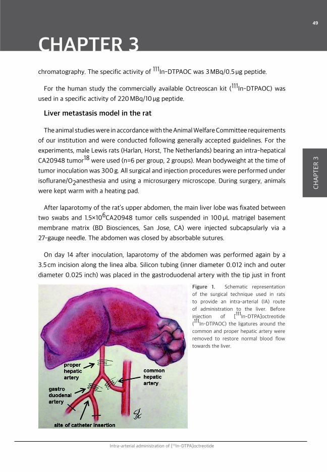
Intra-arterial administration of [111In-DTPA]octreotide
CHAPTER 3
49
CH
AP
TE
R 3
chromatography. The specific activity of 111In-DTPAOC was 3 MBq/0.5 μg peptide.
For the human study the commercially available Octreoscan kit (111In-DTPAOC) was
used in a specific activity of 220 MBq/10 μg peptide.
Liver metastasis model in the rat
The animal studies were in accordance with the Animal Welfare Committee requirements
of our institution and were conducted following generally accepted guidelines. For the
experiments, male Lewis rats (Harlan, Horst, The Netherlands) bearing an intra-hepatical
CA20948 tumor18 were used (n=6 per group, 2 groups). Mean bodyweight at the time of
tumor inoculation was 300 g. All surgical and injection procedures were performed under
isoflurane/O2anesthesia and using a microsurgery microscope. During surgery, animals
were kept warm with a heating pad.
After laparotomy of the rat’s upper abdomen, the main liver lobe was fixated between
two swabs and 1.5×10⁶CA20948 tumor cells suspended in 100 μL matrigel basement
membrane matrix (BD Biosciences, San Jose, CA) were injected subcapsularly via a
27-gauge needle. The abdomen was closed by absorbable sutures.
On day 14 after inoculation, laparotomy of the abdomen was performed again by a
3.5 cm incision along the linea alba. Silicon tubing (inner diameter 0.012 inch and outer
diameter 0.025 inch) was placed in the gastroduodenal artery with the tip just in front
Figure 1. Schematic representation
of the surgical technique used in rats
to provide an intra-arterial (IA) route
of administration to the liver. Before
injection of [111In-DTPA]octreotide
(111In-DTPAOC) the ligatures around the
common and proper hepatic artery were
removed to restore normal blood flow
towards the liver.
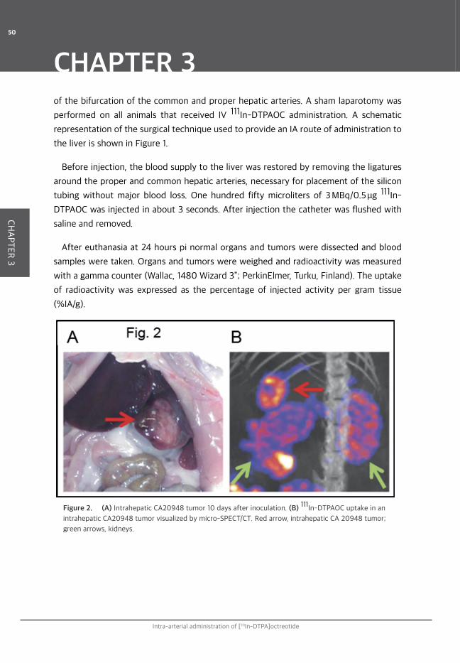
Intra-arterial administration of [111In-DTPA]octreotide
CHAPTER 3
50
CH
AP
TE
R 3
of the bifurcation of the common and proper hepatic arteries. A sham laparotomy was
performed on all animals that received IV 111In-DTPAOC administration. A schematic
representation of the surgical technique used to provide an IA route of administration to
the liver is shown in Figure 1.
Before injection, the blood supply to the liver was restored by removing the ligatures
around the proper and common hepatic arteries, necessary for placement of the silicon
tubing without major blood loss. One hundred fifty microliters of 3 MBq/0.5 μg 111In-
DTPAOC was injected in about 3 seconds. After injection the catheter was flushed with
saline and removed.
After euthanasia at 24 hours pi normal organs and tumors were dissected and blood
samples were taken. Organs and tumors were weighed and radioactivity was measured
with a gamma counter (Wallac, 1480 Wizard 3”; PerkinElmer, Turku, Finland). The uptake
of radioactivity was expressed as the percentage of injected activity per gram tissue
(%IA/g).
Figure 2. (A) Intrahepatic CA20948 tumor 10 days after inoculation. (B) 111In-DTPAOC uptake in an
intrahepatic CA20948 tumor visualized by micro-SPECT/CT. Red arrow, intrahepatic CA 20948 tumor;
green arrows, kidneys.
Intra-arterial administration of [111In-DTPA]octreotide
CHAPTER 3
51
CH
AP
TE
R 3
MicroSPECT/CT imaging
One additional rat with a subcapsular CA20948 tumor in the liver was imaged by
microSPECT/CT imaging. Twenty-four hours before scanning, the rat was injected IV
with 30 MBq/0.5 μg 111In-DTPAOC. Scanning was performed with a four-headed multi-
pinhole NanoSPECT/CT camera (Bioscan, Inc., Washington, DC). Nine pinhole-apertures
with a diameter of 2.5 mm were used with 24 projections (1 minute per projection).
The 111In energy peaks were set at 171 and 245 keV. Guided by the CT topogram, the
upper abdomen was scanned for 60 seconds per projection. The whole procedure was
performed under Isoflurane/O2 anesthesia. SPECT scans were reconstructed iteratively
using InVivoScope software version 1.32 (Bioscan, Inc.) with medium noise reduction, a
voxel size of 0.3 mm3, and standard reconstruction settings.
Patient study
Three patients (age 32, 54, and 64 years) with metastatic nonresectable pancreatic
NETs were enrolled to receive two injections of 111In-DTPAOC; one IV injection and
one IA injection with a 2 week-interval. From previous imaging it was known that these
patients had hepatic metastasis enabling dosimetry measurements. All patients were on
short acting octreotide treatment (Sandostatin; Novartis, Basel, Switzerland), which was
discontinued 24 hours prior to both injections. This 24 hours discontinuation was chosen
because before standard 177Lu-DOTATATE treatment the same period of short acting
octreotide treatment discontinuation is used. The study was performed after written
informed consent from the patient to participate in this study, which was approved by
the Erasmus MC Medical Ethical Committee.
IA administration
IA administration was performed via a catheter placed angiographically through
Seldinger’s technique via the femoral artery with the tip into the common hepatic artery.
Immediately after this procedure the patient was placed on the gamma camera bed in
a supine position. The 111In-DTPAOC was injected in about 10 seconds. The catheter
was flushed with 10 mL of 0.9% saline. The same injection protocol was used for IV
injected 111In-DTPAOC.
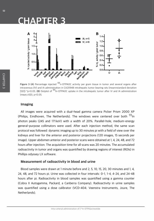
Intra-arterial administration of [111In-DTPA]octreotide
CHAPTER 3
52
CH
AP
TE
R 3
Imaging
All images were acquired with a dual-head gamma camera Picker Prism 2000 XP
(Philips, Eindhoven, The Netherlands). The windows were centered over both 111In
photon peaks (245 and 171 keV) with a width of 20%. Parallel-hole, medium-energy
general-purpose collimators were used. After each injection method, the same scan
protocol was followed: dynamic imaging up to 30 minutes pi with a field of view over the
kidneys and liver for the anterior and posterior projections (120 images, 15 seconds per
image). Upper abdomen anterior and posterior scans were obtained at 1, 4, 24, 48, and 72
hours after injection. The acquisition time for all scans was 20 minutes. The accumulated
radioactivity in tumor and organs was quantified by drawing regions of interest (ROIs) in
Phillips odyssey LX software.
Measurement of radioactivity in blood and urine
Blood samples were drawn at 1 minute before and 2, 5, 10, 15, 20, 30 minutes and 1, 4,
24, 48, and 72 hours pi. Urine was collected in four intervals: 0–1, 1–4, 4–24, and 24–48
hours after pi. Radioactivity in blood samples was quantified using a gamma counter
(Cobra II Autogamma, Packard, a Canberra Company). Radioactivity in urine samples
was quantified using a dose calibrator (VCD-404; Veenstra Instruments, Joure, The
Netherlands).
Figure 3 (A) Percentage injected 111n-DTPAOC activity per gram tissue in tumor and several organs after
intravenous (IV) and IA administration in CA20948 intrahepatic tumor bearing rats (mean±standard deviation
[SD]) *p<0.05. (B) Dotplot of 111In-DTPAOC uptake in the intrahepatic tumor after IV and IA administration
(mean,±SD), p<0.05.
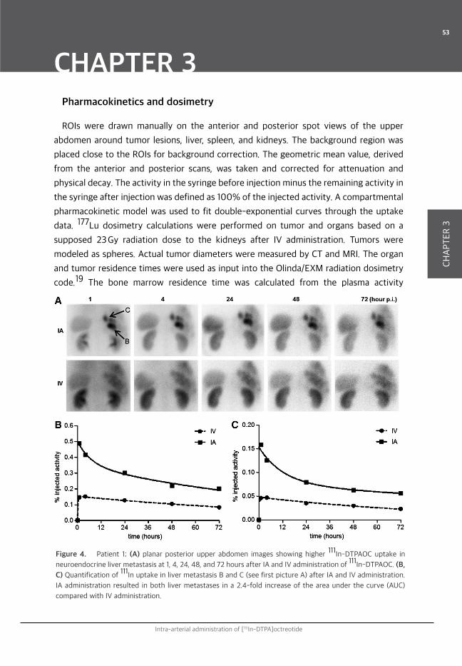
Intra-arterial administration of [111In-DTPA]octreotide
CHAPTER 3
53
CH
AP
TE
R 3
Pharmacokinetics and dosimetry
ROIs were drawn manually on the anterior and posterior spot views of the upper
abdomen around tumor lesions, liver, spleen, and kidneys. The background region was
placed close to the ROIs for background correction. The geometric mean value, derived
from the anterior and posterior scans, was taken and corrected for attenuation and
physical decay. The activity in the syringe before injection minus the remaining activity in
the syringe after injection was defined as 100% of the injected activity. A compartmental
pharmacokinetic model was used to fit double-exponential curves through the uptake
data. 177Lu dosimetry calculations were performed on tumor and organs based on a
supposed 23 Gy radiation dose to the kidneys after IV administration. Tumors were
modeled as spheres. Actual tumor diameters were measured by CT and MRI. The organ
and tumor residence times were used as input into the Olinda/EXM radiation dosimetry
code.19 The bone marrow residence time was calculated from the plasma activity
Figure 4. Patient 1; (A) planar posterior upper abdomen images showing higher 111In-DTPAOC uptake in
neuroendocrine liver metastasis at 1, 4, 24, 48, and 72 hours after IA and IV administration of 111In-DTPAOC. (B,
C) Quantification of 111In uptake in liver metastasis B and C (see first picture A) after IA and IV administration.
IA administration resulted in both liver metastases in a 2.4-fold increase of the area under the curve (AUC)
compared with IV administration.
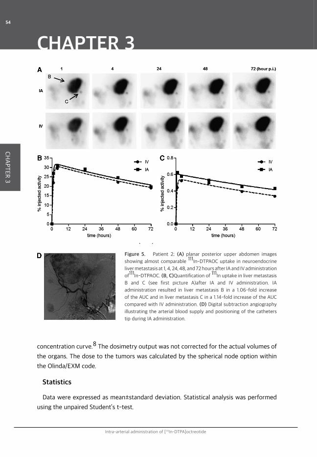
Intra-arterial administration of [111In-DTPA]octreotide
CHAPTER 3
54
CH
AP
TE
R 3
concentration curve.8 The dosimetry output was not corrected for the actual volumes of
the organs. The dose to the tumors was calculated by the spherical node option within
the Olinda/EXM code.
Statistics
Data were expressed as mean±standard deviation. Statistical analysis was performed
using the unpaired Student’s t-test.
Figure 5. Patient 2; (A) planar posterior upper abdomen images
showing almost comparable 111In-DTPAOC uptake in neuroendocrine
liver metastasis at 1, 4, 24, 48, and 72 hours after IA and IV administration
of111In-DTPAOC. (B, C)Quantification of 111In uptake in liver metastasis
B and C (see first picture A)after IA and IV administration. IA
administration resulted in liver metastasis B in a 1.06-fold increase
of the AUC and in liver metastasis C in a 1.14-fold increase of the AUC
compared with IV administration. (D) Digital subtraction angiography
illustrating the arterial blood supply and positioning of the catheters
tip during IA administration.
Intra-arterial administration of [111In-DTPA]octreotide
CHAPTER 3
55
CH
AP
TE
R 3
Results
Liver metastasis model in the rat
Inoculation of CA20948 tumor cells from in vitro cultures, mixed with matrigel, resulted
in a palpable solid tumor (Fig. 2A) 10 days later. The tumor could clearly be visualized by
micro-SPECT scanning 24 hours pi of 111In-DTPAOC (Fig. 2B). Ex vivo biodistribution at
24 hours after injection revealed the tumor uptake of 111In-DTPAOC administered via the
common hepatic artery to be twofold higher (p<0.05) than the uptake after systemic (IV)
administration (Fig. 3A, B). Uptake in kidney, liver, stomach, duodenum, adrenals, blood,
and muscle did not significantly differ after both injection methods. Surprisingly, after IA
administration the radioactivity in the pancreas was higher than after IV administration.
Patient study
Low 111In-DTPAOC uptake (iso-intense compared to the liver) was seen in the liver
metastases of the first patient after IV administration on all images made at 1, 4, 24,
48, and 72 hours pi. After IA administration in this patient clear visualization of the
liver metastases was obtained (Fig. 4A). Quantification data of tumor uptake after IA
administration showed a 2.4-fold higher 111In-DTPAOC uptake (Fig. 4B, C) in these liver
metastases in comparison with that after IV administration. Kidney uptake and urine
radioactivity was not significantly different after either route of administration.
Strikingly, in the second patient IA administration did not result in significantly
higher 111In-DTPAOC uptake in the liver metastases. Quantification showed a 1.06- and
1.14-fold increase of 111In-DTPAOC uptake after IA versus IV administration for metastasis
B and C (Fig. 5A), respectively. When compared with the first patient, the tumor uptake
was exceptionally high in patient 2. At 4 hours pi after IV and IA administration around
30% of the injected activity was located in the very large liver metastasis B (Fig. 5A),
whereas for the first patient the maximum uptake was only 0.5% of the injected activity
in liver metastasis (Fig. 4A, metastasis B). So, tumor uptake in the second patient was
already exceptionally high after IV administration.
In a third patient three liver metastases were clearly visualized at 1, 4, 24, 48, and
72 hours post IV administration. The equivalent scans after IA administration revealed
several additional liver metastatic lesions (Fig. 6A), whereas the lesions that were visible
on the IV scan showed higher uptake after IA injection. Surprisingly, one metastatic
Intra-arterial administration of [111In-DTPA]octreotide
CHAPTER 3
56
CH
AP
TE
R 3
nodule (hotspot 8, Fig. 6A) also shown on the scan after IV injection and located in
liver segment 5 did not show an increased111In-DTPAOC uptake after IA administration.
Analysis of the dynamic scans made during the injection phase, showed that the tip of
the catheter in this patient was not situated in the common hepatic artery, but in the
right hepatic artery (Fig. 6A, picture in the right upper corner). As a result, 111n-DTPAOC
was administered IA to the right part of the liver, whereas the left part received the 111In-
DTPAOC after first pass through the body via systemic administration.
Urine sample data, blood sample data, and region of interest (tumors and kidneys, Fig.
6B) quantification data of this patient were fitted in a compartmental pharmacokinetic
model (Fig. 6C) for IA and IV administration in Figure 6D and E, respectively. Quantification
of 111In-DTPAOC uptake in liver metastasis 4 (in the right part of the liver) over 72 hours
showed a mean 2.3-fold increase after IA versus IV administration (Fig. 6D, E). 111In-
DTPAOC uptake in the metastasis (hotspot 8) located in the left part of the liver showed
to be comparable after IA and IV administration.111In-DTPAOC kidney uptake was 13%
lower after IA administration compared with IV administration. The percentage injected
activity in plasma only differed significantly between IA and IV administration at 2
minutes pi. Five minutes pi, IA and IV plasma values were almost comparable (Fig. 6F).
Based on the compartmental pharmacokinetic model 177Lu dosimetry calculations
were performed (Fig. 6G) for all liver metastasis after IA and IV administration assuming
similar pharmacokinetics for 111In-DTPAOC and 177Lu-DOTATATE. Radiation dose
with 177Lu-DOTATATE PRRT was calculated for all metastases based on a radiation dose
to the kidneys of 23 Gy after IA and IV administration. These calculations showed for
all metastases located in the right part of the liver an increase of the radiation dose
by a factor 1.9–4.5 after IA administration. The calculated radiation dose for hotspot 8
in Figure 6A was not significantly different after IA or IV administration. The estimated
radiation dose to several organs is shown in Figure 6H after an injected activity leading to
a renal radiation dose of 23 Gy. Please note the relative large (30%) renal radiation dose
reduction after IA administration. These calculations are performed on the data collected
from only 1 patient and definitely have no statistical significance in predicting the results
of IA administration in a group of GEPNET patients.
As biodistribution study in rats showed increase of 111In-DTPAOC in the pancreas after
IA administration we looked for pancreas uptake on the patient scans. But none of the
Intra-arterial administration of [111In-DTPA]octreotide
CHAPTER 3
57
CH
AP
TE
R 3
scans showed 111In-DTPAOC uptake in the pancreas. Therefore, a possible increase of
uptake in the pancreas after IA administration via the hepatic artery could not be shown.
Discussion
Both the preclinical and clinical studies indicated that IA hepatic administration of 111In-
DTPAOC can result in a significantly increased 111In-DTPAOC tumor uptake compared
with that after IV administration. This increased uptake in the hepatic NET metastases
will, when applying a therapeutic radiolabeled analogue such as 177Lu-DOTATATE or90Y-
DOTATOC, result in a higher tumor absorbed radiation dose.
In the animal study IA administration resulted in a doubling of the 111In-DTPAOC uptake
in both tumor and pancreas. This unexpected increase in pancreas (a sst2-positive organ
in the rat) uptake is most likely explained by backflow of111In-DTPAOC via the common
hepatic artery and coeliac trunk into the aorta during injection.
In patient 2, IA administration did not result in significantly higher tumor uptake. The
large tumor volume in combination with the high tumor sst2 expression (grade 4) and
the limited amount of peptide, only 10 μg DTPAOC was used, could have played a role
here. Interesting is the fact that in patient 2 within the time frame of 1–4 hours pi after
IA administration the uptake in the liver metastases was still increasing while in both
other patients the slope of the curve was already declining at 1 hours pi. We cannot fully
explain this phenomenon right now, but we assume all receptors in the liver metastases
in patient 1 and 3 to be saturated after IA administration, whereas in patient 2 binding
of 111In-DTPAOC was still possible at relatively low 111In-DTPAOC plasma concentrations
at later time points.
In patient 3, IA administration resulted in a 2.3-fold increase in 111In-DTPAOC uptake
in the metastasis located in the right part of the liver. The fact that the one metastasis
located in the left part of the liver showed to have equal uptake as measured after
systemic administration suggests that 111In-DTPAOC uptake in tumor lesions outside the
liver compartment, which was supplied by the IA-route, was apparently not affected
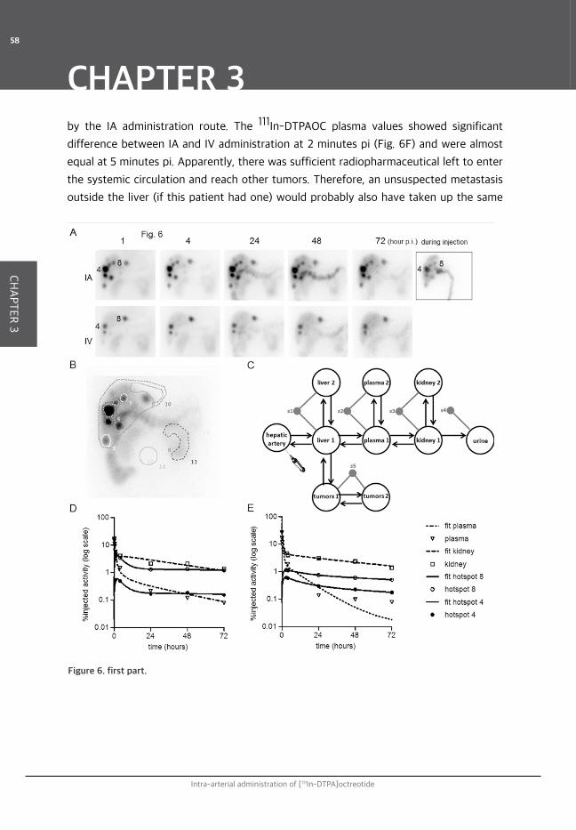
Intra-arterial administration of [111In-DTPA]octreotide
CHAPTER 3
58
CH
AP
TE
R 3
Figure 6. first part.
by the IA administration route. The 111In-DTPAOC plasma values showed significant
difference between IA and IV administration at 2 minutes pi (Fig. 6F) and were almost
equal at 5 minutes pi. Apparently, there was sufficient radiopharmaceutical left to enter
the systemic circulation and reach other tumors. Therefore, an unsuspected metastasis
outside the liver (if this patient had one) would probably also have taken up the same
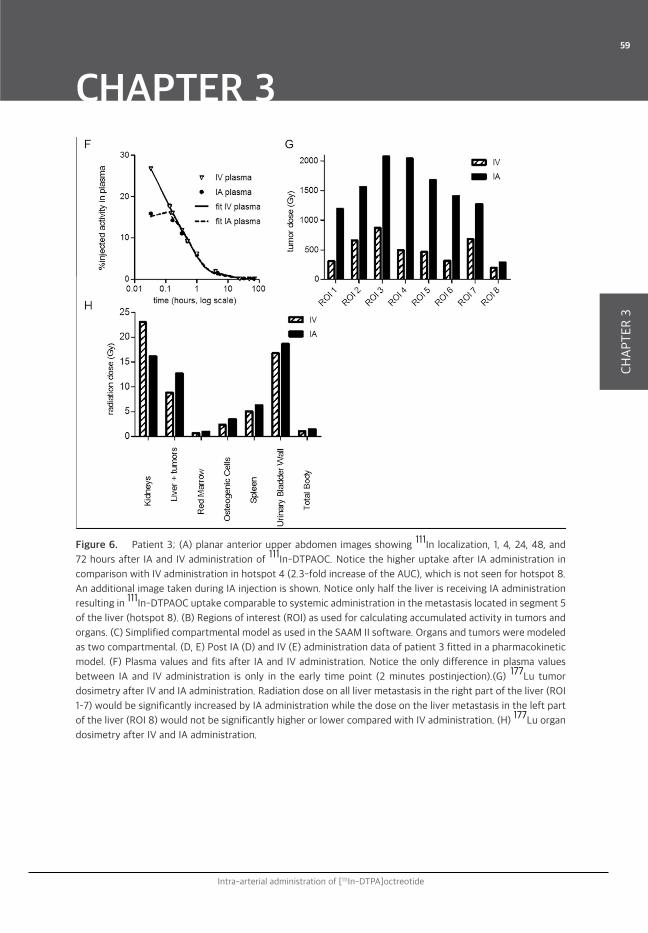
Intra-arterial administration of [111In-DTPA]octreotide
CHAPTER 3
59
CH
AP
TE
R 3
Figure 6. Patient 3; (A) planar anterior upper abdomen images showing 111In localization, 1, 4, 24, 48, and
72 hours after IA and IV administration of 111In-DTPAOC. Notice the higher uptake after IA administration in
comparison with IV administration in hotspot 4 (2.3-fold increase of the AUC), which is not seen for hotspot 8.
An additional image taken during IA injection is shown. Notice only half the liver is receiving IA administration
resulting in 111In-DTPAOC uptake comparable to systemic administration in the metastasis located in segment 5
of the liver (hotspot 8). (B) Regions of interest (ROI) as used for calculating accumulated activity in tumors and
organs. (C) Simplified compartmental model as used in the SAAM II software. Organs and tumors were modeled
as two compartmental. (D, E) Post IA (D) and IV (E) administration data of patient 3 fitted in a pharmacokinetic
model. (F) Plasma values and fits after IA and IV administration. Notice the only difference in plasma values
between IA and IV administration is only in the early time point (2 minutes postinjection).(G) 177Lu tumor
dosimetry after IV and IA administration. Radiation dose on all liver metastasis in the right part of the liver (ROI
1–7) would be significantly increased by IA administration while the dose on the liver metastasis in the left part
of the liver (ROI 8) would not be significantly higher or lower compared with IV administration. (H) 177Lu organ
dosimetry after IV and IA administration.

Intra-arterial administration of [111In-DTPA]octreotide
CHAPTER 3
60
CH
AP
TE
R 3
amount of 111In-DTPAOC after IA administration as after IV administration. Certainly,
this assumption can only be made in this patient. Probably a higher hepatic tumor mass
and/or sst2 density could even result in a lower uptake in extra hepatic tumor lesions
after IA administration compared with IV administration. Despite kidney uptake and
excretion was minimally decreased by IA administration,177Lu dosimetry calculations
in patient 3 showed a 30% dose reduction to the kidneys by IA administration. This
reduced kidney and higher intrahepatic tumor radiation dose resulted in a significant
increase of the therapeutic index. IA 177Lu-DOTATATE PRRT would administer a 1.9–4.5
times higher estimated tumor radiation dose when the kidney radiation dose would be
23 Gy. The radiation dose to the bone marrow did not show a significant increase after
IA administration. For these calculations we assumed the pharmacokinetics for 177In-
DTPAOC and 177Lu-DOTATATE to be equal, but the fact that 177Lu-DOTATATE has a
four-fold higher affinity for the sst2 and also shows some affinity for the sst2 indicates
that our dosimetry calculations are a rough estimation.
In the studies described here, the procedure differed from the routine PRRT treatment
with 177Lu-DOTATATE in our institution. PRRT with 177Lu-DOTATATE is administered
in a 30 minutes infusion whereas in the current study 111In-DTPAOC was injected as a
bolus in 10 seconds. Second, the amount of peptide used in a PRRT setting is 20 times
the amount of peptide we used in this study (200 μg DOTATATE versus 10 μg DTPAOC).
In addition, DTPAOC has a four-fold lower affinity for the sst2 compared DOTATATE.
In our study, DTPAOC and not DOTATATE was used because the scans were included
in the standard clinical workup for PRRT and not for research purposes only. Future
experiments with DOTATATE as ligand using a therapeutic peptide dose and injection
protocol will be performed to show the additional effect of locoregional administration
in a therapeutic setting. In 2008 Limouris et al. showed a relatively high tumor response
rate after IA PRRT with 111In-DTPAOC in patients with GEPNET liver metastasis.13 This
relatively high tumor response could be (partially) caused by an increased 111In uptake
after the IA administration. Recently, Kratochwil et al. showed a mean 3.75-fold
increase of 68Ga-DOTATOC uptake at 40 minutes pi after selective IA administration
in GEPNETs.16 Considering our observations at later time points obtained with 111In-
DTPAOC we feel this mean increase of 3.75-fold cannot be translated to the tumor
radiation dose. At 1 hours pi IA administration the slope in the curve showing the %
injected activity present in the liver metastasis is still quite steep (Fig. 4B, C). Probably
a measurement at 24 hours pi, after the curve has a more stable slope, would be more
Intra-arterial administration of [111In-DTPA]octreotide
CHAPTER 3
61
CH
AP
TE
R 3
predictive in estimating the increase in tumor uptake after IA administration. In this
study, we demonstrated that IA administration resulted in the same (1 patient) or an
increased 111In-DTPAOC uptake in NET liver metastasis up to 2.4-fold compared with IV
administration over a period of 72 hours. The increase in uptake after IA administration
is probably depending on sst2 density, tumor load, and tumor perfusion. We therefore
conclude that locoregional IA administration should be considered as the optimal route of
administration in patients in which the GEPNET tumor load is mainly localized in the liver.
Though, an increase in radionuclide tumor uptake after IA administration in comparison
to IV administration is not guaranteed, as in one of our patients tumor uptake was high,
but similar after IV and IA administration. If IA PRRT will be applied, the positioning of
the catheter should be well planned using contrast enhanced CT for imaging of possible
hepatic arterial vasculature abnormalities.

Intra-arterial administration of [111In-DTPA]octreotide
CHAPTER 3
62
CH
AP
TE
R 3
References
1. O berg K. Pancreatic endocrine tumors. Semin Oncol 2010;37:594.2. Raymond E, Dahan L, Raoul J-L, et al. Sunitinib malate for the treatment of pancreatic neuroendocrine
tumors. N Engl J Med 2011;364:501.3. Yao JC, Shah MH, Ito T, et al. Everolimus for advanced pancreatic neuroendocrine tumors. N Engl J Med
2011;364:514.4. Rinke A, Muller H-H, Schade-Brittinger C, et al. Placebocontrolled, double-blind, prospective,
randomized study on the effect of octreotide LAR in the control of tumor growth in patients with metastatic neuroendocrine midgut tumors: A report from the PROMID Study Group.J Clin Oncol2009;27:4656.
5. Valkema R, de Jong M, Bakker WH, et al. Phase I study of peptide receptor radionuclide therapy with[111In-DTPA] octreotide: The Rotterdam experience. Semin Nucl Med 2002;32:110.
6. Pool SE, Krenning EP, Koning GA, et al. Preclinical and clinical studies of peptide receptor radionuclide therapy. Semin Nucl Med 2010;40:209.
7. Khan S, Krenning EP, van Essen M, et al. Quality of life in265 patients with gastroenteropancreatic or bronchial neuroendocrine tumors treated with [177Lu-DOTA0,Tyr3]octreotate. J Nucl Med 2011;52:1361.
8. Forrer F, Krenning EP, Kooij PP, et al. Bone marrow dosimetry in peptide receptor radionuclide therapy with [177Lu-DOTA0,Tyr3]octreotate. Eur J Nucl Med Mol Imaging 2009;36:1138.
9. Rolleman EJ, Melis M, Valkema R, et al. Kidney protection during peptide receptor radionuclide therapy with somatostatin analogues. Eur J Nucl Med Mol Imaging 2009;37:1018.
10. Kwekkeboom DJ, de Herder WW, Kam BL, et al. Treatment with the radiolabeled somatostatin analog [177Lu-DOTA0,Tyr3]octreotate: Toxicity, efficacy, and survival. J Clin Oncol 2008;26:2124.
11. Steinmu ller T, Kianmanesh R, Falconi M, et al. Consensus guidelines for the management of patients with liver metastases from digestive (neuro)endocrine tumors: Foregut, midgut, hindgut, and unknown primary. Neuroendocrinol- ogy 2008;87:47.
12. McStay MKG, Maudgil D, Williams M, et al. Large-volume liver metastases from neuroendocrine tumors: Hepatic intraarterial 90Y-DOTA-lanreotide as effective palliative therapy. Radiology 2005;237:718.
13. Limouris GS, Chatziioannou A, Kontogeorgakos D, et al. Selective hepatic arterial infusion of In-111-DTPA-Phe1-octreotide in neuroendocrine liver metastases.
Eur J Nucl Med Mol Imaging 2008;35:1827.14. Papakonstantinou K, Karfis I, Lyra M, et al. Super-selective hepatic arterial infusions of Y-90-DOTA-
TOC and/or Lu-177-DOTA-TATE in neuroendocrine liver metastases after selective catheterization of the hepatic artery and permanent port installation, previously treated with high doses of In-111-DTPA-Phe1-ocreotide. Eur J Nucl Med Mol Imaging 2010;37:OP391.
15. Beauregard JM, Eu P, Neels OC et al. Enhanced uptake in neuroendocrine tumours after intraarterial
infusion of [68Ga]/[177Lu]-octreotate. Eur J Nucl Med Mol Imaging 2009;36:OP620.
16. Kratochwil C, Giesel FL, Lopez-Benitez R, et al. Intraindividual comparison of selective arterial versus venous 68Ga-DOTATOC PET/CT in patients with gastro enteropancreatic neuroendocrine tumors. Clin Cancer Res 2010;16:2899.
17. Breeman WAP, Kwekkeboom DJ, Kooij PM, et al. Effect of dose and specific activity on tissue distribution of indium-111-pentetreotide in rats. J Nucl Med 1995;36: 623.
18. Bernard BF, Krenning E, Breeman WAP, et al. Use of the rat pancreatic CA20948 cell line for the comparison of radiolabelled peptides for receptor-targeted scintigraphy and radionuclide therapy. Nucl Med Commun 2000;21: 1079.
19. Stabin MG, Siegel JA, Sparks RB, et al. Contribution to red marrow absorbed dose from total body activity: A correc- tion to the MIRD method. J Nucl Med 2001;42:492.

Intra-arterial administration of [111In-DTPA]octreotide
CHAPTER 3
63
CH
AP
TE
R 3

44
4MTOR INHIBITOR RAD001 PROMOTES METASTASIS IN A RAT MODEL OF PAN-CREATIC NEUROENDOCRINE CANCER4MTOR INHIBITOR RAD001 PROMOTES METASTASIS IN A RAT MODEL OF PAN-CREATIC NEUROENDOCRINE CANCER
Stefan E. Pool Sander Bison Stuart J. Koelewijn Linda M. van der Graaf Marleen Melis Eric P. Krenning Marion de Jong
Cancer Research 2013;73:12-18.
mTOR Inhibitor RAD001 Promotes Metastasis
66
CHAPTER 4
CH
AP
TE
R 4
Abstract
Inhibition of mTOR is commonly considered a valid target in cancer treatment, but this
assertion does not address effects on the immune microenvironment that may be detrimental to
cancer treatment. Here we show how administration of the mTOR inhibitor RAD001 (everolimus)
results in the occurrence of distant metastasis in a rat model of pancreatic cancer. RAD001
was administered twice weekly for 4.5 weeks as a single treatment or combined with [177Lu-
DOTA,Tyr3]octreotate (177Lu-DOTATATE), where the latter targets the somatostatin receptor-2.
The hypothesized synergistic therapeutic effect of RAD001 combined with 177Lu-DOTATATE
was, however, not observed in our experiments. The combination was shown to be less effective
than 177Lu- DOTATATE alone. Unexpectedly, tumor metastasis was observed in 77% of the subjects
treated with RAD001, either alone or as part of the combination treatment. This was a striking
effect, because metastasis did not occur in control or 177Lu-DOTATATE-treated animals, including
those where the primary tumor was surgically removed. These findings may be important clinically
among noncompliant patients or patients that discontinue RAD001 therapy because of adverse
effects. Cancer Res; 73(1); 12–18. ©2012 AACR.
Introduction
Gastroenteropancreatic neuroendocrine tumors (GEPNET) are usually slow growing and
most patients present with already metastasized disease at time of diagnosis. As published by
Missiaglia and colleagues (1) in many pancreatic neuroendocrine tumors (PNET), the mTOR
signal transduction pathway is upregulated. This pathway plays a key role in regulating cell growth,
metabolism, proliferation, and angiogenesis. Inhibition of the mTOR signal transduction pathway in
PNETs by RAD001 has shown promising results with a clinical benefit of 76%, when given as a single
treatment (n = 115), and of 82%, when given in combination with octreotide-LAR (n = 45; ref. 2).
The RADIANT-3 study, a randomized, double-blind, placebo-controlled, multicenter phase III trial
of 10 mg RAD001 daily in PNET-patients (n = 410) has been completed recently. Median progression-
free survival for RAD001 plus best supportive care was 11 months compared with 4.6 months in
the placebo group (P < 0.001) with acceptable toxicity (3). Most recently, the U.S. Food and Drug
Administration approved RAD001 for the treatment of PNET patients.
From the late 1990s, clinical peptide receptor radionuclide therapy (PRRT) studies have been
carried out with radiolabeled somatostatin analogs, such as 177Lu-DOTATATE and [90Y-DOTA,Tyr3]
octreotide (90Y-DOTATOC; Onalta), targeting sst2 overexpressed on most GEPNETs. These studies
mTOR Inhibitor RAD001 Promotes Metastasis
67
CHAPTER 4
CH
AP
TE
R 4
have shown very promising results with regard to tumor response, overall survival, and
quality of life (4, 5).
Considering the promising results of both RAD001 and PRRT for the treatment of
GEPNETs, we combined these 2 therapies in the CA20948 syngeneic pancreatic tumor
model in the rat. This radiosensitive model is commonly used for preclinical PRRT
experiments and also shown to be susceptible to RAD001 treatment (6). For comparison,
studies were also carried out in the xenograft H69 tumor model in nude mice. The aim
of the current study was to investigate the therapeutic effects of combined treatment
with RAD001 and 177Lu-DOTATATE compared with those of the 2 monotherapies.
Materials and Methods
Cell lines
The CA20948 rat pancreatic tumor cell line (derived from a rat pancreas at our
institution) is of acinar origin (7), has high sst2 expression, and was cultured as reported
previously (8, 11). The cells were passaged for a maximum of 20 times and checked for
mycoplasma infection and sst2 expression every 3 months.
The H69 tumor cell line is a human small-cell lung carcinoma (the American
Tissue Culture Collection, Wesel, Germany), has high sst2 expression, was passaged for
a maximum of 10 times, and was cultured according to the supplier’s protocol.
Tumor models
The animal studies were in agreement with the Animal Welfare Committee
requirements of our institution and conducted in accordance with generally accepted
guidelines. For the first 2 experiments, male Lewis rats (Harlan; Horst, the Netherlands)
with a mean body weight of 275 g were used. For tumor induction, 107 CA20948 tumor
cells in 0.5 mL ice-cold PBS per animal were injected subcutaneously in the lower flank.
Monitoring of body weight and tumor size by caliper measurements was carried out by
a technician blinded for the treatment groups. Tumor volume was calculated according
to 0.4 x length x width x height. In the first study (Fig. 1), animals were euthanized when
mTOR Inhibitor RAD001 Promotes Metastasis
68
CHAPTER 4
CH
AP
TE
R 4
tumor size had reached a volume of more than 4 cm3 or when a tumor was bleeding
due to skin penetration. In the second study, the primary subcutaneous tumors
were surgically removed when tumor volume exceeded 4 cm3 or when the tumor was
bleeding due to skin penetration, thus saving the animals for follow-up. Animals were
euthanized when more than 10% loss of body weight was detected.
For the third experiment, 24 male NMRI Nu/Nu mice with a mean body weight of 35
g xenotransplanted sst2-expressing H69 cells were used. Animals were euthanized when
tumor size reached a volume of more than 2 cm3, when a tumor was bleeding due to
skin penetration, or when more than 10% loss of body weight was detected.
RAD001
In the first experiment, RAD001 (kind gift from Novartis, Basel, Switzerland) and
placebo was prepared according to the manufacturer’s protocol. In the second and
third experiment, RAD001 powder (LC laboratories, Woburn, USA) was dissolved in 2 mL
ethanol and further diluted to the appropriate concentration in 5% (w/v) glucose
solution. RAD001 was administered orally by gavage with a blunt needle.
Radionuclides and peptides
DOTA,Tyr3-octreotate was obtained from Mallinckrodt, St Louis, Missouri. 177LuCl3
was obtained from NRG, Petten and was distributed by IDB-Holland, Baarle-Nassau, the
Netherlands. 177Lu-DOTA,Tyr3-octreotate was locally prepared as described previously
(9) in a specific activity of 125 MBq/3.4 mg peptide. Labeling of 111In-DTPA-octreotide
(Octreoscan; Tyco Health Care, Petten, the Netherlands) in a specific activity of 30 MBq 111InCl3/0.5 mg DTPA-octreotide was carried out as previously described (10). For the
mouse experiments, a specific activity of 30 MBq 111InCl3/0.1 mg DTPA-octreotide was
used.
In vitro autoradiography and hematoxylin and eosin staining
These procedures were carried out as previously described in detail (11).
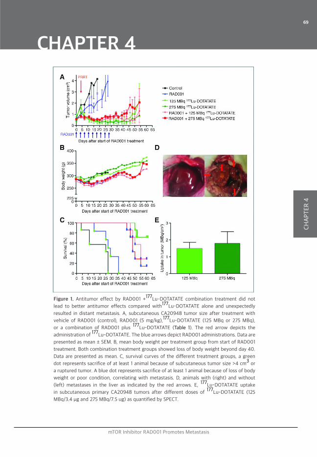
mTOR Inhibitor RAD001 Promotes Metastasis
69
CHAPTER 4
CH
AP
TE
R 4
Figure 1. Antitumor effect by RAD001 +177Lu-DOTATATE combination treatment did not
lead to better antitumor effects compared with177Lu-DOTATATE alone and unexpectedly
resulted in distant metastasis. A, subcutaneous CA20948 tumor size after treatment with
vehicle of RAD001 (control), RAD001 (5 mg/kg),177Lu-DOTATATE (125 MBq or 275 MBq),
or a combination of RAD001 plus 177Lu-DOTATATE (Table 1). The red arrow depicts the
administration of 177Lu-DOTATATE. The blue arrows depict RAD001 administrations. Data are
presented as mean ± SEM. B, mean body weight per treatment group from start of RAD001
treatment. Both combination treatment groups showed loss of body weight beyond day 40.
Data are presented as mean. C, survival curves of the different treatment groups, a green
dot represents sacrifice of at least 1 animal because of subcutaneous tumor size >4 cm3 or
a ruptured tumor. A blue dot represents sacrifice of at least 1 animal because of loss of body
weight or poor condition, correlating with metastasis. D, animals with (right) and without
(left) metastases in the liver as indicated by the red arrows. E, 177Lu-DOTATATE uptake
in subcutaneous primary CA20948 tumors after different doses of 177Lu-DOTATATE (125
MBq/3.4 μg and 275 MBq/7.5 ug) as quantified by SPECT.
mTOR Inhibitor RAD001 Promotes Metastasis
70
CHAPTER 4
CH
AP
TE
R 4
mTOR Inhibitor RAD001 Promotes Metastasis
71
CHAPTER 4
CH
AP
TE
R 4
Figure 2. RAD001 treatment (with or without177Lu-DOTATATE) resulted in the occurrence of distant metastasis.
A, mean body weight per group from start of RAD001 treatment. Again, the dip in body weight in the RAD001-
treated animals was observed from day 40 onward in the high-dose RAD001 group and from day 60 onward in
the low-dose RAD001 group. B, survival curve, censored for metastasis-unrelated death. All RAD001 treatment
groups had significantly lower survival rate compared with control and PRRT-only groups. ′, animal found
dead in cage; no metastases on autopsy. ″, animal did not survive subcutaneous tumor surgical resection; no
metastasis on autopsy. ‴, animal with macroscopically visible tumor spill during subcutaneous tumor surgical
resection. C, subcutaneous H69 tumor size in NMRI Nu/Nu mice was monitored after treatment with 177Lu-
DOTATATE in combination with vehicle of RAD001(control), RAD001 (5 mg/kg, twice a week), and RAD001 (5
mg/kg, daily). On day 21, a second 177Lu-DOTATATE treatment was given for reduction of subcutaneous H69
tumor size, facilitating a longer follow-up. No additional therapeutic effect of RAD001 on 177Lu-DOTATATE
treatment is seen in this tumor model. Data are presented as mean. D, survival curve showing no significant
difference between combination treatment of 177Lu-DOTATATE with vehicle RAD001 (control), RAD001
administered twice weekly, or RAD001 administered daily.
mTOR Inhibitor RAD001 Promotes Metastasis
72
CHAPTER 4
CH
AP
TE
R 4
Control
A
B
C
D
E
F
G
H
177Lu-DOTATATE 120 MBq
RAD001 5mg/kg rat
RAD001 2.5mg/kg rat
Figure 3177Lu 120MBq +
RAD001 5mg/kg
177Lu 120MBq +
RAD001 2.5mg/kg
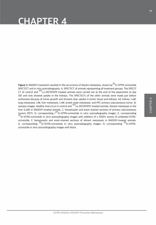
mTOR Inhibitor RAD001 Promotes Metastasis
73
CHAPTER 4
CH
AP
TE
R 4
Figure 3. RAD001 treatment resulted in the occurrence of distant metastasis, shown by111In-DTPA-octreotide
SPECT/CT and in vitro autoradiography. A, SPECT/CT of animals representing all treatment groups. The SPECT/
CT of control and 177Lu-DOTATATE-treated animals were carried out at the end of the experiment at day
150 and only showed uptake in the kidneys. The SPECT/CTs of the other animals were made just before
euthanasia because of tumor growth and showed clear uptake in tumor tissue and kidneys. Kd, kidney; LuM,
lung metastasis; LiM, liver metastasis; LnM, lymph node metastasis; and PST, primary subcutaneous tumor. B,
autopsy images. Healthy livers (Liv) in control and 177Lu-DOTATATE-treated animals. Distant metastasis in the
liver (LvM) in RAD001-treated animals. C, hematoxylin and eosin-stained sections of primary subcutaneous
tumors (PST). D, corresponding 1111In-DTPA-octreotide in vitro autoradiography images. E, corresponding 111In-DTPA-octreotide in vitro autoradiography images with addition of a 1000× excess of unlabeled DTPA-
octreotide. F, hematoxylin and eosin-stained sections of distant metastasis in RAD001-treated animals.
G, corresponding 111In-DTPA-octreotide in vitro autoradiography images. H, corresponding 111In-DTPA-
octreotide in vitro autoradiography images with block.
mTOR Inhibitor RAD001 Promotes Metastasis
74
CHAPTER 4
CH
AP
TE
R 4
Experimental setup
In experiment 1, RAD001 treatment was started 10 days after tumor inoculation,
followed by 177Lu-DOTATATE injection. Animals were randomized into matching
treatment groups with regard to tumor size at the start of treatment. Six treatment
groups were created using 1 dose of RAD001 (5 mg/kg body weight twice a week) and
2 doses of 177Lu-DOTATATE, 125 or 278 MBq, either as monotherapy or in combination
(Table 1). Experiment 1 ended at day 60. To keep animal discomfort as low as possible
and for practical reasons, RAD001 was administered twice weekly.
Experiment 2 was essentially similar as experiment 1; variations were 2.5 next to 5 mg/
kg body weight dose of RAD001, alone or combined with 125 MBq 177Lu-DOTATATE (Table
1). The most important difference in comparison with experiment 1 was the resection of
the subcutaneous tumor when tumor size increased to more than 4 cm3. Experiment 2
was terminated at day 150. All animals still alive at the end of the experiment were checked
for distant metastases by 111In-DTPA-octreotide single-photon emission computed
tomography (SPECT) or computed tomography (CT) and autopsy.
In experiment 3, NMRI Nu/Nu mice bearing subcutaneous H69 tumors were first
treated with 28 MBq 177Lu-DOTATATE injected intraperitoneally. Four days later, 3
combination treatment groups were created at random. The first group received
RAD001 (5 mg/kg body weight) daily, the second group received RAD001 (5 mg/kg body
weight) twice a week with placebo treatment daily for the rest of the week, and the
third group received placebo treatment daily. RAD001/placebo treatment was given for
a total of 4 weeks. Three weeks after the first 177Lu-DOTATATE treatment, a second 25
MBq 177Lu-DOTATATE treatment was given to all animals as an alternative for surgical
removal of the primary tumor. When subcutaneous tumor size exceeded 2 cm3, the
animal was checked for distant metastases by 111In-DTPA-octreotide SPECT/CT and
autopsy.
Surgical procedure
During all surgical procedures, isoflurane/O2 anesthesia was applied and animals were
kept warm using a heating pad. After shaving and disinfection, an incision was made just
mTOR Inhibitor RAD001 Promotes Metastasis
75
CHAPTER 4
CH
AP
TE
R 4
Table 1. Characteristics of treatment groups studies in 1 and 2
Group Treatment n Tumor
V >4 cm3
Study 1 1 Control Vehicle only 7 †
2 RAD001 RAD001 (2×/week, 5 mg/kg bw) 6 †
3 Combination of low-dose 177
Lu-DOTATATE with RAD001 RAD001 (2×/week, 5 mg/kg bw) 7 †
and 125 MBq 177
Lu-DOTATATE
4 Combination of high-dose 177
Lu-DOTATATE with RAD001 RAD001 (2×/week, 5 mg/kg bw) 7 †
and 275 MBq 177
Lu-DOTATATE
5 Low-dose 177
Lu-DOTATATE 125 MBq 177
Lu-DOTATATE 6 †
6 High-dose 177
Lu-DOTATATE 275 MBq 177
Lu-DOTATATE 7 †
Study 2
7 Control Vehicle only 8 Surgery
8 Low-dose RAD001 RAD001 (2×/week, 2,5 mg/kg bw) 8 Surgery
9 High-dose RAD001 RAD001 (2×/week, 5 mg/kg bw) 8 Surgery
10 Combination of low-dose RAD001 with 177
Lu-DOTATATE RAD001 (2×/week, 2,5 mg/kg bw) 7 Surgery
+ 125 MBq 177
Lu-DOTATATE
11 Combination of high-dose RAD001 with 177
Lu-DOTATATE RAD001 (2×/week, 5 mg/kg bw) 8 Surgery
+ 125 MBq 177
Lu-DOTATATE
12 177
Lu-DOTATATE 125 MBq 177
Lu-DOTATATE 8 Surgery
13 RAD001, no tumor RAD001 (2×/week, 5 mg/kg bw) 7 n.a.
NOTE: All animals were bearing CA20948 tumors, except for animals in group 13.
Abbreviations: BW, body weight; n.a., not applicable.
†, euthanasia.
adjacent to the tumor. The capsule surrounding the tumor was carefully dissected
from the surrounding tissue. The wound was closed by using absorbable sutures.
SPECT/CT scanning
After 24 (experiment 1) or 48 hours (experiment 2) of intra-venous (i.v.) injection of 177Lu-DOTATATE, a helical SPECT scan was acquired covering the tumor region using
the 4-headed NanoSPECT/CT system (Bioscan) using Nucline software (v2.01, Mediso)
for the quantification of 177Lu-DOTATATE tumor uptake. Just before euthanizing an
animal, a whole-body SPECT/CT scan was carried out 4 to 24 hours after i.v. injection of 111In-DTPA-octreotide for detection of distant metastasis.
mTOR Inhibitor RAD001 Promotes Metastasis
76
CHAPTER 4
CH
AP
TE
R 4
Results and Discussion
Our first study, described in Table 1 (groups 1 – 6), showed inhibition of CA20948 tumor
growth by RAD001 monotherapy as was previously found by Boulay and colleagues
as well (6). In addition, in clinical studies mTOR inhibition generally seems to elicit
a cytostatic, rather than a cytotoxic, response (12, 13). Boulay and colleagues also
showed twice weekly administration of RAD001 to be as effective as daily administration
in this tumor model (6). All animals in the control and the RAD001-only group had to
be euthanized within 36 days after start of treatment because of tumor growth beyond
4 cm3 or a ruptured tumor. As expected, all animals receiving 177Lu-DOTATATE with or
without RAD001 exhibited significant antitumor response compared with controls (P ≤
0.05). However, 177Lu-DOTATATE in combination with RAD001 did not show evidence
of being more effective than 177Lu-DOTATATE alone (Fig. 1A). A dose–effect relationship
for the low- and high-dose 177Lu-DOTATATE was not found, which is in agreement
with similar levels of 177Lu-DOTATATE tumor uptake in these 2 groups 24 hours after
injection, as quantified on the basis of SPECT (Fig. 1E). Partial saturation of the sst2
receptors on the tumor cells in the high-dose group could play a role in this regard.
Strikingly, from day 40 after start of treatment, most animals in the combination therapy
groups showed unexpected decrease in body weight (Fig. 1B). Tumor metastasis to the
liver and occasionally to the lung was found in these animals on autopsy (Fig. 1D). In the
combination treatment groups, 11 out of 14 animals had to be euthanized because of
loss of body weight and apparent metastasis (Fig. 1C). Metastases could be visualized
after injection of 111In-DTPA-octreotide by SPECT/ CT, indicating sst2 expression. The 177Lu-DOTATATE-only-treated animals did not show loss of body weight and were all
free of metastasis, as confirmed by negative 111In-DTPA- octreotide SPECT/CT scans
and negative autopsies (Fig. 1D). The occurrence of spontaneous metastasis in the
subcutaneous CA20948 tumor model has not been described previously, despite the
fact that this model has often been used in PRRT studies with long follow-up periods (14).
We, therefore, hypothesize that RAD001, alone or in combination with 177Lu-DOTATATE,
or the discontinuation of RAD001 treatment might be the cause of the metastasis.
Unfortunately, the RAD001 monotherapy treatment group in this study did not survive
long enough to develop or to be tested for metastases by SPECT.
For further investigation, a second study was carried out (Table 1; groups 7–13) in
which a subcutaneous tumor reaching a volume of 4 cm3 was surgically removed
mTOR Inhibitor RAD001 Promotes Metastasis
77
CHAPTER 4
CH
AP
TE
R 4
to allow long-term follow-up. Furthermore, in this experiment also a 2.5 mg/kg body
weight dose of RAD001 was applied next to the earlier-used 5 mg/kg body weight,
combined with 125 MBq 177Lu-DOTATATE (Table 1). In an additional group (group 13), 5
mg/kg body weight of RAD001 was given to control rats without tumor.
The therapeutic effects obtained in this second study were in agreement with the first
study, without significant difference with the use of the lower RAD001 dose. PRRT
resulted in complete response of the CA20948 tumor in 3 out of 7 animals, whereas in all
other animals the subcutaneous tumor had to be removed surgically. In 1 animal treated
with 177Lu-DOTATATE, clear tumor spill into the wound bed was observed during the
surgical procedure, resulting in tumor regrowth in the wound bed and metastasis in
an ipsilateral lymph node in the groin. Therefore, this animal was not included in our
analysis. The decrease in body weight seen in the RAD001 + PRRT animals in study 1 was
less explicit in study 2 (Fig. 2B), probably because of earlier intervention. Again, distant
metastasis developed in animals receiving RAD001, either in high or low dose, or in
combination with 177Lu-DOTATATE. (Fig. 2A), whereas control and 177Lu-DOTATATE-
treated animals remained metastasis free. When development of distant metastasis was
suspected on the basis of loss of body weight, 111In-DTPA-octreotide was injected and, 4
to 24 hours later, a SPECT/CT scan (Fig. 3A) was acquired. When a distant metastasis
could be visualized, the animal was euthanized, followed by autopsy as illustrated
by photographic images (Fig. 3B). From the tumor tissue collected, frozen sections
were prepared and used for hematoxylin and eosin staining (Fig. 3C and F) and 111In-
DTPA-octreotide in vitro autoradiography with (Fig. 3E and H) or without (Fig. 3D and
G) a 1000x excess of unlabeled octreotide (block). All metastasis appeared to be sst2-
positive, in agreement with the receptor status of the primary subcutaneous tumors.
In a third study, RAD001 administered daily/twice weekly combined with PRRT in a
suboptimal dose did again not show any significant additional therapeutic effect on
subcutaneous human H69 xenografts in nude mice compared with placebo combined
with PRRT (Fig. 2C + D). In this experiment, no distant metastasis was found after
RAD001 therapy. A possible explanation could be the slow growth rate of the H69
tumor; longer follow-up and surgical removal of the primary subcutaneous tumor as
carried out in the rat experiment 2 was preferred, although this was not allowed
according to the animal ethical protocol for this study.
An explanation of the mechanism of action leading to the unexpected metastases
in lung, liver, and lymph nodes in the rat model cannot be given on the basis of
mTOR Inhibitor RAD001 Promotes Metastasis
78
CHAPTER 4
CH
AP
TE
R 4
these studies. Understanding the pathways involved and how they are interconnected
is needed to explain the current findings. As recently discussed by Ebos and
colleagues (15), sustained suppression of the VEGF pathway may lead to a rebound
in tumor growth, after it is discontinued. Comparable with our findings with RAD001,
acceleration of metastasis was found in preclinical models after short-term treatment
with the vascular endothelial/platelet derived growth factor receptor (VEGFR/PDGFR)
kinase inhibitor sunitinib (16). The fact that, in our studies, RAD001 treatment was
discontinued after 4.5 weeks may have resulted in such rebound effect via VEGFR
as well. The twice-weekly administration of RAD001 could also have resulted in an
incomplete inhibition resulting in an (twice-weekly repeated) upregulation of growth
pathways. Discontinued mTOR inhibition could also have stimulated glucose uptake,
glycolysis, and de novo lipid biosynthesis, which are considered hallmarks of cancer
and cancer metastasis. A possible explanation for the higher therapeutic effects found
for 177Lu-DOTATATE-only treatment compared with the combination treatment of
RAD001 and 177Lu-DOTATATE could be the fact that, in the combination treatment,
the tumor cell proliferation rate is decreased by RAD001, resulting in decreased
radiosensitivity. The acceleration of metastasis could be caused by an effect on
the immune system that could be unique to this particular preclinical CA20948 tumor
model. Therefore, comparable experiments with other preclinical tumor models, such
as the H69 mice model with surgical removal of the primary subcutaneous tumor, will
have to be conducted. If the results of our studies can be translated to humans, mTOR
inhibition treatment should be closely watched, especially after discontinuation
of this therapy, because of adverse effects or in noncompliant patients, despite the
positive therapeutic results of mTOR inhibition in different types of tumors.
mTOR Inhibitor RAD001 Promotes Metastasis
79
CHAPTER 4
CH
AP
TE
R 4
References
1. Missiaglia E, Dalai I, Barbi S, Beghelli S, Falconi M, della Peruta M, et al. Pancreatic endocrine tumors: expression profiling evidences a role for AKT-mTOR pathway. J Clin Oncol: Offi J Am Soc Clin Oncol 2010;28: 245–55.
2. Yao JC, Lombard-Bohas C, Baudin E, Kvols LK, Rougier P, Ruszniewski P, et al. Daily oral everolimus activity in patients with metastatic pancreatic neuroendocrine tumors after failure of cytotoxic chemotherapy: a phase II trial. J Clin Oncol 2010;28:69–76.
3. Yao JC, Shah MH, Ito T, Bohas CL, Wolin EM, Van Cutsem E, et al. Everolimus for advanced pancreatic neuroendocrine tumors. N EnglJ Med 2011;364:514–23.
4. Pool SE, Krenning EP, Koning GA, van Eijck CH, Teunissen JJ, Kam B, et al. Preclinical and clinical studies of peptide receptor radionuclide therapy. Semin Nucl Med 2010;40:209–18.
5. Kwekkeboom DJ, de Herder WW, Kam BL, van Eijck CH, van Essen M, Kooij PP, et al. Treatment with the radiolabeled somatostatin analog [177Lu-DOTA0,Tyr3]octreotate: toxicity, efficacy, and survival. J Clin Oncol 2008;26:2124–30.
6. Boulay A, Zumstein-Mecker S, Stephan C, Beuvink I, Zilbermann F, Haller R, et al. Antitumor efficacy of intermittent treatment schedules with the rapamycin derivative RAD001 correlates with prolonged inactivation of ribosomal protein S6 kinase 1 in peripheral blood mononuclear cells. Cancer Res 2004;64:252–61.
7. Longnecker DS, Lilja HS, French J, Kuhlmann E, Noll W. Transplantation of azaserine-induced carcinomas of pancreas in rats. Cancer Lett 1979;7:197–202.
8. Bernard BF, Krenning E, Breeman WA, Visser TJ, Bakker WH, Srinivasan A, et al. Use of the rat pancreatic CA20948 cell line for the comparison of radiolabelled peptides for receptor-targeted scintigraphy and radionuclide therapy. Nucl Med Commun 2000;21: 1079–85.
9. Kwekkeboom DJ, Bakker WH, Kooij PP, Konijnenberg MW, Srinivasan A, Erion JL, et al. [177Lu-DOTA0Tyr3]octreotate: comparison with [111In-DTPAo]octreotide in patients. Eur J Nucl Med 2001;28: 1319–25.
10. Breeman WA, Kwekkeboom DJ, Kooij PP, Bakker WH, Hofland LJ, Visser TJ, et al. Effect of dose and specific activity on tissue distribution of indium-111-pentetreotide in rats. J Nuclear Med: Offi Publication, Soc Nuclear Med 1995;36:623–7.
11. Melis M, Forrer F, Capello A, Bijster M, Bernard BF, Reubi JC, et al. Up-regulation of somatostatin receptor density on rat CA20948 tumors escaped from low dose [177Lu-DOTA0,Tyr3]octreotate therapy. Q J Nucl Med Mol Imaging 2007;51:324–33
12. Yecies JL, Manning BD. mTOR links oncogenic signaling to tumor cell metabolism. J Mol Med 2011;89:221–8.
13. Blay JY. Updating progress in sarcoma therapy with mTOR inhibitors. Annals Oncol: Offi J Eur Soc Med Oncol/ESMO 2011;22:280–7.
14. de Jong M, Breeman WA, Bernard BF, Bakker WH, Schaar M, van Gameren A, et al. [177Lu-DOTA(0),Tyr3] octreotate for somatostatin receptor-targeted radionuclide therapy. Int J Cancer 2001;92:628–33.
15. Ebos JM, Kerbel RS. Antiangiogenic therapy: impact on invasion, disease progression, and metastasis. Nat Rev Clin Oncol 2011;8: 210–21.
16. Ebos JM, Lee CR, Cruz-Munoz W, Bjarnason GA, Christensen JG, Kerbel RS. Accelerated metastasis after short-term treatment with a potent inhibitor of tumor angiogenesis. Cancer Cell 2009;15:232–9.

5
5PEPTIDE RECEPTOR RADIONUCLIDE
THERAPY (PRRT) WITH [177LU-DOTA0,TYR3]
OCTREOTATE IN COMBINATION WITH
RAD001 TREATMENT; FURTHER
INVESTIGATIONS ON TUMOR
METASTASIS AND RESPONSE IN THE RAT
PANCREATIC CA20948 TUMOR MODEL
Stefan E. Pool Sander M. Bison Stuart J. Koelewijn
Linda M. van der Graaf Harald C. Groen Marleen Melis
Marion de Jong
European Journal of Nuclear Medicine and Molecular Imaging Research. 2014 June;4:21
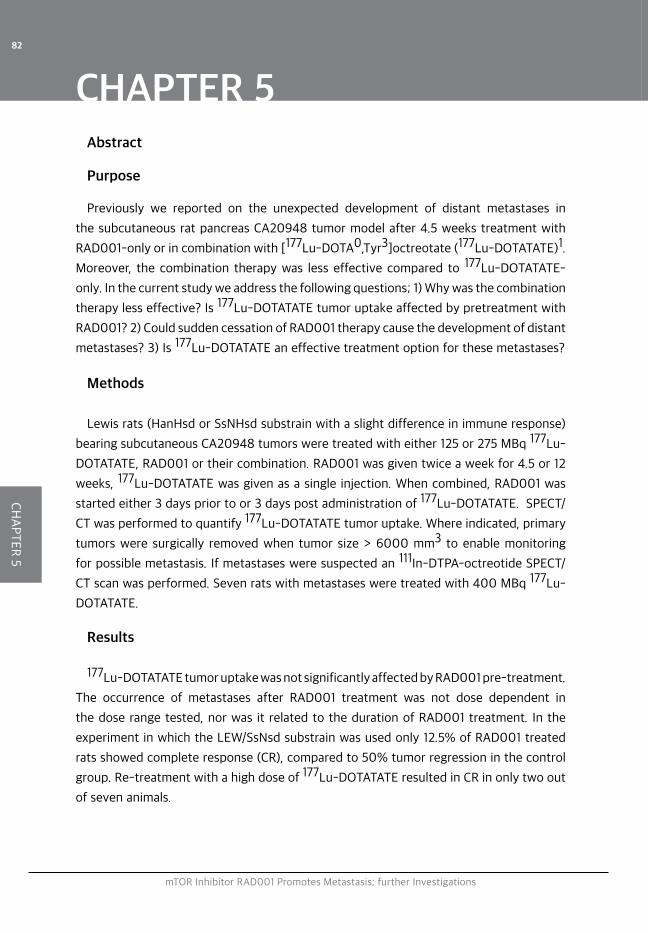
mTOR Inhibitor RAD001 Promotes Metastasis; further Investigations
CHAPTER 5
CH
AP
TE
R 5
82
Abstract
Purpose
Previously we reported on the unexpected development of distant metastases in
the subcutaneous rat pancreas CA20948 tumor model after 4.5 weeks treatment with
RAD001-only or in combination with [177Lu-DOTA0,Tyr3]octreotate (177Lu-DOTATATE)1.
Moreover, the combination therapy was less effective compared to 177Lu-DOTATATE-
only. In the current study we address the following questions; 1) Why was the combination
therapy less effective? Is 177Lu-DOTATATE tumor uptake affected by pretreatment with
RAD001? 2) Could sudden cessation of RAD001 therapy cause the development of distant
metastases? 3) Is 177Lu-DOTATATE an effective treatment option for these metastases?
Methods
Lewis rats (HanHsd or SsNHsd substrain with a slight difference in immune response)
bearing subcutaneous CA20948 tumors were treated with either 125 or 275 MBq 177Lu-
DOTATATE, RAD001 or their combination. RAD001 was given twice a week for 4.5 or 12
weeks, 177Lu-DOTATATE was given as a single injection. When combined, RAD001 was
started either 3 days prior to or 3 days post administration of 177Lu-DOTATATE. SPECT/
CT was performed to quantify 177Lu-DOTATATE tumor uptake. Where indicated, primary
tumors were surgically removed when tumor size > 6000 mm3 to enable monitoring
for possible metastasis. If metastases were suspected an 111In-DTPA-octreotide SPECT/
CT scan was performed. Seven rats with metastases were treated with 400 MBq 177Lu-
DOTATATE.
Results
177Lu-DOTATATE tumor uptake was not significantly affected by RAD001 pre-treatment.
The occurrence of metastases after RAD001 treatment was not dose dependent in
the dose range tested, nor was it related to the duration of RAD001 treatment. In the
experiment in which the LEW/SsNsd substrain was used only 12.5% of RAD001 treated
rats showed complete response (CR), compared to 50% tumor regression in the control
group. Re-treatment with a high dose of 177Lu-DOTATATE resulted in CR in only two out
of seven animals.
mTOR Inhibitor RAD001 Promotes Metastasis; further Investigations
CHAPTER 5
CH
AP
TE
R 5
83
Conclusion
Less effective anti-tumor effects after the combination of RAD001 + 177Lu-DOTATATE
could not be explained by reduced 177Lu-DOTATATE tumor uptake after RAD001.
Our current data support RAD001 induced immune suppression as the reason for
this observation. No evidence was found that cessation of RAD001 treatment caused
development of metastases. Metastases appeared to be less sensitive to 177Lu-DOTATATE
treatment than primary tumors.
Introduction:
Neuroendocrine tumors (NETs) consist of a heterogeneous group of neoplasms
originating from cells characterised by the synthesis and release of amines/peptides2.
Since 1973 the incidence of NETs has been increasing, in which genetic factors might play
a role3 and in addition improved diagnostics contributed to a higher registered incidence
of NETs2. Because NETs are slowly proliferating tumors the prevalence of NETs is much
higher than the incidence, resulting in a relatively high percentage of NET patients in
the population of cancer patients4. In > 50% of the patients, NETs are diagnosed at a
relatively late stage, often with metastatic spread3, which leaves little chance for curative
surgery. As a consequence of the slow proliferation rate most NETs are relatively resistant
to chemotherapeutics.
Most NETs are characterized by overexpression of somatostatin receptors, mainly
subtype 2 (sst2). Targeting these receptors by administration of somatostatin analogs
radiolabeled with e.g. beta particle emitting radionuclides, such as 177Lu or 90Y, allows
peptide receptor radionuclide therapy (PRRT) of NET patients. This therapeutic approach
is being performed since more than 10 years and has proven to be an effective treatment
option in patients with inoperable disease. Therapeutic responses result in a significantly
longer overall survival time compared to other treatments such as chemotherapy or
external beam radiation therapy[5–7]. PRRT also improves patient’s self-assessed quality
of life8. Although PRRT is a successful therapy, complete remissions (CR) in patients with
metastasized disease are still rare, so there is an urgent need for improvement.
The combination of PRRT with the mammalian target of rapamycin (mTOR) inhibitor
Everolimus or RAD001 could be promising in this respect. Everolimus (RAD001) has
recently received FDA approval for the treatment of pancreatic NETs. RAD001 has
been reported to show anti-tumor and anti-angiogenic activity both in vitro as well
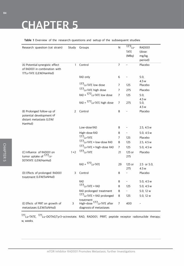
mTOR Inhibitor RAD001 Promotes Metastasis; further Investigations
CHAPTER 5
CH
AP
TE
R 5
84
Table 1 Overview of the research questions and setup of the subsequent studies
Research question (rat strain) Study Groups N 177Lu-
TATE
(MBq)
RAD001
(dose:
mg/kg,
period)
(A) Potential synergistic effect
of RAD001 in combination with
177Lu-TATE (LEW/HanHsd)
1 Control 7 - Placebo
RAD only 6 - 5.0,
4.5 w177Lu-TATE low dose 7 125 Placebo177Lu-TATE high dose 7 275 Placebo
RAD + 177Lu-TATE low dose 7 125 5.0,
4.5 wRAD + 177Lu-TATE high dose 7 275 5.0,
4.5 w
(B) Prolonged follow-up of
potential development of
distant metastasis (LEW/
HanHsd)
2 Control 8 - Placebo
Low-dose RAD 8 - 2.5, 4.5 w
High-dose RAD 8 - 5.0, 4.5 w177Lu-TATE 7 125 Placebo177Lu-TATE + low-dose RAD 8 125 2.5, 4.5 w177Lu-TATE + high-dose RAD 7 125 5.0, 4.5 w
(C) Influence of RAD001 on
tumor uptake of 177Lu-
DOTATATE (LEW/HanHsd)
1 +2 177Lu-TATE 21 125 or
275
Placebo
RAD + 177Lu-TATE 29 125 or
275
2.5 or 5.0,
4.5 w
(D) Effects of prolonged RAD001
treatment (LEW/SsNHsd)
3 Control 8 - Placebo
RAD 8 - 5.0, 4.5 w177Lu-TATE + RAD 8 125 5.0, 4.5 w
RAD prolonged treatment 8 - 5.0, 12 w177Lu-TATE + RAD prolonged
treatment
8 125 5.0, 12 w
(E) Effects of PRRT on growth of
metastases (LEW/SsNHsd)
3 High-dose 177Lu-TATE after
diagnosis of metastases
7 400 -
177Lu-TATE, 177Lu-DOTA0,Tyr3-octreotate; RAD, RAD001; PRRT, peptide receptor radionuclide therapy;
w, weeks.
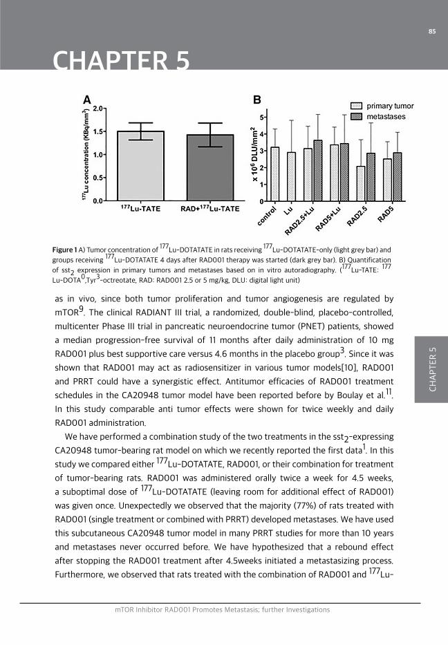
mTOR Inhibitor RAD001 Promotes Metastasis; further Investigations
CHAPTER 5
CH
AP
TE
R 5
85
Figure 1 A) Tumor concentration of 177Lu-DOTATATE in rats receiving 177Lu-DOTATATE-only (light grey bar) and
groups receiving 177Lu-DOTATATE 4 days after RAD001 therapy was started (dark grey bar). B) Quantification
of sst2 expression in primary tumors and metastases based on in vitro autoradiography. (177Lu-TATE: 177
Lu-DOTA0,Tyr3-octreotate, RAD: RAD001 2.5 or 5 mg/kg, DLU: digital light unit)
as in vivo, since both tumor proliferation and tumor angiogenesis are regulated by
mTOR9. The clinical RADIANT III trial, a randomized, double-blind, placebo-controlled,
multicenter Phase III trial in pancreatic neuroendocrine tumor (PNET) patients, showed
a median progression-free survival of 11 months after daily administration of 10 mg
RAD001 plus best supportive care versus 4.6 months in the placebo group3. Since it was
shown that RAD001 may act as radiosensitizer in various tumor models[10], RAD001
and PRRT could have a synergistic effect. Antitumor efficacies of RAD001 treatment
schedules in the CA20948 tumor model have been reported before by Boulay et al.11.
In this study comparable anti tumor effects were shown for twice weekly and daily
RAD001 administration.
We have performed a combination study of the two treatments in the sst2-expressing
CA20948 tumor-bearing rat model on which we recently reported the first data1. In this
study we compared either 177Lu-DOTATATE, RAD001, or their combination for treatment
of tumor-bearing rats. RAD001 was administered orally twice a week for 4.5 weeks,
a suboptimal dose of 177Lu-DOTATATE (leaving room for additional effect of RAD001)
was given once. Unexpectedly we observed that the majority (77%) of rats treated with
RAD001 (single treatment or combined with PRRT) developed metastases. We have used
this subcutaneous CA20948 tumor model in many PRRT studies for more than 10 years
and metastases never occurred before. We have hypothesized that a rebound effect
after stopping the RAD001 treatment after 4.5weeks initiated a metastasizing process.
Furthermore, we observed that rats treated with the combination of RAD001 and 177Lu-
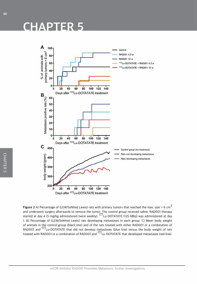
mTOR Inhibitor RAD001 Promotes Metastasis; further Investigations
CHAPTER 5
CH
AP
TE
R 5
86
Figure 2 A) Percentage of (LEW/SsNHsd Lewis) rats with primary tumors that reached the max. size > 6 cm3
and underwent surgery afterwards to remove the tumor. The control group received saline. RAD001 therapy
started at day 4 (5 mg/kg administered twice weekly). 177Lu-DOTATATE (125 MBq) was administered at day
1. B) Percentage of (LEW/SsNHsd Lewis) rats developing metastases in each group. C) Mean body weight
of animals in the control group (black line) and of the rats treated with either RAD001 or a combination of
RAD001 and 177Lu-DOTATATE that did not develop metastases (blue line) versus the body weight of rats
treated with RAD001 or a combination of RAD001 and 177Lu-DOTATATE that developed metastases (red line).
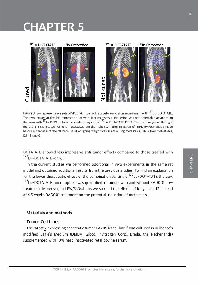
mTOR Inhibitor RAD001 Promotes Metastasis; further Investigations
CHAPTER 5
CH
AP
TE
R 5
87
DOTATATE showed less impressive anti tumor effects compared to those treated with 177Lu-DOTATATE-only.
In the current studies we performed additional in vivo experiments in the same rat
model and obtained additional results from the previous studies. To find an explanation
for the lower therapeutic effect of the combination vs. single 177Lu-DOTATATE therapy, 177Lu-DOTATATE tumor uptake was quantified in tumors with and without RAD001 pre-
treatment. Moreover, in LEW/SsNsd rats we studied the effects of longer, i.e. 12 instead
of 4.5 weeks RAD001 treatment on the potential induction of metastasis.
Materials and methods
Tumor Cell Lines
The rat sst2-expressing pancreatic tumor CA20948 cell line12 was cultured in Dulbecco’s
modified Eagle’s Medium (DMEM, Gibco, Invitrogen Corp., Breda, the Netherlands)
supplemented with 10% heat-inactivated fetal bovine serum.
Figure 3 Two representative sets of SPECT/CT scans of rats before and after retreatment with 177Lu-DOTATATE.
The two images at the left represent a rat with liver metastasis; the lesion was not detectable anymore on
the scan with 111In-DTPA-octreotide made 8 days after 177Lu-DOTATATE PRRT. The two images at the right
represent a rat treated for lung metastases. On the right scan after injection of 1In-DTPA-octreotide made
before euthanasia of the rat because of on-going weight loss. (LuM = lung metastasis, LiM = liver metastases,
Kd = kidney)
mTOR Inhibitor RAD001 Promotes Metastasis; further Investigations
CHAPTER 5
CH
AP
TE
R 5
88
Animals and tumor model
The animal ethics committee of our institution has approved all experiments. Male
Lewis rats (250-300 g) were obtained from Harlan (Heerlen, the Netherlands). We used
LEW/HanHsd Lewis rats and where indicated we also included rats from the LEW/SsNHsd
substrain. In the LEW/SsNHsd substrain the immune system shows an enhanced CD4+
and CD8+ T cell (auto)-immune response13-17. This autoimmunity is linked to an increased
tumor-immunity18,19 One week after arrival, rats were inoculated subcutaneously with
107 CA20948 cells in 0.5 ml HBSS. For all experiments, animals were randomized into
matching treatment groups with regard to tumor size at the start of treatment. A person
blinded for the treatment measured tumor size using a calliper and weighed the rats three
times a week. Tumor volume was calculated using the formula: 0.4 x length x weight x
height. Tumor response was defined as follows: partial response (PR): > 50% reduction
of tumor volume, complete response (CR): 100% reduction of tumor volume. Tumors
were allowed to develop until a maximum of 4-6 cm3and were surgically removed where
indicated. Rats were euthanized when > 10% loss of body weight (BW) was registered.
Anesthesia
2.5% Isoflurane/O2 gas anaesthesia was used at 0.5 ml/min during tumor cell
inoculation, administration of 177Lu-DOTATATE, scanning or surgery.
Surgical procedure to remove the primary tumor
When primary tumors were > 4-6 cm3, surgical tumor resection was performed where
indicated. During surgery a heating pat was used to maintain body temperature. Routine
shaving and disinfecting of the skin was performed. The tumor including the tumor
capsule was carefully dissected from the surrounding tissue. After tumor resection the
skin was closed using individual sutures (vicryl 3/0).
RAD001
RAD001 and its placebo, kindly provided by Novartis Pharmaceuticals; Basel;
Switzerland was used for study 1 and was prepared according to the manufacturers
protocol. For the next experiments RAD001 powder from LC laboratories, Woburn USA,
was dissolved in 2 ml ethanol and diluted with 5% glucose solution in water to obtain 3
mTOR Inhibitor RAD001 Promotes Metastasis; further Investigations
CHAPTER 5
CH
AP
TE
R 5
89
or 6 mg/ml. RAD001 was administered orally by gavage in a volume of 0.2 ml thus 0.8
or 1.6 mg/rat or 2.5 / 5 mg/kg BW was administered depending on treatment group. For
each administration RAD001 was prepared freshly from powder.
Radionuclides and peptides
DOTA0,Tyr3-octreotate was obtained from Mallinckrodt, St Louis, MO, US and 177LuCl2
was obtained from IDB, Baarle-Nassau, the Netherlands. 177Lu-DOTATATE was prepared
as described before20, with a specific activity of 100 MBq/2.75μg peptide, and injected
iv via the tail vein under anesthesia. Labeling of 111In-DTPA-octreotide (OctreoScan,
Covidien, Petten, the Netherlands) in a specific activity of 30 MBq2/0.5μg DTPA-
octreotide was performed as described previously21.
SPECT/CT scanning
Forty-eight hours after injection of 177Lu-DOTATATE, helical SPECT/CT scanning of
the tumor region was performed with the four-headed NanoSPECT/CT system (BioScan,
Washington DC USA). Multi pinhole rat collimators with 9 pinholes (diameter 2.5 mm) per
head were used: 24 projections, 90 sec per projection were applied. SPECT scans were
reconstructed iteratively on a 256x256 matrix, using HiSPECT NG software (Scivis, GmbH
Göttingen Germany) and ordered subset expectation maximization (OSEM). The total
amount of radioactivity (MBq) in the tumor was quantified by drawing a sufficiently large
volume of interest (VOI) around the tumor using InVivoScope software (IVS, Bioscan,
Washington DC USA). To achieve accurate quantification, the camera was calibrated by
scanning a 20 ml polypropylene tube rat phantom filled with a known amount of 177Lu
activity. The in vivo tumor volume was assessed by setting the lower threshold to 90%
of the maximum voxel intensity of the tumor using the IRW program (Siemens). Before
euthanasia, a whole body SPECT/CT scan of rats was acquired 24 h after intravenous
injection of 111In-DTPA-octreotide (50 MBq 111In/ 0,5 μg DTPA-octreotide) to detect
CA20948 metastases. During scanning, the rat body temperature was maintained using
a heated bed.
In vitro autoradiography
Autoradiography was performed on primary tumors as well as metastases. Frozen
mTOR Inhibitor RAD001 Promotes Metastasis; further Investigations
CHAPTER 5
CH
AP
TE
R 5
90
sections of 10 μm (Cryo-Star HM 560 M; Microm, Walldorf, Germany) were mounted on
Superfrost plus slides (Menzel, Braunschweig, Germany) and incubated with 10-10M 111In-
DTPA-octreotide with and without an excess (10-6M) of unlabelled octreotide. Adjacent
sections were used for Hematoxylin/Eosin staining. Tumor sections were exposed to SR
phosphor imaging screens (Packard Instruments Co., Meriden, USA) in X-ray cassettes.
After 48 h exposure, screens were read by a Cyclone phosphor imager and analysed
using OptiQuant 03.00 (Perkin Elmer, Groningen, the Netherlands).
Statistics
Prism software version 5.0 (Graph Pad) was used to analyse tumor growth and
determine statistical significance between groups. An unpaired T-test was used for
statistical analysis of tumor uptake (Figure 1). Results are given as mean +/-SD. A log
rank test was performed for curve comparison in Figures 2A and 2B. Body weight data in
Figure 2C are expressed as mean values.
Experimental design
An overview of the different research questions (A-E) and treatment groups in all
studies (1-3) is given in Table 1 and described below.
A) Potential synergistic effect of RAD001 in combination with 177Lu-DOTATATE
To study whether RAD001 has an additional anti-tumoral effect on 177Lu-DOTATATE,
6 different study groups were included with 6-7 rats per group. Besides the control
group and the RAD001-only therapy groups (5 mg/kg twice a week for 4.5 weeks), 2
groups received 177Lu-DOTATATE as a single therapy in different doses (125 or 275 MBq),
and 2 groups received the same doses of 177Lu-DOTATATE combined with RAD001
treatment (5 mg/kg twice a week for 4.5 weeks starting 3 days prior to PRRT)[1]. Rats
were euthanized when tumor size exceeded 4 cm3.
B) Prolonged follow up of potential development of distant metastasis in all groups
after surgical resection of the primary tumor
Six additional groups of rats were included in the next study with 7-8 rats in each
group. To enable prolonged follow up, in this experiment primary tumors were surgically
removed when >4 cm3. This enabled longer follow up to study the development of
mTOR Inhibitor RAD001 Promotes Metastasis; further Investigations
CHAPTER 5
CH
AP
TE
R 5
91
metastases in relation to the combination of 177Lu-DOTATATE and RAD001 or RAD001-
only. 2.5 or 5.0 mg/kg RAD001 was administered twice weekly for 4.5 weeks, either alone
or 3 days prior to 125 MBq 177Lu-DOTATATE,
C) Influence of RAD001 on tumor uptake of 177Lu-DOTATATE177Lu-DOTATATE-uptake in tumors was quantified based on SPECT/CT scans acquired
48 hours after administration of 177Lu-DOTATATE in experiment 1 and 2 to examine if
previous RAD001 treatment results in reduced 177Lu-retention in CA20948 tumors.
D) Effects of prolonged RAD001 treatment
Five groups of 8 rats were included. 5 mg/kg RAD001 therapy was started 4 days after
125 MBq 177Lu-DOTATATE. RAD001 was administered twice a week and continued for
either 4.5 or 12 weeks. Surgical resection was performed if tumors reached a volume of
6 cm3.
E) Effects of PRRT retreatment on metastases
If rats in experiment 3 showed lethargy or a >10% body weight loss, SPECT/CT
was performed using 111In-DTPA-octreotide. If metastases could be discriminated,
400MBq/10.8μg 177Lu-DOTATATE PRRT was given. 24h after 177Lu-DOTATATE injections,
SPECT/CT was performed to image 177Lu-DOTATATE uptake in metastases. Therapeutic
effect was monitored by follow-up of body weight and 111In-DTPA-octreotide SPECT/CT
when ongoing decrease in body weight was registered.
Results
A) Potential synergistic effect of RAD001 in combination with 177Lu-DOTATATE
In experiment 1 treatment with RAD001-only did not result in any complete or partial
anti-tumor responses, defined as follows: partial response (PR): > 50% reduction of
tumor volume, but no complete response (CR): 100% reduction of tumor volume (Table
2). Groups treated with 177Lu-DOTATATE-only showed 57% CR after 125 MBq 177Lu-
DOTATATE and 71% after 275 MBq 177Lu-DOTATATE. Combination of 177Lu-DOTATATE
and RAD001 however, resulted in only 29% CR after 125 MBq 177Lu-DOTATATE + RAD001
and 14% after 275 MBq 177Lu-DOTATATE + RAD001. So, in contrast to our hypothesis, no
additive effect with regard to tumor response could be achieved by combining RAD001
mTOR Inhibitor RAD001 Promotes Metastasis; further Investigations
CHAPTER 5
CH
AP
TE
R 5
92
and 177Lu-DOTATATE. Moreover, unexpectedly, all rats treated with the combination of
RAD001 and 177Lu-DOTATATE and not showing CR eventually developed metastases as
was reported earlier1.
B) Prolonged follow up of potential development of distant metastasis in all groups
after surgical resection of the primary tumor
Prolonged monitoring after primary tumor removal, the majority (77%) of rats treated
with RAD001 (either 5.0 mg/kg or 2.5 mg/kg), developed metastases that resulted in
mean body weight loss around 43 days after start of treatment. For these two doses no
dose dependence of RAD001 was found (Table 2).
C) Influence of RAD001 on tumor uptake of 177Lu-DOTATATE
The 177Lu-tumor uptake in 177Lu-DOTATATE-only treated rats was 1.51 ± 0,07 kBq/
mm3, while this was 1.42 ± 0,07 kBq/mm3in rats pre-treated with RAD001; no significant
different values were found between the groups (p=0.50, Figure 1A).
D) Effects of prolonged RAD001 treatment
In contrast to previous experiments 50% of the rats in the control group showed a
CR. The rats from the combination groups (177Lu-DOTATATE + RAD001 for 4.5 weeks
resp. 177Lu-DOTATATE + RAD001 for 12 weeks) showed a CR in 87.5% resp. 75% (not
significantly different; p=0.63) of the animals, in comparison to only 12.5% of rats in
both RAD001-only therapy groups. Within these 2 RAD001-only groups, there was no
significant difference regarding both the number of animals that needed surgery as well
as the time until surgery.
With regard to development of metastases, at day 150 p.t. no metastases were
detected in untreated rats (Figure 2B). Also the time of appearance of the metastases in
the combination group was later; 61 d p.t. vs. 91 p.t, respectively.
Monitoring the body weight of rats revealed the effects of treatment and the
development of metastases. Rats in the control group showed a normal gain in body
weight over time (Figure 2C). Rats treated with 177Lu-DOTATATE + RAD001 or RAD001-
only not developing metastases, also showed gain in body weight over time, although at
a slower rate. However, the mean body weight of rats developing metastases stabilized
as a result of their poor condition.
E) Effects of PRRT on growth of metastases
Although SPECT/CT confirmed significant uptake of 177Lu-DOTATATE in metastases
(Figure 3), in only two of the seven rats there was CR after retreatment with high dose
mTOR Inhibitor RAD001 Promotes Metastasis; further Investigations
CHAPTER 5
CH
AP
TE
R 5
93
177Lu-DOTATATE. The average survival time of the non-responsive rats after detection
and treatment of the metastases varied between 8 and 37 days, with an average of
27 days. One rat with CR recovered from a liver metastasis that was clearly visualized
after administration of 177Lu-DOTATATE. Eight days later there was no sign of this lesion
as shown in the 111In-octreotide scan, which was confirmed after dissection. On the
other hand, a rat suffering from lung metastases did not respond to 177Lu-DOTATATE. 8
days after re-treatment further loss in body weight was measured and the 111In-DTPA-
octreotide SPECT/CT still showed extended lung metastases, also found at autopsy.
Determination of sst2-density on CA20948 primary tumors and metastases using in
vitro autoradiography revealed no significant differences (Figure 1B).
Discussion
We previously reported on the disappointing results of the combination of mTOR
inhibitor RAD001 with 177Lu-DOTATATE PRRT in the CA20948 rat tumor model. No
hypothesised synergistic effect was found; the combination treatment appeared even
less effective than 177Lu-DOTATATE-only. This observation cannot be explained by
reduced 177Lu-DOTATATE uptake in the subcutaneous CA20948 tumors after RAD001
treatment as we demonstrated here. (Figure 1A).
Even more striking was the fact that the majority of RAD001 treated animals
developed tumor metastasis to lymph nodes, liver and/or lung. RAD001 initially has been
introduced as an immunosuppressive to protect patients from rejecting allografts after
organ transplantation22. In 2005 Law et al.23 reviewed the immunosuppressive effects
of RAD001 in relation to its anti-tumor effects, and discussed immune suppression
by RAD001 to be tumor growth accentuating. Therefore the application of RAD001
as an anti-tumor agent should be monitored carefully in the clinic, but to the best of
our knowledge as yet no tumor accentuating effects in patients have been reported.
Recently, after the publication of our Priority Report on our first findings, RAD001 has
received FDA approval for the treatment of advanced NETs and is commonly used in
clinical practice nowadays. Although we must consider the fact that RAD001 is used in
patients with already advanced (metastasized) disease, so far no reports on accelerated
metastasis in patients related to RAD001 treatment have been published. We earlier
hypothesized multiple reasons for the occurrence of metastases: the twice-weekly dose
regimen instead of daily dosing as is applied in clinical therapy, effects of RAD001 on
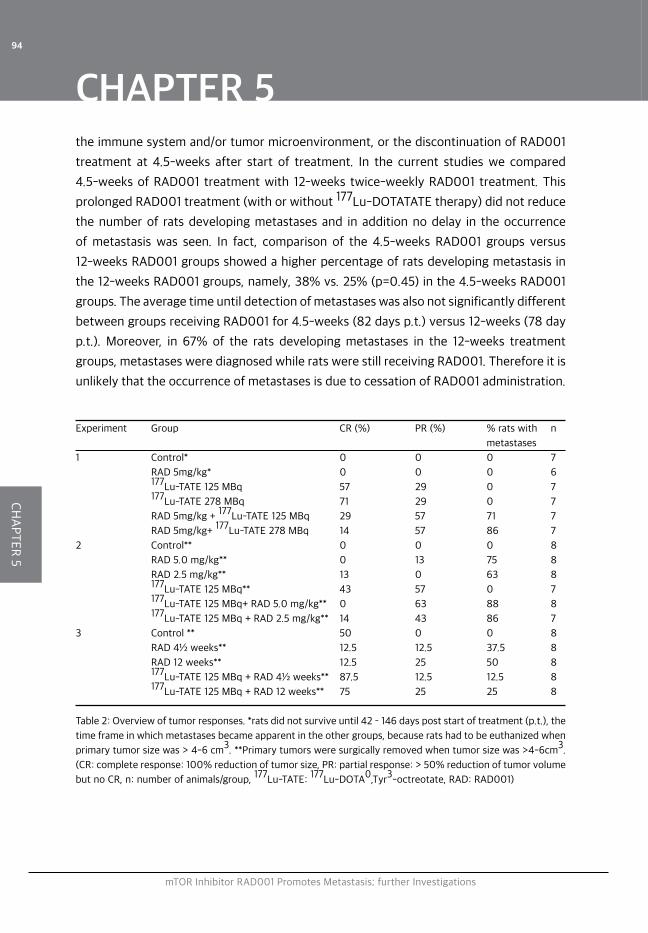
mTOR Inhibitor RAD001 Promotes Metastasis; further Investigations
CHAPTER 5
CH
AP
TE
R 5
94
the immune system and/or tumor microenvironment, or the discontinuation of RAD001
treatment at 4.5-weeks after start of treatment. In the current studies we compared
4.5-weeks of RAD001 treatment with 12-weeks twice-weekly RAD001 treatment. This
prolonged RAD001 treatment (with or without 177Lu-DOTATATE therapy) did not reduce
the number of rats developing metastases and in addition no delay in the occurrence
of metastasis was seen. In fact, comparison of the 4.5-weeks RAD001 groups versus
12-weeks RAD001 groups showed a higher percentage of rats developing metastasis in
the 12-weeks RAD001 groups, namely, 38% vs. 25% (p=0.45) in the 4.5-weeks RAD001
groups. The average time until detection of metastases was also not significantly different
between groups receiving RAD001 for 4.5-weeks (82 days p.t.) versus 12-weeks (78 day
p.t.). Moreover, in 67% of the rats developing metastases in the 12-weeks treatment
groups, metastases were diagnosed while rats were still receiving RAD001. Therefore it is
unlikely that the occurrence of metastases is due to cessation of RAD001 administration.
Experiment Group CR (%) PR (%) % rats with n
metastases
1 Control* 0 0 0 7
RAD 5mg/kg* 0 0 0 6
177Lu-TATE 125 MBq 57 29 0 7
177Lu-TATE 278 MBq 71 29 0 7
RAD 5mg/kg + 177Lu-TATE 125 MBq 29 57 71 7
RAD 5mg/kg+ 177Lu-TATE 278 MBq 14 57 86 7
2 Control** 0 0 0 8
RAD 5.0 mg/kg** 0 13 75 8
RAD 2.5 mg/kg** 13 0 63 8
177Lu-TATE 125 MBq** 43 57 0 7
177Lu-TATE 125 MBq+ RAD 5.0 mg/kg** 0 63 88 8
177Lu-TATE 125 MBq + RAD 2.5 mg/kg** 14 43 86 7
3 Control ** 50 0 0 8
RAD 4½ weeks** 12.5 12.5 37.5 8
RAD 12 weeks** 12.5 25 50 8
177Lu-TATE 125 MBq + RAD 4½ weeks** 87.5 12.5 12.5 8
177Lu-TATE 125 MBq + RAD 12 weeks** 75 25 25 8
Table 2: Overview of tumor responses. *rats did not survive until 42 – 146 days post start of treatment (p.t.), the
time frame in which metastases became apparent in the other groups, because rats had to be euthanized when
primary tumor size was > 4-6 cm3. **Primary tumors were surgically removed when tumor size was >4-6cm3.
(CR: complete response: 100% reduction of tumor size, PR: partial response: > 50% reduction of tumor volume
but no CR, n: number of animals/group, 177Lu-TATE: 177Lu-DOTA0,Tyr3-octreotate, RAD: RAD001)
mTOR Inhibitor RAD001 Promotes Metastasis; further Investigations
CHAPTER 5
CH
AP
TE
R 5
95
Potentially the twice-weekly administration could lead to a repetitive on-off effect on the mTOR
pathway with repetitive up regulation/rebound effects of the mTOR pathway with varying plasma
concentration levels of RAD001 in a twice weekly dose regimen24. In future experiments a daily
RAD001 dose regimen will have to be compared to the twice-weekly dose regimen as was used in
the current studies.
Compared to a treatment with RAD001-only, less animals receiving a combination of 177Lu-
DOTATATE and RAD001 developed metastases and mean time to diagnosis for those metastases
was 30 days later compared to the rats receiving RAD001-only. Results from the combination
therapy groups with 177Lu-DOTATATE administered 4 days before RAD001 therapy suggested
that 177Lu-DOTATATE administered prior to RAD001 therapy reduced both incidence and time of
onset of metastases in comparison to the reverse order combination. When 177Lu-DOTATATE was
administered 4 days after RAD001 therapy however, there was no reduction in the percentage of
rats developing metastases in the combination therapy groups, indicating that rats with (some)
tumor reduction already induced by PRRT were less likely to develop metastases than rats not
treated with PRRT. This is in agreement with the fact that animals that showed CRs after PRRT or
PRRT plus RAD001 did not develop metastases during follow up.
In studies 1 and 2 we used a syngeneic tumor model in rats with an uncompromised immune
system. A significant role for T lymphocytes in the immune response to tumors after or during
ionizing radiation therapy has been described, the latter resulting in upregulation of tumor-specific
antigens25-29. As immune suppression by RAD001 has been proven to be mainly due to suppression
of T-lymphocyte activation and proliferation30,31, to our opinion immune suppression by RAD001
is a likely explanation for reduced tumor response to PRRT in combination with RAD001 as
observed in our study. In studies 1 and 2 LEW/HanHsd rats were used, whereas in study 3 the LEW/
SsNHsd substrain was used, providing the opportunity to test the hypothesis mentioned above.
In this substrain the immune system is more active compared to the HanHsd strain and shows an
enhanced (auto)-immune response13-17. In these rats 50% of tumors in the control group went
into spontaneous regression after reaching an average tumor volume of ≈3cm3, probably due to
an immune response against the growing tumor. In such rats treated with RAD001 (4.5or 12 weeks)
only 12.5% of the tumors went into regression. In a mouse model on rejection of an allogeneic
subcutaneous tumor as created by Hammond-McKribben et al.22, another mTOR inhibitor, the
rapamycine derivate SDZ RAD, was used to prevent rejection of allogeneic tumors. So, immune
suppression by RAD001 might have caused the reduced tumor response in our study with 177Lu-
DOTATATE administered after RAD001 as well.
Contrary to our results, a combination of ionizing radiation (IR) with rapamycin has been proven
to be more effective than IR-only in preclinical studies 10,32,33. These studies however have been
performed in xenograft models using immunodeficient mice lacking T-cells.
As discussed in our priority report, reduced cell proliferation rate caused by a G1 arrest could also
mTOR Inhibitor RAD001 Promotes Metastasis; further Investigations
CHAPTER 5
CH
AP
TE
R 5
96
be an explanation for the reduced tumor response to 177Lu-DOTATATE in rats treated
with the combination of RAD001 and 177Lu-DOTATATE. RAD001 treatment has been
shown to cause a G1 arrest, as mTOR is being linked to phosphatidylinositol 3-kinase
(PI3K) pathways34. Within 24 h after RAD001 administration a significant increase of
cells in G1 phase has been described35,36. As cells with a long cycling time, including
NET cells, have a peak of radioresistance during early G1 phase37, tumor cells may have
been less sensitive to 177Lu-DOTATATE when administered 4 d after the start of RAD001
treatment. Since clinical trials combining RAD001 and PRRT are being performed38, to our
opinion the decreased antitumor response in our study when RAD001 was administered
prior to 177Lu-TATE is rather relevant. Also in a clinical situation the combination of both
therapies might be less effective compared to just PRRT.
For re-treatment of the rats with metastases we used a dose of 400MBq 177Lu-
DOTATATE, which is remarkably higher compared to the initial 125 or 275 MBq treatment
doses. Still only 2 out of 7 rats with metastases were cured, suggesting metastases in our
model to be more resistant to PRRT compared to the primary tumor. As it is quite complex
to study responses of metastases in a preclinical model, there have not been many
preclinical studies on therapies in metastatic models. However, to be able to get more
solid information on sensitivity of metastases to PRRT in a preclinical model, certainly
more studies in different models are necessary. The current CA20948 metastatic tumor
model after RAD001 treatment could be a more realistic metastasis model for future
experiments compared to often-applied metastasis-tumor models in which tumor cell
suspensions are injected intravenously.
Concluding remarks: Results described here confirmed and provided more information
on development of metastasis after RAD001 treatment in our in vivo rat tumor model.
The impaired tumor response to the combination of RAD001 and 177Lu-DOTATATE in
comparison with that after 177Lu-DOTATATE-only could not be attributed to a reduced
tumor uptake of 177Lu-DOTATATE in rats after RAD001 treatment. Moreover, the
occurrence of metastases could not be attributed to the sudden cessation of RAD001
treatment, as we observed treatment for 12-weeks did not result in a lower metastasis
rate compared to treatment for 4.5-weeks. Immune suppression by RAD001 could be a
good explanation for reduced tumor response after RAD001 as well as for development
of metastasis. More studies in different tumor models are needed now to provide proof
and give detailed information on the translational value of these findings to the clinic.
mTOR Inhibitor RAD001 Promotes Metastasis; further Investigations
CHAPTER 5
CH
AP
TE
R 5
97
References
1. Pool SE, Bison S, Koelewijn SJ, van der Graaf LM, Melis M, Krenning EP, et al. mTOR Inhibitor RAD001 Promotes Metastasis in a Rat Model of Pancreatic Neuroendocrine Cancer. Cancer Res. 2012;73:12–8.
2. Modlin IM, Moss SF, Gustafsson BI, Lawrence B, Schimmack S, Kidd M. The archaic distinction between functioning and nonfunctioning neuroendocrine neoplasms is no longer clinically relevant. Langenbecks Arch. Surg. 2011;396:1145–56.
3. Yao JC, Shah MH, Ito T, Bohas CL, Wolin EM, Van Cutsem E, et al. Everolimus for advanced pancreatic neuroendocrine tumors. N. Engl. J. Med. 2011;364:514–23.
4. Yao JC, Hassan M, Phan A, Dagohoy C, Leary C, Mares JE, et al. One Hundred Years After “Carcinoid”: Epidemiology of and Prognostic Factors for Neuroendocrine Tumors in 35,825 Cases in the United States. J. Clin. Oncol. 2008;26:3063–72.
5. Van Essen M, Krenning EP, Kam BL, de Jong M, Valkema R, Kwekkeboom DJ. Peptide-receptor radionuclide therapy for endocrine tumors. Nat. Rev. Endocrinol. 2009;5:382–93.
6. Kwekkeboom DJ, de Herder WW, van Eijck CHJ, Kam BL, van Essen M, Teunissen JJM, et al. Peptide Receptor Radionuclide Therapy in Patients With Gastroenteropancreatic Neuroendocrine Tumors. Semin. Nucl. Med. 2010;40:78–88.
7. Bodei L, Cremonesi M, Grana CM, Fazio N, Iodice S, Baio SM, et al. Peptide receptor radionuclide therapy with 177Lu-DOTATATE: the IEO phase I-II study. Eur. J. Nucl. Med. Mol. Imaging. 2011;38:2125–35.
8. Teunissen JJM. Quality of Life in Patients With Gastroenteropancreatic Tumors Treated With [177Lu-DOTA0,Tyr3]octreotate. J. Clin. Oncol. 2004;22:2724–9.
9. Seeliger H, Guba M, Kleespies A, Jauch K-W, Bruns CJ. Role of mTOR in solid tumor systems: a therapeutical target against primary tumor growth, metastases, and angiogenesis. Cancer Metastasis Rev. 2007;26:611–21.
10. Manegold PC, Paringer C, Kulka U, Krimmel K, Eichhorn ME, Wilkowski R, et al. Antiangiogenic Therapy with Mammalian Target of Rapamycin Inhibitor RAD001 (Everolimus) Increases Radiosensitivity in Solid Cancer. Clin. Cancer Res. 2008;14:892–900.
11. Boulay A, Zumstein-Mecker S, Stephan C, Beuvink I, Zilbermann F, Haller R, et al. Antitumor efficacy of intermittent treatment schedules with the rapamycin derivative RAD001 correlates with prolonged inactivation of ribosomal protein S6 kinase 1 in peripheral blood mononuclear cells. Cancer Res. 2004;64:252–61.
12. Bernard BF, Krenning E, Breeman WAP, Visser TJ, Bakker WH, Srinivasan A, et al. Use of the rat pancreatic CA20948 cell line for the comparison of radiolabelled peptides for receptor-targeted scintigraphy and radionuclide therapy. Nucl. Med. Commun. 2000;21:1079–85.
13. Cutler LS, Rozenski D, Coolens J, Rozing J, Angelillo M, Wayne AL, et al. Experimental autoallergic sialadenitis in the LEW rat: I. Parameters of disease induction. Cell. Immunol. 1991;135:335–45.
14. Greiner DL, Angelillo M, Wayne AL, Fitzgerald KM, Rozenski D, Cutler LS. Experimental autoallergic sialadenitis in the LEW rat: III. Role of CD4+ T cells in EAS induction. Cell. Immunol. 1991;135:354–9.
15. Perlik F, Zidek Z. The susceptibility of several inbred strains of rats to adjuvant-induced arthritis and experimental allergic encephalomyelitis. Z. Für Immunitätsforschung Exp. Klin. Immunol. 1974;147:191.
16. Gasser DL, Palm J, Gonatas NK. Genetic control of susceptibility to experimental allergic encephalomyelitis and the Ag-B locus of rats. J. Immunol. 1975;115:431–3.
17. Friedman I, Ron N, Laufer A, Davies AM. Experimental myocarditis: Enhancement by the use of Pertussis vaccine in Lewis rats. Experientia. 1970;26:1143–5.
18. Gridley G, McLaughlin JK, Ekbom A, Klareskog L, Adami H-O, Hacker DG, et al. Incidence of cancer among patients with rheumatoid arthritis. J. Natl. Cancer Inst. 1993;85:307–11.
19. Sakaguchi S, Takahashi T, Yamazaki S, Kuniyasu Y, Itoh M, Sakaguchi N, et al. Immunologic self tolerance maintained by T-cell-mediated control of self-reactive T cells: implications for autoimmunity and tumor immunity. Microbes Infect. 2001;3:911–8.
20. Kwekkeboom D, Bakker W, Kooij P, Konijnenberg M, Srinivasan A, Erion J, et al. [177Lu-DOTA0,Tyr3]octreotate: comparison with [111In-DTPA0]octreotide in patients. Eur. J. Nucl. Med. Mol. Imaging. 2001;28:1319–25.
21. Breeman WA, Kwekkeboom DJ, Kooij PP, Bakker WH, Hofland LJ. Effect of Dose and Specffic Activity on
mTOR Inhibitor RAD001 Promotes Metastasis; further Investigations
CHAPTER 5
CH
AP
TE
R 5
98
Tissue Distribution of Indium-lil-Pentetreotide in Rats. 1995 [cited 2013 Aug 21]; 22. Hammond-McKibben D, Saulnier M, Zhang J, Risher N, Lake P, Weetall M. Immunologic Pathways in a
Quantitative Model of Immunosuppression Based on Rejection of an Allogeneic or Xenogeneic Tumor Graft: Transplantation. 2005;79:889–96.
23. Law BK. Rapamycin: An anti-cancer immunosuppressant? Crit. Rev. Oncol. Hematol. 2005;56:47–60. 24. Tanaka C, O’Reilly T, Kovarik JM, Shand N, Hazell K, Judson I, et al. Identifying Optimal Biologic Doses
of Everolimus (RAD001) in Patients With Cancer Based on the Modeling of Preclinical and Clinical Pharmacokinetic and Pharmacodynamic Data. J. Clin. Oncol. [Internet]. 2008 [cited 2013 Sep 5]; Available from: http://jco.ascopubs.org/cgi/doi/10.1200/JCO.2007.14.1127
25. North RJ. γ-Irradiation facilitates the expression of adoptive immunity against established tumors by eliminating suppressor T cells. Cancer Immunol. Immunother. 1984;16:175–81.
26. Slone HB, Peters LJ, Milas L. Effect of host immune capability on radiocurability and subsequent transplantability of a murine fibrosarcoma. J. Natl. Cancer Inst. 1979;63:1229–35.
27. Demaria S, Formenti SC. Role of T lymphocytes in tumor response to radiotherapy. Front. Oncol. [Internet]. 2012 [cited 2013 Aug 21];2.
28. Lorimore SA, Coates PJ, Scobie GE, Milne G, Wright EG. Inflammatory-type responses after exposure to ionizing radiation in vivo: a mechanism for radiation-induced bystander effects? Oncogene. 2001;20:7085–95.
29. Apetoh L, Tesniere A, Ghiringhelli F, Kroemer G, Zitvogel L. Molecular interactions between dying tumor cells and the innate immune system determine the efficacy of conventional anticancer therapies. Cancer Res. 2008;68:4026–30.
30. Dumont FJ, Su Q. Mechanism of action of the immunosuppressant rapamycin. Life Sci. 1995;58:373–95. 31. Wicker LS, Boltz RC, Matt V, Nichols EA, Peterson LB, Sigal NH. Suppression of B cell activation by
cyclosporin A, FK506 and rapamycin. Eur. J. Immunol. 1990;20:2277–83. 32. Kim KW, Moretti L, Mitchell LR, Jung DK, Lu B. Combined Bcl-2/Mammalian Target of Rapamycin
Inhibition Leads to Enhanced Radiosensitization via Induction of Apoptosis and Autophagy in Non-Small Cell Lung Tumor Xenograft Model. Clin. Cancer Res. 2009;15:6096–105.
33. Eshleman JS, Carlson BL, Mladek AC, Kastner BD, Shide KL, Sarkaria JN. Inhibition of the mammalian target of rapamycin sensitizes U87 xenografts to fractionated radiation therapy. Cancer Res. 2002;62:7291–7.
34. Fingar DC, Blenis J. Target of rapamycin (TOR): an integrator of nutrient and growth factor signals and coordinator of cell growth and cell cycle progression. Oncogene. 2004;23:3151–71.
35. Loehberg CR, Strissel PL, Dittrich R, Strick R, Dittmer J, Dittmer A, et al. Akt and p53 are potential mediators of reduced mammary tumor growth by Chloroquine and the mTOR inhibitor RAD001. Biochem. Pharmacol. 2012;83:480–8.
36. Sukumari-Ramesh S, Singh N, Dhandapani KM, Vender JR. mTOR inhibition reduces cellular proliferation and sensitizes pituitary adenoma cells to ionizing radiation. Surg. Neurol. Int. [Internet]. 2011 [cited 2013 Aug 21];2.
37. Dodson H, Wheatley SP, Morrison CG. Involvement of centrosome amplification in radiation-induced mitotic catastrophe. Cell Cycle. 2007;6:364–70.
38. Turner JH. Australian Experience of Radiopeptide Therapy of NET. World J. Nucl. Med. 2011;1:52.

mTOR Inhibitor RAD001 Promotes Metastasis; further Investigations
CHAPTER 5
CH
AP
TE
R 5
99

6
6MULTIMODALITY IMAGING OF SOMATOSTATIN RECEPTOR–POSITIVE TUMORS WITH NUCLEAR AND BIOLUMINESCENCE IMAGING
Stefan E. Pool Timo L.M. ten Hagen Stuart Koelewijn
Marion de Jong Gerben A. Koning
Moleculair Imaging. 2012 Feb;11:27-32
CHAPTER 6
102
Multimodality Imaging of Somatostatin Receptor–Positive Tumors
CH
AP
TE
R 6
Molecular Imaging
Multimodal bioluminescence (BLI) and single-photon emission computed tomography/
computed tomography (SPECT/CT) imaging were investigated as means to monitor
somatostatin receptor subtype 2 (SST2)-positive neuroendocrine tumors as both a
subcutaneously implanted and a liver metastasis animal model in mice and rats. Ultimately,
such a model will be of use for studying SST2-targeted peptide receptor radionuclide
therapy (PRRT). CA20948 cells were transfected with a green fluorescent protein/
luciferase plasmid construct. Cells were inoculated subcutaneously in the shoulder of
nude mice: nontransfected cells in the left shoulder and transfected cells in the right
shoulder. BLI, SPECT/CT imaging, biodistribution analysis, and ex vivo autoradiography
of the tumors were performed. BLI and SPECT/CT imaging were also performed on an
intrahepatic tumor model in the rat. Caliper volume measurement of transfected tumors
could be correlated with BLI measurements (R2= .76). SPECT/CT imaging showed high
levels of accumulation of 111In-DTPA-octreotide in control and transfected tumors,
which was confirmed by biodistribution analysis and autoradiography. Subcapsular
inoculation of transfected cells in rat liver resulted in an intrahepatic tumor, which could
be visualized by both SPECT/CT and BLI. Transfection of CA20948 tumor cells did not
alter the growth properties of the cell line or the expression of SST2. Transfected tumors
could be clearly visualized by BLI and SPECT/CT imaging. The transfected SST2-positive
tumor cell line could represent a novel preclinical model for tumor monitoring in studies
that aim at further optimizing PRRT for neuroendocrine tumors.
Introduction
Visualisation of somatostatin receptor–overexpressing neuroendocrine tumors by
radiolabeled somatostatin analogues is being applied clinically for tumor detection, staging,
and selection of therapy.1 Equipping these peptides with therapeutic radionuclides for
peptide receptor radionuclide therapy (PRRT) rendered promising results in preclinical2
and clinical studies.3 Kwekkeboom and colleagues demonstrated an increase in overall
survival ranging from 40 to 72 months from diagnosis, with only minor side effects in
comparison with historical controls in a clinical trial using 177Lu-DOTAtate for treatment
of 310 patients with gastroenteropancreatic neuroendocrine tumor (GEPNET).4 Patients
with liver metastases of GEPNETs have a median overall survival of 2 to 4 years and
hardly benefit from classic therapeutic options such as surgery and chemotherapy,
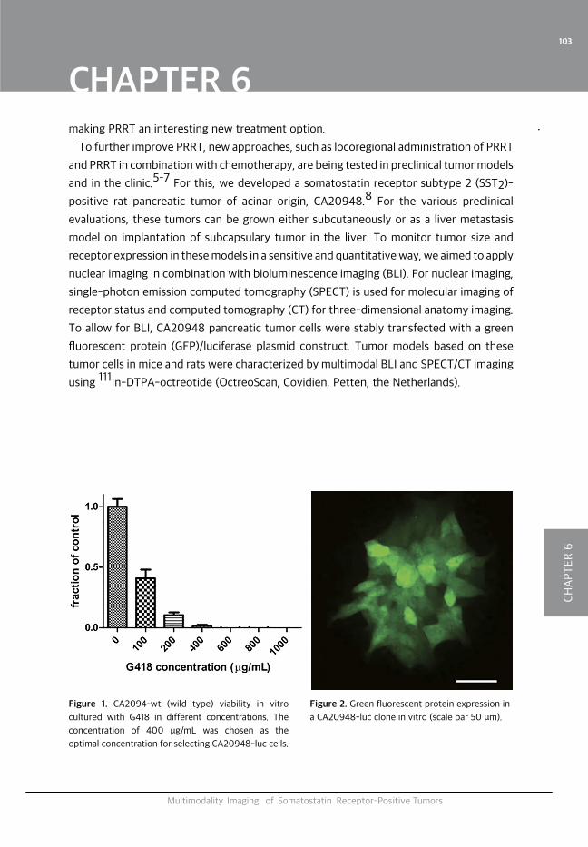
CHAPTER 6
103
Multimodality Imaging of Somatostatin Receptor–Positive Tumors
CH
AP
TE
R 6
making PRRT an interesting new treatment option.
To further improve PRRT, new approaches, such as locoregional administration of PRRT
and PRRT in combination with chemotherapy, are being tested in preclinical tumor models
and in the clinic.5–7 For this, we developed a somatostatin receptor subtype 2 (SST2)-
positive rat pancreatic tumor of acinar origin, CA20948.8 For the various preclinical
evaluations, these tumors can be grown either subcutaneously or as a liver metastasis
model on implantation of subcapsulary tumor in the liver. To monitor tumor size and
receptor expression in these models in a sensitive and quantitative way, we aimed to apply
nuclear imaging in combination with bioluminescence imaging (BLI). For nuclear imaging,
single-photon emission computed tomography (SPECT) is used for molecular imaging of
receptor status and computed tomography (CT) for three-dimensional anatomy imaging.
To allow for BLI, CA20948 pancreatic tumor cells were stably transfected with a green
fluorescent protein (GFP)/luciferase plasmid construct. Tumor models based on these
tumor cells in mice and rats were characterized by multimodal BLI and SPECT/CT imaging
using 111In-DTPA-octreotide (OctreoScan, Covidien, Petten, the Netherlands).
Figure 1. CA2094-wt (wild type) viability in vitro
cultured with G418 in different concentrations. The
concentration of 400 μg/mL was chosen as the
optimal concentration for selecting CA20948-luc cells.
Figure 2. Green fluorescent protein expression in
a CA20948-luc clone in vitro (scale bar 50 μm).
CHAPTER 6
104
Multimodality Imaging of Somatostatin Receptor–Positive Tumors
CH
AP
TE
R 6
Methods
Plasmid and Tranfection
The firefly luciferase gene luc+ was cloned as a < 1.7 kb Nhe1-Xba1 fragment in the
Nhe1 site of a pIRES2-EGFP vector, enabling the expression of a single bicistronic
transcript that encodes both luciferase and GFP. CA20948 cells were transfected with
the construct using lipofectamin (Invitrogen) and cultured under geneticin (G418)
selection. The G418 concentration for selecting transfected tumor cells was based on
the G418 concentration killing almost all CA20948-wt (wild type) cells in an in vitro
cytotoxicity test (SRB [Sulphorodamine B], Sigma Aldrich) of CA20948-wt cells treated
for 24 hours at different concentrations of G418. GFP-positive clones were isolated,
and a clone with high GFP/luciferase expression was selected and characterized in
vitro for expression of SST2.
Internalization Experiments
Twenty-four hours before the experiment, 106 CA20948 cells were plated per well
in six-well plates. The cells were then washed with 2 mL phosphate-buffered saline
(PBS, 37°C) and incubated in 1 mL incubation medium (RPMI-1640 medium [Gibco
BRL] supplemented with 1% bovine serum and 20 mM Hepes) containing 111In-DOTA-
octreotate at a concentration of 10–9M. To determine nonspecific internalization, cells
were incubated with an excess of unlabeled peptide (10–6M octreotate). Cellular
uptake was stopped by removing medium from the cells, followed by washing twice
with 2 mL PBS. To discriminate between internalized and non-internalized (surface
bound) fraction, intact cells were incubated with 1 mL 20 mM sodium acetate (pH 5)
as previously described.9
Fluorescence Microscopy
Cells grown on fibronectin-coated glass coverslips were analyzed by fluorescence
microscopy using a Zeiss Axiovert 100M microscope and an ORCA-II-ER camera
(Hamamatsu).

CHAPTER 6
105
Multimodality Imaging of Somatostatin Receptor–Positive Tumors
CH
AP
TE
R 6
Animals
NMRI Nu/Nu mice (body weight < 27 g) were used. Lewis rats (body weight around
275 g) were used. Animals were kept under standard laboratory conditions (12 hours
light/12 hours dark) and given a standard laboratory diet and water ad libitum. During
SPECT/CT imaging and BLI, animals were placed on a heated bed (38°C) and kept under
isoflurane anesthesia (2.5% at 0.6 L/min). The animal studies conformed with the Animal
Welfare Committee requirements of our institution and were conducted following
generally accepted guidelines.
Figure 3. Internalization assay: comparable
membrane binding (Membrane) and internalization
(Internalized) of 111
In-DOTA-octreotate in
CA20948-luc and -wt cells with and without
blockade with 1,000 times excess octreotate.
Data are presented as mean 6 SEM.
Figure 4. Correlation of caliper and bio-luminescence
imagingmeasurements (R2
=.76).
For tumor induction, 2 x 10⁶ living CA20948 cells in 0.2 mL were injected
subcutaneously in the shoulder of nude mice: nontransfected cells in the left shoulder
and transfected cells (CA20948-luc) in the right shoulder. Tumor growth was measured
by caliper (tumor volume = length x diameter2x 0.4) and BLI.
For the liver metastasis model, two male Lewis rats (Harlan, Horst, the Netherlands)
were used. All surgical and injection procedures used isoflurane/O2 anesthesia and a
microsurgery microscope. During surgery, animals were kept warm with a heating
pad. After laparotomy of the rats upper abdomen by a 1.5 cm incision, the main liver
lobe was fixated between two swabs and around 100 mL CA20948 tumor cells (1.5 x
106cells) suspended in Matrigel basement membrane matrix (BD Biosciences, Breda,
CHAPTER 6
106
Multimodality Imaging of Somatostatin Receptor–Positive Tumors
CH
AP
TE
R 6
the Netherlands) were injected subcapsularly through a 27-gauge needle mounted on
a 1 mL syringe. The abdomen was then closed by absorbable sutures.
Bioluminescence Imaging
BLI (IVIS 200, Xenogen, Hopkinton, MA) of tumor-bearing animals was performed
after intraperitoneal injection of luciferin (0.15 mg/g body weight). During the procedure,
animals were anesthetized with isoflurane and kept at 37°C.
SPECT/CT Imaging
SPECT/CT imaging was performed using the NanoSPECT/CT (Bioscan Inc., Washington,
DC) 4 hours after intravenous injection of 111In-DTPA-octreotide (30 MBq/rat or
mouse). The exact injected activity was determined by measuring the syringe in a
dose calibrator before and after injection. Multi-pinhole mouse and rat collimators
with nine pinholes (1.4 mm diameter for mice and 1.5 mm diameter for rats) per
head were used with a matrix of 256 x 256 and 24 projections (1 minute per projection).
Figure 5. A, Bioluminescence imaging of 4 NMRI Nu/Nu mice 10
minutes after injection of luciferin showing luciferase activity in
CA20948-luc tumor (right shoulder). On the left shoulder, the normal
CA20948 tumor is visible, but without luciferase activity. The green
circles indicate the CA20948- wt tumor. B, 111In-DTPA-octreotide
uptake in the CA20948 (left shoulder) and the CA20948-luc (right
shoulder) tumors 4 hours after injection
Figure 6. Ex vivo autoradiography of
a CA20948-wt and a CA20948-luc
tumor.
CHAPTER 6
107
Multimodality Imaging of Somatostatin Receptor–Positive Tumors
CH
AP
TE
R 6
Tumor Uptake Quantification
Tumor uptake of 111In-DTPA-octreotide was performed by gamma-photon counting
of the tumor using a LKB-1282-Compugammasystem (Perkin Elmer, Waltham, MA)
24 hours after injection (counting time was 60 s/tumor). Before counting the tumor
was weighed.
Ex Vivo Autoradiography
Intratumoral distribution of 111In-DTPA-octreotide was investigated by ex vivo
autoradiography. Tumor tissue isolated 24 hours after injection, was embedded in
Tissue Tek (Sakura, Zoeterwoude, Netherlands) and quickly processed for cryosectioning.
Sections (10 μm) were mounted on glass slides, which were placed on phosphor
imaging screens (Packard Instruments Co., Meriden, CT) for 1 day. The screens were
analyzed using a Cyclone phosphor imager and a computer-assisted OptiQuant 3.00
image processing system (Packard Instruments Co.).
Results
Transfected cells were selected by G418 resistance (400 μg/mL; Figure 1) and cloned.
A clone with the highest GFP activity (Figure 2) was used for further experiments.
Comparable SST2 receptor binding and internalization of 111In-DOTA-octreotate in
the CA20948-wt and CA20948-luc cells were shown in an internalization experiment
Figure 7. Tumor uptake of 111
In-DTPA-octreotide in
CA20948-wt and CA20948-luc tumors as quantified
in a gamma counter 24 hours after injection.
CT was performed at 45 kV. SPECT scans were reconstructed iteratively using InVivoScope
software version 1.32 (Bioscan) with medium noise reduction, a voxel size of 0.3 mm3,
and standard reconstruction settings.
CHAPTER 6
108
Multimodality Imaging of Somatostatin Receptor–Positive Tumors
CH
AP
TE
R 6
(Figure 3). Tumor growth of subcutaneous CA20948-luc and CA20948-wt tumor was
monitored by caliper measurements and BLI and showed good correlation (R2 .76)
(Figure 4). At day 10 after inoculation, the CA20948-luc tumors on the right shoulder
were clearly visible by BLI (Figure 5A), whereas the CA20948 tumors inoculated
in the left shoulder were not. Both tumors were visualized by SPECT/CT imaging on
intravenously injected 111In-DTPA-octreotide, showing comparable levels of uptake and
thus SST2 expression by both the transfected and nontransfected tumors (Figure 5B).
Further comparison by ex vivo autoradiography of tumor sections (Figure 6) and
by radioactivity counting of tumors 24 hours after injection (Figure 7) confirmed
that levels of uptake of 111In-DTPA-octreotide were comparable in transfected and
nontransfected tumors in vivo.
An extra benefit of monitoring tumors by BLI is the possibility of monitoring
orthotopically implanted tumors or metastastatic models. In a CA20948-luc liver
metastasis rat model, intrahepatic tumor could be clearly visualized by both SPECT/CT
(Figure 8A) and BLI (Figure 8B).
Discussion and Conclusion
In this study, we were able to demonstrate that luciferase transfected CA20948
pancreatic tumor cells show SST2 expression and 111In-DTPA-octreotide uptake in vivo
comparable to that of nontransfected CA20948. Transfected tumors could be clearly
visualized by both BLI and SPECT/CT imaging in mice and rats. BLI measurements of
subcutaneous tumors in nude mice showed good correlation with caliper measurements.
The use of BLI for measuring the therapeutic effects of PRRT in our animal models,
especially in the liver metastasis model, is promising. This approach allows for daily
monitoring of tumor growth in the same animal for several weeks. Tissue absorption
of photons by overlying tissue could be a problem when the tumor is situated deep
within the body. In the liver metastasis model used in this study, the tumor is covered
by skin, abdominal wall, and liver capsule. The thickness of these structures is not
expected to change in time. Emitted photons could be absorbed more and more by
the tumor itself during growth of the tumor, making quantitative imaging uncertain. In
our experiments, none of this was noticed. Other imaging modalities, such as CT and
magnetic resonance imaging, could also be used for tumor size monitoring but are more
time consuming. Using BLI, eight mice or four rats can be imaged simultaneously within
10 minutes. Taking these considerations into account, we conclude that BLI can be used
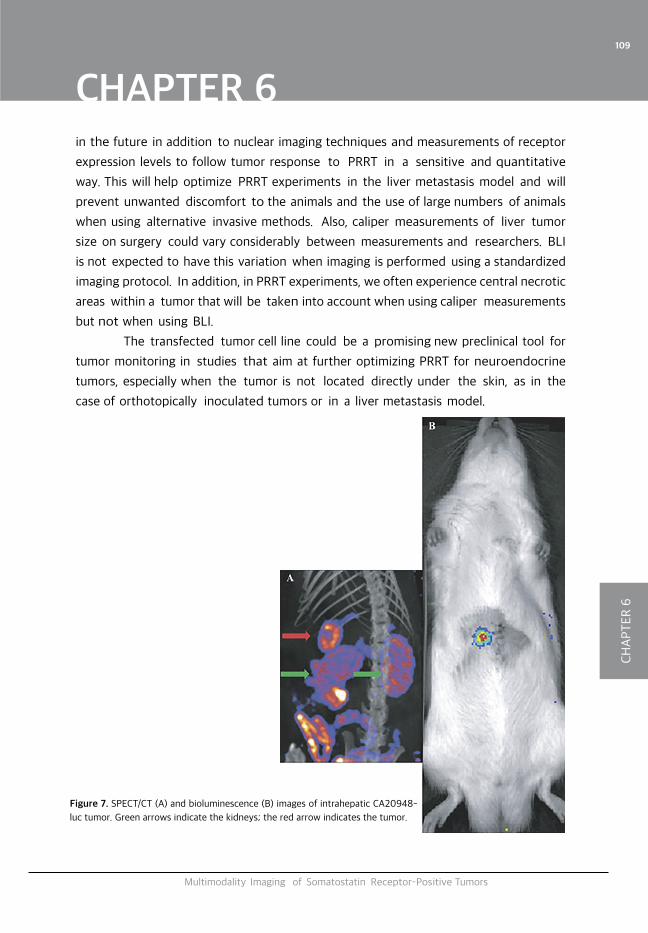
CHAPTER 6
109
Multimodality Imaging of Somatostatin Receptor–Positive Tumors
CH
AP
TE
R 6
in the future in addition to nuclear imaging techniques and measurements of receptor
expression levels to follow tumor response to PRRT in a sensitive and quantitative
way. This will help optimize PRRT experiments in the liver metastasis model and will
prevent unwanted discomfort to the animals and the use of large numbers of animals
when using alternative invasive methods. Also, caliper measurements of liver tumor
size on surgery could vary considerably between measurements and researchers. BLI
is not expected to have this variation when imaging is performed using a standardized
imaging protocol. In addition, in PRRT experiments, we often experience central necrotic
areas within a tumor that will be taken into account when using caliper measurements
but not when using BLI.
The transfected tumor cell line could be a promising new preclinical tool for
tumor monitoring in studies that aim at further optimizing PRRT for neuroendocrine
tumors, especially when the tumor is not located directly under the skin, as in the
case of orthotopically inoculated tumors or in a liver metastasis model.
Figure 7. SPECT/CT (A) and bioluminescence (B) images of intrahepatic CA20948-
luc tumor. Green arrows indicate the kidneys; the red arrow indicates the tumor.
CHAPTER 6
110
Multimodality Imaging of Somatostatin Receptor–Positive Tumors
CH
AP
TE
R 6
References
1. Pool SE, Krenning EP, Koning GA, et al. Preclinical and clinical studies of peptide receptor radionuclide therapy. Semin Nucl Med2010;40:209–18, doi:10.1053/j.semnuclmed.2009.12.001.
2. Breeman WA, Mearadji A, Capello A, et al. Anti-tumor effect and increased survival after treatment with [
177Lu-DOTA
0,Tyr
3] octreotate in a rat liver micrometastases model. Int J Cancer 2003;
104:376–9, doi:10.1002/ijc.10952.3. van Essen M, Krenning EP, Kam BL, et al. Peptide-receptor radionuclide therapy for endocrine tumors.
Nat Rev Endocrinol 2009;5:382–93, doi:10.1038/nrendo.2009.105.4. Kwekkeboom DJ, de Herder WW, Kam BL, et al. Treatment with the radiolabeled somatostatin analog
[177
Lu-DOTA0
,Tyr3
]octreotate: toxicity, efficacy, and survival. J Clin Oncol 2008;26:2124–30, doi:10.1200/JCO.2007.15.2553.
5. McStay MK, Maudgil D, Williams M, et al. Large-volume liver metastases from neuroendocrine tumors: hepatic intraarterial
90Y-DOTA-lanreotide as effective palliative therapy. Radiology 2005;237: 718–
26, doi:10.1148/radiol.2372041203.6. Limouris GS, Chatziioannou A, Kontogeorgakos D, et al. Selective hepatic arterial infusion of In-111-
DTPA-Phe1-octreotide in neuroendocrine liver metastases. Eur J Nucl Med Mol Imaging 2008;35: 1827–37, doi:10.1007/s00259-008-0779-0.
7. van Essen M, Krenning EP, Kam BL, et al. Report on short-term side effects of treatments with 177
Lu-octreotate in combination with capecitabine in seven patients with gastroenteropancreatic neuro endocrine tumours. Eur J Nucl Med Mol Imaging 2008;35:743–8, doi:10.1007/s00259-007-0688-7.
8. Bernard BF, Krenning E, Breeman WA, et al. Use of the rat pancreatic CA20948 cell line for the comparison of radiolabelled peptides for receptor-targeted scintigraphy and radionuclide therapy. Nucl Med Commun 2000;21:1079–85, doi:10.1097/00006231-200011000-00015.
9. De Jong M, Bernard BF, De Bruin E, et al. Internalization of radiolabelled [DTPA0]octreotide and [DOTA0, Tyr3]octreotide: peptides for somatostatin receptor-targeted scintigraphy and radionuclide therapy. Nucl Med Commun 1998;19:283–8, doi:10.1097/ 00006231-199803000-00013.

CHAPTER 6
111
Multimodality Imaging of Somatostatin Receptor–Positive Tumors
CH
AP
TE
R 6

7

7S U M M A R YF U T U R EP E R S P E C T I V E SS A M E N V A T T I N G T O E K O M S T V I S I ED A N K W O O R DC U R R I C U L U M V I T A EL I S T OF P U B L I C A T I O N SP O R T F O L I O
CHAPTER 7
CH
AP
TE
R 7
Summary
114
Summary, Future perspectives
Neuroendocrine tumors are rare neoplasms with an incidence of 2-5 per 100,000
inhabitants. When a NET is diagnosed, in the majority of cases it has already metastasized
and the only curative treatment option available nowadays, i.e. surgery, is not an option any
more. Standard treatment in most of these patients comprise long-acting somatostatin
analogs, possibly in combination with (palliative) surgery, chemotherapy, radiotherapy,
hepatic artery/transarterial (chemo-) embolization (H/TA(C)E), radiofrequency ablation
(RFA), cryoablation and laser-induced thermotherapy (LITT) and intra-arterial radio
embolization with yttrium-90/holmium-166 microspheres.
The widely used long-acting somatostatin analogs bind to somatostatin receptors
(mainly sst2), which are over-expressed on both functioning and non-functioning NETs,
resulting in significant lengthening of time to tumor progression compared to placebo1,2.
Also treatment with somatostatin analogs can reduce hormonal overproduction
of functioning tumors and may result in symptomatic relief in most patients with
metastasized disease3-5. The sst over-expression on NETs is also widely used for
diagnosis/visualization of these tumors using radiolabeled somatostatin analogs, such
as [111Indium-DTPA]-octreotide (Octreoscan®, Covidien, Petten, the Netherlands) and
the more recently developed PET tracers 68Ga-DOTA-Tyr3-octreotide6 or 68Ga-
DOTA-Tyr3-octreotate (68Ga-DOTATATE)7. Based on the same principle, somatostatin
analogs labeled with radionuclides with high-energy beta particle emission, including 177Lu-DOTATATE and 90Y-DOTATOC are commonly being applied for treatment of
the metastasized NETs with most promising results. New is the promising application
of alpha emitters like bismuth-213 (213Bi), which is very likely to be a very effective
strategy because of the high linear energy transfer (LET) of alpha radiation8-10. Some
promising results with 213Bi-DOTATOC PRRT have already been published in seven NET
patients refractory 90Y/177Lu-DOTATOC PRRT10.
In this thesis, several approaches to optimize the treatment of NETs have been
described, as well as their application in different studies and evaluation of the results
thereof. Chapter 1 is a general introduction on NETs and the different treatment
modalities currently being applied. Also, a short overview is given on the animal model
and molecular imaging modalities described and applied in this thesis work.
Chapter 2 is a review of the literature on preclinical and clinical PRRT. The therapeutic
CHAPTER 7
CH
AP
TE
R 7
Summary
115
effects of somatostatin analogs labeled with different radionuclides have been discussed.
Also potential new peptides for PRRT, like glucagon-like peptide 1, gastrin-releasing
peptide, CCK2, epidermal growth factor receptor and αvβ3 integrin targeting peptide
analogues, have been included. Strategies to improve PRRT, including tumor size-specific
use of 177Lu for smaller tumors and 90Y for larger tumors, locoregional administration
and the combination of PRRT with chemotherapy, are described and discussed as well.
Chapter 3 describes the effect of intra-arterial (IA) administration compared to
intravenous (systemic) administration on the [111Indium-DTPA]-octreotide uptake
in liver metastases in an animal model and in 3 patients with NET liver metastases
,varying in sst2 density and tumor load. In rats with intrahepatic sst2-positive tumors,
IA administration resulted in a twice-higher uptake of 111In in the tumor compared to
that after systemic administration. The patient study showed that IA administration can
result in significantly higher 111In uptake in NET liver metastases, although the effect can
differ between patients. The results obtained in one patient further showed accurate
localization of the catheters tip to be essential for optimal IA administration to the whole
liver. In this patient one of the NET liver metastases, situated in a part of the liver that
was not perfused during locoregional administration, showed to have the same uptake as
that in the setting of IV administration. Locoregional IA administration did not implicate
reduction of uptake in tumor lesions elsewhere in the body compared to that after
systemic administration. Estimation of 177Lu dosimetry and pharmacokinetic modeling
was performed based on the results obtained in one patient and showed a 30% reduction
of the estimated radiation dose on the kidneys after intra-arterial administration.
In patients with diffuse or large, somatostatin receptor positive metastasic liver
disease , IA PRRT administration could be preferable over IV administration. Future and
more extended clinical studies will have to reveal if the high uptake after IA administration
results in better tumor response and overall survival as well.
In Chapter 4 we describe the combination study of 177Lu-DOTATATE PRRT with
the mTOR inhibitor RAD001 (Affinitor®, Novartis, Basel, Switzerland) in a NET model in
rats. The aim of the study was to investigate a potential synergistic therapeutic effect
of mTOR inhibition and PRRT, based on earlier reports showing radiosensitizing effects
by mTOR inhibition11,12. Surprisingly, the combination treatment appeared to be less
effective compared to 177Lu-DOTATATE treatment alone, but even more importantly,
we observed rats that received RAD001 treatment twice weekly for 4.5 weeks (with or
CHAPTER 7
CH
AP
TE
R 7
Summary
116
without 177Lu-DOTATATE treatment) to develop distant metastasis, whereas control and 177Lu-
DOTATATE treated animals did not and never did before in our earlier studies during more than 10
years. We hypothesized that the discontinuation of RAD001 treatment at 4.5 weeks after start of
treatment could have caused the occurrence of metastasis, probably by a rebound effect of the
mTOR pathway. Inhibition of the immune system by RAD001 could also be an explanation for the
occurrence of distant metastases.
Chapter 5 describes additional analyses derived from the in Chapter 4 described combination
studies and from an additional animal study in which RAD001 treatment twice weekly for 4.5-weeks
was compared to RAD001 treatment given for 12-weeks. Aim of the study was to investigate if the
cessation of RAD001 after 4.5 weeks of treatment was causing the occurrence of metastasis. The
12-weeks RAD001 treatment also resulted in the development of metastasis. Also 177Lu tumor
uptake after 177Lu-DOTATATE treatment with and without RAD001 co-treatment was quantified,
to see if this could serve as an explanation for the fact that the combination treatment of RAD001
and 177Lu-DOTATATE was less effective compared to that with 177Lu-DOTATATE alone. 177Lu-
DOTATATE tumor uptake was not affected by RAD001 treatment. This experiment was performed
in a different substrain of Lewis rats, often used for auto-immune studies, as these animals
have a more active immune system showing an enhanced CD4+ and CD8+ T cell (auto)-immune
response13-17. This autoimmunity is linked to an increased anti-tumor immunity18,19. In these
rats 50 % of subcutaneous tumors disappeared spontaneously in the control group, whereas in
the RAD001 treatment groups only 12.5 % of the tumors regressed, probably caused by RAD001
induced inhibition of the immune system and in line with our hypothesis.
A recent report by Yin et al.20 showed comparable effects of mTOR inhibition in BALB/c
mice bearing subcutaneous 4T1 murine breast cancer tumors. The mTOR inhibitor rapamycine
accelerated lung metastasis. Rapamycin induced production of immunosuppressive molecules and
cytokines, like TGF-β, arginase-1, indoleamine 2,3-dioxygenase, IL-6 and IL-10, in the lungs. The
authors postulate treatment with rapamycin to result in an immunosuppressive microenvironment
in the metastatic sites.
A definite answer on the occurrence of metastasis in the CA20948 tumor model after RAD001
treatment is not yet fully clear and more studies are needed to solve the still open questions.
The use of mTOR inhibition treatment in the clinical setting should be closely watched for the
development of unexpected, new metastasized disease.
Chapter 6 describes the transfection of CA20948 cells with a plasmid coding for green
fluorescent protein and luciferase, for use of non-invasive tumor follow up by bioluminescence
imaging (BLI) in future PRRT experiments in a liver metastasis rat model. We successfully applied
CHAPTER 7
CH
AP
TE
R 7
Summary
117
these transfected CA20948-luc cells in a subcutaneous nude mouse as well in a rat liver-metastasis
model as described in this chapter.
Future perspectives
Currently all clinical data published on the efficacy of 177Lu-DOTATATE treatment for NETs were
based on non-randomized studies, with data compared to those obtained in historical controls.
In 2012 however, a multicenter, prospective, randomized, clinical trial in patients with inoperable,
progressive midgut NETs, has started. This study randomizes between 177Lu-DOTATATE (four
administrations of 7.4 GBq (200 mCi) at 8±1-week intervals) combined with Octreotide LAR 30
mg (discontinued during PRRT) versus treatment with high dose Octreotide LAR 60 mg. Primary
efficacy endpoint is progression-free survival as measured by objective tumour response, centrally
assessed according to the response evaluation criteria in solid tumors (RECIST)21, every 12±1
weeks from the first treatment date in 280 patients. Concomitant amino acids will be given with
each administration for kidney protection. Estimated Primary Completion Date is December 2014.
(www.clinicaltrials.gov; Identifier: NCT01578239)
Combination of PRRT with several new biologicals and chemotherapeutics could result in a
higher therapeutic effect. For example, a prospective, randomized, clinical trial comparing RAD001
+ Octreotide LAR versus 177Lu-DOTATATE + Octreotide LAR in patients with advanced GEPNETs
would be of very high interest now. Despite our observations in the studies described in Chapters
4 and 5, RAD001-induced metastasis has not been reported in patients.
Currently a randomized clinical trial, comparing 177Lu-DOTATATE treatment with 177Lu-
DOTATATE treatment in combination with the oral chemotherapeutic drug capecitabine (Xeloda ;
Roche, Basel, Switzerland) is ongoing in our center. Earlier studies already showed that combining
these therapies is safe and at least as effective as 177Lu-DOTATATE PRRT alone22,23. The aim
of this study is to show that chemosensitization with capecitabine improves the percentage of
patients with objective tumor responses who are also treated with 177Lu-DOTATATE. In the near
future the results of this study will be published.
Despite the disappointing and surprising results of Everolimus (RAD001) with 177Lu-
DOTATATE treatment combinations as described in Chapters 4 and 5 in this thesis, this combination
therapy has now in Australia been given to patients with metastasized NET with tolerable side
effects. In the future, a phase II randomized controlled trial of 177Lu-DOTATATE with capecitabine/
temozolomide versus 177Lu-DOTATATE with RAD001 is planned to be performed in Australia24.
With our preclinical results in mind, a close watch is needed for possible increase in metastases
after/during RAD001 treatment in patients.
Another very promising targeted therapy, Sunitinib (Sutent®, Pfizer), a VEGF-receptor
CHAPTER 7
CH
AP
TE
R 7
Summary
118
inhibitor25, is an interesting option for comparison to PRRT in future randomized clinical trials.
Combination therapy of Sunitinib with PRRT would be the next step. Transient normalization of
the tumor vasculature by VEGF-receptor inhibition results in decreased intratumoral pressure and
better oxygenation with possible facilitation of higher intratumoral radiopeptide penetration and
increased radiation effects by higher oxygenation26.
Intra-arterial PRRT is of high value in selected NET patients, as described in Chapter 3 of this
thesis and elsewhere27-29. In the near future selected NET patients with a predominant tumor
load in the liver will be offered intra arterial 177Lu-DOTATATE PRRT in our center.
In selected NET patients with the tumor-mass mainly located in the liver, a combined
approach of intra-arterial 177Lu-DOTATATE PRRT with 166Ho-loaded poly(L-lactic acid)
microsphere radio-embolisation30 of NET liver metastases compared to intra-arterial 177Lu-
DOTATATE PRRT alone is a strategy worth investigating. 166Ho-microspheres are preferable over 90Y-microspheres, because 166Ho emits low energy gamma photons for gamma camera imaging
and is paramagnetic, allowing magnetic resonance imaging (MRI).
In the near future, 177Lu-DOTATATE PRRT dosimetry will hopefully be performed at our
institution, resulting in patient tailored dose calculation, resulting in optimal tumor radiation with
minimal radiation damage to the healthy tissues. Possibly many patients treated with the standard
dose of 4 times 7.4 GBq 177Lu-DOTATATE are under-treated and could be treated with higher
dosages without significant side effects and better tumor responses are to be expected. Patients
with, e.g., a suboptimal kidney function could receive a lower dose preventing end stage renal
damage.
Varying the specific activity of 177Lu (amount of radioactivity/gram), the peptide mass of
DOTATATE (now around 200 μg) and infusion rate are very interesting methods of increasing
tumor 177Lu uptake and resulting therapeutic effects. Further (pre)clinical studies will have to be
performed to explore the effects of these strategies.
The use of 177Lu-DOTATATE PRRT as a neoadjuvant treatment in patients with initially
irresectable pancreatic NETs has been performed at our center and is promising. We therefore
propose a prospective study on neo-adjuvant use of 177Lu-DOTATATE PRRT to be initiated.
The recently obtained and promising results on the use of the alpha emitter 213Bi for α-PRRT in
preclinical animal experiments8,9 and in patients with NETs refractory to 90Y- or 177Lu-DOTATOC
β-PRRT10 will certainly result in clinical trials using 213Bi α-PRRT as single treatment for NETs or
in combination with e.g. 90Y or 177Lu β-PRRT. Acute and mid-term toxicity seems to be low, but
CHAPTER 7
CH
AP
TE
R 7
Summary
119
long-term follow up is still needed.
Preclinical and clinical studies showed higher higher uptake of radiolabeled sst antagonists
than of sst agonists31,32 in neuroendocrine tumors and promising tumor responses. Future studies
will have to show the potential benefit of these antagonists over agonists.
In conclusion, the field of PRRT is rapidly evolving and will hopefully result in increasing
progression-free survival and higher quality of life for NET bearing patients. Hopefully, these rapid
developments will ultimately result in curing a higher percentage of these patients.
CHAPTER 7
CH
AP
TE
R 7
Samenvatting
120
Samenvatting, toekomstvisie
Neuro endocriene tumoren zijn relatief zeldzaam met een incidentie van 2-5 per
100.000 inwoners. Bij initiële diagnose is de tumor in de meerderheid van de gevallen
al gemetastaseerd en is de enige behandeling met kans op volledige curatie, chirurgie,
geen optie meer. De standaard behandeling bestaat meestal uit het gebruik van een
langwerkend somatostatine analoog mogelijk gecombineerd met (palliatieve) chirurgie,
chemotherapie, radiotherapie, transarteriële (chemo) embolisatie, radiofrequente ablatie
(RFA), cryoablatie en laser geïnduceerde thermotherapie (LITT) en intra-arteriële radio
embolisatie met yttrium-90 of holmium-166 microsferen.
De veelgebruikte langwerkende somatostatine analogen binden aan
somatostatine receptoren (voornamelijk de somatostatine-2-receptor) die tot
overexpressie worden gebracht op zowel functionele als niet functionele neuro endocriene
tumoren wat resulteert in een significante verlenging van progressie vrije overleving in
vergelijking met placebo1,2. De behandeling met somatostatine analogen kan ook de
overproductie van hormonen door functionele neuro endocriene tumoren remmen en
hiermee een verlichting van de symptomen bij patiënten met gemetastaseerde ziekte 3-5. De over expressie van somatostatine receptoren op neuro-endocrine tumoren wordt
ook gebruikt voor diagnose/visualisatie van deze tumoren door middel van radioactief
gelabelde somatostatine analogen zoals [111Indium-DTPA]-octreotide (Octreoscan®,
Covidien, Petten, Nederland) en de meer recent ontwikkelde positron emissie tomografie
(PET) tracers 68Ga-DOTA-Tyr3-octreotide6 of 68Ga-DOTA-Tyr3-octreotate (68Ga-
DOTATATE)7. Gebaseerd op hetzelfde principe wordt voor therapie gebruik gemaakt
van somatostatine analogen gelabeld met hoog energetische bèta deeltjes uitzendende
radionucliden zoals bijvoorbeeld 177Lu-DOTATATE en 90Y-DOTATOC. De resultaten
bij deze vooralsnog niet geregistreerde middelen zijn veelbelovend. Relatief nieuw
is de toepassing van radionucliden die alfa deeltjes uitzenden. Het gebruik van deze
radionucliden zal waarschijnlijk een erg succesvolle strategie zijn door de hoge lineaire
energie transmissie (LET) van alfa straling8-10. Recent zijn veelbelovende resultaten
gepubliceerd met 213Bi-DOTATOC peptide receptor radionucliden therapie (PRRT)
bij zeven patiënten met neuro endocriene tumoren die weer progressief waren na 90Y/177Lu-DOTATOC PRRT10.
In dit proefschrift zijn verschillende manieren beschreven om de therapie voor neuro
endocriene tumoren te optimaliseren. Zo ook de toepassing en resultaten hiervan in
CHAPTER 7
CH
AP
TE
R 7
Samenvatting
121
verschillende studies. Hoofdstuk 1 is een algemene introductie in het veld van de neuro
endocriene tumoren en in de verschillende behandelingsmogelijkheden. Ook wordt er
een korte uitleg gegeven over het gebruikte diermodel in de beschreven studies in dit
proefschrift.
Hoofdstuk 2 is een review van de literatuur over preklinische en klinische PRRT. De
therapie effecten van somatostatine analogen met verschillende radionucliden worden
er beschreven. Ook mogelijke nieuwe peptiden voor PRRT zoals glucagon-like peptide 1,
gastrin-releasing peptide, CCK2 en peptiden die binden aan de epidermal growth factor
receptor en α2β2 integrine. Mogelijke strategieën zoals bijvoorbeeld het gebruik van 177Lu voor kleinere tumoren en 90Y voor grotere tumoren, locoregionale toediening en
de combinatie PRRT met chemotherapie, worden besproken.
Hoofdstuk 3 beschrijft de effecten van intra-arteriële (IA) toediening in vergelijking
met intra veneuze (systemische) toediening op de opname van [111Indium-DTPA]-
octreotide in levermetastasen (CA20948 tumor) in een diermodel en in 3 patiënten
met neuro endocriene levermetastasen met een variërende somatostatine 2 receptor
expressie en een verschillende tumormassa. In ratten met een somatostatine 2 receptor
positieve tumor in de lever resulteerde IA toediening in een tweemaal zo hoge opname
van 111In in de tumor vergeleken met systemische toediening. In de patiëntenstudie
bleek dat IA toediening ook kan resulteren in een significant hogere 111In opname in
NET levermetastasen maar dat dit effect kan variëren per patiënt. De resultaten bij één
patiënt lieten zien dat lokalisatie van de tip van de katheter essentieel is voor optimale
IA toediening in de gehele lever. Bij deze patiënt bleek dat een in de linker kant van de
lever gelegen levermetastase niet werd geperfundeerd met het radiofarmacon tijdens
de IA toediening. Deze levermetastase bleek uiteindelijk dezelfde hoeveelheid 111In te
bevatten als na systemische toediening. IA toediening via de leverslagader hoeft dus
niet te resulteren in een lagere opname van het radiofarmacon in tumoren elders in het
lichaam in vergelijking met systemische toediening. Farmacokinetische modelering en
theoretische schatting van 177Lu dosimetry werd uitgevoerd op de verkregen metingen
bij één patiënt en liet een 30% reductie van de stralingsdosis op de nieren zien na IA
toediening.
In patiënten met diffuse of grote somatostatine receptor positieve levermetastasen
kan locoregionale PRRT de voorkeur hebben boven systemische toediening. Toekomstige
meer uitgebreide klinische studies zullen moeten bewijzen dat hogere opname van het
CHAPTER 7
CH
AP
TE
R 7
Samenvatting
122
radiofarmacon na IA toediening resulteert in een betere tumor respons en een betere
overleving.
In Hoofdstuk 4 beschrijven wij een studie waarin 177Lu-DOTATATE PRRT wordt
gecombineerd met de mTOR inhibitor RAD001 (Affinitor®, Novartis, Basel, Switzerland)
bij ratten met een onder de huid geplaatste neuro endocriene (CA20948) tumor. Het
doel van deze studie was om te onderzoeken of deze twee behandelingen elkaar zouden
versterken. In eerdere studies is namelijk aangetoond dat RAD001 tumoren gevoeliger
kan maken voor bestraling11,12. Verrassend genoeg bleek de combinatietherapie minder
effectief te zijn dan 177Lu-DOTATATE behandeling alleen. Maar belangrijker was dat
dieren die voor 4½ week twee maal per week RAD001 toegediend hadden gekregen
(in combinatie met / zonder 177Lu-DOTATATE) uiteindelijk uitzaaiingen bleken te
ontwikkelen. Controledieren en dieren die alleen 177Lu-DOTATATE hadden gekregen
bleken geen uitzaaiingen te krijgen. Dit tumormodel wordt op onze afdeling al meer
dan 10 jaar gebruikt en niet eerder hebben wij uitzaaiingen bij deze dieren gevonden.
Onze hypothese was dat het stoppen met de RAD001 therapie na 4½ week mogelijk het
ontstaan van de uitzaaiingen had veroorzaakt. Mogelijk door een versterkte reactivatie
van de intracellulaire mTOR pathway. Ook remming van het immuunsysteem door
RAD001 zou een oorzaak van de metastasering kunnen zijn.
Hoofdstuk 5 beschrijft verdere analyses van de resultaten gevonden bij de in hoofdstuk
4 beschreven combinatiestudies. Ook wordt een bijkomende studie beschreven waarin
RAD001 behandeling gedurende 4½ week wordt vergeleken met 12 weken durende
RAD001 therapie. Het doel van deze studie was om te onderzoeken of het stoppen van
de therapie na 4½ week de oorzaak van het ontwikkelen van uitzaaiingen was. De dieren
die 12 weken RAD001 kregen bleken echter ook uitzaaiingen te ontwikkelen.
Ook werd de mate van 177Lu-DOTATATE opname in de tumor gemeten met en zonder
de combinatie met RAD001. Een lagere opname in de tumor zou namelijk een verklaring
kunnen zijn voor het feit dat de combinatie therapie minder effectief was dan 177Lu-
DOTATATE alleen. De mate van 177Lu-DOTATATE opname in de tumor was echter niet
afhankelijk van de behandeling met RAD001.
In dit experiment werden ratten gebruikt van een andere sub-stam van Lewis ratten die
vaak gebruikt wordt voor auto-immuun studies omdat deze dieren een immuunsysteem
hebben met een hogere CD4+ en CD8+ T cel (auto)-immuun respons13-17. Deze auto-
immuniteit wordt gelinkt aan een hogere anti tumor immuniteit18,19. In deze dieren
CHAPTER 7
CH
AP
TE
R 7
Samenvatting
123
verdwenen 50% van de tumoren spontaan in de controle groep terwijl in de RAD001
groep maar 12½% van de tumoren verdwenen, waarschijnlijk veroorzaakt door remming
van het immuunsysteem en in lijn met onze hypothese.
Een recente publicatie door Yin et al.20 laat soortgelijke effecten zien na behandeling
met een mTOR inhibitor in BALB/c muizen met onderhuidse 4T1 muizen borstkankers.
De mTOR remmer Rapamycine bleek het ontstaan van uitzaaiingen naar de longen
te versterken. Rapamycine bleek de productie van immuun remmende moleculen en
cytokines zoals TGF-β, arginase-1, indoleamine 2,3-dioxygenase, IL-6 en IL-10 in de
longen te induceren. De auteurs postuleren dat behandeling met Rapamycine resulteert
in een immuun remmend microklimaat ter plaatse van de uitzaaiingen.
Een definitief antwoord op het ontstaan van uitzaaiingen bij RAD001 behandeling in
het CA20948 tumormodel in de rat is er helaas nog niet en meer studies zijn nodig om
de resterende vragen te beantwoorden. Het gebruik van mTOR remmers in de klinische
setting zal echter goed moeten worden gemonitord voor het ontstaan van onverwachte
(nieuwe) uitzaaiingen.
Hoofdstuk 6 beschrijft de transfectie (genetische modificatie) van CA20948 tumor
cellen met een plasmide dat het gen voor een groen fluorescerend eiwit en voor het eiwit
luciferase bevat. Dit met het doel om in toekomstige PRRT experimenten de tumoren op
een niet invasieve manier te kunnen vervolgen middels bioluminescentie imaging (BLI).
We hebben, zoals beschreven in dit hoofdstuk, deze getransfecteerde CA20948-luc
cellen succesvol onderhuids in een naakte muis model en in een levermetastase model
in de rat kunnen gebruiken.
Toekomstvisie
De huidige gepubliceerde klinische data over de effectiviteit van 177Lu-DOTATATE
PRRT voor neuro endocriene tumoren zijn gebaseerd op niet gerandomiseerde studies
waarbij de uitkomsten vergeleken worden met historische controles. In 2012 is er echter
een prospectieve gerandomiseerde multicenter klinische studie gestart waarin patiënten
gerandomiseerd 177Lu-DOTATATE (vier toedieningen van 7.4 GBq (200 mCi) met 8±1-
week interval) gecombineerd met Octreotide LAR 30 mg (gediscontinueerd tijdens
PRRT) of alleen behandeling met een hoge dosis Octreotide LAR (60 mg) krijgen. Het
primaire eindpunt van deze studie is progressie vrije overleving gebaseerd op objectieve
centrale meting van tumor respons elke 12±1 weken na de eerste behandeling bij 280
patiënten middels de RECIST criteria (response evaluation criteria in solid tumors)21.
CHAPTER 7
CH
AP
TE
R 7
Samenvatting
124
Voor bescherming van de nieren worden er bij de toediening van 177Lu-DOTATATE
aminozuren gegeven. De geschatte primaire einddatum van deze studie is December
2014. (www.clinicaltrials.gov; Identifier: NCT01578239)
Het combineren van PRRT met nieuwe biologicals en/of chemotherapeutica zou
kunnen resulteren in een beter therapeutisch effect. Een prospectieve gerandomiseerde
klinische studie bij patiënten met gastro entero pancreatische neuro endocriene tumoren
(GEPNET) waarin RAD001 met Octreotide LAR wordt vergeleken met 177Lu-DOTATATE
PRRT + Octreotide LAR zou zeer interessant zijn. Gelukkig zijn de effecten van RAD001
behandeling zoals beschreven in hoofdstuk 4 en 5 niet bij patiënten gerapporteerd.
Op dit moment is er een gerandomiseerde klinische studie gaande in ons ziekenhuis
waarin 177Lu-DOTATATE PRRT wordt vergeleken met 177Lu-DOTATATE PRRT in
combinatie met het orale chemotherapeuticum capecitabine (Xeloda ; Roche, Basel,
Switzerland). Eerdere studies hebben al laten zien dat de combinatie van deze twee
therapieën veilig en tenminste even effectief is als 177Lu-DOTATATE PRRT alleen22,23.
Het doel van de studie in ons ziekenhuis is om aan te tonen dat chemosensitisatie met
capecitabine resulteert in een hoger percentage van patiënten met een objectieve tumor
respons na 177Lu-DOTATATE PRRT. De resultaten van deze studie worden waarschijnlijk
in de nabije toekomst gepubliceerd.
Ondanks de teleurstellende en verrassende resultaten van de combinatie van Everolimus
(RAD001) met 177Lu-DOTATATE PRRT zoals beschreven in hoofdstuk 4 en 5 is deze
combinatietherapie in Australië al gegeven met minimale bijwerkingen aan patiënten
met uitgezaaide neuro endocriene tumoren. In Australië zijn er plannen voor een fase II
gerandomiseerde gecontroleerde trial waarin 177Lu-DOTATATE PRRT met capecitabine/
temozolomide zal worden vergeleken met 177Lu-DOTATATE met RAD00124. Met onze
preklinische resultaten in het achterhoofd zullen deze patiënten extra goed gemonitord
moeten worden voor het ontstaan van nieuwe uitzaaiingen tijdens/na behandeling met
RAD001.
Een andere veelbelovende therapie, Sunitinib (Sutent, Pfizer), een VEGF-receptor
remmer25, is ook een zeer interessante optie om te vergelijken met PRRT in een
gerandomiseerde klinische studie. Combinatie van Sunitinib met PRRT zou dan een
logische volgende stap zijn. Tijdelijke normalisatie van de vaatvoorziening van de tumor
door middel van VEGF-receptor remming resulteert in een verlaging van de intratumorale
druk en een betere zuurstof toevoer met hierdoor facilitatie van een betere penetratie
van het radiopeptide in de tumor en een verhoogd effect van de bestraling door de
CHAPTER 7
CH
AP
TE
R 7
Samenvatting
125
betere oxygenatie26.
Intra-arteriële PRRT is van grote waarde in bepaalde patiënten met neuro endocriene
tumoren zoals beschreven in Hoofdstuk 3 van dit proefschrift en elders27-29. In de nabije
toekomst zullen patiënten met een tumormassa die voornamelijk in de lever is gelegen
IA 177Lu-DOTATATE PRRT worden aangeboden.
Bij deze patiënten zou het ook interessant zijn om een gecombineerde aanpak van
IA 177Lu-DOTATATE PRRT met 166Ho-bevattende poly(L-lactic acid) microsphere radio-
embolisatie30 te vergelijken met IA 177Lu-DOTATATE PRRT alleen. 166Ho-microspheren
hebben de voorkeur ten opzicht van 90Y-microspheren, vanwege het feit dat 166Ho laag
energetische gamma fotonen uitzendt die gebruikt kunnen worden voor gamma camera
imaging. Tevens is 166Ho paramagnetisch, waardoor het goed zichtbaar is met magnetic
resonance imaging (MRI).
Hopelijk zal er op korte termijn 177Lu-DOTATATE PRRT dosimetry worden uitgevoerd.
Dit zal dan resulteren in een patiënt specifieke dosis berekening wat uiteindelijk zal
resulteren in een optimale bestraling van de tumor met een minimale bestralingsdosis
op de gezonde organen. Mogelijk worden veel patiënten die nu de standaard dosis van 4
keer 7,4 GBq 177Lu-DOTATATE krijgen onder behandeld en zouden zij een hogere dosis
kunnen krijgen zonder significante bijwerkingen en waarschijnlijk een hogere tumor
respons. Patiënten met bijvoorbeeld een gestoorde nierfunctie zouden juist een lagere
dosis krijgen waarbij nierfalen voorkomen zou worden.
Het variëren van de specifieke activiteit van 177Lu (hoeveelheid radioactiviteit/
gram), van de peptide massa van DOTATATE (nu ongeveer 200 μg/7,4 GBq) en van de
infusiesnelheid zijn interessante opties om de opname van 177Lu in de tumor te verhogen
en zo mogelijk ook het therapeutisch effect. Meer (pre)klinische studies zullen gedaan
moeten worden om deze strategieën te onderzoeken.177Lu-DOTATATE PRRT als neoadjuvante therapie bij patiënten met initieel niet-
resecteerbare pancreatische neuro endocriene tumoren is reeds gebruikt in ons
centrum en is veelbelovend. Een prospectieve studie waarin 177Lu-DOTATATE PRRT als
neoadjuvante therapie wordt gegeven zou daarom gestart moeten worden.
De onlangs gepubliceerde en veelbelovende resultaten over het gebruik van de alfa
emitter 213Bi voor α-PRRT in preklinische dierexperimenten8,9 en in patiënten met 90Y- of 177Lu-DOTATOC β-PRRT refractaire neuro endocriene tumoren10 zullen zeker
resulteren in klinische studies waarin 213Bi α-PRRT alleen als behandeling voor neuro-
endocriene tumoren wordt gegeven of in combinatie met bijvoorbeeld 90Y or 177Lu
β-PRRT. De acute en middellange bijwerkingen lijken minimaal te zijn maar opvolging op

CHAPTER 7
CH
AP
TE
R 7
Samenvatting
126
de lange termijn is nog nodig.
Preklinische en klinische studies hebben een hogere opname van somatostatine
receptor antagonisten in vergelijking met somatostatine receptor agonisten in neuro-
endocriene tumoren laten zien met een veelbelovende tumor respons.31,32. Toekomstige
studies zullen deze antagonisten moeten vergelijken met de huidige agonisten met
betrekking tot therapeutisch effect en mate van bijwerkingen.
We kunnen in ieder geval concluderen dat het veld van PRRT snel evolueert en dat dit
hopelijk zal resulteren in een steeds hogere progressie vrije overleving van patiënten met
neuro endocriene tumoren met hierbij een hogere kwaliteit van leven. Hopelijk zullen al
de hierboven beschreven ontwikkelingen uiteindelijk resulteren in een hoger percentage
echte curatie van deze patiënten met een neuro endocriene tumor.
Reference list
1. Rinke, A. et al. Placebo-Controlled, Double-Blind, Prospective, Randomized Study on the Effect of Octreotide LAR in the Control of Tumor Growth in Patients With Metastatic Neuroendocrine Midgut Tumors: A Report From the PROMID Study Group. J. Clin. Oncol. 27, 4656–4663 (2009).
2. Caplin, M. E. et al. Lanreotide in Metastatic Enteropancreatic Neuroendocrine Tumors. N. Engl. J. Med. 371, 224–233 (2014).
3. Arnold, R., Benning, R., Neuhaus, C., Rolwage, M. & Trautmann, M. E. Gastroenteropancreatic endocrine tumours: Effect of Sandostatin® on tumour growth. Digestion 54, 72–75 (1993).
4. Janson, E. T. & Öberg, K. Long-Term Management of the Carcinoid Syndrome Treatment with octreotide alone and in combination with alpha-interferon. Acta Oncol. 32, 225–229 (1993).
5. Ducreux, M. et al. The antitumoral effect of the long-acting somatostatin analog lanreotide in neuroendocrine tumors. Am. J. Gastroenterol. 95, 3276–3281 (2000).
6. M., H. et al. Biokinetics and imaging with the somatostatin receptor PET radioligand 68
Ga-DOTATOC: preliminary data. Eur. J. Nucl. Med. Mol. Imaging 28, 1751–1757 (2001).
7. Breeman, W. A. P. et al. 68
Ga-labeled DOTA-Peptides and 68
Ga-labeled Radiopharmaceuticals for Positron Emission Tomography: Current Status of Research, Clinical Applications, and Future Perspectives. Semin. Nucl. Med. 41, 314–321 (2011).
8. Norenberg, J. P. 213
Bi-[DOTA0
, Tyr3
]Octreotide Peptide Receptor Radionuclide Therapy of Pancreatic Tumors in a Preclinical Animal Model. Clin. Cancer Res. 12, 897–903 (2006).
9. Nayak, T. K. et al. Somatostatin-receptor-targeted α-emitting 213Bi is therapeutically more effective than β- emitting
177Lu in human pancreatic adenocarcinoma cells. Nucl. Med. Biol. 34, 185–193 (2007).
10. Kratochwil, C. et al. 213Bi-DOTATOC receptor-targeted alpha-radionuclide therapy induces remission in neuroendocrine tumours refractory to beta radiation: a first-in-human experience. Eur. J. Nucl. Med. Mol. Imaging (2014). doi:10.1007/s00259-014-2857-9
11. Mauceri, H. Everolimus exhibits efficacy as a radiosensitizer in a model of non-small cell lung cancer. Oncol. Rep. (2012). doi:10.3892/or.2012.1666
12. Ekshyyan, O. et al. Comparison of radiosensitizing effects of the mammalian target of rapamycin inhibitor CCI-779 to cisplatin in experimental models of head and neck squamous cell carcinoma. Mol. Cancer Ther. 8, 2255–2265 (2009).
13. Cutler, L. S. et al. Experimental autoallergic sialadenitis in the LEW rat: I. Parameters of disease induction. Cell. Immunol. 135, 335–345 (1991).
14. Greiner, D. L. et al. Experimental autoallergic sialadenitis in the LEW rat: III. Role of CD4+ T cells in EAS
CHAPTER 7
CH
AP
TE
R 7
Samenvatting
127
induction. Cell. Immunol. 135, 354–359 (1991).15. Perlik, F. & Zidek, Z. The susceptibility of several inbred strains of rats to adjuvant-induced arthritis and
experimental allergic encephalomyelitis. Z. Für Immun. Exp. Klin. Immunol. 147, 191 (1974).16. Gasser, D. L., Palm, J. & Gonatas, N. K. Genetic control of susceptibility to experimental allergic
encephalomyelitis and the Ag-B locus of rats. J. Immunol. 115, 431–433 (1975).17. Friedman, I., Ron, N., Laufer, A. & Davies, A. M. Experimental myocarditis: Enhancement by the use of Pertussis
vaccine in Lewis rats. Experientia 26, 1143–1145 (1970).18. Gridley, G. et al. Incidence of cancer among patients with rheumatoid arthritis. J. Natl. Cancer Inst. 85, 307–311
(1993).19. Sakaguchi, S. et al. Immunologic self tolerance maintained by T-cell-mediated control of self-reactive T cells:
implications for autoimmunity and tumor immunity. Microbes Infect. 3, 911–918 (2001).20. Yin, T. et al. Antiangiogenic Therapy Using Sunitinib Combined with Rapamycin Retards Tumor Growth But
Promotes Metastasis. Transl. Oncol. 7, 221–229 (2014).21. Eisenhauer, E. A. et al. New response evaluation criteria in solid tumours: Revised RECIST guideline (version 1.1).
Eur. J. Cancer 45, 228–247 (2009).22. Van Essen, M. et al. Report on short-term side effects of treatments with
177Lu-octreotate in combination with
capecitabine in seven patients with gastroenteropancreatic neuroendocrine tumours. Eur. J. Nucl. Med. Mol. Imaging 35, 743–748 (2008).
23. Claringbold, P. G., Brayshaw, P. A., Price, R. A. & Turner, J. H. Phase II study of radiopeptide 177
Lu- octreotate and capecitabine therapy of progressive disseminated neuroendocrine tumours. Eur. J. Nucl. Med. Mol. Imaging 38, 302–311 (2011).
24. Harvey, J. Australian experience of radiopeptide therapy of NET. World J. Nucl. Med. 52 (2011).25. Raymond, E. et al. Sunitinib malate for the treatment of pancreatic neuroendocrine tumors. N. Engl.
J. Med. 364, 501–513 (2011).26. Wachsberger, P., Burd, R. & Dicker, A. P. Tumor response to ionizing radiation combined with
antiangiogenesis or vascular targeting agents Exploring mechanisms of interaction. Clin. Cancer Res. 9, 1957–1971 (2003).
27. Kratochwil, C. et al. Intraindividual Comparison of Selective Arterial versus Venous 68
Ga-DOTATOC PET/CT in Patients with Gastroenteropancreatic Neuroendocrine Tumors. Clin. Cancer Res. 16, 2899– 2905 (2010).
28. Kratochwil, C. et al. Hepatic arterial infusion enhances DOTATOC radiopeptide therapy in patients with neuroendocrine liver metastases. Endocr. Relat. Cancer 18, 595–602 (2011).
29. Limouris, G. S. et al. Selective hepatic arterial infusion of In-111-DTPA-Phe1-octreotide in neuroendocrine liver metastases. Eur. J. Nucl. Med. Mol. Imaging 35, 1827–1837 (2008).
30. Smits, M. L. et al. Research Holmium-166 radioembolization for the treatment of patients with liver metastases: design of the phase I HEPAR trial. (2010). at <http://www.biomedcentral.com/content/ pdf/1756-9966-29-70.pdf>
31. Ginj, M. et al. Radiolabeled somatostatin receptor antagonists are preferable to agonists for in vivo peptide receptor targeting of tumors. Proc. Natl. Acad. Sci. 103, 16436–16441 (2006).
32. Wild, D. et al. Comparison of Somatostatin Receptor Agonist and Antagonist for Peptide Receptor Radionuclide Therapy: A Pilot Study. J. Nucl. Med. 55, 1248–1252 (2014).
CHAPTER 7
CH
AP
TE
R 7
Dankwoord
128
Dankwoord
Bij de start van mijn promotie onderzoek leek het allemaal redelijk eenvoudig; Even
een tumorcellijn transfecteren, een rattenstudie opzetten en een patiëntenstudie
opzetten. Uiteindelijk bleek het allemaal wat minder makkelijk te gaan. Stug doorzetten
wierp echter toch z’n vruchten af. Met veel plezier kijk ik terug op deze leerzame tijd en
graag wil ik iedereen bedanken die op welke manier dan ook een bijdrage aan dit
proefschrift heeft geleverd.
Allereerst wil ik de patiënten bedanken die hebben deelgenomen aan de klinische
studie. Geheel belangeloos met het oog op een mogelijke verbetering van PRRT voor
toekomstige patiënten met een neuro endocriene tumoren hebben zij meegedaan aan
een tijdrovende en toch best wel belastend (het plaatsen van de IA katheter) onderzoek.
Mijn beide promotoren prof.dr. Marion de Jong, prof.dr. Casper H.J. van Eijck en
voormalig afdelingshoofd nucleaire geneeskunde prof.dr Eric P. Krenning wil ik graag
bedanken voor de onderzoekskansen die ze mij geboden hebben. Beste Marion, veel
dank voor de leerzame en inspirerende samenwerking. Jouw enthousiasme voor
wetenschappelijk onderzoek is zeer groot en daardoor erg motiverend. Je scherpe blik,
het voor oog houden van het grote geheel, je doorzettingsvermogen en geduldigheid,
hebben ervoor gezorgd dat er hier uiteindelijk toch een proefschrift ligt. Veel dank
hiervoor. Daarnaast waardeer ik ook je persoonlijke betrokkenheid. Beste Casper bedankt
voor je klinische inbreng, je scherpe commentaren en begeleiding bij het preklinische en
klinische onderzoek.
Eric, je zeer brede wetenschappelijke kennis, je plezier in wetenschap en voornamelijk
ook het plezier dat je haalde uit het discussiëren over de wetenschap hebben veel indruk
op mij gemaakt. Ook je niet te stoppen enthousiasme voor bijvoorbeeld het opzetten van
een cyclotronfaciliteit in het ErasmusMC zijn inspirerend. Ik ben er trots op met je samen
te hebben gewerkt.
Dr. G.A. Koning, mijn co-promotor, beste Gerben, bedankt voor je gedegen begeleiding
vanuit de chirurgie met brede kennis over farmacokinetiek/dynamiek en ook nucleaire
kennis. Natuurlijk ook voor je persoonlijke betrokkenheid. Je hebt toch best wel eens de
druk moeten opvoeren om dit proefschrift compleet te krijgen. Ook jouw bedank ik voor
de goede gesprekken en het benodigde geduld.

CHAPTER 7
CH
AP
TE
R 7
Dankwoord
129
Mijn paranimfen, Stuart Koelewijn en Nanno Ouwehand.
Beste Stuart, als stagiair van de HLO kwam je onder mij hoede. Je was gelijk erg
enthousiast over het proefdieronderzoek en de te gebruiken chirurgische technieken.
Ik waardeer je grote inzet zeer. Ook dank voor je met de hand getekende illustraties in
publicaties en op de kaft van dit proefschrift.
Nanno, beste vriend vanaf de basisschool, bedankt voor alle tijd voor ontspanning
zoals tijdens het windsurfen, kitesurfen, zeilen, mountainbiken, wakeboarden etc. Toch
best lekker naast al dat werken!
Dr. W.A.P. Breeman, beste Wout, ik kan me de eerste keer op de afdeling Nucleaire
Geneeskunde nog goed herinneren. Een uitleg (bijna een college) over wat radionucliden
therapie nou is. Ik werd gelijk gegrepen door jouw enthousiasme over deze voor mij
toch redelijk onbekende wereld van de radiofarmacie. In de jaren hierna hebben we
vaker uitgebreide gesprekken gehad over de radiochemische aspecten bij PRRT en over
mogelijk uit te voeren experimenten. Ik heb deze gesprekken altijd erg gewaardeerd.
Dr.T.L.M. ten Hagen, beste Timo, bedankt voor je al je scherpe commentaren en goede
discussies tijdens de labmeetings.
Prof.dr. A.M.M. Eggermont, beste Lex, ondanks dat jij niet mijn promotor was wist je
toch altijd wat er speelde. Jouw betrokkenheid ondanks je zeer drukke bestaan waardeer
ik zeer.
Dr. M. Konijnenberg, beste Mark, jouw kennis van farmacokinetiek en dosimetrie gaat
ver. En je past het allemaal zo moeiteloos toe. Bedank voor je hulp en uitleg.
Dr.P.P.M. Kooij, beste Peter, jou bedank ik eveneens voor je hulp bij de analyse van de
patiëntendata.
Drs. B.L.M. Kam, beste Boen, jij hebt mij begeleid bij het opzetten van de patiëntenstudie
en de analyse ervan. Dank daarvoor.
Prof.dr. O.C. Boerman, bedankt voor de gezelligheid tijdens congressen en voor het
lezen en beoordelen van mijn proefschrift.

CHAPTER 7
CH
AP
TE
R 7
Dankwoord
130
Prof.dr. L.J. Hofland, bedankt voor het lezen en beoordelen van mijn proefschift
en voor het delen van je expertise m.b.t. de mTOR pathway.
Natuurlijk ook veel dank aan Marion, Caroline, Nomi (medisch nucleair werkers) voor
de hulp en planning bij het scannen.
Arthur (MNAA) bedankt voor de hulp bij de analyse van de urine monsters.
Chulja en Wanda (verpleegkundig specialisten) eveneens bedankt voor al jullie hulp.
Alle medewerkers en mede onderzoekers op het CIL (centraal isotopen lab) Stuart,
Marleen, Sander, Rogier, Bert, Magda, Ria, Linda, Saskia, Harald, Jan, Suzanne, Monique,
Edgar, Ingrid, Sandra, Panthelis, Hoze en Erik, heel erg bedankt voor de hulp en goede
samenwerking. Natuurlijk ook voor de gezellige koffiepauzes en kerstontbijten.
Alle medewerkers en mede onderzoekers op het lab experimentele chirurgische
oncologie Ann, Asha, Biljana, Marna, Jill, Lili, Lilia, Lucy, Michiel, Thomas, Joost, Cindy,
Csilla, Debby, Gisela, erg bedankt voor de leuke en leerzame tijd. Bedankt voor alle hulp
bij de cel- en dierexperimenten en natuurlijk voor de niet te vergeten borrels tot soms
in de late uurtjes.
Petra, Satish en Peter, het was erg leuk om jullie te mogen begeleiden tijdens jullie
stages op de afdeling. Dank voor al jullie werk.
Ook Ron, Henk en Sandra van het laboratorium experimentele transplantie chirurgie,
dank jullie voor hulp en gezelligheid. Henk met name bedankt voor je hulp bij de
transfectie van de cellijn.
Mijn mede AIOS/oud AOIS Hanneke, Kathleen, Asai, Stoffel, Martijn, Tessa, Laura en
Maureen. Dank voor de gezelligheid tijdens het werk, op cursussen en tijdens de borrels
bijvoorbeeld het skiweekend.
Natuurlijk bedank ik ook iedereen van de afdeling nucleaire geneeskunde voor de hulp
en gezelligheid op de werkvloer en tijdens de afdelingsdagen.

CHAPTER 7
CH
AP
TE
R 7
Dankwoord
131
Fred, Roelf, Lideke, Jasper, Jaap, Boen en Dik, stafartsen op de afdeling nucleaire
geneeskunde, bedankt voor al jullie adviezen, begeleiding en gezelligheid de afgelopen
jaren.
Beste Winnifred, overige stafartsen radiologie en AIOS-en radiologie, heel erg bedankt
voor de zeer leerzame en leuke tijd het afgelopen jaar. Ik kijk uit naar de verdere integratie
van de nucleaire geneeskunde met de radiologie.
Monique, Gabi, Joost en Sandra van de preklinische afdeling radiologie, bedankt voor
jullie hulp en gezelligheid.
Tekla heel erg bedankt voor het opmaken van dit proefschrift. Het was natuurlijk
weer stress op het laatste moment, maar het is toch een mooi geheel geworden.
Familie en schoonfamilie, bedankt voor jullie betrokkenheid de laatste jaren.
Mijn schoonouders wil ik bedanken voor de ontspanning en gezelligheid
tijdens de vele skivakanties en natuurlijk ook voor de mooie dagen in Reeuwijk.
Beste Len, zus, ik ben blij dat je weer in Nederland terug bent. Bedankt voor
alle gezelligheid en afleiding.
Mijn ouders, dankzij jullie heb ik qua studie alles kunnen doen. Dank voor alle liefde,
steun, zorg en vertrouwen.
Lieve Dorien, eindelijk is het proefschrift klaar. Bedankt voor je hulp en begrip de
laatste tijd. Ik ben gelukkig met jou, onze Sofie en wie er nog meer mag komen.

CHAPTER 7
CH
AP
TE
R 7
Curriculum Vitae
132
CHAPTER 7
CH
AP
TE
R 7
Curriculum Vitae
133
Curriculum Vitae
Stefan Pool was born on 6th of March 1978 in Hilversum, the Netherlands. He
grew up with his sister in Capelle aan den IJssel. He attended secondary school
at ‘het Emmaus College’ in Rotterdam from which he graduated the gymnasium
in 1997. Due to numerus fixus he started Medical school in Antwerp, Belgium.
He continued medical school at the ErasmusMC, Rotterdam, the Netherlands.
Beside Medical school he started a Master in Molecular Medicine at the MolMed
Postgraduate school at the ErasmusMC. In 2005 he obtained his medical degree
and his bachelor’s degree in Molecular Medicine.
After finishing Medical school he started in a PhD project in a collaboration of the
Department of Nuclear Medicine and the Department of Surgery, ErasmusMC.
In 2011 he started his residency in Nuclear Medicine under supervision of Dr.
D.J. Kwekkeboom. This will be completed in 2016.
He lives together with Dorien Rijkaart and their child Sofie (2011) and in joyful
expectation of a second child.

CHAPTER 7
CH
AP
TE
R 7
List of publications
134
CHAPTER 7
CH
AP
TE
R 7
List of publications
135
List of publications
Pool SE, Bison SM, Koelewijn SJ, van der Graaf LM, Melis M, Groen HC, de Jong M.
Peptide receptor radionuclide therapy (PRRT) with [177Lu-DOTA0,Tyr3]octreotate in
combination with RAD001 treatment; further investigations on tumor metastasis and
response in a rat pancreatic cancer model. European Journal of Nuclear Medicine and
Molecular Imaging Research. 2014 June;4:21
Pool SE, Kam B, Koning GA, Konijnenberg M, ten Hagen TLM, Breeman WAP, Krenning
EP, de Jong M, van Eijck CHJ. [111In-DTPA]octreotide tumor uptake in GEPNET liver
metastasis after intra-arterial administration – an overview of preclinical and clinical
observations and implications for tumor radiation dose after peptide radionuclide
therapy. Cancer Biotherapy and Radiopharmaceuticals. 2014 May;29(4):179-87
Pool SE, Bison S, Koelewijn SJ, van der Graaf LM, Melis M, Krenning EP, de Jong M. mTOR
Inhibitor RAD001 Promotes Metastasis in a Rat Model of Pancreatic Neuroendocrine
Cancer. Cancer Research 2013 Jan;73(1): 12-8
Pool SE, ten Hagen TL, Koelewijn SJ, de Jong M, Koning GA. Multimodality imaging of
somatostatin receptor-positive tumors with nuclear and bioluminescence imaging. Mol
Imaging. 2012 Feb;11(1):27-32
Pool SE, Krenning EP, Koning GA, van Eijck CH, Teunissen JJ, Kam B, Valkema
R, Kwekkeboom DJ, de Jong M. Preclinical and clinical studies of peptide receptor
radionuclide therapy. Semin Nucl Med. 2010 May;40(3):209-18
Bison SM, Konijnenberg MW, Melis M, Pool SE, Bernsen MR, Teunissen JJM,
Kwekkeboom DJ, de Jong M. Peptide receptor radionuclide therapy using radiolabeled
somatostatin analogs: focus on future developments. Clinical Translational Imaging. 2014
Feb; (2):55-66

CHAPTER 7
CH
AP
TE
R 7
Portfolio
136

CHAPTER 7
CH
AP
TE
R 7
Portfolio
137
PHD PORTFOLIO
Summary of PhD training and teaching
Erasmus MC Departments: Nuclear Medicine and Surgical Oncology
Research School: Molecular Medicine
PhD period: 2005 – 2013
Promotor: Prof.dr. M. de Jong, Prof.dr. C.H.J. van Eijck
Supervisor: Dr. G.A. Koning
1. PhD training
General academic skills Year ECTS
Photoshop and Illustrator CS-4 Workshop (molmed) 2010 0,3
Biomedical English Writing and Communication (Molmed) 2009 2
Research management for PhD-students 2009 1
Get out of your lab days (Molmed) 2006 0,6
JNI weekly meetings 2005-2010 1
JNI oncology lectures 2005-2010 0,3
Journal club meetings LECO and Nuclear medicine 2005-2010 1
Research skills
Course ‘Radiation safety, level 3’ 2013 12
AMIE Imaging workshop & symposium 2009 1,4
BROK (Basiscursus Regelgeving Klinisch Onderzoek) course 2006 0,6
In Vivo Imaging 'From Molecule to Organism' (molmed) 2006 1,8
Course ‘Radiation safety, level 5B’ 2005 1,5
Course ‘proefdierkunde ex art. 9’ 2005 3
In-depth courses
Course ‘Geavanceerde beeldvormende technieken
voor dokters’ 2009 0,2
Analysis of Microarray Gene Expression Data (Molmed) 2005 2
Course Basic and Translational Oncology (molmed) 2005 1,8
Research meetings, Department of Nuclear Medicine 2005-2010 1

CHAPTER 7
CH
AP
TE
R 7
Portfolio
138
Research meetings, Laboratory of
Experimental Surgical Oncology (LECO) 2005-2010 1
Presentations / posters at conferences
European Congress of Radiology (ECR),
Vienna, Austria 2014 2014 1
Annual meeting of the Radiological Society of
North America (RSNA) 2012, Chicaco, USA 2012 1
Annual congress of the Society of Nuclear Medicine (SNM)
2011, San Antonio, USA (poster) 2011 1
Annual meeting of the European Association of
Nuclear Medicine (EANM) 2011, Birmingham, England (oral) 2011 1
EANM 2010, Wien, Austria (2 posters) 2010 1
EANM 2009, Barcelona, Spain (2 posters) 2009 1
EANM 2007, Copenhagen, Denmark (oral) 2007 1
NKRV workshop, UMC St Radboud, Nijmegen,
the Netherlands (oral) 2010 1
COST meeting ‘Targeted radionuclide therapy’,
Freiburg 2010, Germany (oral) 2010 1
COST meeting ‘Advances in targeted radionuclide therapy’,
Nantes 2009, France (oral) 2009 1
Symposium ‘The Netherlands platform for
targeted nanomedicine’, Groningen,
the Netherlands (oral) 2008 1
SEOHS (Symposium Experimenteel Onderzoek
Heelkundige Specialismen), 2008 1
Leiden, the Netherlands (poster)
15th Molmed day, Rotterdam, the Netherlands (poster) 2011 1
14th Molmed day, Rotterdam, the Netherlands (oral) 2010 1
13th Molmed day, Rotterdam, the Netherlands (oral) 2009 1
12th Molmed day, Rotterdam, the Netherlands (poster) 2008 1
Chirurgendagen, Ede, the Netherlands (oral) 2007 1
Chirurgendagen, Veldhoven, the Netherlands (poster)
Imagination meeting, Rotterdam, the Netherlands (poster) 2007 1

CHAPTER 7
CH
AP
TE
R 7
Portfolio
139
Seminars, workshops
Interactieve nascholingscursus Pancreas NET Rotterdam 2012 0,2
Imagination meetings, Rotterdam/Nijmegen 2006-2008 1
NKRV workshop, UMC St Radboud, Nijmegen 2006 0,3
3th symposium of the Netherlands Platform for
Targeted Nanomedicine, Leiden 2010 0,3
2. Teaching activities
Supervising HLO trainees (Satish, Petra, Stuart) 2006-2010 6
Supervising medical student (Peter) 2007 2
Amie imaging course, practical part
(micro-PET + nano-SPECT) 2009-2010 1
Supervising practicals medical students 2th Year 2010-2011 0,3
Education nuclear medicine at start internships 2012-2013 0,5
Total ECTS 62
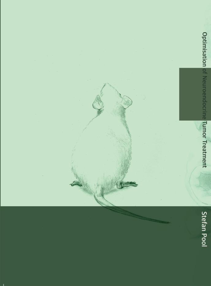
Stefan Pool
Optimisation of Neuroendocrine Tumor Treatment:
Locoregional administration,combination therapy and
multimodal imaging
O
ptim
isation
of N
eu
roe
nd
ocrin
e Tu
mo
r Treatm
en
t S
tefa
n P
oo
l
Uitnodiging
Voor het bijwonenvan de openbareverdediging vanhet proefschrift
Optimisation ofNeuroendocrine
Tumor Treatment:Locoregional
administration,combination therapy
and multimodal imaging
doorStefan Pool
Dinsdag16 december 2014
15:30 uur QueridozaalErasmus MC
Westzeedijk 361Rotterdam
Receptie na afloop
Paranimfen:Nanno Ouwehand
&Stuart Koelewij n
E-mail: promotie.stefan@
gmail.com