Direct effects of the light environment on daily neuroendocrine control
18
https://doi.org/10.1530/JOE-19-0302 https://joe.bioscientifica.com © 2019 Society for Endocrinology Printed in Great Britain Published by Bioscientifica Ltd. Journal of Endocrinology 243:1 R1–R18 S Paul and T M Brown Effects of light on neuroendocrine control REVIEW Direct effects of the light environment on daily neuroendocrine control Sarika Paul and Timothy M Brown Centre for Biological Timing, Faculty of Medicine, Biology and Health, University of Manchester, Manchester, UK Correspondence should be addressed to T M Brown: [email protected] Abstract Endocrine systems function as key mediators of adaptive responses to the external environment. As a reliable predictor of many salient variations in the external world, the light environment thus constitutes an influential source of control over neuroendocrine function. Accordingly, the vast majority of endocrine systems display 24-h variations in activity that are aligned to daily changes in external illumination. While the neural mechanisms responsible for driving these rhythms are still incompletely understood, circadian and light-dependent signals relayed via the suprachiasmatic nucleus of the hypothalamus (SCN) play a key role. Retinal projections to the SCN provide information from rods, cones and melanopsin, which, together, encode variations in the amount and spectral content of ambient light over the solar day. This sensory input, in turn, drives acute modulations in SCN cellular activity and aligns daily rhythms in the electrophysiological output of individual clock neurons. Neural outputs from the SCN can therefore convey both rapid and longer-term information about the light environment to other hypothalamic nuclei responsible for neuroendocrine control. In this review we summarise current understanding of the specific neural pathways by which the light environment influences key neuroendocrine axes, with a particular focus on the retinal and SCN-dependent circuits involved and their known sensory properties. Introduction As one of the key internal control mechanisms that animals use to appropriately adapt their physiology and behaviour according to the external environment, it is no surprise that the release of most, if not all, endocrine signals vary according to time of day (Czeisler & Klerman 1999). Such observations, in large part, reflect the actions of an internal circadian timing mechanism which allows animals to proactively adjust physiology and behaviour in anticipation of the predictable changes in the outside world. In mammals, the master pacemaker for this circadian clock is the suprachiasmatic nucleus (SCN) – a hypothalamic cell group situated just above the optic chiasm. This nucleus receives input from the retina, providing information about time of day, which in turn synchronises SCN clock neurons to provide coordinated rhythmic timing signals to other key hypothalamic regions implicated in neuroendocrine and homeostatic control (Kalsbeek et al. 2006, Brown 2016). As a result of the arrangement outlined above, changes in the light environment can result in important changes in endocrine function, both via comparatively direct circadian control of neuroendocrine systems, as well as secondary to changes in relevant behavioural cycles (e.g. rest/activity, feed/fast and so forth) across the 24-h day. Importantly, however, the influence of light on neuroendocrine function extends beyond the Key Words f circadian rhythms f melanopsin f melatonin f corticosteroids f gonadotrophin-releasing hormone Journal of Endocrinology (2019) 243, R1–R18 Downloaded from Bioscientifica.com at 02/02/2022 08:04:59AM via free access
-
Upload
khangminh22 -
Category
Documents
-
view
1 -
download
0
Transcript of Direct effects of the light environment on daily neuroendocrine control
https://doi.org/10.1530/JOE-19-0302https://joe.bioscientifica.com © 2019 Society for Endocrinology
Printed in Great BritainPublished by Bioscientifica Ltd.
Journal of Endocrinology
243:1 R1–R18S Paul and T M Brown Effects of light on neuroendocrine control
-19-0302
REVIEW
Direct effects of the light environment on daily neuroendocrine control
Sarika Paul and Timothy M Brown
Centre for Biological Timing, Faculty of Medicine, Biology and Health, University of Manchester, Manchester, UK
Correspondence should be addressed to T M Brown: [email protected]
Abstract
Endocrine systems function as key mediators of adaptive responses to the external environment. As a reliable predictor of many salient variations in the external world, the light environment thus constitutes an influential source of control over neuroendocrine function. Accordingly, the vast majority of endocrine systems display 24-h variations in activity that are aligned to daily changes in external illumination. While the neural mechanisms responsible for driving these rhythms are still incompletely understood, circadian and light-dependent signals relayed via the suprachiasmatic nucleus of the hypothalamus (SCN) play a key role. Retinal projections to the SCN provide information from rods, cones and melanopsin, which, together, encode variations in the amount and spectral content of ambient light over the solar day. This sensory input, in turn, drives acute modulations in SCN cellular activity and aligns daily rhythms in the electrophysiological output of individual clock neurons. Neural outputs from the SCN can therefore convey both rapid and longer-term information about the light environment to other hypothalamic nuclei responsible for neuroendocrine control. In this review we summarise current understanding of the specific neural pathways by which the light environment influences key neuroendocrine axes, with a particular focus on the retinal and SCN-dependent circuits involved and their known sensory properties.
Introduction
As one of the key internal control mechanisms that animals use to appropriately adapt their physiology and behaviour according to the external environment, it is no surprise that the release of most, if not all, endocrine signals vary according to time of day (Czeisler & Klerman 1999). Such observations, in large part, reflect the actions of an internal circadian timing mechanism which allows animals to proactively adjust physiology and behaviour in anticipation of the predictable changes in the outside world. In mammals, the master pacemaker for this circadian clock is the suprachiasmatic nucleus (SCN) – a hypothalamic cell group situated just above the optic chiasm. This nucleus receives input from the retina,
providing information about time of day, which in turn synchronises SCN clock neurons to provide coordinated rhythmic timing signals to other key hypothalamic regions implicated in neuroendocrine and homeostatic control (Kalsbeek et al. 2006, Brown 2016).
As a result of the arrangement outlined above, changes in the light environment can result in important changes in endocrine function, both via comparatively direct circadian control of neuroendocrine systems, as well as secondary to changes in relevant behavioural cycles (e.g. rest/activity, feed/fast and so forth) across the 24-h day. Importantly, however, the influence of light on neuroendocrine function extends beyond the
1
Key Words
f circadian rhythms
f melanopsin
f melatonin
f corticosteroids
f gonadotrophin-releasing hormone
Journal of Endocrinology (2019) 243, R1–R18
243
Downloaded from Bioscientifica.com at 02/02/2022 08:04:59AMvia free access
https://doi.org/10.1530/JOE-19-0302https://joe.bioscientifica.com © 2019 Society for Endocrinology
Published by Bioscientifica Ltd.Printed in Great Britain
R2Effects of light on neuroendocrine control
S Paul and T M Brown 243:1Journal of Endocrinology
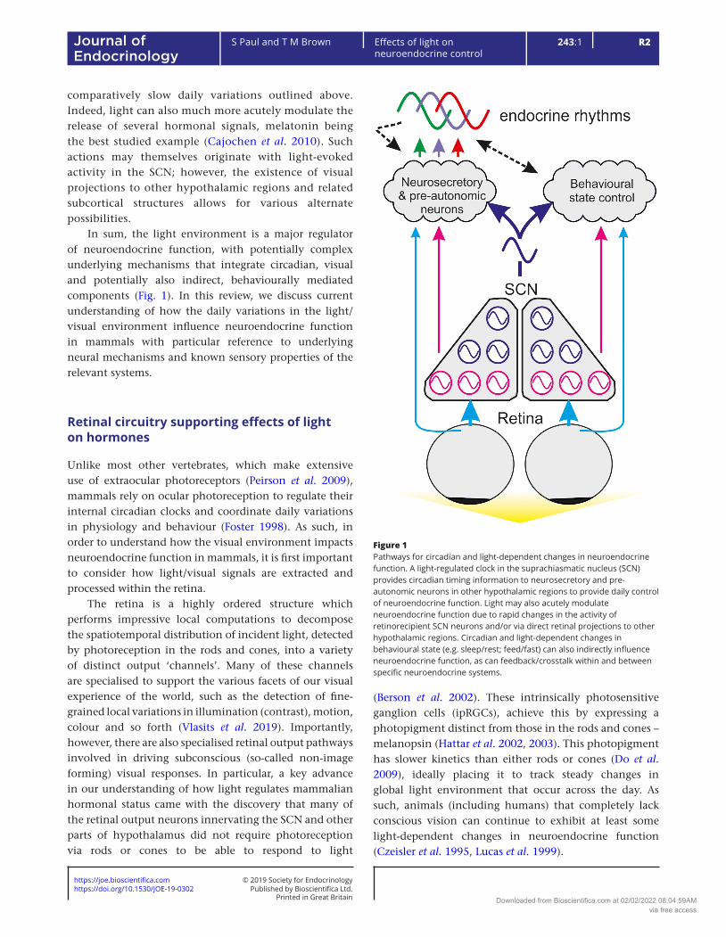
comparatively slow daily variations outlined above. Indeed, light can also much more acutely modulate the release of several hormonal signals, melatonin being the best studied example (Cajochen et al. 2010). Such actions may themselves originate with light-evoked activity in the SCN; however, the existence of visual projections to other hypothalamic regions and related subcortical structures allows for various alternate possibilities.
In sum, the light environment is a major regulator of neuroendocrine function, with potentially complex underlying mechanisms that integrate circadian, visual and potentially also indirect, behaviourally mediated components (Fig. 1). In this review, we discuss current understanding of how the daily variations in the light/visual environment influence neuroendocrine function in mammals with particular reference to underlying neural mechanisms and known sensory properties of the relevant systems.
Retinal circuitry supporting effects of light on hormones
Unlike most other vertebrates, which make extensive use of extraocular photoreceptors (Peirson et al. 2009), mammals rely on ocular photoreception to regulate their internal circadian clocks and coordinate daily variations in physiology and behaviour (Foster 1998). As such, in order to understand how the visual environment impacts neuroendocrine function in mammals, it is first important to consider how light/visual signals are extracted and processed within the retina.
The retina is a highly ordered structure which performs impressive local computations to decompose the spatiotemporal distribution of incident light, detected by photoreception in the rods and cones, into a variety of distinct output ‘channels’. Many of these channels are specialised to support the various facets of our visual experience of the world, such as the detection of fine-grained local variations in illumination (contrast), motion, colour and so forth (Vlasits et al. 2019). Importantly, however, there are also specialised retinal output pathways involved in driving subconscious (so-called non-image forming) visual responses. In particular, a key advance in our understanding of how light regulates mammalian hormonal status came with the discovery that many of the retinal output neurons innervating the SCN and other parts of hypothalamus did not require photoreception via rods or cones to be able to respond to light
(Berson et al. 2002). These intrinsically photosensitive ganglion cells (ipRGCs), achieve this by expressing a photopigment distinct from those in the rods and cones – melanopsin (Hattar et al. 2002, 2003). This photopigment has slower kinetics than either rods or cones (Do et al. 2009), ideally placing it to track steady changes in global light environment that occur across the day. As such, animals (including humans) that completely lack conscious vision can continue to exhibit at least some light-dependent changes in neuroendocrine function (Czeisler et al. 1995, Lucas et al. 1999).
Figure 1Pathways for circadian and light-dependent changes in neuroendocrine function. A light-regulated clock in the suprachiasmatic nucleus (SCN) provides circadian timing information to neurosecretory and pre-autonomic neurons in other hypothalamic regions to provide daily control of neuroendocrine function. Light may also acutely modulate neuroendocrine function due to rapid changes in the activity of retinorecipient SCN neurons and/or via direct retinal projections to other hypothalamic regions. Circadian and light-dependent changes in behavioural state (e.g. sleep/rest; feed/fast) can also indirectly influence neuroendocrine function, as can feedback/crosstalk within and between specific neuroendocrine systems.
Downloaded from Bioscientifica.com at 02/02/2022 08:04:59AMvia free access
https://doi.org/10.1530/JOE-19-0302https://joe.bioscientifica.com © 2019 Society for Endocrinology
Published by Bioscientifica Ltd.Printed in Great Britain
R3
Review
S Paul and T M Brown Effects of light on neuroendocrine control
243:1Journal of Endocrinology
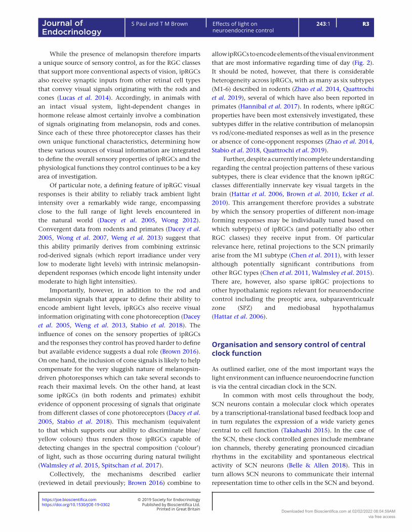
While the presence of melanopsin therefore imparts a unique source of sensory control, as for the RGC classes that support more conventional aspects of vision, ipRGCs also receive synaptic inputs from other retinal cell types that convey visual signals originating with the rods and cones (Lucas et al. 2014). Accordingly, in animals with an intact visual system, light-dependent changes in hormone release almost certainly involve a combination of signals originating from melanopsin, rods and cones. Since each of these three photoreceptor classes has their own unique functional characteristics, determining how these various sources of visual information are integrated to define the overall sensory properties of ipRGCs and the physiological functions they control continues to be a key area of investigation.
Of particular note, a defining feature of ipRGC visual responses is their ability to reliably track ambient light intensity over a remarkably wide range, encompassing close to the full range of light levels encountered in the natural world (Dacey et al. 2005, Wong 2012). Convergent data from rodents and primates (Dacey et al. 2005, Wong et al. 2007, Weng et al. 2013) suggest that this ability primarily derives from combining extrinsic rod-derived signals (which report irradiance under very low to moderate light levels) with intrinsic melanopsin-dependent responses (which encode light intensity under moderate to high light intensities).
Importantly, however, in addition to the rod and melanopsin signals that appear to define their ability to encode ambient light levels, ipRGCs also receive visual information originating with cone photoreception (Dacey et al. 2005, Weng et al. 2013, Stabio et al. 2018). The influence of cones on the sensory properties of ipRGCs and the responses they control has proved harder to define but available evidence suggests a dual role (Brown 2016). On one hand, the inclusion of cone signals is likely to help compensate for the very sluggish nature of melanopsin-driven photoresponses which can take several seconds to reach their maximal levels. On the other hand, at least some ipRGCs (in both rodents and primates) exhibit evidence of opponent processing of signals that originate from different classes of cone photoreceptors (Dacey et al. 2005, Stabio et al. 2018). This mechanism (equivalent to that which supports our ability to discriminate blue/yellow colours) thus renders those ipRGCs capable of detecting changes in the spectral composition (‘colour’) of light, such as those occurring during natural twilight (Walmsley et al. 2015, Spitschan et al. 2017).
Collectively, the mechanisms described earlier (reviewed in detail previously; Brown 2016) combine to
allow ipRGCs to encode elements of the visual environment that are most informative regarding time of day (Fig. 2). It should be noted, however, that there is considerable heterogeneity across ipRGCs, with as many as six subtypes (M1-6) described in rodents (Zhao et al. 2014, Quattrochi et al. 2019), several of which have also been reported in primates (Hannibal et al. 2017). In rodents, where ipRGC properties have been most extensively investigated, these subtypes differ in the relative contribution of melanopsin vs rod/cone-mediated responses as well as in the presence or absence of cone-opponent responses (Zhao et al. 2014, Stabio et al. 2018, Quattrochi et al. 2019).
Further, despite a currently incomplete understanding regarding the central projection patterns of these various subtypes, there is clear evidence that the known ipRGC classes differentially innervate key visual targets in the brain (Hattar et al. 2006, Brown et al. 2010, Ecker et al. 2010). This arrangement therefore provides a substrate by which the sensory properties of different non-image forming responses may be individually tuned based on which subtype(s) of ipRGCs (and potentially also other RGC classes) they receive input from. Of particular relevance here, retinal projections to the SCN primarily arise from the M1 subtype (Chen et al. 2011), with lesser although potentially significant contributions from other RGC types (Chen et al. 2011, Walmsley et al. 2015). There are, however, also sparse ipRGC projections to other hypothalamic regions relevant for neuroendocrine control including the preoptic area, subparaventricualr zone (SPZ) and mediobasal hypothalamus (Hattar et al. 2006).
Organisation and sensory control of central clock function
As outlined earlier, one of the most important ways the light environment can influence neuroendocrine function is via the central circadian clock in the SCN.
In common with most cells throughout the body, SCN neurons contain a molecular clock which operates by a transcriptional-translational based feedback loop and in turn regulates the expression of a wide variety genes central to cell function (Takahashi 2015). In the case of the SCN, these clock controlled genes include membrane ion channels, thereby generating pronounced circadian rhythms in the excitability and spontaneous electrical activity of SCN neurons (Belle & Allen 2018). This in turn allows SCN neurons to communicate their internal representation time to other cells in the SCN and beyond.
Downloaded from Bioscientifica.com at 02/02/2022 08:04:59AMvia free access
https://doi.org/10.1530/JOE-19-0302https://joe.bioscientifica.com © 2019 Society for Endocrinology
Published by Bioscientifica Ltd.Printed in Great Britain
R4Effects of light on neuroendocrine control
S Paul and T M Brown 243:1Journal of Endocrinology
Importantly, however, the properties of individual SCN neurons are highly heterogeneous. When cultured at low density (preventing any intercellular communication) many SCN neurons are capable of sustaining circadian rhythms in spontaneous electrical activity and gene expression, but the circadian periods of those rhythms are highly variable (Welsh et al. 1995, Herzog et al. 2004). This period variability collapses when SCN neurons are measured in intact tissue explants, where intercellular communication allows cells to adopt a common ~24-h periodicity, but is instead replaced by significant variations in phase of rhythmic activity across individual cells (Schaap et al. 2003, Herzog et al. 2004, Brown & Piggins 2009). Thus, cells with intrinsically slower clocks tend to lag behind their counterparts with naturally faster clocks in the intact network.
While the arrangement described earlier allows SCN neurons to generate coherent circadian timing signals, to be of use, such signals need to be appropriately aligned to the external environment. Thus, retinal input to the SCN is critical for adjusting the molecular clockwork across the SCN to precisely match the periodicity of the cycle in environmental illumination and ensuring the electrical output of the SCN neuronal ensemble is appropriately timed (Meijer & Schwartz 2003). Of note, regardless of what temporal niche an animal occupies (nocturnal, diurnal, crepuscular), this daily peak in SCN population output appears to be timed to occur during the middle part of the light period (Schwartz et al. 1983, Challet 2007).
As discussed earlier for their intrinsic circadian properties, however, the influence of retinal input on SCN neurons is also heterogenous. Firstly, not all SCN neurons receive retinal input (Morin & Allen 2006, Lokshin et al. 2015). Indeed, while there seems to be considerable inter-species diversity in the precise arrangements of retinal projections, a general feature of SCN organisation seems to be the presence of a ‘core’ region with dense retinal input and a ‘shell’ region with more sparse retinal input. Secondly, among those cells presumed to receive direct retinal inputs (as evidence by rapid and acute light-evoked changes in neural activity) the influence of visual signals can differ significantly, as described below.
Acute light-dependent changes in SCN neuron activity have been described in many species (Groos & Mason 1980, Meijer et al. 1986, 1989, Mure et al. 2007) but have only been evaluated in detail in mice and rats. As expected based on the dominant contribution of a particular class of ipRGCs (M1) to rodent SCN retinal input (Hattar et al. 2006), the majority of visually response SCN cells exhibit evidence of strong, melanopsin-dependent, sustained changes in firing with increasing light intensity (Brown et al. 2011, Walmsley et al. 2015). Under appropriate conditions, clear evidence of both rod and cone-driven increases in SCN neuronal activity have also been reported, although the nature of cones inputs varies substantially across visually responsive SCN neurons (Aggelopoulos & Meissl 2000, Walmsley et al. 2015, Dobb et al. 2017). Indeed, at least in the mouse, there appears
Figure 2Retinal circuitry supporting effects of light on neuroendocrine function. (A) Schematic of important retinal circuits that supply intrinsically photosensitive retinal ganglion cells (ipRGCs). In addition to intrinsic melanopsin-based phototransduction, ipRGCs receive excitatory cone input via ON bipolar cells and excitatory rod input via rod bipolar cells that couple to the cone bipolars via gap junctions. Widefield amacrine cell connections provide inhibitory input from other cone bipolar cells, potentially allowing from chromatic responses (Stabio et al. 2018). (B) Relationship between light intensity and ipRGC firing, indicating photoreceptive systems that contribute under each condition. Note that natural variations in spectral composition during twilight (indicated by coloured bar) are detectable to cones and can modulate the intensity-dependent firing of ipRGCs.
Downloaded from Bioscientifica.com at 02/02/2022 08:04:59AMvia free access
https://doi.org/10.1530/JOE-19-0302https://joe.bioscientifica.com © 2019 Society for Endocrinology
Published by Bioscientifica Ltd.Printed in Great Britain
R5
Review
S Paul and T M Brown Effects of light on neuroendocrine control
243:1Journal of Endocrinology
to be distinct subsets of neurons that process inputs from different classes of cone photoreceptor in an additive (achromatic) or opponent (chromatic) manner (Walmsley et al. 2015). The latter class is further subdivided into colour responsive cells that are excited by either short (‘blue’) or long (‘yellow’) wavelength light.
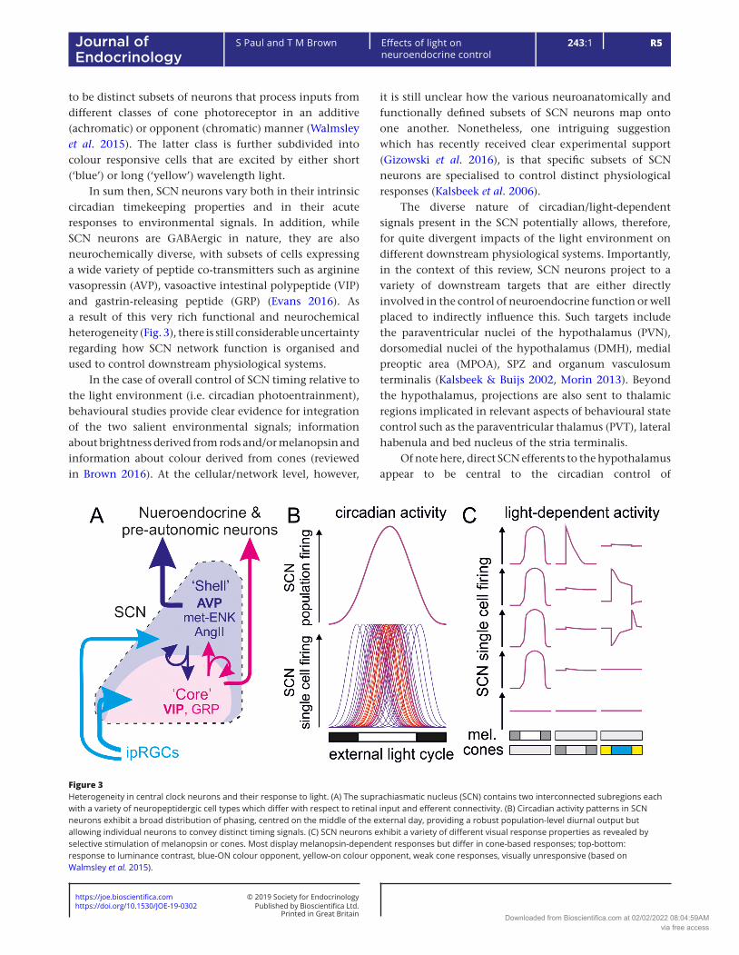
In sum then, SCN neurons vary both in their intrinsic circadian timekeeping properties and in their acute responses to environmental signals. In addition, while SCN neurons are GABAergic in nature, they are also neurochemically diverse, with subsets of cells expressing a wide variety of peptide co-transmitters such as arginine vasopressin (AVP), vasoactive intestinal polypeptide (VIP) and gastrin-releasing peptide (GRP) (Evans 2016). As a result of this very rich functional and neurochemical heterogeneity (Fig. 3), there is still considerable uncertainty regarding how SCN network function is organised and used to control downstream physiological systems.
In the case of overall control of SCN timing relative to the light environment (i.e. circadian photoentrainment), behavioural studies provide clear evidence for integration of the two salient environmental signals; information about brightness derived from rods and/or melanopsin and information about colour derived from cones (reviewed in Brown 2016). At the cellular/network level, however,
it is still unclear how the various neuroanatomically and functionally defined subsets of SCN neurons map onto one another. Nonetheless, one intriguing suggestion which has recently received clear experimental support (Gizowski et al. 2016), is that specific subsets of SCN neurons are specialised to control distinct physiological responses (Kalsbeek et al. 2006).
The diverse nature of circadian/light-dependent signals present in the SCN potentially allows, therefore, for quite divergent impacts of the light environment on different downstream physiological systems. Importantly, in the context of this review, SCN neurons project to a variety of downstream targets that are either directly involved in the control of neuroendocrine function or well placed to indirectly influence this. Such targets include the paraventricular nuclei of the hypothalamus (PVN), dorsomedial nuclei of the hypothalamus (DMH), medial preoptic area (MPOA), SPZ and organum vasculosum terminalis (Kalsbeek & Buijs 2002, Morin 2013). Beyond the hypothalamus, projections are also sent to thalamic regions implicated in relevant aspects of behavioural state control such as the paraventricular thalamus (PVT), lateral habenula and bed nucleus of the stria terminalis.
Of note here, direct SCN efferents to the hypothalamus appear to be central to the circadian control of
Figure 3Heterogeneity in central clock neurons and their response to light. (A) The suprachiasmatic nucleus (SCN) contains two interconnected subregions each with a variety of neuropeptidergic cell types which differ with respect to retinal input and efferent connectivity. (B) Circadian activity patterns in SCN neurons exhibit a broad distribution of phasing, centred on the middle of the external day, providing a robust population-level diurnal output but allowing individual neurons to convey distinct timing signals. (C) SCN neurons exhibit a variety of different visual response properties as revealed by selective stimulation of melanopsin or cones. Most display melanopsin-dependent responses but differ in cone-based responses; top-bottom: response to luminance contrast, blue-ON colour opponent, yellow-on colour opponent, weak cone responses, visually unresponsive (based on Walmsley et al. 2015).
Downloaded from Bioscientifica.com at 02/02/2022 08:04:59AMvia free access
https://doi.org/10.1530/JOE-19-0302https://joe.bioscientifica.com © 2019 Society for Endocrinology
Published by Bioscientifica Ltd.Printed in Great Britain
R6Effects of light on neuroendocrine control
S Paul and T M Brown 243:1Journal of Endocrinology
neuroendocrine rhythms. Hence, while robust behavioural rhythms can be restored to SCN-lesioned animals by transplantation of foetal SCN grafts, this manipulation does not restore neuroendocrine rhythmicity (Lehman et al. 1987, Meyer-Bernstein et al. 1999). Indeed, as discussed in detail below, direct SCN projections target many of the key neurosecretory hypothalamic cell groups including corticotrophin releasing hormone (CRH), thyrotrophin-releasing hormone (TRH), gonadotrophin-releasing hormone (GnRH) and dopaminergic neurons (Kalsbeek et al. 2006). Further, SCN projections to pre-autonomic neurons in the PVN that are relevant for additional roles in the regulation of neuroendocrine function have been identified (Larsen et al. 1998, Buijs et al. 1999, Ueyama et al. 1999, Kalsbeek et al. 2000a).
As alluded to above, the specific properties of SCN neurons projecting to the targets outlined above remain largely unknown. For the remainder of this review, then, we highlight current understanding of how circadian/visual signals (originating in the SCN or elsewhere) influence daily patterns of neuroendocrine secretion, with a focus on those systems where there is the most currently available information.
Daily control of pineal melatonin synthesis
Without doubt, the best studied aspect of how the light environment influences neuroendocrine function relates to the pineal hormone melatonin.
The synthesis and release of melatonin from the pineal gland is strongly rhythmic under constant (low light) conditions and is profoundly inhibited by light (Cajochen et al. 2010). As a result of this arrangement, circulating melatonin levels (which are high during the night in both nocturnal and diurnal mammals) provide information about day-length. This makes melatonin both an important systemic source of daily timing information and a key signal for the photoperiodic control of physiology in many animals (Wood & Loudon 2018, Dardente et al. 2019). Since photoperiodic mechanisms have been discussed extensively previously, we do not tackle these in detail here. Instead we focus on the organisation and sensory properties of the neural pathways regulating pineal melatonin synthesis/release.
The major anatomical pathways for the circadian/diurnal control of pineal melatonin have been known for many years, with initial investigations establishing that this required the SCN and involved sympathetic input from the superior cervical ganglion and the pineal
(Klein et al. 1971, Moore & Klein 1974). Subsequent studies using transneuronal tracers further delineated this pathway, showing that the connections from the SCN pass via pre-autonomic neurons in the PVN, to preganglionic neurons in the spinal cord, and the noradrenergic neurons in the superior cervical ganglion (Larsen et al. 1998, Teclemariam-Mesbah et al. 1999, Kalsbeek et al. 2006).
Ablation or inactivation of neurons at any stage of the pathway described above will impact melatonin synthesis and rhythmicity (Perreau-Lenz et al. 2003, 2004). Of note, however, manipulations performed at the level of the PVN or superior cervical ganglion lead to constitutively low levels of melatonin while removal of SCN input leads to constitutively high levels. This pattern is therefore suggests a model whereby clock and/or light-driven increases in SCN neuronal activity inhibits pre-autonomic PVN neurons involved in regulating melatonin synthesis. Consistent with this view, infusion of a GABA receptor antagonist into the PVN and surrounding areas causes an increase of daytime melatonin concentrations and blocks light-induced nocturnal suppressions (Kalsbeek et al. 1999, 2000b).
While the neural circuits responsible for the daily control of pineal melatonin release are thus well established (Fig. 4), attaining a detailed understanding of the sensory signals that regulate this has proved more challenging. This, in part, likely reflects the challenges associated with obtaining detailed measures of the sensory control of melatonin synthesis in rodents. Nonetheless, by the late 1990s, convergent evidence from humans and mice revealed that light-dependent melatonin suppression persisted in the absence of functional rod/cone photoreception suggesting the involvement of a novel photopigment (Czeisler et al. 1995, Freedman et al. 1999). While we now know this is due to the central role of ipRGCs/melanopsin in conveying light information to the SCN (Guler et al. 2008) there has remained some uncertainty regarding the sensory influences on melatonin synthesis. In particular, the majority of available data across rodent and primate models indicates melanopsin has a peak spectral sensitivity in the region of 480 nm (Lucas et al. 2001, Berson et al. 2002, Dacey et al. 2005, Bailes & Lucas 2013). By contrast, two independent initial reports suggested that the peak spectral sensitivity for melatonin suppression in humans was substantially different from this value (Brainard et al. 2001, Thapan et al. 2001).
More recent studies (including re-evaluation of some of the earlier data) do place the spectral sensitivity of melatonin suppression firmly in the vicinity of 480,
Downloaded from Bioscientifica.com at 02/02/2022 08:04:59AMvia free access
https://doi.org/10.1530/JOE-19-0302https://joe.bioscientifica.com © 2019 Society for Endocrinology
Published by Bioscientifica Ltd.Printed in Great Britain
R7
Review
S Paul and T M Brown Effects of light on neuroendocrine control
243:1Journal of Endocrinology
confirming a dominant role for melanopsin (Najjar et al. 2014, Prayag et al. 2019). Nonetheless, it should also be noted that, while melanopsin photoreception alone seems to well predict the effects of long duration light exposures on melatonin, there is also clear evidence for the involvement of cone photoreception. Indeed, evaluation of the spectral sensitivity of initial light-evoked changes in circulating melatonin suggests a much more dominant role for cones (Gooley et al. 2010). Some previous studies have also suggested the possibility of colour-opponent regulation of melatonin release in humans (Figueiro et al. 2004, 2008), although there is conflicting data in this regard (Revell & Skene 2007, Papamichael et al. 2012). Full confirmation on this point therefore requires additional studies employing chromatic stimuli appropriately controlled for their impact on melanopsin.
One final point to note here is that, despite the central role for the SCN in the control of melatonin synthesis, the sensory mechanism by which light influences this neuroendocrine signal do not exactly recapitulate those of the circadian entrainment mechanism. This includes clear differences in the relative sensitivity to short vs long wavelength light (Gooley et al. 2010) as well as differences in the response to continuous vs intermittent light steps (Rahman et al. 2018). For example, the latter study reveals that, whereas a series of six 15 min bright light pulses
spread over 6.5 h suppresses melatonin much less than 6.5 h of continuous illumination, the two stimuli appear to evoke very similar effects on the circadian phase. Such discrepancies likely reflect processing that occurs within the SCN network downstream of the acute light-dependent changes in activity that are used to acutely regulate melatonin synthesis.
Daily control of the hypothalamic–pituitary–adrenal axis
Unlike melatonin, which is strongly tied to the night time in all mammals, activity of the hypothalamic–pituitary–adrenal (HPA) axis is closely aligned to the animals’ behavioural patterns to provide maximal glucocorticoid release just prior to the onset of activity (Kalsbeek et al. 2012). As a result, the (comparatively less well understood) mechanisms by which circadian and light-dependent signals influence HPA activity likely differ somewhat between nocturnal and diurnal mammals.
The HPA axis itself consists of neurons in the medial PVN, which release CRH. These target corticotrophs in the anterior pituitary gland, which in turn release adrenocorticotrophic-releasing hormone (ACTH) into the blood stream where it acts on the adrenals to drive glucocorticoid secretion. The circulating cortisol/corticosterone (CORT) then feeds back to downregulate HPA activity (Gjerstad et al. 2018). Collectively, HPA axis activity then produces a pulsatile (ultradian) pattern of CORT secretion whose amplitude is strongly modulated by circadian signals from the SCN (Moore & Eichler 1972, Waite et al. 2012). There are now a number of identified and potentially convergent mechanisms by which the output from the SCN clock may achieve this regulation (Fig. 5).
Based on a series of microdialysis studies in rats, Kalsbeek, Buijs and colleagues suggest a central role for AVP cells of the SCN in driving daily rhythms in HPA axis activity (Kalsbeek et al. 2012). By their model, AVP release from the SCN during the early to mid-day activates GABAergic neurons in the DMH and/or SPZ, which in turn inhibit CRH neurons in the PVN, to keep circulating CORT levels low. Consistent with this view, infusion of a V1 receptor antagonist into the DMH substantially enhances HPA axis activity during early-mid portions of the day, while infusion of AVP supresses the evening rise in CORT (Kalsbeek et al. 1996a,b). Nonetheless, the continued presence of rhythms in HPA axis activity while AVP signalling to the DMH is blocked also suggest the
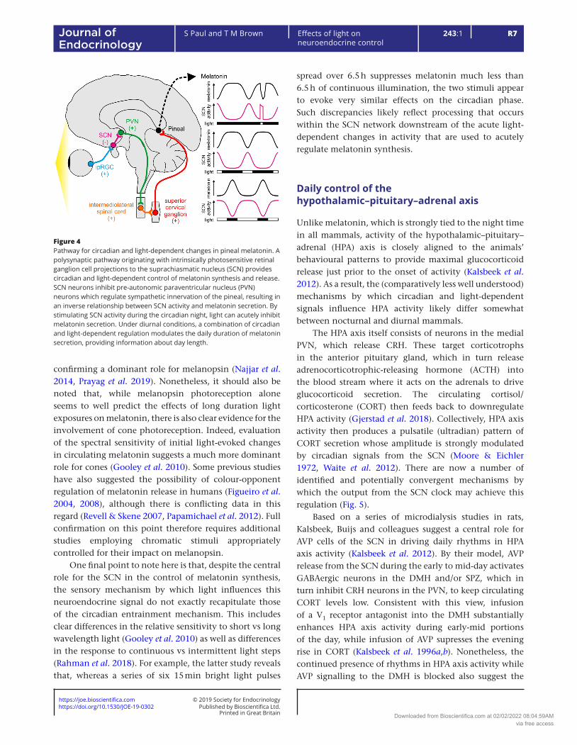
Figure 4Pathway for circadian and light-dependent changes in pineal melatonin. A polysynaptic pathway originating with intrinsically photosensitive retinal ganglion cell projections to the suprachiasmatic nucleus (SCN) provides circadian and light-dependent control of melatonin synthesis and release. SCN neurons inhibit pre-autonomic paraventricular nucleus (PVN) neurons which regulate sympathetic innervation of the pineal, resulting in an inverse relationship between SCN activity and melatonin secretion. By stimulating SCN activity during the circadian night, light can acutely inhibit melatonin secretion. Under diurnal conditions, a combination of circadian and light-dependent regulation modulates the daily duration of melatonin secretion, providing information about day length.
Downloaded from Bioscientifica.com at 02/02/2022 08:04:59AMvia free access
https://doi.org/10.1530/JOE-19-0302https://joe.bioscientifica.com © 2019 Society for Endocrinology
Published by Bioscientifica Ltd.Printed in Great Britain
R8Effects of light on neuroendocrine control
S Paul and T M Brown 243:1Journal of Endocrinology
presence of additional factors regulating daily glucorticoid rhythmicity. The existence of direct SCN projections to CRH neurons provides one such route by which this may be achieved (Vrang et al. 1995). In addition, however, neuroanatomical tracing studies reveal the presence of SCN neurons that are multisynaptically connected to the adrenal via pre-autonomic PVN neurons (Buijs et al. 1999, Ueyama et al. 1999). Via this pathway, the SCN might also regulate CORT secretion by adjusting adrenal sensitivity to circulating ACTH (Kaneko et al. 1980, 1981, Jasper & Engeland 1994).
Interestingly, by contrast to the pronounced inhibitory effect of SCN output on HPA activity described earlier, several studies indicate that light exposure enhances levels of circulating CORT in rodents (Ishida et al. 2005,
Loh et al. 2008, Rahman et al. 2008, Kiessling et al. 2014). This effect is not associated with detectable changes in plasma ACTH, but is accompanied by increases in adrenal sympathetic nerve activity and a significant induction of gene expression across the adrenals (Ishida et al. 2005, Kiessling et al. 2014). Since this effect of light is abolished by SCN lesion (Ishida et al. 2005), it is assumed to involve SCN-dependent stimulation of the autonomic nervous system, via a pathway similar to that described earlier. VIP-expressing cells of the SCN have been suggested as a potential mediator of this effect since mice lacking VIP display greatly attenuated light-driven increases in circulating CORT (Loh et al. 2008). It should be noted, however, that the loss of VIP induces a pronounced global disruption to SCN function (Colwell et al. 2003, Aton et al. 2005, Maywood et al. 2006, Brown et al. 2007), leaving a specific role for VIP cells in such an effect uncertain.
In summary, there appears then to be at least two different routes by which SCN activity can influence circulating CORT levels in rodents. A circadian control which impinges on CRH neurons to drive rhythms in ACTH secretion (Kalsbeek et al. 1996b, Loh et al. 2008) and a light-dependent process which involves activation of sympathetic input to the adrenals (Ishida et al. 2005). Although the sensory properties of this latter pathway have not yet been investigated in detail, it appears to require relatively high light levels to produce noticeable impacts (Kiessling et al. 2014), in stark contrast to the much higher sensitivity of circadian photoentrainment responses (Lall et al. 2010). Given the relatively high light levels required and the requirement for an intact SCN, it seems likely that melanopsin signals relayed by ipRGC projections to the SCN (or nearby regions) play a major role in the effects of light on CORT. Consistent with this possibility, white light sources lacking significant energy in portions of the spectrum where melanopsin is most sensitive (460–480 nm) are remarkably less effective at stimulating CORT secretion in rats (Rahman et al. 2008). This latter study does not provide conclusive evidence for the role of melanopsin, however, since other photoreceptors in the rat (rods and medium-wavelength sensitive cones) also show high sensitivity in this portion of the visible spectrum.
By comparison to the rodent data outlined above, there is considerably less mechanistic understanding of circadian and diurnal sources of control over HPA axis activity in diurnal animals. Hence, while the timing of SCN clock output and its response to light is similar between nocturnal and diurnal mammals, rhythms in circulating CORT are phase inverted (Perlow et al. 1981,
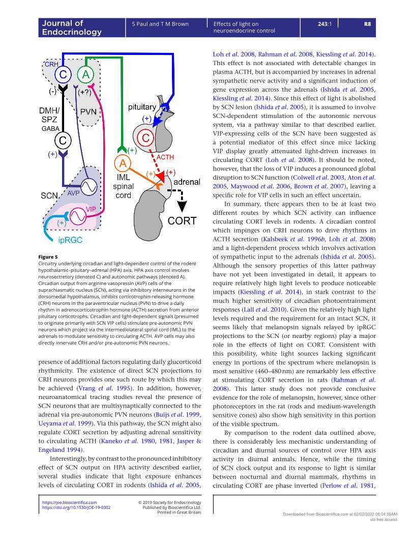
Figure 5Circuitry underlying circadian and light-dependent control of the rodent hypothalamic–pituitary–adrenal (HPA) axis. HPA axis control involves neurosectretory (denoted C) and autonomic pathways (denoted A). Circadian output from arginine vasopressin (AVP) cells of the suprachiasmatic nucleus (SCN), acting via inhibitory interneurons in the dorsomedial hypothalamus, inhibits corticotrophin-releasing hormone (CRH) neurons in the paraventricular nucleus (PVN) to drive a daily rhythm in adrenocorticotrophin hormone (ACTH) secretion from anterior pituitary corticotrophs. Circadian and light-dependent signals (presumed to originate primarily with SCN VIP cells) stimulate pre-autonomic PVN neurons which project via the intermediolateral spinal cord (IML) to the adrenals to modulate sensitivity to circulating ACTH. AVP cells may also directly innervate CRH and/or pre-autonomic PVN neurons.
Downloaded from Bioscientifica.com at 02/02/2022 08:04:59AMvia free access
https://doi.org/10.1530/JOE-19-0302https://joe.bioscientifica.com © 2019 Society for Endocrinology
Published by Bioscientifica Ltd.Printed in Great Britain
R9
Review
S Paul and T M Brown Effects of light on neuroendocrine control
243:1Journal of Endocrinology
Schwartz et al. 1983, Challet 2007). In general, such differences are considered to reflect an inversion of the impact of SCN-derived signals on downstream brain regions (Sato & Kawamura 1984, Brown & Piggins 2007). However, as far as we are aware, there have not yet been any direct investigations of how SCN output influences HPA axis activity in fully diurnal animals.
In general accord with the idea that SCN outputs should have opposite effects on HPA axis activity in diurnal vs nocturnal animals, studies in a crepuscular/diurnal rodent (Arvicanthis ansorgei) do provide convincing evidence for a reversal in the role of endogenous AVP signalling (Kalsbeek et al. 2008). Hence, this latter work reveals that endogenous AVP signalling in the PVN/DMH region is required for morning and evening surges in circulating CORT in Arvicanthis, by contrast to the suppressive daytime effects seen in rats (Kalsbeek et al. 1996b). Whether this apparent stimulatory action reflects a crepuscular pattern of AVP release from the Arvicanthis SCN itself remains unclear, however. Similarly, there is no concrete information regarding differences in the underlying neural circuitry that could produce the inversion of AVP effects relative to those seen in rats. The assumption is that AVP output from the SCN targets excitatory rather than inhibitory DMH interneurons in Arvicanthis (Kalsbeek et al. 2008), although a more direct stimulation of CRH cells in the PVN in this species seems a plausible alternative mechanism.
In either case, given the apparent reversal in the impact of SCN-derived circadian signals on HPA axis activity in nocturnal and diurnal animals, one might expect a similar reversal in the acute response of this system to light. In fact, current literature is rather equivocal on this point. While there have certainly been some studies demonstrating light-induced reductions in CORT levels in humans (Kostoglou-Athanassiou et al. 1998, Jung et al. 2010), there have also been many showing light-induced increases (Scheer & Buijs 1999, Leproult et al. 2001, Figueiro & Rea 2010, Gabel et al. 2013, Petrowski et al. 2019). The origin of these discrepancies remains unclear. Nonetheless, it is noteworthy that light-stimulated changes in CORT in nocturnal rodents seem to involve a different pathway that underlying circadian changes. In this regard, it is possible that, while mechanisms of circadian control diverge between nocturnal and diurnal animals those primarily responsible for acute light-induced changes (i.e. activation of sympathetic outflow to the adrenals) are retained. Indeed, since light increases neural activity in the human SCN (McGlashan et al. 2018), just as it does in rodents, such an arrangement could account for the more
commonly observed light-induced increases, rather than decreases, in human CORT levels.
Daily control of the hypothalamic–pituitary–gonadal axis
Reproductive function, particularly in females, is a highly rhythmic process with appropriate timing crucial to a successful outcome, from maximising chances of fertilisation to ensuring the long-term survival of the offspring. Accordingly there has been extensive research on both the relevant mechanisms of circadian control (Simonneaux & Bahougne 2015, Evans & Anderson 2018) and with respect to photoperiodic seasonal regulation (Dardente et al. 2019). For reasons of space, below we focus on the known circuitry by which SCN and light-dependent signals can most directly modulate the hypothalamic–pituitary–gonadal (HPG) axis.
The key drivers of the HPG axis are the GnRH neurons in the preoptic area of the hypothalamus. GnRH, secreted into the portal circulation, then acts in the anterior pituitary to trigger systemic release of luteinising hormone (LH) and follicle-stimulating hormone (FSH) which in turn stimulate gonadal hormone secretion (Herbison 2016). Rodent studies indicate that the SCN sends direct outputs to GnRH neurons, a projection which, at least in part, originates with VIP-expressing cells (van der Beek et al. 1993, Van der Beek et al. 1997, Mahoney & Smale 2005a, Ward et al. 2009). VIP then excites GnRH neurons, providing a potential route by which HPG axis activity may be controlled according to time of day (Piet et al. 2016). SCN VIP cells may also indirectly excite GnRH neurons by supressing the activity of upstream inhibitory neurons in the DMH expressing RFamide-related peptide 3 (Russo et al. 2015). Similarly, AVP-expressing SCN neurons provide a further indirect source of circadian control by exciting kisspeptin neurons in the anteroventral periventricular nuclei which, in turn, powerfully stimulate GnRH neurons (Vida et al. 2010, Williams et al. 2011, Simonneaux & Bahougne 2015).
The mechanisms described earlier appear to converge to provide circadian regulation of female reproductive function (Fig. 6). In rodents, a surge in LH release, critical for triggering ovulation, is timed to occur towards the end of the day; this requires SCN-dependent circadian timing signals co-incident with high levels of oestradiol, indicative of ovarian follicle maturation (Brown-Grant & Raisman 1977, Wiegand et al. 1980, Lehman et al. 1987, Meyer-Bernstein et al. 1999). Initial studies indicated that
Downloaded from Bioscientifica.com at 02/02/2022 08:04:59AMvia free access
https://doi.org/10.1530/JOE-19-0302https://joe.bioscientifica.com © 2019 Society for Endocrinology
Published by Bioscientifica Ltd.Printed in Great Britain
R10Effects of light on neuroendocrine control
S Paul and T M Brown 243:1Journal of Endocrinology
a reduction in either VIP or AVP signalling could attenuate this LH surge (Harney et al. 1996, Funabashi et al. 1999, van der Beek et al. 1999). Subsequent studies now suggest that AVP-expressing SCN cells, acting via kisspeptin neurons, are likely the primary drivers of the timing of the preovulatory LH surge and its gating by oestradiol (Robertson et al. 2009, Smarr et al. 2012).
In line with the above, appropriately timed (late day) administration of AVP appears sufficient to produce the LH surge in oestradiol-treated ovariectomised rats (Palm et al. 1999, 2001). By contrast the influence of VIP on the proestrous LH surge appears more modulatory in nature (Sun et al. 2012). These time-dependent effects of exogenous peptide application further indicate that rhythms in SCN output cannot be the sole factors dictating the circadian timing of LH release. Certainly, GnRH neurons themselves possess an intrinsic molecular clock
(Hickok & Tischkau 2010) and exhibit circadian variation in their response to key inputs such as Kisspeptin and VIP (Christian & Moenter 2008, Williams et al. 2011), as may other key upstream cell types highlighted above (Russo et al. 2015, Simonneaux & Bahougne 2015).
In summary, the circuitry underlying circadian control of the HPG axis is complex, with multiple pathways that converge to regulate daily rhythms in GnRH neurons. In nocturnal rodents at proestrous this results in an LH surge around dusk, ensuring ovulation occurs during their active phase, when mating is likely. Although far less studied, broadly similar circuits in male animals presumably confer a corresponding daily rhythmicity in the HPG axis (Taya & Igarashi 1974, Roman et al. 2003), ensuring reproductive function is appropriately aligned in both sexes to maximise successful procreation (Sakai & Endo 1988). Of course it is also important to note here that, as discussed earlier for CORT, the timing of rhythms in HPG axis activity will be different in diurnal species (Baumgartner et al. 1993, Mahoney et al. 2004, Caufriez et al. 2018). For example, in female Arvincanthis, GnRH neuronal activity and LH secretion is maximal just prior to dawn (Mahoney et al. 2004). Further, both male and female Arvincanthis display correspondingly enhanced sexual behaviour at this time, as opposed to just after dusk as in nocturnal rodents (Mahoney & Smale 2005b). The mechanisms responsible for this phase inversion are currently unclear but are expected to lie in difference in the intermediary circuitry linking the SCN to GnRH neurons rather than any major difference in the timing of SCN output.
Beyond the circadian control outlined above, it is also important to consider other influences of the light environment on HPG axis function. One such example, which is critical for the seasonal breeding adaptations shown by many mammals, is day-length. Variations in day-length can evoke rapid changes in reproductive status that involve the same circuitry as that engaged by the circadian clock (Angelopoulou et al. 2019). Importantly, however, in this case the primary source of photoperiodic information is rather indirect, coming via a change in duration of melatonin secretion (Wood & Loudon 2018). Nonetheless, given that many SCN neurons are acutely modulated by light, including the VIP cells (Jones et al. 2018) which have known roles in regulating GnRH neuronal activity, one might wonder whether there are also more direct sources of light-dependent control.
Although the possibility of acute effects of light of this nature not been studied in detail, a previous report does indicate that putative GnRH neurons in the monkey
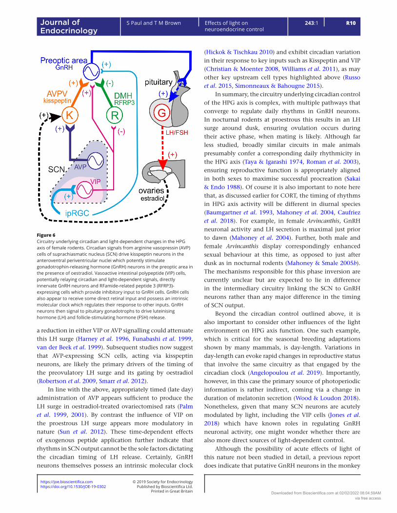
Figure 6Circuitry underlying circadian and light-dependent changes in the HPG axis of female rodents. Circadian signals from arginine vasopressin (AVP) cells of suprachiasmatic nucleus (SCN) drive kisspeptin neurons in the anteroventral periventricular nuclei which potently stimulate gonadotrophin-releasing hormone (GnRH) neurons in the preoptic area in the presence of oestradiol. Vasoactive intestinal polypeptide (VIP) cells, potentially relaying circadian and light-dependent signals, directly innervate GnRH neurons and RFamide-related peptide 3 (RFRP3)-expressing cells which provide inhibitory input to GnRH cells. GnRH cells also appear to receive some direct retinal input and possess an intrinsic molecular clock which regulates their response to other inputs. GnRH neurons then signal to pituitary gonadotrophs to drive luteinising hormone (LH) and follicle-stimulating hormone (FSH) release.
Downloaded from Bioscientifica.com at 02/02/2022 08:04:59AMvia free access
https://doi.org/10.1530/JOE-19-0302https://joe.bioscientifica.com © 2019 Society for Endocrinology
Published by Bioscientifica Ltd.Printed in Great Britain
R11
Review
S Paul and T M Brown Effects of light on neuroendocrine control
243:1Journal of Endocrinology
hypothalamus exhibit acute light-dependent increases in activity (O’Byrne et al. 1993). There are several potential origins for this although, interestingly, direct retinal projections to GnRH neurons have been reported in monkeys (Abizaid et al. 2004). Further, there have been a few reports that acute light exposure can induce very rapid increases in circulating FSH, and perhaps also LH, levels in human females (Miyauchi et al. 1990, 1991, Danilenko & Sergeeva 2015). There are also data supporting the existence of very rapid light-dependent changes in HPG activity in rodents, although the data are conflicting: bright light reportedly enhances the preovulatory LH surge in female rats (Walker & Jimenez 1984) but supresses this in mice (Bronson & Vom Saal 1979). One possible explanation for such discrepancies relates to a differential contribution of melatonin to the observed responses. Hence, the mouse strain used above (CF-1), like many other lab strains (but unlike rats), is expected to lack significant melatonin production (Kasahara et al. 2010).
Daily control of other anterior pituitary hormones
Other anterior pituitary hormones associated with control of reproductive function are also under strong circadian regulation. Prolactin secretion is under tonic inhibitory control from neuroendocrine dopaminergic neurons, found in several hypothalamic sites which are directly targeted by SCN efferent projections (Horvath 1997). Further studies have since revealed that SCN projections to neuroendocrine dopaminergic cells in both nocturnal and diurnal rodents arise, at least in part, with VIP-expressing cells (Gerhold et al. 2001, Mahoney et al. 2007). In addition, however, VIP cells in rat SCN also appear to provide input to another neurosecretory cell type capable of stimulating prolactin secretion – oxytocin neurons of the PVN (Egli et al. 2004). The existence of this additional projection therefore provides a route by which VIP cell activity could bi-directionally control prolactin secretion.
In line with the circuit complexity highlighted above, the timing and diurnal pattern of prolactin secretion seems to exhibit significant flexibility according to species, sex, reproductive status, environmental conditions and so forth (Sinha et al. 1975, Dubey et al. 1983, Meier & Cincotta 1996, Cano et al. 2008, Claustrat et al. 2008, Roelfsema & Pijl 2012, Rietema et al. 2015, van Kerkhof et al. 2015). Nonetheless, the studies listed above (which include data from sheep, monkeys, humans and male nocturnal rodents) typically reveal higher
levels of circulating prolactin during the night. While the mechanisms responsible for controlling the timing of the prolactin rhythms are generally not well understood, the presence of a nocturnal peak could be considered to imply a net inhibitory impact of (presumably day-active; Jones et al. 2015) SCN VIP cells.
To date, however, direct mechanistic investigations of SCN contributions to regulating prolactin secretion, which have focused on female rodents, seem to suggest the opposite. Hence, lesion studies provide evidence that neural output form the SCN drives a reduction in dopamine outflow to the median eminence which, in turn, triggers a late-day surge in prolactin release under conditions mimicking proestrous (Mai et al. 1994). Knockdown of VIP expression in SCN does not seem to influence this apparent stimulatory effect of SCN output on prolactin release (Harney et al. 1996). This does not necessarily rule out an involvement of VIP cells, however, as this cell population could would still be capable of proving GABA-mediated inhibition of the relevant dopaminergic neurons. In addition, cervical stimulation (or mating) induces a biphasic rhythm in prolactin secretion in female rats, and here VIP knockdown does disrupt the late-day (but not morning) peak (Egli et al. 2004). Further, in this paradigm, both morning and evening peaks in prolactin secretion are abolished by SCN-specific clock gene knockdowns (Poletini et al. 2010). In sum, then, these data suggest a net stimulatory role of SCN output on prolactin secretion which involves more than one population of neurons, at least one of which produces VIP.
As discussed above for GnRH neurons, beyond circadian and indirect seasonal related changes (Dardente et al. 2019), there are also several ways that the light environment could acutely regulate prolactin secretion. Indeed, such information could come via light-driven increases in VIP cells activity, via projections to neuroendocrine dopaminergic neurons from visual thalamic neurons (Horvath 1998) and/or via direct retinal projections to this population of cells (Abizaid et al. 2004). Functional evidence for acute light-driven modulation in prolactin secretion is scant, however. There have been a few reports that bright illumination supresses the nocturnal increase in prolactin secretion in human females (Bispink et al. 1990, Miyauchi et al. 1991, Okatani & Sagara 1993); however, other studies have reported no effects (Byerley et al. 1988, Miyauchi et al. 1990, McIntyre et al. 1992, Danilenko & Sergeeva 2015). In sum, while there is evidence consistent with the idea that light may acutely supress prolactin via direct or indirect excitation
Downloaded from Bioscientifica.com at 02/02/2022 08:04:59AMvia free access
https://doi.org/10.1530/JOE-19-0302https://joe.bioscientifica.com © 2019 Society for Endocrinology
Published by Bioscientifica Ltd.Printed in Great Britain
R12Effects of light on neuroendocrine control
S Paul and T M Brown 243:1Journal of Endocrinology
of dopaminergic neuroendocrine neurons, the magnitude of the effect is likely modest.
The SCN also exerts direct daily control over another key mediator of seasonal adaptations, thyroid hormone signalling. Hence, in rat, SCN cells are known to innervate TRH neurons in the PVN which drive thyroid-stimulating hormone (TSH) release from the anterior pituitary (Kalsbeek et al. 2000a). Interestingly, this study also provides evidence that these TRH neurons also form part of the multisynaptic pathway controlling autonomic input to the thyroid gland, providing a potential mechanism for adjusting sensitivity to circulating TSH.
As with prolactin, diurnal patterns of TSH appear to vary depending on sex, species and/or gender studied. However, nocturnal rodents typically display elevated TSH during the early-mid day (Fukuda et al. 1975, Rookh et al. 1979, Wong et al. 1983), while in humans TSH levels are elevated in the early-mid night (Hirschfeld et al. 1996, Leproult et al. 1997, van Kerkhof et al. 2015). In rats, SCN lesions result in significant changes in the diurnal pattern of circulating TSH and thyroid hormone (Abe et al. 1979, Kalsbeek et al. 2000a), confirming a role for the central clock in regulating these. However, there are also potential effects of sleep on the observed diurnal patterns, with sleep known to supress nocturnal TSH levels in humans (Baumgartner et al. 1993, Allan & Czeisler 1994). Further, nocturnal light exposure has been reported to increase human TSH levels (Hirschfeld et al. 1996), although other studies have reported no effect of light on circulating TSH (Leproult et al. 1997, 2001). In sum then, daily patterns of thyroid function are likely a composite of comparatively direct circadian influences as well as behaviourally generated influences (sleep and/or light exposure) which are indirectly influenced by the circadian clock.
Conclusions
As highlighted throughout this review, the light environment has profound and wide-ranging impacts on neuroendocrine function. The existence of multiple pathways by which circadian and/or visual signals can directly influence most of the body’s major hormonal systems (including effects due to interactions with other hormonal systems or relevant behavioural state changes) makes unpicking the key underlying mechanisms challenging. Nonetheless, we currently have a reasonable understanding of the primary pathways responsible for circadian control of many key neuroendocrine signals in rodents. In some cases (e.g. melatonin), this understanding
is directly applicable also to humans and other diurnal animals. In most other cases, however, there is significant uncertainty as to how differences in the underlying circuity are used to adjust the phase of hormonal rhythms to match a diurnal rather than nocturnal lifestyle.
Perhaps the most significant gap in our current knowledge, however, relates to more direct effects of light on neuroendocrine function. Understanding of sensory control of the circadian system itself has advanced substantially in the past 20 years (Brown 2016). In parallel, significant progress is being made understanding the sensory control of melatonin synthesis, highlighting a dominant role for melanopsin-based signals (Najjar et al. 2014, Prayag et al. 2019). Even here, though, the contribution of other sorts of sensory information (e.g. luminance or colour signals) is uncertain. Moreover, there is little to no clear information regarding sensory influences on other hormonal signals. Thus, despite evidence for light-dependent changes (e.g. in CORT) and identified circuity that could support such effects, existing studies have not attempted to dissect the photoreceptive signals involved in detail.
Recent advances in the sophistication of the experimental stimuli used to probe such responses, which allow for selective modulation of specific photoreceptor classes (Walmsley et al. 2015, Allen et al. 2018, Hayter & Brown 2018), now offer a clear path to answering current unknowns in this area. Indeed, when used in combination with the latest intersectional genetics tools for circuit mapping (Jones et al. 2015, Hanna et al. 2017), achieving a detailed understanding of the circadian/sensory control at each stage of the key neuroendocrine control pathways is now within reach.
Declaration of interestThe authors declare that there is no conflict of interest that could be perceived as prejudicing the impartiality of this review.
FundingSupported by Biotechnology and Biological Sciences Research Council (BBSRC, UK) grants BB/N007115/1 and BB/N014901/1 to T M B and BBSRC Doctoral Training Partnership and Strategic Skills Awards to S P.
ReferencesAbe K, Kroning J, Greer MA & Critchlow V 1979 Effects of destruction
of the suprachiasmatic nuclei on the circadian rhythms in plasma corticosterone, body temperature, feeding and plasma thyrotropin. Neuroendocrinology 29 119–131. (https://doi.org/10.1159/000122913)
Downloaded from Bioscientifica.com at 02/02/2022 08:04:59AMvia free access
https://doi.org/10.1530/JOE-19-0302https://joe.bioscientifica.com © 2019 Society for Endocrinology
Published by Bioscientifica Ltd.Printed in Great Britain
R13
Review
S Paul and T M Brown Effects of light on neuroendocrine control
243:1Journal of Endocrinology
Abizaid A, Horvath B, Keefe DL, Leranth C & Horvath TL 2004 Direct visual and circadian pathways target neuroendocrine cells in primates. European Journal of Neuroscience 20 2767–2776. (https://doi.org/10.1111/j.1460-9568.2004.03737.x)
Aggelopoulos NC & Meissl H 2000 Responses of neurones of the rat suprachiasmatic nucleus to retinal illumination under photopic and scotopic conditions. Journal of Physiology 523 211–222. (https://doi.org/10.1111/j.1469-7793.2000.t01-1-00211.x)
Allan JS & Czeisler CA 1994 Persistence of the circadian thyrotropin rhythm under constant conditions and after light-induced shifts of circadian phase. Journal of Clinical Endocrinology and Metabolism 79 508–512. (https://doi.org/10.1210/jcem.79.2.8045970)
Allen AE, Hazelhoff EM, Martial FP, Cajochen C & Lucas RJ 2018 Exploiting metamerism to regulate the impact of a visual display on alertness and melatonin suppression independent of visual appearance. Sleep 41 zsy100. (https://doi.org/10.1093/sleep/zsy100)
Angelopoulou E, Quignon C, Kriegsfeld LJ & Simonneaux V 2019 Functional implications of RFRP-3 in the central control of daily and seasonal rhythms in reproduction. Frontiers in Endocrinology 10 183. (https://doi.org/10.3389/fendo.2019.00183)
Aton SJ, Colwell CS, Harmar AJ, Waschek J & Herzog ED 2005 Vasoactive intestinal polypeptide mediates circadian rhythmicity and synchrony in mammalian clock neurons. Nature Neuroscience 8 476–483. (https://doi.org/10.1038/nn1419)
Bailes HJ & Lucas RJ 2013 Human melanopsin forms a pigment maximally sensitive to blue light (lambdamax approximately 479 nm) supporting activation of G(q/11) and G(i/o) signalling cascades. Proceedings: Biological Sciences/Royal Society 280 20122987. (https://doi.org/10.1016/j.neuron.2017.11.030)
Baumgartner A, Dietzel M, Saletu B, Wolf R, Campos-Barros A, Graf KJ, Kurten I & Mannsmann U 1993 Influence of partial sleep deprivation on the secretion of thyrotropin, thyroid hormones, growth hormone, prolactin, luteinizing hormone, follicle stimulating hormone, and estradiol in healthy young women. Psychiatry Research 48 153–178. (https://doi.org/10.1016/0165-1781(93)90039-J)
Belle MDC & Allen CN 2018 The circadian clock: a tale of genetic-electrical interplay and synaptic integration. Current Opinion in Physiology 5 75–79. (https://doi.org/10.1016/j.cophys.2018.08.002)
Berson DM, Dunn FA & Takao M 2002 Phototransduction by retinal ganglion cells that set the circadian clock. Science 295 1070–1073. (https://doi.org/10.1126/science.1067262)
Bispink G, Zimmermann R, Weise HC & Leidenberger F 1990 Influence of melatonin on the sleep-independent component of prolactin secretion. Journal of Pineal Research 8 97–106. (https://doi.org/10.1111/j.1600-079X.1990.tb00669.x)
Brainard GC, Hanifin JP, Greeson JM, Byrne B, Glickman G, Gerner E & Rollag MD 2001 Action spectrum for melatonin regulation in humans: evidence for a novel circadian photoreceptor. Journal of Neuroscience 21 6405–6412. (https://doi.org/10.1523/JNEUROSCI.21-16-06405.2001)
Bronson FH & Vom Saal FS 1979 The preovulatory surge of luteinizing hormone secretion in mice: variation in magnitude due to ambient light intensity. Biology of Reproduction 20 1005–1008. (https://doi.org/10.1095/biolreprod20.5.1005)
Brown TM 2016 Using light to tell the time of day: sensory coding in the mammalian circadian visual network. Journal of Experimental Biology 219 1779–1792. (https://doi.org/10.1242/jeb.132167)
Brown TM & Piggins HD 2007 Electrophysiology of the suprachiasmatic circadian clock. Progress in Neurobiology 82 229–255. (https://doi.org/10.1016/j.pneurobio.2007.05.002)
Brown TM & Piggins HD 2009 Spatiotemporal heterogeneity in the electrical activity of suprachiasmatic nuclei neurons and their response to photoperiod. Journal of Biological Rhythms 24 44–54. (https://doi.org/10.1177/0748730408327918)
Brown TM, Colwell CS, Waschek JA & Piggins HD 2007 Disrupted neuronal activity rhythms in the suprachiasmatic nuclei of vasoactive
intestinal polypeptide-deficient mice. Journal of Neurophysiology 97 2553–2558. (https://doi.org/10.1152/jn.01206.2006)
Brown TM, Gias C, Hatori M, Keding SR, Semo M, Coffey PJ, Gigg J, Piggins HD, Panda S & Lucas RJ 2010 Melanopsin contributions to irradiance coding in the thalamo-cortical visual system. PLoS Biology 8 e1000558. (https://doi.org/10.1371/journal.pbio.1000558)
Brown TM, Wynne J, Piggins HD & Lucas RJ 2011 Multiple hypothalamic cell populations encoding distinct visual information. Journal of Physiology 589 1173–1194. (https://doi.org/10.1113/jphysiol.2010.199877)
Brown-Grant K & Raisman G 1977 Abnormalities in reproductive function associated with the destruction of the suprachiasmatic nuclei in female rats. Proceedings of the Royal Society of London: Series B, Biological Sciences 198 279–296. (https://doi.org/10.1098/rspb.1977.0098)
Buijs RM, Wortel J, Van Heerikhuize JJ, Feenstra MG, Ter Horst GJ, Romijn HJ & Kalsbeek A 1999 Anatomical and functional demonstration of a multisynaptic suprachiasmatic nucleus adrenal (cortex) pathway. European Journal of Neuroscience 11 1535–1544. (https://doi.org/10.1046/j.1460-9568.1999.00575.x)
Byerley WF, Risch SC, Jachna J, Onstott P & Kripke DF 1988 The effect of bright light on plasma prolactin. Biological Psychiatry 24 843–844. (https://doi.org/10.1016/0006-3223(88)90264-8)
Cajochen C, Chellappa S & Schmidt C 2010 What keeps us awake? The role of clocks and hourglasses, light, and melatonin. International Review of Neurobiology 93 57–90. (https://doi.org/10.1016/S0074-7742(10)93003-1)
Cano P, Jimenez-Ortega V, Larrad A, Reyes Toso CF, Cardinali DP & Esquifino AI 2008 Effect of a high-fat diet on 24-h pattern of circulating levels of prolactin, luteinizing hormone, testosterone, corticosterone, thyroid-stimulating hormone and glucose, and pineal melatonin content, in rats. Endocrine 33 118–125. (https://doi.org/10.1007/s12020-008-9066-x)
Caufriez A, Leproult R & Copinschi G 2018 Circadian profiles of progesterone, gonadotropins, cortisol and corticotropin in cycling and postmenopausal women. Chronobiology International 35 72–79. (https://doi.org/10.1080/07420528.2017.1381971)
Challet E 2007 Minireview: entrainment of the suprachiasmatic clockwork in diurnal and nocturnal mammals. Endocrinology 148 5648–5655. (https://doi.org/10.1210/en.2007-0804)
Chen SK, Badea TC & Hattar S 2011 Photoentrainment and pupillary light reflex are mediated by distinct populations of ipRGCs. Nature 476 92–95. (https://doi.org/10.1038/nature10206)
Christian CA & Moenter SM 2008 Vasoactive intestinal polypeptide can excite gonadotropin-releasing hormone neurons in a manner dependent on estradiol and gated by time of day. Endocrinology 149 3130–3136. (https://doi.org/10.1210/en.2007-1098)
Claustrat B, Valatx JL, Harthe C & Brun J 2008 Effect of constant light on prolactin and corticosterone rhythms evaluated using a noninvasive urine sampling protocol in the rat. Hormone and Metabolic Research 40 398–403. (https://doi.org/10.1055/s-2008-1065330)
Colwell CS, Michel S, Itri J, Rodriguez W, Tam J, Lelievre V, Hu Z, Liu X & Waschek JA 2003 Disrupted circadian rhythms in VIP- and PHI-deficient mice. American Journal of Physiology: Regulatory, Integrative and Comparative Physiology 285 R939–R949. (https://doi.org/10.1152/ajpregu.00200.2003)
Czeisler CA & Klerman EB 1999 Circadian and sleep-dependent regulation of hormone release in humans. Recent Progress in Hormone Research 54 97–130; discussion 130–132.
Czeisler CA, Shanahan TL, Klerman EB, Martens H, Brotman DJ, Emens JS, Klein T & Rizzo JF 1995 Suppression of melatonin secretion in some blind patients by exposure to bright light. New England Journal of Medicine 332 6–11. (https://doi.org/10.1056/NEJM199501053320102)
Dacey DM, Liao HW, Peterson BB, Robinson FR, Smith VC, Pokorny J, Yau KW & Gamlin PD 2005 Melanopsin-expressing ganglion cells in
Downloaded from Bioscientifica.com at 02/02/2022 08:04:59AMvia free access
https://doi.org/10.1530/JOE-19-0302https://joe.bioscientifica.com © 2019 Society for Endocrinology
Published by Bioscientifica Ltd.Printed in Great Britain
R14Effects of light on neuroendocrine control
S Paul and T M Brown 243:1Journal of Endocrinology
primate retina signal colour and irradiance and project to the LGN. Nature 433 749–754. (https://doi.org/10.1038/nature03387)
Danilenko KV & Sergeeva OY 2015 Immediate effect of blue-enhanced light on reproductive hormones in women. Neuro Endocrinology Letters 36 84–90.
Dardente H, Wood S, Ebling F & Saenz de Miera C 2019 An integrative view of mammalian seasonal neuroendocrinology. Journal of Neuroendocrinology 31 e12729. (https://doi.org/10.1111/jne.12729)
Do MT, Kang SH, Xue T, Zhong H, Liao HW, Bergles DE & Yau KW 2009 Photon capture and signalling by melanopsin retinal ganglion cells. Nature 457 281–287. (https://doi.org/10.1038/nature07682)
Dobb R, Martial F, Elijah D, Storchi R, Brown TM & Lucas RJ 2017 The impact of temporal modulations in irradiance under light adapted conditions on the mouse suprachiasmatic nuclei (SCN). Scientific Reports 7 10582. (https://doi.org/10.1038/s41598-017-11184-2)
Dubey AK, Puri CP, Puri V & Anand Kumar TC 1983 Day and night levels of hormones in male rhesus monkeys kept under controlled or constant environmental light. Experientia 39 207–209. (https://doi.org/10.1007/bf01958904)
Ecker JL, Dumitrescu ON, Wong KY, Alam NM, Chen SK, LeGates T, Renna JM, Prusky GT, Berson DM & Hattar S 2010 Melanopsin-expressing retinal ganglion-cell photoreceptors: cellular diversity and role in pattern vision. Neuron 67 49–60. (https://doi.org/10.1016/j.neuron.2010.05.023)
Egli M, Bertram R, Sellix MT & Freeman ME 2004 Rhythmic secretion of prolactin in rats: action of oxytocin coordinated by vasoactive intestinal polypeptide of suprachiasmatic nucleus origin. Endocrinology 145 3386–3394. (https://doi.org/10.1210/en.2003-1710)
Evans JA 2016 Collective timekeeping among cells of the master circadian clock. Journal of Endocrinology 230 R27–R49. (https://doi.org/10.1530/JOE-16-0054)
Evans MC & Anderson GM 2018 Integration of circadian and metabolic control of reproductive function. Endocrinology 159 3661–3673. (https://doi.org/10.1210/en.2018-00691)
Figueiro MG & Rea MS 2010 The effects of red and blue lights on circadian variations in cortisol, alpha amylase, and melatonin. International Journal of Endocrinology 2010 829351. (https://doi.org/10.1155/2010/829351)
Figueiro MG, Bullough JD, Parsons RH & Rea MS 2004 Preliminary evidence for spectral opponency in the suppression of melatonin by light in humans. NeuroReport 15 313–316. (https://doi.org/10.1097/00001756-200402090-00020)
Figueiro MG, Bierman A & Rea MS 2008 Retinal mechanisms determine the subadditive response to polychromatic light by the human circadian system. Neuroscience Letters 438 242–245. (https://doi.org/10.1016/j.neulet.2008.04.055)
Foster RG 1998 Shedding light on the biological clock. Neuron 20 829–832. (https://doi.org/10.1016/S0896-6273(00)80464-X)
Freedman MS, Lucas RJ, Soni B, von Schantz M, Munoz M, David-Gray Z & Foster R 1999 Regulation of mammalian circadian behavior by non-rod, non-cone, ocular photoreceptors. Science 284 502–504. (https://doi.org/10.1126/science.284.5413.502)
Fukuda H, Greer MA, Roberts L, Allen CF, v Critchlow & Wilson M 1975 Nyctohemeral and sex-related variations in plasma thyrotropin, thyroxine and triiodothyronine. Endocrinology 97 1424–1431. (https://doi.org/10.1210/endo-97-6-1424)
Funabashi T, Aiba S, Sano A, Shinohara K & Kimura F 1999 Intracerebroventricular injection of arginine-vasopressin V1 receptor antagonist attenuates the surge of luteinizing hormone and prolactin secretion in proestrous rats. Neuroscience Letters 260 37–40. (https://doi.org/10.1016/s0304-3940(98)00940-9)
Gabel V, Maire M, Reichert CF, Chellappa SL, Schmidt C, Hommes V, Viola AU & Cajochen C 2013 Effects of artificial dawn and morning blue light on daytime cognitive performance, well-being, cortisol and melatonin levels. Chronobiology International 30 988–997. (https://doi.org/10.3109/07420528.2013.793196)
Gerhold LM, Horvath TL & Freeman ME 2001 Vasoactive intestinal peptide fibers innervate neuroendocrine dopaminergic neurons. Brain Research 919 48–56. (https://doi.org/10.1016/s0006-8993(01)02993-6)
Gizowski C, Zaelzer C & Bourque CW 2016 Clock-driven vasopressin neurotransmission mediates anticipatory thirst prior to sleep. Nature 537 685–688. (https://doi.org/10.1038/nature19756)
Gjerstad JK, Lightman SL & Spiga F 2018 Role of glucocorticoid negative feedback in the regulation of HPA axis pulsatility. Stress 21 403–416. (https://doi.org/10.1080/10253890.2018.1470238)
Gooley JJ, Rajaratnam SM, Brainard GC, Kronauer RE, Czeisler CA & Lockley SW 2010 Spectral responses of the human circadian system depend on the irradiance and duration of exposure to light. Science Translational Medicine 2 31ra33. (https://doi.org/10.1126/scitranslmed.3000741)
Groos GA & Mason R 1980 The visual properties of rat and cat suprachiasmatic neurones. Journal of Comparative Physiology A 135 349–356. (https://doi.org/10.1007/BF00657651)
Guler AD, Ecker JL, Lall GS, Haq S, Altimus CM, Liao HW, Barnard AR, Cahill H, Badea TC, Zhao H, et al. 2008 Melanopsin cells are the principal conduits for rod-cone input to non-image-forming vision. Nature 453 102–105. (https://doi.org/10.1038/nature06829)
Hanna L, Walmsley L, Pienaar A, Howarth M & Brown TM 2017 Geniculohypothalamic GABAergic projections gate suprachiasmatic nucleus responses to retinal input. Journal of Physiology 595 3621–3649. (https://doi.org/10.1113/JP273850)
Hannibal J, Christiansen AT, Heegaard S, Fahrenkrug J & Kiilgaard JF 2017 Melanopsin expressing human retinal ganglion cells: subtypes, distribution, and intraretinal connectivity. Journal of Comparative Neurology 525 1934–1961. (https://doi.org/10.1002/cne.24181)
Harney JP, Scarbrough K, Rosewell KL & Wise PM 1996 In vivo antisense antagonism of vasoactive intestinal peptide in the suprachiasmatic nuclei causes aging-like changes in the estradiol-induced luteinizing hormone and prolactin surges. Endocrinology 137 3696–3701. (https://doi.org/10.1210/endo.137.9.8756535)
Hattar S, Liao HW, Takao M, Berson DM & Yau KW 2002 Melanopsin-containing retinal ganglion cells: architecture, projections, and intrinsic photosensitivity. Science 295 1065–1070. (https://doi.org/10.1126/science.1069609)
Hattar S, Lucas RJ, Mrosovsky N, Thompson S, Douglas RH, Hankins MW, Lem J, Biel M, Hofmann F, Foster RG, et al. 2003 Melanopsin and rod-cone photoreceptive systems account for all major accessory visual functions in mice. Nature 424 76–81. (https://doi.org/10.1038/nature01761)
Hattar S, Kumar M, Park A, Tong P, Tung J, Yau KW & Berson DM 2006 Central projections of melanopsin-expressing retinal ganglion cells in the mouse. Journal of Comparative Neurology 497 326–349. (https://doi.org/10.1002/cne.20970)
Hayter EA & Brown TM 2018 Additive contributions of melanopsin and both cone types provide broadband sensitivity to mouse pupil control. BMC Biology 16 83. (https://doi.org/10.1186/s12915-018-0552-1)
Herbison AE 2016 Control of puberty onset and fertility by gonadotropin-releasing hormone neurons. Nature Reviews: Endocrinology 12 452–466. (https://doi.org/10.1038/nrendo.2016.70)
Herzog ED, Aton SJ, Numano R, Sakaki Y & Tei H 2004 Temporal precision in the mammalian circadian system: a reliable clock from less reliable neurons. Journal of Biological Rhythms 19 35–46. (https://doi.org/10.1177/0748730403260776)
Hickok JR & Tischkau SA 2010 In vivo circadian rhythms in gonadotropin-releasing hormone neurons. Neuroendocrinology 91 110–120. (https://doi.org/10.1159/000243163)
Hirschfeld U, Moreno-Reyes R, Akseki E, L’Hermite-Balériaux M, Leproult R, Copinschi G & Van Cauter E 1996 Progressive elevation of plasma thyrotropin during adaptation to simulated jet lag: effects of treatment with bright light or zolpidem. Journal of Clinical
Downloaded from Bioscientifica.com at 02/02/2022 08:04:59AMvia free access
https://doi.org/10.1530/JOE-19-0302https://joe.bioscientifica.com © 2019 Society for Endocrinology
Published by Bioscientifica Ltd.Printed in Great Britain
R15
Review
S Paul and T M Brown Effects of light on neuroendocrine control
243:1Journal of Endocrinology
Endocrinology and Metabolism 81 3270–3277. (https://doi.org/10.1210/jcem.81.9.8784082)
Horvath TL 1997 Suprachiasmatic efferents avoid phenestrated capillaries but innervate neuroendocrine cells, including those producing dopamine. Endocrinology 138 1312–1320. (https://doi.org/10.1210/endo.138.3.4976)
Horvath TL 1998 An alternate pathway for visual signal integration into the hypothalamo-pituitary axis: retinorecipient intergeniculate neurons project to various regions of the hypothalamus and innervate neuroendocrine cells including those producing dopamine. Journal of Neuroscience 18 1546–1558. (https://doi.org/10.1523/JNEUROSCI.18-04-01546.1998)
Ishida A, Mutoh T, Ueyama T, Bando H, Masubuchi S, Nakahara D, Tsujimoto G & Okamura H 2005 Light activates the adrenal gland: timing of gene expression and glucocorticoid release. Cell Metabolism 2 297–307. (https://doi.org/10.1016/j.cmet.2005.09.009)
Jasper MS & Engeland WC 1994 Splanchnic neural activity modulates ultradian and circadian rhythms in adrenocortical secretion in awake rats. Neuroendocrinology 59 97–109. (https://doi.org/10.1159/000126645)
Jones JR, Tackenberg MC & McMahon DG 2015 Manipulating circadian clock neuron firing rate resets molecular circadian rhythms and behavior. Nature Neuroscience 18 373–375. (https://doi.org/10.1038/nn.3937)
Jones JR, Simon T, Lones L & Herzog ED 2018 SCN VIP neurons are essential for normal light-mediated resetting of the circadian system. Journal of Neuroscience 38 7986–7995. (https://doi.org/10.1523/JNEUROSCI.1322-18.2018)
Jung CM, Khalsa SB, Scheer FA, Cajochen C, Lockley SW, Czeisler CA & Wright KP 2010 Acute effects of bright light exposure on cortisol levels. Journal of Biological Rhythms 25 208–216. (https://doi.org/10.1177/0748730410368413)
Kalsbeek A & Buijs RM 2002 Output pathways of the mammalian suprachiasmatic nucleus: coding circadian time by transmitter selection and specific targeting. Cell and Tissue Research 309 109–118. (https://doi.org/10.1007/s00441-002-0577-0)
Kalsbeek A, van der Vliet J & Buijs RM 1996a Decrease of endogenous vasopressin release necessary for expression of the circadian rise in plasma corticosterone: a reverse microdialysis study. Journal of Neuroendocrinology 8 299–307. (https://doi.org/10.1046/j.1365-2826.1996.04597.x)
Kalsbeek A, van Heerikhuize JJ, Wortel J & Buijs RM 1996b A diurnal rhythm of stimulatory input to the hypothalamo-pituitary-adrenal system as revealed by timed intrahypothalamic administration of the vasopressin V1 antagonist. Journal of Neuroscience 16 5555–5565. (https://doi.org/10.1523/JNEUROSCI.16-17-05555.1996)
Kalsbeek A, Cutrera RA, Van Heerikhuize JJ, Van Der Vliet J & Buijs RM 1999 GABA release from suprachiasmatic nucleus terminals is necessary for the light-induced inhibition of nocturnal melatonin release in the rat. Neuroscience 91 453–461. (https://doi.org/10.1016/s0306-4522(98)00635-6)
Kalsbeek A, Fliers E, Franke AN, Wortel J & Buijs RM 2000a Functional connections between the suprachiasmatic nucleus and the thyroid gland as revealed by lesioning and viral tracing techniques in the rat. Endocrinology 141 3832–3841. (https://doi.org/10.1210/endo.141.10.7709)
Kalsbeek A, Garidou ML, Palm IF, Van Der Vliet J, Simonneaux V, Pevet P & Buijs RM 2000b Melatonin sees the light: blocking GABA-ergic transmission in the paraventricular nucleus induces daytime secretion of melatonin. European Journal of Neuroscience 12 3146–3154. (https://doi.org/10.1046/j.1460-9568.2000.00202.x)
Kalsbeek A, Palm IF, La Fleur SE, Scheer FA, Perreau-Lenz S, Ruiter M, Kreier F, Cailotto C & Buijs RM 2006 SCN outputs and the hypothalamic balance of life. Journal of Biological Rhythms 21 458–469. (https://doi.org/10.1177/0748730406293854)
Kalsbeek A, Verhagen LA, Schalij I, Foppen E, Saboureau M, Bothorel B, Buijs RM & Pevet P 2008 Opposite actions of hypothalamic vasopressin on circadian corticosterone rhythm in nocturnal versus diurnal species. European Journal of Neuroscience 27 818–827. (https://doi.org/10.1111/j.1460-9568.2008.06057.x)
Kalsbeek A, van der Spek R, Lei J, Endert E, Buijs RM & Fliers E 2012 Circadian rhythms in the hypothalamo-pituitary-adrenal (HPA) axis. Molecular and Cellular Endocrinology 349 20–29. (https://doi.org/10.1016/j.mce.2011.06.042)
Kaneko M, Hiroshige T, Shinsako J & Dallman MF 1980 Diurnal changes in amplification of hormone rhythms in the adrenocortical system. American Journal of Physiology 239 R309–R316. (https://doi.org/10.1152/ajpregu.1980.239.3.R309)
Kaneko M, Kaneko K, Shinsako J & Dallman MF 1981 Adrenal sensitivity to adrenocorticotropin varies diurnally. Endocrinology 109 70–75. (https://doi.org/10.1210/endo-109-1-70)
Kasahara T, Abe K, Mekada K, Yoshiki A & Kato T 2010 Genetic variation of melatonin productivity in laboratory mice under domestication. PNAS 107 6412–6417. (https://doi.org/10.1073/pnas.0914399107)
Kiessling S, Sollars PJ & Pickard GE 2014 Light stimulates the mouse adrenal through a retinohypothalamic pathway independent of an effect on the clock in the suprachiasmatic nucleus. PLoS ONE 9 e92959. (https://doi.org/10.1371/journal.pone.0092959)
Klein DC, Weller JL & Moore RY 1971 Melatonin metabolism: neural regulation of pineal serotonin: acetyl coenzyme A N-acetyltransferase activity. PNAS 68 3107–3110. (https://doi.org/10.1073/pnas.68.12.3107)
Kostoglou-Athanassiou I, Treacher DF, Wheeler MJ & Forsling ML 1998 Melatonin administration and pituitary hormone secretion. Clinical Endocrinology 48 31–37. (https://doi.org/10.1046/j.1365-2265.1998.00341.x)
Lall GS, Revell VL, Momiji H, Al Enezi J, Altimus CM, Guler AD, Aguilar C, Cameron MA, Allender S, Hankins MW, et al. 2010 Distinct contributions of rod, cone, and melanopsin photoreceptors to encoding irradiance. Neuron 66 417–428. (https://doi.org/10.1016/j.neuron.2010.04.037)
Larsen PJ, Enquist LW & Card JP 1998 Characterization of the multisynaptic neuronal control of the rat pineal gland using viral transneuronal tracing. European Journal of Neuroscience 10 128–145. (https://doi.org/10.1046/j.1460-9568.1998.00003.x)
Lehman MN, Silver R, Gladstone WR, Kahn RM, Gibson M & Bittman EL 1987 Circadian rhythmicity restored by neural transplant. Immunocytochemical characterization of the graft and its integration with the host brain. Journal of Neuroscience 7 1626–1638. (https://doi.org/10.1523/JNEUROSCI.07-06-01626.1987)
Leproult R, Van Reeth O, Byrne MM, Sturis J & Van Cauter E 1997 Sleepiness, performance, and neuroendocrine function during sleep deprivation: effects of exposure to bright light or exercise. Journal of Biological Rhythms 12 245–258. (https://doi.org/10.1177/074873049701200306)
Leproult R, Colecchia EF, L’Hermite-Baleriaux M & Van Cauter E 2001 Transition from dim to bright light in the morning induces an immediate elevation of cortisol levels. Journal of Clinical Endocrinology and Metabolism 86 151–157. (https://doi.org/10.1210/jcem.86.1.7102)
Loh DH, Abad C, Colwell CS & Waschek JA 2008 Vasoactive intestinal peptide is critical for circadian regulation of glucocorticoids. Neuroendocrinology 88 246–255. (https://doi.org/10.1159/000140676)
Lokshin M, LeSauter J & Silver R 2015 Selective distribution of retinal input to mouse SCN revealed in analysis of sagittal sections. Journal of Biological Rhythms 30 251–257. (https://doi.org/10.1177/0748730415584058)
Lucas RJ, Freedman MS, Munoz M, Garcia-Fernandez JM & Foster RG 1999 Regulation of the mammalian pineal by non-rod, non-cone, ocular photoreceptors. Science 284 505–507. (https://doi.org/10.1126/science.284.5413.505)
Downloaded from Bioscientifica.com at 02/02/2022 08:04:59AMvia free access
https://doi.org/10.1530/JOE-19-0302https://joe.bioscientifica.com © 2019 Society for Endocrinology
Published by Bioscientifica Ltd.Printed in Great Britain
R16Effects of light on neuroendocrine control
S Paul and T M Brown 243:1Journal of Endocrinology
Lucas RJ, Douglas RH & Foster RG 2001 Characterization of an ocular photopigment capable of driving pupillary constriction in mice. Nature Neuroscience 4 621–626. (https://doi.org/10.1038/88443)
Lucas RJ, Peirson SN, Berson DM, Brown TM, Cooper HM, Czeisler CA, Figueiro MG, Gamlin PD, Lockley SW, O’Hagan JB, et al. 2014 Measuring and using light in the melanopsin age. Trends in Neurosciences 37 1–9. (https://doi.org/10.1016/j.tins.2013.10.004)
Mahoney MM & Smale L 2005a Arginine vasopressin and vasoactive intestinal polypeptide fibers make appositions with gonadotropin-releasing hormone and estrogen receptor cells in the diurnal rodent Arvicanthis niloticus. Brain Research 1049 156–164. (https://doi.org/10.1016/j.brainres.2005.04.071)
Mahoney MM & Smale L 2005b A daily rhythm in mating behavior in a diurnal murid rodent Arvicanthis niloticus. Hormones and Behavior 47 8–13. (https://doi.org/10.1016/j.yhbeh.2004.07.006)
Mahoney MM, Sisk C, Ross HE & Smale L 2004 Circadian regulation of gonadotropin-releasing hormone neurons and the preovulatory surge in luteinizing hormone in the diurnal rodent, Arvicanthis niloticus, and in a nocturnal rodent, Rattus norvegicus. Biology of Reproduction 70 1049–1054. (https://doi.org/10.1095/biolreprod.103.021360)
Mahoney MM, Ramanathan C & Smale L 2007 Tyrosine hydroxylase positive neurons and their contacts with vasoactive intestinal polypeptide-containing fibers in the hypothalamus of the diurnal murid rodent, Arvicanthis niloticus. Journal of Chemical Neuroanatomy 33 131–139. (https://doi.org/10.1016/j.jchemneu.2007.02.001)
Mai LM, Shieh KR & Pan JT 1994 Circadian changes of serum prolactin levels and tuberoinfundibular dopaminergic neuron activities in ovariectomized rats treated with or without estrogen: the role of the suprachiasmatic nuclei. Neuroendocrinology 60 520–526. (https://doi.org/10.1159/000126789)
Maywood ES, Reddy AB, Wong GK, O’Neill JS, O’Brien JA, McMahon DG, Harmar AJ, Okamura H & Hastings MH 2006 Synchronization and maintenance of timekeeping in suprachiasmatic circadian clock cells by neuropeptidergic signaling. Current Biology 16 599–605. (https://doi.org/10.1016/j.cub.2006.02.023)
McGlashan EM, Poudel GR, Vidafar P, Drummond SPA & Cain SW 2018 Imaging individual differences in the response of the human suprachiasmatic area to light. Frontiers in Neurology 9 1022. (https://doi.org/10.3389/fneur.2018.01022)
McIntyre IM, Norman TR, Burrows GD & Armstrong SM 1992 Melatonin, cortisol and prolactin response to acute nocturnal light exposure in healthy volunteers. Psychoneuroendocrinology 17 243–248. (https://doi.org/10.1016/0306-4530(92)90063-D)
Meier AH & Cincotta AH 1996 Circadian rhythms regulate the expression of the thrifty genotype/phenotype. Diabetes Reviews 4 464–487.
Meijer JH & Schwartz WJ 2003 In search of the pathways for light-induced pacemaker resetting in the suprachiasmatic nucleus. Journal of Biological Rhythms 18 235–249. (https://doi.org/10.1177/0748730403018003006)
Meijer JH, Groos GA & Rusak B 1986 Luminance coding in a circadian pacemaker: the suprachiasmatic nucleus of the rat and the hamster. Brain Research 382 109–118. (https://doi.org/10.1016/0006-8993(86)90117-4)
Meijer JH, Rusak B & Harrington ME 1989 Photically responsive neurons in the hypothalamus of a diurnal ground squirrel. Brain Research 501 315–323. (https://doi.org/10.1016/0006-8993(89)90648-3)
Meyer-Bernstein EL, Jetton AE, Matsumoto SI, Markuns JF, Lehman MN & Bittman EL 1999 Effects of suprachiasmatic transplants on circadian rhythms of neuroendocrine function in golden hamsters. Endocrinology 140 207–218. (https://doi.org/10.1210/endo.140.1.6428)
Miyauchi F, Nanjo K, Kato H, Sasaki T, Yonezawa M & Tsukada Y 1990 [The effects of light exposure on plasma concentrations of melatonin, LH, FSH and prolactin in women]. Nihon Naibunpi Gakkai Zasshi 66 737–746.
Miyauchi F, Nanjo K & Otsuka K 1991 Effects of continuous lighting on secretion of melatonin and pituitary hormones in women. Nihon Sanka Fujinka Gakkai Zasshi 43 529–534.
Moore RY & Eichler VB 1972 Loss of a circadian adrenal corticosterone rhythm following suprachiasmatic lesions in the rat. Brain Research 42 201–206. (https://doi.org/10.1016/0006-8993(72)90054-6)
Moore RY & Klein DC 1974 Visual pathways and the central neural control of a circadian rhythm in pineal serotonin N-acetyltransferase activity. Brain Research 71 17–33. (https://doi.org/10.1016/0006-8993(74)90188-7)
Morin LP 2013 Neuroanatomy of the extended circadian rhythm system. Experimental Neurology 243 4–20. (https://doi.org/10.1016/j.expneurol.2012.06.026)
Morin LP & Allen CN 2006 The circadian visual system, 2005. Brain Research Reviews 51 1–60. (https://doi.org/10.1016/j.brainresrev.2005.08.003)
Mure LS, Rieux C, Hattar S & Cooper HM 2007 Melanopsin-dependent nonvisual responses: evidence for photopigment bistability in vivo. Journal of Biological Rhythms 22 411–424. (https://doi.org/10.1177/0748730407306043)
Najjar RP, Chiquet C, Teikari P, Cornut PL, Claustrat B, Denis P, Cooper HM & Gronfier C 2014 Aging of non-visual spectral sensitivity to light in humans: compensatory mechanisms? PLoS ONE 9 e85837. (https://doi.org/10.1371/journal.pone.0085837)
O’Byrne KT, Thalabard JC, Chiappini SE, Chen MD, Hotchkiss J & Knobil E 1993 Ambient light modifies gonadotropin-releasing hormone pulse generator frequency in the rhesus monkey. Endocrinology 133 1520–1524. (https://doi.org/10.1210/endo.133.4.8404590)
Okatani Y & Sagara Y 1993 Role of melatonin in nocturnal prolactin secretion in women with normoprolactinemia and mild hyperprolactinemia. American Journal of Obstetrics and Gynecology 168 854–861. (https://doi.org/10.1016/s0002-9378(12)90833-9)
Palm IF, Van Der Beek EM, Wiegant VM, Buijs RM & Kalsbeek A 1999 Vasopressin induces a luteinizing hormone surge in ovariectomized, estradiol-treated rats with lesions of the suprachiasmatic nucleus. Neuroscience 93 659–666. (https://doi.org/10.1016/s0306-4522(99)00106-2)
Palm IF, van der Beek EM, Wiegant VM, Buijs RM & Kalsbeek A 2001 The stimulatory effect of vasopressin on the luteinizing hormone surge in ovariectomized, estradiol-treated rats is time-dependent. Brain Research 901 109–116. (https://doi.org/10.1016/s0006-8993(01)02309-5)
Papamichael C, Skene DJ & Revell VL 2012 Human nonvisual responses to simultaneous presentation of blue and red monochromatic light. Journal of Biological Rhythms 27 70–78. (https://doi.org/10.1177/0748730411431447)
Peirson SN, Halford S & Foster RG 2009 The evolution of irradiance detection: melanopsin and the non-visual opsins. Philosophical Transactions of the Royal Society of London: Series B, Biological Sciences 364 2849–2865. (https://doi.org/10.1098/rstb.2009.0050)
Perlow MJ, Reppert SM, Boyar RM & Klein DC 1981 Daily rhythms in cortisol and melatonin in primate cerebrospinal fluid. Effects of constant light and dark. Neuroendocrinology 32 193–196. (https://doi.org/10.1159/000123157)
Perreau-Lenz S, Kalsbeek A, Garidou ML, Wortel J, van der Vliet J, van Heijningen C, Simonneaux V, Pevet P & Buijs RM 2003 Suprachiasmatic control of melatonin synthesis in rats: inhibitory and stimulatory mechanisms. European Journal of Neuroscience 17 221–228. (https://doi.org/10.1046/j.1460-9568.2003.02442.x)
Perreau-Lenz S, Kalsbeek A, Pevet P & Buijs RM 2004 Glutamatergic clock output stimulates melatonin synthesis at night. European Journal of Neuroscience 19 318–324. (https://doi.org/10.1111/j.0953-816X.2003.03132.x)
Petrowski K, Schmalbach B, Niedling M & Stalder T 2019 The effects of post-awakening light exposure on the cortisol awakening response
Downloaded from Bioscientifica.com at 02/02/2022 08:04:59AMvia free access
https://doi.org/10.1530/JOE-19-0302https://joe.bioscientifica.com © 2019 Society for Endocrinology
Published by Bioscientifica Ltd.Printed in Great Britain
R17
Review
S Paul and T M Brown Effects of light on neuroendocrine control
243:1Journal of Endocrinology
in healthy male individuals. Psychoneuroendocrinology 108 28–34. (https://doi.org/10.1016/j.psyneuen.2019.05.016)
Piet R, Dunckley H, Lee K & Herbison AE 2016 Vasoactive intestinal peptide excites GnRH neurons in male and female mice. Endocrinology 157 3621–3630. (https://doi.org/10.1210/en.2016-1399)
Poletini MO, Kennett JE, McKee DT & Freeman ME 2010 Central clock regulates the cervically stimulated prolactin surges by modulation of dopamine and vasoactive intestinal polypeptide release in ovariectomized rats. Neuroendocrinology 91 179–188. (https://doi.org/10.1159/000254379)
Prayag AS, Najjar RP & Gronfier C 2019 Melatonin suppression is exquisitely sensitive to light and primarily driven by melanopsin in humans. Journal of Pineal Research 66 e12562. (https://doi.org/10.1111/jpi.12562)
Quattrochi LE, Stabio ME, Kim I, Ilardi MC, Michelle Fogerson P, Leyrer ML & Berson DM 2019 The M6 cell: a small-field bistratified photosensitive retinal ganglion cell. Journal of Comparative Neurology 527 297–311. (https://doi.org/10.1002/cne.24556)
Rahman SA, Kollara A, Brown TJ & Casper RF 2008 Selectively filtering short wavelengths attenuates the disruptive effects of nocturnal light on endocrine and molecular circadian phase markers in rats. Endocrinology 149 6125–6135. (https://doi.org/10.1210/en.2007-1742)
Rahman SA, St Hilaire MA, Gronfier C, Chang AM, Santhi N, Czeisler CA, Klerman EB & Lockley SW 2018 Functional decoupling of melatonin suppression and circadian phase resetting in humans. Journal of Physiology 596 2147–2157. (https://doi.org/10.1113/JP275501)
Revell VL & Skene DJ 2007 Light-induced melatonin suppression in humans with polychromatic and monochromatic light. Chronobiology International 24 1125–1137. (https://doi.org/10.1080/07420520701800652)
Rietema SE, Blackberry MA, Maloney SK, Martin GB, Hawken PA & Blache D 2015 Twenty-four-hour profiles of metabolic and stress hormones in sheep selected for a calm or nervous temperament. Domestic Animal Endocrinology 53 78–87. (https://doi.org/10.1016/j.domaniend.2015.05.005)
Robertson JL, Clifton DK, de la Iglesia HO, Steiner RA & Kauffman AS 2009 Circadian regulation of Kiss1 neurons: implications for timing the preovulatory gonadotropin-releasing hormone/luteinizing hormone surge. Endocrinology 150 3664–3671. (https://doi.org/10.1210/en.2009-0247)
Roelfsema F & Pijl H 2012 Phase difference between serum prolactin and cortisol rhythms is related to body mass index in humans. Journal of Clinical Endocrinology and Metabolism 97 E2293–E2296. (https://doi.org/10.1210/jc.2012-2404)
Roman O, Seres J, Herichova I, Zeman M & Jurcovicova J 2003 Daily profiles of plasma prolactin (PRL), growth hormone (GH), insulin-like growth factor-1 (IGF-1), luteinizing hormone (LH), testosterone, and melatonin, and of pituitary PRL mRNA and GH mRNA in male long evans rats in acute phase of adjuvant arthritis. Chronobiology International 20 823–836. (https://doi.org/10.1081/CBI-120021085)
Rookh HV, Azukizawa M, DiStefano 3rd JJ, Ogihara T & Hershman JM 1979 Pituitary-thyroid hormone periodicities in serially sampled plasma of unanesthetized rats. Endocrinology 104 851–856. (https://doi.org/10.1210/endo-104-4-851)
Russo KA, La JL, Stephens SB, Poling MC, Padgaonkar NA, Jennings KJ, Piekarski DJ, Kauffman AS & Kriegsfeld LJ 2015 Circadian control of the female reproductive axis through gated responsiveness of the RFRP-3 system to VIP signaling. Endocrinology 156 2608–2618. (https://doi.org/10.1210/en.2014-1762)
Sakai N & Endo A 1988 Effects of delayed mating on preimplantation embryos in spontaneously ovulated mice. Gamete Research 19 381–385. (https://doi.org/10.1002/mrd.1120190409)
Sato T & Kawamura H 1984 Circadian rhythms in multiple unit activity inside and outside the suprachiasmatic nucleus in the diurnal chipmunk (Eutamias sibiricus). Neuroscience Research 1 45–52. (https://doi.org/10.1016/0168-0102(84)90029-4)
Schaap J, Albus H, VanderLeest HT, Eilers PH, Détári L & Meijer JH 2003 Heterogeneity of rhythmic suprachiasmatic nucleus neurons: implications for circadian waveform and photoperiodic encoding. PNAS 100 15994–15999. (https://doi.org/10.1073/pnas.2436298100)
Scheer FA & Buijs RM 1999 Light affects morning salivary cortisol in humans. Journal of Clinical Endocrinology and Metabolism 84 3395–3398. (https://doi.org/10.1210/jcem.84.9.6102)
Schwartz WJ, Reppert SM, Eagan SM & Moore-Ede MC 1983 In vivo metabolic activity of the suprachiasmatic nuclei: a comparative study. Brain Research 274 184–187. (https://doi.org/10.1016/0006-8993(83)90538-3)
Simonneaux V & Bahougne T 2015 A multi-oscillatory circadian system times female reproduction. Frontiers in Endocrinology 6 157. (https://doi.org/10.3389/fendo.2015.00157)
Sinha YN, Salocks CB & Vanderlaan WP 1975 Prolactin and growth hormone levels in different inbred strains of mice: patterns in association with estrous cycle, time of day, and perphenazine stimulation. Endocrinology 97 1112–1122. (https://doi.org/10.1210/endo-97-5-1112)
Smarr BL, Morris E & de la Iglesia HO 2012 The dorsomedial suprachiasmatic nucleus times circadian expression of Kiss1 and the luteinizing hormone surge. Endocrinology 153 2839–2850. (https://doi.org/10.1210/en.2011-1857)
Spitschan M, Lucas RJ & Brown TM 2017 Chromatic clocks: color opponency in non-image-forming visual function. Neuroscience and Biobehavioral Reviews 78 24–33. (https://doi.org/10.1016/j.neubiorev.2017.04.016)
Stabio ME, Sabbah S, Quattrochi LE, Ilardi MC, Fogerson PM, Leyrer ML, Kim MT, Kim I, Schiel M, Renna JM, et al. 2018 The M5 cell: a color-opponent intrinsically photosensitive retinal ganglion cell. Neuron 97 150.e4–163.e4. (https://doi.org/10.1016/j.neuron.2017.11.030)
Sun Y, Shu J, Kyei K & Neal-Perry GS 2012 Intracerebroventricular infusion of vasoactive intestinal peptide rescues the luteinizing hormone surge in middle-aged female rats. Frontiers in Endocrinology 3 24. (https://doi.org/10.3389/fendo.2012.00024)
Takahashi JS 2015 Molecular components of the circadian clock in mammals. Diabetes, Obesity and Metabolism 17 (Supplement 1) 6–11. (https://doi.org/10.1111/dom.12514)
Taya K & Igarashi M 1974 Circadian rhythms in serum LH, FSH and prolactin levels in adult male rats. Endocrinologia Japonica 21 211–215. (https://doi.org/10.1507/endocrj1954.21.211)
Teclemariam-Mesbah R, Ter Horst GJ, Postema F, Wortel J & Buijs RM 1999 Anatomical demonstration of the suprachiasmatic nucleus-pineal pathway. Journal of Comparative Neurology 406 171–182. (https://doi.org/10.1002/(SICI)1096-9861(19990405)406:2<171::AID-CNE3>3.0.CO;2-U)
Thapan K, Arendt J & Skene DJ 2001 An action spectrum for melatonin suppression: evidence for a novel non-rod, non-cone photoreceptor system in humans. Journal of Physiology 535 261–267. (https://doi.org/10.1111/j.1469-7793.2001.t01-1-00261.x)
Ueyama T, Krout KE, Nguyen XV, Karpitskiy V, Kollert A, Mettenleiter TC & Loewy AD 1999 Suprachiasmatic nucleus: a central autonomic clock. Nature Neuroscience 2 1051–1053. (https://doi.org/10.1038/15973)
Van der Beek EM, Horvath TL, Wiegant VM, Van den Hurk R & Buijs RM 1997 Evidence for a direct neuronal pathway from the suprachiasmatic nucleus to the gonadotropin-releasing hormone system: combined tracing and light and electron microscopic immunocytochemical studies. Journal of Comparative Neurology 384 569–579. (https://doi.org/10.1002/(SICI)1096-9861(19970811)384:4<569::AID-CNE6>3.0.CO;2-0)
van der Beek EM, Wiegant VM, van der Donk HA, van den Hurk R & Buijs RM 1993 Lesions of the suprachiasmatic nucleus indicate the presence of a direct vasoactive intestinal polypeptide-containing projection to gonadotrophin-releasing hormone neurons in the
Downloaded from Bioscientifica.com at 02/02/2022 08:04:59AMvia free access
https://doi.org/10.1530/JOE-19-0302https://joe.bioscientifica.com © 2019 Society for Endocrinology
Published by Bioscientifica Ltd.Printed in Great Britain
R18Effects of light on neuroendocrine control
S Paul and T M Brown 243:1Journal of Endocrinology
female rat. Journal of Neuroendocrinology 5 137–144. (https://doi.org/10.1111/j.1365-2826.1993.tb00373.x)
van der Beek EM, Swarts HJ & Wiegant VM 1999 Central administration of antiserum to vasoactive intestinal peptide delays and reduces luteinizing hormone and prolactin surges in ovariectomized, estrogen-treated rats. Neuroendocrinology 69 227–237. (https://doi.org/10.1159/000054423)
van Kerkhof LW, Van Dycke KC, Jansen EH, Beekhof PK, van Oostrom CT, Ruskovska T, Velickova N, Kamcev N, Pennings JL, van Steeg H, et al. 2015 Diurnal variation of hormonal and lipid biomarkers in a molecular epidemiology-like setting. PLoS ONE 10 e0135652. (https://doi.org/10.1371/journal.pone.0135652)
Vida B, Deli L, Hrabovszky E, Kalamatianos T, Caraty A, Coen CW, Liposits Z & Kalló I 2010 Evidence for suprachiasmatic vasopressin neurones innervating kisspeptin neurones in the rostral periventricular area of the mouse brain: regulation by oestrogen. Journal of Neuroendocrinology 22 1032–1039. (https://doi.org/10.1111/j.1365-2826.2010.02045.x)
Vlasits AL, Euler T & Franke K 2019 Function first: classifying cell types and circuits of the retina. Current Opinion in Neurobiology 56 8–15. (https://doi.org/10.1016/j.conb.2018.10.011)
Vrang N, Larsen PJ & Mikkelsen JD 1995 Direct projection from the suprachiasmatic nucleus to hypophysiotrophic corticotropin-releasing factor immunoreactive cells in the paraventricular nucleus of the hypothalamus demonstrated by means of Phaseolus vulgaris-leucoagglutinin tract tracing. Brain Research 684 61–69. (https://doi.org/10.1016/0006-8993(95)00425-p)
Waite EJ, McKenna M, Kershaw Y, Walker JJ, Cho K, Piggins HD & Lightman SL 2012 Ultradian corticosterone secretion is maintained in the absence of circadian cues. European Journal of Neuroscience 36 3142–3150. (https://doi.org/10.1111/j.1460-9568.2012.08213.x)
Walker RF & Jimenez A 1984 Quantitative relationship between light intensity and luteinizing hormone surges in ovariectomized rats treated with estrogen. Biology of Reproduction 30 87–92. (https://doi.org/10.1095/biolreprod30.1.87)
Walmsley L, Hanna L, Mouland J, Martial F, West A, Smedley AR, Bechtold DA, Webb AR, Lucas RJ & Brown TM 2015 Colour as a signal for entraining the mammalian circadian clock. PLoS Biology 13 e1002127. (https://doi.org/10.1371/journal.pbio.1002127)
Ward DR, Dear FM, Ward IA, Anderson SI, Spergel DJ, Smith PA & Ebling FJ 2009 Innervation of gonadotropin-releasing hormone neurons by peptidergic neurons conveying circadian or energy balance information in the mouse. PLoS ONE 4 e5322. (https://doi.org/10.1371/journal.pone.0005322)
Welsh DK, Logothetis DE, Meister M & Reppert SM 1995 Individual neurons dissociated from rat suprachiasmatic nucleus express independently phased circadian firing rhythms. Neuron 14 697–706. (https://doi.org/10.1016/0896-6273(95)90214-7)
Weng S, Estevez ME & Berson DM 2013 Mouse ganglion-cell photoreceptors are driven by the most sensitive rod pathway and by both types of cones. PLoS ONE 8 e66480. (https://doi.org/10.1371/journal.pone.0066480)
Wiegand SJ, Terasawa E, Bridson WE & Goy RW 1980 Effects of discrete lesions of preoptic and suprachiasmatic structures in the female rat. Alterations in the feedback regulation of gonadotropin secretion. Neuroendocrinology 31 147–157. (https://doi.org/10.1159/000123066)
Williams 3rd WP, Jarjisian SG, Mikkelsen JD & Kriegsfeld LJ 2011 Circadian control of kisspeptin and a gated GnRH response mediate the preovulatory luteinizing hormone surge. Endocrinology 152 595–606. (https://doi.org/10.1210/en.2010-0943)
Wong KY 2012 A retinal ganglion cell that can signal irradiance continuously for 10 hours. Journal of Neuroscience 32 11478–11485. (https://doi.org/10.1523/JNEUROSCI.1423-12.2012)
Wong CC, Dohler KD, Atkinson MJ, Geerlings H, Hesch RD & von zur Muhlen A 1983 Influence of age, strain and season on diurnal periodicity of thyroid stimulating hormone, thyroxine, triiodothyronine and parathyroid hormone in the serum of male laboratory rats. Acta Endocrinologica 102 377–385. (https://doi.org/10.1530/acta.0.1020377)
Wong KY, Dunn FA, Graham DM & Berson DM 2007 Synaptic influences on rat ganglion-cell photoreceptors. Journal of Physiology 582 279–296. (https://doi.org/10.1113/jphysiol.2007.133751)
Wood S & Loudon A 2018 The pars tuberalis: the site of the circannual clock in mammals? General and Comparative Endocrinology 258 222–235. (https://doi.org/10.1016/j.ygcen.2017.06.029)
Zhao X, Stafford BK, Godin AL, King WM & Wong KY 2014 Photoresponse diversity among the five types of intrinsically photosensitive retinal ganglion cells. Journal of Physiology 592 1619–1636. (https://doi.org/10.1113/jphysiol.2013.262782)
Received in final form 30 July 2019Accepted 5 August 2019Accepted Preprint published online 6 August 2019
Downloaded from Bioscientifica.com at 02/02/2022 08:04:59AMvia free access