Chemosensory Functions for Pulmonary Neuroendocrine Cells
10
ORIGINAL RESEARCH Chemosensory Functions for Pulmonary Neuroendocrine Cells Xiaoling Gu 1 , Philip H. Karp 3 , Steven L. Brody 2 , Richard A. Pierce 2 , Michael J. Welsh 3 , Michael J. Holtzman 2 , and Yehuda Ben-Shahar 1,2 1 Department of Biology, Washington University in St. Louis, Missouri; 2 Division of Pulmonary and Critical Care Medicine, Department of Internal Medicine, Washington University School of Medicine, St. Louis, Missouri; and 3 Howard Hughes Medical Institute, Departments of Internal Medicine, and Molecular Physiology and Biophysics, Roy J. and Lucille A. Carver College of Medicine, University of Iowa, Iowa City, Iowa Abstract The mammalian airways are sensitive to inhaled stimuli, and airway diseases are characterized by hypersensitivity to volatile stimuli, such as perfumes, industrial solvents, and others. However, the identity and function of the cells in the airway that can sense volatile chemicals remain uncertain, particularly in humans. Here, we show that solitary pulmonary neuroendocrine cells (PNECs), which are morphologically distinct and physiologically undefined, might serve as chemosensory cells in human airways. This conclusion is based on our finding that some human PNECs expressed members of the olfactory receptor (OR) family in vivo and in primary cell culture, and are anatomically positioned in the airway epithelium to respond to inhaled volatile chemicals. Furthermore, apical exposure of primary- culture human airway epithelial cells to volatile chemicals decreased levels of serotonin in PNECs, and the led to the release of the neuropeptide calcitonin gene-related peptide (CGRP) to the basal medium. These data suggest that volatile stimulation of PNECs can lead to the secretion of factors that are capable of stimulating the corresponding receptors in the lung epithelium. We also found that the distribution of serotonin and neuropeptide receptors may change in chronic obstructive pulmonary disease, suggesting that increased PNEC-dependent chemoresponsiveness might contribute to the altered sensitivity to volatile stimuli in this disease. Together, these data indicate that human airway epithelia harbor specialized cells that respond to volatile chemical stimuli, and may help to explain clinical observations of odorant-induced airway reactions. Keywords: olfactory receptor; serotonin; neuropeptide; asthma; chronic obstructive pulmonary disease Clinical Relevance Chronic obstructive pulmonary disease and other diseases of the lung are often associated with increased sensitivity to environmental chemical stimuli. Our study indicates that some pulmonary neuroendocrine cells can directly sense and mediate the physiological response of airways to volatiles, and may represent a key molecular and cellular target for the development of future treatment of airway hypersensitivity. The airway epithelium presents one of the largest surface areas in the body of mammals (1). In contrast to the skin, the airway epithelium is highly permeable and, therefore, highly sensitive to various inhaled and pathogen-borne particles, small molecules, and volatile irritants (2). Consequently, the mammalian airway must have evolved mechanisms to protect itself from inhaled, harmful compounds and particles. Evidence for such protective sensory mechanisms is apparent in the cough reflex, as well as the induction of bronchial constriction by diverse chemical and mechanical stimuli (3). Furthermore, hypersensitivity syndromes related to sensory activation of respiratory airways can also lead to chronic cough and ( Received in original form April 30, 2013; accepted in final form October 4, 2013 ) This work was supported by National Institutes of Health (NIH)/National Heart, Lung, and Blood Institute grants HL29594 and HL121791, National Institutes of Allergy and Infectious Disease Asthma and Allergic Diseases Cooperative Research Center grants U19-AI070489 and NIDCD DC010244. Cells were provided by the In Vitro Models and Cell Culture Core at the University of Iowa, Carver College of Medicine, supported by NIH grants HL51670 and DK54759 and Cystic Fibrosis Foundation grant R458-CR07, and the Airway Epithelial Cell Core at Washington University School of Medicine supported by the Children’s Discovery Institute of Washington University School of Medicine and St. Louis Children’s Hospital. Correspondence and requests for reprints should be addressed to Yehuda Ben-Shahar, Ph.D., Department of Biology, Washington University, One Brookings Drive, Saint Louis, MO 63130. E-mail: [email protected] This article has an online supplement, which is accessible from this issue’s table of contents at www.atsjournals.org Am J Respir Cell Mol Biol Vol 50, Iss 3, pp 637–646, Mar 2014 Copyright © 2014 by the American Thoracic Society Originally Published in Press as DOI: 10.1165/rcmb.2013-0199OC on October 17, 2013 Internet address: www.atsjournals.org Gu, Karp, Brody, et al.: Chemosensory Pulmonary Neuroendocrine Cells 637
Transcript of Chemosensory Functions for Pulmonary Neuroendocrine Cells

ORIGINAL RESEARCH
Chemosensory Functions for Pulmonary Neuroendocrine CellsXiaoling Gu1, Philip H. Karp3, Steven L. Brody2, Richard A. Pierce2, Michael J. Welsh3, Michael J. Holtzman2,and Yehuda Ben-Shahar1,2
1Department of Biology, Washington University in St. Louis, Missouri; 2Division of Pulmonary and Critical Care Medicine, Departmentof Internal Medicine, Washington University School of Medicine, St. Louis, Missouri; and 3Howard Hughes Medical Institute,Departments of Internal Medicine, and Molecular Physiology and Biophysics, Roy J. and Lucille A. Carver College of Medicine,University of Iowa, Iowa City, Iowa
Abstract
The mammalian airways are sensitive to inhaled stimuli, and airwaydiseases are characterized by hypersensitivity to volatile stimuli, suchas perfumes, industrial solvents, and others. However, the identityand functionof the cells in the airway that can sense volatile chemicalsremainuncertain, particularly in humans.Here, we show that solitarypulmonary neuroendocrine cells (PNECs), which aremorphologically distinct and physiologically undefined, might serveas chemosensory cells in human airways. This conclusion is based onour finding that some human PNECs expressed members of theolfactory receptor (OR) family in vivo and in primary cell culture, andare anatomically positioned in the airway epithelium to respond toinhaled volatile chemicals. Furthermore, apical exposure of primary-culture human airway epithelial cells to volatile chemicals decreasedlevels of serotonin in PNECs, and the led to the release of theneuropeptide calcitonin gene-related peptide (CGRP) to the basalmedium. These data suggest that volatile stimulation of PNECs canlead to the secretion of factors that are capable of stimulating thecorresponding receptors in the lung epithelium. We also found thatthe distribution of serotonin and neuropeptide receptorsmay change
in chronic obstructive pulmonary disease, suggesting that increasedPNEC-dependent chemoresponsiveness might contribute to thealtered sensitivity to volatile stimuli in this disease. Together, thesedata indicate that humanairway epithelia harbor specialized cells thatrespond to volatile chemical stimuli, and may help to explain clinicalobservations of odorant-induced airway reactions.
Keywords: olfactory receptor; serotonin; neuropeptide; asthma;chronic obstructive pulmonary disease
Clinical Relevance
Chronic obstructive pulmonary disease and other diseases ofthe lung are often associated with increased sensitivity toenvironmental chemical stimuli. Our study indicates that somepulmonary neuroendocrine cells can directly sense andmediate the physiological response of airways to volatiles, andmay represent a key molecular and cellular target for thedevelopment of future treatment of airway hypersensitivity.
The airway epithelium presents one of thelargest surface areas in the body of mammals(1). In contrast to the skin, the airwayepithelium is highly permeable and,therefore, highly sensitive to variousinhaled and pathogen-borne particles, small
molecules, and volatile irritants (2).Consequently, the mammalian airway musthave evolved mechanisms to protect itselffrom inhaled, harmful compounds andparticles. Evidence for such protectivesensory mechanisms is apparent in the
cough reflex, as well as the induction ofbronchial constriction by diverse chemicaland mechanical stimuli (3). Furthermore,hypersensitivity syndromes related tosensory activation of respiratory airwayscan also lead to chronic cough and
(Received in original form April 30, 2013; accepted in final form October 4, 2013 )
This work was supported by National Institutes of Health (NIH)/National Heart, Lung, and Blood Institute grants HL29594 and HL121791, National Institutes ofAllergy and Infectious Disease Asthma and Allergic Diseases Cooperative Research Center grants U19-AI070489 and NIDCD DC010244. Cells were providedby the In VitroModels and Cell Culture Core at the University of Iowa, Carver College of Medicine, supported by NIH grants HL51670 and DK54759 and CysticFibrosis Foundation grant R458-CR07, and the Airway Epithelial Cell Core at Washington University School of Medicine supported by the Children’s DiscoveryInstitute of Washington University School of Medicine and St. Louis Children’s Hospital.
Correspondence and requests for reprints should be addressed to Yehuda Ben-Shahar, Ph.D., Department of Biology, Washington University, One BrookingsDrive, Saint Louis, MO 63130. E-mail: [email protected]
This article has an online supplement, which is accessible from this issue’s table of contents at www.atsjournals.org
Am J Respir Cell Mol Biol Vol 50, Iss 3, pp 637–646, Mar 2014
Copyright © 2014 by the American Thoracic Society
Originally Published in Press as DOI: 10.1165/rcmb.2013-0199OC on October 17, 2013
Internet address: www.atsjournals.org
Gu, Karp, Brody, et al.: Chemosensory Pulmonary Neuroendocrine Cells 637
inflammation, presenting as asthma orchronic obstructive pulmonary disease(COPD). In spite of the importance ofsensory pathways in airways in both healthand disease, how various sensory stimuliare detected and processed at the cellularand molecular levels in airways is still notentirely understood (3).
As a step toward understandingchemosensation in the lung, we and othershave recently shown that ciliated humanairway epithelial cells express membersof the bitter taste receptor family, and canrespond to inhaled chemicals that areclassified as “bitter” compounds (4–6).Other cells with chemosensory functionsin mammalian airways are the cholinergic“brush cells” (7–10). These cells are presentin the upper and lower airways inmammals, where they play roles such ascontrolling of breathing in rodents (11)and the epithelial response to bacterialcompounds and other harmful chemicalstimuli (12). Furthermore, human geneticvariations in the bitter receptor, TAS2R38,are associated with detection of bacterialproducts and the susceptibility to upperairway infections (6). Although some bitterreceptors can also detect certain volatileirritants at high concentrations (13), theairway sensitivity to a wide range of volatilesubstances indicates that additional classesof chemososensory receptors might also beinvolved in the physiological response ofairways to inhaled chemical agents.
Mammals use specialized sensoryneurons in the nasal epithelium to sensevolatile odors via dedicated receptors ofthe olfactory receptors (ORs) family. ORsrepresent the largest protein family in allmammalian genomes sequenced to date,and are part of the G protein–coupledreceptor superfamily (14). Although ORgenes were originally thought to beexpressed solely in sensory neuronsthat reside in the main olfactory nasalepithelium of the respiratory tract, laterstudies indicated that at least some familymembers function outside the olfactorysystem (15), including in human sperm(16), mouse kidney (17, 18), and otherepithelial tissues (19). The physiologicaland behavioral roles of many of theOR proteins are still unknown, andnoncanonical expression or functions forOR genes in lung tissues have not beendescribed. In this study, we examinedwhether the sensitivity of human airwaysto certain volatile chemicals could be
mediated via direct activation of ORs thatare expressed in the epithelium of thehuman airway.
Materials and Methods
Detailed MATERIALS AND METHODS areprovided in the online supplement.
Airway Epithelial Cell CulturePrimary culture airway epithelial cellpreparations were established by theUniversity of Iowa In Vitro Models andCell Culture Core using cells isolatedfrom trachea and bronchi of lungs thatwere removed for donation. Additionalpreparations were obtained from theWashington University Airway EpithelialCell Core using cells from trachea and mainstem bronchi of transplant donor lungs.Samples were collected with approval of theinstitutional review boards of theUniversity of Iowa and the WashingtonUniversity School of Medicine. Airwayepithelial cells were isolated and culturedon collagen-coated Transwell membranes(Corning, Corning, NY) under air–liquidinterface (ALI) conditions, as previouslydescribed (20, 21).
Gene Expression AnalysisMicroarray expression data were frompreparations of well differentiated primarynormal human airway epithelia from 10independent donors, grown at ALI aspreviously reported (22). Briefly, eachcRNA preparation was labeled andincubated with human U133A GeneChiparrays (Affymetrix, Santa Clara, CA).Data were analyzed with the AffymetrixMicroarray Analysis Suite version 5.0software, as we previously described (5, 22)according to current standards in the field.Gene-specific Affymetrix probes thatwere identified conservatively as “present”by the analysis package were interpreted as“positively expressed” (P , 0.04). Datawere normalized using the global scalingadjustment technique with a targetintensity of 1,500, according to themanufacturer instructions.
Cell Stimulation byVolatile SubstancesTo test for olfactory activation of pulmonaryneuroendocrine cells (PNECs), we used live,well differentiated (more than 3 wk atALI) human airway epithelial cellpreparations grown in 12-mm Transwellinserts (1.2 cm2 membrane) from a single
donor. Each odorant was applied to fourindependent inserts. Pure odorants (Sigma,St. Louis, MO) were first diluted 1:100 inultrapure dimethyl sulfoxide (DMSO) (withthe exception of hexadecanal), followed bya further dilution (1:1,000) in PBS (finaldilution, 1 3 105). To solubilize the waxycompound, hexadecanal, the chemicalwas heated to 608C until completely melted,then diluted 1:100 in DMSO. After asecondary dilution (1:1,000) in PBS,hexadecanal appeared to come out ofsolution. Thus, it was impossible for us tocalculate the precise final concentration ofthis compound. Each insert was stimulatedapically with 100 ml of the final dilutedcompound. A sample of the basal mediumwas collected from each insert 15 minutesafter stimulation, followed by fixationand processing of the cell preparations forimmunostaining, as described in the onlinesupplement. CGRP levels in basal mediawere measured using an ELISA kit(Cayman Chemical, Ann Arbor, MI)according to manufacturer instructions.Ligand concentrations used were in thephysiological range of their respectivereceptors, in accord with previouspublications (see Table E2 in the onlinesupplement).
Results
To determine whether OR genes areexpressed in human airway epithelialcells, we analyzed data from whole-genomeexpression microarrays that wereobtained from primary-culture humantracheobronchial epithelial cells (hTECs)(22, 23). This analysis revealed that severalcanonical OR genes are expressed inhTEC cultures (Figure 1A). Screening forhuman OR gene expression usingcommercially available antibodies identifiedthree distinct receptors (Figures 1B–1D). Incontrast to human bitter taste receptorsthat are enriched in ciliated airwayepithelial cells (5), each of these OR geneproducts was localized to a distinctcell population characterized by aninterdigitating morphology (Figures 1B and1C). Furthermore, unlike olfactory sensoryneurons, which express only one memberof the OR family (24), at least some ofthe cells within hTEC preparationsexpressed more than one type of OR percell (Figure 1C) (25). These data suggestthat OR-expressing airway cells are more
ORIGINAL RESEARCH
638 American Journal of Respiratory Cell and Molecular Biology Volume 50 Number 3 | March 2014

broadly tuned to chemical stimuli thanpreviously described for olfactory sensoryneurons (24). The expression of OR2F1 andOR2W1 in hTEC cultures was verified byprotein (Western) blot analysis of hTECculture lysates that originated from fourdifferent tissue donors. The proteinanalyses data indicate possible variabilityin expression levels of OR2F1, but notOR2W1, among the four studied donors(Figure 1D; see also Figure E1). In situhybridization also confirmed the presenceof OR2W1 mRNA in human airwayepithelium in vivo (Figure 1E).
A previous study in human lung tissuesidentified cells with a morphology similar
to the OR-expressing cells as PNECs (26).To confirm this, we also stained hTECs with5-HT, which identified epithelial cells withmorphology identical to the OR-expressingcells, further supporting the possibilitythat OR-expressing cells in culturedpreparations were PNECs (Figure 1F).These identified cells also expressed theenzyme, tryptophan hydroxylase 1 (FigureE2), but not the serotonin transporter(data not shown). The data are consistentwith a phenotype of OR-expressingneuroendocrine cells. Furthermore, becausethe PNECs are a major source of 5-HT inthe human airway epithelium, it is possiblethat they contribute to the lung serotonin
levels that are implicated in thepathogenesis of asthma and other airwaydiseases (27, 28). In agreement witha possible sensory function, many of theidentified OR-expressing PNECs projectedcellular digits toward the apical surfaceof the differentiated hTEC cultures (5-hydroxytryptamine [5-HT] staining inFigure 1F, arrowheads in lower panels).These morphological and biochemicalfeatures could enable these cells to detectchemicals that enter the lumen of theairways. Primary airway cultures alsoexpressed several other key olfactory systemcomponents, including olfactory G a,adenylate cyclase III, odorant-binding
Figure 1. Olfactory receptors (ORs) expressed in cultured preparations of human airway epithelial cells. (A) OR gene expression by microarray analysis ofmRNAs isolated from primary airway cell preparations. Shown is the mean (6 SEM) from 10 independent donors. (B) Representative photomicrographof immunostaining for the OR, OR2H3 (green), in primary-culture human airway cells demonstrating an interdigitating morphology. Cilia are labeled inred (acetylated a-tubulin, open arrowhead). Images are confocal z-stacks. (C) Colocalization of odor receptors, OR2W1 (green) and OR2F1 (red), inhuman airway preparations. Images are confocal z-stacks. (D) Protein blot analyses. OR2F1 and OR2W1 are expressed in human primary-cultureairway cells. Each lane represents a sample from a single donor. The expected size of the corresponding ORs is indicated (z 45 kD; closed arrows). (E)In situ hybridization of human airway tissue sections using an OR2W1-specific riboprobe. (E1) antisense probe; (E2) enlargement of box in (E1); (E3)sense control riboprobe. Dotted lines represent the basal aspect of the stratified epithelium. Both sense and antisense probes were hybridized tosequential sections of the same tissue. (F) Cultured cells immunostained for 5-HT show extensive interdigitating morphology. Upper panel is a confocalx,y section. Lower panel is a z,y section (“stack”) of the upper panel. Note that some of the cellular extensions show projections to the apical side (whitearrowheads). Nuclei are labeled with 49,6-diamidino-2-phenylindole (blue). Scale bars, 50 mm.
ORIGINAL RESEARCH
Gu, Karp, Brody, et al.: Chemosensory Pulmonary Neuroendocrine Cells 639
proteins, and olfactory marker protein, butnot any of the cyclic nucleotide gatedchannels, including the olfactory-specificsubunits, cyclic nucleotide gated channelalpha 2 (CNGA2) and CNGA4 (FigureE3A). Our attempts to localize othercomponents of the olfactory signaltransduction pathway at the protein levelby using commercially available antibodiesdid not yield specific signals in primarycultures or tissue sections (data not shown).
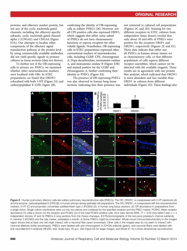
To further test if the OR-expressingcells in airways are PNECs, we examinedwhether other neuroendocrine markerswere localized with ORs. In hTECpreparations, we found that OR2W1colocalized with both 5-HT (Figure 2A) andcarboxypeptidase E (CPE; Figure 2B),
confirming the identity of OR-expressingcells as solitary PNECs (26). However, notall CPE-positive cells also expressed ORW1,which suggests that either some subsetof PNECs do not have chemosensoryfunctions or express receptors for othervolatile ligands. Nonetheless, OR-expressingcells in hTEC preparations expressed otherconventional markers of neuroendocrinecells, including CGRP, CPE, chromograninA, Dopa decarboxylase, monoamine oxidaseA, and monoamine oxidase B (Figure E3B)and stained positive for the CGRP andchromogranin A, further confirming theiridentity as PNECs (Figure E4).
The presence of OR-expressing PNECswas also observed in human lung tissuesections, indicating that their presence was
not restricted to cultured cell preparations(Figures 2C and 2D). Staining for twodifferent receptors in hTEC cultures fromindependent tissue donors reveled thatonly about 30 and 60% of PNECs werepositive for the receptors OR2F1 andOR2W1, respectively (Figures 2E and E1).These data indicate that either notall PNECs in human airway tissues actas chemosensory cells, or that differentpopulations of cells express differentreceptor assemblies, which cannot yet bedetected with the available reagents. Theseresults are in agreement with our proteinblot analyses, which indicated that OR2W1is more abundant and less variable thanOR2F1 in cultures from differentindividuals (Figure 1D). These findings also
Figure 2. Human pulmonary olfactory cells are solitary pulmonary neuroendocrine cells (PNECs). The OR, OR2W1, is coexpressed with 5-HT (serotonin) (A)and the enzyme, carboxipeptidase E (CPE) (B), in human primary airway epithelial cell preparations. The OR, OR2W1, is coexpressed with the neuroendocrinemarkers, 5-HT (C) and proprotein convertase subtilisin/kexin type 2 (PCSK2) (D), in human lung tissue sections. (E) OR abundance in preparations froma single donor. Single culture membranes were cut into two pieces and costained for the specified receptor and the PNEC marker, PCSK2. Mean receptorabundance (% cells) is shown for the receptor and PCSK2 out of the total PCSK2-positive cells. Error bars denote SEM. *P , 0.05 (one-tailed t test; n = 3independent donors). (F and G) PNECs in lung sections from the rhesus macaque. (H) Photomicrographs of the two-pore potassium channel subfamilyK member 3 (KCNK3) and the cilia marker, acetylated a-tubulin. (I) Some PNECs might be innervated. Whole-mount immunohistochemistry in the imagedregion shows that at least one cell might interact with a neuronal fiber (open arrowhead). Other cells in the imaged area do not seem to interact withneuronal afferents (white arrowheads). PNECs were labeled with anti–chromogranin A (CHGA) antibody (green), and neuronal fibers were labeled withanti–neurofilament H antibody (NF200; red). Scale bars, 50 mm. See Figure E6 for larger images, and Movie E1 for a three-dimensional reconstruction.
ORIGINAL RESEARCH
640 American Journal of Respiratory Cell and Molecular Biology Volume 50 Number 3 | March 2014

indicate that OR-expressing PNECs wereselectively localized to the airwayepithelium, and therefore well positionedto act as sentinels for inhaled odorants.
Next, we asked whether OR-expressingPNECs were present in lungs from othermammalian species. Previous studiesindicated the presence of solitary PNECsand neuroendocrine bodies (NEBs; clustersof neuroendocrine cells) in mammals,especially during fetal and neonataldevelopment (29–31). Although CGRP-positive cells have been described in theadult mouse airway epithelium, they arenot serotonergic, and appear to act asa population of stem cells that plays a rolein epithelial recovery from injuries (32).We were not able to detect pulmonaryinterdigitating PNECs in lungs of adultmice or ferrets using 5-HT as a marker.In contrast to mice, which, in our hands,showed serotonergic neuroendocrine cellsin the gut epithelium, but not in the lung(Figure E5A), adult rat lungs did containlung cells positive for 5-HT. However,the identified cells were significantly largerthan human PNECs, did not have aninterdigitating morphology, and werelocated in subepithelial layers of the airways(Figures E5B and E5C). However, OR-expressing PNECs with morphologysimilar to those found in human tissueswere detected in lung tissues from rhesusmacaque monkeys (Figures 2F and 2G).These results indicate that the identifiedPNECs are not specific to lungs of humans,because they are present in nonhumanprimates as well.
Other studies of pulmonary NEBs inrodent models have suggested that theprimary function of these cells is to act asoxygen sensors in the postnatal lung (30,31). Whether adult human lungs can alsodirectly sense oxygen pressure in inhaledair remains uncertain. Current modelspropose that neuroregulation of breathingin adult humans is primarily mediated viaoxygen-sensing neurons in the carotidbodies (33, 34). When we immunostainedadult human lung tissues with an antibodyagainst potassium channel subfamily Kmember 3 (KCNK3), a two-pore potassiumchannel that has been identified as anessential molecular component of theoxygen-sensing receptor complex inmammalian pulmonary NEBs (35), wefound that this channel is enriched inepithelial motile cilia, but is not present inPNECs (Figure 2H). However, similarly to
the rodent NEBs, whole-mountimmunostaining of human airway epitheliarevealed that some human PNECs might bedirectly innervated by afferent neuronalfibers (Figures 2I and E6, and Video E1).Together, our data suggest that primateadult solitary PNECs are chemosensory,but not likely to act as oxygen sensors,and are distinct from PNECs or NEBsidentified during early lung developmentin human infants and nonprimate animalmodels.
Our results suggested that PNECs serveas chemosensory neuroendocrine sentinelsin the human airway epithelium. Therefore,we hypothesized that specific volatilechemical stimuli could induce the localrelease of the neuroendocrine content fromstimulated PNECs. We first examinedwhether the cellular contents of twodifferent neuroendocrine secreted factors,5-HT and CGRP, are correlated with eachother in individual cells. We found that thelevels of 5-HT and CGRP in individualcells were highly correlated, which suggeststhat these factors are coexpressed, and arelikely coreleased on stimulation (Figure 3A).Next, we investigated whether apicalstimulation of well differentiated hTECpreparations is correlated with lower cellcontents of both 5-HT and CGRP(Figure 3B). In agreement with ourhypothesis, apical stimulation of primaryculture cells with a panel of volatilechemicals that have been shown to activatesome of the receptors expressed in hTECs(Figure 1A and Table E2) resulted ina significant increase of CGRP released intothe basal cell medium (Figure 3C). Inaddition, staining of hTEC preparationsthat were treated with the ligands, nonanal(36) or citronellal (37), showed overall lower5-HT signal relative to that in DMSO-treatedcells. Together, these results areconsistent with stimulation-dependentrelease of CGRP and 5-HT (Figures 3Dand 3E).
Because PNECs appear to be asignificant source of 5-HT and CGRP inhuman airway tissues, we next examinedwhich cells might express 5-HT and CGRPreceptors as possible cellular targets formodulation by PNECs. Microarray datafrom human airway cell preparations (asdemonstrated in Figure 1A) indicated that14 out of the 17 known human 5-HTreceptors (HTRs) are expressed in hTECpreparations (Figure 4A). We also foundthat multiple independent receptors for
5-HT and CGRP were expressed at theprotein level in various airway cell types,but not in PNECs themselves, furthersupporting our hypothesis that activationof PNECs by volatile ligands can lead tolocal stimulation of other neighboring celltypes in the epithelium (Figures 4B–4D).Each of the three identified neuroendocrinereceptors (5-HTR2B, HTR1F, andcalcitonin/CGRP receptor [CALCR])showed enrichment in the population ofairway epithelial basal cells. The CGRPreceptor, CALCR, also showed expressionin airway smooth muscles, or possibly inthe neuronal fibers that innervate them(Figure 4D). Thus, our data supportthe model that activation of PNECsin human airways could lead to alocalized activation of airway epithelialcells, and a concerted response to volatileagents (Figure 4E).
It has been proposed that airwaydiseases, such as asthma or COPD,manifest increased airway responses toenvironmental factors, including volatileagents (38). The present observationssuggest that PNECs might contribute tothis chemical hyperresponsiveness inairway disease. In that regard, previousstudies also indicated that neuroendocrinefactors, such as 5-HT, and peptides,such as CGRP, have potent physiologicaleffects on epithelial, airway smoothmuscle, and vascular smooth muscle cellsin the lung (39–41). Thus, our datasuggested the hypothesis that changes inthe activation level of PNECs or theresponsiveness of other airway cell types tostimulation by PNEC contents couldcontribute to olfactory hypersensitivity inairway disease. We therefore determinedthe levels of PNECs in lung tissuesobtained from lung transplant recipientswith severe COPD versus correspondingtissues from lung transplant donorswithout COPD (42, 43). In agreement withour hypothesis, we found increasednumbers of OR2W1- and proproteinconvertase subtilisin/kexin type 2-expressing PNECs in lung tissues fromsubjects with COPD relative to controlsubjects without COPD, without anydetectable differences in PNEC morphology(Figures 5A–5C). Although the complexmorphology of PNECs prevented us fromstudying their morphology in tissuesections, gross observations suggested thatdisease state does not affect their size ormorphology (Figure 5D).
ORIGINAL RESEARCH
Gu, Karp, Brody, et al.: Chemosensory Pulmonary Neuroendocrine Cells 641

Another possible explanation for theincreased sensitivity of patients with COPDto inhaled substances is an increasedlevel of neuroendocrine receptors anda consequent increase in the response toneuropeptides released by volatile-activatedOR-expressing PNECs. Consistent withthis hypothesis, we found that 5-HT andCGRP receptor distributions appeared to bealtered in COPD versus non-COPD airwaytissues (Figure 6). In particular, we foundthat HTR1F was expressed in nearly allbasal cells in subjects with COPD comparedwith a subset of basal cells in those withoutCOPD, on the basis of the extent ofcostaining with the basal cell marker, p63(Figure 6A). In contrast, we detecteddecreased staining levels of the CGRP
receptor, CALCR, in airway epitheliumin patients with COPD , but increasedlocalization to basal cells in subjectswithout COPD (Figure 6B). Similarly,HTR2B changed from diffuse epithelialexpression in COPD to increased basalcell staining in subjects without COPD(Figure 6C). Although these descriptivedata are difficult to quantify, due to thehigh variability of receptor distributionsacross individuals, they do raise thepossibility that neuroendocrine signals tobasal cells may be altered in patients withCOPD. Nevertheless, these data providea potential mechanism for several differentcell types in the airway to respond to theneuroendocrine factors released fromvolatile-activated PNECs.
Discussion
The exacerbation of airway diseases, suchas asthma or COPD, is often mediated byexposure to environmental factors, such asindustrial solvents, various pungent odors,and other volatiles. In recent years, severalkey studies have demonstrated that specificpopulations of cells in the mammalianairway epithelia can be directly activatedby inhaled “bitter” irritants, which includeciliated epithelial cells in lower (5) andupper airways (6), brush cells (7, 12), andairway smooth muscles (4). In spite of thissignificant progress, the cells and receptorsthat mediate the response of humanairways to the majority of volatile chemicalsin health or disease are still largely
Figure 3. PNECs release neuroendocrine factors in response to volatile apical stimuli. (A) Levels of the biogenic amine, 5-HT, and the peptide calcitoningene-related peptide (CGRP) in individual PNECs (R2 = 0.94; n = 27 cells from three independent donors, 7–10 cells per donor). Signals represent averagepixel intensity of cell bodies that were coimmunostained for 5-HT and CGRP. (B) An illustration of the supported membrane insert system used forculture of primary airway epithelial cells (20, 50). Treatment experiments were accomplished by applying the various ligands apically, while measuring thebasal release of neuroendocrine factors. (C) CGRP release in basal medium after apical treatment with indicated volatile. Mean (6 SEM) of CGRPlevels measured by ELISA (P , 0.05, Kruskal-Wallis ANOVA; n = 4 inserts per treatment). (D and E) Treatments of primary cell preparations with nonanalor citronellal followed by immunostaining for 5-HT levels relative to controls. Shown are means (6 SEM) (one-way ANOVA; P , 0.001; n = 4 insertsfrom a single donor per treatment). *P , 0.05; **P , 0.01. Scale bars, 50 mm. DMSO, dimethyl sulfoxide.
ORIGINAL RESEARCH
642 American Journal of Respiratory Cell and Molecular Biology Volume 50 Number 3 | March 2014

unknown. In this study, we identifieda previously unrecognized chemosensoryfunction for a population of solitary PNECsin adult human airways. Although thepresence of solitary PNECs in humanairways has been described (26), noclear physiological functions have beenattributed to these cells. Based on cellmorphology, tissue localization, andmolecular characterization, the adultsolitary PNECs may represent a cell linagethat is independent of similar PNECs thathave been previously described in rodentmodels. For example, in contrast to adultsolitary PNECs and NEBs present in somerodent lungs, human PNECs do not expressoxygen-sensing ion channels, and are foundin major trachea and bronchi, but notalveolar tissues. However, immunostainingof human tissues with the neuronal marker,neurofilament H, revealed a possibleinnervation of human PNECs, as has beendescribed for NEBs in some rodent species.These findings further suggest that human
PNECs are not likely to be homologous tothe previously described neonatal humanPNECs and NEBs, or other types of adultPNECs that have been described in animalmodels (29, 30, 32, 35, 44). We currentlydo not understand why PNECs are differentin primates relative to other mammals.However, species-specific differences inlung cellular and physiological functionsare consistent with the proposal thathuman airways have evolved distinctcell lineages and signaling systems forrespiratory-specific functions. Otherexamples include the marked differencesin the types of airway progenitor cells thatdevelop after viral infection (45). Similarly,cystic fibrosis transmembrane conductanceregulator and chloride channel accessory1–mitogen-activated protein kinase 13 signalsexhibit markedly different function in humanversus rodent lung for control of iontransport and mucus production (46–48).
Our study also highlights novel featuresof OR function in the lung in comparison to
the olfactory system. We show that adultPNECs express more than one OR per cell.This arrangement contrasts with the one inthe olfactory epithelium, in which eacholfactory sensory neuron expresses a singlereceptor in a cellular process that is stillnot well understood (49). In contrast tothe narrowly tuned nasal olfactory sensoryneurons, these differences might alsosuggest that sensing volatiles in the lungis nonneuronal, and is broadly responsiveeven within single cells. Thus, wedemonstrate that airway epithelial cells canrespond to structurally diverse classes ofvolatile chemicals at least in cell culture.Moreover, this response is likely onlya partial profile, because the majority of thereceptors that we identified in humanairways are still orphan in regard to theirligand specificity.
Unexpectedly, we detected noexpression of cyclic nucleotide gatedchannels, including the olfactory-specificsubunits, CNGA2 and CNGA4, in human
Figure 4. (A) 5-HT receptor (HTR) gene expression by microarray analysis of mRNAs isolated from primary airway cell preparations from 10 independentdonors (as in Figure 1A). (B and C) Serotonin receptors, 5-HTR 2B and HTR1F, in basal cells (open arrowheads) detected by immunostaining. White
arrowheads point to solitary PNECs. (D) The calcitonin/CGRP receptor (CALCR) is enriched in basal cells (open arrowheads) and in airway smoothmuscles or possibly the neurons that innervate them (white arrow).White arrowheads in C and D mark PNECs. Dashed white lines show the apical side ofthe stratified epithelium. Scale bars, 50 mm. (E) Model for the possible local impact of olfactory stimulation of PNECs followed by neuroendocrine release of5-HT and CGRP on various cell types in the airways (orange arrows).
ORIGINAL RESEARCH
Gu, Karp, Brody, et al.: Chemosensory Pulmonary Neuroendocrine Cells 643

airway tissue. Because these channels seemto play an important role in the neuronalolfactory signal transduction pathway, thesefindings suggest that stimulation of OR-expressing PNECs does not lead to changesin membrane potential, but rather usesdifferent signaling routes. Nonetheless, wefound that the apical stimulation of airwayepithelia with volatiles can lead to lowercellular contents of neuroendocrine factors,such as 5-HT and CGRP, which is inagreement with a stimulus-dependentrelease. Although we did not directly testthe role of OR stimulation in the volatile-dependent release of 5-HT and CGRP, thepresence of these receptors in PNECs ishighly suggestive that these receptors areresponsible for the activation of PNECsby environmental chemical stimuli.Subsequently, these factors are likely toaffect the physiology of other cell types in
the epithelium, including smooth musclecells, neurons, and basal cells. Thus, PNECscould represent a key cellular pathway forsensing environmental volatile stimuli inlungs.
Furthermore, our studies suggest thatPNECs may contribute to the chemicalhypersensitivity of lungs from patients withairway diseases, such as COPD. This issupported by our findings that the PNECcell frequency is higher in lung tissues ofpatients with COPD relative to healthyindividuals (Figure 5). However, werecognize that our method for quantifyingPNECs in tissues may not fully account forthe overall effects of inflammation, whichcould influence factors such as cell sizeor morphology of nuclei in tissue sections,and potentially bias our calculations of cellnumber. Thus, future studies of PNECdistribution performed using whole-mount
tissue staining in a large number of sampleswill further address this issue. In addition,our study examined only a singleconcentration of each ligand to establishthe response of PNECs to odorants. Werecognize that differences in the numbers ofPNECs in the airway might also influencethe sensitivity of these tissues to odorantstimulation. For example, increased PNEClevels in COPD lungs might result inhypersensitivity to odorants, comparedwith the response in healthy lungs.Therefore, detailed dose–responseassessment will be required to fully assessthe differences in odorant responsesbetween COPD and non-COPD conditions.
Taken together, our study offers thesignificant advance of identifying a new classof chemosensory cells that express ORsin adult human airways. The finding addsto the growing understanding of the
Figure 5. PNECs are more abundant in airways from patients with chronic obstructive pulmonary disease (COPD) relative to healthy donors. (A)Representative confocal scan of a human bronchial tissue section used to quantify PNEC frequencies. (B) Magnification of yellow box in (A); dotted line
indicates an example of the region used to normalize number of cells to epithelial length. Cells in the images shown were positive for both OR2W1and PCSK2. (C) Box plots represent PNEC frequencies in tissue sections from lungs of subjects with and without COPD (*P , 0.05; **P , 0.01;Mann-Whitney U Test; n = 6–7 individuals per group). (D) The morphology and size of OR-expressing PNECs in COPD and non-COPD lungs are similar.Left panels, non-COPD; right panels, COPD. Cells were labeled with an anti-OR2W1 antibody. White arrowheads mark PNECs.
ORIGINAL RESEARCH
644 American Journal of Respiratory Cell and Molecular Biology Volume 50 Number 3 | March 2014

complexity of the sensory response of theairway epithelium to diverse classes ofsensory stimuli, and thereby impacts ourunderstanding of how the human lungresponds to environmental stimuli inboth health and disease. We also detecta possible variation in PNECs and PNEC-responsive receptors between subjectswith and without COPD that suggests arole for this OR–neuroendocrine axis in
the pulmonary response toenvironmental volatile stimuli that mightaccompany chronic lung disease.Although more work is required tocharacterize the sensory functions ofPNECs, it is possible that PNEC-specificpathways may be a therapeutic target tomanage diseases associated with airwayhypersensitivity in asthma, COPD, andrelated conditions. n
Author disclosures are available with the textof this article at www.atsjournals.org.
Acknowledgments: The authors thank L.Belaygorod for technical assistance, C. Mikolsfor assistance with cell isolation and culture,and L. Miller for providing airway tissuesections from rhesus monkeys from theresources of the California National PrimateResearch Center (University of California,Davis).
References
1. Massaro GD, Massaro D. Formation of pulmonary alveoli and gas-exchange surface area: quantitation and regulation. Annu Rev Physiol1996;58:73–92.
2. Patton JS, Byron PR. Inhaling medicines: delivering drugs to the bodythrough the lungs. Nat Rev Drug Discov 2007;6:67–74.
3. Millqvist E, Bende M, Lowhagen O. Sensory hyperreactivity—a possiblemechanism underlying cough and asthma-like symptoms. Allergy1998;53:1208–1212.
4. Deshpande DA, Wang WC, McIlmoyle EL, Robinett KS, Schillinger RM,An SS, Sham JS, Liggett SB. Bitter taste receptors on airway smoothmuscle bronchodilate by localized calcium signaling and reverseobstruction. Nat Med 2010;16:1299–1304.
5. Shah AS, Ben-Shahar Y, Moninger TO, Kline JN, Welsh MJ. Motile ciliaof human airway epithelia are chemosensory. Science 2009;325:1131–1134.
6. Lee RJ, Xiong G, Kofonow JM, Chen B, Lysenko A, Jiang P, Abraham V,Doghramji L, Adappa ND, Palmer JN, et al. T2R38 taste receptorpolymorphisms underlie susceptibility to upper respiratory infection.J Clin Invest 2012;122:4145–4159.
7. Saunders CJ, Reynolds SD, Finger TE. Chemosensory brush cells of thetrachea: a stable population in a dynamic epithelium. Am J Respir CellMol Biol 2013;49:190–196.
8. Merigo F, Benati D, Tizzano M, Osculati F, Sbarbati A. alpha-Gustducin immunoreactivity in the airways. Cell Tissue Res 2005;319:211–219.
9. Tizzano M, Cristofoletti M, Sbarbati A, Finger TE. Expression of tastereceptors in solitary chemosensory cells of rodent airways. BMC PulmMed 2011;11:3.
10. Tizzano M, Gulbransen BD, Vandenbeuch A, Clapp TR, Herman JP,Sibhatu HM, Churchill ME, Silver WL, Kinnamon SC, Finger TE.Nasal chemosensory cells use bitter taste signaling to detectirritants and bacterial signals. Proc Natl Acad Sci USA 2010;107:3210–3215.
11. Krasteva G, Canning BJ, Hartmann P, Veres TZ, Papadakis T, MuhlfeldC, Schliecker K, Tallini YN, Braun A, Hackstein H, et al. Cholinergicchemosensory cells in the trachea regulate breathing. Proc NatlAcad Sci USA 2011;108:9478–9483.
12. Krasteva G, Canning BJ, Papadakis T, Kummer W. Cholinergic brushcells in the trachea mediate respiratory responses to quorum sensingmolecules. Life Sci 2012;91:992–996.
Figure 6. Spatial expression patterns of 5-HT and CGRP receptors are different in normal versus COPD lungs. (A) Representative photomicrographs ofimmunostaining for HTR1F (green). Staining is enriched in basal cells in normal and COPD lungs (basal cells marked with the p63 marker, red nuclei). Notethat the morphology of COPD basal cells is elongated, and the distribution of the receptor uniform, compared with those in normal lungs. (B)Representative photomicrographs of immunostaining for CALCR (green). Staining is highly enriched in basal cells (marked by p63, red nuclei) and smoothmuscle or neuronal fibers (white arrow) in normal donors. However, expression is lower and more diffused in COPD lungs. (C) Representativephotomicrographs of immunostaining for HTR2B (green). Receptor is enriched basal cells of lungs from non-COPD subjects when compared with normallungs. Note that in lungs of subjects with COPD, staining for HTR2B is more diffused and higher in nonbasal epithelial cells relative to basal cells (whitearrow). Scale bars, 50 mm.
ORIGINAL RESEARCH
Gu, Karp, Brody, et al.: Chemosensory Pulmonary Neuroendocrine Cells 645

13. Lin W, Ogura T, Margolskee RF, Finger TE, Restrepo D. TRPM5-expressing solitary chemosensory cells respond to odorous irritants.J Neurophysiol 2008;99:1451–1460.
14. Buck L, Axel R. A novel multigene family may encode odorant receptors:a molecular basis for odor recognition. Cell 1991;65:175–187.
15. Feldmesser E, Olender T, Khen M, Yanai I, Ophir R, Lancet D.Widespread ectopic expression of olfactory receptor genes. BMCGenomics 2006;7:121.
16. Spehr M, Gisselmann G, Poplawski A, Riffell JA, Wetzel CH, ZimmerRK, Hatt H. Identification of a testicular odorant receptor mediatinghuman sperm chemotaxis. Science 2003;299:2054–2058.
17. Pluznick JL, Zou DJ, Zhang X, Yan Q, Rodriguez-Gil DJ, Eisner C, WellsE, Greer CA, Wang T, Firestein S, et al. Functional expression of theolfactory signaling system in the kidney. Proc Natl Acad Sci USA2009;106:2059–2064.
18. Pluznick JL, Protzko RJ, Gevorgyan H, Peterlin Z, Sipos A, Han J,Brunet I, Wan LX, Rey F, Wang T, et al. Olfactory receptorresponding to gut microbiota–derived signals plays a role in reninsecretion and blood pressure regulation. Proc Natl Acad Sci USA2013;110:4410–4415.
19. Braun T, Voland P, Kunz L, Prinz C, Gratzl M. Enterochromaffin cells ofthe human gut: sensors for spices and odorants. Gastroenterology2007;132:1890–1901.
20. Karp PH, Moninger TO, Weber SP, Nesselhauf TS, Launspach JL,Zabner J, Welsh MJ. An in vitromodel of differentiated human airwayepithelia: methods for establishing primary cultures. Methods MolBiol 2002;188:115–137.
21. Tyner JW, Kim EY, Ide K, Pelletier MR, Roswit WT, Morton JD, BattaileJT, Patel AC, Patterson GA, Castro M, et al. Blocking airway mucouscell metaplasia by inhibiting EGFR antiapoptosis and IL-13transdifferentiation signals. J Clin Invest 2006;116:309–321.
22. Zabner J, Scheetz TE, Almabrazi HG, Casavant TL, Huang J, KeshavjeeS, McCray PB Jr. CFTR DeltaF508 mutation has minimal effect onthe gene expression profile of differentiated human airway epithelia.Am J Physiol Lung Cell Mol Physiol 2005;289:L545–L553.
23. Su CY, Menuz K, Reisert J, Carlson JR. Non-synaptic inhibition betweengrouped neurons in an olfactory circuit. Nature 2012;492:66–71.
24. Khan M, Vaes E, Mombaerts P. Regulation of the probability of mouseodorant receptor gene choice. Cell 2011;147:907–921.
25. Gu X, Ben-Shahar Y. Olfactory receptors in human airway epithelia. In:Cresto C, editor. Olfactory receptors: methods and protocols. NewYork City: Humana Press; 2013. pp. 161–169.
26. Weichselbaum M, Sparrow MP, Hamilton EJ, Thompson PJ, Knight DA.A confocal microscopic study of solitary pulmonary neuroendocrinecells in human airway epithelium. Respir Res 2005;6:115.
27. Lechin F, van der Dijs B, Lechin AE. Severe asthma and plasmaserotonin. Allergy 2002;57:258–259.
28. Moore BD, Hyde D, Miller L, Wong E, Frelinger J, Schelegle ES.Allergen and ozone exacerbate serotonin-induced increases inairway smooth muscle contraction in a model of childhood asthma.Respiration 2012;83:529–542.
29. Avadhanam KP, Plopper CG, Pinkerton KE. Mapping the distribution ofneuroepithelial bodies of the rat lung: a whole-mountimmunohistochemical approach. Am J Pathol 1997;150:851–859.
30. Cutz E, Perrin DG, Pan J, Haas EA, Krous HF. Pulmonaryneuroendocrine cells and neuroepithelial bodies in sudden infantdeath syndrome: potential markers of airway chemoreceptordysfunction. Pediatr Dev Pathol 2007;10:106–116.
31. Cutz E, Yeger H, Pan J. Pulmonary neuroendocrine cell system inpediatric lung disease—recent advances. Pediatr Dev Pathol 2007;10:419–435.
32. Hong KU, Reynolds SD, Giangreco A, Hurley CM, Stripp BR. Clara cellsecretory protein–expressing cells of the airway neuroepithelialbody microenvironment include a label-retaining subset and arecritical for epithelial renewal after progenitor cell depletion. Am JRespir Cell Mol Biol 2001;24:671–681.
33. Peers C, Wyatt CN, Evans AM. Mechanisms for acute oxygen sensingin the carotid body. Respir Physiol Neurobiol 2010;174:292–298.
34. Weir EK, Lopez-Barneo J, Buckler KJ, Archer SL. Acute oxygen-sensing mechanisms. N Engl J Med 2005;353:2042–2055.
35. Cutz E, Jackson A. Neuroepithelial bodies as airway oxygen sensors.Respir Physiol 1999;115:201–214.
36. Schmiedeberg K, Shirokova E, Weber HP, Schilling B, Meyerhof W,Krautwurst D. Structural determinants of odorant recognition by thehuman olfactory receptors OR1A1 and OR1A2. J Struct Biol 2007;159:400–412.
37. Krautwurst D, Yau KW, Reed RR. Identification of ligands for olfactoryreceptors by functional expression of a receptor library. Cell 1998;95:917–926.
38. Caceres AI, Brackmann M, Elia MD, Bessac BF, del Camino D,D’Amours M, Witek JS, Fanger CM, Chong JA, Hayward NJ, et al. Asensory neuronal ion channel essential for airway inflammation andhyperreactivity in asthma. Proc Natl Acad Sci USA 2009;106:9099–9104.
39. Maclean MR, Dempsie Y. The serotonin hypothesis of pulmonaryhypertension revisited. Adv Exp Med Biol 2010;661:309–322.
40. Dupont LJ, Pype JL, Demedts MG, De Leyn P, Deneffe G, VerledenGM. The effects of 5-HT on cholinergic contraction in human airwaysin vitro. Eur Respir J 1999;14:642–649.
41. Cazzola I, Matera MG. 5-HT modifiers as a potential treatment ofasthma. Trends Pharmacol Sci 2000;21:13–16.
42. Agapov E, Battaile JT, Tidwell R, Hachem R, Patterson GA, Pierce RA,Atkinson JJ, Holtzman MJ. Macrophage chitinase 1 stratifies chronicobstructive lung disease. Am J Respir Cell Mol Biol 2009;41:379–384.
43. Deslee G, Woods JC, Moore CM, Liu L, Conradi SH, Milne M, GieradaDS, Pierce J, Patterson A, Lewit RA, et al. Elastin expression in verysevere human COPD. Eur Respir J 2009;34:324–331.
44. Domnik NJ, Cutz E. Pulmonary neuroepithelial bodies as airwaysensors: putative role in the generation of dyspnea. Curr OpinPharmacol 2011;11:211–217.
45. Byers DE, Alexander-Brett J, Patel AC, Agapov E, Dang-Vu G, Jin X,Wu K, You Y, Alevy Y, Girard J-P, et al. Long-term IL-33–producingepithelial progenitor cells in chronic obstructive lung disease. J ClinInvest 2013;123:3967–3982.
46. Rogers CS, Stoltz DA, Meyerholz DK, Ostedgaard LS, Rokhlina T, TaftPJ, Rogan MP, Pezzulo AA, Karp PH, Itani OA, et al. Disruption ofthe CFTR gene produces a model of cystic fibrosis in newborn pigs.Science 2008;321:1837–1841.
47. Grubb BR, Boucher RC. Pathophysiology of gene-targeted mousemodels for cystic fibrosis. Physiol Rev 1999;79(1 Suppl):S193–S214.
48. Alevy YG, Patel AC, Romero AG, Patel DA, Tucker J, Roswit WT, MillerCA, Heier RF, Byers DE, Brett TJ, et al. IL-13–induced airway mucusproduction is attenuated by MAPK13 inhibition. J Clin Invest 2012;122:4555–4568.
49. Lomvardas S, Barnea G, Pisapia DJ, Mendelsohn M, Kirkland J, Axel R.Interchromosomal interactions and olfactory receptor choice. Cell2006;126:403–413.
50. Whitcutt MJ, Adler KB, Wu R. A biphasic chamber system formaintaining polarity of differentiation of cultured respiratory tractepithelial cells. In Vitro Cell Dev Biol 1988;24:420–428.
ORIGINAL RESEARCH
646 American Journal of Respiratory Cell and Molecular Biology Volume 50 Number 3 | March 2014