Mechanical ventilation interacts with endotoxemia to induce extrapulmonary organ dysfunction

Biochimica et Biophysica Acta 1813 (2011) 1814–1821
Contents lists available at ScienceDirect
Biochimica et Biophysica Acta
j ourna l homepage: www.e lsev ie r.com/ locate /bbamcr
Glucocorticoids induce mitochondrial gene transcription in HepG2 cellsRole of the mitochondrial glucocorticoid receptor
Anna-Maria G. Psarra a,b,⁎, Constantine E. Sekeris c,1
a Biomedical Research Foundation, Academy of Athens, Center for Basic Research, 4 Soranou Efesiou, 11527, Athens, Greeceb Department of Biochemistry and Biotechnology, University of Thessaly, Larissa, Greecec National Hellenic Research Foundation, Institute of Biological Research and Biotechnology, Laboratory of Molecular Endocrinology, 48 Vas Constantinou Ave, 11635, Athens, Greece
⁎ Corresponding author at: Department of BiochUniversity of Thessaly, 26 Ploutonos and Aiolou, 4122410 565221; fax: +30 2410 565290.
E-mail address: [email protected] (A.-M.G. Psarra1 Prof. Sekeris passed away on 15th September 2009.
0167-4889/$ – see front matter © 2011 Elsevier B.V. Adoi:10.1016/j.bbamcr.2011.05.014
a b s t r a c t
a r t i c l e i n f oArticle history:Received 8 February 2011Received in revised form 23 May 2011Accepted 24 May 2011Available online 2 June 2011
Keywords:Glucocorticoid receptorMitochondrial transcriptionOXPHOSMitochondrial transcription factorMitochondrial DNAMitochondrial ATP production
Glucocorticoids are major regulators of a plethora of cellular functions, acting on target cells throughglucocorticoid receptors (GR) and modulation of gene transcription, among other mechanisms. One main siteof action of glucocorticoids is the hepatocyte, which responds to the hormonal stimulus with induction ofseveral proteins among them enzymes of oxidative phosphorylation (OXPHOS), both nuclearly andmitochondrially encoded. The induction of OXPHOS is regarded as a result of a nuclear action of the receptoron the respective nuclear genes and on genes encoding mitochondrial transcription factors. The presence ofGR in mitochondria and of sequences in the mitochondrial genome similar to glucocorticoid responsiveelements, suggested a direct action of GR on mitochondrial transcription. We demonstrate in HepG2hepatocarcinoma cells specific binding of GR to the regulatory D-loop region of themitochondrial genome andshow that dexamethasone induces the mitochondrial transcription factors A, B1, and B2, the mitochondrialribosomal RNA, and several mitochondrially encoded OXPHOS genes. Applying α-amanitin, the specificinhibitor of DNA-dependent RNA polymerase II, the dexamethasone-induced transcription of themitochondrial genes can still proceeds, whereas the DEX effect on transcription of the mitochondrialtranscription factors is suppressed. Moreover, HepG2 cells overexpressing mitochondrial targeted GR showedincreased RNA synthesis, cytrochrome oxidase subunit I protein expression, and mitochondrial ATPproduction. We conclude that glucocorticoids can stimulate directly mitochondrial transcription by themitochondrially localized GR, affecting OXPHOS enzyme biosynthesis. This takes place in addition to theiraction on mitochondrial genes by way of induction of the nuclearly encoded mitochondrial transcriptionfactors.
emistry and Biotechnology,21, Larissa, Greece. Tel: +30
).
ll rights reserved.
© 2011 Elsevier B.V. All rights reserved.
1. Introduction
Glucocorticoids are important regulators of basic cellular processes,such asmetabolism, growth, differentiation, and survival [1]. Theirmodeof action in target cells has received considerable interest and it is nowclear that they can act in various ways, e.g., on the gene level by way ofcognate receptors and on cytoplasmic processes by way of membranereceptors.Oneof thefirstmodel systemswhich led to thedemonstrationthat glucocorticoids act through cognate cytoplasmic receptors tostimulate transcription of genes and induce the respective proteinswas that of cortisol inducing in the rodent liver enzymes involved ingluconeogenesis [2–6].
In these and later studies, a bulk increase of HnRNA containing thepre-mRNAs of the induced genes, ribosomal RNA and t-RNA was
observed [3–5], which could not account for the increased mRNAsynthesis corresponding to the transcription of the few genes,identified as glucocorticoids-induced genes. Recent results fromstudies involving bio-array analyses [7–9] have shown that theglucocorticoid effect on the hepatocyte and on other target cellsencompasses several genes, and, in part, justifies the observedoverwhelming increase in nuclear RNA synthesis. MitochondrialRNA synthesis is also stimulated by glucocorticoids [3,6]. Thehormonal induction process is highly energy consuming, and thecell makes efforts to guarantee the replenishment of its energyreserves. Therefore, it is understandable that the glucocorticoidinduced mitochondrial RNA synthesis can be correlated to this energyregeneration process, as the mitochondrial genome encodes subunitsof OXPHOS enzymes and RNAs participating in the protein synthesisinmitochondria. Due to the fact that some OXPHOS are encoded in thenuclear genome and others in the mitochondrial genome it isassumed, in analogy to the action of thyroid hormones on liverOXPHOS [reviewed in 10] that the hormonal stimulus affects thetranscription of the mitochondrial encoded OXPHOS indirectly, byinducing nuclear signals, such as mitochondrial transcription factors,

Table 1Primers for human mitochondrial OXPHOS enzymes, COX IV, 12 S, and 16 Smitochondrial RNA.
Forward Reverse
ND I atggccaacctcctactcctcatt ttatggcgtcagcgaagggttgtaND II ccatctttgcaggcacactcatca attatggatgcggttgcttgcgtgND III gccctacaaacaactaacctgcca ataggccagacttagggctaggatND IV agctccatctgcctacgacaaaca taagcccgtgggcgattatgagaaND V cacagcagccattcaagcaatcct acctaattgggctgatttgcctgcND VI ataggatcctcccgaatcaaccct aggattggtgctgtgggtgaaagaCOX I accctagaccaaacctacgccaaa taggccgagaaagtgttgtgggaaCOX II acagatgcaattcccggacgtcta ggcatgaaactgtggtttgctccaCOX III tcacttccactccataacgctcct gtgttacatcgcgccatcattggtATP6 acattactgcaggccacctactca acgtaggcttggattaaggcgacaATP8 accgtatggcccaccataattacc tttatgggctttggtgagggaggtCyt b agtcccaccctcacacgattcttt agtaagccgagggcgtctttgatt12 S aaactgctcgccagaacactacga tgagcaagaggtggtgaggttgat16 S taccctcactgtcaacccaacaca ttaaacatgtgtcactgggcaggcCOX IV agaaagtcgagttgtatcgcatt gataacgagcgcggtgaaac
1815A.-M.G. Psarra, C.E. Sekeris / Biochimica et Biophysica Acta 1813 (2011) 1814–1821
which then, secondarily, positively regulate transcription of themitochondrial genome. However, the demonstration of the mito-chondrially localized glucocorticoid receptor and the presence ofsequences with strong homology and characteristics of glucocorticoidresponse elements (GRE) in the mitochondrial genome [11], raisedthe possibility of a direct effect of the mitochondrial GR onmitochondrial transcription, in addition to the indirect nuclearpathway [12,13].
We now present data demonstrating that dexamethasone (DEX), asynthetic glucocorticoid, induces in HepG2 cells the mitochondrialtranscription factors A, B1, and B2 (TFAM, TFB1M, TFB2M), the 12 S and16 S mitochondrial ribosomal RNA and several mitochondrial DNA(mtDNA) encoded genes. Applyingα-amanitin, the inhibitor of the DNA-dependent RNA polymerase II, we show that the inducing effect ofdexamethasone on the mitochondrial ribosomal RNA and the mtDNAencoded genes can still take place in the presence of the inhibitor,although transcription of the nuclearly encoded mitochondrial transcrip-tion factor genes is reduced. Furthermore, we demonstrate by chromatinimmunoprecipitation (ChIP) analysis, binding of GR to sequences withstrong homology to GREs in the D-loop regulatory region of themitochondrial genome and provide evidence for an involvement of theincreased mitochondrial GR expression in promoting mitochondrial RNAsynthesis and mitochondrial ATP production.
2. Materials and methods
2.1. Chemicals
Dulbecco'smodified Eaglemedium (DMEM) and fetal bovine serum(FBS)were obtained fromGibco-BRL.Molecularweight proteinmarkersand complete protease inhibitors cocktail were purchased fromFermentas and Roche, respectively. Lipofectamin 2000 andMitoTrackerRed CMXRos (CMX) were from Invitrogen. Geneticin (G418) was fromCalbiochem. All other chemicals including α-amanitin were purchasedfrom Sigma-Aldrich.
2.2. Cells culture — HepG2 cells stably expressing a mitochondrialtargeted GFP-GR fused protein (HepG2-mtGFPGR cells)
Human HepG2 cells were maintained in DMEM, supplementedwith 10% FBS, 2 mM glutamine, and penicillin/streptomycin. Cellswere grown at 37 °C in a humidified atmosphere with 5% CO2.
For stable cell lines generation, at day 1 before transfection, HepG2cells were plated in 60-mm culture dishes so that they reached50–70% confluency at the time of transfection. To obtain stableexpression of the human mitochondrial GR the following sequence:atggctcagcgacttcttctgaggaggttcctggcctctgtcatctccaggaagccctctcagggt-cagtggccacccctcacttccagagccctgcagaccccacaatgcagtcctggtggcctgactg-taacacccaacccagcccggacaatatacaccacgaggatctccttgaca, which encodesa mitochondrial targeting peptide was inserted in frame with theenhanced green fluorescence protein (EGFP) gene between NheI andAgeI sites of the pEGFPC2 vector (Clontech) to produce the pmtEGFPC2construct. Subsequently, the human glucocorticoid receptor gene(X03225 accession no.)was inserted into the pmtEGFPC2 construct at theBamHI site (pmtEGFPC2-GR). The produced construct was transfectedinto the HepG2 cells using Lipofectamine 2000, according to themanufacturer's instructions. Control cells (HepG2-mtGFP cells) wereprepared by transfection of the pmtEGFPC2 construct. After 24 hincubation at 37 °C, cellswere thrypsinized and passaged at a 1:5 dilutionin selective growth medium [DMEM supplemented with 10% (v/v) FBS,containing 1.5 mg/ml Geneticin (G418)]. On the next day, themedium inall plateswas replacedwith freshselectivegrowthmediumandcellswerecultured for 2 weeks. G418 resistant colonies were expanded and clonedindependently and analyzed by immunofluoresence, Western blotanalysis, and immunoprecipitation to confirm the expression of themitochondrial GFP (mtGFP) and GFP-GR (mtGFPGR) proteins.
2.3. Inhibition of nuclear RNA synthesis — α-amanitin treatment
HepG2 cells grown on 6 well plates were treated with 10 μg/mlα-amanitin, for 5 h, at 37 °C. Control as well as α-amanitin treatedcells were stimulated either with 10−6 M DEX (diluted in Ethanol), orequal volume of ethanol for 1 h at 37 °C. Subsequently, cells werewashed with phosphate buffer saline and total RNA was extractedusing Trizol followed by DNase treatment (Promega) and reversetranscription into cDNA, using random primers and superscript IIreverse transcriptase (Invitrogen). Expressed levels of mRNA werequantitated using real-time RCR which was performed after mixingthe cDNA with SYBR GreenER qPCR super mix Universal (Invitrogen)and appropriate primers. Products were quantitated with a Chromo4Real-Time System (Bio-Rad). Conditions for PCRwere: 52 °C for 2 min,95 °C for 2 min, 35 cycles of 95 °C for 15 s and 60 °C for 40 s, followedby 60 °C for 10 min. Primers for mitochondrial encoded OXPHOSgenes, cytochrome oxidase subunit IV (COX IV), and 12 S and 16 S RNAare shown in Table 1. Primers for glyceraldehyde 3-phosphatedehydrogenase (GAPDH) as a nuclear reference gene are: GAPDHforward: catgagaagtatgacaacagcct; GAPDH reverse: agtcctttccacga-taccaaagt [13].
2.4. ChIP assay
HepG2 cells, grown in 15 cm Petri dishes were first cross-linkedwith formaldehyde (final concentration, 1%) for 10 min at roomtemperature, and then the reaction was stopped with glycine(0.125 M) for 5 min at room temperature. Cells were then rinsedtwicewith ice-cold PBS, collected into ice cold PBS supplementedwitha protease inhibitor cocktail (Roche). After centrifugation the cellpellets were resuspended in lysis buffer (1% SDS, 10 mM EDTA 50 mMTris–HCl pH 8.1 and protease inhibitors). The lysates were sonicatedon ice at 30% of maximum of power (Sonics' Vibra-cell), to yield DNAfragments of 500 to 200 bp in length. After preclearance of the resul-ting chromatin with salmon sperm DNA-protein A/G agarose 50%slurry (Upstate Biotechnology) for 1 h at 4 °C with agitation,immunoprecipitation was performed overnight at 4 °C either with aGR specific monoclonal antibody (2F8) produced and kindly providedby Dr. M. Alexis (Hellenic Research Foundation) or normal mouse IgGas control. Agarose beads were pelleted by centrifugation and washedextensively according to Upstate Biotechnology instructions. Finallythe bound DNA fragments were eluted in freshly prepared elutionbuffer (1% SDS, 0.1 M NaHCO3), purified, concentrated, amplified andquantified either by semiquantative PCR or by real-time PCR. Bindingto the putative mitochondrial hormone response elements in the D-loop, 12 S, NADH-CoQ-reductase subunit I (ND I) region was checked
1816 A.-M.G. Psarra, C.E. Sekeris / Biochimica et Biophysica Acta 1813 (2011) 1814–1821
[11]. The primer sequences and the positions from transcription startsites are as follows: D-Loop Forward (−16,549 bp): TCGCTCCGGGCCCATAACACTT; D-Loop Reverse (−16,449 bp): GGAACGTGTGGGCTATTTAGGCTT; 12 S Forward (−1253 bp):AAAGGACCTGGCGGTGCTTCATA; 12 S Reverse (−1166 bp):GAGCAAGAGGTGGTGAGGTTGAT; ND IForward (−4168 bp):AGGAACAACATATGACGCACTCTCCC; ND I Re-verse (−4036 bp):GTGTATGAGTTGGTCGTAGCGGAA. Recovery of DNAwas calculated as percentage of input material of the immunoprecipi-tation. Myoglobin gene was used as a negative control for GR chromatinbinding. The primers sequences for myoglobin are; Forward:TGGCACCATGCTTCTTTAAGTC; Reverse:AAGTTTGACAAGTTCAAGCACCTG).
2.5. In organello RNA synthesis
Mitochondrial RNA synthesis was measured in faithfully transcrib-ing isolated mitochondria from HepG2 cells as previously described[14–16]. Briefly, freshly isolated crude mitochondria (10,000×g pellet)(produced as previously described [17]) were incubated at a finalmitochondrial protein concentration of 2 mg/ml in 0.5 ml of incubationbuffer containing 25 mM sucrose, 75 mM sorbitol, 100 mMKCl, 10 mMK2HPO4, 0.05 mM EDTA, 5 mM MgCl2, 1 mM ADP, 10 mM glutamate,2.5 mM malate, 10 mM Tris–HCl (pH 7.4) and 1 mg of bovine serumalbumin. 20 μCi of [α-32P]UTP was added to the medium, andincubation was maintained at 37 °C for 60–90min. For various timeintervals mitochondrial samples were pelleted at 13,000×g for 2 minand the supernatant with the non incorporated [α-32P]UTP wasremoved. Mitochondria were washed once and then lysed in 20 mMTris pH: 7.5 supplementedwith 2% SDS. The acid-insolublematerialwasprecipitated on a glass fiber filter with 5% trichloroacetic acid. Filterwere washed five times with 5% TCA, twice with ether, and dried.Radioactivitywasmeasured by liquid scintillation counting. The [α-32P]UTP was linear up to 60–90 min.
2.6. Mitochondrial ATP content measurements
Mitochondria fromHepG2-mtGFP and HepG2-mtGFPGR cells werefreshly isolated as previously described [17]. ATP from isolatedmitochondria was extracted with 1% TCA and ATP content wasmeasured in a luminometer (Berhold) by bioluminescence using theluciferin-luciferase reaction-Kit (Enliten, Promega) following themanufacturer's instructions.
2.7. Immunoprecipitation-Western blot analysis
Mitochondria from HepG2-mtGFPGR cells were lysed in RIPAbuffer (10 mM Tris/HCl, pH 8.0, 150 mM NaCl, 1 mM EDTA, 0.1%NP-40) supplemented with protease inhibitors and sonicated for3×20 s. Insoluble material was removed by centrifugation at13,000×g for 15 min and the supernatants were preincubated for1 h, with 20 μl protein A sepharose, at 4 °C. Samples were centrifugedfor 2 min at 1500×g, and the supernatants were further incubatedwith the primary antibodies mouse anti-GR (2F8) or mouse anti-GFP(Roche). Samples incubated with normal mouse IgG were used ascontrol. 20 μl of protein A sepharose was added to each sample andincubated for 3 h, at 4 °C. Protein A sepharose was pelleted bycentrifugation at 1500×g for 2 min and washed three times in PBS.The samples were boiled in SDS-PAGE loading buffer, in the presenceof 5% β-mercaptoethanol, for 4 min and centrifuged at 1500×g. Theresulting supernatants were loaded on a 10% SDS-PAGE gel, electro-phoresed and Western blotted with specific rabbit polyclonalantibodies against GR (GR-H300) commercially provided by SantaCruz Biotechnology. Cytochrome oxidase subunit I (COX I) proteinsynthesis was evaluated byWestern blot analysis. Equal amount of totalprotein extracts from HepG2-mtGFP and HepG2-mtGFPGR cells wereelectrophoresed on a 10% SDS-PAGE gel and Western blotted withspecific mouse monoclonal antibodies against COX I (Invitrogen).
Protein expression levels were normalized to β-actin protein expres-sion using specific monoclonal antibodies commercially provided bySigma.
2.8. Immunofluorescence-confocal microscopy
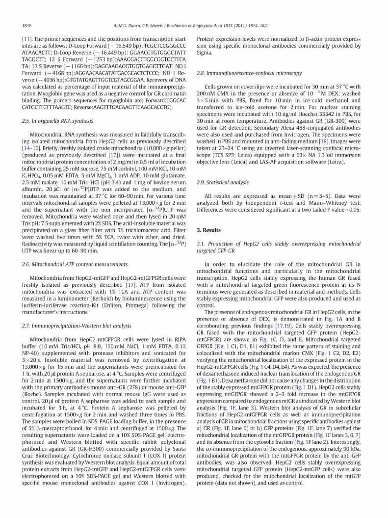
Cells grown on coverslips were incubated for 30 min at 37 °C with200 nM CMX in the presence or absence of 10−6 M DEX; washed3×5 min with PBS, fixed for 10 min in ice-cold methanol andtransferred to ice-cold acetone for 2 min. For nuclear stainingspecimens were incubated with 10 ng/ml Hoechst 33342 in PBS, for30 min at room temperature. Antibodies against GR (GR-300) wereused for GR detection. Secondary Alexa 488-conjugated antibodieswere also used and purchased from Invitrogen. The specimens werewashed in PBS andmounted in anti-fading medium [18]. Images weretaken at 23–24 °C using an inverted laser-scanning confocal micro-scope (TCS SP5; Leica) equipped with a 63× NA 1.3 oil immersionobjective lens (Leica) and LAS-AF acquisition software (Leica).
2.9. Statistical analysis
All results are expressed as mean±SD (n=3–5). Data wereanalyzed both by independent t-test and Mann–Whitney test.Differences were considered significant at a two tailed P value b0.05.
3. Results
3.1. Production of HepG2 cells stably overexpressing mitochondrialtargeted GFP-GR
In order to elucidate the role of the mitochondrial GR inmitochondrial functions and particularly in the mitochondrialtranscription, HepG2 cells stably expressing the human GR fusedwith a mitochondrial targeted green fluorescence protein at its Nterminus were generated as described in material and methods. Cellsstably expressing mitochondrial GFP were also produced and used ascontrol.
The presence of endogenousmitochondrial GR in HepG2 cells, in thepresence or absence of DEX, is demonstrated in Fig. 1A and Bcorroborating previous findings [17,19]. Cells stably overexpressingGR fused with the mitochondrial targeted GFP protein (HepG2-mtGFPGR) are shown in Fig. 1C, D, and E. Mitochondrial targetedGFPGR (Fig. 1 C1, D1, E1) exhibited the same pattern of staining andcolocalized with the mitochondrial marker CMX (Fig. 1 C2, D2, E2)verifying the mitochondrial localization of the expressed protein in theHepG2-mtGFPGR cells (Fig. 1 C4, D4, E4). Aswas expected, the presenceof dexamethasone induced nuclear translocation of the endogenous GR(Fig. 1B1). Dexamethasonedidnot cause any changes in thedistributionof the stably expressedmtGFPGR protein (Fig. 1 D1). HepG2 cells stablyexpressing mtGFPGR showed a 2–3 fold increase in the mtGFPGRexpression compared to endogenousmtGR as indicated byWestern blotanalysis (Fig. 1F, lane 3). Western blot analysis of GR in subcellularfractions of HepG2-mtGFPGR cells as well as immunoprecipitationanalysis ofGR inmitochondrial fractionsusing specific antibodies againsta) GR (Fig. 1F, lane 6) or b) GFP proteins (Fig. 1F, lane 7) verified themitochondrial localization of themtGFPGR protein (Fig. 1F lanes 3, 6, 7)and its absence from the cytosolic fraction (Fig. 1F lane 2). Interestingly,the co-immunoprecipitation of the endogenous, approximately 90 kDa,mitochondrial GR protein with the mtGFPGR protein by the anti-GFPantibodies, was also observed. HepG2 cells stably overexpressingmitochondrial targeted GFP protein (HepG2-mtGFP cells) were alsoproduced, checked for the mitochondrial localization of the mtGFPprotein (data not shown), and used as control.

Fig. 1. Mitochondrial localization of glucocorticoid receptor in HepG2 cells. Mitochondrial localization of GR in HepG2 cells in the absence (A) or presence (B) of DEX. Methanol-aceton fixed specimens pretreated with CMX (A2, B2) were subjected to indirect immunofluorescence staining of GR using the GR-H300 polyclonal antibodies (A1, B1). Whitearrows indicate colocalization of GR with CMX. Expression and mitochondrial localization of mtGFPGR in methanol-aceton fixed specimens of HepG2-mtGFPGR cells in the absence(C, E) or presence (D) of DEX. C1, D1, and E1: mtGFPGR localization; C2, D2, and E2: CMX mitochondrial staining; A3, B3, C3, D3, and E3: nuclear staining of HepG2 cells with theHoechst 33342 dye; and A4, B4, C4, D4, and E4: merged images. Bars in A–B, C–D and E indicate 10, 50 and 25 μm, respectively. F: Western blot analysis of GR in subcellular fractionsof HepG2-mtGFPGR cells and immunoprecipitation analysis of mtGR and mtGFPGR in mitochondrial extracts from HepG2-mtGFPGR cells. Detection of GR in HepG2-mtGFPGR cellswas achieved byWestern blot analysis, applying the GR-H300 antibodies. The anti-GR (2F8) and anti-GFP antibodies were applied in immunoprecipitation studies; lane 1: total cellextract; lane 2: post mitochondrial supernatant; lane 3: 10% of mitochondrial extracts used for immunoprecipitation; lanes 4 and lane 5: 10% of unbound mitochondrial proteinsimmunoprecipitated with the anti-GR and anti-GFP antibodies, respectively; and lanes 6, 7, and 8: immunoprecipitated mitochondrial proteins with the anti-GR, anti-GFPantibodies, and normal mouse IgG, respectively.
1817A.-M.G. Psarra, C.E. Sekeris / Biochimica et Biophysica Acta 1813 (2011) 1814–1821
3.2. Effect of DEX on RNA synthesis of nuclear encoded mitochondrialtranscription factors and of mitochondrial OXPHOS genes in HepG2 cells
HepG2 cells were induced with 1×10⁻⁶ M dexamethasone and thetranscription of the genes for the mitochondrial transcription factors
TFAM, TFB1M, TFB2M, the nuclear and mitochondrial OXPHOS genes,and the mitochondrial 12 S and 16 S ribosomal RNA was assayed byRT-PCR (see Materials and methods). The results are shown in Fig. 2. InHepG2 cells the presence of dexamethasone led to increased transcrip-tion of several genes, among them the mitochondrial transcription
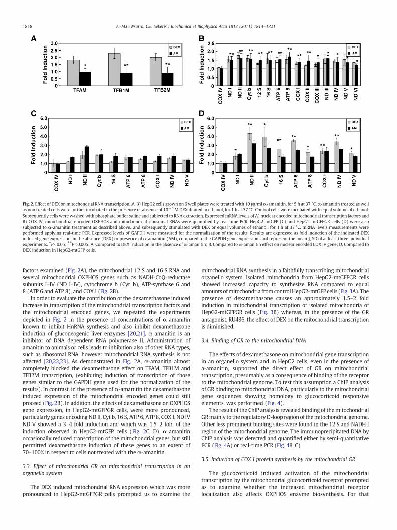
Fig. 2. Effect of DEX onmitochondrial RNA transcription. A, B) HepG2 cells grown on 6 well plates were treated with 10 μg/mlα-amanitin, for 5 h at 37 °C.α-amanitin treated as wellas non treated cells were further incubated in the presence or absence of 10−6 M DEX diluted in ethanol, for 1 h at 37 °C. Control cells were incubated with equal volume of ethanol.Subsequently cells were washedwith phosphate buffer saline and subjected to RNA extraction. ExpressedmRNA levels of A) nuclear encodedmitochondrial transcription factors andB) COX IV, mitochondrial encoded OXPHOS and mitochondrial ribosomal RNAs were quantified by real-time PCR. HepG2-mtGFP (C) and HepG2-mtGFPGR cells (D) were alsosubjected to α-amanitin treatment as described above, and subsequently stimulated with DEX or equal volumes of ethanol, for 1 h at 37 °C. mRNA levels measurements wereperformed applying real-time PCR. Expressed levels of GAPDH were measured for the normalization of the results. Results are expressed as fold induction of the indicated DEXinduced gene expression, in the absence (DEX) or presence of α-amanitin (AM), compared to the GAPDH gene expression, and represent the mean±SD of at least three individualexperiments. ⁎Pb0.05; ⁎⁎Pb0.005; A. Compared to DEX induction in the absence of α-amanitin; B. Compared to α-amanitin effect on nuclear encoded COX IV gene; D. Compared toDEX induction in HepG2-mtGFP cells.
1818 A.-M.G. Psarra, C.E. Sekeris / Biochimica et Biophysica Acta 1813 (2011) 1814–1821
factors examined (Fig. 2A), the mitochondrial 12 S and 16 S RNA andseveral mitochondrial OXPHOS genes such as NADH-CoQ-reductasesubunits I–IV (ND I–IV), cytochrome b (Cyt b), ATP-synthase 6 and8 (ATP 6 and ATP 8), and COX I (Fig. 2B).
In order to evaluate the contribution of the dexamethasone inducedincrease in transcription of the mitochondrial transcription factors andthe mitochondrial encoded genes, we repeated the experimentsdepicted in Fig. 2 in the presence of concentrations of α-amanitinknown to inhibit HnRNA synthesis and also inhibit dexamethasoneinduction of gluconeogenic liver enzymes [20,21]. α-amanitin is aninhibitor of DNA dependent RNA polymerase II. Administration ofamanitin to animals or cells leads to inhibition also of other RNA types,such as ribosomal RNA, however mitochondrial RNA synthesis is notaffected [20,22,23]. As demonstrated in Fig. 2A, α-amanitin almostcompletely blocked the dexamethasone effect on TFAM, TFB1M andTFB2M transcription, (exhibiting induction of transcription of thosegenes similar to the GAPDH gene used for the normalization of theresults). In contrast, in the presence of α-amanitin the dexamethasoneinduced expression of the mitochondrial encoded genes could stillproceed (Fig. 2B). In addition, the effects of dexamethasone on OXPHOSgene expression, in HepG2-mtGFPGR cells, were more pronounced,particularly genes encoding ND II, Cyt b, 16 S, ATP 6, ATP 8, COX I, ND IVND V showed a 3–4 fold induction and which was 1.5–2 fold of theinduction observed in HepG2-mtGFP cells (Fig. 2C, D). α-amanitinoccasionally reduced transcription of the mitochondrial genes, but stillpermitted dexamethasone induction of these genes to an extent of70–100% in respect to cells not treated with the α-amanitin.
3.3. Effect of mitochondrial GR on mitochondrial transcription in anorganello system
The DEX induced mitochondrial RNA expression which was morepronounced in HepG2-mtGFPGR cells prompted us to examine the
mitochondrial RNA synthesis in a faithfully transcribing mitochondrialorganello system. Isolated mitochondria from HepG2-mtGFPGR cellsshowed increased capacity to synthesize RNA compared to equalamounts ofmitochondria fromcontrolHepG2-mtGFP cells (Fig. 3A). Thepresence of dexamethasone causes an approximately 1.5–2 foldinduction in mitochondrial transcription of isolated mitochondria ofHepG2-mtGFPGR cells (Fig. 3B) whereas, in the presence of the GRantagonist, RU486, the effect of DEX on themitochondrial transcriptionis diminished.
3.4. Binding of GR to the mitochondrial DNA
The effects of dexamethasone on mitochondrial gene transcriptionin an organello system and in HepG2 cells, even in the presence ofa-amanitin, supported the direct effect of GR on mitochondrialtranscription, presumably as a consequence of binding of the receptorto the mitochondrial genome. To test this assumption a ChIP analysisof GR binding to mitochondrial DNA, particularly to the mitochondrialgene sequences showing homology to glucocorticoid responsiveelements, was performed (Fig. 4).
The result of the ChIP analysis revealed binding of themitochondrialGRmainly to the regulatoryD-loop region of themitochondrial genome.Other less prominent binding sites were found in the 12 S and NADH Iregion of the mitochondrial genome. The immunoprecipitated DNA byChIP analysis was detected and quantified either by semi-quantitativePCR (Fig. 4A) or real-time PCR (Fig. 4B, C).
3.5. Induction of COX I protein synthesis by the mitochondrial GR
The glucocorticoid induced activation of the mitochondrialtranscription by the mitochondrial glucocorticoid receptor promptedas to examine whether the increased mitochondrial receptorlocalization also affects OXPHOS enzyme biosynthesis. For that

Fig. 3. Mitochondrial RNA synthesis in an organello system. A. Mitochondrial RNA synthesis was measured in equal amounts of isolated mitochondria from HepG2-mtGFP andHepG2-mtGFPGR cells as described in material and methods. Mitochondrial RNA synthesis was evaluated by measuring [α-32P]UTP incorporation into synthesized RNA B.Mitochondrial RNA synthesis wasmeasured in equal amounts of isolatedmitochondria fromHepG2-mtGFPGR cells in the presence or absence of 10−6 MDEX and/or 10−5 M RU486.Results are expressed as fold induction of mitochondrial transcription relative compared to A) HepG2-mtGFP cells, B) untreated HepG2-mtGFPGR cells and represent the mean±SDof at least three individual experiments. ⁎Pb0.05.
1819A.-M.G. Psarra, C.E. Sekeris / Biochimica et Biophysica Acta 1813 (2011) 1814–1821
purpose COX I protein levels were evaluated in HepG2-mtGFP and-mtGFPGR cells, following stimulation of the cells either with DEX orethanol at a final concentration of 10−6 M and 0.1%, respectively, for6 h. β-actin was used for the normalization of the results. As shown inFig. 5, overexpression of mitochondrial GR caused increase in COX Iprotein synthesis in both cases examined.
3.6. Effect of mitochondrial GR on mitochondrial ATP content
Mitochondrial GR translocation should be closely associated withthe energy status of the cell and may constitute a regulatorymechanism of the cell to face its energy demands. In our modelsystem, the activation of the mitochondrial transcription and OXPHOSenzyme biosynthesis by the stable overexpression of mtGR may alsolead to increase in mitochondrial capacity to produce ATP. To test thishypothesis, mitochondrial ATP level of HepG2-mtGFP and HepG2-mtGFPGR cells, preincubated or not with 10−7 M DEX for 16–20 h,was measured as described in material and methods. HepG2-mtGFPGR cells showed an approximately 1.5–2 fold increase in ATP
Fig. 4. Binding of the glucocorticoid receptor to the D-loop of the mitochondrial DNA of HepG(2F8) or nonspecific IgG as described in Materials and methods. Levels of immunoprecimitochondrial D-loop, the NADH dehydrogenase I gene and the 12 S gene were assessed eitgene was used as negative control. The results of three independent experiments as means±DNA sequences. Recovery of DNAwas calculated as percentage of input material of the immuindicated by arrows.
level compared to control cells. DEX causes an approximately 20%increase inmitochondrial ATP content in HepG2-mtGFP cells, whereasATP level in HepG2-mtGFPGR cells is not significantly affected by DEX.Preincubation of cells with 0.2 μM rotenone for 16–20 h in thepresence or absence of DEX caused elimination in mitochondrial ATPproduction in both cell lines examined (Fig. 6).
4. Discussion
The mode of action of glucocorticoids on target cells has been afield of intense experimentation, following the seminal observationthat steroid hormones act by way of gene activation [review in10,12,24]. The induction of gluconeogenic enzymes in the rodent liverwas one of the early models to test the hormone–gene activationhypothesis and was instrumental, among other models, to validatethe hypothesis [2–6]. An early intriguing finding was the massiveinduction of RNA synthesis by glucocorticoids in the liver cells,encompassing all types of RNA-HnRNA, ribosomal RNA, t-RNA, mostof which was degraded in the nucleus. Importantly, it has been
2 cells. HepG2 cells were subjected to a ChIP assay using specific antibodies against GRpitated DNA encompassing the putative hormone response elements located in theher by semiquantative PCR (A) or real-time PCR (B, C). 10% input was used. MyoglobinS.D. are shown in B, C and represent the recovery and occupancy of the mitochondrial
noprecipitation. D. The mammalian mitochondrial genome. Sites of primers location are
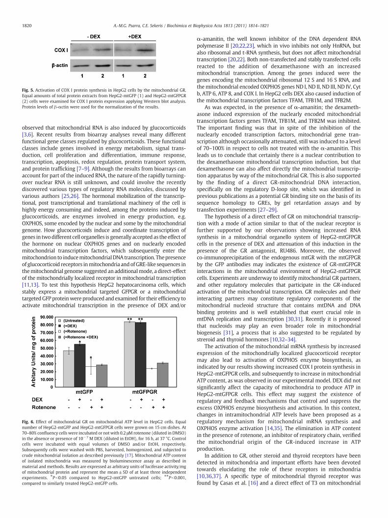
Fig. 5. Activation of COX I protein synthesis in HepG2 cells by the mitochondrial GR.Equal amounts of total protein extracts from HepG2-mtGFP (1) and HepG2-mtGFPGR(2) cells were examined for COX I protein expression applying Western blot analysis.Protein levels of β-αctin were used for the normalization of the results.
1820 A.-M.G. Psarra, C.E. Sekeris / Biochimica et Biophysica Acta 1813 (2011) 1814–1821
observed that mitochondrial RNA is also induced by glucocorticoids[3,6]. Recent results from bioarray analyses reveal many differentfunctional gene classes regulated by glucocorticoids. These functionalclasses include genes involved in energy metabolism, signal trans-duction, cell proliferation and differentiation, immune response,transcription, apoptosis, redox regulation, protein transport system,and protein trafficking [7–9]. Although the results from bioarrays canaccount for part of the induced RNA, the nature of the rapidly turning-over nuclear RNA is still unknown, and could involve the recentlydiscovered various types of regulatory RNA molecules, discussed byvarious authors [25,26]. The hormonal mobilization of the transcrip-tional, post transcriptional and translational machinery of the cell ishighly energy consuming and indeed, among the proteins induced byglucocorticoids, are enzymes involved in energy production, e.g.OXPHOS, some encoded by the nuclear and some by the mitochondrialgenome. How glucocorticoids induce and coordinate transcription ofgenes in twodifferent cell organelles is generally accepted as the effect ofthe hormone on nuclear OXPHOS genes and on nuclearly encodedmitochondrial transcription factors, which subsequently enter themitochondrion to inducemitochondrialDNA transcription. Thepresenceof glucocorticoid receptors inmitochondria andof GRE-like sequences inthemitochondrial genome suggested an additionalmode, a direct-effectof the mitochondrially localized receptor in mitochondrial transcription[11,13]. To test this hypothesis HepG2 hepatocarcinoma cells, whichstably express a mitochondrial targeted GFPGR or a mitochondrialtargetedGFP proteinwere produced andexamined for their efficiency toactivate mitochondrial transcription in the presence of DEX and/or
Fig. 6. Effect of mitochondrial GR on mitochondrial ATP level in HepG2 cells. Equalnumber of HepG2-mtGFP and HepG2-mtGFPGR cells were grown on 15 cm dishes. At70–80% confluency cells were incubated or not with 0.2 μΜ rotenone (diluted in DMSO)in the absence or presence of 10−7 M DEX (diluted in EtOH), for 16 h, at 37 °C. Controlcells were incubated with equal volumes of DMSO and/or EtOH, respectively.Subsequently cells were washed with PBS, harvested, homogenized, and subjected tocrude mitochondrial isolation as described previously [17]. Mitochondrial ATP contentof isolated mitochondria was measured by bioluminescence assay as described inmaterial and methods. Results are expressed as arbitrary units of luciferase activity/mgof mitochondrial protein and represent the mean±SD of at least three independentexperiments. ⁎Pb0.05 compared to HepG2-mtGFP untreated cells; ⁎⁎Pb0.001,compared to similarly treated HepG2-mtGFP cells.
α-amanitin, the well known inhibitor of the DNA dependent RNApolymerase II [20,22,23], which in vivo inhibits not only HnRNA, butalso ribosomal and t-RNA synthesis, but does not affect mitochondrialtranscription [20,22]. Both non-transfected and stably transfected cellsreacted to the addition of dexamethasone with an increasedmitochondrial transcription. Among the genes induced were thegenes encoding the mitochondrial ribosomal 12 S and 16 S RNA, andthemitochondrial encoded OXPHOS genes ND I, ND II, ND III, ND IV, Cytb, ATP 6, ATP 8, and COX I. In HepG2 cells DEX also caused induction ofthe mitochondrial transcription factors TFAM, TFB1M, and TFB2M.
As was expected, in the presence of α-amanitin; the dexameth-asone induced expression of the nuclearly encoded mitochondrialtranscription factors genes TFAM, TFB1M, and TFB2M was inhibited.The important finding was that in spite of the inhibition of thenuclearly encoded transcription factors, mitochondrial gene tran-scription although occasionally attenuated, still was induced to a levelof 70–100% in respect to cells not treated with the α-amanitin. Thisleads us to conclude that certainly there is a nuclear contribution tothe dexamethasone mitochondrial transcription induction, but thatdexamethasone can also affect directly the mitochondrial transcrip-tion apparatus by way of the mitochondrial GR. This is also supportedby the finding of a direct GR-mitochondrial DNA interaction,specifically on the regulatory D-loop site, which was identified inprevious publications as a potential GR binding site on the basis of itssequence homology to GREs, by gel retardation assays and bytransfection experiments [27–29].
The hypothesis of a direct effect of GR on mitochondrial transcrip-tion with a mode of action similar to that of the nuclear receptor isfurther supported by our observations showing increased RNAsynthesis in a mitochondrial organello system of HepG2-mtGFPGRcells in the presence of DEX and attenuation of this induction in thepresence of the GR antagonist, RU486. Moreover, the observedco-immunoprecipitation of the endogenous mtGR with the mtGFPGRby the GFP antibodies may indicates the existence of GR-mtGFPGRinteractions in the mitochondrial environment of HepG2-mtGFPGRcells. Experiments are underway to identify mitochondrial GR partners,and other regulatory molecules that participate in the GR-inducedactivation of the mitochondrial transcription. GR molecules and theirinteracting partners may constitute regulatory components of themitochondrial nucleoid structure that contains mtDNA and DNAbinding proteins and is well established that exert crucial role inmtDNA replication and transcription [30,31]. Recently it is proposedthat nucleoids may play an even broader role in mitochondrialbiogenesis [31], a process that is also suggested to be regulated bystreroid and thyroid hormones [10,32–34].
The activation of the mitochondrial mRNA synthesis by increasedexpression of the mitochondrially localized glucocorticoid receptormay also lead to activation of OXPHOS enzyme biosynthesis, asindicated by our results showing increased COX I protein synthesis inHepG2-mtGFPGR cells, and subsequently to increase in mitochondrialATP content, as was observed in our experimental model. DEX did notsignificantly affect the capacity of mitochondria to produce ATP inHepG2-mtGFPGR cells. This effect may suggest the existence ofregulatory and feedback mechanisms that control and suppress theexcess OXPHOS enzyme biosynthesis and activation. In this context,changes in intramitochondrial ATP levels have been proposed as aregulatory mechanism for mitochondrial mRNA synthesis andOXPHOS enzyme activation [14,35]. The elimination in ATP contentin the presence of rotenone, an inhibitor of respiratory chain, verifiedthe mitochondrial origin of the GR-induced increase in ATPproduction.
In addition to GR, other steroid and thyroid receptors have beendetected in mitochondria and important efforts have been devotedtowards elucidating the role of these receptors in mitochondria[10,36,37]. A specific type of mitochondrial thyroid receptor wasfound by Casas et al. [16] and a direct effect of T3 on mitochondrial

1821A.-M.G. Psarra, C.E. Sekeris / Biochimica et Biophysica Acta 1813 (2011) 1814–1821
transcription in an in organello mitochondrial transcription systemhas been shown establishing the role of the mitochondrial thyroidreceptor [14,15,32,33,38]. Estrogen and androgen, receptors have alsobeen found in mitochondria of several types of cells and theirinvolvement in the regulation of mitochondrial functions has beenproposed [reviewed in 13,36–40]. Recently the involvement of themitochondrial glucocorticoid receptor in the regulation of theglucocorticoid-induced apoptosis andmodulation of cellular plasticityhas been suggested [41,42].
The present work gives the first insights into the molecularmechanisms underlying the GR-mediated mitochondrial transcriptionproviding evidence for a direct involvement of the mitochondriallylocalized glucocorticoid receptor in the regulation of mitochondrialtranscription and OXPHOS enzyme biosynthesis by binding to mtDNAand activation ofmitochondrial gene expression.Moreover, results fromthis study prove that this action takes place in addition to a nuclearaction of glucocorticoids onmitochondrial genes byway of induction ofgenes of the nuclearly encoded mitochondrial transcription factors,which then enter mitochondria to regulate mtDNA transcription andOXPHOS enzyme biosynthesis.
Recently, in addition to nuclear receptors, the mitochondriallocalization of several other transcription factors with nuclear actionsincluding NF-κΒ, p53, AP-1, STAT-3 but with increasing importancefor mitochondrial functions has been suggested [36,43–45]. Themitochondrial localization of these molecules potentiates theirregulatory significance and renders them coordinators of nuclearand mitochondrial actions in important functions, among themenergy production, mitochondrial dependent apoptosis, oncogenictransformation and immune responses [36,39–45].
Acknowledgment
We would like to thank the Bodossaki Foundation, the BiomedicalResearch Foundation of the Academy of Athens, and the ResearchCommittee of the University of Thessaly for the financial support ofthe project. We would like to thank Dr. M. Alexis for kindly providedus with the 2F8 glucocorticoid receptor monoclonal antibody.
References
[1] R.M. Evans, The nuclear receptor superfamily: a rosetta stone for physiology, Mol.Endocrinol. 19 (2005) 1429–1438.
[2] M. Beato, S. Chávez, M. Truss, Transcriptional regulation by steroid hormones,Steroids 61 (1996) 240–251.
[3] F.L. Yu, P. Feigelson, A comparative study of RNA synthesis in rat hepatic nucleiand mitochondria under the influence of cortisone, Biochim. Biophys. Acta 213(1970) 134–141.
[4] W. Schmid, C.E. Sekeris, Nucleolar RNA synthesis in the liver of partiallyhepatectomized and cortisol-treated rats, Biochim. Biophys. Acta 402 (1975)244–252.
[5] W. Schmid, C.E. Sekeris, Sequential stimulation of extranucleolar and nucleolarRNA synthesis in rat liver by cortisol, FEBS Lett. 26 (1972) 109–112.
[6] A.M. Mansour, S. Nass, In vivo cortisol action on RNA synthesis in rat liver nucleiand mitochondria, Nature 228 (1970) 665–667.
[7] N.A. Datson, M.C. Morsink, O.C. Meijer, E.R. de Kloet, Central corticosteroidactions: search for gene targets, Eur. J. Pharmacol. 583 (2008) 272–289.
[8] P.S.Weber, S.A.Madsen-Bouterse, G.J. Rosa, S. Sipkovsky, X. Ren, P.E. Almeida, R. Kruska,R.G. Halgren, J.L. Barrick, J.L. Burton, Analysis of the bovine neutrophil transcriptomeduring glucocorticoid treatment, Physiol. Genomics 28 (2006) 97–112.
[9] J.Y. Jin, R.R. Almon, D.C. DuBois, W.J. Jusko, Modeling of corticosteroid pharmacoge-nomics in rat liver using genemicroarrays, J. Pharmacol. Exp. Ther. 307 (2003) 93–109.
[10] K. Scheller, P. Seibel, C.E. Sekeris, Glucocorticoid and thyroid hormone receptors inmitochondria of animal cells, Int. Rev. Cytol. 222 (2003) 1–61.
[11] C.E. Sekeris, The mitochondrial genome: a possible primary site of action of steroidhormones, In Vivo 4 (1990) 317–320.
[12] A.-M.G. Psarra, C.E. Sekeris, Steroid and thyroid hormone receptors in mitochondria,IUBMB Life 60 (2008) 210–223.
[13] A.-M.G. Psarra, C.E. Sekeris, Glucocorticoid receptors and other nuclear transcriptionfactors in mitochondria and possible functions, Biochim. Biophys. Acta 1787 (2009)431–436.
[14] J.A. Enriquez, P. Fernandez-Silva, A. Perez-Martos, M.J. Lopez-Perez, J. Montoya, Thesynthesis of mRNA in isolatedmitochondria can bemaintained for several hours andis inhibited by high levels of ATP, Eur. J. Biochem. 237 (1996) 601–610.
[15] J.A. Enriquez, P. Fernandez-Silva, N. Garrido-Perez, M.J. Lopez-Perez, A. Perez-Martos, J.Montoya, Direct regulation of mitochondrial RNA synthesis by thyroid hormone, Mol.Cell. Biol. 19 (1999) 657–670.
[16] F. Casas, P. Rochard, A. Rodier, I. Cassar-Malek, S. Marchal-Victorion, R.J. Wiesner, G.Cabello, C. Wrutniak, A variant form of the nuclear triiodothyronine receptor c-ErbAalpha1 plays a direct role in regulation of mitochondrial RNA synthesis, Mol. Cell.Biol. 19 (1999) 7913–7924.
[17] A.-M.G. Psarra, S. Solakidi, I.P. Trougakos, L.H. Margaritis, G. Spyrou, C.E. Sekeris,Glucocorticoid receptor isoforms in human hepatocarcinoma HepG2 and SaOS-2osteosarcoma cells: Presence of glucocorticoid receptor alpha in mitochondria and ofglucocorticoid receptor beta in nucleoli, Int. J. Biochem. Cell Biol. 37 (2005) 2544–2558.
[18] D.A. Lennette, An improved mounting medium for immunofluorescence microscopy,Am. J. Clin. Pathol. 69 (1978) 647–648.
[19] A.-M.G. Psarra, S. Solakidi, C.E. Sekeris, The mitochondrion as a primary site of action ofsteroid and thyroid hormones: presence and action of steroid and thyroid hormonereceptors in mitochondria of animal cells, Mol. Cell. Endocrinol. 246 (2006) 21–33.
[20] K.H. Seifart, C.E. Sekeris, Alpha-amanitin, a specific inhibitor of transcription bymammalian RNA-polymerase, Z. Naturforsch. B 24 (1969) 1538–1544.
[21] V.L. Souliotis, P.P. Sfikakis, L.M. Anderson, S.A. Kyrtopoulos, Intra- and intercellularvariations in the repair efficiency of O6-methylguanine, and their contribution tokinetic complexity, Mutat. Res. 568 (2004) 155–170.
[22] T.J. Lindell, F. Weinberg, P.W. Morris, R.G. Roeder, W.J. Rutter, Specific inhibition ofnuclear RNA polymerase II by alpha-amanitin, Science 170 (1970) 447–449.
[23] C.E. Sekeris, W. Schmid, Action of alpha-amanitin in vivo and in vitro, FEBS Lett. 27(1972) 41–45.
[24] K.R. Yamamoto, Steroid receptor regulated transcription of specific genes and genenetworks, Annu. Rev. Genet. 19 (1985) 209–252.
[25] F. Wahid, A. Shehzad, T. Khan, Y.Y. Kim, MicroRNAs: synthesis, mechanism, function,and recent clinical trials, Biochim. Biophys. Acta 1803 (2010) 1231–1243.
[26] J.S.Mattick, The genetic signatures of noncoding RNAs, PLoS Genet. 5 (2009) e1000459.[27] C. Demonacos, R. Djordjevic-Markovic, N. Tsawdaroglou, C.E. Sekeris, The mitochon-
drion as a primary site of action of glucocorticoids: the interaction of the glucocorticoidreceptor with mitochondrial DNA sequences showing partial similarity to the nuclearglucocorticoid responsive elements, J. Steroid Biochem. Mol. Biol. 55 (1995) 43–55.
[28] C.V. Demonacos, N. Karayanni, E. Hatzoglou, C. Tsiriyiotis, D.A. Spandidos, C.E. Sekeris,Mitochondrial genes as sites of primary action of steroid hormones, Steroids 61 (1996)226–232.
[29] C. Tsiriyotis, D.A. Spandidos, C.E. Sekeris, The mitochondrion as a primary site of actionof glucocorticoids:mitochondrial nucleotide sequences, showing similarity to hormoneresponse elements, confer dexamethasone inducibility to chimaeric genes transfectedin LATK-cells, Biochem. Biophys. Res. Commun. 235 (1997) 349–354.
[30] X.J. Chen, R.A. Butow, The organization and inheritance of the mitochondrial genome,Nat. Rev. Genet. 11 (2005) 815–825.
[31] D.F. Bogenhagen, D. Rousseau, S. Burke, The layered structure of humanmitochondrialDNA nucleoids, J. Biol. Chem. 283 (2008) 3665–3675.
[32] C. Wrutniak, P. Rochard, F. Casas, A. Fraysse, J. Charrier, G. Cabello, Physiologicalimportance of the T3 mitochondrial pathway, Ann. N. Y. Acad. Sci. 839 (1998) 93–100.
[33] B.D. Nelson, K. Luciakova, R. Li, S. Betina, The role of thyroid hormone and promoterdiversity in the regulation of nuclear encoded mitochondrial proteins, Biochim.Biophys. Acta 1271 (1995) 85–91.
[34] K.Weber, P. Brück, Z.Mikes, J.H. Küpper,M. Klingenspor, R.J.Wiesner, Glucocorticoidhormone stimulates mitochondrial biogenesis specifically in skeletal muscle,Endocrinology 143 (2002) 177–184.
[35] B. Kadenbach, Intrinsic and extrinsic uncoupling of oxidative phosphorylation,Biochim. Biophys. Acta 1604 (2003) 77–94.
[36] A.-M.G. Psarra, C.E. Sekeris, Nuclear receptors and other nuclear transcription factors inmitochondria: regulatory molecules in a new environment, Biochim. Biophys. Acta1783 (2008) 1–11.
[37] L.P. Gavrilova-Jordan, T.M. Price, Actions of steroids in mitochondria, Semin. Reprod.Med. 25 (2007) 154–164.
[38] H. Suzuki, Y. Hosokawa, M. Nishikimi, T. Ozawa, Existence of common homologouselements in the transcriptional regulatory regions of human nuclear genes andmitochondrial gene for the oxidative phosphorylation system, J. Biol. Chem. 266(1991) 2333–2338.
[39] J.Q. Chen, P.R. Cammarata, C.P. Baines, J.D. Yager, Regulation of mitochondrialrespiratory chain biogenesis by estrogens/estrogen receptors and physiological,pathological and pharmacological implications, Biochim. Biophys. Acta 1793 (2009)1540–1570.
[40] J.D. Yager, J.Q. Chen, Mitochondrial estrogen receptors—new insights into specificfunctions, Trends Endocrinol. Metab. 18 (2007) 89–91.
[41] R.V. Sionov, O. Cohen, S. Kfir, Y. Zilberman, E. Yefenof, Role of mitochondrialglucocorticoid receptor in glucocorticoid-induced apoptosis, J. Exp. Med. 203 (2006)189–201.
[42] J. Du, Y.Wang, R. Hunter, Y.Wei, R. Blumenthal, C. Falke, R. Khairova, R. Zhou, P. Yuan,R. Machado-Vieira, B.S. McEwen, H.K. Manji, Dynamic regulation of mitochondrialfunction by glucocorticoids, Proc. Natl. Acad. Sci. U.S.A. 106 (2009) 3543–3548.
[43] J. Wegrzyn, R. Potla, Y.J. Chwae, N.B. Sepuri, Q. Zhang, T. Koeck, M. Derecka, K.Szczepanek,M. Szelag, A. Gornicka, A.Moh, S.Moghaddas, Q. Chen, S. Bobbili, J. Cichy,J. Dulak, D.P. Baker, A. Wolfman, D. Stuehr, M.O. Hassan, X.Y. Fu, N. Avadhani, J.I.Drake, P. Fawcett, E.J. Lesnefsky, A.C. Larner, Function of mitochondrial Stat3 incellular respiration, Science 323 (2009) 793–797.
[44] D.J. Gough, A. Corlett, K. Schlessinger, J. Wegrzyn, A.C. Larner, D.E. Levy, MitochondrialSTAT3 supports Ras-dependent oncogenic transformation, Science 324 (2009)1713–1716.
[45] N.C. Reich, STAT3 revs up the powerhouse, Sci. Signal. 2 (2009) e61.
Copyright © 2022 FDOKUMEN