Mechanical ventilation interacts with endotoxemia to induce extrapulmonary organ dysfunction
(CANCER RESEARCH 51, 1078-1085. February 15, 1991]
Topoisomerase II-reactive Chemotherapeutic Drugs Induce Apoptosis inThymocytes1
P. Roy Walker,2 Catherine Smith, Tony Youdale, Julie Leblanc, James F. Whitfield, and Marianna Sikorska
Cellular Oncology Group, Institute for Biological Sciences, National Research Council of Canada, Ottawa, Canada K1A OR6
ABSTRACT
The effects of topoisomerase II-reactive epipodophyllotoxins etoposideand teniposide as well as amsacrine on the viability of thymocytes inprimary culture has been examined. All three drugs were shown toproduce DNA cleavage detectable by resolving isolated DNA by pulsedfield agarose gel electrophoresis. The DNA cleavage was found to havetwo components. The first was due to the interaction of the drugs withtopoisomerase II, whereas the second component was due to endonucleasecleavage caused by the drug-induced entry of the thymocytes into programmed cell death or apoptosis. This second component of the DNAcleavage was also detected in thymocytes undergoing apoptosis followingexposure to the glucocorticoid analogue, dexamethasone. The effect ofthe drugs on programmed cell death is dependent upon new protein andRNA synthesis, indicating that topoisomerase II has a role in the veryfirst stages of the process. These results are discussed in terms of theuse of this class of topoisomerase II-reactive drugs in chemotherapy.
INTRODUCTION
Drugs that interact with the enzyme topoisomerase II havebeen found to be particularly useful in the chemotherapeutictreatment of certain cancers (1-4). These include the epipodophyllotoxins VP163 and VM26, as well as /M-AMSA which isan acridine derivative. All three drugs stabilize the DNA-topo-isomerase II complex during strand passage preventing theresealing reaction from going to completion (1, 5-7). DNAisolated from cells treated with the drugs contains covalentlyattached topoisomerase II subunits and strand breaks are detectable when the protein is degraded. VM26 and VP 16 do notintercalate into DNA and appear to exert their effect, both invivo and in vitro (using purified components), by binding directly, and specifically, to the enzyme molecule (reviewed inRefs. 1 and 8), whereas m-AMSA intercalates into DNA.However, although they are effective therapeutically, the mechanism by which these drugs kill cells is still controversial (3).In general, the drugs are thought to kill proliferating cancercells by inhibiting topoisomerase II and preventing the cellsfrom either completing S phase or undergoing chromosomesegregation at mitosis since these are two cellular processesthat have an absolute requirement for the enzyme. In general,cytotoxicity correlates with drug-induced DNA cleavage (8) andincreases when the drugs are administered during these phasesof the cell cycle. There are, however, many exceptions (3, 9),and since other cellular processes are also dependent upontopoisomerase II activity, particularly those requiring a change
Received 8/22/90; accepted 11/19/90.The costs of publication of this article were defrayed in part by the payment
of page charges. This article must therefore be hereby marked advertisement inaccordance with 18 U.S.C. Section 1734 solely to indicate this fact.
' This paper is published as National Research Council of Canada publication
31904.2To whom requests for reprints should be addressed, at Cellular Oncology
Group, Institute for Biological Sciences, National Research Council, Bldg. MS4,Montreal Road, Ottawa. Ontario, Canada K1A OR6.
3The abbreviations used are: VP16, etoposide; VM26. teniposide; m-AMSA.amsacrine [4'-(9-acridinylamino)methanesulfon-m-anisidine|; PCD. programmed cell death: PBS, phosphate-buffered saline.
in chromatin structure, the precise mechanism of cytotoxicityremains to be established.
These drugs have also become particularly useful in establishing the role of topoisomerase II in biological processes and, inparticular, the role that the enzyme plays in determining andmodulating chromosome structure. During a recent study ofthe structural organization of chromosomes we (10) used drugsto cleave thymocyte DNA at the sites of topoisomerase IIbinding to DNA and interpreted the results in terms of theorganization of chromosomes at the level of 50- and 300-kilobase supercoiled loop domains and rosettes, respectively.Thymocytes were found to be particularly useful in these studiessince the cells have a high content of topoisomerase II andmaintain much of their interphase chromatin in a highly ordered state. In those experiments, the effect of the drugs on thethymocytes was restricted to short incubations (30 min or less).However, more recently, we have noticed much more extensiveDNA cleavage during longer incubations which, we show here,to be caused by the topoisomerase II-reactive drug-mediatedentry of the cells into apoptosis or PCD. Apoptosis is a physiological "suicide" process by which unwanted cells are removed
from embryonic, developing, or somatic tissues without affecting overall tissue function (11-14). In some tissues, the processis an intrinsic part of its function. For example, apoptosis playsa crucial role in determining the levels of many, if not all, ofthe precursor cells in the pathways of hematopoiesis in orderto balance the production of the various differentiated cell types.Apoptosis has a characteristic requirement for energy as wellas RNA and protein synthesis and is recognizably differentfrom tissue necrosis. Cells undergoing apoptosis activate orinduce an endonuclease that cleaves chromatin into nucleo-somes and polynucleosome chains which are observed as acharacteristic ladder of DNA fragments on agarose gels (14-17). The appearance of this DNA ladder coupled with thedependence upon gene expression and protein synthesis, together with morphological criteria, are the typical markers forapoptosis and are well documented for thymocytes (13-22).The results presented here demonstrate that topoisomerase II-reactive chemotherapeutic drugs are able to kill cells by inducingthe process of apoptosis. Thus, the susceptibility of cells, particularly those involved in hematopoiesis (18, 23-25), to apoptosis would be expected to increase their sensitivity to thecytotoxic effects of this group of antineoplastic drugs. Furthermore, these observations may explain in part the increasedsensitivity of certain tumor cells to this class of drugs anddemonstrate that normal nonproliferating cells can also bekilled.
MATERIALS AND METHODS
Preparation and Treatment of Thymocytes. Thymocytes were isolatedas described previously (19). Briefly, thymuses were excised from 3-week-old (150-200 g) Sprague-Dawley rats bred at this facility, washedto remove excess blood, and minced with scissors in 10-12 ml of RPMI
1078
Research. on December 15, 2014. © 1991 American Association for Cancercancerres.aacrjournals.org Downloaded from

TOPOISOMERASE II-REACTIVE DRUGS AND APOPTOSIS
1640 tissue culture medium containing 5% fetal bovine serum. Theminced thymuses were strained through gauze and the suspension wasdiluted 10-fold with medium to about 4 x IO7cells/ml. The cells wererotated at 37°Cfor l h in 10-ml aliquots in an atmosphere of 95% air
and 5% CO2 before treatments. The thymocyte primary cultures werethen treated with either 1 JIM dexamethasone (from a 2 HIM stocksolution in ethanol; Sigma Chemical Co., St. Louis, MO), or 10 ^M m-AMSA (from a 50 mM stock solution in dimethyl sulfoxide; ChemistryBranch, National Cancer Institute, NIH, Bethesda. MD), or 5 tiMVM26 (15 mM stock solution in ethanol), or 50 JIMVP16 (33.3 mMstock solution in 30% ethanol). VM26 and VP16 were obtained fromBristol Laboratories (Candiac, Quebec Province, Canada). In someexperiments, the cells were also treated with 0.1 mM cycloheximide(100 mg/ml stock solution in dimethyl sulfoxide; Sigma) or 1 JIMactinomycin D (l mg/ml in ethanol; Sigma).
Preparation of Agarose Plugs from Cells. Approximately 3x10* cells
(from 8 ml of culture) were resuspended in 1.0 ml of a buffer containing0.15 M NaCl, 2 mM KH2PO4/KOH (pH 6.4), 1 mM ethyleneglycolbis(0-aminoethyl ether)-A',A'>jV'*A"-tetraacetic ac{¿an(j 5 mM MgCl2
(10). In order to prevent reversal of the DNA breaks in the experimentsinvolving treatment with topoisomerase inhibitors the m-AMSA, VP 16,or VM26 were also present in this buffer at the same concentrations asin the medium. Cells were then centrifuged at 170 x g for 2 min at 4°C
and resuspended in 640 ¿ilof the same buffer. One hundred ^1 of thelatter suspension was transferred to an Eppendorf tube, diluted to 250M!with the same buffer, and mixed with 250 n\ of molten 1.5% lowmelting point agarose and 50 ¿tlof 2 mg/ml proteinase K. The mixturewas pipetted into 70 x 5 x 1-mm glass molds made from microscopeslides and left at 4°Cfor 30 min. The agarose blocks were placed in 3
ml of 10 mM NaCl-10 mM Tris-HCl, pH 9.5-25 mM EDTA-1.0% N-lauroyl sarcosine (Sigma) supplemented with 150 p\ of 2 mg/ml proteinase K and incubated at 37°Cfor 18 h with gentle mixing on a
multipurpose rotator. The incubation was followed by rinsing in threechanges of 10 mM Tris-HCl, pH 8-1 mM EDTA at 4°Cfor l h each
and finally suspended in 50 mM EDTA, pH 8.O. The agarose blockscould then be stored in this buffer for more than six months. About 3-mm long plugs were cut from the agarose blocks and used for electro-phoresis. Each plug contained 2-5 ng of DNA.
Pulsed Field Gel Electrophoresis. Electrophoresis was carried outusing a horizontal gel chamber, model 200/20 power supply andPulsewave 760 switcher (all purchased from BioRad Laboratories,Mississauga, Ontario, Canada). The gels were run at 200 V in 0.01 MTris-acétate,pH 8.5-0.5mM EDTA with the ramping rate changingfrom T, = 0.5 s to T2 = 10 s for the first 19 h and from 7", = 10 s to7"2 = 60 s for the next 19 h, with a forward to backward ratio of 3 at5°C,using electrophoresis grade agarose (1.5%) purchased from Be
thesda Research Laboratories (Gaithersburg, MD) (10). Under theseconditions, DNA fragments ranging in size from 10 kilobase pairs toapproximately 750 kilobase pairs are resolved and all DNA fragments>750-800 kilobase pairs that enter the gel migrate at the same rate.Three sets of standards with overlapping molecular weight ranges wereused to determine molecular weights: the commercially available yeastchromosomes (250-1100 kilobase pairs) purchased from BioRad Laboratories (Richmond, CA), polymerized Xphage DNA (50-750 kilobase
pairs) purchased from Clontech Labs Inc. (Palo Alto, CA), and apolymerized plasmid DNA (11.6-200 kilobase pairs) and/or X DNAdigest. Conventional agarose gel electrophoresis was carried out in0.8% agarose gels in 0.04M Tris-acetate, pH 8.5-0.002 M EDTA at 35V for 18 h. After the gels were stained in ethidium bromide, they werephotographed in UV light with a Polaroid camera using Polaroidpositive-negative film 55.
Coulter Counting and Flow Cytometry. One ml of cell suspension wastaken into each of 2 tubes and diluted to 10 ml with PBS. Five hundredfi\ were taken from one of these tubes and further diluted with 19.5 mlof PBS and counted in a model ZM Coulter Counter. The lowerthreshold was initially set to 15 and the upper threshold to 20 and bothwere incremented by 5 for each count. The range of equivalent cellularvolumes was from 20 to 175 Mm3.
The second tube of thymocyte cell suspension was centrifuged at
1200 rpm for 5 min at 4°Cin a Beckman J-6B centrifuge. The cell
pellet was resuspended in 1.0 ml of PBS and fixed by the addition of 4ml of -20°C absolute ethanol. These cells were stored at -20°C
overnight. To prepare the fixed cells for flow cytometry they were twicecentrifuged and washed in PBS as described above before being resuspended in 1.0 ml of PBS. One hundred n\ of 200 /¿g/mlRNAse A(Sigma), which had been treated by boiling for 5 min to remove DNAse,was added and the suspension was incubated at 37°Cfor 30 min. One
hundred ß\of 1 mg/ml propidium iodide (Sigma) was added and thesuspension incubated for a further 30 min. The stained cell suspensionwas diluted 10-fold in PBS immediately before being introduced intothe flow cytometer (model 50H; Ortho Diagnostic Systems).
RESULTS
Induction of Apoptosis in Thymocytes by VP16 and Dexamethasone. A characteristic feature of glucocorticoid-inducedapoptosis is an increase in density of the cells which increasestheir migration on Ficoll gradients (20). This phenomenon alsomanifests itself as a decrease in cell size that can be detectedwith a Coulter Counter (21, 22). Other than the appearance ofthe nucleosome ladder of degraded DNA fragments thesechanges in cell density and size are the only other easily detectedcharacteristics of the onset of apoptosis. Fig. 1 illustrates thechanges in cell size determined by Coulter counting of thymo-cytes during a 6-h period following addition of 50 pM VP 16 incomparison to control cultures that received solvent only. Theactual profiles are shown on the top and a time course of theappearance of the smaller cell population is shown on thebottom of Fig. 1. Freshly isolated thymocytes have a characteristic size profile with a mean volume of approximately 100 ¿im3.
- A. ,,h
Z
I
4(100
8u,O
2000
40 80 120 160
CELL VOLUME (cubic microns)
(i(XM)
i
'S.
s.
2 4
TIME (hours)
Fig. I. Changes in the size of thymocytes incubated in the presence of VP 16(50 n\i) or dexamethasone (l /JM).A. Coulter Counter profiles of cells incubatedfor up to 6 h with VP16 ( ) compared with a control culture incubated for 6h with solvent (0.1 % ethanol, ). B, time course of the appearance of smallersize cells (measured as the number of cells in the peak fraction of the smaller cellpopulation) in thymocyte primary cultures incubated with either VP16 or dexamethasone (DEX) for the times indicated. Point, mean (bar, ±SEM) for 4-10determinations.
1079
Research. on December 15, 2014. © 1991 American Association for Cancercancerres.aacrjournals.org Downloaded from

TOPOISOMERASE II-REACTIVE DRUGS AND APOPTOSIS
together with a small number of somewhat larger cells [theseare cells of the proliferatively competent subpopulation (19)],but virtually no smaller cells. Greater than 99% of these cellswere viable based upon trypan blue staining. After only 2 h ofexposure to the drug a detectable fraction of the cell populationhad undergone a decrease in cell volume to 60 urn3 (Fig. IA).
The fraction of cells in this new, smaller population increasedwith time until by 6-8 h virtually all of the thymocytes weresmaller in size (Fig. 1). The profiles in Fig. IA show that thetwo size populations are very discrete, with no apparent intermediate sizes, and once shifted to the smaller size there was nofurther decrease. During the 8 h of incubation there was only aslight increase in the appearance of smaller cells in the controlculture. Furthermore, after 8 h of incubation with VP 16 therewas no loss of cells and 97% of the thymocytes still excludedtrypan blue, although by 24 h >50% of the cells were lost andapproximately one-half of the remainder stained with trypanblue and were dead (Table 1).
The results of Coulter Counter profile determinations ofthymocytes treated with dexamethasone are shown in Fig. IBwith the data being expressed as the time course of appearanceof the smaller size cells. There were remarkable similaritiesbetween the time courses obtained at various times after addition of dexamethasone and those obtained after addition ofVP 16 described above. By 2 h after addition of the hormoneonly a few smaller size cells were found, but by 6 h andparticularly 8 h a substantial fraction of the thymocyte population was smaller. More than 97% of the thymocytes remainedviable (excluded trypan blue) after 8 h of incubation withdexamethasone and no cells were lost. As with VP 16, approximately one-half of the cells were killed by 24 h and many ofthe remainder were stained with trypan blue (Table 1). Moreover, similar patterns of change were observed for VM26 and/M-AMSA, two other drugs that specifically inhibit the resealingreaction of topoisomerase II (data not shown).
We also examined the change in cell size using flow cytometrywhich allowed us to examine not only changes in cell size butalso changes in the fluorescent intensity of the DNA (Fig. 2).The control culture (freshly isolated thymocytes) containedpredominantly one peak with a characteristic size distributionand fluorescent intensity (Fig. 2A). After 4 h of exposure toVP 16 the appearance of smaller cells in the cell population wasreadily detectable and the fraction of smaller cells was increasedby 6 h (Fig. 2, B and C). Moreover, as shown more clearly inthe inset, there was also a decrease in the DNA fluorescence inthe smaller cells. This decrease in fluorescence may correspondto the change in acridine orange staining of nuclei from steroid-treated thymocytes observed by Alvarez and Truitt (26).
Analysis of cells treated with dexamethasone by flow cytometry showed that the same shifts in cell size and fluorescentintensity were observed after 4 and 6 h of incubation (Fig. 3, Band C) in comparison with a control culture (Fig. 3A) that wasexposed to 0.1% ethanol for 6 h. The size and fluorescent
Table 1 yiabilily of cells exposed lo VP16 and dexamethasoneTreatment Cells/ml x10~6Fresh
thymocytes24 h control"
24 h 50 ^M VP1624 h 1 UMdexamethasone24
h 100 ¡¿Mcycloheximide24 h VP 16 + cycloheximide24 h dexamethasone + cycloheximide13.4
12.45.7
6.39.89.8
11.0%
deadcells0.7
6.544.031.07.0
13.016.0*
Thymocytes incubated in medium + serum.
intensity of the population of smaller cells was identical to thatobtained with VP16. Similar profiles were obtained with VM26and /w-AMSA (data not shown).
Effects of Actinomycin D and Cycloheximide on Changes inCell Size. Steroid-induced apoptosis of thymocytes, as well asapoptosis in other cells (13-17, 20-22, 27, 28), is dependentupon new RNA and protein synthesis which presumably reflectsa requirement for new gene expression. Since the changes incell size that were induced by the topoisomerase inhibitors wereso similar to those that occur during dexamethasone-inducedapoptosis, we sought to determine whether new gene expressionwas required for these drugs to elicit their effect or whether theinteraction of the drugs with topoisomerase II was in itselfsufficient to provoke the changes in nuclear and cell size. Inthis experiment, thymocytes were exposed for 4 h to eitherdexamethasone, VM26, VP 16, or m-AMSA in the presence orabsence of either 1 ^M actinomycin D or 0.1 HIMcycloheximide.At the end of the 4-h period the cells were processed for Coultercounting (Table 2). As expected the dexamethasone-induceddecrease in cell size was abolished by both actinomycin D,which inhibits transcription, and cycloheximide, which inhibitstranslation of mRNA. Furthermore, actinomycin D and cycloheximide prevented the change in cell size that occurs in response to all three topoisomerase H-reactive drugs (Table 2),showing that for the drugs to elicit this response new genesmust be expressed. Although it is known that both cycloheximide and actinomycin D can cause DNA damage (29), neitherone of these compounds alone could cause sufficient damage tocause changes in cell size during the time course of theseexperiments. Moreover, during prolonged incubations, cycloheximide prevented the loss of cells from cultures incubated inthe presence of VP 16 or dexamethasone (Table 1).
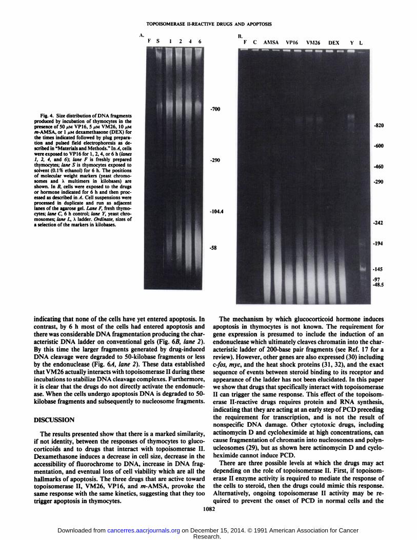
Patterns of DNA Fragmentation Caused by Topoisomerase IIInhibitors and Dexamethasone. Topoisomerase II enzyme molecules are bound to DNA with a periodic spacing along thechromosome fiber. DNA extracted from thymocytes that havebeen treated with VM26 and m-AMSA, when resolved bypulsed field gel electrophoresis, shows a characteristic distribution of fragment sizes (10). To examine this in greater detailin relation to PCD we carried out time courses of fragmentationof thymocyte DNA following exposure to VP 16 as shown inFig. 4A using the same concentrations of the drugs that producethe changes in the size of the thymocytes that were describedin Fig. 1. During 6 h of incubation with 50 pM VP 16 there wasan accumulation of DNA fragments of about 50 kilobases whichparalleled the increase in the number of smaller cells. Only aminor amount of DNA fragmentation occurred in the presenceof the same concentration of ethanol solvent (lane S), indicatingthat the effect is specific for VP 16. A similar pattern of DNAcleavage to 50-kilobase fragments was observed after 6 h ofincubation with /w-AMSA and VM26 (Fig. 4B). Surprisingly,6 h of incubation with dexamethasone also produced a similaramount of DNA cleavage to 50-kilobase fragments as shown inFig. 4B.
To try to establish the relationship between the drug-inducedDNA fragmentation and the appearance of smaller cells weexamined the extent of DNA fragmentation caused by VM26,VP 16, and m-AMSA in the presence of 1 ^M actinomycin Dwhich, as described above, prevents the formation of smallercells (Table 2). As shown in Fig. 5A, actinomycin D decreased,but did not abolish, DNA fragmentation to the 50-kilobasefragments caused by a 4-h exposure to each of the drugs. In thecase of VM26 actinomycin D decreased, or abolished, the
1080
Research. on December 15, 2014. © 1991 American Association for Cancercancerres.aacrjournals.org Downloaded from

TOPOISOMERASE II-REACTIVE DRUGS AND APOPTOSIS
B. c.
-,
.1- -
in -
a -
0 20 JU
celli:
RelativeHucirçsceiHv
Fig. 2. Flow cytometric 3-dimensional plots and density maps of Ihymocytes exposed to VP16. In both casescell size (forward light scatter) is plotted against theintegral of the fluorescence (F). A, control culture of freshly isolated thymocytes; B, 4 h of exposure to 50 JIM VP16; C. 6 h of exposure to VP 16.
Fig. 3. Flow cytometric 3D plots and density maps of thymocytes exposed to dexamethasone (1 fiM). In both casescell size (forward light scatter) is plotted againstthe integral of the fluorescence (F). A, control cultures of thymocytes exposed to 0.1% ethanol for 6h. B, 4h of exposure to 1 »Mdexamethasone. C, 6h of exposureto dexamethasone.
Table ÕEffects of actinomycin D and cycloheximide on topoisomerase ll-reaclive drug-induced apoptosisThymocytes were incubated for 4 h in the presence of either 50 JIM VP16, 20 >iM m-AMSA, 5 UM VM26. or I nM dexamethasone or with no drug or hormone
addition (control). Parallel cultures received either 100 nM cycloheximide or I (/\i actinomycin D. Following the incubation the number of cells in the peak channelof the smaller size cell population were counted. Each data point is the mean ±SEM for 5-15 determinations.
InhibitorNone
CycloheximideActinomycin DDexamethasone3446
±2322094 ±3472149 ±203m-AMSA3524
±2371700 ±4351902 + 436VM263160
±1701610 ±3651984 ±308VP163000
±1421442 ±3462027 ±276Control1610
±991905 ±2062147 ±206
appearance of 50-kilobase fragments but had no effect on theappearance of the 300-kilobase fragments. This observation
suggests that DNA fragmentation in response to the drugs hastwo components. In the presence of actinomycin D there issome fragmentation of DNA by /w-AMSA, VP16, and VM26
as may be expected since the direct action of these drugs ontopoisomerase II does not require RNA (or protein) synthesis.The second component of the fragmentation only occurs whenthe thymocytes become smaller (i.e., in the absence of actinomycin D). Similar results were obtained using cycloheximideas the inhibitor of apoptosis (data not shown). This conclusionis supported by the observation that the steroid-induced fragmentation of DNA to 50-kilobase fragments is completely
abolished in the presence of 1 n\\ actinomycin D (Fig. 5Ä). Allof the DNA cleavage seen with dexamethasone is presumablya result of the process of apoptosis and can only occur afternew genes have been expressed. To try to distinguish furtherbetween DNA cleavage in response to the interaction of thedrugs with topoisomerase II and DNA fragmentation occurringas a result of apoptosis we compared the patterns of DNAfragments on pulsed field gels obtained from cells exposed toVM26 for either 1 or 6 h (Fig. 6). Conventional gels were alsorun to determine whether the endonuclease was active at thesetimes. During the first hour of exposure to the drug DNA wascleaved to fragments of >300 kilobases (Fig. 6/1, lane 1) andthere was no ladder of DNA fragments (Fig. 6B, lane 1),
1081
Research. on December 15, 2014. © 1991 American Association for Cancercancerres.aacrjournals.org Downloaded from
TOPOISOMERASE II-REACTIVE DRUGS AND APOPTOSIS
\.
Fig. 4. Size distribution of DNA fragmentsproduced by incubation of thymocytes in thepresence of 50 JIMVP16, 5 ><MVM26, 10 ¡IMm-AMSA, or 1 JIMdexamethasone (DEX) forthe times indicated followed by plug preparation and pulsed Ik-Id electrophoresis as described in "Materials and Methods." In A, cells
were exposed to VP16 for 1, 2, 4, or 6 h (lanes1, 2, 4, and 6); lane F is freshly preparedthymocytes; lane S is thymocytes exposed tosolvent (0.1% ethanol) for 6 h. The positionsof molecular weight markers (yeast chromosomes and X multimers in kilobases) areshown. In II, cells were exposed to the drugsor hormone indicated for 6 h and then processed as described in A. Cell suspensions wereprocessed in duplicate and run as adjacentlanes of the agarose gel. Lane F, fresh thymocytes; lane C, 6 h control; lane Y, yeast chromosomes; lane L, A ladder. Ordinate, sizes ofa selection of the markers in kilobases.
K S 12-46
urnli.
F C AMSA Vl'16 VM26 DEX Y L
-700
820
-290
-10-1.4
-58
indicating that none of the cells have yet entered apoptosis. Incontrast, by 6 h most of the cells had entered apoptosis andthere was considerable DNA fragmentation producing the characteristic DNA ladder on conventional gels (Fig. 6B, lane 2).By this time the larger fragments generated by drug-inducedDNA cleavage were degraded to 50-kilobase fragments or lessby the endonuclease (Fig. 6A, lane 2). These data establishedthat VM26 actually interacts with topoisomerase II during theseincubations to stabilize DNA cleavage complexes. Furthermore,it is clear that the drugs do not directly activate the endonuclease. When the cells undergo apoptosis DNA is degraded to 50-kilobase fragments and subsequently to nucleosome fragments.
DISCUSSION
The results presented show that there is a marked similarity,if not identity, between the responses of thymocytes to gluco-
corticoids and to drugs that interact with topoisomerase II.Dexamethasone induces a decrease in cell size, decrease in theaccessibility of fluorochrome to DNA, increase in DNA fragmentation, and eventual loss of cell viability which are all thehallmarks of apoptosis. The three drugs that are active towardtopoisomerase II, VM26, VP 16, and m-AMSA, provoke thesame response with the same kinetics, suggesting that they tootrigger apoptosis in thymocytes.
The mechanism by which glucocorticoid hormone inducesapoptosis in thymocytes is not known. The requirement forgene expression is presumed to include the induction of anendonuclease which ultimately cleaves chromatin into the characteristic ladder of 200-base pair fragments (see Ref. 17 for areview). However, other genes are also expressed (30) includingc-fos, myc, and the heat shock proteins (31, 32), and the exactsequence of events between steroid binding to its receptor andappearance of the ladder has not been elucidated. In this paperwe show that drugs that specifically interact with topoisomeraseII can trigger the same response. This effect of the topoisomerase II-reactive drugs requires protein and RNA synthesis,
indicating that they are acting at an early step of PCD precedingthe requirement for transcription, and is not the result ofnonspecific DNA damage. Other cytotoxic drugs, includingactinomycin D and cycloheximide at high concentrations, cancause fragmentation of chromatin into nucleosomes and polyn-ucleosomes (29), but as shown here actinomycin D and cycloheximide cannot induce PCD.
There are three possible levels at which the drugs may actdepending on the role of topoisomerase II. First, if topoisomerase II enzyme activity is required to mediate the response ofthe cells to steroid, then the drugs could mimic this response.Alternatively, ongoing topoisomerase II activity may be required to prevent the onset of PCD in normal cells and the
1082
Research. on December 15, 2014. © 1991 American Association for Cancercancerres.aacrjournals.org Downloaded from

TOPOISOMERASE II-REACTIVE DRUGS AND APOPTOS1S
Fig. 5. Effect of actinomycin D on the sizedistribution of DNA fragments produced byincubation of thymocytes in the presence oftopoisomerase H-reactive drugs and dexameth-asone. In . I, cells were incubated for 4 h withVM26, VP 16, or m-AMSA in the presence (+)or absence (—)of 1 JIMactinomycin D. LanesY and /., yeast and A ladder markers, respectively. In B, the cells were incubated with 1 fiMdexamethasone (DEX) in the presence (+) orabsence (-) of 1 MMactinomycin D for 6 h.Lanes F, S, and C, the same as the equivalentlanes in Fig. 4. Ordinate, sizes of molecularweight standards in kilobases.
-700
-580
-460
-370-290-260
-145.5
-97
-48.5
-700
-97
-48.5
drugs inhibit this reaction and allow PCD to occur. It is alsopossible that the drugs interfere with other normal cellulartopoisomerase II activities and this is sufficiently disruptive tocause the cells to enter PCD. In addition, it cannot be ruled outthat the drugs have some other intracellular target that triggersPCD.
The loss of nuclear architecture which results in the changein cell size appears to be an "all or none" response and is fairly
rapid, since cells of intermediate size were not seen. This mayexplain why cells of increased density were not seen in previousstudies using the antineoplastic drug Adriamycin which alsointercalates into DNA (33), although morphologically identifiable apoptotic cells have been seen in some neoplastic cellstreated with chemotherapeutic agents (34). Thymocytes must,therefore, have a mechanism for rapidly destroying chromosome structure beyond the stage that it can be repaired (the
smaller, apoptotic cells cannot survive) which effectively killsthe cells even though integrity and "viability" based upon dye
exclusion may be maintained for 18-24 hours. Interestingly,the drug-topoisomerase II interaction alone is insufficient totrigger the change in chromatin structure that manifests itselfas a change in size of the nucleus. The experiment (Fig. 5) inwhich the drugs were added in the presence of either actinomycin D or cycloheximide showed that, although the drugs dointeract with topoisomerase II, as evidenced by DNA cleavageon the pulsed field gels, there is no change in the size of thenuclei. Thus, the effect of these drugs on chromatin structureis not equivalent to that caused by endonuclease. Only theendonuclease, induced after the cells enter PCD, can cause theamount of chromosome damage sufficient to irreversibly changethe size of the nucleus and kill the cells.
Flow cytometry is being used increasingly to detect changes1083
Research. on December 15, 2014. © 1991 American Association for Cancercancerres.aacrjournals.org Downloaded from

TOPOISOMERASE II-REACTIVE DRUGS AND APOPTOSIS
1
I
B1 2
Fig. 6. DNA cleavage patterns for cells exposed to 5 MMVM26 for 1 or 6 h.Thymocytes were incubated in the presence of the drug for either 1 or 6 h, andthen the cells were processed for pulsed field or conventional gel electrophoresisas described in "Materials and Methods." A, pulsed field gel, lane 1, 1 h; lane 2,
6 h. B, conventional agarose gel; lane I, 1 h; lane 2, 6 h.
in chromatin structure in a variety of cell types (35-39). Forexample, the accessibility of various fluorochromes to DNAchanges during differentiation of Friend leukemia cells in a waythat can be correlated to changes in chromatin structure (35).Damage to DNA either by digestion of isolated nuclei (36) orfixed chromatin (37) or by incubation of cells with clastogenicdrugs (38) decreases the relative fluorescence of chromatin inthe nuclei. A similar decrease in fluorescence was observed inthe smaller cell population after 4 h of exposure to drugs thatinduce PCD. Although there has been sufficient DNA damageby 4 h to cause the decrease in fluorescence, no DNA has beenlost from the nuclei, indicating that this reflects a change inchromatin structure. Thus, flow cytometry is a convenient wayto measure the progress of apoptosis since both the decrease incell size and change in chromatin structure can be detected and
this cell population is readily quantifiable. Furthermore, itillustrates that the change in cell (i.e., nuclear) size coincideswith the alteration in chromatin structure since we only seesmall cells with decreased fluorescence and never cells of thesame size (100 pm3) with decreased fluorescence or smaller
cells with a normal DNA fluorescence.The data presented here add a new dimension to the cytotoxic
effects and therapeutic value of these drugs and are supported,indirectly, by evidence already in the literature. Kupfer et al.(40) have shown that depletion of intracellular ATP pools doesnot affect antineoplastic drug-induced DNA cleavage-complex
formation, i.e., the drugs are still able to interact with theenzyme but their effect is no longer cytotoxic. This observationshows that drug interaction with topoisomerase II and thecytotoxic effect of the drugs can be uncoupled (i.e., drug interactions with topoisomerase II are not intrinsically cytotoxic)and is consistent with drug-induced apoptosis being the cytotoxic response. Moreover, it has been shown also that thecytotoxic effect of m-AMSA on a mastocytoma cell line iscycloheximide sensitive (41). Apoptosis is an ATP-dependentprocess and, as we have shown here, drug-induced apoptosis ofthymocytes requires protein synthesis and is inhibited by cycloheximide. In addition, Jaxel et al. (42) have noted irreversibleDNA fragmentation in splenocytes incubated for prolongedperiods of time in the presence of VP 16 which is consistentwith endonuclease cleavage of DNA during apoptosis. Furthermore, it has been shown that, when a drug such as m-AMSA isremoved, cleavable complex formation is reversed (2), and wehave shown also that following removal of the drugs thymocytesstill undergo apoptosis.4 Thus, continued presence of the drugs
is not a prerequisite for a lethal effect.Finally, in a recent review of the controversial areas in our
understanding of the mechanism of action of topoisomerase II-reactive, antineoplastic drugs Zwelling (3) concluded that "a
process dependent on ATP and distal to the stabilization of thecleavable complex must critically influence the susceptibility oftreated cells to the lethal effects of topoisomerase H-reactivedrugs" and "etoposide cytotoxicity can be decreased by treatingcells with the protein synthesis inhibitor cycloheximide." The
evidence we have presented here demonstrates that this process(i.e., the mechanism of cytotoxicity) may well be drug-induced
apoptosis. If this is the case, then it is important to determinethe mechanism by which this class of antineoplastic drugstriggers the entry of cells into apoptosis. More specifically, itmust be determined whether this is due to the interaction ofthese drugs with topoisomerase II or whether some other, asyet unknown, intracellular target is involved.
ACKNOWLEDGMENTS
We would like to thank Robert Richards for preparing the photographs. VM26 was kindly supplied by Dr. Kevin Guest, Bristol Laboratories, Ottawa, Ontario, Canada.
REFERENCES
1. Glisson, B. S., and Ross, W. E. DNA topoisomerase II: a primer on theenzyme and its unique role as a multidrug target in cancer chemotherapy.Pharmacol. Ther., 32: 89-106, 1987.
2. Kohn, K. W., Pommier, Y., Kerrigan, D., Markovits, J., and Covey, J. M.Topoisomerase II as a target of anticancer drug action in mammalian cells.Nail. Cancer Inst. Monogr., 4:61-71, 1987.
3. Zwelling, L. A. Topoisomerase II as a target of antileukemia drugs: a reviewof controversial areas. Hum. Pathol., Ì:101-112, 1989.
4 P. R. Walker, unpublished observations.
1084
Research. on December 15, 2014. © 1991 American Association for Cancercancerres.aacrjournals.org Downloaded from

TOPOISOMERASE II-REACTIVE DRUGS AND APOPTOSIS
4. Isseil, B. F., Rudolph, A. R., and Louie, A. C. Etoposide (VP-16-213): anoverview. /n:B. F. Issell, F. M. Muggia, and S. K. Carter (eds.), Etoposide(VP 16), pp. 1-13. Orlando, FL: Academic Press, 1983.
5. Yang, L., Rowe, T. C., and Liu, L. F. Identification of DNA topoisomeraseII as an intracellular target of antitumor epipodophyllotoxins in simian virus40-infected monkey cells. Cancer Res., 45: 5872-5876, 1985.
6. Minford, J., Pommier, Y., Filipski, J., Kohn, K. W., Kerrigan, D., Mattern,M., Michaels, S., Schwartz, R., and Zwelling, L. A. Isolation ofintercalator-dependent protein-linked DNA strand cleavage activity from cell nuclei andidentification as topoisomerase II. Biochemistry, 25: 9-16, 1986.
7. Rowe, T. C., Chen, G. L., hsiang, Y.-W., and Liu. L. F. DNA damage byantitumor acridines mediated by mammalian DNA topoisomerase II. CancerRes., ¿A:2021-2026, 1986.
8. Long, B. H., and Stringfellow, D. A. Inhibitors of topoisomerase II: structure-activity relationships and mechanism of action of podophyllin congeners.Adv. Enzyme Regul., 27: 223-256, 1988.
9. Estey, E., Adlakha, R. C., Hittelman, W. N., and Zwelling. L. A. Cell cyclestage dependent variations in drug-induced topoisomerase II mediated DNAcleavage and cytotoxicity. Biochemistry, 26:4338-4344, 1987.
10. Filipski, J., Leblanc, J., Youdale, T., Sikorska, M., and Walker, P. R.Periodicity of DNA folding in higher order chromatin structures. EMBO J.,9:1319-1327, 1990.
11. Kerr, J. F. R., Wyllie, A. H., and Currie, A. R. Apoptosis: a biologicalphenomenon with wide-ranging implications in tissue kinetics. Br. J. Cancer,26:239-257, 1972.
12. Wyllie, A. H., Kerr, J. F. R., and Currie, A. R. Cell death: the significanceof apoptosis. Int. Rev. Cytol., 68: 251-306, 1980.
13. Wyllie, A. H. Apoptosis. ISI Atlas Sci. Immunol., Vol. 1, pp. 192-196.Philadelphia, PA: Institute for Scientific Information. 1988.
14. Wyllie, A. H. Glucocorticoid-induced thymocyte apoptosis is associated withendogenous endonuclease activation. Nature (Lond.), 284: 555-556, 1980.
15. Cohen, J. J., and Duke, R. C. Glucocorticoid activation of a calcium-dependent endonuclease in thymocyte nuclei leads to cell death. J. Immunol.,152:38-42, 1984.
16. McConkey, D. J., Hartzell, P., Nicotera, P., and Orrenius, S. Calcium-activated DNA fragmentation kills immature thymocytes. FASEB J., 3:1843-1849, 1989.
17. Arends, M. J., Morris, R. G., and Wyllie, A. H. Apoptosis: the role of theendonuclease. Am. J. Pathol., 136: 593-608, 1990.
18. MacDonald, H. R., and Lees, R. K. Programmed death of autoreactivethymocytes. Nature (Lond.), 343: 642-644, 1990.
19. Whitfield, J. F., Perris, A. D., and Youdale, T. Destruction of the nuclearmorphology of thymic lymphocytes by the corticosteroid Cortisol. Exp. CellRes., 52: 349-362, 1968.
20. Wyllie, A. H., and Morris, R. G. Purification and properties of thymocytesundergoing apoptosis after glucocorticoid treatment. Am. J. Pathol., 109:78-87, 1982.
21. Thomas, N., and Bell, P. A. Glucocorticoid-induced cell-size changes andnuclear fragility in rat thymocytes. Mol. Cell. Endocrinol., 22: 71-84, 1981.
22. Thomas, N., Edwards, J. L., and Bell, P. A. Studies of the mechanism ofglucocorticoid-induced pyknosis in isolated rat thymocytes. J. SteroidBiochem., 18: 519-524, 1983.
23. Koury, M. J., and Bondurant, M. C. Erythropoietin retards DNA breakdownand prevents programmed death in erythroid progenitor cells. Science (Washington DC), 248: 378-381, 1990.
24. Williams, G. T., Smith, C. A., Spooncer, E., Dexter, M., and Taylor, D. R.Haemopoietic colony stimulating factors promoter cell survival by suppressing apoptosis. Nature (Lond.), 343: 76-79, 1990.
25. McConkey, D. J., Hartzell, P., Chow, S. C., Orrenhius. S., and Jondal, M.
Interleukin 1 inhibits T cell receptor mediated apoptosis in immature thymocytes. J. Biol. Chem.. 265: 3009-3011, 1990.
26. Alvarez, M. R., and Truitt. A. J. Rapid nuclear cytochemical changes inducedby dexamethasone in thymus lymphocytes of adrenalectomized rats. Exp.Cell Res., 106: 105-110, 1977.
27. Compton. M. M., and Cidlowski. J. A. Rapid in vivoeffects of glucocorticoidson the integrity of rat lymphocyte genomic deoxyribonucleic acid. Endocrinology, 118: 38-45, 1986.
28. Wyllie, A. H., Morris, R. G., Smith, A. L., and Dunlop, D. Chromatincleavage in apoptosis: association with condensed chromatin morphologyand dependence on macromolecular synthesis. J. Pathol., 142: 67-77, 1984.
29. Kaufmann, S. H. Induction of endonucleolytic DNA cleavage in human acutemyelogenous leukemia cells by etoposide, camptothecin, and other cytotoxicanti-cancer drugs: a cautionary note. Cancer Res., 49: 5870-5878, 1989.
30. Tenniswood, M. P., Montpetit, M. L., Leger, J. G., Wong, P., Pineault, J.M., and Rouleau, M. Epithelial-stromal interactions and cell death in theprostate. In: W. E. Farnsworth and R. J. Abiin (eds.). The Prostate as anEndocrine Gland, pp. 187-204. Boca Raton, FL: CRC Press, 1989.
31. Wyllie, A. H., Rose, K. A., Morris, R. G., Steel, C. M., Foster, E., andSpandidos. D. A. Rodent fibroblast tumors expressing human myc and rasgenes: growth, metastasis and endogenous oncogene expression. Br. J. Cancer, 56: 251-260, 1987.
32. Buttyan, R., Zakeri, Z., Lockshin, R., and Wolgemuth, D. Cascade inductionof c-fos, myc, and heat shock 70K transcripts during regression of the ratventral prostate. Mol. Endocrinol., 2: 650-657. 1988.
33. Dysin, J. E. D.. Simmons, D. M., Daniel, J., Mclaughlin. J. M., Quirke, P.,and Bird, C. C. Kinetic and physical studies of cell death induced bychemotherapeutic agents or hyperthermia. Cell Tissue Kinet., 19: 311-324,1986.
34. Searle, J., Lawson, T. A., Abbott, P. J., Harmon, B., and Kerr, J. F. R. Anelectron-microscope study of the mode of cell death induced by cancer-chemotherapeutic agents in populations of proliferating normal and neoplas-tic cells. J. Pathol., 116: 129-138, 1975.
35. Darynkiewicz, Z., Tráganos, F., Kapuscinski, J., Slaiano-Coico, L., andmelamed, M. R. Accessibility of DNA in situ to various fluorochromes:relationship to chromatin changes during erythroid differentiation of Friendleukemia cells. Cytometry, 5: 355-362, 1984.
36. Roti Roti, J. L., Wright, W. D., Higashikubo. R., and Dethefsen, L. A.DNase I sensitivity of nuclear DNA measured by flow cytometry. Cytometry,6: 101-108, 1985.
37. Stokke, T., and Steen, H. B. Distinction of leukocyte classes based onchromatin-structure dependent DNA-binding of 7-aminoactinomycin D. Cytometry, 8: 576-583, 1987.
38. Kubbies, M. Flow cytometric recognition of clastogen induced chromatindamage in G0/G, lymphocytes by non-stoichiometric Hoeschst fluorochromebinding. Cytometry1, ¡1:386-394, 1990.
39. Komitowski, D., Schmitt, S. B., and Muto, S. Quantitative description ofnuclear morphology in assessing resistance of sarcoma 180 to Adriamycin.Cytometry, «.-625-631, 1987.
40. Kupfer, G., Bodley, A. L., and Liu, L. F. Involvement of intracellular ATPin cytotoxicity of topoisomerase II-targeting antitumor drugs. Nati. CancerInst. Monogr., 4: 37-40, 1987.
41. Schneider, E., Lawson, P. A., and Ralph, R. K. Inhibition of protein synthesisreduces the cytotoxicity of 4'-(9-acridinylamino)methanesulfon-m-anisidide
without affecting DNA breakage and DNA topoisomerase II in a murinemastocytoma cell line. Biochem. Pharmacol.. 38: 263-269, 1989.
42. Jaxel, C., Tadou, G., Portemer, C., Mirambeau, G., Panijel, J., and Duguet,M. Topoisomerase inhibitors induce irreversible fragmentation of replicatedDNA in concanavalin A stimulated splenocytes. Biochemistry, 27: 95-99,1988.
1085
Research. on December 15, 2014. © 1991 American Association for Cancercancerres.aacrjournals.org Downloaded from

1991;51:1078-1085. Cancer Res P. Roy Walker, Catherine Smith, Tony Youdale, et al. Apoptosis in ThymocytesTopoisomerase II-reactive Chemotherapeutic Drugs Induce
Updated version
http://cancerres.aacrjournals.org/content/51/4/1078
Access the most recent version of this article at:
E-mail alerts related to this article or journal.Sign up to receive free email-alerts
Subscriptions
Reprints and
To order reprints of this article or to subscribe to the journal, contact the AACR Publications
Permissions
To request permission to re-use all or part of this article, contact the AACR Publications
Research. on December 15, 2014. © 1991 American Association for Cancercancerres.aacrjournals.org Downloaded from
Copyright © 2022 FDOKUMEN