Hydrophobic Surfactant Proteins Induce a Phosphatidylethanolamine to Form Cubic Phases
9
Hydrophobic Surfactant Proteins Induce a Phosphatidylethanolamine to Form Cubic Phases Mariya Chavarha, †‡§ Hamed Khoojinian, †‡§6 Leonard E. Schulwitz Jr., †‡§6 Samares C. Biswas, †‡§ Shankar B. Rananavare, { and Stephen B. Hall †‡§ * † Department of Molecular Biology and Biochemistry, ‡ Department of Medicine, and § Department of Physiology and Pharmacology, Oregon Health & Science University, Portland, Oregon; and { Department of Chemistry, Portland State University, Portland, Oregon ABSTRACT The hydrophobic surfactant proteins SP-B and SP-C promote rapid adsorption of pulmonary surfactant to an air/ water interface. Previous evidence suggests that they achieve this effect by facilitating the formation of a rate-limiting negatively curved stalk between the vesicular bilayer and the interface. To determine whether the proteins can alter the curvature of lipid leaflets, we used x-ray diffraction to investigate how the physiological mixture of these proteins affects structures formed by 1-palmitoyl-2-oleoyl phosphatidylethanolamine, which by itself undergoes the lamellar-to-inverse hexagonal phase transition at 71 C. In amounts as low as 0.03% (w:w) and at temperatures as low as 57 C, the proteins induce formation of bicontinuous inverse cubic phases. The proteins produce a dose-related shift of diffracted intensity to the cubic phases, with minimal evidence of other structures above 0.1% and 62 C, but no change in the lattice-constants of the lamellar or cubic phases. The induction of the bicontinuous cubic phases, in which the individual lipid leaflets have the same saddle-shaped curvature as the hypothetical stalk-intermediate, supports the proposed model of how the surfactant proteins promote adsorption. INTRODUCTION Pulmonary surfactant contains small amounts of two very hydrophobic proteins, SP-B and SP-C, at least one of which is essential for normal function of the lungs. Ventilation in the absence of SP-B produces an injury to the alveolocapil- lary barrier (1–3) equivalent to the insult caused by ventila- tion when surface tension is elevated because the complete mixture of surfactant-constituents is missing (4,5). A defi- ciency of SP-C produces effects with a more gradual onset, but it too leads to altered lungs (6,7). The hydrophobic proteins in vitro greatly accelerate the adsorption of surfac- tant vesicles to an air/water interface (8–10). These results suggest that the essential physiological function of these proteins is to promote rapid formation of the alveolar film. Currently available data specify features that must be present in any model of how the proteins facilitate adsorption (11). In contrast to classical molecular surfactants, which insert into the interface as individual monomers, components of pulmonary surfactant adsorb collectively (12–14), suggest- ing that the surfactant vesicles fuse with the surface to deliver their complete set of constituents. Factors that accelerate adsorption have similar effects whether they are confined to the interface or an adsorbing vesicle (9,15,16), suggesting that a rate-limiting structure must be equally accessible from both locations. Compounds not present in pulmonary surfac- tant generally promote or inhibit adsorption according to their tendency to produce negative or positive curvature (9,17–19), in which the hydrophilic face of a phospholipid leaflet is concave or convex, respectively. Together, these results suggest a model in which components adsorb through nega- tively curved leaflets that extend from the vesicular bilayer to the interface (Fig. 1 A), analogous to the stalk-intermediate proposed as a key step in the fusion of two bilayers (20). The hydrophobic proteins would accelerate adsorption by promoting the formation of this rate-limiting structure. To the best of our knowledge, however, no direct evidence shows that the surfactant proteins (SPs) can affect the ten- dency of lipids to form curved structures. The absence of such data may reflect the lipids with which the proteins have been studied. Like most biological mixtures of lipids, pulmonary surfactant forms lamellar bilayers (9). Any spon- taneous curvature in the individual leaflets is cancelled by the presence of the oppositely oriented, paired leaflet (21). Any effect of the proteins on the ability of the leaflets to curve is undetectable. The studies reported here tested how the proteins affect a phospholipid that does form curved struc- tures. Because 1-palmitoyl-2-oleoyl phosphatidylethanol- amine (POPE) has a sufficiently inverted conical shape, high temperatures convert lamellar bilayers to the inverse hexagonal (H II ) phase, in which unpaired leaflets approxi- mate their spontaneous curvature (21). Lipids that can form such structures should more readily express any effects of the proteins on curvature. MATERIALS AND METHODS Materials POPE was obtained from Avanti Polar Lipids (Alabaster, AL) and used without further characterization or purification. Extracts of pulmonary surfac- tant from calf lungs (ONY, Amherst, NY) were prepared as described Submitted October 5, 2009, and accepted for publication December 15, 2009. 6 Hamed Khoojinian and Leonard E. Schulwitz Jr., contributed equally to this work. *Correspondence: [email protected] Editor: Lukas K. Tamm. Ó 2010 by the Biophysical Society 0006-3495/10/04/1549/9 $2.00 doi: 10.1016/j.bpj.2009.12.4302 Biophysical Journal Volume 98 April 2010 1549–1557 1549
-
Upload
independent -
Category
Documents
-
view
5 -
download
0
Transcript of Hydrophobic Surfactant Proteins Induce a Phosphatidylethanolamine to Form Cubic Phases

Biophysical Journal Volume 98 April 2010 1549–1557 1549
Hydrophobic Surfactant Proteins Induce a Phosphatidylethanolamineto Form Cubic Phases
Mariya Chavarha,†‡§ Hamed Khoojinian,†‡§6 Leonard E. Schulwitz Jr.,†‡§6 Samares C. Biswas,†‡§
Shankar B. Rananavare,{ and Stephen B. Hall†‡§*†Department of Molecular Biology and Biochemistry, ‡Department of Medicine, and §Department of Physiology and Pharmacology,Oregon Health & Science University, Portland, Oregon; and {Department of Chemistry, Portland State University, Portland, Oregon
ABSTRACT The hydrophobic surfactant proteins SP-B and SP-C promote rapid adsorption of pulmonary surfactant to an air/water interface. Previous evidence suggests that they achieve this effect by facilitating the formation of a rate-limiting negativelycurved stalk between the vesicular bilayer and the interface. To determine whether the proteins can alter the curvature of lipidleaflets, we used x-ray diffraction to investigate how the physiological mixture of these proteins affects structures formed by1-palmitoyl-2-oleoyl phosphatidylethanolamine, which by itself undergoes the lamellar-to-inverse hexagonal phase transitionat 71�C. In amounts as low as 0.03% (w:w) and at temperatures as low as 57�C, the proteins induce formation of bicontinuousinverse cubic phases. The proteins produce a dose-related shift of diffracted intensity to the cubic phases, with minimal evidenceof other structures above 0.1% and 62�C, but no change in the lattice-constants of the lamellar or cubic phases. The induction ofthe bicontinuous cubic phases, in which the individual lipid leaflets have the same saddle-shaped curvature as the hypotheticalstalk-intermediate, supports the proposed model of how the surfactant proteins promote adsorption.
INTRODUCTION
Pulmonary surfactant contains small amounts of two very
hydrophobic proteins, SP-B and SP-C, at least one of which
is essential for normal function of the lungs. Ventilation in
the absence of SP-B produces an injury to the alveolocapil-
lary barrier (1–3) equivalent to the insult caused by ventila-
tion when surface tension is elevated because the complete
mixture of surfactant-constituents is missing (4,5). A defi-
ciency of SP-C produces effects with a more gradual onset,
but it too leads to altered lungs (6,7). The hydrophobic
proteins in vitro greatly accelerate the adsorption of surfac-
tant vesicles to an air/water interface (8–10). These results
suggest that the essential physiological function of these
proteins is to promote rapid formation of the alveolar film.
Currently available data specify features that must be
present in any model of how the proteins facilitate adsorption
(11). In contrast to classical molecular surfactants, which
insert into the interface as individual monomers, components
of pulmonary surfactant adsorb collectively (12–14), suggest-
ing that the surfactant vesicles fuse with the surface to deliver
their complete set of constituents. Factors that accelerate
adsorption have similar effects whether they are confined to
the interface or an adsorbing vesicle (9,15,16), suggesting
that a rate-limiting structure must be equally accessible from
both locations. Compounds not present in pulmonary surfac-
tant generally promote or inhibit adsorption according to their
tendency to produce negative or positive curvature (9,17–19),
Submitted October 5, 2009, and accepted for publication December 15,
2009.6Hamed Khoojinian and Leonard E. Schulwitz Jr., contributed equally to
this work.
*Correspondence: [email protected]
Editor: Lukas K. Tamm.
� 2010 by the Biophysical Society
0006-3495/10/04/1549/9 $2.00
in which the hydrophilic face of a phospholipid leaflet is
concave or convex, respectively. Together, these results
suggest a model in which components adsorb through nega-
tively curved leaflets that extend from the vesicular bilayer
to the interface (Fig. 1 A), analogous to the stalk-intermediate
proposed as a key step in the fusion of two bilayers (20). The
hydrophobic proteins would accelerate adsorption by
promoting the formation of this rate-limiting structure.
To the best of our knowledge, however, no direct evidence
shows that the surfactant proteins (SPs) can affect the ten-
dency of lipids to form curved structures. The absence of
such data may reflect the lipids with which the proteins
have been studied. Like most biological mixtures of lipids,
pulmonary surfactant forms lamellar bilayers (9). Any spon-
taneous curvature in the individual leaflets is cancelled by the
presence of the oppositely oriented, paired leaflet (21). Any
effect of the proteins on the ability of the leaflets to curve is
undetectable. The studies reported here tested how the
proteins affect a phospholipid that does form curved struc-
tures. Because 1-palmitoyl-2-oleoyl phosphatidylethanol-
amine (POPE) has a sufficiently inverted conical shape,
high temperatures convert lamellar bilayers to the inverse
hexagonal (HII) phase, in which unpaired leaflets approxi-
mate their spontaneous curvature (21). Lipids that can
form such structures should more readily express any effects
of the proteins on curvature.
MATERIALS AND METHODS
Materials
POPE was obtained from Avanti Polar Lipids (Alabaster, AL) and used
without further characterization or purification. Extracts of pulmonary surfac-
tant from calf lungs (ONY, Amherst, NY) were prepared as described
doi: 10.1016/j.bpj.2009.12.4302

FIGURE 1 Schematic representation of saddle-shaped structures. The
radii R1 and R2 each define a corresponding principal curvature of the struc-
ture, c1 and c2, with c ¼ 1/R. Scales for the two diagrams are different. (A)
Hypothetical rate-limiting kinetic intermediate formed during adsorption of
the vesicular bilayer to the air/water interface. The radii of the leaflets are
defined at the pivotal surface, which lies approximately at the junction
between the hydrophobic acyl tails and the hydrophilic headgroup. (B)
Segment of the inverse bicontinuous cubic phase with space group Im3m.
In contrast to the individual leaflets, the curvature of the bilayer is defined
at its midpoint, which for the inverse bicontinuous cubic phases lies along
an infinite periodic minimal surface at which the two radii of curvature
are equal in magnitude and opposite in sign, resulting in no net curvature.
1550 Chavarha et al.
previously (22). Hydrophobic SPs were obtained from the extracted surfac-
tant by minor modifications of a previously described protocol based on gel
permeation chromatography (23). Samples were eluted from a 150 � 2.5
cm column (Spectrum Chromatography, Houston, TX) packed with LH-20
matrix (24) using a solvent of chloroform/methanol (1:1, v:v) at constant
flow (1 mL/min) driven by gravity. The content of phospholipid and protein
was monitored qualitatively by measuring the optical density of the eluted
fractions at 240 and 280 nm (25). Fractions containing SPs were pooled,
concentrated initially by rotary evaporation and then with a stream of
nitrogen, and stored at 4�C before mixing in appropriate ratios with POPE.
The following reagents were purchased commercially and used without
further purification: chloroform and methanol (Fisher Scientific, Pittsburgh,
PA); Na2EDTA-2H2O (Gibco, Grand Island, NY); and NaN3 (Sigma,
St. Louis, MO). Water was processed and photooxidized with ultraviolet
light using a NANOpure Diamond TOC-UV water-purification apparatus
Biophysical Journal 98(8) 1549–1557
(Barnstead/Thermolyne, Dubuque, IA). All chemicals and solvents were
ACS grade.
Methods
Biochemical determinations
Protein content was determined quantitatively by amido black assay on
material precipitated with trichloroacetic acid using a standard of bovine
serum albumin (26). Contamination of protein by residual phospholipid,
as determined by measuring the content of phosphate (27), was assessed
at 85 ng of lipid for each microgram of protein. This level represented a puri-
fication of the proteins from the initial extracted surfactant by approximately
3 orders of magnitude.
Samples for x-ray diffraction
Samples of protein and POPE were mixed in chloroform, concentrated under
a stream of N2 until they reached a homogeneous viscous consistency,
deposited as a thin uniform film at the bottom of a test tube, and then held
overnight under vacuum at room temperature to remove any residual
solvent. Samples were resuspended in 2 mM EDTA with 0.002% (w/w)
NaN3 for a final phospholipid concentration of 50 mM, flushed with N2
and sealed with Teflon tape, agitated briefly, and then left to hydrate at
4�C overnight. Cyclic freezing and thawing along with vigorous vortexing
achieved homogeneous dispersions. The hydrated samples were transferred
to special glass capillaries (1.0 mm diameter, 0.01 mm wall thickness;
Charles Supper, Natick, MA), both ends of which were sealed first by flame
and then with epoxy. The capillaries were centrifuged at 640 � gmax for
10 min to concentrate the aggregated samples at one end of the tube, and
then stored at 4�C until needed. Heating and cooling of the samples through
the lamellar-HII transition temperature, which is commonly used (28) to
induce formation of inverse cubic phases (QII), was avoided before obtaining
the initial measurements.
The samples contained 0–3% (w:w) protein/phospholipid. Other studies
of similar phenomena with different proteins have expressed compositions
as % (mol:mol) (29). Results from amino acid analyses (30) suggest that
the physiological mixture of SP-B and SP-C obtained by gel permeation
chromatography is roughly equimolar. Given the known molecular weights
of the different constituents, our samples with a protein/phospholipid
content of 1% (w:w) were roughly 0.1% (mol:mol).
Small-angle x-ray diffraction
Measurements of diffraction were conducted on beamline 1-4 at the Stanford
Synchrotron Radiation Lightsource. An x-ray beam with a wavelength of
1.488 A was focused to an elliptical spot with approximate dimensions of
0.3 (horizontal) � 0.1 (vertical) mm. A beryllium window inserted into the
Kapton film used to seal the helium-filled drift-tube between the sample
and detector minimized background scattering from Kapton. Diffraction
images were acquired at a sample-to-detector distance of 0.26 m, producing
a range of accessible q-values from 0.29 to 5.40 nm�1. Spatial calibration
was performed using both cholesterol myristate and silver behenate (31).
Temperature was controlled with water circulated through the sample-
holder, and measurements with a thermocouple established the relationship
between temperatures in the bath and the capillary. With the use of two circu-
lating baths, the temperatures changed rapidly—within 20 s for an increase of
10�C. The samples were then equilibrated for 10 min before diffraction was
measured. Incubations longer than 10 min produced no further changes in
relative intensities. Measurements of diffraction routinely exposed samples
to the beam for 120 s. Continuous exposure of other samples for periods as
long as 2.5 h to test for evidence of radiation-induced damage produced no
changes in diffraction. The diffracted intensities were radially integrated
using the program FIT2D (32). The results presented here, obtained with
a single set of samples during a single visit to the Stanford Synchrotron
Radiation Lightsource, were confirmed with two other sets of samples during
separate visits that included measurements on beamline 4-2.

FIGURE 2 Diffraction patterns. Negative images, obtained by exposing
samples to the x-ray beam for 120 s, record one quadrant of the circularly
symmetric diffraction rings, with the beam-stop located at the upper-right
corner. The brightness and contrast of each entire image are adjusted to
show higher-order rings. (A) POPE alone, 11�C; rings have lamellar spacing
and the increased relative intensity of the fourth-order ring that is character-
istic of the Lb0 phase (33). (B) POPE alone, 30�C; lamellar spacing, consis-
tent with the La phase. (C) POPE alone, 90�C; hexagonal spacing, consistent
with the HII phase. (D) POPE with 0.3% (w:w) SP, 81�C; spacing consistent
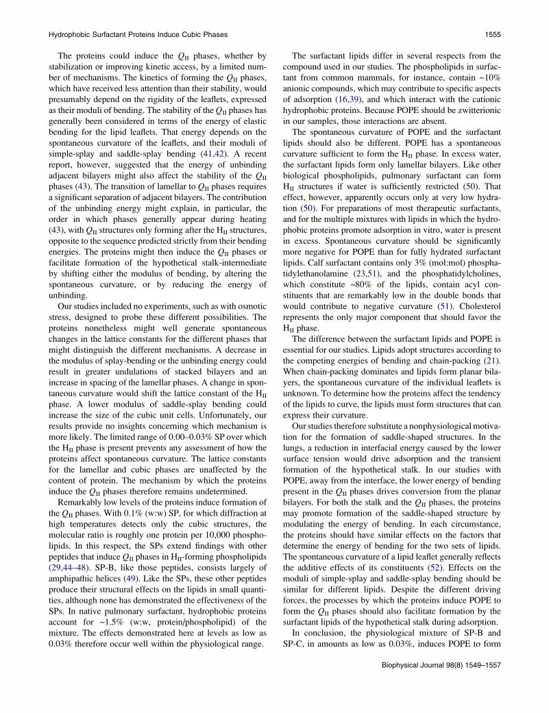
FIGURE 3 Diffraction pattern for POPE with 1% SP at 90�C. The radially
integrated intensity is plotted as a function of the measured q. Labeled
vertical lines assign peaks to diffraction with the indicated Miller indices
from structures with Pn3m (Pn) or Im3m (Im) space groups based on the
fits of the observed spacings to allowed peaks (Fig. 4).
Hydrophobic Surfactant Proteins Induce Cubic Phases 1551
RESULTS
Samples containing POPE with 0–3% (w:w) SP at specific
temperatures of 11–90�C in all cases produced rings indi-
cating powder diffraction. Results obtained with POPE alone
agreed well with previously published results. Below 25�C,
the samples produced diffraction with lamellar spacing and
a prominent fourth-order peak characteristic of the Lb0 phase
(33) (Fig. 2 A). A second set of lamellar peaks appeared at
25�C with lower d-spacing (Fig. 2 B), consistent with forma-
tion of the La phase close to the previously reported main
transition temperature of 26�C (34). The La peaks persisted
to 72�C, above which diffraction converted to the spacing
of a hexagonal phase, which remained present at the highest
temperature of 90�C (Fig. 2 C). These results agreed rea-
sonably with the expected formation of the HII phase at
71�C (35).
The addition of the proteins had essentially no effect on
diffraction at lower temperatures. The lattice constant of the
lamellar phases remained unchanged, and the Lb0-La transi-
tion occurred at approximately the same temperature. Above
53�C, a new set of rings appeared at q-values below the first-
order lamellar peak (Fig. 2 D), suggesting the presence of the
cubic phases found previously with other phosphatidyletha-
nolamines. At the highest temperatures, with the diffraction
peaks spread the farthest (Fig. 3), these peaks had the relative
spacing expected for individual or coexisting cubic struc-
tures with Pn3m and Im3m space groups (Fig. 4). At lower
temperatures, although the shift to lower q-values caused
with simultaneous diffraction from cubic structures with Pn3m and Im3m
space groups, indicating coexistence of inverse bicontinuous cubic (QII)
phases with the diamond and primitive minimal surfaces, respectively.
Biophysical Journal 98(8) 1549–1557

FIGURE 4 Analysis of the diffraction pattern for POPE with 1% SP at
90�C (Fig. 3) (53). The values of q measured for the diffraction peaks are
plotted as horizontal lines. Vertical lines indicate all possible values of
f(h,k,l), where h, k, and l are the Miller indices, and f(h,k,l) is given by h
for the lamellar phases, (h2 þ hk þ k2)1/2 for the hexagonal phase, and
(h2 þ k2 þ l2)1/2 for the cubic phases. Labels on the lower portion of the
vertical lines indicate values that are allowed for diffraction from structures
with the following symmetries, which have been either documented exper-
imentally in other systems of phospholipids or suggested by simulation (54):
lamellar (L; space group pm, No.3 in the International Tables of Crystallog-
raphy (55)); hexagonal (H;p6m, No.17); Pn3m (P; No.224); Im3m (Im;
No.229); Fd3m (F; No.227); and Ia3d (Ia; No.230). Symbols indicate
assignment of peaks to (h,k,l) based on optimizing the linear fit of measured
q-values at allowed values of f(h,k,l). Solid and open symbols fit well the
diffraction predicted for Pn3m and Im3m space groups, respectively. Lines
through the symbols, obtained by least-squares fit, provide the slope, which
yields the lattice-constant (ao) of the unit cell according to (ao ¼ 2p/slope)
for the lamellar and cubic phases, and (ao¼ 4p/(31/2 $ slope)) for the hexag-
onal phase.
1552 Chavarha et al.
the overlap of some peaks, these patterns remained evident.
When the two cubic structures coexisted, their lattice
constants maintained a constant ratio of 1.28 5 0.01. This
value agreed with the ratio of 1.279 expected for the inverse
bicontinuous cubic phases (QII) with those space groups
Biophysical Journal 98(8) 1549–1557
interconverted by Bonnet transformation (28). Because six
other space groups generate the same absent reflections as
Im3m over the range detected here, and one other for
Pn3m, the spacing of the diffraction rings alone was insuffi-
cient to make definitive assignments to those space groups.
In combination with the ratio of lattice constants, however,
the low values of q at which the peaks occurred, and the
previous observations made with similar lipids, the pattern
of diffraction strongly supported the presence of structures
with space groups Pn3m and Im3m, corresponding to QII
phases with diamond (QIID) and primitive (QII
P) infinitely
periodic minimal surfaces, respectively.
The major response to increasing amounts of protein was
an increase in the intensities of the cubic diffraction and the
corresponding loss of signal from coexisting structures
(Fig. 5 and Fig. S1 in the Supporting Material)). At 0.01%
protein, only questionable peaks were present at the highest
temperatures for values of q below the first-order hexagonal
peak to suggest the possible presence of some QII structures.
With 0.03% protein at temperatures above 67�C, definite
rings occurred at low q-values (Fig. 5), with seven peaks
at 76�C and 81�C fitting the spacing for Pn3m diffraction.
For protein R 0.1%, hexagonal diffraction disappeared com-
pletely, and the cubic phases became the only structures
present at high temperatures (Fig. 5). Increasing amounts
of protein shifted intensity from the QIID phase to the larger
QIIP structures. Samples with 0.03% SP produced peaks only
for the QIID phase; diffraction at 3% SP showed only QII
P
(Fig. 5 and Fig. S1). More protein also induced a broadening
of the peaks. Consequently, although the integrated intensity
from the Im3m peaks continued to increase at 3% (Fig. 6),
the height of the peaks fell.
The temperature at which cubic diffraction first emerged
showed limited dependence on the amount of protein pres-
ent. Increasing the protein concentration from 0.03% to
0.10% lowered the temperature at which the peaks first
appeared at low q-values from 67�C to 57�C. At 3% SP,
however, that temperature increased from 57�C to 62�C.
Whether these changes reflected true shifts in the transi-
tion-temperature or simply a dose-dependent variation in
peak-intensity remained unresolved.
FIGURE 5 Diffraction patterns at different temperatures
and contents of protein. Curves give the radially integrated
intensity in arbitrary units, plotted on a logarithmic scale,
over a limited range of q-values for each sample at each
temperature. Traces are offset by a fixed amount for each
temperature.

A
B
FIGURE 6 Intensities of the cubic phases with different quantities of SP.
Intensity is expressed for each phase as the integrated area above the baseline
of the peak with Miller indices 110 (I), normalized relative to its value (I1) at
the temperature and % SP at which I is greatest.
FIGURE 7 Effect of temperature on the lattice constants for POPE with
different amounts of protein. Structural symmetries were assigned based
on the spacing of the diffraction rings (Fig. 4). The slope of q versus
f(h,k,l) provided the lattice constant (ao) (Fig. 4).
Hydrophobic Surfactant Proteins Induce Cubic Phases 1553
The added protein had little effect on the dimensions of
the unit cell for the different diffracting structures (Fig. 7).
At any particular temperature, the lattice constants (ao) for
both the lamellar and the hexagonal structures were essen-
tially invariant with different amounts of protein (Fig. 7
and Fig. S2, A–C). Hexagonal diffraction, however, was
limited to samples containing 0–0.03% SP (Fig. 7 and
Fig. S2 C), which prevented determination of how the
proteins affected the size of the hexagonal lattice. The cubic
lattices showed more dependence on temperature, but
no consistent response to increasing amounts of protein
(Fig. 7 and Fig. S2, D and E). The proteins induced a dose-
related shift in the extent of the two cubic phases, but the
structures formed were roughly constant, with dimensions
that showed no clear change in response to larger amounts
of protein.
The results of these measurements suggested that the SPs
induced POPE to form structures that would not occur for the
lipid alone. Previous theoretical and experimental studies,
however, have suggested that at increasing temperatures,
the QII phases should exist between lamellar and hexagonal
structures (21,36). Cooling from high temperatures, as well
as cycling between temperatures above and below the La-HII
transition, can induce cubic phases in lipids, including POPE
(29,37), that form only lamellar and hexagonal structures
during initial heating. Under the conditions of our experi-
ments, when POPE was cooled from 90�C without protein,
it produced low-intensity rings at q-values consistent with
the QIIP phase (Fig. S3). These peaks persisted to the temper-
ature of the La-Lb0 transition, below which they were absent.
Although the diffraction of any sample during heating from
the Lb0 phase was indistinguishable, regardless of its history,
during sequential cycles of cooling, the intensity of the QIIP
peaks increased (Fig. S3). The marked hysteresis of phase
behavior, with the cubic phases absent during heating but
present during cooling, prevented any estimation of the
La-QII transition temperature. The results confirmed,
however, that although the proteins greatly facilitated forma-
tion of the QII phases, they were not essential components of
those structures.
The cubic phases also showed significant hysteresis in
samples with protein (Fig. 8). In contrast to the samples at
high temperatures that contained only lipid, with the protein
present, the QII phases represented the dominant structures.
Experiments could therefore compare the cubic lattice
constants during heating and cooling as well as the temper-
atures at which the cubic phases first appeared and disap-
peared. Like the samples with POPE alone, the lattice
constant for the lamellar phases depended only on tempera-
ture, irrespective of how the samples had been treated
Biophysical Journal 98(8) 1549–1557

FIGURE 8 Hysteresis of POPE with 0.3% SP during the initial cycle of
heating and cooling. Diffraction was recorded during heating from 11�Cto 90�C, and then during subsequent cooling to 6�C. Open symbols with
solid lines indicate the lattice constants (ao) during the initial heating; solid
symbols with dotted lines provide the values during the subsequent cooling.
1554 Chavarha et al.
previously. With the proteins present, the lattice constants of
the QII phases during cooling were significantly smaller than
during heating, suggesting that the smaller unit cell observed
at higher temperatures grew during cooling with difficulty
(Fig. 8). As with the samples of POPE alone, the QII phases
persisted during cooling to temperatures well below the
levels at which they formed during heating. With the proteins
present, however, the cubic phases reverted during cooling to
lamellar structures well before reaching the Lb0 phase. Crys-
tallization of the acyl chains was unnecessary to promote
return from the QII phases.
DISCUSSION
Our experiments show that the hydrophobic SPs induce
POPE to form QII phases in a dose-dependent fashion. The
bicontinuous structures share important features of the key
kinetic intermediate in the most widely considered model
of how those proteins promote adsorption at the alveolar
air/water interface (Fig. 1). The available evidence suggests
that, at least initially, the lipids flow from the adsorbing
vesicle to an air/water interface through a stalk that connects
the two locations (38,39). The common two-dimensional
representation of this structure emphasizes that the curvature
in the plane perpendicular to the interface (c1) would be
negative (Fig. 1 A). However, the second principal curvature
(c2) in the plane parallel to the interface would be positive
Biophysical Journal 98(8) 1549–1557
(Fig. 1 A). The stalk would have a Gaussian curvature
(c1 $ c2) that would be negative. The correlation between
the molecular shape of added constituents and their effect
on adsorption (11) suggests that the net curvature (c1 þ c2)
of the stalk would be negative. The proteins would promote
adsorption by facilitating formation of this rate-limiting
structure.
In the QII phases, the lipids form bilayers, in contrast to the
unpaired monolayers of the hypothetical stalk (Fig. 1). Each
individual leaflet in the cubic phases, however, shares the
basic structural features of the monolayers in the stalk. In
both cases, the Gaussian curvature is negative (21,28,36).
The net curvature of the individual leaflets in both structures
is also negative. In the QII phases, the midpoint of the bilayer
lies along a minimal surface, at which the two principal
curvatures are equal in magnitude and opposite in sign,
resulting in no net curvature. The individual leaflets are
shifted from that surface. The curvature of the monolayers
that constitute the bilayer is defined empirically at the pivotal
surface, at which the cross-sectional area of constituents
neither expands nor contracts during bending (40). For the
leaflets, the displacement of the pivotal surface (located
roughly at the junction between the headgroup and the
acyl residues (21)) from the minimal surface at the midpoint
of the bilayer has opposite effects on the magnitude of the
two principal curvatures. The leaflet curvature with negative
sign becomes more negative, and the positive curvature
becomes smaller, resulting in a negative net curvature.
Although the leaflets are paired in the cubic phase and
unpaired in the stalk, the net and Gaussian curvatures in
the two cases are at least qualitatively similar.
The proteins may induce formation of the QII phases by
either thermodynamic or kinetic effects. The distinction
depends on the ultimate stability of the different phases for
the lipids alone. Without the proteins, the QII phases only
appear after exposure to temperatures above the La-HII tran-
sition. If this behavior represents true equilibrium, then the
observed formation of the QII structures at much lower
temperatures with the proteins would reflect stabilization.
With the lipid alone, however, the persistence of the QII
phases to these lower temperatures during cooling suggests
that under these conditions, the curved structures might be
more stable than the lamellar phases, but kinetically inacces-
sible. The proteins would then induce the QII phases by
promoting their faster formation. The behavior of POPE by
itself prevents a clear distinction between these two possibil-
ities. The marked hysteresis during heating and cooling
prevents determination of the noncubic/cubic transition
temperature that would allow assessment of thermodynamic
stability. The proteins narrow the hysteresis of the QII phases
(Fig. 8), suggesting that their effect may be kinetic, and that
they accelerate transitions both to and from the cubic struc-
tures. Both mechanisms would be physiologically relevant.
Whether the proteins stabilize the bridging stalk or simply
facilitate its formation, the result would be faster adsorption.
Hydrophobic Surfactant Proteins Induce Cubic Phases 1555
The proteins could induce the QII phases, whether by
stabilization or improving kinetic access, by a limited num-
ber of mechanisms. The kinetics of forming the QII phases,
which have received less attention than their stability, would
presumably depend on the rigidity of the leaflets, expressed
as their moduli of bending. The stability of the QII phases has
generally been considered in terms of the energy of elastic
bending for the lipid leaflets. That energy depends on the
spontaneous curvature of the leaflets, and their moduli of
simple-splay and saddle-splay bending (41,42). A recent
report, however, suggested that the energy of unbinding
adjacent bilayers might also affect the stability of the QII
phases (43). The transition of lamellar to QII phases requires
a significant separation of adjacent bilayers. The contribution
of the unbinding energy might explain, in particular, the
order in which phases generally appear during heating
(43), with QII structures only forming after the HII structures,
opposite to the sequence predicted strictly from their bending
energies. The proteins might then induce the QII phases or
facilitate formation of the hypothetical stalk-intermediate
by shifting either the modulus of bending, by altering the
spontaneous curvature, or by reducing the energy of
unbinding.
Our studies included no experiments, such as with osmotic
stress, designed to probe these different possibilities. The
proteins nonetheless might well generate spontaneous
changes in the lattice constants for the different phases that
might distinguish the different mechanisms. A decrease in
the modulus of splay-bending or the unbinding energy could
result in greater undulations of stacked bilayers and an
increase in spacing of the lamellar phases. A change in spon-
taneous curvature would shift the lattice constant of the HII
phase. A lower modulus of saddle-splay bending could
increase the size of the cubic unit cells. Unfortunately, our
results provide no insights concerning which mechanism is
more likely. The limited range of 0.00–0.03% SP over which
the HII phase is present prevents any assessment of how the
proteins affect spontaneous curvature. The lattice constants
for the lamellar and cubic phases are unaffected by the
content of protein. The mechanism by which the proteins
induce the QII phases therefore remains undetermined.
Remarkably low levels of the proteins induce formation of
the QII phases. With 0.1% (w:w) SP, for which diffraction at
high temperatures detects only the cubic structures, the
molecular ratio is roughly one protein per 10,000 phospho-
lipids. In this respect, the SPs extend findings with other
peptides that induce QII phases in HII-forming phospholipids
(29,44–48). SP-B, like those peptides, consists largely of
amphipathic helices (49). Like the SPs, these other peptides
produce their structural effects on the lipids in small quanti-
ties, although none has demonstrated the effectiveness of the
SPs. In native pulmonary surfactant, hydrophobic proteins
account for ~1.5% (w:w, protein/phospholipid) of the
mixture. The effects demonstrated here at levels as low as
0.03% therefore occur well within the physiological range.
The surfactant lipids differ in several respects from the
compound used in our studies. The phospholipids in surfac-
tant from common mammals, for instance, contain ~10%
anionic compounds, which may contribute to specific aspects
of adsorption (16,39), and which interact with the cationic
hydrophobic proteins. Because POPE should be zwitterionic
in our samples, those interactions are absent.
The spontaneous curvature of POPE and the surfactant
lipids should also be different. POPE has a spontaneous
curvature sufficient to form the HII phase. In excess water,
the surfactant lipids form only lamellar bilayers. Like other
biological phospholipids, pulmonary surfactant can form
HII structures if water is sufficiently restricted (50). That
effect, however, apparently occurs only at very low hydra-
tion (50). For preparations of most therapeutic surfactants,
and for the multiple mixtures with lipids in which the hydro-
phobic proteins promote adsorption in vitro, water is present
in excess. Spontaneous curvature should be significantly
more negative for POPE than for fully hydrated surfactant
lipids. Calf surfactant contains only 3% (mol:mol) phospha-
tidylethanolamine (23,51), and the phosphatidylcholines,
which constitute ~80% of the lipids, contain acyl con-
stituents that are remarkably low in the double bonds that
would contribute to negative curvature (51). Cholesterol
represents the only major component that should favor the
HII phase.
The difference between the surfactant lipids and POPE is
essential for our studies. Lipids adopt structures according to
the competing energies of bending and chain-packing (21).
When chain-packing dominates and lipids form planar bila-
yers, the spontaneous curvature of the individual leaflets is
unknown. To determine how the proteins affect the tendency
of the lipids to curve, the lipids must form structures that can
express their curvature.
Our studies therefore substitute a nonphysiological motiva-
tion for the formation of saddle-shaped structures. In the
lungs, a reduction in interfacial energy caused by the lower
surface tension would drive adsorption and the transient
formation of the hypothetical stalk. In our studies with
POPE, away from the interface, the lower energy of bending
present in the QII phases drives conversion from the planar
bilayers. For both the stalk and the QII phases, the proteins
may promote formation of the saddle-shaped structure by
modulating the energy of bending. In each circumstance,
the proteins should have similar effects on the factors that
determine the energy of bending for the two sets of lipids.
The spontaneous curvature of a lipid leaflet generally reflects
the additive effects of its constituents (52). Effects on the
moduli of simple-splay and saddle-splay bending should be
similar for different lipids. Despite the different driving
forces, the processes by which the proteins induce POPE to
form the QII phases should also facilitate formation by the
surfactant lipids of the hypothetical stalk during adsorption.
In conclusion, the physiological mixture of SP-B and
SP-C, in amounts as low as 0.03%, induces POPE to form
Biophysical Journal 98(8) 1549–1557

1556 Chavarha et al.
QII phases at temperatures 15�C below the point at which the
lipid alone converts during heating from the La to the HII
phase. These results fit with a model in which the proteins
facilitate adsorption by promoting the formation of a rate-
limiting structure with negative Gaussian and net curvatures
that bridges the gap between the vesicular bilayer and the
air/water interface.
SUPPORTING MATERIAL
One table and three figures are available at http://www.biophysj.org/
biophysj/supplemental/S0006-3495(10)00003-2.
The extracted calf surfactant from which the SPs were isolated was provided
by Dr. Edmund Egan (ONY Inc.). Diffraction was measured at the Stanford
Synchrotron Radiation Lightsource, a national user facility operated by
Stanford University on behalf of the Office of Basic Energy Sciences,
U.S. Department of Energy.
This study was funded by the National Institutes of Health (HL 54209).
REFERENCES
1. Nogee, L. M., G. Garnier, ., H. R. Colten. 1994. A mutation in thesurfactant protein B gene responsible for fatal neonatal respiratorydisease in multiple kindreds. J. Clin. Invest. 93:1860–1863.
2. Melton, K. R., L. L. Nesslein, ., T. E. Weaver. 2003. SP-B deficiencycauses respiratory failure in adult mice. Am. J. Physiol. 285:L543–L549.
3. Clark, J. C., S. E. Wert, ., J. A. Whitsett. 1995. Targeted disruption ofthe surfactant protein B gene disrupts surfactant homeostasis, causingrespiratory failure in newborn mice. Proc. Natl. Acad. Sci. USA.92:7794–7798.
4. Robertson, B. 1984. Pathology and pathophysiology of neonatal surfac-tant deficiency (‘‘respiratory distress syndrome,’’ ‘‘hyaline membranedisease’’). In Pulmonary Surfactant. B. Robertson, L. M. G. Van Golde,and J. J. Batenburg, editors. Elsevier Science Publishers, Amsterdam.383–418.
5. Lachmann, B., B. Robertson, and J. Vogel. 1980. In vivo lung lavage asan experimental model of the respiratory distress syndrome. ActaAnaesthesiol. Scand. 24:231–236.
6. Nogee, L. M., A. E. Dunbar, 3rd, ., J. A. Whitsett. 2001. A mutation inthe surfactant protein C gene associated with familial interstitial lungdisease. N. Engl. J. Med. 344:573–579.
7. Glasser, S. W., E. A. Detmer, ., J. A. Whitsett. 2003. Pneumonitis andemphysema in sp-C gene targeted mice. J. Biol. Chem. 278:14291–14298.
8. Whitsett, J. A., B. L. Ohning, ., R. H. Notter. 1986. Hydrophobicsurfactant-associated protein in whole lung surfactant and its impor-tance for biophysical activity in lung surfactant extracts used forreplacement therapy. Pediatr. Res. 20:460–467.
9. Biswas, S. C., S. B. Rananavare, and S. B. Hall. 2005. Effects of gram-icidin-A on the adsorption of phospholipids to the air-water interface.Biochim. Biophys. Acta. 1717:41–49.
10. Wang, Z., S. B. Hall, and R. H. Notter. 1996. Roles of different hydro-phobic constituents in the adsorption of pulmonary surfactant. J. LipidRes. 37:790–798.
11. Rugonyi, S., S. C. Biswas, and S. B. Hall. 2008. The biophysical func-tion of pulmonary surfactant. Respir. Physiol. Neurobiol. 163:244–255.
12. Haller, T., P. Dietl, ., G. Putz. 2004. Tracing surfactant transformationfrom cellular release to insertion into an air-liquid interface. Am.J. Physiol. 286:L1009–L1015.
13. Sen, A., S.-W. Hui, ., E. A. Egan. 1988. Localization of lipidexchange sites between bulk lung surfactants and surface monolayer:freeze fracture study. J. Colloid Interface Sci. 126:355–360.
Biophysical Journal 98(8) 1549–1557
14. Schurch, S., D. Schurch, ., B. Robertson. 1994. Surface activity oflipid extract surfactant in relation to film area compression and collapse.J. Appl. Physiol. 77:974–986.
15. Oosterlaken-Dijksterhuis, M. A., H. P. Haagsman, ., R. A. Demel.1991. Characterization of lipid insertion into monomolecular layersmediated by lung surfactant proteins SP-B and SP-C. Biochemistry.30:10965–10971.
16. Walters, R. W., R. R. Jenq, and S. B. Hall. 2000. Distinct steps in theadsorption of pulmonary surfactant to an air-liquid interface. Biophys.J. 78:257–266.
17. Yu, S.-H., P. G. R. Harding, and F. Possmayer. 1984. Artificialpulmonary surfactant. Potential role for hexagonal HII phase in theformation of a surface-active monolayer. Biochim. Biophys. Acta.776:37–47.
18. Perkins, W. R., R. B. Dause, ., A. S. Janoff. 1996. Role of lipid poly-morphism in pulmonary surfactant. Science. 273:330–332.
19. Biswas, S. C., S. B. Rananavare, and S. B. Hall. 2007. Differentialeffects of lysophosphatidylcholine on the adsorption of phospholipidsto an air/water interface. Biophys. J. 92:493–501.
20. Markin, V. S., M. M. Kozlov, and V. L. Borovjagin. 1984. On thetheory of membrane fusion. The stalk mechanism. Gen. Physiol.Biophys. 3:361–377.
21. Gruner, S. M. 1989. Stability of lyotropic phases with curved interfaces.J. Phys. Chem. 93:7562–7570.
22. Notter, R. H., J. N. Finkelstein, and R. D. Taubold. 1983. Comparativeadsorption of natural lung surfactant, extracted phospholipids, and arti-ficial phospholipid mixtures to the air-water interface. Chem. Phys.Lipids. 33:67–80.
23. Hall, S. B., Z. Wang, and R. H. Notter. 1994. Separation of subfractionsof the hydrophobic components of calf lung surfactant. J. Lipid Res.35:1386–1394.
24. Takahashi, A., and T. Fujiwara. 1986. Proteolipid in bovine lung surfac-tant: its role in surfactant function. Biochem. Biophys. Res. Commun.135:527–532.
25. Perez-Gil, J., A. Cruz, and C. Casals. 1993. Solubility of hydrophobicsurfactant proteins in organic solvent/water mixtures. Structural studieson SP-B and SP-C in aqueous organic solvents and lipids. Biochim.Biophys. Acta. 1168:261–270.
26. Kaplan, R. S., and P. L. Pedersen. 1985. Determination of microgramquantities of protein in the presence of milligram levels of lipid withamido black 10B. Anal. Biochem. 150:97–104.
27. Ames, B. N. 1966. Assay of inorganic phosphate, total phosphate andphosphatases. Methods Enzymol. VIII:115–118.
28. Shearman, G. C., O. Ces, ., J. M. Seddon. 2006. Inverse lyotropicphases of lipids and membrane curvature. J. Phys. Condens. Matter.18:S1105–S1124.
29. Hickel, A., S. Danner-Pongratz, ., G. Pabst. 2008. Influence of antimi-crobial peptides on the formation of nonlamellar lipid mesophases.Biochim. Biophys. Acta. 1778:2325–2333.
30. van Eijk, M., C. G. De Haas, and H. P. Haagsman. 1995. Quantitativeanalysis of pulmonary surfactant proteins B and C. Anal. Biochem.232:231–237.
31. Lee, B., C.-T. Lo, ., R. E. Winans. 2006. Silver behenate as a calibra-tion standard of grazing-incidence small-angle X-ray scattering. J. Appl.Cryst. 39:749–751.
32. Hammersley, A. P., S. O. Svensson, ., D. Hausermann. 1996. Two-dimensional detector software: from real detector to idealised imageor two-q scan. High Press. Res. 14:235–248.
33. Gruner, S. M., M. W. Tate, ., P. R. Cullis. 1988. X-ray diffractionstudy of the polymorphic behavior of N-methylated dioleoylphosphati-dylethanolamine. Biochemistry. 27:2853–2866.
34. Brown, P. M., J. Steers, ., J. R. Silvius. 1986. Role of head groupstructure in the phase behavior of amino phospholipids. 2. Lamellarand nonlamellar phases of unsaturated phosphatidylethanolamineanalogues. Biochemistry. 25:4259–4267.

Hydrophobic Surfactant Proteins Induce Cubic Phases 1557
35. Epand, R. M., and R. Bottega. 1987. Modulation of the phase transition
behavior of phosphatidylethanolamine by cholesterol and oxysterols.
Biochemistry. 26:1820–1825.
36. Anderson, D. M., S. M. Gruner, and S. Leibler. 1988. Geometrical
aspects of the frustration in the cubic phases of lyotropic liquid crystals.
Proc. Natl. Acad. Sci. USA. 85:5364–5368.
37. Rappolt, M., A. Hickel, ., K. Lohner. 2003. Mechanism of the
lamellar/inverse hexagonal phase transition examined by high resolu-
tion x-ray diffraction. Biophys. J. 84:3111–3122.
38. Schram, V., and S. B. Hall. 2001. Thermodynamic effects of the hydro-
phobic surfactant proteins on the early adsorption of pulmonary surfac-
tant. Biophys. J. 81:1536–1546.
39. Klenz, U., M. Saleem, ., H. J. Galla. 2008. Influence of lipid saturation
grade and headgroup charge: a refined lung surfactant adsorption model.
Biophys. J. 95:699–709.
40. Gruner, S. M., V. A. Parsegian, and R. P. Rand. 1986. Directly
measured deformation energy of phospholipid HII hexagonal phases.
Faraday Discuss. Chem. Soc. 81:29–37.
41. Helfrich, W. 1973. Elastic properties of lipid bilayers: theory and
possible experiments. Z. Naturforsch. [C]. 28:693–703.
42. Helfrich, W. 1990. Elasticity and thermal undulations of fluid films of
amphiphiles. In Liquids at Interfaces. J. Charvolin, J. F. Joanny, and
J. Zinn-Justin, editors. North-Holland, Amsterdam. 209–237.
43. Siegel, D. P., and B. G. Tenchov. 2008. Influence of the lamellar phase
unbinding energy on the relative stability of lamellar and inverted cubic
phases. Biophys. J. 94:3987–3995.
44. Keller, S. L., S. M. Gruner, and K. Gawrisch. 1996. Small concentra-
tions of alamethicin induce a cubic phase in bulk phosphatidylethanol-
amine mixtures. Biochim. Biophys. Acta. 1278:241–246.
45. Prenner, E. J., R. N. Lewis, ., R. N. McElhaney. 1997. Nonlamellar
phases induced by the interaction of gramicidin S with lipid bilayers.
A possible relationship to membrane-disrupting activity. Biochemistry.36:7906–7916.
46. El Jastimi, R., and M. Lafleur. 1999. Nisin promotes the formation ofnon-lamellar inverted phases in unsaturated phosphatidylethanol-amines. Biochim. Biophys. Acta. 1418:97–105.
47. Staudegger, E., E. J. Prenner, ., K. Lohner. 2000. X-ray studies on theinteraction of the antimicrobial peptide gramicidin S with microbiallipid extracts: evidence for cubic phase formation. Biochim. Biophys.Acta. 1468:213–230.
48. Angelova, A., R. Ionov, ., G. Rapp. 2000. Interaction of the peptideantibiotic alamethicin with bilayer- and non-bilayer-forming lipids:influence of increasing alamethicin concentration on the lipids supramo-lecular structures. Arch. Biochem. Biophys. 378:93–106.
49. Vandenbussche, G., A. Clercx, ., J. M. Ruysschaert. 1992. Secondarystructure and orientation of the surfactant protein SP-B in a lipid envi-ronment. A Fourier transform infrared spectroscopy study. Biochem-istry. 31:9169–9176.
50. Gulik, A., P. Tchoreloff, and J. Proust. 1994. A conformation transitionof lung surfactant lipids probably involved in respiration. Biophys. J.67:1107–1112.
51. Kahn, M. C., G. J. Anderson, ., S. B. Hall. 1995. Phosphatidylcholinemolecular-species of calf lung surfactant. Am. J. Physiol. 13:L567–L573.
52. Rand, R. P., N. L. Fuller, ., V. A. Parsegian. 1990. Membrane curva-ture, lipid segregation, and structural transitions for phospholipids underdual-solvent stress. Biochemistry. 29:76–87.
53. Sun, R. G., and J. Zhang. 2004. The cubic phase of phosphatidyletha-nolamine film by small angle x-ray scattering. J. Phys. D Appl. Phys.37:463–467.
54. Fuhrmans, M., V. Knecht, and S. J. Marrink. 2009. A single bicontinu-ous cubic phase induced by fusion peptides. J. Am. Chem. Soc.131:9166–9167.
55. Kasper, J. S., K. Lonsdale, and International Union of Crystallography.1985. International Tables for X-Ray Crystallography. published for theInternational Union of Crystallography by D. Reidel, Dordrecht,Boston.
Biophysical Journal 98(8) 1549–1557