
Stress-Induced Legume Root Nodule Senescence. Physiological, Biochemical, and Structural Alterations
Upload
spanalumniCategory
view
1download
0
©20
13 L
ande
s B
iosc
ienc
e. D
o no
t dis
tribu
te.
www.landesbioscience.com Cell Cycle 1433
Cell Cycle 12:9, 1433–1449; May 1, 2013; © 2013 Landes Bioscience REPORT REPORT
*Correspondence to: Carol A. Lange; Email: [email protected]: 02/15/13; Revised: 03/29/13; Accepted: 04/03/13http://dx.doi.org/10.4161/cc.24550
Introduction
Although ovarian cancer accounts for approximately 3% of all cancers among women, it is the deadliest among gynecologic cancers. An estimated 15,500 deaths were expected in 2012,1 a death rate of more than 50% due to late detection of the disease and intrinsic or acquired resistance to current therapeutic strat-egies. The identification of reliable biomarkers for early detec-tion of OC will have a substantial impact on survival rates, while molecular markers that predict outcome may allow for efficacious targeted therapies.
Loss of nuclear progesterone receptors (PR) and low circulating progesterone levels are associated with increased ovarian cancer (OC) risk. However, PR are abundantly expressed in a significant percentage of serous and endometrioid ovarian tumors; patients with PR+ tumors typically experience longer progression-free survival relative to those with PR-null tumors. The molecular mechanisms of these protective effects are poorly understood. To study PR action in OC in the absence of added estrogen (i.e., needed to induce robust PR expression), we created ES-2 OC cells stably expressing vector control or GFP-tagged PR-B (GFP-PR). Progestin (R5020) stimulation of ES-2 cells stably expressing GFP-PR induced cellular senescence characterized by altered cellular morphology, prolonged survival, senescence-associated β-galactosidase activity, G1 cell cycle arrest and upregulation of the cell cycle inhibitor, p21, as well as the Forkhead-box transcription factor, FOXO1; these results repeated in unmodified ER+/PR+ PEO4 OC cells. PR-B and FOXO1 were detected within the same PRE-containing regions of the p21 upstream promoter. Knockdown of p21 resulted in molecular compensation via FOXO1-dependent upregulation of numerous FOXO1 target genes (p15, p16, p27) and an increased rate of senescence. Inhibition of FOXO1 (with AS1842856) or stable FOXO1 knockdown inhibited progestin-induced p21 expression and blocked progestin-induced senescence. Overall, these findings support a role for PR as a tumor suppressor in OC cells, which exhibits inhibitory effects by inducing FOXO1-dependent cellular senescence. Clinical “priming” of the PR-FOXO1-p21 signaling pathway using PR agonists may provide a useful strategy to induce irreversible cell cycle arrest and thereby sensitize OC cells to existing chemotherapies as part of combination “two-step” therapies.
Progesterone receptors induce FOXO1-dependent senescence in ovarian cancer cells
Caroline H. Diep,1,† Nathan J. Charles,1,† C. Blake Gilks,2 Steve E. Kalloger,2 Peter A. Argenta3 and Carol A. Lange1,4,*
1Department of Medicine; Hematology, Oncology, and Transplantation Division; University of Minnesota; Minneapolis, MN USA; 2Department of Pathology and Laboratory Medicine; University of British Columbia; Vancouver, BC Canada; 3Department of Obstetrics, Gynecology, and Women’s Health; Gynecology Oncology Division; University of
Minnesota Medical School; Minneapolis, MN USA; 4Department of Pharmacology; Masonic Cancer Center; University of Minnesota; Minneapolis, MN USA
†These authors contributed equally as co-first authors.
Keywords: progesterone receptor, forkhead transcription factor, FOXO1, AS1842856, p21, senescence, progestin, ovarian cancer, breast cancer
Abbreviations: CDK, cyclin-dependent kinase; ChIP, chromatin immunoprecipitation; E2, 17β-estradiol; ER, estrogen receptor; FOXO1, forkhead box protein 1; MTT, (3-(4,5-Dimethylthiazol-2-yl)-2,5-diphenyltetrazolium bromide; OC, ovarian cancer; OSE, ovarian surface epithelial; PARP, poly (ADP-ribose) polymerase; PR, progesterone receptor; PRE, progesterone response element; p15, cyclin-dependent kinase inhibitor 2B (CDKN2B); p16, cyclin-dependent kinase inhibitor 2A (CDKN2A); p21, cyclin-dependent kinase inhibitor 1 (CDKN1A); p27, cyclin-dependent kinase inhibitor 1B (CDKN1B); SAβgal, senescence-
associate beta-galactosidase; SGK, serum/glucocorticoid regulated kinase; RT-qPCR, real-time quantitative-PCR
Progesterone receptors (PR) belong to the steroid receptor superfamily of related ligand-activated transcription factors that includes estrogen, androgen, mineralocorticoid and glucocorti-coid receptors.2 PR is a classical estrogen-regulated ER-target gene. Two PR isoforms (full-length PR-B and N-terminally truncated PR-A) have been identified and characterized as ligand-activated transcription factors with distinct transcriptional activities, while a third (PR-C) modulates the other two in selected tissues.3-7 Upon ligand binding, PR binds directly to specific progesterone response elements (PREs) or tethers to other DNA-binding tran-scription factors to alter gene expression.2
Dow
nloa
ded
by [6
6.55
.144
.181
] at 2
1:49
19
Mar
ch 2
015

©20
13 L
ande
s B
iosc
ienc
e. D
o no
t dis
tribu
te.
1434 Cell Cycle Volume 12 Issue 9
expression was low to negligible in all cell lines (data not shown), regardless of histologic sub-type (clear cell, ES-2; serous, HEY, OVCAR-3, OVCAR-8, PEO4; endometrioid, TOV-112D). Additionally, RT-qPCR analysis detected minimal levels of PR mRNA in each of these cell lines when compared with T47D human breast cancer cells (Fig. 1B). Cell line models of human tumors frequently lose steroid hormone receptor expression when grown in tissue culture. Alternatively, estrogen responsiveness of the PR promoter may be diminished.35
To determine if ER-positive ovarian cancer cells are capable of inducing endogenous PR expression, PEO4 cells were treated with β-estradiol (E2, 1 nM) and PR mRNA, and protein expres-sion was evaluated (Fig. 1C). E2 treatment of PEO4 cells signifi-cantly induced PR mRNA expression (4.3-fold) compared with vehicle-treated controls. Untreated PEO4 cells contained low lev-els of PR-B protein. The expression of both PR isoforms (PR-A and PR-B) was significantly induced upon estrogen (E2) treat-ment relative to vehicle-treated and R5020-only, while R5020 treatment caused a slight up-shift in gel mobility of both PR-A and PR-B proteins in E2-treated cells (Fig. 1D). The transcrip-tional activity of endogenous PR in PEO4 cells was evaluated in response to increasing doses of the PR-specific agonist, R5020, a synthetic progestin, at 24 and 96 h. In the absence of estrogen, expression of the classic PR-target gene, serum/glucocorticoid regulated kinase (SGK)36,37 was significantly induced by R5020 (10 μM) treatment relative to vehicle controls (Fig. 1E), consis-tent with the modest up-shift in gel mobility of PR-B protein (Fig. 1D, inset). Pre-treatment with estrogen did not further sen-sitize these cells to progestin (data not shown). These data sug-gest that OC cell lines express low abundance functional PR-B, as measured by the ability of progestins to induce PR-target gene (SGK) expression.
Progestin induces non-proliferative cell survival in OC cells. In order to investigate the impact of PR expression and signaling on OC cell biology without the confounding effects of exogenously added estrogen, we created a PR-expressing OC cell line. We chose ES-2 cells due to their inherently aggressive nature, rapid growth rate, ability to form tumors in xenograft38,39 and low endogenous PR mRNA levels (Fig. 1B). Notably, these cells are resistant to estrogen induction of endogenous PRs (data not shown). ES-2 cells were transfected with GFP-tagged PR-B or GFP-only vectors, and PR+ clones were selected for PR pro-tein expression levels that were comparable to similarly created T47D-YB breast cancer cells40 (Fig. 2A, inset). PR transcrip-tional activity in GFP-PR and control ES-2 cells was evaluated by PRE-luciferase reporter assays. R5020 stimulated luciferase activ-ity in GFP-PR cells but not in GFP-vector control cells; PR tran-scriptional activity was blocked by co-treatment of the cells with the competitive PR antagonist RU486 (Fig. 2A). Endogenous PR target genes (p21 and KLF4) were similarly regulated in breast and ovarian cancer cells stably expressing PR-B (data not shown). Fluorescence microscopy indicated that GFP-PR-B accumulated in the nucleus upon R5020 stimulation (Fig. S1).41 Interestingly, we observed an increase in the overall size of nuclei present within GFP-PR-B-expressing cells exposed to R5020 for as little as 24 h (Fig. S1).
PR has become an attractive target in OC. Progesterone deficiencies and a genetic loss of heterozygosity at the PR gene locus (ch 11q23.3–24.3)8 are associated with increased OC risk. While elevated progesterone levels appear to play a protective role, multiparity and elevated circulating progesterone levels (10-fold) during pregnancy, as well as the suppression of ovula-tion, are associated with decreased OC risk.9 Similarly, the use of progestin-containing oral contraceptives is associated with decreased lifetime risk of OC.10 The expression of PR is a favor-able prognostic marker in ovarian tumors and associated with longer progression-free survival.11-19
PR transcriptional activity is commonly linked to the expres-sion of many cell cycle regulators including members of the cyclin, cyclin-dependent kinase (CDK) and p21/p27 families.20 PR is often associated with survival and cell cycle progression in breast and prostate cancer cells.21-23 Specifically, PR-B isoforms are more potent transcription factors in reporter gene assays and at selected PR target genes relative to PR-A isoforms, including genes that encode cell cycle regulators.4,24 PR-B but not PR-A isoforms mediate mammary gland alveologenesis during nor-mal breast development5 and induce cyclin D1-driven prolif-eration and pro-survival in breast cancer cells.25 Interestingly, however, a handful of reports have suggested that progesterone may inhibit these effects in ovarian cancer cells.26-31 Of par-ticular interest, is the association of PR-B expression with the induction of cell cycle arrest first observed in Ras-transformed NIH3T3 cells32 and later extended to include OC cells.33 Furthermore, expression of PR-B isoforms in ovarian tumors is associated with longer progression-free patient survival and an indicator of positive prognosis.15,34 Herein, the goal of our studies was to further investigate the impact of PR-B expression and activation on OC cell proliferation and to determine the signaling mechanisms responsible for PR-B-mediated cell cycle control.
Results
PR expression in OC tumors and cell lines. Studies of limited sample size report decreased or absent PR expression in human OC tissue samples, and little information exists on the rela-tive distribution of PR within OC subtypes.8 We evaluated the percentage of PR-positive tumors from each major histological sub-type of ovarian surface epithelial (OSE) derived OC in a cohort of 504 tissue samples (Fig. 1A). While percentages varied between sub-types, each group contained PR-positive tumors. PR expression was highest in endometrioid (67%) and serous (35%; low-grade, 64%) tumors. Overall, 35% of ovarian tumors were PR-positive, a value consistent with larger mixed cohort studies.17,34 The distribution of estrogen receptor (ER)-positive tumors in the same cohort was similar to that of PR with endo-metrioid (77%) and serous (> 70%) tumor sub-types displaying the highest portion of ER positivity; the overall percentage of ER-positive tumors was 55%.
We next examined expression levels of PR in a panel of six established human OC cell lines of epithelial origin and one immortalized normal OSE cell line (1816-575). PR protein
Dow
nloa
ded
by [6
6.55
.144
.181
] at 2
1:49
19
Mar
ch 2
015

©20
13 L
ande
s B
iosc
ienc
e. D
o no
t dis
tribu
te.
www.landesbioscience.com Cell Cycle 1435
effects of progestin on growth characteristics of GFP-PR ES-2 cells. MTT [3-(4,5-dimethylthiazol-2-yl)-2,5-diphenyltetra-zolium bromide] cell proliferation assays were initially utilized to study long-term effects of progestin treatment. GFP-PR and
Prior studies have shown that progesterone exhibits both pro-liferative28 and anti-proliferative effects on the growth of OC cells42-44 with inhibitory effects observed at particularly high concentrations (≥ 10−6 M) of ligand.28,30,45 We investigated the
Figure 1. PR is expressed in human ovarian cancer tissues and cancer cell lines. (A) Immunohistochemical staining for PR in human ovarian cancer tissues representing the five major sub-types of ovarian surface epithelial (OSE) origin (n = 504). (B) RT-qPCR analysis of PR mRNA expression in a panel of six ovarian cancer cell lines and one immortalized, non-transformed cell line (1816-575) relative to PR+ T47D breast cancer cells. All values were normalized to GAPDH levels. (C) RT-qPCR analysis of PR mRNA expression of PEO4 cells treated with vehicle (ethanol) or β-estradiol (E2, 1 nM) for 24 h (n = 3, **p ≤ 0.01). (D) Western blot analysis of PR and ERα protein expression in PEO4 cells treated with vehicle, R5020 (10 nM, 1 h), E2 (1 nM, 48 h) and E2 (1 nM, 48 h) followed by R5020 (10 nM, 1 h). T47D CO total cell lysate was loaded on the same gel as a positive control for PR expression. Total ERK was used as a loading control. (E) Inset, western blot analysis of PR expression in PEO4 cells treated with vehicle and R5020 (1 and 10 μM) for 48 h. RT-qPCR analysis of SGK mRNA expression after 24 h and 96 h R5020 treatment (1 and 10 μM) in PEO4 cells (n = 3, *p ≤ 0.05, **p ≤ 0.01). All values were normalized to GAPDH levels.
Dow
nloa
ded
by [6
6.55
.144
.181
] at 2
1:49
19
Mar
ch 2
015

©20
13 L
ande
s B
iosc
ienc
e. D
o no
t dis
tribu
te.
1436 Cell Cycle Volume 12 Issue 9
Figure 2. Stable expression of PR in ES-2 cells increases cell survival and inhibits cell colony formation. (A) Inset, western blot analysis showing stable expression of GFP-tagged PR-B in ES-2 cells (GFP-PR) as compared with parental ES-2 cells stably expressing GFP-tagged empty vector construct (empty control) and PR-B expressing T47D breast cancer cells (T47D-YB). ES-2 GFP-PR cells transiently transfected with a progesterone response element (PRE) containing luciferase reporter construct were treated for 48 h with R5020 (10 nM) or RU486 (1 uM). Relative luciferase units (RLU) were normalized to the mean result ± standard deviation (SD) for Renilla luciferase expression (n = 4, *p ≤ 0.05). (B) Inset, western blot analysis of total and cleaved PARP in GFP-PR-containing ES-2 cells treated with R5020 for 4 d. Viable GFP-PR cells continuously treated with R5020 (10 nM) as measured by MTT assay (all values normalized to day 0 readings, mean ± SD, n = 3, *p ≤ 0.05). (C) Empty control and GFP-PR expressing cells grown in soft-agar and stimulated with R5020 (10 nM) for 4 wk. Colonies were stained with crystal violet. (D) Quantification of equal numbers of colonies grown in soft-agar for 4 wk (mean ± SD, n = 3 fields/sample, 102 colonies/field, *p ≤ 0.05). Inset, representative live-colony image taken at 100× magnification demonstrat-ing the presence of viable, single- and two-cell colonies in 4 wk R5020 (10 nM) treated GFP-PR samples.
Dow
nloa
ded
by [6
6.55
.144
.181
] at 2
1:49
19
Mar
ch 2
015

©20
13 L
ande
s B
iosc
ienc
e. D
o no
t dis
tribu
te.
www.landesbioscience.com Cell Cycle 1437
expression of senescence-associated β-galactosidase (SAβGal), altered cell morphology and cell cycle profiling.
The most common marker of cellular senescence is the accu-mulation of endogenous lysosomal β-galactosidase, as measured by assay of senescence-associated β-galactosidase (SAβGal) activity at pH 6.49 GFP-PR cells exposed to R5020 for 4 d sig-nificantly induced SAβGal (40%) relative to minimal SAβGal (10%) in empty vector and vehicle-treated cells, indicating that senescence is induced in a ligand-dependent fashion specific to PR-positive cells (Fig. 3A and B). We examined GFP-PR cells treated with R5020 (12 d) under high magnification (400×) for morphologic changes consistent with senescence (Fig. 3C). SAβGal-positive cells (white arrows) possessed enlarged nuclei and exhibited a wide, flattened appearance relative to SAβGal-negative cells in the same culture (asterisk).50 Finally, cell cycle analysis of GFP-PR cells by propidium iodide staining for DNA content demonstrated a significant increase in the percentage of cells in the G0/G1 phase accompanied with a decrease in the per-centage of cells in the S phase of the cell cycle following 4 d of ligand exposure relative to vehicle controls (Fig. 3D). Quiescent cells, arrested in G0 phase, have lower RNA content levels com-pared with transcriptionally active senescent cells in G1 phase.51 To further discriminate between G0 and G1 populations from the combined G0/G1 compartment, we conducted differential stain-ing of DNA (Hoescht 33342) and RNA (Pyronin-Y) prior to flow cytometry.52,53 We observed a significant decrease in the percent-age of GFP-PR cells in G0 phase relative to vehicle-treated cells, while the percentage of cells accumulating in the G1 phase was significantly increased (Fig. 3E). Taken together, these results suggest that OC cellular senescence occurs by a progestin and PR-B-dependent pathway.
Progestin-induced p21 expression mediates rapid OC cell senescence. Cell cycle mediators are critical drivers of senes-cence.54 p21 is a well-known cell cycle inhibitor best characterized for its ability to prevent the transition from the G1 phase to the S phase by blocking cyclin E-CDK2 activity and to a lesser extent cyclin A-CDK1/2 and cyclin B1-CDK1 activities.55 For this reason, increased p21 expression and activity have been directly linked to the induction of cellular senescence.56-58 Known PR tar-get genes (p21, p15, p16, p27) were examined in progestin-treated GFP-PR and control cells. Notably, in the presence of progestin, p21 exhibited significantly increased mRNA and protein expres-sion (Fig. 4A and B). Similar although somewhat blunted p21 induction was observed in R5020-treated PEO4 cells (Fig. 4D). In addition, we observed increased p21 levels following up to 8 d progestin treatment of primary isolates of PR+ OC cells (Fig. S2B). p21 upregulation occurred at the level of transcription as R5020 (24 h) induced p21 promoter activity, as measured by luciferase reporter-gene assays; p21 transcriptional activity was blocked by co-treatment of the cells with RU486 (Fig. 4C).
Consistent with our results in ES-2 cells expressing GFP-PR, PR+ PEO4 cells exhibited a significant increase in SAβGal fol-lowing progestin exposure (Fig. 4E and F). In addition, primary isolates of human OC cells expressing PR (Fig. S2A) exhibited increased SAβGal activity following 10 d progestin-treatment (Fig. S2C). These data confirm that endogenously expressed PR
vector control cells were plated in equal numbers and stimu-lated with R5020 for 12 d. Measurements taken at 2 d inter-vals revealed an increase in growth, as measured by the number of viable cells expressing GFP-PR (treated or untreated with R5020) relative to vector-matched controls beginning at day 2, with significantly increased growth at days 4, 6 and 8. By day 8, cells containing GFP-PR treated with progestin significantly outnumbered vehicle-treated control cells expressing GFP-PR (Fig. 2B). Interestingly, proliferation of ligand-stimulated cells expressing GFP-PR ceased by day 8, and the number of viable cells present through day 12 remained unchanged. Cell num-bers in all groups began to diminish at late time points, likely due to nutrient (i.e., in media) starvation. However, cells in the R5020-treated cohort failed to die off in a predictable man-ner over a long period of time without media replenishment (Fig. 2B). These findings suggest that PR may positively influ-ence ovarian cancer cell number by promoting increased cell survival. We utilized poly (ADP)-ribose polymerase (PARP) cleavage as an indicator of apoptotic cell death. Beginning as early as day 4, the amount of cleaved-PARP was greater (2.7-fold) in vehicle-treated samples relative to R5020-treated sam-ples, suggesting that PR activity inhibits apoptosis of GFP-PR cells (Fig. 2B, inset).
To further examine the effects of liganded PR on OC cell survival and, specifically, anchorage-independent growth, we performed soft agar colony formation assays where the con-straints of 2D growth and serum starvation are non-limiting over a 4 wk time course. When GFP-PR and vector-matched controls were either cultured with vehicle or R5020 for 4 wk, R5020 stimulation dramatically inhibited the formation of GFP-PR-containing cell colonies compared with GFP control cells (Fig. 2C). Additionally, when an equal number of colonies were objectively sorted based on size by computer analysis, there were significantly more small colonies (0–25 pixels) and fewer large colonies (51–75 and 76–100 pixels) present in the ligand-stim-ulated group of GFP-PR-containing cells compared with vehicle controls (Fig. 2D). After 4 wk of soft-agar growth, single- and two-celled GFP-PR “colonies” or cell clusters remained viable but appeared dormant in the R5020-treated condition (Fig. 2D, inset). In sharp contrast, progestins induce pro-survival but are clearly mitogenic in PR-B+ breast cancer cells (MCF-7, T47D) grown in similar conditions.46,47
PR mediates OC cellular senescence. Contrary to the pro-survival and pro-proliferative impact of progestins in breast can-cer models, our data suggest that in the presence of progestin, PR-B promotes a pro-survival but anti-proliferative phenotype in ES-2 OC cells. This paradoxical scenario, where cells exist in a viable and metabolically active but non-proliferative state, may be explained by the phenomenon of cellular senescence. The exit of proliferating cells from the cell cycle (i.e., into G0) can be sepa-rated into a quiescent arm, where appropriately stimulated cells are capable of re-entering the cell cycle and a senescent arm that is classically defined as a state of permanent cell cycle arrest.48 Therefore, we analyzed whether GFP-PR cells undergo a senes-cent transition following prolonged progestin exposure based on three criteria commonly used to identify cellular senescence: the
Dow
nloa
ded
by [6
6.55
.144
.181
] at 2
1:49
19
Mar
ch 2
015

©20
13 L
ande
s B
iosc
ienc
e. D
o no
t dis
tribu
te.
1438 Cell Cycle Volume 12 Issue 9
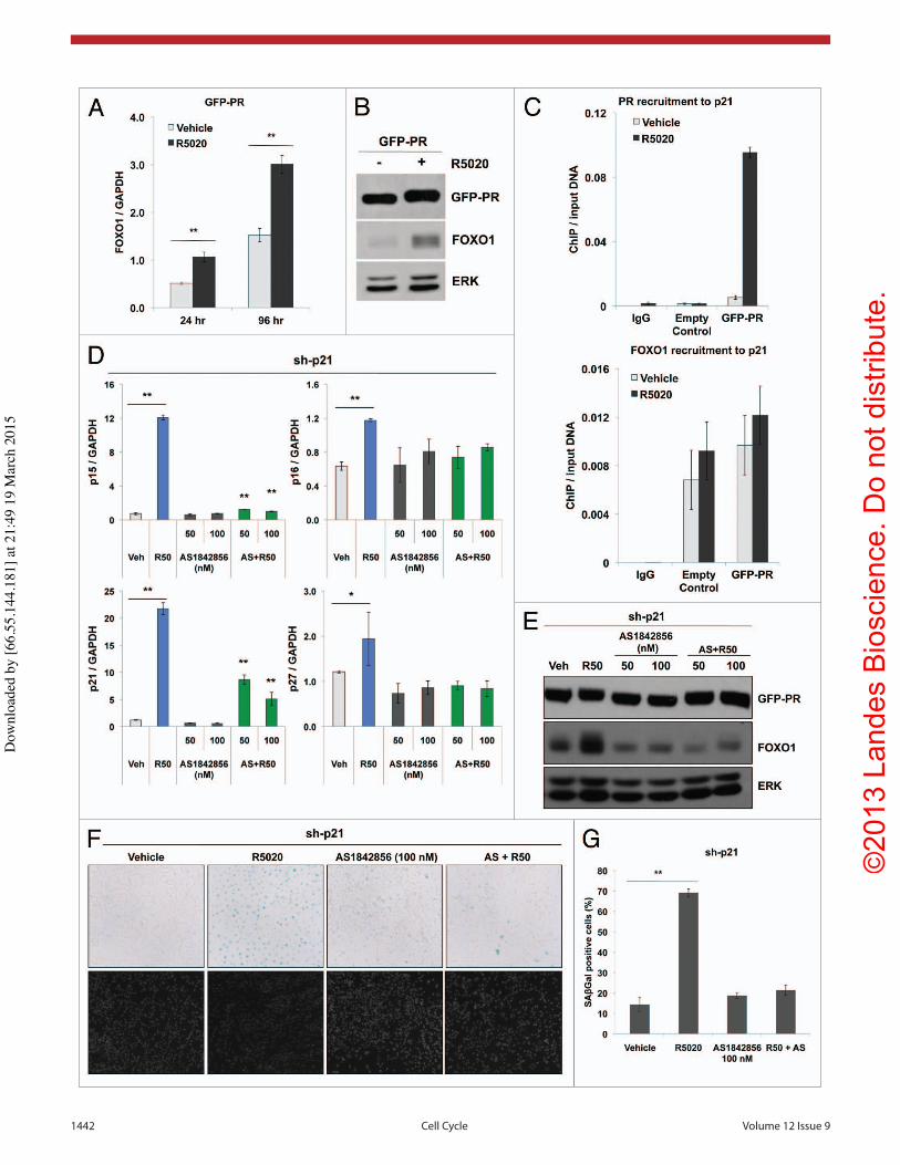
(23-fold), p16 (2-fold) and p27 (2-fold) mRNA levels were signifi-cantly induced in cells expressing sh-p21 relative to vehicle-treated cells or sh-controls. p21 expression was induced upon R5020 treat-ment in cells expressing either sh-control or sh-p21, although total expression was blunted following knockdown. These data suggest a mechanism for progestin-regulated molecular compensation upon knockdown of p21 in cells expressing GFP-PR, and further implicate PR as a driver of cell cycle inhibition and, ultimately, cellular senescence in OC cells.
PR and FOXO1 cooperate to induce p21. Previous studies identified FOXO1 as a PR target gene and master regulator of cell cycle mediators (p15, p16, p21, p27). Notably, FOXO1 and PR interact within transcription complexes in human endome-trial stromal and cancer cells.59,60 Like PR, FOXO1 expression is
drives senescence upon progestin stimulation of unmodified OC cells.
We next sought to determine whether progestin-induced senes-cence was dependent upon p21-induced cell cycle inhibition. We generated GFP-PR cells stably expressing p21-targeted shRNA (sh-p21) or a non-targeting vector control (sh-control); p21 knockdown was confirmed by western blotting (Fig. 5A). We anticipated that p21 loss would block R5020-induced senescence. Surprisingly, we observed a significant increase in SAβGal activity and the devel-opment of a senescent phenotype after only 4 d of R5020 stimula-tion compared with sh-control cells (Fig. 5B and C). To account for the increase in SAβgal activity, we evaluated other senescence-associated cell cycle regulators, p15, p16 and p27 in cells expressing sh-p21 (Fig. 5D). Interestingly, upon R5020 (96 h) treatment, p15
Figure 3. PR expression and activity induces cellular senescence. (A) Representative staining for SAβGal activity of empty control and GFP-PR cells treated with R5020 (10 nM) for 96 h. (magnification = 100×). Cell samples were mounted onto glass slides using ProLong® Gold Antifade Reagent with DAPI (Invitrogen) for brightfield microscopy. (B) Percentage of positive SAβGal cells was determined from quantitating three fields at 100× magnifica-tion. Values were normalized to total nuclei present in each field from DAPI staining (n = 3, **p ≤ 0.01). (C) Exposure of ES-2 cells expressing GFP-PR to R5020 (10 nM) for 12 d induced cellular senescence as indicated by cells (arrowheads) with increased SAβGal activity, while non-senescent cells (asterisk) remain SAβGal-negative. Senescent GFP-PR-containing cells also develop a characteristically larger, more flattened morphology as revealed by TRITC-labeled wheat germ agglutinin (WGA-TRITC) cell membrane staining. Nuclei were identified by DAPI counterstaining. All images were ac-quired at 400× magnification. (D) Cell cycle analysis by propidium iodide staining of GFP-PR-containing cells treated with R5020 (10 nM) for 96 h (n = 3, **p ≤ 0.01). (E) Flow cytometric analysis of DNA and RNA by Hoescht 33342 and Pyronin Y staining, respectively, of GFP-PR-containing cells treated with R5020 (10 nM) (**p ≤ 0.01).
Dow
nloa
ded
by [6
6.55
.144
.181
] at 2
1:49
19
Mar
ch 2
015

©20
13 L
ande
s B
iosc
ienc
e. D
o no
t dis
tribu
te.
www.landesbioscience.com Cell Cycle 1439
downregulated in ovarian cancers.61 FOXO1 mRNA and protein expression was signifi-cantly upregulated following 24 h R5020 treatment (Fig. 6A and B) and remained sus-tained for 96 h. Similar results were observed in progestin-treated PR+ PEO4 cells (data not shown). Chromatin immunoprecipitation (ChIP) assays revealed that upon R5020 treat-ment (1 h) of GFP-PR cells, PR was signifi-cantly recruited (18-fold) to a PRE-containing region downstream of the p21 transcriptional start site identified by PR ChIP-Seq stud-ies conducted in breast cancer models;62 PR recruitment in GFP-control cells was mini-mal and similar to background (IgG control) levels (Fig. 6C). In contrast to the dramatic ligand-dependent recruitment of PR-B, FOXO1 was basally present within the same PRE-containing region in both control and GFP-PR cells; FOXO1 recruitment to this site was not significantly modulated in response to R5020 treatment. Cell fractionation experi-ments indicated that FOXO1 protein was nuclear in both the absence and presence of progestin (data not shown).
Since progestin treatment induces FOXO1 expression in PR-expressing cells, we next evaluated if FOXO1 was required for the induction of p15, p16, p27 and p21 in cells expressing sh-p21 shRNAs. To block the activ-ity of FOXO1, the selective small-molecular inhibitor, AS1842856 (AS), was utilized.63 Upon R5020 (96 h) treatment, p15 (17-fold), p16 (2-fold) and p21 (18-fold) and p27 (2-fold) mRNA levels were again significantly induced in cells expressing sh-p21 relative to vehicle-treated cells (Fig. 6D). Treatment with AS (50 or 100 nM) alone did not affect basal (vehicle) mRNA levels. However, the addi-tion of AS to R5020-treated cells significantly blunted induction of p15, p16, p21 and p27, indicating that FOXO1 activity is required for progestin-induced expression of these cell cycle regulators. Furthermore, AS blocked basal and progestin-induced FOXO1 protein expression in cells expressing sh-p21 (Fig. 6E). Importantly, AS treatment had no effect on GFP-PR-B expression (Fig. 6E). Moreover, progestin-induced senescence, as measured by SAβGal activity, was significantly reduced upon inhibition of FOXO1 (i.e., in the pres-ence of AS) relative to R5020-treated controls (Fig. 6F and G). Together, these data impli-cate FOXO1 as a key mediator of cell cycle gene expression associated with progestin-induced ovarian cancer cell senescence.
Figure 4. Progestins upregulate p21 expression to mediate cellular senescence. (A) RT-qPCR analysis of p21 mRNA levels after 24 and 96 h R5020 (10 nM) treatment in empty control and GFP-PR-containing cells (n = 3, **p ≤ 0.01). (B) Western blot analysis of p21 protein expression in empty control and GFP-PR-containing cells after 8 d of R5020 (10 nM) treatment. (C) ES-2 GFP-PR cells transiently transfected with a p21 promoter-containing luciferase reporter construct were treated for 24 h with R5020 (10 nM) or RU486 (1 uM). Relative luciferase units (RLU) were normalized to the mean result ± standard deviation (SD) for Renilla luciferase expression (n = 4, **p ≤ 0.01). (D) RT-qPCR analysis of p21 mRNA expression after 24 and 96 h R5020 (1 and 10 μM) treatment in PR+ PEO4 cells (n = 3, *p ≤ 0.05, **p ≤ 0.01). (E) Represen-tative staining for SAβGal activity in PEO4 cells treated with R5020 (1 and 10 μM) for 96 h. (magnification = 100×). Cell samples were mounted onto glass slides using ProLong® Gold Antifade Reagent with DAPI (Invitrogen) for brightfield microscopy. (F) Percentage of positive SAβGal cells was determine from quantitating three fields at 100× magnification. Values were normalized to total nuclei present per field from DAPI staining (n = 3, *p ≤ 0.05).
Dow
nloa
ded
by [6
6.55
.144
.181
] at 2
1:49
19
Mar
ch 2
015

©20
13 L
ande
s B
iosc
ienc
e. D
o no
t dis
tribu
te.
1440 Cell Cycle Volume 12 Issue 9
cells stably expressing FOXO1-targeted shRNAs (sh-FOXO1); FOXO1 knockdown was confirmed by western blotting (Fig.7A). Interestingly, PR expression also diminished slightly in cells sta-bly expressing sh-FOXO1; some loss of PR occurred in multiple clones and using distinct FOXO1-targeted shRNAs (not shown), suggesting a mechanism for co-expression of these “coupled” fac-tors. Early passage clones expressing sh-FOXO1 that retained sig-nificant PR expression were used for further study. As expected,
FOXO1 expression is required for PR-induced senescence in OC cells. Our data suggest that in the presence of proges-tin, liganded PR may primarily tether to pre-existing FOXO1 (i.e., located at nearby or distant sites) in order to facilitate hor-mone-regulated expression of p21 and senescence induction. A similar paradigm has been defined for ER tethering to FOXA1 pioneer factors in breast cancer models.64 To test the FOXO1-dependence of PR-induced senescence, we generated GFP-PR
Figure 5. High p21 expression is dispensable for PR-induced cellular senescence. (A) Western blot analysis of p21 expression after lentiviral infection of shRNA oligonucleotides containing a scramble, non-targeting sequence (sh-control) or p21-targeting sequence (sh-p21). Cells were treated with R5020 (10 nM) for 8 d after stable infection of shRNA oligonucleotides. (B) Representative staining for SAβgal activity in cells expressing either sh-control or sh-p21 and treated with R5020 (10 nM) for 96 h (magnification = 100×). Cell samples were mounted onto glass slides using ProLong® Gold Antifade Reagent with DAPI (Invitrogen) for brightfield microscopy. (C) Percentage of positive SAβgal expressing cells was determined from quantitating three fields at 100× magnification. Values were normalized to total nuclei present from DAPI staining (n = 3, *p ≤ 0.05, **p ≤ 0.01). (D) RT-qPCR analysis of p15, p16, p21 and p27 mRNA expression in the sh-p21 knockdown cells treated with R5020 (10 nM) for 96 h (n = 3, *p < 0.05, **p < 0.01).
Dow
nloa
ded
by [6
6.55
.144
.181
] at 2
1:49
19
Mar
ch 2
015

©20
13 L
ande
s B
iosc
ienc
e. D
o no
t dis
tribu
te.
www.landesbioscience.com Cell Cycle 1441
to the presence of two isoforms of the nuclear receptor (PR-A and PR-B).66 Several in vitro studies have demonstrated PR-A to be inhibitory of the transcriptional activity of PR-B, as well as other nuclear receptors, including glucocorticoid, mineralcorticoid, androgen and estrogen receptors.66 PR-B is a more potent tran-scription factor relative to PR-A in gene array studies, but these receptors also regulate overlapping but distinct gene subsets4 that are also tissue-specific. Our studies identify ligand-activated PR-B as a mediator of OC senescence. Notably, progesterone can bind to members of the membrane progesterone receptor fam-ily, mPRα (PAQR7), mPRβ (PAQR8), mPRγ (PAQR5) and progesterone receptor membrane component-1 (PGRMC1) with varying responses that are likely also tissue-specific and context-dependent.68 Namely, PAQR have been shown to weakly acti-vate the JNK1/2 MAPK pathway and induce JNK-dependent BAX mRNA expression in ovarian cancer cells.67 These receptors also rapidly activate p42/p44 MAPKs in breast cancer cells.69 However, the primary signaling mechanisms and associated physiological functions of these receptors remain incompletely understood. PAQR bind progesterone with relatively high affin-ity and do not recognize the synthetic progestin, R5020, or the nuclear PR antagonist, RU486.70 In addition to PR-null cell con-trols, our studies used R5020 (10 nM) in order to avoid activat-ing unrelated but ubiquitous membrane progesterone receptors (i.e., PAQR family members) present in OC cells.67 Interestingly, PR+ PEO4 cells required at least 10 μM progestin for appreciable activation of endogenous PR-B (Fig. 1E inset), as measured by gel mobility up-shift of PR-B and regulation of well-characterized PR target genes (SGK). Subtle up-shift in PR gel mobility is indica-tive of direct global PR phosphorylation, modifications that aug-ment PR nuclear localization and enhance transcriptional activity at selected promoters.65,71 The basis of these concentration effects is unknown; the concentrations used in the PEO4 cell model are similar to physiological levels within the microenvironment of the ovary (≥ 1 μM).45,72 However, ovarian cancer cells may lack the so-called non-genomic or “rapid” signaling events that are integrated with ER-α and PR-B transcriptional activities in response to hormones and are well-characterized in breast cancer models.73,74
Indeed, protein kinases are vital regulatory inputs to steroid hormone receptor action.75 Takahashi and colleagues character-ized cAMP-induced senescence in SKOV ovarian cancer cells overexpressing PR-B and observed induction of both p21 and p27 expression.33 Although these authors did not pursue the mechanisms of these effects, cAMP, via the actions of cAMP-dependent protein kinases and/or activation of Ca2+ channels,76 may induce changes in PR-B phosphorylation that alter ligand-binding and/or co-factor interactions.77 Interestingly, cAMP has been demonstrated to regulate the cell cycle in cancer cells78 as well as induce and/or activate a variety of transcription factors, such as STAT5, C/EBPβ and FOXO1, all proteins that directly interact with PR.79-81 As such, cAMP may lower the requirement for PR ligand-binding and/or tethering to FOXO1 at PR target genes. We detected little to no change in cAMP accumulation following a time course of progesterone or R5020-treatment of OC models.67
p21 mRNA levels were greatly diminished in cells expressing sh-FOXO1 relative to sh-controls in both vehicle- and R5020-treated conditions (Fig. 7B). Similarly, R5020 treatment did not induce p15, p16 or p27 mRNA expression in cells express-ing sh-FOXO1 (Fig. S4). In addition, PR recruitment to the PRE-containing region located downstream of the p21 transcrip-tional start site was also similarly reduced in cells expressing sh-FOXO1 relative to controls, as measured by ChIP assays (7.7-fold vs. 12.1 fold) (Fig. 7D). To confirm that diminished transcrip-tional regulation of p21 was not entirely due to reduced levels of PR protein observed in cells expressing sh-FOXO1 (Fig. 7A), we performed RT-qPCR analysis of additional PR-target genes; R5020 treatment (24 h) of ES-2 GFP-PR cells stably express-ing sh-FOXO1 resulted in significant upregulation of a num-ber of PR-target genes, including HSD11B2, KLF4 and NET1 (Fig. S3). Consistent with loss of progestin-induced p21 expres-sion, R5020 treatment failed to induce G0/G1 cell cycle arrest up to 96 h in cells expressing sh-FOXO1 cells relative to sh-con-trols (Fig. 7C). Interestingly, the percentage of cells in S phase was enhanced upon knockdown of FOXO1 in both vehicle and R5020-treated PR-B+ cells when compared with sh-controls. Consistent with these results, progestin-induced SAβGal activity was also significantly reduced when FOXO1 was stably down-regulated relative to sh-controls (Fig. 7E and F).
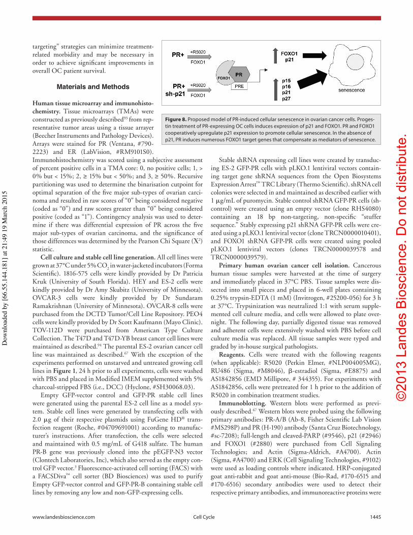
Overall, these data are consistent with a model in which PR-expressing OC cells induce FOXO1-dependent p21 expres-sion in the presence of progestins. FOXO1 is a required factor for PR-induced p21 expression and cellular senescence (Fig. 8).
Discussion
Progesterone is a potent breast mitogen, but functions to inhibit proliferation in the uterus. Indeed, PR action is highly context-dependent and heavily influenced by post-translational modifica-tions65 as well as cofactor availability.66 The detailed molecular mechanisms of progesterone’s protective role in ovarian cancer are not well understood; both proliferative and inhibitory actions of progesterone have been reported,29,43-45 including concentra-tion-dependent biphasic effects within the same model system.28 This is perhaps due in part to the complexity of progesterone action. Numerous progesterone receptors exist (PR-A, PR-B, PR-C, mPRα, mPRβ, mPRγ and PGRMC1), and their activi-ties and potential interactions are still poorly defined.67 In this study, we demonstrated that nuclear PR-B receptors exhibit ligand-dependent anti-proliferative effects by inducing cellular senescence in OC models.
Several independent in vitro studies have demonstrated the inhibitory action of progesterone on OC cell growth, primarily through the induction of apoptosis,28,30,42-45 while other studies report progesterone’s proliferative actions. The opposing cellular responses of progesterone’s effects in normal and malignant ovar-ian surface epithelial cells may be attributed to dosage effects: mitogenic effects were observed at low progesterone concentra-tions (< 10−8 M), while growth inhibition and apoptosis were associated with high progesterone concentrations (≥ 10−6 M).8 Alternatively, opposing effects of progesterone may be attributed
Dow
nloa
ded
by [6
6.55
.144
.181
] at 2
1:49
19
Mar
ch 2
015
©20
13 L
ande
s B
iosc
ienc
e. D
o no
t dis
tribu
te.
1442 Cell Cycle Volume 12 Issue 9
Dow
nloa
ded
by [6
6.55
.144
.181
] at 2
1:49
19
Mar
ch 2
015

©20
13 L
ande
s B
iosc
ienc
e. D
o no
t dis
tribu
te.
www.landesbioscience.com Cell Cycle 1443
progestin-stimulated proliferation and prevented cell cycle arrest. In contrast to studies conducted in 2D culture systems, progestin is clearly mitogenic and a mediator of pro-survival and prolifera-tion in breast cancer cells (T47D, MCF-7) cultured in anchorage-independent (soft agar) conditions. The paradoxical effects of progestins observed in breast relative to ovarian cancer cells may be attributed to differential cross-talk between PR and growth factor-mediated signaling pathways, differential regulation of PR itself via post-translational modifications and/or differen-tial recruitment of required co-factors, such as FOXO1. Related to these studies, forkhead family transcription factors such as FOXO1 and FOXA1 are negatively regulated by phosphoryla-tion events. AKT-dependent phosphorylation prevents their nuclear accumulation and thus impairs target gene regulation.82 As mutations of PI3Ks or PTEN are common events (particu-larly in breast and ovarian cancers), activated AKT may prevent PR-induced senescence signaling by nuclear exclusion of FOXO1 partners or pioneer factors.
To date, the use of progestins, megestrol acetate and medroxy-progesterone acetate, as OC therapies have been evaluated in a total of 14 relatively small phase II clinical trials with variable inclusion criteria and modest response rates.88 Data from these trials support the concept that endometrioid and serous ovarian cancers are frequently sensitive to hormones and thus more likely to respond to endocrine therapy.88 To optimize response rates, the identification of PR-B as a biomarker within ovarian tumors may be advantageous prior to therapy. Genetic loss of heterozygosity of the PR gene (ch. 11q23.3–24.3) occurs in approximately 75% of ovarian tumors.89,90 Alternatively, when the PR gene locus is intact, it may be possible to restore PR expression in PR-low or -null OC. For example, robust isoform-specific re-expression of PR has been demonstrated using activating duplex RNAs that target promoter regions in DNA; these reagents are currently in development for therapeutic use.91
In sum, our studies suggest that activation of nuclear PR-B may provide a means to force ovarian cancer cells out of the cell cycle and into a form of irreversible “stasis” by inducing cellular senes-cence. A clear understanding of the mechanisms and mediators of cellular senescence is highly relevant to modern cancer therapy, as the specific targeting of the senescence pathway in tumor cells is predicted to impede tumor progression to advanced and meta-static disease.92 Senescent cells cannot further divide, but depend upon selected signal transduction pathways for prolonged sur-vival, and thus may be more vulnerable to subsequent therapies
Our study demonstrates a detailed molecular mechanism whereby progestins activate nuclear PR-B to upregulate FOXO1 expression, leading to robust induction of cell cycle mediators of senescence. In this case, p21 expression required both liganded PR-B and expression of FOXO1. Notably, progestin treatment of OC cells expressing sh-p21 promoted a significantly increased senescence response relative to sh-controls. Interestingly, in the context of diminished p21, hormone-bound PR significantly induced other well-characterized pro-senescence effectors that are also FOXO1 target genes, such as p15, p16 and p27. These PR signaling alternatives reveal FOXO1-dependent tumor-suppres-sive or “fail-safe” mechanisms, which may ensure that senescence occurs in the face of impaired p21. Deregulation of FOXO1 is associated with tumorigenesis and cancer progression. FOXO1 is downregulated in several carcinomas, including ovarian,61 through alterations in upstream regulators (i.e., inactivating mutations to PTEN, active PI3K-AKT signaling), post-trans-lational deregulation or by genetic mutations.82 The targeted re-expression and activation of FOXO1 using chemical and/or biological therapeutic strategies may overcome resistance or sen-sitize cancer cells to current therapeutics. Ultimately, our studies showed that OC senescence is dependent and specific to both PR-B and FOXO1; the ablation of FOXO1 (a PR target gene) significantly diminished the emergence of progestin-induced p21 expression and cellular senescence. Linkage of PR and FOXO1 signaling has been reported in the uterus.83 Notably, these mol-ecules trend toward co-occurrence in 570 samples analyzed from The Cancer Genome Atlas (TCGA) ovarian serous cystadeno-carcinoma provisional data set [p = 0.209, Fisher’s exact test, odds ratio (OR) = 2.14, 95% confidence interval: 0.59–7.73],84 suggestive that numerous PR-target genes may be sensitive to FOXO1 expression.
In contrast to our finding that PR-B is a mediator of OC senes-cence, the same receptor, when expressed in breast cancer cells, is clearly mitogenic.2 The opposing biology in ovarian vs. breast cancer cells may be largely dependent on cell context. Studies in T47D breast cancer cells growing in 2D culture conditions demonstrated biphasic effects of progestin on cell cycle progres-sion.85-87 Early effects of progestin treatment are proliferative, as T47D-YB cells accelerate through one or more mitotic cycles; cells are growth inhibited at late time points coincident with induction of p21 and p27. A secondary progestin treatment failed to restore cell cycle progression, although PR levels remained high. Interestingly, epidermal growth factor (EGF) restored
Figure 6. (See opposite page) Progestin treatment of GFP-PR-containing cells stimulates FOXO1 expression and promotes PR recruitment to the p21 promoter. (A) RT-qPCR analysis of FOXO1 mRNA expression after 24 and 96 h R5020 (10 nM) treatment of GFP-PR-containing cells (n = 3, **p ≤ 0.01). (B) Western blot analysis of FOXO1 expression in response to 96 h of R5020 (10 nM) treatment in GFP-PR-containing cells. (C) RT-qPCR analysis of PR and FOXO1 recruitment to p21. Empty control and GFP-PR expressing cells were stimulated with vehicle or R5020 (10 nM) for 1 h. Fixed lysates were chromatin immunoprecipated with antibodies to PR or FOXO1 and qRT-PCR was performed on isolated DNA. (D) RT-qPCR analysis of p15, p16, p21 and p27 mRNA expression in sh-p21 knockdown cells treated with R5020 (10 nM), FOXO1 inhibitor, AS1842856 (AS) (50 and 100 nM), or the combination of AS1842856 and R5020 for 96 h (n = 2, *p < 0.05, **p < 0.01). (E) Western blot analysis of FOXO1 expression in cells expressing sh-p21 after treatment with vehicle, R5020 (10 nM), AS1842856 (50 and 100 nM), or the combination of AS1842856 and R5020 for 96 h. (F) Representative staining for SAβGal activity of sh-p21-knockdown cells treated with R5020 (10 nM), AS1842856 (AS, 100 nM), or the combination of AS1842856 and R5020 for 96 h. (magni-fication = 100×). Cell samples were mounted onto glass slides using ProLong® Gold Antifade Reagent with DAPI (Invitrogen) for brightfield microscopy. (G) Percentage of positive SAβGal cells was determined from quantitating three fields at 100× magnification. Values were normalized to total nuclei present in each field from DAPI staining (n = 2, **p ≤ 0.01).
Dow
nloa
ded
by [6
6.55
.144
.181
] at 2
1:49
19
Mar
ch 2
015

©20
13 L
ande
s B
iosc
ienc
e. D
o no
t dis
tribu
te.
1444 Cell Cycle Volume 12 Issue 9
cells. In particular, translation of these findings may open the way for combination therapies that couple PR-dependent senes-cence induction with targeted therapies aimed at blocking cell survival pathways, including AKT signaling. Such “rational
that target these survival pathways (i.e., senescence creates a form of synthetic lethality). Thus, the induction of PR-mediated cel-lular senescence via progestin therapy may provide a safe and use-ful strategy to limit uncontrolled proliferation of ovarian cancer
Figure 7. PR-induced cellular senescence is dependent on FOXO1 expression. (A) Western blot showing FOXO1 expression in cells expressing either sh-control or sh-FOXO1 after treatment of R5020 (10 nM) for 96 h. (B) RT-qPCR analysis of p21 mRNA expression in sh-control and sh-FOXO1-containing cells after 96 h of R5020 (10 nM) treatment (n = 3, **p ≤ 0.01). (C) Cell cycle analysis by propidium iodide staining of cells expressing either sh-control or sh-FOXO1 and stimulated with R5020 for 96 h. (n = 3, **p ≤ 0.01). (D) RT-qPCR analysis of PR recruitement to p21. Sh-control and sh-FOXO1-containing cells were stimulated with vehicle or R5020 (10 nM) for 1 h. Fixed lysates were chromatin immunoprecipated with an antibody to PR and qRT-PCR was performed on isolated DNA. (E) Representative staining of SAβGal activity of sh-control and sh-FOXO1-containing cells after treatment of R5020 for 96 h. Cell samples were mounted onto glass slides using ProLong® Gold Antifade Reagent with DAPI (Invitrogen) for brightfield microscopy. (F) Per-centage of positive SAβGal cells were determined from quantitating three fields at 100× magnification. Values were normalized to total nuclei present from DAPI staining (n = 3, **p ≤ 0.01).
Dow
nloa
ded
by [6
6.55
.144
.181
] at 2
1:49
19
Mar
ch 2
015
©20
13 L
ande
s B
iosc
ienc
e. D
o no
t dis
tribu
te.
www.landesbioscience.com Cell Cycle 1445
Stable shRNA expressing cell lines were created by transduc-ing ES-2 GFP-PR cells with pLKO.1 lentiviral vectors contain-ing target gene shRNA sequences from the Open Biosystems Expression Arrest™ TRC Library (Thermo Scientific). shRNA cell colonies were selected in and maintained as described earlier with 1 μg/mL of puromycin. Stable control shRNA GFP-PR cells (sh-control) were created using an empty vector (clone RHS4080) containing an 18 bp non-targeting, non-specific “stuffer sequence.” Stably expressing p21 shRNA GFP-PR cells were cre-ated using a pLKO.1 lentiviral vector (clone TRCN0000010401), and FOXO1 shRNA GFP-PR cells were created using pooled pLKO.1 lentiviral vectors (clones TRCN0000039578 and TRCN0000039579).
Primary human ovarian cancer cell isolation. Cancerous human tissue samples were harvested at the time of surgery and immediately placed in 37°C PBS. Tissue samples were dis-sected into small pieces and placed in 6-well plates containing 0.25% trypsin-EDTA (1 mM) (Invitrogen, #25200-056) for 3 h at 37°C. Trypsinization was neutralized 1:1 with serum supple-mented cell culture media, and cells were allowed to plate over-night. The following day, partially digested tissue was removed and adherent cells were extensively washed with PBS before cell culture media was replaced. All tissue samples were typed and graded by in-house surgical pathologists.
Reagents. Cells were treated with the following reagents (when applicable): R5020 (Perkin Elmer, #NLP004005MG), RU486 (Sigma, #M8046), β-estradiol (Sigma, #E8875) and AS1842856 (EMD Millipore, # 344355). For experiments with AS1842856, cells were pretreated for 1 h prior to the addition of R5020 in combination treatment studies.
Immunoblotting. Western blots were performed as previ-ously described.67 Western blots were probed using the following primary antibodies: PR-A/B (Ab-8, Fisher Scientific Lab Vision #MS298P) and PR (H-190) antibody (Santa Cruz Biotechnology, #sc-7208); full-length and cleaved-PARP (#9546), p21 (#2946) and FOXO1 (#2880) were purchased from Cell Signaling Technologies; and Actin (Sigma-Aldrich, #A4700). Actin (Sigma, #A4700) and ERK (Cell Signaling Technologies, #9102) were used as loading controls where indicated. HRP-conjugated goat anti-rabbit and goat anti-mouse (Bio-Rad, #170-6515 and #170-6516) secondary antibodies were used to detect their respective primary antibodies, and immunoreactive proteins were
targeting” strategies can minimize treatment-related morbidity and may be necessary in order to achieve significant improvements in overall OC patient survival.
Materials and Methods
Human tissue microarray and immunohisto-chemistry. Tissue microarrays (TMAs) were constructed as previously described93 from rep-resentative tumor areas using a tissue arrayer (Beecher Instruments and Pathology Devices). Arrays were stained for PR (Ventana, #790-2223) and ER (LabVision, #RM9101S0). Immunohistochemistry was scored using a subjective assessment of percent positive cells in a TMA core: 0, no positive cells; 1, > 0% but < 15%; 2, ≥ 15% but < 50%; and 3, ≥ 50%. Recursive partitioning was used to determine the binarisation cutpoint for optimal separation of the five major sub-types of ovarian carci-noma and resulted in raw scores of “0” being considered negative (coded as “0”) and raw scores greater than “0” being considered positive (coded as “1”). Contingency analysis was used to deter-mine if there was differential expression of PR across the five major sub-types of ovarian carcinoma, and the significance of those differences was determined by the Pearson Chi Square (Χ2) statistic.
Cell culture and stable cell line generation. All cell lines were grown at 37°C under 5% CO2 in water-jacketed incubators (Forma Scientific). 1816-575 cells were kindly provided by Dr Patricia Kruk (University of South Florida). HEY and ES-2 cells were kindly provided by Dr Amy Skubitz (University of Minnesota). OVCAR-3 cells were kindly provided by Dr Sundaram Ramakrishnan (University of Minnesota). OVCAR-8 cells were purchased from the DCTD Tumor/Cell Line Repository. PEO4 cells were kindly provided by Dr Scott Kaufmann (Mayo Clinic). TOV-112D were purchased from American Type Culture Collection. The T47D and T47D-YB breast cancer cell lines were maintained as described.94 The parental ES-2 ovarian cancer cell line was maintained as described.67 With the exception of the experiments performed on unstarved and untreated growing cell lines in Figure 1, 24 h prior to all experiments, cells were washed with PBS and placed in Modified IMEM supplemented with 5% charcoal-stripped FBS (i.e., DCC) (Hyclone, #SH30068.03).
Empty GFP-vector control and GFP-PR stable cell lines were generated using the parental ES-2 cell line as a model sys-tem. Stable cell lines were generated by transfecting cells with 2.0 μg of their respective plasmids using FuGene HD® trans-fection reagent (Roche, #04709691001) according to manufac-turer’s instructions. After transfection, the cells were selected and maintained with 0.5 mg/mL of G418 sulfate. The human PR-B gene was previously cloned into the pEGFP-N3 vector (Clontech Laboratories, Inc), which also served as the empty con-trol GFP vector.3 Fluorescence-activated cell sorting (FACS) with a FACSDiva™ cell sorter (BD Biosciences) was used to purify Empty GFP-vector control and GFP-PR-B containing stable cell lines by removing any low and non-GFP-expressing cells.
Figure 8. Proposed model of PR-induced cellular senescence in ovarian cancer cells. Proges-tin treatment of PR-expressing OC cells induces expression of p21 and FOXO1. PR and FOXO1 cooperatively upregulate p21 expression to promote cellular senescence. In the absence of p21, PR induces numerous FOXO1 target genes that compensate as mediators of senescence.
Dow
nloa
ded
by [6
6.55
.144
.181
] at 2
1:49
19
Mar
ch 2
015
©20
13 L
ande
s B
iosc
ienc
e. D
o no
t dis
tribu
te.
1446 Cell Cycle Volume 12 Issue 9
Propidium iodide staining was detected using a FACSCalibur (BD Biosciences). Cells were gated for cell cycle phases using FlowJo software (Tree Star Inc.). Cells were stained with 2 μg/mL of Hoechst 33342 DNA-specific dye (Invitrogen, #H3570) and 0.5 μg/mL of Pyronin Y RNA-specific dye (Polysciences, #18614-5). FACS analysis performed on a BDTM LSR II Flow Cytometer System (BD Biosciences) was used for cell cycle analysis and separation of the G0 (Hoescht-low and Pyronin Y-low) and G1 (Hoescht-low and Pyronin Y-high) phases of the cell cycle as previously described.52
Chromatin immunoprecipitation (ChIP). Cells were treated for 1 h with either R5020 or ethanol vehicle in 5% DCC, and cell samples were fixed, harvested and lysed according to opti-mized manufacturer’s instructions using the ChIP-IT™ Express Magnetic Chromatin Immunoprecipitation Kit (Active Motif, #53008). Samples were homogenized using a Bioruptor sonica-tor (Diagenode, Inc.). ChIP reactions were incubated overnight on an end-to-end rotator using 95 μL of isolated chromatin and either 2 μg of PR-A/B antibody (Ab-8), FKHR (H-128) anti-body (Santa Cruz Biotechnology, Inc, # sc-11350) or 0.4 μg of normal mouse or rabbit IgG (Santa Cruz Biotechnology, Inc., #sc-2025 # sc-2027). Samples were washed, eluted, reverse cross-linked and treated with Proteinase K according to manufacturer’s instructions (Active Motif). DNA was analyzed by RT-qPCR as described above.
Statistical analysis. All reported values represent the mean ± the standard deviation (SD). Statistical analyses were performed using a Student’s two-tailed t-test, where significance was deter-mined with 95% confidence (*p ≤ 0.05, **p ≤ 0.01).
Disclosure of Potential Conflicts of Interest
No potential conflicts of interest were disclosed.
Acknowledgments
We would like to thank Dr Patricia Kruk (University of South Florida), Dr Amy Skubitz (University of Minnesota), Dr Sundaram Ramakrishnan (University of Minnesota) and Dr Scott Kaufmann (Mayo Clinic) for kindly providing cell lines utilized in this study. We would like to thank members of the University of Minnesota Masonic Cancer Center’s Flow Cytometry Core Facility and the University Imaging Centers for their assistance in data acquisition. This study was supported by NIH grant R01 CA159712 (to C.A.L.), the Minnesota Ovarian Cancer Alliance (to C.A.L.), Cancer Biology Training Grant NIH T32 CA009138 (to C.H.D.) and National Center for Advancing Translational Sciences of the National Institutes of Health Award UL1TR000114 (to C.H.D).
Supplemental Materials
Supplemental materials may be found here: www.landesbioscience.com/journals/cc/article/24550
visualized on Kodak X-OMAT LS film (Carestream Health, #864-6770) following ECL detection with Super Signal® West Pico Maximum Sensitivity Substrate (Pierce, #34087).
Real-time quantitative-PCR (RT-qPCR). Total RNA was extracted from cell samples using TriPure Isolation Reagent (Roche, #11667165001) and isopropanol precipitation. RNA (1.0 μg) was reverse transcribed to cDNA according to manu-facturer’s instructions using the qScript cDNA SuperMix (Quanta Biosciences, #95048-100). qPCR was performed using Light Cycler® FastStart DNA Master SYBR Green I (Roche, #12239264001) on a Light Cycler® 480 II Real-Time PCR System (Roche). Human primer sequences are listed in Table S1. qPCR cycling conditions were as follows: initial denaturation at 95°C for 10 min; denature at 95°C for 10 sec, anneal at 60°C for 10 sec and extension at 72°C for 5 sec for 45 cycles.
Luciferase assays. Cells were co-transfected overnight using FuGene HD® transfection reagent (Roche) according to manu-facturer’s instructions with 0.9 μg of either a PRE-containing4 or a p21 promoter-containing5 firefly luciferase reporter construct and 0.1 μg of a constitutively active pRL-TK-Renilla luciferase construct (Promega, #E2241). Luciferase assays were performed as previously described46 using the dual luciferase reporter assay (Promega, #E1910).
Histology and microscopy. All brightfield and fluorescent cell images described herein were acquired with an Axioplan 2 upright microscope (Zeiss) and captured using a SPOT camera (Diagnostic Instruments, Inc.) with the ProgRes Capture Pro software (Version 2.8.8) (Jenoptik Optical Systems Inc.). Cellular membranes were stained for 15 min at RT with Texas Red®-X-conjugated wheat germ agglutinin (1 μg/mL) (Invitrogen, #W21405), and all cell nuclei were stained with DAPI containing ProLong® Gold anti-fade reagent (Invitrogen, #P-36931). Fixed and live cell soft-agar images were acquired with a Leica DM IL inverted microscope (Leica Microsystems, Inc.) and captured using a MagnaFire® cam-era and MagnaFire® imaging software (Olympus).
Cell proliferation and survival assays. Cell proliferation was measured by using MTT assays as described previously.95
Anchorage-independent growth and survival were measured by using soft agar assays as previously described,47 with the excep-tion of seeding 2 × 10⁴ cells. After 4 wk of growth at 37°C, cell colonies were stained with 0.1% crystal violet for 1 h at RT and washed with PBS. 1,000 randomly chosen cell colonies per well were separated according to size (total pixels/colony area) and quantified using Photoshop® version 7.0 (Adobe Systems, Inc.).
Senescence associated-β-galactosidase (SAβGal) activity assays. Cells were continuously treated for 4 d in 5% DCC. Cells were washed, fixed and stained for SAβGal activity according to manufacturer’s instructions using the Senescence β-Galactosidase Staining Kit (Cell Signaling Technology, #9860).
Flow cytometry. Cells were treated in 5% DCC for 96 h. Cells were collected, fixed and stained as described previously.94
Dow
nloa
ded
by [6
6.55
.144
.181
] at 2
1:49
19
Mar
ch 2
015
©20
13 L
ande
s B
iosc
ienc
e. D
o no
t dis
tribu
te.
www.landesbioscience.com Cell Cycle 1447
28. Syed V, Ulinski G, Mok SC, Yiu GK, Ho SM. Expression of gonadotropin receptor and growth responses to key reproductive hormones in normal and malignant human ovarian surface epithelial cells. Cancer Res 2001; 61:6768-76; PMID:11559549
29. Syed V, Ho SM. Progesterone-induced apoptosis in immortalized normal and malignant human ovarian surface epithelial cells involves enhanced expression of FasL. Oncogene 2003; 22:6883-90; PMID:14534535; http://dx.doi.org/10.1038/sj.onc.1206828
30. Fauvet R, Dufournet Etienne C, Poncelet C, Bringuier AF, Feldmann G, Daraï E. Effects of progesterone and anti-progestin (mifepristone) treatment on pro-liferation and apoptosis of the human ovarian cancer cell line, OVCAR-3. Oncol Rep 2006; 15:743-8; PMID:16525653
31. Zhou H, Luo MP, Schönthal AH, Pike MC, Stallcup MR, Blumenthal M, et al. Effect of reproductive hormones on ovarian epithelial tumors: I. Effect on cell cycle activity. Cancer Biol Ther 2002; 1:300-6; PMID:12432283
32. Horiuchi S, Kato K, Suga S, Takahashi A, Ueoka Y, Arima T, et al. Expression of progesterone receptor B is associated with G0/G1 arrest of the cell cycle and growth inhibition in NIH3T3 cells. Exp Cell Res 2005; 305:233-43; PMID:15817149; http://dx.doi.org/10.1016/j.yexcr.2005.01.003
33. Takahashi A, Kato K, Kuboyama A, Inoue T, Tanaka Y, Kuhara A, et al. Induction of senescence by proges-terone receptor-B activation in response to cAMP in ovarian cancer cells. Gynecol Oncol 2009; 113:270-6; PMID:19211137; http://dx.doi.org/10.1016/j.ygyno.2008.12.032
34. Lenhard M, Tereza L, Heublein S, Ditsch N, Himsl I, Mayr D, et al. Steroid hormone receptor expres-sion in ovarian cancer: progesterone receptor B as prognostic marker for patient survival. BMC Cancer 2012; 12:553; PMID:23176303; http://dx.doi.org/10.1186/1471-2407-12-553
35. Fujimura M, Hidaka T, Kataoka K, Yamakawa Y, Akada S, Teranishi A, et al. Absence of estrogen receptor-alpha expression in human ovarian clear cell adenocarcinoma compared with ovarian serous, endo-metrioid, and mucinous adenocarcinoma. Am J Surg Pathol 2001; 25:667-72; PMID:11342781; http://dx.doi.org/10.1097/00000478-200105000-00016
36. Itani OA, Liu KZ, Cornish KL, Campbell JR, Thomas CP. Glucocorticoids stimulate human sgk1 gene expres-sion by activation of a GRE in its 5'-flanking region. Am J Physiol Endocrinol Metab 2002; 283:E971-9; PMID:12376324
37. Daniel AR, Gaviglio AL, Czaplicki LM, Hillard CJ, Housa D, Lange CA. The progesterone receptor hinge region regulates the kinetics of transcriptional responses through acetylation, phosphorylation, and nuclear retention. Mol Endocrinol 2010; 24:2126-38; PMID:20861224; http://dx.doi.org/10.1210/me.2010-0170
38. Lau DH, Lewis AD, Ehsan MN, Sikic BI. Multifactorial mechanisms associated with broad cross-resistance of ovarian carcinoma cells selected by cyanomor-pholino doxorubicin. Cancer Res 1991; 51:5181-7; PMID:1717140
39. Stany MP, Vathipadiekal V, Ozbun L, Stone RL, Mok SC, Xue H, et al. Identification of novel therapeutic targets in microdissected clear cell ovarian cancers. PLoS ONE 2011; 6:e21121; PMID:21754983; http://dx.doi.org/10.1371/journal.pone.0021121
40. Sartorius CA, Groshong SD, Miller LA, Powell RL, Tung L, Takimoto GS, et al. New T47D breast cancer cell lines for the independent study of progester-one B- and A-receptors: only antiprogestin-occupied B-receptors are switched to transcriptional agonists by cAMP. Cancer Res 1994; 54:3868-77; PMID:8033109
41. Qiu M, Olsen A, Faivre E, Horwitz KB, Lange CA. Mitogen-activated protein kinase regulates nuclear association of human progesterone receptors. Mol Endocrinol 2003; 17:628-42; PMID:12554776; http://dx.doi.org/10.1210/me.2002-0378
15. Akahira J, Inoue T, Suzuki T, Ito K, Konno R, Sato S, et al. Progesterone receptor isoforms A and B in human epithelial ovarian carcinoma: immunohistochemical and RT-PCR studies. Br J Cancer 2000; 83:1488-94; PMID:11076658; http://dx.doi.org/10.1054/bjoc.2000.1463
16. Lee P, Rosen DG, Zhu C, Silva EG, Liu J. Expression of progesterone receptor is a favorable prognostic mark-er in ovarian cancer. Gynecol Oncol 2005; 96:671-7; PMID:15721410; http://dx.doi.org/10.1016/j.ygyno.2004.11.010
17. Høgdall EV, Christensen L, Høgdall CK, Blaakaer J, Gayther S, Jacobs IJ, et al. Prognostic value of estrogen receptor and progesterone receptor tumor expression in Danish ovarian cancer patients: from the ‘MALOVA’ ovarian cancer study. Oncol Rep 2007; 18:1051-9; PMID:17914554
18. Tangjitgamol S, Manusirivithaya S, Khunnarong J, Jesadapatarakul S, Tanwanich S. Expressions of estro-gen and progesterone receptors in epithelial ovarian cancer: a clinicopathologic study. Int J Gynecol Cancer 2009; 19:620-7; PMID:19509560; http://dx.doi.org/10.1111/IGC.0b013e3181a44b62
19. Yang XY, Xi MR, Yang KX, Yu H. Prognostic value of estrogen receptor and progesterone receptor status in young Chinese ovarian carcinoma patients. Gynecol Oncol 2009; 113:99-104; PMID:19178934; http://dx.doi.org/10.1016/j.ygyno.2008.12.018
20. Dressing GE, Lange CA. Integrated actions of pro-gesterone receptor and cell cycle machinery regulate breast cancer cell proliferation. Steroids 2009; 74:573-6; PMID:19118566; http://dx.doi.org/10.1016/j.ste-roids.2008.12.001
21. Migliaccio A, Castoria G, Di Domenico M, Ballaré C, Beato M, Auricchio F. The progesterone recep-tor/estradiol receptor association and the progestin-triggered S-phase entry. Ernst Schering Res Found Workshop 2005:39-54; PMID:15704467; http://dx.doi.org/10.1007/3-540-27147-3_3
22. Migliaccio A, Castoria G, Di Domenico M, de Falco A, Bilancio A, Lombardi M, et al. Steroid-induced androgen receptor-oestradiol receptor beta-Src complex triggers prostate cancer cell proliferation. EMBO J 2000; 19:5406-17; PMID:11032808; http://dx.doi.org/10.1093/emboj/19.20.5406
23. Skildum A, Faivre E, Lange CA. Progesterone recep-tors induce cell cycle progression via activation of mitogen-activated protein kinases. Mol Endocrinol 2005; 19:327-39; PMID:15486045; http://dx.doi.org/10.1210/me.2004-0306
24. Jacobsen BM, Schittone SA, Richer JK, Horwitz KB. Progesterone-independent effects of human proges-terone receptors (PRs) in estrogen receptor-positive breast cancer: PR isoform-specific gene regulation and tumor biology. Mol Endocrinol 2005; 19:574-87; PMID:15563544; http://dx.doi.org/10.1210/me.2004-0287
25. McGowan EM, Russell AJ, Boonyaratanakornkit V, Saunders DN, Lehrbach GM, Sergio CM, et al. Progestins reinitiate cell cycle progression in anti-estrogen-arrested breast cancer cells through the B-isoform of progesterone receptor. Cancer Res 2007; 67:8942-51; PMID:17875737; http://dx.doi.org/10.1158/0008-5472.CAN-07-1255
26. Rodriguez GC, Walmer DK, Cline M, Krigman H, Lessey BA, Whitaker RS, et al. Effect of progestin on the ovarian epithelium of macaques: cancer preven-tion through apoptosis? J Soc Gynecol Investig 1998; 5:271-6; PMID:9773403; http://dx.doi.org/10.1016/S1071-5576(98)00017-3
27. Rodriguez GC, Nagarsheth NP, Lee KL, Bentley RC, Walmer DK, Cline M, et al. Progestin-induced apop-tosis in the Macaque ovarian epithelium: differential regulation of transforming growth factor-beta. J Natl Cancer Inst 2002; 94:50-60; PMID:11773282; http://dx.doi.org/10.1093/jnci/94.1.50
References1. Cancer Facts & Figures. Atlanta: American Cancer
Society 20122. Lange CA, Yee D. Progesterone and breast can-
cer. Womens Health (Lond Engl) 2008; 4:151-62; PMID:19072517; http://dx.doi.org/10.2217/17455057.4.2.151
3. Jacobsen BM, Richer JK, Schittone SA, Horwitz KB. New human breast cancer cells to study progesterone receptor isoform ratio effects and ligand-independent gene regulation. J Biol Chem 2002; 277:27793-800; PMID:12021276; http://dx.doi.org/10.1074/jbc.M202584200
4. Richer JK, Jacobsen BM, Manning NG, Abel MG, Wolf DM, Horwitz KB. Differential gene regulation by the two progesterone receptor isoforms in human breast cancer cells. J Biol Chem 2002; 277:5209-18; PMID:11717311; http://dx.doi.org/10.1074/jbc.M110090200
5. Mulac-Jericevic B, Lydon JP, DeMayo FJ, Conneely OM. Defective mammary gland morphogenesis in mice lacking the progesterone receptor B isoform. Proc Natl Acad Sci USA 2003; 100:9744-9; PMID:12897242; http://dx.doi.org/10.1073/pnas.1732707100
6. Mulac-Jericevic B, Mullinax RA, DeMayo FJ, Lydon JP, Conneely OM. Subgroup of reproductive functions of progesterone mediated by progesterone receptor-B isoform. Science 2000; 289:1751-4; PMID:10976068; http://dx.doi.org/10.1126/science.289.5485.1751
7. Wei LL, Norris BM, Baker CJ. An N-terminally trun-cated third progesterone receptor protein, PR(C), forms heterodimers with PR(B) but interferes in PR(B)-DNA binding. J Steroid Biochem Mol Biol 1997; 62:287-97; PMID:9408082; http://dx.doi.org/10.1016/S0960-0760(97)00044-7
8. Ho SM. Estrogen, progesterone and epithelial ovar-ian cancer. Reprod Biol Endocrinol 2003; 1:73; PMID:14577831; http://dx.doi.org/10.1186/1477-7827-1-73
9. Zheng H, Kavanagh JJ, Hu W, Liao Q, Fu S. Hormonal therapy in ovarian cancer. Int J Gynecol Cancer 2007; 17:325-38; PMID:17362310; http://dx.doi.org/10.1111/j.1525-1438.2006.00749.x
10. Beral V, Doll R, Hermon C, Peto R, Reeves G; Collaborative Group on Epidemiological Studies of Ovarian Cancer. Ovarian cancer and oral contracep-tives: collaborative reanalysis of data from 45 epidemio-logical studies including 23,257 women with ovarian cancer and 87,303 controls. Lancet 2008; 371:303-14; PMID:18294997; http://dx.doi.org/10.1016/S0140-6736(08)60167-1
11. Hempling RE, Piver MS, Eltabbakh GH, Recio FO. Progesterone receptor status is a significant prognostic variable of progression-free survival in advanced epithelial ovarian cancer. Am J Clin Oncol 1998; 21:447-51; PMID:9781597; http://dx.doi.org/10.1097/00000421-199810000-00005
12. Münstedt K, Steen J, Knauf AG, Buch T, von Georgi R, Franke FE. Steroid hormone receptors and long term survival in invasive ovarian cancer. Cancer 2000; 89:1783-91; PMID:11042574; http://dx.doi.org/10.1002/1097-0142(20001015)89:8<1783::AID-CNCR19>3.0.CO;2-D
13. Sinn BV, Darb-Esfahani S, Wirtz RM, Budczies J, Sehouli J, Chekerov R, et al. Evaluation of a hor-mone receptor-positive ovarian carcinoma subtype with a favourable prognosis by determination of pro-gesterone receptor and oestrogen receptor 1 mRNA expression in formalin-fixed paraffin-embedded tissue. Histopathology 2011; 59:918-27; PMID:22092403; http://dx.doi.org/10.1111/j.1365-2559.2011.04028.x
14. Lindgren P, Bäckström T, Mählck CG, Ridderheim M, Cajander S. Steroid receptors and hormones in relation to cell proliferation and apoptosis in poorly differenti-ated epithelial ovarian tumors. Int J Oncol 2001; 19:31-8; PMID:11408919
Dow
nloa
ded
by [6
6.55
.144
.181
] at 2
1:49
19
Mar
ch 2
015

©20
13 L
ande
s B
iosc
ienc
e. D
o no
t dis
tribu
te.
1448 Cell Cycle Volume 12 Issue 9
71. Daniel AR, Knutson TP, Lange CA. Signaling inputs to progesterone receptor gene regulation and pro-moter selectivity. Mol Cell Endocrinol 2009; 308:47-52; PMID:19549591; http://dx.doi.org/10.1016/j.mce.2009.01.004
72. Clement PB. Histology of the ovary. Am J Surg Pathol 1987; 11:277-303; PMID:3565674; http://dx.doi.org/10.1097/00000478-198704000-00006
73. Björnström L, Sjöberg M. Mechanisms of estrogen receptor signaling: convergence of genomic and non-genomic actions on target genes. Mol Endocrinol 2005; 19:833-42; PMID:15695368; http://dx.doi.org/10.1210/me.2004-0486
74. Lange CA. Integration of progesterone receptor action with rapid signaling events in breast cancer mod-els. J Steroid Biochem Mol Biol 2008; 108:203-12; PMID:17964138; http://dx.doi.org/10.1016/j.jsbmb.2007.09.019
75. Weigel NL, Moore NL. Kinases and protein phos-phorylation as regulators of steroid hormone action. Nucl Recept Signal 2007; 5:e005; PMID:17525795
76. Alberts B, Lewis J, Raff M, Roberts K, Walter P. Molecular Biology of the Cell. Garland Science, 2002
77. Hill KK, Roemer SC, Churchill ME, Edwards DP. Structural and functional analysis of domains of the progesterone receptor. Mol Cell Endocrinol 2012; 348:418-29; PMID:21803119; http://dx.doi.org/10.1016/j.mce.2011.07.017
78. Pearce LR, Komander D, Alessi DR. The nuts and bolts of AGC protein kinases. Nat Rev Mol Cell Biol 2010; 11:9-22; PMID:20027184; http://dx.doi.org/10.1038/nrm2822
79. Richer JK, Lange CA, Manning NG, Owen G, Powell R, Horwitz KB. Convergence of progesterone with growth factor and cytokine signaling in breast cancer. Progesterone receptors regulate signal transducers and activators of transcription expression and activity. J Biol Chem 1998; 273:31317-26; PMID:9813040; http://dx.doi.org/10.1074/jbc.273.47.31317
80. Christian M, Pohnke Y, Kempf R, Gellersen B, Brosens JJ. Functional association of PR and CCAAT/enhanc-er-binding protein beta isoforms: promoter-depen-dent cooperation between PR-B and liver-enriched inhibitory protein, or liver-enriched activatory pro-tein and PR-A in human endometrial stromal cells. Mol Endocrinol 2002; 16:141-54; PMID:11773445; http://dx.doi.org/10.1210/me.16.1.141
81. Kim JJ, Buzzio OL, Li S, Lu Z. Role of FOXO1A in the regulation of insulin-like growth factor-binding protein-1 in human endometrial cells: interaction with progesterone receptor. Biol Reprod 2005; 73:833-9; PMID:15987820; http://dx.doi.org/10.1095/biolre-prod.105.043182
82. Myatt SS, Lam EW. The emerging roles of fork-head box (Fox) proteins in cancer. Nat Rev Cancer 2007; 7:847-59; PMID:17943136; http://dx.doi.org/10.1038/nrc2223
83. Lam EW, Shah K, Brosens JJ. The diversity of sex steroid action: the role of micro-RNAs and FOXO transcription factors in cycling endometrium and can-cer. J Endocrinol 2012; 212:13-25; PMID:21382987; http://dx.doi.org/10.1530/JOE-10-0480
84. Cerami E, Gao J, Dogrusoz U, Gross BE, Sumer SO, Aksoy BA, et al. The cBio cancer genomics portal: an open platform for exploring multidimensional cancer genomics data. Cancer Discov 2012; 2:401-4; PMID:22588877; http://dx.doi.org/10.1158/2159-8290.CD-12-0095
85. Groshong SD, Owen GI, Grimison B, Schauer IE, Todd MC, Langan TA, et al. Biphasic regulation of breast cancer cell growth by progesterone: role of the cyclin-dependent kinase inhibitors, p21 and p27(Kip1). Mol Endocrinol 1997; 11:1593-607; PMID:9328342; http://dx.doi.org/10.1210/me.11.11.1593
86. Lange CA, Richer JK, Horwitz KB. Hypothesis: Progesterone primes breast cancer cells for cross-talk with proliferative or antiproliferative signals. Mol Endocrinol 1999; 13:829-36; PMID:10379882; http://dx.doi.org/10.1210/me.13.6.829
57. Brown JP, Wei W, Sedivy JM. Bypass of senescence after disruption of p21CIP1/WAF1 gene in normal diploid human fibroblasts. Science 1997; 277:831-4; PMID:9242615; http://dx.doi.org/10.1126/sci-ence.277.5327.831
58. Demidenko ZN, Blagosklonny MV. Growth stimulation leads to cellular senescence when the cell cycle is blocked. Cell Cycle 2008; 7:3355-61; PMID:18948731; http://dx.doi.org/10.4161/cc.7.21.6919
59. Takano M, Lu Z, Goto T, Fusi L, Higham J, Francis J, et al. Transcriptional cross talk between the fork-head transcription factor forkhead box O1A and the progesterone receptor coordinates cell cycle regulation and differentiation in human endometrial stromal cells. Mol Endocrinol 2007; 21:2334-49; PMID:17609436; http://dx.doi.org/10.1210/me.2007-0058
60. Ward EC, Hoekstra AV, Blok LJ, Hanifi-Moghaddam P, Lurain JR, Singh DK, et al. The regulation and func-tion of the forkhead transcription factor, Forkhead box O1, is dependent on the progesterone receptor in endo-metrial carcinoma. Endocrinology 2008; 149:1942-50; PMID:18096667; http://dx.doi.org/10.1210/en.2007-0756
61. Goto T, Takano M, Hirata J, Tsuda H. The involve-ment of FOXO1 in cytotoxic stress and drug-resistance induced by paclitaxel in ovarian cancers. Br J Cancer 2008; 98:1068-75; PMID:18319717; http://dx.doi.org/10.1038/sj.bjc.6604279
62. Tang Q, Chen Y, Meyer C, Geistlinger T, Lupien M, Wang Q, et al. A comprehensive view of nuclear recep-tor cancer cistromes. Cancer Res 2011; 71:6940-7; PMID:21940749; http://dx.doi.org/10.1158/0008-5472.CAN-11-2091
63. Nagashima T, Shigematsu N, Maruki R, Urano Y, Tanaka H, Shimaya A, et al. Discovery of novel forkhead box O1 inhibitors for treating type 2 diabetes: improve-ment of fasting glycemia in diabetic db/db mice. Mol Pharmacol 2010; 78:961-70; PMID:20736318; http://dx.doi.org/10.1124/mol.110.065714
64. Carroll JS, Liu XS, Brodsky AS, Li W, Meyer CA, Szary AJ, et al. Chromosome-wide mapping of estrogen receptor binding reveals long-range regulation requir-ing the forkhead protein FoxA1. Cell 2005; 122:33-43; PMID:16009131; http://dx.doi.org/10.1016/j.cell.2005.05.008
65. Dressing GE, Hagan CR, Knutson TP, Daniel AR, Lange CA. Progesterone receptors act as sen-sors for mitogenic protein kinases in breast can-cer models. Endocr Relat Cancer 2009; 16:351-61; PMID:19357196; http://dx.doi.org/10.1677/ERC-08-0281
66. Scarpin KM, Graham JD, Mote PA, Clarke CL. Progesterone action in human tissues: regulation by progesterone receptor (PR) isoform expression, nuclear positioning and coregulator expression. Nucl Recept Signal 2009; 7:e009; PMID:20087430
67. Charles NJ, Thomas P, Lange CA. Expression of membrane progesterone receptors (mPR/PAQR) in ovarian cancer cells: implications for progesterone-induced signaling events. Horm Cancer 2010; 1:167-76; PMID:21761364; http://dx.doi.org/10.1007/s12672-010-0023-9
68. Peluso JJ. Non-genomic actions of progesterone in the normal and neoplastic mammalian ovary. Semin Reprod Med 2007; 25:198-207; PMID:17447209; http://dx.doi.org/10.1055/s-2007-973432
69. Hanna R, Pang Y, Thomas P, Zhu Y. Cell-surface expression, progestin binding, and rapid nongenomic signaling of zebrafish membrane progestin receptors alpha and beta in transfected cells. J Endocrinol 2006; 190:247-60; PMID:16899559; http://dx.doi.org/10.1677/joe.1.06694
70. Thomas P, Pang Y, Dong J, Groenen P, Kelder J, de Vlieg J, et al. Steroid and G protein binding character-istics of the seatrout and human progestin membrane receptor alpha subtypes and their evolutionary origins. Endocrinology 2007; 148:705-18; PMID:17082257; http://dx.doi.org/10.1210/en.2006-0974
42. Ivarsson K, Sundfeldt K, Brännström M, Janson PO. Production of steroids by human ovarian surface epithelial cells in culture: possible role of progesterone as growth inhibitor. Gynecol Oncol 2001; 82:116-21; PMID:11426972; http://dx.doi.org/10.1006/gyno.2001.6219
43. Bu SZ, Yin DL, Ren XH, Jiang LZ, Wu ZJ, Gao QR, et al. Progesterone induces apoptosis and up-regulation of p53 expression in human ovari-an carcinoma cell lines. Cancer 1997; 79:1944-50; PMID:9149021; http://dx.doi.org/10.1002/(SICI)1097-0142(19970515)79:10<1944::AID-CNCR15>3.0.CO;2-V
44. Yu S, Lee M, Shin S, Park J. Apoptosis induced by pro-gesterone in human ovarian cancer cell line SNU-840. J Cell Biochem 2001; 82:445-51; PMID:11500921; http://dx.doi.org/10.1002/jcb.1171
45. Keith Bechtel M, Bonavida B. Inhibitory effects of 17beta-estradiol and progesterone on ovarian carci-noma cell proliferation: a potential role for inducible nitric oxide synthase. Gynecol Oncol 2001; 82:127-38; PMID:11426974; http://dx.doi.org/10.1006/gyno.2001.6221
46. Faivre EJ, Lange CA. Progesterone receptors upregulate Wnt-1 to induce epidermal growth factor receptor transactivation and c-Src-dependent sustained acti-vation of Erk1/2 mitogen-activated protein kinase in breast cancer cells. Mol Cell Biol 2007; 27:466-80; PMID:17074804; http://dx.doi.org/10.1128/MCB.01539-06
47. Daniel AR, Faivre EJ, Lange CA. Phosphorylation-dependent antagonism of sumoylation derepresses pro-gesterone receptor action in breast cancer cells. Mol Endocrinol 2007; 21:2890-906; PMID:17717077; http://dx.doi.org/10.1210/me.2007-0248
48. Campisi J. Senescent cells, tumor suppression, and organismal aging: good citizens, bad neighbors. Cell 2005; 120:513-22; PMID:15734683; http://dx.doi.org/10.1016/j.cell.2005.02.003
49. Dimri GP, Lee X, Basile G, Acosta M, Scott G, Roskelley C, et al. A biomarker that identifies senescent human cells in culture and in aging skin in vivo. Proc Natl Acad Sci USA 1995; 92:9363-7; PMID:7568133; http://dx.doi.org/10.1073/pnas.92.20.9363
50. Bayreuther K, Rodemann HP, Hommel R, Dittmann K, Albiez M, Francz PI. Human skin fibroblasts in vitro differentiate along a terminal cell lineage. Proc Natl Acad Sci USA 1988; 85:5112-6; PMID:3393534; http://dx.doi.org/10.1073/pnas.85.14.5112
51. Darzynkiewicz Z, Sharpless T, Staiano-Coico L, Melamed MR. Subcompartments of the G1 phase of cell cycle detected by flow cytometry. Proc Natl Acad Sci USA 1980; 77:6696-9; PMID:6161370; http://dx.doi.org/10.1073/pnas.77.11.6696
52. Darzynkiewicz Z, Juan G, Srour EF. Differential stain-ing of DNA and RNA. Current protocols in cytom-etry/editorial board, J Paul Robinson, managing editor [et al] 2004; Chapter 7:Unit 7 3
53. Shapiro HM. Flow cytometric estimation of DNA and RNA content in intact cells stained with Hoechst 33342 and pyronin Y. Cytometry 1981; 2:143-50; PMID:6170496; http://dx.doi.org/10.1002/cyto.990020302
54. Campisi J, d’Adda di Fagagna F. Cellular senescence: when bad things happen to good cells. Nat Rev Mol Cell Biol 2007; 8:729-40; PMID:17667954; http://dx.doi.org/10.1038/nrm2233
55. Abbas T, Dutta A. p21 in cancer: intricate networks and multiple activities. Nat Rev Cancer 2009; 9:400-14; PMID:19440234; http://dx.doi.org/10.1038/nrc2657
56. Borgdorff V, Lleonart ME, Bishop CL, Fessart D, Bergin AH, Overhoff MG, et al. Multiple microR-NAs rescue from Ras-induced senescence by inhib-iting p21(Waf1/Cip1). Oncogene 2010; 29:2262-71; PMID:20101223; http://dx.doi.org/10.1038/onc.2009.497
Dow
nloa
ded
by [6
6.55
.144
.181
] at 2
1:49
19
Mar
ch 2
015

©20
13 L
ande
s B
iosc
ienc
e. D
o no
t dis
tribu
te.
www.landesbioscience.com Cell Cycle 1449
94. Hagan CR, Regan TM, Dressing GE, Lange CA. ck2-dependent phosphorylation of progesterone receptors (PR) on Ser81 regulates PR-B isoform-specific target gene expression in breast cancer cells. Mol Cell Biol 2011; 31:2439-52; PMID:21518957; http://dx.doi.org/10.1128/MCB.01246-10
95. Knutson TP, Daniel AR, Fan D, Silverstein KA, Covington KR, Fuqua SA, et al. Phosphorylated and sumoylation-deficient progesterone receptors drive proliferative gene signatures during breast can-cer progression. Breast Cancer Res 2012; 14:R95; PMID:22697792; http://dx.doi.org/10.1186/bcr3211
91. Janowski BA, Younger ST, Hardy DB, Ram R, Huffman KE, Corey DR. Activating gene expression in mamma-lian cells with promoter-targeted duplex RNAs. Nat Chem Biol 2007; 3:166-73; PMID:17259978; http://dx.doi.org/10.1038/nchembio860
92. Nardella C, Clohessy JG, Alimonti A, Pandolfi PP. Pro-senescence therapy for cancer treatment. Nat Rev Cancer 2011; 11:503-11; PMID:21701512; http://dx.doi.org/10.1038/nrc3057
93. Prentice LM, Klausen C, Kalloger S, Köbel M, McKinney S, Santos JL, et al. Kisspeptin and GPR54 immunoreactivity in a cohort of 518 patients defines favourable prognosis and clear cell subtype in ovarian carcinoma. BMC Med 2007; 5:33; PMID:18005407; http://dx.doi.org/10.1186/1741-7015-5-33
87. Musgrove EA, Lee CS, Sutherland RL. Progestins both stimulate and inhibit breast cancer cell cycle progres-sion while increasing expression of transforming growth factor alpha, epidermal growth factor receptor, c-fos, and c-myc genes. Mol Cell Biol 1991; 11:5032-43; PMID:1922031
88. Modugno F, Laskey R, Smith AL, Andersen CL, Haluska P, Oesterreich S. Hormone response in ovarian cancer: time to reconsider as a clinical target? Endocr Relat Cancer 2012; 19:R255-79; PMID:23045324; http://dx.doi.org/10.1530/ERC-12-0175
89. Gabra H, Taylor L, Cohen BB, Lessels A, Eccles DM, Leonard RC, et al. Chromosome 11 allele imbalance and clinicopathological correlates in ovarian tumours. Br J Cancer 1995; 72:367-75; PMID:7640220; http://dx.doi.org/10.1038/bjc.1995.340
90. Davis M, Hitchcock A, Foulkes WD, Campbell IG. Refinement of two chromosome 11q regions of loss of heterozygosity in ovarian cancer. Cancer Res 1996; 56:741-4; PMID:8631007
Dow
nloa
ded
by [6
6.55
.144
.181
] at 2
1:49
19
Mar
ch 2
015
Copyright © 2022 FDOKUMEN