Specific genes expressed in association with progesterone receptors in meningioma
10
Specific Genes Expressed in Association with Progesterone Receptors in Meningioma Elizabeth B. Claus, 1,4 Peter J. Park, 3 Rona Carroll, 1 Jennifer Chan, 2 and Peter M. Black 1 Departments of 1 Neurosurgery and 2 Pathology, Brigham and Women’s Hospital; 3 Children’s Hospital Informatics Program and Harvard-Partners Center for Genetics and Genomics, Boston, Massachusetts; and 4 Department of Epidemiology and Public Health, Yale University School of Medicine, New Haven, Connecticut Abstract An association between hormones and meningioma has been postulated. No data exist that examine gene expression in meningioma by hormone receptor status. The data are surgical specimens from 31 meningioma patients undergoing neurosurgical resection at Brigham and Women’s Hospital from March 15, 2004 to May 10, 2005. Progesterone and estrogen hormone receptors (PR and ER, respectively) were measured via immunohistochemistry and compared with gene expression profiling results. The sample is 77% female with a mean age of 55.7 years. Eighty percent were grade 1 and the mean MIB was 6.2, whereas 33% and 84% were ER+ and PR+, respectively. Gene expression seemed more strongly associat- ed with PR status than with ER status. Genes on the long arm of chromosome 22 and near the neurofibromatosis type 2 (NF2 ) gene (22q12) were most frequently noted to have expression variation, with significant up-regulation in PR+ versus PR lesions, suggesting a higher rate of 22q loss in PR lesions. Pathway analyses indicated that genes in collagen and extracellular matrix pathways were most likely to be differentially expressed by PR status. These data, although preliminary, are the first to examine gene expression for meningioma cases by hormone receptor status and indicate a stronger association with PR than with ER status. PR status is related to the expression of genes near the NF2 gene, mutations in which have been identified as the initial event in many meningiomas. These findings suggest that PR status may be a clinical marker for genetic subgroups of meningioma and warrant further examination in a larger data set. [Cancer Res 2008;68(1):314–22] Introduction Meningiomas account for f20% of all intracranial tumors in males and 38% in females, yet little is known about the risk factors associated with these lesions (1). 5 The prevalence of meningioma is estimated to be f97.5/100,000 in the United States with >150,000 individuals currently diagnosed with this tumor. Data from the Central Brain Tumor Registry of the United States reveal an age- adjusted incidence rate (per 100,000 person years) of 5.04 and 2.46 for females and males, respectively. 5 An association between hormones and meningioma risk is suggested by a number of findings including the increased incidence of the disease in women versus men (2:1), 5 the presence of estrogen receptors (ER) and progesterone receptors (PR) in some meningiomas (1–8), a potential association between breast cancer and meningiomas (9), an association in some studies between exogenous or endogenous hormones and meningioma risk (1, 10–16), as well as reports that meningiomas may change in size with menstrual cycle phase, pregnancy, and menopausal status (17–20). Given these observations, a large number of investigators have examined both the prevalence and function of ER and PR in meningioma, with these tumors in general showing a low expression level of ER and a high level of PR (1–8). Although the specific role of both types of receptor as well as their isoforms remains unclear, there is growing evidence to support a role for PR. Several studies have reported that PR+ tumors are more likely to be benign and to have a lower rate of recurrence (5, 7, 8) whereas evidence for the functional role of PR has been obtained from laboratory analyses including in vivo evidence in nude mice implanted with human meningioma and treated with the anti- progestin RU-486 (21), although results from clinical studies remain uncertain (21–23). The prevalence and function of ER in meningiomas remain a more controversial topic and efforts to use antiestrogen drugs such as tamoxifen as a treatment option for meningioma have led to inconclusive results at present (24). To further examine the role that ER and PR might play in meningioma tumors, we carried out microarray-based expression profiling of 31 meningioma specimens from a hospital-based series using oligonucleotide arrays representing >38,000 different genes. Few gene expression analyses exist using meningioma specimens (25–27). Current studies examine expression by grade (25), anatomic location (27), and preparation (26). To our knowledge, none have examined expression by either ER or PR status. Given the strength of the epidemiologic and clinical evidence indicating that hormone exposure is an important risk factor for meningioma development as well as the fact that these receptors may be an important clinical target for treatment of this lesion, we examine gene expression by receptor status in a hospital-based series of meningioma. Materials and Methods The data are surgical specimens collected from 31 patients without a history of neurofibromatosis type 2 (NF2) undergoing first neurosurgical resection of an intracranial meningioma at Brigham and Women’s Hospital from March 15, 2004 to May 10, 2005. Written consent was obtained from study subjects and approval for this study was granted by the Institutional Review Board of Brigham and Women’s Hospital. Cases were included if ( a ) patient consent was obtained, (b) surgery was done on a weekday with Requests for reprints: Elizabeth B. Claus, Department of Epidemiology and Public Health, Yale University School of Medicine, 60 College Street, P.O. Box 208034, New Haven, CT 06520-8034. E-mail: [email protected]. I2008 American Association for Cancer Research. doi:10.1158/0008-5472.CAN-07-1796 5 Central Brain Tumor Registry of the United States (CBTRUS); http://www.cbtrus. org/reports//2005_2006/2006report.pdf, page 21, Fig. 10. Accessed January 8, 2007. Cancer Res 2008; 68: (1). January 1, 2008 314 www.aacrjournals.org Research Article Research. on February 12, 2016. © 2008 American Association for Cancer cancerres.aacrjournals.org Downloaded from
-
Upload
independent -
Category
Documents
-
view
0 -
download
0
Transcript of Specific genes expressed in association with progesterone receptors in meningioma
Specific Genes Expressed in Association with Progesterone
Receptors in Meningioma
Elizabeth B. Claus,1,4Peter J. Park,
3Rona Carroll,
1Jennifer Chan,
2and Peter M. Black
1
Departments of 1Neurosurgery and 2Pathology, Brigham and Women’s Hospital; 3Children’s Hospital Informatics Program andHarvard-Partners Center for Genetics and Genomics, Boston, Massachusetts; and 4Department of Epidemiology and PublicHealth, Yale University School of Medicine, New Haven, Connecticut
Abstract
An association between hormones and meningioma has beenpostulated. No data exist that examine gene expression inmeningioma by hormone receptor status. The data aresurgical specimens from 31 meningioma patients undergoingneurosurgical resection at Brigham and Women’s Hospitalfrom March 15, 2004 to May 10, 2005. Progesterone andestrogen hormone receptors (PR and ER, respectively) weremeasured via immunohistochemistry and compared with geneexpression profiling results. The sample is 77% female with amean age of 55.7 years. Eighty percent were grade 1 and themean MIB was 6.2, whereas 33% and 84% were ER+ and PR+,respectively. Gene expression seemed more strongly associat-ed with PR status than with ER status. Genes on the long armof chromosome 22 and near the neurofibromatosis type 2(NF2) gene (22q12) were most frequently noted to haveexpression variation, with significant up-regulation in PR+versus PR� lesions, suggesting a higher rate of 22q loss inPR� lesions. Pathway analyses indicated that genes incollagen and extracellular matrix pathways were most likelyto be differentially expressed by PR status. These data,although preliminary, are the first to examine gene expressionfor meningioma cases by hormone receptor status andindicate a stronger association with PR than with ER status.PR status is related to the expression of genes near the NF2gene, mutations in which have been identified as the initialevent in many meningiomas. These findings suggest that PRstatus may be a clinical marker for genetic subgroups ofmeningioma and warrant further examination in a larger dataset. [Cancer Res 2008;68(1):314–22]
Introduction
Meningiomas account for f20% of all intracranial tumors inmales and 38% in females, yet little is known about the risk factorsassociated with these lesions (1).5 The prevalence of meningioma isestimated to be f97.5/100,000 in the United States with >150,000individuals currently diagnosed with this tumor. Data from theCentral Brain Tumor Registry of the United States reveal an age-adjusted incidence rate (per 100,000 person years) of 5.04 and2.46 for females and males, respectively.5
An association between hormones and meningioma risk issuggested by a number of findings including the increasedincidence of the disease in women versus men (2:1),5 the presence
of estrogen receptors (ER) and progesterone receptors (PR) in somemeningiomas (1–8), a potential association between breast cancerand meningiomas (9), an association in some studies betweenexogenous or endogenous hormones and meningioma risk(1, 10–16), as well as reports that meningiomas may change insize with menstrual cycle phase, pregnancy, and menopausal status(17–20).
Given these observations, a large number of investigators haveexamined both the prevalence and function of ER and PR inmeningioma, with these tumors in general showing a lowexpression level of ER and a high level of PR (1–8). Although thespecific role of both types of receptor as well as their isoformsremains unclear, there is growing evidence to support a role forPR. Several studies have reported that PR+ tumors are more likelyto be benign and to have a lower rate of recurrence (5, 7, 8) whereasevidence for the functional role of PR has been obtained fromlaboratory analyses including in vivo evidence in nude miceimplanted with human meningioma and treated with the anti-progestin RU-486 (21), although results from clinical studies remainuncertain (21–23). The prevalence and function of ER inmeningiomas remain a more controversial topic and efforts touse antiestrogen drugs such as tamoxifen as a treatment option formeningioma have led to inconclusive results at present (24). Tofurther examine the role that ER and PR might play in meningiomatumors, we carried out microarray-based expression profiling of31 meningioma specimens from a hospital-based series usingoligonucleotide arrays representing >38,000 different genes. Fewgene expression analyses exist using meningioma specimens(25–27). Current studies examine expression by grade (25),anatomic location (27), and preparation (26). To our knowledge,none have examined expression by either ER or PR status. Giventhe strength of the epidemiologic and clinical evidence indicatingthat hormone exposure is an important risk factor for meningiomadevelopment as well as the fact that these receptors may be animportant clinical target for treatment of this lesion, we examinegene expression by receptor status in a hospital-based series ofmeningioma.
Materials and Methods
The data are surgical specimens collected from 31 patients without a
history of neurofibromatosis type 2 (NF2) undergoing first neurosurgicalresection of an intracranial meningioma at Brigham and Women’s Hospital
from March 15, 2004 to May 10, 2005. Written consent was obtained from
study subjects and approval for this study was granted by the Institutional
Review Board of Brigham and Women’s Hospital. Cases were included if (a)patient consent was obtained, (b) surgery was done on a weekday with
Requests for reprints: Elizabeth B. Claus, Department of Epidemiology and PublicHealth, Yale University School of Medicine, 60 College Street, P.O. Box 208034, NewHaven, CT 06520-8034. E-mail: [email protected].
I2008 American Association for Cancer Research.doi:10.1158/0008-5472.CAN-07-1796
5 Central Brain Tumor Registry of the United States (CBTRUS); http://www.cbtrus.org/reports//2005_2006/2006report.pdf, page 21, Fig. 10. Accessed January 8, 2007.
Cancer Res 2008; 68: (1). January 1, 2008 314 www.aacrjournals.org
Research Article
Research. on February 12, 2016. © 2008 American Association for Cancercancerres.aacrjournals.org Downloaded from

laboratory staff available to receive the specimen, and (c) sufficientamounts of RNA were available from the tumor samples remaining after
review by the surgical pathologist. A total of 62 eligible (i.e., nonrecurrent)
cases were available of whom 31 (50%) met the three criteria listed. A
uniform histologic review was then done by the study pathologist (J.C.)using the WHO classification scheme for meningioma (28).
Gene expression analyses. If permitted by surgical pathology, tumor
tissue was snap frozen at the time of surgery and stored at �80jC in the
Brain Tumor Tissue Bank at Brigham and Women’s Hospital. Extraction ofRNA from these fresh-frozen tumor specimens was done. The mRNA was
reverse transcribed to generate cDNA, which was then biotinylated and
hybridized to Affymetrix U133 plus 2.0 expression arrays (Affymetrix, Inc.).
This array contains all the probe sets from the U133 Set as well as anadditional 6,500 to measure a total of 47,000 transcripts. Each probe set
contains eleven 25-mer probes selected from the same target region of
a gene, and each probe in turn contains a perfect match probe and a
Figure 1. Heatmap by study subject PR status (horizontal axis ) and probe (vertical axis ).
Gene Expression and PR Status in Meningioma
www.aacrjournals.org 315 Cancer Res 2008; 68: (1). January 1, 2008
Research. on February 12, 2016. © 2008 American Association for Cancercancerres.aacrjournals.org Downloaded from
mismatch probe. The expression values were computed from the MAS5.0 algorithm (29), which uses a Tukey biweight formula to give lower
weights to outliers in averaging the values across the probes in a probe set.
Arrays were normalized for array-to-array variation by setting the trimmed
mean (excluding the extreme 2% of the data in each tail) to be 100 for allarrays. Those probe sets given the ‘‘absent’’ call by the algorithm in more
than three fourths of the samples were filtered out because they are likelyto be nonexpressed in these samples; 28,925 transcripts remained after this
filtering. Hierarchical clustering was done using the distance measure 1 � r
(where r is the Pearson correlation coefficient) and average linkage.
To identify differentially expressed genes between phenotypes (ER or PRstatus), the groups were compared using the t test. To assess significance
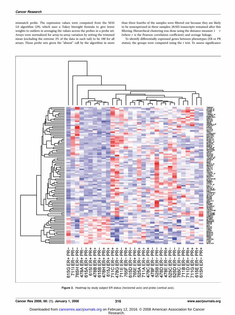
Figure 2. Heatmap by study subject ER status (horizontal axis ) and probe (vertical axis ).
Cancer Research
Cancer Res 2008; 68: (1). January 1, 2008 316 www.aacrjournals.org
Research. on February 12, 2016. © 2008 American Association for Cancercancerres.aacrjournals.org Downloaded from

of these genes, we computed the false discovery rate (FDR; ref. 30) as
described by Storey et al. (31). This is the percentage of genes called
significant that turn out to be false leads. For example, a FDR of 10% for agene means that among all genes that are as significant or more
significant, only 10% are likely to have been nondifferentially expressed but
were incorrectly found to be differentially expressed. When multiplehypotheses are being tested simultaneously, this measure of the rate that
the significant features are truly null is generally more helpful than the
false-positive rate for the truly null features (P value). Unless otherwise
stated, in these analyses a FDR of 5% was used to define statisticalsignificance with respect to differences in gene expression by receptor
status. Pathway analyses were done using software developed by our group
in an effort to define differentially expressed sets of genes that belong to
known genetic groups or pathways (i.e., DNA repair pathway, etc.). Thestatistical language R6 and packages from Bioconductor Project7 were used
for all analyses of the data with the exception that Statistical Analysis
System (SAS) software version 9.1 was used to carry out descriptive
analyses (proportions, means, and SDs) as well as bivariate analyses(Fisher’s exact test; ref. 32).
Immunohistochemistry analyses. Immunohistochemical staining was
done on 5-Am sections of the formalin-fixed paraffin-embedded materialusing the Envision+ DAB system (DAKO) per manufacturer’s protocol along
with mouse monoclonal antibodies against ER (1:100 dilution; DAKO),
PR (1:200 dilution; DAKO), and Ki-67 (1:200 dilution; DAKO). After routine
deparaffinization/rehydration of sections and before quenching of endog-enous peroxidase activity, antigen retrieval was done in a pressure cooker
by heating in 10 mmol/L Na-citrate (pH 6.0) to a temperature of 125jC for
30 s followed by gradual cooling to 90jC over 30 min. Primary antibodies or
IgG isotype–negative controls were diluted in TBS with 1% bovine serumalbumin and were applied for 40 min at room temperature. All washes were
done with TBS with 0.1% Tween 20. Following the Envision+ DAB detection
of staining, slides were counterstained with hematoxylin, dehydrated
through ethanol and xylene, and coverslipped with Permount.
Immunohistochemical stains for ER, PR, and Ki-67 (MIB) wereevaluated by a pathologist blinded to the results of the gene expression
profiling. Staining was visually assessed in at least 1,000 tumor cells in two
separate fields using a 40� objective in the areas of greatestimmunopositivity. For MIB, staining was resported as a proliferation
index (percent of cells proliferating); this was calculated by dividing the
number of tumor cells showing positive nuclear staining by the total
number of tumor nuclei. For ER and PR, the immunostaining wasassessed on a semiquantitative scale of 0 to 4 as follows: score 0, negative;
score 1, positive <10%; score 2, positive 10% to 49%; score 3, positive 50%
to 90%; score 4, positive >90%.
Results
The sample is 77% female with a mean age of 55.7 years. Eightypercent of the meningioma samples were defined as grade 1 and20% as grade 2 (no lesions were categorized as grade 3 ormalignant); the mean MIB was 6.2 whereas 33% and 84% ofsamples were ER and PR positive, respectively. Twelve of thelesions are classified as fibrous, seven as transitional, nine asmeningothelial, and one each as secretory and psammomatous. ERand PR status was not associated with sex, age at diagnosis, MIB,grade, or histologic subtype. Heatmaps for the top 100 genes arepresented by PR and ER status, respectively, in Figs. 1 and 2,whereas Fig. 3 presents a dendrogram for the 31 cases classifiedby ER and PR status using all 29,000 genes examined. A review ofthese figures and associated dendrograms shows clustering ofcases to be more strongly driven by PR rather than ER status. Whenmore formally examined by hormone receptor positivity, up-regulation or down-regulation was noted to be statisticallysignificantly related to PR status for a number of genes (Table 1).In the comparison between ER+ and ER� lesions, no genes seemedto reach statistical significance.
6 http://www.r-project.org7 http://www.bioconductor.org
Figure 3. Dendrogram using 29,000 genes.
Gene Expression and PR Status in Meningioma
www.aacrjournals.org 317 Cancer Res 2008; 68: (1). January 1, 2008
Research. on February 12, 2016. © 2008 American Association for Cancercancerres.aacrjournals.org Downloaded from

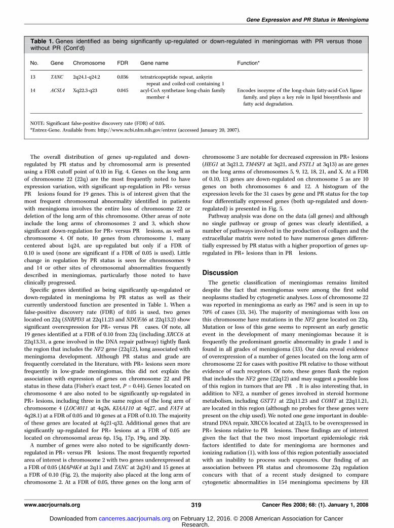
Table 1. Genes identified as being significantly up-regulated or down-regulated in meningiomas with PR versus thosewithout PR
No. Gene Chromosome FDR Gene name Function*
Up-regulated
1 SLC24A3 20p13 0.023 solute carrier family 24 (sodium/potassium/calcium exchanger),
member 3
Encodes plasma membranesodium/calcium exchangers.
2 SERPINB1 6p25 0.032 serpin peptidase inhibitor Stops, prevents, or reduces theactivity of serine-type endopeptidases.
3 FLJ10781 19q13.32 0.032 hypothetical protein FLJ10781
4 KIAA1109 4q27 0.032 KIAA1109 Aspartic-type endopeptidase activity.
5 SNRPD3 22q11.23 0.032 small nuclear ribonucleoproteinD3 polypeptide 18 kDa
These proteins associate with RNA toform core domain of the ribonucleoprotein
particles involved in a variety of RNA
processing events.
6 PEX11A 15q26.1 0.032 peroxisomal biogenesis factor 11A7 NDUFA6 22q13.2-q13.31 0.032 NADH dehydrogenase (ubiquinone)
1a subcomplex, 6, 14 kDa
Protein assists with catalysis of the reaction:
NADH + H+ + ubiquinone = NAD+ + ubiquinol.
8 LOC401152 4q26 0.032 HCV F-transactivated protein 19 FLJ10847 17p11.2 0.037 hypothetical protein FLJ10847 This gene is located within the Smith-Magenis
syndrome region on chromosome 17. It
encodes a protein of unknown function.
10 FAT4 4q28.1 0.045 FAT tumor suppressorhomologue 4 (Drosophila)
Down-regulated
1 PDGFRB 5q31-q32 0.023 platelet-derived growth factorreceptor, h polypeptide
Encodes cell-surface tyrosine kinase receptor formembers of the platelet-derived growth factor
family. The growth factors are mitogens for
cells of mesenchymal origin.2 EGFL5 9q32-q33.3 0.032 EGF-like domain, multiple 5
3 MAP4K4 2q11.2-q12 0.032 mitogen-activated protein kinase
kinase kinase kinase 4
Encodes member of the serine/threonine protein
kinase family.
4 BCL2 18q21.3 0.032 B-cell CLL/lymphoma 2 Encodes an integral outer mitochondrial membrane proteinthat blocks the apoptotic death of some cells such as
lymphocytes. Constitutive expression of BCL2 is thought
to be the cause of follicular lymphoma.
5 SOX4 6p22.3 0.032 SRY (sex determining region Y) box 4 This intronless gene encodes a member of the SOX(SRY-related HMG-box) family of transcription factors
involved in the regulation of embryonic development
and in the determination of the cell fate. The encoded
protein may act as a transcriptional regulator and mayfunction in the apoptosis pathway leading to cell death
as well as to tumorigenesis.
6 BACE2 21q22.3 0.032 h-site amyloid precursorprotein-cleaving enzyme 2
This gene localizes to the ‘‘Down critical region’’ ofchromosome 21. The encoded protein, a member of the
peptidase A1 protein family, is a type I integral membrane
glycoprotein and aspartic protease.
7 NLN 5q12.3 0.032 neurolysin (metallopeptidase M3 family)8 HEG1 3q21.2 0.032 HEG homologue 1 (zebrafish)
9 FMNL3 12q13.12 0.032 formin-like 3 The protein encoded by this gene contains a formin
homology 2 domain.
10 TM4SF1 3q21-q25 0.036 transmembrane 4 L six family member 1 Encodes cell-surface proteins that mediate signal transductionevents that play a role in the regulation of cell development,
activation, growth, and motility. This encoded protein is a
cell-surface antigen and is highly expressed in differentcarcinomas.
11 EDNRA 4 0.036 endothelin receptor type A
12 FSTL1 3q13.33 0.036 follistatin-like 1 Encodes a protein with similarity to follistatin,
an activin-binding protein.
(Continued on the following page)
Cancer Research
Cancer Res 2008; 68: (1). January 1, 2008 318 www.aacrjournals.org
Research. on February 12, 2016. © 2008 American Association for Cancercancerres.aacrjournals.org Downloaded from
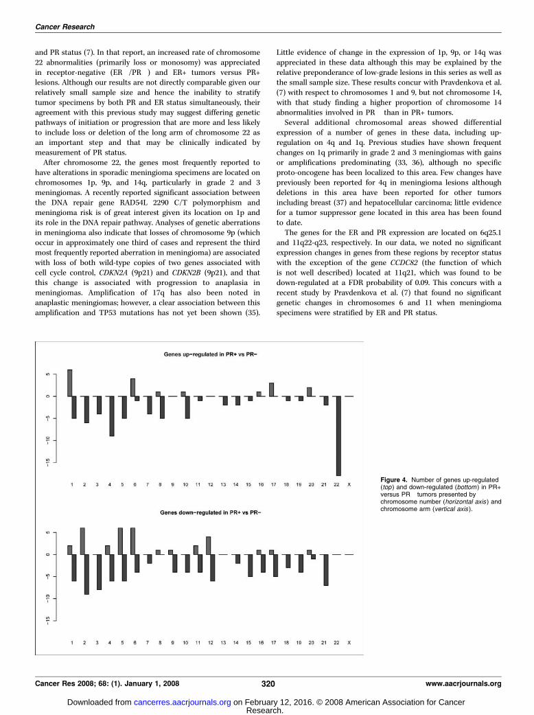
The overall distribution of genes up-regulated and down-regulated by PR status and by chromosomal arm is presentedusing a FDR cutoff point of 0.10 in Fig. 4. Genes on the long armof chromosome 22 (22q) are the most frequently noted to haveexpression variation, with significant up-regulation in PR+ versusPR� lesions found for 19 genes. This is of interest given that themost frequent chromosomal abnormality identified in patientswith meningioma involves the entire loss of chromosome 22 ordeletion of the long arm of this chromosome. Other areas of noteinclude the long arms of chromosomes 2 and 3, which showsignificant down-regulation for PR+ versus PR� lesions, as well aschromosome 4. Of note, 10 genes from chromosome 1, manycentered about 1q24, are up-regulated but only if a FDR of0.10 is used (none are significant if a FDR of 0.05 is used). Littlechange in regulation by PR status is seen for chromosomes 9and 14 or other sites of chromosomal abnormalities frequentlydescribed in meningiomas, particularly those noted to haveclinically progressed.
Specific genes identified as being significantly up-regulated ordown-regulated in meningioma by PR status as well as theircurrently understood function are presented in Table 1. When afalse-positive discovery rate (FDR) of 0.05 is used, two geneslocated on 22q (SNRPD3 at 22q11.23 and NDUFA6 at 22q13.2) showsignificant overexpression for PR+ versus PR� cases. Of note, all19 genes identified at a FDR of 0.10 from 22q (including XRCC6 at22q13.31, a gene involved in the DNA repair pathway) tightly flankthe region that includes the NF2 gene (22q12), long associated withmeningioma development. Although PR status and grade arefrequently correlated in the literature, with PR+ lesions seen morefrequently in low-grade meningiomas, this did not explain theassociation with expression of genes on chromosome 22 and PRstatus in these data (Fisher’s exact test, P = 0.44). Genes located onchromosome 4 are also noted to be significantly up-regulated inPR+ lesions, including three in the same region of the long arm ofchromosome 4 (LOC4011 at 4q26, KIAA110 at 4q27, and FAT4 at4q28.1) at a FDR of 0.05 and 10 genes at a FDR of 0.10. The majorityof these genes are located at 4q21-q32. Additional genes that aresignificantly up-regulated for PR+ lesions at a FDR of 0.05 arelocated on chromosomal areas 6p, 15q, 17p, 19q, and 20p.
A number of genes were also noted to be significantly down-regulated in PR+ versus PR� lesions. The most frequently reportedarea of interest is chromosome 2 with two genes underexpressed ata FDR of 0.05 (MAP4K4 at 2q11 and TANC at 2q24) and 15 genes ata FDR of 0.10 (Fig. 2), the majority also placed at the long arm ofchromosome 2. At a FDR of 0.05, three genes on the long arm of
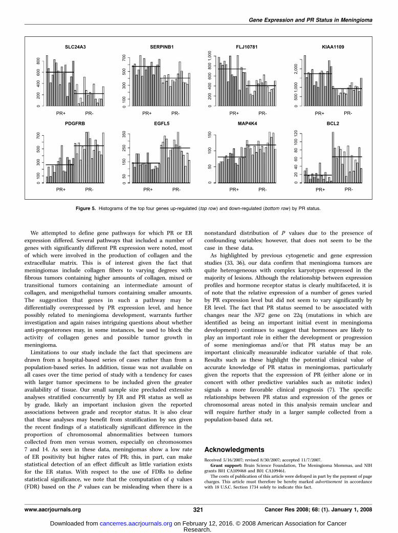
chromosome 3 are notable for decreased expression in PR+ lesions(HEG1 at 3q21.2, TM4SF1 at 3q21, and FSTL1 at 3q13) as are geneson the long arms of chromosomes 5, 9, 12, 18, 21, and X. At a FDRof 0.10, 13 genes are down-regulated on chromosome 5 as are 10genes on both chromosomes 6 and 12. A histogram of theexpression levels for the 31 cases by gene and PR status for the topfour differentially expressed genes (both up-regulated and down-regulated) is presented in Fig. 5.
Pathway analysis was done on the data (all genes) and althoughno single pathway or group of genes was clearly identified, anumber of pathways involved in the production of collagen and theextracellular matrix were noted to have numerous genes differen-tially expressed by PR status with a higher proportion of genes up-regulated in PR+ lesions than in PR� lesions.
Discussion
The genetic classification of meningiomas remains limiteddespite the fact that meningiomas were among the first solidneoplasms studied by cytogenetic analyses. Loss of chromosome 22was reported in meningioma as early as 1967 and is seen in up to70% of cases (33, 34). The majority of meningiomas with loss onthis chromosome have mutations in the NF2 gene located on 22q.Mutation or loss of this gene seems to represent an early geneticevent in the development of many meningiomas because it isfrequently the predominant genetic abnormality in grade 1 and isfound in all grades of meningioma (33). Our data reveal evidenceof overexpression of a number of genes located on the long arm ofchromosome 22 for cases with positive PR relative to those withoutevidence of such receptors. Of note, these genes flank the regionthat includes the NF2 gene (22q12) and may suggest a possible lossof this region in tumors that are PR�. It is also interesting that, inaddition to NF2, a number of genes involved in steroid hormonemetabolism, including GSTT1 at 22q11.23 and COMT at 22q11.21,are located in this region (although no probes for these genes werepresent on the chip used). We noted one gene important in double-strand DNA repair, XRCC6 located at 22q13, to be overexpressed inPR+ lesions relative to PR� lesions. These findings are of interestgiven the fact that the two most important epidemiologic riskfactors identified to date for meningioma are hormones andionizing radiation (1), with loss of this region potentially associatedwith an inability to process such exposures. Our finding of anassociation between PR status and chromosome 22q regulationconcurs with that of a recent study designed to comparecytogenetic abnormalities in 154 meningioma specimens by ER
Table 1. Genes identified as being significantly up-regulated or down-regulated in meningiomas with PR versus thosewithout PR (Cont’d)
No. Gene Chromosome FDR Gene name Function*
13 TANC 2q24.1-q24.2 0.036 tetratricopeptide repeat, ankyrin
repeat and coiled-coil containing 114 ACSL4 Xq22.3-q23 0.045 acyl-CoA synthetase long-chain family
member 4
Encodes isozyme of the long-chain fatty-acid-CoA ligase
family, and plays a key role in lipid biosynthesis and
fatty acid degradation.
NOTE: Significant false-positive discovery rate (FDR) of 0.05.
*Entrez-Gene. Available from: http://www.ncbi.nlm.nih.gov/entrez (accessed January 20, 2007).
Gene Expression and PR Status in Meningioma
www.aacrjournals.org 319 Cancer Res 2008; 68: (1). January 1, 2008
Research. on February 12, 2016. © 2008 American Association for Cancercancerres.aacrjournals.org Downloaded from
and PR status (7). In that report, an increased rate of chromosome22 abnormalities (primarily loss or monosomy) was appreciatedin receptor-negative (ER�/PR�) and ER+ tumors versus PR+lesions. Although our results are not directly comparable given ourrelatively small sample size and hence the inability to stratifytumor specimens by both PR and ER status simultaneously, theiragreement with this previous study may suggest differing geneticpathways of initiation or progression that are more and less likelyto include loss or deletion of the long arm of chromosome 22 asan important step and that may be clinically indicated bymeasurement of PR status.
After chromosome 22, the genes most frequently reported tohave alterations in sporadic meningioma specimens are located onchromosomes 1p, 9p, and 14q, particularly in grade 2 and 3meningiomas. A recently reported significant association betweenthe DNA repair gene RAD54L 2290 C/T polymorphism andmeningioma risk is of great interest given its location on 1p andits role in the DNA repair pathway. Analyses of genetic aberrationsin meningioma also indicate that losses of chromosome 9p (whichoccur in approximately one third of cases and represent the thirdmost frequently reported aberration in meningioma) are associatedwith loss of both wild-type copies of two genes associated withcell cycle control, CDKN2A (9p21) and CDKN2B (9p21), and thatthis change is associated with progression to anaplasia inmeningiomas. Amplification of 17q has also been noted inanaplastic meningiomas; however, a clear association between thisamplification and TP53 mutations has not yet been shown (35).
Little evidence of change in the expression of 1p, 9p, or 14q wasappreciated in these data although this may be explained by therelative preponderance of low-grade lesions in this series as well asthe small sample size. These results concur with Pravdenkova et al.(7) with respect to chromosomes 1 and 9, but not chromosome 14,with that study finding a higher proportion of chromosome 14abnormalities involved in PR� than in PR+ tumors.
Several additional chromosomal areas showed differentialexpression of a number of genes in these data, including up-regulation on 4q and 1q. Previous studies have shown frequentchanges on 1q primarily in grade 2 and 3 meningiomas with gainsor amplifications predominating (33, 36), although no specificproto-oncogene has been localized to this area. Few changes havepreviously been reported for 4q in meningioma lesions althoughdeletions in this area have been reported for other tumorsincluding breast (37) and hepatocellular carcinoma; little evidencefor a tumor suppressor gene located in this area has been foundto date.
The genes for the ER and PR expression are located on 6q25.1and 11q22-q23, respectively. In our data, we noted no significantexpression changes in genes from these regions by receptor statuswith the exception of the gene CCDC82 (the function of whichis not well described) located at 11q21, which was found to bedown-regulated at a FDR probability of 0.09. This concurs with arecent study by Pravdenkova et al. (7) that found no significantgenetic changes in chromosomes 6 and 11 when meningiomaspecimens were stratified by ER and PR status.
Figure 4. Number of genes up-regulated(top ) and down-regulated (bottom ) in PR+versus PR� tumors presented bychromosome number (horizontal axis ) andchromosome arm (vertical axis ).
Cancer Research
Cancer Res 2008; 68: (1). January 1, 2008 320 www.aacrjournals.org
Research. on February 12, 2016. © 2008 American Association for Cancercancerres.aacrjournals.org Downloaded from
We attempted to define gene pathways for which PR or ERexpression differed. Several pathways that included a number ofgenes with significantly different PR expression were noted, mostof which were involved in the production of collagen and theextracellular matrix. This is of interest given the fact thatmeningiomas include collagen fibers to varying degrees withfibrous tumors containing higher amounts of collagen, mixed ortransitional tumors containing an intermediate amount ofcollagen, and menigothelial tumors containing smaller amounts.The suggestion that genes in such a pathway may bedifferentially overexpressed by PR expression level, and hencepossibly related to meningioma development, warrants furtherinvestigation and again raises intriguing questions about whetheranti-progesterones may, in some instances, be used to block theactivity of collagen genes and possible tumor growth inmeningioma.
Limitations to our study include the fact that specimens aredrawn from a hospital-based series of cases rather than from apopulation-based series. In addition, tissue was not available onall cases over the time period of study with a tendency for caseswith larger tumor specimens to be included given the greateravailability of tissue. Our small sample size precluded extensiveanalyses stratified concurrently by ER and PR status as well asby grade, likely an important inclusion given the reportedassociations between grade and receptor status. It is also clearthat these analyses may benefit from stratification by sex giventhe recent findings of a statistically significant difference in theproportion of chromosomal abnormalities between tumorscollected from men versus women, especially on chromosomes7 and 14. As seen in these data, meningiomas show a low rateof ER positivity but higher rates of PR; this, in part, can makestatistical detection of an effect difficult as little variation existsfor the ER status. With respect to the use of FDRs to definestatistical significance, we note that the computation of q values(FDR) based on the P values can be misleading when there is a
nonstandard distribution of P values due to the presence ofconfounding variables; however, that does not seem to be thecase in these data.
As highlighted by previous cytogenetic and gene expressionstudies (33, 36), our data confirm that meningioma tumors arequite heterogeneous with complex karyotypes expressed in themajority of lesions. Although the relationship between expressionprofiles and hormone receptor status is clearly multifaceted, it isof note that the relative expression of a number of genes variedby PR expression level but did not seem to vary significantly byER level. The fact that PR status seemed to be associated withchanges near the NF2 gene on 22q (mutations in which areidentified as being an important initial event in meningiomadevelopment) continues to suggest that hormones are likely toplay an important role in either the development or progressionof some meningiomas and/or that PR status may be animportant clinically measurable indicator variable of that role.Results such as these highlight the potential clinical value ofaccurate knowledge of PR status in meningiomas, particularlygiven the reports that the expression of PR (either alone or inconcert with other predictive variables such as mitotic index)signals a more favorable clinical prognosis (7). The specificrelationships between PR status and expression of the genes orchromosomal areas noted in this analysis remain unclear andwill require further study in a larger sample collected from apopulation-based data set.
Acknowledgments
Received 5/16/2007; revised 8/30/2007; accepted 11/7/2007.Grant support: Brain Science Foundation, The Meningioma Mommas, and NIH
grants R01 CA109468 and R01 CA109461.The costs of publication of this article were defrayed in part by the payment of page
charges. This article must therefore be hereby marked advertisement in accordancewith 18 U.S.C. Section 1734 solely to indicate this fact.
Figure 5. Histograms of the top four genes up-regulated (top row) and down-regulated (bottom row ) by PR status.
Gene Expression and PR Status in Meningioma
www.aacrjournals.org 321 Cancer Res 2008; 68: (1). January 1, 2008
Research. on February 12, 2016. © 2008 American Association for Cancercancerres.aacrjournals.org Downloaded from

References1. Claus EB, Bondy ML, Schildkraut JM, Wiemels J,Wrensch M, Black PM. Epidemiology of intra-cranialmeningioma. Neurosurgery 2005;57:1088–95.2. Donnell MS, Meyer GA, Donegan WL. Estrogen-receptor protein in intracranial meningioma. J Neuro-surg 1979;50:499–502.3. Black PM, Carroll R, Zhang J. The molecular biology ofhormone and growth factor receptors in meningiomas.Acta Neurochir Suppl 1996;65:50–3.4. Carroll RS, Zhang J, Black PM. Expression of estrogenreceptors a and h in human meningiomas. J Neurooncol1999;42:109–16.5. Hsu DW, Efird JT, Hedley-Whyte ET. Progesterone andestrogen receptors in meningiomas: prognostic consid-erations. J Neurosurg 1997;86:113–20.6. Maxwell M, Galanopoulos T, Neville-Golden J, et al.Expression of androgen and progesterone receptors inprimary human meningiomas. J Neurosurg 1993;78:456–62.7. Pravdenkova S, Al-Mefty O, Sawyer J, Husain M.Progesterone and estrogen receptors: opposing prog-nostic indicators in meningioma. J Neurosurg 2006;105:163–73.8. Roser F, Nakamura M, Bellizona M, Rosahl SK,Ostertag H, Samii M. The prognostic value of proges-terone receptor status in meningiomas. J Clin Pathol2004;57:1033–7.9. Custer BS, Koepsell TD, Mueller BA. The associationbetween breast carcinoma and meningioma in women.Cancer 2002;94:1626–35.10. Blitshteyn S, Crook J, Jaeckle KA. Is there anassociation between Meningioma and hormone replace-ment therapy? A study of women at the Mayo ClinicJacksonville. Ninth Annual Meeting of the Society forNeuro-Oncology; 2004 Nov 18–21. Abstract EP-02.11. Custer B, Longstreth WT, Jr., Phillips LE, Koepsell TD,Van Belle G. Hormonal exposures and the risk ofintracranial meningioma in women: a population-basedcase-control study. BMC Cancer 2006;6:152–61.12. Hatch EE, Linet MS, Zhang J, et al. Reproductive andhormonal factors and risk of brain tumors in adultfemales. Int J Cancer 2005;114:797–805.
13. Jhawar BS, Colditz G, Fuchs C, Stampfer M. Sexsteroid hormone exposures and risk for meningiomas. JNeurosurg 2003;99:848–53.14. Lee E, Grutsch J, Persky V, Glick R, Mendes J, Davis F.Association of meningioma with reproductive factors.Int J Cancer 2006;119:1152–7.15. Schlehofer B, Blettner M, Becker N, Martinsohn C,Wahrendorf J. Medical risk factors and the developmentof brain tumors. Cancer 1992;69:2541–7.16. Wigertz A, Lonn S, Mathiesen T, et al. Risk of braintumors associated with exposure to exogenous femalesex hormones. Am J Epidemiol 2006;164:629–36.17. Lambe M, Coogan P, Baron J. Reproductive factorsand the risk of brain tumors: a population-based studyin Sweden. Int J Cancer 1997;72:389–93.18. Longstreth WT, Dennis LK, McGuire VM, DrangsholtMT, Koepsell TD. Epidemiology of intracranial menin-gioma. Cancer 1993;72:639–48.19. Michelsen JJ, New PF. Brain tumour and pregnancy. JNeurol Neurosurg Psychiatry 1969;32:305–7.20. Bickerstaff ER, Small JR, Guest IA. The relapsingcourse of certain meningiomas in relation to pregnancyand menstruation. J Neurol Neurosurg Psychiatry 1958;21:89–91.21. Olson JJ, Beck DW, Schlechte JA, Loh PM. Effect of theantiprogesterone RU-486 on meningioma implantedinto nude mice. J Neurosurg 1987;66:584–7.22. Grunberg SM, Weiss MH, Russell CA, et al. Long-termadministration of mifepristone (RU486): clinical toler-ance during extended treatment of meningioma. CancerInvest 2006;24:727–33.23. Grunberg SM, Weiss MH, Spitz IM, et al. Treatmentof unresectable meningiomas with the antiprogesteroneagent mifepristone. J Neurosurg 1991;74:864–6.24. Goodwin JW, Crowley J, Eyre HJ, Stafford B,Jaeckle KA, Townsend JJ. A phase II evaluation oftamoxifen in unresectable or refractory meningioma:a Southwest Oncology Group study. J Neurooncol1993;15:75–7.25. Wrobel G, Roerig P, Kokocinski F, et al. Microarray-based gene expression profiling of benign, atypical, andanaplastic meningiomas identifies novel genes associ-ated with menigioma progression. Int J Cancer 2005;114:249–56.
26. Sasaki T, Hankins GR, Helm GR. Comparison of geneexpression profiles between frozen original meningio-mas and primary cultures of the meningiomas byGeneChip. Neurosurgery 2003;52:892–9.27. Sayagues JM, Tebernero MD, Maillo A, Trelles O,Espinosa AB, Sarasquete ME. Microarray-based analysisof spinal versus intra-cranial meningiomas: differentclinical, biological, and genetic characteristics associat-ed with distinct patterns of gene expression. J Neuro-pathol Exp Neurol 2006;65:445–54.28. Louis DN, Scheithauer BW, Budka H, von Deimling A,Kepes JJ. Meningiomas. In: Kleihues P, Cavenee WK,editors. Pathology and genetics of tumnors of thenervous system. Lyon: IARC Press; 2000. p. 176–89.29. Microarray suite user guide, version 5. Affymetrix,2001.30. Benjamini Y, Hochberg Y. Controlling the falsediscovery rate: a practical and powerful approach tomultiple testing. J R Stat Soc Ser B 1995;57:289–300.31. Storey JD, Tibshirani R. Statistical significance forgenomewide studies. Proc Natl Acad Sci U S A 2003;100:9440–5.32. SAS Institute, Inc. SAS/STAT user’s guide, version 6.4th ed. Vol. 1. Cary (NC): SAS Institute Inc.; 1989.33. Weber RG, Bostrom J, Wolter M, et al. Analysis ofgenomic alterations in benign, atypical, and anaplasticmeningiomas: toward a genetic model of meningiomaprogression. Proc Natl Acad Sci U S A 1997;47:14719–24.34. Watson MA, Gutmann DH, Peterson K, et al.Molecular characterization of human meningiomas bygene expression profiling using high-density oligonucle-otide microarrays. Am J Pathol 2002;161:665–72.35. Verheijen FM, Sprong M, Kloosterman JM, Blaauw G,Thijssen JH, Blackenstein MA. TP53 mutations inhuman meningiomas. Int J Biol Markers 2002;17:42–8.36. Arsalantas A, Artan S, Oner U, et al. Comparativegenomic hybridization analysis of genomic alterationsin benign, atypical, and anaplastic meningiomas. ActaNeurol Belg 2002;102:53–62.37. Aubele MM, Cummings MC, Mattis AE, et al.Accumulation of chromosomal imbalances from intra-ductal proliferative lesions to adjacent in situ andinvasive ductal breast cancer. Diagn Mol Pathol 2000;9:14–9.
Cancer Research
Cancer Res 2008; 68: (1). January 1, 2008 322 www.aacrjournals.org
Research. on February 12, 2016. © 2008 American Association for Cancercancerres.aacrjournals.org Downloaded from

2008;68:314-322. Cancer Res Elizabeth B. Claus, Peter J. Park, Rona Carroll, et al. Receptors in MeningiomaSpecific Genes Expressed in Association with Progesterone
Updated version
http://cancerres.aacrjournals.org/content/68/1/314
Access the most recent version of this article at:
Cited articles
http://cancerres.aacrjournals.org/content/68/1/314.full.html#ref-list-1
This article cites 33 articles, 5 of which you can access for free at:
Citing articles
http://cancerres.aacrjournals.org/content/68/1/314.full.html#related-urls
This article has been cited by 5 HighWire-hosted articles. Access the articles at:
E-mail alerts related to this article or journal.Sign up to receive free email-alerts
Subscriptions
Reprints and
To order reprints of this article or to subscribe to the journal, contact the AACR Publications
Permissions
To request permission to re-use all or part of this article, contact the AACR Publications
Research. on February 12, 2016. © 2008 American Association for Cancercancerres.aacrjournals.org Downloaded from