Progesterone stimulates the proliferation of female and male cholangiocytes via autocrine/paracrine...
13
Progesterone stimulates the proliferation of female and male cholangiocytes via autocrine/paracrine mechanisms Shannon Glaser, 1,2 Sharon DeMorrow, 1,2 Heather Francis, 2 Yoshiyuki Ueno, 5 Eugenio Gaudio, 3 Shelley Vaculin, 4 Julie Venter, 1 Antonio Franchitto, 3 Paolo Onori, 9 Bradley Vaculin, 1 Marco Marzioni, 6 Candace Wise, 1 Metaneeya Pilanthananond, 1 Jennifer Savage, 1 Lisa Pierce, 8 Romina Mancinelli, 1,3 and Gianfranco Alpini 1,4,7 1 Department of Medicine, 2 Division of Research and Education, Scott & White Hospital and Texas A&M University System Health Science Center, College of Medicine, Temple, Texas; 3 Department of Human Anatomy, University of Rome, La Sapienza, Rome, Italy; 4 Division of Research, Central Texas Veterans Health Care System, Temple, Texas; 5 Division of Gastroenterology, Tohoku University School of Medicine, Sendai, Japan; 6 Department of Gastroenterology, Universita ` Politecnica delle Marche, Ancona, Italy; 7 Department of Systems Biology and Translational Medicine, Texas A&M Health Science Center, College of Medicine, Temple, Texas; 8 Department of Obstetrics and Gynecology, Scott & White Memorial Hospital and Texas A&M Health Science Center, College of Medicine, Temple, Texas; and 9 Department of Experimental Medicine, University of L’Aquila, L’Aquila, Italy Submitted 16 November 2007; accepted in final form 22 May 2008 Glaser S, DeMorrow S, Francis H, Ueno Y, Gaudio E, Vaculin S, Venter J, Franchitto A, Onori P, Vaculin B, Marzioni M, Wise C, Pilanthananond M, Savage J, Pierce L, Mancinelli R, Alpini G. Progesterone stimulates the proliferation of female and male cholan- giocytes via autocrine/paracrine mechanisms. Am J Physiol Gastro- intest Liver Physiol 295: G124 –G136, 2008. First published May 29, 2008; doi:10.1152/ajpgi.00536.2007.—During cholestatic liver dis- eases, cholangiocytes express neuroendocrine phenotypes and re- spond to a number of hormones and neuropeptides by paracrine and autocrine mechanisms. We examined whether the neuroendocrine hormone progesterone is produced by and targeted to cholangiocytes, thereby regulating biliary proliferation during cholestasis. Nuclear (PR-A and PR-B) and membrane (PRGMC1, PRGMC2, and mPR) progesterone receptor expression was evaluated in liver sections and cholangiocytes from normal and bile duct ligation (BDL) rats, and NRC cells (normal rat cholangiocyte line). In vivo, normal rats were chronically treated with progesterone for 1 wk, or immediately after BDL, rats were treated with a neutralizing progesterone antibody for 1 wk. Cholangiocyte growth was measured by evaluating the number of bile ducts in liver sections. The expression of the progesterone synthesis pathway was evaluated in liver sections, cholangiocytes and NRC. Progesterone secretion was evaluated in supernatants from normal and BDL cholangiocytes and NRC. In vitro, NRC were stimulated with progesterone and cholangiocyte supernatants in the presence or absence of antiprogesterone antibody. Aminoglutethimide was used to block progesterone synthesis. Cholangiocytes and NRC express the PR-B nuclear receptor and PRGMC1, PRGMC2, and mPR. In vivo, progesterone increased the number of bile ducts of normal rats, whereas antiprogesterone antibody inhibited cholangio- cyte growth stimulated by BDL. Normal and BDL cholangiocytes expressed the biosynthetic pathway for and secrete progesterone. In vitro, 1) progesterone increased NRC proliferation; 2) cholangio- cyte supernatants increased NRC proliferation, which was partially inhibited by preincubation with antiprogesterone; and 3) inhibition of progesterone steroidogenesis prevented NRC proliferation. In conclu- sion, progesterone may be an important autocrine/paracrine regulator of cholangiocyte proliferation. bile ducts; gastrointestinal hormones; intrahepatic biliary epithelium CHOLANGIOCYTES ARE THE TARGET cells in cholangiopathies in- cluding primary sclerosing cholangitis (PSC) and primary biliary cirrhosis (PBC) (4, 8). During the progression of cholangiopathies, proliferation/loss of cholangiocytes is criti- cal for the maintenance of biliary secretory function and intrahepatic ductal mass (2, 4, 8). In recent years, considerable progress has been made in demonstrating that cholangiocytes differentially respond to cholestasis and liver injury induced by hepatoxins with alterations in proliferation and secretion (4, 8, 26, 27, 43, 44, 48). In fact, proliferating cholangiocytes serve as a neuroendocrine organ during liver disease pathogenesis and, as such, secrete and respond to a number of hormones and neuropeptides contributing to the autocrine and paracrine path- ways, which modulate liver inflammation and fibrosis (8). In rat models of experimental cholestasis including bile duct ligation (BDL), there is increased cholangiocyte growth and ductal secretory activity (1, 2, 8). Progesterone is a steroid hormone synthesized by the ovaries and adrenal glands, by the corpus luteum during pregnancy, and in the central and peripheral nervous system (16, 50). The first step in the biosynthesis of steroids is the conversion of cholesterol to pregnenolone, which is the precursor of all steroid hormones (63). The rate-limiting step of steroidogene- sis is the rate of cholesterol transport from intracellular stores to the inner mitochondrial membrane, which is the location of cytochrome P450 side-chain cleavage (p450scc) (63). The steroidogenic acute regulatory protein (StAR) mediates this rate-limiting step of steroidogenesis by regulating the translo- cation of cholesterol from the outer to the inner mitochondrial membrane (46). Once in the inner mitochondrial membrane, P450scc catalyzes the transformation of cholesterol to preg- Addresses for reprint requests and other correspondence: S. Glaser, Scott & White Hospital, Texas A&M Health Science Center, 702 SW H. K. Dodgen Loop, Temple, TX 76504 (e-mail: [email protected]) and G. Alpini, Medicine and Systems Biology and Translation Medicine, Central Texas Veterans Health Care System, Texas A&M Health Science Center, College of Medicine, Medical Research Bldg., 702 SW H. K. Dodgen Loop, Temple, TX 76504 (e-mail: [email protected] or [email protected]). The costs of publication of this article were defrayed in part by the payment of page charges. The article must therefore be hereby marked “advertisement” in accordance with 18 U.S.C. Section 1734 solely to indicate this fact. Am J Physiol Gastrointest Liver Physiol 295: G124–G136, 2008. First published May 29, 2008; doi:10.1152/ajpgi.00536.2007. 0193-1857/08 $8.00 Copyright © 2008 the American Physiological Society http://www.ajpgi.org G124
Transcript of Progesterone stimulates the proliferation of female and male cholangiocytes via autocrine/paracrine...

Progesterone stimulates the proliferation of female and male cholangiocytesvia autocrine/paracrine mechanisms
Shannon Glaser,1,2 Sharon DeMorrow,1,2 Heather Francis,2 Yoshiyuki Ueno,5 Eugenio Gaudio,3
Shelley Vaculin,4 Julie Venter,1 Antonio Franchitto,3 Paolo Onori,9 Bradley Vaculin,1 Marco Marzioni,6
Candace Wise,1 Metaneeya Pilanthananond,1 Jennifer Savage,1 Lisa Pierce,8 Romina Mancinelli,1,3
and Gianfranco Alpini1,4,7
1Department of Medicine, 2Division of Research and Education, Scott & White Hospital and Texas A&M University SystemHealth Science Center, College of Medicine, Temple, Texas; 3Department of Human Anatomy, University of Rome, LaSapienza, Rome, Italy; 4Division of Research, Central Texas Veterans Health Care System, Temple, Texas; 5Divisionof Gastroenterology, Tohoku University School of Medicine, Sendai, Japan; 6Department of Gastroenterology, UniversitaPolitecnica delle Marche, Ancona, Italy; 7Department of Systems Biology and Translational Medicine, Texas A&M HealthScience Center, College of Medicine, Temple, Texas; 8Department of Obstetrics and Gynecology, Scott & White MemorialHospital and Texas A&M Health Science Center, College of Medicine, Temple, Texas; and 9Department of ExperimentalMedicine, University of L’Aquila, L’Aquila, Italy
Submitted 16 November 2007; accepted in final form 22 May 2008
Glaser S, DeMorrow S, Francis H, Ueno Y, Gaudio E, VaculinS, Venter J, Franchitto A, Onori P, Vaculin B, Marzioni M, WiseC, Pilanthananond M, Savage J, Pierce L, Mancinelli R, Alpini G.Progesterone stimulates the proliferation of female and male cholan-giocytes via autocrine/paracrine mechanisms. Am J Physiol Gastro-intest Liver Physiol 295: G124–G136, 2008. First published May 29,2008; doi:10.1152/ajpgi.00536.2007.—During cholestatic liver dis-eases, cholangiocytes express neuroendocrine phenotypes and re-spond to a number of hormones and neuropeptides by paracrine andautocrine mechanisms. We examined whether the neuroendocrinehormone progesterone is produced by and targeted to cholangiocytes,thereby regulating biliary proliferation during cholestasis. Nuclear(PR-A and PR-B) and membrane (PRGMC1, PRGMC2, and mPR�)progesterone receptor expression was evaluated in liver sections andcholangiocytes from normal and bile duct ligation (BDL) rats, andNRC cells (normal rat cholangiocyte line). In vivo, normal rats werechronically treated with progesterone for 1 wk, or immediately afterBDL, rats were treated with a neutralizing progesterone antibody for1 wk. Cholangiocyte growth was measured by evaluating the numberof bile ducts in liver sections. The expression of the progesteronesynthesis pathway was evaluated in liver sections, cholangiocytes andNRC. Progesterone secretion was evaluated in supernatants fromnormal and BDL cholangiocytes and NRC. In vitro, NRC werestimulated with progesterone and cholangiocyte supernatants in thepresence or absence of antiprogesterone antibody. Aminoglutethimidewas used to block progesterone synthesis. Cholangiocytes and NRCexpress the PR-B nuclear receptor and PRGMC1, PRGMC2, andmPR�. In vivo, progesterone increased the number of bile ducts ofnormal rats, whereas antiprogesterone antibody inhibited cholangio-cyte growth stimulated by BDL. Normal and BDL cholangiocytesexpressed the biosynthetic pathway for and secrete progesterone.In vitro, 1) progesterone increased NRC proliferation; 2) cholangio-cyte supernatants increased NRC proliferation, which was partiallyinhibited by preincubation with antiprogesterone; and 3) inhibition ofprogesterone steroidogenesis prevented NRC proliferation. In conclu-
sion, progesterone may be an important autocrine/paracrine regulatorof cholangiocyte proliferation.
bile ducts; gastrointestinal hormones; intrahepatic biliary epithelium
CHOLANGIOCYTES ARE THE TARGET cells in cholangiopathies in-cluding primary sclerosing cholangitis (PSC) and primarybiliary cirrhosis (PBC) (4, 8). During the progression ofcholangiopathies, proliferation/loss of cholangiocytes is criti-cal for the maintenance of biliary secretory function andintrahepatic ductal mass (2, 4, 8). In recent years, considerableprogress has been made in demonstrating that cholangiocytesdifferentially respond to cholestasis and liver injury induced byhepatoxins with alterations in proliferation and secretion (4, 8,26, 27, 43, 44, 48). In fact, proliferating cholangiocytes serveas a neuroendocrine organ during liver disease pathogenesisand, as such, secrete and respond to a number of hormones andneuropeptides contributing to the autocrine and paracrine path-ways, which modulate liver inflammation and fibrosis (8). Inrat models of experimental cholestasis including bile ductligation (BDL), there is increased cholangiocyte growth andductal secretory activity (1, 2, 8).
Progesterone is a steroid hormone synthesized by the ovariesand adrenal glands, by the corpus luteum during pregnancy,and in the central and peripheral nervous system (16, 50). Thefirst step in the biosynthesis of steroids is the conversion ofcholesterol to pregnenolone, which is the precursor of allsteroid hormones (63). The rate-limiting step of steroidogene-sis is the rate of cholesterol transport from intracellular storesto the inner mitochondrial membrane, which is the location ofcytochrome P450 side-chain cleavage (p450scc) (63). Thesteroidogenic acute regulatory protein (StAR) mediates thisrate-limiting step of steroidogenesis by regulating the translo-cation of cholesterol from the outer to the inner mitochondrialmembrane (46). Once in the inner mitochondrial membrane,P450scc catalyzes the transformation of cholesterol to preg-Addresses for reprint requests and other correspondence: S. Glaser, Scott &
White Hospital, Texas A&M Health Science Center, 702 SW H. K. DodgenLoop, Temple, TX 76504 (e-mail: [email protected]) and G. Alpini, Medicineand Systems Biology and Translation Medicine, Central Texas VeteransHealth Care System, Texas A&M Health Science Center, College of Medicine,Medical Research Bldg., 702 SW H. K. Dodgen Loop, Temple, TX 76504(e-mail: [email protected] or [email protected]).
The costs of publication of this article were defrayed in part by the paymentof page charges. The article must therefore be hereby marked “advertisement”in accordance with 18 U.S.C. Section 1734 solely to indicate this fact.
Am J Physiol Gastrointest Liver Physiol 295: G124–G136, 2008.First published May 29, 2008; doi:10.1152/ajpgi.00536.2007.
0193-1857/08 $8.00 Copyright © 2008 the American Physiological Society http://www.ajpgi.orgG124

nenolone (46, 74). Pregnenolone moves from the mitochondriato the microsomal compartment where it is converted to the�4,3-keto steroid progesterone by 3�-hydroxysteroid dehydro-genase (3�-HSD) (72).
The biological responses triggered by progesterone are me-diated by both genomic and nongenomic mechanisms. Thegenomic action of progesterone is mediated by two progester-one receptor (PR) isoforms, PR-A and PR-B, that are membersof the nuclear receptor family of ligand-dependent transcrip-tion factors (45). The progesterone gene contains multiplepromoters from which PR-A and PR-B are transcribed (45).PR-B differs from PR-A only in that PR-B contains an addi-tional stretch of 164 amino acids at the NH2 terminus of theprotein (45). The binding of progesterone to the PR promotesits translocation from the cytoplasm to the nucleus and initiatesreceptor dimerization, DNA binding, and subsequent transcrip-tion of genes containing progesterone response elements (41).In most cases, PR-A and PR-B have distinct transcriptionactivities despite having similar steroid and DNA-bindingactivities (19). PR-B is a strong activator of gene transcription,whereas PR-A can act as a ligand-dependent trans-repressor ofPR-B (19). The large majority of PR target genes have beenidentified in breast cancer cells (60). Most of these target genewere regulated by PR-B, which included upregulation of theexpression of proteins regulating cell adhesion, cell growth,membrane bound receptors, transcription and protein process-ing (60). In addition, progesterone can activate the gene tran-scription of vascular endothelial growth factor (VEGF) inendometrial adenocarcinoma cells (52). Another mechanismhas also recently been uncovered by which PR can mediatenongenomic responses through their ability to directly recruitand activate c-Src (11). In addition to genomic actions, pro-gesterone has been recently shown to activate nongenomicmembrane-initiated signaling mechanisms (54, 57). These ef-fects are mediated by the progesterone receptor membranecomponent protein (PGRMC1 and PGRMC2) (12, 38). Acti-vation of progesterone membrane receptors has been shown tostimulate increased intracellular Ca2� in oocytes (75). How-ever, the downstream signaling mechanisms have not beenfully explored. The human homolog of PGRMC1, hpr6, playsa role in decreasing the susceptibility of cancer cells to che-motherapeutic drugs (17, 18). PGRMC1 is a dioxin-induciblegene, which suggests a possible role in stress responses and hasbeen implicated in mediating progesterone’s neuroprotectiveeffects (29, 39, 64).
A number of studies have shown that sex hormones regulatecholangiocyte functions. Estrogens enhance proliferation andsecretion of male cholangiocytes by interaction with estrogenreceptors (7). We have also shown (6) that 1) ovariectomy toBDL female rats induced a decrease in the number of bile ductsand 2) administration of 17-� estradiol to BDL, ovariecto-mized rats prevented the decrease in intrahepatic ductal mass.In addition, we have shown that estrogens in male cholangio-cytes activate the Ras/Raf/Src/Shc/ERK1/2 pathway, therebysynergizing the effects of different growth factors includingnerve growth factor (NGF) and insulin-like growth factor I (9,26). In addition, we have shown that prolactin stimulatescholangiocyte proliferation of female rats by activation of theCa2�/PKC signaling pathway (70). Studies have demonstratedthe uptake of (7 �-3H) progesterone by mouse liver tissue (71).In adult female prairie dogs, progesterone impairs gallbladder
emptying in response to cholecystokinin (73). Furthermore,progesterone has been shown to induce cell proliferation andan abnormal mitotic process in male rat liver (10). However,very little information is known concerning the effects ofprogesterone on the biliary system and more specifically, theeffects of progesterone on cholangiocyte proliferation of fe-male and male rats have not been evaluated.
The present study was performed to address 1) the presenceof progesterone receptors in both female and male cholangio-cytes and 2) the role of progesterone in the regulation ofcholangiocyte proliferation of both female and male normaland BDL rats. We found that cholangiocytes express both thenuclear (PR-A and PR-B) receptors and the PGRMC1 andPGRMC2 membrane receptors and the newly identified mPR�(13). In both female and male rats, we demonstrated thatchronic progesterone treatment increases bile duct mass ofnormal rats, whereas administration of antiprogesterone to ratswith extrahepatic cholestasis induced by BDL prevents cho-lestasis-induced proliferation. In addition, since we demon-strated that cholangiocytes possess the enzymatic pathway forsteroidogenesis, we propose that the proproliferative effectsinduced by progesterone on cholangiocytes are regulated by anautocrine pathway (in addition to paracrine mechanisms).
MATERIALS AND METHODS
Materials. Reagents were purchased from Sigma Chemical (St.Louis, MO) unless otherwise indicated. The monoclonal mouse anti-body reacting with proliferating cellular nuclear antigen (PCNA) waspurchased from DAKO (Kyoto, Japan). The substrate for �-glutamyl-transpeptidase (�-GT), N-(�-L-glutamyl)-4-methoxy-2-naphthylamidewas purchased from Polysciences (Warrington, PA). ProgesteroneEIA kits for the measurement of progesterone levels in serum andcholangiocyte supernatant from the selected groups of animals werepurchased from Cayman Chemical, Ann Arbor, MI. The rabbit poly-clonal antibody for the nuclear progesterone receptor PR (C-20)(PR-A and PR-B), the rabbit polyclonal antibody for StAR (FL-285),the goat polyclonal antibody for 3�-HSD (C-18), and the goat poly-clonal antibody for cytokeratin-19 (CK-19; G-14) were purchasedfrom Santa Cruz Biotechnology (Santa Cruz, CA). The rabbit poly-clonal antibody for the membrane progesterone receptor component(PGRMC1) was purchased from Atlas Antibodies (Stockholm, Swe-den). The mouse monoclonal antibody for PGRMC2 was purchasedfrom Affinity Bioreagents (Golden, CO). The rabbit polyclonal anti-body for p450scc was purchased from Chemicon-Millipore (Te-mecula, CA). The mouse monoclonal antibody for CK-19 was pur-chased from Novocastra Laboratories (Newcastle, UK).
Animals. Female and male 344 Fischer rats (150–175 g) werepurchased from Charles River (Wilmington, MA) and maintained in atemperature-controlled environment (20–22°C) with a 12:12-h light-dark cycle. Animals were fed ad libitum standard rat chow and hadfree access to drinking water. To evaluate the in vivo effect ofprogesterone on cholangiocyte growth, normal female and male ratswere treated with a daily intraperitoneal (IP) injection of 1) NaCl or2) progesterone (50 mg/kg body wt per day, a dose similar to that usedin other studies in rats) (10) for 1 wk. We evaluated the effect ofin vivo administration of antiprogesterone (a sheep polyclonal anti-body to antiprogesterone, Abcam, Cambridge, MA) on cholangiocyteproliferation of female and male BDL rats. Immediately after BDL(2), rats received daily IP injections of either nonimmune serum or apolyclonal neutralizing progesterone antibody (6.5 nmol/dose), thesame dose used in other studies in pregnant hamsters (30), for 7 days.Before each experimental procedure, animals were injected withpentobarbital sodium (50 mg/kg body wt ip) following the regulationsof the panel on euthanasia of the American Veterinarian Medical
G125PROGESTERONE REGULATION OF CHOLANGIOCYTE PROLIFERATION
AJP-Gastrointest Liver Physiol • VOL 295 • JULY 2008 • www.ajpgi.org

Association and local authorities. Study protocols were performed incompliance with institutional guidelines and were approved by theIACUC of Scott and White Hospital.
Purification of cholangiocytes. Cholangiocytes were isolated byimmunoaffinity separation (24, 27, 33), by using a mouse monoclonalantibody (IgM, kindly provided by Dr. R. Faris, Brown University,Providence, RI) that recognizes an unidentified antigen expressed byall intrahepatic rat cholangiocytes (33). The purity (98–99%) ofcholangiocytes was evaluated by �-GT histochemistry (62). Cellviability (by Trypan blue exclusion) ranged from 95 to 98%. Normalmale rat intrahepatic cholangiocyte cultures (NRC) were developed,characterized, and maintained in culture as described by us (3).
Evaluation of progesterone receptor expression. Nuclear (PR-A/B)and membrane (PGRMC1 and PGRMC2) progesterone receptor ex-pression was evaluated by 1) immunofluorescence in frozen liversections from female and male normal and BDL rats and male NRC;and 2) RT-PCR in total RNA (0.75 �g) from uterus (positive control)or purified cholangiocytes from female and male normal and BDL ratsand male NRC. Since antibodies are not available, we evaluated theexpression of mPR� in total RNA (0.75 �g) isolated cholangiocytesand male NRC by RT-PCR. PR-A/B expression was also evaluated byimmunoblotting of protein (10 �g) from whole cell lysate from uterus(positive control) or isolated cholangiocytes from female and malenormal and BDL rats and male NRC.
Immunofluorescence and immunohistochemistry. For immunofluo-rescence, liver sections (20 �m thick; n � 3 per each group ofanimals) from normal and BDL female and male rats were fixed in 4%paraformaldehyde (in 1� PBS) for 10 min, followed by tissuepermeabilization in PBST (1� PBS with 0.2% Triton X-100). Sec-tions were then blocked in 4% BSA (in PBST). Primary antibodies formouse anti-CK-19 (1:50), goat anti-CK-19 (1:10 used for PGRMC2costaining), rabbit anti-PR (1:10), rabbit anti-PGRMC1 (1:50), andmouse anti-PGRMC2 (1:10) were diluted in 1% BSA (in PBST).Sections were incubated overnight at 4°C and washed three times for10 min each with 1� PBST at room temperature. Sections wereincubated with appropriate secondary antibodies [Cy2 anti-mouse(1:50), Cy2 anti-goat (1:50), Cy3 anti-mouse (1:50), and Cy3 anti-rabbit (1:50)] (Jackson Immunochemicals, West Grove, PA) for 2 h atroom temperature protected from light. Following incubation, theslides were washed in PBST at room temperature and coverslippedwith Antifade gold containing 4,6-diamidino-2-phenylindole (DAPI)as a counterstain (Molecular Probes, Eugene, OR). Images werevisualized with an Olympus IX-71 (Tokyo, Japan) confocal micro-scope.
Liver sections from normal and BDL female and male rat (fixed inBouin solution) were mounted on glass slides coated with acetoneaminopropyltriehoxylan (2%) solution. After deparaffination, endog-enous peroxidase activity was blocked by incubation (30 min) inmethanolic hydrogen peroxide (2.5%). Sections were hydrated ingraded ethanol and rinsed in 1� PBS. The endogen biotin wasblocked by the Biotin Blocking System (Dako Cytomation, Glostrup,Denmark). Following washed in 1� PBS, sections were incubatedovernight at 4°C with antibodies for PR, PGRMC1, and PGRMC2(1:100 dilution). Samples were rinsed with 1� PBS, incubated for 10min at room temperature with a secondary biotinylated antibody
(Dako Cytomation LSAB Plus System-HRP), then with Dako ABC(Dako Cytomation LSAB Plus System-HRP), developed with 3,3-diaminobenzidine, and counterstained with hematoxylin. For all im-munoreactions negative controls (preimmune serum substituted forprimary antibody) were included. Observations and light microscopyphotographs of liver sections were taken by Leica Microsystems DM4500 B Light Microscopy (Weltzlar, Germany) with a Jenoptik ProgRes C10 Plus Videocam (Jena, Germany).
For immunofluorescence in male NRC, cells were seeded oncoverslips in a six-well plate (500,000 cells/well) and allowed toadhere overnight. The coverslips were transferred into a new six-wellplate containing cold PBS and washed for 5 min at room temperature.Next, the coverslips were washed three times for 10 min each at roomtemperature in PBST (1� PBS with 0.2% Triton) and blocked 1 h atroom temperature in 4% BSA in 1� PBS. The blocking solution wasremoved and the coverslips were incubated with the primary antibod-ies for PR, PGRMC1, and PGRMC2 diluted in 1% BSA/PBS 24 h4°C. The next day, the coverslips were washed three times for 10 minat room temperature in PBST. Next, they were incubated with aCy2-conjugated anti-rabbit or mouse secondary antibody 1:50 (Jack-son Immunochemicals, West Grove, PA) in 1% BSA/PBS at roomtemperature for 2 h and washed three times for 10 min each withPBST. Following incubation, the coverslips were mounted into mi-croscope slides with Antifade gold containing DAPI as a counterstain(Molecular Probes). Images were taken on an Olympus IX71 fluores-cence microscope with a DP70 digital camera.
RT-PCR. We evaluated by RT-PCR the expression of the messagefor the nuclear receptor [PRA/B (common) and PRB], membraneprogesterone receptor components (PGRMC1 and PGRMC2) and thenewly identified mPR� (13) in total RNA (0.75 �g) from uterus(positive control), male and female cholangiocytes from normal andBDL rats, and male NRC. We first performed RT-PCR for the nuclear(PRA/B and PRB) membrane progesterone receptor components(PGRMC1 and PGRMC2) and the newly identified mPR� to deter-mine that cholangiocytes express the expected molecular weight bandfor these four receptor isoforms and glyceraldehyde-3-phosphatedehydrogenase (GAPDH), the housekeeping gene (1). Total RNA wasextracted utilizing the RNeasy Mini Kit (Qiagen, Valencia, CA)according to the instructions provided by the vendor. RNA wasextracted from normal and BDL cholangiocytes (1 � 105) and reversetranscribed with the Reaction Ready First Strand cDNA synthesis kit(SuperArray). These reactions were used as templates for the PCRassays. Primers PR-A/B, PGRMC1, PGRMC2 and mPR� were syn-thesized by Integrated DNA Technologies (Coralville, IA). The prim-ers for common PR-A/B were previously reported by others (28, 53).The primers used for the common PR-A and PR-B amplificationregion that corresponds to the hormone binding domain of the recep-tor were 5-CTC CTG GAT GAG CCT GAT GGT G-3 (sense) and5-CAC CAT GCC CGC CAG GAT CTT G-3 (antisense), whichgenerate a 283-bp amplification product. The primers for themembrane components of the progesterone receptor PGRMC1and PGRMC2 were designed according to the following sequences:PGRMC1 (NCBI GenBank Accession no. NM 021766) (49) andPGRMC2 (NCBI GenBank Accession no. NM 001008374) (68). Theprimers for PGRMC1 were 5-CTC TCA ACC TGC TGC TCC TT-3
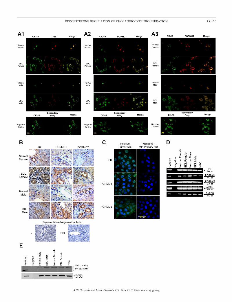
Fig. 1. A: immunofluorescence for progesterone receptors (PR, PGRMC1, and PGRMC2) in liver sections from normal and bile duct-ligated (BDL) female andmale rats demonstrates that bile ducts express these receptors (red staining). Colocalization with CK-19 (green staining, a cholangiocyte-specific marker) of thebile ducts expressing the progesterone receptor is also visible. Bar � 20 �m. B: immunohistochemistry for progesterone receptors (PR, PGRMC1, and PGRMC2)in liver secretion from normal and BDL female male rats confirms receptor expression by bile ducts. Low-level expression of the progesterone receptor wasexpected in hepatocytes (36). C: by immunofluorescence, progesterone receptors (PR, PGRMC1, and PGRMC2) were expressed by normal rat intrahepaticcholangiocyte cultures (NRC). Specific immunoreactivity is shown in green and nuclei were stained with 4,6-diamidino-2-phenylindole (DAPI; blue). No stainingwas visible when primary antibodies were omitted. Bar � 50 �m. D: RT-PCR in pure cholangiocytes from normal and BDL female and male rats and NRCshows that cholangiocytes express the PR-A/B and PGRMC1, PGRMC2, and mPR�. The expression of GAPDH, the housekeeping gene, was similarly expressedby freshly isolated cholangiocytes and NRC. E: by immunoblotting, cholangiocytes from normal and BDL female and male rats and NRC express the PR-B (butnot PR-A). The expression of �-actin, the housekeeping gene, was similar in freshly isolated cholangiocytes and NRC. PR, progesterone receptor; PGRM,progesterone receptor membrane component protein.
G126 PROGESTERONE REGULATION OF CHOLANGIOCYTE PROLIFERATION
AJP-Gastrointest Liver Physiol • VOL 295 • JULY 2008 • www.ajpgi.org
G127PROGESTERONE REGULATION OF CHOLANGIOCYTE PROLIFERATION
AJP-Gastrointest Liver Physiol • VOL 295 • JULY 2008 • www.ajpgi.org

(sense) and 5-CGC TCC TTC AAG CAG TTT TC-3 (antisense),which generate a 443-bp amplification product. The primers forPGRMC2 were 5-AGT CTG GCT GTG ATG CTC CT-3 (sense)and 5-AAC AGG ATC CCT TGC TCC TT-3 (antisense), whichgenerate a 408-bp amplification product. The primer set for mPR�was previously described by others (13). The primers for mPR�5-CTG GAA GCC GTA CAT CTA TGC-3 (sense) and 5-GAAGCT GTA ATG GCC AGA ACT C-3 (antisense), which generate a289-bp amplification product. The primer for GAPDH was purchasedfrom SuperArray. Standard RT-PCR conditions were used with 0.75�g of total mRNA (35 step cycles: 30 s at 94°C, 30 s at 60°C, and 45 sat 72°C). The PCR samples for progesterone receptors were run onagarose gels, and the bands excised and removed from the gel with theQiaquick Gel Extraction Kit (Qiagen). The purified fragments weresubsequently sequenced by Davis Sequencing (Davis, CA).
Immunoblots. We evaluated the expression of the nuclear (PR-A/B)progesterone receptor by immunoblots in protein (10 �g) from wholecell lysates from rat uterus (positive control) and cholangiocytes fromnormal and BDL female and male rats and male NRC. Blots werenormalized by �-actin (5). The intensity of the bands was determinedby scanning video densitometry using the phospho-imager, Storm860, Amersham Biosciences (Piscataway, NJ) using the ImageQuantTLV 2003.02 (Little Chalfont, Buckinghamshire, UK).
Effect of in vivo administration of progesterone to normal rats orantiprogesterone antibody to BDL rats on portal inflammation, lobulardamage, necrosis, progesterone serum levels, and cholangiocyte apo-ptosis and proliferation.
We evaluated the effect of chronic in vivo administration of1) saline or progesterone (50 mg/kg body wt per day) (10) to normalfemale or male rats in the absence or presence of RU-486 (15 mg/kgbody wt), a nuclear progesterone receptor antagonist (40), and2) nonimmune serum or antiprogesterone antibody to male or femaleBDL rats (immediately after BDL) on portal inflammation, lobulardamage, necrosis, cholangiocyte apoptosis and proliferation, and se-rum progesterone levels. Paraffin-embedded liver sections (5 �m, 3sections analyzed per group) were stained with hematoxylin and eosin(H&E), and lobular damage, necrosis, and the degree of portalinflammation were evaluated in a coded fashion. A score of 0 wasused when inflammatory cells were found in only one to two portaltracts, a score of 1 when less than 25% of the total portal tractpresented inflammatory cells, a score of 2 if less than 50% of theportal tracts was infiltrated by inflammation, and a score of 3 when wefound inflammation in more than 50% of the total portal tract.
Measurement of cholangiocyte apoptosis was evaluated by termi-nal deoxynucleotidyltransferase-mediated dUTP nick-end labeling(TUNEL) analysis (42) using a commercially available kit (WakoChemicals, Tokyo, Japan). Following staining, sections (5 �m, 3slides analyzed per group) were evaluated in coded fashion with amicroscope (Olympus Optical, U-PMTVC, Tokyo, Japan); 200 cellsper slide were counted in a coded fashion in 10 nonoverlapping fields.
Cholangiocyte proliferation was evaluated by quantitative evalua-tion of the number of PCNA- and CK-19-positive cholangiocytes inparaffin-embedded liver sections (5 �m, 3 slides analyzed for eachgroup). Following staining, sections were counterstained with hema-toxylin and examined with a microscope (Olympus Optical, U-PMTVC, Tokyo, Japan). Over 100 cholangiocytes were counted in arandom, blinded fashion in three different fields for each group ofanimals. Data were expressed as number of PCNA- or CK-19-positivecholangiocytes per each 100 cholangiocytes. Serum progesteronelevels of normal, BDL, normal � progesterone-, and BDL � anti-progesterone-treated male and female rats were determined by ELISAby commercially available kits (Cayman Chemical, Ann Arbor, MI).
Evaluation of expression of the message and protein for p450scc,3�-HSD, and StAR and secretion of progesterone by cholangiocytes.To evaluate the pathway of progesterone steroidogenesis in cholan-giocytes (which we propose regulates intrahepatic biliary growth byan autocrine pathway), we evaluated 1) the expression of p450scc
(which initiates the biosynthesis of all steroid hormones) (59, 61) and3�-HSD (which converts pregnenolone to progesterone), two keyenzymes in the steroidogenesis pathway of progesterone (59, 61), andStAR (which mediates the rapid increase in pregnenolone synthesisstimulated by trophic hormones) (46) by immunofluorescence, RT-PCR, and real-time PCR; and 2) the amount of progesterone secretedby primary cultures (6 h) of normal and BDL female and malecholangiocytes and male NRC.
Immunofluorescence for StAR, p450scc, and 3�-HSD in liversections and male NRC was performed as described for progesteronereceptor with the difference that other primary antibodies were used:rabbit anti-StAR (1:20), rabbit anti-p450scc (1:200), and goat anti-3�-HSD (1:20) diluted in 1% BSA (in PBST). Immunohistochemistryfor StAR, p450scc, and 3�-HSD in liver sections was performed asdescribed for progesterone receptors with the difference that otherprimary antibodies were used: StAR, p450scc, and 3�-HSD (all at1:100).
The expression of the messages for p450scc, 3�-HSD, and StARwas evaluated by RT-PCR in total RNA (0.75 �g) from cholangio-cytes isolated from normal and BDL female and male rats and maleNRC. Specific oligonucleotide primers for the rat StAR were designedbased on the rat StAR sequence (35) (sense 5-AGCTCGGCTCA-GAGTAGCAG-3 and antisense 5-ACAGCCATCAGGAGCAC-TCT-3, 417 bp). The primers for the rat p450scc were purchasedfrom SuperArray. Specific oligonucleotide primers for the rat 3�-HSDwere designed based on the rat 3�-HSD sequence (32) (sense 5-CTCCATAGCCAACCCAGTGT-3 and antisense 5-CCCTCTTC-CCATCATTGAGA-3, 480 bp). The comparability of the RNA usedwas assessed by RT-PCR for the housekeeping gene, GAPDH (Su-perarray). Standard RT-PCR conditions were used with 1 �g of totalmRNA (35 step cycles: 30 s at 94°C, 30 s at 60°C, and 45 s at 72°C).The PCR samples for StAR and 3�-HSD were run on agarose gels,and the bands were excised and removed from the gel with theQiaquick Gel Extraction Kit (Qiagen). The purified fragments weresequenced by Davis Sequencing.
The amount of progesterone secreted by cholangiocytes was eval-uated as previously described by us (25, 26, 48). Briefly, purifiedcholangiocytes from normal and BDL female and male rats and maleNRC were incubated at 37°C for 0 and 6 h. Thereafter, cells werecentrifuged at 1,500 rpm for 10 min, and the supernatant was trans-ferred to a tube and stored at 70°C before analysis for progesteronelevels by ELISA using commercially available kits (Cayman Chem-ical). Cholangiocyte progesterone secretion was calculated as thedifference between the amount of progesterone detected at 6 h and theamount detected at time zero.
To evaluate whether progesterone secreted by cholangiocytes stim-ulates cholangiocyte proliferation, the supernatant of isolated cholan-giocytes (after 6 h of incubation at 37°C) from normal and BDLfemale and male rats was transferred into plates containing NRC (3).In addition, NRC was also pretreated with antiprogesterone antibodyfor 30 min prior to the addition of cholangiocyte supernatant. After24 h of incubation, cholangiocyte proliferation was evaluated byutilizing a commercially available ViaLight Plus High Sensitivity CellProliferation Kit (Lonza Rockland, Rockland, ME) (31). This is thesame approach that we have used for evaluating the stimulatoryeffects of NGF on the proliferation of cholangiocytes (26).
Inhibition of progesterone steroidogenesis by aminoglutethimide.To further provide evidence that progesterone regulates cholangiocyteproliferation by an autocrine pathway, we propose studies to demon-strate that inhibition of cholangiocyte progesterone steroidogenesis byaminoglutethimide (AMG) (51) induces a decrease in cholangiocytereplication evaluated by measurement of PCNA protein expression(27) by immunoblots (27). Briefly, NRC were plated in a six-wellplate (800,000 cells per well) and cells were incubated overnight at37°C for 24 h with 0.2% BSA (basal) or AMG (100 nM, a dose usedin other studies) (65), which inhibits cytochrome p450scc (51).
G128 PROGESTERONE REGULATION OF CHOLANGIOCYTE PROLIFERATION
AJP-Gastrointest Liver Physiol • VOL 295 • JULY 2008 • www.ajpgi.org

Subsequently, cholangiocyte proliferation was assessed by PCNAimmunoblots (27).
Statistical analysis. All data are expressed as means � SE. Differ-ences between groups were analyzed by the Student’s unpaired t-testwhen two groups were analyzed and ANOVA when more than twogroups were analyzed, followed by an appropriate post hoc test. Avalue of P � 0.05 was considered significant.
RESULTS
Cholangiocytes express progesterone receptors. By immu-nofluorescence in liver sections from normal and BDL femaleand male rats, intrahepatic bile ducts express progesteronereceptors; PR, PGRMC1, and PGRMC2 (Fig. 1A). Colocaliza-tion with CK-19 (a cholangiocyte-specific marker) (42) of thebile ducts expressing the progesterone receptor is visible inFig. 1A. Immunoreactivity for PR, PGRMC1, and PGRMC2was also confirmed by immunohistochemistry in liver sections(Fig. 1B). Low-level expression of the progesterone receptorwas expected in hepatocytes (36). Similarly, progesteronereceptors (PR, PGRMC1, and PGRMC2) were expressed bymale NRC (Fig. 1C).
By RT-PCR, we demonstrated that freshly isolated cholan-giocytes and NRC express the mRNAs for PR-A/B (with aprimer that recognizes both sequences), PGRMC1, PGRMC2,and mPR� (Fig. 1D). The expression of GAPDH mRNA wassimilar in freshly isolated cholangiocytes and NRC (Fig. 1D). Thesequence of the PCR fragment for rat PR-B (both A/B) was 100%homologous to the rat PR sequence (32) (NM022847). Thesequence of the PCR fragment for rat PGRMC1 was 100%homologous to the rat PGRMC1 sequence (35) (NM021766). Thesequence of the PCR fragment for rat PGRMC2 was 100%homologous to the rat PGRMC2 sequence (35) (NM001008374).By immunoblots, the nuclear PR-B (but not PR-A) was detectedin normal and BDL male and female freshly isolated cholangio-cytes and male NRC (Fig. 1E).
Effect of in vivo administration of progesterone to normalrats or antiprogesterone antibody to BDL rats on portalinflammation, lobular damage, necrosis, and cholangiocyteapoptosis and proliferation. Light microscopy of H&E-stainedparaffin-embedded liver sections demonstrated that there wereno significant differences in the degree of portal inflammation,necrosis, apoptosis, and lobular damage in all groups of ani-mals (Table 1). Administration of progesterone to normalfemale or male rats increased cholangiocyte proliferation evi-denced by the increased number of PCNA- (Table 1) andCK-19-positive (Table 1 and Fig. 2) cholangiocytes comparedwith the corresponding normal female or male rats treated withsaline. The administration of the nuclear progesterone receptorantagonist RU-486 (40) to normal female or male rats partlyprevented the progesterone-induced increase in cholangiocytegrowth (Table 1 and Fig. 2).
Administration of antiprogesterone antibody to female ormale BDL rats (immediately after BDL) decreased the numberof PCNA- (Table 1) and CK-19-positive-positive cholangio-cytes (Table 1 and Fig. 2) compared with liver sections fromthe corresponding normal female or male BDL rats treated withnonimmune serum. The decrease in cholangiocyte growth (bythe administration of antiprogesterone antibody to BDL rats)was associated with enhanced cholangiocyte apoptosis (dem-onstrated by TUNEL staining) compared with BDL rats treatedwith nonimmune serum (Table 1). T
able
1.M
easu
rem
ent
ofpo
rtal
infla
mm
atio
n,ne
cros
is,
lobu
lar
dam
age,
apop
tosi
san
dch
olan
gioc
yte
prol
ifer
atio
n
Fem
ale
Mal
e
Para
met
ers
Nor
mal
�Sa
line
Nor
mal
�Pr
ogN
orm
al�
RU
-486
�Pr
og
BD
L�
Non
imm
une
Seru
m
BD
L�
Ant
i-Pr
ogA
ntib
ody
Nor
mal
�Sa
line
Nor
mal
�Pr
ogN
orm
al�
RU
-486
�Pr
og
BD
L�
Non
imm
une
Seru
mB
DL
�A
nti-
Prog
Ant
ibod
y
Lob
ular
dam
age
(n�
5)N
otde
tect
ed0.
1�0.
1N
otde
tect
ed1.
4�0.
11.
4�0.
1N
otde
tect
ed0.
1�0.
1N
otde
tect
ed1.
2�0.
11.
4�0.
1N
ecro
sis
(n�
5)N
otde
tect
edN
otde
tect
ed0.
2�0.
11.
8�0.
11.
8�0.
1N
otde
tect
edN
otde
tect
edN
otde
tect
ed1.
2�0.
11.
4�0.
1Po
rtal
infla
mm
atio
n(n
�5)
Not
dete
cted
Not
dete
cted
Not
dete
cted
1.4�
0.1
1.5�
0.1
Not
dete
cted
Not
dete
cted
Not
dete
cted
1.4�
0.2
1.4�
0.2
Cho
lang
iocy
teap
opto
sis
(n�
5)0.
1�0.
10.
3�0.
1N
otde
tect
ed1.
6�0.
13.
8�0.
5 †0.
1�0.
10.
2�0.
1N
otde
tect
ed1.
1�0.
32.
5�0.
1 †PC
NA
-pos
itive
chol
angi
ocyt
es(n
�5)
0.7�
0.2
5.2�
1.0*
0.2�
0.2
22.6
�1.
216
.6�
1.0 †
0.5�
0.1
6.7�
2.4*
0.4�
0.2
25.2
�1.
818
.4�
1.0 †
CK
-19-
posi
tive
chol
angi
ocyt
es(n
�5)
25.6
�1.
838
.0�
2.4*
30.8
�1.
4*13
1.0�
3.5
111.
2�3.
2 †30
.2�
2.0
45.2
�5.
2*28
.3�
1.2*
128.
2�4.
011
1.6�
2.4 †
Val
ues
are
mea
ns�
SEof
cum
ulat
ive
valu
esob
tain
edfr
omth
eev
alua
tion
of10
port
altr
acts
from
5di
ffer
ent
slid
esfo
rea
chgr
oup.
BD
L,
bile
duct
ligat
ion;
CK
-19,
cyto
kera
tin-1
9;PC
NA
,pr
olif
erat
ing
cellu
lar
nucl
ear
antig
en;
Prog
,pro
gest
eron
e.*P
�0.
05vs
.cor
resp
ondi
ngva
lues
ofno
rmal
rats
trea
ted
with
salin
efo
r1
wk.
†P�
0.05
vs.c
orre
spon
ding
valu
esof
BD
Lra
tstr
eate
dw
ithno
nim
mun
ese
rum
for
1w
k.
G129PROGESTERONE REGULATION OF CHOLANGIOCYTE PROLIFERATION
AJP-Gastrointest Liver Physiol • VOL 295 • JULY 2008 • www.ajpgi.org

Cholangiocytes express StAR, p450scc, and 3�-HSD andsecrete progesterone: possible regulation of cholangiocyteproliferation by an autocrine mechanism. By immunofluores-cence in liver sections, cholangiocytes from normal and BDLfemale and male rats express the proteins for StAR, p450scc,and 3�-HSD (Fig. 3A). Immunoreactivity for StAR, p450scc,and 3�-HSD was also confirmed by immunohistochemistry inliver sections (Fig. 3B). Immunohistochemistry indicates thathepatocytes may also express the proteins required for steroi-dogenesis, which will need to be evaluated in future studies.NRC also expressed StAR, p450scc, and 3�-HSD by immu-
nofluorescence (Fig. 3C). By RT-PCR, purified cholangiocytesfrom normal and BDL female and male rats and NRC ex-pressed the mRNA messages for StAR, p450scc, and 3�-HSD(Fig. 3D). The expression of GAPDH mRNA was similar infreshly isolated cholangiocytes and NRC (Fig. 3D). The se-quence of the PCR fragment for rat 3�-HSD was 100%homologous to the rat 3�-HSD sequence (32) (NM017265).The sequence of the PCR fragment for rat StAR was 100%homologous to the rat StAR sequence (35) (NM031558).
During short-term (6 h) culture, normal and BDL female andmale cholangiocytes secrete progesterone (Fig. 4A). The levels
Fig. 2. Measurement of the number of cyto-keratin-19 (CK-19)-positive cholangiocytesin liver sections from the selected groups ofanimals. A: female rats. B: male rats. Admin-istration of progesterone to normal female ormale rats increased the number of CK-19-positive cholangiocytes compared with thecorresponding normal female or male ratstreated with saline. The administration of thenuclear progesterone receptor antagonist RU-486 to normal female or male rats preventedprogesterone-induced increase in cholangio-cyte growth. In addition, administration ofantiprogesterone antibody to female or maleBDL rats (immediately after BDL) decreasedthe number of CK-19-positive cholangio-cytes compared with liver sections from thecorresponding female or male BDL ratstreated with nonimmune serum. Originalmagnification, �10. Data are means � SE of5 values obtained from the 3 slides evaluatedper each group of animal.
G130 PROGESTERONE REGULATION OF CHOLANGIOCYTE PROLIFERATION
AJP-Gastrointest Liver Physiol • VOL 295 • JULY 2008 • www.ajpgi.org

Fig. 3. A: immunofluorescence for key proteins in the progesterone steroidogenesis pathway [steroidogenic acute regulatory protein (StAR), P450 side-chaincleavage (p450scc), and 3�-hydroxysteroid dehydrogenase (3�-HSD)] in liver sections from normal and BDL female and male rats demonstrates that bile ductsexpress these steroidogenesis pathway proteins (red staining). Colocalization with CK-19 (green staining, a cholangiocyte-specific marker) of the bile ductsexpressing StAR, p450scc, and 3�-HSD is also visible. Bar � 20 �m. B: immunohistochemistry in liver sections from normal and BDL female and male ratsfor StAR, p450scc, and 3�-HSD confirms bile duct expression, with expression of some components in hepatocytes. C: by immunofluorescence, NRC expressedStAR, p450scc, and 3�-HSD, key proteins in the progesterone steroidogenesis pathway. Specific immunoreactivity is shown in green and nuclei were stainedwith DAPI (blue). No staining was visible when primary antibodies were omitted. Bar � 50 �m. D: RT-PCR in pure cholangiocytes from normal and BDL femaleand male rats and NRC shows that cholangiocytes express mRNA messages for StAR, p450scc, and 3�-HSD.
G131PROGESTERONE REGULATION OF CHOLANGIOCYTE PROLIFERATION
AJP-Gastrointest Liver Physiol • VOL 295 • JULY 2008 • www.ajpgi.org

of progesterone were significantly higher in the supernatant ofnormal female cholangiocytes compared with the supernatantof normal male cholangiocytes (Fig. 4A). However, we ob-served a significant decrease in progesterone levels in super-natants from female and male BDL cholangiocytes comparedwith their corresponding normal cholangiocytes (Fig. 4A).Serum progesterone levels were significantly higher in normalfemale rats compared with normal male rats (Fig. 4, B and C).BDL had no significant effects on serum progesterone levels.
Chronic administration of progesterone to normal male andfemale rats significantly elevated serum progesterone levels(Fig. 4, B and C). Administration of antiprogesterone signifi-cantly reduced serum progesterone levels in both BDL femaleand male rats (Fig. 4, B and C).
Consistent with the concept that cholangiocytes can regulatetheir own growth by an autocrine mechanism, the supernatantfrom primary cultures of normal and BDL female and malecholangiocytes stimulated the proliferation of NRC comparedwith basal, unstimulated NRC (Fig. 5, A and B). As expected,supernatants from BDL female and male rats stimulated pro-liferation to a lesser extent as that of the normal female andmale supernatants. In vitro pretreatment of NRC with antipro-gesterone antibody, prior to stimulation with cholangiocytesupernatant, partly inhibited progesterone-stimulated cholan-giocyte proliferation (Fig. 5, A and B).
Inhibition of progesterone steroidogenesis by aminoglute-thimide prevents NRC proliferation. Progesterone induced adose-dependent increase in NRC. An increase in the prolifer-ative response was observed with varying doses of progester-one (Fig. 6). Furthermore, inhibition of progesterone steroido-genesis by in vitro treatment of NRC with AMG preventedbasal cholangiocyte proliferative activity evaluated by immu-noblots for PCNA (Fig. 7).
DISCUSSION
The present study provides the following new findings:1) female and male rat cholangiocytes express both nuclear andmembrane receptors that bind progesterone (PR, PGRMC1,PGRMC2, and mPR�); 2) chronic administration of proges-terone to normal female and male rats stimulates biliary pro-liferation, which can be partly prevented by the simultaneousadministration of the nuclear progesterone receptor antagonistRU-486 (40); 3) biliary proliferation stimulated by BDL (1, 2,27) in female and male rats can be partially prevented by theadministration of a neutralizing antiprogesterone antibody(30); and 4) cholangiocytes from both female and male ratspossess the enzymatic pathway for the steroidogenesis ofprogesterone and secrete progesterone, which indicates that inaddition to a paracrine pathway cholangiocytes regulate theirown growth in an autocrine mechanism.
It is well known that cholangiocyte proliferation is coordi-nately regulated by a number of hormones, neuropeptides, andother factors such as bile acids during normal and cholestaticstates (4, 7, 8, 23, 24, 27). With regard to sex hormones, wehave shown that estrogens play a key role in the regulation ofhyperplastic and neoplastic cholangiocyte growth (7, 8). Fur-thermore, the hormone prolactin stimulates the proliferation ofnormal female cholangiocytes by an autocrine mechanism(70). In support of the stimulatory effect of progesterone oncell growth (10, 14, 34), the chronic administration of proges-terone stimulates a significant increase in biliary mass ofnormal female and male rats. As expected, chronic adminis-tration of progesterone induced a significant increase in serumprogesterone levels.
To evaluate the potential role of progesterone during cho-lestasis, female and male rats with BDL were treated with aneutralizing antiprogesterone antibody for 1 wk (30). Treat-ment with antiprogesterone antibody significantly lowered se-rum progesterone levels of male and female rats with BDL
Fig. 4. Progesterone levels in cholangiocyte supernatants and serum fromnormal and BDL female and male rats. A: progesterone levels in normal femalecholangiocyte supernatants were significantly higher than those of normal malerats. Following BDL, there is a significant decrease in progesterone in thesupernatants of both female and male rat cholangiocytes compare to normalcholangiocytes. Data are means � SE of 5 experiments. *P � 0.05 normalfemale vs. normal male. #P � 0.05 normal female vs. BDL female. &P � 0.05normal male vs. BDL male. B and C: serum progesterone levels weresignificantly higher in normal female rats compared with normal male rats.BDL had no significant effects on serum progesterone levels. Chronic admin-istration of progesterone to normal male and female rats significantly elevatedserum progesterone levels. Administration of antiprogesterone significantlyreduced serum progesterone levels in both BDL female and male rats. Data aremeans � SE of 5 experiments. *P � 0.05 normal vs. normal � progesterone.#P � 0.05 BDL vs. BDL � antiprogesterone.
G132 PROGESTERONE REGULATION OF CHOLANGIOCYTE PROLIFERATION
AJP-Gastrointest Liver Physiol • VOL 295 • JULY 2008 • www.ajpgi.org

compared with both normal and BDL controls, which indicateseffective immunoneutralization of progesterone (21). Admin-istration of antiprogesterone significantly diminished intrahe-patic biliary mass compared with control-treated animals.These findings are parallel with our previous studies showingthat administration of neutralizing antibodies for prolactin,NGF, and VEGFs decreases the circulating levels of theseproteins, thus reducing intrahepatic ductal mass (25, 26, 70).Consistent with previous studies (25, 26, 42), this decrease ofintrahepatic biliary mass was associated with an increase incholangiocyte apoptosis.
Consistent with progesterone-induced alterations in cholan-giocyte proliferation, we found that both normal and BDLfemale and male cholangiocytes express PGRMC1, PGRMC2,and mPR� receptors. The membrane receptors have beenassociated with rapid nongenomic signaling in several celltypes, which involve the activation of intracellular calciumsignaling mechanisms (54, 56, 57). In addition to the recentlydiscovered progesterone membrane receptors, cholangiocytesalso express the nuclear PR-B receptor. Nuclear progesteronereceptors are members of the nuclear receptor family of ligand-
dependent transcription factors (45). In some contexts, cyto-plasmically located PR interact directly with c-Src and activatedownstream signaling mechanisms (66). Activation of PR hasbeen shown to increase the proliferation of a number of normaland neoplastic cell types (10, 14, 34). In our study, wedemonstrate that progesterone-induced biliary proliferation innormal female and male rats can be prevented by administra-tion of the nuclear PR antagonist, RU-486 (mifepristone).RU-486 acts as a competitive receptor antagonist at the nuclearprogesterone receptor and does not affect the membrane pro-gesterone receptor (22). Because of the blockage of progester-one-induced biliary proliferation with in vivo administration ofRU-486, we speculate that the proliferative signaling mecha-nism occurs predominantly via PR-B. However, the presentstudy cannot evaluate or rule out the exact contribution of the
Fig. 6. Dose-dependent effects of progesterone (1010 to 106 M for 24 h) onNRC proliferation. Progesterone stimulates a dose-dependent increase in NRCproliferation. Data are means � SE of 5 experiments. *P � 0.05 vs. basal.
Fig. 7. Effect of aminoglutethimide (AMG) inhibition of cholangiocyte ste-roidogenesis on NRC proliferation. Inhibition of progesterone steroidogenesisby in vitro treatment of NRC with AMG prevented cholangiocyte proliferationevaluated by immunoblots for PCNA. *P � 0.05 vs. basal. Data are means �SE of 4 experiments. *P � 0.05 vs. basal.
Fig. 5. Effect of normal and BDL cholangiocyte supernatant (in the absence or presence of antiprogesterone antibody) on NRC proliferation. A: consistent withthe concept that cholangiocytes regulate their own growth by an autocrine mechanism, the supernatant from primary cultures of normal female and malecholangiocytes stimulated the proliferation of NRC compared with basal unstimulated NRC. In vitro pretreatment of NRC with antiprogesterone antibody, priorto stimulation with cholangiocyte supernatant, partly inhibited progesterone-stimulated cholangiocyte proliferation. Data are means � SE of 5 experiments.B: in a similar fashion, the supernatant from primary cultures of BDL female and male cholangiocytes also stimulated the proliferation of NRC compared withbasal unstimulated NRC (although to a smaller degree than normal supernatant). The proliferation was also partly inhibited by antiprogesterone. Data aremeans � SE of 5 experiments. *P � 0.05 vs. corresponding basal value. #P � 0.05 vs. NRC treated with supernatant from normal (or BDL) femalecholangiocytes. &P � 0.05 vs. NRC treated with supernatant from normal male (or BDL) cholangiocytes.
G133PROGESTERONE REGULATION OF CHOLANGIOCYTE PROLIFERATION
AJP-Gastrointest Liver Physiol • VOL 295 • JULY 2008 • www.ajpgi.org

PGRMC1, PGRMC2, and mPR� in the stimulation of cholan-giocyte proliferation. The exact signaling mechanism of pro-gesterone-induced proliferation was beyond the scope of thepresent study and will be delineated in future studies.
In addition to progesterone receptor expression, we demon-strate the novel findings that cholangiocytes express the enzy-matic pathway (StAR, p450scc, and 3�-HSD) for the synthesisof progesterone. In confirmation of cholangiocyte expressionof the progesterone steroidogenesis pathway, we also show thatcholangiocytes from normal and BDL female and male rats andNRC secrete progesterone. These findings are similar to ourprevious studies demonstrating that cholangiocytes synthesizeand secrete a number of growth promoting factors such asNGF, VEGF, prolactin, and serotonin (25, 26, 48, 70). There-fore, our present findings indicate that progesterone can play akey role in the regulation of cholangiocyte proliferation by notonly by paracrine but also autocrine signaling mechanisms. Wepredict that the local autocrine secretion of progesterone bycholangiocyte augments the overall paracrine effects of circu-lating progesterone. To shed light on the potential autocrinesignaling mechanism, we demonstrate that supernatants fromnormal and BDL male and female cholangiocytes stimulateNRC proliferation. The proliferative response stimulated bynormal and BDL cholangiocyte supernatants was partiallyblocked by an antiprogesterone antibody. The partial, but notfull, blockage of proliferation is most likely due to other factorssecreted by cholangiocytes into the supernatant that we havepreviously demonstrated to be growth promoting for cholan-giocytes, such as NGF, prolactin, and VEGF (25, 26, 70). BDLsupernatants stimulated proliferation to a lesser extent thatsupernatants from normal rats, which confirms our finding thatprogesterone levels are lower in BDL cholangiocyte superna-tants. However, the BDL supernatants significantly inducedcholangiocyte proliferation, which was partially blocked byantiprogesterone, indicating that although the level of proges-terone in the supernatant was low progesterone remains acritical factor in cholangiocyte proliferation in vitro. We pos-tulate that the decrease in cholangiocyte synthesis by cholan-giocytes was due to a counterregulatory mechanism acting todownregulate or modulate cholangiocyte proliferation in thechronic model of BDL where proliferation is leveling off at day7 and thereafter. In further confirmation of an autocrine sig-naling mechanism, we demonstrate that inhibition of the syn-thesis of progesterone with AMG, a p450scc inhibitor, preventsNRC proliferation. A number of studies have shown thatprogesterone has previously been shown to regulate prolifera-tion and protect cells from apoptosis in autocrine/paracrinemechanisms (15, 47, 55). Since cholangiocyte secretion ofprogesterone is downregulated by BDL, serum levels of pro-gesterone (which are considerably elevated in serum comparedwith cholangiocyte supernatants) indicate that the paracrinesignaling mechanism may play a role during cholestasis.
In contrast to a previous report that serum progesteronelevels are elevated during BDL in rabbits (20), we observedthat serum progesterone levels remained unchanged in femaleand male rats with BDL compared with normal control ani-mals. We postulate that our observed findings are merely dueto species differences and decreased cholangiocyte progester-one secretion. We also observed that there was a significantdownregulation of progesterone secretion in cholangiocytesisolated from BDL rats. The finding is in contrast to our
previous publications that demonstrated that other growthpromoting factors are upregulated during cholestasis (8, 25,26). We believe that the downregulation of progesterone se-cretion during cholestasis may be a counterregulatory mecha-nism to regulate biliary proliferation, since cholangiocyteshave less need to secrete the prostimulatory factor, progester-one, to sustain cholangiocyte proliferation. We hypothesizethat progesterone synthesis is reduced during cholestasis byshunting of pregnenolone to the synthesis of other steroidhormones, such as estrogen. In fact, estrogens prevent theincrease of cholangiocyte apoptosis and loss of cholangiocyteproliferation induced by the biliary-digestive diversion in theBDL rat (69). Steroids such as progesterone and estrogen havebeen shown to have neuroprotective and antiapoptotic effectsin a number of cell types (37, 58, 67). Regulation of steroidproduction by cholangiocytes may be a key mechanism incontrolling cholangiocyte growth and responses to injury anddeserves further evaluation. Antiprogesterone therapy mightbenefit patients in conditions with cholangiocyte proliferationsuch as conditions during extrahepatic cholestasis.
ACKNOWLEDGMENTS
We acknowledge Anna Webb and the Texas A&M Health Science CenterMicroscopy Imaging Center for assistance with microscopy.
We would like to thank Meg Chrisler and Glen Cryer, Division of Com-munication, Scott & White, for assistance in editing the manuscript.
GRANTS
This work was supported by a grant award to S. Glaser from Scott & WhiteHospital and the Dr. Nicholas C. Hightower Centennial Chair of Gastroenterologyfrom Scott & White Hospital, a Veterans Affairs Research Scholar Award andVeterans Affairs Merit award to Dr. Alpini, a National Institutes of Healthmentored award (KO1 DK-078532) to S. DeMorrow, Progetti di Ricerca diInteresse Nazionale funds 2005–2007 from Italian Ministero dell ’Istruzione, dell’Università e della Ricerca and Faculty funds from Rome. University La Sapienzato E. Gaudio, and Health and Labour Sciences Research Grants (from the Ministryof Health, Labour and Welfare of Japan) for the Research on Measures forIntractable Diseases and Grant-in-Aid for Scientific Research C (16590573) fromJapan Society for the Promotion of Science to Y. Ueno.
REFERENCES
1. Alpini G, Glaser S, Ueno Y, Pham L, Podila PV, Caligiuri A, LeSageG, LaRusso NF. Heterogeneity of the proliferative capacity of rat cholan-giocytes after bile duct ligation. Am J Physiol Gastrointest Liver Physiol274: G767–G775, 1998.
2. Alpini G, Lenzi R, Sarkozi L, Tavoloni N. Biliary physiology in ratswith bile ductular cell hyperplasia. Evidence for a secretory function ofproliferated bile ductules. J Clin Invest 81: 569–578, 1988.
3. Alpini G, Phinizy JL, Glaser S, Francis H, Benedetti A, Marucci L,LeSage G. Development and characterization of secretin-stimulated se-cretion of cultured rat cholangiocytes. Am J Physiol Gastrointest LiverPhysiol 284: G1066–G1073, 2003.
4. Alpini G, Prall RT, LaRusso NF. The pathobiology of biliary epithelia.In: The Liver: Biology and Pathobiology (4th ed.), edited by Arias IM,Boyer JL, Chisari FV, Fausto N, Jakoby W, Schachter D, and Shafritz DA.Philadelphia, PA: Lippincott Williams & Wilkins, 2001, p. 421–435.
5. Alpini G, Ueno Y, Glaser SS, Marzioni M, Phinizy JL, Francis H,Lesage G. Bile acid feeding increased proliferative activity and apical bileacid transporter expression in both small and large rat cholangiocytes.Hepatology 34: 868–876, 2001.
6. Alvaro D, Alpini G, Onori P, Franchitto A, Glaser S, Le Sage G,Gigliozzi A, Vetuschi A, Morini S, Attili AF, Gaudio E. Effect ofovariectomy on the proliferative capacity of intrahepatic rat cholangio-cytes. Gastroenterology 123: 336–344, 2002.
7. Alvaro D, Alpini G, Onori P, Perego L, Svegliata Baroni G, FranchittoA, Baiocchi L, Glaser SS, Le Sage G, Folli F, Gaudio E. Estrogensstimulate proliferation of intrahepatic biliary epithelium in rats. Gastro-enterology 119: 1681–1691, 2000.
G134 PROGESTERONE REGULATION OF CHOLANGIOCYTE PROLIFERATION
AJP-Gastrointest Liver Physiol • VOL 295 • JULY 2008 • www.ajpgi.org

8. Alvaro D, Mancino MG, Glaser S, Gaudio E, Marzioni M, Francis H,Alpini G. Proliferating cholangiocytes: a neuroendocrine compartment inthe diseased liver. Gastroenterology 132: 415–431, 2007.
9. Alvaro D, Metalli VD, Alpini G, Onori P, Franchitto A, Barbaro B,Glaser SS, Francis H, Cantafora A, Blotta I, Attili AF, Gaudio E. Theintrahepatic biliary epithelium is a target of the growth hormone/insulin-like growth factor 1 axis. J Hepatol 43: 875–883, 2005.
10. Boada LD, Zumbado M, del RI, Blanco A, Torres S, Monterde JG,Afonso JL, Cabrera JJ, Diaz-Chico BN. Steroid hormone progesteroneinduces cell proliferation and abnormal mitotic processes in rat liver. ArchToxicol 75: 707–716, 2002.
11. Boonyaratanakornkit V, Scott MP, Ribon V, Sherman L, AndersonSM, Maller JL, Miller WT, Edwards DP. Progesterone receptor con-tains a proline-rich motif that directly interacts with SH3 domains andactivates c-Src family tyrosine kinases. Mol Cell 8: 269–280, 2001.
12. Cahill MA. Progesterone receptor membrane component 1: an integrativereview. J Steroid Biochem Mol Biol 105: 16–37, 2007.
13. Cai Z, Stocco C. Expression and regulation of progestin membranereceptors in the rat corpus luteum. Endocrinology 146: 5522–5532, 2005.
14. Carnevale RP, Proietti CJ, Salatino M, Urtreger A, Peluffo G,Edwards DP, Boonyaratanakornkit V, Charreau EH, Bal de KierJoffe E, Schillaci R, Elizalde PV. Progestin effects on breast cancercell proliferation, proteases activation, and in vivo development ofmetastatic phenotype all depend on progesterone receptor capacity toactivate cytoplasmic signaling pathways. Mol Endocrinol 21: 1335–1358, 2007.
15. Chaffkin LM, Luciano AA, Peluso JJ. The role of progesterone inregulating human granulosa cell proliferation and differentiation in vitro.J Clin Endocrinol Metab 76: 696–700, 1993.
16. Compagnone NA, Mellon SH. Neurosteroids: biosynthesis and functionof these novel neuromodulators. Front Neuroendocrinol 21: 1–56, 2000.
17. Crudden G, Chitti RE, Craven RJ. Hpr6 (heme-1 domain protein)regulates the susceptibility of cancer cells to chemotherapeutic drugs.J Pharmacol Exp Ther 316: 448–455, 2006.
18. Crudden G, Loesel R, Craven RJ. Overexpression of the cytochromep450 activator hpr6 (heme-1 domain protein/human progesterone recep-tor) in tumors. Tumour Biol 26: 142–146, 2005.
19. Edwards DP. Regulation of signal transduction pathways by estrogen andprogesterone. Annu Rev Physiol 67: 335–376, 2005.
20. Elias AN, Hoefs J, Parker L, Haw T, Lifrak ET. Effect of short-termbile duct ligation on peripheral blood steroids, urinary PGE2 and the rateof sodium excretion in male rabbits. Gen Pharmacol 15: 427–430, 1984.
21. Ellis ST, Heap RB, Butchart AR, Rider V, Richardson NE, WangMW, Taussig MJ. Efficacy and specificity of monoclonal antibodies toprogesterone in preventing the establishment of pregnancy in the mouse.J Endocrinol 118: 69–80, 1988.
22. Engmann L, Losel R, Wehling M, Peluso JJ. Progesterone regulation ofhuman granulosa/luteal cell viability by an RU486-independent mecha-nism. J Clin Endocrinol Metab 91: 4962–4968, 2006.
23. Fava G, Marucci L, Glaser S, Francis H, De Morrow S, Benedetti A,Alvaro D, Venter J, Meininger C, Patel T, Taffetani S, Marzioni M,Summers R, Reichenbach R, Alpini G. gamma-Aminobutyric acidinhibits cholangiocarcinoma growth by cyclic AMP-dependent regulationof the protein kinase A/extracellular signal-regulated kinase 1/2 pathway.Cancer Res 65: 11437–11446, 2005.
24. Francis H, Glaser S, Ueno Y, LeSage G, Marucci L, Benedetti A,Taffetani S, Marzioni M, Alvaro D, Venter J, Reichenbach R, Fava G,Phinizy JL, Alpini G. cAMP stimulates the secretory and proliferativecapacity of the rat intrahepatic biliary epithelium through changes in thePKA/Src/MEK/ERK1/2 pathway. J Hepatol 41: 528–537, 2004.
25. Gaudio E, Barbaro B, Alvaro D, Glaser S, Francis H, Ueno Y,Meininger CJ, Franchitto A, Onori P, Marzioni M, Taffetani S, FavaG, Stoica G, Venter J, Reichenbach R, De Morrow S, Summers R,Alpini G. Vascular endothelial growth factor stimulates rat cholangiocyteproliferation via an autocrine mechanism. Gastroenterology 130: 1270–1282, 2006.
26. Gigliozzi A, Alpini G, Baroni GS, Marucci L, Metalli VD, Glaser SS,Francis H, Mancino MG, Ueno Y, Barbaro B, Benedetti A, Attili AF,Alvaro D. Nerve growth factor modulates the proliferative capacity of theintrahepatic biliary epithelium in experimental cholestasis. Gastroenter-ology 127: 1198–1209, 2004.
27. Glaser S, Benedetti A, Marucci L, Alvaro D, Baiocchi L, Kanno N,Caligiuri A, Phinizy JL, Chowdhury U, Papa E, LeSage G, Alpini G.Gastrin inhibits cholangiocyte growth in bile duct-ligated rats by interac-
tion with cholecystokinin-B/gastrin receptors via D-myo-inositol 1,4,5-triphosphate-, Ca(2�)-, and protein kinase C alpha-dependent mechanisms.Hepatology 32: 17–25, 2000.
28. Gonzalez-Arenas A, Villamar-Cruz O, Guerra-Araiza C, Camacho-Arroyo I. Regulation of progesterone receptor isoforms expression by sexsteroids in the rat lung. J Steroid Biochem Mol Biol 85: 25–31, 2003.
29. Gonzalez SL, Labombarda F, Deniselle MC, Mougel A, Guennoun R,Schumacher M, De Nicola AF. Progesterone neuroprotection in spinalcord trauma involves upregulation of brain-derived neurotrophic factor inmotoneurons. J Steroid Biochem Mol Biol 94: 143–149, 2005.
30. Greenwald GS, Wang MW. A monoclonal antibody to progesteroneinterrupts pregnancy in the hamster by curtailing secretion of the luteo-tropic complex of prolactin and follicle-stimulating hormone. Endocrinol-ogy 129: 1735–1743, 1991.
31. Hallahan AR, Pritchard JI, Hansen S, Benson M, Stoeck J, HattonBA, Russell TL, Ellenbogen RG, Bernstein ID, Beachy PA, Olson JM.The SmoA1 mouse model reveals that notch signaling is critical for thegrowth and survival of sonic hedgehog-induced medulloblastomas. Can-cer Res 64: 7794–7800, 2004.
32. Hashimoto N, Yamanaka H, Mizushima T, Noguchi K. Increasedexpression of 3 beta-hydroxysteroid dehydrogenase mRNA in dorsal rootganglion neurons of adult rats following peripheral nerve injury. NeurosciLett 340: 45–48, 2003.
33. Ishii M, Vroman B, LaRusso NF. Isolation and morphological charac-terization of bile duct epithelial cells from normal rat liver. Gastroenter-ology 97: 1236–1247, 1989.
34. Ismail PM, Amato P, Soyal SM, DeMayo FJ, Conneely OM, O’MalleyBW, Lydon JP. Progesterone involvement in breast development andtumorigenesis—as revealed by progesterone receptor “knockout” and“knockin” mouse models. Steroids 68: 779–787, 2003.
35. Jo Y, Stocco DM. Regulation of steroidogenesis and steroidogenic acuteregulatory protein in R2C cells by DAX-1 (dosage-sensitive sex reversal,adrenal hypoplasia congenita, critical region on the X chromosome,gene-1). Endocrinology 145: 5629–5637, 2004.
36. Jorge AD, Stati AO, Roig LV, Ponce G, Jorge OA, Ciocca DR. Steroidreceptors and heat-shock proteins in patients with primary biliary cirrho-sis. Hepatology 18: 1108–1114, 1993.
37. Kaur P, Jodhka PK, Underwood WA, Bowles CA, de Fiebre NC, deFiebre CM, Singh M. Progesterone increases brain-derived neurotrophicfactor expression and protects against glutamate toxicity in a mitogen-activated protein kinase- and phosphoinositide-3 kinase-dependent man-ner in cerebral cortical explants. J Neurosci Res 85: 2441–2449, 2007.
38. Krebs CJ, Jarvis ED, Chan J, Lydon JP, Ogawa S, Pfaff DW. Amembrane-associated progesterone-binding protein, 25-Dx, is regulatedby progesterone in brain regions involved in female reproductive behav-iors. Proc Natl Acad Sci USA 97: 12816–12821, 2000.
39. Labombarda F, Gonzalez SL, Deniselle MC, Vinson GP, SchumacherM, De Nicola AF, Guennoun R. Effects of injury and progesteronetreatment on progesterone receptor and progesterone binding protein25-Dx expression in the rat spinal cord. J Neurochem 87: 902–913, 2003.
40. Lee HY, Sherwood OD. The effects of blocking the actions of estrogenand progesterone on the rates of proliferation and apoptosis of cervicalepithelial and stromal cells during the second half of pregnancy in rats.Biol Reprod 73: 790–797, 2005.
41. Leonhardt SA, Boonyaratanakornkit V, Edwards DP. Progesteronereceptor transcription and non-transcription signaling mechanisms. Ste-roids 68: 761–770, 2003.
42. LeSage E, Alvaro D, Benedetti A, Glaser S, Marucci L, Baiocchi L,Eisel W, Caligiuri A, Phinizy JL, Rodgers R, Francis H, Alpini G.Cholinergic system modulates growth, apoptosis, and secretion of cholan-giocytes from bile duct-ligated rats. Gastroenterology 117: 191–199,1999.
43. LeSage GD, Benedetti A, Glaser S, Marucci L, Tretjak Z, Caligiuri A,Rodgers R, Phinizy JL, Baiocchi L, Francis H, Lasater J, Ugili L,Alpini G. Acute carbon tetrachloride feeding selectively damages large,but not small, cholangiocytes from normal rat liver. Hepatology 29:307–319, 1999.
44. LeSage GD, Glaser SS, Marucci L, Benedetti A, Phinizy JL, RodgersR, Caligiuri A, Papa E, Tretjak Z, Jezequel AM, Holcomb LA, AlpiniG. Acute carbon tetrachloride feeding induces damage of large but notsmall cholangiocytes from BDL rat liver. Am J Physiol Gastrointest LiverPhysiol 276: G1289–G1301, 1999.
45. Li X, Lonard DM, O’Malley BW. A contemporary understanding ofprogesterone receptor function. Mech Ageing Dev 125: 669–678, 2004.
G135PROGESTERONE REGULATION OF CHOLANGIOCYTE PROLIFERATION
AJP-Gastrointest Liver Physiol • VOL 295 • JULY 2008 • www.ajpgi.org

46. Lin D, Sugawara T, Strauss JF 3rd, Clark BJ, Stocco DM, Saenger P,Rogol A, Miller WL. Role of steroidogenic acute regulatory protein inadrenal and gonadal steroidogenesis. Science 267: 1828–1831, 1995.
47. Makrigiannakis A, Coukos G, Christofidou-Solomidou M, Montas S,Coutifaris C. Progesterone is an autocrine/paracrine regulator of humangranulosa cell survival in vitro. Ann NY Acad Sci 900: 16–25, 2000.
48. Marzioni M, Glaser S, Francis H, Marucci L, Benedetti A, Alvaro D,Taffetani S, Ueno Y, Roskams T, Phinizy JL, Venter J, Fava G,Lesage GD, Alpini G. Autocrine/paracrine regulation of the growth of thebiliary tree by the neuroendocrine hormone serotonin. Gastroenterology128: 121–137, 2005.
49. Meffre D, Delespierre B, Gouezou M, Leclerc P, Vinson GP, Schu-macher M, Stein DG, Guennoun R. The membrane-associated proges-terone-binding protein 25-Dx is expressed in brain regions involved inwater homeostasis and is up-regulated after traumatic brain injury. J Neu-rochem 93: 1314–1326, 2005.
50. Micevych PE, Chaban V, Ogi J, Dewing P, Lu JK, Sinchak K.Estradiol stimulates progesterone synthesis in hypothalamic astrocytecultures. Endocrinology 148: 782–789, 2007.
51. Moore RW, Jefcoate CR, Peterson RE. 2,3,7,8-Tetrachlorodibenzo-p-dioxin inhibits steroidogenesis in the rat testis by inhibiting the mobiliza-tion of cholesterol to cytochrome P450scc. Toxicol Appl Pharmacol 109:85–97, 1991.
52. Mueller MD, Vigne JL, Pritts EA, Chao V, Dreher E, Taylor RN.Progestins activate vascular endothelial growth factor gene transcription inendometrial adenocarcinoma cells. Fertil Steril 79: 386–392, 2003.
53. Park-Sarge OK, Mayo KE. Regulation of the progesterone receptor geneby gonadotropins and cyclic adenosine-3,5-monophosphate in rat gran-ulosa cells. Endocrinology 134: 709–718, 1994.
54. Peluso JJ. Non-genomic actions of progesterone in the normal andneoplastic mammalian ovary. Semin Reprod Med 25: 198–207, 2007.
55. Peluso JJ. Progesterone as a regulator of granulosa cell viability. J SteroidBiochem Mol Biol 85: 167–173, 2003.
56. Peluso JJ. Rapid actions of progesterone on granulosa cells. Steroids 69:579–583, 2004.
57. Peluso JJ, Fernandez G, Pappalardo A, White BA. Membrane-initiatedevents account for progesterone’s ability to regulate intracellular freecalcium levels and inhibit rat granulosa cell mitosis. Biol Reprod 67:379–385, 2002.
58. Peluso JJ, Pappalardo A, Losel R, Wehling M. Progesterone membranereceptor component 1 expression in the immature rat ovary and its role inmediating progesterone’s antiapoptotic action. Endocrinology 147: 3133–3140, 2006.
59. Petersen JJ, Watson DW, Pawson BM. Evaluation of field propagationof Muscidifurax zaraptor (Hymenoptera: Pteromalidae) for control of fliesassociated with confined beef cattle. J Econ Entomol 85: 451–455, 1992.
60. Richer JK, Jacobsen BM, Manning NG, Abel MG, Wolf DM, HorwitzKB. Differential gene regulation by the two progesterone receptor iso-forms in human breast cancer cells. J Biol Chem 277: 5209–5218, 2002.
61. Roby KF, Larsen D, Deb S, Soares MJ. Generation and characterizationof antipeptide antibodies to rat cytochrome P-450 side-chain cleavageenzyme. Mol Cell Endocrinol 79: 13–20, 1991.
62. Rutenburg AM, Kim H, Fischbein JW, Hanker JS, Wasserkrug HL,Seligman AM. Histochemical and ultrastructural demonstration of �-glu-tamyl transpeptidase activity. J Histochem Cytochem 17: 517–526, 1969.
63. Schumacher M, Guennoun R, Mercier G, Desarnaud F, Lacor P,Benavides J, Ferzaz B, Robert F, Baulieu EE. Progesterone synthesis
and myelin formation in peripheral nerves. Brain Res Brain Res Rev 37:343–359, 2001.
64. Selmin O, Lucier GW, Clark GC, Tritscher AM, Vanden Heuvel JP,Gastel JA, Walker NJ, Sutter TR, Bell DA. Isolation and characteriza-tion of a novel gene induced by 2,3,7,8-tetrachlorodibenzo-p-dioxin in ratliver. Carcinogenesis 17: 2609–2615, 1996.
65. Shirakawa H, Katsuki H, Kume T, Kaneko S, Akaike A. Aminoglu-tethimide prevents excitotoxic and ischemic injuries in cortical neurons.Br J Pharmacol 147: 729–736, 2006.
66. Shupnik MA. Crosstalk between steroid receptors and the c-Src-receptortyrosine kinase pathways: implications for cell proliferation. Oncogene 23:7979–7989, 2004.
67. Singh M. Mechanisms of progesterone-induced neuroprotection. Ann NYAcad Sci 1052: 145–151, 2005.
68. Strausberg RL, Feingold EA, Grouse LH, Derge JG, Klausner RD,Collins FS, Wagner L, Shenmen CM, Schuler GD, Altschul SF,Zeeberg B, Buetow KH, Schaefer CF, Bhat NK, Hopkins RF, JordanH, Moore T, Max SI, Wang J, Hsieh F, Diatchenko L, Marusina K,Farmer AA, Rubin GM, Hong L, Stapleton M, Soares MB, BonaldoMF, Casavant TL, Scheetz TE, Brownstein MJ, Usdin TB, ToshiyukiS, Carninci P, Prange C, Raha SS, Loquellano NA, Peters GJ,Abramson RD, Mullahy SJ, Bosak SA, McEwan PJ, McKernan KJ,Malek JA, Gunaratne PH, Richards S, Worley KC, Hale S, GarciaAM, Gay LJ, Hulyk SW, Villalon DK, Muzny DM, Sodergren EJ, LuX, Gibbs RA, Fahey J, Helton E, Ketteman M, Madan A, RodriguesS, Sanchez A, Whiting M, Madan A, Young AC, Shevchenko Y,Bouffard GG, Blakesley RW, Touchman JW, Green ED, Dickson MC,Rodriguez AC, Grimwood J, Schmutz J, Myers RM, Butterfield YS,Krzywinski MI, Skalska U, Smailus DE, Schnerch A, Schein JE, JonesSJ, Marra MA. Generation and initial analysis of more than 15,000full-length human and mouse cDNA sequences. Proc Natl Acad Sci USA99: 16899–16903, 2002.
69. Svegliati-Baroni G, Ghiselli R, Marzioni M, Alvaro D, Mocchegiani F,Saccomanno S, Sisti V, Ugili L, Orlando F, Alpini G, Saba V,Benedetti A. Estrogens maintain bile duct mass and reduce apoptosis afterbiliodigestive anastomosis in bile duct ligated rats. J Hepatol 44: 1158–1166, 2006.
70. Taffetani S, Glaser S, Francis H, DeMorrow S, Ueno Y, Alvaro D,Marucci L, Marzioni M, Fava G, Venter J, Vaculin S, Vaculin B, LamIP, Lee VH, Gaudio E, Carpino G, Benedetti A, Alpini G. Prolactinstimulates the proliferation of normal female cholangiocytes by differen-tial regulation of Ca2�-dependent PKC isoforms. BMC Physiol 7: 6, 2007.
71. Taylor W, Wright DE. Uptake of (7 alpha-3H)progesterone by mouseliver. J Endocrinol 48: 53–54, 1970.
72. Thomas JL, Mason JI, Brandt S, Spencer BR Jr, Norris W. Structure/function relationships responsible for the kinetic differences betweenhuman type 1 and type 2 3beta-hydroxysteroid dehydrogenase and for thecatalysis of the type 1 activity. J Biol Chem 277: 42795–42801, 2002.
73. Tierney S, Nakeeb A, Wong O, Lipsett PA, Sostre S, Pitt HA, LillemoeKD. Progesterone alters biliary flow dynamics. Ann Surg 229: 205–209,1999.
74. Tuckey RC. Progesterone synthesis by the human placenta. Placenta 26:273–281, 2005.
75. Wasserman WJ, Pinto LH, O’Connor CM, Smith LD. Progesteroneinduces a rapid increase in [Ca2�] in of Xenopus laevis oocytes. ProcNatl Acad Sci USA 77: 1534–1536, 1980.
G136 PROGESTERONE REGULATION OF CHOLANGIOCYTE PROLIFERATION
AJP-Gastrointest Liver Physiol • VOL 295 • JULY 2008 • www.ajpgi.org