Macrophage migration inhibitory factor induces cardiomyocyte apoptosis
Upload
wwwuniroma1Category
view
1download
0
Gastrointestinal, Hepatobiliary and Pancreatic Pathology
Prostate Apoptosis Response-4 Is Expressed inNormal Cholangiocytes, Is Down-Regulated inHuman Cholangiocarcinoma, and PromotesApoptosis of Neoplastic Cholangiocytes WhenInduced Pharmacologically
Antonio Franchitto,* Alessia Torrice,†
Rossella Semeraro,† Cristina Napoli,†
Gennaro Nuzzo,‡ Felice Giuliante,‡
Gianfranco Alpini,§ Guido Carpino,¶
Pasquale Bartolomeo Berloco,� Luciano Izzo,**Antonio Bolognese,** Paolo Onori,††
Anastasia Renzi,* Alfredo Cantafora,†
Eugenio Gaudio,* and Domenico Alvaro†
From the Department of Human Anatomy,* University of Rome
“Sapienza,” Rome, Italy; the Division of Gastroenterology,† the
Department of Clinical Medicine, University of Rome,
“Sapienza,” Rome, Italy, Polo Pontino; the Department of
Surgery,‡ Hepatobiliary Unit, Catholic University of the Sacred
Heart School of Medicine, Rome, Italy; the Division Research,§
Central Texas Veterans Health Care System, Scott & White and
Texas A&M University Health Science Center College of Medicine,
Temple, Texas; the Department of Health Science,¶ University of
Rome “Foro Italico,” Rome, Italy; “Paride Stefanini” the
Department of General Surgery, and Organ Transplantation, �
University of Rome, “Sapienza,” Rome, Italy; the Department of
Surgery “P. Valdoni,”** “Sapienza” University of Rome; and the
Department of Experimental Medicine,†† Section of Human and
Clinical Anatomy, State University of L’Aquila, Italy
Prostate apoptosis response-4 (Par-4) is a tumor sup-pressor protein that sensitizes cells to apoptosis;therefore, Par-4 modulation has therapeutic poten-tial. No data currently exist on Par-4 expression incholangiocarcinoma (CCA). We evaluated the expres-sion of Par-4 in normal and neoplastic cholangiocytesand the effects of its pharmacological or genetic mod-ulation. The study was performed in human and ratliver, CCA patient biopsies, and two CCA cell lines.PAR-4 was expressed in normal rat and humancholangiocytes, but its expression levels decreased inboth human CCA and CCA cell lines. In both intrahe-
patic and extrahepatic CCA, Par-4 expression (asshown by immunohistochemistry) was inversely cor-related with markers of proliferation (eg, proliferat-ing cellular nuclear antigen) and directly correlatedwith apoptotic markers (eg, Bax and Bax/BCL2 ratio).Par-4 expression was decreased during CCA cell pro-liferation but was enhanced after apoptosis induc-tion. Pharmacological induction of Par-4 expressionin CCA cell lines by diindolymethane or withaferin Apromoted activation of apoptosis and inhibition ofproliferation. In contrast, specific Par-4 silencing bysmall-interfering RNA determined activation of CCAcell line proliferation. Par-4 is expressed in rat and humancholangiocytes and is down-regulated in both human CCAand CCA cell lines. Par-4 protein levels decrease during cellproliferation but increase during apoptosis. Pharmaco-logical or genetic induction of Par-4 determines apopto-sis of CCA cells, suggesting Par-4 targeting as a CCAtreatment strategy. (Am J Pathol 2010, 177:1779–1790;DOI: 10.2353/ajpath.2010.091171)
Cholangiocarcinoma (CCA) is a devastating cancer witha bad prognosis and scarce response to chemother-
Supported by the Dr. Nicholas C. Hightower Centennial Chair of Gastro-enterology from Scott & White, the Veterans Administration (VA) ResearchScholar Award, and a VA Merit Award (G.A.); D.A. was supported by ItalianMinistry of Education, University and Research (MUIR) grants: PRIN#2007and prot. 2007HPT7BA_003. E.G. was supported by MIUR grants:PRIN#2007, prot. 2007HPT7BA_001, and Federate Atheneaum funds fromthe University “Sapienza” of Rome.
A.F. and A.T. contributed equally to this work.
Accepted for publication June 10, 2010.
Supplemental material for this article can be found on http://ajp.amjpathol.org.
Address reprint requests to Domenico Alvaro, M.D., via R. Rossellini 51,Division of Gastroenterology, Department of Clinical Medicine, University ofRome “Sapienza,” 00137 Rome, Italy. E-mail: [email protected].
The American Journal of Pathology, Vol. 177, No. 4, October 2010
Copyright © American Society for Investigative Pathology
DOI: 10.2353/ajpath.2010.091171
1779

apy.1–3 CCA arises from the neoplastic transformation ofcholangiocytes, the epithelial cells lining the biliary tree.Although important advances have been obtained in thelast few years, the mechanisms leading to neoplastictransformation, growth, and spreading of CCA cells areundefined.4,5 As in other cancers, dysregulation of mul-tiple mechanisms modulating cell proliferation and apo-ptosis have been described in CCA.1,2,4,5 Unfortunately,a progressive increase in incidence and mortality forCCA has been reported worldwide, which mainly affectthe intrahepatic form of CCA.3,6 Half of CCA are notcandidate for surgical resection at the time of diagno-sis1–3 and no efficacious treatment exists. Therefore, re-search aimed to find a new therapeutic target is currentlyan important challenge.
Prostate apoptosis response-4 (Par-4), a tumor sup-pressor protein that sensitizes cells to apoptotic stim-uli,6–16 was first identified in prostate cancer cells thatwere induced to undergo apoptosis. Par-4 is a leucinezipper domain protein widely expressed in diverse nor-mal and cancerous cell types and tissues and resides inboth the cytoplasm and the nucleus. Endogenous PAR-4itself does not cause apoptosis, yet it is essential forapoptosis induced by a variety of exogenous insults.6–16
Recent studies suggest how Par-4 serves as an intracel-lular repressor of topoisomerase 1 catalytic activity, andregulates DNA topology to suppress cellular transforma-tion.7,8 Consistent with its tumor suppressor function,Par-4 is silenced or mutated in different types of can-cers,11–13 and experimental models of Par-4 knockoutspontaneously develop tumors in various organs.17 Incontrast, transgenic mice overexpressing Par-4 showedresistance to development of spontaneous and onco-gene-induced, autochthonous tumors.18 Therefore, Par-4modulation has tremendous therapeutic potential and,indeed, genetic or pharmacological strategies to inducePar-4 expression are currently under investigation forcancer prevention or treatment.9,19–21 To this regard, it isnoteworthy that different natural compounds inducingPar-4 expression have been identified and proposed forcancer chemoprevention.19–21 No data exist on the roleof Par-4 in modulating cholangiocyte proliferation andapoptosis, and the expression of Par-4 in human CCA isunknown. The aim of our study was to evaluate the ex-pression of Par-4 in normal and neoplastic cholangio-cytes and the effects of its pharmacological or geneticmodulation on cell proliferation and apoptosis. Our find-ings demonstrate how Par-4 is involved in modulatingapoptosis in CCA, suggesting Par-4 as a new potentialtherapeutic target for this devastating cancer.
Materials and Methods
Reagents were purchased from Sigma Chemical Co (St.Louis, MO) unless otherwise indicated. HuH-28 cell linederived from intrahepatic CCA was acquired from Can-cer Cell Repository, Tohoku University (Sendai, Japan)and maintained in CRML 1066 medium containing 10%fetal bovine serum (FBS). TFK-1 cell line (derived fromextra-hepatic CCA) was kindly provided by Dr. Yoshiyuki
Ueno form Cancer Cell Repository, Tohoku University,and maintained in RPMI medium containing 10% FBS.The hepatocellular carcinoma HepG2 cell line was ac-quired from Sigma Chemical Co and maintained in min-imal essential medium (BioConcept’s AMIMED, Switzer-land) containing 10% FBS, 1% NEAA, 1% Na-pyruvate,and 1% L-glutamine. 3,3�-diindolylmethane (B-DIM) wasobtained from BioResponse (Boulder, CO). Withaferin A(WA), a major constituent of the medicinal plant Withaniasomnifera, was obtained from Chromadex (Santa Ana,CA). Media and additives for cell culture were obtainedfrom Gibco (BRL, Invitrogen Corporation s.r.l., S. GiulianoMilanese, Italy) unless otherwise indicated.
Human Liver Samples
The use of human material has been approved by thelocal Institutional Review Board. Samples of CCA and/orliver were obtained from (1) 10 patients (five female pa-tients, aged 64 to 73 years, and five male patients, aged67 to 75 years) with intrahepatic CCA presenting as asingle mass lesion within the liver and submitted to sur-gical resection; (2) seven patients affected by extra-he-patic CCA (Klatskin tumor), (three female patients, aged69 to 77 years, and four male patients, aged 68 to 78years) submitted to radical surgery; and (3) five biopsiesfrom liver donors with a normal histology (three femalepatients and two male patients, aged 49 to 54 years).
Light Microscopy and Immunohistochemistry
Liver fragments (0.5 cm) were fixed in 10% bufferedformalin for 2 to 4 hours, embedded in low-temperature-fusion paraffin (55°C to 57°C): 3- to 4-�m sections werestained with hematoxylin-eosin and Masson’s trichrome.For immunohistochemistry (IHC), sections were mountedon glass slides coated with 0.1% poly-L-lysine. Afterdeparaffination, endogenous peroxidase activity wasblocked by a 30-minute incubation in methanolic hydro-gen peroxide (2.5%). The endogen biotin was thenblocked by Biotin Blocking System (Dako, code X0590,Dako, Copenhagen, Denmark) according to the instruc-tions supplied by the vendor. Sections were hydrated ingraded alcohol and rinsed in PBS (pH 7.4) before apply-ing the primary antibody. Sections were incubated over-night at 4°C with antibodies for (1) Par-4 (Santa Cruz,Inc., Santa Cruz, CA; sc-1666, mouse monoclonal;1:100); (2) tumor suppressor phosphatase and tensinhomolog deleted in chromosome 10 (PTEN; Dako,M3627; mouse monoclonal; 1:20); (3) proliferating cellu-lar nuclear antigen (PCNA; Dako, PC10; mouse monoclo-nal; 1:100); (4) bcl-2-associated X protein (BAX; SantaCruz, Inc., sc-7480, mouse monoclonal; 1:200); (5) B celllymphoma gene-2 (pBCL2; Santa Cruz, Inc., sc-7382,mouse monoclonal; 1:100); and (6) nuclear factor-�B(pNF�B; Santa Cruz, Inc., sc-33039-R, rabbit polyclonal;1:50). Samples were then rinsed with PBS for 5 minutes,incubated for 20 minutes at room temperature with sec-ondary biotinylated antibody (Dako LSAB Plus System,HRP, Milan, Italy), then with Dako ABC (Dako LSAB Plus
1780 Franchitto et alAJP October 2010, Vol. 177, No. 4
System, HRP) and finally developed with 3–3� diamino-benzidine. For all immunoreactions, negative controls(the primary antibody was replaced with pre-immune se-rum) were also included. Light microscopy and IHC ob-servation were taken by Olympus BX-5 1 Light Micros-copy (Tokyo, Japan) with a Videocam (Spot Insight,Diagnostic Instrument, Inc., Sterling Heights, MI) andprocessed with an Image Analysis System (IAS-DeltaSistemi, Rome, Italy). Light microscopy and IHC obser-vations were independently performed by three patholo-gists in a blind fashion. Briefly, six slides were analyzedper each specimen of normal liver or CCA. Neoplastic ornormal cholangiocytes were counted in a random,blinded fashion in six nonoverlapping fields (magnifica-tion �20) of each slide and the data expressed as per-cent positive cells.
Isolated Bile Duct Units from Human Liver
Fragments of intrahepatic bile ducts, averaging 20 �m indiameter, were isolated from human liver (five liver do-nors, 1 Gram pieces) as previously described for isola-tion and characterization of bile duct units from rat liver.22
Cell Cultures
HuH-28 or TFK-1 cells were platted on six-well plates andmaintained in appropriate medium supplemented with10% FBS, 100 U/ml penicillin, 100 mg/ml streptomycin, inhumidified atmosphere of 5% CO2.
HuH-28 or TFK-1 cells were exposed to 17�-estradiol,insulin-like growth factor 1 (IGF1), Withaferin A or B-DIM,and proliferation or apoptosis evaluated as follows. 17�-estradiol, Withaferin A, and B-DIM were prepared asstock solutions in dimethyl sulfoxide (DMSO) while IGF1was dissolved in saline. 17�-estradiol stock solutionswere then diluted (1:1,000,000), Withaferin A or B-DIMstock solutions were diluted (1:10,000), and IGF1 stocksolutions were then diluted 1:10,000 into serum-free cul-ture medium. Control cells were exposed to DMSO orsaline only.
Proliferation and Apoptosis
Cell proliferation was assessed as described23,24 by both3-(4,5-dimethylthiazol-2-yl)-5-(3-carboxymethoxyphenyl)-2-(4-sulfophenyl)-2H-tetrazolium (MTS) proliferation assayand PCNA protein expression (Western blot). For MTS pro-liferation assay, we used a commercially available colori-metric cell proliferation assay (Cell Titer 96 AQueous Non-Radioactive Cell Proliferation Assay, MTS Kit, Promega,Madison, WI), by following the manufacturer’s instructions.Proliferation index was calculated as the ratio (multiplied �100) between cell numbers in unstimulated and stimulatedcultures.
Apoptosis was induced in cell lines by different ma-neuvers including serum depletion for 48 hours, expo-sure to human recombinant TRAIL (tumor necrosis factor-related apoptosis-inducing ligand), or to beauvericin.TRAIL was dissolved in PBS (10 �g/ml stock solution),
stored at �20°C in multiple aliquots, and then added(1:1000, 100 ng/ml) for 24 hours into HuH-28 cell culturedin the appropriate medium containing 10% FBS. Beau-vericin was dissolved in methanol (3 mmol/L stock solu-tion), stored at �20°C in multiple aliquots, and thenadded (1:10,000; 25 �mol/L) for 8 hours in HuH-28 cellscultured in the appropriate medium containing 10% FBS.Control cells were incubated in a medium containing anequivalent amount of methanol for 8 hours.
Apoptosis was evaluated23,24 by measuring caspase-3and -8 activity. For caspase-3, we used a colorimetric assaykit (Sigma-Aldrich), based on the hydrolysis of the peptidesubstrate acetyl-Asp-Glu-Val-Asp p-nitroanilide (Ac-DEVD-pNA) by caspase-3, resulting in the release of the p-nitroa-nilide (p-NA). For this assay, cells were lysed in the appro-priate lysis buffer provided by the vendor (50 mmol/LHEPES, pH 7.4, 5 mmol/L 3-[(3-cholamidopropyl)dimethyl-ammonio]-1-propanesulfonate (CHAPS), and 5 mmol/LDTT). The spectrophotometric absorbance of each sample,calculated at 405 nm, was normalized with its protein con-tent and expressed as caspase-3 activity in relative absor-bance intensity. For caspase-8, we used a colorimetric as-say kit (Sigma-Aldrich), based on the hydrolysis of thepeptide substrate acetyl-lle-Glu-Thr-Asp p-nitroaniline bycaspase 8, resulting in the release of the p-NA. For thisassay, cells were lysed in the appropriate lysis buffer pro-vided by the vendor (50 mmol/L HEPES, pH 7.4, 5 mmol/LCHAPS, and 5 mmol/L DTT). The spectrophotometric ab-sorbance of each sample, calculated at 405 nm, was nor-malized with its protein content and expressed ascaspase-8 activity in relative absorbance intensity.
Quantitative RT-PCR
Pieces of dissected tissues (about 30 mg) were immedi-ately submerged in RNAlater stabilization solution (Am-bion, Inc., Austin, TX). Each sample was left 10 minutes atroom temperature and then stored at �20°C until beingsubmitted to RNA extraction. The tissue fragment re-moved from RNAlater was transferred into a clean Ep-pendorf tube where the total RNA was extracted by usingthe TRI REAGENT (Sigma-Aldrich) and 1-bromo-3-chlo-ropropane by following the manufacturer’s instructions.The isolated RNA was dissolved in 55 �l of RNase-freewater. RNA quality and quantity was evaluated by theRNA 6000 Nano LabChip kit with the Agilent 2100 Bio-analyzer (Agilent Technologies, Waldbronn, Germany) aspreviously described.25
An amount of 2.5 �g of total RNA was used for the RTreaction primed by the random hexamer (Invitrogen).This was conducted in a 20-�l volume with a geneticallyengineered version of the M-MuLV reverse transcriptase(Expand Reverse Trancriptase, Roche DiagnosticsGmbH, Mannheim, Germany) according the manufactur-er’s directions. The quality of cDNA produced, ie, thelength of the transcript and the lack of DNA contamina-tion, was tested by PCR with the RNA/cDNA Inspector kit(Sigma-Aldrich) and the DNA1000 LabChip kit with theAgilent 2100 BioAnalyzer as we previously described.26
Gene expression was determined by real-time PCRwith a MX3000P instrument (Stratagene, La Jolla, CA) by
Par-4 and Cholangiocarcinoma 1781AJP October 2010, Vol. 177, No. 4
using the averaged cycle threshold (Ct) automaticallycomputed by the built-in software from three replicas ofeach sample. PCR amplifications were conducted into avolume of 25 �l, with 1.0 �l of cDNA template, 12.5 �l of2� SYBR Green Brilliant QPCR Master Mix (Stratagene),3 pmoles each of upstream and downstream primer forthe gene analyzed, and 0.3 �l of diluted reference dye(6-Carboxyl-X-Rhodamine [ROX] at a final concentration30 nmol/L). All real-time PCR amplifications were con-ducted with the following cycling program: 10 minutes at95°C followed by 40 cycles (30 seconds at 95°C, 30seconds at 60°C, and 30 seconds at 72°C). The fluores-cence detection was performed during the extensionstep of each cycle.
The expression of the gene of interest, ie, the PRKCApoptosis WT1 Regulator (PAWR), was normalized bythree endogenous reference genes, ie, hypoxanthinephosphoribosyl-transferase I (HPRT1), hydroxymethyl-bi-lane synthase (HMBS), and glyceraldehyde-3-phosphatedehydrogenase (GAPDH), as suggested.6 The sequencesof primers used are as reported in Table 1.
The normalization factors calculated from internal con-trol genes using the geometric averaging described byVandesompele et al27 allowed us to evidence small rel-ative differences of the PAWR gene expression in thevarious samples by the accurate ranking of gene of in-terest determinations.
Western Blot
As previously described,23,24 cells were harvested,washed with PBS, and whole cell lysate was prepared bysuspending the cells in added 100 to 150 �l of radioim-munoprecipitation assay buffer (Sigma) with 1 mmol/Lphenylmethylsulfonyl fluoride, 2 �g/ml aprotinin on ice for10 to 15 minutes. Protein concentration was determinedwith the Bio-Rad Protein Assay-Dye Reagent (Bio-Rad
Laboratories GmbH, Germany). For immunoblotting,each extract was prepared as described above, and 30�g total protein was separated through SDS-polyacryl-amide gel electrophoresis, electrotransferred onto nitro-cellulose membranes, and probed with the following:anti-Par-4 mouse monoclonal, anti-PCNA mouse mono-clonal (Santa Cruz Biotechnology), and anti-�-actinmouse monoclonal antibody (Sigma Chemical Co.). De-tection of specific proteins was carried out with an en-hanced chemiluminescence Western blotting kit (QdotWestern blotting kit, Invitrogen).
Small-Interfering RNA
The following duplexed RNA oligonucleotides (StealthRNAi) were synthesized by Invitrogen (Table 1). As acontrol, we used Stealth RNAi negative control duplexes(Invitrogen). The effectiveness of the RNAi experiments inour experimental conditions was confirmed by usingStealth RNAi GAPDH positive control duplexes (Invitro-gen), according to the manufacturer’s instructions.
HuH-28 cells were cultured in 12-well plates and usedat 50% to 70% density the day of transfection. Cells weretransfected with Lipofectamine RNAiMAX (Invitrogen) ac-cording to the manufacturer’s instruction.
Following small-interfering RNAs (siRNAs) treatment(72 hours), HuH-28 cells were harvested and assessedfor total Par-4 content by Western blotting with anti Par-4mouse monoclonal antibodies (Santa Cruz Biotechnol-ogy), and proliferation and apoptosis were studied byWestern blotting for PCNA protein expression, MTS-as-say, and Caspase3-activity assay.
Statistical Analysis
Data are presented as arithmetic mean � SD. Statisticalanalysis was conducted by using the paired or unpaired
Table 1. Sequences of Primers Used for Amplifying or Silencing the PAR-4 Gene
Sequences of primer pairs (sense and antisense, respectively) used for amplifying PAWR or PAR-4 and three internal controlgenes used for the geometric averaging normalization procedure (HPRT1, HMBS, and GAPDH)
Gene Primer sequence Length (nt) Product (bp) Origin
PAWR 5�-AGGGATGCAAATGTTTCAGG-3� 20 112 15�-TCACAAGTCTTAGGTTTTCTTGTCTT-3� 23
HPRT1 5�-TGACACTGGCAAAACAATGC-3� 20 99 25�-GGTCCTTTTCACCAGCAAGCT-3� 21
HMB 5�-GGCAATGCGGCTGCAA-3� 16 72 25�-GGGTACCCACGCGAATCAC-3� 19
GAPDH 5�-AGCCACATCGCTCAGACAC-3� 19 66 15�-GCCCAATACGACCAAATCC-3� 19
1. Primers designed by PROBEFINDER software (Exiqon A/S, Vedbaek, Denmark). 2. Primers according to Lee et al.6
Sequences of Primer Pairs (Sense and Antisense, respectively) Used for Silencing PAR-4
Gene Primer sequence Length (nt)
PAWRHSS107599 5�-UCCCUAGAUAUAACAGGGAUGCAAA-3� 255�-UUUGCAUCCCUGUUAUAUCUAGGGA-3� 25
PAWRHSS107600 5�-ACUUGUGAGACUGAUGCAAGAUAAA-3� 255�-UUUAUCUUGCAUCAGUCUCACAAGU-3� 25
PAWRHSS181749 5�-GCGGAAACGAGAAGAUGCAAUUACA-3� 255�-UGUAAUUGCAUCUUCUCGUUUCCGC-3� 25
1782 Franchitto et alAJP October 2010, Vol. 177, No. 4

Student’s t-test as appropriate or the analysis of the vari-ance when multiple comparisons were performed. A Pvalue �0.05 was considered statistically significant. Cor-relations between variables were performed by using theSpearman’s correlation coefficient.
Results
Par-4 IHC Expression in Human Liver and CCA
In the IHC, Par-4 was expressed in the normal humanliver by both hepatocytes and cholangiocytes. The stain-ing pattern is located at both cytoplasmic and nuclearlevel (Figure 1A). Par-4 was also expressed by intrahe-patic and extra-hepatic CCA, but the percentage of pos-itive cancer cells was markedly decreased in comparisonwith the percentage of positive cholangiocytes in normallivers (P � 0.05; Table 2). Moreover, 4/10 intrahepatic
and 3/7 extra-hepatic CCA were virtually negative forPar-4 expression (�5% of tumoral cells positive).
Par-4 is currently considered an important player in thePTEN network. Phosphorylation of Par-4 by Akt, the lattercontrolled by PTEN, allows the chaperone 14–3-3 to bindand sequester Par-4 in the cytoplasm. Conversely, inacti-vation of Akt induces Par-4–dependent apoptosis.10,11 Si-multaneous inactivation of PTEN and Par-4 leads PI3K/Aktand NF-�B pathways to be activated and to offer comple-mentary advantage to cells to progress toward an invasivephenotype.10 When evaluated by IHC, PTEN was ex-pressed by hepatic stellate cells within the liver lobule, byportal myofibroblasts and, in particular, by myofibroblastsencircling bile ducts (Figure 1A). Moreover, cholangiocytesin interlobular bile ducts as well as hepatocytes were im-munoreactive for PTEN. In intrahepatic and extra-hepaticCCA, PTEN expression in neoplastic cells was significantlylower in comparison with the percentage of positive cholan-
Figure 1. IHC expression of Par-4, PTEN,pBCL2, BAX, PCNA, and pNF�B in normal hu-man liver, intrahepatic CCA, and extrahepaticCCA. A: Par-4 and PTEN. In normal livers, Par-4was expressed by cholangiocytes (yellow ar-rows) and few hepatocytes; the inset shows (athigher magnification) the nuclear and cytoplas-matic expression of Par-4 in cholangiocytes ofnormal liver. In CCA samples, Par-4 expressionis reduced, and neoplastic cells were mostlynegative. In normal livers, PTEN was expressedby portal myofibroblasts and hepatic stellatecells (red arrowheads) and by cholangiocytes(yellow arrows). In CCA samples, both neo-plastic cells and stromal cells were mostly PTEN-negative. Representative of five normal livers, 10intrahepatic CCA, and seven extrahepatic CCA.Original magnification, �40. B: pBCL2, BAX,PCNA, and pNF�B. BAX expression was de-creased in both intrahepatic and extrahepaticCCA with respect to cholangiocytes of normalliver. The IHC expression levels of PCNA,pBCL2, and pNF�B were increased in both in-trahepatic and extrahepatic CCA with respect tonormal cholangiocytes. Representative of fivenormal livers, 10 intrahepatic CCA, and sevenextrahepatic CCA. Original magnification, �40.
Table 2. Immunohistochemistry in Normal Liver and CCA Samples
Markers of apoptosisor proliferation
Normal human liver(% positive cholangiocytes)
Intrahepatic CCA(% positive tumor cells)
Extrahepatic CCA(% positive tumor cells)
Par-4 24.60 � 3.96 8.80 � 1.98* 12.14 � 3.23*PTEN 33.80 � 3.61 9.60 � 2.69* 5.57 � 1.27*BAX 32.00 � 5.15 8.30 � 1.48* 12.29 � 2.60*pBCL-2 23.80 � 3.34 50.30 � 4.03* 45.00 � 3.78*BAX/pBCL2 1.50 � 0.32 0.19 � 0.05* 0.31 � 0.08*PCNA 12.40 � 1.12 25.90 � 2.15* 27.43 � 2.71*pNF�B 15.00 � 1.58 23.80 � 2.73* 27.14 � 4.21*
Mean � SD of five normal livers, 10 intrahepatic CCA, and seven extrahepatic CCA.*P � 0.05 versus normal human liver.
Par-4 and Cholangiocarcinoma 1783AJP October 2010, Vol. 177, No. 4
giocytes in normal livers (P � 0.05; Table 2; Figure 1A). Inaddition, in CCA specimens, stromal cells surrounding thetumor cells were mostly PTEN negative. Moreover, 2/10intrahepatic and 3/7 extra-hepatic cholangiocarcinomasamples were virtually negative for PTEN expression (� 5%of tumoral cell positive). All PTEN negative CCA specimenswere simultaneously Par-4 negative.
The expression of the anti-apoptotic protein pBCL2(Figure 1B; Table 2) was higher in intrahepatic and extra-hepatic CCA compared with cholangiocytes of normallivers (P � 0.05). In contrast, the expression of the pro-apoptotic protein BAX was lower (Figure 1B, Table 2; P �0.05) in CCA cells in comparison with normal cholangio-cytes. Moreover, in intrahepatic and extra-hepatic CCAsamples, the BAX/pBCL2 ratio (Table 2), a parameter ofapoptosis,25 was directly correlated with Par-4 expres-sion (r � 0.79; P � 0.01).
The nuclear expression of PCNA, a marker of prolifer-ation,28 and of NF-�B, which activates anti-apoptotic/prosurvival genes and is inhibited during Par-4 mediatedapoptosis,11 were higher in intrahepatic and extra-hepat-ic-CCA compared with cholangiocytes of normal livers(P � 0.05; Figure 1B; Table 2) and was inversely corre-lated with the expression of Par-4 (PCNA: r � �0.80, P �0.05; pNF�B: r � �0.81, P � 0.05).
These findings altogether indicate that Par-4 wasdown-regulated in either intrahepatic and extra-hepaticCCA, where it was directly correlated with markers ofapoptosis (BAX, BAX/pBCL2 ratio) and inversely corre-lated with markers indicative of activated proliferation(PCNA, pNF�B) and inhibited apoptosis (pBCL2).
Quantitative RT-PCR Analysis of Par-4 inHuman Intrahepatic CCA
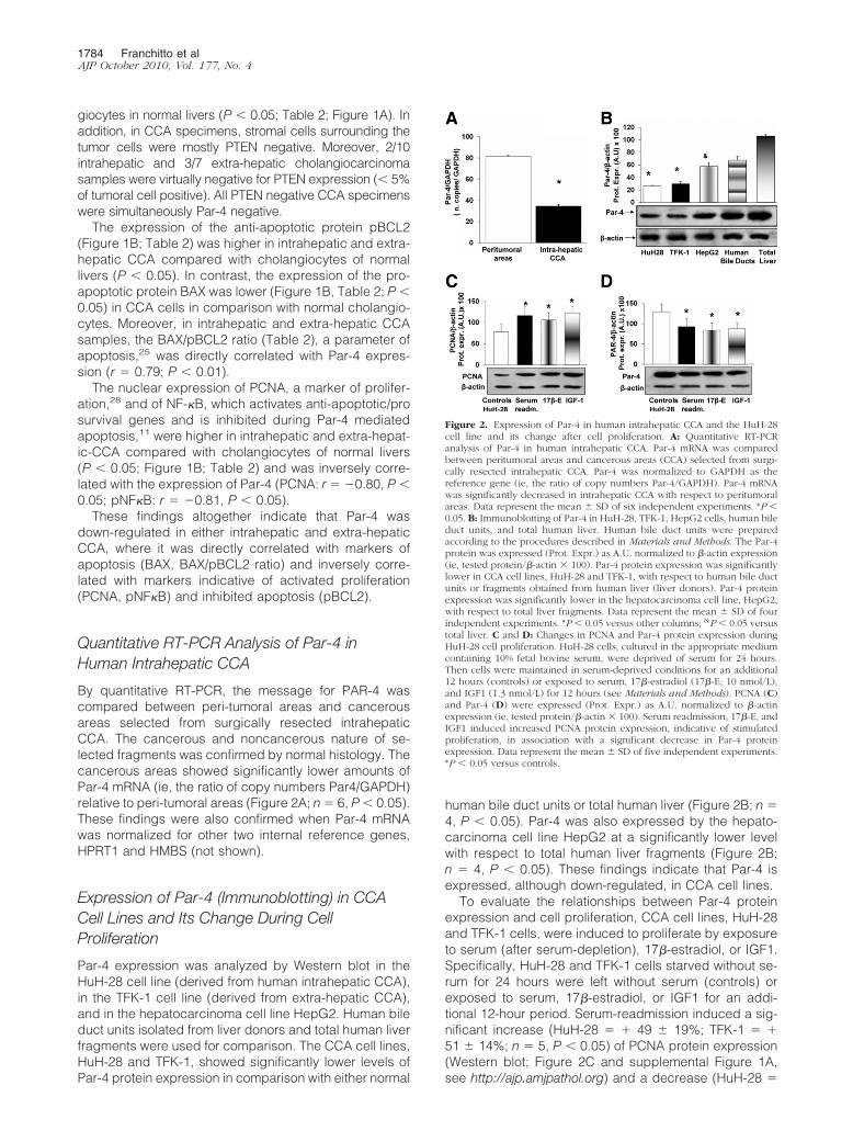
By quantitative RT-PCR, the message for PAR-4 wascompared between peri-tumoral areas and cancerousareas selected from surgically resected intrahepaticCCA. The cancerous and noncancerous nature of se-lected fragments was confirmed by normal histology. Thecancerous areas showed significantly lower amounts ofPar-4 mRNA (ie, the ratio of copy numbers Par4/GAPDH)relative to peri-tumoral areas (Figure 2A; n � 6, P � 0.05).These findings were also confirmed when Par-4 mRNAwas normalized for other two internal reference genes,HPRT1 and HMBS (not shown).
Expression of Par-4 (Immunoblotting) in CCACell Lines and Its Change During CellProliferation
Par-4 expression was analyzed by Western blot in theHuH-28 cell line (derived from human intrahepatic CCA),in the TFK-1 cell line (derived from extra-hepatic CCA),and in the hepatocarcinoma cell line HepG2. Human bileduct units isolated from liver donors and total human liverfragments were used for comparison. The CCA cell lines,HuH-28 and TFK-1, showed significantly lower levels ofPar-4 protein expression in comparison with either normal
human bile duct units or total human liver (Figure 2B; n �4, P � 0.05). Par-4 was also expressed by the hepato-carcinoma cell line HepG2 at a significantly lower levelwith respect to total human liver fragments (Figure 2B;n � 4, P � 0.05). These findings indicate that Par-4 isexpressed, although down-regulated, in CCA cell lines.
To evaluate the relationships between Par-4 proteinexpression and cell proliferation, CCA cell lines, HuH-28and TFK-1 cells, were induced to proliferate by exposureto serum (after serum-depletion), 17�-estradiol, or IGF1.Specifically, HuH-28 and TFK-1 cells starved without se-rum for 24 hours were left without serum (controls) orexposed to serum, 17�-estradiol, or IGF1 for an addi-tional 12-hour period. Serum-readmission induced a sig-nificant increase (HuH-28 � � 49 � 19%; TFK-1 � �51 � 14%; n � 5, P � 0.05) of PCNA protein expression(Western blot; Figure 2C and supplemental Figure 1A,see http://ajp.amjpathol.org) and a decrease (HuH-28 �
Figure 2. Expression of Par-4 in human intrahepatic CCA and the HuH-28cell line and its change after cell proliferation. A: Quantitative RT-PCRanalysis of Par-4 in human intrahepatic CCA. Par-4 mRNA was comparedbetween peritumoral areas and cancerous areas (CCA) selected from surgi-cally resected intrahepatic CCA. Par-4 was normalized to GAPDH as thereference gene (ie, the ratio of copy numbers Par-4/GAPDH). Par-4 mRNAwas significantly decreased in intrahepatic CCA with respect to peritumoralareas. Data represent the mean � SD of six independent experiments. *P �0.05. B: Immunoblotting of Par-4 in HuH-28, TFK-1, HepG2 cells, human bileduct units, and total human liver. Human bile duct units were preparedaccording to the procedures described in Materials and Methods. The Par-4protein was expressed (Prot. Expr.) as A.U. normalized to �-actin expression(ie, tested protein/�-actin � 100). Par-4 protein expression was significantlylower in CCA cell lines, HuH-28 and TFK-1, with respect to human bile ductunits or fragments obtained from human liver (liver donors). Par-4 proteinexpression was significantly lower in the hepatocarcinoma cell line, HepG2,with respect to total liver fragments. Data represent the mean � SD of fourindependent experiments. *P � 0.05 versus other columns; &P � 0.05 versustotal liver. C and D: Changes in PCNA and Par-4 protein expression duringHuH-28 cell proliferation. HuH-28 cells, cultured in the appropriate mediumcontaining 10% fetal bovine serum, were deprived of serum for 24 hours.Then cells were maintained in serum-deprived conditions for an additional12 hours (controls) or exposed to serum, 17�-estradiol (17�-E; 10 nmol/L),and IGF1 (1.3 nmol/L) for 12 hours (see Materials and Methods). PCNA (C)and Par-4 (D) were expressed (Prot. Expr.) as A.U. normalized to �-actinexpression (ie, tested protein/�-actin � 100). Serum readmission, 17�-E, andIGF1 induced increased PCNA protein expression, indicative of stimulatedproliferation, in association with a significant decrease in Par-4 proteinexpression. Data represent the mean � SD of five independent experiments.*P � 0.05 versus controls.
1784 Franchitto et alAJP October 2010, Vol. 177, No. 4

�46 � 4%; TFK-1 � �52 � 8%; n � 4, P � 0.05, notshown) of caspase 3 activity (apoptosis) with respect tocontrols. In these experiments, in association with prolif-eration induced by serum-readmission, a 28 � 19%(HuH-28, n � 5, P � 0.05) and a 27 � 12% (TFK-1, n �5, P � 0.05) decrease of Par-4 protein expression withrespect to controls was observed (Figure 2D and supple-mental Figure 1B, see http://ajp.amjpathol.org). Similarfindings were observed when serum-deprived (24 hours)HuH-28 and TFKI-1 cells were induced to proliferate byexposure to 10 nmol/L 17�-estradiol or 1.3 nmol/L IGF1.As previously described,23,24,28 17�-estradiol and IGF1induced a significant increase of PCNA protein expres-sion (HuH-28 � � 36 � 15% for 17�-estradiol and �58 � 14% for IGF1; TFK-1 � � 40 � 12% for 17�-estradiol and � 40 � 11% for IGF1; n � 5, P � 0.05versus controls; Figure 2C and supplemental Figure 1A,see http://ajp.amjpathol.org) and a significant decrease ofapoptosis (caspase 3 activity: 17�-estradiol: HuH-28 ��48 � 5.1%; TFK-1 � �44 � 4%; IGF1: HuH-28 ��47 � 4%; TFK-1 � �50 � 6%; n � 5, P � 0.05 versuscontrols, not shown). In association with induced prolif-eration, Par-4 protein expression (Western blot) wasfound to be decreased in HuH-28 cells by 34 � 18% and31 � 14% (n � 5, P � 0.05), respectively, by 17�-estradiol or IGF1 with respect to controls (Figure 2D). InTFK-1 cells, Par-4 protein expression was found to bedecreased by 28 � 15% and 28 � 14% (n � 5, P � 0.05;supplemental Figure 1B, see http://ajp.amjpathol.org),respectively, by 17�-estradiol or IGF1 with respect to con-trols. These findings indicate that in CCA cell lines, cell prolif-eration is associated with decreased expression of Par-4.
Changes of PAR-4 Expression during Apoptosisof CCA Cell Lines
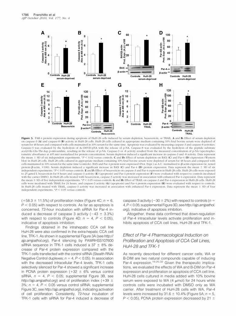
Par-4 protein has been shown to function as effector ofcell death in response to various apoptotic stimuli thattrigger mitochondria and membrane receptor-mediatedcell death pathways.10,29 To investigate the relationshipsbetween Par-4 protein expression and apoptosis of CCAcells, we evaluated different apoptotic stimuli, includingserum depletion or exposure to beauvericin or TRAIL.
As previously described,23,24,28 HuH-28 cells culturedin media containing 10% bovine serum were depleted ofserum (ie, exposed to serum-free medium) for 48 hours.After 48 hours of serum-depletion, the activities ofcaspase-3 (�48 � 6% versus controls, n � 6, Figure 3A)and caspase-8 (� 55 � 3% versus controls, n � 6, Figure3B) were significantly (P � 0.03) increased in HuH-28cells and this was associated with enhanced BAX proteinexpression (�38 � 2% versus controls, n � 6, Figure3C). In these experiments, in association with apoptosisinduced by serum depletion, the protein expression ofPar-4 was significantly increased (�49 � 9.8% versuscontrols, Figure 3D; n � 6, P � 0.05).
Beuavericin induces apoptosis by multiple cellular/mo-lecular pathways.30 After exposure of HuH-28 cells to 25�mol/L beauvericin for 8 hours, caspase-3 activity in-creased by 74 � 4% versus controls (Figure 3E, n � 5,
P � 0.02) and caspase-8 activity increased from 2.85 �0.15 (controls) to 5.28 � 0.20 (spectrophotometric absor-bance at 405 nm, arbitrary densitometric units [A.U.]; n �5, P � 0.05) confirming effective apoptosis, and this wasassociated with a significant increase (�76 � 10% ver-sus controls, Figure 3F; n � 5, P � 0.05) of Par-4 proteinexpression.
Finally, selective activation of extrinsic pathway of ap-optosis by TRAIL31 induced a 43 � 5% increase ofcaspase-3 activity (P � 0.05 versus controls, Figure 3G;n � 4) and caspase-8 activity (from 2.80 � 0.16 in con-trols to 8.67 � 0.92 spectrophotometric absorbance at405 nm, A.U.; n � 4; P � 0.01) in association with asignificant increase of Par-4 protein expression (�65 �9% versus controls, Figure 3H; n � 4, P � 0.05).
Results obtained with HuH-28 cells were also con-firmed in the extra-hepatic CCA cell line, TFK-1. In fact, inTFK-1 cells depleted of serum for 48 hours, the activity ofcaspase-3 (�70 � 4% versus controls, n � 4, P � 0.05;supplemental Figure 2A, see http://ajp.amjpathol.org) wasincreased and this was associated with a significant in-crease of Par-4 protein expression (�64 � 4% versuscontrols, n � 4, P � 0.05; supplemental Figure 2B, seehttp://ajp.amjpathol.org).
Together, these findings indicate that Par-4 proteinexpression increases during apoptosis of CCA cell linesinduced by different stimuli.
Effect of Selective Par-4 Silencing onProliferation and Apoptosis of HuH-28 Cells
To evaluate the effects of decreased Par-4 intracellularlevels on proliferation and apoptosis, HuH-28 cells wereincubated for 72 hours with specific Par-4 siRNA or con-trol (scrambled) RNA. We tested three different se-quences of Stealth RNAi kit, synthesized by Invitrogen.The PAWRHSS107600 sequence induced a 50 � 2.6%decrease of Par-4 protein expression compared with thecells transfected with the control siRNA (Stealth RNAiNegative Control duplexes; Figure 4A; n � 4, P � 0.02).The other two tested sequences (PAWRHSS107599 andPAWRHSS181749) induced respectively a 37 � 3.9%and 28 � 7.1% decrease of Par-4 protein expressioncompared with the cells transfected with the controlsiRNA (n � 4, Stealth RNAi Negative Control duplexes;data not shown). On the basis of these findings, wetherefore evaluated the effect of Par-4 silencing byPAWRHSS107600 siRNA sequence on proliferation andapoptosis of HuH-28 cells. As positive control, we usedStealth RNAi GAPDH positive control duplexes (Invitro-gen), according to the manufacturer’s instructions. Inassociation with the decreased intracellular Par-4 levels,HuH-28 cells selectively silenced for Par-4 showed asignificant increase in PCNA protein expression (Westernblot), indicative of induced cell proliferation (Figure 4B).In fact, PCNA protein expression was significantly in-creased (�46.8 � 4.2%) in HuH-28 silenced cells withrespect to control siRNA (Figure 4B; n � 4, P � 0.02).These data were confirmed by MTS-assay. In fact, treat-ment with Par-4 siRNA induced a marked increase
Par-4 and Cholangiocarcinoma 1785AJP October 2010, Vol. 177, No. 4
(�58.3 � 11.5%) of proliferation index (Figure 4C; n � 6,P � 0.05) with respect to controls. As far as apoptosis isconcerned, 72-hour incubation with siRNA for Par-4 in-duced a decrease of caspase 3 activity (�43 � 3.3%)with respect to controls (Figure 4D; n � 4, P � 0.05),indicative of apoptosis inhibition.
Findings obtained in the intrahepatic CCA cell lineHuH-28 were also confirmed in the extra-hepatic CCA cellline, TFK-1. As shown in supplemental Figure 3A (see http://ajp.amjpathol.org), Par-4 silencing by PAWRHSS107600siRNA sequence in TFK-1 cells induced a 37 � 8% de-crease of Par-4 protein expression compared with theTFK-1 cells transfected with the control siRNA (Stealth RNAiNegative Control duplexes; n � 4, P � 0.05). In associationwith the decreased intracellular Par-4 levels, TFK-1 cellsselectively silenced for Par-4 showed a significant increasein PCNA protein expression (�32 � 6% versus controlsiRNA, n � 4, P � 0.05; supplemental Figure 3B, seehttp://ajp.amjpathol.org) and of proliferation index (�39 �3%; n � 4, P � 0.05 versus control siRNA; supplementalFigure 3C, see http://ajp.amjpathol.org), indicating activationof cell proliferation. Consistently, 72-hour incubation ofTFK-1 cells with siRNA for Par-4 induced a decrease of
caspase 3 activity (�30 � 2%) with respect to controls (n �4,P�0.05;supplementalFigure3D,seehttp://ajp.amjpathol.org), indicative of apoptosis inhibition.
Altogether, these data confirmed that down-regulationof Par-4 intracellular levels activate proliferation and in-hibits apoptosis of CCA cell lines, HuH-28 and TFK-1.
Effect of Par-4 Pharmacological Induction onProliferation and Apoptosis of CCA Cell Lines,HuH-28 and TFK-1
As recently described for different cancer cells, WA orB-DIM are two natural compounds capable of inducingPar-4 expression.19,20,32 Given the therapeutic implica-tions, we evaluated the effects of WA and B-DIM on Par-4expression and proliferation or apoptosis of CCA cell line.HuH-28 cells cultured in media added with 10% bovineserum were exposed to WA (4 �mol) for 24 hours whilecontrols cells were incubated with DMSO only as WAcarrier. After treatment of HuH-28 cells with WA, Par-4levels were increased by 31.6 � 10.4% (Figure 5A; n � 5,P � 0.05), PCNA protein expression decreased by 21 �
Figure 3. PAR-4 protein expression during apoptosis of HuH-28 cells induced by serum depletion, beauvericin, or TRAIL. A and B: Effect of serum depletionon caspase-3 (A) and caspase-8 (B) activity in HuH-28 cells. HuH-28 cells cultured in appropriate medium containing 10% fetal bovine serum were depleted ofserum for 48 hours and compared with cells maintained in 10% serum for the same time. Apoptosis was evaluated by measuring caspase-3 and caspase-8 activities.Caspase-3 was evaluated by the hydrolysis of Ac-DEVD-pNA with the release of p-NA. Caspase-8 was evaluated by the hydrolysis of the peptide substrateacetyl-lle-Glu-Thr-Asp p-nitroaniline, resulting in the release of p-NA. Caspase-3 or -8 activity resulted from the measured concentration of p-NA (spectropho-tometric absorbance at 405 nm) normalized for protein concentration. Serum depletion induced a significant increase in caspase-3 and -8 activity. Data representthe mean � SD of six independent experiments. *P � 0.02 versus controls. C and D: Effect of serum depletion on BAX (C) and Par-4 (D) expression (Westernblot) in HuH-28 cells. HuH-28 cells cultured in appropriate medium containing 10% fetal bovine serum were depleted of serum for 48 hours and compared withcells maintained in 10% serum for the same time (controls). BAX and Par-4 protein were expressed (Prot. Expr.) as A.U. normalized to �-actin expression (ie, testedprotein/�-actin, �100). Serum depletion induced a significant increase in BAX (C) and Par-4 (D) protein expression. Data represent the mean � SD of sixindependent experiments. *P� 0.05 versus controls. E and F: Effect of beuavericin on caspase-3 and Par-4 expression in HuH-28 cells. HuH-28 cells were exposedto 25 �mol/L beuavericin for 8 hours and caspase-3 activity (E) (apoptosis) and Par-4 protein expression (F) were evaluated with respect to controls incubatedwith the carrier DMSO. In HuH-28 cells treated with beuavericin, caspase-3 activity was increased in association with enhanced Par-4 expression. Data representthe mean � SD of five independent experiments. *P � 0.05 versus controls. G and H: Effect of TRAIL on caspase-3 and Par-4 expression in HuH-28 cells. HuH-28cells were incubated with TRAIL for 24 hours, and caspase-3 activity (G) (apoptosis) and Par-4 protein expression (H) were evaluated with respect to controls.In HuH-28 cells treated with TRAIL, caspase-3 activity was increased in association with enhanced Par-4 expression. Data represent the mean � SD of fourindependent experiments. *P � 0.05 versus controls.
1786 Franchitto et alAJP October 2010, Vol. 177, No. 4
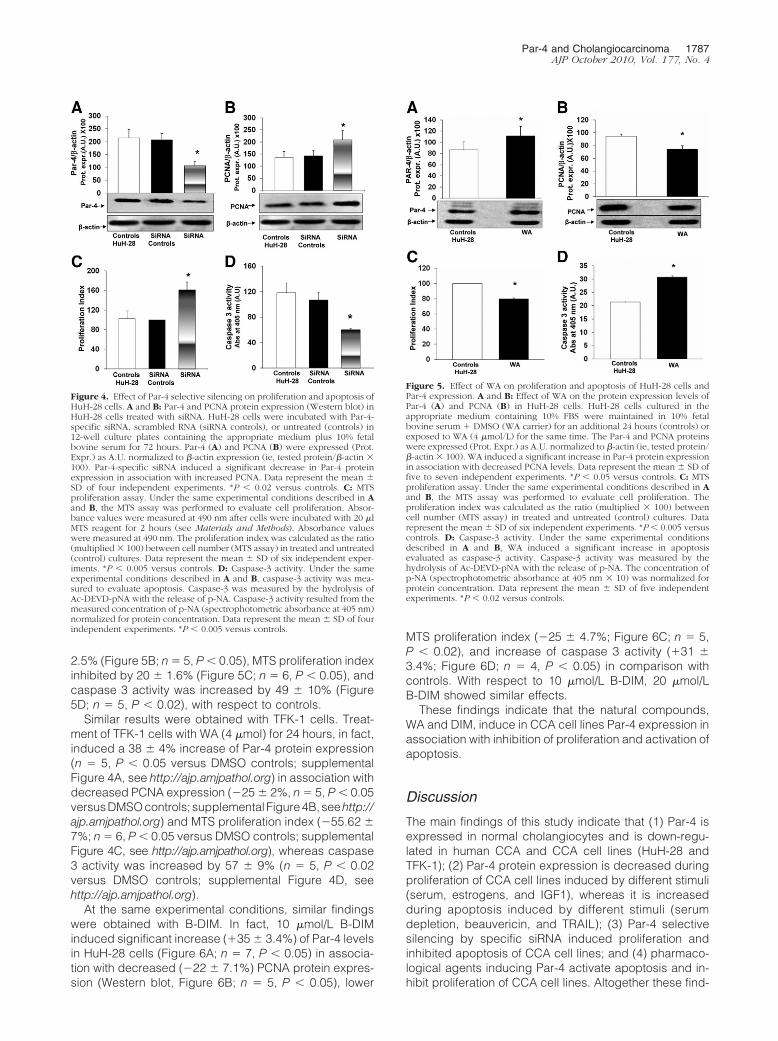
2.5% (Figure 5B; n � 5, P � 0.05), MTS proliferation indexinhibited by 20 � 1.6% (Figure 5C; n � 6, P � 0.05), andcaspase 3 activity was increased by 49 � 10% (Figure5D; n � 5, P � 0.02), with respect to controls.
Similar results were obtained with TFK-1 cells. Treat-ment of TFK-1 cells with WA (4 �mol) for 24 hours, in fact,induced a 38 � 4% increase of Par-4 protein expression(n � 5, P � 0.05 versus DMSO controls; supplementalFigure 4A, see http://ajp.amjpathol.org) in association withdecreased PCNA expression (�25 � 2%, n � 5, P � 0.05versusDMSOcontrols; supplemental Figure4B,seehttp://ajp.amjpathol.org) and MTS proliferation index (�55.62 �7%; n � 6, P � 0.05 versus DMSO controls; supplementalFigure 4C, see http://ajp.amjpathol.org), whereas caspase3 activity was increased by 57 � 9% (n � 5, P � 0.02versus DMSO controls; supplemental Figure 4D, seehttp://ajp.amjpathol.org).
At the same experimental conditions, similar findingswere obtained with B-DIM. In fact, 10 �mol/L B-DIMinduced significant increase (�35 � 3.4%) of Par-4 levelsin HuH-28 cells (Figure 6A; n � 7, P � 0.05) in associa-tion with decreased (�22 � 7.1%) PCNA protein expres-sion (Western blot, Figure 6B; n � 5, P � 0.05), lower
MTS proliferation index (�25 � 4.7%; Figure 6C; n � 5,P � 0.02), and increase of caspase 3 activity (�31 �3.4%; Figure 6D; n � 4, P � 0.05) in comparison withcontrols. With respect to 10 �mol/L B-DIM, 20 �mol/LB-DIM showed similar effects.
These findings indicate that the natural compounds,WA and DIM, induce in CCA cell lines Par-4 expression inassociation with inhibition of proliferation and activation ofapoptosis.
Discussion
The main findings of this study indicate that (1) Par-4 isexpressed in normal cholangiocytes and is down-regu-lated in human CCA and CCA cell lines (HuH-28 andTFK-1); (2) Par-4 protein expression is decreased duringproliferation of CCA cell lines induced by different stimuli(serum, estrogens, and IGF1), whereas it is increasedduring apoptosis induced by different stimuli (serumdepletion, beauvericin, and TRAIL); (3) Par-4 selectivesilencing by specific siRNA induced proliferation andinhibited apoptosis of CCA cell lines; and (4) pharmaco-logical agents inducing Par-4 activate apoptosis and in-hibit proliferation of CCA cell lines. Altogether these find-
Figure 4. Effect of Par-4 selective silencing on proliferation and apoptosis ofHuH-28 cells. A and B: Par-4 and PCNA protein expression (Western blot) inHuH-28 cells treated with siRNA. HuH-28 cells were incubated with Par-4-specific siRNA, scrambled RNA (siRNA controls), or untreated (controls) in12-well culture plates containing the appropriate medium plus 10% fetalbovine serum for 72 hours. Par-4 (A) and PCNA (B) were expressed (Prot.Expr.) as A.U. normalized to �-actin expression (ie, tested protein/�-actin �100). Par-4-specific siRNA induced a significant decrease in Par-4 proteinexpression in association with increased PCNA. Data represent the mean �SD of four independent experiments. *P � 0.02 versus controls. C: MTSproliferation assay. Under the same experimental conditions described in Aand B, the MTS assay was performed to evaluate cell proliferation. Absor-bance values were measured at 490 nm after cells were incubated with 20 �lMTS reagent for 2 hours (see Materials and Methods). Absorbance valueswere measured at 490 nm. The proliferation index was calculated as the ratio(multiplied � 100) between cell number (MTS assay) in treated and untreated(control) cultures. Data represent the mean � SD of six independent exper-iments. *P � 0.005 versus controls. D: Caspase-3 activity. Under the sameexperimental conditions described in A and B, caspase-3 activity was mea-sured to evaluate apoptosis. Caspase-3 was measured by the hydrolysis ofAc-DEVD-pNA with the release of p-NA. Caspase-3 activity resulted from themeasured concentration of p-NA (spectrophotometric absorbance at 405 nm)normalized for protein concentration. Data represent the mean � SD of fourindependent experiments. *P � 0.005 versus controls.
Figure 5. Effect of WA on proliferation and apoptosis of HuH-28 cells andPar-4 expression. A and B: Effect of WA on the protein expression levels ofPar-4 (A) and PCNA (B) in HuH-28 cells. HuH-28 cells cultured in theappropriate medium containing 10% FBS were maintained in 10% fetalbovine serum � DMSO (WA carrier) for an additional 24 hours (controls) orexposed to WA (4 �mol/L) for the same time. The Par-4 and PCNA proteinswere expressed (Prot. Expr.) as A.U. normalized to �-actin (ie, tested protein/�-actin � 100). WA induced a significant increase in Par-4 protein expressionin association with decreased PCNA levels. Data represent the mean � SD offive to seven independent experiments. *P � 0.05 versus controls. C: MTSproliferation assay. Under the same experimental conditions described in Aand B, the MTS assay was performed to evaluate cell proliferation. Theproliferation index was calculated as the ratio (multiplied � 100) betweencell number (MTS assay) in treated and untreated (control) cultures. Datarepresent the mean � SD of six independent experiments. *P � 0.005 versuscontrols. D: Caspase-3 activity. Under the same experimental conditionsdescribed in A and B, WA induced a significant increase in apoptosisevaluated as caspase-3 activity. Caspase-3 activity was measured by thehydrolysis of Ac-DEVD-pNA with the release of p-NA. The concentration ofp-NA (spectrophotometric absorbance at 405 nm � 10) was normalized forprotein concentration. Data represent the mean � SD of five independentexperiments. *P � 0.02 versus controls.
Par-4 and Cholangiocarcinoma 1787AJP October 2010, Vol. 177, No. 4

ings indicate that Par-4 plays a major role in modulatingproliferation and apoptosis of CCA cells.
Par-4, a tumor suppressor protein showing pro-apop-totic functions, has been shown to function as an effectorof cell death in response to various apoptotic stimuli thattrigger mitochondria and membrane receptor-mediatedcell death pathway.10–16,29 Epigenetic silencing of Par-4has been described in different tumors, and Par-4 knock-out mice develop spontaneous tumors in various tis-sues.11–13,17,18 At experimental level, it has been shownthat endogenous Par-4 sensitizes cells to diverse apo-ptotic stimuli and that ectopic expression of Par-4 canselectively induce apoptosis in cancer cells.14–18 Thecancer-specific pro-apoptotic action of Par-4 resides inits centrally located SAC (selective for apoptosis in can-cer cells) domain where SAC transgenic mice displaynormal development and life span, and, most importantly,are resistant to spontaneous, as well as oncogene-inducedautochthonous tumors.18 As far as mechanism of apoptosisinduction is concerned, in rat fibroblasts it has been recentlydemonstrated that Par-4 binds its interaction partner DAP-like kinase (Dlk/ZIP kinase) and induces translocation of thekinase from the nucleus to the actin filaments, with subse-quent myosin light chain phosphorylation and induction of
apoptosis.10,13,29,33,34 More recently, it was noted that Par-4protein is spontaneously secreted by normal and cancercells in culture as well as by tissues from transgenic miceoverexpressing Par-4 and showing resistance to spontane-ous tumors.13 In the same study, exposure to endoplasmicreticulum stress-inducing agents further increased cellularsecretion of Par-4, where extracellular Par-4 induced apo-ptosis by binding to the stress response surface protein,glucose-regulated protein-78.13 The interaction of extracel-lular Par-4 with glucose-regulated protein-78 leads to apo-ptosis via endoplasmic reticulum (ER) stress and activationof the Fas-associated death domain protein (FADD)/caspase-8/caspase-3 pathway. Also TRAIL inducible apo-ptosis was found to be dependent from extracellular Par-4signaling via cell surface glucose-regulated protein-78.13 Inthe last few years, Par-4 received large attention as a thera-peutic target since different pharmacological agents inducedPar-4 and showed, at the experimental levels, anticancer ef-fects.19–21,32 Most importantly, natural compounds able toinduce Par-4 expression have been identified and pro-posed for cancer chemoprevention with the hypothesisthat they should be able to suppress, reverse, or pre-vent the carcinogenic process from turning into ag-gressive cancer.19–21
CCA is a devastating cancer with frustrating responseto chemotherapeutics.1–5 In this study we focus on theexpression of Par-4 in normal and neoplastic cholangio-cytes and on the role of Par-4 in the modulation of prolif-eration and apoptosis of CCA cells.
Par-4 expression was investigated by IHC in smallfragments of intrahepatic CCA and peritumoral tissuesobtained from patients submitted to surgical resection. Inthese samples, the presence of CCA and its absence inperitumoral areas was demonstrated by normal histology.The IHC analysis showed Par-4 expression at the cyto-plasmic and nuclear levels. Although nuclear Par-4 rep-resents the most active form, different studies demon-strated that Par-4 may be active, in regulating cellproliferation/apoptosis, also in the cytoplasm where itmay act on specific PKC isoforms, which in turn mayinfluence the activity of both NF-�B (via IKK) andAKT.35,36 However, it is of interest that in normal cholan-giocytes Par-4 staining was also found in the nucleussuggesting a potential role in maintaining these cells intoa quiescent status. Most importantly, we showed howPar-4 IHC expression was markedly down-regulated inintrahepatic CCA cells in comparison with both peritu-moral areas and cholangiocytes of normal liver. Consis-tent with IHC findings, we also demonstrated by RT-PCRthat the message for Par-4 is decreased by 2.5-fold inintrahepatic CCA with respect to peri-tumoral areas, andthese findings were confirmed by normalizing RT-PCRresults with three different reference proteins. Par-4 ex-pression is also decreased in extra-hepatic CCA, and inour samples, Par-4 staining results directly correlatedwith the markers of apoptosis, BAX and BAX/BCL2 ra-tio,28 and was inversely correlated with direct (PCNA) orindirect (NF-�B) markers of proliferation.28 These findingsare suggestive that also in CCA, as occurs in other can-cers, Par-4 is strongly involved in the positive modulationof apoptotic machineries. In addition, the IHC investiga-
Figure 6. Effect of B-DIM on proliferation, apoptosis of HuH-28 cells, andPar-4 expression. A and B: Effect of B-DIM on the protein expression levelsof Par-4 (A) and PCNA (B) in HuH-28 cells. HuH-28 cells cultured in theappropriate medium containing 10% FBS were maintained in 10% FBS �DMSO (B-DIM carrier) for an additional 24 hours (controls) or exposed toB-DIM (10 or 20 �mol/L) for the same period. The Par-4 and PCNA proteinswere expressed (Prot. Expr.) as A.U. normalized to �-actin (ie, tested protein/�-actin � 100). B-DIM induced a significant increase in Par-4 protein expres-sion in association with decreased PCNA levels. No differences were foundbetween 10 or 20 �mol/L B-DIM. Data represent the mean � SD of five toseven independent experiments. *P � 0.05 versus controls. C: MTS prolifer-ation assay. Under the same experimental conditions described in A and B,the MTS assay was performed to evaluate cell proliferation. The proliferationindex was calculated as the ratio (multiplied � 100) between cell number(MTS assay) in treated and untreated (control) cultures. B-DIM (10 or 20�mol/L) induced a significant increase in the proliferation index. Data rep-resent the mean � SD of five independent experiments. *P � 0.02 versuscontrols. D: Caspase-3 activity. Under the same experimental conditionsdescribed in A and B, B-DIM (10 or 20 �mol/L) induced a significant increasein apoptosis evaluated as caspase-3 activity. Caspase-3 activity was measuredby the hydrolysis of Ac-DEVD-pNA with the release of p-NA. The concen-tration of p-NA (spectrophotometric absorbance at 405 nm � 10) wasnormalized for protein concentration. Data represent the mean � SD of fourindependent experiments. *P � 0.05 versus controls.
1788 Franchitto et alAJP October 2010, Vol. 177, No. 4

tion showed how the decreased Par-4 expression in CCAwas closely associated with decreased positivity for PTEN,both being correlated with increased indices of proliferation(bcl2 staining) and decreased indices of apoptosis (Baxstaining). This is in keeping with what demonstrated in hu-man prostatic cancer where Par-4 loss is concomitantlyassociated with PTEN loss.7,8 In experimental models, Par-4null mice or PTEN-heterozygous mice only develop benignlesions while only concomitant Par-4 ablation and PTEN-heterozygosity lead to invasive prostate cancer in mice, thetumorigenic Par-4/PTEN cooperation being anticipated inthe preneoplastic prostate epithelium by an additive in-crease in Akt and NF-�B activation.11 Furthermore, Par-4deficiency cooperates with PTEN haploinsufficiency in pros-tate cancer initiation and progression and their simulta-neous inactivation, in addition to enhancing Akt activation,causes the synergistic activation of NF-�B. PTEN expres-sion has been demonstrated to be decreased in extra-hepatic CCA and to correlate with worst survival,37 andintrahepatic CCA has been experimentally induced by liver-specific disruption of Smad4 and PTEN in mice.38 There-fore, impairment of PTEN pro-apoptotic functions certainlyplays a role in CCA pathogenesis. Our IHC findings inhuman CCA samples suggest, as shown in other can-cers,10 a close relationship between PTEN and Par-4 down-regulation and CCA cell proliferation.
We next investigated, in functional studies, in two dif-ferent CCA cell lines, derived from intrahepatic (ie, HuH-28) or extra-hepatic (ie, TFK-1) human CCA, the expres-sion of Par-4 and its relationships with cell proliferationand apoptosis. The two CCA cell lines showed lowerlevels of Par-4 protein expression with respect to totalliver and intrahepatic bile ducts. A hepatocarcinoma cellline, HepG2, also showed Par-4 expression at lower levelwith respect to total liver indicating that, in the liver, Par-4is not exclusively involved in modulating cholangiocytebut also hepatocyte pathophysiology. As far as functionalstudies are concerned, we demonstrated that when CCAcell lines were induced to proliferate by serum-readmis-sion or exposure to IGF1 or 17�-estradiol, in all theseconditions, a significant decrease of Par-4 expressionwas observed. As far as apoptosis is concerned, Par-4 iscurrently considered an effector of cell death in responseto various apoptotic stimuli that trigger mitochondria andmembrane receptor-mediated cell death pathways.29
Therefore, we investigated apoptosis of CCA cell linesinduced by serum depletion, beuavericin, and by TRAIL,the latter being a selective activator of extrinsic apoptoticpathways.31 With all these different apoptotic stimuli,Par-4 protein expression was up-regulated in associationwith enhanced caspase-3 and -8 activities, respectivelyterminator and initiator of the apoptotic cascade, and withenhanced expression of the pro-apoptotic protein BAX.This latter finding is consistent with the IHC results ob-tained in human CCA biopsies and showing a positivecorrelation between BAX and Par-4 staining. Therefore,the results of functional studies confirmed the role ofPar-4 in CCA cells as pro-apoptotic and anti-proliferativeprotein documented in other different cell types. This wasdefinitively confirmed by selective Par-4 silencing, whichinduced proliferation of HuH-28 and TKF-1 cells. In these
latter experiments, a 50% decrease of Par-4 protein leveldetermined a 58% increase of proliferation index.
Given the relevance of Par-4 in controlling apoptoticpathways, a bulk of recent literature deals with pharma-cological or genetic induction of Par-4 expression asanti-cancer strategy.19–21 Specific attention was given tocancer chemoprevention, which focuses on natural, syn-thetic, or biological agents capable to suppress, reverse,or prevent the carcinogenic process from turning intoaggressive cancer. We tested in HuH-28 and/or TFK-1cells, two natural compounds (WA and B-DIM) with ther-apeutic potential against cancer and minimal toxicityagainst normal cells and which have been described asinducers of Par-4.19,20 WA, a highly oxygenated C-28ergostane-type steroid, is a major constituent of the me-dicinal plant W. somnifera, identified as natural agent withtherapeutic potential against cancer and preferably min-imal toxicity against normal cells.19,39,40 The growth-in-hibitory properties in tumor cell culture studies have beenlargely documented with the evidence that its action isdependent on Par-4 function in both androgen-responsiveand androgen refractory prostate cancer cells.19,39,40
Our results indicate that WA induces apoptosis and in-hibit proliferation of CCA cell lines, HuH-28 and TFK-1,and this is in association with an increased expression ofPar-4, increased caspase-3 activity, and decreasedPCNA expression. The same effect was found with B-DIM, tested only in HuH-28 cells, which is another naturalcompound found in cruciferous vegetables, recently de-scribed as one of the most effective phytonutrients withpotential in the prevention and treatment of breast, pros-tate, colon, and pancreatic cancers.20,21 We tested twodifferent doses of B-DIM without differences in Par-4induction, which, in addition, was of the similar entity withrespect to the induction obtained with WA. In a previousstudy,32 DIM has been shown to promote Fas-mediatedapoptosis of CCA cell lines by inhibiting phosphorylationof AKT and activation of FLICE (Fas-associated deathdomain-like interleukin-1beta-converting enzyme) like-in-hibitory-protein, FLIP. In keeping with previous stud-ies,20,21 our findings indicate that Par-4 induction is anadditional mechanism involved in B-DIM induced apo-ptosis of CCA cells, where PTEN (AKT antagonist) couldalso be taken into consideration. We demonstrated thatWA and B-DIM impair proliferation and induce apoptosisof HuH-28 cells without affecting viability of normalcholangiocytes, thus excluding toxicity for normal cells.
Cancer chemoprevention is defined as the use of natural,synthetic, or biological agents to suppress, reverse, or pre-vent the carcinogenic process from turning into aggressivecancer. A number of pathological conditions are at risk ofCCA development including primary sclerosing cholangitis(PSC), liver flukes, surgical bilio-enteric anastomosis, etc.1–
5,41 For all these categories at risk, no chemopreventionstrategy has been so far investigated. Our findings showingthe major role of Par-4 as modulator of apoptosis shouldstimulate clinical trials aimed to evaluate the role ofnatural compounds inducing Par-4 as chemotherapeu-tic agents preventing CCA developments. Finally,since no chemotherapy has been demonstrated to beof great benefit in CCA treatment at this time, drugs or
Par-4 and Cholangiocarcinoma 1789AJP October 2010, Vol. 177, No. 4

genetic strategies selectively targeting Par-4 should beinvestigated.
References
1. Blechacz B, Gores GJ: Cholangiocarcinoma: advances in pathogen-esis, diagnosis, and treatment. Hepatology 2008, 48(1):308–321
2. Khan SA, Thomas HC, Davidson BR, Taylor-Robinson SD: Cholan-giocarcinoma. Lancet 2005, 366(9493):1303–1314
3. Patel T: Worldwide trends in mortality from biliary tract malignancies.BMC Cancer 2002, 2:10
4. Fava G, Marzioni M, Benedetti A, Glaser S, DeMorrow S, Francis H,Alpini G: Molecular pathology of biliary tract cancers. Cancer Lett2007, 250(2):155–167
5. Alvaro D, Mancino MG: New insights on the molecular and cell biologyof human cholangiopathies. Mol Aspects Med 2008, 29(1–2):50–57
6. Alvaro D, Crocetti E, Ferretti S, Bragazzi MC, Capocaccia R; the AISF“Cholangiocarcinoma” Committee. Descriptive epidemiology of cholan-giocarcinoma in Italy. Dig Liver Dis 2009, Dec 18. [Epub ahead of print]
7. Sells SF, Wood DP, Joshi-Barve SS, Muthukumar S, Jacob RJ, CristSA, Humphreys S, Rangnekar VM: Commonality of the gene pro-grams induced by effectors of apoptosis in androgen-dependent and-independent prostate cells. Cell Growth Differ 1994, 5:457–466
8. Goswami A, Qiu S, Dexheimer TS, Ranganathan P, Burikhanov R,Pommier Y, Rangnekar VM: Par-4 binds to topoisomerase 1 andattenuates its DNA relaxation activity. Cancer Res 2008, 68:1–9
9. Lee TJ, Lee JT, Kim SH, Choi YH, Song KS, Park JW, Kwon TK:Overexpression of Par-4 enhances thapsigargin-induced apoptosisvia down-regulation of XIAP and inactivation of Akt in human renalcancer cells. J Cell Biochem 2008, 103(2):358–368
10. Diaz-Meco MT, Abu-Baker S: The Par-4/PTEN connection in tumorsuppression. Cell Cycle 2009, 8(16):2518–2522
11. Fernandez-Marcos PJ, Abu-Baker S, Joshi J, Galvez A, Castilla EA,Canamero M, Collado M, Saez C, Moreno-Bueno G, Palacios J,Leitges M, Serrano M, Moscat J, Diaz-Meco MT: Simultaneous inac-tivation of Par-4 and PTEN in vivo leads to synergistic NF-kappaBactivation and invasive prostate carcinoma. Proc Natl Acad Sci USA2009, 106(31):12962–12967
12. Kline CL, Shanmugavelandy SS, Kester M, Irby RB: Delivery of PAR-4plasmid in vivo via nanoliposomes sensitizes colon tumor cells sub-cutaneously implanted into nude mice to 5-FU. Cancer Biol Ther2009, 22:1831–1837
13. Burikhanov R, Zhao Y, Goswami A, Qiu S, Schwarze SR, RangnekarVM: The tumor suppressor Par-4 activates an extrinsic pathway forapoptosis. Cell 2009, 138(2):377–388
14. Moscat J, Diaz-Meco MT, Wooten MW: Of the atypical PKCs, Par-4and p62: recent understandings of the biology and pathology of aPB1-dominated complex. Cell Death Differ 2009, 16(11):1426–1437
15. Moreno-Bueno G, Fernandez-Marcos PJ, Collado M, Tendero MJ, Ro-driguez-Pinilla SM, Garcia-Cao I, Hardisson D, Diaz-Meco MT, MoscatJ, Serrano M, Palacios J: Inactivation of the candidate tumor suppressorpar-4 in endometrial cancer. Cancer Res 2007, 67:1927–1934
16. García-Cao I, Duran A, Collado M, Carrascosa MJ, Martín-CaballeroJ, Flores JM, Diaz-Meco MT, Moscat J, Serrano M: Tumour-suppres-sion activity of the proapoptotic regulator Par4. EMBO Rep 2005,6:577–583
17. Barradas M, Monjas A, Diaz-Meco MT, Serrano M, Moscat J: The down-regulation of the pro-apoptotic protein Par-4 is critical for Ras-inducedsurvival and tumor progression. EMBO J 1999, 18(22):6362–6369
18. Zhao Y, Burikhanov R, Qiu S, Lele SM, Jennings CD, Bondada S,Spear B, Rangnekar VM: Cancer resistance in transgenic mice ex-pressing the SAC module of Par-4. Cancer Res 2007, 67:9276–9285
19. Srinivasan S, Ranga RS, Burikhanov R, Han SS, Chendil D: Par-4-dependent apoptosis by the dietary compound withaferin A in pros-tate cancer cells. Cancer Res 2007, 67(1):246–253
20. Azmi AS, Ahmad A, Banerjee S, Rangnekar VM, Mohammad RM,Sarkar FH: Chemoprevention of pancreatic cancer: characterizationof Par-4 and its modulation by 3,3� diindolylmethane (DIM). PharmRes 2008, 25(9):2117–2124
21. Azmi AS, Wang Z, Burikhanov R, Rangnekar VM, Wang G, Chen J,Wang S, Sarkar FH, Mohammad RM: Critical role of prostate apopto-sis response-4 in determining the sensitivity of pancreatic cancer
cells to small-molecule inhibitor-induced apoptosis. Mol Cancer Ther2008, 7(9):2884–2893
22. Mennone A, Alvaro D, Cho W, Boyer JL: Isolation of small polarizedbile duct units. Proc Natl Acad Sci USA 1995, 92(14):6527–6531
23. Alvaro D, Barbaro B, Franchitto A, Onori P, Glaser SS, Alpini G,Francis H, Marucci L, Sterpetti P, Ginanni-Corradini S, Onetti Muda A,Dostal DE, De Santis A, Attili AF, Benedetti A, Gaudio E: Estrogensand insulin-like growth factor 1 modulate neoplastic cell growth inhuman cholangiocarcinoma. Am J Pathol 2006, 169(3):877–888
24. Mancino A, Mancino MG, Glaser SS, Alpini G, Bolognese A, Izzo L,Francis H, Onori P, Franchitto A, Ginanni-Corradini S, Gaudio E,Alvaro D: Estrogens stimulate the proliferation of human cholangio-carcinoma by inducing the expression and secretion of vascularendothelial growth factor. Dig Liver Dis 2009, 41(2):156–163
25. Schroeder A, Mueller O, Stocker S, Salowsky R, Leiber M, GassmannM, Lightfoot S, Menzel W, Granzow M, Ragg T: The RIN: an RNAintegrity number for assigning integrity values to RNA measurements.BMC Mol Biol 2006, 7:3
26. Cantafora A, Blotta I, Rivabene R, Pisciotta L, Bertolini S: Evaluation ofRNA messengers involved in lipid trafficking of human intestinal cellsby reverse-transcription polymerase chain reaction with competimertechnology and microchip electrophoresis. S Electrophoresis 2003,24:3748–3754
27. Vandesompele J, De Preter K, Pattyn F, Poppe B, Van Roy N, DePaepe A, Speleman F: Accurate normalization of real-time quantita-tive RT-PCR data by geometric averaging of multiple internal controlgenes. Genome Biol 2002, 3(7):RESEARCH0034
28. Onori P, Alvaro D, Floreani AR, Mancino MG, Franchitto A, Guido M,Carpino G, De Santis A, Angelico M, Attili AF, Gaudio E: Activation ofthe IGF1 system characterizes cholangiocyte survival during pro-gression of primary biliary cirrhosis. J Histochem Cytochem 2007,55(4):327–334
29. Lee TJ, Jang JH, Noh HJ, Park EJ, Choi KS, Kwon TK: Overexpres-sion of Par-4 sensitizes TRAIL-induced apoptosis via inactivation ofNF-kappaB and Akt signaling pathways in renal cancer cells. J CellBiochem 2010, 109(5):885–895
30. Lin HI, Lee YJ, Chen BF, Tsai MC, Lu JL, Chou CJ, Jow GM: Involve-ment of Bcl-2 family, cytochrome c and caspase 3 in induction ofapoptosis by beauvericin in human non-small cell lung cancer cells.Cancer Lett 2005, 230(2):248–259
31. Mellier G, Huang S, Shenoy K, Pervaiz S: TRAILing death in cancer.Mol Aspects Med 2010, 31(1):93–112
32. Chen Y, Xu J, Jhala N, Pawar P, Zhu ZB, Ma L, Byon CH, McDonald JM:Fas-mediated apoptosis in cholangiocarcinoma cells is enhanced by3,3�-diindolylmethane through inhibition of AKT signaling and FLICE-likeinhibitory protein. Am J Pathol 2006, 169(5):1833–1842
33. Zhao Y, Rangnekar VM: Apoptosis and tumor resistance conferred byPar-4. Cancer Biol Ther 2008, 7(12):1867–1874
34. Page G, Kogel D, Rangnekar V, Scheidtmann KH: Interaction partners ofDlk/ZIP kinase: co-expression of Dlk/ZIP kinase and Par-4 results incytoplasmic retention and apoptosis. Oncogene 1999, 18:7265–7273
35. Díaz-Meco MT, Municio MM, Frutos S, Sanchez P, Lozano J, Sanz L,Moscat J: The product of par-4, a gene induced during apoptosis,interacts selectively with the atypical isoforms of protein kinase C.Cell 1996, 86:777–786
36. Diaz-Meco MT, Lallena MJ, Monjas A, Frutos S, Moscat J: Inactivationof the inhibitory kappaB protein kinase/nuclear factor kappaB path-way by Par-4 expression potentiates tumor necrosis factor alpha-induced apoptosis. J Biol Chem 1999, 274(1):9606–9612
37. Chung JY, Hong SM, Choi BY, Cho H, Yu E, Hewitt SM: The expres-sion of phospho-AKT, phospho-mTOR, and PTEN in extrahepaticcholangiocarcinoma. Clin Cancer Res 2009, 15(2):660–667
38. Xu X, Kobayashi S, Qiao W, LiC J: Induction of intrahepatic cholan-giocellular carcinoma by liver-specific disruption of Smad4 and Ptenin mice. Clin Invest 2006, 116(7):1843–1852
39. Kaul MK, Kumar A, Ahuja A, Mir BA, Suri KA, Qazi GN: Productiondynamics of Withaferin A in Withania somnifera (L.) Dunal complex.Nat Prod Res 2009, 23(14):1304–1311
40. Yu Y, Hamza A, Zhang T, Gu M, Zou P, Newman B, Li Y, GunatilakaAA, Zhan CG, Sun D: Withaferin A targets heat shock protein 90 inpancreatic cancer cells. Biochem Pharmacol 2010, 79(4):542–551
41. Shaib YH, El-Serag HB, Davila JA, Morgan R, McGlynn KA: Riskfactors of intrahepatic cholangiocarcinoma in the United States: acase-control study. Gastroenterology 2005, 128(3):620–626
1790 Franchitto et alAJP October 2010, Vol. 177, No. 4
Copyright © 2022 FDOKUMEN