Apoptin : oncogenic transformation & tumor-selective apoptosis
272
Apoptin : oncogenic transformation & tumor-selective apoptosis Zimmerman, R.M.E. Citation Zimmerman, R. M. E. (2011, December 21). Apoptin : oncogenic transformation & tumor- selective apoptosis. BOXPress, Oisterwijk. Retrieved from https://hdl.handle.net/1887/18268 Version: Corrected Publisher’s Version License: Licence agreement concerning inclusion of doctoral thesis in the Institutional Repository of the University of Leiden Downloaded from: https://hdl.handle.net/1887/18268 Note: To cite this publication please use the final published version (if applicable).
-
Upload
khangminh22 -
Category
Documents
-
view
1 -
download
0
Transcript of Apoptin : oncogenic transformation & tumor-selective apoptosis

Apoptin : oncogenic transformation & tumor-selective apoptosisZimmerman, R.M.E.
CitationZimmerman, R. M. E. (2011, December 21). Apoptin : oncogenic transformation & tumor-selective apoptosis. BOXPress, Oisterwijk. Retrieved fromhttps://hdl.handle.net/1887/18268 Version: Corrected Publisher’s Version
License: Licence agreement concerning inclusion of doctoral thesis in theInstitutional Repository of the University of Leiden
Downloaded from: https://hdl.handle.net/1887/18268 Note: To cite this publication please use the final published version (if applicable).

Apoptin
Oncogenic Transformation & Tumor-selective Apoptosis
Rhyenne M.E. Zimmerman


Apoptin
Oncogenic Transformation & Tumor-selective Apoptosis
PROEFSCHRIFT
ter verkrijging van de graad van Doctor aan de Universiteit Leiden,
op gezag van de Rector Magnificus prof. mr. P.F. van der Heijden,
volgens besluit van het College voor Promoties
te verdedigen op woensdag 21 december 2011
klokke 15.00 uur
door
Rhyenne Misjenou Eline Zimmerman
geboren te Curaçao in 1982

Promotiecommissie
Promotor
Prof. dr. M.H.M. Noteborn
Co-promotor
Dr. C. Backendorf
Overige leden
Prof. dr. J.P. Abrahams
Prof. dr. J. Brouwer
Prof. dr. H.P. Spaink
Prof. dr. J. Sun (Huazhong University, Wuhan, China)
Dr. Y.H. Zhang
Cover art by Prof. Dr. Mathieu H.M. Noteborn
ISBN: 978-9088-91-362-4
Published by: Uitgeverij BOXPress, Oisterwijk, the Netherlands
Publication of this thesis was supported by generous donations from
ORCO Bank Curaçao, Maduro & Curiel’s Bank Curaçao, Seguros
Willems, and Fundashon Bon Intenshon, Willemstad, Curaçao.

Dedicated to Mary and the Holy Trinity,
in loving memory of those who taught me about Love and Faith

Si kaka ta tin wesu, awe l’e la lanta balia rumba riba mesa

Table of contents
CHAPTER 1 9 Thesis outline
CHAPTER 2 15 Introduction Cellular proliferation and oncogenic transformation: uncovering the fundamental principles for specific killing of cancer cells
CHAPTER 3 93 Family at last: highlights of the first international meeting on proteins killing tumour cells
CHAPTER 4 109 Cellular partners of the apoptin-interacting protein 3 FAM96B
CHAPTER 5 131 Apoptin interaction with chromatin
CHAPTER 6 167 PP2A inactivation is a crucial step in triggering apoptin-induced tumor-selective cell killing
CHAPTER 7 191 Discussion, outlook and conclusions Mechanisms behind the tumor-specific apoptosis inducing protein apoptin: clues from apoptin-interacting proteins
APPENDICES 237 Summary 239 Samenvatting 245 Kompilashon di tésis 253 Acknowledgements 261 Curriculum vitae 267 List of publications 268


Chapter 1
Thesis outline

Chapter 1
10

Thesis outline
11
Cancer is one of the leading causes of death worldwide. Treatment is
hampered by an incomplete understanding of the mechanisms
underlying carcinogenesis and, consequently, by the absence of
therapies to specifically eradicate cancer cells without harming
normal, healthy cells. Intriguingly, the avian-virus derived protein
apoptin was found to selectively induce apoptosis in transformed and
tumor cells, heralding the advent of a new era in cancer treatment.
The aim of this thesis was to discover the path followed by apoptin to
distinguish between normal and cancer cells, and selectively kill the
latter, in order to a) get to the root of the problem that is cancer, and
b) provide the knowledge which is necessary to design novel, more
selective, more effective, safe anti-tumor therapies. To this end, we
identified a number of apoptin-interacting proteins, and studied their
roles in tumor-selective apoptin-induced apoptosis.
Chapter 2 summarizes current knowledge on normal regulation of
cellular proliferation and the derailments thereof leading to malignant
transformation, as well as novel strategies in cancer treatment. Since
the discovery of apoptin, a number of other cellular and viral proteins
have also been shown to induce tumor-selective cytotoxicity; in
chapter 3, an overview is presented of apoptin, and these other
proteins killing tumor cells (PKTC).
Chapter 4 introduces a novel apoptin-interacting protein, FAM96B.
Functional analysis implicates FAM96B in the regulation of the cell
cycle, including the processes of sensing DNA damage and
establishing sister chromatid cohesion. In chapter 5, apoptin’s
activities in the tumor cell nucleus are investigated, and chromatin-
bound apoptin is found to associate with various nucleolar proteins
that are involved in the regulation of ribosome biogenesis, the DNA
damage response and cell cycle regulation. The data suggest that
apoptin coordinates tumor-selective apoptosis at least partially from

Chapter 1
12
within the nucleolus. Chapter 6 analyzes the roles of the apoptin-
interacting breast cancer associated protein BCA3 and that of the
major tumor suppressor protein phosphatase 2A (PP2A) in the
phosphorylation of apoptin.
Finally, the data are compiled in chapter 7, where novel insights into
the cancer blueprint, the path taken by apoptin to sense it and
effectuate cancer cell death, as well as the relevance for the design of
future cancer therapies are discussed.

Thesis outline
13


Chapter 2
Introduction
Cellular proliferation and oncogenic transformation:
uncovering the fundamental principles for specific
killing of cancer cells
Chapter 2
16
Abstract
The Book of Genesis gives a detailed account of how God created our
planet in 7 days - or, rather, 6 - through a set of specific, sequential
actions. In his On the Origin of Species, Charles Darwin postulated
that all species of life emerged from a limited number of common
ancestors, evolving over time through natural selection (Darwin,
1859). However large the contradiction, both books served an identical
purpose: to explain the origin of life. So too in medicine, it was
believed that illnesses were the result of supernatural or divine forces,
until Hippocrates first argued that disease was the product of
environmental factors, diet, and living habits (Jones, 1868). Although
many of his assumptions turned out to be erroneous, the so-called
‘father of medicine’ did launch the idea of pathogenesis, a concept
fundamental to modern life science research. Combining the insights
of Hippocrates and Darwin, and of many of their colleagues in-
between and since, intense scientific effort has been directed at
understanding the pathogenesis of one of the world’s largest
contemporary health problems: cancer (WHO, 2008). While the
elaborate molecular mechanisms behind tumorigenesis are being
elucidated more and more clearly, therapy is still lacking in safety and
effectiveness. Here, I will review the current knowledge on
carcinogenic cell transformation, as well as therapeutic approaches
stemming from these findings. Next, I will describe exciting new
prospects in both research and therapy, where, finally, I will highlight
the anti-cancer potential of the Chicken Anemia Virus-derived protein
apoptin.

Introduction
17
2.1 In the beginning, there was chaos – on the origin of cancer
Cancer is the general term for a class of diseases, characterized by
uncontrolled cellular proliferation. Research has indicated that cancer
development (tumorigenesis) originates with the stepwise
accumulation of genetic changes, driving the progressive
transformation of normal cells into highly malignant progeny (Hahn
and Weinberg, 2002). These genetic changes include mutations,
deletions and amplifications, producing oncogenes with dominant gain
of function, and tumor suppressor genes with recessive loss of
function. The vast majority of all known tumor suppressor genes are
involved in DNA repair and genomic regulation (Lengauer, et al.,
1998), so that tumor cells almost invariably display a large degree of
genomic instability, resulting in further accumulation of malignant
genetic changes.
Random mutations in the approximately six billion basepairs
comprising the human genome could theoretically give rise to a huge
number of different combinations of genetic alterations. However,
research indicates that the process of carcinogenesis is not a random
one, and it has been suggested that the more than 100 different types
of human cancer share at least six crucial characteristics, the so-
called core ‘hallmarks’ of cancer (Hanahan and Weinberg, 2000, 2011;
Stratton, et al., 2009):
1. self-sufficiency in growth signals
2. insensitivity to growth-inhibitory signals
3. evasion of programmed cell death
4. limitless replicative potential
5. sustained angiogenesis
6. tissue invasion and metastasis
Researchers now also propose two additional alterations, namely a
change in cellular metabolism (Weinberg and Chandel, 2009), and
evasion of immune destruction (Hanahan and Weinberg, 2011). As will

Chapter 2
18
be discussed in following sections, each of these acquired capabilities
represents the breach of regulatory mechanisms tightly controlling the
cell cycle and hence normal proliferation and homeostasis, upsetting
the balance between cell survival and proliferation, and cell death. The
genomic instability discussed above is regarded as an enabling
characteristic, as is the tumor micro-environment, which can secrete
growth and inflammatory factors to promote neoplastic progression
(see below).
2.2 Normal proliferation and homeostasis: the cell cycle
At the basis of cellular proliferation and homeostasis lies the cell
cycle. This set of strictly organized processes dictates if, when and
under which conditions a cell reproduces itself, and provides safe-
guarding mechanisms to dispose of aberrant cells.
The most fundamental function of the cell cycle is to accurately
duplicate the cell’s chromosomal DNA and then segregate the copies
precisely into two genetically identical daughter cells. These processes
define the two major phases of the cell cycle (Figure 2.1) (Heichman
and Roberts, 1994). DNA duplication occurs during S phase (S for
synthesis), and chromosome segregation and cell division occur in M
phase (M for mitosis). Before each of these phases, eukaryotic cells go
through a so-called ‘gap’ phase – G1 between M and S phase, and G2
between S and M phase. This is partly to allow time for growth, but
also importantly to provide time for the cell to monitor the internal
and external environment, ensuring that conditions are suitable and
all preparations have been completed. The G1 phase is especially
important in this respect. Its length can vary greatly depending on
external conditions and extracellular signals from other cells. If
extracellular conditions are unfavorable, for example, cells delay
progress through G1 and may even enter a specialized resting state
known as quiescence, or G0, in which they can remain for days,
weeks, or even years before resuming proliferation (Pardee, 1989). In

Introduction
19
fact, many cells remain permanently in G0 until they or the organism
dies. Such cells have either differentiated into specialized states, or
have become senescent, and do not have the ability to return to G1.
Typically, cells in G2 that do not meet the requirements for completion
of the cell cycle, e.g. because of extensive DNA damage, are killed.
This is achieved through various modes of cell death (see section
2.5.1).
Figure 2.1. A. The eukaryotic cell cycle is traditionally divided into four sequential phases: G1, S, G2, and M. G1, S, and G2 together are called interphase. B. During interphase, the centrioles are also replicated, forming small daughter centrioles. Early prophase: the centrosomes, each with a daughter centriole, begin moving toward opposite poles of the cell. Chromosome condensation and nuclear membrane disintegration are initiated. Late prophase: chromosome condensation is completed; each visible chromosome structure is composed of two chromatids held together at their centromeres. The microtubular spindle fibers begin to radiate from the regions just adjacent to the centrosomes, which are moving closer to their poles. Some spindle fibers reach from pole to pole; most go to chromatids and attach at kinetochores. Metaphase: the chromosomes move toward the equator of the cell, where they become aligned in the equatorial plane. Anaphase: the two sister chromatids separate into independent chromosomes and move to one spindle pole each. Simultaneously, the cell elongates, and cytokinesis begins as the cleavage furrow starts to form. Telophase: new nuclear membranes form around the daughter nuclei; the chromosomes uncoil and become decondensed; and the nucleolus becomes visible again. Cytokinesis is nearly complete, and the spindle disappears as the microtubules and other fibers depolymerize. Upon the completion of cytokinesis, each daughter cell enters the G1 phase of the cell cycle and is ready to proceed again around the cycle. Adapted from Lodish et al. (1999)
Interphase Mitosis

Chapter 2
20
Table 2-1. Specific Cyclin-Cdk complexes act to promote each phase of the cell cycle. Cell cycle phase Cyclin Cdk G1 Cyclin D Cdk4/6 G1/S Cyclin E Cdk2 S Cyclin A Cdk2 M Cyclin B Cdk1
Below, the four phases of the cell cycle are discussed in further detail.
2.2.1 G1
During the G1 phase of the cell cycle, cells respond to extracellular
signals by either advancing toward another division or withdrawing
from the cycle into G0 (Sherr, 1996). G1 progression normally relies
on stimulation by mitogens, e.g. Ras, and can be blocked by anti-
proliferative cytokines, e.g. TNFβ.
Early in G1, D-type cyclins (see Box 1) assemble into holoenzyme
complexes with one of two catalytic subunits, Cdk4 or Cdk6 (Sherr,
1994). Transcription of the cyclin D1 gene and assembly with Cdk4
depend strongly on receptor-mediated Ras and PI3-K signaling (Figure
Box 1. Cyclins and CDKs control the cell cycle At the heart of the cell-cycle control system is a family of protein kinases known as cyclin-dependent kinases (Cdks), which are sequentially activated to trigger the various steps of the cell cycle (Norbury and Nurse, 1991, 1992). Cdks are activated by the binding of cyclins – as indicated by their name – as well as by phosphorylation and dephosphorylation of the kinase. They are inactivated by various Cdk inhibitory proteins (CKIs), such as p16Ink4a, p27Kip1, and p21Cip1, and by degradation of the cyclin subunits at specific stages of the cell cycle (Elledge and Harper, 1994). Each cyclin is specific for a given phase of the cell cycle, and the levels of the various cyclins rise and fall as the cell progresses through the cycle. This results directly in cyclical changes in the phosphorylation and (in)activation of intracellular proteins that initiate or regulate the major events of the cell cycle: DNA replication, mitosis, and cytokinesis. The major cell-cycle regulatory proteins are summarized in Table 2-1.

Introduction
21
2.2A) (Marshall, 1999). Persistent mitogenic stimulation leads to
progressive accumulation of cyclin D-dependent kinases within the
cell nucleus; here they collaborate with cyclin E-Cdk2 to
phosphorylate pRb and pRb family members p107 and p130,
canceling their growth inhibitory functions by disrupting the
interaction with E2F, resulting in activation of G1/S and S-phase
cyclins, thereby activating the DNA replication machinery and
facilitating S phase entry (Reed, 1992).
Figure 2.2. Molecular pathways comprising the four phases of the cell cycle. A. In G1, growth stimulatory such as Ras, and growth inhibitory signals such as TGFβ, converge on the cyclinD1/Cdk4 complex. A net balance of positive signals lead to activation of cyclinD1/Cdk4, which cooperates with cyclinE/Cdk2 to phosphorylate pRb, thus liberating E2F and initiating DNA replication. ORC, origin recognition complex. B. Following DNA replication, CyclinB1/Cdk1 is activated through the actions of Polo like kinase. This activity is however subject to two G2/M control checkpoints, namely the DNA structure checkpoint, which ensures the absence of unreplicated or damaged DNA, and the spindle assembly checkpoint, which ensures the attachment of all sister chromatids to microtubules connecting them to opposite poles of the spindle. Successful clearance of these checkpoints results in activation of the APC, which results in sister chromatid separation and completion of cell division. See text for further details.

Chapter 2
22
The phosphorylation and thus inactivation of pRb constitutes a so-
called restriction point (Blomen and Boonstra, 2007; Pardee, 1974);
after this, the cells become refractory to extracellular growth
regulatory signals, and are committed to enter S phase and complete
the cell cycle. Beyond this point, the cell cycle can only be halted by
activation of the cell cycle checkpoints (see Box 2).
2.2.2 S phase
S phase begins with the activation of the pre-replication complexes by
cyclin A/E-Cdk2 (Wuarin and Nurse, 1996). The DNA pre-replication
complexes are assembled on replication origins during G1, and are
kept inactive by the binding of Cdc6. Phosphorylation of Cdc6 by S-
phase Cdk complexes not only activates initiation of DNA replication
but also prevents re-assembly of new pre-replication complexes.
Because of this inhibition, each chromosome is replicated just once
during passage through the cell cycle, ensuring that the proper
chromosome number is maintained in the daughter cells.
Box 2. The G1/S cell cycle checkpoint Although cell cycle transitions depend on the underlying CDK cycle, superimposed checkpoint controls help ensure that certain processes are completed before others begin. Components of checkpoint control need not be essential to the workings of the cycle; instead, their role is to brake the cycle in the face of stress or damage. By allowing repair to take place, they become crucial in maintaining genomic stability (Sancar, et al., 2004). At the transition from G1 to S, there is an important such checkpoint: if the cell’s DNA is damaged, p53 (along with its family members p63 and p73) is activated (Bartek, 2001). One of its roles is to ensure that, in response to genotoxic damage, cells arrest in G1 and attempt to repair their DNA before it is replicated. If the damage is too severe to be repaired, continued activation of p53 leads to programmed cell death (see section 2.5.1). If however, the damage is repaired, p53 is again inactivated, and the cell continues through to S phase.

Introduction
23
2.2.3 G2
At the end of S-phase, before progression to M-phase, there are two
checkpoints (Sancar, et al., 2004): one in early G2, to ensure all DNA
has been replicated, and one in late G2, ensuring that the replicated
DNA is error-free. If both checkpoints are cleared successfully, Polo-
like kinase activates Cdc25c, which itself activates cyclinB/Cdk1 by
removing the inhibitory phosphorylations catalyzed by the Myt1 and
Wee1 kinases.
2.2.4 Mitosis
Following its activation by Cdc25c, the cyclinB/Cdk1 complex triggers
chromosome condensation, assembly of the mitotic spindle, nuclear
envelope breakdown, and rearrangement of the actin cytoskeleton,
Golgi apparatus, and ER (Figure 2.2B) (Colanzi and Corda, 2007;
Güttinger, et al., 2009). At the metaphase-to-anaphase transition,
there is a final, major checkpoint: the spindle-attachment checkpoint
(Musacchio and Salmon, 2007). At this point, the cell contains 4n
DNA, with each replicated chromosome consisting of two identical
sister chromatids glued together along their length by the action of
protein complexes called cohesins. The two sister chromatids are
attached to opposite poles of the mitotic spindle, with cohesion being
enforced by the action of securin. Upon the initiation of anaphase,
Cdc20 activates the anaphase promoting complex (APC), which then
targets securin for proteolysis, freeing separase, which itself cleaves
the cohesin complexes, allowing segregation of the sister chromatids
(Sullivan and Morgan, 2007).
The spindle-assembly checkpoint (SAC) operates to ensure that all
chromosomes are properly attached to the spindle before sister-
chromatid segregation occurs. The SAC depends on a sensor
mechanism that monitors the state of the kinetochore, the specialized
region of the chromosome that attaches to microtubules of the
spindle. The kinetochore comprises the chromosome centromere,

Chapter 2
24
which is defined by the incorporation of specific histone variants,
including CENP-A (Cleveland, et al., 2003), and achievement of proper
kinetochore tension is dependent on proper formation of pericentric
heterochromatin, which is characterized by trimethylation of histone
H3 lysine 9 and H4 lysine 20 (Heit, et al., 2009). The generation of
stable kinetochore-microtubule attachments depends on the B56
regulatory subunit-containing protein phosphatase PP2A, which is
enriched at centromeres/kinetochores of unattached chromosomes
(Foley, et al., 2011).
Any kinetochore that is not properly attached to the spindle sends out
a negative signal to the cell-cycle control system, blocking Cdc20-APC
activation and sister-chromatid segregation. The nature of the signal
generated by an unattached kinetochore is not clear, although several
proteins, including Mad2, are recruited to unattached kinetochores
and are required for the SAC to function. Even a single unattached
kinetochore in the cell results in Mad2 binding and the inhibition of
Cdc20-APC activity and securin destruction. Furthermore, proteins
such as BubR1 sense kinetochore tension, activating the SAC upon
lack of proper, amphitelic (bi-oriented) attachment of sister
chromatids. Thus, sister-chromatid segregation cannot occur until the
final kinetochore has been attached, and sister chromatids are
attached to opposite poles of the spindle.
After the chromosomes have segregated to the spindle poles, the cell
must reverse the complex changes of early mitosis. The spindle must
be disassembled, the chromosomes decondensed, and the nuclear
envelope reformed. Cytokinesis then ensues, the cytoplasm is pinched
off, and two identical daughter cells are produced, completing the cell
cycle. The exit from mitosis is triggered by the inactivation of
cyclinB/Cdk1 (Wolf, et al., 2007). This inactivation occurs mainly by
ubiquitin-dependent proteolysis of cyclin B, triggered by the same
Cdc20-APC complex that promotes the destruction of securin at the

Introduction
25
metaphase-to-anaphase transition. Thus, the activation of the Cdc20-
APC complex leads not only to anaphase, but also to inactivation of
the cyclin B/Cdk1 complex — which in turn leads to all of the other
events that take the cell out of mitosis.
Recent studies have shown that the cyclin B/Cdk1 complex can also
be inactivated by phosphorylation and inactivation of Cdk1, providing
an important contribution to the exit from mitosis. Phosphorylation of
Cdk1 is achieved by inactivation of Cdc25c, which again is achieved
through the activities of PP2A, specifically PP2A complexes containing
the B56δ subunit (Forester, et al., 2007).
2.3 Mechanisms underlying uncontrolled proliferation in cancer:
hallmarks and enabling characteristics
As indicated before, human cancer cells have acquired certain
capabilities, which allow them to breach the regulatory mechanisms of
the normal cell cycle, conferring upon themselves the aforementioned
trademark characteristics. Each trait is described below, with a few
examples illustrating the strategies by which they are acquired in
human cancers.
Self-sufficiency in proliferative signaling
Oncogenic processes exert their greatest effect by targeting particular
regulators of G1 phase progression. Cancer cells commonly achieve
autonomy from normal growth signaling through three molecular
strategies, involving alteration of:
- Extracellular growth signals: many cancer cells acquire the
ability to synthesize the growth factors to which they are
responsive, e.g. PDGF (Ostman and Heldin, 2007; Wang, et al.,
2010), EGF and TGFα (Kalyankrishna and Grandis, 2006).
Alternatively, cancer cells may send signals to stimulate the
release of growth factors by surrounding (normal) stromal cells
(Bhowmick, et al., 2004; Cheng, et al., 2008).

Chapter 2
26
- Transcellular transducers of those signals: growth factor
receptors are often overexpressed or structurally altered in
many cancers, e.g. Her2/neu in breast cancer (Freudenberg, et
al., 2009), either allowing cells to become hyperresponsive to
ambient levels of growth factors that normally would not trigger
proliferation, or eliciting ligand-independent signaling,
respectively.
- Intracellular circuits that translate those signals into action: e.g.
the B-Raf protein is activated in about 40% of human
melanomas, continuously stimulating proliferation. Similarly,
activating mutations in the catalytic subunit of PI3K are being
detected in an array of tumor types (Jiang and Liu, 2009; Yuan
and Cantley, 2008).
Recent results have also highlighted the importance of the disruption
of negative-feedback loops in cancer cells. In approximately 20% of
human tumors, the Ras oncogene is activated (Davies, 2002;
Downward, 2003; Karnoub and Weinberg, 2008). However, its
oncogenic effects do not result from a concomitant hyperactivation of
its downstream signaling pathways. Instead, Ras GTPase activity,
which normally operates as an intrinsic negative-feedback mechanism
to ensure that active signaling is transitory, is compromised.
Circumventing growth-inhibitory signaling
As discussed in paragraph 2.2.1, up to the restriction point,
progression through the cell cycle is controlled by the effects of
extracellular signals on pRb; beyond this point, control is executed via
the cell cycle checkpoints. Hence, to achieve insensitivity to inhibitory
signaling, cells must disable the TGFβ-pRb pathway, as well as the
cell cycle checkpoints.

Introduction
27
Disruption of the TGFβ-pRb signaling circuit, thereby acquiring
insensitivity to anti-growth signals (Massagué, 2004), can be achieved
in a number of ways:
- downregulation or mutation of the TGF-β receptors (Levy and
Hill, 2006);
- elimination of intracellular signal transducers, e.g. by mutation
of the gene encoding for Smad4 (Levy and Hill, 2006);
- loss of functional pRb; in fact, the pRb gene was the first tumor
suppressor gene to be identified (Knudson, 1971; Sherr and
McCormick, 2002).
The first and most important cell-cycle checkpoint (Box 2) involves the
activation of another major tumor suppressor protein, p53. Whereas
pRb acts in response to signals from the outside, p53 responds to
signals from within the cell. If there is significant damage to the cell’s
genome, or if the levels of growth-promoting signals, nucleotide pools,
glucose, or oxygenation are suboptimal, p53 can halt further cell-cycle
progression until these conditions have normalized, or, in the face of
overwhelming or irreparable damage to such cellular subsystems, p53
may trigger apoptosis. Accordingly, p53 function is lost in over 50% of
human tumors, either directly as a result of mutations in the p53
gene, or indirectly through binding to (viral) proteins, or as a result of
alterations in genes whose products interact with p53 or transmit
information to or from p53 (Vogelstein, et al., 2000).
Evasion of cell death
The normal cell possesses the ability to detect cellular stress,
including abnormal mitogenic stimulation, and responds by
preventing further division through either cell cycle arrest or
programmed cell death (see section 2.5.1), preventing the survival and
proliferation of cells with various disease-promoting mutations.
Though the exact mechanisms underlying this ‘sensing’ ability remain
to be fully elucidated, several key players have been identified.

Chapter 2
28
For example, excessive mitogenic stimulation leads to the production
of a cell-cycle inhibitor protein called p14ARF, which binds and
inhibits the p53-inhibitor Mdm2, therefore causing p53 levels to
increase, inducing either cell-cycle arrest or, if prolonged, apoptotic
cell death (Sherr, 2001). As discussed before, p53 is also activated in
response to DNA damage. Furthermore, insufficient survival factor
signaling can also trigger apoptosis (section 2.5.1).
Cancer cells acquire resistance to apoptosis through various
mechanisms:
- the p53 tumor suppressor gene is inactivated by mutation in
approximately half of all human cancers (Brosh and Rotter,
2009; Sherr and McCormick, 2002);
- the anti-apoptotic Bcl-2 oncogene is often up-regulated (Reed,
2008);
- the Fas death-inducing signal has been shown to be titrated
away from the Fas death receptor by upregulation of a non-
functional (decoy) Fas ligand in cancer cell lines (Pitti, et al.,
1998).
Besides apoptosis, emerging evidence suggests that still other devices
are in place to prevent abnormal cellular proliferation. These include
autophagy, necrosis and senescence. However, it also seems that
tumor cells might actively engage in these processes in order to
achieve survival. Each pathway is discussed in detail in paragraph
2.5.1, though senescence will also be discussed in the next section.
Acquiring limitless replicative potential
In principle, the combination of growth signal autonomy, insensitivity
to anti-growth signals and resistance to apoptosis should suffice to
enable the generation of the vast cell mass constituting a tumor.
However, Hayflick showed that cells in culture have a finite replication
potential and stop growing after a certain number of doublings (60-70

Introduction
29
for normal human cells) – a process termed senescence (Hayflick,
1965; Hayflick and Moorhead, 1961). Others showed that senescence
could be circumvented by disabling the p53 and pRb tumor
suppressor proteins, after which cells continue to multiply until they
enter a second state, labeled crisis, which is characterized by massive
cell death and end-to-end fusion of chromosomes (Hara, et al., 1991;
Shay, et al., 1991).
It is this latter trait that provided the clue to cellular immortalization.
The ends of chromosomes, telomeres, are progressively shortened with
each cycle of cell division, due to the inability of DNA polymerases to
completely replicate the 3’ ends of the linear chromosomal DNA during
S phase (Harley, et al., 1990; Zhao, et al., 2009). Once telomeres are
shortened beyond a critical length, the protein complexes capping the
ends are lost, and they are no longer able to protect the ends of
chromosomal DNA. The unprotected chromosomal ends trigger a
widespread DNA damage response, resulting in end-to-end fusions
and death of the cell (Blackburn, 2000; d'Adda di Fagagna, et al.,
2003).
In order to prevent telomere shortening and achieve immortalization,
malignant cells must therefore activate a system for telomere
maintenance (Samassekou, et al., 2010). The large majority (85-90%)
does so by upregulating the expression of the telomerase enzyme
(Counter, et al., 1994; Kim, et al., 1994; Shay and Bacchetti, 1997),
which elongates telomeric DNA, while the remainder uses a
mechanism termed “alternative lengthening of telomeres” (ALT), which
appears to maintain telomeres through recombination-based
interchromosomal exchanges (Bryan, et al., 1997, 1998; Morrish and
Greider, 2009).

Chapter 2
30
Angiogenesis
In order to attain and sustain their rapid proliferation rate, tumor
cells need to generate an ample amount of ATP for energy and de novo
synthesis of nucleotides, lipids and proteins. This results on the one
hand in an increased demand for oxygen cq vasculature, and on the
other hand a fundamental switch in cellular metabolism (the ‘seventh’
hallmark, see below). The oxygen and nutrients supplied by the
vasculature are crucial for cell function and survival, obligating
virtually all cells in a tissue to reside within 100 µm of a capillary
blood vessel. In order to progress to a larger size, tumors must
therefore develop angiogenic ability (Bergers and Benjamin, 2003).
This “angiogenic switch” is activated by changing the balance of
angiogenesis inducers and countervailing inhibitors. One common
strategy involves increased expression of vascular endothelial growth
factor (VEGF) (Cook and Figg, 2010); VEGF gene expression can be
up-regulated by both hypoxia and oncogene signaling (Carmeliet,
2005; Ferrara, 2009; Mac Gabhann and Popel, 2008). Surprisingly, in
both animal and human models, angiogenesis was found to be
induced relatively early during the development of invasive cancers. It
is therefore likely that the angiogenesis switch also contributes to the
premalignant phase of neoplastic progression.
Tissue invasion and metastasis
In reality, the vast majority of human cancer deaths are not caused by
the primary tumor, but rather by the metastases arising from it.
Successful invasion and metastasis depend on the other hallmark
acquired capabilities, as well as on the loss of adherence with the
surrounding tissue. The most widely observed alteration in cell-cell
adhesion in cancer involves E-cadherin (Berx and van Roy, 2009).
Normally, coupling of adjacent cells by E-cadherin bridges results in
the transmission of anti-growth and other signals via cytoplasmic
contacts with beta-catenin to intracellular signaling circuits. Such
“contact inhibition” is further enhanced by the actions of e.g. Merlin,

Introduction
31
and LKB1. However, in the majority of epithelial cancers E-cadherin
function is lost (e.g. by promoter hypermethylation), freeing the path
to metastasis (Lombaerts, et al., 2006). Though it remains to be seen
how frequently Merlin is compromised in human cancers, it is already
known that the loss of the NF2 gene, which encodes Merlin, triggers a
form of human neurofibromatosis. Similarly LKB1 has been identified
as a tumor suppressor gene that is lost in certain human
malignancies (Shaw, 2009), and suppression of LKB1 expression
destabilizes epithelial integrity and renders epithelial cells susceptible
to Myc-induced transformation (Hezel and Bardeesy, 2008; Partanen,
et al., 2009).
The multistep process of invasion and metastasis has been
schematized as a sequence of discrete steps, often termed the
invasion-metastasis cascade (Talmadge and Fidler, 2010). This
depiction envisions a succession of cell-biologic changes, beginning
with local invasion, then intravasation by cancer cells into nearby
blood and lymphatic vessels, transit of cancer cells through the
lymphatic and hematogenous systems, followed by escape of cancer
cells from the lumina of these vessels into the parenchyma of distant
tissues (extravasation), the formation of small nodules of cancer cells
(micrometastases), and finally the growth of micrometastatic lesions
into macroscopic tumors, this last step being termed colonization. The
epithelial-mesenchymal transition (EMT), a program normally
occurring during embryonic development and wound healing, has
become prominently implicated in this cascade. Several of the
transcription factors responsible for EMT (e.g. Snail, and Slug) can
directly repress E-cadherin gene expression, and have been shown in
experimental models of carcinoma formation to be causally important
for programming invasion; ectopic over-expression of some of these
factors has even been found to elicit metastasis (Micalizzi, et al.,
2010; Schmalhofer, et al., 2009). It remains to be determined whether
EMT also contributes to invasion of non-epithelial tumor types,

Chapter 2
32
although expression of EMT-inducing transcription factors has been
observed in some cases.
Two additional, distinct modes of cancer cell invasion have been
identified (Friedl and Wolf, 2008). In one, termed “collective invasion”,
nodules of cancer cells advance en masse into adjacent tissues. This
is characteristic of e.g. squamous cell carcinomas; coincidentally,
these cancers are rarely metastatic, suggesting that collective invasion
lacks certain functional attributes to facilitate metastasis. The second
mode of invasion, in which individual cancer cells gain morphological
plasticity, enabling them to slither through existing interstices in the
extracellular matrix, is termed “amoeboid” (Madsen and Sahai, 2010).
It is not yet clear whether either of these modes of invasion employs
any components of the EMT program, or whether there are still other
cell-biologic pathways contributing to invasion and metastasis.
The physical dissemination of cancer cells from the primary tumor to
distant tissues is only one aspect of metastasis; the other major phase
of metastasis relates to the adaptation of these cells to foreign tissue
micro-environments, resulting in successful colonization. Little is
known about the precise steps involved in colonization. Carcinoma
cells that have undergone EMT during initial invasion and metastasis,
might - when no longer under the influence of EMT-inducing signals
from the original tumor micro-environment, - undergo a reversal
process (termed the mesenchymal-epithelial transition, or MET),
resulting in the formation of new tumor colonies. The explosive
metastatic growth observed in the clinic for certain cancers, soon after
resection of the primary tumor, suggests that the primary tumor
might release factors that initially render micrometastases dormant.
On the other hand, metastases that erupt decades after treatment of
the primary tumor reflect the heterogeneity of the primary tumor (see
below): the disseminated cells might lack certain hallmark
capabilities, such as sustained proliferative signaling in the absence of

Introduction
33
growth factors in the new micro-environment, insensitivity to growth
signals present in this new micro-environment, or induction of
angiogenesis. Nutrient starvation might induce intense autophagy (see
2.5.1), causing cells to adopt a state of dormancy, which is reversed
upon favorable changes in the new micro-environment.
Alternatively, metastatic dissemination may also lead to "re-seeding"
of cancer cells at the site of the primary lesion. It is likely that the
micro-environment at the primary tumor site is intrinsically
hospitable to malignant cells that ‘return home’, resulting in
successful recolonization. Finally, while metastatic dissemination is
generally regarded as the final step in neoplastic progression, there
are reports indicating that cells can disseminate remarkably early,
dispersing from noninvasive premalignant lesions in both mice and
humans (Coghlin and Murray, 2010; Klein, 2009). The clinical
significance of this phenomenon is however yet to be established, as
the ability of such premalignant cells to successfully colonize distant
sites remains unproven.
Alteration of cellular metabolism
As briefly alluded to before, the onset of proliferation introduces
important problems in not only the cell cycle, but in cellular
metabolism as well, for each passage through the cycle requires a
doubling of total biomass. Consequently, if cells are to proliferate
rapidly and uncontrollably, as is the case in cancer, a profound
metabolic reprogramming is required (DeBerardinis, et al., 2008).
At rest, basal levels of growth-factor signaling allow cells to take up
sufficient nutrients to provide for the low levels of ATP production and
macromolecular synthesis needed to maintain cellular homeostasis. In
the absence of any extrinsic signals, mammalian cells lose surface
expression of nutrient transporters. To survive in the absence of the
ability to take up extracellular nutrients, growth-factor-deprived cells

Chapter 2
34
engage in autophagic degradation of macromolecules and organelles.
This is a finite survival strategy, which can ultimately result in cell
death. In contrast, mitogenic signaling instructs cells to begin taking
up nutrients at a high rate and to allocate them into metabolic
pathways that support production of ATP and macromolecules
including proteins, lipids, and nucleic acids. The resulting increase in
aerobic glycolysis, de novo lipid biosynthesis, and glutamine-
dependent anaplerosis, culminating in a net increase in cellular
biomass (growth) and, ultimately, the formation of daughter cells, is
now regarded as the seventh hallmark of tumorigenicity (Hanahan
and Weinberg, 2011; Weinberg and Chandel, 2009).
These features were first observed by Otto Warburg over 80 years ago,
who noted that rapidly proliferating tumor cells consume glucose at a
higher rate than normal cells, secreting most of the glucose-derived
carbon as lactate rather than oxidizing it completely (a phenomenon
known as the ‘Warburg effect’) (Warburg, 1925, 1956). Many reports
have since corroborated that an increase in (aerobic) glycolysis is
indeed a hallmark of tumorigenicity (Gatenby and Gillies, 2004),
though aerobic glycolysis itself is not unique to tumor cells, as it also
occurs in rapidly proliferating primary cells. The high glycolitic rate
provides several advantages for proliferating cells. It allows cells to use
the most abundant extracellular nutrient, glucose, to produce
abundant ATP. Notably, the glucose transporter GLUT1 is up-
regulated in many human tumors (DeBerardinis, et al., 2008).
Although the yield of ATP per glucose consumed is lower compared to
oxidative phosphorylation, the rate of ATP production during
glycolysis is higher (Pfeiffer, et al., 2001). Also, further compensating
for the lower efficiency of aerobic glycolysis compared to oxidative
phosphorylation, is the fact that glucose degradation provides cells
with intermediates needed for biosynthetic pathways (van der Heiden,
et al., 2009). There is even advantage in the clinic, where positron
emission tomography (PET) exploits the increased uptake and

Introduction
35
utilization of glucose in cancer cells by using a radio-labeled analog of
glucose (18F-fluorodeoxyglucose, FDG) to visualize metastatic lesions.
The molecular mechanism behind the metabolic switch observed in
tumor cells is regulated by the PI3K/AKT/mTOR pathway. PI3K
activation can increase glucose uptake and utilization through AKT
(Elstrom, et al., 2004; Rathmell, et al., 2003); mTOR stimulation
activates the transcription factor HIF-1 (Majumder, et al., 2004),
which enhances glycolysis by increasing the expression of genes that
encode glycolytic enzymes and glucose transporters (Semenza, 2000,
2007). Oncogenes such as Ras and Myc also stimulate glycolysis
through induction of glycolytic enzymes and glucose transporters
(Dang and Semenza, 1999), and activating mutations have been
reported for the isocitrate dehydrogenase 1/2 (IDH) enzymes in certain
types of cancer (Yen, et al., 2010). Furthermore, the PI3K/AKT/mTOR
pathway also stimulates ribosome biogenesis, which is fundamental to
achieve rapid cell growth and proliferation (Dufner and Thomas, 1999;
Gingras, et al., 2004).
Evasion of immune destruction
Yet another particular feature of cancer cells concerns their
relationship to the immune system. Ordinarily, cells of the innate and
adaptive immune response cooperate to protect the body against
harmful agents, including bacteria, viruses and parasites. Evidence
suggests, however, that these cells also function in “tumor
surveillance”, in which cells and tissues are constantly monitored for
nascent tumors, recognizing and eliminating incipient cancer cells.
While this is obviously plausible for virus-induced cancers, it seems
less so for the >80% of tumors of non-viral etiology. Still, human
tumors frequently have defects in MHC class I antigen presentation
(Seliger, 2008), and deficiencies in the development or function of
cytotoxic T lymphocytes (CTLs), helper T cells or natural killer (NK)
cells each led to demonstrable increases in cancer incidence in mouse

Chapter 2
36
models (Kim, et al., 2007; Teng, et al., 2008). Clinical epidemiology
also increasingly supports the existence of anti-tumoral immune
responses in human cancer; for example, patients with colon and
ovarian tumors that are heavily infiltrated with CTLs and NK cells
have a better prognosis than those lacking this abundant immune
response (Bindea, et al., 2010). Furthermore, cancer cells may
paralyze infiltrating CTLs and NK cells by secreting e.g. TGFβ (Yang,
et al., 2010), or suppress their actions by recruiting inflammatory
cells that are actively immunosuppressive, such as regulatory T cells
and myeloid-derived suppressor cells (MDSC) (Mougiakakos, et al.,
2010; Ostrand-Rosenberg and Sinha, 2009).
Another class of cells pertaining to the immune system comprises the
dendritic cells (DCs). As antigen-presenting cells, DCs play a central
role in both innate and adaptive immunity. DCs can be found in
tumors in both humans and mice; however, cancer cells have been
shown to suppress DCs through the expression of cytokines such as
IL-6 and -10, and VEGF (which, coincidentally, also stimulates
angiogenesis). Alternatively, tumors may condition DCs to form
suppressive T cells, and studies have shown that in multiple
myeloma, DCs even support clonogenic growth (Steinman and
Banchereau, 2007, and references therein). Thus, much like certain
infectious agents (e.g. HIV), cancer cells have developed strategies to
evade, and in some instances even exploit, DCs.
Taken altogether, the data imply that anti-tumor immunity might be a
significant barrier to tumor formation and progression, imposing upon
tumor cells the need to acquire the ability to either evade immune
suppression, or adapt it to promote proliferation.
Genomic instability
Acquisition of the features discussed above depends in large part on a
succession of alterations in the genomes of neoplastic cells. This

Introduction
37
entails mutations, but also epigenetic modifications. Ordinarily,
genome maintenance systems (often referred to as the caretakers of
the genome) ensure that the rates of spontaneous mutations per cell
cycle are very low. Additionally, as discussed above, p53, the
“guardian of the genome”, plays a central role in the surveillance
systems that normally monitor genomic integrity and inhibit
proliferation of genetically damaged cells. Analysis of cancer cell
genomes has shown that many tumor cells appear to specifically
target the caretakers and guardians of the genome for deletions and
inactivating mutations, further accelerating the accumulation of
tumor-promoting genomic alterations. Conversely, other genomic
regions, harboring genes whose expression favors neoplastic
progression, are often amplified in cancer cells. Genomic imbalance is
thus an enabling characteristic, exploited by cancer cells to acquire
the hallmark capabilities required for malignant transformation.
Telomerase has ambiguous roles in this regard: in the absence of
telomerase expression, sustained proliferation results in loss of
telomeric DNA, leading to end-to-end fusions and general karyotypic
instability. While the resulting genetic alterations could be
advantageous to the cancer cell, they may also induce cellular
senescence. Increased expression of telomerase, while bypassing
senescence, may reduce genomic instability and delay neoplastic
progression; prolonged expression of telomerase may again lead to
genomic imbalance due to fusion and breakage of excessively
elongated telomeres.
The immune system and other cells of the tumor micro-environment
As discussed before, some tumors are densely infiltrated by cells of
both the innate and adaptive arms of the immune system. What’s
more, it’s becoming increasingly clear that practically every neoplastic
lesion contains immune cells – ranging from subtle infiltrations to
gross inflammations. This is largely though to reflect an attempt by

Chapter 2
38
the immune system to eradicate cancerous cells. However, the tumor-
associated inflammatory response has been shown to have a
paradoxical effect, enhancing tumorigenesis and progression, in fact
helping incipient neoplasias to acquire hallmark capabilities.
Inflammatory cells supply growth factors to sustain proliferative
signaling, survival factors limiting cell death, pro-angiogenic factors,
extracellular matrix-modifying enzymes facilitating angiogenesis,
invasion and metastasis, and EMT-inducing signals (DeNardo, 2010;
Grivennikov, 2010; Karnoub and Weinberg, 2006, 2007; Kessenbrock,
et al., 2010; Qian and Pollard 2010), and have even been shown to
release mutagenic factors, promoting genomic imbalance
(Grivennikov, 2010). Concurrently, inflammation is in some cases
evident at the earliest stages of neoplastic progression, and is
demonstrably capable of fostering the development of incipient
neoplasias into full-blown cancers (Qian and Pollard, 2010: de Visser,
2006). The tumor-stroma interaction is not one-sided: not only do
cancer cells secrete factors to suppress elimination by the cells of the
immune system, but they have also been shown to stimulate these
cells. In an experimental model of metastatic breast cancer, the cancer
cells secreted CSF-1, stimulating tumor-associated macrophages,
while the latter reciprocated by supplying epidermal growth factor
(EGF) to the breast cancer cells (Qian and Pollard, 2010).
Evidently, these interactions also extend to the other cells in the
tumor micro-environment. For contrary to earlier views, tumors are
now regarded as complex, organized networks of heterogeneous,
specialized cells – comparable to organs. Besides the cells of the
immune system, these include endothelial cells and pericytes, which
form the tumor-associated vasculature, as well as fibroblasts and
other stromal cells.

Introduction
39
Another important constituent of the tumor micro-environment
concerns the so-called “cancer stem cells” (CSCs). Traditionally,
tumors have been portrayed as reasonably homogeneous cell
populations – principally arising from a single cell that managed to
acquire the hallmark capabilities - until relatively late in the course of
tumor progression, when hyperproliferation combined with increased
genetic instability would spawn distinct clonal subpopulations.
However, there is increasing evidence that certain cancer cells assume
a stem cell-like character. CSCs, like their normal counterparts, may
self-renew as well as spawn more differentiated derivatives. The
origins of these CSCs is not entirely clear, though it is proposed that
they arise either through de-differentiation, or through oncogenic
transformation of normal tissue stem cells (Cho and Clarke, 2008;
Lobo, et al., 2007). Additionally, CSCs have been shown to express
markers of their corresponding normal tissue stem cells (Al-Hajj, et
al., 2003). They were originally implicated in the pathogenesis of
hematopoietic malignancies, but have now also been identified in e.g.
breast carcinomas and neuroectodermal tumors. In fact, induction of
the EMT program in certain model systems has been shown to induce
many of the defining features of stem cells (Mani, et al., 2008).
One important implication of the above-discussed, recently acquired
knowledge on the tumor micro-environment, is that all the core
hallmark capabilities might not need to reside within a single cell. For
instance, the ability to negotiate the invasion-metastasis cascade may
be acquired in certain cancers via inflammatory cells in their micro-
environment, without the requirement that the cancer cells
themselves undergo additional mutations beyond those that were
needed for primary tumor formation. Another is that the dynamic
interactions between cancer cells and their micro-environment, and
the development of CSCs, complicates not only the elucidation of the
mechanisms of cancer pathogenesis, but also the development of
novel therapies to successfully target primary and metastatic tumors.

Chapter 2
40
2.4 Oncogenic transformation: the making of a human tumor cell
Regardless of the many remaining uncertainties, the set of cancer-
typical traits discussed above does allow for a tentative model of
oncogenic transformation (Figure 2.3). Experiments using the viral
oncoproteins Simian Virus 40 (SV40) large and small T antigens have
elegantly demonstrated that full malignant transformation of human
cells can be achieved in a limited number of steps, requiring (Hahn
and Weinberg, 2002b):
- Oncogenic activation of Ras, e.g. through activating mutations,
conferring growth signal autonomy;
- Bypassing replicative senescence and evasion of apoptosis by
the introduction of SV40 LT, which binds to and inhibits the
functions of pRb and p53, respectively (Ali and DeCaprio, 2001);
- Activation of telomerase to achieve immortalization;
- Co-expression of SV40 ST, which associates with PP2A and
alters its cellular function (Yu, et al., 2001). Though PP2A has
many cellular functions and has been shown to be an important
tumor suppressor, exactly how inhibition of PP2A contributes to
malignant transformation remains unclear (Mumby, 2007).
Intriguingly, while the fifth and sixth hallmarks are not required for
malignant transformation, but rather promote continued proliferation,
invasion and metastasis once the tumor has been formed, the seventh
hallmark is indeed activated by Ras. Similarly, the eighth proposed
hallmark appears not to be required for initial malignant
transformation, though one might speculate that the SV40 antigens
could perhaps either trigger the activation of the immune system,
eliciting tumor-promoting inflammation, or actively suppress antigen
presentation, aiding in immune escape of infected cells. Furthermore,
owing to the inhibition of pRb and p53, cells are predisposed to
genomic instability, facilitating the acquisition of the remaining
hallmarks and thus further neoplastic progression.

Introduction
41
Figure 2.3 Experimental findings demonstrate that only a few steps are necessary for malignant transformation of human cells. Over-expression of Ras confers independence from mitogenic signaling, while inactivation of the tumor suppressors pRb and p53 confer immortalization, which is sustained by upregulation of telomerase. Ras over-expression also induces angiogenesis and the seventh proposed hallmark, namely the metabolic switch, which is postulated to be required to provide the energy and nutrients necessary for rapid cellular proliferation. PP2A inactivation by SV40 ST has been demonstrated to be required for full malignant transformation, though how this contributes to tumorigenesis has yet to be elucidated. Adapted from Hahn and Weinberg, 2002b.
2.5 Killing tumor cells in the 21st century
Cancer is traditionally treated by debulking through surgery, and
killing any remaining cells by a combination of radio- and
chemotherapy. As the conventional therapies have been designed to
target rapidly proliferating cells in general, and do not target the
tumor cells specifically, they are also toxic to normally rapidly
proliferating cells, causing serious side-effects, such as anemia, and
suppression of the immune system. Furthermore, they rely heavily on
the induction of apoptosis, whereas, as discussed previously, cancer
cells typically accumulate alterations to the apoptotic machinery,
conferring on them the ability to evade apoptosis. Recent

Chapter 2
42
understanding of the molecular pathogenesis of cancer has led to the
development of targeted therapies, and increasing attention is being
directed towards other types of cell death, including autophagy,
mitotic catastrophe, necrosis and senescence. The various pathways
leading to cell death are discussed in section 2.5.1, and the novel
anticancer strategies designed to effectuate cancer cell death are
presented in section 2.5.2.
2.5.1 Cell death pathways and response to antitumor therapy
The various modes of cell death have long been classified according to
their morphological features (Kroemer, et al., 2009). Recent
breakthroughs in cell death research have, however, allowed for the
tentative introduction of a novel characterization based on measurable
biochemical features (Galluzzi, et al., 2011). Both the morphological
and biochemical features of the various cell death types are summed
up in Table 2-2 and schematically depicted in Figure 2.4. Even though
the various modes of cell death are discussed as separate entities, one
must keep in mind that many interconnections exist: e.g., the
apoptosis and autophagy pathways share a number of components
(Maiuri, et al., 2007), while autophagy is required to mediate the
senescence transition (Young, et al., 2009).
Apoptosis
Apoptosis is the term for programmed cell death, in which the cell
membrane is disrupted, the cytoplasmic and nuclear skeletons are
broken down, the nucleus is fragmented, chromosomes are degraded,
and the shriveled cell corpse, neatly packaged, is engulfed by nearby
cells and disappears, without eliciting an inflammatory response
(Kroemer, et al., 2009).
The apoptotic machinery, depicted in Figure 2.4A, consists of sensor
proteins and a family of effector proteins called caspases (Kurokawa

Introduction
43
Table 2-2. The morphological features of the different modes of cell death. Adapted from Wlodkowic, et al., 2010 and Galluzzi, et al., 2011. MAP1LC3, micro-tubule-associated protein 1 light chain 3; SQSTM1, sequestosome 1 Type of cell death
Morphological features Distinctive biochemical features
Apoptosis Rounding-up of the cell Reduction of cellular and nuclear volume Nuclear fragmentation Plasma membrane blebbing Minor modification of cytoplasmic organelles Engulfment by resident phagocytes in vivo
Internucleosomal DNA fragmentation Phosphatidylserine exposure Intrinsic apoptosis
- caspase-dependent, cytochrome c release
- caspase-independent Extrinsic apoptosis
- death receptor signaling, caspase-8/-10 activation
- dependence receptor signaling, caspase-9 activation
Autophagy Lack of DNA fragmentation Accumulation of (double-membraned) autophagic vacuoles Little or no uptake by phagocytic cells in vivo
Increased lysosomal activity Initially perceived as caspase-independent although recent reports indicate cross-talk with apoptosis MAP1LC3 lipidation SQSTM1 degradation
Necrosis Dissolution of chromatin Swelling of cytoplasm and cytoplasmic organelles Rupture of plasma membrane
Lack of caspase cascade activation RIP1/3 activation
Mitotic catastrophe
During mitosis: multiple micronuclei, aberrant mitotic spindles Following mitotic failure: formation of giant polykaryons
Mitotic arrest Caspase-2 activation (in some cases) p53/p73 activation (in some cases)
Senescence Appearance of characteristic heterochromatic foci Flattened cytoplasm Increased cellular granularity
Initiated by telomere shortening Activation of SA-β-gal Caspase-independent

Chapter 2
44
Figure 2.4. Schematic depiction of the various modes of cell death, and a general
overview of the most important molecular players involved, also indicating the
cross-talks existing between the different pathways. A. Apoptosis is characterized by
nuclear chromatin condensation and fragmentation, cell shrinkage and blebbing of
the cytoplasmic membrane. It can be induced extrinsically by stimulation of death
receptors, e.g. FADD and insufficient survival signaling, or intrinsically, by e.g. DNA damage. Both pathways converge on the activation of the executioner caspase,
caspase-3; however, DNA damage-induced activation of caspase-2 can also result in
cell cycle arrest and mitotic catastrophe. Release of AIF, EndoG, and HTRA2
proteins from the mitochondria can also induce caspase-independent apoptosis.
MOMP, mitochondrial outer membrane permeabilization. B. Autophagic cell death is
characterized by the appearance of double-membraned autophagic vacuoles and the
lack of chromatin condensation. Autophagy is induced by starvation and/or growth
factor deprivation, which stimulates PI3K to induce the formation of
autophagosomes comprising Beclin-1 and various Atg proteins. Other cellular stress
signals, such as hypoxia and low energy also stimulate autophagy, respectively by
removing Bcl-2 sequestration of Beclin-1, and mTOR suppression of autophagosome assembly. C. Necrotic cell death is characterized by chromatin dissolution,
cytoplasmic swelling and rupture of the cell membrane. The kinase RIP1 and its
homolog RIP3 are central players in this process, and induce necrosis in the case of
caspase inhibition. D. Mitotic catastrophe is the result of damaged DNA and
aberrant mitotic spindle formation. Abrupt interruption of mitosis at
metaphase/anaphase results in the formation of multiple micronuclei. Otherwise, in
the case of mitotic failure, the spindle is disassembled and cells enter G1 without
having undergone cytokinesis, forming giant polykaryons (not depicted). E. DNA-
damage- and oncogene-induced senescence is characterized by the appearance of
characteristic heterochromatic foci, cytoplasmic granules, and flattening of the
cytoplasmic membrane. See text for further details.

Introduction
45
and Kornbluth, 2009). Apoptosis can be initiated by two distinct
pathways, respectively conveying intra- and extracellular stress
signals. Intracellular stress signals, such as growth factor withdrawal,
DNA damage, oxidative stress or oncogene activation, lead to release
of cytochrome c from the intermembrane space of the mitochondria to
the cytoplasm. This process is tightly regulated by the Bcl-2 family of
both pro- and anti-apoptotic proteins, and results in the activation of
caspase-9. The extrinsic pathway is activated in one of two ways:
either by the binding of death-inducing ligands, such as Fas and
TNFα, inducing formation of the death-inducing signaling complex
(DISC), and activation of caspase-8 and -10, or alternatively, through
the actions of “dependence receptors”, when the concentration of their
specific ligands fall below a certain threshold (Mehlen and Bredesen,
2011). Both apoptotic pathways lead to activation of the executioner
caspases, caspase-3, -6 and -7, which are the main proteases
responsible for cellular degradation.
In addition, experiments with caspase inhibitors, wherein cell death
could be delayed but not inhibited, led to the proposal of a caspase-
independent mode of intrinsic apoptosis. This would entail the release
of AIF, EndoG and HTRA2 from the mitochondria in response to
intrinsic stress signals, leading to large-scale DNA fragmentation and
cleaving of a wide array of proteins, including cytoskeletal proteins.
The last stage of apoptosis involves the uptake of apoptotic cells by
phagocytosis. This process is initiated by externalization of
phosphatidylserine on the surface of apoptotic cells, facilitating
recognition, uptake and removal of apoptotic cell debris by
phagocytes.
Autophagy
Autophagy is characterized by the sequestration of cytoplasmic
material (proteins and organelles) within autophagosomes for bulk

Chapter 2
46
degradation by lysosomes (Kroemer, et al., 2009). Typically,
autophagic cell death occurs in the absence of chromatin
condensation, but is accompanied by massive autophagic
vacuolization of the cytoplasm. These so-called “autophagosomes”
originate from two conjugation systems, involving the autophagy-
associated Atg proteins (de Bruin and Medema, 2008) (Figure 2.4B).
In fact, lipidation of Atg8 (MAP1LC3) is a defining biochemical feature
of autophagy, as is degradation of the autophagic substrate
sequestosome 1 (SQSTM1) (Table 2-2). The autophagic pathway is
regulated by the PI3K/AKT/mTOR pathway (Petiot, et al., 2000; Wang
and Klionsky, 2003), which, coincidentally, is also responsible for the
metabolic switch observed in rapidly proliferating cells (the seventh
hallmark of cancer).
Rather than being simply a cell death pathway, autophagy is actually
quite important for cell survival, providing an alternative source of
nutrients (Klionsky and Emr, 2000). In yeast, autophagy is induced
under nutrient-limiting conditions as a mechanism to survive;
however, in Drosophila melanogaster, autophagic structures are
formed during morphogenesis, corroborating its role in cell death
(Baehrecke, 2003). It has therefore been considered that, under
conditions of cellular stress, autophagy might start as an adaptive
response in order to enhance cell survival, but that, beyond a certain
threshold, it can result in cell death. Importantly, some reports
indicate that cells displaying features of autophagic cell death can still
recover upon withdrawal of the death-inducing stimulus (Boya, et al.,
2005).
During cellular transformation, autophagy may prevent a normal cell
from becoming a malignant one by degrading damaged organelles and
thereby reducing cellular stress, or by degrading specific proteins that
enhance tumor formation (Jin and White, 2007; Mathew, et al., 2007).
It may also limit chromosome instability and thereby tumor

Introduction
47
progression (Mathew, et al., 2007). Alternatively, autophagy may
prevent tumorigenesis by killing premalignant cells (Karantza-
Wadsworth, et al., 2007). Besides its potential tumor-suppressive
roles in the early stages of tumorigenesis, autophagy has also been
proposed to play a tumor-promoting role during the later stages of
tumor growth (Amaravadi, et al., 2007; Lum, et al., 2005). In this
case, autophagy protects cells against stressful conditions. Notably
radio- and chemotherapy treatment can induce autophagy, leading to
a state of reversible dormancy, enabling the resistance, persistence
and regrowth of tumors (Apel, et al., 2009; White and DiPaola, 2009).
Necrosis
Necrotic cell death is characterized by cellular swelling, rupture of the
plasma membrane and subsequent loss of intracellular contents, often
provoking an inflammatory response (Kroemer, et al., 2009). As
opposed to apoptosis, necrosis has long been considered to be an
uncontrolled form of cell death. However, evidence is accumulating
that the execution of necrotic cell death may be finely regulated by
death domain receptors and Toll-like receptors, and is dependent on
the activity of the kinase RIP1 and its homolog RIP3 (Festjens, et al.,
2007) (Figure 2.4C).
Neither the precise role of the kinase activity of RIP1 nor its
downstream targets are known. Previously, it was shown that
mitochondria-produced reactive oxygen species (ROS) are important
players in the execution of necrotic cell death (Festjens, et al., 2006).
Therefore, it is conceivable that RIP1 directly or indirectly targets
mitochondria. Indeed, in tumor necrosis factor (TNF)-stimulated cells,
RIP1 translocates to the mitochondria. In addition, RIP1 has also been
shown to be essential for TNF-induced production of ceramide, the
latter mediating TNF-induced caspase-independent cell death. As the
phospholipase cPLA2 contributes to TNF-induced necrosis (Thon, et
al., 2005), it is conceivable that a RIP1-cPLA2-acid sphingomyelinase
Chapter 2
48
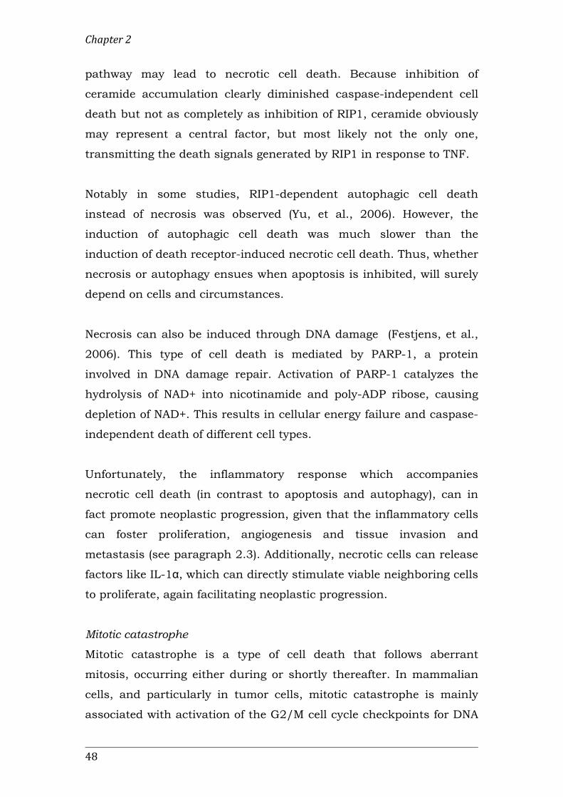
pathway may lead to necrotic cell death. Because inhibition of
ceramide accumulation clearly diminished caspase-independent cell
death but not as completely as inhibition of RIP1, ceramide obviously
may represent a central factor, but most likely not the only one,
transmitting the death signals generated by RIP1 in response to TNF.
Notably in some studies, RIP1-dependent autophagic cell death
instead of necrosis was observed (Yu, et al., 2006). However, the
induction of autophagic cell death was much slower than the
induction of death receptor-induced necrotic cell death. Thus, whether
necrosis or autophagy ensues when apoptosis is inhibited, will surely
depend on cells and circumstances.
Necrosis can also be induced through DNA damage (Festjens, et al.,
2006). This type of cell death is mediated by PARP-1, a protein
involved in DNA damage repair. Activation of PARP-1 catalyzes the
hydrolysis of NAD+ into nicotinamide and poly-ADP ribose, causing
depletion of NAD+. This results in cellular energy failure and caspase-
independent death of different cell types.
Unfortunately, the inflammatory response which accompanies
necrotic cell death (in contrast to apoptosis and autophagy), can in
fact promote neoplastic progression, given that the inflammatory cells
can foster proliferation, angiogenesis and tissue invasion and
metastasis (see paragraph 2.3). Additionally, necrotic cells can release
factors like IL-1α, which can directly stimulate viable neighboring cells
to proliferate, again facilitating neoplastic progression.
Mitotic catastrophe
Mitotic catastrophe is a type of cell death that follows aberrant
mitosis, occurring either during or shortly thereafter. In mammalian
cells, and particularly in tumor cells, mitotic catastrophe is mainly
associated with activation of the G2/M cell cycle checkpoints for DNA

Introduction
49
damage/structure and spindle assembly, and involves numerous
players involved in these checkpoints, including Chk2, cyclinB/Cdk1,
and members of the p53 family, including p53 and the p73 variant
TAp73 (Figure 2.4D) (de Bruin and Medema, 2008). Following mitotic
catastrophe, cells are ultimately killed by engaging the apoptotic or
necrotic pathways, or by induction of cellular senescence (Figure 2.4E)
(Galluzzi, et al., 2011).
At least two subtypes of mitotic catastrophe can be distinguished
(Castedo, et al., 2004). First, mitotic catastrophe can kill the cell
during or close to metaphase, in a p53-independent manner involving
the activation of caspase-2. Second, mitotic catastrophe can occur
after failed mitosis, in a partially p53-dependent manner involving the
activation of the polyploidy checkpoint in G1. Even though mitotic
catastrophe is accompanied by chromatin condensation and
mitochondrial release of apoptosis-inducing factor and cytochrome c,
which are key features of apoptosis, there are a number of
fundamental differences. Importantly, it has been shown that over-
expression of Bcl-2 does not block and might actually enhance mitotic
catastrophe (Lock and Stribinskiene, 1996). Cell death occurring
during the metaphase-to-anaphase transition is characterized by the
activation of caspase-2, which is activated in the nucleus in response
to DNA damage (Lassus, et al., 2002; Paroni, et al., 2002), and is a
process that cannot be inhibited by Bcl-2 (Peart, et al., 2003; Read, et
al., 2002; Robertson, et al., 2002).
Senescence
Analogously to the replicative senescence induced in primary cells as
a result of shortened telomeres, treatment of malignancies may result
in a permanently growth-arrested state (Gewirtz, et al., 2008). This is
termed ‘accelerated senescence’, and is sensed as a permanent state
of DNA damage, while the cell remains viable and metabolically active
(Figure 2.4E). DNA damage signaling activates the p53 and pRb

Chapter 2
50
proteins (or their respective family members, as p53 and pRb function
are often lost during tumorigenesis), which respectively results in first
a temporary, then a prolonged arrest in G1. Senescent cells
characteristically display senescent associated DNA damage foci (SDF)
and senescent associated heterochromatin foci (SAHF) (Campisi and
d'Adda di Fagagna, 2007); SAHF are often found at the promoters of
E2F target genes where they are thought to inhibit transcription,
thereby enforcing growth arrest (Narita, et al., 2003).
2.5.2 Novel approaches to the treatment of cancer
The success of anticancer therapies depends on their ability to
distinguish between normal and cancer cells and specifically exert
their toxic effect on the malignant cells. Novel anticancer strategies
therefore involve a targeted approach, utilizing knowledge of the
cancer hallmarks and enabling characteristics discussed above, and
the tumor suppressor and oncogenic pathways involved. Accordingly,
the following strategies will be discussed:
- inhibition of growth signaling pathways
- induction of programmed cell death
- disruption of telomere maintenance and, hence, cellular
immortalization
- targeting the tumor and its micro-environment to prevent
angiogenesis and metastasis
- attenuation of tumor cell metabolism
- targeting cancer cells for immune destruction
- exploiting genomic instability
- proteins selectively killing tumor cells
Clinical experience with therapies selectively targeting only one each
of these characteristics has shown that the effect is often transitory.
This suggests the existence of at least some (partial) redundancy, in
the form of multiple pathways governing each capability, and/or an
adaptive shift from one capability to another, facilitated by genomic

Introduction
51
instability and the tumor micro-environment. Hence, successful
cancer therapies must comprise a combination of modalities.
Targeting growth signaling pathways in cancer
The first two acquired capabilities discussed in section 2.3 concerned
self-sufficiency in growth signaling and insensitivity to growth-
inhibitory signaling. Thus, growth-signaling pathways, and especially
the receptor proteins, are interesting targets for anticancer strategies
(Christoffersen, et al., 2009). Therapeutic approaches involve both
hormone therapy and monoclonal antibodies. For example, targeting
the EGF dependent signaling pathway has been successfully applied
in the clinic. One of the receptors in this pathway is Her2 (Her2/neu,
ErbB2), and it has been shown to be overexpressed in 20-25% of
breast tumors. Targeting this receptor via the antibody Herceptin
(trastuzumab) has proven to be very effective in the treatment of this
type of cancer (Chang, 2010).
Another approach involves the development of Cdk inhibitors to halt
the cell cycle, with several compounds already being evaluated in
clinical trials (De Falco and De Luca, 2010).
Inducing programmed cell death
Overturning the cancer cell’s apoptosis blockade has been an
appealing approach in the design of anti-cancer therapies. For
example, in cells where p53 function has been lost, this might be
substituted for by activation of the p53 family member p73, either
alone or in combination with other anti-cancer therapies (El-Rifai and
Zaika, 2008; Vilgelm, 2008). The same might also be achieved by
inhibition of the negative apoptosis regulator Bcl-2 (Kang and
Reynolds, 2009), e.g. through so-called BH3-mimicking compounds
(Chonghaile and Letai, 2008). Another promising approach is the
development of small therapeutic compounds, referred to as Smac
mimetics, which are designed to block the function of members of the

Chapter 2
52
inhibitor of apoptosis (IAP) protein family (Gyrd-Hansen and Meier,
2010). Alterations in IAPs are found in many types of human cancer
and are associated with chemoresistance, disease progression and
poor prognosis (Hunter, et al., 2007; LaCasse, et al., 2008). Consistent
with the idea that different types of cancer cells are dependent on
(“addicted to”) IAPs for their survival, the inactivation of IAPs,
particularly when combined with other treatments, results in the
death of most tumour cells in vitro. Though inactivation of IAPs does
not seem to be detrimental to normal cells, loss of IAPs is also
associated with the development of certain types of cancer. Several
compounds are therefore currently being assessed for safety in phase I
clinical trials.
As induction of the other modes of cell death may cause undesirable
tumor-promoting effects, studies to define optimal strategies to
modulate and exploit these other forms of cell death for cancer
therapy are still ongoing. For instance, several autophagy modulators
are currently being investigated (Chen and Karantza, 2011), and
mechanisms being considered for the induction of senescence include
restoration and/or promotion of p53 function, modulation of the cell
cycle through Cdk inhibitors, and inhibition of telomerase action (see
below) (Nardella, et al., 2011).
Attacking cellular immortalization via disruption of telomere
maintenance
While normal human somatic cells do not or only transiently express
telomerase and therefore shorten their telomeres with each cell
division, most human cancer cells typically express high levels of
telomerase and show unlimited cell proliferation (acquired capability
4). Telomerase is thus an attractive therapeutic cancer target, and
novel anti-cancer strategies include the direct targeting of components
of telomerase: the protein component hTERT or RNA component hTR
(Phatak and Burger, 2007). Examples of such agents include the small

Introduction
53
molecule hTERT inhibitor BIBR1532 and Imelstat (GRN163L), a thio-
phosphoramidate oligonucleotide targeting the template region of hTR
as a "template antagonist" (Kelland, 2007). Anti-tumor effects of both
compounds have been observed in cell lines and, particularly for
Imelstat, also in xenografted human tumors in mice. Imelstat
treatment of human glioblastoma tumor-initiating cells in vitro led to
progressive telomere shortening, reduced rates of proliferation, and
eventually cell death (Marian, et al., 2010). In combination with
radiation and the DNA alkylating agent temozolomide, Imelstat had a
dramatic effect on cell survival and activated the DNA damage
response pathway. In vivo, chronic systemic treatment produced a
marked decrease in the rate of xenograft subcutaneous tumor growth.
Effects of anti-telomerase treatment are largely dependent upon initial
telomere length, which can result in a substantial lag before
antitumor activity is observed in tumors possessing relatively long
telomeres. An alternative approach is therefore to target the telomere
itself (Telomere Targeting Agents, TTAs) (Kelland, 2005). Several
classes of small molecules have been described that induce the G-rich
single-stranded overhang of telomeric DNA to fold into 4-stranded G-
quadruplex structures. Such folding is incompatible with telomerase
function and may induce rapid telomere uncapping. These molecules
have shown potent telomerase inhibition in nanomolar concentrations
in vitro and the rapid induction of senescence in cancer cells. The TTA
BRACO19 has demonstrated single agent activity against human
tumour xenografts with anti-tumour effects apparent from only 7 days
of treatment.
So far, Imelstat is the only drug of its class in clinical trials. In the
near future, it is expected that other direct telomerase targeted agents
as well as those targeting telomeres (e.g., AS1410 based on BRACO19)
will enter Phase I clinical trials (Parkinson and Minty, 2007).

Chapter 2
54
Targeting angiogenesis and metastasis
As discussed above, tumor growth requires the malignant cell to be
able to e.g. form new vasculature, as well as produce autologous
growth factors. These characteristics need not be united in a single
cell, as cells in the tumor micro-environment can contribute to
neoplastic progression by providing the necessary factors, aiding not
only in e.g. the angiogenic switch, but also in fostering tissue invasion
and metastasis.
Furthermore, the cancer stem cells (CSCs) found in many tumors
have important properties that must be considered in the development
of effective cancer therapies. Their relative quiescence allows them to
escape cell death by therapies that attack only rapidly proliferating
cells. Also, their ability for self-renewal allows them to recover from
the anti-cancer attack, repair their DNA and again initiate the
formation of a new tumor. Thus, even if the cancer appears to be
cured, the CSCs survive, and in time the tumor reappears.
Furthermore, as CSCs are able to modify the tumor
microenvironment, providing trophic factors to support tumor growth,
they harbor a considerable potential for recurrence, as well as
successful colonization of distant metastases.
Therapeutic strategies that could be employed to target angiogenesis
and metastasis in general (acquired capabilities 5 and 6), and cancer
stem cells in particular, are:
- anti-angiogenic therapy, e.g. through inhibition of VEGF using
the monoclonal antibody bevacizumab, to reduce the
vasculature, thereby depleting the tumor of oxygen and
essential nutrients (Cook and Figg, 2010)
- blocking adhesion between cells and to the extracellular matrix
to prevent successful colonization of metastases, e.g. by
targeting tumor-specific cadherins and integrins (Blaschuk and
Devemy, 2009; Desgrosellier and Cheresh, 2010)

Introduction
55
- another way to prevent metastasis might be through blocking
the EMT chemotaxis pathways that are active in the tumor and
its micro-environment (Nieto, 2011; Roussos, et al., 2011)
- inducing differentiation, e.g. through BMP signaling to revert
the capacity of CSCs to form tumors and increase their
sensitivity to therapy (Ghotra, et al., 2009)
- targeting self-renewal and quiescence, e.g. through PTEN or Wnt
signaling pathways, to decrease the CSC population (Ghotra, et
al., 2009)
Targeting cancer cell metabolism
In addition to acquiring a complex array of genetic changes, tumor
cells develop an alteration in the metabolism of glucose and oxygen
(acquired capability 7). As this altered metabolism does not appear to
be subject to the high genetic variability of tumors, it may represent a
more reliable target for cancer therapy.
The altered metabolism of cancer cells is associated with increased
glycocolytic activity and repression of oxidative phosphorylation. The
harmful effects of the concurrent increase in H2O2 production is
counterbalanced by the increase in glycolytic activity, creating a self-
reinforced loop. This loop can be interrupted by increasing the cellular
levels of H2O2 (using e.g. pro-oxidant agents), or by attenuating
glycolysis (using glycolysis inhibitors), or a combination of both
(López-Lázaro, 2010).
In fact, many anticancer agents, such as paclitaxel, doxorubicin and
arsenic trioxide, produce H2O2 (Alexandre, et al., 2006; Jing, et al.,
1999; Ubezio and Civoli, 1994). Also, using several cancer and normal
cell lines, Chen, et al. (2005) observed that high concentrations of
ascorbic acid selectively killed cancer cells and that this effect was
mediated by H2O2. Several glycolysis inhibitors have shown anticancer
effects (e.g. 2-deoxy-D-glucose, lonidamine, 3-bromopyruvate and

Chapter 2
56
dichloroacetate) and some of them have already entered clinical trials
(Chen, et al., 2007; Gatenby and Gillies, 2007; Lopez-Lazaro, 2008;
Martin, 2006; Pelicano, 2006; Xu and Huang, 2006). For example, it
has been shown that dichloroacetate, a known glycolysis inhibitor
that has been used in humans for decades in the treatment of lactic
acidosis and inherited mitochondrial diseases, induced marked
anticancer effects in mice (Bonnet, et al., 2007). Other strategies that
might also be used to exploit the increased glycolytic activity of cancer
cells and selectively kill these cells, are inhibition of the Na+/K+-
ATPase pump (e.g. by cardiac glycosides), or of cellular systems
involved in the extrusion of (acid-death inducing) protons from the
cytosol.
Because of the dual role of autophagy in cancer, it is difficult to
predict whether inhibition or stimulation of autophagy may result in
tumor cell death (Apel, et al., 2009). Preclinical studies with
chloroquine, which inhibits lysosome acidification and thereby
autophagy, in conjunction with alkylating agents, showed remarkable
efficacy inhibiting tumour growth in mice (Amaravadi, et al., 2007).
Alternatively, promoting autophagy might also be expected to limit
tumor progression. Hence, before autophagy can be targeted for the
treatment of cancer, further studies investigating the dichotomy of its
roles in tumor prevention and promotion are warranted (Rosenfeldt
and Ryan, 2009).
Another aspect of the metabolic switch that takes place during
neoplastic progression concerns the concurrent increase in protein
production, and hence, ribosome biogenesis. Though this has long
remained a largely unexploited target in cancer therapy, increasing
attention is being paid to inhibitors of rRNA synthesis for the
development of novel therapeutic strategies (Drygin, et al., 2010). For
example, CX-3543, a small molecule nucleolus-targeting agent that
selectively disrupts nucleolin/rDNA complexes in the nucleolus,

Introduction
57
thereby inhibiting Pol I transcription and inducing apoptosis in cancer
cells, is currently being evaluated for treatment of
carcinoid/neuroendocrine tumors in a phase II clinical trial (Drygin, et
al., 2009). Some classic anticancer therapeutics, including cisplatin
and 5-fluorouracil, have even been shown to exert their activity, at
least partially, through disruption of ribosome biogenesis (Ghoshal
and Jacob, 1997; Jordan and Carmo-Fonseca, 1998).
In addition, studies indicate that many common and specialized
mRNA export factors, including CRM1 and eukaryotic translation
initiation factor 4E (eIF4E), are disregulated in cancer, making them
also attractive therapeutic targets (Siddiqui and Borden, 2011).
Indeed, specific targeting of the eIF4E-dependent mRNA export
pathway in a phase II proof-of-principle trial with ribavirin led to
impaired eIF4E-dependent mRNA export, correlating with clinical
responses including remissions in leukemia patients (Assouline, et al.,
2009).
Immune destruction of cancer cells
The ability of cells of the immune response system to infiltrate tumors
presents a unique opportunity for combating cancer cells. Tumors are
replete with potential antigens, which can become immunogenic when
presented by DCs, activating the different arms of cell-mediated
resistance (Steinman and Banchereau, 2007). This means that the
resulting immune attack can encompass multiple targets, diminishing
the cancer’s chances of immune escape. Following recognition of
tumor-specific antigens, T lymphocytes exert their cytotoxic effects on
tumor cells via the extrinsic apoptosis pathway, involving Fas, and via
the secretion of perforin and serine proteases granzyme A and B
(Pardo, et al., 2004). Perforin is a transmembrane pore-forming
molecule, which allows granzyme A and B to enter the target cell and
induce apoptosis. Granzyme A activates caspase-independent
pathways by inducing single-stranded DNA damage, while granzyme B

Chapter 2
58
directly activates caspase-3, and can also cleave Bid to induce the
release of cytochrome c (Rousalova and Krepela, 2010). There is also
evidence that DCs themselves can acquire killer activity and express
granzyme and perforin.
Several strategies are being employed in the field of tumor
immunology, including the use of monoclonal antibodies against
specific tumor-associated antigens to achieve steric inhibition and
neutralization, complement activation, and activation of cell-mediated
cytotoxicity, and so-called “cancer vaccines” (Dougan and Dranoff,
2009).
Vaccination against infectious diseases has proven to be one of the
great successes of modern medicine, inducing efficient, specific
activation of cytotoxic T lymphocytes, as well as the generation of
memory cells, protecting against future infection. Translation of this
knowledge for the prevention and treatment of cancer has not been
straightforward, with, among other things, the selection of appropriate
target antigens proving a difficult task, as well as the design and
interpretation of clinical trials for this novel class of cancer
therapeutics (Lesterhuis, et al., 2011; Palucka, et al., 2011). One
example showing great promise with regard to cancer prevention is
the development of two human papilloma virus-derived vaccines for
the prevention of cervical cancer (Lowy and Schiller, 2006). As for
therapeutic vaccination, a series of clinical trials have recently yielded
encouraging results. First, treatment of metastatic prostate cancer
with sipuleucel-T, a cellular vaccine based on enriched blood DCs
briefly cultured with a fusion protein of prostatic acid phosphatase
with GM-CSF, resulted in an approximately 4-month–prolonged
median survival in phase III trials (Kantoff, et al., 2010). Sipuleucel-T
has been approved by the FDA for treatment of metastatic prostate
cancer, thereby paving the clinical development and regulatory path
for the next generation of cellular immunotherapy products. Second, a

Introduction
59
phase III trial in metastatic melanoma testing peptide vaccine in
combination with high dose IL-2 versus IL-2 alone showed significant
improvement in overall response rate and progression-free survival in
patients who received the vaccine (Schwartzentruber, et al., 2011).
Third, a phase III trial in patients with follicular lymphoma showed
that idiotype vaccine therapy (BiovaxID) significantly prolongs the
duration of chemotherapy-induced remission (Morse and Whelan,
2010). Furthermore, a randomized phase II trial of a poxviral-based
vaccine targeting a prostate-specific antigen (PROSTVAC) in men with
metastatic castration-resistant prostate cancer showed an improved
overall survival in patients when compared with patients receiving
control vectors (an observed difference in median survival of 8.5
months) (Kantoff, et al., 2010b).
In the sipuleucel-T example described above, DCs are generated ex
vivo, loaded with tumor antigens, and re-injected to induce strong T-
cell and perhaps also natural killer immunity. Another, novel,
approach to cancer vaccines is based on the delivery of antigens
directly to dendritic cells (DCs) in vivo, using chimeric proteins made
of anti-DC receptor antibody fused to a selected antigen (DC
targeting). Studies in mice demonstrate that DC targeting results in
considerable potentiation of antigen-specific T cell immunity. The
induction of immunity is observed only when the DC maturation
signal is provided, as, otherwise, tolerance ensues (Bonifaz, et al.,
2002; Hawiger, et al., 2001). A major challenge of this approach will
be to elicit T cell responses that are sufficiently robust and long
lasting so as to be clinically active. Indeed, the efficacy of DC targeting
in vivo needs to be established in clinical trials in patients, and early
studies are ongoing.
Besides the molecular make-up of the tumor itself,
immunotherapeutic approaches also need to consider that of the
tumor micro-environment, and aim to trigger a multi-facetted immune

Chapter 2
60
response involving humoral, cellular, and innate immunity (Poschke,
et al., 2011). Recent studies have attempted to relieve the suppression
of immune activity in the tumor micro-environment (imposed by the
cancer cells) by blocking inhibitory signals using monoclonal
antibodies, showing promising results (such as blocking cytotoxic T-
lymphocyte antigen-4, CTLA-4, with the monoclonal antibody
ipilimumab) (Hoos, et al., 2010). Additional efforts to either directly
target MDSC or their suppressive mechanisms should aid the
development of successful combination-immunotherapies. Due to
their immature nature, a potential way to remove MDSC is to force
them to differentiate, for example, by using all-trans retinoic acid
(ATRA) or vitamin D3, which promotes myeloid differentiation and has
been clinically applied (Lathers, et al., 2004; Mirza, et al., 2006).
Exploiting genomic instability
The genetic instability that is characteristic of cancer cells can be both
good and bad for anticancer therapy. Although it seems to provide an
Achilles' heel that many conventional therapies exploit, genetic
instability can also make eradicating cancer more difficult. Because of
the abnormally high mutability of many cancer cells, most malignant
tumor cell populations are heterogeneous in many respects, which
may make them difficult to target with a single type of treatment.
Moreover, this mutability allows many cancers to evolve resistance to
therapeutic drugs at an alarming rate (Rajagopalan and Lengauer,
2004).
The novel “synthetic lethal” approach aims to exploit defects in DNA
repair pathways using a new theory (Yap, et al., 2011). This theory
proposes that targeting tumor cells, genetically defective in one given
pathway, with a specific molecular therapy, designed to inhibit a
“synthetic lethal” gene partner involved in a complementary pathway,
results in selective tumor cell killing. Studies have shown that breast
cancer cells, defective in homologous recombination due to BRCA1/2

Introduction
61
defects are highly sensitive to blockade of the base excision repair
(BER) pathway via inhibition of the poly (ADP-ribose) polymerase
(PARP) enzyme, providing strong evidence for the clinical application of
this approach (Bryant, et al., 2005; Farmer, et al., 2005). PARP
inhibitors might also be used in combination with standard radio- and
chemotherapy to sensitize cells to cytotoxic DNA damage; additionally,
some PARP inhibitors seem to possess anti-angiogenic activity,
making their application in the clinic even more attractive (Mangerich
and Bürkle, 2011).
The synthetic lethal concept has also been applied for the treatment of
p53-deficient cancers, which, as discussed before, occur rather
frequently. Loss of p53 function renders cells dependent on the
checkpoint kinase Chk1 for activation of cell cycle checkpoints; hence,
inhibition of Chk1 in the presence of DNA damage or replicative stress
should lead to mitotic catastrophe and cell death in p53-deficient
tumors while sparing normal cells. Several Chk1 inhibitors have now
been developed; after showing promising results in preclinical models,
they are currently being evaluated in phase I clinical trials (Ma, et al.,
2011).
Proteins selectively killing tumor cells
In recent years, a unique set of molecules possessing a remarkable
ability have been identified: these proteins are able to detect the
malfunctioning of a presumably shared set of cancer-critical genes,
and selectively kill the corresponding tumor cells, leaving the normal
cells unharmed. Among these proteins killing tumor cells (PKTC) are
the human cytokines TRAIL and MDA7/IL24, the frog-derived
Brevinin-2R, the human pro-apoptotic Par-4 and Noxa proteins and
the organic cation transporter-like 3 (ORCTL3), the alpha-lactalbumin
and oleic acid complex HAMLET, and the viral proteins adenovirus
E4ORF4, parvovirus NS1, and the Chicken Anemia Virus-encoded
protein apoptin (Bruno, et al., 2009; Grimm and Noteborn, 2010;
Chapter 2
62
Argiris, et al., 2011). As these PKTC appear to function independently
of the type of cancer, their therapeutic potential seems
unprecedented. Apoptin, the first of these proteins to be discovered,
and the subject of the present thesis, will be discussed in the next
section.
2.6 Apoptin
Apoptin is the protein product of the VP3 gene of the Chicken Anemia
Virus (CAV; Noteborn, et al., 1994). The pathogenesis of CAV infection
and discovery of apoptin will be discussed in section 2.6.1, while the
characteristics of apoptin and perspectives for application as an anti-
cancer agent are discussed in sections 2.6.2 and 2.6.3, respectively.
2.6.1 Chicken Anemia Virus
In young chicks, CAV infection results in severe anemia and
immunodeficiency (among other symptoms), owing to apoptosis of the
cells in the thymus, bone marrow and spleen (Noteborn and Koch,
1995). Early after infection, a 2.3 kb polycistronic mRNA is
transcribed, encoding three genes: the capsid protein VP1, the scaffold
protein and phosphatase VP2, and VP3 (apoptin) (Noteborn, 2004).
Later on in the infection, splicing of the CAV mRNA produces
additional RNA products; it is however not known whether these
result in functional proteins (Kamada, et al., 2006).
Studies in transformed chicken cells in culture proved that apoptin
was responsible for the CAV-induced apoptosis (Noteborn, et al.,
1994). Apoptin demonstrated the same apoptosis activity in human
cancer cell lines, but, surprisingly, not in normal human cells
(Backendorf, et al., 2008; Danen-van Oorschot, et al., 1997;
Noteborn, et al., 2005; Tavassoli, et al., 2005). This makes apoptin
itself an interesting protein for therapeutic treatment of cancer cells,
but also an interesting tool to investigate the process of oncogenic
transformation, thereby uncovering other novel targets for anticancer

Introduction
63
therapies. Furthermore, apoptin may also prove useful in the
diagnosis of cancer, e.g. based on its ability to be specifically
phosphorylated by tumor cell lysates (see below).
2.6.2 Characteristics of apoptin-induced apoptosis
Apoptin is a small (121 aa), proline-rich protein, exhibiting little
homology to any other known protein – including its functional
equivalent TAIP, encoded by the TT virus (Kooistra, et al., 2004).
Tumor-selective apoptosis induction by apoptin is preceded by a)
phosphorylation at T108 (Rohn, et al., 2002), and b) nuclear
translocation (Danen-van Oorschot, et al., 2003) (Figure 2.5). These
effects are characteristic for the transformed environment, as they are
also elicited upon transient transformation of normal human cells by
the SV40 LT and ST proteins (Zhang, et al., 2004).
Figure 5. Characteristics of tumor-selective apoptosis induction by apoptin. A. In normal cells (left panel), apoptin is located in the cytoplasm, and can be found in granules extending to the cell membrane. In tumor cells (right panel), apoptin is located in the nucleus and induces apoptosis (arrow indicates dying cell). B. In tumor cells, but not in normal cells, apoptin is phosphorylated at position T108.

Chapter 2
64
The detailed mechanisms behind both the up- and downstream
pathways of apoptin activity in transformed and cancer cells are yet to
be elucidated. A number of apoptin-interacting proteins have been
identified, including Hippi, the protein interactor and apoptosis co-
mediator of the huntingtin interacting protein 1 (Cheng, et al., 2003),
and Rybp/DEDAF, a transcription factor and proapoptotic protein
known to associate with the death effector domain-containing DNA
binding protein DEDD (Danen-van Oorschot, et al., 2004).
Furthermore, apoptin has been shown to act through the intracellular
apoptosis pathway, involving the translocation of Nur77 from the
nucleus to the cytoplasm, as well as p73 and PUMA activity (Klanrit,
et al., 2008; Maddika, et al., 2005). Apoptin expression enhances the
level of the tumor suppressor ceramide in tumor cells, also implicating
the involvement of sphingolipids in apoptin-induced cell death (Liu, et
al., 2006a, b).
2.6.3 Perspectives for apoptin anti-tumor therapy
Though its pathway of action remains to be clarified, apoptin has
demonstrated several features that make it well suited for cancer
therapy. Besides the fact that it has been shown to selectively kill
tumor and transformed cells, apoptin has also been shown to act
independently of p53 (Teodoro, et al., 2004; Zhuang, et al., 1995).
Furthermore, it is insensitive to BCR-ABL and Bcl-XL (Backendorf, et
al., 2008; Noteborn, et al., 2005), which suggests that apoptin might
induce apoptosis in cases where other (chemo)therapeutics might fail.
The results of the first preclinical therapeutic studies, discussed
below, are very promising.
Apoptin cancer therapy using adenoviral vectors
Pietersen and colleagues developed a strategy for the use of apoptin in
cancer therapy based on adenoviral vectors (Pietersen, et al., 1999). A
single injection of the apoptin-producing adenovirus in xenografted

Introduction
65
hepatomas in nude mice resulted in a delay in tumor growth. The
number of proliferating cells as detected by BrdU-labeling was
dramatically decreased in the apoptin-transduced regions versus
control-treated tumors (Van der Eb, et al., 2002). Importantly, the
apoptin-producing adenovirus did not have appreciable toxic effects
when injected intraperitoneally, intravenously, or subcutaneously into
healthy rats. Further studies using these and other non-replicative
viruses, including a fowlpox virus–based vector, used a regimen of
multiple intratumoral injections during several days. These
approaches resulted in a significant overall survival benefit for the
apoptin-treated mice and, in some cases, depending on the overall
transduction efficiency, complete regression of the established tumor
(Noteborn, 2009).
PDT4-apoptin - topical cancer treatment
Guelen and coworkers fused apoptin to the HIV-TAT protein
transduction domain, demonstrating efficient transduction of apoptin
into both normal and tumor cells, while preserving its tumor-selective
apoptotic properties (Guelen, et al., 2004). Sun and colleagues used a
similar fusion product, employing protein transduction domain 4
(PTD4)-mediated transduction of recombinant apoptin protein, to treat
tumors that had been xenografted onto nude mice (Sun, et al., 2009).
In contrast to the gene therapy-based studies discussed above, PTD4-
apoptin was administered by simple application onto the epidermis.
Remarkably, though the protein could be detected in the epidermal
tissue covering the subcutaneous tumor tissue and in several internal
organs of the mice, cell death was only observed inside the tumor
mass.
Recently, Jin and colleagues (Jin, et al., 2011) reported that PTD4-
apoptin protein and dacarbazine acted synergistically to reduce tumor
growth in a mouse melanoma model. Importantly, the combination
with PTD4-apoptin allowed for a 50% reduction in the dosage of

Chapter 2
66
dacarbazine, resulting in comparable reduction of tumor-growth,
without any detectable hematological side-effect.
Systemic apoptin treatment and organ-specific targeting
Peng et al. showed that apoptin can be safely administered
systemically, and used a specific ligand to the asialoglycoprotein
receptor to target apoptin specifically to the liver (Peng, et al., 2007).
Delivery of this Asor-apoptin via the tail vein into mice bearing in situ
hepatocarcinomas resulted in specific and efficient distribution of
apoptin in both hepatocarcinoma cells and normal liver cells. Whereas
the former cells showed significant signs of regression, the normal
hepatocytes were clearly not affected.
Specific targeting of apoptin to the brain is currently being developed
by de Boer and coworkers. Using the nontoxic ligand CRM197 (a
mutant of the diphtheria toxin), the membrane-bound precursor of
heparin-binding epidermal growth factor (HB-EGF), also known as the
diphtheria toxin receptor (DTR), can be targeted. This receptor is
constitutively present at the blood-brain barrier and is strongly up-
regulated in many tumors, including human glioblastoma (Mishima,
et al., 1998). Biopharmaceutical drugs have been selectively delivered
to the brain via this receptor (Gaillard, et al., 2005). An apoptin-
expressing plasmid coupled to CRM197 has been successfully
delivered to human glioblastoma cells in vitro through receptor-
mediated endocytosis (Rip, et al., 2009). The combination of apoptin
antitumor therapy and CRM197-targeting technology thus provides a
great opportunity for development of targeted therapy for brain
tumors.
Combination therapy with apoptin and chemo- or radiotherapy
The combination of apoptin therapy with chemotherapeutic agents
has been reported to enhance cytotoxicity to human tumor cells in
vitro (Olijslagers, et al., 2007). Combined treatment of recombinant

Introduction
67
adenovirus expressing apoptin with different concentrations of
etoposide clearly showed an additive cytotoxic effect in human
osteosarcoma U2OS cells. Paclitaxel combined with apoptin acted
additively in p53-positive human osteosarcoma U2OS and nonsmall
cell lung carcinoma A549 cells, p53-negative osteosarcoma Saos-2
cells, and p53-mutant prostate cancer Du145 cells. Finally, apoptin
was proven to be coeffective when combined with the
chemotherapeutic agent methotrexate (Zhang, et al., 2007).
Recently, apoptin has also been combined with other treatments in
vivo. As discussed above, apoptin expression modulates the ceramide-
sphingolipid pathways leading to enhanced ceramide levels (Liu, et al.,
2006). The majority of prostate tumors have elevated acid ceramidase
levels compared with neighboring normal prostate tissue (Liu, et al.,
2006). In vitro, up-regulated acid-ceramidase protected cells from
apoptin-induced apoptosis, whereas cotreatment with the acid-
ceramidase inhibitor LCL204 sensitized cells for apoptosis. In vivo,
combined treatment enhanced the antitumor activity of apoptin in
xenografted prostate tumors in mice, resulting in significantly reduced
tumor growth and increased animal survival.
Lian and colleagues combined apoptin treatment with interleukin-18
(IL-18) and reported that combined administration results in an even
higher induction of an effective antitumor immune response and
tumor regression (Lian, et al., 2007). IL-18 and apoptin appear to
affect tumors via complementary pathways. Whereas apoptin directly
targets the tumor cells, IL-18 treatment appears to act via enhancing
the immune response toward tumor cells. Finally, Schoop et al.
showed that treatment of a radioresistant head- and neck cancer cell
line with apoptin concurrently with exposure to irradiation sensitized
these cells to apoptosis (Schoop, et al., 2010).

Chapter 2
68
Tumor-selective targeting using apoptin NLS
Rather than using apoptin itself, or one of its cellular targets, to
design anti-cancer therapies, Wagstaff and Jans propose the use of
apoptin’s tumor-selective NLS (74-121) to target drugs specifically to
the nucleus of tumor cells (Wagstaff and Jans, 2009). Delivery of
cytotoxic agents directly to the heart of the tumor cell should result in
efficient tumor cell killing without affecting healthy neighboring cells.
2.7 Synopsis
The new era of genomics and proteomics carries with it a great
promise: that for every disease, the blueprint of the defective cell can
be compared with that of a healthy one, thereby not only facilitating
the identification of the root of the problem, but also, and more
importantly, allow one to charter an efficient route to fix it. Drawing
up the blueprint of a cancer cell has proven to be a difficult task. Even
so, a complex map of intracellular pathways is starting to emerge,
with a number of essential traits being seemingly shared by all
transformed cells. The CAV-derived apoptin apparently senses these
characteristics and effectively charters its own route to selectively kill
malignant cells. The properties of apoptin and functionally related
proteins, combined with newly developed therapeutics targeting the
hallmark capabilities and enabling characteristics of cancer cells,
should finally lead to the development of selective, efficient and robust
therapies for cancer – instead of the long sought-after “magical bullet”
cure for cancer, we will be able to build a magical cluster bomb.

Introduction
69
References
Al-Hajj M, Wicha M, Benito-Hernandez A, Morrison S and Clarke, M.
(2003) Prospective identification of tumorigenic breast cancer cells.
PNAS 100, 3983-3988.
Alexandre J, Batteux F, Nicco C, Chéreau C, Laurent A, Guillevin L et
al. (2006) Accumulation of hydrogen peroxide is an early and crucial
step for paclitaxel-induced cancer cell death both in vitro and in vivo.
Int J Cancer 119, 41-48.
Ali S and DeCaprio J (2001) Cellular transformation by SV40 large T
antigen: interaction with host proteins. Semin Cancer Biol 11, 15-23.
Amaravadi R, Yu D, Lum J, Bui T, Christophorou M, Evan G et al.
(2007) Autophagy inhibition enhances therapy-induced apoptosis in a
myc-induced model of lymphoma . J Clin Invest 117, 326-336.
Apel A, Zentgraf H, Buchler M and Herr I (2009) Autophagy-a double
edged sword in oncology. Int J Cancer 125, 991-995.
Argiris K, Panethymitaki C and Tavassoli M (2011) Naturally
occurring, tumor-specific, therapeutic proteins. Exp Biol Med 236,
524-536.
Assouline S, Culjkovic B, Cocolakis E, Rousseau C, Beslu N, Amri A et
al. (2009) Molecular targeting of the oncogene eIF4E in acute myeloid
leukemia (AML): a proof-of-principle clinical trial with ribavirin. Blood
114, 257–260.
Backendorf C, Visser AE, De Boer AG, Zimmerman R, Visser M,
Voskamp P, Zhang Y-H and Noteborn MHM (2008) Apoptin:
therapeutic potential of an early sensor of carcinogenic
transformation. Annu Rev Pharmacol Toxicol 48, 143-169.
Baehrecke E (2003) Autophagic programmed cell death in Drosophila.
Cell Death Differ 10, 940-945.
Chapter 2
70
Bergers G and Benjamin L (2003) Tumorigenesis and the angiogenic
switch. Nat Rev Cancer 3, 401-410.
Berx G and van Roy F (2009) Involvement of members of the cadherin
superfamily in cancer. Cold Spring Harb Perspect Biol 1, a003129.
Bhowmick N, Neilson E and Moses H (2004) Stromal fibroblasts in
cancer initiation and progression. Nature 432, 332-337.
Bindea G, Mlecnik B, Fridman W, Pagès F and Galon J (2010) Natural
immunity to cancer in humans. Curr Opin Immunol 22, 215-222.
Blackburn E (2000) Telomere states and cell fates. Nature 408, 53-56.
Blaschuk O and Devemy E (2009) Cadherins as novel targets for anti-
cancer therapy. Eur J Pharmacol 625, 195-198.
Blomen V and Boonstra J (2007) Cell fate determination during G1
phase progression. Cell Mol Life Sci 64, 3084-3104.
Bonifaz L, Bonnyay D, Mahnke K, Rivera M, Nussenzweig M and
Steinman R (2002) Efficient targeting of protein antigen to the
dendritic cell receptor DEC-205 in the steady state leads to antigen
presentation on major histocompatibility complex class I products and
peripheral CD8+ T cell tolerance. J Exp Med 196, 1627–1638.
Bonnet S, Archer S, Allalunis-Turner J, Haromy A, Beaulieu C,
Thompson R et al. (2007) A mitochondria-K+ channel axis is
suppressed in cancer and its normalization promotes apoptosis and
inhibits cancer growth. Cancer Cell 11, 37-51.
Boya P, González-Polo R, Casares N, Perfettini J, Dessen P, Larochette
N et al. (2005) Inhibition of macroautophagy triggers apoptosis. Mol
Cell Biol 25, 1025-1040.
Brosh R and Rotter V (2009) When mutants gain new powers: news
from the mutant p53 field. Nat Rev Cancer 9, 701-713.

Introduction
71
Bruno P, Brinkmann C, Boulanger M, Flinterman M, Klanrit P, Landry
M et al. (2009) Family at last: highlights of the first international
meeting on proteins killing tumour cells. Cell Death and Differentiation
16, 184-186.
Bryan T, Englezou A, Dalla-Pozza L, Dunham M and Reddel R (1997)
Evidence for an alternative mechanism for maintaining telomere
length in human tumors and tumor-derived cell lines. Nat Med 3,
1271-1274.
Bryan T, Englezou A, Dunham M and Reddel R (1998) Telomere length
dynamics in telomerase-positive immortal human cell populations.
Exp Cell Res 239, 370–378.
Bryant H, Schultz N, Thomas H, KM P, Flower D, Lopez E et al. (2005)
Specific killing of BRCA2-deficient tumours with inhibitors of
poly(ADP-ribose) polymerase. Nature 434, 913-917.
Campisi J and d'Adda di Fagagna F (2007) Cellular senescence: when
bad things happen to good cells. Nat Rev Mol Cell Biol 8, 729-740.
Carmeliet P (2005) VEGF as a key mediator of angiogenesis in cancer.
Oncology 69, 4-10.
Castedo M, Perfettini J, Roumier T, Andreau K, Medema R and
Kroemer G (2004) Cell death by mitotic catastrophe: a molecular
definition. Oncogene 23, 2825-2837.
Chang H (2010) Trastuzumab-based neoadjuvant therapy in patients
with HER2-positive breast cancer. Cancer 116, 2856-2867.
Chen N and Karantza V (2011) Autophagy as a therapeutic target in
cancer. Cancer Biol Ther 11, 157-168.
Chen Q, Espey M, Krishna M, Mitchell J, Corpe C, Buettner G et al.
(2005) Pharmacologic ascorbic acid concentrations selectively kill
cancer cells: action as a pro-drug to deliver hydrogen peroxide to
tissues. Proc Natl Acad Sci U S A 102, 13604-13609.

Chapter 2
72
Chen Z, Lu W, Garcia-Prieto C and Huang P (2007) The Warburg effect
and its cancer therapeutic implications. J Bioenerg Biomembr 39, 267–
274.
Cheng C, Huang S, Chang Y, Chung W and Yuo C (2003) The viral
death protein Apoptin interacts with Hippi, the protein interactor of
Huntingtin-interacting protein 1. Biochem Biophys Res Commun 305,
359-364 .
Cheng N, Chytil A, Shyr Y, Joly A and Moses H (2008) Transforming
growth factor-beta signaling-deficient fibroblasts enhance hepatocyte
growth factor signaling in mammary carcinoma cells to promote
scattering and invasion. Mol Cancer Res 6, 1521-1533.
Cho R and Clarke M (2008) Recent advances in cancer stem cells. Curr
Opin Genet Dev 18, 48-53.
Chonghaile T and Letai A (2008) Mimicking the BH3 domain to kill
cancer cells. Oncogene 27, S149-S1457.
Christoffersen T, Guren T, Spindler K, Dahl O, Lønning P and
Gjertsen B (2009) Cancer therapy targeted at cellular signal
transduction mechanisms: strategies, clinical results, and unresolved
issues. Eur J Pharmacol 625, 6-22.
Cleveland D, Mao Y and Sullivan K (2003) Centromeres and
kinetochores: from epigenetics to mitotic checkpoint signaling. Cell
112, 407-421.
Colanzi A and Corda D (2007) Mitosis controls the Golgi and the Golgi
controls mitosis. Curr Opin Cell Biol 19, 386-393.
Cook K and Figg W (2010) Angiogenesis inhibitors: current strategies
and future prospects. CA Cancer J Clin 60, 222-243.
Counter C, Hirte H, Bacchetti S and Harley C (1994) Telomerase
activity in human ovarian carcinoma. Proc Natl Acad Sci U S A 91,
2900-2904.

Introduction
73
d'Adda di Fagagna F, Reaper P, Clay-Farrace L, Fiegler H, Carr P, Von
Zglinicki T et al. (2003) A DNA damage checkpoint response in
telomere-initiated senescence. Nature 426, 194-198.
Danen-van Oorschot A, Fischer D, Grimbergen J, Klein B, Zhuang S-
M, Falkenburg J et al. (1997) Apoptin induces apoptosis in human
transformed and malignant cells but not in normal cells. Proc. Natl.
Acad. Sci. U.S.A. 94, 5843-5847.
Danen-van Oorschot A, Voskamp P, Seelen M, van Miltenburg M, Bolk
M, Tait S et al. (2004) Human death effector domain-associated factor
interacts with the viral apoptosis agonist Apoptin and exerts tumor-
preferential cell killing. Cell Death and Differentiation 11, 564-573.
Danen-van Oorschot A, Zhang Y-H, Leliveld S, Rohn J, Seelen M, Bolk
M et al. (2003) Importance of nuclear localization of apoptin for tumor-
specific induction of apotosis. J Biol Chem 278, 27729-27736.
Dang C and Semenza G (1999) Oncogenic alterations of metabolism.
Trends Biochem Sci 24, 68-72.
Darwin C (1859) On the Origin of Species by Means of Natural
Selection, or the Preservation of Favoured Races in the Struggle for Life.
London: John Murray.
Davies H (2002) Mutations of the BRAF gene in human cancer. Nature
417, 949−954.
de Bruin E and Medema J (2008) Apoptosis and non-apoptotic deaths
in cancer development and treatment response. Biochem Pharmacol
76, 947-957.
De Falco M and De Luca A (2010) Cell cycle as a target of
antineoplastic drugs. Curr Pharm Des 16, 1417-1426.
DeBerardinis R, Lum J, Hatzivassiliou G and Thompson C (2008) The
biology of cancer: metabolic reprogramming fuels cell growth and
proliferation. Cell Metab 7, 11-20.
Chapter 2
74
Desgrosellier J and Cheresh D (2010) Integrins in cancer: biological
implications and therapeutic opportunities. Nat Rev Cancer 10, 9-22.
Dougan M and Dranoff G (2009) Immune therapy for cancer. Annu
Rev Immunol 27, 83-117.
Downward J (2003) Targeting RAS signalling pathways in cancer
therapy. Nat Rev Cancer 3, 11-22.
Drygin D, Rice W and Grummt I (2010) The RNA polymerase I
transcription machinery: an emerging target for the treatment of
cancer. Annu Rev Pharmacol Toxicol 50, 131-156.
Drygin D, Siddiqui-Jain A, O'Brien S, Schwaebe M, Lin A, Bliesath J
et al. (2009) Anticancer activity of CX-3543: a direct inhibitor of rRNA
biogenesis. Cancer Res 69, 7653-7661.
Dufner A and Thomas G (1999) Ribosomal S6 kinase signaling and
the control of translation. Exp Cell Res 253, 100-109.
Elstrom R, Bauer D, Buzzai M, Karnauskas R, Harris M, Plas D et al.
(2004) Akt stimulates aerobic glycolysis in cancer cells. Cancer Res
64, 3892–3899.
Farmer H, McCabe N, Lord C, Tutt A, Johnson D, Richardson T et al.
(2005) Targeting the DNA repair defect in BRCA mutant cells as a
therapeutic strategy. Nature 434, 917-921.
Ferrara N (2009) Vascular endothelial growth factor. Arterioscler
Thromb Vasc Biol 29, 789–791.
Festjens N, Vanden Berghe T and Vandenabeele P (2006) Necrosis, a
well-orchestrated form of cell demise: signalling cascades, important
mediators and concomitant immune response. Biochim Biophys Acta
1757, 1371–1387.
Introduction
75
Festjens N, Vanden Berghe T, Cornelis S and Vandenabeele P (2007)
RIP1, a kinase on the crossroads of a cell's decision to live or die. Cell
Death Differ 14, 400-410.
Foley E, Maldonado M, and Kapoor T (2011) Formation of stable
attachments between kinetochores and microtubules depends on the
B56-PP2A phosphatase. Nat Cell Biol, 13, 1265-1271.
Forester C, Maddox J, Louis J, Goris J and Virshup D (2007) Control
of mitotic exit by PP2A regulation of Cdc25C and Cdk1. Proc Natl Acad
Sci USA 104, 19867-19872.
Freudenberg J, Wang Q, Katsumata M, Drebin J, Nagatomo I and
Greene M (2009) The role of HER2 in early breast cancer metastasis
and the origins of resistance to HER2-targeted therapies. Exp Mol
Pathol 87, 1-11.
Friedl P and Wolf K (2008) Tube travel: the role of proteases in
individual and collective cancer cell invasion. Cancer Res 68, 7247-
7249.
Gaillard P, Brink A and de Boer A (2005) Diphtheria toxin receptor-
targeted brain drug delivery. Int Congr Ser 1277, 185–195.
Galluzzi L, Vitale I, Abrams J, Alnemri E, Baehrecke E, Blagosklonny
M et al. (2011) Molecular definitions of cell death subroutines:
recommendations of the Nomenclature Committee on Cell Death
2012. Cell Death Differ Epub ahead of print.
Gatenby R and Gillies R (2004) Why do cancers have high aerobic
glycolysis? Nat Rev Cancer 4, 891-899.
Gatenby R and Gillies R (2007) Glycolysis in cancer: a potential target
for therapy. Int J Biochem Cell Biol 39, 1358–1366.
Gewirtz D, Holt S and Elmore L (2008) Accelerated senescence: an
emerging role in tumor cell response to chemotherapy and radiation.
Biochem Pharmacol 76, 947-957.

Chapter 2
76
Ghoshal K and Jacob S (1997) An alternative molecular mechanism of
action of 5-fluorouracil, a potent anticancer drug. Biochem Pharmacol
53, 1569–1575.
Ghotra V, Puigvert J and Danen E (2009) The cancer stem cell
microenvironment and anti-cancer therapy. Int J Radiat Biol 85, 955-
692.
Gingras A, Raught B and Sonenberg N (2004) mTOR signaling to
translation. Curr Top Microbiol Immunol 279, 169-197.
Grimm S and Noteborn M (2010) Anti-cancer genes: inducers of
tumour-specific cell death signalling. Trends in Molecular Medicine 16,
88-96.
Guelen L, Paterson H, Gäken J, Meyers M, Farzaneh F and Tavassoli
M (2004) TAT-apoptin is efficiently delivered and induces apoptosis in
cancer cells. Oncogene 23, 1153-1165.
Gyrd-Hansen M and Meier P (2010) IAPs: from caspase inhibitors to
modulators of NF-kappaB, inflammation and cancer. Nat Rev Cancer
10, 561-574.
Hahn W and Weinberg R (2002) Modeling the molecular circuitry of
cancer. Nat Rev Cancer 331, 331-341.
Hahn W and Weinberg R (2002b) Rules for making human tumor
cells. N Engl J Med 347, 1593-1603.
Hanahan D and Weinberg R (2000) The Hallmarks of cancer. Cell 100,
57-70.
Hanahan D and Weinberg R (2011) Hallmarks of cancer: the next
generation. Cell 144, 646-674.
Hara E, Tsurui H, Shinozaki A, Nakada S and Oda K (1991)
Cooperative effect of antisense-Rb and antisense-p53 oligomers on the

Introduction
77
extension of life span in human diploid fibroblasts, TIG-1. Biochem
Biophys Res Commun 179, 528-534.
Harley C, Futcher A and Greider C (1990) Telomeres shorten during
ageing of human fibroblasts. Nature 345, 458-460.
Hawiger D, Inaba K, Dorsett Y, Guo M, Mahnke K, Rivera M et al.
(2001) Dendritic cells induce peripheral T cell unresponsiveness under
steady state conditions in vivo. J Exp Med 194, 769–779.
Hayflick L (1965) The limited in vitro lifetime of human diploid cell
strains. Exp Cell Res 37, 614-636.
Hayflick L and Moorhead P (1961) The serial cultivation of human
diploid cell strains. Exp Cell Res 25, 585-621.
Heichman K and Roberts J (1994) Rules to replicate by. Cell 79, 557-
562.
Heit R, Rattner J, Chan G and Hendzel M (2009) G2 histone
methylation is required for the proper segregation of chromosomes. J
Cell Sci 122, 2957-2968.
Hezel A and Bardeesy N (2008) LKB1; linking cell structure and tumor
suppression. Oncogene 27, 6908-6919.
Hoos A, Ibrahim R, Korman A, Abdallah K, Berman D, Shahabi V et
al. (2010) Development of ipilimumab: contribution to a new paradigm
for cancer immunotherapy. Semin Oncol 37, 533-546.
Hunter A, LaCasse E and Korneluk R (2007) The inhibitors of
apoptosis (IAPs) as cancer targets. Apoptosis 12, 1543-1568.
Jiang B and Liu L (2009) PI3K/PTEN signaling in angiogenesis and
tumorigenesis. Adv Cancer Res 102, 19-65.
Jin J, Gong J, Yin T, Lu Y, Xia J, Xie Y et al. (2011) PTD4-apoptin
protein and dacarbazine show a synergistic antitumor effect on B16-
F1 melanoma in vitro and in vivo. Eur J Pharmacol 654, 17-25.

Chapter 2
78
Jin S and White E (2007) Role of Autophagy in Cancer. Autophagy 3,
28-31.
Jing Y, Dai J, Chalmers-Redman R, Tatton W and Waxman S. (1999)
Arsenic trioxide selectively induces acute promyelocytic leukemia cell
apoptosis via a hydrogen peroxide-dependent pathway. Blood 94,
2102–2111.
Jones W (1868) Hippocrates Collected Works I. Cambridge: Harvard
University Press.
Jordan P and Carmo-Fonseca M (1998) Cisplatin inhibits synthesis of
rRNA in vivo. Nucleic Acids Res 26, 2831–2836.
Kalyankrishna S and Grandis J (2006) Epidermal growth factor
receptor biology in head and neck cancer. J Clin Oncol 24, 2666-2672.
Kamada K, Kuroishi A, Kamahora T, Kabat P, Yamaguchi S and Hino
S (2006) Spliced mRNAs detected during the life cycle of chicken
anemia virus. J Gen Virol 87, 2227–2233.
Kang M and Reynolds C (2009) Bcl-2 inhibitors: targeting
mitochondrial apoptotic pathways in cancer therapy. Clin Cancer Res
15, 1126-1132.
Kantoff P, Higano C, Shore N, Berger E, Small E, Penson D et al.
(2010) Sipuleucel-T immunotherapy for castration-resistant prostate
cancer. N Engl J Med 363, 411-422.
Kantoff P, Schuetz T, Blumenstein B, Glode L, Bilhartz D, Wyand M et
al. (2010b) Overall survival analysis of a phase II randomized
controlled trial of a Poxviral-based PSA-targeted immunotherapy in
metastatic castration-resistant prostate cancer. J Clin Oncol 28, 1099-
1105.
Karantza-Wadsworth V, Patel S, Kravchuk O, Chen G, Mathew R, Jin
S, et al. (2007) Autophagy mitigates metabolic stress and genome
damage in mammary tumorigenesis. Genes Dev 21, 1621-1635.

Introduction
79
Karnoub A and Weinberg R (2008). Ras oncogenes: split personalities.
Nat Rev Mol Cell Biol 9, 517-531.
Kelland L (2005) Overcoming the immortality of tumour cells by
telomere and telomerase based cancer therapeutics--current status
and future prospects. Eur J Cancer 41, 971-979.
Kelland L (2007). Targeting the limitless replicative potential of cancer:
the telomerase/telomere pathway. Clin Cancer Res 13, 4960-4963.
Kessenbrock K, Plaks V and Werb Z (2010) Matrix metalloproteinases:
regulators of the tumor microenvironment. Cell 141, 52-67.
Kim N, Piatyszek M, Prowse K, Harley C, West M, Ho P et al. (1994)
Specific association of human telomerase activity with immortal cells
and cancer. Science 266, 2011-2015.
Kim R, Emi M and Tanabe K (2007) Cancer immunoediting from
immune surveillance to immune escape. Immunology 121, 1–14.
Klanrit P, Flinterman M, Odell E, Melino G, Killick R, Norris J et al.
(2008) Specific isoforms of p73 control the induction of cell death
induced by the viral proteins, E1A or apoptin. Cell Cycle 7, 205-215.
Klionsky D and Emr S (2000) Autophagy as a regulated pathway of
cellular degradation. Science 290, 1717-1721.
Knudson AJ (1971) Mutation and cancer: statistical study of
retinoblastoma. Proc Natl Acad Sci U S A 68, 820-823.
Kooistra K, Zhang Y-H, Henriquez N, Weiss B, Mumberg D and
Noteborn M (2004) TT virus-derived apoptosis-inducing protein
induces apoptosis preferentially in hepatocellular carcinoma-derived
cells. J Gen Virol 85, 1445-1450.
Kroemer G, Galluzzi L, Vandenabeele P, Abrams J, Alnemri E,
Baehrecke E et al. (2009) Classification of cell death:

Chapter 2
80
recommendations of the Nomenclature Committee on Cell Death
2009. Cell Death Differ 16, 3-11.
Kurokawa M and Kornbluth S (2009) Caspases and kinases in a death
grip. Cell 138, 838-854.
LaCasse E, Mahoney D, Cheung H, Plenchette S and Korneluk, R.
(2008) IAP-targeted therapies for cancer. Oncogene 27, 6252-6275.
Lassus P, Opitz-Araya X and Lazebnik Y (2002) Requirement for
caspase-2 in stress-induced apoptosis before mitochondrial
permeabilization. Science 297, 1352-1354.
Lathers D, Clark J, Achille N and Young M (2004) Phase 1B study to
improve immune responses in head and neck cancer patients using
escalating doses of 25-hydroxyvitamin D3. Cancer Immunol
Immunother 53, 422–430.
Lengauer C, Kinzler K and Vogelstein B (1998) Genetic instabilities in
human cancers. Nature 396, 643-649.
Lesterhuis W, Haanen J and Punt C (2011) Cancer immunotherapy-
revisited. Nat Rev Drug Discov 10, 591-600.
Levy L and Hill C (2006) Alterations in components of the TGF-beta
superfamily signaling pathways in human cancer. Cytokine Growth
Factor Rev 17, 41-58.
Lian H, Jin N, Li X, Mi Z, Zhang J, Sun L et al. (2007) Induction of an
effective anti-tumor immune response and tumor regression by
combined administration of IL-18 and Apoptin. Cancer Immunol
Immunother 56, 181-192.
Liu X, Elojeimy S, El-Zawahry A, Holman D, Bielawska A, Bielawski J
et al. (2006) Modulation of ceramide metabolism enhances viral
protein apoptin's cytotoxicity in prostate cancer. Mol Ther 14, 637-
646.

Introduction
81
Liu X, Zeidan Y, Elojeimy S, Holman D, El-Zawahry A, Guo G et al.
(2006) Involvement of sphingolipids in apoptin-induced cell killing.
Mol Ther 14, 627–636.
Lobo N, Shimono Y, Qian D and Clarke M (2007) The biology of cancer
stem cells. Annu Rev Cell Dev Biol 23, 675-699.
Lock R and Stribinskiene L (1996) Dual modes of death induced by
etoposide in human epithelial tumor cells allow Bcl-2 to inhibit
apoptosis without affecting clonogenic survival. Cancer Res 56, 4006-
4012.
Lodish H, Berk A, Zipursky SL, Matsudaira P, Baltimore D and
Darnell JE (1999) Molecular Cell Biology. New York: W. H. Freeman
and Co.
Lombaerts M, van Wezel T, Philippo K, Dierssen J, Zimmerman R,
Oosting J et al. (2006) E-cadherin transcriptional downregulation by
promoter methylation but not mutation is related to epithelial-to-
mesenchymal transition in breast cancer cell lines. Br J Cancer 94,
661-671.
López-Lázaro M (2008) The Warburg effect: why and how do cancer
cells activate glycolysis in the presence of oxygen? Anticancer Agents
Med Chem 8, 305–312.
López-Lázaro M (2010) A new view of carcinogenesis and an
alternative approach to cancer therapy. Mol Med 16, 144-153.
Lowy D and Schiller J (2006) Prophylactic human papillomavirus
vaccines. J Clin Invest 116, 1167-1173.
Lum J, Bauer D, Kong M, Harris M, Li C, Lindsten T et al. (2005)
Growth factor regulation of autophagy and cell survival in the absence
of apoptosis. Cell 120, 237-248.

Chapter 2
82
Ma C, Janetka J and Piwnica-Worms H (2011) Death by releasing the
breaks: CHK1 inhibitors as cancer therapeutics. Trends Mol Med 17,
88-96.
Mac Gabhann F and Popel A (2008) Systems biology of vascular
endothelial growth factors. Microcirculation 15, 715-738.
Maddika S, Booy E, Johar D, Gibson S, Ghavami S and Los M (2005)
Cancer-specific toxicity of apoptin is independent of death receptors
but involves the loss of mitochondrial membrane potential and the
release of mitochondrial cell-death mediators by a Nur77-dependent
pathway. J Cell Sci 118, 4485–4493.
Madsen C and Sahai E (2010) Cancer dissemination--lessons from
leukocytes. Dev Cell 19, 13-26.
Maiuri M, Zalckvar E, Kimchi A and Kroemer G (2007) Self-eating and
self-killing: crosstalk between autophagy and apoptosis. Nat Rev Mol
Cell Biol 8, 741-752.
Majumder P, Febbo P, Bikoff R, Berger R, Xue Q, McMahon L et al.
(2004) mTOR inhibition reverses Akt-dependent prostate
intraepithelial neoplasia through regulation of apoptotic and HIF-1-
dependent pathways. Nat Med 10, 594-601.
Mangerich A and Bürkle A (2011) How to kill tumor cells with
inhibitors of poly(ADP-ribosyl)ation. Int J Cancer 128, 251-265.
Mani S, Guo W, Liao M, Eaton E, Ayyanan A, Zhou A et al. (2008) The
epithelial-mesenchymal transition generates cells with properties of
stem cells. Cell 133, 704–715.
Marian C, Cho S, McEllin B, Maher E, Hatanpaa K, Madden C et al.
(2010) The telomerase antagonist, imetelstat, efficiently targets
glioblastoma tumor-initiating cells leading to decreased proliferation
and tumor growth. Clin Cancer Res 16, 154-163.

Introduction
83
Massagué J (2004) G1 cell-cycle control and cancer. Nature 432, 298-
306.
Mathew R, Karantza-Wadsworth V and White E (2007) Role of
autophagy in cancer. Nat Rev Cancer 7, 961-967.
Mathew R, Kongara S, Beaudoin B, Karp C, Bray K, Degenhardt K et
al. (2007) Autophagy suppresses tumor progression by limiting
chromosomal instability. Genes Dev 21, 1367-1381.
Mehlen P and Bredesen D (2011). Dependence receptors: from basic
research to drug development. Sci Signal 4, mr2.
Micalizzi D, Farabaugh S and Ford H (2010) Epithelial-mesenchymal
transition in cancer: parallels between normal development and tumor
progression. J Mammary Gland Biol Neoplasia 15, 117-134.
Mirza N, Fishman M, Fricke I, Dunn M, Neuger A, Frost T et al. (2006)
All-trans-retinoic acid improves differentiation of myeloid cells and
immune response in cancer patients. Cancer Res 66, 9299–9307.
Mishima K, Higashiyama S, Asai A, Yamaoka K, Nagashima Y,
Taniguchi N et al. (1998) Heparin-binding epidermal growth factor-like
growth factor stimulates mitogenic signaling and is highly expressed
in human malignant gliomas. Acta Neuropathol 96, 322-328.
Morrish T and Greider C (2009) Short telomeres initiate telomere
recombination in primary and tumor cells. PLoS Genet 5, e1000357.
Morse M and Whelan M (2010) A year of successful cancer vaccines
points to a path forward. Curr Opin Mol Ther 12, 11-13.
Mougiakakos D, Choudhury A, Lladser A, Kiessling R and Johansson
C (2010) Regulatory T cells in cancer. Adv Cancer Res 107, 57-117.
Mumby M (2007) PP2A: unveiling a reluctant tumor suppressor. Cell
130, 21-24.

Chapter 2
84
Nardella C, Clohessy J, Alimonti A and Pandolfi P (2011) Pro-
senescence therapy for cancer treatment. Nat Rev Cancer 11, 503-511.
Narita M, Nũnez S, Heard E, Narita M, Lin A, Hearn S et al. (2003) Rb-
mediated heterochromatin formation and silencing of E2F target genes
during cellular senescence. Cell 113, 703-716.
Nieto M (2011) The ins and outs of the epithelial to mesenchymal
transition in health and disease. Annu Rev Cell Dev Bio 27, 347-376.
Noteborn M (2004) Chicken anemia virus induced apoptosis:
underlying molecular mechanisms. Vet Microbiol 98, 89-94.
Noteborn M (2005) Apoptin acts as a tumor-specific killer: potentials
for an antitumor therapy. Cell Mol Biol 51, 49–60.
Noteborn M (2009) Proteins selectively killing tumor cells. Eur J
Pharmacol 625, 165-173.
Noteborn M and Koch G (1995) Chicken anaemia virus infection:
molecular basis of pathogenicity. Avian Pathol 24, 11-31.
Noteborn M, Todd D, Verschueren C, de Gauw H, Curran W,
Veldkamp S et al. (1994) A single chicken anemia virus protein
induces apoptosis. J Virol 68, 346-351.
Olijslagers S, Zhang Y, Backendorf C and Noteborn M (2007) Additive
cytotoxic effect of apoptin and chemotherapeutic agents paclitaxel and
etoposide on human tumour cells. Basic Clin Pharmacol Toxicol 100,
127–131.
Ostman A and Heldin C (2007) PDGF receptors as targets in tumor
treatment. Adv Cancer Res 97, 247-274.
Ostrand-Rosenberg S and Sinha P (2009) Myeloid-derived suppressor
cells: linking inflammation and cancer. J Immunol 182, 4499-4506.
Palucka K, Ueno H and Banchereau J (2011) Recent developments in
cancer vaccines. J Immunol 186, 1325-1331.

Introduction
85
Pardo J, Bosque A, Brehm R, Wallich R, Naval J, Müllbacher A et al.
(2004) Apoptotic pathways are selectively activated by granzyme A
and/or granzyme B in CTL-mediated target cell lysis. J Cell Biol 167,
457-468.
Parkinson E and Minty F (2007) Anticancer therapy targeting
telomeres and telomerase: current status. BioDrugs 21, 375-385.
Paroni G, Henderson C, Schneider C and Brancolini C (2002)
Caspase-2 can trigger cytochrome C release and apoptosis from the
nucleus. J Biol Chem 277, 15147-15161.
Partanen J, Nieminen A and Klefstrom J (2009) 3D view to tumor
suppression: Lkb1, polarity and the arrest of oncogenic c-Myc. Cell
Cycle 8, 716-724.
Peart M, Tainton K, Ruefli A, Dear A, Sedelies K, O'Reilly L et al.
(2003). Novel mechanisms of apoptosis induced by histone deacetylase
inhibitors. Cancer Res 63, 4460-4471.
Pelicano H, Martin D, Xu R and Huang P (2006) Glycolysis inhibition
for anticancer treatment. Oncogene 25, 4633–4646.
Peng D, Sun J, Wang Y, Tian J, Zhang Y, Noteborn M et al. (2007)
Inhibition of hepatocarcinoma by systemic delivery of Apoptin gene via
the hepatic asialoglycoprotein receptor. Cancer Gene Ther 14, 66-73.
Petiot A, Ogier-Denis E, Blommaart E, Meijer A and Codogno P (2000)
Distinct classes of phosphatidylinositol 3'-kinases are involved in
signaling pathways that control macroautophagy in HT-29 cells. J Biol
Chem 275, 992-998.
Pfeiffer T, Schuster S and Bonhoeffer S (2001) Cooperation and
compe- tition in the evolution of ATP-producing pathways. Science
292, 504–507.
Phatak P and Burger A (2007) Telomerase and its potential for
therapeutic intervention. Br J Pharmacol 152, 1003-1011.

Chapter 2
86
Pietersen A, van der Eb M, Rademaker H, van den Wollenberg D,
Rabelink, M, Kuppen P et al. (1999) Specific tumor-cell killing with
adenovirus vectors containing the apoptin gene. Gene Ther 6, 882–
892.
Pitti RM, Marsters SA, Lawrence DA, Roy M, Kischkel FC, Dowd P et
al. (1998) Genomic amplification of a decoy receptor for Fas ligand in
lung and colon cancer. Nature 396, 699-703.
Poschke I, Mougiakakos D and Kiessling R (2011) Camouflage and
sabotage: tumor escape from the immune system. Cancer Immunol
Immunother 60, 1161-1171.
Qian B-Z and Pollard J (2010) Macrophage Diversity Enhances Tumor
Progression and Metastasis. Cell 141, 39-51.
Rajagopalan H and Lengauer C (2004) Aneuploidy and cancer. Nature
432, 338-341.
Rathmell J, Fox C, Plas D, Hammerman P, Cinalli R and Thompson C
(2003) Akt-directed glucose metabolism can prevent Bax conformation
change and promote growth factor-independent survival. Mol Cell Biol
23, 7315–7328.
Read S, Baliga B, Ekert P, Vaux D and Kumar S (2002) A novel Apaf-
1-independent putative caspase-2 activation complex. J Cell Biol 159,
739-745.
Reed J (2008) Bcl-2-family proteins and hematologic malignancies:
history and future prospects. Blood 111, 3322–3330.
Rip J, de Groot A, Voskamp P, Gaillard P, Noteborn M and de Boer A
(2009) CRM197-targeted gene delivery to brain tumors. In Proteins
Killing Tumour Cells (pp. 245-57).
Robertson J, Enoksson M, Suomela M, Zhivotovsky B and Orrenius S
(2002) Caspase-2 acts upstream of mitochondria to promote

Introduction
87
cytochrome c release during etoposide-induced apoptosis. J Biol Chem
277, 29803-29809.
Rohn J, Zhang Y-H, Aalbers R, Otto N, den Hertog J, Henriquez N et
al. (2002) A tumor-specific kinase activity regulates the viral death
protein apoptin. J Biol Chem 277, 50820-50827.
Rosenfeldt M and Ryan K (2009) The role of autophagy in tumour
development and cancer therapy. Expert Rev Mol Med 11, e36.
Rousalova I and Krepela E (2010) Granzyme B-induced apoptosis in
cancer cells and its regulation. Int J Oncol 37, 1361-1378.
Roussos E, Condeelis J and Patsialou A (2011) Chemotaxis in cancer.
Nat Rev Cancer 11, 573-587.
Samassekou O, Gadji M, Drouin R and Yan J (2010) Sizing the ends:
Normal length of human telomeres. Ann Anat, Epub ahead of print.
Schmalhofer O, Brabletz S and Brabletz T (2009) E-cadherin, beta-
catenin, and ZEB1 in malignant progression of cancer. Cancer
Metastasis Rev 28, 151-166.
Schoop R, Verdegaal E, de Jong R and Noteborn M (2010) Apoptin
enhances radiation-induced cell death in poorly responding head and
neck squamous cell carcinoma cells. Basic Clin Pharmacol Toxicol 106,
130-134.
Schwartzentruber D, Lawson D, Richards J, Conry R, Miller D,
Treisman J et al. (2011) gp100 peptide vaccine and interleukin-2 in
patients with advanced melanoma. N Engl J Med 364, 2119-2127.
Seliger B (2008) Molecular mechanisms of MHC class I abnormalities
and APM components in human tumors. Cancer Immunol Immunother
57, 1719-1726.
Semenza G (2000) HIF-1 and human disease: one highly involved
factor. Genes Dev 14, 1983-1991.

Chapter 2
88
Semenza G (2007) HIF-1 mediates the Warburg effect in clear cell
renal carcinoma. J Bioenerg Biomembr 39, 231-234.
Shaw R (2009) Tumor suppression by LKB1: SIK-ness prevents
metastasis. Sci Signal 2, pe55.
Shay J and Bacchetti S (1997) A survey of telomerase activity in
human cancer. Eur J Cancer 33, 787-791.
Shay J, Pereira-Smith O and Wright W (1991) A role for both RB and
p53 in the regulation of human cellular senescence. Exp Cell Res 196,
33-39.
Sherr C (1996) Cancer cell cycles. Science 274, 1672-1677.
Sherr C (2001) The INK4a/ARFnetwork in tumour suppression. Nat
Rev Mol Cell Biol 2, 731–737.
Sherr C and McCormick F (2002) The RB and p53 pathways in cancer.
Cancer Cell 2, 103-112.
Siddiqui N and Borden K (2011) mRNA export and cancer. Wiley
Interdiscip Rev RNA, Epub ahead of print.
Steinman R and Banchereau J (2007) Taking dendritic cells into
medicine. Nature 449, 419-426.
Stratton M, Campbell P and Futreal P (2009) The cancer genome.
Nature 458, 719-724.
Sullivan M and Morgan D (2007) Finishing mitosis, one step at a time.
Nat Rev Mol Cell Biol 8, 894-903.
Sun J, Yan Y, Wang X, Liu X, Peng D, Wang M et al. (2009) PTD4-
apoptin protein therapy inhibits tumor growth in vivo. International
Journal of Cancer 124, 2973-2981.
Talmadge J and Fidler I (2010) AACR centennial series: the biology of
cancer metastasis: historical perspective. Cancer Res 70, 5649-5669.

Introduction
89
Tavassoli M, Guelen L, Luxon B and Gaken J (2005) Apoptin: specific
killer of tumor cells? Apoptosis 10, 717–724.
Teng M, Swann J, Koebel C, Schreiber R and Smyth M (2008)
Immune-mediated dormancy: an equilibrium with cancer. J Leukoc
Biol 84, 988-993.
Thon L, Möhlig H, Mathieu S, Lange A, Bulanova E, Winoto-Morbach
S et al. (2005) Ceramide mediates caspase-independent programmed
cell death. FASEB J, 19, 1945-1956.
Ubezio P and Civoli F (1994) Flow cytometric detection of hydrogen
peroxide production induced by doxorubicin in cancer cells. Free
Radic Biol Med 16, 509–516.
van der Eb M, Pietersen A, Speetjens F, Kuppen P, van de Velde C,
Noteborn M et al. (2002) Gene therapy with apoptin induces
regression of xenografted human hepatom. Cancer Gene Ther 9, 53-
61.
van der Heiden M, Cantley L and Thompson C (2009) Understanding
the Warburg effect: the metabolic requirements of cell proliferation.
Science 324, 1029-1033.
Vilgelm A, El-Rifai W and Zaika A (2008) Therapeutic prospects for
p73 and p63: rising from the shadow of p53. Drug Resist Updat 11,
152-163.
Vogelstein B, Lane D and Levine A (2000). Surfing the p53 network.
Nature 408, 307-310.
Wagstaff K and Jans D (2009). Nuclear drug delivery to target tumour
cells. Eur J Pharmacol 625, 174-180.
Wang C and Klionsky D (2003) The molecular mechanism of
autophagy. Mol Med 9, 65-76.
Chapter 2
90
Wang Z, Ahmad A, Li Y, Kong D, Azmi A, Banerjee S et al. (2010)
Emerging roles of PDGF-D signaling pathway in tumor development
and progression. Biochim Biophys Acta 1806, 122-130.
Warburg O (1925) Ueber den Stoffwechsel der Carcinomzelle. Klin
Wochenschr 4, 534-536.
Warburg O (1956) On respiratory impairment in cancer cells. Science
124, 269–270.
Weinberg F and Chandel N (2009) Mitochondrial metabolism and
cancer Hypoxya and consequences. Ann NY Acad Sci 1177, 66-73.
White E and DiPaola R (2009) The double-edged sword of autophagy
modulation in cancer. Clin Cancer Res 15, 5308-5316.
WHO (World Health Organization) (2008) World Cancer Report. (P.
Boyle, and B. Levin, Eds.) International Agency for Research on
Cancer (IARC).
Wlodkowic D, Skommer J and Darzynkiewicz Z (2010) Cytometry in
cell necrobiology revisited. Recent advances and new vistas. Cytometry
A 77, 591-606.
Wuarin J and Nurse P (1996) Regulating S phase: CDKs, licensing and
proteolysis. Cell 85, 785-787.
Yang L, Pang Y and Moses H (2010) TGF-β and immune cells: an
important regulatory axis in the tumor microenvironment and
progression. Trends in Immunology 31, 220–227.
Yap T, Sandhu S, Carden C and de Bono J (2011) Poly(ADP-ribose)
polymerase (PARP) inhibitors: Exploiting a synthetic lethal strategy in
the clinic. CA Cancer J Clin 61, 31-49.
Yen K, Bittinger M, Su S and Fantin V (2010) Cancer-associated IDH
mutations: biomarker and therapeutic opportunities. Oncogene 29,
6409-6417.

Introduction
91
Young A, Narita M, Ferreira M, Kirschner K, Sadaie M, Darot J et al.
(2009). Autophagy mediates the mitotic senescence transition. Genes
Dev 23, 798-803.
Yu J, Boyapati A and Rundell K (2001) Critical role for SV40 small-t
antigen in human cell transformation. Virology 290, 192-198.
Yu L, Wan F, Dutta S, Welsh S, Liu Z, Freundt E et al. (2006)
Autophagic programmed cell death by selective catalase degradation.
Proc Natl Acad Sci U S A, 103, 4952–4957.
Yuan T and Cantley L (2008) PI3K pathway alterations in cancer:
variations on a theme. Oncogene 27, 5497-5510.
Zhang Y, Kooistra K, Pietersen A, Rohn J and Noteborn M (2004)
Activation of the tumor-specific death effector apoptin and its kinase
by an N-terminal determinant of simian virus 40 large T antigen. J
Virol 78, 9965-9976.
Zhang Y, Olijslagers S, Backendorf C and Noteborn MHM (2007)
Apoptin close-ups: perspectives towards novel therapies. J Med Mol
Biol 3, 401–406.
Zhao Y, Sfeir A, Zou Y, Buseman C, Chow T, Shay J et al. (2009)
Telomere extension occurs at most chromosome ends and is
uncoupled from fill-in in human cancer cells. Cell 138, 463-475.
Zhuang S, Shvarts A, van Ormondt H, Jochemsen A, van der Eb A
and Noteborn M (1995) Apoptin, a protein derived from chicken
anemia virus, induces p53-independent apoptosis in human
osteosarcoma cells. Cancer Research 55, 486-48


Chapter 3
Family at last: highlights of the first international
meeting on proteins killing tumour cells
P Bruno1, CR Brinkmann2, M-C Boulanger3, M Flinterman1,
P Klanrit1, M-C Landry3, D Portsmouth4, J Borst5, M Tavassoli1,
M Noteborn6, C Backendorf6 and RME Zimmerman6
1Head and Neck Oncology Group King's College London Dental
Institute, London, UK 2Institute of Molecular Biology, Aarhus University, Aarhus, Denmark 3Cancer Research Centre, University of Laval, Quebec, Canada 4Research Institute for Virology and Biomedicine, University of
Veterinary Medicine, Vienna, Austria 5Netherlands Cancer Institute, Amsterdam, The Netherlands 6Faculty of Science, Department of Molecular Genetics, University of
Leiden, Leiden, The Netherlands
Correspondence: C Backendorf, Faculty of Science, Molecular
Genetics, University of Leiden, Leiden 2333CC, The Netherlands. Tel:
+31 7 152 744 09; Fax: +317 152 743 40; E-mail:
Cell Death and Differentiation (2009) 16, 184–186

Chapter 3
94
Abstract
Recently, a select number of viral and cellular proteins sharing a
unique property have been identified, namely apoptin, HAMLET,
TRAIL, MDA7, E4ORF4, NS1 and Brevinin-2R, where all have the
remarkable ability to selectively kill transformed and/or tumour cells,
while causing no or only minor cytotoxicity in normal and non-
transformed cells. Research worldwide is now starting to unravel the
molecular signalling pathways by which each of these proteins exerts
its tumour-selective function, and the first (pre)clinical studies are
very promising. The workshop ‘Proteins Killing Tumour Cells’ brought
together, for the first time, researchers working on this novel class of
intriguing proteins. Participants from 14 different countries shared
their views on (possible common) mechanisms behind tumour-specific
apoptosis induction and strategies to implement this knowledge in the
clinic.

Proteins killing tumor cells
95
Cellular Transformation and Apoptosis
The ability of certain viral and cellular proteins to induce tumour-
selective apoptosis implies the existence of a common set of
characteristics, shared by all tumour cells, and recognized by these
tumour-killing proteins (see Table 3-1). Mathieu Noteborn highlighted
the delicate balance between survival and death pathways in normal
and transformed cells, thereby effectively setting the theme of the
meeting. Michael Green (University of Massachusetts, USA), using a
genome-wide RNA interference screen, implicated secreted IGFBP7
(insulin-like growth factor binding protein 7) in the induction of
senescence and apoptosis by oncogenic Braf. Paradoxically,
overexpressing both proteins in normal melanocytes induced
senescence, whereas in melanoma cells this resulted in apoptosis.
James Pipas (University of Pittsburgh, USA) discussed the role of
different domains of the SV40 large T antigen in oncogenic
transformation. For instance, the N-terminal J-domain, in
combination with the LXCXE domain, induces cellular proliferation by
inactivating Rb and liberating E2Fs from repression. Additionally,
microarray analysis revealed a new ‘detoxification’ function for the C-
terminal domain of the large T antigen via the p450 pathway. Jason
Arroyo from the Hahn group (Dana Farber Cancer Institute, USA)
explained how the SV40 small T antigen contributes to the induction
of transformation by inhibiting the protein phosphatase 2A (PP2A)
family. Specific subunits of these heterotrimeric proteins are
implicated in cell transformation: RNAi knockdown of the B56γ-
subunit and mutation of the Aβ-subunit both promote transformation,
whereas deletion of the Aα-subunit results in apoptosis. A common
target of both transforming and tumour-selective apoptosis-inducing
viral proteins appears to be the anaphase-promoting complex (APC).
As illustrated by Jose Teodoro (McGill University, Canada),
knockdown of the APC1 subunit by siRNA resulted in G2M cell cycle
arrest and apoptosis in human tumour cells. Strikingly, this effect is
mimicked by APC inactivation through interaction with either apoptin

Chapter 3
96
or adenovirus E4orF4, two proteins discussed during this meeting,
thus revealing a common pathway.
Proteins Killing Tumour Cells
HAMLET
Catharina Svanborg (Lund University, Sweden) introduced HAMLET
(Human α-lactalbumin made lethal to tumour cells), a complex
between the milk protein α-lactalbumin and oleic acid. HAMLET kills
tumour and immature cells but not healthy differentiated cells by a
mechanism independent of caspase activation, Bcl-2 and p53.
Svanborg showed that macroautophagy is involved in HAMLET-
induced cell death, as siRNA downregulation of the Beclin1 and Atg5
markers rescued HAMLET-treated cells. In clinical trials with bladder
cancer patients, HAMLET exerted a direct and selective cytotoxic effect
on the cancer tissue, whereas in patients with skin papillomas it
resulted in complete resolution of 29 out of 35 treated papillomas.
Christel Rothe Brinkmann discussed a bovine variant of HAMLET with
similar cytotoxicity as the human homologue.
Brevinin-2R
Brevinin-2R-triggered cell death also involves autophagy and does not
require caspase activation. The 25-aa peptide, presented by Marek Los
(University of Manitoba, Canada), is a non-haemolytic defensin
isolated from the skin of the frog Rana ridibunda and induces cell
death in several cancer cell lines, with little toxicity towards primary
cells.
Apoptin
Los also reported on the chicken anaemia virus-encoded protein
apoptin, which induces apoptosis in numerous cancer and
transformed cells, but leaves normal healthy cells unharmed. Los
identified the nuclear orphan receptor Nur77 as a crucial component

Proteins killing tumor cells
97
of apoptin-induced cell death. In addition, he found that apoptin
interacts with and activates Akt kinase, leading to nuclear localization
of Akt.
Apoptin's tumour-selective cytotoxicity appears to be associated with
nuclear localization and phosphorylation on T108. The nuclear
targeting signal within apoptin comprises a bipartite nuclear import
signal flanking a nuclear export signal. David Jans (Monash
University, Australia) proposed a model where, selectively in cancer
cells, NES function is inhibited by phosphorylation of apoptin on
Thr108, resulting in nuclear accumulation. Dongjun Peng (Huazhong
University, China) showed, in collaboration with the Leiden group,
that transient expression in normal fibroblasts of the SV40
large/small T antigens is sufficient to induce apoptin's nuclear
localization, phosphorylation and apoptosis. Interestingly,
suppression of PP2A activity by the small T antigen (see Arroyo, above)
appears to play a major role in this process. Paola Bruno from the
Tavassoli group pointed out that low levels of apoptin phosphorylation
can be detected in normal early passage fibroblasts infected with
adenovirus expressing apoptin fused to GFP. Apparently, apoptin
kinase activity is present in normal cells but has very low activity,
whereas in tumour cells it is highly activated.
Rhyenne Zimmerman introduced a number of apoptin-interacting
partners (AIPs) identified by the yeast two-hybrid technique.
Strikingly, these AIPs are all known or putative tumour-suppressor
genes or oncogenes, likely to be implicated in the differential
behaviour of apoptin in normal and cancer cells. Poramaporn Klanrit
showed for the first time (in collaboration with the Melino group
(University of Rome, Italy) that apoptin is able to activate TAp73 and
its downstream pro-apoptotic target PUMA, independently of p53
function. Joseph Cheng from the Norris group (University of South
Carolina, USA), reported on the implication of the ceramide pathway

Chapter 3
98
in the tumour-selective activity of apoptin. In prostate cancer cells,
apoptin expression results in ceramide upregulation and acid
ceramidase inhibitors can significantly enhance apoptin cytotoxicity
both in prostate cancer cell lines and tumour xenografts.
Several groups discussed the use of a protein transduction strategy to
deliver apoptin via the TAT protein transduction domain from HIV-1.
Joop Gäken (King's College London, UK) investigated the potential of
transduced apoptin to purge haemopoietic stem cells of malignant
leukaemia cells, for use in autologous transplantation. His-tagged
TAT-apoptin protein was shown to induce apoptosis in leukaemic cells
but not in normal blood lymphocytes. Marcella Flinterman presented
a novel mammalian secretable delivery system, using a modified TAT-
tag. Jun Sun (Huazhong University, China) reported topical
application of PTD4-apoptin onto the tumour bearing skin of immune-
deficient mice. Apoptosis and disruption of the tumour integrity were
apparent, whereas the surrounding normal tissue was not affected.
E4orf4
Similar to HAMLET and apoptin, over-expression of the adenoviral
E4orf4 protein triggers p53-independent cell death in transformed and
cancer cells, without necessitating caspase activation or mitochondrial
dysfunction. However, as explained by Josée Lavoie (Laval University,
Canada), E4orf4 killing requires Src tyrosine kinases and RhoGTPase-
dependent perturbations of actin dynamics on endosomes. The traffic
of recycling endosomes to the trans-Golgi is deregulated in a Cdc42-
and Rab11a-dependent way, contributing to cell death. Furthermore,
E4orf4 inhibits, by direct binding, v-Src-induced morphological
transformation. These data support a key function for Src tyrosine
kinases and actin-regulated endosome traffic in the tumour-selective
killing activity of E4orf4.

Proteins killing tumor cells
99
TRAIL
TRAIL (tumour necrosis factor-related apoptosis-inducing ligand)
selectively induces death receptor-mediated apoptosis in several
cancer cell lines. Robbert Cool (University of Groningen, the
Netherlands) discussed the binding of TRAIL to the death receptors
DR4 and DR5, the decoy receptors DcR1, DcR2 and osteoprotegerin.
To avoid sequestering by the decoy receptors, Cool developed DR5
receptor-selective TRAIL variants with improved thermal stability.
Henning Walczak (DKFZ Heidelberg, Germany) introduced the TRAIL
receptor agonists currently enrolled in phase I and II clinical trials,
including TRAIL/Apo2L and antibodies against DR4 and DR5. TRAIL
monotherapy is rarely effective, as most primary tumours seem to be
TRAIL resistant. Walczak discussed how this might be circumvented
by combination therapy with either chemotherapy or ionizing
radiation (IR). Jannie Borst showed how TRAIL, in a soluble
isoleucine-zippered form, as produced by Walczak and coworkers,
enhances indeed the efficacy of radiotherapy. The apoptotic pathway
induced by IR is blocked by Bcl-2, indicating that a mitochondrial
contribution is required for caspase activation and cell death.
Interestingly, pretreatment of Bcl-2 overexpressing cancer cells with
IR makes the cells more susceptible to TRAIL-induced cell death.
MDA7
Rajagopal Ramesh (University of Texas, USA) showed that expression
of the melanoma differentiation associated gene-7 (MDA7), also known
as interleukin-24 (IL-24), in lung cancer and melanoma is inversely
correlated with cancer progression and patient survival. MDA7/IL-24
selectively killed human cancer cells both in vitro and in vivo, with
minimal effect on normal cells. In clinical phase I trials MDA7 was
well tolerated and showed evidence of significant clinical activity
(melanoma and solid tumours).

Chapter 3
100
Parvovirus MVM-NS1
Jean Rommelaere (DKFZ, Heidelberg, Germany) discussed the
tumour-specific cell killing properties of parvovirus minute virus of
mice (MVM) and its encoded protein NS1, responsible for MVM-
induced cytotoxicity. MVM can kill tumour cells resistant to standard
cytotoxic agents such as cisplatin or TRAIL. The efficacy of MVM in a
rat glioma model was illustrated by the complete remission of
intracranial tumours, without side effects, and with no tumour
relapse for over 1 year. Investigations of the molecular mechanism of
NS1-dependent cell killing revealed that acidic vesicles accumulate in
the cytosol of MVM-infected cells, leading to the cytosolic
accumulation of cathepsins and activation of cathepsin B, which, in
turn, leads to the activation of autophagy. NS1 exerts its toxicity by
interacting with casein kinase II-α (the catalytic domain of CKII).
Conclusions
Apoptin, E4ORF4, NS1, HAMLET, TRAIL, MDA7 and Brevinin-2R are
part of a new family of proteins, consisting of both cellular and viral
proteins that use various mechanisms of cell death, but share one key
feature: their killing activity is efficient in transformed and cancer
cells and negligible in normal and healthy cells. A potential new
member of this tumour killing protein family, IGFBP7, was introduced
during the meeting. Besides the obvious applications for tumour
targeting, these proteins also constitute very sensitive probes to zoom-
in onto essential molecular and cellular changes during carcinogenic
transformation. The workshop was unanimously assessed as very
stimulating, bringing together for the first time various research
groups working on different proteins but all with similar potentials for
cancer therapeutics. As one participant stated: ‘We all thought we
were orphans, but now we know we are family’.

Proteins killing tumor cells
101
Table 3-1 Direct comparison of various properties of proteins killing tumour cells.
Apoptin HAMLET TRAIL MDA7 E4orf4 NS1 Brevinin-2R Protein origin
Chicken anaemia virus
Complex between human milk protein -lactalbumin and oleic acid
TNF family-related apoptosis-inducing ligand
IL-10 cytokine family member
Adenovirus type 2
Parvovirus minute virus of mice (MVM)
Rana ridibunda
Subcellular localization
Cytoplasm (normal cells) nucleus (cancer)
Cytoplasm (normal cells) nucleus (cancer)
Extracellular receptor binding
Extracellular receptor binding
Nucleus (early) cytoplasm (late)
Cytoplasm Cytoplasm (lysosomes)
Mechanism of (tumour-selective) cell death
Apoptosis – intrinsic pathway
Apoptosis, autophagy
Apoptosis – extrinsic pathway
Apoptosis – extrinsic pathway
Apoptosis-like
Autophagy Autophagy
p53-dependence
No No No No No ? ?
Therapeutic status
Preclinical studies
Phase I and II clinical trials
Phase I and II clinical trials
Phase I and II clinical trials
Preclinical studies
Preclinical studies
Preclinical studies

Chapter 3
102
Addendum
Since the first PKTC meeting, a number of other proteins exhibiting tumor-
selective toxicity have been identified. These will be discussed below.
Noxa
Noxa is a BH3-only protein with proapoptotic activity that functions
downstream of the p53-mediated apoptotic pathway. It was recently found to
selectively induce apoptosis in tumor cells (Suzuki, et al., 2009). Infection
with a recombinant adenovirus, contrived to express the Noxa gene, induced
apoptosis in several human breast cancer cell lines in vitro, but not in
normal mammary epithelial cell lines. Furthermore, intratumoral injection of
the Noxa-expressing adenovirus resulted in marked shrinkage of
transplanted tumors derived from breast cancer cells, without any notable
adverse effect on the surrounding normal tissue. In contrast, the expression
of Puma, another BH3-only protein that also functions downstream of the
p53 pathway, induced apoptosis in both cancer and normal cells. Thus, the
results suggest a mechanism wherein Noxa, but not Puma, selectively
induces apoptosis in human tumor cells.
ORCTL3
The organic-cation transporter like-3 gene (ORCTL3) was identified in a
systematic, high-throughput screen for genes specifically inducing cell death
in transformed tumor cells (Irshad, et al., 2009). ORCTL3 was found to be
inactive in normal rat kidney cells (NRK), but induced apoptosis in NRK cells
transformed by oncogenic H-ras. ORCTL3 also induced cell death in v-src-
transformed cells and in various human tumor cell lines but not in normal
cells or untransformed cell lines. Accordingly, ORCTL3 was shown to be
down-regulated in human kidney tumors. Though ORCTL3 is a member of
the organic-cation transporter gene family, data indicate that it induces
apoptosis independently of its putative transporter activity. Rather,
experimental evidence suggest that ORCTL3-induced apoptosis is executed
via an ER stress mediated mechanism.

Proteins killing tumor cells
103
Par-4
The prostate apoptosis response-4 (par-4) gene was first identified as an
immediate early apoptotic gene, which was up-regulated in response to
elevated intracellular Ca2+ concentration in rat prostate cancer cells treated
with ionomycin. Human Par-4, which was found to share significant
sequence similarity with its rat counterpart, was subsequently identified in
yeast-two hybrid studies as a partner of the atypical Protein Kinase C (Diaz-
Meco, et al., 1996) and tumor suppressor Wilm’s tumor-1 (Johnstone, et al.,
1996). Studies conducted in cell culture models show that over-expression of
Par-4 is sufficient to directly induce apoptosis in many cancer cell types, and
that this ability is associated with Par-4 nuclear translocation. Moreover, the
apoptotic action of Par-4 can overcome cell protective mechanisms, such as
the presence of Bcl-xL, Bcl-2, or absence of wild-type p53 or PTEN function
(Shrestha-Bhattarai and Rangnekar, 2010). Interestingly, Par-4 is retained
in the cytoplasm of normal and immortalized cells through an as yet
unidentified mechanism, rendering it incapable of inducing apoptosis in
these cells.
Accordingly, Par-4 was found to be down-regulated in many cancers,
including renal cell carcinoma (Cook, et al., 1999), neuroblastoma (Kogel, et
al., 2001), endometrial cancer (Moreno-Bueno, et al., 2007), and breast
cancer (Zapata-Benavides, et al., 2009). Animal studies have demonstrated
that Par-4 knockout mice are prone to spontaneous development of tumors
in various tissues, e.g. lungs, liver, urinary bladder and endometrium, and
Par-4 knockout mice are more susceptible to chemical- or hormone-induced
lesions, exhibiting a significantly shorter life span compared to wild-type
animals, due to death by spontaneous tumors (Garcia-Cao, et al., 2005).
Par-4 is down-regulated by oncogenic Ras via the MEK-ERK pathway, and
this is considered an important step towards Ras-induced transformation.
Restoration of Par-4 levels, either by MEK inhibition or by stable expression
of ectopic Par-4, abrogates cellular transformation. This tumor-suppressor
action of Par-4 appears to be distinct from its apoptotic function (Barradas,
et al., 1999; Pruitt, et al., 2005; Qiu, et al., 1999). In the case of prostate

Chapter 3
104
cancer, Par-4 is not down-regulated, silenced or mutated, but inactivated
due to phosphorylation by Akt1, which prevents nuclear translocation of
Par-4, thereby retaining it in the cytoplasm and rendering it incapable of
causing apoptosis (Goswami, et al., 2005).
In contrast, phosphorylation of the threonine 155 residue of Par-4 by Protein
Kinase A (PKA), as well as its nuclear translocation, is essential for Par-4
apoptotic activity (Shrestha-Bhattarai and Rangnekar, 2010). The apoptotic
effect of Par-4 involves either an activation of the cellular apoptotic
machinery or inhibition of the cellular pro-survival mechanisms. Par-4
induces apoptosis in hormone-independent cancer cells by activation of the
extrinsic apoptosis cascade through FADD. Therefore, in these cancer cells,
over-expression of Par-4 is a sufficient signal for cell death (Chakraborty, et
al., 2001). In parallel, Par-4 translocates to the nucleus and inhibits NF-κB-
mediated cell survival mechanisms (Nalca, et al., 1999).
While most studies on Par-4 focused on the apoptotic effect mediated by
intracellular Par-4, recent findings indicate it is also secreted by both normal
and transformed mammalian cells (Burikhanov, et al., 2009). In vitro, Par-4
secretion could be induced by exogenous stimuli capable of causing stress in
the endoplasmic reticulum (ER), and secreted Par-4 was shown to induce
apoptosis via the cell-surface protein GRP78, which is coincidentally also
found in the ER lumen. Par-4 was also found to be secreted in vivo, and
could be detected in the serum of Par-4 transgenic mice. In addition, Par-4
activity in the serum was able to induce apoptosis specifically in cancer
cells.
In view of these findings, both systemic (extracellular) and intracellular Par-4
may be applied for cancer therapy. In fact, TRAIL has been shown to cause
the nuclear translocation of Par-4, and this mechanism is purportedly
responsible for the ionizing radiation-induced bystander effect triggered in
response to high-dose X-rays (Shareef, et al., 2007).

Proteins killing tumor cells
105
References
Barradas M, Monjas A, Diaz-Meco M, Serrano M and Moscat J (1999) The
downregulation of the proapoptotic protein Par-4 is critical for Ras-induced
survival and tumor progression. EMBO J 18, 6362–6369.
Burikhanov R, Zhao Y, Goswami A, Qiu S, Schwarze S and Rangnekar V
(2009) The tumor suppressor Par-4 activates an extrinsic pathway for
apoptosis. Cell 138, 377–388.
Chakraborty M, Qiu S, Vasudevan K and Rangnekar V (2001) Par-4 drives
trafficking and activation of Fas and Fasl to induce prostate cancer cell
apoptosis and tumor regression. Cancer Res 61, 7255–7263.
Cook J, Krishnan S, Ananth S, Sells S, Shi Y, Walther M et al. (1999)
Decreased expression of the proapoptotic protein Par-4 in renal cell
carcinoma. Oncogene 18, 1205–1208.
Diaz-Meco M, Municio M, Frutos S, Sanchez P, Lozano J, Sanz L et al. (1996)
The product of par-4, a gene induced during apoptosis, interacts selectively
with the atypical isoforms of protein kinase C. Cell 86, 777–786.
Garcia-Cao I, Duran A, Collado M, Carrascosa M, Martin-Caballero J, Flores
J et al. (2005) Tumoursuppression activity of the proapoptotic regulator
Par4. EMBO Rep 6, 577–583.
Goswami A, Burikhanov R, de Thonel A, Fujita N, Goswami M, Zhao Y et al.
(2005) Binding and phosphorylation of par-4 by akt is essential for cancer
cell survival. Mol Cell 20, 33–44.
Irshad S, Mahul-Mellier A, Kassouf N, Lemarie A and Grimm S. (2009)
Isolation of ORCTL3 in a novel genetic screen for tumor-specific apoptosis
inducers. Cell Death Differ 16, 890-898.
Johnstone R, See R, Sells S, Wang J, Muthukkumar S, Englert C et al.
(1996) A novel repressor, par-4, modulates transcription and growth

Chapter 3
106
suppression functions of the Wilms’ tumor suppressor WT1. Mol Cell Biol 16,
6945–6956.
Kogel D, Reimertz C, Mech P, Poppe M, Fruhwald M, Engemann H et al.
(2001) Dlk/ZIP kinase-induced apoptosis in human medulloblastoma cells:
requirement of the mitochondrial apoptosis pathway. Br J Cancer 85, 1801–
1808.
Moreno-Bueno G, Fernandez-Marcos P, Collado M, Tendero M, Rodriguez-
Pinilla S, Garcia-Cao I et al. (2007) Inactivation of the candidate tumor
suppressor par-4 in endometrial cancer. Cancer Res 67, 1927–1934.
Nalca A, Qiu S, El-Guendy N, Krishnan S and Rangnekar V (1999)
Oncogenic Ras sensitizes cells to apoptosis by Par-4. J Biol Chem 274,
29976–29983.
Pruitt K, Ulku A, Frantz K, Rojas R, Muniz-Medina V and Rangnekar V
(2005) Ras-mediated loss of the pro-apoptotic response protein Par-4 is
mediated by DNA hypermethylation through Rafindependent and Raf-
dependent signaling cascades in epithelial cells. J Biol Chem 280, 23363–
23370.
Qiu S, Krishnan S, el-Guendy N and Rangnekar V (1999) Negative regulation
of Par-4 by oncogenic Ras is essential for cellular transformation. Oncogene
18, 7115–7123.
Shareef M, Cui N, Burikhanov R, Gupta S, Satishkumar S, Shajahan S et al.
(2007) Role of tumor necrosis factor-alpha and TRAIL in high-dose radiation-
induced bystander signaling in lung adenocarcinoma. Cancer Res 67,
11811–11820.
Shrestha-Bhattarai T and Rangnekar V (2010) Cancer-selective apoptotic
effects of extracellular and intracellular Par-4. Oncogene 29, 3873-3880.
Suzuki S, Nakasato M, Shibue T, Koshima I and Taniguchi T (2009)
Therapeutic potential of proapoptotic molecule Noxa in the selective
elimination of tumor cells. Cancer Sci 100, 759-769.

Proteins killing tumor cells
107
Zapata-Benavides P, Mendez-Vazquez J, Gonzalez-Rocha T, Zamora-Avila D,
Franco-Molina M, Garza-Garza R et al. (2009) Expression of prostate
apoptosis response (par-4) is associated with progesterone receptor in breast
cancer. Arch Med Res 40, 595–599.


Chapter 4
Cellular partners of the apoptin-interacting protein 3
FAM96B
RME Zimmerman1, J Sun1,2, J Tian1,2, M de Smit1, AA Danen-van Oorschot1,
M Rotman1, P Snel1, P Voskamp1, MHM Noteborn1 and C Backendorf1*
1Department of Molecular Genetics, Leiden Institute for Chemistry, Leiden
University, Leiden, the Netherlands. 2Department of Biochemistry and
Molecular Biology, Tongji Medical College, Huazhong University of Science
and Technology, Wuhan, China
*Corresponding author: C Backendorf, Department of Molecular Genetics,
Leiden Institute of Chemistry, Leiden University, Einsteinweg 55, 2333 CC
Leiden, The Netherlands. Tel: +31 715274409; Fax: +31 715274430; e-mail:
Keywords: apoptin, apoptosis, cancer, apoptin-interacting partners;
chromosome segregation, DNA repair
Abbreviations: apoptin, apoptosis inducing protein; AIP3, apoptin
interacting protein 3; CGI, comparative gene identification; FAM96B, Family
with sequence similarity 96, member B; NER, nucleotide excision repair
Submitted for publication

Chapter 4
110
Abstract
In tumor cells apoptin is phosphorylated, locates to the nucleus and induces
apoptosis in contrast to normal cells. The cellular mechanisms providing
this selectivity are largely unknown. Here, we describe the identification of a
new interacting partner for apoptin, apoptin interacting protein 3 (AIP3), also
known as FAM96B, and examine which processes are targeted by apoptin
via its interaction with this protein. We show that FAM96B interacts with
apoptin independently of the latter’s tumor-selective phosphorylation.
Interaction studies including yeast two-hybrid and co-immunoprecipitation
experiments demonstrated that FAM96B associates with proteins involved in
DNA transcription, replication and repair, as well as components of the actin
cytoskeleton. Furthermore, ectopic over-expression of FAM96B alone in
cancer cells induced apoptosis. The results indicate that, through its
interaction with FAM96B, apoptin is linked to the fundamental cellular
processes of DNA repair and cell division. This discovery highlights an
important part of the mechanisms responsible for tumor-selective cell death
induced by apoptin.

Cellular partners of AIP3 FAM96B
111
Introduction
Recent years have witnessed the development of a new area of cancer
research, focusing on proteins credited with the ability to selectively kill
tumor cells (Backendorf, et al., 2009). These proteins are expected to target
malignant cells more specifically than the anticancer agents that are
presently used (Los, et al., 2009). Also, whereas current therapies mostly
result in necrosis of the cancer cells, thus entailing a considerable amount of
inflammation, these proteins induce cell death through mechanisms such as
apoptosis and autophagy (Bruno, et al., 2009), potentially providing
substantial improvement to the treatment of patients.
The chicken-anemia-virus derived protein apoptin has opened the field for
this type of tumor-selective research. Apoptin has been shown to efficiently
and safely kill tumors in various preclinical models (Grimm & Noteborn,
2010). In tumor cells, apoptin becomes phosphorylated and is located in the
nucleus, whereas in normal cells it is unmodified and present in the
cytoplasm (Backendorf, et al., 2008). Studies on the underlying mechanisms
of apoptin-induced tumor-selective cell death have revealed that apoptin
interacts with proteins such as DEDAF (Danen-van Oorschot, et al., 2004)
and APC/C1 (Teodoro, et al., 2004). However, the cellular mechanisms
underlying the activation of apoptin, and its subsequent execution of cell
death are not yet clear (Agriris, et al., 2011).
Here, we describe that apoptin interacts with apoptin-interacting protein 3
(AIP3), previously designated FAM96B. Further analysis revealed that
FAM96B itself interacts with a number of cellular proteins, mostly known to
be involved in either DNA repair or cytoskeletal organization, offering
important insights into the mechanisms behind tumor-selective cell death
induced by apoptin.

Chapter 4
112
Materials & methods
Yeast two-hybrid screens
The Matchmaker yeast two-hybrid kit System 3 was purchased from
Clontech (Leusden, The Netherlands), and experiments were performed
according to manufacturer protocol, with a few slight modifications. Briefly,
a cDNA library derived from human keratinocytes was cloned into the
leucine-selectable pGAD10yeast expression vector, downstream of the gene
encoding the GAL4 activation domain (AD). Full-length apoptin or FAM96B
was cloned downstream of the gene encoding the GAL4 DNA-binding domain
(BD) in the tryptophane-selectable plasmid pGBKC3 and used as bait in the
yeast two-hybrid screen. After proper selection, resulting clones were
sequenced by LGTC (Leiden, the Netherlands).
Cells, plasmids & transfections
Positive clones from the apoptin yeast two-hybrid screen were digested with
appropriate restriction enzymes to generate cDNA fragments, which were
subcloned into pMT2SM-myc, providing the fragments with an in-frame N-
terminal myc-tag (9E10) (Gebbink, et al., 1997) The sequence encoding myc-
tagged FAM96B was then cloned into the pcDNA 3.1 (+) vector to obtain
pcDNA-myc-FAM96B.
Positive clones from the FAM96B yeast two-hybrid screen were digested with
appropriate restriction enzymes to generate cDNA fragments, which were
subcloned into the pIBA105 vector, providing the fragments with an in-frame
N-terminal strep-tag (Schmidt and Skerra, 2007) (IBA GmbH, Mannheim,
Germany).
The DNA sequence encoding apoptin was synthesized by Base-Clear (Leiden,
the Netherlands), according to the published apoptin sequences (Noteborn,
et al., 1991) and cloned into the mammalian expression vector pcDNA. The
final construct was checked by sequencing and named pcDNA-apoptin. Next,
we generated the pcDNA-Flag-apoptin plasmid encoding apoptin fused with

Cellular partners of AIP3 FAM96B
113
a Flag-tag at its N-terminus. Subsequently, mutations were introduced to
create flag-apoptinT108E, encoding a mutant apoptin mimicking constitutive
phosphorylation, and flag-apoptin5Ala106, encoding Flag-apoptin in which
amino acids 106-110 were replaced by alanines, resulting in a
phosphorylation-negative mutant apoptin.
The human osteosarcoma cell line Saos-2, VH10/SV40 and human foreskin
fibroblasts were grown in DMEM, supplemented with 10% newborn bovine
serum (NBS), and 2 mM L-glutamine.
For transfection experiments, nucleofection technology was used in
conjunction with cell type specific Nucleofector solution (AMAXA Biosystems,
Cologne, Germany).
Co-immunoprecipitation and Western blot analysis
For co-immunoprecipitation experiments, cells were lysed 24h after
transfection. Cells were washed with cold phosphate buffered saline and
harvested in ice-cold mild lysis buffer (50mM Tris (pH 7.5), 5mM EDTA,
250mM NaCl, 0.1% Triton X-100, 5 mM NaF, 1mM Na3VO4, 20mM beta-
glycerolphosphate, and Protease Inhibitor Cocktail (Roche, Almere, the
Netherlands), followed by incubation on ice for 30 min. The lysate
supernatant was prepared by centrifugation at 13,000 × g and 4°C for 30
min, and incubated with indicated antibodies for 90 min, followed by
incubation with Protein A/G Microbeads (Miltenyi Biotech, Bergisch
Gladbach, Germany). Magnetic immunoprecipitation was performed
according to manufacturer protocol. Samples were resolved on sodium
dodecyl sulfate-polyacrylamide gel, followed by Western blotting analysis
with appropriate antibodies.
Bioinformatics analysis
The STRING database was used to identify functional network connectivity
(Jensen, et al., 2009).

Chapter 4
114
Results
Yeast two-hybrid analysis reveals a novel apoptin interacting protein:
AIP3/FAM96B
In order to gain more insight into the mechanism by which apoptin induces
apoptosis in human tumor cells, we performed a yeast two-hybrid screen
using full-length apoptin as bait (Danen-van Oorschot, et al., 2004). One of
the interacting proteins identified in this manner was AIP3. This protein of
unknown function had previously been designated FAM96B (Finn, et al.,
Figure 4.1 The AIP3 (also known as FAM96B) gene is conserved in chimpanzee, dog, cow, mouse, rat, chicken, zebrafish, fruit fly, mosquito, C.elegans, S.pombe, S.cerevisiae, K.lactis, E.gossypii, M.grisea, N.crassa, A.thaliana, and rice. Shown here is a ClustalW alignment of the amino acid sequences of human FAM96B (NP_057146.1) and its orthologs in the mouse Mus musculus (NP_081029.1), Drosophila melanogaster (NP_648416.1), Caenorhabditis elegans (NP_499777.1), and Saccharomyces cerevisiae (NP_011990.1). Small, hydrophobic amino acids are coloured red; acidic amino acids are blue, basic amino acids are magenta, and polar amino acids are shown in green. "*" means that the residues or nucleotides in that column are identical in all sequences in the alignment. ":" means that conserved substitutions have been observed, according to the colours indicated above. "." means that semi-conserved substitutions are observed (Chenna, et al. 2003).

Cellular partners of AIP3 FAM96B
115
2010). Figure 4.1 shows the alignment of the amino acid sequence of
human FAM96B with that of the mouse, C.elegans, Drosophila, and
S.cerevisiae, demonstrating the conservation of this protein across a wide
range of species.
Apoptin interacts with FAM96B in normal and cancer cells,
independently of its phosphorylation status
Next, we examined whether FAM96B associates with phosphorylated and/or
non-phosphorylated apoptin in Saos-2 tumor cells and in primary
fibroblasts. The cells were co-transfected with plasmids encoding Flag-tagged
apoptin, Flag-apoptinT108E mimicking constitutively phosphorylated
apoptin, phosphorylation deficient-mutant Flag-apoptin5Ala106 and myc-
tagged FAM96B or an empty vector. Co-immunoprecipitation and Western
blot analysis were carried out as described in the Materials and Methods
section.
Figure 4.2A shows the Western blots of the co-immunoprecipitation analysis
of normal and tumor cells expressing Flag-apoptin protein with or without
FAM96B. Immunoprecipitation with antibodies against the Flag-tag of
apoptin resulted for both cell types in co-immunoprecipitated FAM96B
protein product. Similar co-immunoprecipitation results were obtained with
lysates derived from normal and tumor cells co-expressing FAM96B and
Flag-apoptinT108E or Flag-apoptin5Ala106 mutants (Figures 4.2B and C,
respectively). In all cases, myc-tagged FAM96B co-immunoprecipitated with
Flag-apoptinT108E or Flag-apoptin5Ala106.
These results indicate that the interaction between FAM96B and apoptin is
independent of the phosphorylation status of apoptin, and can occur both in
normal and cancer cells.

Chapter 4
116
Figure 4.2 FAM96B interacts with apoptin in human cells, independently of apoptin’s phosphorylation status. A. Cells were transfected with plasmids encoding flag-apoptin and myc-tagged FAM96B, or control plasmid (neo) in the indicated combinations. Total lysates (L) or protein complexes immunoprecipitated with antibody against the flag-tag were separated by SDS-PAGE and analyzed by Western blotting. Left panel normal human foreskin fibroblasts; right panel human Saos-2 tumor cells. B. As A, using flag-apoptinT108E, which mimicks constitutively phosphorylated apoptin. C. As A, using flag-apoptin5Ala106, in which amino acids 106-110 have been replaced with a 5-aa stretch of alanines, thereby abolishing the phosphorylation site at aa108.
Association of FAM96B with DNA repair and the cytoskeleton
A yeast two-hybrid screen was then performed using human FAM96B as
bait, with the purpose of understanding the cellular processes involving
FAM96B, and how these could be involved in the induction of apoptosis by
apoptin. Twenty different proteins were found to associate with FAM96B
(Table 4-1). For all but one of these interactants, the association could be
confirmed by co-immunoprecipitation in a human cellular background
(Figure 4.3).

Cellular partners of AIP3 FAM96B
117
Figure 4.3 Co-immunoprecipitation of FAM96B in human cells with interaction partners identified by yeast two-hybrid. VH10/SV40 cells were transfected with plasmids encoding myc-tagged FAM96B and strep-tagged candidate interaction partners. Twenty-four hours post transfection, cells were lysed and protein complexes immunoprecipitated with antibodies recognizing either the myc- or strep-tag. MAP3K7IP2 was the only candidate partner out of 20 that did not interact with FAM96B in a human cell background.
Bioinformatics analysis of the cellular pathways in which the newly
identified FAM96B interactants were involved, revealed the distribution of
these partners in two main groups of proteins, namely DNA transcription
and repair, and cytoskeleton organization and dynamics, with many linked
specifically to chromosome segregation (Table 4-2 and Figure 4.4). Among
these proteins, there are clear links to tumor-related processes: the
nucleoside diphosphate kinase NM23B, for instance, has been shown to
suppress metastasis (Roymans, et al., 2002), while UHRF2 is a putative
tumor suppressor gene (Bronner, et al., 2007; Sjöblom, et al., 2006), and
CIAO1 is linked to the Wilms tumor suppressor protein 1 (WT1) (Johnstone,

Chapter 4
118
et al,1998). FAM96B was also found to associate with protein chaperones
such as BIP (HSPA5) and HSPE1 (Hsp10), both of which have been shown to
be differentially expressed in cancer (Zhuang, et al., 2009; Czarnecka, et al.,
2006), Interestingly, both BIP and DEDAF, a transcriptional repressor
involved in DNA damage-induced apoptosis, has previously also been shown
to interact with apoptin, and over-expression of DEDAF in tumor cells has
been shown to induce apoptosis (Danen- van Oorschot, et al., 2004; Novak
and Phillips, 2008; Sato, et al., 2010).
As depicted in figure 4.4, there are also many links between the FAM96B
interactants themselves. From this network of proteins, it emerges that
FAM96B might be involved in the regulation of cell division, particularly
chromosome segregation, and that this regulation is likely subject to DNA
repair pathways.
Figure 4.4 Schematic representation of the FAM96B interaction network, as derived from our yeast two-hybrid experiments (blue lines) and literature (green lines). The STRING database was used to identify functional network connectivity (Jensen, et al. 2009).

Cellular partners of AIP3 FAM96B
119
Table 4-1 Proteins associating with FAM96B. FAM96B interactants were identified through yeast two-hybrid analysis and confirmed as FAM96B partners in a human cellular background. Besides the protein name, the table also lists the proposed function (Funcbase annotation according to Beaver, et al., 2010) and, if applicable, links to cancer.
Protein name
Proposed function FuncBase annotation
Links to cancer
CIAO1 Cytosolic Fe–S cluster maturation, transcriptional regulation: Depletion of Cia1 impairs the export of the large ribosomal subunit from the nucleus
Positive regulation of cell proliferation
Deletion lethal in S. cerevisiae. Modulates the transactivation activity of Wilms tumor suppressor protein (WT1)
NM23B Nucleoside diphosphate kinase Metastasis suppressor
Suppressor of metastasis
MMS19 Nucleotide excision repair and POLII transcription
Nucleotide excision repair
Common variation associated with increased risk of pancreatic cancer. May also function as transcriptional co-activator of estrogen receptor (ER)
DEDAF (RYBP)
PcG protein with transcriptional co-repressor activity Induces cell-cycle arrest and is involved in the p53 response to DNA damage
Transcriptional repressor
Decreased in human cancer tissues
UHRF2 (NIRF)
E3 ubiquitin protein ligase Induces G1 arrest and associates with Cdk2/cyclin E
E3 ubiquitin ligase, cell cycle regulator
Putative tumor suppressor gene
MAP3K7IP2 (TAB2)
MAPK signaling pathway (Links MAP3K7 with TRAF6; also interacts with TRAF2
NF kappa B signaling
ZNF341 May be involved in transcriptional regulation
DNAJB6 (MRJ)
DNA J-domain containing molecular chaperone
Regulation of caspase activity, unfolded protein reponse, intermediate filament cytoskeletal reorganization
Expression is lost in breast cancer Over-expression partially reverses mesenchymal phenotype and reduces malignant activity of breast cancer
CAP1 Directly regulates filament dynamics Cell morphology and motility
Over-expression is involved in aggressive behavior of pancreatic cancer
CDC42BPA Serine/threonine protein kinase Actin cytoskeleton reorganization
May act as a downstream effector of CDC42 in cytoskeletal reorganization, contributes to cell invasion
FN1 Cell adhesion and motility Cell adhesion and migration
FNBP4 Formin binding protein Cell cycle checkpoint

Chapter 4
120
Table 4-1. continued.
ITGB4 Laminin receptor, structural role in the hemi-desmosomes of epithelial cells
Cytokine binding, cell migration
Likely to play a pivotal role in the biology of invasive carcinoma.
FLNA/B/C Connects cell membrane constituents to the actin cytoskeleton
Actin cytoskeleton Aberrant FLN’s are involved in e.g. acute myelomonoblastic leukaemia
KRT5 Cytokeratin Cytoskeletal keratin; cell migration
BIP Heat shock protein; probably plays a role in the assembly of multimeric protein complexes in the ER
Unfolded protein response
Possibly involved in pathogenesis of renal cell carcinoma, expression appears to correlate with melanoma progression
HSPE1 Mitochondrial heat shock protein Unfolded protein response, caspase regulation
Expression up-regulated in cancer
C15orf23 (SKAP)
Associates with kinetochores; promotes metaphase-to-anaphase transition
Table 4-2 FAM96B-associating proteins are grouped according their biological function. Protein names in italics are assigned to more than one group, and the asterisk (*) denotes the only protein identified by yeast two-hybrid, for which the interaction could not be confirmed through co-immunoprecipitation in a human cellular background.
DNA transcription & repair Cytoskeleton organization & dynamics
CIAO1 NM23B MMS19 DEDAF UHRF2 MAP3K7IP2* ZNF341
CIAO1 NM23B DNAJB6 C15orf23 CAP1 CDC42BPA FN1 FNBP4 ITGB4 FLNA FLNB FLNC KRT5
Protein chaperone
BIP HSPE1 DNAJB6

Cellular partners of AIP3 FAM96B
121
FAM96B induces apoptosis in cancer cells
To investigate this last hypothesis, we analyzed the effect of FAM96B over-
expression on cancer cells. As the DNA repair pathways in cancer cells are
presumed to be faulty, we expected over-expression of FAM96B in these cells
to halt cellular proliferation. To this end, human cancer cells were
transfected with plasmids encoding myc-tagged FAM96B and green
fluorescent protein (GFP) as a negative control. At twenty-four, forty-eight
and seventy-two hours after transfection, cells were fixed and stained with
DAPI to determine apoptosis activity. Twenty four hours after transfection, a
small percentage of FAM96B-positive cells exhibited morphological features
charactristic of apoptosis, steadily increasing up to approximately 40% after
72 hours. In contrast, GFP-expressing cells did not exceed the level of 5%
morphologically apoptotic cells. The results clearly demonstrate that
expression of FAM96B induced apoptosis in cancer cells, whereas the
negative control GFP did not (Figure 4.5).
Figure 4.5 Over-expression of FAM96B in apoptosis in human cancer cells. Human Saos-2 tumor cells were transfected with plasmid encoding myc-tagged FAM96B or control and grown on coverslips. Twenty-four, forty-eight, and seventy-two hours post transfection coverslips were fixed and stained with appropriate antibodies for immunofluorescence analysis. Apoptotic cells were scored according to their nuclear morphology as determined by DAPI staining. Data is representative of 3 independent experiments, each in which at least 100 cells were scored.

Chapter 4
122
Discussion
We have identified cellular factors providing insights in the action of the
tumor-selective apoptosis inducer apoptin. Our protein-protein interaction
studies revealed firstly the association of apoptin with human FAM96B, and
secondly of FAM96B with proteins involved in DNA transcription, replication
and repair as well as cytoskeleton dynamics. The fact that FAM96B interacts
with apoptin in both its phosphorylated and non-phosphorylated state
indicates its importance to apoptin’s activity: the interaction likely occurs in
an already early phase, preceding apoptin’s phosphorylation and activation
of its tumor-selective apoptosis induction.
Though the exact function of FAM96B cannot yet be established, several
clues are provided. FAM96B is highly conserved among eukaryotes, and
contains a well conserved domain (DUF59), found in proteins serving various
functions, including phenylacetic acid degradation, iron-sulfur cluster
biosynthesis, and calcium-dependent protein kinase, but also nucleotide-
binding and chromosome partitioning (Finn, et al., 2010). Studies in
S.cerevisiae suggest that the yeast homolog of FAM96B is an essential
protein (Davierwala, et al., 2005), and further analysis even indicates its
requirement for the establishment of sister chromatid cohesion (Ben-Aroya,
et al., 2008). Recently, Ito and colleagues have reported that a protein
complex containing FAM96B is involved in chromosome segregation (Ito, et
al., 2010).
Our analyses concur with these findings, indicating a role for FAM96B in
linking chromatin-related processes to the cytoskeleton. For example, one of
the proteins identified as an FAM96B interactor is filamin A, a cytoskeletal
protein, which has recently been shown to interact with the tumor
suppressor protein BRCA1 and is required for efficient DNA repair (Velkova,
et al., 2010). We found that FAM96B associates with MMS19, which itself is
required for nucleotide excision repair (NER) and RNA polymerase II
transcription (Kou, et al., 2008). Through MMS19 and DNAJB6, FAM96B is

Cellular partners of AIP3 FAM96B
123
also linked to PHB2 and MLF1IP, respectively, which have functions in
protecting chromatid cohesion (Takata, et al., 2007) and centromere
assembly (Foltz, et al., 2006). Another FAM96B-protein interactor, NM23B,
has been identified as a constituent of the centrosome (Roymans, et al.,
2001), and connects both FAM96B interaction spheres on account of its
roles in transcription as well as regulation of cytoskeleton dynamics and
suppression of metastasis (Lim, et al., 1998). Importantly, FAM96B-
interactor C15orf23, or SKAP, has been shown to promote metaphase-to-
anaphase transition by mediating separase activation after spindle assembly
checkpoint is satisfied in mitosis (Fang, et al., 2009).
These findings are further supported by the fact that FAM96B is linked to
formin, which is essential for contractile ring assembly in animal cells
(Glotzer, 2005), as well as Cdc42, which regulates the assembly and
organization of the actin cytoskeleton, and is required for kinetochore
attachment as well as G1 progression and S phase entry (Hall, 2009).
UHRF2, with which FAM96B interacts directly, is also capable of inducing
G1 arrest, and associates with CDK2 (Li, et al., 2004). CDK2 complexed with
cyclins A and E is essential for DNA replication and G1/S transition,
respectively (Sherr, 1994). Intriguingly, Maddika, et al. (2009) reported
apoptin phosphorylation and activation by CDK2.
We therefore propose that FAM96B functions in the regulation of cellular
proliferation through control of chromosome segregation, and that this
regulation is subject to control by DNA repair pathways. Consistent with this
proposed function, we also showed that over-expression of FAM96B in
cancer cells induces apoptosis. This apoptotic activity is highest 72 hours
post transfection, and thus most likely represents a late-phase response,
presumably upon failure to activate and/or execute a proper DNA damage
response in a tumorigenic environment.
In conclusion, our protein-protein interaction analysis revealed that apoptin
interacts with FAM96B, which appears to link the cell division process to the

Chapter 4
124
essential process of DNA repair. Further research is underway, seeking to
characterize and more precisely define the role of FAM96B in these
processes, and the involvement in apoptin-induced tumor-selective cell
death.
Note added in proof:
During preparation of this manuscript, Ito et al. described that FAM96B
(named MIP18 by the authors) is part of a multiprotein complex involved in
DNA repair and chromosome segregation in human cells (Ito, et al., 2010).
Acknowledgements
This work was supported by a grant from the Dutch Royal Society of Arts
and Sciences (06CDP010).

Cellular partners of AIP3 FAM96B
125
References
Agriris K, Panethymitaki C and Tavassoli M (2011) Naturally occurring,
tumor-specific, therapeutic proteins. Exp Biol Med 236, 524-536.
Backendorf C, Noteborn MHM, Tavassoli M. Proteins killing tumour cells.
(2009) Transworld Research Network, Kerala, India.
Backendorf C, Visser A, De Boer A, Zimmerman R, Visser M, Voskamp P et
al. (2008) Apoptin: Therapeutic potential of an early sensor of carcinogenic
transformation. Annu Rev Pharmacol Toxicol 48, 143-169.
Beaver J, Tasan M, Gibbons F, Tian W, Hughes T, and Roth F. (2010)
FuncBase: A resource for quantitative gene function annotation.
Bioinformatics 26, 1806-1807.
Ben-Aroya S, Coombes C, Kwok T, O'Donnell K, Boeke J, and Hieter P (2008)
Toward a comprehensive temperature-sensitive mutant repository of the
essential genes of Saccharomyces cerevisiae. Mol Cell 30, 248-258.
Bronner C, Achour M, Arima Y, Chataigneau T, Saya H, Schini-Kerth V et al.
(2007) The UHRF family: oncogenes that are drugable targets for cancer
therapy in the near future? Pharmacol Ther 115, 419–434.
Bruno P, Brinkman CR, Boulanger MC, Flinterman M, Klanrit P, Landry MC
et al. (2009) Family at last: highlights of the first international meeting on
proteins killing tumour cells. Cell Death Diff 16, 184-186.
Chenna R, Sugawara H, Koike T, Lopez R, Gibson TJ, Higgins DG et al.
(2003) Multiple sequence alignment with the Clustal series of programs.
Nucleic Acids Res 31, 3497-3500.
Czarnecka AM, Campanella C, Zummo G, Cappello F. (2006) Heat shock
protein 10 and signal transduction: a "capsula eburnea" of carcinogenesis?
Cell Stress Chaperones 11, 287-294.

Chapter 4
126
Danen-van Oorschot A, Voskamp P, Seelen M, van Miltenburg M, Bolk M,
Tait S et al. (2004) Human death effector domain associated factor interacts
with the viral apoptosis agonist Apoptin and exerts tumor-preferential cell
killing. Cell Death Differ 11, 564-573.
Davierwala A, Haynes J, Li Z, Brost R, Robinson M, Yu L et al. (2005) The
synthetic genetic interaction spectrum of essential genes. Nat Genet 37,
1147-1152.
Fang L, Seki A, Fang G. (2009) SKAP associates with kinetochores and
promotes the metaphase-to-anaphase transition. Cell Cycle 8, 2819-2827.
Finn R, Mistry J, Tate J, Coggill P, Heger A, Pollington J et al. (2010) The
Pfam protein families database. Nucleic Acids Res 38, D211-222.
Foltz D, Jansen L, Black B, Bailey A, Yates J 3 and Cleveland D (2006) The
human CENP-A centromeric nucleosome-associated complex. Nat Cell Biol 8,
458-469.
Gebbink M, Kranenburg O, Poland M, van Horck F, Houssa B, Moolenaar W
(1997) Identification of a novel, putative Rho-specific GDP/GTP exchange
factor and a RhoA-binding protein: control of neuronal morphology. J Cell
Biol 137, 1603-1613.
Glotzer, M. (2005) The molecular requirements for cytokinesis. Science 307,
1735-1739.
Grimm S, Noteborn M (2010) Anticancer genes: inducers of tumour-specific
cell death signalling. Trends Mol Med 16, 88-96.
Hall A (2009) The cytoskeleton and cancer. Cancer Metastasis Rev 28, 5-14.
Ito S, Tan LJ, Andoh D, Narita T, Deki M, Hirano Y et al. (2010) MMXD, a
TFIIH-independent XPD-MMS19 protein complex involved in chromosome
segregation. Cell 39, 632-640.

Cellular partners of AIP3 FAM96B
127
Jensen L, Kuhn M, Stark M, Chaffron S, Creevey C, Muller J, et al. (2009)
STRING 8--a global view on proteins and their functional interactions in 630
organisms. Nucleic Acids Res 37, D412-416.
Johnstone R, Wang J, Tommerup N, Vissing H, Roberts T, and Shi Y (1998)
Ciao 1 is a novel WD40 protein that interacts with the tumor suppressor
protein WT1. Biol Chem 273, 10880-10887.
Kou H, Zhou Y, Gorospe R, and Wang Z (2008) Mms19 protein functions in
nucleotide excision repair by sustaining an adequate cellular concentration
of the TFIIH component Rad3. Proc Natl Acad Sci U S A 105, 15714-15749.
Li Y, Mori T, Hata H, Homma Y and Kochi H (2004) NIRF induces G1 arrest
and associates with Cdk2. Biochem Biophys Res Commun 25, 464-468.
Lim S, Lee H, and Lee H (1998) Inhibition of colonization and cell-matrix
adhesion after nm23-H1 transfection of human prostate carcinoma cells.
Cancer Lett 133, 143-149.
Los M, Panigrahi S, Rashedi I, Mandal S, Stetefeld J, Essmann F, et al.
(2009) Apoptin, a tumor-selective killer. Biochim Biophys Acta 1793, 1335-
1342.
Losada A, Hirano M and Hirano T (1998) Identification of Xenopus SMC
protein complexes required for sister chromatid cohesion. Genes Dev 12,
1986-1997.
Maddika S, Panigrahi S, Wiechec E, Wesselborg S, Fischer U, Schulze-
Osthoff K, et al. (2009) Unscheduled Akt-triggered activation of cyclin-
dependent kinase 2 as a key effector mechanism of apoptin's anticancer
toxicity. Mol Cell Biol 29, 1235-1248.
Noteborn M, de Boer G, van Roozelaar D, Karreman C, Kranenburg O, Vos J
et al. (1991) Characterization of cloned chicken anemia virus DNA that
contains all elements for the infectious replication cycle. J Virol 65, 3131-
3139.

Chapter 4
128
Novak R, and Phillips A (2008) Adenoviral-mediated Rybp expression
promotes tumor cell-specific apoptosis. Cancer Gene Ther 15, 713-722.
Roymans D, Vissenberg K, De Jonghe C, Willems R, Engler, G, Kimura N et
al. (2001) Identification of the tumor metastasis suppressor Nm23-
H1/Nm23-R1 as a constituent of the centrosome. Exp Cell Res 262, 145-
153.
Roymans D, Willems R, Van Blockstaele D, and Slegers H (2002) Nucleoside
diphosphate kinase (NDPK/NM23) and the waltz with multiple partners:
possible consequences in tumor metastasis. Clin Exp Metastasis 19, 465-
476.
Sato M, Yao V, Arap W, and Pasqualini R (2010) GRP78 signaling hub a
receptor for targeted tumor therapy. Adv Genet 69, 97-114.
Schmidt T and Skerra A (2007) The Strep-tag system for one-strep
purification and high-affinity detection or capturing of proteins. Nat Protoc 2,
1528-1535.
Sherr C (1994) G1 phase progression: cycling on cue. Cell 79, 551–555.
Sjöblom T, et al. (2006) The consensus coding sequences of human breast
and colorectal cancers. Science 314, 268-274.
Takata H, Matsunaga S, Morimoto A, Ma N, Kurihara D, Ono-Maniwa R et
al. (2007) PHB2 protects sister-chromatid cohesion in mitosis. Curr Biol 17,
1356-1366.
Teodoro JG, Heilman DW, Parker AE and Green MR (2004) The viral protein
apoptin associates with the anaphase-promoting complex to induce G2/M
arrest and apoptosis in the absence of p53. Genes Dev 18, 1952-1957.
Velkova A, Carvalho M, Johnson J, Tavtigian S and Monteiro A (2010)
Identification of filamin A as a BRCA1-interacting protein required for
efficient DNA repair. Cell Cycle 9, 1421-1433.

Cellular partners of AIP3 FAM96B
129
Zhuang L, Scolyer R, Lee C, McCarthy S, Cooper W, Zhang X, et al. (2009)
Expression of glucose-regulated stress protein GRP78 is related to
progression of melanoma. Histopathology 54, 46


Chapter 5
Apoptin interaction with chromatin
RME Zimmerman1, BI Florea2, C Backendorf1, MHM Noteborn1,*
1Department of Molecular Genetics and 2Bio-organic Synthesis, Leiden
Institute for Chemistry, Leiden University, Leiden, The Netherlands
*Corresponding author: MHM Noteborn, Department of Molecular
Genetics, Leiden Institute of Chemistry, Leiden University,
Einsteinweg 55, 2333 CC Leiden, The Netherlands. Tel: +31
715274544; Fax: +31 715274430; e-mail:
Keywords: apoptosis, cancer, chromatin immunoprecipitation,
heterochromatin, nucleolus
Abbreviations: apoptin, apoptosis inducing protein; ATR, ataxia
telangiectasia mutated and Rad3 related; CAV, chicken anemia virus;
Cdc5L, cell cycle regulated phosphatase 5L; Cdk, cyclin-dependent
kinase; ChIP, chromatin immunoprecipitation; Chk1, checkpoint
kinase 1; DBPA, DNA-binding protein A; DDX5, dead box proteins
p68; DEDAF, death effector domain-associated factor; DEDD, DED-
containing DNA-binding protein; EEF/EIF, translation elongation and
initiation factors; hnRNP, heterogenous nuclear ribonuclear proteins;
FAM96B, Family with sequence similarity 96, member B; HCMV,
human cytomegalovirus; HPV, human papilloma virus; HSV, herpes
simplex virus; HTLV, Human T-lymphotropic virus; MDM2, mouse
double minute 2 protein; MS, mass spectrometry; mTOR, mechanistic
target of rapamycin, serine/threonine kinase; Nur77 orphan nuclear

Chapter 5
132
receptor 77; NONO, non-POU domain containing, octamer-binding;
NPM, nucleophosmin; PCBP2, poly(rC)-binding protein 2; PP2A,
protein phosphatase 2a; pRb, retinoblastoma susceptibility protein;
RP, ribosomal protein; RRP1B, ribosomal RNA processing 1 homolog
B; SAF-A, scaffold attachment factor A; SARS, Severe Acute
Respiratory Syndrome; SV40, simian virus 40; U2AF, U2 snRNP
auxiliary factor
Manuscript in preparation
Abstract
The Chicken Anemia Virus-derived protein apoptin has been identified
as one of a select number of proteins preferentially exhibiting toxicity
towards cancer cells. Coupled to this tumor-selective behavior is a
differential subcellular localization: in tumor cells, apoptin-induced
apoptosis involves its nuclear translocation, whereas in normal cells,
it remains cytoplasmic, where it is degraded without killing the cell. To
obtain a comprehensive nuclear interaction map of apoptin in the
cancer cell nucleus, and shed light on the mechanisms underlying its
tumor cell-killing abilities, we designed a proteomic strategy based on
chromatin immunoprecipitation (ChIP) coupled with mass
spectrometry. We found that apoptin localizes to chromatin containing
proteins relevant for ribosome biogenesis, RNA metabolism, DNA
damage response and cell cycle regulation. Our data suggest that in
transformed cells, apoptin might induce apoptosis from within the
nucleolus, where it senses the activation of the DNA damage response
and interferes with cellular protein synthesis.

Apoptin interaction with chromatin
133
Introduction
The viral protein apoptin has emerged as a promising new instrument
in the development of effective anti-cancer therapies (Backendorf, et
al., 2009; Li, et al., 2010; Wagstaff and Jans, 2009). Apoptin has been
shown to induce apoptosis specifically in tumor cells, compared to
normal tissue (Danen-van Oorschot, et al., 1997; Sun, et al., 2009).
Apoptosis induction by apoptin is preceded by its phosphorylation and
nuclear accumulation (Danen-van Oorschot, et al., 2003; Maddika, et
al., 2009; Rohn, et al., 2002;). In-vitro experiments with recombinant
MBP-apoptin have demonstrated that apoptin is able to interact with
both single- and double-stranded DNA, with little or no sequence
specificity (Leliveld, et al., 2004). Intriguingly, however, experiments
using actinomycin D, an RNA synthesis inhibitor, and two protein
synthesis inhibitors (emetine and puromycin), demonstrated that
apoptin’s cell-killing effects did not require de novo transcription or
translation (Danen-van Oorschot, et al., 2003). Furthermore, apoptin
specifically requires a tumorigenic nucleus: enforced nuclear
localization of apoptin in normal human cells by fusion of a general
nuclear localization signal to apoptin (Danen-van Oorschot, et al.,
2003) or by direct micro-injection of recombinant MBP-apoptin protein
in the nucleus (Zhang, 2004a) did not result in apoptosis. In other
experiments, apoptin remained in the cytoplasm of normal human
cells, and only translocated to the nucleus upon cellular
transformation by ectopic expression of the oncogenic SV40 large T
antigen (Zhang, 2004b).
In view of these properties, we aimed to further characterize the
nuclear activities of apoptin, and how these result in apoptosis
induction in cancer cells. We present evidence that chromatin-bound
apoptin complexes contain proteins involved in ribosome biogenesis
and RNA metabolism, the DNA damage response pathway and
regulation of the cell cycle. Taken altogether, our data indicate that

Chapter 5
134
apoptin might coordinate its tumor-specific apoptosis-inducing
activity from inside the nucleolus.
Materials & methods
Cell lines, plasmids and transfections
The human SV40 transformed fibroblast cell line VH10/SV40 (Klein,
et al., 1990) was grown in DMEM, supplemented with 10% newborn
bovine serum (NBS). The human normal diploid skin fibroblast strain
F44 (up to passage 15) was grown in 1:1 DMEM/Ham’s F12,
supplemented with 10% fetal calf serum (FCS), and 2 mM L-
glutamine. All culture media were obtained from Invitrogen (Breda,
The Netherlands) and contained penicillin and streptomycin.
Plasmid pcDNA-Flag-apoptin containing the DNA sequences encoding
Flag-tagged apoptin have been described elsewhere (Zimmerman, et
al., 2011a). For transfection experiments, nucleofection technology
was used in conjunction with cell type specific Nucleofector solution
(AMAXA Biosystems, Cologne, Germany), according to manufacturer
instructions.
Immunofluorescence
Cells were grown on glass cover slips. After transfection, the cover
slips were first washed once with phosphate buffered saline, and
subsequently fixed with methanol/acetone (50%/50%) for 10 min at
room temperature. After air-drying, the slides were used for
immunocytochemical staining or stored at -20°C for further analysis.
Immunocytochemical staining was carried out as described by Danen-
Van Oorschot, et al. (1997). Antibody α-Flag (Sigma-Aldrich,
Zwijndrecht, Netherlands), a mouse monoclonal antibody against the
Flag-tag fused to apoptin, was used to detect the presence and cellular
localization of apoptin. The fluorescein isothiocyanate (FITC) or
Rhodamine conjugated goat antibodies (Jackson ImmunoResearch

Apoptin interaction with chromatin
135
Laboratories, Suffolk, UK) were used as secondary antibodies. At least
100 cells were scored per sample in two independent experiments.
In vivo cross-linking
Twenty-four hours after transfection, SV40-transformed VH10
fibroblasts were subjected to in vivo cross-linking and chromatin
immunoprecipitation (Fousteri, et al., 2006). All procedures were
carried out at 4°C unless otherwise stated. Briefly, 3 × 10⁷ cells were
cross-linked with 1% formaldehyde (HCHO) prepared from an 11%
stock (0.05 M HEPES (pH 7.8), 0.1 M NaCl, 1 mM EDTA, 0.5 mM
EGTA, 11% HCHO) for 16 min. Next, 0.125 M (final concentration) of a
glycine solution was added, and the cells were collected by scraping in
cold PBS. All buffers used for cell extraction and ChIP contained, in
addition to the specified components, 0.1 mM EDTA, 0.5 mM EGTA, 1
mM PMSF, and a mixture of proteinase and phosphatase inhibitors.
The cell pellet was resuspended in lysis (CL) buffer (50 mM HEPES
(pH 7.8), 0.15 M NaCl, 0.5% NP-40, 0.25% Triton X-100, and 10%
glycerol) and rotated for 10 min. After centrifugation (1300 rpm, 5
min), the supernatant was removed. The pellet was washed with
buffer consisting of 0.01 M Tris-HCl (pH 8.0), 0.2 M NaCl, 0.5 mM
DTT, and resuspended in 1 × RIPA buffer (0.01 M Tris-HCl (pH 8.0),
0.14 M NaCl, 1% Triton X-100, 0.1% Na-deoxycholate, 0.1% SDS). The
nuclear suspension was sonicated on ice using a Branson Sonifier
250 (Danburry, CT, USA), yielding fragments between 200 and 1000
bp. The supernatant containing the cross-linked chromatin was
collected by centrifugation (13200 rpm, 10 min) and stored in aliquots
at -80°C.
Chromatin Immunoprecipitation
For each ChIP reaction, an equal amount of cross-linked chromatin
was immunoprecipitated with 0.5–2 µg of the specific anti-Flag
antibody or mouse IgG (negative control) in RIPA buffer during o/n
incubation. The immunocomplexes were collected by adsorption (3 hr)

Chapter 5
136
to pre-cleared protein G Sepharose beads (Upstate Biotechnology, Inc.,
Lake Placid, NY, USA) in RIPA buffer containing 0.1 mg/ml sonicated
salmon sperm DNA (ssDNA). The beads were next washed twice with
20 vol of RIPA, once with RIPA containing ssDNA, and twice with RIPA
containing ssDNA and 0.3 M NaCl. Finally, the beads were washed
with 20 vol of LiCl buffer (0.02 M Tris (pH 8.0), 0.25 M LiCl, 0.5%
Triton X-100, 0.5% Na-deoxycholate) and the immunocomplexes were
resuspended in TE buffer.
Mass Spectrometry
The proteins obtained by ChIP analysis were identified by mass
spectrometry (MS) analysis as follows. The ChIP samples were
processed by chloroform-methanol precipitation (Wessel and Flugge,
1984), then solubilized in urea and submitted to in-solution
proteolytic digestion according to the filter-aided sample preparation
(FASP) protocol using a 10K filter, as described by Wiśniewski, et al.
(2009). Before proceeding with MS, samples were purified, desalted
and concentrated using StageTips (Rappsilber, et al., 2007). The
samples were analyzed on a LTQ-orbitrap mass spectrometer
(Thermo-Fischer, Breda, the Netherlands) and MS and MSMS was
run. Sequence identification was performed using Mascot Daemon
(Matrix Science Inc, London, United Kingdom).
Results and Discussion
Apoptin localizes to specific regions of the cancer cell nucleus
We set out to gain a better understanding of the mechanisms behind
tumor-selective apoptosis induction by apoptin, starting with its
particular nuclear localization. Earlier studies have shown that, in
human cancer cells, apoptin forms distinct intranuclear granules,
localizing to heterochromatin and nucleoli (Leliveld, et al., 2003). In
order to gain further evidence for these findings, we transfected both
normal and SV40-transformed human fibroblasts with plasmid DNA

Apoptin interaction with chromatin
137
encoding flag-tagged apoptin. Twenty-four hours after transfection,
cells were fixed and the subcellular localization of apoptin was
determined by immunofluorescence assay. As shown in figure 5.1,
apoptin was localized in the cytoplasm of the normal cells, whereas in
the transformed cells, apoptin exhibited its characteristic intranuclear
localization pattern.
Figure 5.1 Differential localization of apoptin in normal and transformed cells. Normal fibroblasts (upper panel) or stably SV40-transformed fibroblasts (lower panel) were transfected with plasmid DNA encoding flag-tagged apoptin. Twenty-four hours post-transfection, cells were fixed and stained for indirect immune fluorescence analysis as indicated in the Materials & Methods section.
Chromatin Immunoprecipitation identifies the interaction of
apoptin with various nuclear proteins
Next, we examined whether apoptin could be found in specific protein
complexes interacting with nuclear chromatin. To this end, we
designed a proteomic strategy based on chromatin
immunoprecipitation (ChIP) coupled with mass spectrometry (Figure
5.2). SV40-immortalized fibroblasts were transfected with flag-
apoptin, and chromatin-protein complexes were precipitated using an
antibody against the flag-tag. As a negative control, chromatin-protein
complexes were incubated with human IgG.

Chapter 5
138
Several proteins, which were clearly absent in the negative control,
were found to co-precipitate with chromatin-associated apoptin
complexes (Figure 5.3). Following in-solution digestion, the identity of
these proteins was determined by mass spectrometry analysis.
Proteins identified in the negative control were considered aspecific to
the apoptin-chromatin interaction, and were excluded from further
analysis. The list of remaining proteins, specifically present in
chromatin-bound apoptin complexes, is presented in Table 5-1.
Figure 5.2 Schematic representation of the proteomics approach undertaken in our study to obtain a comprehensive nuclear interaction map of apoptin in the cancer cell nucleus. Stably SV40-transformed fibroblasts were transfected with flag-tagged apoptin. Twenty-four hours post transfection, proteins were cross-linked to chromatin using 1% formaldehyde, after which cells were lysed. Nuclei were pelleted, washed, resuspended, then sonicated to yield chromatin fragments between 200 and 1000 bp. Size of DNA fragments was checked by DNA gel electrophoresis (data not shown). Apoptin-bound complexes were precipitated using an anti-flag antibody* (recognizing the flag-tag fused to apoptin). After in-solution proteolytic digestion, proteins were identified using mass spectrometry analysis. *Alternatively, proteins were incubated with mouse IgG as a negative control.

Apoptin interaction with chromatin
139
Figure 5.3 Interaction profile of chromatin-bound apoptin complexes, isolated from SV40-transformed cells, 24 hours post transfection with flag-tagged apoptin. Chromatin immunoprecipitation was carried out using flag-antibody (bait) or mouse IgG (control). Specifically interacting proteins were subsequently resolved by SDS-PAGE and silver-stained according to manufacturer protocol (Bio-Rad, Hercules, California, USA). The resulting protein profile obtained with the anti-flag antibody is specific and composed of bands of distinct size and intensity, representing putative proteins interacting with chromatin-bound apoptin. The band belonging to flag-apoptin is indicated (this was determined by western blot analysis, data not shown).
The identification of histone proteins proves that we indeed
precipitated chromatin-bound apoptin complexes, while the
identification of nucleolar proteins is consistent with the previously
observed nucleolar localization of apoptin in cancer cells. Additionally,
apoptin has previously been shown to interact with importin beta-1
(Poon, et al., 2005) and tubulin alpha and beta (Teodoro, et al. 2004),
confirming the specificity of our analysis. Many of the identified
proteins have been demonstrated to harbor cell cycle-related functions
and possess either oncogenic or tumor suppressive activities. In
addition, several of these proteins have been shown to interact with
viral proteins, and are involved in the replication of viral genomes.

Chapter 5
140
Table 5-1 Proteins identified by mass spectrometry analysis of purified apoptin-chromatin immunocomplexes. Proteins unspecifically coprecipitating with the IgG negative control were subtracted from this list. Human proteins Mass (kDa)
Protein name Alternative names
Function Links to cancer
14.1 histone H2a Nucleosome constituent
13.9 histone H2B 15.4 histone H3.2 16.5 40S ribosomal protein S16
Ribosome constituent
23 40S ribosomal protein S5 14.9 40S ribosomal protein S15a 26.8 40S ribosomal protein S3 30.2 40S ribosomal protein S3a 22.1 40S ribosomal protein S7 29.8 40S ribsosomal protein S4, x 15.4 40S ribosomal protein S24 18 60S ribosomal protein L12 18.6 60S ribosomal protein L21 14.8 60S ribosomal protein L22 17.9 60S ribosomal protein L24 14.5 60S ribosomal protein L31 12.6 60S ribosomal protein L35A
34.5 60S acidic ribosomal protein P0
29.2 60S ribosomal protein L7

Apoptin interaction with chromatin
141
Table 5-1 continued.
Mass (kDa)
Protein name Alternative names
Function Links to cancer
38.9 hnRNP A1
Formation, packaging, processing, and nuclear-cytoplasmic transport of mRNA
Over-expression is associated with non-small cell lung cancer. (Boukakis, et al., 2010)
36.3 hnRNP A/B
37.5 hnRNP A2/B1 Over-expressed in non-small cell lung cancer (Boukakis, et al., 2010), gastric cancer (Jing, et al., 2011), and hepatocellular carconoma (Cui, et al., 2010).
33.7 hnRNP C1/C2
46 hnRNP F Expression is up-regulated in colon cancer (Balasubramani, et al., 2006)
49.5 hnRNP H 60.7 hnRNP L 77.7 hnRNP M 91.2 hnRNP U SAF-A 39 PCBP2 hnRNP E2 Down-regulated in oral cancer (Roychoudhury, et al., 2007)
28.4 U2AF Splicing factor
54.3 NONO p54NRB
RNA-binding protein, which plays various roles in the nucleus, including transcriptional regulation and RNA splicing, as well as the DNA damage response
Highly expressed in malignant melanoma (Shiffner, et al., 2011)
16.7 NFAT5 Transcription factor
Activity promotes carcinoma invasion (Jauliac, et al., 2002) 40.1 DBPA CSDA Up-regulated in gastric cancer (Wang, et al., 2009)
Eukaryotic translation initiation and elongation
Putative oncogenes (Lee and Surh, 2009; Lew, et al., 1992; Tang, et al., 2010).
50.8 EEF1A2 50.4 EEF1G 17 EIF5A-1,2

Chapter 5
142
Table 5-1 continued. Mass (kDa)
Protein name Alternative names
Function Links to cancer
76.6 Nucleolin C23 Nucleolar proteins, involved in the synthesis and maturation of ribosomes, as well as regulation of many other cellular processes, including DNA damage repair, cell cycle and apoptosis. See text for further details. 32.7 Nucleophosmin B23, NPM1
19.6 Nucleophosmin-3 NPM3
33.5 SET TAF-I PP2A inhibitor, involved in the regulation of cell cycle progression; see text for further details.
92.4 CDC5L Splicing factor and essential regulator of G2/M progression (Bernstein and Coughlin, 1998). Also regulates S-phase checkpoint in response to DNA damage (Zhang, et al., 2009).
69.6 DDX5 p68 DEAD-box RNA helicase implicated in cellular growth and division; see text for further details.
Overexpressed in prostate cancer (Clark, et al., 2008)
122.9 A26C1 Pro-apoptotic actin isoform (Liu, et al., 2009)
Overexpressed in hepatocellular carcinoma (Chang, et al., 2006)
28.3 Filamin A Cytoskeletal proteins 50 Tubulin alpha, beta
58.2 Importin subunit alpha-2
Nuclear protein import 98.4 Importin subunit beta 1 Viral proteins Mass (kDa)
Protein name
13.5 apoptin 82.4 SV40 large T antigen

Apoptin interaction with chromatin
143
To facilitate interpretation, we grouped the proteins identified in our
apoptin ChIP assay into the following 8 functional groups (Table 5-1,
Figure 5.4): 1) Histones; 2) Ribosome constituents and other proteins
involved in ribosomal biogenesis; 3) hnRNPs; 4) translation factors; 5)
transcription factors; 6) regulatory nucleolar proteins; 7) proteins
regulating progression through the cell cycle; 8) cytoskeletal proteins.
Below, we will discuss a select number of these proteins in more
detail, highlighting possible novel insights into the mechanisms
underlying tumor-selective cell killing by apoptin.
Figure 5-4 The STRING database (Jensen, et al. 2009) was used to identify functional network connectivity among the proteins identified by apoptin chromatin immunoprecipitation. See text for further details.

Chapter 5
144
Apoptin: functions in and out of the nucleolus
Ribosome biogenesis
The nucleolus is generally regarded as a cellular hub for ribosomal
biogenesis, vital to cellular proliferation (Montanaro, et al., 2008). The
identification of apoptin in chromatin containing ribosomal
constituents confirms not only its presence at this important hub, but
also its propensity for a transformed environment, given that the
presence of multiple, large nucleoli has long been regarded as an
immunohistochemical hallmark of cancer cells (Pianese, 1896). In
fact, emerging evidence suggests that quantitative and qualitative
changes in rRNA synthesis may be among the most important
molecular alterations occurring in cancer cells, and several approved
anticancer therapeutics are proposed to exert their activity at least in
part through interference with this process (Drygin, et al., 2010). For
instance, cisplatin, which is traditionally believed to work primarily
through the induction of DNA damage, was shown to block rRNA
synthesis through inhibition of the rRNA polymerase Pol-I; in contrast,
its clinically ineffective analog transplatin, although still being able to
damage DNA, had no effect on rRNA synthesis (Jordan and Carmo-
Fonseca, 1998). This data imply a role for apoptin interference with
ribosome biogenesis as part of its mechanism for inducing apoptosis
in cancer cells.
Sensing cellular stress/DNA damage
Recent research has provided evidence that, more than a mere
ribosome factory, the nucleolus actively contributes to the regulation
of cellular survival and proliferation, playing crucial roles in many
fundamental cellular processes, including (regulation of) DNA repair,
cell cycle checkpoints in mitosis, and apoptosis (Boisvert, et al., 2007;
Boulon, et al., 2010). These pathways are mainly influenced through
sequestration of regulators, such as p53, MDM2, and pRB (Tembe and
Henderson, 2007). A group of ribosomal proteins (RPs), including

Apoptin interaction with chromatin
145
RPL5, RPL11, RPL23 and RPS7, were shown to serve as stress signal
transmitters: following stress, they are released from the nucleolus
and bind Mdm2, thereby activating p53 (Zhang and Lu, 2009). RPS7
was among the many ribosomal proteins identified in our apoptin
chromatin precipitation assay, as was nucleophosmin, a major
nucleolar phosphoprotein that has also been shown to act as a
nucleolar stress sensor (Yao, et al., 2010). Nucleophosmin associates
with all three components of the p14(ARF)-p53-MDM2 cascade,
(Bertwistle, et al., 2004; Colombo, et al., 2005; Kurki, et al., 2004),
and is further linked to the DNA damage response through its
interaction with the checkpoint kinase Chk1 (Chen, et al., 2009). It is
also involved in numerous other cellular processes, including
ribosome biogenesis (Okuwaki, et al., 2002). Nucleophosmin-regulated
ribosome export is a fundamental process in cell growth, and
inhibition of nucleophosmin shuttling can block cellular proliferation
(Maggi, et al., 2008). It is plausible that the interaction with apoptin
achieves just this, thereby contributing to tumor-selective apoptosis
induction by apoptin.
Regulation of mitosis
Our assay also identified the nucleophosmin-interacting protein
nucleolin, a major nucleolar phosphoprotein with important roles in
chromatin structure, rDNA transcription, rRNA maturation,
nucleocytoplasmic transport, and ribosome assembly (Ginisty, et al.,
1999; Liu and Yung, 1999; Srivastava and Pollard, 1999). Both
nucleolin and nucleophosmin are highly expressed in proliferating and
cancerous cells (Eichler and Craig, 1994; Pianta, et al., 2010).
Aside from their nucleolar functions, both proteins have important
functions in the regulation of mitosis. Nucleolin localizes to the
chromosome periphery in mitotic cells, is associated with the spindle
poles from prometaphase to anaphase, and is involved in chromosome
congression and spindle formation (Ma, et al., 2007), while

Chapter 5
146
nucleophosmin is present at centromeres (Foltz, et al., 2006) and at
spindle poles in metaphase cells, and is required for proper
chromosome alignment on the equatorial planes during metaphase
(Rousselet, 2009). Nucleophosmin is also required for centrosome
duplication (Moss and Stefanovsky, 2002; Okuda, et al., 2000), the
formation of functional and stable spindles with intact centrosomes
and for proper kinetochore-microtubule attachments (Amin, et al.,
2008). Apoptin was previously shown to interact with FAM96B, which
also localizes to the mitotic spindle and is required for proper sister
chromatid cohesion and chromosome segregation (Zimmerman, et al.,
2011b; Ito, et al, 2010). The association with nucleolin and
nucleophosmin, as well as actin, filamin and tubulin, again places
apoptin at the mitotic spindle, further accentuating its important
mechanistic function in apoptin-induced apoptosis.
Further evidence comes from the identification of Cdc5L and SET in
our assay. Cdc5L is the human homolog of Schizosaccharomyces
pombe Cdc5, and like its yeast counterpart, it is an important cell
cycle regulator of G2/M transition (Bernstein and Coughlin, 1998). It
is also required for progression through the S-phase of the cell cycle,
with recent studies describing a function for Cdc5L in the cellular
response to DNA damage (Zhang, et al., 2005), demonstrating that it
interacts physically with the cell-cycle checkpoint kinase ataxia-
telangiectasia and Rad3-related (ATR), and is required for the
activation of downstream effectors or mediators of ATR checkpoint
function, including Chk1 and Rad17 (Zhang, et al., 2009). SET, a
potent inhibitor of the tumor suppressor protein phosphatase 2A
(PP2A) (Li, et al., 1996) and an important modulator of chromatin
remodeling and condensation, also regulates progression through the
cell cycle (Vera, et al., 2007; Leung, et al., 2011). It binds directly to
p21CIP1 and reverts the inhibitory effect of the latter protein on cyclin
E-Cdk2 kinase activity, thus allowing progression through S-phase
(Estanyol, et al., 1999). SET also regulates G2/M transition by

Apoptin interaction with chromatin
147
binding to cyclin B and inhibiting cyclin B-Cdk1 activity (Canela, et
al., 2003). Hence, through its interactions with nucleophosmin,
nucleolin, Cdc5L, and SET, and their roles in the pathways regulating
the DNA damage response as well as progression through mitosis, we
gain valuable new insights into apoptin’s modus operandi inside the
cancer cell nucle(ol)us.
hnRNPs and other RNA-related proteins (RNPs)
In our chromatin immunoprecipitation experiments apoptin was also
found to associate with a large number of heterogeneous nuclear
ribonucleoproteins (hnRNPs).
hnRNPs comprise a family of RNA-binding proteins that are
principally involved in RNA metabolism (Han, et al., 2010). Within the
nucleus, hnRNP proteins participate in RNA splicing, 3’-end
processing, transcriptional regulation, and immunoglobulin gene
recombination. hnRNP proteins are also involved in nucleo-
cytoplasmic transport of mRNAs, mRNA localization, translation and
mRNA stability. Recently, evidence has been provided that hnRNPs are
also associated with heterochromatin, at least in Drosophila
(Piacentini, et al., 2009). hnRNP-U has been found to associate with
the heterochromatin protein 1 alpha (HP1 alpha) in both the
nucleoplasm and in chromatin (Ameyar-Zazoua, et al., 2009); hnRNP-
A2/B1 was shown to associate with DNA-bound proteins (Guha, et al.,
2009); and hnRNP-C1/C2 proteins were demonstrated to bind to
chromatin in a DNA damage-dependent manner (Wardleworth and
Downs, 2005). Additionally, it has been suggested that hnRNP
proteins play critical and well-defined roles in carcinogenesis. hnRNP-
E1, -A1, and -D have been shown to form a heteromeric complex on
telomeric repeats in vitro (Ishikawa, et al. 1993), suggesting that they
may be involved in the maintenance of telomeric length. This notion is
reinforced by the observation that deficient expression of hnRNPs can
cause significant telomere shortening and oncogenic transformation

Chapter 5
148
(LaBranche, et al. 1998). A recent study has indeed confirmed that
hnRNP-A1 participates in telomere maintenance (Flynn, et al., 2011).
hnRNP-A2/B1 is over-expressed in lung, breast, pancreas and liver
cancer (Tauler, et al., 2010), and hnRNP-A1 has been identified as a
potential biomarker for colorectal cancer (Ma, et al., 2009). Moreover,
hnRNP-E1, hnRNP-K, and FUS (or hnRNP-P2) were identified as
intricate constituents of spreading initiation centers, which are
ribonucleoprotein complexes involved in the initiation of cell spreading
during cancer metastasis (de Hoog, et al. 2004). Down-regulation of
hnRNP-E1 stimulated cell spreading, consistent with the observation
that shRNA-mediated silencing of hnRNP-E1 induces constitutive
EMT, another prerequisite for metastatic progression (Chaudhury, et
al. 2010; de Hoog, et al. 2004). Reduced hnRNP-E1 expression is also
a prerequisite for human papillomavirus (HPV) proliferation and
subsequent incidence of cervical carcinoma from cervical dysplasia
(Pillai, et al. 2003). A study on the molecular mechanisms by which
hnRNP proteins regulate cell proliferation in cancer, showed that
hnRNP-A1 and hnRNP-A2 proteins control the alternative splicing of
pyruvate kinase mRNA, which facilitates the metabolic shift from
oxidative phosphorylation to aerobic glycolysis in cancer (Chen, et al.,
2010). Another study demonstrated that hnRNP-F was involved in the
regulation of cell proliferation via the mTOR/S6 kinase 2 pathway
(Goh, et al., 2010). Finally, importantly, hnRNP-U (SAF-A), is
specifically phosphoryated in response to double-stranded DNA
breaks (Berglund and Clarke, 2009), and was shown to be a novel
spindle regulator with an essential role in kinetochore-microtubule
attachment and mitotic spindle organization (Ma, et al., 2011).
The identification of this many RNPs in our assay likely signifies an
important association between apoptin and the regulation of RNA
metabolism, a conclusion which is further strengthened by the above-
described, recently discovered roles of hnRNPs in carcinogenesis.

Apoptin interaction with chromatin
149
Viral replication
As briefly alluded to before, several of the proteins identified in our
assay have been shown to interact with viral genomes and proteins,
and take part in the process of viral replication (see also Table 5-2).
Table 5-2 Proteins involved in viral replication identified by ChIP-MS analysis of chromatin-bound apoptin protein complexes.
Protein Relevant to viral replication of Nucleolin Human papilloma virus-16
Herpes simplex virus-1 Cytomegalovirus
DDX5 SARS virus Hepatitis C virus
PCBP2 Hepatitis C virus Dengue virus
hnRNP F Influenza A virus hnRNP C Poliovirus hnRNP A1 HLTV-1
Nucleolin, for example, binds to the HPV16 genome (Sato, et al., 2009)
and has also been suggested to play a role in replication of HSV-1
DNA (Callé, et al., 2008). Furthermore, nucleolin is required for viral
DNA synthesis and efficient virus production in human
cytomegalovirus (HCMV)-infected cells (Strang, et al., 2010). The RNA
helicase DDX5 is required for SARS coronavirus replication (Chen, et
al., 2009), and is also involved in the replication of Hepatitis Virus C
(HCV) (Goh, et al., 2004). The poly(rC)-binding protein (PCBP2, or
hnRNP-E2) directs HCV RNA replication (Wang, et al., 2011), and is
also involved in dengue virus replication (Rodenhuis-Zybert, 2010).
Similarly, hnRNP-F, hnRNP-C and hnRNP-A1 are involved in the
replication of the influenza A virus (Lee, et al., 2010), poliovirus
(Brunner, et al., 2010), and HTLV-1, respectively (Kress, et al., 2005).
As apoptin is itself a product of the Chicken Anemia Virus (CAV), the
question arises whether these interactions might also be relevant for
CAV replication. Coincidentally, CAV can only replicate in tumorigenic
chicken cell lines, where it is localized in the nucleus, but does not
replicate in normal chicken cell cultures (Noteborn, 2004).

Chapter 5
150
Finally, the SV40 large T antigen was also identified in our analysis.
Its presence in our experimental system is explained by the fact that
this antigen was used to stably transform the cells used in our assay,
as described in the Materials and Methods section. The oncogenic
transformation potential of SV40 LT relies on its interaction with
several tumor suppressors, including p53 and pRb (Ahuja, et al.,
2005; Cheng, et al., 2009) and its expression has been shown to
swiftly activate the DNA damage response (Boichuk, et al., 2010; Hein,
et al., 2009). We have previously demonstrated (Zimmerman, et al.,
2011a) that transient transformation of normal cells with SV40 LT is
sufficient to activate apoptin, inducing its nuclear translocation, and
subsequently, apoptosis. It is, however, the first time that we find an
association, either directly or indirectly, between apoptin and SV40
LT.
Conclusion
The results presented here attest that the previously observed
nucleolar localization of apoptin is not a random one, and that
interaction with nucleolar proteins and R-chromatin is likely essential
for apoptin’s tumor-selective apoptosis-inducing activity. Importantly,
nucleolin and nucleophosmin link apoptin to the DNA damage
response; CDC5L and SET1 provide a connection to the regulation of
mitosis, while the interactions with tubulin, nucleophosmin and
nucleolin also corroborate the association of apoptin with the mitotic
spindle apparatus. Furthermore, the interactions with proteins
involved in ribosome biogenesis and RNA metabolism suggest a
mechanism whereby apoptin shuts down the cellular factory and
orchestrates its apoptotic effects from within the nucleolus.
A comparable mechanism has recently been demonstrated for the
mouse Polo-like kinase 5, which also localizes to the nucleolus, and
induces apoptosis in response to DNA damage (Andrysik, et al., 2010).

Apoptin interaction with chromatin
151
Recently, the pRb-regulated E2F family member E2F1 has been
shown to induce DNA-damaged mediated apoptosis by transcriptional
upregulation of the nucleolar protein RRP1B (Paik, et al., 2010). E2F1
itself is activated by Nur77 (Mu and Chang, 2003), which has been
shown to participate in apoptin-induced apoptosis (Maddika, et al.,
2005). Furthermore, apoptin has been shown to interact with DEDAF
(Danen-van Oorschot, et al., 2004), which associates with DEDD in
the nucleolus (Zheng, et al., 2001). It has indeed been reported that
DEDD inhibits the Cdk1/cyclin B complex (Arai, et al., 2007), thereby
arresting mitosis, and transferring the apoptotic signal to the
nucleolus to shut off biosynthesis (Stegh, et al., 1998). Furthermore,
apoptin has been shown to localize to the nucleus in response to DNA
damage (Kucharski, et al., 2011), and induce G2/M arrest (Teodoro, et
al., 2004). We therefore predict that apoptin’s tumor-selective nuclear
localization functions in recognizing activation of the DNA damage
response, interfering with biosynthesis and arresting the mitotic cycle
as a result, triggering apoptosis.
Acknowledgements
We are indebted to Maria Fousteri (Leiden University Medical Center,
Leiden, the Netherlands) for her advice and instruction on performing
the chromatin immunoprecipitation experiments.

Chapter 5
152
References
Ahuja D, Sáenz-Robles M, and Pipas J (2005) SV40 large T antigen
targets multiple cellular pathways to elicit cellular transformation.
Oncogene 24, 7729-7745.
Ameyar-Zazoua M, Souidi M, Fritsch L, Robin P, Thomas A, Hamiche
A et al. (2009) Physical and functional interaction between
heterochromatin protein 1alpha and the RNA-binding protein
heterogenous nuclear ribonucleoprotein U. J Biol Chem 284, 27974-
27979.
Amin M, Matsunaga S, Uchiyama S and Fukui K (2008)
Nucleophosmin is required for chromosome congression, proper
mitotic spindle formation, and kinetochore-microtubule attachment in
HeLa cells. FEBS Lett 582, 3839-3844.
Andrysik Z, Bernstein W, Deng L, Myer D, Li Y, Tischfield J et al.
(2010) The novel mouse Polo-like kinase 5 responds to DNA damage
and localizes in the nucleolus. Nucleic Acids Res 38, 2931-2943.
Arai S, Miyake K, Voit R, Nemoto S, Wakeland E, Grummt I et al.
(2007) Death-effector domain-containing protein DEDD is an inhibitor
of mitotic Cdk1/cyclin B1. Proc Natl Acad Sci U S A 104, 2289-2294.
Backendorf C, Visser A, De Boer A, Zimmerman R, Visser M, Voskamp
P et al. (2009) Apoptin: Therapeutic potential of an early sensor of
carcinogenic transformation. Annu Rev Pharmacol Toxicol 48, 143-169.
Balasubramani M, Day B, Schoen R and Getzenberg R (2006) Altered
expression and localization of creatine kinase B, heterogeneous
nuclear ribonucleoprotein F, and high mobility group box 1 protein in
the nuclear matrix associated with colon cancer. Cancer Res 66, 763-
769.

Apoptin interaction with chromatin
153
Berglund F and Clarke P (2009) hnRNP-U is a specific DNA-dependent
protein kinase substrate phosphorylated in response to DNA double-
strand breaks. Biochem Biophys Res Commun 381, 59-64.
Bernstein H and Coughlin S (1998) A mammalian homolog of fission
yeast Cdc5 regulates G2 progression and mitotic entry. J Biol Chem
273, 4666-4671.
Bertwistle D, Sugimoto M and Sherr C (2004) Physical and functional
interactions of the Arf tumor suppressor protein with
nucleophosmin/B23. Mol Cell Biol 24, 985-996.
Boichuk S, Hu L, Hein J and Gjoerup O (2010) Multiple DNA damage
signaling and repair pathways deregulated by simian virus 40 large T
antigen. J Virol 84, 8007-8020.
Boisvert F, van Koningsbruggen S, Navascués J and Lamond A (2007)
The multifunctional nucleolus. Nat Rev Mol Cell Biol 8, 574-585.
Boukakis G, Patrinou-Georgoula M, Lekarakou M, Valavanis C and
Guialis A (2010) Deregulated expression of hnRNP A/B proteins in
human non-small cell lung cancer: parallel assessment of protein and
mRNA levels in paired tumour/non-tumour tissues. BMC Cancer 10,
434.
Boulon S, Westman B, Hutten S, Boisvert F and Lamond A (2010) The
nucleolus under stress. Mol Cell 40, 216-227.
Brunner J, Ertel K, Rozovics J and Semler B (2010) Delayed kinetics
of poliovirus RNA synthesis in a human cell line with reduced levels of
hnRNP C proteins. Virology 400, 240-247.
Callé A, Ugrinova I, Epstein A, Bouvet P, Diaz J and Greco A (2008)
Nucleolin is required for an efficient herpes simplex virus type 1
infection. J Virol 82, 4762-4773.
Canela N, Rodriguez-Vilarrupla A, Estanyol J, Diaz C, Pujol, M, Agell
N et al. (2003) The SET protein regulates G2/M transition by

Chapter 5
154
modulating cyclin B-cyclin-dependent kinase 1 activity. J Biol Chem
278, 1158-1164.
Chang K, Yang P, Lai H, Yeh T, Chen T and Yeh C (2006) Identification
of a novel actin isoform in hepatocellular carcinoma. Hepatol Res 36,
33-39.
Chaudhury A, Hussey G, Ray P, Jin G, Fox P and Howe P (2010) TGF-
β-mediated phosphorylation of hnRNP E1 induces EMT via transcript-
selective translational induction of Dab2 and ILEI. Nat Cell Biol 12,
286–293.
Chen J, Chen W, Poon K, Zheng B, Lin X, Wang Y et al. (2009)
Interaction between SARS-CoV helicase and a multifunctional cellular
protein (Ddx5) revealed by yeast and mammalian cell two-hybrid
systems. Arch Virol 154, 507-512.
Chen M, Zhang J and Manley J (2010) Turning on a fuel switch of
cancer: hnRNP proteins regulate alternative splicing of pyruvate
kinase mRNA. Cancer Res 70, 8977-8980.
Chen S, Maya-Mendoza A, Zeng K, Tang C, Sims P, Loric J et al.
(2009) Interaction with checkpoint kinase 1 modulates the
recruitment of nucleophosmin to chromatin. J Proteome Res 8, 4693-
4704.
Cheng J, DeCaprio J, Fluck M and Schaffhausen B (2009) Cellular
transformation by Simian Virus 40 and Murine Polyoma Virus T
antigens. Semin Cancer Biol 19, 218-228.
Clark E, Coulson A, Dalgliesh C, Rajan P, Nicol S, Fleming S et al.
(2008) The RNA helicase p68 is a novel androgen receptor coactivator
involved in splicing and is overexpressed in prostate cancer. Cancer
Res 68, 7938-7946.

Apoptin interaction with chromatin
155
Colombo E, Bonetti P, Denchi E, Martinelli P, Zamponi R, Marine J et
al. (2005) Nucleophosmin is required for DNA integrity and p19Arf
protein stability. Mol Cell Biol 25, 8874-8886.
Cui H, Wu F, Sun Y, Fan G and Wang Q (2010) Up-regulation and
subcellular localization of hnRNP A2/B1 in the development of
hepatocellular carcinoma. BMC Cancer 10, 356.
Danen-van Oorschot A, Fischer D, Grimbergen J, Klein B, Zhuang S-
M, Falkenburg J et al. (1997) Apoptin induces apoptosis in human
transformed and malignant cells but not in normal cells. Proc Natl
Acad Sci U S A 94, 5843-5847.
Danen-van Oorschot A, Voskamp P, Seelen M, van Miltenburg M, Bolk
M, Tait S et al. (2004) Human death effector domain-associated factor
interacts with the viral apoptosis agonist Apoptin and exerts tumor-
preferential cell killing. Cell Death Differ 11, 564-573.
Danen-van Oorschot A, Zhang Y-H, Leliveld S, Rohn J, Seelen M, Bolk
M et al. (2003) Importance of nuclear localization of apoptin for tumor-
specific induction of apotosis. J Biol Chem 278, 27729-27736.
de Hoog C, Foster L and Mann M (2004) RNA and RNA binding
proteins participate in early stages of cell spreading through spreading
initiation centers. Cell 117, 649–662.
Drygin D, Rice W and Grummt I (2010) The RNA Polymerase I
Transcription Machinery: An Emerging Target for the Treatment of
Cancer. Annu Rev Parm Tox 50, 131-156.
Eichler D and Craig N (1994) Processing of eukaryotic ribosomal RNA.
Prog Nucleic Acid Res Mol Biol 49, 197-239.
Estanyol J, Jaumot M, Casanovas O, Rodriguez-Vilarrupla A, Agell N
and Bachs O (1999) The protein SET regulates the inhibitory effect of
p21(Cip1) on cyclin E-cyclin-dependent kinase 2 activity. J Biol Chem
274, 33161-33165.

Chapter 5
156
Flynn R, Centore R, O'Sullivan R, Rai R, Tse A, Songyang Z et al.
(2011) TERRA and hnRNPA1 orchestrate an RPA-to-POT1 switch on
telomeric single-stranded DNA. Nature 471, 532-536.
Foltz D, Jansen L, Black B, Bailey A, Yates J 3rd,and Cleveland D
(2006) The human CENP-A centromeric nucleosome-associated
complex. Nat Cell Biol 8, 458-469.
Fousteri M, Vermeulen W, van Zeeland A and Mullenders L (2006)
Cockayne syndrome A and B proteins differentially regulate
recruitment of chromatin remodeling and repair factors to stalled RNA
polymerase II in vivo. Mol Cell 23, 471-482.
Ginisty H, Sicard H, Roger B and Bouvet P (1999) Structure and
functions of nucleolin. J Cell Sci 112, 761-772.
Goh E, Pardo O, Michael N, Niewiarowski A, Totty N, Volkova D et al.
(2010) Involvement of heterogeneous ribonucleoprotein F in the
regulation of cell proliferation via the mammalian target of
rapamycin/S6 kinase 2 pathway. J Biol Chem 285, 17065-17076.
Goh P, Tan Y, Lim S, Tan Y, Lim S, Fuller-Pace F et al. (2004) Cellular
RNA helicase p68 relocalization and interaction with the hepatitis C
virus (HCV) NS5B protein and the potential role of p68 in HCV RNA
replication. J Virol 78, 5288-5298.
Guha M, Pan H, Fang J and Avadhani N (2009) Heterogenous nuclear
ribonucleoprotein A2 is a common transcriptional coactivator in the
nuclear transcription response to mitochondrial respiratory stress.
Mol Cell Biol 20, 4107-4119.
Han S, Tang Y and Smith R (2010) Functional diversity of the hnRNPs:
past, present and perspectives. Biochem J 430, 379-392.
Hein J, Boichuk S, Wu J, Cheng Y, Freire R, Jat P et al. (2009) Simian
virus 40 large T antigen disrupts genome integrity and activates a
DNA damage response via Bub1 binding. J Virol 83, 117-127.

Apoptin interaction with chromatin
157
Ishikawa F, Matunis M, Dreyfuss G and Cech T (1993). Nuclear
proteins that bind the pre-mRNA 3' splice site sequence r(UUAG/G)
and the human telomeric DNA sequence d(TTAGGG)n. Mol Cell Biol
13, 4301-4310.
Ito S, Tan LJ, Andoh D, Narita T, Deki M, Hirano Y et al. (2010)
MMXD, a TFIIH-independent XPD-MMS19 protein complex involved in
chromosome segregation. Cell 39, 632-640.
Jauliac S, López-Rodriguez C, Shaw L, Brown L, Rao A and Toker A
(2002) The role of NFAT transcription factors in integrin-mediated
carcinoma invasion. Nat Cell Biol 4, 540-544.
Jing G, Xu D, Shi S, Li Q, Wang S, Wu F et al. (2011) Aberrant
expression and localization of hnRNP-A2/B1 is a common event in
human gastric adenocarcinoma. J Gastroenterol Hepatol 26, 108-115.
Jordan P, and Carmo-Fonseca M. (1998). Cisplatin inhibits synthesis
of rRNA in vivo. Nucleic Acids Res, 26, 2831–2836.
Klein B, Pastink A, Odijk H, Westerveld A and Van der Eb A (1990)
Transformation and immortalization of diploid xeroderma
pigmentosum fibroblasts. Exp Cell Res 191, 256-262.
Kress E, Baydoun H, Bex F, Gazzolo L and Duc Dodon M (2005)
Critical role of hnRNP A1 in HTLV-1 replication in human transformed
T lymphocytes. Retrovirology 2, 8.
Kucharski T, Gamache I, Gjoerup O and Teodoro J (2011) DNA
Damage Response Signaling Triggers Nuclear Localization of the
Chicken Anemia Virus Protein Apoptin. J Virol, Epub ahead of print.
Kurki S, Peltonen K, Latonen L, Kiviharju T, Ojala P, Meek D et al.
(2004) Nucleolar protein NPM interacts with HDM2 and protects
tumor suppressor protein p53 from HDM2-mediated degradation.
Cancer Cell 5, 465-475.

Chapter 5
158
LaBranche H, Dupuis S, Ben-David Y, Bani M, Wellinger R and
Chabot B (1998) Telomere elongation by hnRNP A1 and a derivative
that interacts with telomeric repeats and telomerase. Nat Genet 19,
199-202.
Lee J, Kim S, Pascua P, Song M, Baek Y, Jin X et al. (2010) Direct
interaction of cellular hnRNP-F and NS1 of influenza A virus
accelerates viral replication by modulation of viral transcriptional
activity and host gene expression. Virology 397, 89-99.
Leliveld S, Dame R, Mommaas M, Koerten H, Wyman C, Danen-van
Oorschot A, et al. (2003). Apoptin protein multimers form distinct
higher-order nucleoprotein complexes with DNA. Nucleic Acids Res,
31, 4805-4813.
Leliveld S, Dame R, Rohn J, Noteborn M and Abrahams J (2004)
Apoptin's functional N- and C-termini independently bind DNA. FEBS
Lett 557, 155-158.
Leung J, Leitch A, Wood J, Shaw-Smith C, Metcalfe K, Bicknell L et al.
(2011) SET nuclear oncogene associates with microcephalin/MCPH1
and regulates chromosome condensation. J Biol Chem 286, 21393-
21400.
Li M, Makkinje A and Damuni Z (1996) The myeloid leukemia-
associated protein SET is a potent inhibitor of protein phosphatase
2A. J Biol Chem 271, 11059-11062.
Li X, Liu Y, Wen Z, Li C, Lu H, Tian M et al. (2010). Potent anti-tumor
effects of a dual specific oncolytic adenovirus expressing apoptin in
vitro and in vivo. Mol Cancer 9, 10.
Liu H and Yung B (1999) In vivo interaction of nucleophosmin/B23
and protein C23 during cell cycle progression in HeLa cells. Cancer Let
144, 45-54.

Apoptin interaction with chromatin
159
Liu X, Bera T, Liu L and Pastan I (2009) A primate-specific POTE-actin
fusion protein plays a role in apoptosis. Apoptosis 14, 1237-1244.
Ma N, Matsunaga S, Morimoto A, Sakashita G, Urano T, Uchiyama S
et al. (2011) The nuclear scaffold protein SAF-A is required for
kinetochore-microtubule attachment and contributes to the targeting
of Aurora-A to mitotic spindles. J Cell Sci 124, 394-404.
Ma N, Matsunaga S, Takata H, Ono-Maniwa R, Uchiyama S and Fukui
K (2007) Nucleolin functions in nucleolus formation and chromosome
congression. J Cell Sci 120, 2091-2105.
Ma Y, Peng J, Zhang P, Huang L, Liu W, Shen T et al. (2009)
Heterogenous nuclear ribonucleoprotein A1 is identified as a potential
biomarker for colorectal cancer based on differential proteomics
technology. J Proteome Res 8, 4525-4535.
Maddika S, Booy E, Johar D, Gibson S, Ghavami S and Los M (2005)
Cancer-specific toxicity of apoptin is independent of death receptors
but involves the loss of mitochondrial membrane potential and the
release of mitochondrial cell-death mediators by a Nur77-dependent
pathway. J Cell Sci 118, 4485–4493.
Maddika S, Panigrahi S, Wiechec E, Wesselborg S, Fischer U, Schulze-
Osthoff K et al. (2009) Unscheduled Akt-triggered activation of cyclin-
dependent kinase 2 as a key effector mechanism of apoptin's
anticancer toxicity. Mol Cell Biol 29, 1235-1248.
Maggi L J, Kuchenruether M, Dadey D, Schwope R, Grisendi S,
Townsend, R et al. (2008) Nucleophosmin serves as a rate-limiting
nuclear export chaperone for the mammalian ribosome. Mol Cell Biol
28, 7050-7065.
Montanaro L, Treré D and Derenzini M (2008) Nucleolus, ribosomes,
and cancer. Am J Pathol 173, 301-310.

Chapter 5
160
Moss T and Stefanovsky V (2002) At the center of eukaryotic life. Cell
109, 545-548.
Mu X and Chang C (2003) TR3 orphan nuclear receptor mediates
apoptosis through up-regulating E2F1 in human prostate cancer
LNCaP cells. J Biol Chem 278, 42840-42845.
Noteborn M (2004). Chicken anemia virus induced apoptosis:
underlying molecular mechanisms. Vet Microbiol 98, 89-94.
Okuda M, Horn H, Tarapore P, Tokuyama Y, Smulian A, Chan P et al.
(2000) Nucleophosmin/B23 is a target of CDK2/cyclin E in
centrosome duplication. Cell 103, 127-140.
Okuwaki M, Tsujimoto M and Nagata K (2002) The RNA binding
activity of a ribosome biogenesis factor, nucleophosmin/B23, is
modulated by phosphorylation with a cell cycle-dependent kinase and
by association with its subtype. Mol Biol Cell 13, 2016-2030.
Paik J, Wang B, Liu K, Lue J and Lin W (2010) Regulation of E2F1-
induced apoptosis by the nucleolar protein RRP1B. J Biol Chem 285,
6348-6363.
Piacentini L, Fanti L, Negri R, Del Vescovo V, Fatica A, Altieri F and
Pimpinelli S (2009) Heterochromatin protein 1 (HP1a) positively
regulates euchromatic gene expression through RNA transcript
association and interaction with hnRNPs in Drosophila. PLosOne
Genet, e1000670.
Pianese G (1896) Beitrag zur Histologie und Aetiologie der Carcinoma.
Histologische und experimentelle Untersuchungen. Beitr Pathol Anat
Allgem Pathol 142, 1-193.
Pianta A, Puppin C, Franzoni A, Fabbro D, Di Loreto C, Bulotta S et
al. (2010) Nucleophosmin is overexpressed in thyroid tumors. Biochem
Biophys Res Commun 397, 499-504.

Apoptin interaction with chromatin
161
Pillai M, Chacko P, Kesari L, Jayaprakash P, Jayaram H and Antony A
(2003) Expression of folate receptors and heterogeneous nuclear
ribonucleoprotein E1 in women with human papillomavirus mediated
transformation of cervical tissue to cancer. J Clin Pathol 56, 569–574.
Poon I, Oro C, Dias M, Zhang J and Jans D (2005) Apoptin nuclear
accumulation is modulated by a CRM1-recognized nuclear export
signal that is active in normal but not in tumor cells. Cancer Res 65,
7059-7064.
Rappsilber J, Mann M and Ishihama Y (2007) Protocol for micro-
purification, enrichment, pre-fractionation and storage of peptides for
proteomics using StageTips. Nat Protoc 8, 1896-1906.
Rodenhuis-Zybert I, Wilschut J and Smit J (2010). Dengue virus life
cycle: viral and host factors modulating ingfectivity. Cell Mol Life Sci
67, 2773-2786.
Rohn J, Zhang Y-H, Aalbers R, Otto N, den Hertog J, Henriquez N et
al. (2002) A tumor-specific kinase activity regulates the viral death
protein apoptin. J Biol Chem 277, 50820-50827.
Rousselet A (2009) Inhibiting Crm1 causes the formation of excess
acentriolar spindle poles containing NuMA and B23, but does not
affect centrosome numbers. Biol Cell 101, 679-693.
Roychoudhury P, Paul R, Chowdhury R and Chaudhuri K (2007)
HnRNP E2 is down-regulated in human oral cancer cells and the over-
expression of hnRNP E2 induces apoptosis. Mol Carcinog 46, 198-207.
Sato H, Kusumoto-Matsuo R, Ishii Y, Mori S, Nakahara T, Shinkai-
Ouchi F et al. (2009) Identification of nucleolin as a protein that binds
to human papillomavirus type 16 DNA. Biochem Biophys Res Commun
387, 525-530.

Chapter 5
162
Schiffner S, Zimara N, Schmid R and Bosserhoff A (2011) p54nrb is a
new regulator of progression of malignant melanoma. Carcinogenesis
32, 1176-1182.
Srivastava M and Pollard H (1999) Molecular dissection of nucleolin's
role in growth and cell proliferation: new insights. FASEB J 13, 1911-
1922.
Stegh A S (1998) DEDD, a novel death effector domain-containing
protein, targeted to the nucleolus. EMBO J 17, 5974-5986.
Strang B, Boulant S and Coen D (2010) Nucleolin associates with the
human cytomegalovirus DNA polymerase accessory subunit UL44 and
is necessary for efficient viral replication. J Virol 84, 1771-1784.
Sun J, Yan Y, Wang X, Liu X, Peng D, Wang M et al. (2009) PTD4-
apoptin protein therapy inhibits tumor growth in vivo. Int J Cancer
124, 2973-2981.
Tauler J, Zudaire E, Liu H, Shih J and Mulshine J (2010) hnRNP
A2/B1 modulates epithelial-mesenchymal transition in lung cancer
cell lines. Cancer Res 70, 7137-7147.
Tembe V and Henderson B (2007) Protein trafficking in response to
DNA damage. Cell Signal 19, 1113-1120.
Teodoro J, Heilman D, Parker A, and Green, M. (2004) The viral
protein Apoptin associates with the anaphase-promoting complex to
induce G2/M arrest and apoptosis in the absence of p53. Genes Dev
18, 1952-1957.
Vera J, Estanyol J, Canela N, Llorens F, Agell N, Itarte E et al. (2007)
Proteomic analysis of SET-binding proteins. Proteomics 7, 578-587.
Wagstaff K and Jans D (2009) Nuclear drug delivery to target tumour
cells. Eur J Pharmacol 625, 174-180.
Apoptin interaction with chromatin
163
Wang G, Zheng Y, Che X, Wang X, Zhao J, Wu K et al. (2009)
Upregulation of human DNA binding protein A (dbpA) in gastric
cancer cells. Acta Pharmacol Sin 30, 1436-1442.
Wang L, Jeng K and Lai M (2011) Poly(C)-binding protein 2 interacts
with sequences required for viral replication in the hepatitis C virus
(HCV) 5' untranslated region and directs HCV RNA replication through
circularizing the viral genome. J Virol 85, 7954-7964.
Wardleworth B and Downs J (2005) Spotting new DNA damage-
responsive chromatin binding proteins. Biochem J 388, e1-2.
Wessel D and Flugge U (1984) A method for the quantitative recovery
of protein in dilute solution in the presence of detergents and lipids.
Anal Biochem 138 141-143.
Wiśniewski J, Zougman A, Nagaraj N and Mann M (2009) Universal
sample preparation method for proteome analysis. Nat Methods 6,
359-362.
Yao Z, Duan S, Hou D, Wang, W, Wang G, Liu Y et al. (2010) B23 acts
as a nucleolar stress sensor and promotes cell survival through its
dynamic interaction with hnRNPU and hnRNPA1. Oncogene 29, 1821-
1834.
Zhang N, Kaur R, Akhter S and Legerski R (2009) Cdc5L interacts
with ATR and is required for the S-phase cell-cycle checkpoint. EMBO
Rep 10, 1029-1035.
Zhang N, Kaur R, Lu X, Shen X, Li L and Legerski R (2005) The Pso4
mRNA splicing and DNA repair complex interacts with WRN for
processing of DNA interstrand cross-links. J Biol Chem 280, 40559-
40567.
Zhang Y-H (2004a). Activation of apoptin, the tumor-specific viral
death effector. PhD Thesis, Leiden University

Chapter 5
164
Zhang Y-H, Kooistra K, Pietersen A, Rohn JL and Noteborn, M.
(2004b). Activation of the tumor-specific death effector apoptin and its
kinase by an N-terminal determinant of simian virus 40 large T
antigen. J Virol 78, 9965-9976.
Zhang Y and Lu H (2009) Signaling to p53: ribosomal proteins find
their way. Cancer Cell 16, 369-377.
Zheng L, Schickling O, Peter M and Lenardo M (2001) The death
effector domain-associated factor plays distinct regulatory roles in the
nucleus and cytoplasm. J Biol Chem 276, 31945–31952.
Zimmerman R, Peng D, Zhang Y-H, Danen-Van Oorschot A, Qu S,
Backendorf C and Noteborn, M. (2011a). Nuclear PP2A inactivation is
a crucial step in triggering apoptin-induced tumor-selective cell
killing. Submitted for publication.
Zimmerman RME, Sun J, Tian J, de Smit M, Danen-van Oorschot AA,
Rotman M et al. (2011b). Cellular partners of the apoptin-interacting
protein 3. Submitted for publication.

Apoptin interaction with chromatin
165


Chapter 6
PP2A inactivation is a crucial step in triggering apoptin-
induced tumor-selective cell killing
Rhyenne Zimmerman1,*, Dongjun Peng1,2,*, Henriette Lanz1, Ying-Hui
Zhang1, Astrid Danen-Van Oorschot1, Shen Qu2, Claude Backendorf1 and
Mathieu Noteborn1,2,
1Department of Molecular Genetics, Leiden Institute for Chemistry, Leiden
University, Leiden, The Netherlands, 2Department of Biochemistry and
Molecular Biology, Tongji Medical College, Huazhong University of Science
and Technology, Wuhan, China
¶Corresponding author: M.H.M. Noteborn, Department of Molecular
Genetics, Graduate School of Chemistry, Leiden University, Einsteinweg 55,
2333 CC Leiden, The Netherlands. Tel: +31 71 5274544; Fax: +31 71
5274430; E-mail: [email protected]
*contributed equally to the manuscript
Keywords: apoptin, apoptosis, BCA3/AKIP1, PP2A, PKA, SV40 LT and ST
Abbreviations: apoptin, apoptosis inducing protein; AKIP1, A-kinase
interacting protein; BCA3, breast cancer-associated gene 3; DAPI, 2,4-
diamidino-2-phenylindole; NLS, nuclear localization signal; PKA, protein
kinase A; PP2A, protein phosphatase 2A; Rb, Retinoblastoma protein; RNAi,
RNA interference; RSV, Rous sarcoma virus; SV40 LT, SV40 large T antigen;
SV40 ST, SV40 small t antigen
Submitted for publication

Chapter 6
168
Abstract
Apoptin harbors tumor-selective characteristics, making it a potentially safe
and effective anticancer agent. Apoptin becomes phosphorylated and induces
apoptosis in a large panel of human tumor but not normal cell lines. Here,
we explored minimal cellular factors required for the activation of apoptin.
Flag-apoptin was introduced into normal fibroblasts together with the
transforming SV40 LT and ST antigens. We found that expression of
nuclearly located SV40 ST in normal cells was sufficient to induce
phosphorylation of apoptin. Mutational analysis showed that C103S
mutation within the protein phosphatase 2A (PP2A)-binding domain of ST
counteracted this effect. Knock-down of the ST-interacting PP2A B56γ
subunit in normal fibroblasts mimicked the effect of nuclear ST expression,
resulting in induction of apoptin phosphorylation. The same effect was
observed upon down-regulation of the PP2A B56δ subunit, which is targeted
by PKA. Apoptin interacts with the PKA-associating protein BCA3 (AKIP1),
and inhibition of PKA in tumor cells by treatment with H89 increased the
phosphorylation of apoptin, whereas the PKA activator cAMP partially
reduced it. We therefore conclude that inactivation of PP2A, particularly of
the B56γ and B56δ subunits, is a crucial step in triggering apoptin-induced
tumor-selective cell death.

Role of PP2A in apoptin activation
169
Introduction
Tumor formation occurs due to a complicated set of processes roughly based
on enhanced survival and limited cell death activities (Wenner, 2010).
Remarkably, a set of viral and cellular proteins has been found to selectively
induce cell death in tumor cells (Bruno, et al., 2009). Among these proteins
is the avian virus protein apoptin, which has been shown to induce p53-
independent apoptosis in a broad spectrum of human transformed cells
(Backendorf, et al., 2008). Recent preclinical studies demonstrated the
therapeutic potential of apoptin as a safe and efficient anticancer agent
(Grimm and Noteborn, 2010). Therefore, it is of interest to study the
mechanisms underlying apoptin-induced apoptosis, particularly the ‘switch’
responsible for its activation during oncogenic transformation.
SV40 T antigens are known to be involved in oncogenic transformation
through interference with many cellular processes (Pipas, 2009). Distinct
domains on LT that bind and inactivate tumor suppressors p53 and Rb,
have long been known to play crucial roles in tumor formation (Eichhorn, et
al., 2009). The SV40 ST protein enforces transformation of normal cells via
negative effects on the protein phosphatase 2A (Sablina, et al., 2010).
This feature is in accordance with the reduced PP2A levels found in various
human tumor cell types (Sablina and Hahn, 2008), and accumulating
evidence supporting major tumor suppressive roles for PP2A (Westermarck
and Hahn, 2008). Another major cellular regulator implicated in
carcinogenesis is protein kinase A (PKA) (Naviglio, et al., 2009). Its targets
include PP2A (Usui, et al., 1998), and for instance, the PKA interacting
protein Breast Cancer-associated gene 3 (BCA3) is known to be highly
expressed in breast and prostate cancer cells compared to the normal
surrounding tissue (Kitching, et al., 2003).
In this study, we investigated the minimal steps leading to the activation of
apoptin upon malignant transformation. We previously showed that

Chapter 6
170
transient expression of the SV40 large and small T antigens in normal
human fibroblasts results in activation of apoptin, displaying all three of its
characteristic features, namely phosphorylation, nuclear localization, and
apoptosis (Zhang, et al., 2004). Here, we analyzed which domains of the
SV40 large and/or small T antigens were responsible for this activity. In
parallel, BCA3 was identified as an apoptin-interacting protein, and the
effects of PKA on apoptin phosphorylation were examined. Analysis of both
transformation-related pathways revealed that inactivation of PP2A is crucial
for the activation of apoptin.
Materials and Methods
Cells and Cell culture
Human diploid foreskin F9 fibroblasts, isolated from neonatal foreskin, were
obtained in the late 1980’s from Dr. M. Ponec (Dept. Dermatology, Leiden
University Medical Center). Cells were batch-frozen after careful
morphological inspection. At subsequent passages cells were regularly
screened for their typical fibroblast-like morphological appearance. All cells
used were below passage 15 and cultured in Dulbecco’s modified Eagle’s
medium (DMEM) containing 10% newborn calf serum, 100 U/ml penicillin
and 100 µg/ml streptomycin (Invitrogen, Breda, the Netherlands). The
human Saos-2 osteosarcoma and the HeLa cervical carcinoma cell lines were
purchased from the American Type Culture Collection (ATCC) and cultured
in the same medium as mentioned above. Cultures were regularly tested to
ensure the absence of Mycoplasma infection. Cell morphology was regularly
monitored to control the absence of cross-contamination. The sensitivity to
apoptin is characteristic of the various cell types used (Danen-van Oorschot,
et al., 1997) and is regularly assessed (see below).
DNA plasmids
The DNA sequence encoding apoptin was synthesized by BaseClear (Leiden,
the Netherlands) according to the apoptin sequence published by Noteborn
et al. (1991), and cloned into the mammalian expression vector pcDNA3.1(+)

Role of PP2A in apoptin activation
171
(Invitrogen). The oligonucleotide fragment encoding the Flag-tag (Invitrogen)
was inserted to create the pcDNA-Flag-apoptin plasmid encoding apoptin
fused with a Flag-tag at its N-terminus.
Plasmid pRSV-TN136 encoding the first 136 N-terminal amino acids of SV40
LT, including the region coding for SV40 ST, was a kind gift from Dr. J.M.
Pipas (Department of Biological Sciences, University of Pittsburgh,
Pittsburgh, PA, USA). From pRSV-TN136, we generated the pcDNA-
LT136/ST plasmid, which encodes the LT136 (The N-terminally truncated LT
fragment containing the first 1-136 aa) and full length ST. pcDNA-LT136,
encoding LT136 only, was constructed by introducing an intron deletion
disabling ST expression (Yamashita, et al., 1990). pcDNA expression vectors
encoding only ST sequences were derived from pRSV-ST. pcDNA-ST encodes
for ST; pcDNA-NLS-ST contains a ST fused to a nuclear localization signal
(NLS-ST) and pcDNA-NLS-ST(C103S) encodes the N-terminal NLS-ST fusion
protein containing the C103S mutation within the PP2A binding site
(Gjoerup, et al., 2001).
The sequences encoding the N-terminal 136 aa of LT together with either
full-length ST or the C103S ST-mutant were cloned into pEXPR-IBA105
vector to generate pEXPR-IBA105-LT136/ST and pEXPR-IBA105-
LT136/STC103S respectively. These plasmids expressed Strep-tagged
LT136/ST or LT136/ST C103S proteins enabling interaction studies with
PP2A (see below) (Schmidt and Skerra, 2007). pCEP-4HA-B56γ, encoding
4HA-tagged B56γ, was a kind gift from Dr. M. Mumby (University of Texas,
USA).
Apoptin-interacting partners were obtained by yeast two-hybrid screening
and verified by immunoprecipitation assays in mammalian cells, as
previously described by Danen-Van Oorschot et al. (Danen-van Oorschot, et
al., 2004). Positive clones from the yeast two-hybrid screen were digested
with Xho-I to generate cDNA fragments, and subcloned into pMT2SM-myc to
provide the fragments with an in-frame N-terminal myc-tag. The cDNA

Chapter 6
172
fragment encoding BCA3, including the myc-tag, was subsequently cloned
into pcDNA.
Transfection Methods
We used transfection reagent DOTAP (N-[1-(2,3-dioleoyloxy)propyl]-N,N,N-
trimethylammonium methylsulfate) (Danen-van Oorschot, et al., 1997) or
AMAXA nucleofection technology in conjunction with cell type specific
NucleofectorTM solution (Lonza, Cologne, Germany) (Rohn, et al., 2002) for
DNA delivery into cells. When co-transfection or triple-transfection was
performed, the ratio of each plasmid was 1:1 or 1:1:1 (in micrograms). In
addition to the analysis by Western blot, the cells were seeded on several
glass cover slips to allow parallel analysis at the single-cell level by
immunofluorescence assay.
Western Blot analysis
Cells were lysed directly in Laemmli buffer (2% SDS, 10% Glycerol, 60mM
Tris-Cl [pH6.8], 2% β-mercaptoethanol, 0.002% bromophenol blue). Cell
lysates were separated by sodium dodecyl-sulfate-polyacrylamide gel
electrophoresis, and electroblotted onto polyvinylidene difluoride membranes
(Bio-Rad). Blots were then incubated with antibodies against
phosphorylated-apoptin (α-108-P; Zhang, et al., 2004), Flag-apoptin (α-Flag
M2, Sigma-Aldrich), SV40 LT (PAb416, Pab419, Calbiochem), SV40 ST
(Pab280, Calbiochem), PP2A Aα (C-20, Santa Cruz), PP2A B56γ (α-B56γ, kind
gift from Dr. Marc Mumby, Health Science Center, University of Texas,
Texas, USA), PP2A B56δ (α-B56δ, Santa Cruz Biotechnology), myc-tagged
BCA3 (α-myc, BD Biosciences), and actin (α-actin, Santa Cruz). Horseradish
peroxidase-conjugated goat antibody against rabbit or mouse
immunoglobulin G, or rabbit antibody against goat immunoglobulin G
(Sigma-Aldrich) was used as secondary antibody for signal detection by
enhanced chemiluminescence.

Role of PP2A in apoptin activation
173
Protein interaction assays
Detection of a possible interaction of ST or ST-C103S mutant protein with
PP2A in human HeLa cells was performed as follows. Twenty-four hours
after DNA transfection, cells were washed twice with phosphate buffered
saline and harvested in ice-cold mild lysis buffer (50mM Tris [pH 7.5], 5mM
EDTA, 250mM NaCl, 0.1% Triton X-100, 5 mM NaF, 1mM Na3VO4, 20mM
beta-glycerolphosphate, and Protease Inhibitor Cocktail (Roche), followed by
incubation on ice for 30 min. The supernatant of the lysates was prepared by
centrifugation at 13,000 × g and 4°C for 30 min. Strep-tagged proteins and
their interacting proteins were captured using the One-strep kit (IBA,
Germany) according to the manufacturer’s protocol, and resolved on sodium
dodecyl sulfate-polyacrylamide gel, followed by Western blotting analysis
with appropriate antibodies.
RNA interference assay
For human PP2A B56γ, the target sequence was: 5’-
GATGAACCAACGTTAGAAG-3’; for PP2A B56δ two sequences were targeted
(1): 5’ GTGTGTCTCTAGCCCCCAT 3’ (Arnold, et al., 2006) and (2) 5’
GACCATTTTGCATCGCATC 3’ (van Kanegan and Strack 2009) (data not
shown). The pSUPER vector was designated for shRNA plasmid
constructions (Boutros and Ahringer, 2008). The amplification and
purification of plasmids were performed as specified by manufacturer’s
instruction (GeneService, Cambridge, UK). Cells transfected with shRNA
plasmids were lysed at 24-48 h after transfection, and then analyzed by
Western blot assay as described above.
Immunofluorescence assay
Cells were grown on glass cover slips. At indicated time points after
transfection, cover slips were first washed once with phosphate buffered
saline, and subsequently fixed with methanol/acetone (50%/50%) for 5-10
min at room temperature. After air-drying, the slides were used for
immunocytochemical staining or stored at -20°C for further analysis.
Immunocytochemical staining was carried out as described by Danen-Van

Chapter 6
174
Oorschot et al. (2004). The following antibodies were used: α-108-P, a rabbit
polyclonal antibody recognizing phosphorylated apoptin at T108 and α-Flag,
a mouse monoclonal antibody recognizing Flag-apoptin. SV40 proteins were
visualized with PAb416, a mouse monoclonal antibody recognizing the
epitope residing in amino acids 83-128 of LT and non-reactive with ST or
PAb280, a mouse monoclonal antibody against the C terminus of ST. The
fluorescein isothiocyanate (FITC) or rhodamine conjugated goat antibodies
(Jackson ImmunoResearch Laboratories) were used as secondary antibodies.
Nuclei were stained with 2,4-Diamidino-2-phenylindole (DAPI) and apoptosis
was assessed according to characteristic morphological changes (Danen-van
Oorschot et al., 1997).
PKA inhibition and stimulation
Saos-2 cells were transfected with pcDNA-Flag-apoptin and incubated 24
hours post-transfection with 10µM H89 (Millipore) for 1 hour (PKA
inhibition), or 30 minutes with H89 followed by incubation with 1mM cAMP
(Sigma) for another 30 minutes (PKA stimulation). Cells were then lysed in
Laemmli buffer and the cell lysates analyzed for apoptin phosphorylation by
Western blotting.
Results
Transient expression of N-terminal SV40 LT136/ST activates apoptin in
normal human fibroblasts
Apoptin activity can be induced in normal cells by transient expression of
transforming SV40 antigens (Zhang, et al., 2004). Here, we examined the
minimal SV40 domains responsible for this effect. Expression of the SV40
LT136/ST DNA sequence results, due to alternative splicing, in 2 proteins:
LT136, comprising the N-terminal 136 a.a. of LT, and the entire ST protein,
consisting of 174 amino acids (Figure 6.1A) (Srinivasan, et al., 1997). LT136
contains a DNA J domain, an Rb-binding site and a nuclear localization
signal (NLS). ST shares its DNA J domain with LT136, but has a unique C-
terminal domain encompassing a PP2A binding site (Pipas, 2009).

Role of PP2A in apoptin activation
175
Figure 6.1 Transient expression of N-terminal determinants of SV40 T antigens activate apoptin. A. Schematic representation of the domains in SV40 LT136/ST. The J domain (a.a. 1-82) is identical in both LT136 and ST. The Rb binding domain and nuclear localization signal (NLS) in LT136 are also shown. ST is expressed by differential splicing and has a unique C-terminus, which contains the PP2A binding domain. B. Human Fibroblasts (F9) were co-transfected with plasmids pcDNA-Flag-apoptin and pcDNA-LT136/ST (LT136/ST) or pcDNA-neo (neo) by AMAXA nucleofector transfection. Twenty-four hours post-transfection, cells were lysed for Western blot analysis with the indicated antibodies. Mock-transfected F9 cells were used as control. Antibody α-108-P specifically recognizes phospho-apoptin at its Thr108. Flag-apoptin, LT and ST show the respective total protein amounts in the transfected cells. Actin was used as loading control. C-E. Cells were fixed for immunofluorescence analysis at each given time point after transfection and then stained with the indicated antibodies by indirect immunofluorescence assay. Scale bar = 20 µm.

Chapter 6
176
In normal human foreskin fibroblasts, co-transfection of the SV40 LT136
and ST proteins with Flag-tagged apoptin resulted in the latter’s
phosphorylation (Figure 6.1B). Immunofluorescence analysis of F9 cells
expressing all three proteins showed that Flag-apoptin became nuclear
already 1 day after transfection. Nuclear Flag-apoptin was also shown to be
phosphorylated (Figure 6.1C), and 3 and 5 days post transfection, apoptin
was shown to induce apoptosis (Figure 6.1D, E). In contrast, F9 fibroblasts
expressing apoptin alone contained mainly cytoplasmic Flag-apoptin, which
was, as expected, not phosphorylated (Alvisi, et al., 2006; Rohn, et al., 2004)
and did not induce apoptosis (Figure 6.1C-E).
Our results clearly reveal that transient expression of LT136 and ST proteins
in human F9 fibroblasts results in tumor-selective activation of apoptin,
providing us with the possibility to explore which domains and respective
cellular targets of LT136/ST were responsible for this activity.
Nuclearly localized ST triggers apoptin activation
Next, we examined whether expression of either LT136 or ST protein alone
was sufficient to activate apoptin phosphorylation. F9 fibroblasts were co-
transfected with plasmids encoding Flag-tagged apoptin and either one of the
following: a) pcDNA-LT136, encoding LT136, b) pcDNA-ST, encoding ST, c)
pcDNA-LT136/ST, where both LT136 and ST are produced via alternative
splicing, or d) pcDNA-neo, as a negative control. In each experiment,
activation of apoptin was assayed by its phosphorylation at position T108,
assessed by means of Western blotting. Expression of LT136 alone did not
trigger apoptin phosphorylation, whereas expression of ST clearly induced
apoptin phosphorylation, albeit at a low level (Figure 6.2). Co-transfection of
Flag-tagged apoptin with ST fused to an artificial nuclear location signal
(NLS-ST) increased the level of apoptin phosphorylation significantly (Figure
6.3B).

Role of PP2A in apoptin activation
177
Figure 6.2 Nuclear targeting of SV40 ST activates apoptin. In normal human fibroblasts, apoptin was co-transfected with vector only (Neo), LT136, ST, or both LT136 and ST. Twenty-four hours post-transfection, cells were lysed for Western blot analysis with the antibodies indicated.
C103S mutation within PP2A-binding domain of ST disables activation
of apoptin
Besides its J domain, shared with LT136, ST contains a unique site for the
binding and inactivation of PP2A. This domain has been shown to contribute
to cellular transformation (Pipas, 2009). A single amino-acid mutation
C103S within the ST protein drastically diminishes the interaction of ST with
PP2A (Figure 6.3A) and its transforming capacity (Fahrbach, et al., 2008).
Therefore, we studied the effect of the C103S mutation within the PP2A
binding site on the activation of apoptin by (nuclear) ST in normal human
cells.
F9 cells were analyzed for phosphorylation of Flag-apoptin upon co-
expression with ST, NLS-ST or NLS-ST(C103S) protein. Figure 6.3B shows
that expression of NLS-ST clearly induced apoptin phosphorylation.
Introduction of the NLS-ST(C103S) mutation completely abolished this
induction, although apoptin protein was expressed at a similar level. These
results suggest that ST interaction with PP2A is crucial to apoptin activation,
and that inactivation of PP2A by ST might be sufficient to activate apoptin.

Chapter 6
178
Figure 6.3 Apoptin activation via inhibition of PP2A by ST. A. PP2A binding to ST is abolished by C103S mutation. LT136 and ST with or without the C103S mutation (LT136/ST or LT136/ST(C103S) were fused with a Strep-tag at their N-terminus. Cell lysates were prepared at 24h post-transfection for protein-protein interaction assays as indicated in Material and Methods. The final elutions were analyzed by Western Blot with antibodies against PP2Aα subunit, LT and ST, respectively. Actin was taken as equal loading control. The first lane (input control) indicated total amount of endogenous proteins in direct cell lysates. B. pcDNA-Flag-apoptin was co-transfected with plasmids encoding the indicated proteins or vector DNA into F9 primary fibroblasts. Western blot assays were performed with the indicated antibodies at 24h post-transfection.
Knock-down of PP2A B56γ via RNAi activates apoptin phosphorylation
in normal human fibroblasts
Two independent studies reported that ST interaction with PP2A resulted in
the inhibition of the B56γ regulatory subunit, resulting in cellular
transformation (Chen, et al., 2007; Cho, et al., 2007). Therefore, we
examined whether down-regulation of B56γ via RNAi could trigger
phosphorylation of apoptin in normal cells. Our shRNA sequence was
verified to reduce ectopic expression of B56γ (Figure 6.4A). Normal F9
fibroblasts co-expressing both apoptin and shRNA directed against B56γ
mRNA manifested a clear level of phosphorylated apoptin in comparison to
the cells transfected with apoptin and the RNAi control vector (Figure 6.4C).
Our data thus indicate that inhibition of the PP2A B56γ subunit is a crucial
and sufficient step for apoptin phosphorylation.

Role of PP2A in apoptin activation
179
Figure 6.4 Knock-down of PP2A B56γ and B56δ subunits triggers apoptin phosphorylation in normal cells. A. Down-regulation of PP2A B56γ subunit by shRNA. HeLa cells were co-transfected with pCEP-4HA-B56γ expressing 4HA-tagged B56γ, and either shB56γ or control pSuper vector and lysed 48h post-transfection, followed by western blotting analysis with the indicated antibodies. B. Down-regulation of PP2A B56δ subunit by shRNA. F9 cells were transfected with pSuper vector encoding shRNA directed against PP2A B56δ or pSuper vector control; 24h post-transfection, cell lysates were prepared and subsequently analyzed by Western blot. C. F9 cells were co-transfected with Flag-apoptin and either pSuper vector encoding shRNA directed against PP2A B56γ, δ or control; 24-48h post-transfection, cell lysates were prepared and subsequently analyzed by Western blot.
Over-expression of PKA-interacting protein BCA3 stimulates apoptin
activity in tumor cells
Analogous to the enhancing effect of ST on apoptin phosphorylation in
normal cells, we observed an enhancement of apoptin phosphorylation in
tumor cells by BCA3. BCA3 was identified as an apoptin-interacting protein
by means of a yeast two-hybrid assay, and interacts with apoptin in a
human cellular background (Figure 6.5A). Co-expression of BCA3 and Flag-
apoptin in human Saos-2 tumor cells resulted in a significant increase in the
apoptosis activity of apoptin (Figure 6.5B). In fact, as early as 6 hours after
transfection, phosphorylated apoptin could readily be detected in Saos-2
cells expressing both apoptin and BCA3, whereas in cells expressing apoptin
alone (control), apoptin phosphorylation was not yet visible at this early
time-point (Figure 6.5C).
As BCA3 (9) has been shown to interact with the catalytic subunit of PKA
(Sastri, et al., 2005), the involvement of PKA in apoptin phosphorylation was
investigated. Treatment of Saos-2 cells with H89, a known PKA inhibitor
(Lochner and Moolman, 2006), enhanced apoptin phosphorylation. In

Chapter 6
180
contrast, addition of the PKA activator cAMP (Bulun and Simpson, 2008)
diminished apoptin phosphorylation (Figure 6.5D). These results suggest
that interference with PKA activity favors activation of apoptin. Taking into
account the fact that PP2A has been shown to be a target of PKA (Ahn, et al.,
2007), this provides another link pointing to the involvement of PP2A in the
activation of apoptin.
Figure 6.5 BCA3 interacts with apoptin and stimulates its activity. A. Apoptin interacts with BCA3 in a cellular background. Normal human foreskin fibroblasts were transfected with plasmids encoding myc-tagged BCA3 and Flag-tagged apoptin, or control plasmid in the indicated combinations. Total lysates (L) or protein complexes immunoprecipitated (IP) with antibody against the myc- (left panel) or Flag-tags (right panel) were separated by SDS-PAGE and analyzed by Western blot. B. Expression of myc-BCA3 together with Flag-apoptin results in increased induction of apoptosis. Human Saos-2 tumor cells were transfected with plasmids encoding Flag-tagged apoptin and myc-tagged BCA3, or vector control in the indicated combinations and grown on glass coverslips. Forty-eight hours post-transfection, slides were fixed and stained with appropriate antibodies for immunofluorescence analysis (right panel). pMaxGFP (Amaxa) was used as a negative control for apoptosis. Data are representative of 3 independent experiments, in which at least 100 cells were scored. C. Co-expression of myc-BCA3 enhances apoptin phosphorylation. Saos-2 cells transfected with BCA3 and apoptin, or apoptin alone, were lysed 6 hours
post-transfection and analyzed for apoptin phoshphorylation by Western blot analysis. D. Inhibition of PKA results in increased apoptin phosphorylation. Saos-2 cells were transfected with Flag-tagged apoptin, and treated with the PKA inhibitor H89 (1h, 10µM) or activator cAMP (30 minutes, 10µM H89, followed by 30 minutes 1mM cAMP) 24hs post-transfection. Cells were then lysed and their contents analyzed by Western blot using indicated antibodies.

Role of PP2A in apoptin activation
181
Down-regulation of PP2A B56δ subunit activates apoptin
phosphorylation in normal cells
Ahn et al. reported that the PP2A B56δ subunit is phosphorylated by PKA,
thereby increasing the overall activity of PP2A. The activity of a B56δ mutant
that cannot be phosphorylated by PKA is significantly less than its wild-type
counterpart (Ahn, et al., 2007). Therefore, one can assume that inhibition of
PKA results in a diminished functional PP2A B56δ subunit, and hence
aberrant PP2A activity.
We examined whether inhibition of the expression of the PP2A B56δ subunit
in human F9 cells positively affected the phosphorylation of apoptin. Down-
regulation of B56δ protein expression through RNAi was confirmed in normal
F9 fibroblasts (Figure 6.4B). Co-expression of shRNA targeting B56δ mRNA
and apoptin in normal F9 cells clearly resulted in the activation of apoptin
phosphorylation, as compared to F9 cells transfected with apoptin and the
RNAi control vector (Figure 6.4C).
Our results imply that PP2A complexes containing the regulatory subunits
B56 γ and δ are essential for maintaining a normal cell environment, as the
loss of either one of these subunits results in the activation of apoptin,
which is typical for cancer cells.
Discussion
Activation of tumor-selective apoptosis-inducing proteins is an intriguing
phenomenon, and revealing the molecular switch behind this process could
allow important insights for developing anticancer therapies. Apoptin was
the first protein known to harbor apoptosis activity selectively in transformed
human cells (Danen-van Oorschot, et al., 1997). However, the molecular
mechanisms underlying apoptin’s tumor-selective activity are largely
unknown (Grimm and Noteborn, 2010). Here, our studies reveal that apoptin
becomes activated by inhibition of normal PP2A function. Two independent

Chapter 6
182
lines of research pointed to a fundamental role of PP2A in the regulation of
apoptin activity.
In one study, we examined the effect of specific domains within the
transforming SV40 proteins LT136 and ST on the activation of apoptin. The
C-terminal ST PP2A-binding transformation domain (Arroyo and Hahn,
2005; Pipas, 2009), when targeted to the nucleus of normal cells, turned out
to be crucial for the tumor-characteristic activation of apoptin. We showed
that a C103S mutation within the ST PP2A binding site (Chen, et al., 2004)
abrogated the phosphorylation of apoptin induced by NLS-ST in normal
human fibroblasts. RNA interference studies confirmed that inactivation of
the B56γ protein promotes phosphorylation of apoptin in human fibroblasts.
A second line of research indicated that interference with the PP2A B56δ
domain also led to the activation of apoptin. Over-expression of the apoptin-
and PKA-interacting partner BCA3, or inhibition of PKA by treatment with
H89 both resulted in enhanced phosphorylation and apoptosis activity of
apoptin in human cancer cells. PKA is known to phosphorylate the B56δ
subunit of PP2A leading to increased PP2A activity (Ahn, et al., 2007). RNA
interference studies showed that inhibition of the synthesis of the PKA target
PP2A-B56δ indeed activated apoptin phosphorylation in normal human
cells.
Our studies, carried out from two different angles of research, reveal that
apoptin senses PP2A inactivation during malignant cellular transformation.
Interestingly, the delta and gamma subunits are the only nuclear PP2A B56
subunits (Chen, et al., 2004; McCright, et al., 1996), and nuclear localization
is important for both LT/ST-induced cell transformation and apoptin-
induced tumor-selective apoptosis. PP2A complexes containing B56δ
domains prevent entry of cells into mitosis upon DNA damage (Virshup and
Shenolikar, 2009), averting genetic instability that might contribute to cell
transformation. B56γ mediates dephosphorylation and stabilization of the
tumor suppressor protein p53 upon DNA damage, inhibiting cellular

Role of PP2A in apoptin activation
183
proliferation and transformation (Li, et al., 2007). Studies have shown that
derailment of B56γ results in aberrancies in functioning of e.g. cell cycle and
tumor suppressor proteins, resulting in cell transformation (Chen, et al.,
2004; Sablina and Hahn, 2008).
PP2A has also been shown to play a role in the activation of other tumor-
selective apoptosis-inducing proteins. Besides apoptin, also the adenovirus
E4orf4 protein has been shown to selectively induce apoptosis in human
cancer cells (Branton, et al., 2001; Kleinberger, 2000). Direct interaction of
E4orf4 with PP2A regulatory B domains is essential for the tumor-selective
apoptosis activity of E4orf4 (Strichman, et al., 2000). Interaction of E4orf4
with the PP2A B55 subunit results in down-regulation of the expression of
the Myc oncogene (Ben-Israel, et al., 2008). In addition to SV40 ST, other
viral transforming proteins also interact with PP2A, showing its relevance in
cellular transformation (Zhao and Elder, 2005). These findings corroborate
earlier reports that aberrant PP2A functioning activates transforming
processes, which then elicit the activation of tumor-selective apoptosis-
inducing proteins such as apoptin or E4orf4.
Further steps within the development of tumorigenic cells seem at least not
critical for apoptin’s tumor-selective apoptosis characteristics. These
conclusions are in accordance with the observations by others and ourselves
that apoptin is able to induce apoptosis in a very broad panel of tumor types
(Backendorf, et al., 2008; Grimm and Noteborn, 2010; Maddika, et al., 2006;
Tavassoli, et al., 2005). If one assumes that tumor cells arise by a wide
variety of mechanisms, all the while sharing a limited number of key
characteristics, then apoptin simply needs to recognize one (or a subset) of
these characteristics.
We have shown that inactivation of the nuclear PP2A B56 γ and/or δ
subunits is sufficient to trigger apoptin’s tumor-selective apoptosis activity.
PP2A provides a central phosphatase activity affecting many cellular
signaling pathways, and derailment of PP2A activity seems to be one of the
Chapter 6
184
fundamental events occurring during oncogenic transformation. This implies
that apoptin might be applied for the treatment of tumors arising from a
wide range of origins.
Acknowledgements
We thank dr James Pipas, University of Pittsburg, and dr Marc Mumby,
University of Texas for their kind gifts and dr Maarten de Smit, Leiden
University for advice. This work was supported by a grant from the Dutch
Royal Society of Arts and Sciences (06CDP010).

Role of PP2A in apoptin activation
185
References
Ahn JH, McAvoy T, Rakhilin SV, Nishi A, Greengard P, Nairn AC (2007)
Protein kinase A activates protein phosphatase 2A by phosphorylation of the
B56δ subunit. Proc Natl Acad Sci U S A 104, 2979-2984.
Alvisi G, Poon IK and Jans DA (2006) Tumor-specific nuclear targeting:
promises for anti-cancer therapy. Drug Resist Updat 9, 40-50.
Arnold HK and Sears RC (2006) Protein phosphatase 2A regulatory subunit
B56alpha associates with c-myc and negatively regulates c-myc
accumulation. Mol Cell Biol 26, 2832-2844.
Arroyo JD and Hahn WC (2005) Involvement of PP2A in viral and cellular
transformation. Oncogene 24, 7746-7755.
Backendorf C, Visser A, De Boer A, Zimmerman R, Visser M, Voskamp P et
al. (2008) Apoptin: Therapeutic potential of an early sensor of carcinogenic
transformation. Annu Rev Pharmacol Toxicol 48, 143-169.
Ben-Israel H, Sharf R, Rechavi G and Kleinberger T (2008) Adenovirus
E4orf4 protein down-regulates Myc expression through interaction with
PP2A-B55 subunit. J Virol 82, 9381-9388.
Boutros M and Ahringer J (2008) The art and design of genetic screens: RNA
interference. Nat Rev Genet 9, 554-566.
Branton PE and Roopchand DE (2001) The role of adenovirus E4orf4 protein
in viral replication and cell killing. Oncogene 20, 7855-7865.
Bruno P, Brinkmann C, Boulanger M, Flinterman M, Klanrit P, Landry M et
al. (2009) Family at last: highlights of the first international meeting on
proteins killing tumour cells. Cell Death and Differentiation 16, 184-186.
Bulun SE and Simpson ER (2008) Aromatase expression in women’s
cancers. Adv Exp Med Biol 630, 112-132.

Chapter 6
186
Chen W, Possemato R, Campbell KT, Plattner CA, Pallas DC, Hahn WC
(2004) Identification of specific PP2A complexes involved in human cell
transformation. Cancer Cell 5, 127-136.
Chen Y, Xu Y, Bao Q, Xing Y, Li Z, Lin Z et al. (2007) Structural and
biochemical insights into the regulation of protein phosphatase 2A by small t
antigen of SV40. Nat Struc Mol Biol 6, 527-534.
Cho US, Morrone S, Sablina AA, Arroyo JD, Hahn WC, Xu W (2007)
Structural basis of PP2A inhibition by small t antigen. PLoS Biol 5, e202.
Danen-Van Oorschot AAAM, Fischer DF, Grimbergen JM, Klein B, Zhuang
Y-H, Falkenburg JHF et al. (1997) Apoptin induces apoptosis in human
transformed and malignant cells but not in normal cells. Proc Natl Acad Sci U
S A 94, 5843-5847.
Danen-Van Oorschot AAAM, Voskamp P, Seelen MCMJ, Miltenburg MHAM,
Bolk MW, Tait SW et al. (2004) Human death effector domain-associated
factor interacts with the viral apoptosis agonist Apoptin and exerts tumor-
preferential cell killing. Cell Death Differ 11, 564-573.
Eichhorn PJ and Creyghton MP, Bernards R (2009) Protein phosphatase 2A
regulating subunits and cancer. Biochem Biophys Acta 2795,1-15.
Fahrbach KM, Katzman RB and Rundell K (2008) Role of SV40 ST antigen in
the persistent infection of mesothelial cells. Virology 379, 255-263.
Gjoerup O, Zaveri D, and Roberts TM (2001) Induction of p53-independent
apoptosis by simian 40 small t antigen. J Virol 75, 9142-9155.
Grimm S and Noteborn M (2010) Anti-cancer genes: inducers of tumour-
specific cell death signalling. Trends in Molecular Medicine 16, 88-96.
Kitching R, Li H, Wong MJ, Kanaganayakam S, Kahn H, and Seth A (2003)
Characterization of a novel human breast cancer associated gene (BCA3)
encoding an alternatively spliced proline-rich protein. Biochim Biophys Acta
1625, 116-121.

Role of PP2A in apoptin activation
187
Kleinberger T (2000) Induction of apoptosis by adenovirus E4orf4 protein.
Apoptosis 5, 211-215.
Li H, Cai X, Shouse GP, Piluso LG and Liu X (2007) A specific PP2A
regulatory subunit, B56γ, mediates DNA damage-induced dephosphorylation
of p53 at Thr55. EMBO J 26, 402–411.
Lochner A and Moolman JA (2006) The many faces of H89: a review.
Cardiovasc Drug Rev 24, 261-274.
Maddika S, Mendosa FJ, Hauff K, Zamzow CR, Paranjothy T, Los M (2006)
Cancer-selective therapy of the future: apoptin and its mechanism of action.
Cancer Biol Ther 5, 10-19.
McCright B, Rivers AM, Audin S and Virshup DM (1996) The B56 family of
protein phosphatise 2A (PP2A) regulatory subunits encodes differentiation-
induced phosphoproteins that target PP2A to both nucleus and cytoplasm. J
Biol Chem 271, 22081-22089.
Naviglio S, Caraglia M, Abbruzzese A, Chiosi E, Di Gesto D, Marra M et al.
(2009) Protein kinase A as a biological target in cancer therapy. Expert Opin
Ther Targets 13, 83-92.
Noteborn MHM, De Boer GF, Van Roozelaar DJ, Karreman C, Kranenburg O,
Vos JG et al. (1991) Characterization of cloned chicken anemia virus DNA
that contains all elements for the infectious replication cycle. J Virol 65,
3131-3139.
Pipas J (2009) SV40: Cell transformation and tumorigenesis. Virology 384,
294-303.
Rohn JL and Noteborn MHM. (2004) The viral death effector apoptin reveals
tumor-specific processes. Apoptosis 9, 315-322.
Rohn JL, Zhang Y-H, Aalbers RIJM, Otto N, Den Hertog J, Henriquez NV, et
al. (2002) A tumor-specific kinase activity regulates the viral death protein
apoptin. J Biol Chem 277, 50820-50827.

Chapter 6
188
Sablina AA and Hahn WC (2008) SV40 small T antigen and PP2A
phosphatase in cell transformation. Cancer Metastasis Rev 27,137-146.
Sablina AA, Hector M, Colpaert N and Hahn WC (2010) Identification of PP2A
complexes and pathways involved in cell transformation. Cancer Res 70,
10474-10484.
Sastri M, Barraclough DM, Carmichael PT and Taylor SS (2005) A-kinase-
interacting protein localizes protein kinase A in the nucleus. Proc Natl Acad
Sci U S A 102, 349-354.
Schmidt TGM and Skerra A (2007) The Strep-tag system for one-strep
purification and high-affinity detection or capturing of proteins. Nature
Protocols 2, 1528-1535.
Srinivasan A, McClellan AJ, Vartikar J, Marks I, Cantalupo P, Li Y et al.
(1997) The amino-terminal transforming region of simian virus 40 large T
and small t antigens functions as a J domain. Mol Cell Biol 17,4761-4773.
Strichman R, Sharf R and Kleinberger T (2000) Adenovirus E4orf4 protein
interacts with both Balpha and B’ subunits of protein phosphatase 2A but
E4orf4-induced apoptosis is mediated only by its interaction with Balpha.
Oncogene 19, 3757-3763.
Tavassoli M, Guelen L, Luxon BA and Gaken J (2005) Apoptin: specific killer
of tumor cells? Apoptosis 10, 717-724.
Usui H, Inoue R, Tanabe O, Nishito Y, Shimizu M, Hayashi H et al. (1998)
Activation of protein phosphatase 2A by cAMP-dependent protein kinase-
catalyzed phosphorylation of the 74-kDa B’’ (delta) regulatory subunit in
vitro and identification of the phosphorylation sites. FEBS Lett 430, 312-316.
Van Kanegan MJ and Strack S (2009) The protein phosphatase 2A
regulatory subunits B'beta and B'delta mediate sustained TrkA neurotrophin
receptor autophosphorylation and neuronal differentiation. Mol Cell Biol 29,
662-674

Role of PP2A in apoptin activation
189
Virshup DM and Shenolikar S (2009) From promiscuity to precision: protein
phosphatases get a makeover. Molecular Cell 33, 537-545.
Wenner CE (2010) Cell signaling and cancer - possible targets for therapy. J
Cell Physiol 223, 299-308.
Westermarck J and Hahn W (2008) Multiple pathways regulated by the
tumor suppressor PP2A in transformation. Trends Mol Med 14,152-60.
Yamashita K, Ikenaka Y, Kakutani T, Kawaharada H, and Watanabe K
(1990) Comparison of human lymphotoxin gene expression in CHO cells
directed by genomic DNA or cDNA sequences. Agric Biol Chem 54, 2801-
2809.
Zhang YH, Kooistra K, Pietersen AM, Rohn JL, and Noteborn MH (2004)
Activation of the tumor-specific death effector apoptin and its kinase by an
N-terminal determinant of simian virus 40 large T antigen. J Virol 78, 9965-
9976.
Zhao RY and Elder RT (2005) Viral infections and cell cycle G2/M regulation.
Cell Res 115, 143-149.


Chapter 7
Discussion, outlook and conclusions
Mechanisms behind the tumor-specific apoptosis inducing
protein apoptin: clues from apoptin-interacting proteins

Chapter 7
192
Abstract
With the discovery of apoptin’s unique capability to induce tumor-specific
apoptosis, came the promise of the development of a potentially powerful, yet
safe and effective, novel concept in anti-cancer therapy. Efforts to uncover
the mechanisms underlying the differential behavior of apoptin in normal
and transformed cells identified a number of interacting proteins – including
Dedaf in tumor cells, and Hippi in normal cells – while a central dogma
emerged: in tumor cells, apoptin was postulated to be phosphorylated, move
to the nucleus and induce apoptosis, whereas in normal cells, according to
the same dogma, apoptin would not be phosphorylated, remained in the
cytoplasm and would eventually be eliminated from the cell. Recent research
presented in this thesis, has provided important new clues into the workings
of this enigmatic protein. This chapter discusses these results against the
backdrop of nearly two decades of apoptin research, highlighting the roles of
new, key interacting partners of apoptin in the human cell. Novel insights
into the pathways underlying apoptin cellular function are discussed, as well
as how these could be used to improve cancer therapy.

Discussion, outlook and conclusions
193
The discovery of apoptin and its establishment as a Protein Killing
Tumor Cells
As introduced in chapter 2, apoptin is a small, proline-rich, protein encoded
by the Chicken Anemia Virus (CAV; figure 7.1). Infection of young chicks
with CAV causes anemia and immunodeficiency due to apoptosis of bone
marrow, splenic and thymus cells, and apoptin was identified as the agent
responsible for the induction of apoptosis (Jeurissen, et al., 1992; Noteborn,
et al., 1994; Noteborn and Koch, 1995). Upon over-expression in human
tumor cells, apoptin was shown to induce apoptosis in these cells as well
(Zhuang, et al., 1995, 1995b), and even more excitingly, it did not do so
when overexpressed in normal human cells (Danen-van Oorschot, et al.,
1997; Noteborn, et al., 1998).
Figure 7.1 Schematic representation of the apoptin protein. A putative β-strand and two α-helices, as predicted by the PSIPRED software, are indicated. The N-terminal (iso)leucine-rich, multimerization and protein-protein interaction domain is indicated in blue, the bi-partite NLS is indicated in yellow, surrounding the NES in green, and the T108 phosphorylation site in red. Adapted from Backendorf, et al., 2008.
Further comparison of the differential behavior of apoptin in normal and
cancer cells revealed that apoptin had a cytoplasmic localization in the
former, versus a nuclear localization in the latter (Danen-van Oorschot, et
al., 1997). A bipartite nuclear localization signal (Poon, et al., 2005; Danen-
van Oorschot, et al., 2003) surrounding a nuclear export signal was
identified between amino acids 80-121 (Poon, et al., 2005b). Located
precisely in this region, a threonine amino acid at position 108 was identified

Chapter 7
194
as the target of a yet unknown kinase activity, which appeared to be present
in tumor cells but lacking in normal cells (Rohn, et al., 2002). Hence, it was
hypothesized that apoptin was able to shuttle between the nucleus and
cytoplasm, accumulating in the nucleus of tumor cells upon phoshorylation
of T108, which would block the nuclear export signal (NES) while leaving the
nuclear location signal (NLS) exposed. In normal cells, it would remain in the
cytoplasm as a result of the exposed NES, where it would eventually be
degraded and eliminated from the cell (Zhang, et al., 2003). Poon et al.
(2005b) postulated that the phosphorylation of apoptin would most likely
take place in the (tumor) cell nucleus. This was, however, challenged by the
fact that phosphorylated apoptin could also be found in the cytoplasm of
tumor cells (Zhang, et al., 2004; R. Zimmerman, unpublished results),
consistent with recent findings in our laboratory that apoptin kinase activity
can be found in both the cytoplasm and nucleus of various cancer cell lines
(Lanz, et al., 2012).
Immunoelectron microscopy determined that, within the nucleus, apoptin
colocalized with heterochromatin and nucleoli, and apoptin was shown to
bind DNA in vitro (Leliveld, et al., 2003, 2004). Intriguingly, however,
experiments using actinomycin D, an RNA synthesis inhibitor, and two
protein synthesis inhibitors (emetine and puromycin), demonstrated that
apoptin’s cell-killing effects did not require de novo transcription or
translation (Danen-van Oorschot, et al., 2003). Biochemical analysis further
showed that apoptin functioned as a multimeric protein, with each multimer
consisting of approximately 20-40 monomeric subunits (Leliveld, et al.,
2003). An N-terminal amphiphatic helix (amino acids 29-49) was identified
as the multimerization domain; this multimerization domain was also
postulated to be the interacting domain with other cellular proteins.
Apoptin and its interacting proteins
In an attempt to elucidate the molecular mechanisms involved in a) apoptin’s
discrimination between normal and human cells, and b) the subsequent
induction of apoptosis, human cellular proteins interacting with apoptin

Discussion, outlook and conclusions
195
were sought and identified. Below, we recapitulate the most important
interactions found in literature, as well as the novel interacting proteins
presented in this thesis (see Table 7-1 at the end of this chapter).
PI3-K and Akt
Class I phosphatidylinositol 3-kinases (PI3-Ks) play a central role in various
regulatory processes, such as cell growth, survival and differentiation. As
discussed in chapter 2, they are also involved in autophagic cell death, and
can function both to limit and promote tumor development. The downstream
effectors of PI3K include the Akt kinase (Marte and Downward, 1997;
Vanhaesebroeck and Waterfield, 1999), which modulates the function of
numerous substrates related to the regulation of cell proliferation, such as
cyclin-dependent kinase inhibitors, p21Cip1/Waf1 (Li, et al., 2002), p27kip1,
(Fruman, et al., 1998; Song, et al., 2005) and Nur77 (Pekarsky, et al., 2001).
Another important function of activated PI3-K/Akt in cells is maintaining cell
survival by inhibition of apoptosis, e.g. through Akt phosphorylation of Bad
(Coffer, et al., 1998).
Apoptin was shown to interact with the p85 regulatory subunit of PI3-K in
both normal and cancer cells, leading to constitutive activation of PI3-K
(Maddika, et al., 2008). Inhibition of PI3-K activation either by chemical
inhibitors or by genetic approaches severely impaired cell death induced by
apoptin. Downstream of PI3-K, Akt was activated and translocated to the
nucleus together with apoptin. Direct interaction between apoptin and Akt
was documented, and co-expression of nuclear Akt significantly potentiated
cell death induced by apoptin (Maddika, et al., 2007). Accordingly, Nur77,
one of the targets of activated Akt, was previously shown to translocate from
the nucleus to mitochondria during apoptin-induced cell death (Maddika, et
al., 2005).
Hippi
In chapter 2, we briefly alluded to the interaction between apoptin and
Hippi. Hippi is the protein interactor and apoptosis co-mediator of

Chapter 7
196
Huntingtin interacting protein 1 (Hip1). Diverse cellular functions have been
described for Hippi (also known as estrogen-related receptor beta like 1 or
intraflagellar transport 57 homolog). In Huntington’s disease, the Huntingtin
(Htt) protein undergoes polyglutamine repeat expansion and loses its ability
to interact with the endocytic protein Hip1. This allows Hip1/Hippi
complexes to form and in turn trigger caspase-8-mediated apoptosis
(Gervais, et al., 2002). Moreover, Hippi was found to be a transcriptional
regulator of caspase expression: exogenous expression of HIPPI increases
expression of caspases-1, -8 and -10 in HeLa and Neuro2A cells and induces
apoptosis (Majumder, et al., 2007; 2007b). In addition to this possible
caspase-dependent mechanism for pathological cell death in Huntington’s
disease (Ferrier, 2002), Hippi was also reported to activate the mitochondrial
(intrinsic) apoptotic pathway in neuronal cells through Bid cleavage
(Majumder, et al., 2006), and to bind other apoptosis-related proteins,
including apoptin, and the apoptin-interacting protein Rybp/DEDAF
(Stanton, et al., 2007; see below).
Mapping studies indicate that Hippi binds within the self-multimerization
domain of apoptin, and apoptin binds to the C-terminal half of Hippi,
including its death effector domain-like motif (Cheng, et al., 2003).
Subcellular localization studies showed that Hippi and apoptin perfectly
colocalized in the cytoplasm of normal human HEL cells, whereas in
cancerous HeLa cells most apoptin and Hippi were located separately in the
nucleus and cytoplasm, respectively. These results suggested that the
interaction with Hippi might play a role in the suppression of apoptin-
induced apoptosis in normal cells. However, over-expression of Hippi in
HeLa cells did not prevent apoptin nuclear accumulation, nor did it inhibit
its apoptotic activity, indicating that there is still another factor involved in
determining the cytoplasmic localization of apoptin and inhibition of
apoptin-induced apoptosis in normal cells (Cheng, et al., 2003).

Discussion, outlook and conclusions
197
FAM96B (Chapter 4)
Family with sequence similarity 96, member B (FAM96B) is a highly
conserved protein, whose function was until recently unknown. Proteins
sharing its conserved domain DUF59 serve a variety of roles, including
phenylacetic acid degradation, iron-sulfur cluster biosynthesis, nucleotide-
binding and chromosome partitioning, and calcium-dependent protein
phosphorylation (Finn, et al., 2010).
We found that FAM96B interacts with apoptin in both normal and tumor
cells (chapter 4). Interestingly, this function was independent of apoptin
phosphorylation. Proteomic analysis suggests FAM96B is involved in the
establishment of sister chromatid cohesion and subsequent chromosome
segregation (chapter 4; Ben Arroya, et al., 2008; Ito, et al., 2010). FAM96B
associates with various proteins involved in DNA replication and repair, as
well as, cytoskeletal components, effectively linking the DNA damage
response to sister chromatid cohesion and segregation, and cell cycle
regulation. Preliminary results indicate FAM96B is down-regulated in tumor
cells (R. Zimmerman, J. Tian and C. Backendorf, unpublished results).
Furthermore, studies in yeast (Ben-Aroya, et al., 2008) and preliminary
observations in zebrafish (R. Zimmerman, unpublished results) indicate that
FAM96B is an essential gene required for normal embryonic development.
This implies that ectopic expression of FAM96B reimposes a cellular control
mechanism (most likely regarding its function in linking the DNA damage
response to control of the mitotic spindle), which is recognized by apoptin: in
normal cells, this prevents apoptin-induced apoptosis, whereas in tumor
cells, where this control mechanism is lacking, apoptin-induced apoptosis is
triggered.
RYBP/DEDAF and other nucleolar proteins (chapter 5)
One of the proteins found to associate with FAM96B is Rybp/DEDAF
(chapter 4). Coincidentally, the same protein was also found to interact with
both Hippi (Stanton, et al., 2007) and apoptin (Danen-van Oorschot, et al.,
2004).

Chapter 7
198
Rybp (RING1 and YY1 binding protein) belongs to the Rybp/Yaf2 family of
small, basic, Npl4 zinc finger (NZF)-containing proteins that have been
highly conserved throughout evolution. Initially, Rybp was characterized as
an interacting partner for proteins involved in transcriptional regulation,
such as YY1 and members of the E2F family, as well as with transcriptional
co-repressors such as Polycomb group proteins (Bejarano, et al., 2005;
Garcia, et al., 1999; Sawa, et al., 2002; Schlisio, et al., 2002; Trimarchi, et
al., 2001). Importantly, Rybp is involved in the p53 response to DNA
damage, by interacting with MDM2 and decreasing MDM2-mediated p53
ubiquitination, leading to stabilization of p53 and cell-cycle arrest (Chen, et
al., 2009).
Beyond this, Rybp has been implicated in the promotion of apoptosis. Rybp
binds heterotypically to several DED-containing apoptotic mediators, and
accordingly is also known as DEDAF, for death effector domain associated
factor (Zheng, et al., 2001). Homophilic interactions of death effector
domains (DEDs) are crucial for the signaling pathways of death receptor-
mediated apoptosis. Rybp/DEDAF interacts with FADD, procaspase-8, and
procaspase-10 in the cytosol, as well as with the DED-containing DNA-
binding protein (DEDD) in the nucleus. It can enhance both death receptor-
and caspase-10 DED-mediated apoptosis in lymphoma cell lines (Zheng, et
al., 2001). At the cell membrane, it augmented the formation of CD95-FADD-
caspase-8 complexes and enhanced death receptor- as well as DED-
mediated apoptosis. In the nucleus, Rybp/DEDAF caused the DEDD protein
to relocalize from subnuclear structures to a diffuse distribution in the
nucleoplasm.
As mentioned before, Rybp/DEDAF also interacts with apoptin and has been
suggested, like apoptin, to induce apoptosis preferentially in transformed cell
lines (Danen-van Oorschot, et al., 2004; Novak and Phillips, 2008).
Accordingly, expression of Rybp/DEDAF is decreased in human cancer
tissues compared with adjacent normal tissues. Rybp/DEDAF is
predominantly nuclear and partially co-localizes with apoptin in intact and

Discussion, outlook and conclusions
199
apoptotic tumor cells, but not in normal cells, where it is nuclear while
apoptin remains cytoplasmic.
In chapter 5, apoptin was demonstrated to interact with chromatin, and
possibly nucleolar (R-)chromatin. Indeed, proteins co-purifying with
chromatin-associated apoptin complexes included the nucleolar proteins
Cdc5L, nucleophosmin and nucleolin, as well as components of the mitotic
spindle and the PP2A inhibiting, cell cycle regulatory protein SET1. Like
DEDAF, both Cdc5L and nucleophosmin play important roles in the
response to DNA damage and maintenance of genomic stability; in addition,
Cdc5L and SET1 are involved in the progression of both S-phase and the
G2/M transition. Nucleophosmin is also involved in cellular growth through
nucleocytoplasmic export of ribosomes, and centrosome duplication and
mitotic spindle formation, with nucleolin sharing similar functions. Together
with the identification of proteins involved in ribosome biogenesis and RNA
metabolism, the data suggest that apoptin orchestrates its apoptotic effects
from within the nucleolus, where it senses DNA damage and shuts down the
cellular protein factory.
Rsf-1
One of the apoptin-interacting proteins found by yeast-two-hybrid assay is
Rsf-1. We found that Rsf-1 interacted and colocalized with apoptin in the
nucleus of human tumor cells (R. Zimmerman and A. Danen-van Oorschot,
unpublished results). RSF is a heterodimer of the PHD-finger containing
protein Rsf-1 and the SNF2H ATPase belonging to the ISWI family, and a
unique chromatin remodeling factor that can assemble regularly spaced
nucleosomal arrays without the aid of additional histone chaperones (Loyola,
et al., 2001; 2003). Such nucleosome remodeling is essential for
transcriptional activation or repression (Vignali, et al., 2000), DNA
replication (Flanagan and Peterson, 1999), and cell cycle progression
(Cosma, et al., 1999). Rsf-1 was found to play a role in silent chromatin
formation by promoting histone H2Av replacement. H2Av is the Drosophila
variant of mammalian H2AZ, a histone H2A variant that is essential for

Chapter 7
200
establishing proper chromatin structure in many organisms (Raisner and
Madhani, 2006; Greaves, et al., 2007; Rangasamy, et al., 2004), and is
involved in the formation of constitutive, as well as, facultative
heterochromatin (Greaves, et al., 2006; Sarcinella, et al., 2007). Drosophila
H2Av mutants exhibit reduced H3 lysine 9 (H3K9) methylation and HP1
binding (Swaminathan, et al., 2005), both of which are required to recruit
Suv420h1/2 for the establishment of heterochromatin (Schotta, et al., 2004).
In Drosophila melanogaster, loss of RSF function reduced the levels of the
histone variant H2Av and histone H3-K9 methylation, and suppressed
silencing of transcription in a euchromatic region near the centromeric
heterochromatin (Hanai, et al., 2008).
RSF was also found to interact with CENP-A chromatin, a histone H3 variant
that is crucial to the formation of centromeric chromatin (Perpelescu, et al.,
2009). Rsf-1 depletion induced loss of centromeric CENP-A, and purified RSF
complex reconstituted and spaced CENP-A nucleosomes in vitro, further
implicating the involvement of Rsf-1 in this process.
The sequence of the gene for Rsf-1 is located on chromosome 11q13, and
includes that for HBXAP, which was postulated to be involved in the
transcriptional regulation of the hepatitis B virus (Shamay, et al., 2002).
HBXAP actually contains a 252-amino-acid truncation of the amino terminus
of Rsf-1, which lacks the ISWI-interaction domain, and therefore is not
functional within the RSF complex. Amplification at the 11q13 locus is
commonly observed in breast, ovarian, head and neck, oral, and esophageal
cancer (Mao, et al., 2006; Schwab, 1998; Shih Ie, et al., 2005; Nakayama, et
al., 2007). Patients with Rsf-1 amplification or over-expression had a
significantly shorter overall survival than those without (Shih Ie, et al.,
2005). In vitro, over-expression of the Rsf-1 gene stimulated cell proliferation
and transformation of nonneoplastic cells by conferring serum-independent
and anchorage-independent growth. Furthermore, Rsf-1 gene knockdown
inhibited cell growth in OVCAR3 cells, which harbor Rsf-1 amplification
(Shih Ie, et al., 2005). The association of Rsf-1 amplification cq over-

Discussion, outlook and conclusions
201
expression with worse survival in (ovarian) cancer patients, combined with
the stimulatory effect on cell proliferation in vitro, is similar to the properties
of known oncogenes, including HER2/neu in breast cancer (Borg, et al.,
1990; Tsuda, et al., 1989) and N-myc in neuroblastoma (Rubie, et al., 1997).
This suggests that Rsf-1 itself has oncogenic potential; consistent with such
a function, Rsf-1 was found to be up-regulated in paclitaxel-resistant
ovarian cancer cell lines, and Rsf-1 gene knockdown sensitized tumor cells
to paclitaxel (Choi, et al., 2009). Down-regulation of hSNF2H or disruption of
hSNF2H and Rsf-1 interaction also enhanced paclitaxel sensitivity in tumor
cells with Rsf-1 up-regulation. On the other hand, ectopic expression of Rsf-
1 significantly enhanced paclitaxel resistance in ovarian cancer cells.
Preliminary observations showed that co-expression of Rsf-1 together with
apoptin stimulates apoptin-induced apoptosis (R. Zimmerman, unpublished
results), corroborating both the oncogenic potential of Rsf-1 and the
preference of apoptin for a transformed environment.
Suv420h1
Suppressor of variegation 4-20 homolog 1, or Suv420h1, is a SET-domain
containing protein, which was found to interact with apoptin in a yeast two-
hybrid assay (A. Danen-van Oorschot, unpublished results). SET domains
are found in histone methyl transferases (HMTs) (Qian and Zhou, 2006), and
also serve as interaction domains with dual-specificity protein phosphatases
(dsPTPases) (Cui, et al., 1998). Intriguingly, CAV VP2 has been shown to
encode such a dsPTPase (Peters, et al., 2002), and SET domain-dsPTPase
interactions appear to be critically important for regulating the growth
properties of lymphoid progenitors (De Vivo, et al., 1998), which are among
the preferentially targeted cell types during CAV infection (Adair, 2000).
Drosophila Suv4-20 is a mixed product specificity methyltransferase that
dimethylates approximately 90% and trimethylates less than 5% of total H4
at lysine 20 in S2 cells (Yang, et al., 2008). Similar to the Drosophila
enzyme, human Suv4-20h1/h2 enzymes generate di- and trimethyl H4K20

Chapter 7
202
(Schotta, et al., 2004; 2008; Yang, et al., 2008). Dimethyl H4K20 has been
shown to recruit the p53 binding protein 53BP1 to DNA damage foci in the
fission yeast S. pombe, and is essential for the proper execution of the DNA
damage response (Sanders, et al., 2004; Greeson, et al., 2008). Depletion of
Suv4-20h1/2 in human HeLa cells impaired the formation of 53BP1 foci
(Yang, et al., 2008), suggesting that dimethyl H4K20 is also required for a
proper DNA damage response in human cells. The proposed mechanism
involves exposure of normally buried H4K20me2 epitopes upon DNA double
strand breaks (Yang and Mizzen, 2009; Botuyan, et al., 2006).
Histone H4 lysine 20 trimethylation (H4K20me3) has been implicated in the
formation of constitutive heterochromatin, particularly at (peri)centromeric
and (sub)telomeric regions (Schotta, et al., 2004; Benetti, et al., 2007;
Kourmouli, et al., 2004; Sims, et al., 2006). This requires trimethylation of
H3K9 by Suv39h1/2 enzymes and the subsequent binding of HP1 proteins,
which function to recruit Suv420h1/2 (Schotta, et al., 2004). Accordingly,
trimethylation of H4K20, but not dimethylation, is reduced in Drosophila
larvae lacking HP1 (Yang, et al., 2008).
H4K20me3 has been shown to be down-regulated in human cancer (Fraga,
et al., 2005; Van Den Broeck, et al., 2008). This might be achieved via
downregulation of the Suv420h1/2 enzymes (Van Den Broeck, et al., 2008;
Pogribny, et al., 2006; Tryndyak, et al., 2006), but also by alteration of HP1
binding dynamics (Siddiqui, et al., 2007). Loss of H4K20me3 might lead to
activation of transcription of previously repressed genes (through alteration
of chromatin structure), unstable kinetochore attachment sites, as well as
uncapped telomeres (Benetti, et al., 2007a; 2007b). In a normal cell, these
effects might lead to cell cycle arrest and/or programmed cell death;
however, in a malignant cell, this might only serve to increase genomic
instability, contributing to further tumorigenesis. In addition,
downregulation of the Suv420h enzymes also impairs DNA damage signaling
through loss of H4K20me2, further contributing to genomic instability
(Schotta, et al., 2008; Rouse and Jackson, 2002; Zhou and Elledge, 2000).

Discussion, outlook and conclusions
203
We found that apoptin interacts with Suv420h1 in human tumor cells, and
both proteins seem to cooperate to induce apoptosis in these cells (R.
Zimmerman, unpublished results). Preliminary results suggest that
Suv420h1 itself was also able to induce tumor-selective apoptosis, as its
over-expression induced apoptosis in tumor cells but not in normal cells.
Furthermore, we found that Suv420h1 interacted with HP1 beta in both
normal and cancer cells (R. Zimmerman and H. Lanz, unpublished results).
This concurs with reports that HP1 beta is involved in DNA damage signaling
originating from DNA breaks in H3K9me3-containing chromatin (Ayoub, et
al., 2008).
BCA3/AKIP1 (Chapter 6)
Another protein identified as an interacting partner of apoptin in our yeast
two-hybrid assay is BCA3 (chapter 6). Breast cancer associated gene 3
(BCA3) was identified as a gene that was overexpressed in breast cancer
compared to surrounding normal stroma (Kitching, et al., 2003; Leon and
Canaves, 2003). It has been shown to interact with a number of proteins,
including the catalytic subunit of protein kinase A (hence the alternative
name A-kinase interacting protein 1, or AKIP1) (Sastri, et al., 2005), the
transcription factor KyoT2 (Qin, et al., 2004), and Rac1 (Yu, et al., 2007).
Under normal cellular conditions, PKA resides in the cytoplasm as an
inactive holoenzyme consisting of two regulatory subunits complexed with
two catalytic subunits. A-kinase associated proteins, or AKAPs, interact with
the regulatory subunits and control PKA activity by anchoring the
holoenzyme complexes at specific subcellular localizations. Upon activation
by cAMP, the regulatory subunits dissociate, freeing the catalytic subunits.
AKIP1 binds to the latter, reportedly facilitating its translocation into the
nucleus (Sastri, et al., 2005).
Over-expression of BCA3 appeared to induce apoptosis in human tumor
cells, and co-expression with apoptin led to an increase in tumor cell death
(Chapter 6). The negative effect of BCA3 on cellular proliferation had been

Chapter 7
204
observed previously in osteoclasts (Yu, et al., 2007). In these cells, it was
shown to interact with Rac1, a small GTPase that belongs to the Ras
superfamily and participates in a wide range of biological processes,
including rearrangement of the actin cytoskeleton, cell motility, cell
transformation, gene transcription, and cell cycle progression (Michiels and
Collard, 1999; Hall, 2005). The interaction of BCA3 with Rac1 attenuated
colony stimulating factor-1 (CSF-1) induced cell spreading.
Another interaction partner of BCA3 was identified as TAp73 (Leung and
Ngan, 2010). TAp73 is highly similar to p53, and exhibits growth-inhibitory,
tumor-suppressive, and proapoptotic functions (Kaghad, et al., 1997; Jost,
et al., 1997). TAp73 binds and stabilizes BCA3 in the cervical cancer cell line
HeLa (Leung and Ngan, 2010). When coexpressed with TAp73, BCA3
interacts and colocalizes with TAp73 at the mitochondria. Furthermore,
BCA3 augments the transactivation activity of TAp73 on Bax promoter,
enhancing Bax protein expression. In addition, the expression of BCA3 also
increases the sensitivity of TAp73-transfected cells to γ-irradiation–induced
apoptosis, inducing activation of caspase-7 and caspase-9. Interestingly,
Klanrit et al. (2008) reported that over-expression of TAp73 was able to
sensitize p53-negative tumor cells to apoptin-induced cell death.
APC
In transformed cells but not in normal cells, apoptin associates with APC1,
the largest subunit of the anaphase-promoting complex/cyclosome (APC/C)
(Teodoro, et al., 2004) and an essential component of the mitotic checkpoint
apparatus (see chapter 2). Apoptin expression, or depletion of APC1 by RNA
interference, inhibits APC/C function in p53 null cells, resulting in G2/M
arrest and apoptosis. Furthermore, apoptin expression in transformed cells
induces the formation of nuclear bodies and recruits APC/C to these
subnuclear structures (Heilman, et al., 2006); the interaction with PML is,
however, not required for induction of apoptosis by apoptin (Janssen, et al.,
2007).

Discussion, outlook and conclusions
205
Teodoro, et al. (2004) also reported the interaction of apoptin with α-tubulin,
β-tubulin and β-actin, suggesting an association with filamentous networks.
As subunits of the APC/C, including APC1, have been shown to localize to
the active centromere of dicentric chromosomes (Saffery, et al., 2000), they
proposed that the observed association of apoptin with α- and β-tubulin
might result from interaction between apoptin and the spindle complex. The
interaction of apoptin with FAM96B (which controls chromosome
segregation, chapter 4) and BCA3 (which localizes to the centrosome,
chapter 6), as well as the identification of tubulin in chromatin-bound
apoptin complexes (chapter 5), indeed appears to be in agreement with this
suggestion.
Apoptin phosphorylation
Alas, the list of apoptin interacting proteins lacks one crucial protein: a
kinase, which is able to phosphorylate apoptin at T108, and which is only
active in transformed and tumor cells. For, though they were found to
interact with apoptin, neither PI3K nor PKA were able to phosphorylate it.
Maddika, et al. (2009) described that Cdk2 complexed with cyclin A, but not
E, could phosphorylate apoptin at T108, yet could not prove that this
phosphorylation was specific to the tumorigenic environment. PKCβ has also
been implicated in apoptin phosphorylation (Jiang, et al., 2010), Recently,
however, our laboratory showed that that immunoprecipitation of both CDK2
and PKCβ significantly reduced the levels of these proteins in tumor cell
lysates, without affecting the level of apoptin phosphorylation by these
tumor cell lysates in an in vitro kinase assay (Lanz, et al., 2012). Therefore,
at the moment no conclusive statement can be drawn about the kinase
modifying and activating apoptin.
Zhang, et al. (2004) did find that the apoptin kinase activity could be
specifically turned on by transient transformation of normal human
fibroblasts expressing apoptin with the SV40 large T antigen. These results
were further investigated in chapter 6. Here, we demonstrated that upon

Chapter 7
206
expression of the SV40 large T antigen (LT), the small T antigen (ST) was also
expressed. In fact, co-expression of the ST was essential to induce apoptin
phosphorylation upon cellular transformation by SV40 LT. Analysis of ST
functional domains, through stepwise mutational inactivation, proved that
the interaction of ST with B56 gamma-containing PP2A was required for
apoptin activation. Simultaneously, analysis of the effect of PKA activity on
apoptin phosphorylation pointed to its involvement in the regulation of PP2A
B56 delta activity. Subsequently, we found that down-regulation of PP2A
B56 gamma and B56 delta expression through RNAi in normal cells indeed
resulted in apoptin phosphorylation. Hence, the data presented in this thesis
argue for an apoptin normal-cell-specific phosphatase next to a tumor-
specific kinase, even not excluding the possibility that apoptin might be
phosphorylated by more than one kinase, and that its differential activation
between normal and tumor cells rather depends on the activity of the tumor
suppressor phosphatase PP2A.
Outlook
Synthesis – how might the pieces of the apoptin puzzle fit together?
The experimental results with the newly discovered apoptin-interacting
proteins presented in this thesis, combined with knowledge from previously
published analyses, lead to the proposal of the following model for apoptin
behavior in the human cell (figure 7.2).
In the normal human cell (figure 7.2A), upon expression of the apoptin
protein, apoptin is at first free to move between the nucleus and cytoplasm,
owing to its NLS and NES. Apoptin might be phosphorylated, but it is also
dephosphorylated by PP2A, reaching an equilibrium which finds it mostly
unphosphorylated. Apoptin is mainly located in the cytoplasm, where it
interacts with Hippi and accumulates in cytoplasmic granules. The exact
nature of these cytoplasmic granules is not known; immune fluorescence
analysis has revealed partial co-localization with markers for the lysosome,
Golgi, ER and endosomal vesicles as well (R. Zimmerman, unpublished

Discussion, outlook and conclusions
207
results). Whichever the mechanism, apoptin does in fact disappear from the
normal cell, without inducing cell death.
Figure 7.2 Model of apoptin tumor-specific cell death pathways. A. In the normal cell, apoptin phosphorylation is opposed by the action of PP2A B56 gamma- and delta- containing complexes. The presence of FAM96B safeguards the cell cycle, while H4K20 di- and trimethylation function to recruit DNA repair machinery and telomere-capping proteins, respectively, preventing genomic instability. As a result of these actions, apoptin is redirected to the cytoplasm, where it interacts with e.g. Bip in the ER and Hippi in Golgi vesicles, and is eventually eliminated from the cell without inducing apoptosis. See text for further details.
In the transformed cell (figure 7.2B), many cellular processes have gone
awry: the activity of (potential) tumor suppressor proteins PP2A, FAM96B
and Suv420h1 is derailed, while the expression of (potentially) oncogenic
proteins, Rsf-1 and BCA3 has increased. Loss of FAM96B results in a failure
to arrest the cell cycle upon DNA damage, whereas loss of H4K20
trimethylation results in telomere uncapping and lack of proper pericentric
heterochromatin formation, resulting in inappropriate kinetochore
attachment sites. Over-expression of Rsf-1 and BCA3 might also contribute
to the formation of aberrant mitotic spindles; altogether, the resulting

Chapter 7
208
genomic instability would constantly signal to the DNA damage response
machinery, whereas the ability to arrest the cell cycle and/or induce
apoptosis has been lost. Loss of PP2A activity means apoptin
phosphorylation is no longer opposed, leading to the accumulation of
apoptin in the tumor cell nucleus. Within the nucleus, apoptin localizes to
the nucleoli, where it cooperates with Rybp/DEDAF and e.g. nucleophosmin
in sensing the many DNA damage signals. Furthermore, apoptin interacts
with Rsf-1 at chromosome centromeres, where it inhibits APC1 activity. As a
result, the APC cyclosome cannot catalyze chromosome segregation; in
addition, interference of apoptin and BCA3 with the mitotic spindle
contributes to the resulting mitotic failure and the inevitable cell death
following it (figure 7.2 C, D).
Figure 7.2 Model of apoptin tumor-specific cell death pathways. B. In the cancer cell, apoptin phosphorylation is no longer opposed, and phosphorylated apoptin accumulates in the nucleus, colocalizing with DEDAF and other proteins in the nucleolus, while also colocalizing with BCA3 at the centrosome. See text for further details.

Discussion, outlook and conclusions
209
Figure 7.2 Model of apoptin tumor-specific cell death pathways. C. Over-expression of Rsf-1 and BCA3 contribute to the formation of faulty mitotic spindles. Lack of H4K20 methylation means DSBs are no longer repaired, and telomeres become uncapped. Apoptin recognizes the activation of the DNA damage response, and inhibits APC1 activity, halting the anaphase-promoting complex and inducing G2/M arrest. See text for further details.
How apoptin might distinguish between normal and tumor cells
From the proposed model, it results that apoptin’s differential recognition of
normal and transformed cells likely depends on three key properties of
malignant cells: (1) sustained cellular proliferation and (2) the lack of
execution of programmed cell death in the face of (3) severe DNA damage.
Though apoptin has been shown to possess two different death domains, one
acting from within the cytoplasm (Danen-van Oorschot, et al., 2003), it
seems that the crucial step in blocking apoptin-induced apoptosis in normal
cells is inhibiting its nuclear accumulation. Still, even if apoptin is forced to
the nucleus of normal cells, it has been confirmed that this is not enough to
induce apoptosis (Danen-van Oorschot, et al., 2003) – probably because of
the lack of signals that cellular proliferation has gone awry, including
silencing of tumor suppressor genes (e.g. FAM96B, PP2A), over-expression of

Chapter 7
210
oncogenes (e.g. Rsf-1, and possibly BCA3), and the lack of DNA damage. It is
the combination of these events that appears to tip the equilibrium to the
side of apoptin phosphorylation, and hence, activation. Indeed, recent
experiments (Kucharski, et al., 2011) demonstrated that induction of DNA
damage was sufficient to activate apoptin, resulting in its nuclear
translocation and induction of apoptosis in primary cells.
Figure 7.2 Model of apoptin tumor-specific cell death pathways. D. These effects, in addition to apoptin association with the mitotic spindle, lead to mitotic catastrophe and apoptotic cell death. See text for further details.
Mechanism of apoptin-induced apoptosis in tumor cells
In the model proposed above, activated apoptin induces cell death through
several modes of action: i) it localizes to the nucleoli, where it ia) senses DNA
damage, activating DEDAF, among other proteins, to induce apoptosis, and
ib) interacts with ribosomal chromatin and constituents, effectively shutting
off cellular biosynthesis; ii) it induces mitotic failure through a doubly lethal
combination of iia) interaction cq interference with several components of the
mitotic spindle; and iib) inhibition of APC-catalyzed segregation of
chromosomes. Not only is the cell therefore physically unable to divide,

Discussion, outlook and conclusions
211
leading to mitotic catastrophe, but also, even if the cell would manage to
survive this event, it would die as a result of the lack of cellular biosynthesis.
The results described in this thesis correlate with the previous findings in
literature, stating that apoptin-induced tumor-selective apoptosis does not
require de novo gene transcription/translation (Danen-van Oorschot, et al.,
2003). The same is true for the independence of p53 activity (Zhuang, et al.,
1995b), and the paradoxical relation with Bcl-2. As discussed in chapter 2,
during mitosis, DNA damage activates the ATM/ATR signaling pathways;
DNA single strand breaks (SSBs) activate Chk1, which inhibits the action of
Cdc25c, leading to G2/M cell cycle arrest – as previously observed for
apoptin, and independently of p53. DNA double strand breaks (DSBs)
activate Chk2, which activates both p53 and p73; intriguingly, p73 has
indeed been shown to be involved in apoptin-induced apoptosis.
Furthermore, DNA damage-induced signaling, through an as yet unclear
mechanism, leads to the activation of caspase-2, which directly stimulates
cytochrome c release, G2/M cell cycle arrest and mitotic catastrophe,
independently of either p53 or Bcl-2. In fact, it has been reported that over-
expression of Bcl-2 resulted in an enhanced frequency of mitotic catastrophe
(see chapter 2). This is consistent with reports that over-expression of Bcl-2
does not necessarily impede apoptin-induced apoptosis, and may even have
a stimulatory effect (Danen-Van Oorschot, et al., 1999; Danen-Van
Oorschot, et al., 1999b).
Implications for CAV pathogenesis
One cannot help but wonder how all this applies to the natural function of
apoptin in CAV pathogenesis. In young chicks, CAV has been shown to
preferentially target the cells of the bone marrow and thymus (Adair, 2000);
besides rapid proliferation, these cells are also constantly undergoing
rearrangements of their DNA, which could be perceived by apoptin as
damaged DNA. In addition, many viral proteins have been shown to localize
to the nucleolus (Hiscox, 2007), where they hijack the cell’s protein factory
and redirect it towards their own multiplication. The subsequent induction

Chapter 7
212
of apoptosis then leads to release of the viral progeny, and, concurrently, to
further spreading of the infection, as nearby cells engulf the apoptotic bodies
containing newly formed viral particles.
Chickens develop a resistance to CAV-induced disease by two weeks of age;
furthermore, experiments in normal human lymphocytes have shown that
apoptin is not toxic to these cells. In addition, systemic expression of apoptin
in mice does not induce apoptosis in erythroid or lymphogenic cells or their
precursors, indicating that apoptin is indeed safe for anti-tumor therapy in
humans (Pietersen, et al., 2005; Peng, et al., 2007).
Implications for anticancer therapies
A comparison between apoptin and the other PKTC presented in chapter 3
reveals a number of common strategies, which could be exploited in the
design of novel anticancer therapies.
Apoptin (Liu, et al., 2006a, 2006b), TRAIL (Voelkel-Johnson, et al., 2005)
and MDA-7/IL-24 (Sauane, et al., 2010) have all been shown to up-regulate
ceramide production, indicating that this might be a convenient strategy in
killing cancer cells. Autophagy seems to be a common theme, with HAMLET
(Aits, et al., 2009), NS1 (Bruno, et al., 2009), and Brevinin-2R (Ghavami, et
al., 2008) all employing autophagic cell death to specifically kill tumor cells.
As one of the pathways mitigating the metabolic switch (the seventh cancer
hallmark), this indeed seems warranted. Perhaps related to this, the
adenovirus E4orf4 protein has been demonstrated to kill tumor cells by
perturbing the traffic of endosomal vesicles (Landry, et al., 2009). E4orf4-
induced cell death relies on Src tyrosine kinases and RhoGTPase-dependent
perturbation of actin dynamics (Robert, et al., 2006); similarly, apoptin has
also been shown to target the cytoskeleton and mitotic spindle (chapters 4-
6), and both apoptin (Teodoro, et al., 2004) and E4orf4 (Kornitzer, et al.,
2001) have been shown to induce G2/M cell cycle arrest by targeting the
APC/C anaphase promoting complex. With respect to the various
microtubule-destabilizing agents that are now being used, the APC/C

Discussion, outlook and conclusions
213
cyclosome might well represent a novel target for the development of cancer-
combating agents (Heilman, et al., 2005).
The interaction between apoptin and Rsf-1 and Suv420h1 (A. Danen-van
Oorschot and R. Zimmerman, unpublished results) suggests employing
molecules that target epigenetic chromatin modifiers. Besides the inhibitors
of histone deacetylases (HDACs), which are already being used, alteration of
the activities of histone methylases and chromatin remodeling complexes
could either limit genomic instability and thus tumor progression, or,
alternatively, promote genomic instability beyond a point compatible with
life. The over-expression of Rsf-1 in tumor cells has also made it an
attractive target for immunotherapies. In fact, transduction of dendritic cells
with plasmid DNA encoding the Rsf-1 gene demonstrably generated specific
cytotoxic T lymphocytes against ovarian cancer in vitro (Sun, et al., 2010).
In chapter 5, we inferred the localization of apoptin in the nucleoli of cancer
cells, which in fact are the ribosome factories facilitating the rapid cell
growth characteristic of tumor cells. Thus, complementary to the proteasome
inhibitors already employed in current anticancer therapies, shutting off
excessive ribosomal production might also prove an effective strategy in the
fight against cancer.
Conclusions
As discussed in chapter 2, the tumor-killing potential of the apoptin protein
has already been established in vivo, as demonstrated by experiments in our
laboratory with apoptin-producing adenovirus (Pietersen, et al., 1999), Asor-
apoptin (Peng, et al., 2007) and PTD4-apoptin (Sun, et al., 2009; Jin, et al.,
2011), all in which apoptin reduced tumor cell mass and prolonged survival
of the affected organism, without harming normal cells.
In this thesis, the molecular aspects of apoptin-induced tumor-selective
apoptosis have been investigated. The basis for apoptin’s tumor-selectivity
has been further deciphered, as well as the mechanisms leading to apoptin-

Chapter 7
214
induced apoptosis in malignant cells. These involve the recognition by
apoptin of several of the cancer hallmarks, including:
- genomic instability (apoptin senses DNA damage through e.g.
Rybp/DEDAF, and telomere uncapping by loss of H4K20
trimethylation).
- insensitivity to inhibitory cell cycle checkpoints (lack of FAM96B,
aberrant chromatin and spindle structures through interaction with
overexpressed Rsf-1 and BCA3);
- evasion of programmed cell death (apoptin-induced apoptosis is
stimulated by over-expression of Bcl-2);
- activation of the metabolic switch (apoptin interacts with PI3K and
Akt)
Thus, recognition of malignant transformation by apoptin occurs already in
the early stages of transformation, before the acquisition of angiogenic,
metastatic and immune evasive potential, and possibly even before
activation of telomere maintenance and cellular immortalization. As
described above, apoptin also utilizes these cancer hallmarks to attack the
tumor cell at various points and bring about cell death.
It seems that apoptin, but also the other PKTC presented in chapter 3,
employ several different strategies to ensure death of the cancer cell.
Considering this, as well as the fact that cancer cells, owing to their
propensity to rapidly accumulate genetic changes, are essentially moving
targets, it might be naïve to assume that we can win the fight against cancer
by employing a single strategy. Thus, rather than trying to devise a magic
bullet, we might instead compose a magical cluster bomb, containing
subparticles aimed at each of the above-described tumor-specific targets,
attacking cancer from every conceivable angle, and attaining cell death,
without the harmful side-effects of radiation and conventional
chemotherapy.
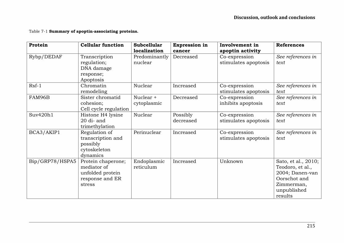
Discussion, outlook and conclusions
215
Table 7-1 Summary of apoptin-associating proteins.
Protein Cellular function Subcellular
localization Expression in cancer
Involvement in apoptin activity
References
Rybp/DEDAF Transcription regulation; DNA damage response; Apoptosis
Predominantly nuclear
Decreased Co-expression stimulates apoptosis
See references in text
Rsf-1 Chromatin remodeling
Nuclear Increased Co-expression stimulates apoptosis
See references in text
FAM96B Sister chromatid cohesion; Cell cycle regulation
Nuclear + cytoplasmic
Decreased Co-expression inhibits apoptosis
See references in text
Suv420h1 Histone H4 lysine 20 di- and trimethylation
Nuclear Possibly decreased
Co-expression stimulates apoptosis
See references in text
BCA3/AKIP1 Regulation of transcription and possibly cytoskeleton dynamics
Perinuclear Increased Co-expression stimulates apoptosis
See references in text
Bip/GRP78/HSPA5 Protein chaperone; mediator of unfolded protein response and ER stress
Endoplasmic reticulum
Increased Unknown Sato, et al., 2010; Teodoro, et al., 2004; Danen-van Oorschot and Zimmerman, unpublished results

Chapter 7
216
Table 7-1 continued. Protein Cellular function Subcellular
localization Expression in cancer
Involvement in apoptin activity
References
Nmi Transcription regulation
Primarily cytosolic
Increased Uknown Sun, et al., 2002; Zhou, et al., 2000 Danen-van Oorschot and Zimmerman, unpublished results
Hippi Intracellular transport; Apoptosis
Golgi Unknown Possibly involved in apoptin elimination from the normal cell
See references in text
Ppil3 Protein-folding chaperone catalyzing cis-trans peptidylprolyl somerisation
Nuclear + cytoplasmic
Possibly decreased
Co-expression enhances apoptin cytoplasmic localization in cancer, but has no effect on apoptosis activity
Huo, Yi, & Yang, 2008
APC1 E3 ubiquitin ligase; subunit of anaphase-promoting complex
Nuclear + cytoplasmic
Unknown Inhibition by apoptin results in G2/M arrest and apoptosis
See references in text
PML Transcription regulation; DNA damage response
Nucleolus; PML bodies
Increased; decreased in advanced stages
Interaction not essential for apoptosis induction
See references in text

Discussion, outlook and conclusions
217
Table 7-1 continued. Protein Cellular function Subcellular
localization Expression in cancer
Involvement in apoptin activity
References
PKAc Catalytic subunit of cAMP-dependent protein kinase, involved in many intracellular signalling patways, including those stimulating proliferation and survival
Various compartments, dependent on association with AKAPs
Various isoforms up-regulated
Stimulation reduces apoptin phosphorylation
See references in text
p85 Regulatory subunit of PI3K, a kinase strongly implicated in tumorigenesis, with functions in proliferation, survival, angiogenesis, autophagy.
Cytoplasmic Activating gain-of-function mutations
Activation stimulates apoptin-induced apoptosis
See references in text
Akt Downstream effector of PI3K signalling
Nuclear + cytoplasmic
Increased Co-expression of nuclear Akt stimulates apoptin-induced apoptosis
See references in text
Cdk2/cyclin A Promotes G1/S transition and replication of DNA and centrosomes in S-phase
Nuclear + cytoplasmic
Unknown Phosphorylates apoptin directly in vitro
See references in text

Chapter 7
218
Table 7-1 continued. Protein Cellular function Subcellular
localization Expression in cancer
Involvement in apoptin activity
References
PKCβ Diverse intracellular signaling pathways, including transformation, proliferation, (inhibition of) apoptosis
Nuclear + Cytoplasmic
Unclear, though increased expression is correlated with poorer prognosis
Phosphorylates apoptin directly in vitro
See references in text
PP2A B56γ Protein phosphatase with important tumor suppressor functions, including regulation of DNA damage response and several stages of cell cycle progression
Nuclear, centromeric
Decreased/inactivated in cancer
Dephosphorylates apoptin directly in vitro
See references in text
*AKAPs, A-kinase anchoring proteins.

Discussion, outlook and conclusions
219
References
Abrams S, Lakum T, Lin K, Jones G, Treweeke A, Farahani M et al. (2007) B-
cell receptor signaling in chronic lymphocytic leukemia cells is regulated by
overexpressed active protein kinase CbetaII. Blood 109, 1193-1201.
Adair B (2000) Immunopathogenesis of chicken anemia virus infection. Dev
Comp Immunol 24, 247-255.
Aits S, Gustafsson L, Hallgren O, Brest P, Gustafsson M, Trulsson M et al.
(2009) HAMLET (human alpha-lactalbumin made lethal to tumor cells)
triggers autophagic tumor cell death. Int J Cancer 124, 1008-1019.
Arroyo J and Hahn W (2005) Involvement of PP2A in viral and cellular
transformation. Oncogene 24, 7746-7755.
Ayoub N, Jeyasekharan A, Bernal J and Venkitaraman A (2008) HP1-beta
mobilization promotes chromatin changes that initiate the DNA damage
response. Nature 453, 682-686.
Backendorf C, Visser A, De Boer A, Zimmerman R, Visser M, Voskamp P et
al. (2008) Apoptin: Therapeutic potential of an early sensor of carcinogenic
transformation. Annu Rev Pharmacol Toxicol 48, 143-169.
Bejarano F, Gonzalez I, Vidal M and Busturia A (2005) The Drosophila RYBP
gene functions as a Polycomb-dependent transcriptional repressor. Mech Dev
122, 1118-1129.
Ben-Aroya S, Coombes C, Kwok T, O'Donnell K, Boeke J and Hieter P (2008).
Toward a comprehensive temperature-sensitive mutant repository of the
essential genes of Saccharomyces cerevisiae. Molecular Cell 30, 248.
Benetti R, García-Cao M and Blasco M (2007) Telomere length regulates the
epigenetic status of mammalian telomeres and subtelomeres. Nat Genet 39,
243-250.

Chapter 7
220
Benetti R, Gonzalo S, Jaco I, Schotta G, Klatt P, Jenuwein T et al. (2007)
Suv4-20h deficiency results in telomere elongation and derepression of
telomere recombination. J Cell Biol 178, 925-936.
Borg A, Tandon A, Sigurdsson H, Clark, G, Fernö M, Fuqua S et al. (1990)
HER-2/neu amplification predicts poor survival in node-positive breast
cancer. Cancer Res 50, 4332-4337.
Botuyan M, Lee J, Ward I, Kim J, Thompson J, Chen J et al. (2006)
Structural basis for the methylation state-specific recognition of histone H4-
K20 by 53BP1 and Crb2 in DNA repair. Cell 127, 1361-1373.
Bruno P, Brinkmann C, Boulanger M, Flinterman M, Klanrit P, Landry M et
al. (2009) Family at last: highlights of the first international meeting on
proteins killing tumour cells. Cell Death and Differentiation 16, 184-186.
Chen D, Zhang J, Li M, Rayburn E, Wang H and Zhang R (2009) RYBP
stabilizes p53 by modulating MDM2. EMBO Rep 10, 166-172.
Cheng C, Huang S, Chang Y, Chung W and Yuo C (2003) The viral death
protein Apoptin interacts with Hippi, the protein interactor of Huntingtin-
interacting protein 1. Biochem Biophys Res Commun 305, 359-364.
Choi J, Sheu J, Guan B, Jinawath N, Markowski P, Wang T et al. (2009)
Functional analysis of 11q13.5 amplicon identifies Rsf-1 (HBXAP) as a gene
involved in paclitaxel resistance in ovarian cancer. Cancer Res 69, 1407-
1415.
Coffer P, Jin J and Woodgett J (1998) Protein kinase B (c-Akt): a
multifunctional mediator of phosphatidylinositol 3-kinase activation.
Biochem J 335, 1-13.
Cosma M, Tanaka T and Nasmyth K (1999) Ordered recruitment of
transcription and chromatin remodeling factors to a cell cycle- and
developmentally regulated promoter. Cell 97, 299-311.
Discussion, outlook and conclusions
221
Cui X, De Vivo I, Slany R, Miyamoto A, Firestein R and Cleary M (1998)
Association of SET domain and myotubularin-related proteins modulates
growth control. Nat Genet 18, 331-337.
Danen-van Oorschot A, Fischer D, Grimbergen J, Klein B, Zhuang S-M,
Falkenburg J et al. (1997) Apoptin induces apoptosis in human transformed
and malignant cells but not in normal cells. Proc Natl Acad Sci U S A 94,
5843-5847.
Danen-Van Oorschot A, van der Eb A and Noteborn M (1999) BCL-2
stimulates Apoptin-induced apoptosis. Adv Exp Med Biol 457, 245-249.
Danen-van Oorschot A, Voskamp P, Seelen M, van Miltenburg M, Bolk M,
Tait S et al. (2004) Human death effector domain-associated factor interacts
with the viral apoptosis agonist Apoptin and exerts tumor-preferential cell
killing. Cell Death Differ 11, 564-573.
Danen-van Oorschot A, Zhang Y-H, Leliveld S, Rohn J, Seelen M, Bolk M et
al. (2003) Importance of nuclear localization of apoptin for tumor-specific
induction of apotosis. J Biol Chem 278, 27729-27736.
Danen-Van Oorschot A, Zhang Y-H, Erkeland S, Fischer D, van der Eb A and
Noteborn M (1999) The effect of Bcl-2 on Apoptin in 'normal' vs transformed
human cells. Leukemia 13, S75-S77.
De Vivo I, Cui X, Domen J and Cleary M (1998) Growth stimulation of
primary B cell precursors by the anti-phosphatase Sbf1. Proc Natl Acad Sci U
S A 95, 9471-9476.
Eichhorn P, Creyghton M and Bernards R (2009) Protein phosphatase 2A
regulatory subunits and cancer. Biochim Biophys Acta 1795, 1-15.
Ferrier V (2002) Hip, hip, hippi! Nat Cell Biol 4, E30.
Finn R, Mistry J, Tate J, Coggill P, Heger A, Pollington J et al. (2010) The
Pfam protein families database. Nucleic Acids Res 38, D211-222.

Chapter 7
222
Flanagan J and Peterson C (1999) A role for the yeast SWI/SNF complex in
DNA replication. Nucleic Acids Res 27, 2022-2028.
Fraga M, Ballestar E, Villar-Garea A, Boix-Chornet M, Espada J, Schotta G
et al. (2005) Loss of acetylation at Lys16 and trimethylation at Lys20 of
histone H4 is a common hallmark of human cancer. Nat Genet 37, 391-400.
Fruman D, Meyers R and Cantley L (1998) Phosphoinositide kinases. Annu
Rev Biochem 67, 481–507.
Garcia E, Marcos-Gutierrez C, del Mar Lorente M, Moreno J and Vidal M
(1999) RYBP, a new repressor protein that interacts with components of the
mammalian Polycomb complex, and with the transcription factor YY1. EMBO
J 18, 3404–3418.
Gervais F, Singaraja R, Xanthoudakis S, Gutekunst C, Leavitt B, Metzler M
et al. (2002) Recruitment and activation of Caspase-8 by the Huntingtin-
interacting protein Hip-1 and a novel partner Hippi. Nat Cell Biol 4, 95-105.
Ghavami S, Asoodeh A, Klonisch T, Halayko A, Kadkhoda K, Kroczak T et al.
(2008) Brevinin-2R(1) semi-selectively kills cancer cells by a distinct
mechanism, which involves the lysosomal-mitochondrial death pathway. J
Cell Mol Med 12, 1005-1022.
Greaves K, Rangasamy D, Devoy M, Marshall Graves A and Tremethick J
(2006) The X and Y chromosomes assemble into H2A.Z, containing facultive
heterochromatin, following meiosis. Mol Cell Biol 26, 5394–5405.
Greaves K, Rangasamy D, Ridgway P and Tremethick J (2007) H2A.Z
contributes to the unique 3D structure of the centromere. Proc Natl Acad Sci
U S A 104, 525–530.
Greeson N, Sengupta R, Arida A, Jenuwein T and Sanders S (2008) Di-
methyl H4 lysine 20 targets the checkpoint protein Crb2 to sites of DNA
damage. J Biol Chem 283, 33168-33174.
Hall A (2005) Rho GTPases and the control of cell behaviour. Biochem Soc
Trans 33, 891-895.

Discussion, outlook and conclusions
223
Hanai K, Furuhashi H, Yamamoto T, Akasaka K and Hirose S (2008) RSF
governs silent chromatin formation via histone H2Av replacement. PLoS
Genet 4, e1000011.
Heilman D, Green M and Teodoro J (2005) The anaphase promoting
complex: a critical target for viral proteins and anti-cancer drugs. Cell Cycle
4, 560-563.
Hiscox J (2007) RNA viruses: hijacking the dynamic nucleolus. Nat Rev
Microbiol 5, 119-127.
Huang W, Batra S, Atkins B, Mishra V and Mehta K (2005) Increases in
intracellular calcium dephosphorylate histone H3 at serine 10 in human
hepatoma cells: potential role of protein phosphatase 2A-protein kinase
CbetaII complex. J Cell Physiol 205, 37-46.
Huo D, Yi L and Yang J (2008) Interaction with Ppil3 leads to the
cytoplasmic localization of Apoptin in tumor cells. Biochem Biophys Res
Commun 372, 14-18.
Ito S, Tan L, Andoh D, Narita T, Seki M, Hirano Y et al. (2010) MMXD, a
TFIIH-independent XPD-MMS19 protein complex involved in chromosome
segregation. Mol Cell 39, 632-640.
Janssen K, Hofmann T, Jans D, Hay R, Schulze-Osthoff K and Fischer U
(2007) Apoptin is modified by SUMO conjugation and targeted to
promyelocytic leukemia protein nuclear bodies. Oncogene 26, 1557-1566.
Jeurissen S, Wagenaar F, Pol J, van der Eb A and Noteborn M (1992)
Chicken anemia virus causes apoptosis of thymocytes after in vivo infection
and of cell lines after in vitro infection. J Virol 66, 7383–7388.
Jiang J, Cole D, Westwood N, Macpherson L, Farzaneh F, Mufti G et al.
(2010) Crucial roles for protein kinase C isoforms in tumor-specific killing by
Apoptin. Cancer Res 70, 7242-7252.

Chapter 7
224
Jin J, Gong J, Yin T, Lu Y, Xia J, Xie Y et al. (2011). PTD4-apoptin protein
and dacarbazine show a synergistic antitumor effect on B16-F1 melanoma in
vitro and in vivo. Eur J Pharmacol 654, 17-25.
Jost C, Marin M and Kaelin WJ (1997) p73 is a simian [correction of human]
p53-related protein that can induce apoptosis. Nature 389, 191-194.
Kaghad M, Bonnet H, Yang A, Creancier L, Biscan J, Valent A et al. (1997)
Monoallelically expressed gene related to p53 at 1p36, a region frequently
deleted in neuroblastoma and other human cancers. Cell 90, 809-819.
Kitching R, Li H, Wong M, Kanaganayakam S, Kahn H and Seth A (2003)
Characterization of a novel human breast cancer associated gene (BCA3)
encoding an alternatively spliced proline-rich protein. Biochim Biophys Acta
1625, 116-121.
Klanrit P, Flinterman M, Odell E, Melino G, Killick R, Norris J et al. (2008).
Specific isoforms of p73 control the induction of cell death induced by the
viral proteins, E1A or apoptin. Cell Cycle 7, 205-215.
Kornitzer D, Sharf R and Kleinberger T (2001) Adenovirus E4orf4 protein
induces PP2A-dependent growth arrest in Saccharomyces cerevisiae and
interacts with the anaphase-promoting complex/cyclosome. J Cell Biol 154,
331-344.
Kotadia S, Kao L, Comerford S, Jones R, Hammer R and Megraw T (2008)
PP2A-dependent disruption of centrosome replication and cytoskeleton
organization in Drosophila by SV40 small tumor antigen. Oncogene 27,
6334-6346.
Kourmouli N, Jeppesen P, Mahadevhaiah S, Burgoyne P, Wu R, Gilbert D et
al. (2004) Heterochromatin and tri-methylated lysine 20 of histone H4 in
animals. J Cell Sci 117, 2491-2501.
Kucharski T, Gamache I, Gjoerup O and Teodoro J (2011) DNA Damage
Response Signaling Triggers Nuclear Localization of the Chicken Anemia
Virus Protein Apoptin. J Virol, Epub ahead of print.

Discussion, outlook and conclusions
225
Landry M, Sicotte A, Champagne C and Lavoie J (2009) Regulation of cell
death by recycling endosomes and golgi membrane dynamics via a pathway
involving Src-family kinases, Cdc42 and Rab11a. Mol Biol Cell 20, 4091-
4106.
Lanz H, Florea B, Noteborn MHM, Backendorf C (2012) Development and
application of an in vitro apoptin kinase assay. Anal Biochem, in press
Leliveld S, Dame R, Mommaas M, Koerten H, Wyman C, Danen-van
Oorschot A et al. (2003) Apoptin protein multimers form distinct higher-
order nucleoprotein complexes with DNA. Nucleic Acids Res 31, 4805-4813.
Leliveld S, Dame R, Rohn J, Noteborn M and Abrahams J (2004) Apoptin's
functional N- and C-termini independently bind DNA. FEBS Lett 557, 155-
158.
Leliveld S, Zhang Y, Rohn J, Noteborn M and Abrahams J (2003) Apoptin
induces tumor-specific apoptosis as a globular multimer. J Biol Chem 278,
9042-9051.
Leon D and Canaves J (2003) In silico study of breast cancer associated gene
3 using LION Target Engine and other tools. Biotechniques 35, 1222-1226,
1228, 1230-1231.
Leung T and Ngan H (2010) Interaction of TAp73 and breast cancer-
associated gene 3 enhances the sensitivity of cervical cancer cells in
response to irradiation-induced apoptosis. Cancer Res 70, 6486-6496.
Li Y, Dowbenko D and Lasky L (2002) AKT/PKB phosphorylation of
p21Cip/WAF1 enhances protein stability of p21Cip/WAF1 and promotes cell
survival. J Biol Chem 277, 11352–11361.
Liu X, Elojeimy S, El-Zawahry A, Holman D, Bielawska A, Bielawski J et al.
(2006) Modulation of ceramide metabolism enhances viral protein apoptin's
cytotoxicity in prostate cancer. Mol Ther 14, 637-646.

Chapter 7
226
Liu X, Zeidan Y, Elojeimy S, Holman D, El-Zawahry A, Guo G et al. (2006)
Involvement of sphingolipids in apoptin-induced cell killing. Mol Ther 14,
627–636.
Loyola A, Huang J, LeRoy G, Hu S, Wang Y, Donnelly R et al. (2003)
Functional analysis of the subunits of the chromatin assembly factor RSF.
Mol Cell Biol 23, 6759-6768.
Loyola A, LeRoy G, Wang Y and Reinberg D (2001) Reconstitution of
recombinant chromatin establishes a requirement for histone-tail
modifications during chromatin assembly and transcription. Genes Dev 15,
2837-2851.
Maddika S, Bay G, Kroczak T, Ande S, Maddika S, Wiechec E et al. (2007)
Akt is transferred to the nucleus of cells treated with apoptin, and it
participates in apoptin-induced cell death. Cell Prolif 40, 835–848.
Maddika S, Booy E, Johar D, Gibson S, Ghavami S and Los M (2005)
Cancer-specific toxicity of apoptin is independent of death receptors but
involves the loss of mitochondrial membrane potential and the release of
mitochondrial cell-death mediators by a Nur77-dependent pathway. J Cell
Sci 118, 4485–4493.
Maddika S, Panigrahi S, Wiechec E, Wesselborg S, Fischer U, Schulze-
Osthoff K et al. (2009) Unscheduled Akt-triggered activation of cyclin-
dependent kinase 2 as a key effector mechanism of apoptin's anticancer
toxicity. Mol Cell Biol 29, 1235-1248.
Maddika S, Wiechec E, Ande S, Poon I, Fischer U, Wesselborg S et al. (2008)
Interaction with PI3-kinase contributes to the cytotoxic activity of apoptin.
Oncogene 27, 3060-3065.
Majumder P, Chattopadhyay B, Mazumder A, Das P and Bhattacharyya N
(2006) Induction of apoptosis in cells expressing exogenous Hippi, a
molecular partner of huntingtin-interacting protein Hip1. Neurobiol Dis 22,
242–256.

Discussion, outlook and conclusions
227
Majumder P, Chattopadhyay B, Sukanya S, Ray T, Banerjee M,
Mukhopadhyay D et al. (2007) Interaction of HIPPI with putative promoter
sequence of Caspase-1 in vitro and in vivo. Biochem Biophys Res Commun
353, 80-95.
Majumder P, Choudhury A, Banerjee M, Lahiri A and Bhattacharyya N
(2007) Interactions of HIPPI, a molecular partner of Huntingtin interacting
protein HIP1, with the specific motif present at the putative promoter
sequence of the Caspase-1, Caspase-8 and Caspase-10 genes. FEBS J 274,
3886–3899.
Mao T, Hsu C, Yen M, Gilks B, Sheu J, Gabrielson E et al. (2006) Expression
of Rsf-1, a chromatin-remodeling gene, in ovarian and breast carcinoma.
Hum Pathol 37, 1169-1175.
Marte B and Downward J (1997) PKB/Akt: connecting phosphoinositide 3-
kinase to cell survival and beyond. Trends Biochem Sci 22, 355-358.
Michiels F and Collard J (1999) Rho-like GTPases: Their role in cell adhesion
and invasion. Biochem Soc Symp 65, 125-146.
Nakayama K, Nakayama N, Jinawath N, Salani R, Kurman R, Shih Ie M et
al. (2007) Amplicon profiles in ovarian serous carcinomas. Int J Cancer 120,
2613–2617.
Noteborn M and Koch G (1995) Chicken anaemia virus infection: molecular
basis of pathogenicity. Avian Pathol 24, 11-31.
Noteborn M, Todd D, Verschueren C, de Gauw H, Curran W, Veldkamp S et
al. (1994) A single chicken anemia virus protein induces apoptosis. J Virol
68, 346-351.
Noteborn M, Zhang Y and van der Eb A (1998) Apoptin specifically causes
apoptosis in tumor cells and after UV-treatment in untransformed cells from
cancer-prone individuals: a review. Mutat Res 400, 447-455.
Novak R and Phillips A (2008) Adenoviral-mediated Rybp expression
promotes tumor cell-specific apoptosis. Cancer Gene Ther 15, 713-722.

Chapter 7
228
Pekarsky Y, Hallas C, Palamarchuk A, Koval A, Bullrich F, Hirata Y et al.
(2001) Akt phosphorylates and regulates the orphan nuclear receptor Nur77.
Proc Natl Acad Sci U S A 98, 3690–3694.
Peng D, Sun J, Wang Y, Tian J, Zhang Y, Noteborn M et al. (2007) Inhibition
of hepatocarcinoma by systemic delivery of Apoptin gene via the hepatic
asialoglycoprotein receptor. Cancer Gene Ther 14, 66-73.
Perpelescu M, Nozaki N, Obuse C, Yang H and Yoda K (2009) Active
establishment of centromeric CENP-A chromatin by RSF complex. J Cell Biol
185, 397-407.
Peters M, Jackson D, Crabb B and Browning G (2002) Chicken anemia virus
VP2 is a novel dual specificity protein phosphatase. J Biol Chem 277, 39566-
39573.
Pietersen A, Erkeland S, Rutjes S, Philipsen S, Medema J and Noteborn M
(2005) Continuous apoptin expression in transgenic mice does not interfere
with lymphocyte development and proliferation. J Med Mol Biol 2, 321–330.
Pietersen A, van der Eb M, Rademaker H, van den Wollenberg D, Rabelink
M, Kuppen P et al. (1999) Specific tumor-cell killing with adenovirus vectors
containing the apoptin gene. Gene Ther 6, 882–892.
Pogribny I, Ross S, Tryndyak V, Pogribna M, Poirier L and Karpinets T (2006)
Histone H3 lysine 9 and H4 lysine 20 trimethylation and the expression of
Suv4-20h2 and Suv-39h1 histone methyltransferases in
hepatocarcinogenesis induced by methyl deficiency in rats. Carcinogenesis
27, 1180–1186.
Poon I, Oro C, Dias M, Zhang J and Jans D (2005) A tumor cell-specific
nuclear targeting signal within chicken anemia virus VP3/apoptin. J Virol
79, 1339-1341.
Poon I, Oro C Dias M, Zhang J and Jans D (2005b). Apoptin nuclear
accumulation is modulated by a CRM1-recognized nuclear export signal that
is active in normal but not in tumor cells. Cancer Res 65, 7059-7064.

Discussion, outlook and conclusions
229
Porrás A, Bennett J, Howe A, Tokos K, Bouck N, Henglein B et al. (1996) A
novel simian virus 40 early-region domain mediates transactivation of the
cyclin A promoter by small-t antigen and is required for transformation in
small-t antigen-dependent assays. J Virol 70, 6902-6908.
Qian C and Zhou M (2006) SET domain protein lysine methyltransferases:
Structure, specificity and catalysis. Cell Mol Life Sci 63, 2755-2763.
Qin H, Wang J, Liang Y, Taniguchi Y, Tanigaki K and Han H (2004) RING1
inhibits transactivation of RBP-J by Notch through interaction with LIM
protein KyoT2. Nucleic Acids Res 32, 1492-1501.
Raisner M and Madhani D (2006) Patterning chromatin: from and function
for H2A.Z variant nucleosomes. Curr Opin Genet Dev 16, 119–124.
Rangasamy D, Greaves K and Tremethick J (2004) RNA interference
demonstrates a novel role for H2A.Z in chromosome segregation. Nat Struct
Mol Biol 11, 650–655.
Robert A, Smadja-Lamère N, Landry M, Champagne C, Petrie R, Lamarche-
Vane N et al. (2006) Adenovirus E4orf4 hijacks rho GTPase-dependent actin
dynamics to kill cells: a role for endosome-associated actin assembly. Mol
Biol Cell 17, 3329-3344.
Rohn J, Zhang Y-H, Aalbers R, Otto N, den Hertog J, Henriquez N et al.
(2002) A tumor-specific kinase activity regulates the viral death protein
apoptin. J Biol Chem, 277, 50820-50827.
Rouse J and Jackson S (2002) Interfaces between the detection, signaling,
and repair of DNA damage. Science 297, 547-551.
Rubie H, Hartmann O, Michon J, Frappaz D, Coze C, Chastagner P et al.
(1997) N-Myc gene amplification is a major prognostic factor in localized
neuroblastoma: results of the French NBL 90 study. J Clin Oncol 15, 1171-
1182.

Chapter 7
230
Saffery R, Irvine D, Griffiths B, Kalitsis P and Choo K (2000). Components of
the human spindle checkpoint control mechanism localize specifically to the
active centromere on dicentric chromosomes. Hum Genet 107, 376-384.
Sanders S, Portoso M, Mata J, Bähler J, Allshire R and Kouzarides T (2004)
Methylation of histone H4 lysine 20 controls recruitment of Crb2 to sites of
DNA damage. Cell 119, 603-614.
Sarcinella E, Zuzarte C, Lau N, Draker R and Cheung P (2007)
Monoubiquitylation of H2A.Z distinguishes its association with euchromatin
or facultive heterochromatin. Mol Cell Biol 27, 6457–6468.
Sastri M, Barraclough D, Carmichael P and Taylor S (2005) A-kinase-
interacting protein localizes protein kinase A in the nucleus. Proc Natl Acad
Sci U S A 102, 349-354.
Sato M, Yao V, Arap W and Pasqualini R (2010) GRP78 signaling hub a
receptor for targeted tumor therapy. Adv Genet 69, 97-114.
Sauane M, Su Z, Dash R, Liu X, Norris J, Sarkar D et al. (2010) Ceramide
plays a prominent role in MDA-7/IL-24-induced cancer-specific apoptosis. J
Cell Physiol 222, 546-555.
Sawa C, Yoshikawa T, Matsuda-Suzuki F, Delehouzee S, Goto M, Watanabe
H et al. (2002) YEAF1/RYBP and YAF-2 are functionally distinct members of
a cofactor family for the YY1 and E4TF1/hGABP transcription factors. J Biol
Chem 277, 22484–22490.
Schlisio S, Halperin T, Vidal M and Nevins J (2002) Interaction of YY1 with
E2Fs, mediated by RYBP, provides a mechanism for specificity of E2F
function. EMBO J 21, 5775–5786.
Schotta G, Lachner M, Sarma K, Ebert A, Sengupta R, Reuter G et al. (2004)
A silencing pathway to induce H3-K9 and H4-K20 trimethylation at
constitutive heterochromatin. Genes Dev 18, 1251-1262.
Schotta G, Sengupta R, Kubicek S, Malin S, Kauer M, Callén E et al. (2008)
A chromatin-wide transition to H4K20 monomethylation impairs genome

Discussion, outlook and conclusions
231
integrity and programmed DNA rearrangements in the mouse. Genes Dev 22,
2048-2061.
Schwab M (1998) Amplification of oncogenes in human cancer cells.
Bioessays 20, 473–479.
Shamay M, Barak O, Doitsh G, Ben-Dor I and Shaul Y (2002) Hepatitis B
virus pX interacts with HBXAP, a PHD finger protein to coactivate
transcription. J Biol Chem 277, 9982-9988.
Shih Ie M, Sheu J, Santillan A, Nakayama K, Yen M, Bristow R et al. (2005)
Amplification of a chromatin remodeling gene, Rsf-1/HBXAP, in ovarian
carcinoma. Proc Natl Acad Sci U S A, 102, 14004-14009.
Siddiqui H, Fox S, Gunawardena R and Knudsen E (2007) Loss of RB
compromises specific heterochromatin modifications and modulates
HP1alpha dynamics. J Cell Physiol 211, 131-137.
Sims J, Houston S, Magazinnik T and Rice, J. (2006) A trans-tail histone
code defined by monomethylated H4 Lys-20 and H3 Lys-9 demarcates
distinct regions of silent chromatin. J Biol Chem 281, 12760-12766.
Skoczylas C, Henglein B and Rundell K (2005) PP2A-dependent
transactivation of the cyclin A promoter by SV40 ST is mediated by a cell
cycle-regulated E2F site. Virology 332, 596-601.
Song G, Ouyang G and Bao S (2005) The activation of Akt/PKB signaling
pathway and cell survival. J Cell Mol Med 9, 59-71.
Stanton S, Blanck J, Locker J and Schreiber-Agus N (2007) Rybp interacts
with Hippi and enhances Hippi-mediated apoptosis. Apoptosis 12, 2197-
2206.
Sun G, Tong X, Dong Y, Mei Z and Sun Z (2002) Identification of a protein
interacting with apoptin from human leucocyte cDNA library by using yeast
two-hybrid screening. Sheng Wu Hua Xue Yu Sheng Wu Wu Li Xue Bao 34,
369–372.

Chapter 7
232
Sun,J, Yan Y, Wang X, Liu X, Peng D, Wang M et al. (2009) PTD4-apoptin
protein therapy inhibits tumor growth in vivo. Int J Cancer, 124, 2973-2981.
Sun L, Kong B, Sheng X, Sheu J and Shih I (2010) Dendritic cells
transduced with Rsf-1/HBXAP gene generate specific cytotoxic T
lymphocytes against ovarian cancer in vitro. Biochem Biophys Res Commun
394, 633-638.
Swaminathan J, Baxter E and Corces V (2005) The role of histone H2Av
variant replacement and histone H4 acetylation in the establishment of
Drosophila heterochromatin. Genes Dev 19, 65–76.
Teodoro J, Heilman D, Parker A and Green M (2004) The viral protein
Apoptin associates with the anaphase-promoting complex to induce G2/M
arrest and apoptosis in the absence of p53. Genes Dev 18, 1952-1957.
Trimarchi J, Fairchild B, Wen J and Lees J (2001) The E2F6 transcription
factor is a component of the mammalian Bmi1-containing polycomb
complex. Proc Natl Acad Sci U S A 98, 1519–1524.
Tryndyak V, Kovalchuk O and Pogribny I (2006) Loss of DNA methylation
and histone H4 lysine 20 trimethylation in human breast cancer cells is
associated with aberrant expression of DNA methyltransferase 1, Suv4-20h2
histone methyltransferase and methyl-binding proteins. Cancer Biol Ther 5,
65–70.
Tsuda H, Hirohashi S, Shimosato Y, Hirota T, Tsugane S, Yamamoto H et al.
(1989) Correlation between long-term survival in breast cancer patients and
amplification of two putative oncogene-coamplification units: hst-1/int-2
and c-erbB-2/ear-1. Cancer Res 49, 3104-3108.
Van Den Broeck A, Brambilla E, Moro-Sibilot D, Lantuejoul S, Brambilla C,
Eymin B et al. (2008) Loss of histone H4K20 trimethylation occurs in
preneoplasia and influences prognosis of non-small cell lung cancer. Clin
Cancer Res 14, 7237-7245.

Discussion, outlook and conclusions
233
Vanhaesebroeck B and Waterfield M (1999) Signaling by distinct classes of
phosphoinositide 3-kinases. Exp Cell Res 253, 239–254.
Vignali M, Hassan A, Neely K and Workman J (2000) ATP-dependent
chromatin-remodeling complexes. Mol Cell Biol 20, 1899-1910.
Voelkel-Johnson C, Hannun Y and El-Zawahry A (2005) Resistance to TRAIL
is associated with defects in ceramide signaling that can be overcome by
exogenous C6-ceramide without requiring down-regulation of cellular FLICE
inhibitory protein. Mol Cancer Ther 4, 1320-1327.
Yang H and Mizzen C (2009) The multiple facets of histone H4-lysine 20
methylation. Biochem Cell Biol 87, 151-161.
Yang H, Pesavento J, Starnes T, Cryderman D, Wallrath L, Kelleher N et al.
(2008) Preferential dimethylation of histone H4 lysine 20 by Suv4-20. J Biol
Chem 283, 12085-12092.
Yu K, Itokawa T, Zhu M, Syam S, Seth A and Insogna K (2007) Breast
cancer-associated gene 3 (BCA3) is a novel Rac1-interacting protein. J Bone
Miner Res 22, 628-637.
Zhang Y-H, Leliveld S, Kooistra K, Molenaar C, Rohn J, Tanke H et al. (2003)
Recombinant apoptin multimers kill tumor cells but are nontoxic and
epitope-shielded in a normal-cell-specific fashion. Exp Cell Res 289, 36-46.
Zhang Y, Kooistra K, Pietersen A, Rohn J and Noteborn M (2004) Activation
of the tumor-specific death effector apoptin and its kinase by an N-terminal
determinant of simian virus 40 large T antigen. J Virol 78, 9965-9976.
Zheng L, Schickling O, Peter M and Lenardo M (2001) The death effector
domain-associated factor plays distinct regulatory roles in the nucleus and
cytoplasm. J Biol Chem 276, 31945–31952 .
Zhou B and Elledge S (2000) The DNA damage response: putting
checkpoints in perspective. Nature 408, 433-439.
Chapter 7
234
Zhou X, Liao J, Meyerdierks A, Feng L, Naumovski L, Bottger E et al. (2000)
Interferon-alpha induces nmi-IFP35 heterodimeric complex formation that is
affected by the phosphorylation of IFP35. J Biol Chem 275, 21364-21371.
Zhuang S, Landegent J, Verschueren C, Falkenburg J, van Ormondt H, van
der Eb, A et al. (1995) Apoptin, a protein encoded by chicken anemia virus,
induces cell death in various human hematologic malignant cells in vitro.
Leukemia 9, S118-120.
Zhuang S, Shvarts A, van Ormondt H, Jochemsen A, van der Eb A and
Noteborn M (1995). Apoptin, a protein derived from chicken anemia virus,
induces p53-independent apoptosis in human osteosarcoma cells. Cancer
Res 55, 486-489.

Discussion, outlook and conclusions
235


Appendices

Appendices
238

Summary
239
Summary
The human body consists of very many cells, in the order of trillions.
Each of these trillions of cells has its own function, and together,
groups of cells with similar functions make up the various organs of
which we are comprised. Remarkably enough, all of these cells, as
different as they are, – consider the cells making up the heart muscle,
the ones in our brain, the ones lining our stomach and intestines,
even the ones making our bones – all originate from one single cell:
the fertilized oocyte. The fertilized oocyte lies at the very basis of life,
and has the potential to replicate and specialize (i.e. differentiate) into
each of the different types of cells required in the developing embryo.
It does so according to a strictly regulated set of rules, that dictate
which cells are to divide, when they are to do so, when they are to
become more specialized, even which cells must die and when – e.g.,
to create the spaces between our fingers and toes. After birth, and
throughout life, these rules continue to govern cellular replication,
ensuring that old, damaged and/or dead cells are replaced, and
wounds are healed, without creating any excess tissue or disturbing
the functions of the different organs.
Cancer occurs as a violation of these rules. When the growth and
death of a cell are no longer controlled, it can divide unlimitedly, and
the resulting tumor could eventually invade and disrupt surrounding
tissue, hindering organ function. If tumor cells manage to invade the
blood stream, they could also travel to other sites in the body, where
they can continue to replicate; this is termed metastasis. Standard
therapy – surgery, radio- and/or chemotherapy, is not inherently
selective for cancer cells. This results in lower efficiency and a host of
unwanted side-effects, ranging from surgical amputation and life-
threatening bone marrow depression, to the less dangerous but often
equally upsetting loss of hair. Thus, in order to improve on efficiency

Appendices
240
and minimize side-effects, more targeted therapies are needed. In
order to design such therapies, we must understand how a cancer cell
works. Otherwise stated, we must learn how it manages to extricate
itself from the mechanisms that normally control cell division.
The hallmarks of a cancer cell
Research over the past several decades has shown that cancer cells
share a number of features, which are characteristic of the process of
tumorigenesis (chapter 2). First, cancer cells have the ability to
produce their own growth factors, inducing proliferation
independently of signals in their environment. Alternatively, they may
stimulate surrounding cells (i.e., the cells in the tumor environment)
to produce the growth factors for them. Second, cancer cells have
found a way to circumvent the influence of growth inhibitory signals.
Together with the first feature, this renders the cancer cell insensitive
to extracellular control. This normally triggers the activation of
internal controls, which program the cell to die. This type of cell death
is termed apoptosis, and evasion of apoptosis is the third trait of
malignant transformation. Yet cells still have one more method of
intracellular control, which cancer cells must overcome in order to
become truly immortal. With each round of cell division, the
chromosomes, which carry the cell’s DNA, become a little bit shorter.
After a certain number of doublings, the chromosomes have become
too short, and cell division is halted. Consequently, the fourth feature
of carcinogenesis requires cancer cells to find a way to elongate the
chromosome ends. Once cancer cells have acquired these first four
traits, they are able to divide unlimitedly. In order to support this
growth, they must develop the ability to create new vessels to ensure
their blood supply (angiogenesis, the fifth cancer hallmark); the ability
to invade these and other vessels and give rise to metastases is the
sixth cancer hallmark. Cancer growth further requires a fundamental
change in cellular metabolism (the seventh proposed hallmark), and,

Summary
241
importantly, evasion of destruction by the cells of the immune system
(the eighth hallmark).
The acquisition of these cancer-typical traits is achieved by alterations
in the cellular DNA, including mutations, deletions and amplifications.
The majority of genes affected are those that encode the proteins
involved in repairing damaged DNA. As a result, cancer cells
accumulate errors in their DNA (a phenomenon termed genomic
instability), which again enables them to acquire further cancer-
typical features.
Apoptin and other proteins killing tumor cells
A number of proteins have recently been discovered to selectively kill
cancer cells. That is, these proteins are able to distinguish between
the normal and tumor cells, and kill the latter, while leaving the
normal ones unharmed. This group of proteins killing tumor cells
(PKTC) might therefore have the potential to be used to develop the
ideal anti-cancer therapy.
The PKTC comprise proteins of various origins, including human
proteins TRAIL, MDA7/IL24 and Par-4, the frog-derived Brevinin-2R,
HAMLET, a complex between the milk protein alpha-lactalbumin and
oleic acid (a component of human fat tissue, often used in
detergents), and the viral proteins adenovirus E4ORF4, parvovirus
NS1, and Chicken Anemia Virus-encoded protein apoptin.
While the PKTC are discussed in chapter 3, this thesis focuses on
apoptin, which was the first of the PKTC to be discovered. Studies
were carried out to gain more information on the mechanisms used by
apoptin to sense oncogenic transformation and induce tumor-selective
apoptosis. Knowledge on the proteins with which apoptin interacts
within the human cell, and how these help apoptin to distinguish
between normal and tumor cells, preventing its activation in normal

Appendices
242
cells, and helping it to kill malignant cells, should aid in the design
and development of novel, effective, selective, and hence more efficient
therapies for cancer.
Apoptin interacts with FAM96B
In order to decipher the mechanisms behind apoptin’s cellular
activities and uncover new targets for anti-cancer therapies, we
performed an analysis of apoptin-interacting proteins. One of the
proteins identified in this way is the protein FAM96B (chapter 4).
Identification of the FAM96B partners implicates a role of FAM96B in
the regulation of the cell cycle, including the processes of sensing DNA
damage and establishing sister chromatid cohesion. Through its
interaction with FAM96B, apoptin is very likely also linked to these
processes.
Apoptin & the nucleolus
One of the characteristic features of apoptin is its nuclear localization
in tumor cells, versus its cytoplasmic localization in normal cells.
Within the tumor cell nucleus, apoptin can be found in specialized
regions, one of which is the nucleolus (chapter 5). The nucleolus is
the cell’s ribosome factory, which produces the ribosomes required to
drive protein synthesis. Furthermore, the nucleolus has roles in
coordinating the DNA damage response and inducing apoptosis.
Apoptin is found to associate with nucleolar chromatin as well as
various nucleolar proteins, suggesting that apoptin localizes to the
nucleolus of tumor cells to shut off protein synthesis and induce
apoptosis in response to DNA damage.
Apoptin interacts with BCA3 and is dephosphorylated by PP2A
Another characteristic feature of apoptin activity is its
phosphorylation, which can be detected only in tumor cells, and is
likely to be the trigger for apoptin nucle(ol)ar localization and
apoptosis induction. In chapter 6 we discuss the discovery of another

Summary
243
apoptin-interacting protein, BCA3. BCA3 led us to the involvement of
PP2A B56δ in apoptin phosphorylation, while a second line of
investigation led us to the involvement of PP2A B56γ in the same
process. Dephosphorylation by the major tumor suppressor protein
phosphatase 2A (PP2A) appears to fulfill the important function of
keeping apoptin unphosphorylated, and, hence, inactive, in normal
cells, while the loss of PP2A activity in tumor cells results in
phosphorylation, and hence activation of apoptin.
Future perspectives
Apoptin is a small, avian-virus derived protein that has the
remarkable ability to sense differences between normal and tumor
cells, and is able to kill the latter while leaving the former unharmed.
As discussed in chapter 7, novel insights into the mechanisms
underlying this remarkable behavior should lead not only to an
improved understanding of fundamental cellular biology, but also,
importantly, to the development of new and improved anti-cancer
strategies.

Appendices
244

Samenvatting
245
Samenvatting
Het menselijk lichaam bestaat uit triljoenen cellen. Elk van deze cellen
heeft zijn eigen functie, en groepen van cellen met een vergelijkbare
functie vormen samen de verschillende organen in het lichaam.
Ondanks de grote verschillen tussen de verschillende celtypen – denk
bijvoorbeeld aan de cellen die samen de hartspier vormen, de cellen in
onze hersenen, die van onze darmen, en de cellen die onze botten
vormen – ontstaan al deze cellen uit één en dezelfde cel: de bevruchte
eicel. De bevruchte eicel is een zogenaamde ‘stamcel’, en bevat de
unieke eigenschap dat hij zich kan vermenigvuldigen, maar dat
sommige van de nieuw ontstane cellen zich ook kunnen specialiseren
tot een bepaald type cel (oftewel, differentiëren). Op deze wijze ontstaat
een volledig ontwikkeld embryo, met alle benodigde organen, uit deze
eerste stamcel.
De deling en differentiatie van cellen is strikt gereguleerd, zodat elke
cel weet of en wanneer het zich moet delen, wanneer het zich moet
specialiseren en tot welk celtype, en zelfs wanneer het dood moet
gaan, bijvoorbeeld om ruimte te creëren tussen de vingers en tenen.
Ook na de geboorte, en zelfs gedurende het gehele leven, blijft deze
regulatie van toepassing. Hierdoor kunnen oude, beschadigde, en/of
dode cellen vervangen en wonden geheeld worden, zonder dat er ook
maar één cel te veel of te weinig wordt aangemaakt, en weefselfunctie
behouden blijft.
Kanker ontstaat juist wanneer deze regels geschonden worden.
Wanneer de groei en dood van een cel niet meer gecontroleerd kunnen
worden, kan deze cel zich blijven vermenigvuldigen. De zo ontstane
tumor kan het omringende weefsel verstoren, waarbij de functie van
het orgaan in gedrang kan komen. Tumorcellen kunnen bijvoorbeeld
ook in de bloedbaan terechtkomen, en naar een ander deel van het

Appendices
246
lichaam reizen, waar ze een nieuwe tumor kunnen vormen:
metastasering.
De standaardbehandeling voor kanker bestaat uit chirurgie, gevolgd
door radio- en/of chemotherapie, en is niet selectief voor kankercellen.
Dit heeft een lagere therapie-efficiëntie tot gevolg, evenals een lange
lijst aan ongewenste neveneffecten, variërend van chirurgische
amputatie en levensbedreigende beenmergsuppressie, tot het minder
gevaarlijke maar evenwel aangrijpende haaruitval. Om tot betere
therapieën te komen is daarom een doelgerichtere aanpak vereist,
waarbij gebruik wordt gemaakt van kennis van de eigenschappen van
kankercellen.
De karakteristieke eigenschappen van een kankercel
Decennialang onderzoek heeft uitgewezen dat kankercellen een aantal
karakteristieke eigenschappen bezitten (hoofdstuk 2). Ze kunnen ten
eerste hun eigen groeifactoren produceren, waarmee ze hun eigen
groei in stand kunnen houden, zonder daarvoor afhankelijk te zijn van
hun omgeving. Andersom kunnen ze de cellen in hun omgeving ook
zodanig beïnvloeden dat zij de benodigde groeistimulerende factoren
uitscheiden. Kankercellen kunnen ten tweede signalen omzeilen, die
anders voor inhibitie van de celgroei zouden zorgen. Deze twee
eigenschappen zorgen dat kankercellen in principe ongevoelig worden
voor controle van buitenaf. Echter, ze zijn dan nog wel gevoelig voor
intracellulaire controlemechanismen. Een dergelijk mechanisme
bestaat uit apoptose, een vorm van geprogrammeerde celdood, die
wordt geactiveerd wanneer cellen zich ongecontroleerd delen. Het
onderdrukken van apoptose vormt dan ook de derde typische
eigenschap van kankercellen. De vierde eigenschap heeft te maken
met de lengte van de chromosomen. Chromosomen bevatten het
cellulaire DNA, en worden bij elke celdeling een stukje korter. Worden
ze te kort, dan treedt er een mechanisme in werking waarbij de
celdeling wordt stopgezet. Om door te kunnen groeien, zullen

Samenvatting
247
kankercellen dus een manier moeten vinden om de uiteinden van de
chromosomen weer te verlengen, bijvoorbeeld door het aanzetten van
een bepaald eiwit, of door stukjes DNA aan elkaar te plakken en weer
op andere plaatsen te knippen. Wanneer een kankercel deze vier
eigenschappen bezit, is het in principe onsterfelijk geworden en kan
het zich ongecontroleerd vermenigvuldigen. Hoe groter de tumor, hoe
groter de behoefte aan zuurstof en voedingsstoffen; om hierin te
kunnen voorzien zullen de kankercellen dus ook nieuwe bloedvaten
aan moeten kunnen leggen: dit is de vijfde eigenschap. Komt een
kankercel eenmaal in een bloed- of ander vat terecht, dan kan dit
aanleiding geven tot metastasering (de zesde eigenschap). Om snel en
veel te kunnen blijven groeien, ondergaan kankercellen bovendien een
verandering in hun metabolisme (de voorgestelde zevende eigenschap);
daarnaast zorgen kankercellen ervoor dat ze niet ontdekt en dus
opgeruimd worden door cellen van het immuunsysteem.
Kankercellen verkrijgen de bovengenoemde typische eigenschappen
door middel van veranderingen in hun DNA, waaronder mutaties,
deleties en amplificaties. De genen die het vaakst door deze
veranderingen worden getroffen, coderen vaak voor eiwitten die een rol
spelen bij het herstel van DNA schade. Hierdoor kunnen genetische
veranderingen zich ophopen (dit fenomeen wordt genetische
instabiliteit genoemd), waardoor het weer makkelijker wordt voor de
kankercel om de verdere benodigde eigenschappen te verkrijgen.
Apoptin en andere eiwitten die kankercellen doden
Recentelijk zijn er een aantal eiwitten ontdekt, waarvan bewezen is dat
zij specifiek kankercellen doden. Deze eiwitten kunnen onderscheid
maken tussen normale en tumorcellen, en doden de tumorcellen
zonder schade aan te richten in de gezonde cellen. Deze groep eiwitten
(in het Engels PKTC genoemd, voor “proteins killing tumor cells”)
zouden dus de basis kunnen vormen voor de ontwikkeling van
misschien wel hèt ideale medicijn tegen kanker!

Appendices
248
De PKTC groep bestaat uit eiwitten van diverse origine, waaronder de
menselijke eiwitten TRAIL, MDA7/IL24 en Par-4, het kikkereiwit
Brevinin-2R, en HAMLET, een complex bestaande uit het melkeiwit
alfa-lactalbumine en oliezuur, het meest voorkomende vetzuur in
menselijk vetweefsel en een veelgebruikt bestanddeel in zeep.
Daarnaast zijn er nog een aantal virale eiwitten, zoals E4ORF4,
afkomstig van het adenovirus, NS1, afkomstig van het parvovirus, en
apoptin, afkomstig van het Chicken Anemia Virus.
De verschillende PKTC zijn besproken in hoofdstuk 3, terwijl apoptin,
die als eerste PKTC werd ontdekt, het hoofdonderwerp vormt van dit
proefschrift. Er zijn experimenten uitgevoerd om informatie te
verkrijgen over de mechanismen die door apoptin worden gebruikt om
maligne transformatie te herkennen en tumorselectieve apoptose te
induceren. Kennis over de cellulaire eiwitten waarmee apoptin een
interactie aangaat, alsmede de manier waarop deze eiwitten bijdragen
aan het onderscheid door apoptin tussen normale en tumorcellen, het
aanzetten tot celdood in tumorcellen, en het voorkomen van activatie
in normale cellen, zou belangrijke aanknopingspunten moeten
opleveren voor de ontwikkeling van nieuwe, effectieve, selectieve en
dus efficiëntere vormen van behandeling voor kanker.
Apoptin bindt aan FAM96B
Om aanknopingspunten te vinden voor het ontrafelen van de
mechanismen achter het karakteristieke gedrag van apoptin en de
ontwikkeling van nieuwe antikanker medicijnen, zijn we op zoek
gegaan naar eiwitten die in de cel aan apoptin binden. Een van de
eiwitten die we op deze manier gevonden hebben, was het tot dan toe
nog relatief onbekende eiwit FAM96B (hoofdstuk 4). Analyses naar
eiwitten die met FAM96B een interactie aangaan, hebben laten zien
dat FAM96B betrokken is bij de regulatie van de celdeling, o.a. bij het
herkennen van de aanwezigheid van genetische schade, en de
verdeling van genetisch materiaal over de dochtercellen tijdens de

Samenvatting
249
deling. Vanwege de associatie met FAM96B, lijkt het aannemelijk dat
apoptin ook bij deze processen is betrokken.
Apoptin & de nucleolus
Een van de karakteristieke eigenschappen van apoptin betreft zijn
kernlokalisatie in tumorcellen, in tegenstelling tot zijn lokalisatie in
normale cellen, waar apoptin in het cytoplasma verblijft. Binnen de
kern van de tumorcel houdt apoptin zich in specifieke gebieden op,
waaronder de nucleolus (hoofdstuk 5). De nucleolus is de plek waar
alle ribosomen worden gemaakt; deze zijn op hun beurt weer nodig
zijn voor de eiwitsynthese in de cel. Daarnaast is het betrokken bij het
coördineren van herstel van DNA schade en de inductie van apoptose.
Apoptin bindt aan nucleolair chromatine (chromatine is het complex
van DNA plus de eiwitten die ervoor zorgen dat het netjes in de
celkern is opgevouwen) en wordt geassocieerd met verscheidene
nucleolaire eiwitten, wat suggereert dat apoptin zich naar de
nucleolus van kankercellen begeeft om eiwitsynthese (en dus
celdeling) stop te zetten en apoptose te induceren in reactie op de
aanwezigheid van DNA schade, hetgeen zoals eerder besproken
veelvuldig voorkomt in tumorcellen.
Apoptin bindt aan BCA3 en wordt door PP2A gedefosforyleerd
Een andere typische eigenschap van apoptin, betreft zijn fosforylering:
deze is alleen in kankercellen aan te tonen, en is waarschijnlijk de
aanzet tot transport naar de kern/nucleolus en inductie van apoptose.
Hoofdstuk 6 beschrijft de identificatie van nog een interactiepartner
van apoptin, namelijk BCA3. Via BCA3 en een ander, parallel lopend
onderzoek, kwamen we op het spoor van het eiwit PP2A, welke een
belangrijke rol lijkt te spelen in de fosforylering van apoptin. PP2A is
een eiwit dat fosforylering kan verwijderen, en het lijkt erop dat het
zorgt dat apoptin in de normale cel ongefosforyleerd blijft, terwijl in
tumorcellen, waar PP2A ontbreekt, apoptin door toedoen van een nog
onbekende kinase gefosforyleerd wordt en blijft.

Appendices
250
Vooruitzichten voor de toekomst
Apoptin is een klein, vogelvirus eiwit dat de bijzondere eigenschap
heeft dat het onderscheid kan maken tussen normale en tumorcellen,
en de laatste categorie cellen kan doden, terwijl het de eerste categorie
cellen ongemoeid laat. Zoals besproken in hoofdstuk 7, zouden nieuw
opgedane inzichten in de werkingsmechanismen van dit bijzondere
eiwit niet alleen moeten leiden tot een beter fundamenteel begrip van
celbiologie, maar ook, zeer belangrijk, tot de ontwikkeling van nieuwe,
betere, antikanker middelen.

Samenvatting
251

Appendices
252

Kompilashon di tésis
253
Kompilashon di tésis
Tradusí ku yudansa di S.F. de Lima-Willems
E kurpa humano ta konsistí di miónes di sèl. Kada unu di e sèlnan tin
su propio funshon i grupo di sèlnan ku funshonnan similar ta forma e
diferente órganonan. Apesar di e gran diferensianan entre e diferente
tipo di sèlnan, konsiderá por ehèmpel e sèlnan ku ta forma e múskulo
di kurason, e sèlnan den nos serebro, esnan di nos intestino i e
sèlnan ku ta forma nos wesunan, tur esakinan ta originá for di un
solo sèl: e óvulo fekundá. E óvulo fekundá ta e asina yamá “sélula
madre”: e ta kontené e karakterístika úniko ku e por multipliká su
mes, i tambe ku kada un di e sèlnan resien formá por spesialisá nan
mes den un partikular tipo di sèl (mihó bisá diferensiá), asina ku e por
yega na forma kada un di e tipo di sèlnan nesesario pa forma un ser
humano.
E divishon i diferensiashon aki di e sèlnan ta estriktamente regulá, pa
asina kada sèl sa si i ki ora e mester dividí, ki ora e mester spesialisá i
den kua tipo di sèl, i asta ki ora e mester muri, por ehèmpel pa krea
espasio entre nos dede i tenchinan. E regulashon aki ta keda na vigor
despues di nasementu i asta durante henter nos bida. Dor di esaki
sèlnan bieu, gastá, i/òf morto por ser remplasá i heridanan por kura,
turestén sin ku ni sikiera un sèl di mas òf di ménos wòrdu formá,
manteniendo e funshon di e tehido.
Kanser ta surgi presisamente ora e reglanan aki ser violá. Ora ku e
kresementu i morto di un sèl no por ser kontrolá mas, e sèl aki por
keda multipliká su mes. E tumor ku ta surgi por perhudiká e tehido
rondó di dje, lokual tin komo konsekuensia ku e funshon di e órgano
por ser afektá. Sèlnan di e tumor por drenta tambe den e sirkulashon

Appendices
254
di sanger i biaha pa otro partinan di kurpa, kaminda nan por forma
un tumor nobo, esta metástasis.
E tratamentu standardisá pa kanser ta konsistí di sirugia, siguí pa
radio i/o chemoterapia, i no nesesariamente ta selektivo pa sèlnan
maligno. Esaki tin komo konsekuensia un efisiensia abou di terapia,
meskos ku un lista largu di efektonan sekundario indeseabel, ku ta
varia for di amputashon kirúrgiko i depreshon di medula ku por ta
mortal, te e esun ménos peligroso pero si mas konosí i sigur hopi
konmovedor, ku ta kaida di kabei. Pa yega na terapianan mihó, ta
nesesario un aserkamentu mas efikas, kaminda ta hasi uso di
konosementu di karakterístikanan di sèlnan di kanser.
E kualidatnan karakterístiko di un sèl di kanser
Investigashon di desena di aña a saka na kla ku sèlnan di kanser ta
poseé algun kualidatnan karakterístiko pa medio di kual nan ta
manten’é nan mes kresementu, sin ku nan ta dependé den esei di nan
ambiente. Kontrali na esaki nan por influensiá e sèlnan den nan
ambiente tambe asina ku nan por ekspresá e faktornan di kresementu
nesesario. Na di dos lugá, sèlnan kanseroso por aludí siñalnan ku di
otro manera por a sòru pa inhibishon di e kresementu di sèlnan. E
dos kualidatnan aki ta sòru ku sèlnan kanseroso en prinsipio ta bira
insensibel pa kontròl di pafó. Sinembargo nan ta ketu bai sensibel p’e
mekanismo di kontròl entre e sèlnan. Un mekanismo asina ta konsistí
di apóptosis, un forma di morto selular programá ku ta ser aktivá ora
sèlnan dividí fuera di kontròl. Pa e motibu ei, supreshon di apóptosis
ta forma e di tres kualidat típiko di sèlnan kanseroso. E di kuater
kualidat tin di haber ku e largura di e kromosómonan.
Kromosómonan ta konten’é DNA di e sèl i ta bira mas kòrtiku kada
bes ku esaki dividí. E momento ku nan bira muchu kòrtiku, un
mekanismo ta drenta den akshon ku ta para e divishon di e sèl. Pa
por sigui krese, e sèlnan kanseroso mester haña un manera pa alargá
e puntanan di e kromosómonan, por ehèmpel dor di konvertí un

Kompilashon di tésis
255
proteina determiná, òf dor di plak pida pida DNA na otro. E momentu
ku un sèl di kanser ta poseé e kuater kualidatnan aki, en prinsipio el
a bira inmortal i e por sigui multipliká su mes na un manera
inkontrolabel. Mas grandi e tumor bira, mas grandi e nesesidat na
oksígeno i nutrishon; pa por proporshoná den esaki, e sèlnan
kanseroso tambe lo mester forma tubu di sanger nobo: esaki ta e di
sinku kualidat. Si un sèl kanseroso resultá den un tubu di sanger òf
kualkier otro tubu esaki por tin metastatis komo konsekuensia (e di
seis kualidat). Pa por sigui krese rápidamente, sèlnan kanseroso ta
pasa den un kambio di metabolismo (e di shete kualidat); banda di
esei, sèlnan kanseroso ta sòru pa nan no ser deskubrí i pikí dor di e
sèlnan di e sistema inmune.
Sèlnan di kanser ta atkerí tur e kualidatnan típiko aki pa medio di
kambionan den nan DNA, manera mutashon, eliminashon, i
amplifikashon. E gènnan ku mas ta ser afektá dor di e kambionan
aki, ta kodifiká hopi bes pa proteinanan ku ta hunga un ròl den
reparashon di DNA. Pa e motibu aki kambionan genétiko ta akumulá
(e fenómeno aki ta ser yamá instabilidat genétiko), fasilitando e
rekuperashon di e sobra kualidatnan nesesario pa yega na un estado
maligno.
Apòptin i otro proteina ku ta mata sèlnan kanseroso
Resientemente a deskubrí algun proteina, di kual a proba ku nan ta
mata sèlnan kanseroso spesífikamente. E proteinanan aki por
distinguí entre sèlnan normal i esnan maligno, i ta mata e sèlnan
kanseroso sin daña esnan salú. E proteinanan aki (yamá na ingles
“PKTC”, esta “Proteins Killing Tumor Cells”) kisas por bai forma e base
pa desaroyo di e medisina ideal kontra kanser!
E grupo di PKTC ta konsistí di proteina di diferente orígen, bou di
kual e proteinanan humano TRAIL, MDA7/IL24 i Par-4, e proteina
sapu Brevinin-2R, i HAMLET, un kompleho konsistiendo di e proteina

Appendices
256
di lechi alfa-lactalbumina i ásido di zeta, e ásido di vèt ku mas ta
paresé den tehido di vèt humano i ku a la bes ta un ingrediente hopi
uzá den habon. Banda di esei tin un kantidat di proteina viral,
manera E4ORF4, prosedente for di e vírus di adeno, NS1 prosedente
for di e vírùs di parvo, i apòptin prosedente for di e vírus di anemia di
galiña.
E diferente PKTC a ser tratá den kapítulo 3, miéntras ku apòptin,
kual a ser deskubrí komo e promé PKTC, ta forma e tema prinsipal di
e tésis aki. Eksperimentunan a ser kondusí pa haña informashon
tokante di e mekanismonan ku apòptin ta uza pa rekonos’é
transformashon maligno i introdusí apóptosis selektivo di e sèlnan
tumorigeniko.
Konosementu di e diferente proteinanan selular ku kual apòptin ta
drenta den interakshon, i tambe e manera ku e proteinanan aki ta
aportá na e distinshon dor di apòptin entre sèlnan normal i esnan
kanseroso, i e sistema pa aktivá apóptosis den esnan kanseroso,
mientras evitando aktivashon den e sèlnan normal, mester bira e
puntonan importante pa desaroyo di formanan di tratamentu di
kanser nobo, kual lo ta mas efektivo, selektivo, i efisiente.
Apòptin ta uni su mes na FAM96B
Pa buska punto di salida pa esklaresimentu di e mekanismonan tras
di komportashon karakterístiko di apòptin i e desaroyo di remedi nobo
anti-kanseroso, nos a kuminsá buska proteina ku ta uni nan mes na
apòptin den e sèl humano. Un di e proteinanan ku nos a haña na e
manera aki ta FAM96B, kual ta un proteina ku te na e momento ei
tabata relativamente deskonosí (kapítulo 4). Un análisis di e
proteinanan ku ta den interakshon ku FAM96B a mustra ku esaki ta
involukrá den regulashon di e divishon selular, inkluso e
rekonosimentu di presensia di daño genétiko i distribushon di e
kromosómonan durante e divishon. Debido na e asosashon ku

Kompilashon di tésis
257
FAM96B, ta parse akseptabel ku apòptin tambe ta involukrá den e
proseso aki.
Apòptin i e núkleo
Un di e kualidatnan karakterístiko di apòptin ta su lokalisashon den e
núkleo di sèlnan di tumorigeniko, esaki kontrali na su lokalisashon
den sèlnan normal, kaminda apòptin ta situá pafó di e núkleo, den e
sitoplasma. Den e núkleo di e sèl maligno, apòptin ta ubiká den
áreanan spesífiko por ehèmpel den e nukléulo. (kapítulo 5). E
nukléulo ta e lugá kaminda tur ribosomanan ta ser formá: esakinan
na nan turno ta nesesario pa e síntesis di proteinanan den e sèl.
Ademas e nukléulo ta partisipá den e kordinashon di rekuperashon di
daño den DNA i indukshon di apóptosis. A deskubri ku apòptin ta
asosiá ku tantu kromatin komo ku vários proteina di e nukléulo,
sugeriendo ku apòptin ta dirigí su mes pa nukléulo di sèlnan maligno
pa para e síntesis di proteina i indusi apóptosis den reakshon riba
presensia di daño di DNA, ku manera ya menshoná ta surgi
frekuentamentu den e sèlnan kanseroso.
Apotin ta uni ku BCA3 anto ta ser dephoshorilatá dor di PP2A
Un otro kualidat típiko di apòptin ta e echo ku e por ser fosforilatá:
esaki por ser demonstrá solamente den e sèlnan kanseroso, i
probablemente e ta e empuhe pa transporte di apòptin pa e núkleo i
nukléulo, i indukshon di apóptosis. Kapitulo 6 ta dskribí e
identifikashon di un otro partner di interakshon di apòptin, esta
BCA3. Pa medio di BCA3 i un otro investigashon ku a kore paralelo,
nos a bin kontra e proteina PP2A, ku a resultá di tin un papel
prinsipal den e proseso di fosforilá apòptin. PP2A ta un proteina ku
por kita fosforilashon, i ta parse ku e ta sòru pa apòptin keda no
fosforilá den e sèl normal, miéntras ku den e sèlnan di tumor,
kaminda ta karesé di PP2A, apòptin ta bira i keda fosforilá.

Appendices
258
Perspektivanan pa futuro
Apòptin ta originá di un vírùs di para chikitu, i tin e karakterístika
partikular ku e por hasi distinshon entre sèlnan normal i sèlnan di
tumor, i por mata e último kategoria di sèl , miéntras e ta laga e
promé kategoria ileso. Manera ta bini dilanti den kapítulo 7,
konosementu nobo di e mekanismonan di e proteina partikular aki no
solamente mester kondusí pa un mihó komprendementu fundamental
di biologia selular, pero tambe hopi importante, pa un desaroyo di
medionan nobo i mihó kontra kanser.

Kompilashon di tésis
259

Appendices
260

Acknowledgements
261
Acknowledgements
Upon attaining a doctorate degree in philosophy, one tends to look
back at one’s life. I’ve been blessed with many teachers, young and
old, in and outside of the classroom, lab, hospital, and church. Some
have regrettably passed away, others have moved on, but their lessons
remain with me forever. To these, I’d like to say thank you.
To my parents, sister, grandparents, godparents, aunts, uncles,
cousins, and my friends, who are very much a part of my family: I
don’t know where and how to begin thanking you. Whether we were
close by, or an ocean apart, I always felt your love and unwavering
support. Thank you for the laughter and tears, hopes and fears,
gossip, meals and more we’ve shared. Mami: thank you for being you –
a strong woman of undying faith, selfless, loving and dedicated in all
that you do, and the best role model I could have ever wished for;
thank you for sharing in my dreams while keeping me ever grounded,
but thank you most for the phone calls. Papa: thank you for being the
quiet voice of reason, providing some balance to my sometimes wild
ambitions and passionate beliefs; thank you for always striving to
provide our family with the very best, and thank you above all for the
gift of music. Rhéna: we share a bond like no other – one only sisters
can. Though we are two vastly different persons, we have so much in
common; we can co-exist peacefully, or argue and disagree, but no
matter what, we know that we can count on each other, for
everything. So thank you for that – and for the series, and for all the
times I was able to come to you for computer aid XD. I’m proud of you
and I can’t wait to see all the things you’re going to achieve. To the
Planters (aka Nereida, Gillmore and Dely): boiled, baked, or salted – I
love you every which way. Thank you for all the things I cannot
mention here. Tante Joan: thank you for actually sometimes making
us feel like we were back home. Tante Stella: thank you for your help

Appendices
262
with the translation in Papiamentu. Tante Rita and family: thank you
for your love and support. Papai: thank you for being the proudest
grandfather in the world.
Martina: when we first met, all those years ago in London, we didn’t
even speak the same language; however, that didn’t stop us from
recognizing kindred spirits, laughing incessantly, shopping like siuras,
and indulging our taste buds – all that, of course, while studying very
hard for our PhDs. Thank you for being such a beautiful person, and
more importantly, my friend. Avró sempre stampato il sorriso delle
nostre risate. Natascha: you were the very first friend I made in the
Netherlands. Not only did we live in the same apartment complex, but
it turned out we had enrolled for the same study program as well!
Coincidence, or Divine Providence? From those early days, until now,
over a decade later, we’ve shared so very much – movies, books, art,
fashion, recipes, first loves, break-ups, new loves, new homes, new
jobs: the good, the bad and the ugly. I’m looking forward to crossing
off the items on our bucket list! Henriëtte: you were first my student,
and then quickly became my colleague and dear friend. We too have
shared a whole lot, in and out of the lab. Thank you for the posters,
the brownies, and all the discussions about experiments, books,
relationships, and more. Thank you for carefully proofreading this
thesis, and basically thank you for being there, and for being you.
Your sarcastic wit always has me in stitches, and I’m pretty sure I’ll
never be able to hear another ABBA or Lily Allen song, or the word
‘aura’ without thinking of you.
Together with our other colleagues, you made my time at the Gorlaeus
unforgettable. My thanks go out to Patrick and Maarten, the founding
members of our Biological Chemistry group, Astrid Danen, the ‘mother
of the AIPs’, and Yinghui, who welcomed me warmly into the apoptin
family, and from whom I learned a lot, including micro-injection. I
fondly remember the time spent sharing an office with Jan Pieter’s

Acknowledgements
263
BFSC group – I should especially thank Daniël for teaching me how to
purify proteins on the AKTA, and, together with Ellen for their general
advice to a starting PhD candidate, Jasper for the pink poster
collection, Linhua for keeping me company when it got late, Raj for the
(unfortunately very sporadic) muffins with real chocolate chips, Rag
for keeping the order, Maxim and Jan for emergency shoe aid, and
Hans for rodent control. WJ, though you joined the group a bit later, I
have always appreciated your calm, thoughtful manner and have
enjoyed our many conversations over cups of tea and glasses of wine. I
would also like to thank the members of the MolGen group, which we
later joined, and especially Hans for his help with Real Time PCR,
Tineke for her advice on the yeast transformations, Helen and
Annalies for providing all the reagents, and Helen, Marta and Anna for
their invaluable assistance with administrative procedures. Wilbert,
you were the more experienced PhD candidate I could always turn to
for help and advice. I also learned a lot from our colleagues from
Huazhong University, China: Dongjun, Jun Sun, Jun Tian,
Sinterklaas will never be the same without you! Thank you also to all
the students who worked in our lab, and particularly to Pauline and
Maarten for their work on the AIP3 partners, and Marit for optimizing
the flow cytometry protocols; Greg, though your friendship with cell
culture didn’t last, I’m glad ours did.
I am furthermore indebted to all those wonderful people in other
departments who were willing to spend some time teaching me new
techniques: Bobby Florea (mass spectrometry), Maria Fousteri (ChIP),
Rolf Vossen (methylation and mutation analysis by MCA), Hans de
Bont (confocal microscopy), Chao Cui, Annemarie Meijer, and Herman
Spaink (zebrafish). Thanks also to Anne-Marie Cleton-Jansen, Maaike
Vreeswijk, and Peter Devilee for providing breast cancer samples and
normal controls.

Appendices
264
Mathieu, as prof. Tavassoli put it: you are the grandfather of apoptin.
If it weren’t for your discovery of apoptin, this PhD would never have
been possible. Thank you for being a very involved promotor and
never giving up, and for always having your door open, calling us in
even from across the hallway and inviting us to discuss the latest IFAs
or blots, or anything else. Backey, you became my co-promotor during
the second year of my PhD, in order to provide much-needed help with
experimental setups; in the beginning, we hit it right off, and I’m still
very grateful for the rides to the station; in the end, you were most
helpful with critical reading of the manuscripts. Jaap, you played a
major role in my decision to embark on this thesis, as well as in the
completion of it; thank you for allowing me to simultaneously pursue
a medical degree, and particularly for attending the Nobel Laureates
meeting in Lindau.
I’d also like to thank my colleagues and mentors in the medical realm
for their support, Mirjam and Dick, who from the first days of LST
encouraged and aided me in exploring every possibility, and Mary and
Willeke from the Echo Foundation, who continue to inspire not only
myself, but also so many other students, providing a podium on which
they can develop their talents and express their beliefs, and in turn
inspire others.
The true alpha and omega in all of this is of course God, my father,
counselor and loyal friend, to whom I owe everything. My sincere
gratitude goes out to Parokia Rumannan Uni for providing me with a
platform to practice my faith throughout the years.
Unfortunately, it is impossible for me to name all of my teachers,
colleagues, mentors, role models, and friends here. If I haven't
mentioned you: thank you still – consider it not a sign of ingratitude,
but rather, that you are such an integral part of my life and education
that I’m certain that my gratitude goes without saying.

Acknowledgements
265

Appendices
266

Curriculum vitae
267
Curriculum vitae
The author of this thesis was born July 31, 1982 on the Dutch
Caribbean island of Curaçao, and raised in the fisherman's village of
Boka Samí (St. Michael). Her native language is Papiamentu, and she
is also fluent in Dutch, English, Spanish, French and Italian. After
graduating from Radulphus College in 2000 with the highest honors,
she moved to the Netherlands. Here, she studied Life Science &
Technology at the Delft University of Technology and Leiden
University, obtaining her Masters degree in 2005 with a double profile
in Functional Genomics and Cell Diagnostics, with honors. During
that time, she also earned her Unitech degree, having studied
Business and Management at the Paris Institute of Technology (Ecole
de l’INA PG), Paris, France, completed with an internship at Siemens
Medical Health Services in Milan, Italy. She then started working on
her PhD thesis with prof. Mathieu Noteborn, in his newly appointed
Biological Chemistry group. Having completed her doctorate in
Medicine (2006-2008), she left the lab in 2009 to carry out her clinical
rotations. In August 2011, she obtained her Medical Degree with
honors. Following the defense of her thesis, she plans to return to
Curaçao, where she will combine her post-doctoral research with
clinical residency at the Sint Elisabeth Hospital.

Appendices
268

List of publications
269
List of publications
Zimmerman RME, Sun J, Tian J, de Smit M, Danen-van Oorschot
AA, Rotman M, Snel P, Voskamp P, Noteborn MHM, Backendorf C.
Cellular partners of the apoptin-interacting protein 3. Submitted
Zimmerman RME, Florea BI, Backendorf C, Noteborn MHM. Apoptin
interaction with chromatin. Manuscript in preparation.
Zimmerman RME*, Peng DJ*, Lanz HL, Zhang YH, Danen-van
Oorschot AA, Qu S, Backendorf C, Noteborn MHM. PP2A inactivation
is a crucial step in triggering apoptin-induced tumor-selective cell
killing. Submitted.
Bruno P, Brinkmann CR, Boulanger MC, Flinterman M, Klanrit P,
Landry MC, Portsmouth D, Borst J, Tavassoli M, Noteborn M,
Backendorf C, Zimmerman RM. Family at last: highlights of the first
international meeting on proteins killing tumour cells. Cell Death
Differ. 2009; 16:184-6.
Backendorf C, Visser AE, de Boer AG, Zimmerman R, Visser M,
Voskamp P, Zhang YH, Noteborn M. Apoptin: therapeutic potential of
an early sensor of carcinogenic transformation. Annu. Rev.
Pharmacol. Toxicol. 2008;48:143-69.
Cleton-Jansen AM, van Eijk R, Lombaerts M, Schmidt MK, Van't Veer
LJ, Philippo K, Zimmerman RM, Peterse JL, Smit VT, van Wezel T,
Cornelisse CJ. ATBF1 and NQO1 as candidate targets for allelic loss
at chromosome arm 16q in breast cancer: absence of somatic ATBF1
mutations and no role for the C609T NQO1 polymorphism.BMC
Cancer. 2008;8:105.

Appendices
270
Lombaerts M, van Wezel T, Philippo K, Dierssen JW, Zimmerman
RM, Oosting J, van Eijk R, Eilers PH, van de Water B, Cornelisse CJ,
Cleton-Jansen AM. E-cadherin transcriptional downregulation by
promoter methylation but not mutation is related to epithelial-to-
mesenchymal transition in breast cancer cell lines. Br. J. Cancer.
2006;94:661-71.
*contributed equally to the manuscript

Dile a la mañana que se acerca mi sueño,
que lo que se espera con paciencia se logra
- Juan Luis Guerra -
Tell the morning that my dream is near,
for that which is awaited patiently is achieved