Oncogenic KRAS: Signaling and Drug Resistance - MDPI
12
cancers Review Oncogenic KRAS: Signaling and Drug Resistance Hyeon Jin Kim 1,† , Han Na Lee 1,† , Mi Suk Jeong 2, * and Se Bok Jang 1, * Citation: Kim, H.J.; Lee, H.N.; Jeong, M.S.; Jang, S.B. Oncogenic KRAS: Signaling and Drug Resistance. Cancers 2021, 13, 5599. https:// doi.org/10.3390/cancers13225599 Academic Editor: Chiara Ambrogio Received: 28 September 2021 Accepted: 5 November 2021 Published: 9 November 2021 Publisher’s Note: MDPI stays neutral with regard to jurisdictional claims in published maps and institutional affil- iations. Copyright: © 2021 by the authors. Licensee MDPI, Basel, Switzerland. This article is an open access article distributed under the terms and conditions of the Creative Commons Attribution (CC BY) license (https:// creativecommons.org/licenses/by/ 4.0/). 1 Department of Molecular Biology, College of Natural Sciences, Pusan National University, Jangjeon-dong, Geumjeong-gu, Busan 46241, Korea; [email protected] (H.J.K.); [email protected] (H.N.L.) 2 Institute for Plastic Information and Energy Materials and Sustainable Utilization of Photovoltaic Energy Research Center, Pusan National University, Jangjeon-dong, Geumjeong-gu, Busan 46241, Korea * Correspondence: [email protected] (M.S.J.); [email protected] (S.B.J.); Tel.: +82-51-510-2523 (S.B.J.); Fax: +82-51-581-2544 (S.B.J.) † Authors contributed equally. Simple Summary: KRAS is one of the oncogenic proteins and best well-known GTPase proteins. Although KRAS serves as regulation of cellular process, abnormal mutated and GTP-bound KRAS is highly expressed in human cancers. Understanding biological characteristics and precise signaling of KRAS proteins is essential for targeted cancer therapy. This review explains signal transduction and exploration of targeted anticancer by activated KRAS. Abstract: RAS proteins play a role in many physiological signals transduction processes, including cell growth, division, and survival. The Ras protein has amino acids 188-189 and functions as GTPase. These proteins are switch molecules that cycle between inactive GDP-bound and active GTP-bound by guanine nucleotide exchange factors (GEFs). KRAS is one of the Ras superfamily isoforms (N-RAS, H-RAS, and K-RAS) that frequently mutate in cancer. The mutation of KRAS is essentially performing the transformation in humans. Since most RAS proteins belong to GTPase, mutated and GTP-bound active RAS is found in many cancers. Despite KRAS being an important molecule in mostly human cancer, including pancreatic and breast, numerous efforts in years past have persisted in cancer therapy targeting KRAS mutant. This review summarizes the biological characteristics of these proteins and the recent progress in the exploration of KRAS-targeted anticancer, leading to new insight. Keywords: KRAS; GTPase; signaling; mutant; inhibitor; drug resistance 1. KRAS Protein KRAS Structure, Function KRAS is one of the RAS superfamilies or Ras-like GTPase and belongs to the group of small-guanosine triphosphate (GTP) binding proteins. The Ras protein is one of the superfamilies that shares functional small GTPases and is composed of RAS, RHO, RAB, RAC, RAN, and ARF (Figure 1)[1]. The RAS family is divided into six subfamilies (RAS, RAL, RAP, RHEB, RAD, and RIT). The RAS family commonly has a catalytic G domain, which generates GTPase. Also frequently studied in the RAS family are a Harvey-Ras (H-RAS), neuroblastoma-Ras (N-RAS), and two variants of Kristen-RAS (K-RAS). While the first variant KRAS4A is weakly expressed in the human cell, the second variant KRAS4B is excessively expressed [2,3]. KRAS protein contains 188 amino acids, with a molecular mass of 23.2kDa, and is performed as the first sensor of intracellular signaling pathway [2,4]. Activation of KRAS is regulated by the on-off switch, resulting in an intracellular effec- tor pathway. The ‘on’ and ‘off’ conformation is proceeded by binding of GTP and GDP (Figure 2). Under physiological conditions, the transition between GDP and GTP is in- volved in GEF by promoting GDP for GTP exchange and GAP by accelerating RAS- mediated GTP hydrolysis [2,5]. KRAS protein-bounded GTP is undergoing conformation Cancers 2021, 13, 5599. https://doi.org/10.3390/cancers13225599 https://www.mdpi.com/journal/cancers
-
Upload
khangminh22 -
Category
Documents
-
view
3 -
download
0
Transcript of Oncogenic KRAS: Signaling and Drug Resistance - MDPI

cancers
Review
Oncogenic KRAS: Signaling and Drug Resistance
Hyeon Jin Kim 1,† , Han Na Lee 1,† , Mi Suk Jeong 2,* and Se Bok Jang 1,*
�����������������
Citation: Kim, H.J.; Lee, H.N.; Jeong,
M.S.; Jang, S.B. Oncogenic KRAS:
Signaling and Drug Resistance.
Cancers 2021, 13, 5599. https://
doi.org/10.3390/cancers13225599
Academic Editor: Chiara Ambrogio
Received: 28 September 2021
Accepted: 5 November 2021
Published: 9 November 2021
Publisher’s Note: MDPI stays neutral
with regard to jurisdictional claims in
published maps and institutional affil-
iations.
Copyright: © 2021 by the authors.
Licensee MDPI, Basel, Switzerland.
This article is an open access article
distributed under the terms and
conditions of the Creative Commons
Attribution (CC BY) license (https://
creativecommons.org/licenses/by/
4.0/).
1 Department of Molecular Biology, College of Natural Sciences, Pusan National University, Jangjeon-dong,Geumjeong-gu, Busan 46241, Korea; [email protected] (H.J.K.); [email protected] (H.N.L.)
2 Institute for Plastic Information and Energy Materials and Sustainable Utilization of Photovoltaic EnergyResearch Center, Pusan National University, Jangjeon-dong, Geumjeong-gu, Busan 46241, Korea
* Correspondence: [email protected] (M.S.J.); [email protected] (S.B.J.); Tel.: +82-51-510-2523 (S.B.J.);Fax: +82-51-581-2544 (S.B.J.)
† Authors contributed equally.
Simple Summary: KRAS is one of the oncogenic proteins and best well-known GTPase proteins.Although KRAS serves as regulation of cellular process, abnormal mutated and GTP-bound KRAS ishighly expressed in human cancers. Understanding biological characteristics and precise signaling ofKRAS proteins is essential for targeted cancer therapy. This review explains signal transduction andexploration of targeted anticancer by activated KRAS.
Abstract: RAS proteins play a role in many physiological signals transduction processes, includingcell growth, division, and survival. The Ras protein has amino acids 188-189 and functions as GTPase.These proteins are switch molecules that cycle between inactive GDP-bound and active GTP-boundby guanine nucleotide exchange factors (GEFs). KRAS is one of the Ras superfamily isoforms(N-RAS, H-RAS, and K-RAS) that frequently mutate in cancer. The mutation of KRAS is essentiallyperforming the transformation in humans. Since most RAS proteins belong to GTPase, mutated andGTP-bound active RAS is found in many cancers. Despite KRAS being an important molecule inmostly human cancer, including pancreatic and breast, numerous efforts in years past have persistedin cancer therapy targeting KRAS mutant. This review summarizes the biological characteristics ofthese proteins and the recent progress in the exploration of KRAS-targeted anticancer, leading tonew insight.
Keywords: KRAS; GTPase; signaling; mutant; inhibitor; drug resistance
1. KRAS ProteinKRAS Structure, Function
KRAS is one of the RAS superfamilies or Ras-like GTPase and belongs to the groupof small-guanosine triphosphate (GTP) binding proteins. The Ras protein is one of thesuperfamilies that shares functional small GTPases and is composed of RAS, RHO, RAB,RAC, RAN, and ARF (Figure 1) [1]. The RAS family is divided into six subfamilies (RAS,RAL, RAP, RHEB, RAD, and RIT). The RAS family commonly has a catalytic G domain,which generates GTPase. Also frequently studied in the RAS family are a Harvey-Ras(H-RAS), neuroblastoma-Ras (N-RAS), and two variants of Kristen-RAS (K-RAS). While thefirst variant KRAS4A is weakly expressed in the human cell, the second variant KRAS4B isexcessively expressed [2,3]. KRAS protein contains 188 amino acids, with a molecular massof 23.2kDa, and is performed as the first sensor of intracellular signaling pathway [2,4].Activation of KRAS is regulated by the on-off switch, resulting in an intracellular effec-tor pathway. The ‘on’ and ‘off’ conformation is proceeded by binding of GTP and GDP(Figure 2). Under physiological conditions, the transition between GDP and GTP is in-volved in GEF by promoting GDP for GTP exchange and GAP by accelerating RAS-mediated GTP hydrolysis [2,5]. KRAS protein-bounded GTP is undergoing conformation
Cancers 2021, 13, 5599. https://doi.org/10.3390/cancers13225599 https://www.mdpi.com/journal/cancers

Cancers 2021, 13, 5599 2 of 12
change at two regions. The first region (amino acids 30–38) and the second region (aminoacids 59–67) form an effector loop, controlling the binding of GTPase to effector molecules.Once conformational change occurs, the effector molecules of GTP activity, such as GTPase-activating proteins (GAPs) and guanine-exchange/releasing factors (GEFs/GRFs), affectthe interaction [2]. The GAPs are able to amplify the GTPase activity of KRAS protein100,000-fold more than RAS protein-bounded GDP [6]. Contrariwise to activation of GTPactivity, the GEFs/GRFs reduce GTPase activity by promoting the release of GTP [2]. Thus,the mutation of KRAS protein impairs GTPase activity, which is mainly found in the onco-gene RAS form. The mutation of RAS p21 tends to rapidly exchange GDP for GTP, whichinduces an active form of RAS. An aberrant form of KRAS protein affects the effector thatis related to the important cellular pathway. Accordingly, hyperactivation of RAS signalingis related to cancer and human disease such as RASopathies [7].
Cancers 2021, 13, x 2 of 13
change at two regions. The first region (amino acids 30–38) and the second region (amino
acids 59–67) form an effector loop, controlling the binding of GTPase to effector molecules.
Once conformational change occurs, the effector molecules of GTP activity, such as
GTPase-activating proteins (GAPs) and guanine-exchange/releasing factors (GEFs/GRFs),
affect the interaction [2]. The GAPs are able to amplify the GTPase activity of KRAS pro-
tein 100,000-fold more than RAS protein-bounded GDP [6]. Contrariwise to activation of
GTP activity, the GEFs/GRFs reduce GTPase activity by promoting the release of GTP [2].
Thus, the mutation of KRAS protein impairs GTPase activity, which is mainly found in
the oncogene RAS form. The mutation of RAS p21 tends to rapidly exchange GDP for
GTP, which induces an active form of RAS. An aberrant form of KRAS protein affects the
effector that is related to the important cellular pathway. Accordingly, hyperactivation of
RAS signaling is related to cancer and human disease such as RASopathies [7].
Figure 1. The branched tree of RAS superfamily. RAS superfamily comprises 150 human members and is divided into five
different major branches. The Ras sarcoma (Ras) family comprises 36 members and has been the subject of intense research.
The Ras homologous (Rho) family comprises 20 members, RhoA, Rac1, and Cdc42 being the best studied. The Ras-like
proteins in brain (Rab), the largest branches of the superfamily, comprise 61 members. The ADP-ribosylation factor (Arf)
family comprises 27 members; Rab and Arf families have similar functions. The Ras-like nuclear (Ran) family presents
only one member [1,8].
Figure 1. The branched tree of RAS superfamily. RAS superfamily comprises 150 human membersand is divided into five different major branches. The Ras sarcoma (Ras) family comprises 36 mem-bers and has been the subject of intense research. The Ras homologous (Rho) family comprises20 members, RhoA, Rac1, and Cdc42 being the best studied. The Ras-like proteins in brain (Rab),the largest branches of the superfamily, comprise 61 members. The ADP-ribosylation factor (Arf)family comprises 27 members; Rab and Arf families have similar functions. The Ras-like nuclear(Ran) family presents only one member [1,8].
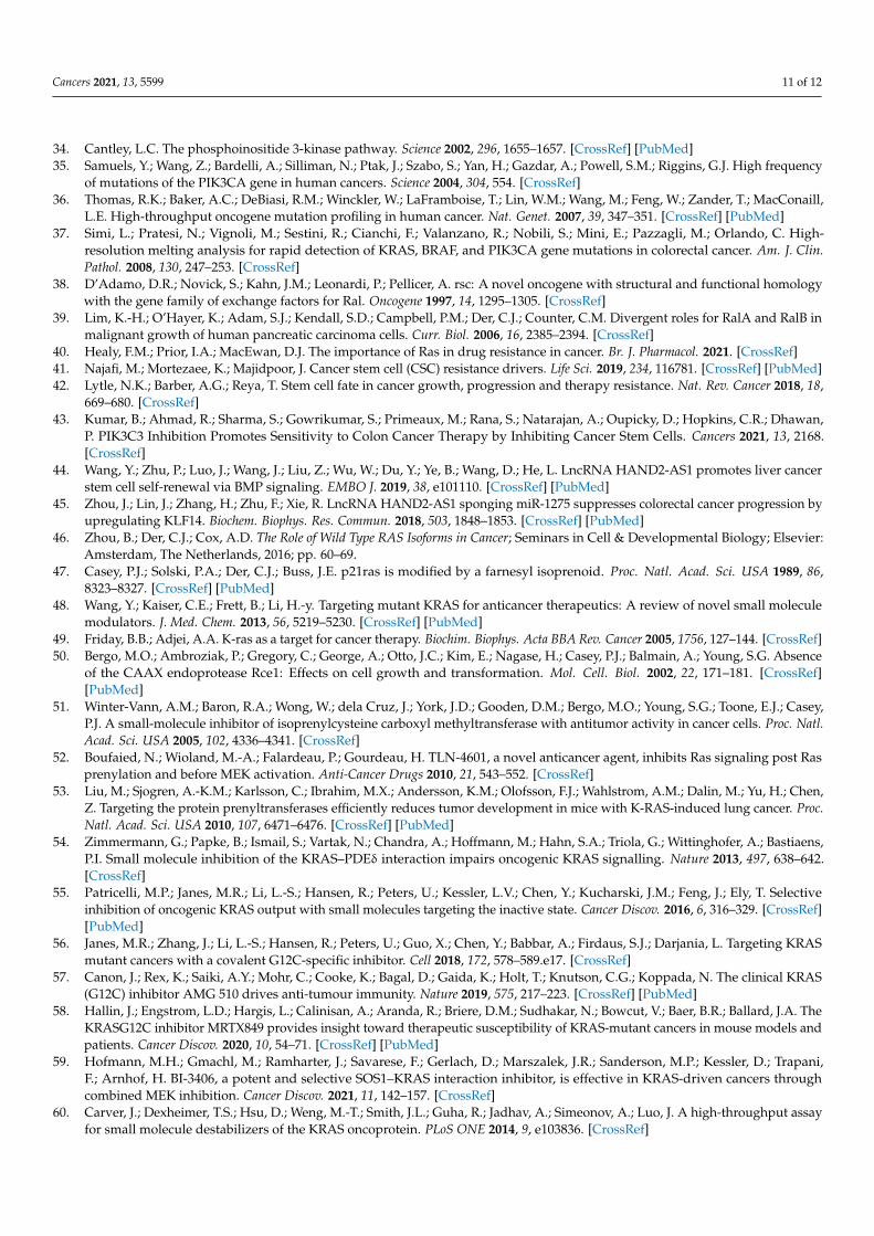
RAS has three domains (Figure 3). RAS isoforms share G-domain that highly se-quences homology (90%). The G-domain (residues 1–166) consists of an effector lobe(residues 1–86) and an allosteric lobe (87–166). This domain is composed of the six beta-strands and is surrounded by five alpha-helices [9]. KRAS has hypervariable regions(HVRs) present in the C-terminal element. The HVRs region undergoes a post-translationalmodification (PTM), including by farnesylation, palmitoylation, methylation, phosphoryla-tion, nitrosylation, and ubiquitination [10]. Particularly, the carboxyl terminal PTMs arerelated to affinity for membrane and trafficking with cellular compartments. According toresearch, palmitoylated HRAS is located in the plasma membrane, but depalmitoylatedHRAS is accumulated in the ER and Golgi [11]. The HVRs play a role in anchoring RASto the membrane, which displays among Ras proteins less than 15% of sequence homol-ogy [12]. The effector lobe mainly interacts with RAS effectors such as rapidly acceleratedfibrosarcoma (RAF), PI3K, and RalGEF. The allosteric lobe plays a role in intra-proteincommunication, which connects the effector lobe to membrane-interacting residues [13,14].The effector lobe is divided into two switch regions, switch-I (residues 30–38) and switch-II(residues 59–76), controlling the specificity of the binding to effector molecule and affectinginteraction of transducer to cellular downstream [2,7]. Different from the Ras protein, theeffector lobe of KRAS additionally has P-loop-phosphate binding loops (residues 10–16
Cancers 2021, 13, 5599 3 of 12
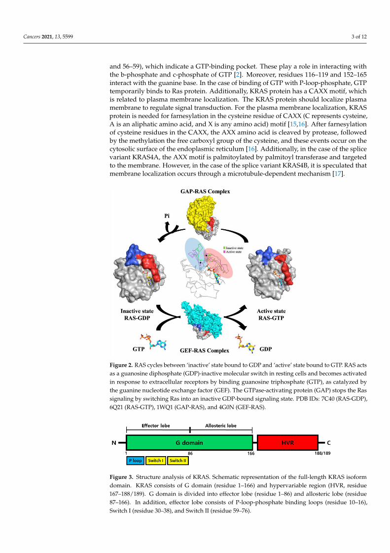
and 56–59), which indicate a GTP-binding pocket. These play a role in interacting withthe b-phosphate and c-phosphate of GTP [2]. Moreover, residues 116–119 and 152–165interact with the guanine base. In the case of binding of GTP with P-loop-phosphate, GTPtemporarily binds to Ras protein. Additionally, KRAS protein has a CAXX motif, whichis related to plasma membrane localization. The KRAS protein should localize plasmamembrane to regulate signal transduction. For the plasma membrane localization, KRASprotein is needed for farnesylation in the cysteine residue of CAXX (C represents cysteine,A is an aliphatic amino acid, and X is any amino acid) motif [15,16]. After farnesylationof cysteine residues in the CAXX, the AXX amino acid is cleaved by protease, followedby the methylation the free carboxyl group of the cysteine, and these events occur on thecytosolic surface of the endoplasmic reticulum [16]. Additionally, in the case of the splicevariant KRAS4A, the AXX motif is palmitoylated by palmitoyl transferase and targetedto the membrane. However, in the case of the splice variant KRAS4B, it is speculated thatmembrane localization occurs through a microtubule-dependent mechanism [17].
Cancers 2021, 13, x 3 of 13
Figure 2. RAS cycles between ‘inactive’ state bound to GDP and ‘active’ state bound to GTP. RAS
acts as a guanosine diphosphate (GDP)-inactive molecular switch in resting cells and becomes acti-
vated in response to extracellular receptors by binding guanosine triphosphate (GTP), as catalyzed
by the guanine nucleotide exchange factor (GEF). The GTPase-activating protein (GAP) stops the
Ras signaling by switching Ras into an inactive GDP-bound signaling state. PDB IDs: 7C40 (RAS-
GDP), 6Q21 (RAS-GTP), 1WQ1 (GAP-RAS), and 4G0N (GEF-RAS).
RAS has three domains (Figure 3). RAS isoforms share G-domain that highly se-
quences homology (90%). The G-domain (residues 1–166) consists of an effector lobe (res-
idues 1–86) and an allosteric lobe (87–166). This domain is composed of the six beta-
strands and is surrounded by five alpha-helices [9]. KRAS has hypervariable regions
(HVRs) present in the C-terminal element. The HVRs region undergoes a post-transla-
tional modification (PTM), including by farnesylation, palmitoylation, methylation, phos-
phorylation, nitrosylation, and ubiquitination [10]. Particularly, the carboxyl terminal
PTMs are related to affinity for membrane and trafficking with cellular compartments.
According to research, palmitoylated HRAS is located in the plasma membrane, but de-
palmitoylated HRAS is accumulated in the ER and Golgi [11]. The HVRs play a role in
anchoring RAS to the membrane, which displays among Ras proteins less than 15% of
sequence homology [12]. The effector lobe mainly interacts with RAS effectors such as
rapidly accelerated fibrosarcoma (RAF), PI3K, and RalGEF. The allosteric lobe plays a role
in intra-protein communication, which connects the effector lobe to membrane-interacting
residues [13,14]. The effector lobe is divided into two switch regions, switch-Ⅰ (residues
30–38) and switch-Ⅱ (residues 59–76), controlling the specificity of the binding to effector
molecule and affecting interaction of transducer to cellular downstream [2,7]. Different
from the Ras protein, the effector lobe of KRAS additionally has P-loop-phosphate bind-
ing loops (residues 10–16 and 56–59), which indicate a GTP-binding pocket. These play a
role in interacting with the b-phosphate and c-phosphate of GTP [2]. Moreover, residues
116–119 and 152–165 interact with the guanine base. In the case of binding of GTP with P-
loop-phosphate, GTP temporarily binds to Ras protein. Additionally, KRAS protein has a
CAXX motif, which is related to plasma membrane localization. The KRAS protein should
Figure 2. RAS cycles between ‘inactive’ state bound to GDP and ‘active’ state bound to GTP. RAS actsas a guanosine diphosphate (GDP)-inactive molecular switch in resting cells and becomes activatedin response to extracellular receptors by binding guanosine triphosphate (GTP), as catalyzed bythe guanine nucleotide exchange factor (GEF). The GTPase-activating protein (GAP) stops the Rassignaling by switching Ras into an inactive GDP-bound signaling state. PDB IDs: 7C40 (RAS-GDP),6Q21 (RAS-GTP), 1WQ1 (GAP-RAS), and 4G0N (GEF-RAS).
Cancers 2021, 13, x 4 of 13
localize plasma membrane to regulate signal transduction. For the plasma membrane lo-
calization, KRAS protein is needed for farnesylation in the cysteine residue of CAXX (C
represents cysteine, A is an aliphatic amino acid, and X is any amino acid) motif [15,16].
After farnesylation of cysteine residues in the CAXX, the AXX amino acid is cleaved by
protease, followed by the methylation the free carboxyl group of the cysteine, and these
events occur on the cytosolic surface of the endoplasmic reticulum [16]. Additionally, in
the case of the splice variant KRAS4A, the AXX motif is palmitoylated by palmitoyl trans-
ferase and targeted to the membrane. However, in the case of the splice variant KRAS4B,
it is speculated that membrane localization occurs through a microtubule-dependent
mechanism [17].
Figure 3. Structure analysis of KRAS. Schematic representation of the full-length KRAS isoform do-
main. KRAS consists of G domain (residue 1–166) and hypervariable region (HVR, residue 167–
188/189). G domain is divided into effector lobe (residue 1–86) and allosteric lobe (residue 87–166).
In addition, effector lobe consists of P-loop-phosphate binding loops (residue 10–16), Switch I (res-
idue 30–38), and Switch II (residue 59–76).
2. KRAS Signaling
In the cell membrane, KRAS undergoes inactive state by binding of guanosine di-
phosphate (GDP) and active state by binding of guanosine triphosphate (GTP). KRAS pro-
tein is normally maintained in oncogenesis, but mutated KRAS is decreased in interac-
tions with GTPase activator protein [18]. Moreover, the mutated KRAS tends to prefer
binding with GTP than GDP. The activation form of GTP-bound Ras protein influences
the activation downstream effector even in the absence of growth factor [19]. Typically,
the Ras signal pathway is activated by the protein tyrosine kinase receptor. The epidermal
growth factor receptor (EGFR) and the platelet-derived growth factor receptor (PDGFR)
are well-known protein tyrosine kinase receptors at the Ras signal pathway. For the Ras
signal to be activated, the ligand binds the EGF receptor, followed by induces oligomeri-
zation of the receptor [20]. This can allow activation of the kinase activity and transphos-
phorylation of catalytic domains [20]. The sequence homology 2 (SH2) domain of the re-
ceptor is a recognized adaptor protein such as growth factor receptor-bound protein 2
(Grb2), which in turn recruits guanine nucleotide exchange factors (GEFs) like son of
sevenless homolog 1 (SOS-1) to the cell membrane [19]. The GEFs are able to interact with
Ras protein to promote a conformational change and exchange GDP for GTP. After acti-
vation of Ras protein, various downstream effectors such as canonical Raf/Mek/Erk, phos-
phatidylinositol 3-kinase (PI3K)/3-phosphoinositide-dependent protein kinase-1
(Pdk1)/Akt, and Ral-GEF are recruited (Figure 4) [5,21].
Figure 3. Structure analysis of KRAS. Schematic representation of the full-length KRAS isoformdomain. KRAS consists of G domain (residue 1–166) and hypervariable region (HVR, residue167–188/189). G domain is divided into effector lobe (residue 1–86) and allosteric lobe (residue87–166). In addition, effector lobe consists of P-loop-phosphate binding loops (residue 10–16),Switch I (residue 30–38), and Switch II (residue 59–76).

Cancers 2021, 13, 5599 4 of 12
2. KRAS Signaling
In the cell membrane, KRAS undergoes inactive state by binding of guanosine diphos-phate (GDP) and active state by binding of guanosine triphosphate (GTP). KRAS protein isnormally maintained in oncogenesis, but mutated KRAS is decreased in interactions withGTPase activator protein [18]. Moreover, the mutated KRAS tends to prefer binding withGTP than GDP. The activation form of GTP-bound Ras protein influences the activationdownstream effector even in the absence of growth factor [19]. Typically, the Ras signalpathway is activated by the protein tyrosine kinase receptor. The epidermal growth factorreceptor (EGFR) and the platelet-derived growth factor receptor (PDGFR) are well-knownprotein tyrosine kinase receptors at the Ras signal pathway. For the Ras signal to be ac-tivated, the ligand binds the EGF receptor, followed by induces oligomerization of thereceptor [20]. This can allow activation of the kinase activity and transphosphorylationof catalytic domains [20]. The sequence homology 2 (SH2) domain of the receptor is arecognized adaptor protein such as growth factor receptor-bound protein 2 (Grb2), whichin turn recruits guanine nucleotide exchange factors (GEFs) like son of sevenless homolog1 (SOS-1) to the cell membrane [19]. The GEFs are able to interact with Ras protein topromote a conformational change and exchange GDP for GTP. After activation of Ras pro-tein, various downstream effectors such as canonical Raf/Mek/Erk, phosphatidylinositol3-kinase (PI3K)/3-phosphoinositide-dependent protein kinase-1 (Pdk1)/Akt, and Ral-GEFare recruited (Figure 4) [5,21].
Cancers 2021, 13, x 5 of 13
Figure 4. Signaling pathway of KRAS protein. Activation of RAS occurs when epidermal growth
factor (EGF) binds to tyrosine kinases receptor such as epidermal growth factor receptor (EGFR).
KRAS protein is activated by binding to GTP, transducing the cytoplasmic signal, which activates
the RAL, RAF-MEK-ERK, and PI3K-AKT cascade. These cascades regulate cell growth, division,
and differentiation.
2.1. RAF-MEK-ERK Pathway
The first well-known Ras effector pathway is identified as the RAF-MEK-ERK path-
way. This pathway is involved in the mitogenic signaling of tyrosine kinase receptors,
following a wide range of growth, differentiation, inflammation, and apoptosis [22]. Raf
is a member of the family of serine/tyrosine kinase. Serine/tyrosine kinase includes Raf-1,
A-Raf, and B-Raf [19]. This family binds to the effector region of RAS-GTP, resulting in
translocation of the Raf protein to the plasma membrane [23,24]. According to the re-
search, Ras activated either by point mutation (G12V) or by GTP-bound interacts with
Raf-1 through guanylyl-imidodiphosphate (GMP-PNP). However, effector domain muta-
tion (Ile36Ala) of Ras does not interact with Raf-1[25]. Following Ras interacting with Raf,
Raf protein is activated and phosphorylated by protein kinase. Raf activation induces a
signaling cascade, which phosphorylates mitogen-activated protein kinase (MAPK),
which in turn, phosphorylates and activates the downstream effector such as extracellular
signal-regulated kinase 1 and 2 (ERK1 and ERK2) [19]. Activation of ERK kinases plays a
role in Ras-induced cellular responses and translocation to the nucleus [21]. Therefore,
ERK activates and phosphorylates the nuclear transcription factor and kinases such as
eukaryotic initiation factor 2alpha kinase 1 (EIK-1) and protein C-ets-1,2 (c-Ets1, c-Ets2)
[19]. Besides activation of ERK, MAPK, also called MEK, is activated in signal response
that promotes cell survival and apoptosis through various mediators such as c-Jun N-ter-
minal kinases (JNK), stress-activated protein kinase (SAPK), and nuclear factor kappa-
light-chain-enhancer of activated B cells (NF-KB) [26].
In cancer, studies targeting Raf kinase have been demonstrated. According to the re-
search for the influence of RAF-MEK-ERK pathway in tumor growth, expressing domi-
nant-negative MEK in tumor cells affects a reduction in endogenous Raf-1 and MEK-1
activity [27]. Moreover, research demonstrated that clones expressing the dominant-neg-
ative MEK form the smallest tumors and appear to increase survival [27]. Phosphorylation
of MEK occurs in residue serine 218 and 222, but MEK point mutation in which these
serines are substituted by alanine (S218A/S222A) is not able to be activated Raf [28]. Ad-
Figure 4. Signaling pathway of KRAS protein. Activation of RAS occurs when epidermal growthfactor (EGF) binds to tyrosine kinases receptor such as epidermal growth factor receptor (EGFR).KRAS protein is activated by binding to GTP, transducing the cytoplasmic signal, which activatesthe RAL, RAF-MEK-ERK, and PI3K-AKT cascade. These cascades regulate cell growth, division,and differentiation.
2.1. RAF-MEK-ERK Pathway
The first well-known Ras effector pathway is identified as the RAF-MEK-ERK pathway.This pathway is involved in the mitogenic signaling of tyrosine kinase receptors, followinga wide range of growth, differentiation, inflammation, and apoptosis [22]. Raf is a memberof the family of serine/tyrosine kinase. Serine/tyrosine kinase includes Raf-1, A-Raf, andB-Raf [19]. This family binds to the effector region of RAS-GTP, resulting in translocation ofthe Raf protein to the plasma membrane [23,24]. According to the research, Ras activatedeither by point mutation (G12V) or by GTP-bound interacts with Raf-1 through guanylyl-imidodiphosphate (GMP-PNP). However, effector domain mutation (Ile36Ala) of Ras does

Cancers 2021, 13, 5599 5 of 12
not interact with Raf-1 [25]. Following Ras interacting with Raf, Raf protein is activatedand phosphorylated by protein kinase. Raf activation induces a signaling cascade, whichphosphorylates mitogen-activated protein kinase (MAPK), which in turn, phosphorylatesand activates the downstream effector such as extracellular signal-regulated kinase 1and 2 (ERK1 and ERK2) [19]. Activation of ERK kinases plays a role in Ras-inducedcellular responses and translocation to the nucleus [21]. Therefore, ERK activates andphosphorylates the nuclear transcription factor and kinases such as eukaryotic initiationfactor 2alpha kinase 1 (EIK-1) and protein C-ets-1,2 (c-Ets1, c-Ets2) [19]. Besides activationof ERK, MAPK, also called MEK, is activated in signal response that promotes cell survivaland apoptosis through various mediators such as c-Jun N-terminal kinases (JNK), stress-activated protein kinase (SAPK), and nuclear factor kappa-light-chain-enhancer of activatedB cells (NF-KB) [26].
In cancer, studies targeting Raf kinase have been demonstrated. According to the re-search for the influence of RAF-MEK-ERK pathway in tumor growth, expressing dominant-negative MEK in tumor cells affects a reduction in endogenous Raf-1 and MEK-1 activ-ity [27]. Moreover, research demonstrated that clones expressing the dominant-negativeMEK form the smallest tumors and appear to increase survival [27]. Phosphorylation ofMEK occurs in residue serine 218 and 222, but MEK point mutation in which these serinesare substituted by alanine (S218A/S222A) is not able to be activated Raf [28]. Additionally,inhibition of the RAF-MEK-ERK pathway via mutant MEK demonstrates reversion of thecancer phenotype, and inhibitors of Raf kinase are targeted anti-tumor therapies [29].
2.2. PI3K Pathway
The second well-known Ras effector family is phosphoinositide 3-kinases (PI3Ks).PI3K plays a role in mediators of Ras-mediated cell survival and proliferation [30]. Activa-tion of PI3K converts phosphatidylinositol (4,5)-biphosphate (PIP2) into phosphatidylinosi-tol (3,4,5)-triphosphate (PIP3). PIP3, in turn, binds the pleckstrin homology (PH) domainof Akt/PKB. Akt/PKB stimulates phosphorylation of proteins that affect cell growth,cell cycle, and survival [21]. To activate PI3K, binding of a ligand to receptor tyrosinekinase (RTK) first occurs. Following binding of the ligand in RTK, RTK is dimerized andautophosphorylated, leading to interaction with PI3K effector via Src homology 2 (SH2)domains [31]. Adaptor protein of PI3K, GRB2 binds to phosphor-YXN motifs of the RTK.In turn, GRB2 binds and activates SOS, which then activates RAS. Activation of RAS acti-vates p110 of p85 [32]. The tumor suppressor phosphatase and tensing homology (PTEN)dephosphorylates PIP3 to PIP2. PIP3 induces intracellular signaling via binding to proteinwith PH domains, such as phosphoinositide-dependent kinase (PDK1) and AKT [33]. ThePI3K-AKT pathway promotes cell growth and survival [21,34].
The importance of PI3K in the RAS-signaling pathway is highly correlated withoncogenic signaling. According to the research, there is evidence that studies have foundsomatic missense mutations in PIK3CA in human tumors, including those of the brain,breast, and colon [35–37].
2.3. Ral-GEF Protein
The Ral-GEF protein also became known as the Ras effector. Ral-GEF members arecomposed of RalGDS, RGL, RGL2, and RGL3 [38]. The members are activated the RalAand RalB small GTPase via the link with Ras proteins. Thus, the RalGEF protein is asmall GTPase that provides GTP to the Ras superfamily. According to the research, GTP-bound RalA and RalB are discovered to have high levels in human pancreatic ductaladenocarcinoma (PDAC). Moreover, inhibition of RalA expression reduces tumorigenicgrowth [39].
3. RAS Inhibitors and Resistance
Acquired resistance occurs for several reasons. Pre-existing and de novo mutationsprovide chemoresistance and acquired resistance [40]. Resistance mechanisms happen in a

Cancers 2021, 13, 5599 6 of 12
very rare subpopulation of cells, such as cancer stem cells [41,42]. Kumar, B. et al. revealedthat enhanced autophagy induces cancer cell stemness and promotes resistance to colorectalcancer therapy. Moreover, 36-077 promotes the efficacy of colorectal cancer therapy. 36-077inhibits phosphatidylinositol 3-kinase (PIK3C3/VPS34 kinase) signaling, a key regulatorof autophagy, and a potent target for inhibiting autophagy [43]. Long noncoding RNA(lncRNA) is highly expressed in cancer stem cells. lncRNA HAND2-AS1 is required forself-renewal of cancer stem cells, through activation of the bone morphogenetic protein(BMP) signaling [44,45]. Taken together, many pathways and associated factors play a keyrole in mediating drug resistance.
3.1. Inhibiting the Post-Translation Processing of RAS
RAS gene encodes three genes, including HRAS, NRAS, and KRAS. KRAS has twoalternatively spliced mRNA variants, KRAS4A and KRAS4B [46]. These proteins withamino-terminal residues 1–165 share 92~98% sequence identity [47]. RAS proteins carry23~24 carboxyl-terminal residues, including the CAAX box. CAAX box undergoes four-stage post-translational lipid modification: (1) farnesylation by FTase, (2) prenylation atthe CAAX cysteine residue followed by proteolytic [42] removal of the terminal -AAX byRac converting CAXX endopeptidase 1 (RCE1), (3) carboxymethylation of the C-terminalcysteine by isoprenylcysteine carboxyl methyltransferase (ICMT), and (4) palmitoylationof cysteine residues located upstream of the C-terminus of the protein [48,49].
The excision of RCE1 eliminated RAS endoproteolytic processing and mislocalizationof RAS proteins. Moreover, the loss of RCE1 significantly reduces the growth of cellsin the skin carcinoma cell line [50]. Treatment of cysmethynil, ICMT inhibitor, resultsin mislocalization of RAS and inhibition of cell growth and KRAS-induced oncogenictransformation in nude mice [51] (Table 1). TLN-4601 has been shown to disrupt RASmembrane anchorage and anti-tumor activity in several xenograft models. Interestingly,the compound induces Raf-1 proteasomal-dependent degradation and may inhibit theMEK/EKR pathway by depleting the Raf-1 protein [52]. Liu et al. demonstrated thatinhibitors of FTase and geranylgeranyltransferase-I were shown to reduce tumor growthand improved survival in mice with K-RAS-induced lung cancer [53]. Waldmann andcolleagues targeted the small-molecule inhibitor deltarasin that interferes with the bindingof mammalian phosphodiesterase δ (PDEδ) to KRAS and suppresses KRAS localization toendomembrane [54].
Table 1. Summary of therapeutic strategies towards RAS-driven cancer.
Category Name Target Mode of Inhibition Ref.
Inhibition ofpost-translational
processing
Cysmethynil ICMT inhibitor mislocalization of RAS [51]
TLN-4601 MEK/EKR pathway disrupts RAS membrane anchorage [52]
deltarasin PDEδ suppresses KRAS localizationto endomembrane [54]
K-RAS targeteddrug
ARS-583 KRAS G12C inhibits downstream MAPK/PI3K signaling [55]
ARS-1620 KRAS G12C anti-tumor activity in subcutaneousxenograft models [56]
AMG 510 KRAS G12C induces pro-inflammatory tumormicroenvironment [57]
MRTX849 KRAS G12C tumor regression in KRAS G12C positivecell lines [58]
BI-3406 KRAS-mutant of G12and G13
interacts with the catalytic domain of SOS,resulting in interference with the interaction
with KRAS[59]
ponatinib andAMG-47a KRAS G12V reduces the levels of KRAS G12V proteins [60]

Cancers 2021, 13, 5599 7 of 12
Table 1. Cont.
Category Name Target Mode of Inhibition Ref.
Inhibition of RASsignalingnetworks
WZ4002 and MEKinhibitor EGFR and MEK
effective strategy of drug-resistant cancersand prevents the emergence of
drug-resistant clones[61]
gefitinib and Srcinhibitor EGFR and Src potent strategy to overcome Src-mediated
ERK reactivation [62]
gilteritinib, crenolanib,and midostaurin FLT3 inhibitor
designed to have a high affinity for theATP-binding region of the active
conformation of FLT3[63–65]
Discovered byvirtual screening
BAY-293 KRAS-SOS1interaction
suppresses the RAS-RAF-MEK-ERK pathwayand inhibits the activation of RAS in
tumor cells[66]
tetrahydropyridopyrimidines KRAS G12C locks KRAS in its inactive GDP-bound form [67]
C19 KRAS4B-PDEδ suppresses the viability and proliferation ofcolorectal cancer cells [68]
D14 and C22 KRAS4B-PDEδ decreases RAS-GTP activity and ERK andAKT pathways in pancreatic cancer cells [69]
Other inhibitors
H-REV 107 peptide KRAS G12V blocks the activation function of KRAS [70]
cisplatin, carboplatin,and oxaliplatin widely used to treat several cancers [71]
ICMT: isoprenylcysteine carboxyl methyltransferase, MEK: mitogen-activated protein kinase, ERK: extracellular signal-regulated kinase,PDEδ: phosphodiesterase δ, EGFR: epidermal growth factor receptor, FLT3: FMS-like receptor tyrosine kinase-3.
3.2. K-RAS Targeted Drug
The most mutation sites of oncogenic mutation in RAS are residues 12 and 13 in theP-loop, and residue 61 in switch II [72]. KRAS, which is most frequently mutated, contains86% of RAS mutation. KRAS mutations occur in pancreatic, colorectal, and lung cancers.The vast majority of hotspot positions are on 12. NRAS mutations occur in melanomaand myeloid leukemia, with the most common site of mutation being residue 61. HRASmutations occur with the highest frequency in bladder cancer, and hotspots are 12 and61 residues [73] (Table 2).
Table 2. Frequency of RAS isoform mutations in human cancer. Table shows the distribution ofmutations in the various primary tissue types. Data are from the COSMIC database. Numbers inparentheses indicate sequence of total tested sample.
Primary Tissue NRAS (%) KRAS (%) HRAS (%)
pancreas 0.77 (4016) 52.65 (13,817) 0.11 (3542)
colon 3.87 (16,337) 32.37 (80,177) 0.94 (6095)
small intestine 0.45 (443) 21.34 (1256) 0.3 (336)
lung 0.91 (17,505) 14.19 (45,776) 0.6 (7890)
ovary 1.71 (1932) 13.03 (6932) 0.24 (1693)
liver 1.07 (3174) 3.15 (3614) 0.24 (2912)
kidney 0.34 (3498) 0.93 (4423) 0.25 (3265)
skin 15.36 (15,960) 2.8 (6833) 9.42 (7067)
endometrium 3.22 (1648) 15.45 (4674) 0.58 (2395)
biliary tract 2.7 (2256) 18.05 (5518) 0.56 (1794)

Cancers 2021, 13, 5599 8 of 12
ARS-583 reacts with the GDP-bound state of KRAS G12C. ARS-583 inhibits down-stream MAPK/PI3K signaling across the group of cell lines and directly inhibits thetreatment of patients with the KRAS G12C mutation, which comprises 20% of lung can-cers [55]. ARS-1620 selectively and with high potency targets the switch II pocket in KRASG12C. ARS-1620 achieves anti-tumor activity in subcutaneous xenograft models bearingKRAS G12C mutation [56]. Ryan et al. observed rapid adaptive RAS pathway feedbackreactivation of MAPK signaling after treatment with ARS-1620 in KRAS G12C-driven lung,pancreatic, and colon cancer cells, suggesting secondary resistance via upregulated RTKsignaling [74]. Treatment with AMG 510 regresses in KRAS G12C mouse xenografts andinduces pro-inflammatory tumor microenvironment [57]. A covalent KRAS G12C inhibitor,MRTX849, selectively modifies mutant cysteine 12 in a GDP-bound state. MRTX849 hastumor regression in KRAS G12C positive cell lines and patient-derived xenograft modelsfrom multiple tumor types [58]. Small-molecule SOS1 inhibitor, BI-3406, interacts withthe catalytic domain of SOS, resulting in interference with the interaction with KRAS andthereby restricting tumor cell proliferation in KRAS-mutant cancer, including G12 andG13 [59]. BI 1701963, which is a BI-3406 analog, is currently in a phase I trial, testingpreliminary efficacy of BI 1701963 alone and in combination with trametinib in patientswith KRAS mutant solid tumors [75]. Compounds of ponatinib and AMG-47a selectivelyreduce the levels of KRAS G12V proteins [60].
3.3. Inhibition of RAS Signaling Networks
Upstream RAS-mediated pathways have a common mechanism of resistance to tyro-sine kinase inhibitors (TKIs) such as those targeting RTKs, including EGFR and FMS-likereceptor tyrosine kinase-3 (FLT3) [40]. Treatment with anti-EGFR monoclonal antibodiescetuximab or panitumumab is successful in a subset of patients with colorectal cancer,although these stimulate MAPK reactivation, driving secondary resistance [76]. Resis-tance to EGFR inhibitor WZ4002 can induce reactivation of ERK1/2 signaling. Ercan et al.found that the combination of WZ4002 and MEK inhibitor may be an effective strategy totreat drug-resistant cancers [61]. EGFR inhibitor gefitinib showed drug resistance via Src-mediated ERK reactivation. Ochi et al. demonstrated that a combination of gefitinib andSrc inhibitor may increase the potency of the RAS-inhibitor resistance mechanism to over-come this resistance [62]. Many different FLT3 inhibitors including gilteritinib, crenolanib,and midostaurin treat varying stages. Gilteritinib and midostaurin are FDA-approvedwhereas crenolanib is in phase II trials [63–65]. However, gilteritinib develops secondaryresistance via activating RAS/MAPK pathway signaling in acute myeloid leukemia, mostcommonly in NRAS or KRAS [77]. The MRAS-SHOC2-PPI complex acts as a key rolein the RAF-ERK pathway. Jones et al. reported that inhibition of SHOC2 inhibits tumordevelopment in murine KRAS-driven lung cancer models. However, this compound ac-tivates other RAS-dependent pathways such as PI3K-AKT signaling, resulting in cancerthat can persist [40,78]. Adachi and colleagues observed that epithelial-to-mesenchymaltransition (EMT) led to intrinsic and acquired resistance by activation of PI3K signalingin the presence of KRAS G12C inhibitor. Treatment with the combination of KRAS G12Cinhibitor, PI3K inhibitor, and SHP2 inhibitor resulted in tumor regression in mouse modelsof acquired resistance to KRAS G12C inhibitor, AMG510 [79].
3.4. KRAS Inhibitors Discovered by Virtual Screening
Covalent quinazoline-based switch II compounds effectively inhibit GTP loading ofKRAS G12V, MAPK phosphorylation, and the growth of cancer cells driven by KRASG12C [80]. Compound 23 (BAY-293) selectively inhibits the KRAS-SOS1 interaction andblocks reloading of KRAS with GTP. The compound suppresses the RAS-RAF-MEK-ERKpathway and inhibits the activation of RAS in tumor cells [66]. Fell et al. reported thattetrahydro pyridopyrimidines act as irreversible covalent inhibitors of KRAS G12C andcan bind to the residue 12 and lock KRAS in its inactive GDP-bound form [67]. C19 boundand stabilized the KRAS4B-PDEδ complex. This compound suppresses the viability and

Cancers 2021, 13, 5599 9 of 12
proliferation of colorectal cancer cells and decreases tumor size in a xenograft mouse modelvia inhibiting phosphorylation of ERK and AKT signaling [68]. In addition, D14 and C22stabilize the interaction with KRAS4B and PDEδ. D14 and C22 significantly decreaseRAS-GTP activity and ERK and AKT pathways in pancreatic cancer cells [69].
3.5. Other Inhibitors Targeting RAS
CW Han et al. elucidated that there is direct interaction between KRAS G12V andH-REV 107 peptide with high affinity with the crystal structure. This peptide was shown tointeract and stabilize with KRAS G12V inactive state resulting in the block of the activationfunction of KRAS. The peptide can inhibit pancreatic cancer and colon cancer cell linesin cell proliferation assay [70]. Treatment with platinum-based agents, such as cisplatin,carboplatin, and oxaliplatin, is used in a variety of cancers, including head and neck squamouscell carcinoma, testicular cancer, and non-small cell lung cancer [71,81,82]. Cisplatin inducesmitochondrial ROS, which further increases DNA damage and induces increasing cell death.However, cisplatin shows an observed resistance mechanism, including the involvementof oncogenic KRAS mutations via NRF2 overexpressing [71].
4. Conclusions
RAS proteins are membrane-bound proteins and are frequently mutated in humancancers, with mutations in about 30% of all cancers. RAS exists in three ubiquitouslyexpressed genes, HRAS, KRAS, and NRAS, with high sequence homology. RAS proteinscycle between an inactive (GDP-bound) and active (GTP-bound) state. When activatedby a ligand-bound RTK such as EGFR and FLT3, RAS triggers diverse signaling cascades,including PI3K and MAPK/ERK signaling, to induce cell growth, differentiation, andsurvival. Oncogenic mutations of RAS in several upstream or downstream pathways occurin most tumors, indicating that inhibition of RAS-dependent signaling is the essentialrequirement for tumorigenesis. While RAS proteins have been seen as an attractive targetfor cancer therapy, continuous efforts to inhibit RAS either directly or indirectly throughinhibiting post-translational modification or RAS-dependent signaling to date have failedto develop approved therapies for RAS mutant cancer. Previous failures of RAS-targetingtreatments, including farnesyltransferase inhibitors, have resulted in RAS being generallyconsidered undruggable. Additionally, because of the high picomolar affinity between RASand GTP, a competitive inhibitor is not expressly possible. However, the discovery of thestructure of RAS has given an understanding of the key residues with new promising com-pounds. Despite evidence supporting the cancer stem cell theory, resistance to inhibition ofRAS appears to be diverse. Durable response to these treatments is yet to be achieved dueto complex and diverse mechanisms of adaptive resistance. However, it is expected thatemploying this rationale will lead to better treatments and a better outcome for patients.
Author Contributions: H.J.K. and H.N.L. wrote the manuscript. M.S.J. and S.B.J. designed andprovided analysis of the manuscript. All authors have read and agreed to the published version ofthe manuscript.
Funding: This research was funded by MEST (2018R1D1A1B07043701) for S.B.J. and (2016R1D1A1B02011142) for M.S.J.
Acknowledgments: This study was supported by the Basic Science Research Program through theNational Research Foundation of Korea (NRF) funded by the Ministry of Education, Science andTechnology (2018R1D1A1B07043701) for S.B.J. and (2016R1D1A1B02011142) for M.S.J.
Conflicts of Interest: The authors declare no conflict of interest.
References1. Wennerberg, K.; Rossman, K.L.; Der, C.J. The Ras superfamily at a glance. J. Cell Sci. 2005, 118, 843–846. [CrossRef]2. Jancík, S.; Drábek, J.; Radzioch, D.; Hajdúch, M. Clinical relevance of KRAS in human cancers. J. Biomed. Biotechnol. 2010,
2010, 150960. [CrossRef]

Cancers 2021, 13, 5599 10 of 12
3. Merrick, B.A.; Phadke, D.P.; Bostrom, M.A.; Shah, R.R.; Wright, G.M.; Wang, X.; Gordon, O.; Pelch, K.E.; Auerbach, S.S.; Paules,R.S. Arsenite malignantly transforms human prostate epithelial cells in vitro by gene amplification of mutated KRAS. PLoS ONE2019, 14, e0215504. [CrossRef] [PubMed]
4. McGrath, J.P.; Capon, D.J.; Smith, D.H.; Chen, E.Y.; Seeburg, P.H.; Goeddel, D.V.; Levinson, A.D. Structure and organization of thehuman Ki-ras proto-oncogene and a related processed pseudogene. Nature 1983, 304, 501–506. [CrossRef] [PubMed]
5. Pylayeva-Gupta, Y.; Grabocka, E.; Bar-Sagi, D. RAS oncogenes: Weaving a tumorigenic web. Nat. Rev. Cancer 2011, 11, 761–774.[CrossRef] [PubMed]
6. Gideon, P.; John, J.; Frech, M.; Lautwein, A.; Clark, R.; Scheffler, J.E.; Wittinghofer, A. Mutational and kinetic analyses of theGTPase-activating protein (GAP)-p21 interaction: The C-terminal domain of GAP is not sufficient for full activity. Mol. Cell. Biol.1992, 12, 2050–2056. [CrossRef] [PubMed]
7. Simanshu, D.K.; Nissley, D.V.; McCormick, F. RAS proteins and their regulators in human disease. Cell 2017, 170, 17–33. [CrossRef]8. Hodge, R.G.; Schaefer, A.; Howard, S.V.; Der, C.J. RAS and RHO family GTPase mutations in cancer: Twin sons of different
mothers? Crit. Rev. Biochem. Mol. Biol. 2020, 55, 386–407. [CrossRef]9. Pantsar, T. The current understanding of KRAS protein structure and dynamics. Comput. Struct. Biotechnol. J. 2020, 18, 189–198.
[CrossRef]10. Ahearn, I.; Zhou, M.; Philips, M.R. Posttranslational modifications of RAS proteins. Cold Spring Harb. Perspect. Med. 2018,
8, a031484. [CrossRef]11. Choy, E.; Chiu, V.K.; Silletti, J.; Feoktistov, M.; Morimoto, T.; Michaelson, D.; Ivanov, I.E.; Philips, M.R. Endomembrane trafficking
of ras: The CAAX motif targets proteins to the ER and Golgi. Cell 1999, 98, 69–80. [CrossRef]12. Abraham, S.J.; Muhamed, I.; Nolet, R.; Yeung, F.; Gaponenko, V. Expression, purification, and characterization of soluble K-Ras4B
for structural analysis. Protein Expr. Purif. 2010, 73, 125–131. [CrossRef]13. Mo, S.P.; Coulson, J.M.; Prior, I.A. RAS variant signalling. Biochem. Soc. Trans. 2018, 46, 1325. [CrossRef]14. Parker, J.A.; Mattos, C. The K-Ras, N-Ras, and H-Ras isoforms: Unique conformational preferences and implications for targeting
oncogenic mutants. Cold Spring Harb. Perspect. Med. 2018, 8, a031427. [CrossRef]15. Hancock, J.F.; Magee, A.I.; Childs, J.E.; Marshall, C.J. All ras proteins are polyisoprenylated but only some are palmitoylated. Cell
1989, 57, 1167–1177. [CrossRef]16. Aicart-Ramos, C.; Valero, R.A.; Rodriguez-Crespo, I. Protein palmitoylation and subcellular trafficking. Biochim. Biophys. Acta
BBA Biomembr. 2011, 1808, 2981–2994. [CrossRef]17. Thissen, J.A.; Gross, J.M.; Subramanian, K.; Meyer, T.; Casey, P.J. Prenylation-dependent association of Ki-Ras with microtubules:
Evidence for a role in subcellular trafficking. J. Biol. Chem. 1997, 272, 30362–30370. [CrossRef] [PubMed]18. Avruch, J.; Khokhlatchev, A.; Kyriakis, J.M.; Luo, Z.; Tzivion, G.; Vavvas, D.; Zhang, X. Ras activation of the Raf kinase: Tyrosine
kinase recruitment of the MAP kinase cascade. Recent Prog. Horm. Res. 2001, 56, 127–155. [CrossRef]19. Molina, J.R.; Adjei, A.A. The ras/raf/mapk pathway. J. Thorac. Oncol. 2006, 1, 7–9. [CrossRef]20. Lemmon, M.A.; Schlessinger, J. Cell signaling by receptor tyrosine kinases. Cell 2010, 141, 1117–1134. [CrossRef] [PubMed]21. Castellano, E.; Downward, J. RAS interaction with PI3K: More than just another effector pathway. Genes Cancer 2011, 2, 261–274.
[CrossRef]22. Roux, P.P.; Blenis, J. ERK and p38 MAPK-activated protein kinases: A family of protein kinases with diverse biological functions.
Microbiol. Mol. Biol. Rev. 2004, 68, 320–344. [CrossRef] [PubMed]23. Warne, P.H.; Vician, P.R.; Downward, J. Direct interaction of Ras and the amino-terminal region of Raf-1 in vitro. Nature 1993, 364,
352–355. [CrossRef]24. Zhang, X.-F.; Settleman, J.; Kyriakis, J.; Takeuchi-Suzuki, E.; Elledge, S.J.; Marshall, M.S.; Bruder, J.T.; Rapp, U.R.; Avruch, J.
Normal and oncogenic p21 ras proteins bind to the amino-terminal regulatory domain of c-Raf-1. Nature 1993, 364, 308–313.[CrossRef]
25. Moodie, S.A.; Willumsen, B.M.; Weber, M.J.; Wolfman, A. Complexes of Ras. GTP with Raf-1 and mitogen-activated proteinkinase kinase. Science 1993, 260, 1658–1661. [CrossRef]
26. Schlesinger, T.K.; Fanger, G.R.; Yujiri, T.; Johnson, G.L. The tao of MEKK. Front. Biosci 1998, 3, D1181–D1186. [CrossRef]27. Arboleda, M.J.; Eberwein, D.; Hibner, B.; Lyons, J.F. Dominant negative mutants of mitogen-activated protein kinase pathway.
Methods Enzymol. 2001, 332, 353–367. [PubMed]28. Zheng, C.-F.; Guan, K.-L. Activation of MEK family kinases requires phosphorylation of two conserved Ser/Thr residues. EMBO
J. 1994, 13, 1123–1131. [CrossRef]29. Lyons, J.; Wilhelm, S.; Hibner, B.; Bollag, G. Discovery of a novel Raf kinase inhibitor. Endocr.-Relat. Cancer 2001, 8, 219–225.
[CrossRef] [PubMed]30. Castellano, E.; Downward, J. Role of RAS in the regulation of PI 3-kinase. Phosphoinositide 3-Kinase Health Dis. 2010, 1, 143–169.31. Schlessinger, J. Ligand-induced, receptor-mediated dimerization and activation of EGF receptor. Cell 2002, 110, 669–672.
[CrossRef]32. Pawson, T. Specificity in signal transduction: From phosphotyrosine-SH2 domain interactions to complex cellular systems. Cell
2004, 116, 191–203. [CrossRef]33. Liu, P.; Cheng, H.; Roberts, T.M.; Zhao, J.J. Targeting the phosphoinositide 3-kinase pathway in cancer. Nat. Rev. Drug Discov.
2009, 8, 627–644. [CrossRef]
Cancers 2021, 13, 5599 11 of 12
34. Cantley, L.C. The phosphoinositide 3-kinase pathway. Science 2002, 296, 1655–1657. [CrossRef] [PubMed]35. Samuels, Y.; Wang, Z.; Bardelli, A.; Silliman, N.; Ptak, J.; Szabo, S.; Yan, H.; Gazdar, A.; Powell, S.M.; Riggins, G.J. High frequency
of mutations of the PIK3CA gene in human cancers. Science 2004, 304, 554. [CrossRef]36. Thomas, R.K.; Baker, A.C.; DeBiasi, R.M.; Winckler, W.; LaFramboise, T.; Lin, W.M.; Wang, M.; Feng, W.; Zander, T.; MacConaill,
L.E. High-throughput oncogene mutation profiling in human cancer. Nat. Genet. 2007, 39, 347–351. [CrossRef] [PubMed]37. Simi, L.; Pratesi, N.; Vignoli, M.; Sestini, R.; Cianchi, F.; Valanzano, R.; Nobili, S.; Mini, E.; Pazzagli, M.; Orlando, C. High-
resolution melting analysis for rapid detection of KRAS, BRAF, and PIK3CA gene mutations in colorectal cancer. Am. J. Clin.Pathol. 2008, 130, 247–253. [CrossRef]
38. D’Adamo, D.R.; Novick, S.; Kahn, J.M.; Leonardi, P.; Pellicer, A. rsc: A novel oncogene with structural and functional homologywith the gene family of exchange factors for Ral. Oncogene 1997, 14, 1295–1305. [CrossRef]
39. Lim, K.-H.; O’Hayer, K.; Adam, S.J.; Kendall, S.D.; Campbell, P.M.; Der, C.J.; Counter, C.M. Divergent roles for RalA and RalB inmalignant growth of human pancreatic carcinoma cells. Curr. Biol. 2006, 16, 2385–2394. [CrossRef]
40. Healy, F.M.; Prior, I.A.; MacEwan, D.J. The importance of Ras in drug resistance in cancer. Br. J. Pharmacol. 2021. [CrossRef]41. Najafi, M.; Mortezaee, K.; Majidpoor, J. Cancer stem cell (CSC) resistance drivers. Life Sci. 2019, 234, 116781. [CrossRef] [PubMed]42. Lytle, N.K.; Barber, A.G.; Reya, T. Stem cell fate in cancer growth, progression and therapy resistance. Nat. Rev. Cancer 2018, 18,
669–680. [CrossRef]43. Kumar, B.; Ahmad, R.; Sharma, S.; Gowrikumar, S.; Primeaux, M.; Rana, S.; Natarajan, A.; Oupicky, D.; Hopkins, C.R.; Dhawan,
P. PIK3C3 Inhibition Promotes Sensitivity to Colon Cancer Therapy by Inhibiting Cancer Stem Cells. Cancers 2021, 13, 2168.[CrossRef]
44. Wang, Y.; Zhu, P.; Luo, J.; Wang, J.; Liu, Z.; Wu, W.; Du, Y.; Ye, B.; Wang, D.; He, L. LncRNA HAND2-AS1 promotes liver cancerstem cell self-renewal via BMP signaling. EMBO J. 2019, 38, e101110. [CrossRef] [PubMed]
45. Zhou, J.; Lin, J.; Zhang, H.; Zhu, F.; Xie, R. LncRNA HAND2-AS1 sponging miR-1275 suppresses colorectal cancer progression byupregulating KLF14. Biochem. Biophys. Res. Commun. 2018, 503, 1848–1853. [CrossRef] [PubMed]
46. Zhou, B.; Der, C.J.; Cox, A.D. The Role of Wild Type RAS Isoforms in Cancer; Seminars in Cell & Developmental Biology; Elsevier:Amsterdam, The Netherlands, 2016; pp. 60–69.
47. Casey, P.J.; Solski, P.A.; Der, C.J.; Buss, J.E. p21ras is modified by a farnesyl isoprenoid. Proc. Natl. Acad. Sci. USA 1989, 86,8323–8327. [CrossRef] [PubMed]
48. Wang, Y.; Kaiser, C.E.; Frett, B.; Li, H.-y. Targeting mutant KRAS for anticancer therapeutics: A review of novel small moleculemodulators. J. Med. Chem. 2013, 56, 5219–5230. [CrossRef] [PubMed]
49. Friday, B.B.; Adjei, A.A. K-ras as a target for cancer therapy. Biochim. Biophys. Acta BBA Rev. Cancer 2005, 1756, 127–144. [CrossRef]50. Bergo, M.O.; Ambroziak, P.; Gregory, C.; George, A.; Otto, J.C.; Kim, E.; Nagase, H.; Casey, P.J.; Balmain, A.; Young, S.G. Absence
of the CAAX endoprotease Rce1: Effects on cell growth and transformation. Mol. Cell. Biol. 2002, 22, 171–181. [CrossRef][PubMed]
51. Winter-Vann, A.M.; Baron, R.A.; Wong, W.; dela Cruz, J.; York, J.D.; Gooden, D.M.; Bergo, M.O.; Young, S.G.; Toone, E.J.; Casey,P.J. A small-molecule inhibitor of isoprenylcysteine carboxyl methyltransferase with antitumor activity in cancer cells. Proc. Natl.Acad. Sci. USA 2005, 102, 4336–4341. [CrossRef]
52. Boufaied, N.; Wioland, M.-A.; Falardeau, P.; Gourdeau, H. TLN-4601, a novel anticancer agent, inhibits Ras signaling post Rasprenylation and before MEK activation. Anti-Cancer Drugs 2010, 21, 543–552. [CrossRef]
53. Liu, M.; Sjogren, A.-K.M.; Karlsson, C.; Ibrahim, M.X.; Andersson, K.M.; Olofsson, F.J.; Wahlstrom, A.M.; Dalin, M.; Yu, H.; Chen,Z. Targeting the protein prenyltransferases efficiently reduces tumor development in mice with K-RAS-induced lung cancer. Proc.Natl. Acad. Sci. USA 2010, 107, 6471–6476. [CrossRef] [PubMed]
54. Zimmermann, G.; Papke, B.; Ismail, S.; Vartak, N.; Chandra, A.; Hoffmann, M.; Hahn, S.A.; Triola, G.; Wittinghofer, A.; Bastiaens,P.I. Small molecule inhibition of the KRAS–PDEδ interaction impairs oncogenic KRAS signalling. Nature 2013, 497, 638–642.[CrossRef]
55. Patricelli, M.P.; Janes, M.R.; Li, L.-S.; Hansen, R.; Peters, U.; Kessler, L.V.; Chen, Y.; Kucharski, J.M.; Feng, J.; Ely, T. Selectiveinhibition of oncogenic KRAS output with small molecules targeting the inactive state. Cancer Discov. 2016, 6, 316–329. [CrossRef][PubMed]
56. Janes, M.R.; Zhang, J.; Li, L.-S.; Hansen, R.; Peters, U.; Guo, X.; Chen, Y.; Babbar, A.; Firdaus, S.J.; Darjania, L. Targeting KRASmutant cancers with a covalent G12C-specific inhibitor. Cell 2018, 172, 578–589.e17. [CrossRef]
57. Canon, J.; Rex, K.; Saiki, A.Y.; Mohr, C.; Cooke, K.; Bagal, D.; Gaida, K.; Holt, T.; Knutson, C.G.; Koppada, N. The clinical KRAS(G12C) inhibitor AMG 510 drives anti-tumour immunity. Nature 2019, 575, 217–223. [CrossRef] [PubMed]
58. Hallin, J.; Engstrom, L.D.; Hargis, L.; Calinisan, A.; Aranda, R.; Briere, D.M.; Sudhakar, N.; Bowcut, V.; Baer, B.R.; Ballard, J.A. TheKRASG12C inhibitor MRTX849 provides insight toward therapeutic susceptibility of KRAS-mutant cancers in mouse models andpatients. Cancer Discov. 2020, 10, 54–71. [CrossRef] [PubMed]
59. Hofmann, M.H.; Gmachl, M.; Ramharter, J.; Savarese, F.; Gerlach, D.; Marszalek, J.R.; Sanderson, M.P.; Kessler, D.; Trapani,F.; Arnhof, H. BI-3406, a potent and selective SOS1–KRAS interaction inhibitor, is effective in KRAS-driven cancers throughcombined MEK inhibition. Cancer Discov. 2021, 11, 142–157. [CrossRef]
60. Carver, J.; Dexheimer, T.S.; Hsu, D.; Weng, M.-T.; Smith, J.L.; Guha, R.; Jadhav, A.; Simeonov, A.; Luo, J. A high-throughput assayfor small molecule destabilizers of the KRAS oncoprotein. PLoS ONE 2014, 9, e103836. [CrossRef]

Cancers 2021, 13, 5599 12 of 12
61. Ercan, D.; Xu, C.; Yanagita, M.; Monast, C.S.; Pratilas, C.A.; Montero, J.; Butaney, M.; Shimamura, T.; Sholl, L.; Ivanova, E.V.Reactivation of ERK signaling causes resistance to EGFR kinase inhibitors. Cancer Discov. 2012, 2, 934–947. [CrossRef]
62. Ochi, N.; Takigawa, N.; Harada, D.; Yasugi, M.; Ichihara, E.; Hotta, K.; Tabata, M.; Tanimoto, M.; Kiura, K. Src mediates ERKreactivation in gefitinib resistance in non-small cell lung cancer. Exp. Cell Res. 2014, 322, 168–177. [CrossRef] [PubMed]
63. Galanis, A.; Ma, H.; Rajkhowa, T.; Ramachandran, A.; Small, D.; Cortes, J.; Levis, M. Crenolanib is a potent inhibitor of FLT3 withactivity against resistance-conferring point mutants. Blood J. Am. Soc. Hematol. 2014, 123, 94–100. [CrossRef] [PubMed]
64. Levis, M. Midostaurin approved for FLT3-mutated AML. Blood J. Am. Soc. Hematol. 2017, 129, 3403–3406. [CrossRef] [PubMed]65. Zhang, H.; Savage, S.; Schultz, A.R.; Bottomly, D.; White, L.; Segerdell, E.; Wilmot, B.; McWeeney, S.K.; Eide, C.A.; Nechiporuk, T.
Clinical resistance to crenolanib in acute myeloid leukemia due to diverse molecular mechanisms. Nat. Commun. 2019, 10, 1–13.[CrossRef]
66. Hillig, R.C.; Sautier, B.; Schroeder, J.; Moosmayer, D.; Hilpmann, A.; Stegmann, C.M.; Werbeck, N.D.; Briem, H.; Boemer, U.;Weiske, J. Discovery of potent SOS1 inhibitors that block RAS activation via disruption of the RAS–SOS1 interaction. Proc. Natl.Acad. Sci. USA 2019, 116, 2551–2560. [CrossRef] [PubMed]
67. Fell, J.B.; Fischer, J.P.; Baer, B.R.; Ballard, J.; Blake, J.F.; Bouhana, K.; Brandhuber, B.J.; Briere, D.M.; Burgess, L.E.; Burkard, M.R.Discovery of tetrahydropyridopyrimidines as irreversible covalent inhibitors of KRAS-G12C with in vivo activity. ACS Med.Chem. Lett. 2018, 9, 1230–1234. [CrossRef]
68. Cruz-Nova, P.; Schnoor, M.; Correa-Basurto, J.; Bello, M.; Briseño-Diaz, P.; Rojo-Domínguez, A.; Ortiz-Mendoza, C.M.; Guerrero-Aguirre, J.; García-Vázquez, F.J.; Hernández-Rivas, R. The small organic molecule C19 binds and strengthens the KRAS4b-PDEδcomplex and inhibits growth of colorectal cancer cells in vitro and in vivo. BMC Cancer 2018, 18, 1–13. [CrossRef]
69. Casique-Aguirre, D.; Briseño-Díaz, P.; García-Gutiérrez, P.; González-de la Rosa, C.H.; Quintero-Barceinas, R.S.; Rojo-Domínguez,A.; Vergara, I.; Medina, L.A.; Correa-Basurto, J.; Bello, M. KRas4B-PDE6δ complex stabilization by small molecules obtained byvirtual screening affects Ras signaling in pancreatic cancer. BMC Cancer 2018, 18, 1–16. [CrossRef]
70. Han, C.W.; Jeong, M.S.; Ha, S.C.; Jang, S.B. A H-REV107 Peptide Inhibits Tumor Growth and Interacts Directly with OncogenicKRAS Mutants. Cancers 2020, 12, 1412. [CrossRef]
71. Silva, M.M.; Rocha, C.R.R.; Kinker, G.S.; Pelegrini, A.L.; Menck, C.F.M. The balance between NRF2/GSH antioxidant mediatedpathway and DNA repair modulates cisplatin resistance in lung cancer cells. Sci. Rep. 2019, 9, 1–11.
72. Prior, I.A.; Lewis, P.D.; Mattos, C. A comprehensive survey of Ras mutations in cancer. Cancer Res. 2012, 72, 2457–2467. [CrossRef]73. Ostrem, J.M.; Shokat, K.M. Direct small-molecule inhibitors of KRAS: From structural insights to mechanism-based design. Nat.
Rev. Drug Discov. 2016, 15, 771–785. [CrossRef]74. Ryan, M.B.; de la Cruz, F.F.; Phat, S.; Myers, D.T.; Wong, E.; Shahzade, H.A.; Hong, C.B.; Corcoran, R.B. Vertical pathway
inhibition overcomes adaptive feedback resistance to KRASG12C inhibition. Clin. Cancer Res. 2020, 26, 1633–1643. [CrossRef]75. Gerlach, D.; Gmachl, M.; Ramharter, J.; Teh, J.; Fu, S.-C.; Trapani, F.; Kessler, D.; Rumpel, K.; Botesteanu, D.-A.; Ettmayer, P.; et al.
BI-3406 and BI 1701963: Potent and selective SOS1:: KRAS inhibitors induce regressions in combination with MEK inhibi-tors oririnotecan. In Proceedings of the AACR Annual Meeting, Philadelphia, PA, USA, 22–24 June 2020; Volume 80, p. 1091.
76. Pietrantonio, F.; Vernieri, C.; Siravegna, G.; Mennitto, A.; Berenato, R.; Perrone, F.; Gloghini, A.; Tamborini, E.; Lonardi, S.;Morano, F. Heterogeneity of acquired resistance to anti-EGFR monoclonal antibodies in patients with metastatic colorectal cancer.Clin. Cancer Res. 2017, 23, 2414–2422. [CrossRef]
77. McMahon, C.M.; Ferng, T.; Canaani, J.; Wang, E.S.; Morrissette, J.J.; Eastburn, D.J.; Pellegrino, M.; Durruthy-Durruthy, R.; Watt,C.D.; Asthana, S. Clonal selection with RAS pathway activation mediates secondary clinical resistance to selective FLT3 inhibitionin acute myeloid leukemia. Cancer Discov. 2019, 9, 1050–1063. [CrossRef]
78. Jones, G.G.; Del Río, I.B.; Sari, S.; Sekerim, A.; Young, L.C.; Hartig, N.; Zubiaur, I.A.; El-Bahrawy, M.A.; Hynds, R.E.; Lei, W.SHOC2 phosphatase-dependent RAF dimerization mediates resistance to MEK inhibition in RAS-mutant cancers. Nat. Commun.2019, 10, 1–16. [CrossRef]
79. Adachi, Y.; Ito, K.; Hayashi, Y.; Kimura, R.; Tan, T.Z.; Yamaguchi, R.; Ebi, H. Epithelial-to-Mesenchymal Transition is a Causeof Both Intrinsic and Acquired Resistance to KRAS G12C Inhibitor in KRAS G12C–Mutant Non–Small Cell Lung Cancer. Clin.Cancer Res. 2020, 26, 5962–5973. [CrossRef]
80. Zeng, M.; Lu, J.; Li, L.; Feru, F.; Quan, C.; Gero, T.W.; Ficarro, S.B.; Xiong, Y.; Ambrogio, C.; Paranal, R.M. Potent and selectivecovalent quinazoline inhibitors of KRAS G12C. Cell Chem. Biol. 2017, 24, 1005–1016.e3. [CrossRef]
81. De Vries, G.; Rosas-Plaza, X.; van Vugt, M.A.; Gietema, J.A.; de Jong, S. Testicular cancer: Determinants of cisplatin sensitivityand novel therapeutic opportunities. Cancer Treat. Rev. 2020, 88, 102054. [CrossRef] [PubMed]
82. Weykamp, F.; Seidensaal, K.; Rieken, S.; Green, K.; Mende, S.; Zaoui, K.; Freier, K.; Adeberg, S.; Debus, J.; Welte, S.E. Age-dependent hemato-and nephrotoxicity in patients with head and neck cancer receiving chemoradiotherapy with weekly cisplatin.Strahlenther. Onkol. 2020, 196, 515–521. [CrossRef]