Considerations Regarding the Ethical Viability of Voluntary Active ...

Intrinsic Structural Disorder Confers Cellular Viability onOncogenic Fusion ProteinsHedi Hegyi1, Laszlo Buday1,2, Peter Tompa*1
1 Institute of Enzymology, Biological Research Center, Hungarian Academy of Sciences, Budapest, Hungary, 2 Department of Medical Chemistry, Semmelweis University
Medical School, Budapest, Hungary
Abstract
Chromosomal translocations, which often generate chimeric proteins by fusing segments of two distinct genes, representthe single major genetic aberration leading to cancer. We suggest that the unifying theme of these events is a high level ofintrinsic structural disorder, enabling fusion proteins to evade cellular surveillance mechanisms that eliminate misfoldedproteins. Predictions in 406 translocation-related human proteins show that they are significantly enriched in disorder(43.3% vs. 20.7% in all human proteins), they have fewer Pfam domains, and their translocation breakpoints tend to avoiddomain splitting. The vicinity of the breakpoint is significantly more disordered than the rest of these already highlydisordered fusion proteins. In the unlikely event of domain splitting in fusion it usually spares much of the domain or splitsat locations where the newly exposed hydrophobic surface area approximates that of an intact domain. The mechanisms ofaction of fusion proteins suggest that in most cases their structural disorder is also essential to the acquired oncogenicfunction, enabling the long-range structural communication of remote binding and/or catalytic elements. In this respect,there are three major mechanisms that contribute to generating an oncogenic signal: (i) a phosphorylation site and atyrosine-kinase domain are fused, and structural disorder of the intervening region enables intramolecular phosphorylation(e.g., BCR-ABL); (ii) a dimerisation domain fuses with a tyrosine kinase domain and disorder enables the two subunits withinthe homodimer to engage in permanent intermolecular phosphorylations (e.g., TFG-ALK); (iii) the fusion of a DNA-bindingelement to a transactivator domain results in an aberrant transcription factor that causes severe misregulation oftranscription (e.g. EWS-ATF). Our findings also suggest novel strategies of intervention against the ensuing neoplastictransformations.
Citation: Hegyi H, Buday L, Tompa P (2009) Intrinsic Structural Disorder Confers Cellular Viability on Oncogenic Fusion Proteins. PLoS Comput Biol 5(10):e1000552. doi:10.1371/journal.pcbi.1000552
Editor: Roland Dunbrack, Fox Chase Cancer Center, United States of America
Received February 5, 2009; Accepted September 30, 2009; Published October 30, 2009
Copyright: � 2009 Hegyi et al. This is an open-access article distributed under the terms of the Creative Commons Attribution License, which permitsunrestricted use, distribution, and reproduction in any medium, provided the original author and source are credited.
Funding: This research was supported by grants OTKA K60694 and NK71582 from the Hungarian Scientific Research Fund, ETT 245/2006 from the HungarianMinistry of Health to PT, grant OTKA K61555 and a Polanyi Mihaly Grant (KFKT-1-2006-0006) to LB and a Marie Curie reintegration grant IRG-046572 from theEuropean Commission to HH. The funders had no role in study design, data collection and analysis, decision to publish, or preparation of the manuscript.
Competing Interests: The authors have declared that no competing interests exist.
* E-mail: [email protected]
Introduction
Chromosomal translocations are the major genetic aberration in
cancers, such as leukemias, lymphomas and sarcomas [1–4].
Translocation links two distinct chromosomes, and either fuses one
gene to the regulatory region of another gene, or results in a
chimera by the fusion of two unrelated genes. The resulting
misregulation of the expression of a normal gene or appearance of
a unique fusion protein is the cause of neoplastic transformations
in many cases. Molecular understanding of the translocation event
is of paramount importance in devising strategies against these
diseases [3,4]. Translocation has been extensively studied at the
genetic level, leading to the recognition that its primary cause is a
double-strand break (DSB) of DNA, erroneously repaired by
joining two remote chromosomal segments [1]. Fusion events have
also been well characterized in terms of the functions of genes/
gene products involved. A dominance of DNA-binding and
transcription regulatory functions have been observed, whereas at
the domain level kinases and DNA-binding motifs occur most
frequently [2,5–7].
Much less is known about the structural implications of protein
fusion. The proteins involved are often quite long and complex,
heterogeneous in sequence and structure, and contain only a few
dispersed domains, usually avoided by the translocation break-
points [3,4,8]. This is particularly true of proteins that appear in
chromosomal translocations recurrently, such as MLL [9], CBP
[8], or EWS [10]. This has led to the suggestion that the cellular
survival of the protein chimera can be explained by its structural
disorder, because it enables the cellular viability of a protein
generated from segments of two unrelated proteins [8]. The
rationale of this notion rests on the prevalence of intrinsically
disordered/unstructured proteins (IDPs/IUPs) or protein regions
(IDRs), which exist and function without well-defined 3D
structures [8,11,12]. Structural disorder reaches high levels in
proteins of regulatory and transcriptional functions [13,14], and
shows significant evolutionary increase in eukaryotes, compared to
prokaryotes [15]. IDPs/IDRs can function as disordered linkers,
but most often they carry out their functions by molecular
recognition, in which they bind other protein(s), DNA or RNA in a
binding-induced folding process [8,11]. Disorder plays a promi-
nent role in cancer-associated proteins [13] and alternative
splicing (AS), a process which generates distinct protein products
from the same initial transcript [16]. AS may connect disjoint
segments of proteins into a viable protein product, in a process
PLoS Computational Biology | www.ploscompbiol.org 1 October 2009 | Volume 5 | Issue 10 | e1000552
conceptually similar to protein fusion. In fact, for two fusion
products, EFP [10] and CBP-NOZ [8], the involvement of
structural disorder in fusion has been explicitly stated. Neverthe-
less, its role in either the cellular survival of the fusion product or
the ensuing oncogenic function has neither experimentally nor
statistically been addressed in these or other works.
Motivated by these inferences, we have tested the association of
structural disorder with chromosomal translocations. We collected
406 human fusion proteins (255 with identifiable breakpoints), and
analyzed their disorder by the IUPred prediction algorithm [17].
We found that fusion proteins have a very high level of disorder,
their translocation breakpoints tend to avoid domains, and
disorder appears to play a major role in their oncogenic functions.
These findings shed new light on the structural background of how
protein products generated by chromosomal translocations are
selected for by cellular proliferation and clonal expansion in
cancer, and suggest novel strategies of intervention against the
ensuing oncogenic transformations.
Results
Length dependence of translocationAs suggested in the Introduction, translocation is initiated by the
DSB of DNA, which may occur either at random or in
conjunction with some special feature of DNA sequence/structure.
Both scenarios suggest that longer proteins/genes are more likely
to undergo DSB and subsequent translocation. This has already
been shown in a study on a smaller database of translocation
proteins among 291 cancer proteins [18], which suggested that
translocated cancer proteins tend to have longer genes, whereas
cancer genes with point mutations tend to encode longer proteins,
both significantly longer than average human genes/proteins.
Because of the noted increase of structural disorder with the length
of proteins [19,20], we repeated this analysis on our database of
406 proteins involved in translocations. To this end, the frequency
of translocation as a function of the length of the protein
(Figure 1A) or the length of the gene (Figure S1) in question was
determined. Both functions have an inverse relationship, which
suggests that proteins/genes become involved in translocation
event(s) roughly in proportion to their length. Nevertheless, the
frequency of translocation increases with protein length at a
greater power than with gene length (0.82 vs. 0.59), which suggests
that additional structural/functional forces of selection besides
mere chance of random DNA DSB events operate at the protein
level. Figure 1B shows the distribution of the ratio of protein and
gene length for both proteins involved in translocation and all
human proteins. Again, a clear difference between the two sets of
proteins, where the translocating protein set falls off with a steeper
slope, indicate an even greater relative gene length for this set of
proteins, which suggests that additional selection forces act at the
protein level.
Disorder in translocation proteinsHuman translocation proteins have an extensive disorder
predicted by IUPred [17], with a significant excess of proteins of
70–80% disorder (Figure 2). Comparing the distributions with a
chi-square test we found that proteins with an established
translocation breakpoint differ very significantly from that of all
Swissprot human proteins (p-value,1e-14) whereas those without
a known breakpoint differ from all of Swissprot less significantly
(p,0.00058). The two sets of translocating proteins also differ
from each other (p,0.00667). The mean disorder of all human
proteins is 20.7% whereas that of proteins with known
translocation breakpoint(s) is 43.3%. Translocation proteins
without a known breakpoint have a small local maximum at
60% disorder probably reflecting so far undiscovered breakpoint(s)
in some of them, whereas for others the breakpoint is located
outside the coding region. The mean disorder for this latter set is
32.1%. The disorder distribution of 5006255 randomly drawn
human Swissprot proteins matched in length to translocation
proteins with breakpoint is very similar to the total of 18,609
human proteins currently in Swissprot (with a mean disorder
21.9%). The slight difference 1.2% could be attributed to longer
proteins having somewhat higher disorder also observed by others
[19,20]. This bias, however, does not account for the elevated level
of disorder in translocating proteins (cf. Figure 2).
The ratio of the number of all human proteins divided by all
translocating proteins with a breakpoint as a function of disorder is
shown in Figure S2. The figure shows the increasing frequency of
protein translocation with increasing disorder (to be more precise,
the chance of such proteins surviving the translocation event
increases). As the best fitting trend line is a parable (with an almost
perfect fit, R2 = 0.9587) the relationship appears quadrant, i.e.
twice as much disorder entails four times more frequent
translocation (with the exception of the disorder range 90–100%
where the tendency seems to reverse itself). All the proteins in this
set, their disorder, length and breakpoints are listed in Table S1.
Disorder at the breakpointTo address if disorder at the point of fusion is preferred for the
survival of the protein chimera we assessed the average level of
disorder in translocation proteins. With respect to the actual
breakpoint and its vicinity, we predicted disorder around the
breakpoint separately for the N- and C-terminal partners within
the range of [250,50] (Figure 3A). In the N-terminal partners the
highest values appear in the [220, 0] region, whereas the values to
the right of the breakpoint gradually fall off, in accord with the fact
that these parts are eliminated during fusion. In the C-terminal
partners it is the values right to the breakpoint that increase in the
parts retained in the fusion products. Averaging the averages in the
[250,50] range around the breakpoint we found that the total
average for this region is 0.49 (with SD = 0.016), which is
Author Summary
Chromosomal translocations generate chimeric proteinsby fusing segments of two distinct genes and arefrequently associated with cancer. The proteins involvedare large and fairly heterogeneous in sequence andtypically have only a few dispersed structural domainsconnected by long uncharacterized regions. It has neverbeen studied from a structural perspective how thesechimeras survive losing significant portions of the originalproteins and acquire new oncogenic functions. Byanalyzing a collection of 406 human translocation proteinswe show here that the answer to both questions lies to alarge extent in the high level of structural disorder in thefusion partner proteins (on average, they are twice asdisordered as all human proteins). The translocationbreakpoints usually avoid globular domains. In rare caseswhen a globular domain is truncated by the fusion, ithappens at a location in the domain where the hydro-phobicity exposed by the split is favorable (i.e., not toohigh). Disorder on average is significantly higher in thevicinity of the breakpoint than in the rest of the fusionproteins. Disorder also plays a pivotal role in the acquiredoncogenic function by bringing distant/disparate fusionsegments together that enables novel intra- and/orintermolecular interactions.
Structural Disorder in Oncogenic Fusion Proteins
PLoS Computational Biology | www.ploscompbiol.org 2 October 2009 | Volume 5 | Issue 10 | e1000552

significantly higher than in the rest of these proteins (0.43, cf.
Figure 2 and Figure 3B; the difference is more than 3 SD-s).
The mean values for the overall disorder of the translocation
partners are shown in Figure 3B (with SDs indicated). Apparently
they have a very high level of disorder, e.g. more than twice as
high as for all human proteins in Swissprot, with a tendency of
fusion products to have an even higher one. Comparing them to
proteins experimentally determined to be fully disordered (e.g.
those in DisProt) and fully ordered (e.g. those in PDB), it can be
ascertained that selection following chromosomal translocation
strongly favors fusion proteins in which structural disorder
dominates.
Translocation and domains in the fusion partner proteinsA clear implication of the above findings is that a protein
product is highly disfavored if its site of joining falls in ordered
domains, which would most probably lead to the creation of
structurally aberrant chimeras. To check this assumption, we have
analyzed translocation proteins for the occurrence of Pfam
domains. We found that the average coverage of translocation
proteins by Pfam domains is 36.3%, whereas this value for a
human Swissprot protein is 42.5%. On the other hand, in proteins
generated by fusion (where each gene pair is considered only once,
with the longest fusion protein for each) Pfam coverage decreases
to 30.9%. A chi-square test showed that all three values (and the
underlining distributions) differ significantly, each set differs from
the total of human Swissprot with p-value,1e-9, and even the
translocation partner proteins and the fusion proteins differ from
one another regarding their coverage by Pfam domains with p-
value = 0.012.
To check if the breakpoint tends to ‘‘avoid’’ Pfam domains (i.e.
such proteins are selected against by cellular proliferation and
clonal expansion), we compared the number of actually truncated
domains with that of a set of proteins with a randomly generated
breakpoint. We found that while the actual number of truncated
domains in the 255 translocation partner proteins with an
Figure 1. Length of proteins and genes involved in translocation. (A) Ratio of all human and translocating proteins as a function of proteinlength, shown by increments of 100 amino acids. The data were fitted with a power function. (B) The percentage distribution of the ratio of proteinand gene length for translocating partner proteins (cyan) and all human proteins (magenta). Both functions show a linear tendency whenrepresented on a logarithmic scale (as shown here), which is characteristic of the power law function. The explicit trendline is also shown for both setsof proteins.doi:10.1371/journal.pcbi.1000552.g001
Structural Disorder in Oncogenic Fusion Proteins
PLoS Computational Biology | www.ploscompbiol.org 3 October 2009 | Volume 5 | Issue 10 | e1000552

established breakpoint was 48, the random breakpoints (repeating
the random selection process 200 times) resulted in 76.2 domain
truncations on average with a standard deviation of 5.6, that is, the
difference is highly significant, with a Z-value of 4.87.
In addition, the average disorder of Pfam domains occurring in
the fusion products is 14.3% (not significantly different from that in
all human proteins, 13.1%), whereas the average disorder of Pfam
domains actually truncated is significantly higher, 26.8%. In cases
where the missing or retained fragment is more than 10% of the
original size of the domain, this disorder value increases to 29.6%.
Fate of truncated domains in the fusion productsA closer look at actually truncated Pfam domains located in the
fusion proteins (Table 1) provides further evidence for the
structural bias of protein products generated by fusion events
(for a compendium of all Pfam domain matches where the
breakpoint falls in a domain, see Table S2). In several cases the
Pfam domain is actually a coiled-coil, which is structurally rather
indifferent to the location of truncation. In addition, the remainder
of the domain very often has a significant level of disorder, or it is
apposed with a rather disordered segment on the fusion partner.
Furthermore, almost all cases of truncation of a globular
domain (indicated in Table 1) can be structurally rationalized for
the viability of the fusion product (see below), with the exception of
the Interleukin 2 domain in IL2-TNFRSF17 where the fusion
transcript appears not to be translated into a viable protein [21].
We analyzed in detail the severely truncated protein kinase
domain in CDK6-MLL. It has low disorder and the domain
retains 40% of its original size, corresponding to 114 amino acids.
It appears to apply a clever strategy for cellular survival: for this
domain, the accessible non-polar area resulting from sequential
truncations calculated by the CHASA program [22] approaches
the theoretical value of an intact domain at around 90–94 amino
acids only (Figure 4A). Because this length is close to the actual 114
amino-acid truncation, this partial domain almost behaves as
intact, and it is most certainly not recognized by the cellular
degradation system. Figure 4B shows the missing and retained
portions of the kinase fold where it also becomes clear that the fold
consists of two sub-domains and only a small part of the second
one is retained in the fusion protein, which probably does not fold
at all. In contrast, the truncated Interleukin 2 domain does not
seem to survive the loss of the last 19 amino acids of its 4-alpha-
helix-like fold in the IL2-TNFRSF17 fusion transcript as the
exposed hydrophobic surface of the truncated domain is
prohibitively high at the site of truncation (Figure S3).
We found that several other types of domains display similar
behavior, such as the von Willebrand factor A, the Fork head
domain, the PHD-finger and several others (indicated in bolditalic in the last column of Table 1). For the DAZAP1-MEF2D
fusion protein the truncated RNA recognition motif, 2dgs_A has
both its ends disordered within the last 11 and 14 amino acids.
The same is true of the N-terminal 18 amino acids of the truncated
1666_A SAM domain in MN1-ETV6 fusion protein. In the
ATIC-ALK fusion protein, the truncated 1p4r_A chain (592
amino acids long) is split into two structural domains by SCOP
[23] at residue 200, therefore the truncated segment is in reality
only 30 amino acids long, corresponding to a fraction of 0.07 of
the second SCOP domain, the latter being 392 residues long.
There are also several occurrences of consecutive short repeats
and also one case of an elongated coil of alpha-helical repeats
(Clathrin), which also seem to survive because repetitive structures
in general are particularly well-suited for survival after truncation.
Finally, there are 9 truncated domains (belonging to 7 different
Pfam domain families) where there is no representative structure in
the PDB, raising the suspicion (especially those with high predicted
disorder values) that they could not be crystallized because they
are intrinsically disordered.
Contribution of disorder to the transforming activity offusion proteins
The oncogenic function of fusion proteins suggests that disorder
not only is involved in the cellular survival of the fusion product,
Figure 2. Structural disorder in proteins involved in translocation. Intrinsic disorder percentage distribution of translocating proteins. Thepercent intrinsic disorder of proteins involved in chromosomal translocation with (255, blue diamonds) and without (151, orange triangles) a knownbreakpoint and all human proteins in Swissprot (18,609, magenta squares), is shown. Bin size is 10%. Each of the 406 translocating proteins representsa different gene, the longest known protein isoform was chosen for each. Gray dotted line shows the disorder distribution of 500 randomly selectedsets of 255 human Swissprot proteins with length that match that of the 255 translocating proteins with breakpoints.doi:10.1371/journal.pcbi.1000552.g002
Structural Disorder in Oncogenic Fusion Proteins
PLoS Computational Biology | www.ploscompbiol.org 4 October 2009 | Volume 5 | Issue 10 | e1000552

but also in the novel function generated by the interplay of the two
segments that became fused. Thus, clonal selection of cells
harboring the fusion protein is promoted by the mechanism
enabled by structural disorder. By considering functional infor-
mation for some of the best characterized fusion proteins (Table 2)
and the pattern of their predicted disorder (Figure 5), we argue
that there are three basic mechanisms by which disorder
contributes to the newly emerging oncogenic function.
(i) The first type is exemplified by BCR-ABL, where a Tyr-
kinase phosphorylation motif in BCR gets fused with the Tyr-
kinase domain within ABL [5]. Permanent phosphorylation of
Tyr177 by ABL Tyr-kinase domain creates a binding site for the
adapter protein Grb2 and sends continuous proliferation signals to
the nucleus [5]. The phosphorylation site and kinase domain are
465 residues apart in the sequence, with a continuous stretch of
disorder of about 257 amino acids (Figure 5A), which permits the
chimera to fold back and undergo autocatalytic modification.
(ii) In the second type of mechanism, dimerisation and/or
cytoplasmic relocalisation bring about permanent activation of
receptor Tyr-kinases. In TFG-ALK, the TRK-fused gene (TFG)
segment carries a coiled-coil dimerisation motif at a distance of 175
amino acids from the Tyr-kinase domain of ALK, which undergoes
Figure 3. Structural disorder in translocation proteins and other proteins. (A) Structural disorder in translocation proteins was predictedseparately for the protein providing the N-terminal (blue) and C-terminal (magenta) segment of the fusion protein generated by the translocationevent. Error bars represent SDs. (B) Mean disorder values for translocating proteins (tp), N-, and C-terminal segments (tp-N-seg, tp-C-seg) andfusion products (fusp) were predicted with IUPred. For reference, the mean disorder of proteins in the PDB database (pdb40, 40% non-redundant insequence similarity), all human proteins in Swissprot (SwissProt) and experimentally determined disordered proteins/segments in DisProt (DisProt),are also shown. Error bars represent SDs.doi:10.1371/journal.pcbi.1000552.g003
Structural Disorder in Oncogenic Fusion Proteins
PLoS Computational Biology | www.ploscompbiol.org 5 October 2009 | Volume 5 | Issue 10 | e1000552

auto-activation due to phosphorylation and presents a novel binding
site to SH2 domain containing signaling proteins [24]. The
intervening region has 138 of its amino acids disordered, which is
probably critical for enabling the Tyr-kinase domains to engage in
multiple mutual intermolecular phosphorylation reactions
(Figure 5B). A similar principle may apply to TPR-MET [25],
TEL-JAK2 [6] and NPM-ALK [26]. In each case, the N-terminal
part of the chimera provides the dimerisation domain, the C-terminal
part contributes the Tyr-kinase domain, and two Tyr-kinases thus
brought into vicinity will phosphorylate each other permanently.
EML4-ALK represents an interesting variation on this theme
[27], because it is a special fusion protein that occurs in non-small-
cell lung cancer, i.e. a solid tumor, where disorder appears to play
a double role in pathological activation of the protein [27]. On the
one hand, the basic domain of EML4 brings about the
dimerisation of the fusion product, enabling the mutual phos-
phorylation of the Tyr-kinase domain of ALK and its phosphor-
ylation region. On the other hand, EML4 also contains two other
domains that are important for oncogenic activation, HELP and
WD, which activate the Tyr-kinase domain via direct interaction.
(iii) In the third type of mechanism the fusion of a DNA-binding
element to a trans-activator domain results in an aberrant
transcription factor, where disorder enables the interplay of remote
binding elements. For example, the fusion of Ewing sarcoma (EWS)
oncogene with transcription factors ATF1 and Fli1 [10] creates
oncogenic EWS fusion proteins (EFPs), which are potent transcrip-
tional activators that combine the highly repetitive, disordered EWS
activation domain (EAD) and the DNA-binding region of the fusion
Table 1. Truncated domains in the fusion proteins.
fusion protein fplen bp Pfid Dlen Dbeg Dend N/C Dfract IUleft IUright Pfam Desc Mode of Survival?
MLL-ELL 1966 1406 PF10390 237 4 237 C 0.99 80.1 43.5 RNA pol II ef no PDB
Col1A1-PDGFB 488 269 PF04692 76 2 76 C 0.99 97.3 64.3 Platelet gf almost full domain
CBFB-MYH11 553 165 PF02312 171 1 165 N 0.96 38.2 61.4 Core bind f a almost full domain
RUNX1-CBFA2T3 806 204 PF00853 135 1 130 N 0.96 41.9 67.4 Runt domain almost full domain
NSD1-ANKRD28 811 412 PF00023 33 3 33 C 0.94 38.6 24.0 Ankyrin short repeats
TPM4-ALK 784 221 PF00261 237 1 210 N 0.89 52.1 24.4 Tropomyosin coiled-coil
DAZAP1-MEF2D 390 154 PF00076 70 1 60 N 0.86 50.0 63.5 RNA rec motif 2dgs_A termini IUP (11, 14 aa)
IL2-TNFRSF17 298 117 PF00715 144 1 123 N 0.85 14.8 12.5 Interleukin 2 artefact
MN1-ETV6 1653 1256 PF02198 87 14 87 C 0.85 50.7 30.9 SAM domain 1666_A N-terminal 18 aa IUP
TMPRSS2-ETV1 422 5 PF04621 333 61 333 C 0.82 62.3 55.1 PEA3 ETS tf N no PDB
DDX10-NUP98 1462 219 PF00270 171 1 133 N 0.78 22.7 61.7 DEAD b h-ase 1vec_A 151
CDK6-MLL 2599 123 PF00628 53 13 53 C 0.77 29.0 12.5 PHD-finger 1weu_A 12, 17
E2A-PRL 825 483 PF03792 200 56 200 C 0.73 65.7 48.4 PBC domain no PDB
CLTC-TFE3 1212 932 PF00637 140 1 101 N 0.72 19.7 42.3 Clathrin rep elongated coil of a-helices
MLL-AFF1 2308 1444 PF05110 1201 338 1201 C 0.72 67.2 72.1 AF-4 oncoprot no PDB
RUNX1-AFF3 1184 322 PF05110 1205 341 1205 C 0.72 54.3 62.9 AF-4 oncoprot no PDB
MYH9-ALK 2207 1644 PF01576 858 1 605 N 0.71 55.6 41.4 Myosin tail no PDB
RABEP1-PDGFRB 1317 738 PF09311 196 1 127 N 0.65 24.8 15.4 Rab5 binding coiled-coil
NPM-RAR 563 133 PF03066 199 1 121 N 0.61 39.6 21.2 Nucleoplasmin nucleophosmin 2p1b_H, 122 aa
COL6A3-CSF1 2089 1738 PF00092 174 1 100 N 0.57 40.7 77.8 VWA 1dzi_A 104,114
Col1A1-PDGFB 488 269 PF01391 60 1 34 N 0.57 97.3 64.3 Collagen coiled-coil, repeat
ETV6-JAK2 622 154 PF07714 261 118 261 C 0.55 31.8 28.0 Tyr kinase 2f4j_A 157, 167
MLL-FOXO3 1910 1444 PF00250 98 50 98 C 0.51 49.1 58.0 Fork head 1jxs_A 43, 53, 63
MSN-ALK 1005 448 PF00769 240 1 111 N 0.46 62.3 34.9 Ezrin 1e5w_A 106, 206, 286, 306
CBFB-MYH11 553 165 PF01576 858 512 858 C 0.40 38.2 61.4 Myosin tail no PDB
CDK6-MLL 2599 123 PF00069 288 1 114 N 0.40 29.0 12.5 Protein kinase 1g3n_A 89, 94, 104
COL6A3-CSF1 1571 1211 PF05337 268 181 268 C 0.33 32.6 81.2 Mphag CSF1 no PDB
ATIC-ALK 792 229 PF01808 328 1 96 N 0.29 38.2 36.7 AIC/IMPCHase 1p4r_A 200(scop)
RPN1-EVI1 1156 87 PF04597 429 1 59 N 0.14 26.8 31.6 Ribophorin I no PDB
MLL-CALM 1803 1406 PF07651 267 237 267 C 0.12 71.6 58.8 ANTH domain 1hf8_A 99, 139, 219
Nontrivial cases of fusion proteins are shown where breakpoint falls into a Pfam domain. The abbreviated column identifiers are as follows: Pfid, Pfam identifier; fplen,fusion protein length; bp, breakpoint; Dlen, domain length, Dbeg, Dend, domain match beginning and end, respectively; N/C, the retained half of the truncateddomain; Dfract, the retained fraction of the truncated domain; IUleft, IUright, the predicted disorder for the truncated domain and its ‘‘mirror’’ (same number ofamino acids as in the truncated domain) on the opposite side of the breakpoint. In the IUleft/IUright columns the value for the truncated domain is italicized whereasthe disorder value for its ‘‘mirror’’ is shown in bold. In the last column possible strategies are shown for the truncated domains to follow to avoid elimination by theproteasomal degradation system [49]. ‘‘No PDB’’ indicates the lack of any PDB structures associated with the protein family in question, which together with highpredicted disorder values raises the suspicion of the domain being intrinsically disordered. When a PDB code is shown with a list of numbers (shown in bold italic) theyindicate positions in the actual domains that are presumably indifferent to truncation based on the exposed hydrophobic surface of the truncated domain (as shown indetail in Figure 4).doi:10.1371/journal.pcbi.1000552.t001
Structural Disorder in Oncogenic Fusion Proteins
PLoS Computational Biology | www.ploscompbiol.org 6 October 2009 | Volume 5 | Issue 10 | e1000552

partner (Figure 5C). Their trans-activation function is located within
the N-terminal 86 amino acids of EWS [28]. Thus, EFPs promote
abnormal cellular growth due to deregulation of transcription of
target genes. A similar mechanism of transformation may apply in
the case of mixed-lineage leukemia (MLL) fusion products in
hematological malignancies. MLL is involved in translocations with
about 40 different partners [9], such as CREB-binding protein
(CBP), a transcription co-activator. CBP has histone-acetyltransfer-
ase (HAT) activity, which is probably mis-targeted by the fused
DNA-binding domains of MLL, three AT-hook motifs and a DNA-
methyltransferase (DNMT) homology region. Critical for trans-
forming activity in CBP are its HAT domain and the adjacent
bromodomain [29], which cooperate in histone remodeling under
the aberrant control of the DNA-binding region. When MLL gets
fused with the transcription factor ENL [7] in myeloid leukemias,
both the AT hooks and DNMT homology domain within MLL and
the C-terminal transactivator domain of ENL contribute to the
transactivator function of the fusion product.
Discussion
Chromosomal translocations generating novel oncogenic fusion
proteins represent the leading cause of neoplastic transformations in
leukemias, lymphomas and sarcomas [1–4], but the structural
characterization of proteins involved in fusion and/or the ensuing
fusion proteins is largely incomplete thus far. Often, long regions of
the proteins involved in the fusion event lack recognizable similarity
to any other known protein, which is an indication of their likely
structural disorder, as suggested in the case of CBP [8] and EFP
[10]. Here we report that chromosomal translocations are highly
correlated with structural disorder, and disorder also contributes to
the oncogenic function elicited by the fusion event. Whereas
translocating proteins tend to be longer than average human
proteins, and longer proteins tend to be more disordered [19,20],
the elevated disorder of translocation proteins cannot be accounted
for by their increased length. Rather, there is strong and specific
selection for proteins with elevated structural disorder following
translocation/fusion, which has many interesting implications.
The signs of selection at the protein level have already been
suggested in a previous study on the non-random position of
breakpoints in translocation genes [30]. The major findings, i.e. that
the breakpoint is almost invariably located in-frame and there is only
a limited and highly biased set of domain combinations in
translocating proteins, point to selection forces that act at the level
of proteins. Clearly, a protein that can be translated into a viable
product which has functional advantages in cellular proliferation and
clonal expansion, is significantly more likely to be observed in cancer.
Our results on structural disorder of the protein chimera extend these
findings and provide a structural rationale at the whole-protein level.
Our key conclusion is that structural disorder makes the fusion
product of two unrelated proteins look like a natural protein that
does not activate cellular degradation pathways. Joining two
truncated globular domains at random would generate a folding-
incompetent protein, unable to pass the quality control of the cell;
thus it would be rapidly degraded without any chance to confer a
proliferative advantage on the cell that harbors it. Because IDPs
are depleted of hydrophobic amino acids [11,12], the fusion of two
IDRs does not expose more hydrophobic regions and does not
cause major structural disturbances. Our findings are in accord
with the suggestion that the cost the cell has to pay for the
functional advantages conferred by structural disorder is that it is
structurally permissive to joining unrelated proteins [8]. This is
also underlined by the observation that breakpoints tend to avoid
domains, the domains involved have an increased level of disorder
and/or a close-to-normal ratio of hydrophobic residues exposed
upon fusion. Overall, these preferences suggest that a high level
and proper spacing of disordered regions increase solubility of
truncated folding-incompetent segments probably in accord with
an intramolecular chaperone effect [31].
In addition to survival in the cell, disorder may also be involved
in the novel function(s) gained by fusion proteins. In several cases,
the conformational freedom provided by long disordered segments
enable the pathological interplay of remote functional elements
brought together by the fusion event. There are several
considerations in support of the role of disorder in these functions.
For example, the transforming function of fusion products is rather
insensitive to the actual position of the breakpoint, as shown by
both the frequent occurrence of multiple breakpoints within the
same protein, and a range of mutation studies where deletion of
large regions of the fusion proteins leave their transforming activity
intact [9,10]. These observations are highly reminiscent of the
insensitivity of the function of disordered proteins to deleting/
scrambling their residues, which have led to the notion of fuzziness
[32]. In addition, disorder is critical in the physiological functions
of proteins analogous to the oncogenic functions discussed here.
Autophosphorylation of remote regulatory elements enabled by
disorder is a recurring theme in the activation of signaling Tyr-
Figure 4. Effect of truncation on the accessible hydrophobicsurface of a kinase domain. (A) Theoretical and actual values for theaccessible nonpolar surface area (Anp) of a cyclin-dependent kinase.The C-terminus of the protein structure was gradually truncated andthe actual values of Anp (full magenta circles) for the truncatedfragments were determined with the CHASA program [22]. Theycoincide with the theoretical values for an intact domain of the samesize (blue contiguous line) around residue 90. (B) Structure of the cyclin-dependent kinase (PDB code 1g3n, chain A). The C-terminal portionmissing due to the translocation is colored grey.doi:10.1371/journal.pcbi.1000552.g004
Structural Disorder in Oncogenic Fusion Proteins
PLoS Computational Biology | www.ploscompbiol.org 7 October 2009 | Volume 5 | Issue 10 | e1000552
kinases, such as Src [33]. Dimerisation and mutual phosphoryla-
tion are reminiscent of the molecular mechanism of the activation
of receptor Tyr-kinases [34]. Physiological transcription factors are
also noted for the involvement and high level of disorder [14].
The elevated level of disorder in translocation proteins also
suggests novel strategies of intervention against the ensuing cancers.
Because IDPs often carry out their function by molecular
recognition mediated by short recognition motifs, the interfaces of
their binding partners resemble the active sites of enzymes or
binding sites of receptors, thus they can be targeted by small-
molecule inhibitors [35]. In addition, the segment around the site is
structurally adaptable, which provides hopes for applying the
Antibody-antigen Interaction Dependent Apoptosis (Aida) technique
[36]. This technique relies on binding two specific caspase3-fused
antibodies to a cancer-specific target, so that caspase can dimerise
and auto-activate, eliciting an apoptotic response in the cell.
In conclusion, our work offers novel structural insight into the
cellular survival and proliferative functioning of oncogenic fusion
proteins generated by chromosomal translocations. If two proteins
are joined within their IDRs, the chimera generated is likely to
evade structural surveillance mechanisms of the cell and live long
enough to manifest its altered function(s). This novel insight also
raises some hope that interference with the emerging oncogenic
function may be devised by taking the unique structural status of
fusion proteins into consideration.
Methods
Acquiring and/or reconstructing the fusion proteinsHuman chromosomal translocations and the relevant genes/
proteins were collected by sifting through Swissprot, NCBI’s
GenBank and TICdb [37–39], searching the annotations for key
expressions such as ‘‘chromosomal translocation’’, ‘‘chromosome
translocation’’ or ‘‘fusion protein’’. Breakpoints and gene names
were recorded when this information was available in the
databank entries. We focused on protein-coding entries: whenever
there was a partial peptide sequence in the annotation part of a
GenBank fusion entry, we used NCBI’s Tblastn [40] to compare
the nucleotide sequence of the GenBank entry to the non-
redundant set of all human proteins. The two most closely
matching proteins (with percentage identity .95%), matching
either end of the GenBank entry, were picked. This procedure
resulted in 739 highly redundant protein identifiers. After
comparing these proteins to one another with Blastp and replacing
42 gene names with their synonyms, the 739 proteins were found
to belong to 305 different genes. We culled 101 more genes from
the latest version of TicDB [39], altogether resulting in 406
translocation-related genes (Table S1, Supplementary material).
We next recorded the breakpoints in the annotation of the
proteins or assigned them whenever translocation proteins from
chimeric nucleic acid sequences in GenBank could be recon-
structed; at least one breakpoint within the coding region could be
identified in 146 genes. Using the transcript information in TicDB
and the corresponding proteins in the Ensembl [41] database, the
final number of translocation partner proteins increased to 255.
Fusion proteins were next reconstructed from the chimeric nucleic
acid sequences in GenBank by running Blastx to query NCBI’s
non-redundant protein database, and also from the coordinates
provided by TicDB, which resulted in the reconstruction of 187
fusion proteins. These correspond to 171 non-redundant fusion
sequences at a sequence identity threshold of 90%.
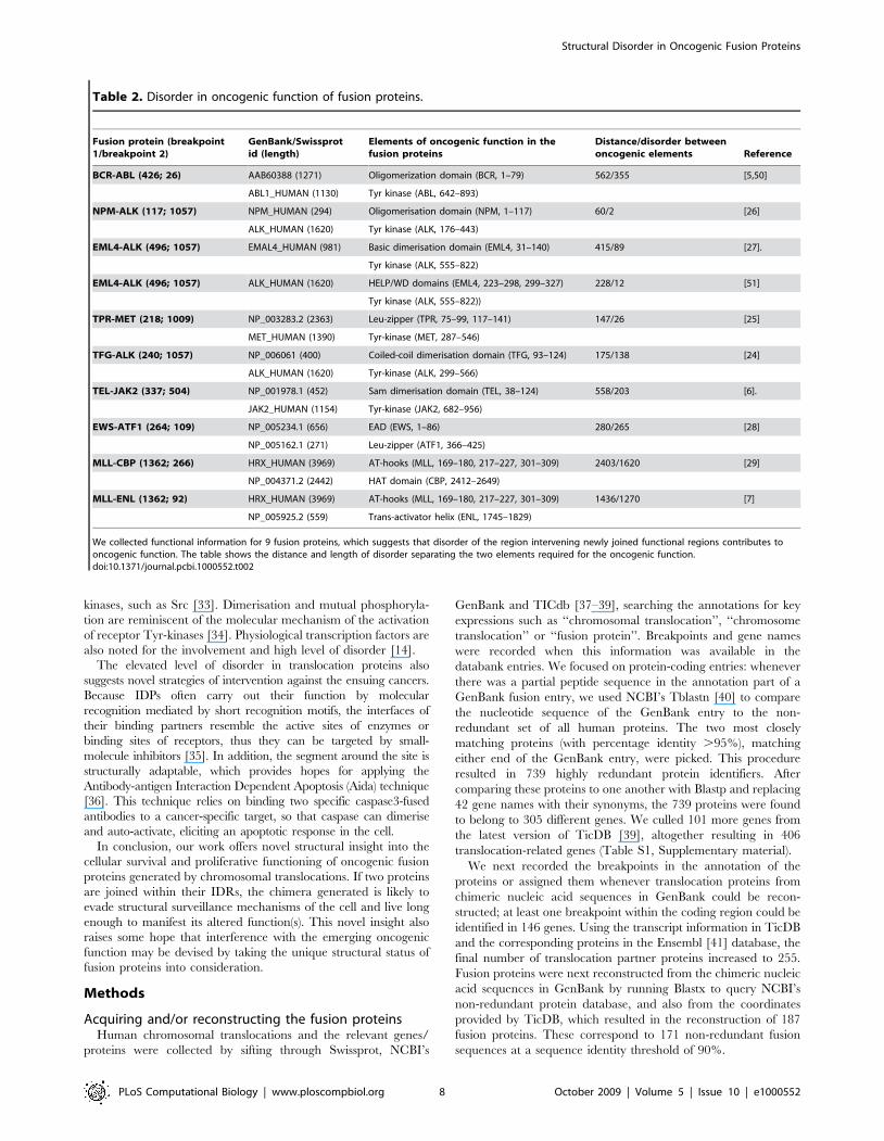
Table 2. Disorder in oncogenic function of fusion proteins.
Fusion protein (breakpoint1/breakpoint 2)
GenBank/Swissprotid (length)
Elements of oncogenic function in thefusion proteins
Distance/disorder betweenoncogenic elements Reference
BCR-ABL (426; 26) AAB60388 (1271) Oligomerization domain (BCR, 1–79) 562/355 [5,50]
ABL1_HUMAN (1130) Tyr kinase (ABL, 642–893)
NPM-ALK (117; 1057) NPM_HUMAN (294) Oligomerisation domain (NPM, 1–117) 60/2 [26]
ALK_HUMAN (1620) Tyr kinase (ALK, 176–443)
EML4-ALK (496; 1057) EMAL4_HUMAN (981) Basic dimerisation domain (EML4, 31–140) 415/89 [27].
Tyr kinase (ALK, 555–822)
EML4-ALK (496; 1057) ALK_HUMAN (1620) HELP/WD domains (EML4, 223–298, 299–327) 228/12 [51]
Tyr kinase (ALK, 555–822))
TPR-MET (218; 1009) NP_003283.2 (2363) Leu-zipper (TPR, 75–99, 117–141) 147/26 [25]
MET_HUMAN (1390) Tyr-kinase (MET, 287–546)
TFG-ALK (240; 1057) NP_006061 (400) Coiled-coil dimerisation domain (TFG, 93–124) 175/138 [24]
ALK_HUMAN (1620) Tyr-kinase (ALK, 299–566)
TEL-JAK2 (337; 504) NP_001978.1 (452) Sam dimerisation domain (TEL, 38–124) 558/203 [6].
JAK2_HUMAN (1154) Tyr-kinase (JAK2, 682–956)
EWS-ATF1 (264; 109) NP_005234.1 (656) EAD (EWS, 1–86) 280/265 [28]
NP_005162.1 (271) Leu-zipper (ATF1, 366–425)
MLL-CBP (1362; 266) HRX_HUMAN (3969) AT-hooks (MLL, 169–180, 217–227, 301–309) 2403/1620 [29]
NP_004371.2 (2442) HAT domain (CBP, 2412–2649)
MLL-ENL (1362; 92) HRX_HUMAN (3969) AT-hooks (MLL, 169–180, 217–227, 301–309) 1436/1270 [7]
NP_005925.2 (559) Trans-activator helix (ENL, 1745–1829)
We collected functional information for 9 fusion proteins, which suggests that disorder of the region intervening newly joined functional regions contributes tooncogenic function. The table shows the distance and length of disorder separating the two elements required for the oncogenic function.doi:10.1371/journal.pcbi.1000552.t002
Structural Disorder in Oncogenic Fusion Proteins
PLoS Computational Biology | www.ploscompbiol.org 8 October 2009 | Volume 5 | Issue 10 | e1000552

As controls, the sequences of experimentally verified IDPs were
downloaded from the DisProt database [42] (http://www.disprot.
org/) and fully ordered proteins were obtained from the PDB
(http://www.rcsb.org/). A set of all human protein-coding genes
and their transcript variants were obtained from the Ensembl
website (http://www.ensembl.org). As of March 5, 2008, there
were 22,297 protein-coding genes in the human dataset. We used
as reference proteins the longest transcript for each human
protein-coding gene. A set of human proteins (altogether 15,945)
were also obtained from the Swissprot databank (http://expasy.
org/sprot/) [38]. The Pfam domain database [43] was download-
ed from the Pfam website (http://pfam.sanger.ac.uk/).
Analysis of disorder and Pfam domains occurrence in thefusion proteins
Intrinsic disorder was predicted by the IUPred algorithm [17],
which can predict disorder with a sensitivity of 76% at 5% false
positive rate. Average percentage disorder was defined as the percent
of amino acids with a disorder score $0.5.
Fusion proteins were analyzed for the occurrence of Pfam domains
running Blastp against the entire set of Pfam-A domain sequences [43]
and also the human subset of Pfam domains derived from Swissprot
proteins. We set the thresholds for a domain match at an e-value,1e-
06 and sequence similarity .60%. We found that at this similarity
level there was less than 1% difference between the two sets of domain
matches, so analyzed all 18,609 human Swissprot proteins for the
occurrence of Pfam domains using only the Swissprot-derived human
subset of Pfam. (Ideally, when looking for non-overlapping domain
matches, choosing the best ones we would find only identity matches.
We found that out of 29,848 non-overlapping Pfam domains in
14,541 human Swissprot proteins with at least one domain match
there were only 1275 domain matches with less than 100% sequence
similarity.) We further analyzed the translocation proteins with in-
house Perl scripts, namely, (i) statistical significance of the difference
between any two distributions was evaluated with the chi-square test;
(ii) p-values corresponding to the calculated chi-square values and
degrees of freedom were calculated by a computer program courtesy
of Zsuzsa Dosztanyi; (iii) percentage values of disorder, length, etc.
distributions were also calculated by own Perl scripts.
Calculation of exposed hydrophobic surface in thetruncated Pfam domains
Actual values for the accessible nonpolar surface area (Anp) of
truncated domains were determined as follows: The C-terminus of
the protein structure was gradually truncated and the actual values
of Anp for the truncated fragments were determined with the
CHASA program [22] as suggested by [44]. The theoretical values
were calculated using the formula by Chothia [45] and Janin [46].
The truncated domain structures were drawn and annotated
using NCBI’s Cn3D program [47]. The high-resolution images in
Figures 4 and S3 were created using the Polyview-3D server [48].
Supporting Information
Figure S1 Translocation frequency as a function of gene length.
Found at: doi:10.1371/journal.pcbi.1000552.s001 (1.45 MB TIF)
Figure S2 Translocation frequency as a function of protein
disorder. Ratio of the number of all human proteins and
translocation proteins with breakpoint as a function of disorder
(%IU). The best fitting trendline is a parable.
Found at: doi:10.1371/journal.pcbi.1000552.s002 (0.13 MB EPS)
Figure S3 Effect of truncation on the Interleukin 2 domain. (A)
On the top the theoretical and actual values for the accessible
nonpolar surface area (Anp) are shown, calculated as described for
Figure 4; (B) Structure of the IL2 domain (pdb code: 1irl). The C-
terminal portion missing due to the translocation is colored grey.
Found at: doi:10.1371/journal.pcbi.1000552.s003 (1.29 MB TIF)
Table S1 List of proteins observed in chromosomal transloca-
tions. 151 proteins without (Table S1A) and 255 proteins with
(Table S1B) breakpoint (latter also indicated). The percentage
disorder is also shown.
Found at: doi:10.1371/journal.pcbi.1000552.s004 (0.07 MB XLS)
Table S2 List of all truncated Pfam domains and their mapping
to the fusion proteins using Blastp.
Found at: doi:10.1371/journal.pcbi.1000552.s005 (0.01 MB
TXT)
Author Contributions
Conceived and designed the experiments: PT. Analyzed the data: HH LB.
Wrote the paper: HH PT.
Figure 5. Predicted disorder and domain structure of selectfusion proteins. Disorder predicted by the IUPred algorithm anddomain structure identified by Pfam are shown for BCR-ABL, TFG-ALK,and EWS-ATF1, values above 0.5 are considered disordered. Theposition of the breakpoint is marked by a vertical line in the disorderplot whereas the elements critical for the oncogenic function are shownas colored rectangles (arrowhead for breakpoint) on the domain modelsbelow. The critical elements are connected by long segments ofstructural disorder.doi:10.1371/journal.pcbi.1000552.g005
Structural Disorder in Oncogenic Fusion Proteins
PLoS Computational Biology | www.ploscompbiol.org 9 October 2009 | Volume 5 | Issue 10 | e1000552

References
1. Aplan PD (2006) Causes of oncogenic chromosomal translocation. Trends Genet
22(1): 46–55.2. Futreal PA, Coin L, Marshall M, Down T, Hubbard T, Wooster R, Rahman N,
Stratton MR (2004) A census of human cancer genes. Nat Rev Cancer 4(3):177–83.
3. Rabbitts TH, Stocks MR (2003) Chromosomal translocation products engendernew intracellular therapeutic technologies. Nat Med 9(4): 383–6.
4. Rowley JD (2001) Chromosome translocations: dangerous liaisons revisited. Nat
Rev Cancer 1(3): 245–50.5. Goga A, McLaughlin J, Afar DE, Saffran DC, Witte ON (1995) Alternative
signals to RAS for hematopoietic transformation by the BCR-ABL oncogene.Cell 82(6): 981–8.
6. Lacronique V, Boureux A, Valle VD, Poirel H, Quang CT, Mauchauffe M,
Berthou C, Lessard M, Berger R, Ghysdael J, Bernard OA (1997) A TEL-JAK2fusion protein with constitutive kinase activity in human leukemia. Science
278(5341): 1309–12.7. Slany RK, Lavau C, Cleary ML (1998) The oncogenic capacity of HRX-ENL
requires the transcriptional transactivation activity of ENL and the DNA binding
motifs of HRX. Mol Cell Biol 18(1): 122–9.8. Dyson HJ, Wright PE (2005) Intrinsically unstructured proteins and their
functions. Nat Rev Mol Cell Biol 6(3): 197–208.9. Collins EC, Rabbitts TH (2002) The promiscuous MLL gene links chromosomal
translocations to cellular differentiation and tumour tropism. Trends Mol Med8(9): 436–42.
10. Ng KP, Potikyan G, Savene RO, Denny CT, Uversky VN, Lee KA (2007)
Multiple aromatic side chains within a disordered structure are critical fortranscription and transforming activity of EWS family oncoproteins. Proc Natl
Acad Sci U S A 104(2): 479–84.11. Tompa P (2002) Intrinsically unstructured proteins. Trends Biochem Sci 27(10):
527–533.
12. Uversky VN, Oldfield CJ, Dunker AK (2005) Showing your ID: intrinsicdisorder as an ID for recognition, regulation and cell signaling. J Mol Recognit
18(5): 343–84.13. Iakoucheva L, Brown C, Lawson J, Obradovic Z, Dunker A (2002) Intrinsic
Disorder in Cell-signaling and Cancer-associated Proteins. J Mol Biol 323(3):573–584.
14. Minezaki Y, Homma K, Kinjo AR, Nishikawa K (2006) Human transcription
factors contain a high fraction of intrinsically disordered regions essential fortranscriptional regulation. J Mol Biol 359(4): 1137–49.
15. Ward JJ, Sodhi JS, McGuffin LJ, Buxton BF, Jones DT (2004) Prediction andfunctional analysis of native disorder in proteins from the three kingdoms of life.
J Mol Biol 337(3): 635–45.
16. Romero PR, Zaidi S, Fang YY, Uversky VN, Radivojac P, Oldfield CJ,Cortese MS, Sickmeier M, LeGall T, Obradovic Z, Dunker AK (2006)
Alternative splicing in concert with protein intrinsic disorder enables increasedfunctional diversity in multicellular organisms. Proc Natl Acad Sci U S A
103(22): 8390–5.17. Dosztanyi Z, Csizmok V, Tompa P, Simon I (2005) IUPred: web server for the
prediction of intrinsically unstructured regions of proteins based on estimated
energy content. Bioinformatics 21(16): 3433–4.18. Furney SJ, Higgins DG, Ouzounis CA, Lopez-Bigas N (2006) Structural and
functional properties of genes involved in human cancer. BMC Genomics 7: 3.19. Dosztanyi Z, Chen J, Dunker AK, Simon I, Tompa P (2006) Disorder and
sequence repeats in hub proteins and their implications for network evolution.
J Proteome Res 5(11): 2985–95.20. Ekman D, Light S, Bjorklund AK, Elofsson A (2006) What properties
characterize the hub proteins of the protein-protein interaction network ofSaccharomyces cerevisiae? Genome Biol 7(6): R45.
21. Laabi Y, Gras MP, Carbonnel F, Brouet JC, Berger R, Larsen CJ, Tsapis A
(1992) A new gene, BCM, on chromosome 16 is fused to the interleukin 2 geneby a t(4;16)(q26;p13) translocation in a malignant T cell lymphoma. Embo J
11(11): 3897–904.22. Fleming PJ, Fitzkee NC, Mezei M, Srinivasan R, Rose GD (2005) A novel
method reveals that solvent water favors polyproline II over beta-strandconformation in peptides and unfolded proteins: conditional hydrophobic
accessible surface area (CHASA). Protein Sci 14(1): 111–8.
23. Andreeva A, Howorth D, Chandonia JM, Brenner SE, Hubbard TJ, Chothia C,Murzin AG (2008) Data growth and its impact on the SCOP database: new
developments. Nucleic Acids Res 36(Database issue): D419–25.24. Roccato E, Pagliardini S, Cleris L, Canevari S, Formelli F, Pierotti MA, Greco A
(2003) Role of TFG sequences outside the coiled-coil domain in TRK-T3
oncogenic activation. Oncogene 22(6): 807–18.25. Mak HH, Peschard P, Lin T, Naujokas MA, Zuo D, Park M (2007) Oncogenic
activation of the Met receptor tyrosine kinase fusion protein, Tpr-Met, involvesexclusion from the endocytic degradative pathway. Oncogene 26(51): 7213–21.
26. Bischof D, Pulford K, Mason DY, Morris SW (1997) Role of the nucleophosmin
(NPM) portion of the non-Hodgkin’s lymphoma-associated NPM-anaplasticlymphoma kinase fusion protein in oncogenesis. Mol Cell Biol 17(4): 2312–25.
27. Soda M, Choi YL, Enomoto M, Takada S, Yamashita Y, Ishikawa S, Fujiwara S,Watanabe H, Kurashina K, Hatanaka H, Bando M, Ohno S, Ishikawa Y,
Aburatani H, Niki T, Sohara Y, Sugiyama Y, Mano H (2007) Identification ofthe transforming EML4-ALK fusion gene in non-small-cell lung cancer. Nature
448(7153): 561–6.
28. Pan S, Ming KY, Dunn TA, Li KK, Lee KA (1998) The EWS/ATF1 fusionprotein contains a dispersed activation domain that functions directly. Oncogene
16(12): 1625–31.29. Lavau C, Du C, Thirman M, Zeleznik-Le N (2000) Chromatin-related
properties of CBP fused to MLL generate a myelodysplastic-like syndrome that
evolves into myeloid leukemia. Embo J 19(17): 4655–64.30. Ortiz de Mendibil I, Vizmanos JL, Novo FJ (2009) Signatures of selection in
fusion transcripts resulting from chromosomal translocations in human cancer.PLoS One 4(3): e4805.
31. Tompa P, Csermely P (2004) The role of structural disorder in the function of
RNA and protein chaperones. FASEB J 18(11): 1169–75.32. Tompa P, Fuxreiter M (2008) Fuzzy complexes: polymorphism and structural
disorder in protein-protein interactions. Trends Biochem Sci 33(1): 2–8.33. Harrison SC (2003) Variation on an Src-like theme. Cell 112(6): 737–40.
34. Schlessinger J, Lemmon MA (2003) SH2 and PTB domains in tyrosine kinasesignaling. Sci STKE 2003(191): RE12.
35. Cheng Y, Legall T, Oldfield CJ, Mueller JP, Van YY, Romero P, Cortese MS,
Uversky VN, Dunker AK (2006) Rational drug design via intrinsicallydisordered protein. Trends Biotechnol 24(10): 435–42.
36. Rabbitts TH (2001) Chromosomal translocation master genes, mouse modelsand experimental therapeutics. Oncogene 20(40): 5763–77.
37. Benson DA, Karsch-Mizrachi I, Lipman DJ, Ostell J, Wheeler DL (2008)
GenBank. Nucleic Acids Res 36(Database issue): D25–30.38. Boeckmann B, Bairoch A, Apweiler R, Blatter MC, Estreicher A, Gasteiger E,
Martin MJ, Michoud K, O’Donovan C, Phan I, Pilbout S, Schneider M (2003)The SWISS-PROT protein knowledgebase and its supplement TrEMBL in
2003. Nucleic Acids Res 31(1): 365–70.39. Novo FJ, de Mendibil IO, Vizmanos JL (2007) TICdb: a collection of gene-
mapped translocation breakpoints in cancer. BMC Genomics 8: 33.
40. Shiryev SA, Papadopoulos JS, Schaffer AA, Agarwala R (2007) ImprovedBLAST searches using longer words for protein seeding. Bioinformatics 23(21):
2949–51.41. Flicek P, Aken BL, Beal K, Ballester B, Caccamo M, Chen Y, Clarke L,
Coates G, Cunningham F, Cutts T, Down T, Dyer SC, Eyre T, Fitzgerald S,
Fernandez-Banet J, Graf S, Haider S, Hammond M, Holland R, Howe KL,Howe K, Johnson N, Jenkinson A, Kahari A, Keefe D, Kokocinski F, Kulesha E,
Lawson D, Longden I, Megy K, Meidl P, Overduin B, Parker A, Pritchard B,Prlic A, Rice S, Rios D, Schuster M, Sealy I, Slater G, Smedley D, Spudich G,
Trevanion S, Vilella AJ, Vogel J, White S, Wood M, Birney E, Cox T,Curwen V, Durbin R, Fernandez-Suarez XM, Herrero J, Hubbard TJ,
Kasprzyk A, Proctor G, Smith J, Ureta-Vidal A, Searle S (2008) Ensembl 2008.
Nucleic Acids Res 36(Database issue): D707–14.42. Sickmeier M, Hamilton JA, LeGall T, Vacic V, Cortese MS, Tantos A, Szabo B,
Tompa P, Chen J, Uversky VN, Obradovic Z, Dunker AK (2007) DisProt: theDatabase of Disordered Proteins. Nucleic Acids Res 35(Database issue):
D786–93.
43. Sammut SJ, Finn RD, Bateman A (2008) Pfam 10 years on: 10,000 families andstill growing. Brief Bioinform 9(3): 210–9.
44. Peisajovich SG, Rockah L, Tawfik DS (2006) Evolution of new proteintopologies through multistep gene rearrangements. Nat Genet 38(2): 168–74.
45. Chothia C (1976) The nature of the accessible and buried surfaces in proteins.
J Mol Biol 105(1): 1–12.46. Janin J (1976) Surface area of globular proteins. J Mol Biol 105(1): 13–4.
47. Wang Y, Geer LY, Chappey C, Kans JA, Bryant SH (2000) Cn3D: sequenceand structure views for Entrez. Trends Biochem Sci 25(6): 300–2.
48. Porollo A, Meller J (2007) Versatile annotation and publication qualityvisualization of protein complexes using POLYVIEW-3D. BMC Bioinformatics
8: 316.
49. Jung T, Catalgol B, Grune T (2009) The proteasomal system. Mol Aspects Med30(4): 191–296.
50. Zhao X, Ghaffari S, Lodish H, Malashkevich VN, Kim PS (2002) Structure ofthe Bcr-Abl oncoprotein oligomerization domain. Nat Struct Biol 9(2): 117–20.
51. Choi YL, Takeuchi K, Soda M, Inamura K, Togashi Y, Hatano S, Enomoto M,
Hamada T, Haruta H, Watanabe H, Kurashina K, Hatanaka H, Ueno T,Takada S, Yamashita Y, Sugiyama Y, Ishikawa Y, Mano H (2008) Identification
of novel isoforms of the EML4-ALK transforming gene in non-small cell lungcancer. Cancer Res 68(13): 4971–6.
Structural Disorder in Oncogenic Fusion Proteins
PLoS Computational Biology | www.ploscompbiol.org 10 October 2009 | Volume 5 | Issue 10 | e1000552
Copyright © 2022 FDOKUMEN