Rad Modulation of the L-type Calcium Channel Confers ...
233
University of Kentucky University of Kentucky UKnowledge UKnowledge Theses and Dissertations--Physiology Physiology 2021 Rad Modulation of the L-type Calcium Channel Confers Systolic Rad Modulation of the L-type Calcium Channel Confers Systolic Advantage in the Heart Advantage in the Heart Brooke Mildred Ahern University of Kentucky, [email protected] Author ORCID Identifier: https://orcid.org/0000-0003-2649-6000 Digital Object Identifier: https://doi.org/10.13023/etd.2021.146 Right click to open a feedback form in a new tab to let us know how this document benefits you. Right click to open a feedback form in a new tab to let us know how this document benefits you. Recommended Citation Recommended Citation Ahern, Brooke Mildred, "Rad Modulation of the L-type Calcium Channel Confers Systolic Advantage in the Heart" (2021). Theses and Dissertations--Physiology. 50. https://uknowledge.uky.edu/physiology_etds/50 This Doctoral Dissertation is brought to you for free and open access by the Physiology at UKnowledge. It has been accepted for inclusion in Theses and Dissertations--Physiology by an authorized administrator of UKnowledge. For more information, please contact [email protected].
-
Upload
khangminh22 -
Category
Documents
-
view
0 -
download
0
Transcript of Rad Modulation of the L-type Calcium Channel Confers ...

University of Kentucky University of Kentucky
UKnowledge UKnowledge
Theses and Dissertations--Physiology Physiology
2021
Rad Modulation of the L-type Calcium Channel Confers Systolic Rad Modulation of the L-type Calcium Channel Confers Systolic
Advantage in the Heart Advantage in the Heart
Brooke Mildred Ahern University of Kentucky, [email protected] Author ORCID Identifier:
https://orcid.org/0000-0003-2649-6000 Digital Object Identifier: https://doi.org/10.13023/etd.2021.146
Right click to open a feedback form in a new tab to let us know how this document benefits you. Right click to open a feedback form in a new tab to let us know how this document benefits you.
Recommended Citation Recommended Citation Ahern, Brooke Mildred, "Rad Modulation of the L-type Calcium Channel Confers Systolic Advantage in the Heart" (2021). Theses and Dissertations--Physiology. 50. https://uknowledge.uky.edu/physiology_etds/50
This Doctoral Dissertation is brought to you for free and open access by the Physiology at UKnowledge. It has been accepted for inclusion in Theses and Dissertations--Physiology by an authorized administrator of UKnowledge. For more information, please contact [email protected].

STUDENT AGREEMENT: STUDENT AGREEMENT:
I represent that my thesis or dissertation and abstract are my original work. Proper attribution
has been given to all outside sources. I understand that I am solely responsible for obtaining
any needed copyright permissions. I have obtained needed written permission statement(s)
from the owner(s) of each third-party copyrighted matter to be included in my work, allowing
electronic distribution (if such use is not permitted by the fair use doctrine) which will be
submitted to UKnowledge as Additional File.
I hereby grant to The University of Kentucky and its agents the irrevocable, non-exclusive, and
royalty-free license to archive and make accessible my work in whole or in part in all forms of
media, now or hereafter known. I agree that the document mentioned above may be made
available immediately for worldwide access unless an embargo applies.
I retain all other ownership rights to the copyright of my work. I also retain the right to use in
future works (such as articles or books) all or part of my work. I understand that I am free to
register the copyright to my work.
REVIEW, APPROVAL AND ACCEPTANCE REVIEW, APPROVAL AND ACCEPTANCE
The document mentioned above has been reviewed and accepted by the student’s advisor, on
behalf of the advisory committee, and by the Director of Graduate Studies (DGS), on behalf of
the program; we verify that this is the final, approved version of the student’s thesis including all
changes required by the advisory committee. The undersigned agree to abide by the statements
above.
Brooke Mildred Ahern, Student
Dr. Jonathan Satin, Major Professor
Dr. Kenneth S. Campbell, Director of Graduate Studies

RAD MODULATION OF THE L-TYPE CALCIUM CHANNEL CONFERS
SYSTOLIC ADVANTAGE IN THE HEART
________________________________________
DISSERTATION
________________________________________
A dissertation submitted in partial fulfillment of the
requirements for the degree of Doctor of Philosophy in the
College of Medicine
at the University of Kentucky
By
Brooke Mildred Ahern
Lexington, Kentucky
Director: Dr. Jonathan Satin, Professor of Physiology
Lexington, Kentucky
2021
Copyright © Brooke Mildred Ahern 2021
https://orcid.org/0000-0003-2649-6000

ABSTRACT OF DISSERTATION
RAD MODULATION OF THE L-TYPE CALCIUM CHANNEL CONFERS
SYSTOLIC ADVANTAGE IN THE HEART
Heart failure is a major public health problem and a leading cause of mortality.
This clinical condition affects populations of all ages, and is the result of various
cardiomyopathies. Almost half of these patients suffer specifically from heart failure
with reduced ejection fraction; these hearts have decreased performance due to a failure
of the heart to contract with sufficient force to meet demand. While there are therapies
available to increase contractility, none of these enhance contraction without also further
promoting pathological signaling and remodeling.
Under normal physiological conditions, the body elevates cardiac output through
the fight-or-flight response. This response activates -adrenergic receptors (-AR) at the
level of individual cardiomyocytes, which leads to enhanced calcium handling in order to
increase contraction. One of the major targets of -AR downstream signaling is the L-
type calcium channel (LTCC). The influx of calcium through the LTCC (ICa,L) provides
the trigger for calcium induced calcium release from the sarcoplasmic reticulum in order
to produce a contraction; LTCC activity is significantly increased when -ARs are
activated. However, -ARs are chronically activated in heart failure, leading to
pathological remodeling and further development of heart failure. This has served as a
foundation to establish dogma that increasing ICa,L in a manner that reflects -AR
activation necessarily promotes pathology. Because -AR signaling is a principle
physiological mechanism to increase cardiac output, understanding this pathway and how
to increase calcium safely is critical to successfully treating heart failure. Discovering a
mechanism to increase cardiac output downstream of -AR signaling would be ideal so
as to preserve the fight-or-flight response while also boosting cardiac performance in
order to meet demand.
The mechanism by which LTCC activity is increased under -AR signaling,
known as modulation, has been a major focus of study for many years; however, it
remains unknown. The LTCC is a heteromultimeric protein complex, and is inhibited by
an endogenous small monomeric GTPase called Rad. Studies in heterologous expression
systems show overexpression of Rad blocks calcium current through the LTCC; absence
of Rad yields a significant increase in calcium current. Whole-body Rad knock out mouse
models demonstrate calcium current that mirrors calcium current stimulated by -AR
signaling; however, this also promoted significant growth of the heart. To investigate the
effect of Rad deletion without contributions from non-cardiac tissue, a cardiomyocyte-
restricted inducible Rad knock out mouse model was created. The work of this

dissertation utilizes this mouse to better understand the mechanism by which Rad inhibits
the LTCC by studying the effects on channel function, cellular calcium handling, and
overall cardiac structure and function in the absence of Rad.
Using an array of methods and techniques, the studies in this dissertation establish
Rad as a critical target of the LTCC to respond to -AR stimulation. When Rad is
depleted specifically from cardiomyocytes, ICa,L is increased in a safe, stable manner that
mirrors LTCC modulation, both in sinoatrial node and in the ventricle. This regulation is
governed specifically by the C-terminus of Rad. Elevated ICa,L in the absence of Rad
promotes enhanced calcium handling and increased cardiac output without progression to
heart failure, and occurs independently of -AR activation. Enhanced calcium cycling in
the absence of Rad is balanced by accelerated inactivation of the LTCC so as to promote
positive inotropy without instigating arrhythmogenesis. This allows for cardiac
protection under conditions of pressure-overload induced heart failure. In summary, the
work of this dissertation supports Rad deletion specifically from cardiomyocytes as an
ideal positive inotrope for heart failure treatment due to the novel mechanism to increase
ICa,L in a manner that preserves structure, function, and the fight-or-flight response within
the heart.
KEYWORDS: L-type calcium channel, Rad, -adrenergic receptor signaling, Fight-or-
flight response, Heart Failure
Brooke Mildred Ahern
(Name of Student)
04/23/2021
Date

RAD MODULATION OF THE L-TYPE CALCIUM CHANNEL CONFERS
SYSTOLIC ADVANTAGE IN THE HEART
By
Brooke Mildred Ahern
Jonathan Satin, Ph.D.
Director of Dissertation
Kenneth S. Campbell, Ph.D,
Director of Graduate Studies
04/23/2021
Date

To my parents, my grandparents, and my sister

iii
ACKNOWLEDGMENTS
Completing this dissertation work would not have been possible for me without
the help and guidance from my support system. I would like to acknowledge the
following people who have helped me succeed and grow as a student and individual.
First, thank you to my mentor, Dr. Jonathan Satin. Thank you for challenging me
daily to become a better scientist, critical thinker, writer, electrophysiologist, and person.
Your passion for all things cardiac and calcium quickly convinced me to switch mindsets
from neuroscience to studying the mechanisms of the heart, and I am very grateful for
that. Thank you for seeing my potential, and helping me to succeed and grow these past
five years.
I would also like to express sincere gratitude for the members of my committee:
Dr. Doug Andres, thank you for pushing me to think differently (not always as an
electrophysiologist) and to see both science and life from a different perspective. Dr.
Brian Delisle, thank you for always providing a listening ear and a sounding board for my
projects and my life; also, thank you for teaching me how to patch-clamp – a technique
that has taught me the true meaning of patience and perseverance. Dr. Ahmed Abdel-
Latif, thank you for providing helpful feedback on my work from a clinical perspective,
and giving me career advice as I have tried to navigate my future. Dr. John McCarthy,
thank you for your support of my work, my love for space, and for allowing me to be an
honorary member of your lab. And to Dr. Sanda Despa, thank you for serving as my
outside examiner and taking the time to review my dissertation.
Thank you to the American Heart Association for awarding me a pre-doctoral
fellowship that funded me for two years. I am sincerely grateful for the time and service

iv
by volunteers within that wonderful organization that provided the financial support so I
could pursue my passion for understanding Rad. I am also very grateful for the NIH T32
that I was given my first year within the department and the work from faculty within the
department to have that available for students.
I have also been blessed to share the lab with two amazing women, Bryana
Levitan and Andrea Sebastian. Thank you Bryana (aka lab mom) for your support and
teaching me what you have learned about the heart and about life. Thank you Andrea (the
science fairy) for your constant support in all of my experiments, for all of the therapy
sessions while isolating cells or making solutions, and for being a great friend. I am a
much better person for knowing both of you.
I was also very lucky to join the best department on campus, the Physiology
Department. The faculty and staff made each day great – thank you to all those that have
given me advice over the years, shared a laugh or taught me both in and out of the
classroom. I also appreciate that we had the best lunches, best seminar treats (always had
Diet Coke!), and the best holiday parties. I will cherish the fun I had with all of you.
To all of the friends I have made in graduate school: Ben Shaw, Beth Oates, and
Sarah Sternbach – the original dream team of first year and the reason I survived. Ryan
Cloyd, Brandon Farmer – the wonderful additions to the crew. Our adventure lunches,
Mario Kart, parties, therapy sessions and of course the pet therapy from all of you have
helped me maintain my sanity throughout the years here. Words do not truly express my
gratitude; thank you guys for being a major part of my support system. To Taylor
Valentino, Yuan Wen, Ivan Vechetti, and Brooks Mobley – thank you for all of your
support while I figured out my life and career, teaching me, letting me vent, always

v
providing chocolate and Diet Coke, and giving me honorary member status in the lab. I
am lucky to have been able to call your lab, as well as the Satin lab, home. To those that I
have met in graduate school but have not mentioned thank you for supporting me! I have
been very blessed to meet many wonderful people, and call them friends.
To Kate Holbrook and Trent Simmons – friends who have known me for a long
time and have been critical support for much of my life. Thank you for the late night
phone calls, the supportive texts, and for being the best cheerleaders even though you
both are so far away. I would not have gotten this far without you two.
And last, but certainly not least, I want to thank my family. To my wonderful
parents, David and Heidi Ahern – thank you for always pushing me to be the best I can
be. You have always believed in me, encouraged me to follow my passions, and given
me confidence when I doubted myself. I cannot express in words how grateful I am for
your sacrifices so that I could make it this far. To my amazing grandparents, Donald and
Abbey Schluter and Rel Ahern, who have always been the most supportive of anything I
do in life, especially in graduate school. Thank you for always being there, whether in
person, in spirit, or on Facetime; I am so grateful to have you in my life. And to the best
sister anyone could ask for, Alexandra Ahern – thank you for being my best friend
through everything, for understanding all aspects of my life (including getting a PhD),
and for inspiring me daily to be better than I was the day before. Thank you for always
being the ideal example in my life.
To those that I have not mentioned but have helped me, given me advice, and
made my life better, thank you!

vi
TABLE OF CONTENTS
ACKNOWLEDGMENTS ............................................................................................................................ iii
LIST OF FIGURES ....................................................................................................................................... x
CHAPTER 1 Introduction ............................................................................................................................ 1
1.1 Significance ....................................................................................................................................... 1
1.2 L-Type Calcium Channels ................................................................................................................. 3
1.2.1 Voltage Gated Calcium Channels ............................................................................................ 3
1.2.2 Structure of the L-type Calcium Channel ................................................................................ 4
1.2.2.1 The pore-forming subunit: 1C ....................................................................................... 4
1.2.2.2 CaV subunit .................................................................................................................. 5
1.2.2.3 2 subunit ..................................................................................................................... 5
1.2.2.4 subunit ......................................................................................................................... 6
1.2.2.5 Calmodulin ..................................................................................................................... 6
1.2.3 Function of the L-type Calcium Channel ................................................................................ 7
1.2.3.1 Excitation Contraction Coupling.................................................................................... 7
1.2.3.2 Activation of the L-type Calcium Channel .................................................................... 8
1.2.3.3 Inactivation of the L-type Calcium Channel ................................................................ 10
1.2.3.4 L-type Calcium Channel Regulation by Facilitation ................................................... 12
1.2.3.5 Modulation of the L-type Calcium Channel ................................................................ 13
1.2.4 Role of the L-type Calcium Channel in Cardiac Pathophysiology........................................ 17
1.2.4.1 Arrhythmias.................................................................................................................. 17
1.2.4.2 Early After Depolarizations ......................................................................................... 18
1.2.4.3 Timothy Syndrome....................................................................................................... 19
1.2.4.4 Brugada Syndrome ....................................................................................................... 20
1.2.4.5 Heart Failure................................................................................................................. 21
Cardiac Hypertrophy ............................................................................................... 22
Loss of −Adrenergic Receptor Stimulation........................................................... 23
1.3 Rad................................................................................................................................................... 24
1.3.1 RGK Proteins ......................................................................................................................... 24
1.3.2 Structure of Rad ..................................................................................................................... 25
1.3.3 Function of Rad ..................................................................................................................... 26
1.4 Working Hypothesis ........................................................................................................................ 29
CHAPTER 2. Materials and Methods ....................................................................................................... 33
2.1 Animal Model .................................................................................................................................. 33
2.2 TAC .................................................................................................................................................. 34
2.3 Immunoblotting ............................................................................................................................... 35
2.4 Quantification of myofibril protein phosphorylation ...................................................................... 36
2.5 Ventricular myocyte isolation ......................................................................................................... 36
2.6 Electrophysiological recordings ..................................................................................................... 37
2.7 Calcium Transients.......................................................................................................................... 38

vii
2.8 Single Cell Database Analysis ........................................................................................................ 39
2.9 Quantitative Proteomics Database Analysis ................................................................................... 40
2.10 RNA In Situ Hybridization .......................................................................................................... 41
2.11 Quantitative RT-PCR.................................................................................................................. 41
2.12 Histology..................................................................................................................................... 42
2.13 Echocardiography ...................................................................................................................... 42
2.14 Radiotelemetry ............................................................................................................................ 43
2.15 Surface ECG ............................................................................................................................... 44
2.16 Open Behavioral Assessment ..................................................................................................... 44
2.17 Statistical Analysis...................................................................................................................... 44
CHAPTER 3. Myocardial-Restricted Ablation of the GTPase RAD Results in a Pro-Adaptive Heart
Response in Mice.......................................................................................................................................... 46
3.1 Preface............................................................................................................................................. 46
3.2 Introduction ..................................................................................................................................... 46
3.3 Results ............................................................................................................................................. 48
3.3.1 Development of conditional cardiac-restricted RAD-deficient transgenic mice ................... 48
3.3.2 Cardiac-restriced RAD-deficient mice (RAD) show improved function with no changes in
heart dimensions................................................................................................................................... 49
3.3.3 RAD modification of voltage-gated calcium current, ICa,L ................................................. 51
3.3.4 RAD enhances cellular calcium homeostasis ..................................................................... 52
3.3.5 Basal ICa,L in RAD cardiomyocytes reflects a modulated ICa,L .......................................... 54
3.3.6 RAD basal heart function is elevated in vivo, yet retains a partial ISO response .............. 54
3.3.7 RAD retains −adrenergic receptor modulation of calcium and sarcomere dynamics ...... 55
3.4 Discussion ....................................................................................................................................... 56
3.4.1 Increased calcium does not necessarily promote cardiac hypertrophy .................................. 57
3.4.2 Myocardial-restricted RAD deletion differs from whole body RAD deletion ...................... 60
CHAPTER 4. Rad-GTPase Contributes to Heart Rate Via L-type Calcium Channel Regulation ..... 76
4.1 Preface............................................................................................................................................. 76
4.2 Introduction ..................................................................................................................................... 76
4.3 Results ............................................................................................................................................. 78
4.3.1 SANcm express Rad .............................................................................................................. 78
4.3.2 L-type calcium current (ICa,L) in SANcm............................................................................... 80
4.3.3 RAD reduction increases intrinsic heart rate ......................................................................... 81
4.3.4 Rad-deletion effects on HR are greatest when Sympathetic Drive is Relatively Low .......... 82
4.4 Discussion ....................................................................................................................................... 83
4.4.1 Rad is a key contributor to SANcm ICa,L modulation ............................................................ 84
4.4.2 Relationship among SANcm heterogeneity, Rad expression, and SAN function ................. 86
4.4.3 Diurnal impact and ANS drive with Rad reduction............................................................... 87
4.5 Limitations ....................................................................................................................................... 88
4.6 Conclusions ..................................................................................................................................... 89

viii
CHAPTER 5. Enhanced Inactivation Balances Increased Peak L-Type Calcium Current in the
Absence of Rad ............................................................................................................................................. 99
5.1 Preface............................................................................................................................................. 99
5.2 Introduction ..................................................................................................................................... 99
5.3 Results ........................................................................................................................................... 102
5.3.1 cRadKO mirrors modulated LTCC ..................................................................................... 102
5.3.2 Increased Ca2+ and Rad both contribute to CDI .................................................................. 103
5.3.3 Rad does not alter the late component of LTCC decay ....................................................... 104
5.3.4 Faster kinetics in cRadKO prevent alterations in cardiac electrophysiology ...................... 105
5.3.5 cRadKO yields modulated kinetics of LTCC downstream of −AR signaling .................. 107
5.4 Discussion ..................................................................................................................................... 109
5.4.1 Rad contributes to regulation of LTCC inactivation ........................................................... 110
5.4.2 Rad is a key contributor to LTCC Modulation .................................................................... 111
5.4.3 Rad regulation of LTCC is independent of −adrenergic receptor signaling...................... 113
5.5 Limitations ..................................................................................................................................... 114
5.6 Conclusions ................................................................................................................................... 114
CHAPTER 6. Alterations to Rad Structure Modulate LTCC Gating ................................................. 129
6.1 Preface........................................................................................................................................... 129
6.2 Introduction ................................................................................................................................... 129
6.3 Results ........................................................................................................................................... 131
6.3.1 Rad-Flag A277X increases cardiac function and wall thickness ......................................... 131
6.3.2 Rad-Flag A277X enhances LTCC gating ............................................................................ 132
6.4 Discussion ..................................................................................................................................... 133
CHAPTER 7. Myocardial Rad deletion Preserves Cardiac Function in Pressure-Overload Induced
Heart Failure .............................................................................................................................................. 141
7.1 Preface........................................................................................................................................... 141
7.2 Introduction ................................................................................................................................... 141
7.3 Results ........................................................................................................................................... 143
7.3.1 cRadKO protects in vivo heart function from pressure-overload induced heart failure ...... 143
7.3.2 cRadKO enhances in vivo heart function after pressure-overload ....................................... 144
7.3.3 cRadKO maintains elevated calcium cycling after pressure-overload ................................ 145
7.3.4 Response to −adrenergic receptor stimulation remains blunted in cRadKO after pressure-
overload 147
7.3.5 cRadKO promotes recovery of function in advanced stages of pressure-overload ............. 147
7.3.6 cRadKO prevents progression of cardiac dysfunction in advanced stages of pressure-
overload 148
7.3.7 cRadKO improves quality of life in advanced stages of pressure-overload ........................ 150
7.4 Discussion ..................................................................................................................................... 150
7.4.1 Myocardial-restricted Rad deletion preserves cardiac function in pressure-overload induced
heart failure ........................................................................................................................................ 151
7.4.2 Myocardial-restricted Rad deletion differs from whole-body knockout of Rad ................. 153
CHAPTER 8. Discussion ........................................................................................................................... 172

ix
8.1 Rad is a critical regulatory site for LTCC modulation ................................................................. 172
8.2 Rad governs ICa,L by multiple mechanisms .................................................................................... 173
8.3 Hypertrophic signaling is not exclusively dependent on elevated ICa,L ......................................... 176
8.4 Enhanced ICa,L does not necessarily promote electrical dysfunction ............................................ 178
8.5 Targeting Rad represents the ideal positive inotrope ................................................................... 180
8.6 Future directions ........................................................................................................................... 182
8.7 Limitations ..................................................................................................................................... 184
8.8 Conclusions ................................................................................................................................... 184
APPENDIX ................................................................................................................................................. 186
REFERENCES .......................................................................................................................................... 192
VITA ........................................................................................................................................................... 213

x
LIST OF FIGURES
Figure 1.1 L-type Calcium Channel Complex. ................................................. 31
Figure 1.2 L-type Calcium Channel pore-forming subunit 1C. .................... 32
Figure 3.1 Myocardial deletion of Rad. ............................................................ 62
Figure 3.2 Cardiomyocyte-restricted Rad deletion does not induce markers of
myocardial pathology. ........................................................................................ 63
Figure 3.3 Figure 3.3. Myocardial Rad deletion results in a rapid, stable gain
of cardiac function without pathological structural remodeling in vivo. ...... 64
Figure 3.4 Myocardial Rad deletion results in increased calcium current
(ICa,L). .................................................................................................................... 65
Figure 3.5 Rad∆/∆ enhances cellular calcium handling. ................................... 66
Figure 3.6 RAD∆/∆ enhances sarcomere shortening and increases the tension-
integral. ................................................................................................................ 67
Figure 3.7 ISO does not alter RAD∆/∆ ICa,L. ...................................................... 68
Figure 3.8 β-AR-mediated responsiveness retained following Rad deletion in
vivo ........................................................................................................................ 69
Figure 3.9 Cardiomyocyte cytosolic Ca2+ handling responds to β-AR-mediated
activation in RAD∆/∆. ........................................................................................... 70
Figure 3.10 Sarcomere dynamics and proteins respond to β-AR-mediated
activation in RAD∆/∆. ........................................................................................... 71
Supplemental Figure 3.11 Myocardial Rad deletion results in increased
calcium current (ICa,L). ....................................................................................... 72
Supplemental Figure 3.12 RAD∆/∆ ICa,L response after -adrenergic receptor
stimulation. .......................................................................................................... 73
Supplemental Figure 3.13 RAD∆/∆ cellular response after β-adrenergic
receptor stimulation. ........................................................................................... 74
Supplemental Figure 3.14 No effect on sarcomere dynamics after treatment
with ISO. .............................................................................................................. 75
Figure 4.1 RRAD is expressed in SANcm. ........................................................ 90
Figure 4.2 RAD is expressed in SANcm. ........................................................... 91
Figure 4.3 Rad reduction increases SANcm ICa,L. ............................................ 92
Figure 4.4 β-AR stimulation has no significant effect on cRadKO SANcm
ICa,L. ................................................................................................................... 93
Figure 4.5 Intrinsic heart rate increases after Rad deletion. .......................... 94
Figure 4.6 Rad deletion increases heart rate in the sleep phase. .................... 95
Figure 4.7 Rad deletion demonstrates preserved Heart Rate Variability
(HRV). .................................................................................................................. 96
Supplemental Figure 4.8 No significant difference between males and females
of ICa,L in CTRL and cRadKO. .......................................................................... 97
Figure 4.9 Rad Deletion Increases Sinus Heart Rate....................................... 98
Figure 5.1 Rad deletion ICa,L phenocopies modulated ICa,L. .......................... 116

xi
Figure 5.2 Rad deletion accelerates fast component of ICa,L decay. .............. 117
Figure 5.3 Rad regulation of ICa,L kinetics requires SR Ca2+ release. ....... 118
Figure 5.4 Rad does not alter slow component of ICa,L decay. ...................... 119
Figure 5.5 Rad deletion enhances VDI. ........................................................... 120
Figure 5.6 Rad deletion does not prolong action potential duration or QT
interval. .............................................................................................................. 121
Figure 5.7 Rad deletion increases heart function exclusive of β1β2-Adrenergic
Receptors. .......................................................................................................... 122
Figure 5.8 Rad deletion yields modulated ICa,L in the absence of β1β2-
Adrenergic Receptors. ...................................................................................... 123
Figure 5.9 Rad modulates ICa,L independent of β1β2-Adrenergic Signaling to
Confer Systolic Advantage. .............................................................................. 124
Supplemental Figure 5.10 Two-way ANOVA 95% confidence intervals and
tabular results.................................................................................................... 125
Supplemental Figure 5.11 Two-way ANOVA 95% confidence intervals and
tabular results.................................................................................................... 126
Supplemental Figure 5.12 Surface ECG Measurements. .............................. 127
Supplemental Figure 5.13 Whole Heart measurements of dKO and tKO. . 128
Figure 6.1 Schematic of Rad-Flag and Rad-Flag A277X transgenic mice. . 137
Figure 6.2 Rad-Flag A277X enhances cardiac function and increases wall
thickness. ............................................................................................................ 138
Figure 6.3 Rad-Flag A277X enhanced LTCC function ................................. 139
Figure 6.4 Rad-Flag A277X enhanced inactivation. ...................................... 140
Figure 7.1 Loss of cardiomyocyte Rad before pressure overload prevents
progression of cardiac dysfunction.................................................................. 155
Figure 7.2 ANOVA 95% confidence intervals and tabular results. ............. 156
Figure 7.3 Loss of cardiomyocyte Rad before pressure overload does not
protect all pathological signaling. .................................................................... 157
Figure 7.4 One-way ANOVA 95% confidence intervals and tabular results.
............................................................................................................................. 158
Figure 7.5 Loss of cardiomyocyte Rad before pressure overload promotes
survival and enhances cardiac function. ......................................................... 159
Figure 7.6 ANOVA 95% confidence intervals and tabular results. ............. 160
Figure 7.7 Loss of cardiomyocyte Rad increases ICa,L after pressure-
overload. ............................................................................................................. 161
Figure 7.8 ANOVA 95% confidence intervals and tabular results. ............. 162
Figure 7.9 Loss of cardiomyocyte Rad enhances cellular calcium handling
after pressure-overload. ................................................................................... 163
Figure 7.10 Loss of cardiomyocyte Rad does not alter sarcomere dynamics
after pressure-overload. ................................................................................... 164
Figure 7.11 -AR stimulation does not alter ICa,L in Rad deletion after
pressure-overload. ............................................................................................. 165

xii
Figure 7.12 -AR stimulation response in cellular calcium handling is blunted
after pressure-overload. ................................................................................... 166
Figure 7.13 Loss of cardiomyocyte Rad after pressure overload prevents
progression of cardiac dysfunction.................................................................. 167
Figure 7.14 ANOVA 95% confidence intervals and tabular results. ........... 168
Figure 7.15 Loss of cardiomyocyte Rad after pressure overload prevents
progression of cardiac dysfunction.................................................................. 169
Figure 7.16 ANOVA 95% confidence intervals and tabular results. ........... 170
Figure 7.17 Loss of cardiomyocyte Rad after pressure overload improves
locomotive activity. ........................................................................................... 171

1
CHAPTER 1. INTRODUCTION
1.1 Significance
The incidence of heart failure (HF) is steadily increasing, with projections putting
heart failure as the leading cause of mortality and morbidity in developed countries by the
year 2030 [1, 2]. Many cardiovascular disorders, including atherosclerosis,
cardiomyopathy, and hypertension, result in the development of HF. Of these patients,
~45% suffer from HF with reduced ejection fraction (HFrEF). HFrEF results in a
decrease in cardiac performance due to a failure of the heart to contract with sufficient
force to meet demand; this is soon followed by problems in other organ systems, such as
neurohormonal, circulatory, and renal systems [3, 4]. Current therapies are limited to
increasing survival and reducing symptoms, but are accompanied by side effects and
ultimately fail to increase systolic function or inhibit HF progression [3-5].
Cardiac contractility is governed by calcium cycling within individual
cardiomyocytes. During a cardiac action potential, calcium enters the cell via voltage-
gated calcium channels, specifically L-type calcium channels (LTCC). This calcium
triggers calcium release from the sarcoplasmic reticulum (SR), raising cytosolic calcium
concentration ~ten-fold to activate muscle contraction. Upon relaxation, calcium is either
taken back up into the SR via SERCA2a (Sarco/endoplasmic reticulum calcium ATPase)
or extruded from the cell via the Na+/Ca2+ exchanger (NCX) [3, 4, 6]. In HF, calcium
transients are depressed, resulting in decreased contractile function,[6, 7]. Logically, it
would follow that a treatment for HF would therefore be to increase calcium within the
cardiomyocyte in order to increase contraction. Indeed, the body naturally increases

2
cardiac contractility by increasing sympathetic drive and activating the -adrenergic
receptor (-AR) signaling cascade. However, chronic stimulation of this cascade triggers
pathological remodeling of the heart, thereby promoting the development of HF ([4, 6, 8,
9]. Studies focusing on the utilization of positive inotropes have proven to be acutely
effective, but chronically induce further damage on the heart by initiating hypertrophic
signaling and triggering arrhythmias [5-7, 9, 10]. It has therefore become dogma that
increasing calcium within a cardiomyocyte directly causes pathology and ultimately
results in HF.
Many studies have focused on the SR, [3-5, 7] the pathways associated with -AR
signaling [8, 11], or certain components of the LTCC complex [12-15] as potential
therapeutic targets for HF; unfortunately, they have yet to find a target that increases
contraction without instigating more pathology as a side effect. A recent new potential
therapeutic target that has emerged is Rad, a small monomeric GTPase that serves as an
endogenous inhibitor to the LTCC [16-24]. The studies of this dissertation focus
primarily on the mechanism by which the absence of Rad confers systolic advantage on
the heart independently of -AR signaling, thereby preserving the cardiac response to
autonomic input without pathological structural remodeling.
-AR stimulation is the principle physiological mechanism whereby contractility
and cardiac output is increased; the guiding principle for treating heart failure, and this
dissertation, is that enhancing systolic function exclusive of adrenergic activation is safe
and beneficial [5].

3
1.2 L-Type Calcium Channels
1.2.1 Voltage Gated Calcium Channels
Voltage-gated calcium channels (VGCC) are found in all excitable cells; these
channels support longer depolarizing responses because they can maintain inward
current, and also are the only link between non-electrical components and excitation [25,
26]. Calcium entering via VGCCs serves as an elemental secondary messenger that
initiates numerous intracellular events, including contraction [25, 27-31]. Signal
transduction via VGCCs differs depending on cell type, thereby requiring different
molecular subtypes of channels to have unique physiological, pharmacological and
regulatory properties [25, 28]. VGCCs were first differentiated based on activation
threshold, classified as either low-voltage-activated (LVA) or high-voltage-activated
(HVA) [25, 28, 29]. Calcium channels are also large heteromeric proteins and are
categorized based on structure of the complex [29]. The first purified calcium channel
was isolated from skeletal muscle and comprised of five components: 1 (170 kDa), 2
(150 kDa), (52 kDa), (17-25 kDa), and (32 kDa) in an approximately stoichiometric
ratio (Figure 1.1A; [28-30]. This complex was also termed the dihydropyridine receptor
for its sensitivity to 1,4-dihydropyridines (DHPs); because 1 was identified as the
subunit of the complex that bound DHPs, it was established that it was the pore-forming
subunit [29]. As of now, there are 10 different 1 subunits that have been discovered and
all exhibit distinct functions (Figure 1.1B,C). These include L-type, P/Q-type, N-type,
R-type, and T-type calcium channels [25, 28, 29]. HVA channels that were sensitive to
DHPs were identified as ‘long-lasting’ or L-type channels because of slow voltage
dependent inactivation and are long lasting when barium is used as the charge carrier [25,

4
28]; these channels form the CaV1 family [25, 28, 29, 32, 33]. CaV1.1 expression is
restricted to skeletal muscle, while CaV1.4 expression is restricted to the retina [29].
CaV1.2 and CaV1.3 are found in many different excitable cell types with both often
expressed in the same cell [29], such as adrenal chromaffin cells, neurons, and sinoatrial
and atrial cardiomyocytes [29]. The remainder of this chapter will focus primarily on
CaV1.2 and its role in cardiac function.
1.2.2 Structure of the L-type Calcium Channel
1.2.2.1 The pore-forming subunit: 1C
The 1 subunit that comprises CaV1.2 is encoded by the gene cacna1c [34]. The
1 subunit is approximately 2000 amino acid residues and is organized into four repeated
domains (I-IV) that each contain six transmembrane segments (S1-S6) and a membrane-
associated loop between transmembrane segments S5 and S6 (Figure 1.2; [29, 35]. The
domains are connected by intracellular loops linking domain I-II, domain II-III, and
domain III-IV [36], and end in NH2- and COOH-terminal intracellular segments [34]. In
each domain, S1-S4 segments form a voltage-sensing domain [37], with S4 being the
primary ‘voltage sensor’ for activation [25, 28, 29]. In every third position within the
helix of the S4 are positively charged Arg or Lys that sense depolarization of the
membrane [37]. Changes in the electric field of the membrane cause the S4 segment to
rotate outwards, thereby changing the conformation of the channel to open the pore [25,
28]. The S5 and S6 segments and the associated loop form the pore lining of the channels
[25, 28, 29, 38]. The external portion contains a pair of glutamate residues within the
pore loop that act as the selectivity filter for the channel so that it is calcium-specific; the

5
inner pore is formed by the S6 segments and contains the receptor sites for antagonist,
pore-blocking drugs specific for L-type calcium channels [25, 28, 29, 38]. Though a
structural model of the CaV1.2 pore has been suggested [39], a detailed three-dimensional
structure of calcium channels is not known [25, 28]. For clarity and consistency, the
remainder of the dissertation will refer to CaV1.2 simply as LTCC (L-type calcium
channel).
1.2.2.2 CaV subunit
A requirement for normal LTCC function is the auxiliary CaVβ subunit [40].
There are four distinct genes that encode four subfamilies of CaVβ (β1-β4), each of which
regulate LTCC differently [29, 40]. Each consist of a core region, composed of two
highly conserved regions homologous to the SRC homology 3 (SH3) and guanylate
kinase (GK) domains; these are joined together by the HOOK region [25, 40]. CaVβ
binds to the 1 subunit in a 1:1 stoichiometric fashion with high affinity to a single site
within the α-interaction domain (AID), the first half of the cytoplasmic linker connecting
domains I and II of the 1 subunit (Figure 1.2; [25, 28, 40, 41]. The AID forms an α-
helix that binds tightly to a groove within the GK domain of CaVβ, and is critical for
CaVβ regulation of calcium channels [25, 40]. The GK domain of CaVβ also contains the
interaction sites for other proteins that regulate channel function, such as RGK GTPases
[20, 40]. The interaction between CaVβ and LTCC is crucial for normal channel function,
and modulates channel activity [25, 28, 40, 41], which will be further discussed in the
next section.
1.2.2.3 2 subunit

6
The LTCC complex also contains the α2δ subunit (Figure 1.1A). α2 and δ are
produced from a single gene and proteolytically cleaved to form separate proteins but still
attached via a disulfide bond [25, 29, 40, 42]. There are also four distinct genes that
encode α2δ (α2δ1-4), that correspond to specific tissues, with α2δ1 highly expressed in
skeletal muscle [42, 43]. The δ peptide of the subunit anchors the subunit to the
extracellular membrane via a glycosylphosphatidylinositol linker [41, 42]. The main
function of α2δ is to increase calcium channel expression and retention within the
membrane, and contributes minorly to channel gating properties [25, 28, 29, 40, 42].
1.2.2.4 subunit
The subunit of LTCC complex is a glycoprotein that has four transmembrane
segments and intracellular amino and carboxy termini (Figure 1.1A) [25, 28, 41]. There
are eight different genes that encode subunits that all have diverse functions, depending
on the tissue in which they are expressed, though the function of each subunit is not fully
known [41]. Of these eight genes, only CaV1 and CaV6 are considered to be a subunit of
voltage-gated calcium channels [34]. In regards to calcium channel function, subunits
primarily contribute to voltage dependence of activation and inactivation in a small but
significant manner [25, 28, 41, 44]. Although CaV1 was found in skeletal muscle, it
appears there are no CaV subunits expressed in the heart [13].
1.2.2.5 Calmodulin
A critical protein that associates with the LTCC complex is a ubiquitous calcium-
sensing protein called calmodulin (CaM) [45]. CaM associates with the carboxyl-

7
terminus of the 1 subunit within the EF hand, pre-IQ, and IQ regions and serves as a key
regulator of channel function and targeting LTCC to the cell surface (Figure 1.2; [45-47].
For every channel, there is one CaM molecule in association [48, 49]. Each molecule of
CaM is composed of two lobes, each made of two EF hands [49]. The N-terminal lobe
acts as a ‘global’ calcium sensor for the channel, sensing an elevation of calcium within
the couplon or junction; the C-terminal lobe acts as a ‘local’ calcium sensor, triggering
regulatory signaling of the channel based on calcium flowing through individual channels
[49]. Specific interactions within the IQ domain and the two lobes cause different
calcium-dependent regulatory processes to occur within the channel [46], which will be
discussed in the next section.
1.2.3 Function of the L-type Calcium Channel
1.2.3.1 Excitation Contraction Coupling
Calcium is a critical intracellular second messenger that initiates multiple signaling
pathways within the cell [5, 6]. In a cardiomyocyte, the membrane is depolarized in
response to an action potential and LTCCs open to provide an influx of calcium needed for
excitation contraction coupling [6]. LTCCs also maintain membrane depolarization during
the plateau phase of the cardiac action potential [44]. Because alterations in calcium influx
affect action potential duration and refractory period duration, the LTCC contributes to
electrical balance that allows contraction and relaxation of the myocardium [44, 50].
LTCCs are expressed within the transverse tubules of the membrane in close
proximity (~12-20 nm) to ryanodine receptors (RyR2) so that the calcium influx from the
channel induces calcium induced calcium release (CICR) from the sarcoplasmic reticulum

8
(SR) [6, 45]. This provides the necessary elevation in cytosolic calcium to activate
myofilaments and generate contractile activity [6, 51]. At the junction between plasma
membrane and the sarcoplasmic reticulum, ~25 LTCCs and ~100 individual RyR2 form a
unit known as the couplon [51]. Couplons are spaced between 1-2 µm away from each
other so that neighboring couplons can activate calcium release synchronously and provide
a unified contraction within the entire cardiomyocyte [6, 51].
There is also a subset of LTCCs that are expressed within the caveolae of
ventricular myocytes, a type of lipid raft that create small invaginations within the plasma
membrane and characterized by the expression of caveolin-3 [45]. The fraction of LTCCs
found in the caveolae range depending on species, but it is estimated that in mice, ~50% of
LTCCs are found in the caveolae; the mechanisms by which LTCCs are sorted either to
transverse tubules versus caveolae remain unknown [45]. It is unclear whether LTCCS
that exist in the caveolae contribute to excitation contraction coupling within the entire
cardiomyocyte, or rather in a more localized fashion [45]. However, studies have shown
these channels play a critical role in NFAT signaling and β2-adrenergic signaling [52, 53].
Because cardiac LTCCs are expressed in multiple microdomains within the plasma
membrane and play a major role in critical signaling pathways within the cardiomyocyte,
channel activity is tightly regulated to prevent cellular and cardiac dysfunction [45].
1.2.3.2 Activation of the L-type Calcium Channel
The activation of a channel occurs via charge movement in four domains, with a resulting
‘concerted’ step of opening the pore [54]. Modifications in activation could occur due to
changes in the four domains, or in the step of the pore opening [55]. For LTCCs, channels

9
activate once the membrane has depolarized to about -30 mV [46]. Gating charge
movement is governed by the S4 segments within each domain, though the specific
segment contributions is unclear. One study suggests S4 from domain I and domain III
determine the voltage dependence of channel opening and channel closing, while all four
voltage sensors contribute to stabilizing the channel in an open state [55]; a different study
observed that S4 segments from domain II and III played a major role in activation, while
domain I only played a minor role [37]. The cooperative step of opening the pore results
from the interaction between the S4 segments in domain I, II, and III with the S6 segment
in all four domains [55]. The specific S6 segments that contribute to activation of the LTCC
still remain undetermined, though studies suggest the binding of CaVβ to the AID induces
a conformational change between the S6 of domain I and the linker between domain I and
II that contributes to activation [34, 56]. CaVβ causes a hyperpolarized shift in voltage
dependence of activation, ~10-15 mV, accelerates activation, and increases the open
probability of the channel when present in the complex [40, 41].
An emerging hypothesis is that individual LTCCs can activate additional channels
in close proximity through physical tethers between their C-terminal tails [57-59]. When
multiple channels are adjoined, the resulting clusters coordinate activity to open
synchronously to provide a larger calcium influx, thereby causing a larger calcium release
from the SR [57, 58]. The clustering is dictated by calcium complexing with CaM [58].
When one channel opens during an action potential, the resulting calcium sparklet1 induces
binding between calcium and CaM; this promotes the physical interaction between the C-
1Footnote: A sparklet is a local calcium release event by a single RyR2 (190).

10
termini of the open channel and a neighboring LTCCs [57, 58]. As more channels
physically interact as more calcium flows in, clusters form; once individual channels begin
to inactivate, coupled channels begin to disassemble and activity decreases [58]. Some
channels remain clustered, forming a ‘molecular memory’ and instant increases in calcium
influx upon subsequent action potentials [58]. This mechanism of cooperativity is
hypothesized to be a way that LTCCs are regulated, either during β-adrenergic receptor
stimulation or during facilitation [58, 59]. These regulatory processes will be discussed
later.
1.2.3.3 Inactivation of the L-type Calcium Channel
Inactivation of the LTCC is a key regulatory mechanism to concomitantly control
action potential plateau duration and levels of intracellular calcium [46]. During
depolarization, calcium current (ICa,L) decays in a non-exponential fashion but is divided
into two components; investigators have described this complex process by fitting a double
exponential function to the data [34, 60]. These studies have yielded a fast component and
a slow component with time constants of 10 ms and 90 ms, respectively [34, 50, 60-63].
ICa,L decay occurs by two distinct processes: calcium dependent inactivation (CDI) and
voltage dependent inactivation (VDI) [64, 65].
CDI is hypothesized to dominate the early, fast component of ICa,L [60, 66]. Calcium
induced inactivation was first proposed by Eckert and Tillotson; their experiments
illustrated that the depression of calcium currents seemed to be based on a calcium
dependent mechanism [67]. It is now known that this is dictated by calcium directly
associating with CaM [34, 49]. Calcium complexes with CaM and the LTCC undergoes a

11
conformational change to induce rapid inactivation [45, 48, 49, 68-72]. For LTCC, the C-
lobe of CaM is responsible for the rapid phase of CDI [45, 48, 49, 68-72]; the N-lobe of
CaM contributes minimally to CDI [72]. CaM binds to the IQ motif on the C-terminus of
LTCC α1C, specifically amino acids 1624-1635; disruption of this region abolished CDI
[34, 49, 72]. Amino acid 1624 of this region is isoleucine, and is required for CaM binding;
studies in heterologous expression systems demonstrated that the mutation I1624A
decreased the affinity of CaM binding and abrogated CDI [34, 49]. Two other elements
that are essential for CDI are CaVβ and a rigid IS6-AID linker; mutations that disrupted the
α-helix of IS6-AID linker or that disrupted CaVβ binding to CaVα1 both drastically slowed
CDI [41, 73]. The source of calcium to induce CDI can be from the sarcoplasmic reticulum
(SR) or from entry through the LTCC [45, 48, 49, 66, 68-72]. Calcium released from the
SR is thought to control CDI in the initial phase of the action potential, and calcium
entering through the channel is suggested to control CDI in the remainder of the action
potential [66].
It is generally accepted that the late, slow component of ICa,L decay is dominated
by VDI [60, 66]; however, some studies claim that VDI contributes to both components
but is dampened when calcium influx increases [60, 74-76]. Early studies demonstrated
that inactivation of the LTCC occurred independent of ion permeation, and displayed a
monotonic dependency on membrane potential [77-79]. Many of these experiments used
barium as the charge carrier in order to isolate inactivation dependent on calcium [34, 77]
[78, 79]. The mechanism by which VDI occurs remains controversial. Key players
involved in the mechanism include: the S4 segments of α1, the cytosolic ends of the S6
segments of α1, the linker between domain I and II, the N- and C-termini of LTCC, the

12
AID of LTCC, and CaVβ [34, 55, 67, 80]. One hypothesis has been that all four S4 segments
contribute to a global conformational change to induce inactivation, though specific roles
of each segment remain unknown [55] [80]. Other studies have suggested that the
interaction between CaVβ and the AID modifies VDI and CDI through a conformational
change between the S6 of domain I and the linker between domain I and II so that the linker
occludes the channel pore; indeed, the presence of CaVβ in the complex shifts voltage
dependence of inactivation more negative and increases the rate of inactivation, though this
is dependent on isoform [25, 28, 34, 40, 41]. Depending on which isoform of CaVβ is part
of the complex, the different isoforms of the subunit also induce a hyperpolarizing shift
in VDI [41]. A recent study argued that the linker between domain I and II does not occlude
the pore; rather inactivation is based on a displacement of the AID and the S6 of domain I
towards the pore axis, initiating closure [67]. Though much is known about the major
contributors to VDI and CDI, the molecular mechanisms by which LTCC inactivation is
governed remains elusive.
1.2.3.4 L-type Calcium Channel Regulation by Facilitation
Changes in the stimulation frequency can regulate LTCC activity that either
decreases or increases macroscopic calcium current through a mechanism known as
facilitation [81-87]. This mechanism is critical to cardiac function because it regulates
calcium influx at fast heart rates [34, 88]. Early studies demonstrated that in response to
repetitive channel activation or a strong pre-pulse to positive potentials resulted in an
increase in ICa,L amplitude [85, 86]. The conclusion was this was governed in a voltage-
dependent manner; however, increase in current due to short-pulse trains was lost when

13
calcium was replaced with barium [87]. Facilitation was also blocked by BAPTA, a fast
calcium chelator, but was unaffected by EGTA, a slower calcium buffer, which indicated
that the mechanism of facilitation is calcium-dependent and localized to the calcium
channel [34, 82, 84]. It was then established that facilitation was mediated by CaM and
CaMKII (Calmodulin Kinase II) [34, 81, 82, 84]. Recent studies have shown that inhibiting
CaM or CaMKII results in blocking CDF; CaMKIIδ-knock out mouse displayed reduced
CDF [34, 88]. It is known that the IQ motif is required for CDF to occur [89]. The proposed
phosphorylation sites for CaMKII on the α1 subunit are S1512 and S1570, though alanine
mutations of these sites do not completely abolish CDF [90]. T500 on CaVβ is also a
phosphorylation site for CaMKII, but deletion of this site did not affect CDF at all [91]. It
has also been suggested that CDF is dependent on channel clustering [58]. When channels
couple together to form a cluster, the entire population of channels within the cluster
activate together to amplify influx during high-frequency stimulation, thereby potentially
demonstrating a CaMKII-independent mechanism of facilitation [58]. While much is
known about this regulatory process, the molecular mechanisms by which the LTCC is
facilitated remains elusive.
1.2.3.5 Modulation of the L-type Calcium Channel
During the fight-or-flight response, LTCC activity is enhanced by β-adrenergic
receptor (β-AR) stimulation to increase contractility [25, 28, 92-95]. This form of
regulation has been defined as LTCC modulation and is the most thoroughly studied LTCC
regulatory mechanism [25, 28]. Voltage-clamp experiments first described that addition of
adrenaline or norepinephrine resulted in increased inward ICa and increased conductance

14
that translated to increased plateau height and duration of the action potential [94, 95].
Later studies demonstrated these effects were mediated by cAMP and of cAMP-dependent
protein kinase; direct injection of cAMP or the catalytic subunit of cAMP-dependent
protein kinase yielded ICa identical to ICa treated with adrenaline or norepinephrine [94, 96-
100]. While cAMP can be increased due to activation of multiple receptors, LTCC
modulation occurs specifically through the signaling downstream of β-AR stimulation
[101]. cAMP localized to the t-tubule and caveole compartments of the cardiomyocyte has
been shown to be the critical microdomain for β-AR stimulation, compared to cAMP in
the cytosolic compartments [101, 102]. It has now known that the pathway includes the
following cascade: β-AR stimulation, activation of adenylyl cyclase, increase in cAMP,
activation of PKA and phosphorylation of the LTCC [103]. Upon activation of β-ARs, ICa,L
increases significantly, voltage dependent activation is shifted to hyperpolarized potentials,
and CDI is enhanced; the overall result is a gain of LTCC function [103].
Studies in Chinese hamster ovary cells first revealed that modulation of the LTCC
required the activation of the cAMP-dependent protein kinase A (PKA; [28, 104, 105].
Phosphorylation of LTCC by PKA results in a two- to four-fold increase in ICa,L in the cell,
and was long thought to be the main mechanism of LTCC modulation [28, 31, 106-109].
Ser1928, Ser1700, and Thr1704 on α1C and Ser459, Ser478, and Ser479 on CaVβ were all
eligible to be the critical site or sites needed for LTCC modulation; however, single
mutations of each site did not eliminate LTCC modulation [91, 110, 111]. When all of the
serines and threonines were mutated at the same time in vivo, treatment with isoproterenol
still induced LTCC modulation [112]. However, recent studies demonstrate that alanine

15
substitutions at Ser1700 and Thr1704 in transgenic mice markedly decreased basal and
modulated ICa,L [107, 108, 113].
LTCCs can undergo proteolytic cleavage at the distal C-terminus (dCT), resulting
in increased channel activity [114]. The C-terminus of LTCC contains Ser1928, suggesting
that the dCT was the major substrate of PKA-mediated regulation [114]. Expressing a
truncated channel and the dCT in non-muscle cells demonstrated that the dCT is still bound
to the channel non-covalently and serves as an auto-inhibitor to the channel [114]. Within
the C-terminus of the LTCC lies a modified-leucine zipper that interacts with A-kinase
anchoring protein-15 (AKAP15), a scaffolding protein that anchors PKA to the LTCC [31,
108]. The interaction between the dCT, AKAP15, and PKA is essential for modulation;
disruption of the interaction ablates LTCC response to activation of cAMP via forskolin
[108, 114, 115]. In the absence of dCT, the effects of modulation by PKA were more
prominent [116]. However, disruption of the substrate recognition site within the C-
terminus for proteolytic cleave by calpain-like proteases did not affect LTCC modulation
[112]. The importance of phosphorylation of the LTCC in regards to modulation remains
inconclusive due to inconsistent results from both cell lines and animal models; this
suggests that modulation may require key protein-protein interactions in order to enhance
LTCC activity [31, 109].
CaVβ has been shown to be a critical component of modulation for the LTCC [31,
40, 41, 109]. It was originally thought that the main role of CaVβ was to traffic channels to
the membrane and modulated LTCC activity through phosphorylation [25, 28, 40, 41, 91].
However, disruption of the phosphorylation sites did not affect modulation [91]. A study
in transgenic mice where the interaction with the AID of LTCC and CaVβ was disrupted

16
demonstrated that the AID-CaVβ interaction was essential for modulation, not trafficking;
without CaVβ, channels did not respond to β-adrenergic stimulation [117]. When flexibility
is introduced to the domain I-II linker that connects the AID to the pore, current parallels
channels that cannot bind CaVβ, further suggesting that the coupling between CaVβ and the
AID is crucial for modulation [118]. CaVβ also tightly binds to a 700 kDa protein termed
“ahnak” [103, 119]. When bound to CaVβ, Ahnak represses ICa,L; repression is relieved by
PKA phosphorylation [103, 119]. However, transgenic mice that were Ahnak-deficient
showed intact β-AR responsiveness, demonstrating Ahnak is not critical to LTCC
modulation [120]. CaVβ also bind to RGK proteins, which serve as endogenous inhibitors
of the LTCC and potential targets of PKA phosphorylation [17, 103]. This will be further
discussed in the next section, as well as explored through the studies described in this
dissertation.
Another proposed mechanism of LTCC modulation is channel clustering [31, 59].
Functional channels that reside in the transverse tubules of a cardiomyocyte can form
clusters to enhance calcium influx [58, 59]. In response to β-AR stimulation, cAMP levels
increase in the transverse tubule microdomain, and channels cluster together to form
‘super-clusters’ that have enhanced physical interactions between LTCC C-termini and
amplification of calcium influx to the level of modulated channels [59]. This could be due
to a ‘reserve’ population of channels that could be mobilized specifically in response to β-
AR stimulation to form the ‘super-clusters’ and enhance ICa,L [59]. However, clustering
does not necessarily explain the hyperpolarized shift in activation seen in modulated
LTCCs. It is also unclear which element within the complex response to PKA
phosphorylation to mediate the initial influx of calcium to trigger the physical interactions

17
between CaM on neighboring LTCCs [59]. Therefore, clustering may be a supportive
component to enhanced ICa,L, it does not appear to be the underlying mechanism for
modulation. Though many studies have focused on β-AR stimulation and its regulation of
the LTCC, a conclusive mechanism for modulation remains undetermined [31].
1.2.4 Role of the L-type Calcium Channel in Cardiac Pathophysiology
Because of the central role that LTCC and ICa,L plays in normal cardiac function,
disruption of either structure or function of the LTCC contributes to electrical and
mechanical dysfunction within the cardiomyocyte [45]. Alterations in channel function
contribute to numerous cardiovascular diseases; understanding how the LTCC is modified
in various pathological states will potentially uncover new therapeutic targets and agents
to treat these diseases [121].
1.2.4.1 Arrhythmias
Cardiac arrhythmias are generally categorized into two major groups:
enhanced/abnormal impulse generation (also known as focal activity) and conductance
disturbances (also known as reentry) [122, 123]. Enhanced focal activity promotes the
spontaneous generation of action potentials, due to disease conditions, such as in heart
failure (HF), that cause the membrane to rest at more depolarized potentials [122]. The
most common causes of enhanced focal activity are: early afterdepolarizations (EADs) that
occur before full repolarization of the action potential and delayed afterdepolarizations
(DADs) that occur after full repolarization [122]. Reentry occurs when a group of cells or
fibers not activated during an initial wave of depolarization recover excitability and

18
activate while the rest of the tissue is in the refractory period, creating reciprocating
tachycardia [123]. Arrhythmias can also occur due to mutations within calcium-handling
proteins that cause dysfunctional calcium channels, transports, or related proteins [122]. In
this section, arrhythmias specifically associate with the LTCC will be discussed.
1.2.4.2 Early After Depolarizations
EADs instigate local focal activity by depolarizing neighboring tissue that then
promotes ventricular tachyarrhythmias associated with HF and long QT syndrome (LQTS)
[122, 123]. These events typically occur during bradycardia under conditions of reduced
repolarization reserve, either due to significantly decreased outward current or enhanced
inward currents [61, 63, 122, 123]. One major contributor to EAD production is
reactivation of the LTCC during phase 2 or phase 3 of the action potential, leading to
excessive prolongation of the action potential [122]. Other ionic currents contribute to EAD
formation, but inward current such as ICa,L is required for EADs to propagate [63]. LTCCs
can reactivate when steady-state activation and inactivation overlap, known as the window
current, between -40 and 0 mV [62, 63, 124, 125]. In many cardiac pathological conditions
that are at high risk for ventricular tachycardia or ventricular fibrillation (ex. activation of
CaMKII, Timothy Syndrome, Long QT Syndrome), the rate of ICa,L inactivation can be
slowed significantly, thereby generating an increase in late inward depolarizing current
[61-63]. This creates a pool of LTCCs that never inactivate [61-63]. Three possibilities to
treat EADs include: 1) shift ICa,L steady-state activation in the depolarizing direction by <
5 mV; 2) shifting steady-state inactivation in the hyperpolarzing direction by <5 mV; or 3)
reducing the non-inactivating component [62]. The crucial aspect is to adjust the kinetics

19
of ICa,L to reduce window current without reducing peak ICa,L; this is a major downfall to
traditional treatments like verapamil or nifedipine [62]. Indeed, reducing peak ICa,L is not
needed to suppress EAD formation, but rather only decreasing the late, arrhythomogenic
component of ICa,L [62, 63]. This way, excitation contraction coupling and overall
contractility is unaffected. One drug of promise that seems to decrease the late ICa,L without
affecting peak ICa,L is roscovitine, cyclin-dependent kinase inhibitor that is part of a new
class of drug known as ‘gate modifiers’ [62, 63, 126-130].
1.2.4.3 Timothy Syndrome
Genetic mutations in the C-terminus of LTCC, either G402S or G406R, revealed a
novel arrhythmia syndrome that was termed Timothy Syndrome (TS; [131, 132]. Because
LTCC is expressed in multiple tissues, this syndrome disrupts multiple organ systems [131,
132]. In the original 1992 study, children affected by the mutation displayed varying
severity in symptoms, such as 60% of patients had autism, 50% had cavities in their teeth,
and 36% suffered from hypoglycemia [131]. However, 100% of patients had QT
prolongation, 71% had ventricular tachyarrhythmia and 94% had bradycardia; arrhythmias
were the deadliest for these children, with 12 of 17 children dying due to arrhythmic
episodes [131]. The mutations in the channel causes almost complete loss of inactivation,
thereby delaying repolarization of the action potential, prolonging QT interval, and
increasing the risk of spontaneous, abnormal secondary depolarizations, arrhythmias and
sudden death [131-133]. A single mutation of G406R induces the classical TS by
disrupting voltage dependent inactivation [121]. When both mutations are present, TS is
more severe and exhibits long QT syndrome [121]. Both mutations also contribute to
20
disrupting calcium-dependent inactivation, with G406R causing more serious disruption
[134]. Therapies available include: i) verapamil, a calcium channel blocker, decreases the
number of ventricular fibrillation episodes but does not shorten QT interval and is not
handled well by many TS patients [126, 135]; ii) mexiletine is a class IB antiarrhythmic
drug used to shorten QT interval, but does not demonstrate any significant effect on LTCC
[126, 136]; iii) Nifedipine, a dihydropyridine calcium channel antagonist, normalized VDI
kinetics, but can alter calcium transients that could affect activation of the slow-activating
potassium current [63, 126, 127, 137, 138]; iv) Roscovitine is a cyclin-dependent kinase
inhibitor that dampens ICa,L amplitude, slows activation, enhances inactivation, and
normalizes the calcium influx during an action potential, but has been found to block
voltage-gated potassium channels, including HERG [126-130]. Though the underlying
mechanism of TS still remains unknown, much has been discovered about the role of
calcium channel function in regards to other cardiac pathology, such as Long QT
syndrome; this has provided new technologies and avenues for potential therapeutics that
not only applies to TS but also arrhythmias in general [126].
1.2.4.4 Brugada Syndrome
Brugada Syndrome is characterized by elevation of the ST segment of the ECG,
followed by a negative T wave [139, 140]. While this syndrome is typically associated with
mutations within the NaV1.5 complex, Brugada Syndrome 3 and 4 are caused by mutations
in α1C (A39V, G490R, E1115K, R1880Q, V2014I, D2130N, E1829_Q1833, C1873Y, and
E850del), CaVβ (S481L), and α2δ (S709N, D550Y, Q917H) of the LTCC, respectively
[139, 141, 142]. Patients that have either of these specific types of Brugada Syndrome

21
display shortened QT intervals in addition to elevation of ST and a negative T wave [141].
This is because all of these mutations result in a significant decrease in ICa,L [141, 142]. For
A39V, current was decreased due to dysfunctional channel trafficking to the membrane;
G490R displayed normal trafficking so may affect open probability of the channels within
the membrane [141]. E1115K reduced single channel conductance without changing
gating [142, 143]. V2014I dampened peak current density and shifted half-inactivation
voltage to more negative potentials [142]. E1829_Q1833 yielded almost complete
suppression of ICa,L [142, 143]. For CaVβ, S481L is located near the binding domain for
CaVβ- α1C, thereby potentially interfering with that association in order to lead to a decrease
in ICa,L [142]. The changes in calcium channel surface expression or activity remain
unknown for mutations in the α2δ subunit [142, 143]. Available treatments for Brugada
Syndrome include implantable cardioverter-defibrillators and ventricular fibrillation
suppression drugs, such as quinidine, denopamine, or cilostazol, and ventricular ablation;
however, the multiple side effects that accompany any form of treatment demonstrate the
need for refined treatment [144].
1.2.4.5 Heart Failure
Heart failure (HF) is a leading cause of mortality, and is defined as any abnormality
in cardiac structure or function that results in the failure of the heart to meet the metabolic
demands of the body [1, 2, 5, 122]. Heart failure with reduced ejection fraction, or systolic
heart failure, is caused by reduced contractility due to alterations in cellular calcium
handling [5, 122, 145]. ICa,L remains unchanged or decreases in HF, potentially due to
internalization of channels [52]. However, ICa,L mainly contributes to the progression of

22
HF through two key mechanisms: cardiac hypertrophy and chronic -AR stimulation [5,
45, 52, 145-147].
Cardiac Hypertrophy
Increases in calcium influx under pathological conditions trigger activation of
pathological hypertrophy and HF [52, 145-147]. Intracellular calcium binding to CaM
activates calcineurin, a serine-threonine protein phosphatase that mediates nuclear factor
of activated T cells (NFAT) and myocyte enhancer factor 2 (MEF2) [146, 147].
Dephosphorylation of NFAT and MEF2 by calcineurin causes activation of transcription
of hypertrophic response genes [146]. The source of calcium to activate calcineurin is
thought to be the LTCC [52, 145, 148]. Early studies first hypothesized this when
overexpression of either LTCC or CaV resulted in significant cardiac hypertrophy and
heart failure in a range of a few weeks to a few months [12, 146, 149]. Indeed, calcium
channel antagonists prevented cardiac hypertrophy [146, 150, 151]. It was therefore
hypothesized that the LTCC provided the ‘hypertrophic’ calcium necessary to induce
hypertrophy [146, 148]. It was then discovered that LTCCs that reside specifically in the
microdomain of caveolin-3 provide the calcium required to induce activation of the
calcineurin-NFAT pathway [52]. However, a follow-up study by the same group could not
replicate these findings in a pressure-overload induced heart failure mouse model [152].
This indicated that cardiac hypertrophy was not solely due to the activation of
calcineurin/NFAT [152, 153].
Another major calcium-dependent hypertrophic signaling pathway is the calcium-
calmodulin-CaMKII-HDAC pathway [147, 153-155]. Activation of CaMKII has been seen

23
in experimental models of hypertrophy as well as patients with heart failure; CaMKII is
activated by the binding of calcium to CaM [153, 154]. Under pathological conditions,
CaMKII activity is upregulated and contributes to defective excitation-contraction
coupling and excitation transcription coupling in mechanisms that favor pathological
hypertrophy, such as activation of MEF2 [147, 154]. Indeed, transgenic mice
overexpressing specific isoforms of CaMKII resulted in hypertrophy and HF; mice
depleted of other isoforms of CaMKII demonstrated resistance to pathological remodeling
[154]. One source of activation of CaMKII is the LTCC [153]. Several studies have
demonstrated that increased ICa,L in response to -AR stimulation facilitates CaMKII
activity [119, 153, 154]. Increased influx through caveolae LTCC has also been shown to
induce activation of CaMKII [153]. Classical treatment of heart failure with reduced
ejection fraction has included positive inotropic agents in order to increase cardiac
performance; however, agents that increase ICa,L induce long-term maladaptive effects,
partly due to CaMKII-induced cardiac remodeling [5]. Understanding the role of
microdomain-specific signaling in regards to the LTCC, CaMKII, and calcineurin could
provide selective inhibitory targets that can better improve systolic function without
instigating pathological remodeling [5, 153].
Loss of -Adrenergic Receptor Stimulation
In order to increase contractility, a failing heart is under persistent adrenergic
activation [156]. In heart failure, the LTCC becomes less responsive to PKA
phosphorylation resulting in reduced contractility and a diminished fight-or-flight response
[103]. Chronic stimulation of -ARs promotes LTCC internalization and

24
hyperphosphorylation of the LTCC, thereby increasing the availability and open
probability of the channels, but limits the response to -AR agonists [103, 157, 158]. The
increase in function leads to an increase in calcium influx, which in turn promotes the
hypertrophic signaling pathways described above [5, 103, 119, 147, 153, 154]. Heart
failure also promotes disruption of transverse tubule structure, thereby disrupting LTCC
regulation [103, 159, 160]. Dislocation of LTCCs from transverse tubules disrupts highly
localized regulation of the LTCC by -ARs [103, 159, 160]. Because of all these major
effects on the LTCC in regards to chronic -AR stimulation, the hypothesis is that the
LTCC plays a central role in the altered calcium handling seen in HF [157]. Current
therapies for HF include -blockers in order to prevent further structural and functional
damage to the heart; however, these drugs do not address the critical issue of diminished
contractility [156]. These treatments also can only serve a small population of patients
suffering from HF, depending on their stage of HF and the presence of comorbidities [156].
A potentially successful treatment for HF would be a positive inotrope that increases ICa,L
downstream of -ARs in a domain-specific manner so as not to activate hypertrophic
pathways but increases cardiac performance [5, 153].
1.3 Rad
1.3.1 RGK Proteins
RGK proteins are a subfamily of small Ras-related G proteins that bind to CaV. In
over-expression systems, all tested RGK proteins profoundly inhibit all CaV1 and CaV2
channels [17, 20]. There are four members: Rad, Gem, Rem, and Rem2 [20]. Rad (Ras
associated with diabetes) was the founding member of the family, first identified to be

25
overexpressed in skeletal and cardiac muscle of patients with type II diabetes [161]. Gem
was the next member of the family identified in human peripheral blood T cells as a
mitogen-induced gene and hypothesized to participate in receptor-mediated signal
transduction [162]. Rem was then identified based on cloning using a degenerate PCR
strategy based on structural similarity to Rad and Gem, and found to be expressed in
skeletal and cardiac muscle, lung and kidney [163]. Rem2 was the last member to be
discovered in neuronal tissue [164]. The common structure of all RGK proteins includes:
conserved Rad-related core domain, series of nonconservative amino acid substitutions
within regions involved in guanine nucleotide binding and hydrolysis, conserved polybasic
C-terminal extension, and a large N-terminal extension relative to other Ras family proteins
[17]. The C-terminus extension of RGK proteins lacks the CAAX (Cysteine, Aliphatic,
Aliphatic, X is any amino acid) prenylation motif that is common to many Ras superfamily
proteins, and directs the proteins to anchor to the membrane; instead, the C-terminus
contains basic and hydrophic residues to target the plasma membrane [20]. RGK proteins
have been implicated in numerous functions in different cell types, including cell shape
remodeling, apoptosis of cardiomyocytes, synapse development, and neuronal
proliferation [20]. This section and the remainder of the chapter will focus on Rad, and the
physiological significance of Rad within the LTCC complex and overall cardiac function.
1.3.2 Structure of Rad
Rad is expressed in excitable and non-excitable tissues, and exists as a monomer,
containing five -helices (A1-A5) and one large -sheet, comprised of two extended anti-
parallel -strands (B2, B3) and five extended parallel -strands (B3, B1, B4-B6) [20, 165-
172]. There are also three loop regions: switch I (residues 116-124), switch II (residues

26
147-153), and loop A3-B5 [165]. Unlike other small GTPases in the Ras family, the crystal
structures of switch I and II in Rad are disordered, suggesting conformational flexibility
within these regions; however, it must be noted that these structures do not include the N-
and C-terminal extensions [165, 173]. This flexibility potentially allows optimal shape
complementarity between Rad and the GK domain of CaVβ; the residues on Rad required
for CaVβ association are Arg208 and His237 [166, 174]. The C-terminus of Rad contains
crucial binding sites for localization and function; CaM binds to the C-terminus of Rad in
the region of residues 278-297 in a calcium dependent manner [175]. CaM, along with 14-
3-3 proteins, contribute to the localization of Rad [176]. 14-3-3 proteins are involved in
various signaling pathways, and associate with Rad in a phosphorylation-dependent
manner, specifically at Ser39 and Ser301[166, 176]. The C-terminus of Rad also includes
a region that can bind to negatively charged phospholipids in the plasma membrane, such
as PI(3,4,5)P3 and PI (4,5)P2; when the plasma membrane is depleted of PI (4,5)P2, Rad
was unable to target to the plasma membrane [92, 177]. Rad also serves as a substrate for
multiple kinases, including PKA (Ser272, Ser301), PKC, CaMKII (Ser273, Ser299) and
casein kinase II that could serve as critical modulators of Rad function [92, 166, 175].
1.3.3 Function of Rad
When first identified in type II diabetic patients, Rad was hypothesized to be an
inhibitory Ras GTPase that increased activity as a compensatory response in muscle when
patients became insulin resistant [161]. Rad was then overexpressed in skeletal muscle and
adipocyte cell lines to assess the contribution of Rad to insulin-mediated signaling and
found to be a negative regulator of glucose uptake [178]. β-tropomyosin was also shown
to interact with Rad, with associations enhanced after an increase in calcium influx; this

27
suggested that Rad was involved in skeletal muscle motor function and cytoskeletal
organization [178, 179]. Following this study, Gem was reported to act as an inhibitor of
high voltage-activated calcium channels through a direction interaction with the β-subunit
[16]. This prompted the investigation of the role of Rad in regards to calcium channel
inhibition in HEK cells; coexpression of wild-type Rad with LTCC and CaVβ2a resulted in
complete inhibition of detectable ionic current through direct interaction with CaVβ [17].
Inhibition of ICa,L by Rad also required the expression of the C-terminal tail [17]. This
finding was accompanied by multiple studies of human heart failure: one study
demonstrated that a nonsynonymous single-nucleotide polymorphism within the Rad gene
(RadQ66P) was found in patients with congestive heart failure [180]; myocardium collected
from end-stage heart failure patients displayed significantly decreased protein levels of Rad
[23, 181]. Because of these studies, Rad was hypothesized to play a central role in cellular
calcium homeostasis.
Loss of function of Rad, either through overexpression of a dominant suppressive
mutant of Rad (S105N) or via RNAi-mediated knockdown, yielded an increase in ICa,L,
calcium transients, and contractility; these results were accompanied by action potential
prolongation, QT prolongation and ventricular arrhythmias [182]. However, this mutant
may not represent a dominant-negative protein; biochemical studies of Rad S105N reported
this mutant may be a non-native protein that fails to bind nucleotide or exhibit only partial
occupancy of the nucleotide, despite the site being equivalent to the known dominant
negative mutation in Ras (S17N)[173]. The pathology seen in response to increased ICa,L
from the suppression of Rad could have been due to activated CaMKII; Rad-/- mice
exhibited phosphorylated CaMKII, and increased heart growth in response to pressure

28
overload [181]. However, that particular study did not report the effects of Rad deletion
on myocardial function; indeed, the same group also reported Rad was a critical negative
regulator of vascular lesion formation [181]. Assessment of the effect of Rad-/- on cardiac
function demonstrated significantly elevated systolic function in response to enhanced
calcium handling and increased calcium current; interestingly, ICa,L, calcium handling and
cardiac function that phenocopied β-AR stimulation [21, 23]. This was supported by the
report of a large, sustained decrease in Rad in neonatal cardiac myocytes that were exposed
to the adrenergic agonist phenylephrine [181]. However, Rad had also been reported to
selectively negate β-AR signaling in regards to ICa,L; Rad knockdown to 30% of normal
expression did not block the increase in current in response to isoproterenol [183]. Taken
together, these varying results presented a complex cardiac phenotype that could include
noncardiac influences, thereby making interpretation of the mechanism by which Rad
regulates the LTCC confusing.
Impaired calcium handling within cardiomyocytes contributes to the loss of
contractility in heart failure [6, 51, 147]. Current positive inotropes can be used acutely,
but trigger pathological signaling when used long-term [5]. Because Rad protein
expression is decreased in patients with heart failure, the small GTPase emerged as a
potential therapeutic target to increase positive inotropy [21, 23, 181]. Indeed, Rad-/-
displayed elevated cardiac function and preserved cardiac output with aging [22]. Under
conditions of acute myocardial infarction, Rad-/- reduced mortality, contractile dysfunction,
and scar development independent of preserving tissue viability, possibly by mediating an
inflammatory response within the heart [184]. However, Rad deficiency promotes cardiac
fibrosis through a lack of inhibition on expression of connective tissue growth factor [181].

29
It therefore remains unclear as to whether the down regulation of Rad in the context of
heart failure is more of a pathological or compensatory response.
1.4 Working Hypothesis
It is well established that Rad is critical to regulation of the LTCC [166]. However,
the resulting cardiac phenotype in Rad-/- models is puzzling due to the potential
contributions of Rad deletion in noncardiac cell types obscuring the positive inotropic
effects on the heart. Another level of complexity is the contradicting effects of β-AR
stimulation on LTCC function with and without Rad present in the complex; these studies
support Rad playing a critical role in PKA-mediated modulation of the LTCC but the
mechanism remains unclear. Rad also seems to play a critical role in the cardiac response
to heart failure, but the underlying mechanism to which this role belongs, pathological or
compensatory, has yet to be determined.
The main objective of this dissertation is test the hypothesis that the deletion of Rad
solely from cardiomyocytes provides a safe, stable target to increase positive inotropy
downstream of β-AR signaling. Myocardial-specific deletion of Rad increases ICa,L in a
manner that parallels LTCC modulation that enhances calcium cycling and systolic
function without promoting pathological remodeling or progression to heart failure
(Chapter 3). Chronic, modulated ICa,L as a result of Rad deletion is also seen in sinoatrial
nodal cardiomyocytes, thereby promoting elevated sinus heart rate (Chapter 4).
Inactivation of the LTCC in the absence of Rad in a manner that mirrors LTCC modulation
(Chapter 5). LTCC regulation by Rad is governed by the C-terminus of Rad (Chapter
6). The positive inotropic effect of Rad deletion serves to protect and partially rescue

30
calcium handling and contractile function under conditions of pressure-overload induced
heart failure (Chapter 7). Taken together, the results of this dissertation support the
hypothesis that Rad serves as a major component of PKA-mediated LTCC modulation, and
represents a promising therapeutic target for heart failure with reduced ejection fraction.

31
Figure 1.1 L-type Calcium Channel Complex.
A) Structure of the voltage gated calcium channel: the pore-forming subunit 1,
the extracellular 2 subunit connected to the transmembrane subunit via a
disulfide bridge, the intracellular subunit and the transmembrane subunit
Modified from Ninomiya et. al 2020 [43]. B) A dendrogram of the three
subfamilies for calcium channel genes showing the percent identity between the
different cloned calcium channels. Modified from Hille, 1984 [27]. C) Tissue
expression of the different LTCC isoforms. Modified from Zamponi et. al 2015
[30].

32
Figure 1.2 L-type Calcium Channel pore-forming subunit 1C.
Schematic of 1C subunit topology showing subunit binding to AID motif,
calmodulin (CaM) associating with the C-terminus of 1C, and Rad interacting with
both the subunit and the plasma membrane. Modified from Papa et. al 2021
[118].

33
CHAPTER 2. MATERIALS AND METHODS
2.1 Animal Model
All experimental procedures and protocols were approved by the Animal Care and
Use Committee of the University of Kentucky and conformed to the National Institute of
Health “Guide for the Care and Use of Laboratory Animals.”
Transgenic animals were generated on a C57BL/6J background in the Transgenic
Animal and Genome Editing Core at Cincinnati Children's Hospital Medical Center. The
conditional RRAD allele was made via a sequential insertion strategy using CRISPR-Cas9
technology, in which exons 2 and 3 were flanked by Cre recombinase-dependent loxP
(flox: fl) recognition sequences (see Chapter 3, Figure 3.1A). The sgRNA's were
engineered to contain either HindIII (5′) or EcoRI (3′) restriction sites, with offspring
screened by genomic PCR analysis, and loxP insertion confirmed by Sanger sequencing.
These mice (RADfl/fl) were then crossed (or not, as controls) with mice expressing a
tamoxifen-inducible Cre recombinase (MerCreMer) under α-myosin heavy chain promoter
[185] to produce RADfl/fl-MHC animals. RAD deficiency was induced by a single
intraperitoneal injection in control (RADfl/fl) and experimental mice with tamoxifen
dissolved in sunflower seed oil (40 mg/kg body weight). All mice received tamoxifen and
were used ≥2 weeks after tamoxifen treatment unless otherwise stated. Radfl/fl-MHC after
receiving tamoxifen are abbreviated ‘cRadKO,’ and control mice expressing endogenous
Rad are abbreviated, CTRL. This single tamoxifen injection protocol minimizes
cardiomyopathological effects observed with multiday administration of tamoxifen [186].
Male and female mice were used; the age of the mice was 2-8 months, except for the long-

34
term follow up study in chapter 3. The age of the oldest mice used for study in chapter 3
were limited to 18 months, but the cellular studies were from mice <12 months of age.
cRadKO was used for study in chapters 3-5, and 7. For telemetry studies in chapter 4,
recordings were made before and after tamoxifen treatment in the same mice. For chapter
5, 1- and 2-adrenergic receptor double knock-out mice were from The Jackson
Laboratory, JAX stock #003810 [187]. These mice were bred onto our RADfl/fl-MHC animals
to produce the triple knock out after administration of tamoxifen. For chapter 6,
CRISPR/Cas9 gene engineering techniques were used to sequentially introduce a 3xFlag
epitope tag to the N-terminus of the endogenous mouse Rrad gene (to generate 3xFlag-Rad
mice), and then CRISPR/Cas9 was used to introduce a stop codon at A277 into the
endogenous Flag-tagged Rrad gene to generate a second Flag-RadA277X mouse
(generating a C-terminal truncation). See Chapter 6, Figure 6.1 for schematic.
2.2 TAC
Transverse aortic constriction was performed essentially as described previously
[188]. Briefly, mice were sedated in an isoflurane sedation box (induction 3% and
maintenance 1.5%) and anesthetized to a surgical plane with an i.p. dose of ketamine (50
mg/kg) and xylazine (2.5 mg/kg). Anesthetized mice were intubated, and a midline cervical
incision was made to expose the trachea and carotid arteries. A blunt 20-gauge needle was
inserted into the trachea and connected to a volume-cycled rodent ventilator on
supplemental oxygen at a rate of 1 L/min and respiratory rate of 140 bpm/min. Aortic
constriction was performed by tying a 7–0 nylon suture ligature against a 27- gauge needle,
which was promptly removed to yield a constriction of 0.4 mm in diameter. Pressure

35
gradients were determined by in vivo hemodynamics, and mice with gradients 30 mmHg
were used.
2.3 Immunoblotting
Hearts freshly excised from RADfl/fl (CTRL) and RADΔ/Δ (cRadKO) were flash
frozen, and tissue was pulverized using Freezer Mill (6775, SPEX SamplePrep) before
sonication in cell lysis buffer containing (in mmol/liter), 50 Hepes, pH 7.5, 150 NaCl, 0.5%
Triton X-100, 1 EDTA, 1 EGTA, 10 NaF, 2.5 NaVO4, complete Protease Inhibitor Mixture
(Roche 11697498001-1 tablet per 50 ml), phosphatase inhibitor mixture 2 (Sigma, P5726,
×100 solution), and phosphatase inhibitor mixture 3 (Sigma, P0044, ×100 solution) (500
μl of each inhibitor mixture). Lysates were centrifuged at 14,000 rpm and protein
concentrations of supernatants were measured. Lysates were prepared in 4× SDS-PAGE
loading buffer with 40.0 μg of total protein per lane resolved on 8, 15, or 4–20% SDS-
PAGE gels and transferred to nitrocellulose/PVDF membranes. Immunoblotting was
performed with: calsequestrin (1:3000, Abcam); RAD (1:2000, Everest Biotech); Cav1.2
(1:1000, Alomone); CaMKIId (1:1000, Santa Cruz Biotechnology); pCaMKII (1:1000,
Santa Cruz Biotechnology); SERCA2a (1:1000, Badrilla); phospholamban phosphoserine
16 (1:1000, Badrilla); phosphor-threonine 17 (1:1000, Badrilla), and total PLN (1:1000,
Invitrogen) antibodies, CTGF (1:1000, Santa Cruz Biotech) and GAPDH (1:1000, Cell
Signaling Technology). Proteins were detected using SuperSignal enhanced
chemiluminescence (Pierce), and immunoblots were developed and quantified using the
Bio-Rad ChemiDoc MP Imaging System. The total protein loaded was quantified by

36
staining the membranes with Coomassie staining after the detection of the desired proteins
or using stain-free gels (Bio-Rad).
2.4 Quantification of myofibril protein phosphorylation
Myofibrils were prepared from frozen mouse ventricular tissue homogenized in
relaxing solution containing protease and phosphatase inhibitors (PhosSTOP and cOmplete
ULTRA Tablets, Roche Applied Science). Samples were skinned for 15 min using 1%
Triton X-100, centrifuged at 10,000 × g for 5 min, and resuspended in fresh relax. Samples
were reduced using 2-mercaptoethanol in Laemmli buffer and boiled for 5 min. 5 μg of the
solubilized myofibrils were loaded and electrophoretically separated using 4–20% Tris
glycine minigels at 160 V for 70 min. Gels were fixed and stained with Pro-Q diamond
phosphoprotein stain (Invitrogen) per the manufacturer's protocol. The same gels were then
stained with Coomassie Blue for total protein expression. Densitometry of bands in Pro-Q
and Coomassie-stained gels was performed using ImageJ software (National Institutes of
Health). The degree of phosphorylation of a given protein was determined by the ratio of
the Pro-Q signal to the protein contented loaded.
2.5 Ventricular myocyte isolation
Isolated ventricular cardiomyocytes were prepared as previously described [19].
Prior to heart excision mice were anesthetized with ketamine + xylazine (90 + 10 mg/kg
intraperitoneal). Hearts were excised from adult RADfl/fl (CTRL) and RADΔ/Δ (cRadKO)
and immediately perfused on a Langendorff apparatus with a high-potassium Tyrode buffer
and then digested with 5 to 7 mg of liberase (Roche Applied Science). After digestion, atria

37
were removed and ventricular myocytes were mechanically dispersed. Calcium
concentrations were gradually restored to physiological levels in a stepwise fashion, and
only healthy quiescent ventricular myocytes were used for electrophysiological analysis or
calcium imaging within 12 h.
2.6 Electrophysiological recordings
ICa,L was recorded in the whole-cell configuration of the patch clamp technique as
previously described [19]. All recordings were performed at room temperature (20 to 22
°C). The pipette solution consisted of (in mmol/liter) 125 Cs-methanosulfonate, 15 TEA-
Cl, 1 MgCl2, 10 EGTA, and 5 Hepes, 5 MgATP, 5 phosphocreatine, pH 7.2. Bath solution
contained (in mmol/liter) 140 NaCl, 5.4 KCl, 1.2 KH2PO4, 5 Hepes, 5.55 glucose, 1 MgCl2,
1.8 CaCl2, pH 7.4. Once a cell was successfully patched, zero sodium bath solution was
introduced into the chamber (mmol/liter) 150 N-methyl-D-glucamine, 2.5 CaCl2, 1 MgCl2,
10 glucose, 10 Hepes, 4-amino-pyridine, pH 7.2. Recordings of isoproterenol response
were recorded in zero sodium bath solution containing 300 nM isoproterenol. Activation
voltage dependence parameters were obtained by first transforming the peak
current/voltage relationship to a conductance transform by fitting the ascending phase
(typically Vtest +15 to +40 mV) to a linear regression to obtain a reversal potential.
Using G = I/V the conductance as a function of voltage transform was then fitted to a
Boltzmann distribution of the form G(V) = Gmax/(1 + exp(V1/2/k)), where Gmax is the
maximal conductance and V1/2 is the activation midpoint. For steady-state availability
curve, we pre-pulsed cells to Vpre ranging from −40 to +10 mV in 5-mV increments and
recorded a Vtest to 0 mV. Peak currents were plotted as a function of Vpre, and a Boltzmann

38
distribution was fitted to the resulting curve. Recordings of barium response were recorded
in zero sodium bath solution containing 2.5 mmol/L BaCl2. Recordings of Isoproterenol
response were recorded in zero sodium bath solution containing 300 nM isoproterenol.
Traces were normalized to peak ICa,L, and then mean amplitude was measured at 30 ms and
150 ms after peak. Remaining current was then calculated by subtracting the mean
amplitude from 1. AP recordings were performed in the physiological Tyrodes bath
solution as used for the other recordings. For AP recordings, the pipette solution contained
(in mmol/L) 115 K-glutamate, 45 KCl, 3 Mg-ATP, and 10 HEPES, pH 7.21 (KOH). All
AP recordings were performed at room temperature (20° to 22°C).
2.7 Calcium Transients
Calcium transients were recorded from ventricular cardiomyocytes loaded with cell
permeable fura2-AM (Invitrogen) at 1.0 Hz to determine transient amplitude, upstroke
velocity, and rate of decay. Sarcomere dynamics were simultaneously recorded by
acquiring an optical signal of visible striations of the cardiomyocyte. All measurements
were made following >2 min of conditioning of 1-Hz field stimuli to induce steady-state.
Transients were recorded at 1 Hz. All Ca2+ transient/sarcomere dynamic data were analyzed
using IonOptix IonWizard 6.3. Background fluorescence (Fbackground)
for F380 and F340 were determined from cell-free regions. Data are expressed as F340/380 and
were corrected for Fbackground.
Calcium transients were also recorded from ventricular cardiomyocytes loaded with
cell permeable fluo4-AM (Invitrogen) at 1.0 Hz to determine the transient rate of decay.
All measurements were made following >5 min of conditioning of 1-Hz field stimuli to

39
induce steady-state. Using confocal microscopy, a line scan was taken of an individual
cardiomyocyte. Transient decays from line scans were then fitted with a single exponential
to determine the rate of decay.
2.8 Single Cell Database Analysis
Fastq files from single-cell RNA-sequencing (scRNA-seq) analysis of isolated
mouse sinus atrial node (SAN) tissue, as reported by Linscheid and colleagues [189], were
retrieved from NCBI Gene Expression Omnibus (accession number GSE130710, sample
H4). Cell Ranger 3.1 pipeline (10X Genomics, USA) was used for read alignment using
the mouse mm10 release 93 reference genome modified to include pre-mRNA. Cell calls
were made using default parameters for Cell Ranger. Unique molecular identifier counts
for all partitions identified as cells were greater than 2150. The re-analyze function in Cell
Ranger was used to eliminate cell clusters enriched in mitochondrial reads as described on
the 10x website (https://kb.10xgenomics.com). Only one cluster was eliminated because
of enrichment in mitochondrial genes (the topmost differentially expressed genes were
mitochondrial genes indicating that these RNAseq reactions were from dead cells). The
remaining 5,472 cells used for analysis. K means clustering was used to define clusters. In
Loupe Cell Browser, cells containing RRAD were selected. For cell type determination,
we used the genes that were found by Linscheid et al to be the most differentially expressed
in each cell type [189]. To be defined as a specific cell type cluster, the cluster had to have
the following genes as their most deferentially expressed genes: sinus node myocytes:
Myh6, Ctnna3, Ryr2, Rbm20, Dmd, Ttn, and Tbx5; fibroblasts: Col1a1, Fbn1, Ddr2,
Lama2, Lamc1, Pcsk6, Gpc6, Mecom, Rbms3, and 4930578G10Rik; macrophages: Maf,

40
F13a1, Cd163, C3ar1, P2yr6, Mrc1, Mgl2, Adgre1, and Dab2; vascular endothelial: Ptprb,
Icam1, Vwf, Ldb2, Pecam1 and Cdh13; adipocytes: Ucp1, Cidea, Prdm16, Pparg, Lep,
Ghr, Slc1a5, Pde3b, Sorbs1, Acsl1, and Adopr2; endocardial: Npr3, Cdh13, Engm Hmcn1
and Gmds and epicardial cells: CTRL1, Rbfox1, Kcnd2, Grip1, Plxna4 and Syne2.
2.9 Quantitative Proteomics Database Analysis
The mass spectrometry proteomics data was downloaded from the ProteomeXchange
Consortium via the PRIDE repository (https://www.ebi.ac.uk/pride/). Dataset for mouse
SAN cells (PXD008736) include 6 samples with 12 fractions per sample performed in
technical duplicate. Raw MS data were analyzed using MaxQuant v1.6.8.0 (Max-Planck
Institute of Biochemistry, Department of Proteomics and Signal Transduction, Munich,
Germany). Peptide search was performed using the UniProt reference proteome for Mus
musculus (Proteome ID UP000000589). False-discovery rate (FDR) was set to 1% for
peptide, protein, and side decoy identification with base FDR calculated on delta score.
Unmodified, unique and razor peptides were used for protein quantification to address high
amino acid sequence similarity between paralogous proteins. All other parameters were
kept at default. For positive protein identification the minimum number of peptides was 2.
Fixed and variable modifications included in the analysis were carbamidomethylation,
methionine oxidation, and N-terminal acetylation with a maximum allowed number of 5
modifications per peptide. To ensure that sensitivity was adequate and that the cells
isolated were SAN, we quantified the intensity of Catenin Alpha3 (CTNNA3) and
Hyperpolarization-activated cyclic nucleotide gated channel 4 (HCN4) proteins, and

41
eliminated any samples that did not exhibit a significant quantity for each of these proteins.
The remaining samples were used to quantify Rad protein.
2.10 RNA In Situ Hybridization
The in situ hybridization experiment was done using RNAscope® [190] (Advanced
Cell Diagnostics). Hearts were fixed with 10% neutral buffered formalin for 16-32 hours
at room temperature (RT). Washed the tissue in 100% ethanol and embedded in paraffin.
Prepared 5µM paraffin sections on a charge slide in RNAase free condition. Sections were
cut from the right atrial wall/appendage bounded by inferior and superior vena cava.
RNAscope Multiplex Fluorescent Detection Reagent V2 kit (ACD, Cat. No. 323110) was
used to probe Rrad and Hcn4 on the section as per the manufacturer’s protocol. A custom
designed probe for Rrad was generated (Mm-Rrad-O1, 885451). The Rrad probe
recognized sequence corresponding to transcript downstream of the flox site shown to be
reduced in heart lysate by qRT-PCR [19, 191]. HCN4 probe was from the ACD catalog
(Mm-Hcn4-C2, 421271-C2).
2.11 Quantitative RT-PCR
Mice were anesthetized with ketamine and xylazine and hearts were quickly
excised, after which the apex of the left ventricle was removed and snap frozen in liquid
nitrogen. Frozen tissue was then homogenized, and RNA was isolated using the
RNAqueous kit (Life Technologies) and quantified using a Nanodrop (ThermoScientific).
cDNA was generated from 500 ng of RNA, which was then amplified via RT-PCR using
TaqMan probes from Life Technologies: gapdh (Mm99999915_g1), nppa

42
(Mm01255747_g1), rcan1 (Mm01213406_m1), cacna1c (Mm01188822_m1), cacna1d (
Mm01209927_g1), and ctgf (Mm01192933_g1). Threshold values (CT) for nppa, rrad,
and rcan1 were normalized by subtraction from gapdh, and WT (CTRL) was then
subtracted from cRadKO (ΔΔCT) and fold-changes were calculated as 2−ΔΔCT.
2.12 Histology
Mice were anesthetized with ketamine and xylazine and hearts were perfused with
PBS followed by 10% formalin in PBS. Fixed hearts were halved along the short axis with
papillary muscle visible, and both basal and apical regions were sectioned at 5-μm sections.
Picosirius red staining (Direct red 80, Sirius red, Sigma 365548; icric acid, Sigma P6744;
in acetic acid) for 1 h at room temperature was used to assess fibrosis (collagen). Sections
were 100% ethanol treated and cleared with xylene. Bright field images were captured with
polarized light and analyzed with FIJI (ImageJ2).
2.13 Echocardiography
Transthoracic echocardiography was performed using the Visual Sonics 770 imaging
system equipped with a 30-MHz probe. Mice underwent transthoracic echocardiography,
under light anesthesia (inhaled Isoflurane, 1–2%), with heart rate (350–500 beats per
minute) and core temperature (37 °C) continuously monitored. The heart was visualized in
2-dimensional form from modified parasternal long axis, short axis, and apical views. The
left ventricular dimensions and calculated left ventricular EF were measured from the short
axis M-mode display. All measurements were obtained in triplicate and averaged. The

43
sonographer was blinded to animal genotype, and data analysis was performed with animal
genotype blinded.
For the pharmacological stress echocardiogram, a single intraperitoneal injection of
isoproterenol (ISO, 30 mg/kg, USP) was given immediately after baseline
echocardiography measurements were recorded. Heart rate was monitored, within 5 min
of increased heart rate the drug effect was confirmed, and echocardiography measures were
repeated.
2.14 Radiotelemetry
For chapter 3, RADfl/fl-αMHC-CreERT2 mice were chronically instrumented in the
left common carotid artery with a radiotelemetry probe (HD-X11, Data Sciences
International; St. Paul, MN). Recordings commenced 2 weeks after surgical implantation
of probes. Blood pressure was recorded continuously for 1 week prior to tamoxifen
administration and then continuously for 2 weeks after tamoxifen.
For chapter 4, mice were anesthetized with isoflurane, and telemetry transmitter units
(PhysioTel ETA-F10; Data Sciences International) were implanted in the peritoneal cavity
under aseptic conditions. The two ECG leads were secured near the apex of the heart and
the right acromion. Mice were housed singly and allowed to recover for at least 2 weeks
prior to the start of baseline recordings. ECG data was analyzed for RR interval and PR
interval using Ponemah software (Data Sciences International). For intrinsic heart rate
atropine (1mg/kg) + propranolol (1mg/kg) was administered ip during the sleep phase. For
heart rate variability we used Ponemah Software Platform (Data Sciences International) to

44
calculated SD of HR from normal to normal intervals for data collected from 3 runs of 1-3
minute continuous periods without noise events before and after ISO; for the air jet we
limited the analysis to cover the ~5 minutes of sporadic agitation of mice by air jets. Traces
were manually examined to verify absence of noise events.
2.15 Surface ECG
Mice were anaesthetized using 2% isoflurane and supplemented with O2 at 1.5
L/min. Mice were placed in prone position on a Mouse Monitor S (Indus Instruments)
and recorded according to the manufacturer’s recommended settings for 5 min under the
default filter set. ECGs were recorded for leads I, II, III and aVL for each animal and
analyzed. For intrinsic heart rate, atropine (1 mg/kg) + propranolol (1 mg/kg) were
administered IP.
2.16 Open Behavioral Assessment
Each subject was placed in its own 2x2 box and tracked using the EthoVision 12
software (Cincinnati, OH USA) for 15 min. Animals were assessed a month after surgery
and again a month after tamoxifen treatment (two months post-surgery). Time duration
(heat map), track visualization and distance traveled were collected.
2.17 Statistical Analysis
For statistical analyses performed on all cellular observations the mouse is the
primary unit of analysis. Cellular mean ± S.E. values are represented in the figures. The

45
within-mouse averages of the cellular observations were used for analysis to perform the
analysis on the level of the experimental units in the mice. The factors of mouse type and
isoproterenol treatment (2 × 2 factorial design) were analyzed using 2-way ANOVA with
multiple comparisons (specific test noted in figure legend). In the studies of responses
across multiple time points, a repeated-measures ANOVA was performed. Post hoc t tests
were performed to compare particular groups of interest. In the analyses involving the
difference of means between CTRL and cRadKO mice under the assumption of normality
of response variable, 2-sample t tests were used, and paired t tests were used before and
after isoproterenol treatment comparisons. For ECG telemetry, Students paired t-test was
used for baseline versus post-tamoxifen. In the analyses involving the difference of means
between CTRL TAC and cRadKO mice, 2-sample t tests were used with Welch’s
correction, and paired t tests were used before and after isoproterenol treatment
comparisons. Survival curves were compared using a Gehan-Breslow-Wilcoxon test for
difference. p < 0.05 was considered statistically significant. All statistical analyses were
performed using GraphPad Prism 9 (San Diego, CA).

46
CHAPTER 3. MYOCARDIAL-RESTRICTED ABLATION OF THE GTPASE RAD RESULTS IN A
PRO-ADAPTIVE HEART RESPONSE IN MICE
3.1 Preface
The work of chapter 3 has been published [24]. The entirety of the published paper
is presented. The nomenclature for cRadKO is labeled RADΔ/Δ and CTRL is labeled
RADfl/fl in this chapter for consistency with what has been published. The results and
methods attributed to others are presented for clarity and flow are italicized. The figure
numbering of the chapter has been edited from the published work for consistency within
the dissertation.
3.2 Introduction
There is an unfulfilled need to identify inotropic agents for the heart [5]. However,
long-term use of inotropic agents is associated with adverse outcomes in patients [9, 10]
that may be related to Ca2+-homeostasis- or protein kinase A-induced maladaptive
remodeling [192, 193]. The L-type Ca2+ channel complex (LTCC) provides trigger
Ca2+ for Ca2+-induced Ca2+-release and serves as an upstream control point for modulating
the inotropic response. The LTCC complex contains a pore-forming α-subunit (CaV1.2 in
the ventricular myocardium) and auxiliary subunits including CaVβ2, α2δ, and calmodulin
[194]. In addition to established contributions to channel trafficking and gating, CaVβ
subunits mediate RGK (Rem, Rad, Rem2, and Gem/Kir) GTPase inhibition of LTCC
function in muscle, neuronal, and endocrine cells [16-18, 195-197]. RAD (Ras associated
with diabetes) GTPase, was originally identified as a gene up-regulated in the skeletal
muscle of a subset of patients with type 2 diabetics [161], and is the founding member of

47
the RGK subfamily of Ras-related small GTPases [166]. In heterologous expression
systems, RAD shares the common RGK protein property of binding CaVβ subunits and
profoundly inhibiting CaV1/CaV2 channel trafficking and activity, with exogenous RAD
overexpression potently inhibiting ICa,L in adult and embryonic ventricular myocytes [16-
20]. In vivo, RAD deficiency promotes positive inotropic effects [21, 23]. In contrast,
genetic deletion of the Rem GTPase displays no evidence for inotropic effects secondary
to only modest increases of ICa,L [19]. RAD is abundantly expressed in the heart, and our
findings, as well as others showed that RAD protein levels fall in patients with end-stage
nonischemic heart failure (heart failure with reduced ejection fraction, HFrEF) [23], and in
a mouse model of cardiac hypertrophy [181], suggesting that RAD down-regulation may
be an integral signaling component in myocardial adaptation.
A growing literature indicates that RAD serves as an endogenous inhibitor of
myocardial LTCC activity and attenuates β-AR signaling [21, 22, 183]. Whole body RAD
deletion (gRAD−/−) results in progressive structural changes to the heart wall and increased
myocyte size [22], increased up-regulation of the canonical fetal gene program associated
with cardiac hypertrophy [22], and cardiac fibrosis [198]. In keeping with these molecular
changes, RAD-deficient mice are reported to be more susceptible to cardiac hypertrophy
[181]. These results are in agreement with earlier studies of genetically modified mouse
models designed to augment trigger Ca2+. Pore-forming CaV1.2 transgenic mice exhibit
increased ICa,L and develop heart failure within about 8 months [12]. CaVβ2 overexpressing
mice show a more rapid progression to pathological cardiac hypertrophy and death [149].
The conclusion from these early studies is that increased ICa,L promotes pathological
cardiac hypertrophy.

48
RAD protein is expressed in myocardial and nonheart tissues including, but not
limited to, vascular smooth muscle [171] and in cells in the bone marrow [172]. Thus the
complex cardiac phenotype observed in gRAD−/− mice may include noncardiac influences
on cardiac remodeling that are superimposed over direct positive inotropic effect arising
from increased trigger Ca2+-current in the myocardium. The purpose of this study is address
the key question: are pro-adaptive, positive-inotropic effects of RAD-deletion obscured by
the loss of RAD in noncardiac cell types? A key hypothesis maintains that RAD deletion,
by targeting LTCC downstream of β-AR signaling provides a gain of systolic function.
Therefore, we engineered an inducible, myocardial-restricted RAD knockout mouse
(RADΔ/Δ). Our results show that RADΔ/Δ mice have enhanced heart function into
senescence without evidence for structural remodeling. These major new findings
challenge the dogmatic assumption that increased myocardial Ca2+ necessarily promotes
pathology, and suggests that the heart pathology observed in gRAD−/− mice are the result
of noncardiac influences of RAD loss.
3.3 Results
3.3.1 Development of conditional cardiac-restricted RAD-deficient transgenic mice
Standard gene targeting of RRAD in the mouse results in a complex phenotype,
including cardiac remodeling [22] and fibrosis [198], with increased cardiac output [21].
To examine the role of RAD signaling selectively in cardiomyocytes, we used conditional
gene targeting with the Cre-loxP system. To achieve cardiomyocyte-specific deletion,
CRISPR/Cas9 techniques were used to genetically target RRAD, introducing loxP (flox: fl)

49
recombination sites flanking exons 3 and 4 (RADfl/fl; Figure 3.1A). These exons encode a
large portion of the RAD GTPase core that directs both guanine nucleotide binding and
hydrolysis, and the C-terminal membrane targeting motif required for LTCC regulation
[199] such that any transcribed mRNA that is translated following recombination would
produce a truncated protein lacking biological activity. Successful introduction of loxP
sites was confirmed by genomic PCR (Figure 3.1B). To permit cardiomyocyte-specific
RAD deletion, RADfl/fl mice were crossed with a mouse line expressing a tamoxifen-
inducible Cre recombinase under control of the α-myosin heavy chain promoter (αMHC-
CreERT2) [185]. RAD is abundantly expressed in the heart and other tissues [17, 161,
171, 172, 200], and administration of tamoxifen to RADfl/fl-MHC mice (Figure 3.1A) resulted
in RAD deletion from the myocardium (RADΔ/Δ) but not the spleen (Figure 3.1C).
Disruption of RAD expression in the heart was confirmed by qRT-PCR (Figure 3.1D).
RADfl/fl without αMHC-CreERT2 (RADfl/fl) mice served as littermate controls, with RAD
expression unaffected following tamoxifen challenge (Figure 3.1C,D). Unless stated, all
RADΔ/Δ and RADfl/fl mice were subjected to analysis >2 weeks following tamoxifen
exposure.
3.3.2 Cardiac-restriced RAD-deficient mice (RAD) show improved function with no
changes in heart dimensions
Global constitutive RAD knockout mice (gRAD−/−) showed increased ICa,L [21]
along with hypertrophic fetal gene program expression, structural remodeling [181], and
fibrosis [198]. By contrast, ANF and RCAN1, markers of the fetal gene program, were not
altered in RADΔ/Δ (Figure 3.2A). There was also no difference in the ratio of heart to body

50
weight (Figure 3.2B) and RADΔ/Δ shows no significant difference in fibrosis compared with
RADfl/fl (Figure 3.2C,D). This is in stark contrast to gRAD−/− hearts, in which RAD
deficiency has been found to increase connective tissue growth factor (CTGF) expression,
leading to greater extracellular matrix production and basal cardiac fibrosis [198].
Importantly, cardiomyocyte-restricted RAD deletion failed to increase CTGF expression
(Figure 3.2E,F). Global RADKO mice experience hypertension [184], and this may be a
significant driver of the cardiac remodeling observed in these mice, as gRAD−/− hearts
have likely undergone adaptive alterations secondary to pressure overload. Importantly,
RADΔ/Δ showed no change in aortic pressure (Figure 3.2G). Taken together, these data
demonstrate that myocardial-restricted RAD deficiency improves cardiac output without
instigating structural remodeling.
To test the effect of myocardial-restricted RAD deficiency on heart function we
longitudinally followed RADΔ/Δ mice from induced knockout in early adulthood for an
additional 15 months (Figure 3.3A). Ejection fraction (EF) significantly increased 7 days
after tamoxifen administration and remained increased continuously for 15 months
(Figure 3.3B). There was no difference in heart chamber size (Figure 3.3C) or in heart
wall thickness (Figure 3.3D,E). Cardiomyocyte-restricted RAD deletion effects on in
vivo heart function were not different between males and females (Figure 3.3F-H). EF has
a shallow dependence on heart rate (HR) that is preserved in RADΔ/Δ. Regression analysis
supports elevated EF in RADΔ/Δ across the range of HR measured (Figure 3.3I).

51
3.3.3 RAD modification of voltage-gated calcium current, ICa,L
A straightforward explanation for enhanced heart function would be increased
ICa,L in RADΔ/Δ. Previous studies of whole body, constitutive RAD knockout showed that
LTCC conductance increased, voltage dependence shifted negatively, and the predicted
ICaL window increased [21]; however, constitutive global RAD knockout hearts displayed
structural remodeling. Thus, it is possible that changes in gRAD−/− ICa,L were secondary to
myocardial remodeling. To interrogate the contribution of RAD in structurally unaltered
hearts, we measured ICa,L in cardiomyocytes from mature RADΔ/Δ hearts. Figure 3.4A
shows representative families of ICa,L traces from RADfl/fl and RADΔ/Δ for evaluating
current/voltage (I(V)) relationships, and Figure 3.4B shows representative current traces
generated by the steady-state availability voltage protocol. These representative traces
highlight the increased current density and accelerated kinetics of RADΔ/Δ. The I(V) curves
for ICa,L show that RAD loss results in a greater current density compared with
RADfl/fl myocytes (Figure 3.4C). RAD deficiency results in greater maximal conductance
(Figure 3.4D,E; maximal conductance: RADΔ/Δ = 254 ± 19 pS/pF, n = 15; RADfl/fl = 144
± 12 pS/pF, n = 18; p < 10−4). The normalized conductance voltage curves superimposed
on the steady-state inactivation curves (Figure 3.4F) highlight the selective shift of steady-
state activation with RAD ablation; activation midpoint is shifted toward more negative
membrane potential (Figure 3.4F,G; RADΔ/Δ = −18.3 ± 1.0 mV, n = 15; RADfl/fl = −8.1 ±
1.9 mV, n = 18; p < 10−4). Steady-state availability shows no significant difference of
midpoint between RADfl/fl and RADΔ/Δ (RADΔ/Δ = −24.2 ± 1.0 mV, n = 9; RADfl/fl = −22.1
± 1.7 mV, n = 11; p = 0.11). There is no sexual dimorphism in the effect of RAD deletion
on ICa,L (data not shown). The increase in ICa,L could stem from an increase in CaV1.2. To

52
test this, we performed qRT-PCR for CaV1.2 from RADfl/fl and RADΔ/Δ hearts and found
CaV1.2 mRNA did not change (Figure 3.4H). CaV1.2 protein levels from RADΔ/Δ hearts
was also not significantly different from that of RADfl/fl (Figure 3.4I). Taken together these
data show that myocardial-restricted RAD deficiency is sufficient to increase ICa,L by
modulating LTCC function.
RADΔ/Δ ICa,L decay is biphasic with a prominent fast and slow decay phase compared
with monophasic decaying RADfl/fl (Figure 3.4A; Supplemental Figure 3.11). At −5 mV
the fast and slow decaying amplitudes of RADΔ/Δ ICa,L are roughly similar (Supplemental
Figure 3.11); the slow-decay component is significantly slower than RADfl/fl decay. The
fast ICa,L decay is >10-fold faster than the monotonic ICa,L time constant observed in
RADfl/fl (Supplemental Figure 3.11). In summary, myocardial RAD-reduction results in
larger, but faster decaying ICa,L.
3.3.4 RAD enhances cellular calcium homeostasis
ICa,L provides trigger Ca2+ for myocardial Ca2+-induced Ca2+ release. Whole-cell
cytosolic Ca2+ dynamics are significantly larger in amplitude for RADΔ/Δ compared with
RADfl/fl (Figure 3.5A,B), and the upstroke velocity of the cytosolic Ca2+-transient is
accelerated in RADΔ/Δ (Figure 3.5C). To assess Ca2+ re-uptake we measured the time
constant of the Ca2+ transient decay (τ). The value of the mean τ of RADΔ/Δ was faster than
that for RADfl/fl but did not reach statistical significance using ratiometric imaging of fura2
in cardiomyocytes (Figure 3.5D). We augmented ratiometric imaging with a separate
series of experiments using fluo4 for high speed 2-dimensional cytosol-restricted
Ca2+ dynamics. A benefit of fluo4 imaging is the elimination of contaminating, slower

53
nuclear Ca2+ signals. Two-dimensional cytosolic Ca2+ decay was statistically significantly
faster in RADΔ/Δ than RADfl/fl (Figure 3.5E; RADΔ/Δ = 0.07 ± 0.003, n = 67, RADfl/fl =
0.10 ± 0.005, n = 69; p < 10−4). To probe mechanisms of re-uptake we assessed SERCA2a
protein levels (Figure 3.5F). RADΔ/Δ hearts expressed elevated SERCA2a protein levels
(p < 0.05) but unchanged phospholamban (PLN) protein levels (see below). Taken together
these data suggest that faster ICa,L trigger (Figure 3.4) and increased SERCA2a expression
conspire to contribute to accelerate Ca2+ transient dynamics following RAD knockout.
Sarcomere dynamics is an important determinant of myocardial function [201]. We
therefore measured sarcomere length and dynamics (Figure 3.6A) simultaneously with
calcium transients. Elevated calcium dynamics (Figure 3.5) accompanying RAD
deficiency coincided with unchanged resting sarcomere length (Figure 3.6A,B), increased
fractional shortening (Figure 3.6C; RADΔ/Δ = 16.1 ± 0.9, n = 24, RADfl/fl = 9.9 ± 1.0, n =
15; p < 10−4), and the rate at which the cell shortened (Figure 3.6D; RADΔ/Δ = −4.8 ±
0.4, n = 24; RADfl/fl = −2.8 ± 0.3, n = 15; p = 0.0002). Overall, these results suggest an
increase in contractility in RADΔ/Δ compared with RADfl/fl, consistent with cellular
dynamics driving improved heart function with RADΔ/Δ.
Davis et al. [202] introduced a predictive model of cardiac growth based on
integrated tension measured from mean twitch tensions or sarcomere shortening. A
negative score of this tension-integral model predicts dilated cardiomyopathy; a positive
score trends toward hypertrophic remodeling. Using this model, we calculated the index
score for our RADΔ/Δ model to predict the direction and intensity of hypertrophy. The
RADΔ/Δ tension index score is +0.03 relative to RADfl/fl (Figure 3.6E), predicting only a
modest tendency toward concentric hypertrophy. This provides novel evidence that

54
increasing trigger calcium does not necessarily promote significant cardiac hypertrophy
and pathological remodeling.
3.3.5 Basal ICa,L in RAD cardiomyocytes reflects a modulated ICa,L
The increase of ICa,L in response to β-adrenergic stimulation is atop a constellation
of effectors that enhances cardiac output in the fight or flight response. We therefore next
tested the effect of acute isoproterenol (ISO) treatment on ICa,L and calcium dynamics.
RADfl/fl ventricular cardiomyocyte ICa,L conductance was significantly increased (Figure
3.7A,Bi, Supplemental Figure 3.12); however, ISO had no detectable effect on
RADΔ/Δ ICa,L (Figure 3.7A,Bii, Fig. S2) resulting in ISO-treated RADfl/fl conductance
rising to a level that was indistinguishable from that of unstimulated
RADΔ/Δ cardiomyocytes (Supplemental Figure 3.12). Comparison of the voltage-
dependence of activation of ICa,L reveals that the acute ISO effect in RADfl/fl, whereas
shifted negative, remained more positive than that for RADΔ/Δ (Figure
3.7D,E, Supplemental Figure 3.12). Acute ISO had no detectable effect on ICa,L kinetics
(data not shown). Two summary conclusions are apparent: 1) RADΔ/Δ ICa,L is maximally
modulated; and 2) RADfl/fl–modulated ICa,L remains at a more positive activation midpoint
potential than that for RADΔ/Δ. We conclude that basal RADΔ/Δ phenocopies β-AR–
modulated RADfl/fl ICa,L.
3.3.6 RAD basal heart function is elevated in vivo, yet retains a partial ISO response
Given that RAD deficiency results in ICa,L resembling β-AR augmentation we next
performed an echocardiography ISO stress test in RADfl/fl and RADΔ/Δ mice at 1 and 12

55
months after tamoxifen administration. Acute in vivo ISO resulted in a significant gain of
EF in RADfl/fl and in RADΔ/Δ mice at 1-month post-tamoxifen treatment (Figure 3.8). The
ISO effect was preserved at 1 year (16-month-old mice). We conclude that myocardial-
restricted RAD deletion provides stable inotropic support, without globally disrupting
myocardial β-AR responsiveness.
3.3.7 RAD retains -adrenergic receptor modulation of calcium and sarcomere
dynamics
To explore the source of ISO responsiveness in RAD knockout cardiomyocytes we
next interrogated Ca2+ and sarcomere dynamical response to ISO. Acute ISO enhanced
calcium dynamics in RADΔ/Δ and RADfl/fl ventricular cardiomyocytes (Figure 3.9A). For
cytosolic Ca2+ handling, RADfl/fl ISO response demonstrated an increase in amplitude, a
faster upstroke velocity, and a faster rate of calcium reuptake compared with basal
conditions, as expected (Figure 3.9B-D, Supplemental Figure 3.13). RADΔ/Δ ISO
response also demonstrated an increase in amplitude, faster upstroke velocity, and a faster
rate of Ca2+-reuptake. Compared with RADfl/fl with ISO, RADΔ/Δ with ISO had a relatively
larger amplitude (RADΔ/Δ = 2.6 ± 0.1, n = 49; RADfl/fl = 2.0 ± 0.2, n = 37; p = 0.008), faster
upstroke velocity (RADΔ/Δ = 63 ± 3.0, n = 49; RADfl/fl = 47 ± 5.0, n = 37; p = 0.009), and
faster rate of calcium reuptake (RADΔ/Δ = 0.11 ± 0.003, n = 49; RADfl/fl = 0.13 ± 0.006, n =
38; p = 0.05; Supplemental Figure 3.13). To further test for active β-AR signaling in
RADΔ/Δ we assessed PLN-serine 16 phosphorylation status. At baseline, PLN Ser-16P and
Thr-17P relative to total PLN protein were not different between RADΔ/Δ and
RADfl/fl (Figure 3.9E). Acute ISO stimulation significantly increase phosphorylation of

56
PLN–Ser-16P protein from RADΔ/Δ hearts (Figure 3.9F) providing a basis for accelerated
Ca2+-reuptake.
To test for preservation of acute β-AR signaling to contractile function we
measured sarcomere dynamics in response to isoproterenol (Figure 3.10A). Fractional
shortening (Figure 3.10B, Supplemental Figure 3.14) and sarcomere relaxation was
accelerated in RADΔ/Δ (Figure 3.10C, Supplemental Figure 3.14). Acute ISO elevated
the tension-integral index in RADfl/fl approaching the level observed for RADΔ/Δ (Figure
3.10D,E). To further investigate contractile mechanisms underlying the response to ISO,
we assessed the phosphorylation status of sarcomeric proteins that modulate contractility
[203, 204]. Acute ISO significantly increased phosphorylation of cMyBPC and TnI
(Figure 3.10F). These data support the conclusion that RADΔ/Δ does not exhibit de-
sensitized myocardial β-AR signaling. The reduction of RAD imposes a chronic modulated
ICa,L while preserving β-AR modulation of critical targets, such as sarcoplasmic reticulum
Ca2+-reuptake and contractile apparatus proteins.
3.4 Discussion
There is need for inotropic agents; however, targeting excitation-contraction
coupling has been met with failure [205]. Consistent with this literature, early findings with
constitutive global RAD−/− (gRAD−/−) suggested that elevated trigger Ca2+ led to increased
cardiac hypertrophy [181]. The importance of the present study is the finding that
cardiomyocyte-restricted RAD deletion results in a salutary cardiac phenotype, devoid of
the wall remodeling observed in gRAD−/− mouse studies. Here, we successfully achieved
conditional cardiomyocyte selective RAD deletion. Through functional measurements and

57
biochemical analysis, we demonstrate that: 1) RAD is expressed in cardiomyocytes of the
heart and myocardial RAD deficiency promotes chronic enhanced heart function without
structural remodeling or fibrosis. 2) Cardiomyocyte-specific RAD deletion leads to
enhanced basal ICa,L resembling β-AR modulated ICa,L. 3) Myocardial physiological
function (Ca2+ handling and sarcomere dynamics) remain β-AR responsive. Moreover,
RAD protein is reduced in nonischemic heart failure patients [23] suggesting that
regulation of RAD contributes to a compensatory response.
There are two dogmatic paradigms that our present results challenge: first, that
increases of calcium dynamics necessarily promote pathogenic cardiac hypertrophy; and
second, that absence of RAD exacerbates pathological cardiac remodeling [19, 198]. Each
of these are discussed below.
3.4.1 Increased calcium does not necessarily promote cardiac hypertrophy
A major finding of this study is that myocardial RAD deletion, with increased ICa,L,
did not demonstrate structural remodeling (Figure 3.3). Previous studies have reported a
connection between calcium influx through the LTCC and adverse growth of the heart wall
[52, 148, 149]. Increasing ICa,L through overexpression of the pore subunit α1C or the β2
subunit of the LTCC led to cardiac hypertrophy, and eventual heart failure growth [12]. An
ICa,L phenotypic difference with these over-expression models and RADΔ/Δ is in the
kinetics of ICa,L. RADΔ/Δ demonstrates a biphasic decay of current with fast and slow
components (Figure 3.4, Supplemental Figure 3.11). The time course of ICa,L is
reminiscent of calcium-dependent fast inactivation contributed by the calcium load of the
sarcoplasmic reticulum [206]. Fast decay in RADΔ/Δ ICa,L attributed to a large calcium

58
release from the SR might serve as a regulatory negative feedback mechanism to limit total
LTCC Ca2+ flux, and in turn might not necessarily activate pathways to initiate
pathological hypertrophy. Thus, even if dynamical total Ca2+ influx was increased in the
intact myocardium, there may not be obligatory hypertrophic signaling by LTCC [152].
Over-expression of SERCA2a preserves myocardial function under conditions that
otherwise would be expected to produce failure [207-210]. RADΔ/Δ hearts display
increased SERCA2a expression without a detectable change in PLN phosphorylation
(Figure 3.5,3.9), resulting in a change in the ratio of SERCA2a to PLN that is consistent
with overall faster Ca2+ transient decay dynamics. This favorable alteration toward less
regulated SERCA2a complexes in our model has also demonstrated cardiac protection in
high-intensity exercise models [207] and PLN-null mice [145], as well as in phase 1 and
phase 2 trials of patients suffering from advanced heart failure [209, 210]. To summarize,
we observe contemporaneous mechanistic changes in RADΔ/Δ myocardium that plausibly
sum to a stable gain of heart function including: increased, faster-decaying trigger Ca2+ and
increased SERCA2a.
Davis et al. [202] proposed a tension-integral index model that distinguishes
between hypertrophic and dilated cardiac remodeling. A key insight of the tension-integral
index study was that the tension-integral predicted myocardial structural changes, but there
were poor correlations to changes in calcium handling indices [202]. The
RADΔ/Δ approximates a WT tension-integral as a consequence of larger amplitude
contractions offset by faster kinetics. Regardless, the RADΔ/Δ heart showed elevated EF.
Thus, how RADΔ/Δ accommodates the increased demands of chronic increased cardiac
function will be an important area of follow-up studies.

59
In a similar vein to sarcomere dynamics, the faster kinetics of ICa,L in RADΔ/Δ could
also potentially serve to attenuate arrhythmogenic potential. Mahajan et al. [50] proposed
modifying ICa,L kinetics as a superior way to prevent arrhythmias, rather than through
inhibiting ICa,L activation. This would allow early peak ICa,L to initiate normal SR calcium
release, and diminish the effects of late ICa,L on repolarization. As encouraging as the
present results are for suggesting myocardial RAD deletion as a therapeutic direction for
cardioprotection, thorough analysis of arrhythmogenic potential will require further
studies, including, but not limited to alternative model systems to the mouse.
The RADΔ/Δ model also differs from other models of increased ICa,L because
responsiveness to β-adrenergic receptor (β-AR) stimulation is retained. A hallmark
feature of HFrEF is insensitivity of β-AR to chronic stimulation [145] that ultimately
limits the contractile reserve of the heart. RADΔ/Δ hearts retain β-AR responsiveness at
the level of the cell (Figure 3.9,3.10) and whole heart (Figure 3.8), and did not develop
heart failure. ICa,L did not change after treatment with isoproterenol in our RADΔ/Δ model
(Figure 3.7), similar to overexpression of the α1C model [197]. ICa,L was larger in
RADΔ/Δ than in RADfl/fl stimulated with ISO. This suggests that RAD may play a critical
role in the ability for the LTCC to be modulated by β-AR stimulation; when RAD is
deleted, basal ICa,L is larger than currently seen following typical modulation. Further
studies are warranted to define the molecular role for RAD in β-adrenergic Ca2+ current
modulation. Finally, enhancing Ca2+ dynamics via targeting of β-AR signaling by G-
protein coupling receptor kinase [211] or Raf kinase inhibition [212] also provides
protection against pressure overload induced heart failure. In summary, increasing

60
Ca2+ dynamics in a manner that bypasses chronic β-AR activity is a potentially attractive
target of therapeutic strategies for heart failure therapy.
3.4.2 Myocardial-restricted RAD deletion differs from whole body RAD deletion
Key differences exist between RADΔ/Δ and the whole body deletion of RAD
leading to the existing erroneous conclusion that RAD-reduction is pro-hypertrophic
[181]. In the global RAD knockout model (gRAD−/−), channel function and cellular
calcium dynamics were similar to RADΔ/Δ, and in both models the myocardium retained
responsiveness to β-AR stimulation [21]. However, myocardial RAD deficiency
promotes enhanced heart function without structural remodeling or fibrosis. A major
consideration is that gRAD−/− mice experience hypertension [184], whereas RADΔ/Δ do
not. This may result from loss of RAD in vascular smooth muscle [171, 213]; increased
ICa,L in vascular smooth muscle could lead to increased vascular resistance to progress
toward hypertension. Regardless, gRAD−/− hearts have likely undergone adaptive
signaling and therefore interpretation of studies of Ca2+ dynamics in gRAD−/− must
consider the myriad of myocardial alterations secondary to pressure overload.
In summary, our findings suggest that increasing myocardial calcium handling does not
necessarily promote pathological remodeling, in fact, by imposing a chronic modulated
ICa,L phenotype in the basal state we show that heart function increases stably into
senescence. These findings are consistent with the notion that decreased RAD in heart
failure patients [23, 181] contributes to a compensatory response. There is a major unmet
need for treatments targeting inotropy [5]. We conclude that myocardial RAD deletion

61
might provide a potential new therapeutic target for positive inotropy without introducing
damaging effects to the heart.

62
Figure 3.1 Myocardial deletion of Rad.
A) Graphic representation of the RRAD conditional targeting strategy. Flanking
exons 3 and 4 of RRAD with loxP sites generated the floxed allele (fl). Cre-mediated
recombination of the loxP sites results in the cKO allele () The approximate
locations of the PCR primers used for genotyping (a and b) are shown. B)
Genotyping PCR of tail genomic DNA yielded the following genotypes: RRADwt
(lane 1), RRADwt/fl (lane 2), and RRADfl/fl (lane 3). Amplicons were generated by
primers a and b as shown in A). C) Western blot analysis for RAD in protein lysates
from wildtype (WT), global RAD knockout (gRADKO), RADfl/fl and RAD (2
weeks post-tamoxifen treatment) from total heart (top panel) and spleen (bottom
panel). Note that RAD expression is retained in the spleen of RAD mice
following tamoxifen treatment. D) qRT-PCR analysis in mRNA samples from
RADfl/fl and RAD hearts. MW, 100 bp DNA ladder. E) Quantification by
Western blotting of RAD levels in the heart 4 weeks post-tamoxifen treatment (n =
7); ***p = 0.001.

63
Figure 3.2 Cardiomyocyte-restricted Rad deletion does not induce markers of
myocardial pathology.
A) qRT-PCR for ANF and RCAN1 mRNA expression levels. Radfl/fl (n=6) and Rad∆/∆
(n=5) as shown. B) Heart weight was not significantly different between Rad ∆/∆and
Radfl/fl. C) Representative pictures of picroSirius Red staining with fibrosis quantification
in D). Radfl/fl (n=5) and Rad∆/∆ (n=7) as shown. E) Western blot analysis with
quantification in F) demonstrates no difference in CTGF expression. Lanes run on the same
gel, but noncontiguous lanes are marked by the black line. Radfl/fl (n=7) and Rad∆/∆
(n=9). **p=0.008. G) Aortic pressure recordings, 4 continuous days from baseline (prior
to tamoxifen), and from same mice 2 weeks after tamoxifen administration (Rad∆/∆). Data
are average of 3 mice. Data were fitted to a sine wave with fixed cycle length of 24h. Active
(night-time) and resting (daytime) blood pressures were 129±2 mmHg (baseline), 132±5
mmHg (0.5 months); and 118±2 mmHg (baseline), 122±6 (0.5 months), respectively.

64
Figure 3.3 Figure 3.3. Myocardial Rad deletion results in a rapid, stable gain
of cardiac function without pathological structural remodeling in vivo.
A) Representative M-mode short-axis echocardiography of female mice 1- week through
15-months following tamoxifen injection. Scale bars of depth: RADfl/fl baseline = 0.69
mm; RADfl/fl 15 months = 0.67 mm; RAD/ baseline = 0.69 mm; RAD/ 15 months =
0.76 mm; all images have a duration of 1 s. B) ejection fraction; C) left ventricular inner
dimension; D) left ventricular anterior and E) posterior wall thickness. Dimensions (C-E)
are shown in diastole. RADfl/fl (n=7) and RAD∆/∆ (n=17); by genetic modification, p =
0.0002; F = 20; by time p = 0.04; F = 2; by gene x time, p = 0.04; F = 2). F-H)
Echocardiography of male and female mice 3 months after tamoxifen treatment. F)
Ejection fraction. Sidak’s multiple comparison test showed RAD/ increased in EF (p <
10-3 males, p < 10-4 females; by genetic modification, p < 10-4; F = 49; by sex, p < 0.01; F
= 8; no interaction). RADfl/fl 2-way ANOVA shows female gender contributed 8% to
variance suggesting more sensitivity than males to RAD/ (p < 0.06). G) Anterior wall
thickness and H) Posterior wall thickness was not difference by RAD/ or by male
versus female. I) Linear regression of EF as a function of HR shows HR differences do
not account for elevated EF in RAD/. The dashed lines show 95% confidence limits.
The slope is not different between RAD∆/∆ and RADfl/fl; elevation of EF is significantly
different (p < 10-4). **p=0.006, ****p<10-4.

65
Figure 3.4 Myocardial Rad deletion results in increased calcium current
(ICa,L).
A) Representative family of ICa,L traces, Vtest ranging from -75 to +45 mV in 10-mV
increments, with voltage protocol schematic shown above. B) Representative family of
ICa,L traces, Vtest ranging from -50 to 0 mV in 10-mV increments for determing steady-
state availability, with voltage protocol schematic shown above. Scale bars: 500 pA and
200 ms. Scale bars common to panels A) and B). C) Current voltage curve shows that
ICa,L density is increased in cardiomyocytes from RAD∆/∆ compared to RADfl/fl. D)
Conductance transform of the current/voltage curve demonstrates higher maximal
conductance in RAD∆/∆ compared with RADfl/fl with quantification shown in E). F)
Conductance-voltage curve normalized to maximal conductance illustrates RAD∆/∆
cardiomyocytes have shifted the activation midpoint. Smooth curves are Boltzmann
distribution fitted to data. Steady-state availability was not different. Note that voltage for
the availability curve is the value for the 500-ms pre-puls potential step (see schematic in
A). G) Activation midpoint is significantly negative-shifted in RAD∆/∆. Current data are
from RADfl/fl (N = 7 mice, n = 18 cells), RAD∆/∆ (N = 7 mice, n = 17 cells). ****p<10- 4.
H) Western blot analysis demonstrates no difference in CaV1.2 expression between
RADfl/fl and RAD∆/∆. Lanes run on the same gel, but noncontiguous lanes are marked by
the black line; total protein stained with Coomassie Blue and quantification are shown on
the right. Western blot data from RADfl/fl (7 mice) and RAD∆/∆ (9 mice).

66
Figure 3.5 Rad∆/∆ enhances cellular calcium handling.
A) Representative calcium transients from isolated ventricular cardiomyocytes loaded
with fura2-AM, RADfl/fl (top) and RAD∆/∆ (bottom) paced at 1 Hz. B) Amplitude of the
transients from RAD∆/∆ are higher than in RADfl/fl. C) The velocity at which calcium
enters the cytosol (upstroke of the transient) is faster in RAD∆/∆ than in RADfl/fl. D, E)
Calcium transient decay is faster in RAD∆/∆ than in RADfl/fl. Measurements in E) used
fluo4 AM and high speed 2- dimensional imaging. Measurements in A-D) used fura2
AM, RADfl/fl (N = 10 mice, n = 38), RAD∆/∆ (N = 8 mice, n = 49). For E) RADfl/fl (N = 10
mice, n = 69), RAD∆/∆ (N = 8 mice, n = 67). F) Western blot analysis demonstrates
increased expression of SERCA2a, quantification is shown on the right (p = 0.035).
Lanes run on the same gel, but noncontiguous lane marked by the black line. Below, total
protein stained with Coomassie Blue. RADfl/fl (7 mice) and RAD∆/∆ (8 mice). *p=0.014,
**p=0.008, ****p<10-4

67
Figure 3.6 RAD∆/∆ enhances sarcomere shortening and increases the tension-
integral.
A) Representative sarcomere traces of RADfl/fl and RAD∆/∆ paced at 1 Hz. B) Diastolic
sarcomere length was not different between RADfl/fl and RAD∆/∆. C) Sarcomere fractional
shortening was higher in RAD∆/∆ than in RADfl/fl. D) The velocity of shortening was
faster in Rad∆/∆ than in Radfl/fl. E) Integral of sarcomere length is larger for RAD∆/∆ than in
RADfl/fl. RADfl/fl (N = 7 mice, n = 15 cells) and Rad∆/∆ (N = 8 mice, n = 24 cells).
***p<0.001; ****p<10-4

68
Figure 3.7 ISO does not alter RAD∆/∆ ICa,L.
A) Representative ICa,L traces at Vtest of -5 mV before (black line) and after treatment with
ISO (colored line; blue for RADfl/fl and red for Rad∆/∆). Scale bars: 500 pA and 200 ms.
B) Current voltage curves superimposing basal and acute 1M ISO treatment for RADfl/fl
(B,i) and RAD∆/∆ (B,ii). C) Fractional change of maximal conductance between baseline
and ISO response of RADfl/fl (mean = 32.68%) and RAD∆/∆ (mean = 0.84%). D)
Conductance-voltage curve normalized to maximal conductance for ISO treated cells
demonstrates RAD∆/∆ cardiomyocytes have negative shifted activation midpoint relative
to RADfl/fl in ISO. Smooth curves are Boltzmann distribution fitted to data. Steady-state
availability was not different. E) Difference between activation midpoints between
baseline and ISO response of RADfl/fl (mean = - 4.47) and RAD∆/∆ (mean = -0.19).
RADfl/fl (N = 4 mice) and RAD∆/∆ (N = 4 mice). *p =0.02

69
Figure 3.8 β-AR-mediated responsiveness retained following Rad deletion in
vivo
Mice scanned at baseline (filled symbol) and after injection of isoproterenol (open
symbol with connecting line). Ejection fraction increased with addition of isoproterenol
in both RADfl/fl and RAD∆/∆. This response was retained 1 month and 12 months after
induction of knock out. Data displayed from RADfl/fl (N = 7) and RAD∆/∆ (N = 17) using
Student paired t test, ****p < 0.0001.

70
Figure 3.9 Cardiomyocyte cytosolic Ca2+ handling responds to β-AR-
mediated activation in RAD∆/∆.
A) Representative calcium transients from RADfl/fl and RAD∆/∆ cardiomyocytes paced at 1
Hz in 1M ISO. B) Fractional change (ISO/baseline) of Ca2+ transient amplitude. C)
Upstroke velocity, and D) Rate of decay. RADfl/fl (N = 10 mice, n = 38 cells), RAD∆/∆ (N
= 8 mice, n = 49 cells) are shown. E) Western blot analysis of total and phosphorylated
PLN, RAD, and calsequestrin (15% SDS-PAGE). Lower panel, quantification shows no
significant effect of myocardial Rad deletion. RADfl/fl (N = 6 mice) and RAD∆/∆ (N = 9
mice) are shown. F) Acute ISO increased PLN Ser16P in RAD∆/∆ (4-20% SDS-PAGE).
RAD∆/∆ (N = 5 mice) is shown. For all panels: *p<0.05; **p =0.005.; ****p<10-4
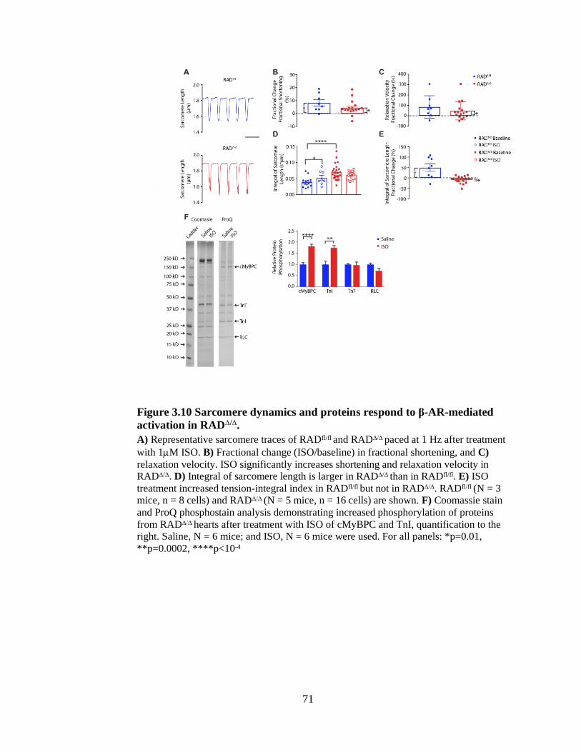
71
Figure 3.10 Sarcomere dynamics and proteins respond to β-AR-mediated
activation in RAD∆/∆.
A) Representative sarcomere traces of RADfl/fl and RAD∆/∆ paced at 1 Hz after treatment
with 1M ISO. B) Fractional change (ISO/baseline) in fractional shortening, and C)
relaxation velocity. ISO significantly increases shortening and relaxation velocity in
RAD∆/∆. D) Integral of sarcomere length is larger in RAD∆/∆ than in RADfl/fl. E) ISO
treatment increased tension-integral index in RADfl/fl but not in RAD∆/∆. RADfl/fl (N = 3
mice, n = 8 cells) and RAD∆/∆ (N = 5 mice, n = 16 cells) are shown. F) Coomassie stain
and ProQ phosphostain analysis demonstrating increased phosphorylation of proteins
from RAD∆/∆ hearts after treatment with ISO of cMyBPC and TnI, quantification to the
right. Saline, N = 6 mice; and ISO, N = 6 mice were used. For all panels: *p=0.01,
**p=0.0002, ****p<10-4

72
Supplemental Figure 3.11 Myocardial Rad deletion results in increased
calcium current (ICa,L).
A) Representative ICa,L traces for Vtest at -5 mV, with voltage protocol shown above. B)
and C) Amplitufe and rate of decay were measured using a fitted exponential function on
current traces. RADfl/fl was measured using a single exponential function. RAD∆/∆
required the sum of two exponential functions. B) Total amplitude was larger in RAD∆/∆
than in RADfl/fl at -5 mV. C) Rate of decay (t) was faster RAD∆/∆ than in RADfl/fl, both in
the fast and slow phases at -5 mV. Data displayed from RADfl/fl (7 mice, 18 cells) and
RAD∆/∆ (7 mice, 17 cells). ***p=0.0004, ****p<0.0001

73
Supplemental Figure 3.12 RAD∆/∆ ICa,L response after -adrenergic receptor
stimulation.
A) No difference in maximal conductance in RAD∆/∆ compared to RADfl/fl (p = 0.161). B)
Activation midpoint is also significantly negative-shifted in RAD∆/∆. Rate of decay of
current was measured using a fitted exponential function on current traces. RADfl/fl was
measured using a fitted exponential function. RAD∆/∆ had two different phases, and was
fitted best using two exponential functions. C) Fractional change in tau, in RADfl/fl at -5
mV (mean = 4%) and RAD∆/∆ (mean: slow = 2%; fast = 7%). Data displayed from
RADfl/fl (10 mice, n = 38) and RAD∆/∆ (8 mice, n = 49). *p = 0.04.

74
Supplemental Figure 3.13 RAD∆/∆ cellular response after β-adrenergic
receptor stimulation.
A) Amplitude of the transients from RAD∆/∆ are higher than in RADfl/fl. B) The upstroke
velocity is faster in RAD∆/∆ than in RADfl/fl. C) and D) Rate of decay is faster in RAD∆/∆
than in RADfl/fl. Measurements in A-C) used 5 mM Fura2 AM and measurements in D)
used 5 mM Fluo4 AM (p = 0.53). Data displayed from RADfl/fl (10 mice, n = 38) and
RAD∆/∆ (8 mice, n = 49). *p = 0.04, **p = 0.008

75
Supplemental Figure 3.14 No effect on sarcomere dynamics after treatment
with ISO.
A) Diastolic sarcomere length was not different after ISO between RADfl/fl and RAD∆/∆.
B) Percentage of fractional shortening was not different after ISO between RADfl/fl and
RAD∆/∆. C) Relaxation velocity was no different after ISO between RADfl/fl and RAD∆/∆.
D) Shortening velocity was not different after ISO between RADfl/fl and RAD∆/∆. Data
displayed from RADfl/fl (3 mice, 8 cells) and RAD∆/∆ (5 mice, 16 cells).
76
CHAPTER 4. RAD-GTPASE CONTRIBUTES TO HEART RATE VIA L-TYPE CALCIUM
CHANNEL REGULATION
4.1 Preface
The work of chapter 4 has been published [214] . The entirety of the published paper
is presented. The results and methods attributed to others are presented for clarity and flow
are italicized. The figure numbering of the chapter has been edited from the published work
for consistency within the dissertation.
4.2 Introduction
Spontaneous electrical activity of the sinoatrial node (SAN) initiates cardiac rhythm
and is a critical determinant of heart rate (HR). Cardiomyocytes of the SAN (SANcm) are
highly specialized cells that generate automatic activity and form the cellular basis for
cardiac pacemaking. SANcm exhibit a spontaneous diastolic depolarization. The early
phase of diastolic depolarization is dominated by inward cationic funny current (If [215]
carried by HCN4 [216]). The latter phase of diastolic depolarization is driven by voltage-
gated L-type calcium channels (LTCC, with the main pore forming subunit, CaV1.2 or
CaV1.3) [217] . To a large extent, If and the LTCC complex determine the ‘membrane
clock’ that interacts with the calcium clock in the SAN [218, 219]. The inter-relation of the
membrane clock and calcium clock is underscored by studies of the genetic model of loss
of CaV1.3 impairing sarcoplasmic reticulum Ca2+-release [220, 221], and by computational
studies linking feedback loops among sarcoplasmic Ca2+-release, NCX, and ICa,L [222,
223]. The ability of ICa,L to simultaneously contribute to membrane- and calcium-clocks

77
suggests that targeted regulation of the LTCC is a potentially powerful approach to control
HR. Although cellular and molecular mechanisms that contribute to pacemaker activity are
well established [224], bradyarrhythmia treatment options are limited mainly to electronic
devices that are insensitive to autonomic nervous system regulation. Experimentally,
nifedipine (an LTCC antagonist) has a negative chronotropic effect on the leading
pacemaker site of the SAN [225]; similarly, genetic knockout of CaV1.3 induces
bradycardia [226]. A fundamental premise of this study is that modulation of LTCC
activity conferred by Rad provides a central nodal mechanism for sympathetic nervous
system (SNS) control of SAN rhythm.
The LTCC is a heteromultimeric protein complex [194]. The pore-forming subunit
of the LTCC in SANcm is carried by CaV1.3 [226-228] and CaV1.2 [217] channels. Key
auxiliary subunits of the LTCC complex include CaVβ2, α2δ, and CaM [194]. Recent work
[117] highlights the importance of RGK proteins [166] as regulators of ICa,L via association
with the β subunits [17, 21, 24, 184]. Rad is a constituent of the LTCC complex in the
myocardium [17, 92]. Sympathetic nervous system drive acts via β-AR signaling cascades,
resulting in the activation of PKA which in turn modulates LTCC activity [229]. β-AR
modulation is maintained in transgenic mouse models expressing mutant CaV1.2 and CaVβ
that cannot be phosphorylated by PKA [91, 117], suggesting that a non-channel PKA
target is central to channel modulation. Using proximity biotinylation to analyze β-
adrenergic-dependent changes within the LTCC complex, Rad was recently shown to be
depleted from the LTCC complex following acute β-AR stimulation. Rad depletion from
the LTCC relieves the constitutive inhibition imposed by Rad association [17, 21, 24, 183].
In keeping with its presumptive role as an endogenous inhibitor of LTCC function, we

78
showed that LTCC activity is increased In Rad-null mice (Rad-/-) with properties mirroring
β-AR modulation of ICa,L [21]. ECG telemetry showed that Rad-/- mice have a complex
phenotype that includes differential vascular and inflammatory properties [184]. However,
this model has multi-organ involvement [172, 184] and possible development-related
effects that potentially confound analysis of heart rhythm modification originating from
channel modulation in cardiomyocytes. To circumvent these effects, we recently developed
an inducible myocardium-restricted Rad knockout (cRadKO) mouse [24]. Cardiomyocyte
selective Rad deletion was shown to phenocopy β-AR modulated LTCC properties,
increasing basal ventricular contractile function [24]. Using this same conditional KO
model, we now test the hypothesis that myocardial Rad-knockout will modulate sinus heart
rate. Here we show that cRadKO mice exhibit an elevated intrinsic heart rate, and elevated
sleep phase heart rate. Mechanistically, Rad regulation of HR appears to be driven by
modulated ICa,L following Rad loss in SANcm. This work reveals Rad – LTCC interactions
as a novel target for future therapeutics for symptomatic bradycardia.
4.3 Results
4.3.1 SANcm express Rad
In the cRadKO mouse model cre-recombinase is driven by a myosin heavy chain 6
promoter limiting expression to all cardiomyocytes (reference [185], and personal
communication Jeff Molkentin). Following tamoxifen-mediated cre-recombination Rad
protein levels are not detected from the whole hearts of cRadKO mice [24], suggesting
cardiomyocyte-restricted expression of Rad in the heart. Similarly, single nucleus RNAseq

79
(snRNAseq) analysis of the mouse SAN showed that RRAD transcript is enriched ~10-
fold in SAN cardiomyocytes (SANcm) compared to non-cardiomyocytes from the SAN
region [189]. However, the SAN contains a heterogeneous mixture of cardiomyocytes
[230]. To assess Rad expression among SANcm, we re-examined a proteomics and
snRNAseq dataset using tissue obtained from a segment of the right atrial wall, containing
SAN and surrounding tissue [189]. t-distributed stochastic neighbor embedding (t-SNE)
shows 203 (of 5,472) cells analyzed can be classified as SANcm (cardiomyocytes) based
on the expression of Myh6, Ctnna3, RyR2, Rbm20, Dmd, Ttn, and Tbx5 [189, 231]. In
addition to SANcm, clusters were classified as fibroblasts, epicardial, endocardial,
macrophage, adipocyte, and an unidentified cluster [189]. Rad RNA was heterogeneously
expressed in the SANcm cluster and enriched in a sub-population of SANcm. (Figure
4.1A). Within the SANcm cluster 38% of cells expressed RRAD transcript (77 of 203 cells).
Re-analysis of scRNAseq data from van Eif and colleagues [231] also showed RRAD
transcript in SANcm (data not shown). Analysis of the available proteomic database also
support SAN expression of Rad protein. For context we also re-analyzed data for
expression of HCN4, RyR2, and CaV1.2 (Figure 4.1B). To examine Rad expression in
SANcm in CTRL mice we performed RNA in situ hybridization. HCN4+ cells prominently
express Rad (Figure 4.2, upper panels). By contrast, cRadKO hearts show dramatically
reduced Rad transcript (Figure 4.2, middle panels). Consistent with our previously
published study [24] , tamoxifen induction of the floxed-Rad gene results in loss of Rad
from cardiomyocytes across the heart. Taken together, these new data suggest that Rad is
expressed in SANcm; thus, the loss of Rad might impact SAN function.

80
4.3.2 L-type calcium current (ICa,L) in SANcm
Cells dispersed from the SAN region of cRadKO were morphologically
indistinguishable from those of control mice (Figure 4.3A). Cells selected for recording
beat spontaneously in physiological salt solution, were spindle shaped (Figure 4.3A). Cell
capacitance was not different in cells from cRadKO mice (42+4pA/pF, n=17 and
42+4pA/pF, n=21 for CTRL and cRadKO, respectively; Figure 4.3B). SANcm from
cRadKO mice showed larger current density (Figure 4.3C) with the peak of the peak ICa,L
occurring at +5mV for CTRL and 0mV for cRadKO (Figure 4.3D). A Boltzmann fit of
the I(V) curves yielded maximal LTCC conductance showing a significant increase with
Rad loss (4.8+0.8pS/pF, n=17, and 10+1.3pS/pF, n=21, p=0.0015 for CTRL and cRadKO,
respectively, Figure 4.3E). The midpoint of current activation (V½) was -7.5+1.4mV for
CTRL and -10.1+1.4 for cRadKO (Figure 4.3F). Steady state inactivation was not different
between CTRL and cRadKO ICa,L (data not shown). There was no significant difference
in ICa,L properties (current density, maximal conductance, or voltage dependence between
females and males, Supplemental Figure 4.8). These data are consistent with alterations
observed in ICa,L from ventricular cardiomyocytes following Rad ablation [24], suggesting
a major contribution of Rad to basal ICa,L in SANcm.
A growing literature suggests that Rad, and other members of the RGK GTPase
subfamily, play critical roles in calcium-channel modulation, serving as constitutive LTCC
inhibitors, whose actions are regulated by protein phosphorylation [17, 92, 166]. In
agreement with this model, we previously showed that Rad deletion results in ventricular
ICa,L that phenocopies the current in wild-type cardiomyocytes following acute β-AR

81
activation by isoproterenol (ISO) [24]. We next tested the effect of acute ISO on ICa,L in
SANcm from CTRL and cRadKO hearts. ICa,L from CTRL SANcm was significantly
increased (Figure 4.4Ai). Maximal conductance increased (+2.8+0.9pS/pF; Figure 4.4Bi)
and the midpoint of activation shifted negative (-2.1+0.8mV; Figure 4.4Ci). By contrast,
ICa,L in SANcm from cRadKO hearts show no significant increase of conductance (-
0.9+1.2pS/pF; Figure 4.4Bii) and no effect on activation midpoint (-0.2+0.6mV; Figure
4.4Cii). These results are consistent with our earlier findings from cardiac ventricular
cardiomyocytes [21, 24], and support the working hypothesis that Rad functions as a key
mediator of -AR modulation of ICa,L.
4.3.3 RAD reduction increases intrinsic heart rate
Rad deletion instills a PKA modulated ICa,L under otherwise unstimulated
conditions in a cell autonomous fashion (Figure 4.3) [21, 23, 24]. ICa,L contributes to the
latter phase of the pacemaker depolarization in SANcm [224, 232-234] ; thus, modulated-
like ICa,L arising from Rad silencing predicts an elevated intrinsic SAN heart rate. To
examine the effect of Rad loss on HR in vivo, Radfl/fl.MHC mice were implanted with
radiotelemetry units. Measurements were made at baseline and 3 weeks after tamoxifen
administration to compare the impact of Rad loss on the same mice. Combined -AR-block
(propranolol) and cholinergic antagonist (atropine) revealed intrinsic HR from
ambulatory mice. Induced knockout of cardiomyocyte Rad resulted in a significant elevated
intrinsic HR (Figure 4.5A). In females, intrinsic HR increased 153+19bpm between
baseline and >3 weeks after tamoxifen (483+22bpm and 637+18bpm, for baseline and
post-tamoxifen, respectively, Figure 4.5B). Male mice also showed significantly elevated

82
intrinsic HR after tamoxifen (mean difference 63+12bpm; 502+8bpm and 565+11bpm, for
baseline and post-tamoxifen, respectively, Figure 4.5C).
4.3.4 Rad-deletion effects on HR are greatest when Sympathetic Drive is Relatively
Low
The autonomic nervous system (ANS) modulation of heart function is apparent as
the diurnal variation of HR. Sympathetic nervous system (SNS) drive is mediated in
cardiomyocytes via activation of -ARs. Figure 4.6A shows representative ECG traces
during the sleep (day) and active phases (night) along with a 48h continuous recording of
the moving average of HR at baseline and after Rad loss (Figure 4.6B). Sleep phase HR
was significantly elevated with cRadKO (535+15bpm and 593+13bpm, baseline and
cRadKO, respectively; Figure 4.6C) but the active phase HR was not different (652+8 and
671+10, baseline and post-tamoxifen, respectively; Figure 4.6D). Similar results were
obtained for male mice (sleep phase, 511+11bpm and 598+11bpm; active phase, 647+9
and 671+13bpm, baseline and post-tamoxifen, respectively; Figures 4.6E,F). Diurnal
variation remained, but the difference between active and sleeping phase HR diminished
after Rad silencing. Taken together these results are consistent with elevated ICa,L in
cRadKO SANcm driving more rapid sinus rhythm unmasked by reduced SNS drive during
the sleep phase.
We observed no evidence of pathological effects on the heart into senescence with
the knockout of Rad [22, 24]. Heart rate variability (HRV) reflects ANS input to the heart,
and a decrease of HRV is associated with pathologies including progression to heart failure
[235]. This coupled with a literature indicating that abnormal calcium homeostasis can

83
contribute to heart failure progression [236] motivated us to measure HRV. Cardiac Rad
deletion has no effect on HRV in either the active or sleep phases (mean differences
before/after tamoxifen 2.2+4ms and 1.5+6ms, for sleep and active phase, respectively;
Figure 4.7A,B). In our recently published work we noted that despite tonic modulated ICa,L,
the heart in cRadKO mice retains responsiveness to acute β-AR stimulation [24] consistent
with the function of a healthy heart. To test for retention of autonomic nervous control of
HR in cRadKO we measured the acute responses to a rapid air jet puff and exogenous ISO.
The rapid air jet activates the endogenous ANS ‘fight or flight’ response, whereas
exogenous ISO acutely over-rides ANS control of HR. The air jet acutely increased HR
+33+23bpm (593+13 to 623+24bpm; p=0.18), and HRV (39+6ms and 68+14ms, before
and after air-jet, respectively; Figure 4.7C) whereas exogenous ISO increased HR
+59+10bpm (674+11 to 733+8bpm, p<0.01), and reduced HRV (46+3ms and 18+2ms,
for post-tam and ISO, respectively; Figure 4.7D). Also, Poincare plots during the sleep
and active phases also showed similar patterns in CTRL and cRadKO (Figures 4.7E,F).
4.4 Discussion
We showed previously that Rad-deficiency confers a β-AR –modulated phenotype
on basal ICa,L without structural or functional remodeling of the heart [23]. The main
findings of this study are that in SANcm, the deletion of Rad results in elevated ICa,L with
properties approximating SNS modulated ICa,L. Second, the absence of Rad elevates
intrinsic HR. Third, HR in ambulatory mice is unchanged during the active phase, but is
significantly elevated during the sleep/resting phase. Early studies showed that modulated
ICa,L is a major contributor to an increased steepness of the diastolic depolarization of

84
SANcm [237]. Thus, these results support a key contribution of Rad – LTCC regulation of
sinus rhythm in the heart (Figure 4.9).
SANcm have relatively low membrane capacitance and high input resistances;
consequently, in theory, any relatively small inward cation flux, such as Ca2+ current, can
accelerate pacemaker depolarizations [234, 238]. The contribution of ICa,L to sinus rhythm
was experimentally validated by demonstration of the slower HR of CaV1.3 knockout mice
[226]. There has been some controversy in the field striving to attribute dominant
mechanisms to pace-making. For example, some studies suggested that the sarcoplasmic
reticulum Ca2+ clock shapes the spontaneous rate, but may not be a dominating factor [239].
Here we now show that ICa,L modulation by Rad GTPase regulates the intrinsic SAN firing
rate. Our findings do not challenge the primacy of the membrane versus Ca2+ clock; rather,
the interdependence of the membrane and Ca2+ clock [224] underscores the critical impact
of altered ICa,L on SANcm autonomous firing. Perhaps most importantly, our work shows
that Rad-LTCC regulation is a key molecular signaling node for SNS regulation of sinus
rhythm.
4.4.1 Rad is a key contributor to SANcm ICa,L modulation
SANcm express ICa,L carried by pore forming CaV1.2 and CaV1.3 proteins. CaV1.3
contributes to pacemaker activity [226] and regulates the calcium clock in nodal cells
[221]. CaV1.2 resides mainly in non-junctional plasma membrane where it is thought to
mainly contribute to the membrane clock [240]. Our approaches do not allow us to dissect
whether Rad has biased influence on CaV1.2 or CaV1.3. Liu et al showed that CaV1.2 and
CaV1.3 indistinguishably respond to PKA to increase ICa,L [92]. There are 3 independent

85
lines of evidence to support the contention that Rad functions in cardiomyocytes to regulate
ICa,L modulation. First, our early studies of global, constitutive Rad-KO mice showed Rad
deletion phenocopies acute stimulation of the heart [23], and in this model ICa,L was
modulated at baseline [21]. Second, in the inducible, cardiac-restricted RadKO (cRadKO,
as in the present study), ventricular ICa,L was modulated at baseline [24]. Third, proximity
proteomics evaluation in transgenic mice showed that following acute ISO stimulation,
Rad association was reduced with LTCC proteins CaVβ2 and CaV1.2 [92]. Taken together
with the present findings, we conclude that Rad interacts with LTCC; thus, dynamical
regulation by PKA of the LTCC heteromultimeric complex is an important contributor to
SNS elevation of HR. In native myocardium the signal for Rad-LTCC rearrangements is
likely in response to the β-AR –PKA signaling axis. The Rad knockout model provides an
extreme, whereby the absence of Rad would represent a maximally depleted CaV1.2
microenvironment, relieving any Rad-dependent inhibition with respect to LTCC function.
There is an imperfect correspondence between intrinsic HR and ICa,L by biological
sex in cRadKO (Supplementary Figure 4.8, Figure 4.3, 4.6). Intrinsic HR response in
cRadKO is greater in females than males, but there is not a significant difference in ICa,L
by sex. This finding raises new questions, principally, ‘What male-specific factors
contribute to lower intrinsic HR in the absence of Rad?’ This doesn’t necessarily mean
that Rad influences other ion channels or Ca-clock components in a sex-specific manner;
rather, this line of reasoning suggests that there are sex-specific intrinsic HR regulatory
mechanisms, exclusive of Rad-LTCC signaling. For example, our early work showing Rad
– ICa,L regulation established absence of effects on T-type calcium channel current [17].
Nevertheless, while Rad might directly influence other channels, to date the only known

86
channel interaction partners with Rad are CaVβ subunits [166] and the C-terminus of the
L-type channel [241]. There are a large number of signaling systems that sum to setting
intrinsic HR [242] any one of which possibly can be altered in a sex specific fashion. The
important conclusion is that regardless of sex, a common effect of Rad-deletion is increase
ICa,L along with intrinsic HR.
4.4.2 Relationship among SANcm heterogeneity, Rad expression, and SAN function
In rabbits, peripheral pieces of SAN tissue from regions bordering the crista
terminalis have faster firing rates than the central pacemaker site, and pieces bordering the
left atrium were quiescent [243]. The leading pacemaker site is not, however, a fixed
location. During autonomic nervous system signaling different firing rates are driven by
different cells in the SAN[244] and depending on external stimuli subsidiary areas can be
activated [245]. Cell-cell coupling creates complex behavior [246]. For this reason, it is
important to interpret cellular studies in context of tissue-level, or as in the present case in
vivo studies of SAN function. Our finding that 38% of SANcm express enriched Rad
transcript suggests an additional, new mechanism of heterogeneity that should be tested in
future studies. It follows that the SANcm sub-types that do not express Rad may not
possess adaptability to speed heart rate via a modulated ICa,L mechanism, or that other RGK
GTPases may play yet to be investigated regulatory roles. Conversely, Rad-expressing
SANcm theoretically have the ability to increase rate and shape the ultimate overall SAN
rate because the fastest firing SANcm should determine rate, which is consistent with the
elevated HR in cRadKO mice. Future studies employing sophisticated in situ single cell

87
mapping might help resolve whether Rad-ICa,L contributes to anatomical pacemaker shifts
during sympathetic nervous system activation.
Modifiers of sinus rhythm may extend beyond regulation of HR per se, to secondary
impacts on heart failure evidenced as promising effects of ivabradine in heart failure
patients [247]. Moreover, β-AR blockers have been employed as a first line of HF treatment
[248]. In the present study we show Rad reduction increases HR, and we previously
showed Rad reduction increases cardiac inotropy without pathological consequences [21-
24]. Thus, the suggestion that Rad knockout provides beneficial effects in the setting of HF
is paradoxical to HR slowing effects of ivabradine and β-AR blockers; however, Rad
reduction would directly treat loss of cardiac inotropy in failing heart, and thus we maintain
is worthy of further study.
4.4.3 Diurnal impact and ANS drive with Rad reduction
Rad deletion elevates intrinsic HR consistent with the SANcm origin of the increased
sinus rhythm. ANS net input is the sum of SNS drive and parasympathetic input, and the
level of ANS drive is regulated by the baroreflex. With elevated HR, hence elevated cardiac
output in cRadKO, it follows there should be compensatory reduced SNS drive. More
direct measures of sympathetic nerve activity and circulating catecholamines are necessary
to examine this issue, yet are highly invasive and can be difficult to interpret secondary to
agitation of animals. For example, blood cannot be drawn from conscious mice without
provoking a fight or flight response. Anesthesia can be used, but dramatically depresses
measurements of circulating catecholamines (for example, compare [249] to [250]). To
summarize, our data support the notion that we are not ‘knocking-out’ SNS input to the

88
cRadKO heart, rather we suggest that modulated ICa,L in cRadKO dominant pacemaker cell
may usurp the reflexive requirement for added SNS to balance net autonomic drive. In this
way, Rad reduction strategies might aid aging related [251, 252] symptomatic bradycardia
which has been attributed to depressed SANcm excitability, possibly secondary to declines
of CaV1.2 protein [225].
4.5 Limitations
Heterogeneity of function at the cellular level is not fully explored here. We did not
measure spontaneous AP nor spontaneous firing frequencies. Many of the dispersed SAN
cells displayed irregular automatic beating or quiescence. A range of spontaneous activity
from fast, regular activity to irregular to quiescence has been noted in dispersed SANcm
[253]. Heterogeneity among SANcm is also demonstrated in single nucleus[189] and tbx3+
cell-sorted transcriptomics studies [231]. It is also possible that differential dispersed cell
activity might arise from differential cellular damage artifact during enzymatic dispersal.
We also did not consider Rad-deletion effects on non-LTCC targets. If is a major
contributor to sinus heart rate [215, 218, 219], and while there is no evidence for Rad
interactions in the cardiomyocyte beyond protein interactions of with LTCC-component
proteins, we cannot completely rule out other interactions with Rad. Studies here are
limited to the mouse. In future studies it will be important to test the impact of Rad
reduction on human or other large mammalian SANcm retained in ex vivo tissue
preparations.

89
4.6 Conclusions
In summary, we have demonstrated that ICa,L contributes to sinus rate by a
mechanism including Rad-regulated control of LTCC [24, 92]. We previously showed that
Rad- ablation promotes a beneficial elevation in Ca2+ dynamics, stimulating heart
ventricular function [23], albeit without any apparent pathological consequence [24].
Taken together, Rad reduction within all cardiomyocyte types is a powerful potential
therapeutic approach for increasing cardiac output as it simultaneously increases HR and
stroke volume. Future novel therapeutics for symptomatic bradycardia targeting Rad –
LTCC may elevate HR while retaining βAR responsiveness.

90
Figure 4.1 RRAD is expressed in SANcm.
A) Two-dimensional t-SNE (Stochastic Neighbor Embedding) plot outlines the
major populations of the sinus node. Analysis of database from Linscheid et al.
2019. Each point represents a single nucleus. Cell populations are colored
according to intensity of RRad expression, with an expanded view of the SANcm.
B) Quantification of data mined from Linscheid et al. 2019, of the expression of
Rad protein in relation to other proteins known to be expressed specifically in
SANcm (HCN4) and cardiomyocytes in general (RyR2 and CaV1.2).

91
Figure 4.2 RAD is expressed in SANcm.
Representative images of single-molecule fluorescence in situ hybridization against Rad
(green) and HCN4 (red) in CTRL (top images) and cRadKO (middle images) in SANcm.
Lower panel, probe for bacterium DapB gene serves as a background control (bottom
images). Heart tissue was collected >2 weeks after tamoxifen treatment. Tissue samples
were stained with DAPI (blue). Images of channels merged displayed on the right.
Representative of 3 CTRL and 3 cRadKO hearts. Scale bar: 20 mm.

92
Figure 4.3 Rad reduction increases SANcm ICa,L.
A) Representative photomicrographs of SANcm selected for patching studies
from CTRL (Ai) and cRadKO (Aii) hearts. B) Cell capacitance is not significantly
different (p = 0.77). C) Family of ICa,L for CTRL (blue) and cRadKO (red). Scale
bars: 5 pA/pF, 100 ms. D) Current-voltage relationship for peak ICa,L from CTRL
and cRadKO SANcm. E) Maximal conductance is significantly increased (**p <
0.01) and F) activation midpoint trends toward a negative shift with Rad deletion
(p=0.2). N = 8, n = 17 for CTRL; N = 9, n = 21 for cRadKO.

93
Figure 4.4 β-AR stimulation has no significant effect on cRadKO SANcm
ICa,L.
A) Current-voltage relationships for CTRL (Ai) and cRadKO (Aii) for basal
(closed symbols) and after 100 nM ISO (open symbols). B) Maximal conductance
before and ISO for CTRL (Bi, **p =0.01) and cRadKO (Bii, p = 0.45). C)
Activation midpoint before and ISO for CTRL (Ci, *p =0.05) and cRadKO (Cii, p
= 0.68). N = 5, n = 9 for CTRL; N = 4, n = 9 for cRadKO.

94
Figure 4.5 Intrinsic heart rate increases after Rad deletion.
A) Representative ECG signals during the sleep phase demonstrate an increase in
intrinsic heart rate after tamoxifen treatment. B,C) Heart rate increases after
tamoxifen treatment in B) females (***p < 10-3, N=7 mice) and in C) males (**p
= 0.002, N=7 mice).

95
Figure 4.6 Rad deletion increases heart rate in the sleep phase.
A) Representative ECG signals demonstrate an increase in heart rate during sleep
after tamoxifen treatment, but no change while active. B) Continuous recording of
heart rate over 48 hours, before (Baseline) and after tamoxifen treatment (Post-
TAM). C) Heart rate in the sleep phase increases after tamoxifen treatment in
females (***p < 10-3, N = 7 mice). D) No significant difference in heart rate
during the active phase after tamoxifen treatment in females (p = 0.13). E) Heart
rate during the sleep phase increases after tamoxifen treatment in males (**p =
0.0003, N = 7 mice). F) Heart rate in the active phase increases after tamoxifen
treatment in males (*p = 0.05).

96
Figure 4.7 Rad deletion demonstrates preserved Heart Rate Variability
(HRV).
A) HRV is not significantly different after tamoxifen treatment in the sleep phase
(p = 0.59), or B) in the active phase (p = 0.80). C) HRV increases in Post-TAM
after exposed to an airjet (*p = 0.03). D) HRV increases Post-TAM after an
injection of isoproterenol (***p < 10-3, N = 7 mice). E), F) Poincare plots in
which the RR interval is plotted against the subsequent RR interval (RR + 1) in a
representative mouse during the sleep phase E) and during the active phase F).

97
Supplemental Figure 4.8 No significant difference between males and females
of ICa,L in CTRL and cRadKO.
A) Current-voltage relationship for peak ICa,L from CTRL males (blue circles) and
females (black circles). B) Current-voltage relationship for peak ICa,L from
cRadKO males (red squares) and females (black squares). C) Maximal
conductance for CTRL (males and females, p = 0.10) and cRadKO (males and
females, p = 0.10). D) Activation for CTRL (males and females, p = 0.62) and
cRadKO (males and females, p = 0.34). CTRL: Males: N = 6 mice, n = 12 cells;
Females: N = 2 mice, n = 5 cells. cRadKO: Males: N = 8 mice, n = 19 cells;
Females: N = 2 mice, n = 7 cells.

98
Figure 4.9 Rad Deletion Increases Sinus Heart Rate.
Rad deletion in sinoatrial cardioyocytes increases ICa,L and increases heart rate consistent
with -adrenergic receptor modulated ICa,L regulation. Heart rate during the sleep cycle is
selectively elevated by Rad deletion. Rad-LTCC association may be a useful target for
future therapeutics to treat symptomatic bradycardia.

99
CHAPTER 5. ENHANCED INACTIVATION BALANCES INCREASED PEAK L-TYPE CALCIUM
CURRENT IN THE ABSENCE OF RAD
5.1 Preface
The work of chapter 5 has been submitted for review . The entirety of the paper is
presented. The results and methods attributed to others are presented for clarity and flow
are italicized. The figure numbering of the chapter has been edited from the submitted work
for consistency within the dissertation.
5.2 Introduction
The L-type calcium channel (LTCC) is a heteromultimeric protein complex that
governs the entry of calcium necessary to initiate a contraction, and the LTCC contributes
to the cardiac action potential (AP) plateau phase [64]. Cardiomyocyte LTCC activity is
tightly regulated to prevent calcium overload [254] and after-depolarizations of the AP
[124, 125]. Decay kinetics of ICa,L is a key point of control. Inactivation of the LTCC occurs
by two distinct mechanisms: calcium dependent inactivation (CDI) and voltage dependent
inactivation (VDI) [64, 65]. The early peak of L-type calcium current (ICa,L) triggers
calcium induced calcium release (CICR) from the sarcoplasmic reticulum (SR) that is
essential for excitation contraction coupling; calcium released from the SR, or entering
through the channel itself, binds to calmodulin (CaM) prebound to the EF hand of the
carboxyl-terminus of the main pore-forming subunit of the LTCC [255]. Ca2+-CaM
complexed within the LTCC causes a conformational change to induce CDI [68-71]. In
addition, ICa,L decay occurs by voltage dependent inactivation contemporaneously with the
plateau phase of the AP [60, 74-76]. Under basal conditions, the decay of ICa,L demonstrates

100
non-exponential decay with relatively early, fast and late, slow components [50, 61-63]. It
is generally accepted that CDI contributes to the early, fast component and the late, slow
component is dominated by VDI [60, 66]. Other studies claim that VDI contributes to both
components but is dampened when calcium influx increases [60, 74-76].
The regulation of LTCC decay becomes particularly critical when the channel is
modulated upon -adrenergic receptor (-AR) stimulation. During the fight-or-flight
response, ICa,L amplitude increases, voltage dependence of activation shifts to more
negative potentials, and inactivation rates accelerate [60, 66, 256, 257]. Regulation of the
LTCC by -adrenergic receptor (-AR) stimulation has been thoroughly investigated, yet
complete understanding remains elusive, especially in regards to how inactivation is
accelerated. Many studies claim that CDI is the major contributor to ICa,L decay under
modulated conditions because elevated calcium influx leads to more calcium interaction
with CaM [60, 66]; however, increased ICa,L due to overexpression of 1C, the pore-
forming subunit of the LTCC, or of CaV2a did not alter decay kinetics compared to the
controls [12, 149]. Others argue that inactivation is accelerated due to interactions of the
N-terminus and C-terminus with each other or other proteins [258-260]. Because -
adrenergic receptor (-AR) stimulation initiates multiple molecular mechanisms, it is
difficult to assess the main contributors to LTCC regulation [31].
In recent studies, Rad has emerged as a major contributor to LTCC modulation [23,
24, 92]. Rad is a member of the RGK subfamily of Ras-related small GTPases [166], and
shares the common RGK protein property of profoundly inhibiting CaV1/ CaV2 channel
activity. Overexpression of Rad in heterologous expression systems potently inhibits ICa,L
in adult and embryonic ventricular myocytes [16-20]. In vivo, Rad deficiency promotes

101
positive inotropy and is a critical determinant of current amplitude [24] and modulation
[23, 92]. Our recent work showed that the absence of Rad from the channel complex results
in increased LTCC activity that mirrors -AR modulated ICa,L and accelerates ICa,L decay
[21-24]. These findings were definitively supported utilizing proximity biotinylation to
show that under conditions of -AR stimulation, protein kinase A-mediated Rad
phosphorylation alleviated constitutive inhibition caused by Rad association with the
LTCC [92].
If Rad loss from the LTCC complex is essential for -AR modulation of ICa,L, then
the absence of Rad in myocardium could serve as a novel tool to investigate LTCC
regulation under modulated conditions without interfering contributions of other signaling
pathways normally activated under -AR stimulation. Using a cardiomyocyte-restricted
Rad deletion mouse model, we re-visit the hypothesis that modulation of the LTCC by -
AR stimulation is governed by the presence of Rad in the CaV1.2 macromolecular complex.
Mechanistically, the absence of Rad yields increased inactivation to offset the increase in
peak ICa,L. Here, we show that CDI and VDI are both accelerated in the absence of Rad.
The combination of greater peak and accelerated kinetics conspires to prevent significant
changes to action potential duration or QT interval, despite large peak ICa,L. The
preservation of Rad knockout-instilled ICa,L modulation in the absence of -AR expression;
thus showing that Rad imparts channel complex modulation independent of -AR
signaling. This work suggests Rad-LTCC interactions may be a novel target for future
therapeutics to confer systolic advantage.

102
5.3 Results
5.3.1 cRadKO mirrors modulated LTCC
ICa,L decay consists of an early, fast component and a late, slow component [50, 61-
63, 65]. CDI is the dominant mechanism of inactivation of the fast component and under
conditions of β-adrenergic stimulation [61, 68, 71, 74]; this idea, coupled with the emergent
model that phosphorylated Rad is re-oriented from the LTCC during -adrenergic
stimulation to drive ICa,L modulation [21, 23, 24, 92], predicts that Rad-deletion (in the
basal state) should yield ICa,L properties that are not different from wild-type (CTRL)
following -adrenergic modulation. We therefore compared the kinetics of ICa,L in
cardiomyocytes from CTRL after the addition of isoproterenol with basal ICa,L in
cardiomyocytes from cRadKO (Figure 5.1). For brevity, we will henceforth refer to CTRL
ICa,L and cRadKO ICa,L as a stand-in for ICa,L from CTRL or cRadKO ventricular
cardiomyocytes. EGTA was used to allow a readout of the influence on global Ca2+. ICa,L
traces from cRadKO and ISO-stimulated CTRL normalized to peak current demonstrates
that basal cRadKO ICa,L decay is not different from modulated CTRL (Figure 5.1A).
Remaining early current (r30) was not different between modulated CTRL ICa,L and
cRadKO ICa,L (Figure 5.1B; Supplemental Figure 5.10A for 2-way ANOVA tabular
results); similarly, there was no difference in the late component (r150) [50, 63, 261, 262],
at individual voltage steps (Figure 5.1C, Supplemental Figure 5.10B for 2-way ANOVA
tabular results). cRadKO displayed larger current density at r30 (Figure 5.1E) and r150
(Figure 5.1F) compared to CTRL. Taken together, these results demonstrate that the
kinetic signature of basal cRadKO approximates modulated ICa,L kinetics.

103
5.3.2 Increased Ca2+ and Rad both contribute to CDI
If CDI is the dominant mechanism when the LTCC is modulated, then the source
of calcium contributing to CDI could emanate from the sarcoplasmic reticulum, or from
local calcium entering through the channel itself [263]. To evaluate global versus local
Ca2+, we investigated ICa,L decay kinetics in the presence of either EGTA or BAPTA. These
calcium buffers have similar steady-state binding affinities for calcium, but differ in
binding rate constants [264]. EGTA buffers slower than BAPTA and allows investigation
of the influence of global calcium on CDI; by contrast, the use of BAPTA limits CDI to
local sources, mainly Ca2+ fluxing in the nanodomain of the channel [264, 265].
Figure 5.2 summarizes the analysis of r30 between cRadKO ICa,L and CTRL ICa,L
under conditions reporting global Ca2+ (EGTA used as calcium buffer). Traces from
cRadKO and CTRL ICa,L demonstrate that cRadKO ICa,L fast decay is greater than that for
CTRL (Figure 5.2A). Across multiple voltage steps, there is less r30 in cRadKO ICa,L than
in CTRL ICa,L (Figure 5.2B, Supplemental Figure 5.10C for 2-way ANOVA tabular
results). Current density was larger in cRadKO compared to CTRL (Figure 5.2C). To
assess whether remaining current depended on elevated current density, we plotted r30 as a
function of current density (Figure 5.2D). CTRL trends towards a negative slope meaning
that as current density increases, there is less remaining current, as expected for early, fast
inactivation by CDI [60]. In contrast, cRadKO has a positive slope; as current density
increases, there is more remaining current. This finding suggests a Ca2+-independent
contribution to the early, fast component of decay in a Rad-less LTCC.

104
We next assessed r30 in the presence of BAPTA to limit the pool of calcium local
to the channel complex [264, 265] (Figure 5.3). Representative traces reveal an apparent
smaller difference in decay between cRadKO and CTRL (Figure 5.3A). r30 across
individual test potentials demonstrates no significant difference between cRadKO and
CTRL ICa,L (Figure 5.3B, Supplemental Figure 5.10D for 2-way ANOVA tabular
results). In the presence of BAPTA, cRadKO maintains a larger current density compared
to CTRL (Figure 5.3C). Local buffering by BAPTA eliminates the negative correlation
of r30 with current density for CTRL ICa,L; however, the positive correlation of cRadKO
r30 and current density remains in the presence of BAPTA (Figure 5.3D, compare with
Figure 5.2D). Taken together these results suggest that the early, fast decay of ICa,L in
cRadKO is driven in part by SR released calcium, and in part by a Ca2+-independent
mechanism.
5.3.3 Rad does not alter the late component of LTCC decay
Late ICa,L contributes to repolarization and SR calcium loading [50]; impaired
inactivation of the late phase can result in AP duration prolongation and EADs [50, 61-63,
262]. We therefore analyzed ICa,L r150 in cRadKO and CTRL in the presence of EGTA.
Representative traces demonstrate no difference between cRadKO and CTRL ICa,L (Figure
5.2A, superimposed star). There was no difference in r150 between the models at multiple
test potentials (Figure 5.4A, Supplemental Figure 5.10E for 2-way ANOVA tabular
results) but the interaction between voltage and CTRL versus cRadKO was significantly
different, as shown by the crosses in Figure 5.4A. cRadKO, in comparison to CTRL, has
larger current density at r150 (Figure 5.4B). A positive correlation is seen in both cRadKO

105
and CTRL when r150 is displayed as a function of current density (Figure 5.4C). In the
presence of BAPTA, there was no difference in r150 (see Figure 5.3A, superimposed star;
Figure 5.4D, Supplemental Figure 5.10F for 2-way ANOVA tabular results). Current
density was not different for cRadKO compared to CTRL ICa,L (Figure 5.4E). r150 plotted
against current density demonstrated a positive correlation in both cRadKO and CTRL
(Figure 5.4F). These data show that Rad deletion does not promote alterations to late ICa,L
kinetics.
The late, slow component of ICa,L decay is thought to be dominated by voltage
dependent inactivation (VDI) [60, 66, 74]. We therefore measured VDI in cRadKO by
using barium as the charge carrier (Figure 5.5). Representative traces demonstrate faster
decay in cRadKO compared to CTRL both at r30 and r150 (Figure 5.5A). There was less
r30 in cRadKO (Figure 5.5B,D; Supplemental Figure 5.11A for 2-way ANOVA tabular
results for B). At r150, remaining current in cRadKO was not significantly less compared
to CTRL (Figure 5.5C,E; Supplemental Figure 5.11B for 2-way ANOVA tabular results
for C). A positive correlation is seen in cRadKO when either r30 or r150 are plotted as a
function of current density (Figure 5.5F,G). This data, in concert with the regression plots
from Figures 5.2 and 5.3, show that the absence of Rad enhances VDI. This suggests the
presence of Rad contributes to dampening VDI.
5.3.4 Faster kinetics in cRadKO prevent alterations in cardiac electrophysiology
ICa,L plays a critical role in regulating cardiac action potential duration; slight
alterations to current density or inactivation of the LTCC can lead to electrical
dysfunction, such as action potential prolongation, early after depolarizations, and

106
prolonged QT intervals [50, 61-63, 131, 132, 262]; Sympathetic stimulation via the -AR
signaling axis modulates trigger calcium, and shortens action potential duration [266].
Studies have shown that female mice exhibit prolonged action potential repolarization
and longer QT intervals [267], making females more sensitive to reveal if Rad deletion
has an effect on action potential duration and QT interval. We therefore measured action
potentials from isolated ventricular cardiomyocytes, and used surface electrocardiogram
(ECG) recordings to assess QT interval in female mice. Action potentials (AP) measured
at 1 Hz from CTRL ventricular cardiomyocytes showed a pronounced inflection point
positive to 0 mV (Figure 5.6A, arrow), rapid repolarization, and a slower repolarization
phase negative to -50 mV (Figure 5.6A). cRadKO APs (Figure 5.6A, red line)
demonstrated similar morphology to CTRL at baseline (Figure 6A, blue line) and after
addition of isoproterenol [268] (Figure 5.6B; cRadKO: black line, CTRL: green line).
Baseline AP amplitudes were not significantly different between models (Figure 5.6C;
cRadKO: 127.41.0 mV, CTRL: 123.03.3 mV;). Comparing baseline action potential
duration at 50% repolarization (APD50) and at 80% repolarization (APD80) demonstrates
no significant difference between cRadKO and CTRL (Figure 5.6D: cRadKO: 6.50.98
ms, CTRL: 6.20.58 ms). At 80% repolarization (APD80), cRadKO demonstrates faster
repolarization than CTRL (Figure 5.6E, cRadKO: 17.11.5 ms, CTRL: 21.21.1 ms).
CTRL action potential amplitude was not significantly different from cRadKO after ISO
(Figure 5.6F). cRadKO APD50 was slightly shorter compared to baseline, but not
significantly different from CTRL (Figure 5.6G; APD50 after ISO: cRadKO: 6.41.2 ms,
CTRL: 6.00.71 ms). CTRL was longer at APD80 compared to baseline, but not

107
significantly different from cRadKO (Figure 5.6H; APD80 after ISO: cRadKO: 18.01.8
ms, CTRL: 23.42.0 ms).
We next assessed in vivo electrical function. The increase in heart rate in response
to sympathetic stimulation is the result of integrative mechanisms in both branches of the
autonomic nervous system (ANS) [266], which could obscure a potential effect on QT
interval due to a selective increase in ICa,L in the absence of Rad. We therefore measured
the ECG under ANS block by treating the mice with atropine and propranolol [269].
Baseline ECG measurements demonstrated no significant difference in QT or QTc
(Supplement Figure 5.12). After administration of atropine and propranolol,
representative surface ECG traces showed no apparent differences between CTRL and
cRadKO (Figure 5.6I). Raw QT interval (Figure 5.6J; cRadKO: 406 ms, CTRL: 507
ms; p=0.35) and corrected QT interval (QTc) demonstrated no significant difference
(Figure 5.6K; cRadKO: 335 ms, CTRL: 405 ms; p=0.41). This data, combined with
the action potential measurements, support the idea that the faster kinetics balance the
increase in peak ICa,L, thereby preventing significant alterations in cardiac
electrophysiology.
5.3.5 cRadKO yields modulated kinetics of LTCC downstream of -AR signaling
1-AR activation mediates positive chronotropic and inotropic responses of
cardiomyocytes, and 2-AR is thought to contribute to these processes more locally
within cardiomyocytes [270]. To determine whether these receptors were contributing to
the modulation effect of ICa,L seen in cRadKO, we bred RADfl/fl-MHC-MCM onto a 1,2-AR

108
double knockout mouse [271] (dKO) to create an induced RAD/,1,2-AR triple knock
out mouse (tKO) after administration of tamoxifen. Analysis of cardiac function through
echocardiography demonstrated that ejection fraction was significantly different between
CTRL and dKO after acute isoproterenol, demonstrating that 1,2-AR response was
disrupted (Supplement Figure 5.12A; CTRL: 931%, dKO: 603%; p<0.0001; data for
CTRL+ISO echocardiography taken from our previously published dataset [24];
Supplement Figure 5.13A). Disruption of Rad expression in the heart in tKO was
confirmed by qRT-PCR (Supplement Figure 5.13B). After the deletion of Rad, ejection
fraction significantly increased in the tKO compared to dKO (Figure 5.7A,B; tKO:
722%, dKO: 514%; p<0.0001). Left ventricular inner dimensions decreased in the tKO
(Figure 5.7C; tKO: 3.60.1 mm, dKO: 4.20.1 mm; p=0.003) without significant
changes in the anterior or posterior walls (Figure 5.7D,E; For F: tKO: 0.880.1 mm,
dKO: 0.800.1 mm; p=0.43; For G: tKO: 0.860.05 mm, dKO: 0.820.1 mm; p=0.52).
Surface ECG showed no significant difference in raw QT interval (Figure 5.7F,G; tKO
394 ms, dKO 306 ms; p=0.19), corrected QT interval (QTc) (Figure 5.7H; tKO 314
ms, dKO 234 ms; p=0.14) or RR interval (Figure 5.7I; tKO 314 ms, dKO 234 ms;
p=0.14) between dKO and tKO female mice. This data supports that the absence of Rad
enhances cardiac function independent of 1,2-AR signaling.
At the level of channel current, peak ICa,L was significantly increased in tKO
compared to dKO (Figure 5.8A,B). Rad deletion also resulted in a greater maximal
conductance (Figure 5.8C,D; Gmax: tKO: 50833 pS/pF, dKO: 23032 pS/pF;
p<0.0001) and a negative shift in activation midpoint (Figure 5.8E; tKO: -13.12.4 mV,
109
dKO: -6.121.7 mV; p=0.04). Analysis of kinetics demonstrated a trend of less
remaining current in the tKO at r30 (Figure 5.8F,G,I,) and no difference at r150 (Figure
5.8F,H,J). The interaction of voltage and dKO versus tKO was significantly different
both at r30 and r150 (crosses in Figure 5.8G,H). Taken together, these results support the
hypothesis that the deletion of Rad increases peak ICa,L that mirrors modulated LTCCs
without stimulation of 1,2-AR signaling.
5.4 Discussion
The main findings of this study are that in the absence of Rad, increased peak L-
type calcium current (ICa,L) is balanced by accelerated calcium dependent inactivation
(CDI) and voltage dependent inactivation (VDI) (Figure 5.9). Accelerated LTCC
kinetics prevent significant action potential and QT interval prolongation. In fact, APD80
is shorter in cRadKO (Figure 5.6). The loss of Rad from the L-type calcium channel
(LTCC) complex results in modulated ICa,L independent of -adrenergic receptor (-AR)
signaling. To our knowledge, the cardiomyocyte-restricted Rad deletion model is the
only known model to increase ICa,L without disrupting calcium homeostasis, and without
promoting pathological signaling [12, 149]. Therefore, myocardial Rad-LTCC interaction
is a potential therapeutic target for positive inotropy while preserving -AR signaling
cascades beyond the LTCC.

110
5.4.1 Rad contributes to regulation of LTCC inactivation
A major implication of this study is the presence of Rad slows inactivation of the
LTCC. The deletion of Rad accelerated only the early, fast component of ICa,L decay
without significantly altering the late, proarrhythmogenic component [50, 61-63, 262]
(Figure 5.1-5.5). CaV1.2 channels have been shown to form clusters through physical
interactions of their C-terminal tails [57-59, 272]. Calcium binding to calmodulin (CaM)
on the LTCC promotes the physical interactions between channels, allowing channels to
have synchronous activity [58]. This process is thought to occur in response to -AR
stimulation in order to enhance calcium influx through calcium dependent facilitation
(CDF), but also to accelerate inactivation through CDI to prevent the development of
arrhythmogenic events [59]; hyperactivation of a few channels could dictate the activity
of the entire cluster [57, 58]. Rad, as well as the other members of the RGK protein
family, bind to CaM in a calcium dependent manner via a conserved binding site on their
C-termini [166]. It has been well established that CDI occurs when calcium binds to
CaM and causes a conformational change to induce LTCC inactivation [68-71]. With
CaM playing a major role in CDI and channel clustering, and Rad binding to CaM, it is
therefore possible that Rad contributes to channel clustering and calcium dependent
inactivation. In the absence of Rad, more channels may cluster together, thereby
promoting an increase in peak ICa,L [24] and an acceleration of the fast component of ICa,L
decay (Figure 5.1-5.3). Future studies investigating clustering in the absence of Rad
might give further insight into how the LTCC undergoes CDF and CDI.

111
5.4.2 Rad is a key contributor to LTCC Modulation
The activation of protein kinase A (PKA) downstream of -AR stimulation results
in an increase in ICa,L, an increase in channel open probability, and an increase in the
number of functional channels [109, 256], also known as ‘modulation’ of the LTCC.
Phosphorylation of CaV1.2 was long thought to be the underlying mechanism of
modulation [31], but recent studies have proposed that the mechanism of modulation
relies more on protein-protein interactions within the LTCC or on LTCC clustering [24,
31, 57-59, 92, 109, 272]. It was also recently shown that Rad is essential for cAMP-PKA
regulation of CaV1.2 [92]. Our studies have demonstrated that Rad-LTCC interaction is a
key molecular signaling axis for LTCC modulation both in the ventricle and in sinoatrial
node cardiomyocytes [24, 214] (see Chapter 4), and that the absence of Rad from the
complex yields modulation of the LTCC. Our current findings expand on this through
kinetic analysis, demonstrating that CTRL ICa,L stimulated with isoproterenol decayed in
a manner that was indistinguishable from cRadKO ICa,L (Figure 5.1). When treated with
isoproterenol, channels may form “super-clusters” along the t-tubules to amplify Ca2+
influx [59] and modify kinetics. The absence of Rad could promote these “super-
clusters” to form under basal conditions, which could in turn promote more calcium
induced calcium release from the sarcoplasmic reticulum to feedback onto the clusters to
then accelerate CDI. The “super-clusters” that form in response to sympathetic
stimulation via the -AR axis signaling modulates trigger calcium, which could shorten
action potential duration [266]. At the tissue level, sympathetic drive reduces transmural
dispersion of action potentials [273]. If ICa,L is selectively increased, all else remaining

112
equal, the resulting ventricular action potential duration would prolong, appearing on the
ECG as a longer QT interval [274]. In cRadKO cardiomyocytes, increased LTCC peak
current is offset by faster kinetics; thus, raising the possibility that the net effect of Rad
loss on cardiac electrophysiology for action potential duration and QT prolongation is
nullified [21] (Figure 5.6,5.9).
An alternate idea stems from Findlay who proposed that modulation of LTCC
from -AR stimulation resulted in a switch from VDI to CDI. Under basal conditions, it
was thought that CDI contributed to decay at more negative potentials, while VDI
dominated as the membrane became more depolarized. Findlay and others showed that
VDI has a biphasic decay, and that rapid VDI contributes to the fast component of ICa,L,
not just CDI [60, 74-76, 275]. However, in the presence of isoprenaline, CDI dominated
at all voltages, and VDI was suppressed. Findlay’s prediction was that CDI would then
have little contribution to the action potential under basal conditions, but could promote
severe slowing of ICa,L decay and therefore elongation of the action potential after -AR
stimulation [60]. Our results demonstrate that the absence of Rad accelerates VDI both in
the fast and slow components of ICa,L decay (Figure 5.5). This does not result in
elongation of the action potential nor does it significantly prolong the QT interval
(Figure 5.6). It is therefore possible that Rad contributes to LTCC regulation by allowing
this switch between VDI and CDI. Under basal conditions, the presence of Rad regulates
VDI; when -ARs are activated, Rad phosphorylation causes dissociation from the LTCC
[92] and CDI then becomes the dominant inactivation mechanism as channels cluster
together and elevate intracellular calcium. The synchronous activity of these clusters then
promotes accelerated CDI so that the increase in ICa,L is offset so as not to significantly

113
alter action potentials. Further studies into how phosphorylation of Rad alters its location
within the complex or its interactions with other proteins could help elucidate how Rad
contributes to LTCC inactivation, especially under conditions of -AR stimulation.
5.4.3 Rad regulation of LTCC is independent of -adrenergic receptor signaling
Our work has demonstrated that Rad is a critical component in LTCC modulation
by -AR stimulation [21, 23, 24]. Indeed, cardiomyocyte-restricted Rad-deletion
demonstrates the extreme by which the loss of Rad yields maximally modulated ICa,L that
translates from channel function up to enhanced, in vivo cardiac function. Data collected
from the 12-AR x Rad-triple knockout demonstrate that the maximal modulation of
ICa,L is due to the absence of Rad from the LTCC, exclusive of activation of -ARs
(Figure 5.7-5.9). Others who have utilized 1-AR knock out, 2-AR knock out, or 12-
AR double knock models have reported minimal impacts on basal cardiac function,
suggesting that there are potentially other controls for critical physiological functions,
such as cardiac rate and contractility, to compensate for the loss of these receptors [187,
270, 276-279]. In 12-AR double knockout neonatal myocytes, 3-AR was shown to
stimulate contraction after treatment with isoproterenol, though desensitization occurred
more rapidly compared to 1-AR or 2-ARs [279]. It is therefore possible there is a
minor contribution from other sources to how the heart responds to increased sympathetic
drive. However, our data clearly demonstrates a significant increase in function both at
the level of whole heart and at the level of the channel when Rad is deleted in the 12-
AR double knock out without significant prolongation of the QT interval. We have
previously shown that Rad deletion allows for systolic advantage without promoting

114
damaging effects that are normally associated with the chronic use of positive inotropes
[21, 24]. Therefore, we suggest that Rad reduction could serve as a treatment for heart
failure that enhances systolic function while preserving -AR signaling pathways, and
without increasing proarrhythmic late ICa,L [50, 61-63].
5.5 Limitations
It is possible that differential dispersed cell activity might arise from cellular
damage artifact during enzymatic dispersal. However, our findings in this study are
similar to other reports of Rad deletion on cardiac function [17, 21, 24, 92]. These studies
are limited to the mouse. Mice have a larger sarcoplasmic reticulum calcium content and
greater SERCA activity compared to larger species [257, 268]. Repolarizing current is
mostly carried by Ito and IKur in the mouse, while larger species express IKr and IKs [268].
Despite these differences, the cardiac fight-or-flight response is conserved across
multiple species, and this study gives further insight into how LTCC activity is
modulated.
5.6 Conclusions
In summary, we have demonstrated that modulation of the LTCC is governed by
the presence of Rad in the CaV1.2 macromolecular complex. The absence of Rad yields
an increase in peak ICa,L that is offset by an increase in both VDI and CDI so that action
potential and QT interval are not significantly prolonged. -AR mediated modulation of
the LTCC is governed by Rad. Taken together, Rad reduction within cardiomyocytes is a
compelling potential therapeutic approach for increasing cardiac function without the

115
need for -blockers, thereby improving quality and quantity of life for those that suffer
from heart failure.

116
Figure 5.1 Rad deletion ICa,L phenocopies modulated ICa,L.
A) Exemplar Ca2+ currents with 10 mM EGTA for CTRL with ISO (green) and
cRadKO (no ISO, red), traces normalized to peak current at 0 mV Scale bar: 100
ms. Black dots indicate r30, Black stars indicate r150. Remaining current across
voltage steps 30 ms after peak B) and 150 ms after peak C) . ICa,L current density
30 ms D) and 150 ms E) after peak. Data in (B,C) analyzed by two-way ANOVA
plus Šídák’s multiple comparisons test (For B: voltage x CTRL v cRadKO: †††p
= 0.0009; For C: voltage x CTRL v cRadKO: ††p = 0.007). N = 4 mice, n = 7
cells for CTRL; N = 10 mice, n = 23 cells for cRadKO.

117
Figure 5.2 Rad deletion accelerates fast component of ICa,L decay.
A) Exemplar Ca2+ currents with 10 mM EGTA for CTRL (blue) and cRadKO
(red), traces normalized to peak current at 0 mV Scale bar: 100 ms. Black dots
indicate r30. B) Remaining current 30 ms after peak across voltage steps. C) ICa,L
current density 30 ms after peak. D) Regression plot of the absolute values of
current density versus r30 (CTRL: slope = -0.01, deviation from zero: p = 0.28, r2
= 0.5; cRadKO: slope = 0.03, deviation from zero: p = 0.002, r2 = 0.4; dotted lines
represent 95% confidence intervals). Data in (B) analyzed by two-way ANOVA
plus Šídák’s multiple comparisons test (For between CTRL and cRadKO: *p <
0.05, **p < 0.01, ***p < 0.001 and ****p < 0.0001, alternating asterisks for
presentation clarity; For voltage x CTRL v cRadKO: ††††p < 0.0001). Data in
(D) analyzed by simple linear regression. N = 10 mice, n = 25 cells for CTRL; N
= 10 mice, n = 23 cells for cRadKO.

118
Figure 5.3 Rad regulation of ICa,L kinetics requires SR Ca2+ release.
A) Exemplar Ca2+ currents with 10 mM BAPTA for CTRL (blue) and cRadKO
(red), traces normalized to peak current at 0 mV Scale bar: 100 ms. Black dots
indicate r30. B) Remaining current 30 ms after peak across voltage steps. C) ICa,L
current density 30 ms after peak. D) Regression plot of the absolute values of
current density versus r30 (CTRL: slope = -0.003, deviation from zero: p = 0.91,
r2 = 0.002; cRadKO: slope = 0.03, deviation from zero: p = 0.03, r2 = 0.6; dotted
lines represent 95% confidence intervals). Data in (B) analyzed by two-way
ANOVA plus Šídák’s multiple comparisons test (For voltage x CTRL v cRadKO:
†p = 0.03). Data in (D) analyzed by simple linear regression. N = 4 mice, n = 8
cells for CTRL; N = 4 mice, n = 7 cells for cRadKO.

119
Figure 5.4 Rad does not alter slow component of ICa,L decay.
A-C) Current measured with EGTA; D-F) Current measured with BAPTA; A)
Remaining current 150 ms after peak across voltage steps. B) ICa,L current density
150 ms after peak. C) Regression plot of the absolute values of current density
versus r150 (CTRL: slope = 0.04, deviation from zero: p = 0.03, r2 = 0.2; cRadKO:
slope = 0.03, deviation from zero: p = 0.09, r2 = 0.2; dotted lines represent 95%
confidence intervals). N = 10 mice, n = 25 cells for CTRL; N = 10 mice, n = 23
cells for cRadKO. D) Remaining current 150 ms after peak across voltage steps.
E) ICa,L current density 150 ms after peak. F) Regression plot of the absolute
values of current density versus r150 (CTRL: slope = 0.02, deviation from zero: p
= 0.54, r2 = 0.1; cRadKO: slope = 0.04, deviation from zero: p = 0.003, r2 = 0.9;
dotted lines represent 95% confidence intervals). N = 4 mice, n = 8 cells for
CTRL; N = 4 mice, n = 7 cells for cRadKO. Data in (A,D) analyzed by two-way
ANOVA plus Šídák’s multiple comparisons test. (For A: For voltage x CTRL v
cRadKO: †††p = 0.0009; For D: For between CTRL and cRadKO: *p = 0.03; For
voltage x CTRL v cRadKO: ††p = 0.003. Data in (C,F) analyzed by simple linear
regression.

120
Figure 5.5 Rad deletion enhances VDI.
A) Exemplar Ba2+ currents with 10 mM EGTA for CTRL (blue) and cRadKO
(red), traces normalized to peak current at 0 mV Scale bar: 100 ms. Black dots
indicate r30, Black stars indicate r150. B) Remaining current 30 ms after peak
across voltage steps. C) Remaining current 150 ms after peak across voltage
steps. D) IBa,L current density 30 ms after peak. E) IBa,L current density 150 ms
after peak. F) Regression plot of the absolute values of current density versus r30
(CTRL: slope = -0.006, deviation from zero: p = 0.33, r2 = 0.2; cRadKO: slope =
0.02, deviation from zero: p = 0.10, r2 = 0.4; dotted lines represent 95%
confidence intervals). G) Regression plot of the absolute values of current density
versus r150 (CTRL:slope = 0.04, deviation from zero: p = 0.73, r2 = 0.03; cRadKO:
slope = 0.03, deviation from zero: p = 0.16, r2 = 0.3; dotted lines represent 95%
confidence intervals). N = 3 mice, n = 7 cells for CTRL; N = 4 mice, n = 8 cells
for cRadKO. Data in (A,D) analyzed by two-way ANOVA plus Šídák’s multiple
comparisons test (For A: For voltage x CTRL v cRadKO: †††p = 0.0002; For D:
For voltage x CTRL v cRadKO: p = 0.42). Data in (C,F) analyzed by simple
linear regression.

121
Figure 5.6 Rad deletion does not prolong action potential duration or QT
interval.
Exemplar action potentials at A) baseline from CTRL (blue) and cRadKO (red),
and after ISO B) from CTRL (green) and cRadKO (black); Scale bar: 20 mV, 50
ms; arrow indicating inflection point. C) Action potential ampltiude at baseline (p
= 0.18) D) Action potential duration at 50% repolarization at baseline (p = 0.81).
E) Action potential duration at 80% repolarization at baseline (*p = 0.04). N = 2
mice, n = 9 cells for CTRL; N = 2 mice, n = 7 cells for cRadKO. F) Action
potential ampltiude after ISO (CTRL: p = 0.10, cRadKO: p = 0.20). G) Action
potential duration at 50% repolarization after ISO (CTRL: p=0.45; cRadKO: p =
0.38). H) Action potential duration at 80% repolarization after ISO (CTRL: *p =
0.05; cRadKO: p=0.18). N = 2 mice, n = 9 cells for CTRL with ISO; N = 2 mice,
n = 6 cells for cRadKO. I) Representative raw QT interval of intrinsic heart rate
from surface ECG of CTRL and cRadKO, Scale bar: 0.5 mV, 50 ms. J) Raw QT
interval (ms) is not significantly different (p = 0.35). K) Corrected QT interval
(ms) is not significantly different (p = 0.41). CTRL: N = 6 mice; cRadKO: N = 7
mice. P values calculated using Student unpaired t-test comparing CTRL to
cRadKO (C-E,F-H, J-K) and Student paired t-test comparing baseline to ISO (F-
H). (I-J) measured after administration of atropine (1 mg/kg) and propranolol (1
mg/kg).

122
Figure 5.7 Rad deletion increases heart function exclusive of β1β2-
Adrenergic Receptors.
A) Representative M-mode short axis echocardiography from dKO and tKO mice.
Scale bars: 1 sec; 2 mm. B) Ejection fraction (****p<0.0001). C) Left
ventricular inner dimensions (**p=0.003). D) Left ventricular anterior and E)
Left ventricular posterior wall thickness (LVAW: p=0.43; LVPW: p=0.52).
Dimensions (C-E) in diastole. dKO: N = 11 mice; tKO: N = 15 mice. F)
Representative raw QT interval from surface ECG of dKO and tKO, Scale bar:
0.5 mV, 50 ms. G) Raw QT interval (ms) is not significantly different (p = 0.19).
H) Corrected QT interval (ms) is not significantly different (p = 0.14). I) RR
interval (ms) is not significantly different (p = 0.27). dKO: N = 6 mice; tKO: N =
8 mice. P values calculated using Student unpaired t-test for (B-E, G-I).

123
Figure 5.8 Rad deletion yields modulated ICa,L in the absence of β1β2-
Adrenergic Receptors.
A) Exemplar family of Ca2+ currents of dKO (dark blue) and tKO (dark red),
Scale bar: 1 nA, 100 ms. B) Peak ICa,L current density is larger in tKO compared
to dKO. C) Conductance transform of current voltage curve demonstrates higher
maximal conductance in tKO compared to dKO with quantification shown in D)
(****p < 0.0001). E) Activation midpoint is significantly negative-shifted in tKO
(*p = 0.04). F) Exemplar Ca2+ currents with EGTA for dKO (dark blue) and tKO
(dark red), traces normalized to peak current at 0 mV Scale bar: 100 ms. Black
dots indicate r30, Black stars indicate r150. Remaining current across voltage steps
30 ms after peak G) and 150 ms after peak H). ICa,L current density 30 ms I) and
150 ms J) after peak. dKO: N = 3 mice, n = 7 cells; tKO: N = 3 mice, n = 9 cells.
P values calculated using Student unpaired t-test for (D,E). Data in (G,H)
analyzed by two-way ANOVA plus Šídák’s multiple comparisons test (For G: For
voltage x CTRL v cRadKO: ††††p < 0.0001; For H: For voltage x CTRL v
cRadKO: ††††p < 0.0001).

124
Figure 5.9 Rad modulates ICa,L independent of β1β2-Adrenergic Signaling to
Confer Systolic Advantage.
The absence of Rad results in modulated ICa,L that enhances cardiac contraction
(Early Phase) without promoting electrical dysfunction because of accelerated
decay kinetics (Late Phase).

125
Supplemental Figure 5.10 Two-way ANOVA 95% confidence intervals and
tabular results.
A) 95% confidence intervals and tabular results for Figure 5.1B. B) 95%
confidence intervals and tabular results for Figure 5.1C. C) 95% confidence
intervals and tabular results for Figure 5.2B. D) 95% confidence intervals and
tabular results for Figure 5.3B. E) 95% confidence intervals and tabular results for
Figure 5.4A. F) 95% confidence intervals and tabular results for Figure 5.4D.

126
Supplemental Figure 5.11 Two-way ANOVA 95% confidence intervals and
tabular results.
A) 95% confidence intervals and tabular results for Figure 5.5B. B) 95%
confidence intervals and tabular results for Figure 5.5C. C) 95% confidence
intervals and tabular results for Figure 5.8G. D) 95% confidence intervals and
tabular results for Figure 5.8H.

127
Supplemental Figure 5.12 Surface ECG Measurements.
A) Representative raw QT interval of baseline heart rate from surface ECG of
CTRL and cRadKO, Scale bar: 0.5 mV, 50 ms. B) Raw QT interval (ms) is not
significantly different (p = 0.16). C) Corrected QT interval (ms) is not
significantly different (p = 0.14). D) Baseline RR interval (ms) is not significantly
different (p = 0.97). E) Intrinsic RR interval (ms) is not significantly different (p
= 0.48) CTRL: N = 6 mice; cRadKO: N = 7 mice. P values calculated using
Student unpaired t-test for (B-E).

128
Supplemental Figure 5.13 Whole Heart measurements of dKO and tKO.
A) Ejection fraction of CTRL (N = 23) and dKO (N = 6) after acute isoproterenol
(30 mg/kg). Data for CTRL+ISO taken from published dataset in [24].
(****p<0.0001). B) qRT-PCR analysis of samples from dKO and tKO hearts.

129
CHAPTER 6. ALTERATIONS TO RAD STRUCTURE MODULATE LTCC GATING
6.1 Preface
The results and methods attributed to others are presented for clarity and flow are
italicized.
6.2 Introduction
The L-type calcium channel (LTCC) is a critical signal transducer within
cardiomyocytes that translates the electrical signal from an action potential into calcium
release needed for a contraction [25]. Because of this essential role, the LTCC is tightly
regulated to prevent calcium overload and electrical dysfunction [6, 124, 125]. Regulation
of the LTCC is governed by auxiliary subunits within the complex, protein associations,
and transiently interacting signaling molecules [25, 92, 280-282]. CaVβ, a key auxiliary
subunit within the channel complex, contributes to channel trafficking and gating, and
serves as a mediator of protein associations with the LTCC, including RGK proteins [16,
17, 195-197]. Studies in heterologous expression systems demonstrate all RGK proteins
inhibit LTCC current (ICa,L) [16, 17, 161, 283-285]; data from these studies suggest that
RGK proteins contribute to LTCC regulation through stabilization of voltage sensing,
reduced channel open probability, and cell surface stabilization [199, 241, 286, 287].
Rad was the founding member of the RGK subfamily, first identified as a gene
upregulated in skeletal muscle [161]. Similar to other RGK proteins, Rad binds to CaVβ
and acts as an endogenous inhibitor of the LTCC [16-20]. Overexpression of Rad blocks
LTCC calcium current (ICa,L); Rad deletion from the complex increases ICa,L, shifts

130
activation to hyperpolarized potentials, and enhances inactivation [16, 17, 19, 20, 166, 195]
(see chapter 3,5). Rad also plays a central role in modulation of the LTCC by β-adrenergic
stimulation [92, 118] (see chapter 3,5). These studies strongly implicate Rad as a key
regulator of LTCC activity; however, the mechanism by which Rad governs channel
function remains unknown [288].
Rad-mediated regulation of ICa,L requires the presence of Rad’s C-terminal tail, but
does not necessarily require an interaction with CaVβ [196, 199]. Indeed, the absence of
the C-terminus of Rad eliminates LTCC inhibition; replacement of the tail with an artificial
membrane anchor restores ICa,L blockade [199]. Because the C-terminus of Rad includes
binding regions for key regulators of LTCC, such as 14-3-3, PKA, and calmodulin, it is
possible that the C-terminus is the mediator of Rad regulation of the LTCC [55, 92, 175,
177, 241, 281, 289]. In order to assess the structural mechanisms by which Rad governs
ICa,L in vivo, two mouse models were developed: Rad-Flag and Rad-Flag A277X. Both
models contain three copies of the Flag epitope tag attached to the N-terminus of Rad, with
Rad-Flag A277X bearing a C-terminal truncation of the Flag-tagged Rad gene. In the
absence of the C-terminus, cardiac function was enhanced and wall thickness increased.
This was due to elevated ICa,L; in fact, ICa,L in Rad-Flag A277X mirrored modulated ICa,L
and Rad deletion because current density was increased, voltage dependent activation
shifted in a hyperpolarizing direction, and inactivation was accelerated [24, 103] (see
chapter 3,5). This work confirms the requirement of the C-terminus of Rad for inhibition
and modulation of the LTCC.

131
6.3 Results
Studies in heterologous expression systems report Rad mediated regulation of the
LTCC requires the C-terminus of Rad [17, 92, 199, 280, 282]; however, this has not been
tested in vivo. Therefore, we developed two mouse models: a wild-type 3xFlag-Rad (Figure
6.1, red; abbreviated Rad-Flag) and 3xFlag-RadA277X (Figure 6.1, blue; abbreviated as
Rad-Flag A277X) that bears a C-terminal truncation within the endogenous Flag-tagged
Rad gene. The 3x indicates the three copies of Flag epitope introduced in frame to the
amino terminus of endogenous wild-type Rrad gene. This allows investigation of potential
interactions and binding partners of Rad that are currently unknown while preserving the
native protein stoichiometry; this also provides a background to compare how these
interactions change when the C-terminus of Rad is truncated. Therefore, Rad-Flag is used
as a control for Rad-Flag A277X. For comparison, the means of published WT and
cRadKO are also included in graphs as black dotted lines.
6.3.1 Rad-Flag A277X increases cardiac function and wall thickness
Transgenic mouse models that alter components of the L-type calcium channel
complex can promote pathological hypertrophy and death [12, 149, 290]. It was therefore
important to assess cardiac structure and function through echocardiography to determine
if there were any adverse effects from knocking in 3X Flag-Rad (referred to as Rad-Flag)
or truncating the C-terminus of Rad (Rad-Flag A277X). Surprisingly, we found that Rad-
Flag hearts demonstrated a phenotype similar to dilated cardiomyopathy compared to
published data for WT (Figure 6.2). Ejection fraction (EF) was significantly decreased in
Rad-Flag while Rad-Flag A277X trended towards an increase in EF (Figure 6.2A). Left

132
ventricular inner dimensions were significantly smaller in Rad-Flag A277X compared to
Rad-Flag (Figure 6.2B). Rad-Flag A277X exhibited significantly thicker walls in both the
anterior wall and posterior wall compared to Rad-Flag (Figure 6.2 C,D, respectively).
This data suggests the absence of the C-terminus of Rad enhances cardiac function while
also promoting cardiac hypertrophy.
6.3.2 Rad-Flag A277X enhances LTCC gating
A potential explanation for an increase in cardiac performance and wall thickness
in Rad-Flag A277X could be due to increased L-type calcium current (ICa,L) density [12,
149]. I therefore measured ICa,L from isolated ventricular cardiomyocytes from Rad-Flag
and Rad-Flag A277X mice (Figure 6.3). Figure 6.3A shows representative families of ICa,L
traces from Rad-Flag (teal) and Rad-Flag A277X (pink) for evaluating current/voltage
(I(V)) relationships. The I(V) curve for Rad-Flag A277X shows increased current density
compared Rad-Flag (Figure 6.3B). Capacitance is significantly larger in Rad-Flag A277X
than in Rad-Flag (Figure 6.3C). Rad-Flag A277X also demonstrated a larger maximal
conductance compared to Rad-Flag (Figure 6.3D). The normalized conductance voltage
curves superimposed on the steady-state inactivation curves (Figure 6.3E) highlight the
negative shift in activation and inactivation in Rad-Flag A277X; activation midpoint was
significantly shifted toward more negative membrane potential (Figure 6.3F). Analysis of
the decay kinetics revealed Rad-Flag A277X enhances inactivation (Figure 6.4). Traces
from Rad-Flag and Rad-Flag A277X ICa,L demonstrated that Rad-Flag A277X ICa,L decay
is faster than that for Rad-Flag (Figure 6.4A). Across multiple voltage steps, there was
less remaining current in the early component of decay (r30) in Rad-Flag A277X ICa,L than

133
in Rad-Flag ICa,L (Figure 6.4B, Figure 6.4F for 2-way ANOVA tabular results). Current
density of r30 was larger in Rad-Flag A277X compared to Rad-Flag (Figure 6.4C).
Analysis of the late component of decay (r150) revealed less remaining current in Rad-Flag
A277X (Figure 6.4D, Figure 6.4G for 2-way ANOVA tabular results) with larger current
density at r150 (Figure 6.4E). These data support that Rad regulates channel gating through
interactions with its C-terminus; in the absence of the C-terminus, LTCC activity is
enhanced.
6.4 Discussion
The major finding of this study is that the C-terminus of Rad is required for Rad-
mediated regulation of the LTCC. By deleting the C-terminus of Rad, ICa,L significantly
increases and activates at more negative potentials; inactivation is also accelerated. These
effects mirror LTCC modulation and when Rad is not present in the channel complex [24,
103]. These alterations in LTCC function further support that Rad plays a critical role in
physiological modulation of the LTCC.
The mechanism by which Rad governs LTCC activity remains unclear [288]. As a
regulator of ICa,L, Rad must govern I in a manner that alters one or multiple variables of the
equation: I = NT X pf X po x I where I represents macroscopic calcium current of the cell,
NT is the total number of channels, pf is the probability that a given channel is available to
open, po is the probability the channel will open, and i is the unitary current through an
open pore [282, 291, 292]. Voltage-dependence of activation of CaV1.2 is thought to be
controlled by the interactions of multiple components within the channel complex,
including the S4 and S6 segments of domain I of the α1 subunit (IS4, IS6 respectively), the

134
AID (α-interacting domain), the I-II linker between domain I and II of the α1 subunit, and
CaVβ [55, 118, 293]. A recent study also implicated that Rad inhibits LTCC function
through an interaction with CaVβ that alters the IS6-AID connection and is alleviated after
PKA phosphorylation [55]. A hallmark feature of PKA-mediated LTCC modulation is a
hyperpolarizing shift in activation [103]. Rad serves as a critical target of phosphorylation
by PKA, and the absence of Rad from the complex results in modulated-like ICa,L [92, 103]
(see chapter 3,5). Studies in heterologous expression systems demonstrate the C-terminus
of RGK proteins is critical to inhibition of the LTCC [17, 92, 280], and Rad-Flag A277X
supports this in vivo. When the C-terminus of Rad is deleted, ICa,L mirrors LTCC
modulation: increase in ICa,L current density, hyperpolarizing shift in activation, and
accelerated inactivation [103]. Results from these studies support that the C-terminus of
Rad is essential for Rad-mediated regulation of the LTCC.
The C-terminus of Rad includes a region that can bind to negatively charged
phospholipids in the plasma membrane, such as PI(3,4,5)P3 and PI (4,5)P2 [92, 177]; when
the plasma membrane is depleted of PI (4,5)P2, Rad was unable to target to the plasma
membrane [177]. It is therefore possible that Rad-Flag A277X mirrors Rad deletion
because Rad is not present in the complex. The C-terminus of Rad also includes key sites
of PKA phosphorylation that induce modulation of the LTCC [92]. It is therefore possible
that Rad regulates the LTCC through interactions with the C-terminus of Rad, S4 and S6
segments of α1C, and CaVβ. Upon phosphorylation of one or multiple sites within the C-
terminus of Rad, Rad is somehow removed from the LTCC that may allow CaV to
stabilize the linker between IS6 and the AID, thereby promoting a higher Po [92, 118].

135
Rad inhibition of LTCC can occur either through mechanisms dependent on binding
to CaVβ or mechanisms independent of binding to CaVβ [281]; these independent
mechanisms could be through calmodulin regulation since the C-terminus of Rad also
contains a binding region for calmodulin [175, 281, 289]. CaM plays a critical role in LTCC
inactivation and channel clustering and binds to Rad in a calcium-dependent manner [58,
59, 68-71, 166]. Clustered channels promote cooperative gating of neighboring channels
and larger calcium influx, especially under conditions of β-adrenergic receptor stimulation
[58, 59]. In the absence of the C-terminus of Rad, there is larger calcium influx (Figure
6.3) and enhanced inactivation, specifically the early component of inactivation that is
dominated by CDI (Figure 6.4); the large increase in ICa,L mirrors modulated ICa,L that is
not drastically different from a lack of Rad in the channel complex [24, 50, 61-63] (Figure
6.3; see chapter 3,5). A clear mechanism by which Rad regulates LTCC activity and
modulation remains unknown; however, it is evident from these studies that Pf and Po of
the LTCC are mediated, in part, by multiple interactions between the C-terminus of Rad
and other components of the LTCC.
A limitation to this study is the unexpected alterations in cardiac function and
structure exhibited by the Rad-Flag mouse. Compared to multiple parameters published
for healthy WT, Rad-Flag had decreased function and chamber dilation, indicating that
inserting a Flag-tag on Rad somehow induced dilated cardiomyopathy [290] (Figure 6.2).
In the presence of 3XFlag, Rad gains a net -12 charge. Because of the location of Rad
within the complex, binding with CaVβ [92, 195, 280, 281], it is possible that the new net
negative charge in Rad-Flag interacts with the positive charged S4 segments of the LTCC
to stabilize them through a surface charge screening effect [294]. As a result, activation

136
midpoint is shifted more positive (Figure 6.3) and cardiac function is compromised
(Figure 6.2). The changes in cardiac structure and function make it difficult to truly
compare Rad-Flag to Rad-Flag A277X. However, even in this complex phenotype, it is
evident that Rad is in close proximity to the LTCC and the absence of the C-terminus of
Rad induces similar effects of Rad-deletion: large increase in calcium current density, a
hyperpolarized shift in activation, and accelerated inactivation (Figure 6.2,6.3). Future
studies utilizing a different knock-in strategy, such as a biotin ligase-based system like
BioID, may be better suited to analyze the protein-protein interactions that are crucial for
Rad-LTCC since recent published work demonstrates this model system does not affect
cardiac function or structure [295].
In summary, these studies confirm that Rad-mediated regulation of the LTCC
requires the C-terminus of Rad. Deletion of the C-terminus of Rad induced a modulated
state of the LTCC, similar to a LTCC complex devoid of Rad, further supporting Rad as a
critical component to LTCC modulation. Though the mechanism of Rad inhibition of the
LTCC is unresolved [288], it is clear that the mechanism relies on the C-terminus of Rad.
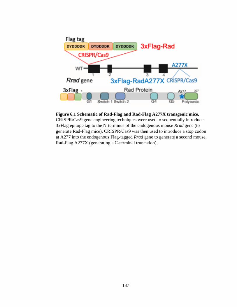
137
Figure 6.1 Schematic of Rad-Flag and Rad-Flag A277X transgenic mice.
CRISPR/Cas9 gene engineering techniques were used to sequentially introduce
3xFlag epitope tag to the N-terminus of the endogenous mouse Rrad gene (to
generate Rad-Flag mice). CRISPR/Cas9 was then used to introduce a stop codon
at A277 into the endogenous Flag-tagged Rrad gene to generate a second mouse,
Rad-Flag A277X (generating a C-terminal truncation).

138
Figure 6.2 Rad-Flag A277X enhances cardiac function and increases wall
thickness.
A) Representative M-mode short axis echocardiography from Rad-Flag and Rad-
Flag A277X mice. Scale bars: 1 sec; 2 mm. B) Ejection fraction (****p <
0.0001). C) Left ventricular inner dimensions (****p < 0.0001). D) Left
ventricular anterior and E) Left ventricular posterior wall thickness (LVAW:
****p < 0.0001; LVPW: ****p < 0.0001). Dimensions (C-E) in diastole. Dotted
lines represent mean summary of published WT and published cRadKO (Ahern
et. al 2019, [24]). Data displayed from female mice: Rad-Flag: N = 11 mice; Rad-
Flag A277X: N = 15 mice. P values calculated using Student unpaired t-test for
(B-E).

139
Figure 6.3 Rad-Flag A277X enhanced LTCC function
A) Representative family of ICa,L currents from Vtest ranging from -75 mV to +45
mV in 10 mV increments, with voltage protocol schematic shown above. Scale
bars: 10 pA/pF, 100 ms. B) Current-voltage relationship for peak ICa,L from Rad-
Flag and Rad-Flag A277X ventricular cardiomyocytes. C) Cell capacitance is
significantly larger in Rad-Flag A277X compared to Rad-Flag (*p = 0.01). D)
Maximal conductance is significantly increased in Rad-Flag A277X compared to
Rad-Flag (***p = 0.0003). E) Conductance-voltage curves normalized to
maximal conductance in Rad-Flag and Rad-Flag A277X. Smooth curves are
Boltzmann distribution fitted to data. F) Activation midpoint is significantly
negative-shifted in Rad-Flag A277X compared to Rad-Flag (****p < 0.0001).
Data for C,D,F analyzed by unpaired Student’s t-test. Data displayed from female
mice: Rad-Flag (N = 3 mice, n = 9 cells) and Rad-Flag A277X (N = 3 mice, n =
13 cells).

140
Figure 6.4 Rad-Flag A277X enhanced inactivation.
A) Exemplar Ca2+ currents with EGTA for Rad-Flag (teal) and Rad-Flag A277X
(pink), traces normalized to peak current at 0 mV Scale bar: 100 ms. Black dots
indicate r30, Black stars indicate r150. B) Remaining current across voltage steps
30 ms after peak (*p < 0.05; **p < 0.009; ***p < 0.0002; ****p < 0.0001; p
values alternated between above and below for clarity). C) ICa,L current density 30
ms after peak. D) Remaining current across voltage steps 150 ms after peak (*p <
0.02; ***p < 0.0052; p values alternated between above and below for clarity).
E) ICa,L current density 150 ms after peak. Data for B,C analyzed by two-way
ANOVA plus Šídák’s multiple comparisons test with F) 95% confidence intervals
and tabular results for Figure 6.3B and G) 95% confidence intervals and tabular
results for Figure 6.3D. Data displayed from female mice: Rad-Flag: N = 3 mice,
n = 6 cells; Rad-Flag A277X: N = 3 mice, n = 13 cells.

141
CHAPTER 7. MYOCARDIAL RAD DELETION PRESERVES CARDIAC FUNCTION IN
PRESSURE-OVERLOAD INDUCED HEART FAILURE
7.1 Preface
The results and methods attributed to others are presented for clarity and flow are
italicized.
7.2 Introduction
Heart failure (HF) is an emerging epidemic. About 50% of HF patients die within
five years of their diagnosis [296]. Heart failure occurs when a substantial amount of
functional myocardial cells is lost (or altered) in response to injury, leading to left
ventricular systolic dysfunction [297]. In an effort to maintain adequate function, the heart
initiates several compensatory mechanisms to improve contractility; however, these
processes contribute to the progression of failure [297]. Leading therapeutics include
positive inotropic agents, but these drugs induce pathological signaling when used
chronically [5]. Therefore, an ideal goal would be to identify a novel target that could safely
improve inotropy and cardiac function without promoting pathological signaling and
remodeling.
The L-type calcium channel (LTCC) is a heteromultimeric protein complex that
provides trigger calcium necessary to induce a contraction, and is an upstream regulator of
modulating inotropy within cardiomyocytes. The LTCC complex is comprised of a pore-
forming α-subunit (CaV1.2 in the myocardium), CaVβ2, α2δ, and calmodulin [194]. LTCC
function is tightly regulated to prevent calcium overload and stimulation of multiple
hypertrophic signaling pathways that promote heart failure [6, 12, 148, 149]. A key
142
regulator of LTCC activity is Rad (Ras associated with diabetes), a member of the RGK
subfamily (Rem, Rad, Rem2, and Gem/Kir) of Ras-related small GTPases; Rad serves as
an endogenous inhibitor of LTCC activity and is abundantly expressed in the heart [16-18,
195-198]. Rad deficiency yields an increase in ICa,L that promotes enhanced cardiac
function [21, 23, 24]; expression levels of Rad significantly decrease in patients suffering
from heart failure with reduced ejection fraction [23] as well as in a mouse model of cardiac
hypertrophy [181]. This suggests that Rad down-regulation may be a critical component
in the signaling for ventricular remodeling.
Studies utilizing whole-body Rad knockout (gRAD-/-) mice reported progressive
structural remodeling, increased myocyte size, increased up-regulation of the fetal gene
program associated with cardiac hypertrophy, and cardiac fibrosis [21, 198]. These results
are in line with other mouse models that have dramatically increased ICa,L as seen in the
absence of Rad [12, 149]. However, our recent studies have shown that myocardial Rad
deficiency increases ICa,L to enhance cardiac function into senescence without evidence for
structural remodeling or progression to heart failure [24]. This may be because Rad
expression has been maintained in non-cardiac tissue, such as vascular smooth muscle
[171] and in cells in the bone marrow [172], that could influence cardiac remodeling in
response to increased positive inotropy. A key hypothesis is that decreased Rad
contributes to a compensatory response under conditions of a failing heart [23, 181]. Given
that myocardial Rad deletion improves systolic heart function without pathological
remodeling, we hypothesized that myocardial Rad deletion will mitigate myocardial
remodeling in advanced stages of heart failure. Utilizing our inducible, myocardial-
restricted Rad knockout (cRadKO) mouse, we investigated the effect of Rad deletion in

143
response to pressure-overload induced heart failure by transverse aortic constriction
(TAC). We assessed cardiac function and structure when Rad was depleted before TAC,
when the induction of Rad deletion was initiated at time of surgery, and when Rad deletion
was induced one-month post-TAC surgery. We found in all three paradigms that cRadKO
had better survival and improved cardiac function compared to our CTRL. This was
commensurate with enhanced trigger calcium and preserved excitation contraction
coupling. These major new findings suggest Rad as a novel potential target to safely
enhance positive inotropy to rescue cardiac function under failing conditions.
7.3 Results
7.3.1 cRadKO protects in vivo heart function from pressure-overload induced heart
failure
cRadKO was induced 4 weeks prior to TAC and mice were followed for 3 months
after surgery (Figure 7.1A). Survival was significantly improved in cRadKO mice
subjected to TAC compared to CTRL mice (Figure 7.1B). In vivo cardiac function was
assessed by echocardiography before surgery and at time points shown in the timeline
(Figure 7.1A). At 3-months post-surgery, cRadKO with TAC maintained an elevated
ejection fraction compared to CTRL (Figure 7.1C; Figure 7.2A for 2-way ANOVA tabular
results). There was no difference in heart chamber size in cRadKO with TAC compared to
sham (Figure 7.1D; Figure 7.2B for 2-way ANOVA tabular results) or in heart wall
thickness (Figure 7.1E-F; Figure 7.2C,D respectively for 2-way ANOVA tabular results);
by contrast, CTRL with TAC demonstrated significant chamber dilatation. TAC

144
significantly elevated heart weight/body weight in CTRL but not in cRadKO, consistent
with the echocardiography in vivo measures (Figure 7.1G; Figure 7.2E for 2-way ANOVA
tabular results). TAC significantly increased fibrosis in CTRL and in cRadKO compared
to CTRL Sham, but there was no difference in the degree of fibrosis with respect to
knockout. (Figure 7.3B,C; Figure 7.4A for 1-way ANOVA tabular results). qRT-PCR of
ANF displayed no significant difference in any of the groups (Figure 7.3D; Figure 7.4B
for 1-way ANOVA tabular results). Taken together these data suggest that Rad reduction
confers a gain of systolic function that promotes survival and prevents structural
remodeling.
7.3.2 cRadKO enhances in vivo heart function after pressure-overload
Current management of advanced heart failure is limited in improving quality of life,
and ultimately does not prevent the progression of heart failure to advanced stages;
however, HF patients demonstrate functional benefit to inotropic agents [298]. Using ECG
telemetry we previously observed tamoxifen increase of heart rate within 3 to 7 days. In
the present experiment we administered tamoxifen at the time of surgery; consequently,
myocardial Rad reduction is expected to occur by ~7days after surgery (see timeline,
Figure 7.5A). No mortality was observed in cRadKO after TAC, while 25% mortality was
seen in our CTRL mice (Figure 7.5B). Echocardiographic assessment of these mice at
baseline (see timeline) demonstrated no significant difference between groups in function
or structure, as expected (Figure 7.5C-F; Figure 7.6A-D for 2-way ANOVA tabular
results). However, one month after surgery, cRadKO maintained normal function while
CTRL mice declined significantly (Figure 7.5C). cRadKO mice also maintained normal

145
left ventricular inner dimensions while CTRL mice had enlarged inner dimensions (Figure
7.5D). cRadKO demonstrated thicker anterior and posterior walls compared to CTRL
mice (Figure 7.5E,F). Three months after surgery, we measured heart weight/body weight
from the surviving animals and found no significant difference between groups (Figure
7.5G). This data supports that in the early stages of heart failure, Rad reduction preserves
systolic function.
7.3.3 cRadKO maintains elevated calcium cycling after pressure-overload
The increase in left anterior wall and posterior wall thickness seen in cRadKO could
be due to defective excitation-contraction coupling within cardiomyocytes [181, 299-303].
Therefore, we isolated cardiomyocytes from our CTRL and cRadKO TAC mice that were
given tamoxifen at time of surgery and measured ICa,L (Figure 7.7; see timeline, Figure
7.7A) and calcium handling (Figure 7.9; see timeline, Figure 7.9A). Representative ICa,L
traces in Figure 7.7B,G highlight increased current density and accelerated kinetics,
respectively. The current/voltage (I(V)) curves for ICaL show that cRadKO had a greater
current density compared to CTRL (Figure 7.7C). Maximal conductance was greater for
cRadKO (Figure 7.7D; maximal conductance: cRadKO = 450 ± 41 pS/pF, n=12; CTRL =
195 ± 25 pS/pF, n=8; ****p<0.0001) and the activation midpoint was shifted towards more
negative membrane potential (Figure 7.7E,F; cRadKO = -10.3 ± 1.1 mV, n=12; CTRL =
-3.2 ± 2.1 mV, n=8; *p=.01). Analysis of kinetics demonstrated less remaining current in
cRadKO TAC compared to CTRL TAC in the early component (r30) (Figure 7.7G,H;
Figure 7.8A for 2-way ANOVA tabular results) and no difference in the late component
(r150) of decay (Figure 7.7G,I; Figure 7.8B for 2-way ANOVA tabular results).

146
Whole-cell cytosolic calcium dynamics were also significantly larger in amplitude
in cRadKO compared to CTRL (Figure 7.9B,C; cRadKO = 2.2 ± 0.2 F340/380, n=11; CTRL
= 1.5 ± 0.2 F340/380, n=21; *p=.03). Upstroke velocity of the calcium transient was also
accelerated in cRadKO compared to CTRL (Figure 7.9D; cRadKO = 55.2 ± 5.1
(F340/380)/ms, n=11; CTRL = 39.1 ± 4.8 (F340/380)/ms, n=21; *p=.03). To assess cytosolic
clearance of Ca2+, we measured the time constant (τ) of the calcium transient decay, and
found that it was faster in cRadKO compared to CTRL (Figure 7.9E; cRadKO = 0.16 ±
0.01 s, n=11; CTRL = 0.12 ± 0.01 s, n=21; *p=.02).
Under normal physiological heart rates, the contractility of failing myocytes is
significantly less than normal [303, 304]; increasing the pacing rate of isolated myocytes
decreases contractility while normal myocytes demonstrate an increase in contractility
[303]. We therefore measured sarcomere dynamics from failing myocytes isolated from
cRadKO and CTRL simultaneously with calcium transients (Figure 7.10). Elevated
calcium dynamics accompanying Rad deletion coincided with unchanged resting
sarcomere length (Figure 7.10B,C), unchanged fractional shortening (Figure 7.10D), and
no change to the rate at which the cell shortened (Figure 7.10E). Using the model
published by Davis et. al [202], we calculated the integrated tension and found the index
score for cRadKO is +0.06 and CTRL is +0.05 (Figure 7.10F), indicating a modest trend
toward concentric hypertrophy in response to pressure overload in both models. This is
further supported by the increase in wall thickness we observed in both CTRL and cRadKO
(Figure 7.1,7.5). Taken together, this data suggests that excitation-contraction coupling is
not defective, but rather calcium homeostasis is enhanced to preserve systolic function.

147
7.3.4 Response to -adrenergic receptor stimulation remains blunted in cRadKO after
pressure-overload
A hallmark feature of heart failure is the down-regulation of β-adrenergic receptors,
resulting in a blunted response in contractility [303, 305]. We therefore measured ICa,L and
calcium transients after the addition of isoproterenol to assess whether there was a β-
adrenergic receptor response (Figure 7.11,7.12). Given that ICa,L from cRadKO resembles
β-adrenergic modulated ICa,L, we did not expect any response. As expected, we did not see
any change in current density (Figure 7.11Bii), maximal conductance (Figure 7.11C,D)
or activation midpoint (Figure 7.11E,F) in response to isoproterenol in cRadKO with
TAC. Compared to CTRL, cRadKO had larger maximal conductance and trended towards
a more negative activation midpoint (Figure 7.11C,E). For cytosolic calcium handling,
there was no difference between cRadKO and CTRL in calcium transient amplitude,
upstroke velocity or the rate of decay (Figure 7.12B-E). There was also no response to
isoproterenol in either CTRL or cRadKO in amplitude or upstroke velocity (Figure
7.12F,G). The rate of decay accelerated in CTRL in response to isoproterenol, with no
change in cRadKO (Figure 7.12H). Taken together, this data demonstrates that ICa,L is
maximally modulated in the absence of Rad, and responsiveness to β-adrenergic
stimulation remains blunted in both CTRL TAC and cRadKO TAC.
7.3.5 cRadKO promotes recovery of function in advanced stages of pressure-overload
Increased positive inotropy due to Rad deletion preserved cardiac structure and
function both before the onset of pressure-overload induced heart failure (Figure 7.1-7.4)

148
and in the early stages of onset (Figure 7.5-7.12). We therefore induced Rad deletion in
mice one month after TAC surgery to assess more advanced heart failure (Figure 7.13A,
timeline). Survival was assessed beginning at the time of surgery (Figure 7.13B). One
month after Rad knockout (two months post-surgery) resulted in 71% survival in cRadKO
TAC and 26% survival in CTRL TAC. Echocardiographic assessment showed enhanced
ejection fraction after Rad deletion that was not significantly different from CTRL Sham
(Figure 7.13C; Figure 7.14A for 2-way ANOVA tabular results). There was no
significant difference in heart chamber size after Rad deletion compared to CTRL Sham,
while CTRL TAC showed significant chamber dilatation (Figure 7.13D; Figure 7.14B for
2-way ANOVA tabular results). Left ventricular anterior wall thickness significantly
increased after Rad deletion compared to CTRL Sham, while no difference was detected
in left ventricular posterior wall (Figure 7.13E,F; Figure 7.14C,D for 2-way ANOVA
tabular results). Taken together, these data suggest that the reduction of Rad in the presence
of pressure-overload induced heart failure promotes increased cardiac function through a
compensatory response.
7.3.6 cRadKO prevents progression of cardiac dysfunction in advanced stages of
pressure-overload
Left ventricular ejection fraction can appear normal under conditions of left
ventricular systolic function [306]. To further assess left ventricular systolic function, we
also used speckle-tracking echocardiography to evaluate global longitudinal strain and
circumferential strain in CTRL TAC and cRadKO TAC compared to CTRL Sham (Figure
7.15B,C). One month after surgery, before the deletion of Rad, both TAC groups

149
demonstrated significantly lower longitudinal strain compared to Sham; however, after
Rad deletion cRadKO TAC returned to the level of CTRL Sham and cRadKO Sham, while
CTRL TAC remained lower (Figure 7.15B; Figure 7.16A for 1-way ANOVA tabular
results). Analysis of circumferential strain resulted in a similar improvement in cRadKO
TAC compared to CTRL Sham, while CTRL TAC remained low (Figure 7.15C; Figure
7.16B for 1-way ANOVA tabular results). These data further support that the reduction of
Rad improves left ventricular systolic function in the presence of pressure-overload
induced heart failure.
Hearts that undergo hypertrophy without left ventricular dysfunction demonstrate an
increase in heart weight/body weight and in wall thickness without changes to lung
weight/body weight or left ventricular chamber size [307]. We therefore measured heart
weight/body weight and lung weight/body weight in CTRL TAC and cRadKO TAC
compared to respective Sham groups (Figure 7.15D-F). CTRL TAC heart weight/body
weight was significantly increased compared to CTRL Sham, as expected, while cRadKO
TAC heart weight/body weight was not significantly increased compared to CTRL Sham
(Figure 7.15D; Figure 7.16C for 1-way ANOVA tabular results). Lung weight/body
weight was significantly increased in CTRL TAC, with no significant difference in cRadKO
TAC, compared to CTRL Sham (Figure 7.15E; Figure 7.16D for 1-way ANOVA tabular
results). Both of these measurements correlate with contractile function and heart failure
[307]. Plotting this correlation for CTRL TAC, cRadKO TAC, and our Sham groups
support this and reveal that cRadKO TAC develops hypertrophy without progressing
towards failure (Figure 7.15F). Many of the cRadKO TAC were in the same quadrant as

150
both Sham groups, demonstrating that Rad reduction promotes recovery from pressure-
overload induced heart failure.
7.3.7 cRadKO improves quality of life in advanced stages of pressure-overload
Locomotor activity is significantly reduced in mice that suffer from congestive heart
failure [308]. To determine if elevated cardiac function due to Rad loss improved behavior
and quality of life, we subjected our mice to an open field test to assess locomotive behavior
(Figure 7.17). Mice were assessed a month after surgery, and again one month after
tamoxifen treatment (2 months post-surgery; see timeline in Figure 7.17A). cRadKO TAC
mice spent more time walking around the cage, while CTRL TAC mice mostly resided in
the corner in which they were placed (lower right corner; see heat map and track
visualization, Figure 7.17B,C respectively). When compared to TAC mice before
tamoxifen, cRadKO TAC mice covered similar distance within the cage while CTRL TAC
mice covered significantly less distance (Figure 7.17D; Figure 7.17E for 1-way ANOVA
tabular results). Taken together, our data supports the hypothesis that the deletion of Rad
provides a positive inotropic boost that promotes survival and improves quality of life in
advanced stages of heart failure.
7.4 Discussion
The main finding of this study is Rad depletion from the myocardium improves
survival and preserves cardiac function against pressure-overload induced heart failure.
This is demonstrated by reduced mortality, reduced lung congestion, elevated cardiac
function, physiological hypertrophy, and improved strain in response to increased trigger

151
calcium and preserved excitation contraction coupling. In combination with previous
studies that report reduced Rad protein in nonischemic heart failure patients [23, 181], our
study demonstrates that regulation of Rad contributes to the compensatory response and by
depleting Rad, cardiac function is preserved in heart failure. This suggests Rad should be
considered as a potential therapeutic target to safely enhance positive inotropy without
promoting further damage to the heart.
7.4.1 Myocardial-restricted Rad deletion preserves cardiac function in pressure-
overload induced heart failure
Classical inotropes (such as dobutamine, dopamine, and PDE-inhibitors) have
failed due to adverse outcomes when used long-term due to maladaptive cardiac
remodeling in response to increased adrenergic activity [5, 192]. In theory, treatments that
could enhance inotropy without stimulating adrenergic activity would be ideal in order to
increase contractility without long-term consequences of adrenergic activation [5]. Indeed,
it has been shown that antagonists of β1-adrenergic receptors can reverse remodeling,
improve left ventricular function and prolong survival [192]. Our current study, combined
with our previous work with myocardial-restricted depletion of Rad, supports Rad as a
unique target for a new generation of positive inotropes to achieve this ideal therapy. We
have previously shown that depleting the myocardium of Rad serves as a positive inotrope
without stimulating pathological remodeling into senescence; this was possible because
calcium cycling was enhanced downstream of β-adrenergic signaling [24]. In conditions
of pressure-overload, the reduction of Rad continues to enhance positive inotropy through
increased trigger calcium and preserved excitation contraction coupling (Figure 7.7,7.9).

152
This translates to protection from pressure-overload induced heart failure (Figure 7.1,7.3)
as well as the ability to rescue cardiac function and structure in advanced stages of pressure-
overload induced heart failure (Figure 7.5,7.13,7.15,7.17). By increasing contractility
independent of adrenergic activation, cRadKO preserves the compensatory phase so that
function and structure remain in a healthy range (Figure 7.15) reminiscent of endurance
training [309] rather than the canonical heart failure phenotypes. Our work is further
supported by evidence of down-regulation of Rad protein seen in patients with nonischemic
heart failure [23, 181]. As encouraging as the present results are for suggesting Rad as a
therapeutic direction for cardioprotection, future studies should investigate the effects of
Rad deletion in heart failure in larger animal species.
A major consideration in using positive inotropes as a treatment for heart failure is
the energetic demands that these drugs incur. In normal cardiomyocytes, calcium activates
both ATP-consumption and ATP-regeneration in order to maintain the redox state of
NADH and FADH2 [5]. However, under conditions of failure, calcium uptake into the
mitochondria is disrupted and leads to oxidative stress [5, 310, 311]. This promotes
arrhythmias, systolic dysfunction, and pathological remodeling [5, 311]. A theory to avoid
this mismatch is to increase sarcoplasmic reticulum calcium content and release, supported
by animal models and patients with improved left ventricular ejection fraction having
significant upregulation of SERCA [312, 313]. We have previously shown that myocardial
Rad deletion promotes upregulation of SERCA without a reciprocal upregulation of PLN
in the presence of elevated systolic function and no pathological remodeling [24]. Data
shown in chapter 2 and 3 also show no detectable arrhythmias or electrical dysfunction in
cRadKO without heart failure; after pressure-overload, cRadKO still demonstrated

153
accelerated kinetics that resemble ICa,L kinetics measured from healthy cRadKO myocytes
(Figure 7.7). It is therefore possible that the deletion of Rad is preserving the balance of
ATP-consumption and ATP-regeneration, allowing cardiac function to be protected in the
presence of pressure-overload induced heart failure. Future studies should explore the role
in which Rad contributes to energy production within the myocardium, and the mechanism
by which the absence of Rad potentially improves mitochondrial function. This would give
more insight into how positive inotropes could balance excitation contraction coupling and
the energetic demands of the heart to provide better, more sustainable treatment [5].
7.4.2 Myocardial-restricted Rad deletion differs from whole-body knockout of Rad
In the TAC model the heart experiences an abrupt and chronic increase of afterload.
Despite a large number of studies utilizing the TAC model for pressure overload
hypertrophy the initial signaling effects that signal compensatory changes in the
myocardium are poorly understood [192]. When Rad is deleted before surgery, cRadKO
showed an increase of fibrosis (Figure 7.3), suggesting that while myocardial Rad deletion
spares functional and myocardial architectural remodeling, the heart remains responsive to
effectors activated in response to TAC surgery. In whole-body Rad knockout mice, CTGF
is elevated and fibrosis spontaneously develops as early as ten weeks of age [198]; in
contrast, myocardial Rad deletion did not demonstrate any spontaneous fibrosis or
increased expression of CTGF [24], except under the stress of pressure-overload in our
current study. Rad has also been associated with non-LTCC functions [21, 145, 211], thus,
we cannot rule out contributions to transcriptional regulation or cell signaling outside the
LTCC signalosome. Our finding that TAC induced fibrosis is present without detriment to

154
function following TAC also raises an interesting question – namely, the suggestion here
is that cRadKO illuminates a possible distinction between fibrotic signaling and protection
against loss of function as an association rather than a necessary cause-effect relationship,
at least in certain cases. In summary, in sharp contrast to global Rad knockout studies, we
now show that myocardial Rad deletion is beneficial for heart function in health in response
to imposed chronic pressure overload.
In conclusion, our findings suggest that increasing positive inotropy through
myocardial-restricted Rad deletion protects the cardiac function from pressure-overload
induced heart failure by maintaining a compensated-like heart. Deleting Rad also rescues
cardiac function in advanced stages of pressure-overload induced heart failure. There is a
major unmet need for treatments targeting inotropy [5]; these results support Rad as a
potential therapeutic approach for increasing cardiac output safely without promoting
pathological remodeling.

155
Figure 7.1 Loss of cardiomyocyte Rad before pressure overload prevents
progression of cardiac dysfunction.
A) Experimental timeline. Red arrows indicate time of tamoxifen injection and
TAC surgery. Black arrows above timeline indicate echocardiography time
points following surgery. B) Kaplan-Meier survival plot of CTRL with TAC,
cRadKO with TAC, and Sham. Between group differences in survival were tested
by Gehan-Breslow-Wolcoxon test (*p = 0.03). C-F) Echocardiographic
assessment of ejection fraction (C, *p = 0.05), left ventricular inner dimensions in
diastole (D), and left ventricular wall thickness of anterior wall (E, ††p = 0.007,
##p = 0.009); posterior wall in diastole (F, †p = 0.01). Data in C-F analyzed by 2-
way repeated measures ANOVA with Dunnett’s multiple comparisons test. Data
displayed from male mice; CTRL (Sham: N = 3; TAC: N = 3) and cRadKO
(Sham: N = 6; TAC: N = 9). G) Heart-to-body weight ratio at 3 months post-TAC
(**p = 0.002). Data from mice that did not survive not included. Data in G
analyzed by 1-way ANOVA with Dunnett’s multiple comparisons test. Data
displayed from male mice; CTRL (Sham: N = 5; TAC: N = 3) and cRadKO
(Sham: N = 5; TAC: N = 4). For all panels: *CTRL Sham v CTRL TAC; †CTRL
Sham v cRadKO TAC; #CTRL Sham v cRadKO Sham.

156
Figure 7.2 ANOVA 95% confidence intervals and tabular results.
A) 95% confidence intervals and tabular results for two-way ANOVA for Figure
7.1C. B) 95% confidence intervals and tabular results for two-way ANOVA for
Figure 7.1D. C) 95% confidence intervals and tabular results for two-way
ANOVA for Figure 7.1E. D) 95% confidence intervals and tabular results for
two-way ANOVA for Figure 7.1F. E) 95% confidence intervals and tabular
results for one-way ANOVA for Figure 7.1G.

157
Figure 7.3 Loss of cardiomyocyte Rad before pressure overload does not
protect all pathological signaling.
A) Experimental timeline. B) Representative pictures of picrosirius Red staining
with fibrosis quantification in C) (*p = 0.02; ††p = 0.001); Scale bar: 100 µm.
Data displayed from male mice; CTRL (Sham: N = 6; TAC: N = 3) and cRadKO
(Sham: N = 4; TAC: N = 5). D) qRT-PCR for ANF mRNA expression levels.
Data analyzed by one-way ANOVA plus Dunnett’s multiple comparisons test.
Data displayed from male mice; CTRL (Sham: N = 8; TAC: N = 8) and cRadKO
(Sham: N = 5; TAC: N = 7). For all panels: *CTRL Sham v CTRL TAC; †CTRL
Sham v cRadKO TAC; #CTRL Sham v cRadKO Sham.

158
Figure 7.4 One-way ANOVA 95% confidence intervals and tabular results.
A) 95% confidence intervals and tabular results for two-way ANOVA for Figure
7.3C. B) 95% confidence intervals and tabular results for two-way ANOVA for
Figure 7.3D.

159
Figure 7.5 Loss of cardiomyocyte Rad before pressure overload promotes
survival and enhances cardiac function.
A) Experimental timeline. Red arrows indicate time of tamoxifen injection and
TAC surgery. Black arrows above timeline indicate echocardiography time
points following surgery. B) Kaplan-Meier survival plot of CTRL with TAC and
cRadKO with TAC. Between group differences in survival were tested by Gehan-
Breslow-Wolcoxon test (p = 0.05). C-F) Echocardiographic assessment of
ejection fraction (C, *p = 0.04), left ventricular inner dimensions in diastole (D),
and left ventricular wall thickness of anterior wall (E); posterior wall in diastole
(F). Data in C-F analyzed by 2-way repeated measures ANOVA with Šídák’s
multiple comparisons test. Data displayed from female mice; CTRL (N = 4) and
cRadKO (N = 8). G) Heart-to-body weight ratio at 3 months post-TAC and post
Rad deletion. Data from mice that did not survive not included. Data in G
analyzed by Student’s unpaired t-test with Welch’s correction. Data displayed
from female mice; CTRL (N = 4) and cRadKO (N = 6).

160
Figure 7.6 ANOVA 95% confidence intervals and tabular results.
A) 95% confidence intervals and tabular results for two-way ANOVA for Figure
7.5C. B) 95% confidence intervals and tabular results for two-way ANOVA for
Figure 7.5D. C) 95% confidence intervals and tabular results for two-way
ANOVA for Figure 7.5E. D) 95% confidence intervals and tabular results for
two-way ANOVA for Figure 7.5F.

161
Figure 7.7 Loss of cardiomyocyte Rad increases ICa,L after pressure-
overload.
A) Experimental timeline showing study design. B) Exemplar family of Ca2+
currents of CTRL TAC (blue) and cRadKO TAC (red) from Vtest ranging from
-75 to +50 mV in 10 mV increments (voltage protocol shown above); Scale bar: 5
pA/pF, 100 ms. C) Current-voltage relationship for peak ICa,L current density from
CTRL TAC and cRadKO TAC ventricular myocytes. D) Maximal conductance is
significantly increased in cRadKO TAC compared to CTRL TAC (****p <
0.0001; dotted lines represent maximal conductance for published unoperated WT
and cRadKO, included for reference). E) Conductance-voltage curves normalized
to maximal conductance in CTRL TAC and cRadKO TAC. Smooth curves are
Boltzmann distribution fitted to data. F) Activation midpoint is significantly
negative-shifted in cRadKO TAC (*p = 0.01). G) Exemplar Ca2+ currents with
EGTA for CTRL TAC (blue) and cRadKO TAC (red), traces normalized to peak
current at 0 mV Scale bar: 100 ms. Black dots indicate r30, Black stars indicate
r150. Remaining current across voltage steps 30 ms after peak H) and 150 ms after
peak I). P values calculated using Student unpaired t-test for (D,F). Data in (H,I)
analyzed by two-way ANOVA plus Šídák’s multiple comparisons test (**p <
0.009; *p < 0.03). Data displayed from female mice; CTRL (N = 4 mice, n = 8
cells) and cRadKO (N = 4 mice; n = 12 cells).

162
Figure 7.8 ANOVA 95% confidence intervals and tabular results.
A) 95% confidence intervals and tabular results for two-way ANOVA for Figure
7.7H. B) 95% confidence intervals and tabular results for two-way ANOVA for
Figure 7.7I.

163
Figure 7.9 Loss of cardiomyocyte Rad enhances cellular calcium handling
after pressure-overload.
A) Experimental timeline showing study design. B) Representative calcium
transients from isolated ventricular cardiomyocytes loaded with fura2-AM, CTRL
TAC (top, blue) and cRadKO TAC (bottom, red) paced at 1 Hz. Scale bar: 1
second. C) Amplitude of the calcium transients from cRadKO TAC are larger
compared to CTRL TAC (*p = 0.03). D) The velocity at which calcium enters the
cytosol (upstroke of the transient) is significantly faster in cRadKO TAC
compared to CTRL TAC (*p = 0.03). E) Rate at which calcium leaves the cytosol
(downstroke of the transient) is significantly faster in cRadKO TAC compared to
CTRL TAC (*p = 0.02). P values calculated using Student unpaired t-test for (C-
E). Data displayed from female mice; CTRL (N = 5 mice, n = 21 cells) and
cRadKO (N = 3 mice; n = 11 cells).

164
Figure 7.10 Loss of cardiomyocyte Rad does not alter sarcomere dynamics
after pressure-overload.
A) Experimental timeline showing study design. B) Exemplar sarocomere length
traces from isolated ventricular cardiomyocytes loaded with fura2-AM, CTRL
TAC (top, blue) and cRadKO TAC (bottom, red) paced at 1 Hz. Scale bar: 1
second. C) Diastolic sarcomere length (dotted lines represent means of diastolic
length for published unoperated WT and cRadKO, included for reference). D)
Sarcomere fractional shortening (dotted lines represent means of diastolic length
for published unoperated WT and cRadKO, included for reference). E) Sarcomere
shortening velocity (dotted lines represent means of diastolic length for published
unoperated WT and cRadKO, included for reference). F) Integral of sarcomere
length (dotted lines represent means of diastolic length for published unoperated
WT and cRadKO, included for reference). P values calculated using Student
unpaired t-test for (C-F). Data displayed from female mice; CTRL (N = 5 mice, n
= 8 cells) and cRadKO (N = 3 mice; n = 4 cells).

165
Figure 7.11 -AR stimulation does not alter ICa,L in Rad deletion after
pressure-overload.
A) Experimental timeline showing study design. Bi,ii) Current-voltage
relationship for peak ICa,L current density from CTRL TAC (left, blue) and
cRadKO TAC (right, red) from isolated ventricular myocytes after treatment with
300 nM isoproterenol (ISO). C) Maximal conductance is significantly increased
in cRadKO TAC compared to CTRL TAC (***p = 0.0007; dotted lines represent
maximal conductance for published unoperated WT + ISO and cRadKO + ISO,
included for reference). D) Fractional change of maximal conductance between
baseline and ISO response of CTRL TAC (mean = 47%) and cRadKO (mean =
7%). Neither group demonstrated a significant response, as measured by deviation
from zero. E) Activation midpoint is not significantly negative-shifted in
cRadKO TAC compared to CTRL TAC after treatment with ISO (dotted lines
represent maximal conductance for published unoperated WT + ISO and cRadKO
+ ISO, included for reference). F) Difference in activation midpoint between
baseline and ISO response of CTRL TAC (mean = -3.1 mV) and cRadKO (mean
= 0.51 mV). CTRL TAC demonstrated a significant response, as measured by
deviation from zero (*p = 0.03). P values calculated using Student unpaired t-test
for (C,E) and one sample t-test for (D,F). Data displayed from female mice;
CTRL (N = 3 mice, n = 5 cells) and cRadKO (N = 4 mice; n = 9 cells).

166
Figure 7.12 -AR stimulation response in cellular calcium handling is
blunted after pressure-overload.
A) Experimental timeline showing study design. B) Representative calcium
transients from isolated ventricular cardiomyocytes loaded with fura2-AM, CTRL
TAC (top, blue) and cRadKO TAC (bottom, red) after treatment with 1 µM
isoproterenol (ISO), paced at 1 Hz. Scale bar: 1 second. C) Amplitude of the
calcium transients from cRadKO TAC and CTRL TAC after treatment with ISO.
D) The velocity at which calcium enters the cytosol (upstroke of the transient) in
cRadKO TAC and CTRL TAC after treatment with ISO. E) Rate at which
calcium leaves the cytosol (downstroke of the transient) in cRadKO TAC and
CTRL TAC after treatment with ISO. F) Fractional change of amplitude between
baseline and ISO response of CTRL TAC (mean = 78%) and cRadKO (mean =
7%). Neither group demonstrated a significant response, as measured by deviation
from zero. G) Fractional change of upstroke velocity between baseline and ISO
response of CTRL TAC (mean = 7%) and cRadKO (mean = 3%). Neither group
demonstrated a significant response, as measured by deviation from zero. H)
Fractional change of rate of decay between baseline and ISO response of CTRL
TAC (mean = -19%) and cRadKO (mean = -6%). CTRL demonstrated a
significant response, as measured by deviation from zero (*p = 0.04). P values
calculated using Student unpaired t-test for (C-E) and one sample t-test for (F-H).
Data displayed from female mice; CTRL (N = 4 mice, n = 10 cells) and cRadKO
(N = 3 mice; n = 8 cells).

167
Figure 7.13 Loss of cardiomyocyte Rad after pressure overload prevents
progression of cardiac dysfunction.
A) Experimental timeline. Red arrows indicate time of TAC surgery and time of
tamoxifen injection 1-month post-surgery. Black arrows above timeline indicate
echocardiography time points following surgery. B) Kaplan-Meier survival plot of
CTRL with TAC, cRadKO with TAC, and Sham. Between group differences in
survival were tested by Gehan-Breslow-Wolcoxon test (****p < 0.0001). C-F)
Echocardiographic assessment of ejection fraction (C, ****p < 0.0001; †††p =
0.0003; ##p = 0.002), left ventricular inner dimensions in diastole (D, **p =
0.003), and left ventricular wall thickness of anterior wall (E, ††p = 0.009);
posterior wall in diastole (F, *p = 0.02). Data in C-F analyzed by 2-way repeated
measures ANOVA with Dunnett’s multiple comparisons test. Data displayed from
male mice; CTRL (Sham: N = 6; TAC: N = 10) and cRadKO (Sham: N = 7; TAC:
N = 10). Data from mice that did not survive not included. For all panels: *CTRL
Sham v CTRL TAC; †CTRL Sham v cRadKO TAC; #CTRL Sham v cRadKO
Sham.

168
Figure 7.14 ANOVA 95% confidence intervals and tabular results.
A) 95% confidence intervals and tabular results for two-way ANOVA for Figure
7.13C. B) 95% confidence intervals and tabular results for two-way ANOVA for
Figure 7.13D. C) 95% confidence intervals and tabular results for two-way
ANOVA for Figure 7.13E. D) 95% confidence intervals and tabular results for
two-way ANOVA for Figure 7.13F.

169
Figure 7.15 Loss of cardiomyocyte Rad after pressure overload prevents
progression of cardiac dysfunction.
A) Experimental timeline. B) Pooled data for longitudinal strain. Rad deletion
increased global longitudinal strain (****p < 0.0001; ††p = 0.0003; **p = 0.03).
C) Pooled data for circumferential strain. Rad deletion enhanced circumferential
strain (*p = 0.01; †p = 0.02; ****p < 0.0001). D) Heart weight to body weight
ratio measured two-months post-surgery, one-month post Rad deletion (***p =
0.0002). E) Lung weight to body weight ratio measured two-months post-surgery,
one-month post Rad deletion (**p = 0.002). F) Linear regression of lung weight
body weight ratio as a function of heart weight body weight ratio. Orange region
indicates hearts with left ventricular hypertrophy and in failure; green region
represents hearts with left ventricular hypertrophy but not in failure. Data in (B,C)
analyzed by two-way repeated measures ANOVA with Dunnett’s multiple
comparisons test. Data in (D,E) analyzed by one-way ANOVA with Dunnett’s
multiple comparisons test. Data displayed from male mice; CTRL (Sham: N = 6;
TAC: N = 10) and cRadKO (Sham: N = 7; TAC: N = 11). Data from mice that did
not survive not included. For all panels: *CTRL Sham v CTRL TAC; †CTRL
Sham v cRadKO TAC; #CTRL Sham v cRadKO Sham.

170
Figure 7.16 ANOVA 95% confidence intervals and tabular results.
A,B) 95% confidence intervals and tabular results for two-way ANOVA for
Figure 7.15B,C respectively. C,D) 95% confidence intervals and tabular results
for two-way ANOVA for Figure 7.13D,E respectively.

171
Figure 7.17 Loss of cardiomyocyte Rad after pressure overload improves
locomotive activity.
A) Experimental timeline. Black arrows indicate when behavior was measured (1
month after surgery, before tamoxifen injection, labeled as TAC Before TAC;2
months after surgery, 1 month after tamoxifen injection labeled as CTRL TAC or
cRadKO TAC). B) Heat maps of activity in the open field, color is defined as the
average time spent in seconds in a given location. Blue represents the least
duration to Red, representing the highest duration. cRadKO TAC spent more time
moving around within the open field compared to CTRL TAC. C) Representative
15 min. of tracks in the open field of CTRL TAC (left) and from cRadKO TAC
(right). cRadKO TAC demonstrated more movement compared to CTRL TAC.
D) Quantification of the distance covered within the open field. CTRL TAC mice
covered significantly less distance compared to their one month time point (TAC
Before TAM) while cRadKO covered similar distance (*p = 0.03). Data analyzed
by one-way ANOVA plus Dunnett’s multiple comparisons test, with 95%
confidence intervals and tabular results in E). Data displayed from male mice;
TAC Before TAM (N = 15), CTRL TAC (N = 8), and cRadKO TAC (N = 7). For
panel D: *TAC Before TAM v CTRL TAC; †TAC Before TAM v cRadKO TAC.

172
CHAPTER 8. DISCUSSION
A novel treatment for heart failure with reduced ejection fraction would be to improve
contractility in a manner that preserves the fight-or-flight response within the heart. The
main objective of this dissertation was to test the hypothesis that disrupting Rad inhibition
of the L-Type calcium channel (LTCC) can stably enhance cardiac function by increasing
LTCC calcium current (ICa,L) downstream of -adrenergic receptor (-AR) stimulation;
this improves cardiac performance without inducing pathological remodeling and
progression to heart failure. It is well established that the absence of Rad results in
increased ICa,L [17, 19-24, 288, 314]; however, the mechanism by which Rad inhibits the
LTCC remains unclear [288]. The work of this dissertation contributes to the understanding
of Rad-mediated LTCC inhibition by: 1) revealing the role of Rad in LTCC modulation,
and 2) unmasking a potential new target to selectively enhance ICa,L without promoting
pathology. Each of these are discussed below.
8.1 Rad is a critical regulatory site for LTCC modulation
A major mechanism for modulating calcium cycling is sympathetic stimulation [6,
25, 28, 31, 92, 94, 95, 103]. cRadKO leads to enhanced basal ICa,L resembling β-AR
modulated ICa,L (Chapter 3, Chapter 4, Chapter 5). The hallmark features of PKA-
mediated LTCC modulation are: significant increase in ICa,L, hyperpolarized shift in
voltage dependent activation, and increased rate of inactivation; all result in a gain of LTCC
function [103]. In the absence of Rad, ICa,L increases ~three fold compared to control
(CTRL), voltage dependent activation is shifted to more negative potentials, and the rate
of inactivation is accelerated in ventricular and sinoatrial cardiomyocytes (Chapter 3,
Figure 4; Chapter 4, Figure 3; Chapter 5, Figure 1-5 ). The effect of Rad deletion on
ICa,L is not dependent on signaling from β-ARs, demonstrating no detectable response to
isoproterenol (Chapter 3, Figure 4; Chapter 4, Figure 3) and increasing ICa,L in the
absence of β-ARs (Chapter 5, Figure 7-9). These results led to the conclusion that Rad
may be absent from the LTCC in order for modulation to occur. Proximity proteomics
evaluation in transgenic mice further contributed to this hypothesis, demonstrating that

173
following acute isoproterenol treatment, Rad association with the CaV1.2 and CaVβ was
reduced [92]. Taken together, the results of this dissertation and that of recent publications
establish Rad as the critical phosphorylation site by PKA to induce LTCC modulation [21-
24, 92].
8.2 Rad governs ICa,L by multiple mechanisms
Amplification of ICa,L can be described by the classic equation: I = NT x Pf x Po x i,
where I represents macroscopic calcium current of the cell, NT is the total number of
channels, Pf is the probability that a given channel is available, Po is the probability that the
channel will be open, and i is the unitary current through an open pore [282, 291, 292]. Rad
regulation of ICa,L can occur by manipulating one or multiple variables of this equation to
produce an increase in macroscopic current.
It is well established PKA phosphorylation increases ICa,L by elevating Pf and Po
[315]. Indeed, single channel recordings of CaV1.2 depleted of Rad displayed almost no
blank sweeps, and most sweeps exhibited either intermediate or high levels of opening
[92]. The mechanism of voltage-dependent of activation of CaV1.2 remains unclear but has
been proposed to include a number of critical elements: S4 and S6 segments of domain I
of the α1 subunit (IS4, IS6 respectively), the AID (α-interacting domain), the I-II linker
between domain I and II of the α1 subunit, and CaVβ [55, 118, 293]. CaVβ binding to CaV1.2
enhanced Po and caused a hyperpolarizing shift in activation; this required a rigid linker
between IS6 and the AID [92, 316]. It has been speculated that Rad interaction with CaVβ
may structurally alter the IS6-AID linker as a way to lower Po-gating, and thereby inhibit
the LTCC [118]. The C-terminal tail of Rad is the domain that binds to CaVβ and is critical
to Rad-mediated LTCC regulation [17]. Sites for PKA and CaMKII phosphorylation,
binding for 14-3-3, binding for CaM, and association with plasma membrane all reside
within the C-terminus of Rad [175, 177, 281]. These studies suggest that Rad-mediated
inhibition of the LTCC, and therefore LTCC modulation, is regulated through its C-
terminal tail. Indeed, the truncation of the C-terminus of Rad results in ICa,L that is
indistinguishable from cRadKO (Chapter 6, Figures 6.2-6.4), supporting the hypothesis
that LTCC modulation relies on the C-terminus of Rad. This raises the possibility that upon

174
Rad phosphorylation, Rad is sequestered from the complex by 14-3-3 binding to sites both
on the N- and C-terminal extensions, alleviating the association with CaVβ so that the IS6-
AID linker can be stabilized to support high Po gating [118, 166, 281].
Another emerging hypothesis of the mechanism enhancing Pf and Po, and thereby
LTCC modulation, is channel clustering. Calcium bound to CaM allows channels to form
physical connections between the C-terminal tails of neighboring channels to induce
clustering (~8 channels within an average cluster) [57-59]. The activity of the cluster would
be driven by the channel(s) with the highest activity within the cluster [57]. These channels
also inactivate more rapidly through calcium-dependent inactivation in order to prevent
arrhythmogenesis; this causes coupling between channels to fade [58]. However, coupling
persists longer than the current that evoked it, thereby acting as a type of ‘molecular
memory’ that might serve to augment calcium influx in times of repetitive membrane
depolarization, such as seen in calcium-dependent facilitation or an increase in action
potential frequency [58]. Upon β-AR stimulation, channels can form ‘super-clusters’
(~32% increase in cluster size) that allow them to gate cooperatively [59]. These ‘super-
clusters’ were localized to transverse tubules, same microdomain as the source of cAMP
used for signaling downstream of β-AR stimulation [59, 101, 102]. Rad binds to CaM in a
calcium dependent manner through a region located on the C-terminus of Rad [166]. With
CaM playing a central role in calcium-dependent facilitation, calcium dependent
inactivation, and clustering, it is possible that Rad inhibits LTCC PF and Po through binding
to CaM and blocking cluster formation. Upon phosphorylation by PKC, Rad binding to
CaM is reduced [317]. Therefore, the absence of Rad from the complex would result in a
constant state of channel clustering that would display enhanced calcium influx and
accelerated inactivation, as seen both in cRadKO and when the C-terminus of Rad is absent
(Rad-Flag A277X) (Chapter 3, Figure 3.4; Chapter 4, Figure 4.3; Chapter 5, Figure
5.1-5.5; Chapter 6, Figure 6.2-6.4). The reduction of Rad binding could also cause a
conformational change within the LTCC so that CaV could stabilize the IS6-AID linker.
Future studies investigating channel clustering in the absence of Rad might shed light onto
how the LTCC undergoes modulation, facilitation, and inactivation.

175
A potential explanation for enhanced ICa,L seen in the absence of Rad is an increase
in the total number of channels expressed in the plasma membrane. An initial hypothesis
for RGK regulation of CaV1.2 channels was by regulating surface expression and limiting
channel trafficking to the membrane [16, 176, 314, 318]. Indeed, in PC12 cells, Rad
blocked surface expression of LTCCs by binding to CaVβ3 and sequestering the subunit to
the nucleus so that α-subunits could not be trafficked to the membrane [176]. In a
transgenic mouse model that expressed a dominant negative mutant of Rad (S105N), α1C
protein expression was significantly increased [182]. Based on these studies, the prediction
would be that Rad governs channel activity by simply limiting channel formation and
proper surface trafficking; therefore, in the absence of Rad, channel expression would
increase. Interestingly, qRT-PCR for CaV1.2 mRNA was not significantly different
between CTRL and cRadKO nor was CaV1.2 total protein expression different; however,
this does not reveal whether protein was trafficked to the membrane (Chapter 3, Figure
3.4). Investigation of channel trafficking in cRadKO was not pursued in this dissertation;
however, previous studies have demonstrated that RGK inhibition, including Rad, occurs
at the cell surface rather than through altering α1 surface expression or trafficking [20, 88,
183, 282, 319]. A recent study proposed that -AR stimulation causes an increase in CaV1.2
surface expression by triggering rapid mobilization of CaV1.2-cargo-carrying endosomes
to the sarcolemma surface; the hypothesis is that channel activity is augmented through
insertion and recycling of CaV1.2 [320]. It is speculated that when Rad is phosphorylated,
the dissociation of Rad from CaVβ releases the channel complex and permits trafficking to
the surface [320]. Studies in heterologous expression systems support this hypothesis,
demonstrating Rem decreased CaV1.2 surface density by 65%, though inhibition of Rem
on CaV1.2 was not relieved after -AR stimulation [282, 314]. Therefore, it is possible that
the absence of Rad promotes modulation of the LTCC simply by permitting the insertion
of a pool of channels normally lying in reserve for when -ARs are activated.
Though the mechanisms of LTCC modulation and RGK-mediated inhibition have
been studied extensively, a conclusive explanation remains elusive. It is possible that
regulation by Rad is a combination of the mechanisms described above, or potentially
dependent on protein-protein interactions with Rad and the LTCC that are currently
unknown. Indeed, the role of Rad in LTCC modulation has just been established [24, 92].

176
Advances in microscopy techniques and technology, combined with new FRET biosensors
may allow investigation of LTCC nanodomains, and specific protein-protein interactions
to better understand how the presence and absence of Rad contributes to LTCC regulation
[31].
8.3 Hypertrophic signaling is not exclusively dependent on elevated ICa,L
A central dogma of calcium cycling is increasing ICa,L inevitably leads to myocyte
hypertrophy [12, 148, 149, 321-329]. Indeed, overexpression of α1C or CaVβ in transgenic
mice yields enhanced ICa,L followed by rapid progression to pathological hypertrophy, heart
failure and death [12, 149, 327]. An early study of global Rad knockout also reported
increased hypertrophy due to activated CaMKII, without any cardiac functional
evaluations [181]; Crucial observations from these overexpression models that are
important to note: the α1C overexpression model exhibited minimal difference in left
ventricular inner dimensions and in posterior wall thickness compared to non-transgenic
controls [12]; CaVβ overexpression reported cardiac hypertrophy without functional
assessment by echo, or indication of activation of CaMKII or calcineurin [330]. The global
Rad knockout model reported hypertrophy based on hyperphosphorylation of CaMKII, but
no cardiac functional evaluation [181]. Contrary to these studies, decreased LTCC activity
also produces severe hypertrophy and remodeling due to calcineurin and CaMKII
activation [290]. Cardiac-specific Rad deletion exhibited no significant changes in cardiac
structure compared to CTRL under conditions of elevated ICa,L and enhanced calcium
handling (Chapter 3, Figures 3.3-3.5; Chapter 5, Figures 5.7,5.8; Chapter 6, Figures
6.2-6.4); there was also no indication of hyperphosphorylation of phospholamban by
CaMKII, suggesting CaMKII was not activated in cRadKO (Chapter 3, Figures 3.5,3.9).
These contradicting results indicate increased ICa,L does not necessarily guarantee
activation of hypertrophic signaling.
Cardiac growth can be predicted through a model based on integrated tension
measured from mean twitch tensions or sarcomere shortening. A negative score in this
model forecasts dilated cardiomyopathy, and a positive score indicates potential
hypertrophic remodeling [202]. When developing the model, the study reported that

177
predicting heart remodeling solely on any singular calcium parameter was poor; the model
had to include mechanical parameters in order to achieve accurate predictions [202]. Based
on this model, cRadKO approached a WT tension-integral as a consequence of larger
calcium transient amplitudes (Chapter 3, Figures 3.5,3.6). This could be due to enhanced
ICa,L kinetics (Chapter 3, Figure 3.3; Chapter 5, Figures 5.2-5.5) and/or due to increased
expression of SERCA2a (Chapter 3, Figure 3.5). Activation of calcineurin requires a high
and sustained calcium signal [146]; the acceleration of ICa,L decay in cRadKO may be
sufficient to prevent activation of calcineurin and hypertrophic signaling even though peak
ICa,L is increased. Even in conditions of pressure-overload induced heart failure, the
integrated tension model predicted only modest concentric hypertrophy (Chapter 7,
Figure 7.10), which is consistent with the slight increase in wall thickness observed in all
three TAC paradigms of Rad deletion (Chapter 7, Figures 7.1,7.5,7.13). Elevated
SERCA2a without a mirroring change in phospholamban, as seen in cRadKO, would also
prevent a high and sustained calcium signal within the cytosol. This beneficial
modification would allow enhanced calcium uptake without the need for phosphorylation
of phospholamban; indeed, this type of regulation displays cardioprotection in conditions
of high-intensity exercise and advanced heart failure [207-210]. Hypertrophic signaling
activation therefore is not as straightforward as simply an increase in peak ICa,L; rather,
enhanced calcium current may be a weak inducer or contributes to hypertrophic signaling
secondary to chronic stimulation of -ARs.
An intriguing finding of this work is that attaching an epitope tag, Flag, to Rad caused
significant alterations to cardiac function and structure (Chapter 6). Many studies have
utilized transgenic mouse models that express an epitope tag such as Flag to investigate
LTCC gating properties and binding partners; however, none of these studies report
functional assessments to demonstrate healthy cardiac function and structure [57-59, 92,
117, 118, 166, 331]. These types of genetic manipulations have the potential to trigger
indirect, secondary effects on that confound the direct effects which could cause
misinterpretations of the results [332]. An example of this were studies recently conducted
investigating the role of junctophilin-2 (JPH-2) in formation and stabilization of transverse
tubules. When all cardiomyocytes are depleted of JPH-2, there was a dramatic loss of
transverse tubules and severe cardiac dysfunction; this led to the conclusion that JPH-2 was

178
critical for transverse tubule maturation and preserving cardiac function [333, 334].
However, loss of transverse tubules is a hallmark feature of pathological remodeling [335].
Therefore, the question remains of whether loss of JPH-2 promotes transverse tubule loss
that then leads to heart failure, or heart failure promotes loss of transverse tubule, causing
JPH-2 to decrease. Utilizing a genetic mosaic model to deplete either a low percentage or
high percentage of JPH-2 within cardiomyocytes demonstrated that low percentage loss of
JPH-2 yielded minimal transverse tubule disorganization and no disturbances to cardiac
function; this was only seen in high percentage loss of JPH-2 [332]. This study
demonstrated JPH-2 does not play as critical of a role in transverse tubule maturation as
originally demonstrated [332]. This study combined with the results from Rad-Flag
highlight how direct effects on cardiac gene manipulation may be misinterpreted due to
confounding, secondary effects, such as heart failure conditions, especially when
manipulating genes that affect the LTCC.
8.4 Enhanced ICa,L does not necessarily promote electrical dysfunction
Experimental and theoretical studies have also identified ICa,L as a critical component
in arrhythmogenesis due to its central role in action potential duration; specifically,
reactivation of ICa,L in the late phase of decay induces early afterdepolarizations (EADs)
that trigger fatal arrhythmias [50, 61-63]. Recent studies have demonstrated that reducing
the non-inactivating component of ICa,L, known as the pedestal current, can reduce the
formation of EADs without affecting excitation contraction coupling [50, 61]. This
suggests a new class of drug action that act as ‘gating modifiers’ to promote subtle changes
in the biophysical parameters of ICa,L to reduce the late, arrhythmogenic component without
affecting contractility or action potential duration [62]. A prototype drug that appears to
achieve this is a drug called roscovitine [62, 126-130, 138]. The work of this dissertation
supports Rad as another potential ‘gating modifier.’ The absence of Rad reduced the
pedestal current (Chapter 3, Figure 3.3) and the late component of ICa,L was not
significantly different between cRadKO and CTRL (Chapter 5, Figures 5.2-5.5). Action
potential duration and QT interval were not significantly different between cRadKO or
CTRL (Chapter 5, Figure 5.6), demonstrating that Rad could be a safe ‘gating modifier’
drug target since this type of regulation can occur without activation of β-AR stimulation

179
(Chapter 5, Figure 5.7,5.8). In fact, this work establishes that the C-terminus of Rad could
be the target for a small peptide inhibitor since Rad-Flag A277X also demonstrated
accelerated inactivation without altering the late component of ICa,L that paralleled cRadKO
(Chapter 6, Figure 6.2-6.4).
Another key consideration is potential electrical dysfunction within the sinoatrial
node. Alterations in sinus node function parallel autonomic imbalance in the ventricles;
this imbalance is evident in heart failure and can result in sudden cardiac death due to a
decrease in heart rate variability [336]. Treatment with -AR blockers suggest lowering
heart rate contributes to survival for patients with heart failure; higher heart rates promote
arrhythmogenesis and decreased contractility [336]. Contrary to these studies, cRadKO
exhibited higher intrinsic heart rate without evidence of arrhythmogenesis on the
background of enhanced ICa,L in sinoatrial cardiomyocytes, and increased contractility in
the ventricle (SANcm; Chapter 3, Figure 3.3; Chapter 4, Figure 4.4,4.5). These
paradoxical results suggest that modulated ICa,L may promote balance of autonomic input
without the required increase in sympathetic drive. Previous studies indicate there are
mechanisms that increase and decrease sinus rate in order to preserve autonomic balance
[336]. In fact, a recent study discovered SANcm undergo a nonfiring mode; the SANcm
tissue network rhythm is set by the balance between firing and nonfiring cells [337]. Cells
that are electrically coupled are in a phasic entrainment, and are grouped together either in
a firing mode, or a nonfiring mode; a firing cell that interacts with a nonfiring cell is called
tonic entrainment [337]. In tonic entrainment, cells that are in a nonfiring mode
electronically draw the flow of cations from more depolarized, firing cells via gap
junctions, thereby creating a bradycardic rhythm [337]. Under sympathetic stimulation,
cells shift more towards a firing mode in order to accelerate rhythm [337]. In the absence
of Rad, it is possible that more SANcm could transition to a firing mode, thereby
accelerating HR and sinus rhythm without necessarily disrupting the balance of nonfiring
versus firing cells in a pathological manner, as demonstrated by the lack of change in heart
rate variability. Indeed, this further supports the role of Rad in not only LTCC regulation
of -AR stimulation, but whole heart regulation by sympathetic stimulation. Taken
together, targeting Rad is a powerful potential therapeutic approach because the absence
180
of Rad enhances positive inotropy and positive chronotropy in a manner that follows -AR
stimulation while still preserving cardiac responsiveness to adrenergic activation.
8.5 Targeting Rad represents the ideal positive inotrope
Currently, the ideal, theoretical treatment for heart failure would restore calcium
handling in a failing heart without activating adrenergic signaling [5]. Depleting the LTCC
of Rad could serve as a therapeutic target in order to make this ideal treatment a reality.
First, the absence of Rad enhances calcium handling under basal conditions in a manner
that preserves β-AR signaling within the cell and at the organ level (Chapter 3, Figures
3.8-3.10). Enhanced cardiac function is dependent on the presence of Rad in the LTCC
complex, rather than solely on activating the fight-or-flight response (Chapter 5, Figures
5.7,5.8), thereby providing a positive inotropic target downstream of adrenergic signaling.
Unlike studies conducted in global Rad knockout models, cRadKO does not promote
pathological signaling [181-183] (Chapter 3, Figures 3.3-3.5). In fact, Rad deletion before
the onset of pressure-overload induced heart failure provides cardiac protection and
preserves survival (Chapter 7, Figure 7.1). This protection remained even when Rad was
depleted at the time or four weeks after the induction of pressure-overload (Chapter 7,
Figures 7.5,7.13-7.17). Calcium handling was still enhanced in cRadKO despite the
exposure to thoracic aortic constriction surgery (TAC) (Chapter 7, Figures 7.7-7.10),
though responsiveness to adrenergic stimulation was no longer detectable (Chapter 7,
Figures 7.11,7.12). These results fit with earlier studies that reported Rad protein is
reduced in nonischemic heart failure patients, and support the hypothesis that Rad
contributes to the cardiac compensation in response to heart failure [23, 181].
Cardiac protection through the depletion of Rad does not extend to all pathological
signaling seen in heart failure. When Rad was deleted prior to surgery, fibrosis increased
significantly compared to Sham, and was not different from CTRL that underwent surgery
(Chapter 7, Figure 7.3). This is not surprising since others have reported that the absence
of Rad elevated connective tissue growth factor (CTGF) expression and increased fibrosis;
however, the difference between that study and cRadKO is that fibrosis was not seen under
normal physiological conditions or in Sham, but only when the heart was exposed to stress

181
under pressure-overload [24, 198] (Chapter 3, Figure 3.2). However, in response to
myocardial infarction (MI), global Rad knockout mice displayed less fibrosis development
compared to WT [184]. This raises an interesting notion – namely, fibrotic signaling and
protection of cardiac function may not necessarily have a cause-effect relationship [338],
at least in certain cases. These exceptions may be dependent on the type of injury the heart
experiences as well as the tissue in which Rad is expressed/depleted. In response to TAC,
gRad-/- mice displayed increased collagen accumulation and elevated CTGF to promote
fibrosis [198]. In response to MI, upregulation of inflammatory cytokines and fibrosis
were both significantly reduced in gRad-/- compared to WT [184]. Because Rad is
expressed in other tissues, it is possible that gRad-/- undergoes adaptive signaling in
response to different injuries as part of the cardiac compensatory response. Further
investigation is needed to better assess the contributions of Rad to heart function outside
of cardiac tissue; this may provide insight into how the depletion of Rad in other tissues
may aid in cardiac protection in specific injuries.
A major concern with positive inotropes is the energy demanded of the heart in order
to maintain elevated function [5]. In failing hearts, energy metabolism undergoes
remodeling that leads to a 30% reduction in concentration of ATP, and ultimately results
in impaired contractile reserve [339]. Under healthy conditions, calcium activates both
ATP-consumption and -regeneration [5, 310]. However, calcium uptake is impaired in the
failing heart, thereby promoting a mismatch between ATP consumption and production [5,
311]. One mechanism to potentially alleviate this mismatch is increasing expression of
SERCA2a [5, 207-210]. As stated above, cRadKO results in an increase in SERCA2a
expression, which may aid in preventing hypertrophic signaling as well as a potential
metabolic mismatch (Chapter 3, Figure 3.5). However, Rad has also been implicated in
metabolism. Expression of Rad negatively correlates with resting metabolic rate in skeletal
muscle [340]. In cancer cells, Rad inhibited glucose transporter translocation to the plasma
membrane, which is the first rate-limiting step in glucose metabolism; knockdown of Rad
enhanced glycolysis [341, 342]. Rad was also found to be a negative regulator of glucose
uptake in cultured myotubes, adiopocytes, and ovarian cancer cells [178, 200]. From these
studies and the drastic increase in cardiac function seen in cRadKO, it is very likely that
Rad plays a significant role in cardiac metabolism. The specific role in which Rad mediates

182
these mechanisms remains unclear; however, this understanding is crucial in determining
how beneficial Rad would be as a positive inotrope target.
8.6 Future directions
The work in this dissertation establishes Rad as a central regulator of LTCC activity
and modulation. However, there are questions that remain unanswered that encourage
further investigation of Rad.
It is clear that modulation the LTCC occurs when Rad is not present, or in close
proximity of the channel complex. However, it remains unclear as to where Rad is
localized upon phosphorylation; it is possible that Rad remains close to the complex
because of associations with structural proteins within the LTCC-Ryanodine Receptor
junction like JPH-2 or Speg [343]. It is also possible that Rad is sequestered to a different
location within the cell, possibly to regulate another mechanism in response to -AR
stimulation [320]. Future experiments could utilize the Rad-Flag A277X model to
determine localization within the LTCC or cytosol in response to modulation since results
from this model resemble a modulated LTCC or Rad-depleted channel. This is especially
important since Rad is potentially implicated in other critical mechanisms such as channel
trafficking, channel recycling, and metabolism [178, 200, 320, 340-342]. This may also be
better suited for a model system that allows the fusion of an epitope tag such as Flag that
manages to preserve healthy cardiac structure and function; a recent study demonstrated
biotin-ligase system, BioID, was better suited for studies investigating protein localization
and protein-protein interactions compared to other knock-in transgenic models [295]. This
study reported normal cardiac function and structure, though the target protein was JPH-2,
not a protein with the LTCC complex [295]. The BioID strategy may provide clearer
insights into Rad localization and important binding partners, especially in regards to
baseline conditions versus after -AR stimulation.
The confirmation that the C-terminus of Rad is critical for regulation of LTCC
activity encourages new targets to focus on understand how Rad inhibits the LTCC.
Critical binding sites for CaV, 14-3-3, CaM, and negatively charged phospholipids in the
plasma membrane all reside in the C-terminus of Rad [17, 92, 166, 177, 281]. Mutations

183
in binding could help resolve the critical components needed for Rad-mediated inhibition
of the LTCC. For example, disruption of binding between Rad-CaM may promote channel
clustering without the need for adrenergic activation. It is currently unknown how channels
cluster together in the absence of Rad; this should be explored both in cRadKO and Rad-
Flag A277X. Then, understanding how Rad binding to CaM affects clustering could give
further insight into how channels cluster together, both at baseline and under modulated
conditions.
Phosphorylation sites for PKA and CaMKII are also found on the C-terminus of Rad
[17, 92, 166, 177, 281]. Mutations of these regulatory sites within the C-terminus could
provide insight into which are critical for modulation of the LTCC by Rad. For instance,
mutations at the phosphorylation sites for PKA and/or CaMKII to create phospho-mimetics
may help elucidate what protein-protein interactions are needed for higher Po, and whether
this is a CaV-dependent or -independent mechanism [282]. Phospho-mimetics may also
contribute to understanding of Rad localization within the complex or cytosol under
modulated conditions, and whether ‘super-clusters’ of LTCCs form without stimulation of
-ARs. These studies would not only help understand modulation in the ventricle, but also
the sinoatrial node.
The effects of the C-terminal deletion of Rad were not investigated in the sinoatrial
node in this dissertation. Future studies should investigate whether Rad-Flag A277X
displays the same ICa,L phenotype and exhibits the same effect on heart rate and heart rate
variability as cRadKO. The prediction would be that Rad-Flag A277X would be
indistinguishable from cRadKO in regards to SANcm ICa,L, heart rate, and heart rate
variability. This would be critical to know if Rad is to serve as a therapeutic target as groups
begin to develop small peptide inhibitors specifically made to inhibit calcium channels by
altering the C-termini of RGK proteins [282].
Another potentially intriguing pursuit of investigation would be to develop a N-
terminal deletion of Rad, and assess how this affects LTCC activity. The functional
significant of the N-terminus extension of Rad, or any RGK protein, remains unknown
[20]. One study removed the N-terminus of Gem and reported reduced activity of the
GTPase, but did not report any effects on LTCC function [344]. The N-terminus of Rad,

184
and all RGK proteins, contains a binding site for 14-3-3 [166]. This may be a crucial site
for removal of Rad from the LTCC upon phosphorylation so that the IS6-AID linker can
be stabilize higher Po [118, 166, 281]. The N-terminus may also contain other key binding
sites that have yet to be discovered that could help explain how Rad-mediated inhibition
occurs.
8.7 Limitations
A major implication of the studies conducted in this dissertation support Rad as a
potential therapeutic target for positive inotropy. However, these studies were limited to
mouse models; mice have a larger sarcoplasmic reticulum calcium content and greater
SERCA activity compared to larger species that may accommodate the drastic change in
ICa,L better than larger animals [257, 268]. Repolarizing currents are also mostly conducted
via Ito and IKur in mice, while larger species express IKr and IKs which could alter action
potentials and QT interval in larger species that is not accounted for in mice [268]. It
therefore is crucial that cardiac-specific Rad deletion be explored in larger animals or in
model systems that better emulate human cardiac function.
A critical factor that is unexplored in this dissertation is the effect of Rad deletion on
other ion channels. While there is no current evidence to suggest that Rad interacts with
other channel complexes besides the LTCC, interactions may still exist. It is also possible
that other ion channels that are affected by intracellular calcium could experience
secondary effects due to Rad deletion. Indeed, it is possible that calcium-activated
potassium channels could be activated in cRadKO so that balance is maintained in the
plateau phase of the action potential and QT interval is not prolonged. This knowledge
would become especially important in understanding the fight-or-flight response since this
mechanism is conserved across multiple species.
8.8 Conclusions
Based on the results of the studies in this dissertation and literature that has been
published during the course of this work, I speculate that Rad governs CaV1.2 channel
availability and open probability through multiple interactions within the C-terminus of

185
Rad; this inhibition is relieved under conditions of -AR stimulation due to
phosphorylation of Rad by PKA that allows different proteins to interact and deplete the
LTCC of Rad. Because Rad serves as the critical modulator of LTCC in response to
sympathetic drive, depleting the LTCC of Rad yields a chronically modulated channel. The
resulting increase in peak ICa,L stimulates a larger calcium induced calcium release from
the sarcoplasmic reticulum to enhance contraction; this is balanced through enhanced
inactivation so that hypertrophic signaling is not initiated, nor is the late, arrhythmogenic
component of ICa,L increased. Therefore, Rad deletion specifically from cardiomyocytes
could serve as an ideal positive inotrope for heart failure treatment due to the novel
mechanism to increase ICa,L in a manner that preserves structure, function, and the fight-or-
flight response within the heart.

186
APPENDIX
List of Abbreviations
AID α-Interaction Domain
AKAP15 A-kinase anchoring protein-15
ANF Atrial Natriuertic Factor
ANOVA Analysis of Variance
ANS Autonomic Nervous System
AP Action Potential
APD Action Potential Duration
-AR -Adrenergic Receptor
BAPTA 1,2-bis(o-aminophenoxy)ethane-N,N,N′,N′-tetraacetic acid
BW Body Weight
CaM Calmodulin
CaMKII Calmodulin Kinase II
cAMP cyclic-AMP
CDF Calcium Dependent Facilitation
CDI Calcium Dependent Inactivaiton

187
CICR Calcium Induced Calcium Release
cRadKO Cardiac-specific Rad Knockout (also known as Rad)
CTGF Connective Tissue Growth Factor
CTNNA3 Catenin Alpha3
CTRL Control (also known as Radfl/fl)
DAD Delayed Afterdepolarization
DHP 1,4-dihydropyridine
dKO 12-Adrenergic Receptor Double Knockout
EAD Early Afterdepolarization
ECG Electrocardiogram
EF Ejection Fraction
EGTA Ethylene glycol-bis(β-aminoethyl ether)-N,N,N′,N′-tetraacetic acid
FDR False Discovery Rate
GAPDH Glyceraldehyde-3-phosphate Dehydrogenase
GK Guanylate kinase
gRAD-/- Whole Body Rad Deletion
HCN4 Hyperpolarization-activated Cyclic Nucleotide Gated Channel 4
HDAC Histone Deacetylase

188
HERG human Ether-à-go-go-Related Gene Potassium Channel
HF Heart Failure
HFrEF Heart Failure with Reduced Ejection Fraction
HR Heart Rate
HRV Heart Rate Variability
HVA High-voltage-activated
HW Heart Weight
I Macroscopic Current
i Unitary Current Through an Open Pore
ICa,L L-type Calcium Current
ISO Isoproterenol
LTCC L-type Calcium Channel
LQTS Long QT Syndrome
LVA Low-voltage-activated
LVAW Left Ventricular Anterior Wall
LVID Left Ventricular Inner Dimension
LVPW Left Ventricular Posterior Wall
LW Lung Weight

189
MEF2 Myocyte Enhancer Factor 2
MerCreMer Tamoxifen-Inducible Cre Recombinase
MHC -Myosin Heavy Chain
MI Myocardial Infarction
NCX Na+/Ca2+ Exchanger
NFAT Nuclear Factor of Activated T Cells
NT Total Number of Channels
PCR Polymerase Chain Reaction
PDE Phosphodiesterase
Pf Probability that a Given Channel is Available
PKA Protein Kinase A
PKC Protein Kinase C
PLN Phospholamban
Po Probability that the Channel will be Open
qRT Quantitative Real Time
QTc Corrected QT Interval
RAD Rad Associated with Diabetes
Radfl/fl RAD with loxP sites flanking exons 3-4 (control group for Rad)

190
Rad Inducible, Myocardial-Restricted RAD Knockout
Rad-Flag Wild-Type 3xFlag-Rad
Rad-Flag A277X 3xFlag-RadA277X
RCAN1 Regulator of Calcineurin 1
RT Room Temperature
RyR2 Ryanodine Receptor 2
r30 Remaining Current 30 ms After Peak
r150 Remaining Current 150 ms After Peak
SAN Sinoatrial Node
SANcm Sinoatrial Node Cardiomyocyte
scRNA-seq Single-cell RNA-sequencing
SD Standard Deviation
SEM Standard Error of Mean
SERCA2a Sarco/Endoplasmic Reticulum Calcium ATPase
SH3 SRC Homology 3
SNS Sympathetic Nervous System
SR Sarcoplasmic Reticulum
TAM Tamoxifen

191
TAC Transverse Aortic Constriction
tKO 12-Adrenergic Receptor, Rad Triple Knockout
TS Timothy Syndrome
t-SNE t-distributed Stochastic Neighbor Embedding
VGCC Voltage-Gated Calcium Channel
VDI Voltage Dependent Inactivation
WT Wild-type

REFERENCES
1. Mathers, C.D. and D. Loncar, Projections of global mortality and burden of
disease from 2002 to 2030. PLoS medicine, 2006. 3(11): p. e442.
2. Kamouh, A. and G.S. Francis, Contemporary management and research
directions in advanced heart failure: where are we going? Congestive Heart
Failure, 2011. 17(5): p. 241-247.
3. Marks, A.R., Calcium cycling proteins and heart failure: mechanisms and
therapeutics. The Journal of clinical investigation, 2013. 123(1): p. 46-52.
4. Lompré, A.-M., et al., Ca2+ cycling and new therapeutic approaches for heart
failure. Circulation, 2010. 121(6): p. 822-830.
5. Maack, C., et al., Treatments targeting inotropy: a position paper of the
Committees on Translational Research and Acute Heart Failure of the Heart
Failure Association of the European Society of Cardiology. European heart
journal, 2019. 40(44): p. 3626-3644.
6. Bers, D.M., Cardiac excitation–contraction coupling. Nature, 2002. 415(6868): p.
198-205.
7. Shannon, T.R. and W.Y. Lew, Diastolic release of calcium from the sarcoplasmic
reticulum: a potential target for treating triggered arrhythmias and heart failure.
2009, American College of Cardiology Foundation Washington, DC.
8. Lefkowitz, R.J., H.A. Rockman, and W.J. Koch, Catecholamines, cardiac beta-
adrenergic receptors, and heart failure. Circulation, 2000. 101(14): p. 1634-7.
9. Mebazaa, A., et al., Short-term survival by treatment among patients hospitalized
with acute heart failure: the global ALARM-HF registry using propensity scoring
methods. Intensive care medicine, 2011. 37(2): p. 290-301.
10. Tacon, C.L., J. McCaffrey, and A. Delaney, Dobutamine for patients with severe
heart failure: a systematic review and meta-analysis of randomised controlled
trials. Intensive care medicine, 2012. 38(3): p. 359-367.
11. Port, J.D. and M.R. Bristow, Altered beta-adrenergic receptor gene regulation
and signaling in chronic heart failure. Journal of molecular and cellular
cardiology, 2001. 33(5): p. 887-905.
12. Muth, J.N., et al., Cardiac-specific Overexpression of the α1 Subunit of the L-type
Voltage-dependent Ca2+ Channel in Transgenic Mice LOSS OF
ISOPROTERENOL-INDUCED CONTRACTION. Journal of Biological
Chemistry, 1999. 274(31): p. 21503-21506.
13. Bodi, I., et al., The L-type calcium channel in the heart: the beat goes on. The
Journal of clinical investigation, 2005. 115(12): p. 3306-3317.
14. Hullin, R., et al., Cardiac L-type calcium channel β-subunits expressed in human
heart have differential effects on single channel characteristics. Journal of
Biological Chemistry, 2003. 278(24): p. 21623-21630.
15. Chien, A.J., et al., Roles of a membrane-localized βsubunit in the formation and
targeting of functional L-type Ca2+ channels. Journal of Biological Chemistry,
1995. 270(50): p. 30036-30044.
16. Béguin, P., et al., Regulation of Ca 2+ channel expression at the cell surface by
the small G-protein kir/Gem. Nature, 2001. 411(6838): p. 701.

193
17. Finlin, B.S., et al., Regulation of voltage-gated calcium channel activity by the
Rem and Rad GTPases. Proceedings of the National Academy of Sciences, 2003.
100(24): p. 14469-14474.
18. Gunton, J.E., et al., Mice deficient in GEM GTPase show abnormal glucose
homeostasis due to defects in beta-cell calcium handling. PloS one, 2012. 7(6): p.
e39462.
19. Magyar, J., et al., Rem-GTPase regulates cardiac myocyte L-type calcium current.
Channels, 2012. 6(3): p. 166-173.
20. Yang, T. and H.M. Colecraft, Regulation of voltage-dependent calcium channels
by RGK proteins. Biochimica et Biophysica Acta (BBA)-Biomembranes, 2013.
1828(7): p. 1644-1654.
21. Manning, J.R., et al., Rad GTPase deletion increases L-type calcium channel
current leading to increased cardiac contraction. Journal of the American Heart
Association, 2013. 2(6): p. e000459.
22. Manning, J.R., et al., Loss of Rad-GTPase produces a novel adaptive cardiac
phenotype resistant to systolic decline with aging. American Journal of
Physiology-Heart and Circulatory Physiology, 2015. 309(8): p. H1336-H1345.
23. Levitan, B.M., et al., Rad-deletion phenocopies tonic sympathetic stimulation of
the heart. Journal of cardiovascular translational research, 2016. 9(5-6): p. 432-
444.
24. Ahern, B.M., et al., Myocardial-restricted ablation of the GTPase RAD results in
a pro-adaptive heart response in mice. Journal of Biological Chemistry, 2019.
294(28): p. 10913-10927.
25. Catterall, W.A., Voltage-gated calcium channels. Cold Spring Harbor
perspectives in biology, 2011. 3(8): p. a003947.
26. Hille, B., Ionic channels ofexcitable membranes. Sinauer Asso, 1984.
27. Bertil, H. and H. Bertil, Ion channels of excitable membranes. Sunderland, Mass.:
Sinauer Associates. 3rd Edition.–2007, 2001.
28. Catterall, W.A., Structure and regulation of voltage-gated Ca2+ channels.
Annual review of cell and developmental biology, 2000. 16(1): p. 521-555.
29. Dolphin, A.C., A short history of voltage‐gated calcium channels. British journal
of pharmacology, 2006. 147(S1): p. S56-S62.
30. Zamponi, G.W., et al., The physiology, pathology, and pharmacology of voltage-
gated calcium channels and their future therapeutic potential. Pharmacological
reviews, 2015. 67(4): p. 821-870.
31. Pallien, T. and E. Klussmann, New aspects in cardiac L-type Ca2+ channel
regulation. Biochemical Society Transactions, 2020. 48(1): p. 39-49.
32. Reuter, H., Calcium channel modulation by neurotransmitters, enzymes and
drugs. Nature, 1983. 301(5901): p. 569-574.
33. Hess, P., J.B. Lansman, and R.W. Tsien, Different modes of Ca channel gating
behaviour favoured by dihydropyridine Ca agonists and antagonists. Nature,
1984. 311(5986): p. 538-544.
34. Hofmann, F., et al., L-type CaV1. 2 calcium channels: from in vitro findings to in
vivo function. Physiological reviews, 2014. 94(1): p. 303-326.
35. Tanabe, T., et al., Primary structure of the receptor for calcium channel blockers
from skeletal muscle. Nature, 1987. 328(6128): p. 313-318.

194
36. Liao, P., et al., Splicing for alternative structures of Cav1. 2 Ca2+ channels in
cardiac and smooth muscles. Cardiovascular research, 2005. 68(2): p. 197-203.
37. Pantazis, A., et al., Functional heterogeneity of the four voltage sensors of a
human L-type calcium channel. Proceedings of the National Academy of
Sciences, 2014. 111(51): p. 18381-18386.
38. Yang, J., et al., Molecular determinants of Ca 2+ selectivity and ion permeation
in L-type Ca 2+ channels. Nature, 1993. 366(6451): p. 158-161.
39. Stary, A., et al., Structural model of the Cav1. 2 pore. Channels, 2008. 2(3): p.
210-215.
40. Buraei, Z. and J. Yang, Structure and function of the β subunit of voltage-gated
Ca2+ channels. Biochimica Et Biophysica Acta (BBA)-Biomembranes, 2013.
1828(7): p. 1530-1540.
41. Buraei, Z. and J. Yang, The β subunit of voltage-gated Ca2+ channels.
Physiological reviews, 2010. 90(4): p. 1461-1506.
42. Davies, A., et al., Functional biology of the α2δ subunits of voltage-gated calcium
channels. Trends in pharmacological sciences, 2007. 28(5): p. 220-228.
43. Ninomiya, W., et al., Electroretinographic abnormalities associated with
pregabalin: a case report. Documenta Ophthalmologica, 2020: p. 1-9.
44. Harvey, R.D. and J.W. Hell, CaV1. 2 signaling complexes in the heart. Journal of
molecular and cellular cardiology, 2013. 58: p. 143-152.
45. Shaw, R.M. and H.M. Colecraft, L-type calcium channel targeting and local
signalling in cardiac myocytes. Cardiovascular research, 2013. 98(2): p. 177-186.
46. Feng, T., S. Kalyaanamoorthy, and K. Barakat, L-type calcium channels:
structure and functions. Ion Channels in Health and Sickness, 2018. 77305.
47. Zühlke, R.D., et al., Calmodulin supports both inactivation and facilitation of L-
type calcium channels. Nature, 1999. 399(6732): p. 159-62.
48. Mori, M.X., M.G. Erickson, and D.T. Yue, Functional stoichiometry and local
enrichment of calmodulin interacting with Ca2+ channels. Science, 2004.
304(5669): p. 432-435.
49. Ben-Johny, M. and D.T. Yue, Calmodulin regulation (calmodulation) of voltage-
gated calcium channels. Journal of General Physiology, 2014. 143(6): p. 679-692.
50. Mahajan, A., et al., Modifying L-type calcium current kinetics: consequences for
cardiac excitation and arrhythmia dynamics. Biophysical journal, 2008. 94(2): p.
411-423.
51. Bers, D.M. and T. Guo, Calcium signaling in cardiac ventricular myocytes.
Annals of the New York Academy of Sciences, 2005. 1047(1): p. 86-98.
52. Makarewich, C.A., et al., A Caveolae-Targeted L-Type Ca2+ Channel Antagonist
Inhibits Hypertrophic Signaling Without Reducing Cardiac ContractilityNovelty
and Significance. Circulation research, 2012. 110(5): p. 669-674.
53. Calaghan, S. and E. White, Caveolae modulate excitation–contraction coupling
and β2-adrenergic signalling in adult rat ventricular myocytes. Cardiovascular
research, 2006. 69(4): p. 816-824.
54. Zagotta, W.N., et al., Shaker potassium channel gating. II: Transitions in the
activation pathway. The Journal of general physiology, 1994. 103(2): p. 279-319.

195
55. Beyl, S., et al., Upward movement of IS4 and IIIS4 is a rate-limiting stage in Ca v
1.2 activation. Pflügers Archiv-European Journal of Physiology, 2016. 468(11): p.
1895-1907.
56. Kudrnac, M., et al., Coupled and independent contributions of residues in IS6 and
IIS6 to activation gating of CaV1. 2. Journal of Biological Chemistry, 2009.
284(18): p. 12276-12284.
57. Dixon, R.E., et al., Ca2+ signaling amplification by oligomerization of L-type
Cav1. 2 channels. Proceedings of the National Academy of Sciences, 2012.
109(5): p. 1749-1754.
58. Dixon, R.E., et al., Graded Ca2+/calmodulin-dependent coupling of voltage-
gated CaV1. 2 channels. Elife, 2015. 4: p. e05608.
59. Ito, D.W., et al., β‐adrenergic‐mediated dynamic augmentation of sarcolemmal
CaV1. 2 clustering and co‐operativity in ventricular myocytes. The Journal of
physiology, 2019. 597(8): p. 2139-2162.
60. Findlay, I., Physiological modulation of inactivation in L‐type Ca2+ channels:
one switch. The Journal of physiology, 2004. 554(2): p. 275-283.
61. Madhvani, R.V., et al., Shaping a new Ca2+ conductance to suppress early
afterdepolarizations in cardiac myocytes. The Journal of physiology, 2011.
589(24): p. 6081-6092.
62. Karagueuzian, H., et al., Enhanced late Na and Ca currents as effective
antiarrhythmic drug targets. Frontiers in pharmacology, 2017. 8: p. 36.
63. Madhvani, R.V., et al., Targeting the late component of the cardiac L-type Ca2+
current to suppress early afterdepolarizations. Journal of General Physiology,
2015. 145(5): p. 395-404.
64. Breijo-Marquez, F.R., Cardiac Arrhythmias: New Considerations. 2012: BoD–
Books on Demand.
65. Pelzer, D., S. Pelzer, and T.F. McDonald, Properties and regulation of calcium
channels in muscle cells, in Reviews of Physiology, Biochemistry and
Pharmacology, Volume 114. 1990, Springer. p. 107-207.
66. Morales, D., T. Hermosilla, and D. Varela, Calcium-dependent inactivation
controls cardiac L-type Ca2+ currents under β-adrenergic stimulation. Journal of
General Physiology, 2019. 151(6): p. 786-797.
67. Korkosh, V.S., et al., Atomic mechanisms of Timothy syndrome-associated
mutations in calcium channel Cav1. 2. Frontiers in physiology, 2019. 10: p. 335.
68. Peterson, B.Z., C.D. DeMaria, and D.T. Yue, Calmodulin is the Ca2+ sensor for
Ca2+-dependent inactivation of L-type calcium channels. Neuron, 1999. 22(3): p.
549-558.
69. Tadross, M.R., I.E. Dick, and D.T. Yue, Mechanism of local and global Ca2+
sensing by calmodulin in complex with a Ca2+ channel. Cell, 2008. 133(7): p.
1228-1240.
70. Alseikhan, B.A., et al., Engineered calmodulins reveal the unexpected eminence
of Ca2+ channel inactivation in controlling heart excitation. Proceedings of the
National Academy of Sciences, 2002. 99(26): p. 17185-17190.
71. Zamponi, G.W., Calmodulin lobotomized: novel insights into calcium regulation
of voltage-gated calcium channels. Neuron, 2003. 39(6): p. 879-881.

196
72. Fallon, J.L., et al., Structure of calmodulin bound to the hydrophobic IQ domain
of the cardiac Cav1. 2 calcium channel. Structure, 2005. 13(12): p. 1881-1886.
73. Findeisen, F. and D.L. Minor, Jr., Disruption of the IS6-AID Linker Affects
Voltage-gated Calcium Channel Inactivation and Facilitation. Journal of General
Physiology, 2009. 133(3): p. 327-343.
74. Findlay, I., Voltage‐dependent inactivation of l‐type ca2+ Currents in Guinea‐Pig
Ventricular Myocytes. The Journal of Physiology, 2002. 545(2): p. 389-397.
75. Findlay, I., β‐adrenergic and muscarinic agonists modulate inactivation of l‐type
ca2+ Channel Currents in Guinea‐Pig Ventricular Myocytes. The Journal of
physiology, 2002. 545(2): p. 375-388.
76. Findlay, I., β‐Adrenergic stimulation modulates Ca2+‐and voltage‐dependent
inactivation of L‐type Ca2+ channel currents in guinea‐pig ventricular myocytes.
The Journal of physiology, 2002. 541(3): p. 741-751.
77. McDonald, T.F., et al., Regulation and modulation of calcium channels in
cardiac, skeletal, and smooth muscle cells. Physiological reviews, 1994. 74(2): p.
365-507.
78. McDonald, T.F., et al., Voltage-dependent properties of macroscopic and
elementary calcium channel currents in guinea pig ventricular myocytes. Pflügers
Archiv, 1986. 406(5): p. 437-448.
79. McMorn, S., et al., Comparison of ultra‐slow, voltage‐dependent inactivation of
the cardiac L‐type Ca2+ channel with Ca2+ or Ba2+ as the charge carrier in
ferret ventricular myocytes. Experimental Physiology: Translation and
Integration, 1996. 81(4): p. 565-575.
80. Spaetgens, R.L. and G.W. Zamponi, Multiple structural domains contribute to
voltage-dependent inactivation of rat brain α1E calcium channels. Journal of
Biological Chemistry, 1999. 274(32): p. 22428-22436.
81. Anderson, M.E., et al., Multifunctional Ca2+/calmodulin-dependent protein
kinase mediates Ca (2+)-induced enhancement of the L-type Ca2+ current in
rabbit ventricular myocytes. Circulation Research, 1994. 75(5): p. 854-861.
82. Yuan, W. and D.M. Bers, Ca-dependent facilitation of cardiac Ca current is due
to Ca-calmodulin-dependent protein kinase. American Journal of Physiology-
Heart and Circulatory Physiology, 1994. 267(3): p. H982-H993.
83. Dzhura, I., et al., Calmodulin kinase determines calcium-dependent facilitation of
L-type calcium channels. Nature cell biology, 2000. 2(3): p. 173-177.
84. Saucerman, J.J. and D.M. Bers, Calmodulin binding proteins provide domains of
local Ca2+ signaling in cardiac myocytes. Journal of molecular and cellular
cardiology, 2012. 52(2): p. 312-316.
85. Lee, K.S., Potentiation of the calcium-channel currents of internally perfused
mammalian heart cells by repetitive depolarization. Proceedings of the National
Academy of Sciences, 1987. 84(11): p. 3941-3945.
86. Noble, S. and Y. Shimoni, Voltage‐dependent potentiation of the slow inward
current in frog atrium. The Journal of physiology, 1981. 310(1): p. 77-95.
87. Fedida, D., D. Noble, and A. Spindler, Mechanism of the use dependence of Ca2+
current in guinea‐pig myocytes. The Journal of physiology, 1988. 405(1): p. 461-
475.

197
88. Xu, L., et al., Alterations of L-type calcium current and cardiac function in
CaMKIIδ knockout mice. Circulation research, 2010. 107(3): p. 398-407.
89. Poomvanicha, M., et al., Facilitation and Ca2+-dependent inactivation are
modified by mutation of the Cav1. 2 channel IQ motif. Journal of Biological
Chemistry, 2011. 286(30): p. 26702-26707.
90. Blaich, A., et al., Facilitation of murine cardiac L-type Cav1. 2 channel is
modulated by Calmodulin kinase II–dependent phosphorylation of S1512 and
S1570. Proceedings of the National Academy of Sciences, 2010. 107(22): p.
10285-10289.
91. Brandmayr, J., et al., Deletion of the C-terminal phosphorylation sites in the
cardiac β-subunit does not affect the basic β-adrenergic response of the heart and
the Cav1. 2 channel. Journal of Biological Chemistry, 2012. 287(27): p. 22584-
22592.
92. Liu, G., et al., Mechanism of adrenergic Ca V 1.2 stimulation revealed by
proximity proteomics. Nature, 2020: p. 1-6.
93. Kumari, N., H. Gaur, and A. Bhargava, Cardiac voltage gated calcium channels
and their regulation by β-adrenergic signaling. Life sciences, 2017.
94. Reuter, H., Localization of beta adrenergic receptors, and effects of
noradrenaline and cyclic nucleotides on action potentials, ionic currents and
tension in mammalian cardiac muscle. The Journal of Physiology, 1974. 242(2):
p. 429-451.
95. Vassort, G., et al., Effects of adrenaline on membrane inward currents during the
cardiac action potential. Pflügers Archiv, 1969. 309(1): p. 70-81.
96. Morad, M., C. Sanders, and J. Weiss, The inotropic actions of adrenaline on frog
ventricular muscle: relaxing versus potentiating effects. The Journal of
physiology, 1981. 311(1): p. 585-604.
97. Tsien, R., Adrenaline-like effects of intracellular iontophoresis of cyclic AMP in
cardiac Purkinje fibres. Nature New Biology, 1973. 245(143): p. 120-122.
98. Tsien, R., W. Giles, and P. Greengard, Cyclic AMP mediates the effects of
adrenaline on cardiac Purkinje fibres. Nature New Biology, 1972. 240(101): p.
181-183.
99. Trautwein, W., J. Taniguchi, and A. Noma, The effect of intracellular cyclic
nucleotides and calcium on the action potential and acetylcholine response of
isolated cardiac cells. Pflügers Archiv, 1982. 392(4): p. 307-314.
100. Osterrieder, W., et al., Injection of subunits of cyclic AMP-dependent protein
kinase into cardiac myocytes modulates Ca 2+ current. Nature, 1982. 298(5874):
p. 576-578.
101. Warrier, S., et al., cAMP microdomains and L‐type Ca2+ channel regulation in
guinea‐pig ventricular myocytes. The Journal of physiology, 2007. 580(3): p. 765-
776.
102. Bhogal, N.K., A. Hasan, and J. Gorelik, The development of compartmentation of
cAMP signaling in cardiomyocytes: the role of T-tubules and caveolae
microdomains. Journal of cardiovascular development and disease, 2018. 5(2): p.
25.

198
103. Kumari, N., H. Gaur, and A. Bhargava, Cardiac voltage gated calcium channels
and their regulation by β-adrenergic signaling. Life sciences, 2018. 194: p. 139-
149.
104. Yoshida, A., et al., Cyclic AMP‐dependent phosphorylation and regulation of the
cardiac dihydropyridine‐sensitive Ca channel. FEBS letters, 1992. 309(3): p. 343-
349.
105. Sculptoreanu, A., et al., Voltage-dependent potentiation of the activity of cardiac
L-type calcium channel alpha 1 subunits due to phosphorylation by cAMP-
dependent protein kinase. Proceedings of the National Academy of Sciences,
1993. 90(21): p. 10135-10139.
106. Fuller, M.D., et al., Molecular mechanism of calcium channel regulation in the
fight-or-flight response. Science signaling, 2010. 3(141): p. ra70-ra70.
107. Fu, Y., et al., Phosphorylation sites required for regulation of cardiac calcium
channels in the fight-or-flight response. Proceedings of the National Academy of
Sciences, 2013. 110(48): p. 19621-19626.
108. A Catterall, W., Regulation of cardiac calcium channels in the fight-or-flight
response. Current molecular pharmacology, 2015. 8(1): p. 12-21.
109. Ahern, B.M. and J. Satin, The L-type calcium channel current modulation
mechanism: the plot thickens and fogs. The Journal of clinical investigation, 2019.
129(2): p. 496-498.
110. Ganesan, A.N., et al., β-adrenergic stimulation of L-type Ca2+ channels in
cardiac myocytes requires the distal carboxyl terminus of α1C but not serine
1928. Circulation research, 2006. 98(2): p. e11-e18.
111. Yang, L., et al., β-adrenergic regulation of the L-type Ca2+ channel does not
require phosphorylation of α1C Ser1700. Circulation research, 2013. 113(7): p.
871-880.
112. Katchman, A., et al., Proteolytic cleavage and PKA phosphorylation of α1C
subunit are not required for adrenergic regulation of CaV1. 2 in the heart.
Proceedings of the National Academy of Sciences, 2017. 114(34): p. 9194-9199.
113. Fu, Y., et al., Basal and β-adrenergic regulation of the cardiac calcium channel
CaV1. 2 requires phosphorylation of serine 1700. Proceedings of the National
Academy of Sciences, 2014. 111(46): p. 16598-16603.
114. Hulme, J.T., et al., Autoinhibitory control of the CaV1. 2 channel by its
proteolytically processed distal C‐terminal domain. The Journal of physiology,
2006. 576(1): p. 87-102.
115. Fu, Y., et al., Deletion of the distal C terminus of CaV1. 2 channels leads to loss
of β-adrenergic regulation and heart failure in vivo. Journal of Biological
Chemistry, 2011. 286(14): p. 12617-12626.
116. Oz, S., et al., Protein kinase A regulates C‐terminally truncated CaV1. 2 in
Xenopus oocytes: roles of N‐and C‐termini of the α1C subunit. The Journal of
physiology, 2017. 595(10): p. 3181-3202.
117. Yang, L., et al., Cardiac Ca V 1.2 channels require β subunits for β-adrenergic–
mediated modulation but not trafficking. The Journal of clinical investigation,
2019. 129(2): p. 647-658.
118. Papa, A., et al., Adrenergic CaV1. 2 activation via rad phosphorylation converges
at α1C I-II Loop. Circulation Research, 2021. 128(1): p. 76-88.

199
119. Weiss, S., et al., Regulation of cardiac L-type Ca2+ channel CaV1. 2 via the β-
adrenergic-cAMP-protein kinase A pathway: old dogmas, advances, and new
uncertainties. Circulation research, 2013. 113(5): p. 617-631.
120. Pankonien, I., et al., Ahnak1 is a tuneable modulator of cardiac Ca (v) 1.2
calcium channel activity. Journal of muscle research and cell motility, 2011. 32(4-
5): p. 281-290.
121. Hu, Z., M.C. Liang, and T.W. Soong, Alternative splicing of L-type CaV1. 2
calcium channels: implications in cardiovascular diseases. Genes, 2017. 8(12): p.
344.
122. Landstrom, A.P., D. Dobrev, and X.H. Wehrens, Calcium signaling and cardiac
arrhythmias. Circulation research, 2017. 120(12): p. 1969-1993.
123. Zipes, D.P., Mechanisms of clinical arrhythmias. Journal of cardiovascular
electrophysiology, 2003. 14(8): p. 902-912.
124. January, C.T., J.M. Riddle, and J.J. Salata, A model for early afterdepolarizations:
induction with the Ca2+ channel agonist Bay K 8644. Circ Res, 1988. 62(3): p.
563-71.
125. January, C.T. and J.M. Riddle, Early afterdepolarizations: mechanism of
induction and block. A role for L-type Ca2+ current. Circ Res, 1989. 64(5): p.
977-90.
126. Han, D., et al., Dysfunctional Cav1. 2 channel in Timothy syndrome, from cell to
bedside. Experimental Biology and Medicine, 2019. 244(12): p. 960-971.
127. Sheng, X., et al., Two mechanistically distinct effects of dihydropyridine
nifedipine on CaV1. 2 L-type Ca2+ channels revealed by Timothy syndrome
mutation. European journal of pharmacology, 2012. 685(1-3): p. 15-23.
128. Yarotskyy, V. and K. Elmslie, Roscovitine, a cyclin‐dependent kinase inhibitor,
affects several gating mechanisms to inhibit cardiac L‐type (Ca (V) 1.2) calcium
channels. British journal of pharmacology, 2007. 152(3): p. 386-395.
129. Yarotskyy, V., et al., The Timothy syndrome mutation of cardiac CaV1. 2 (L‐type)
channels: multiple altered gating mechanisms and pharmacological restoration of
inactivation. The Journal of physiology, 2009. 587(3): p. 551-565.
130. Yazawa, M., et al., Using induced pluripotent stem cells to investigate cardiac
phenotypes in Timothy syndrome. Nature, 2011. 471(7337): p. 230-234.
131. Splawski, I., et al., Cav1. 2 calcium channel dysfunction causes a multisystem
disorder including arrhythmia and autism. Cell, 2004. 119(1): p. 19-31.
132. Splawski, I., et al., Severe arrhythmia disorder caused by cardiac L-type calcium
channel mutations. Proceedings of the National Academy of Sciences, 2005.
102(23): p. 8089-8096.
133. Lorenzon, N.M. and K.G. Beam, Disease-causing mutations of calcium channels.
Channels, 2008. 2(3): p. 163-179.
134. Dick, I.E., et al., Arrhythmogenesis in Timothy Syndrome is associated with
defects in Ca 2+-dependent inactivation. Nature communications, 2016. 7(1): p.
1-12.
135. Jacobs, A., et al., Verapamil decreases ventricular tachyarrhythmias in a patient
with Timothy syndrome (LQT8). Heart Rhythm, 2006. 3(8): p. 967-970.
136. Li, G. and L. Zhang, The role of mexiletine in the management of long QT
syndrome. Journal of Electrocardiology, 2018. 51(6): p. 1061-1065.

200
137. Nitta, J.-i., et al., Subcellular mechanism for Ca (2+)-dependent enhancement of
delayed rectifier K+ current in isolated membrane patches of guinea pig
ventricular myocytes. Circulation research, 1994. 74(1): p. 96-104.
138. Tohse, N., Calcium-sensitive delayed rectifier potassium current in guinea pig
ventricular cells. American Journal of Physiology-Heart and Circulatory
Physiology, 1990. 258(4): p. H1200-H1207.
139. Campuzano, O., R. Brugada, and A. Iglesias, Genetics of Brugada syndrome.
Current opinion in cardiology, 2010. 25(3): p. 210-215.
140. Antzelevitch, C., Role of spatial dispersion of repolarization in inherited and
acquired sudden cardiac death syndromes. American Journal of Physiology-Heart
and Circulatory Physiology, 2007. 293(4): p. H2024-H2038.
141. Antzelevitch, C., et al., Clinical perspective. Circulation, 2007. 115(4): p. 442-
449.
142. Zhang, Q., et al., Mutations in voltage-gated L-type calcium channel: implications
in cardiac arrhythmia. Channels, 2018. 12(1): p. 201-218.
143. Burashnikov, E., et al., Mutations in the cardiac L-type calcium channel
associated with inherited J-wave syndromes and sudden cardiac death. Heart
Rhythm, 2010. 7(12): p. 1872-1882.
144. Mizusawa, Y. and A.A. Wilde, Brugada syndrome. Circulation: Arrhythmia and
Electrophysiology, 2012. 5(3): p. 606-616.
145. Goldhaber, J.I. and M.A. Hamilton, Role of inotropic agents in the treatment of
heart failure. Circulation, 2010. 121(14): p. 1655-60.
146. Wilkins, B.J. and J.D. Molkentin, Calcium–calcineurin signaling in the
regulation of cardiac hypertrophy. Biochemical and biophysical research
communications, 2004. 322(4): p. 1178-1191.
147. Bers, D.M., Calcium cycling and signaling in cardiac myocytes. Annu. Rev.
Physiol., 2008. 70: p. 23-49.
148. Gao, H., et al., Ca2+ influx through L-type Ca2+ channels and transient receptor
potential channels activates pathological hypertrophy signaling. Journal of
molecular and cellular cardiology, 2012. 53(5): p. 657-667.
149. Chen, X., et al., Calcium influx through Cav1. 2 is a proximal signal for
pathological cardiomyocyte hypertrophy. Journal of molecular and cellular
cardiology, 2011. 50(3): p. 460-470.
150. Shimada, T., et al., Long acting calcium antagonist amlodipine prevents left
ventricular remodeling after myocardial infarction in rats. Cardiovascular
research, 1998. 37(3): p. 618-626.
151. Massart, P.-E., et al., Carvedilol and lacidipine prevent cardiac hypertrophy and
endothelin-1 gene overexpression after aortic banding. Hypertension, 1999.
34(6): p. 1197-1201.
152. Correll, R.N., et al., Caveolae-localized L-type Ca2+ channels do not contribute
to function or hypertrophic signalling in the mouse heart. Cardiovascular
research, 2017. 113(7): p. 749-759.
153. Tanaka, S., Y. Fujio, and H. Nakayama, Caveolae-specific CaMKII signaling in
the regulation of voltage-dependent calcium channel and cardiac hypertrophy.
Frontiers in physiology, 2018. 9: p. 1081.

201
154. Anderson, M.E., J.H. Brown, and D.M. Bers, CaMKII in myocardial hypertrophy
and heart failure. J Mol Cell Cardiol, 2011. 51(4): p. 468-73.
155. Ikeda, S., et al., Blockade of L-type Ca 2+ channel attenuates doxorubicin-
induced cardiomyopathy via suppression of CaMKII-NF-κB pathway. Scientific
reports, 2019. 9(1): p. 1-14.
156. Bristow, M.R., β-Adrenergic receptor blockade in chronic heart failure.
Circulation, 2000. 101(5): p. 558-569.
157. Chen, X., et al., L-type Ca2+ channel density and regulation are altered in failing
human ventricular myocytes and recover after support with mechanical assist
devices. Circulation research, 2002. 91(6): p. 517-524.
158. Yang, L., et al., Loss of β-adrenergic–stimulated phosphorylation of CaV1. 2
channels on Ser1700 leads to heart failure. Proceedings of the National Academy
of Sciences, 2016. 113(49): p. E7976-E7985.
159. Balijepalli, R.C., et al., Localization of cardiac L-type Ca2+ channels to a
caveolar macromolecular signaling complex is required for β2-adrenergic
regulation. Proceedings of the National Academy of Sciences, 2006. 103(19): p.
7500-7505.
160. Hong, T.-T., et al., BIN1 is reduced and Cav1. 2 trafficking is impaired in human
failing cardiomyocytes. Heart rhythm, 2012. 9(5): p. 812-820.
161. Reynet, C. and C.R. Kahn, Rad: a member of the Ras family overexpressed in
muscle of type II diabetic humans. Science, 1993. 262(5138): p. 1441-1444.
162. Maguire, J., et al., Gem: an induced, immediate early protein belonging to the Ras
family. Science, 1994. 265(5169): p. 241-244.
163. Finlin, B.S. and D.A. Andres, Rem is a new member of the Rad-and Gem/Kir Ras-
related GTP-binding protein family repressed by lipopolysaccharide stimulation.
Journal of Biological Chemistry, 1997. 272(35): p. 21982-21988.
164. FINLIN, B.S., et al., Rem2, a new member of the Rem/Rad/Gem/Kir family of
Ras-related GTPases. Biochemical Journal, 2000. 347(1): p. 223-231.
165. Yanuar, A., et al., Crystal structure of human Rad GTPase of the RGK‐family.
Genes to Cells, 2006. 11(8): p. 961-968.
166. Correll, R.N., et al., The RGK family of GTP-binding proteins: regulators of
voltage-dependent calcium channels and cytoskeleton remodeling. Cellular
signalling, 2008. 20(2): p. 292-300.
167. Szymanowska-Narloch, A., et al., Molecular profiles of non-small cell lung
cancers in cigarette smoking and never-smoking patients. Advances in medical
sciences, 2013. 58(2): p. 196-206.
168. Sonna, L.A., et al., Selected contribution: effect of acute heat shock on gene
expression by human peripheral blood mononuclear cells. Journal of Applied
Physiology, 2002. 92(5): p. 2208-2220.
169. Kannangai, R., et al., Hepatic angiomyolipoma and hepatic stellate cells share a
similar gene expression profile. Human pathology, 2005. 36(4): p. 341-347.
170. Hawke, T.J., et al., Rad is temporally regulated within myogenic progenitor cells
during skeletal muscle regeneration. American Journal of Physiology-Cell
Physiology, 2006. 290(2): p. C379-C387.
171. Fu, M., et al., Rad GTPase attenuates vascular lesion formation by inhibition of
vascular smooth muscle cell migration. Circulation, 2005. 111(8): p. 1071-1077.

202
172. Withers, C.N., et al., Rad GTPase is essential for the regulation of bone density
and bone marrow adipose tissue in mice. Bone, 2017. 103: p. 270-280.
173. Sasson, Y., et al., RGK family G-domain: GTP analog complex structures and
nucleotide-binding properties. Journal of molecular biology, 2011. 413(2): p.
372-389.
174. Béguin, P., et al., RGK small GTP-binding proteins interact with the nucleotide
kinase domain of Ca2+-channel β-subunits via an uncommon effector binding
domain. Journal of Biological Chemistry, 2007. 282(15): p. 11509-11520.
175. Moyers, J.S., et al., Rad and Rad-related GTPases interact with calmodulin and
calmodulin-dependent protein kinase II. Journal of Biological Chemistry, 1997.
272(18): p. 11832-11839.
176. Beguin, P., et al., Nuclear sequestration of β-subunits by Rad and Rem is
controlled by 14-3-3 and calmodulin and reveals a novel mechanism for Ca2+
channel regulation. Journal of molecular biology, 2006. 355(1): p. 34-46.
177. Do Heo, W., et al., PI (3, 4, 5) P3 and PI (4, 5) P2 lipids target proteins with
polybasic clusters to the plasma membrane. Science, 2006. 314(5804): p. 1458-
1461.
178. Moyers, J.S., et al., Overexpression of Rad inhibits glucose uptake in cultured
muscle and fat cells. Journal of Biological Chemistry, 1996. 271(38): p. 23111-
23116.
179. Zhu, J., et al., Rad, a novel Ras-related GTPase, interacts with skeletal muscle β-
tropomyosin. Journal of Biological Chemistry, 1996. 271(2): p. 768-773.
180. Lynch, R.A., et al., Novel and nondetected human signaling protein
polymorphisms. Physiological genomics, 2002. 10(3): p. 159-168.
181. Chang, L., et al., Rad GTPase deficiency leads to cardiac hypertrophy.
Circulation, 2007. 116(25): p. 2976-2983.
182. Yada, H., et al., Dominant negative suppression of Rad leads to QT prolongation
and causes ventricular arrhythmias via modulation of L-type Ca2+ channels in
the heart. Circulation research, 2007. 101(1): p. 69-77.
183. Wang, G., et al., Rad as a novel regulator of excitation–contraction coupling and
β-adrenergic signaling in heart. Circulation research, 2010. 106(2): p. 317-327.
184. Manning, J.R., et al., Rad GTPase Deletion Attenuates Post-Ischemic Cardiac
Dysfunction and Remodeling. JACC: Basic to Translational Science, 2018. 3(1):
p. 83-96.
185. Sohal, D.S., et al., Temporally Regulated and Tissue-Specific Gene Manipulations
in the Adult and Embryonic Heart Using a Tamoxifen-Inducible Cre Protein.
Circulation Research, 2001. 89(1): p. 20-25.
186. Lexow, J., et al., Cardiac fibrosis in mice expressing an inducible myocardial-
specific Cre driver. Disease models & mechanisms, 2013. 6(6): p. 1470-1476.
187. Rohrer, D.K., et al., Cardiovascular and metabolic alterations in mice lacking
both β1-and β2-adrenergic receptors. Journal of Biological Chemistry, 1999.
274(24): p. 16701-16708.
188. Gold, J.I., et al., Determining the absolute requirement of g protein–coupled
receptor kinase 5 for pathological cardiac hypertrophy. Circulation research,
2012. 111(8): p. 1048-1053.

203
189. Linscheid, N., et al., Quantitative proteomics and single-nucleus transcriptomics
of the sinus node elucidates the foundation of cardiac pacemaking. Nature
communications, 2019. 10(1): p. 1-19.
190. Wang, F., et al., RNAscope: a novel in situ RNA analysis platform for formalin-
fixed, paraffin-embedded tissues. The Journal of molecular diagnostics, 2012.
14(1): p. 22-29.
191. Ahern, B.M., et al., Myocardial-restricted ablation of the GTPase RAD results in
a pro-adaptive heart response in mice. Journal of Biological Chemistry, 2019.
192. Bristow, M.R., Treatment of chronic heart failure with β-adrenergic receptor
antagonists: a convergence of receptor pharmacology and clinical cardiology.
Circulation research, 2011. 109(10): p. 1176-1194.
193. Lohse, M.J., S. Engelhardt, and T. Eschenhagen, What Is the Role of β-Adrenergic
Signaling in Heart Failure? Circulation Research, 2003. 93(10): p. 896-906.
194. Satin, J., Regulation of cardiac calcium channels, in Cardiac Electrophysiology:
From Cell to Bedside. 2014, Elsevier. p. 103-113.
195. Finlin, B.S., et al., Analysis of the complex between Ca2+ channel β-subunit and
the Rem GTPase. Journal of Biological Chemistry, 2006. 281(33): p. 23557-
23566.
196. Puckerin, A.A., et al., Engineering selectivity into RGK GTPase inhibition of
voltage-dependent calcium channels. Proceedings of the National Academy of
Sciences, 2018. 115(47): p. 12051-12056.
197. Puhl III, H.L., et al., Ancient origins of RGK protein function: Modulation of
voltage-gated calcium channels preceded the protostome and deuterostome split.
PloS one, 2014. 9(7).
198. Zhang, J., et al., Rad GTPase inhibits cardiac fibrosis through connective tissue
growth factor. Cardiovascular research, 2011. 91(1): p. 90-98.
199. Correll, R.N., et al., Plasma membrane targeting is essential for Rem-mediated
Ca2+ channel inhibition. Journal of Biological Chemistry, 2007. 282(39): p.
28431-28440.
200. Wang, Y., et al., Ras-induced epigenetic inactivation of the RRAD (Ras-related
associated with diabetes) gene promotes glucose uptake in a human ovarian
cancer model. Journal of Biological Chemistry, 2014. 289(20): p. 14225-14238.
201. Kobirumaki-Shimozawa, F., et al., Nano-imaging of the beating mouse heart in
vivo: Importance of sarcomere dynamics, as opposed to sarcomere length per se,
in the regulation of cardiac function. The Journal of general physiology, 2016.
147(1): p. 53-62.
202. Davis, J., et al., A tension-based model distinguishes hypertrophic versus dilated
cardiomyopathy. Cell, 2016. 165(5): p. 1147-1159.
203. Gresham, K.S., et al., Sarcomeric protein modification during adrenergic stress
enhances cross-bridge kinetics and cardiac output. Journal of applied physiology,
2017. 122(3): p. 520-530.
204. Gresham, K.S. and J.E. Stelzer, The contributions of cardiac myosin binding
protein C and troponin I phosphorylation to β‐adrenergic enhancement of in vivo
cardiac function. The Journal of physiology, 2016. 594(3): p. 669-686.
205. Bertero, E. and C. Maack, Metabolic remodelling in heart failure. Nature
Reviews Cardiology, 2018. 15(8): p. 457-470.

204
206. Sipido, K.R., G. Callewaert, and E. Carmeliet, Inhibition and rapid recovery of
Ca2+ current during Ca2+ release from sarcoplasmic reticulum in guinea pig
ventricular myocytes. Circulation research, 1995. 76(1): p. 102-109.
207. Bupha-Intr, T., J. Laosiripisan, and J. Wattanapermpool, Moderate intensity of
regular exercise improves cardiac SR Ca2+ uptake activity in ovariectomized
rats. Journal of Applied Physiology, 2009. 107(4): p. 1105-1112.
208. Freeman, K., et al., Alterations in cardiac adrenergic signaling and calcium
cycling differentially affect the progression of cardiomyopathy. The Journal of
clinical investigation, 2001. 107(8): p. 967-974.
209. Jaski, B.E., et al., Calcium upregulation by percutaneous administration of gene
therapy in cardiac disease (CUPID Trial), a first-in-human phase 1/2 clinical
trial. Journal of cardiac failure, 2009. 15(3): p. 171-181.
210. Jessup, M., et al., Calcium upregulation by percutaneous administration of gene
therapy in cardiac disease (CUPID) a phase 2 trial of intracoronary gene therapy
of sarcoplasmic reticulum Ca2+-ATPase in patients with advanced heart failure.
Circulation, 2011. 124(3): p. 304-313.
211. Raake, P.W., et al., Cardiac G-protein–coupled receptor kinase 2 ablation
induces a novel Ca2+ handling phenotype resistant to adverse alterations and
remodeling after myocardial infarction. Circulation, 2012. 125(17): p. 2108-2118.
212. Schmid, E., et al., Cardiac RKIP induces a beneficial β-adrenoceptor–dependent
positive inotropy. Nature medicine, 2015. 21(11): p. 1298-1306.
213. Wang, J.-N., et al., Response gene to complement 32 promotes vascular lesion
formation through stimulation of smooth muscle cell proliferation and migration.
Arteriosclerosis, thrombosis, and vascular biology, 2011. 31(8): p. e19-e26.
214. Levitan, B.M., et al., Rad-GTPase contributes to heart rate via L-type calcium
channel regulation. Journal of Molecular and Cellular Cardiology, 2021. 154: p.
60-69.
215. DiFrancesco, D., et al., Properties of the hyperpolarizing‐activated current (if) in
cells isolated from the rabbit sino‐atrial node. The Journal of physiology, 1986.
377(1): p. 61-88.
216. Chandler, N., et al., Molecular architecture of the human sinus node: insights into
the function of the cardiac pacemaker. Circulation, 2009. 119: p. 1562-1575.
217. Bohn, G., et al., Expression of T-and L-type calcium channel mRNA in murine
sinoatrial node. FEBS letters, 2000. 481(1): p. 73-76.
218. Lyashkov, A.E., et al., Calcium cycling protein density and functional importance
to automaticity of isolated sinoatrial nodal cells are independent of cell size.
Circulation research, 2007. 100(12): p. 1723-1731.
219. Vinogradova, T.M., et al., Rhythmic ryanodine receptor Ca2+ releases during
diastolic depolarization of sinoatrial pacemaker cells do not require membrane
depolarization. Circulation research, 2004. 94(6): p. 802-809.
220. Torrente, A.G., et al., L-type Cav1.3 channels regulate ryanodine receptor-
dependent Ca2+ release during sino-atrial node pacemaker activity. Cardiovasc
Res, 2016. 109(3): p. 451-61.
221. Torrente, A.G., et al., L-type Cav1. 3 channels regulate ryanodine receptor-
dependent Ca2+ release during sino-atrial node pacemaker activity.
Cardiovascular Research, 2016. 109(3): p. 451-461.

205
222. Lyashkov, A.E., et al., Positive Feedback Mechanisms among Local Ca Releases,
NCX, and I(CaL) Ignite Pacemaker Action Potentials. Biophys J, 2018. 114(5): p.
1176-1189.
223. Lyashkov, A.E., et al., Positive feedback mechanisms among local Ca releases,
NCX, and ICaL ignite pacemaker action potentials. Biophysical journal, 2018.
114(5): p. 1176-1189.
224. Yaniv, Y., E.G. Lakatta, and V.A. Maltsev, From two competing oscillators to
one coupled-clock pacemaker cell system. Frontiers in physiology, 2015. 6: p. 28.
225. Jones, S.A., M.R. Boyett, and M.K. Lancaster, Declining into failure: the age-
dependent loss of the L-type calcium channel within the sinoatrial node.
Circulation, 2007. 115(10): p. 1183-1190.
226. Mangoni, M.E., et al., Functional role of L-type Cav1. 3 Ca2+ channels in
cardiac pacemaker activity. Proceedings of the National Academy of Sciences,
2003. 100(9): p. 5543-5548.
227. Christel, C.J., et al., Distinct localization and modulation of Cav1.2 and Cav1.3 L-
type Ca2+ channels in mouse sinoatrial node. The Journal of Physiology, 2012.
590(24): p. 6327-6341.
228. Mangoni, M.E., et al., Functional role of L-type Cav1.3 Ca2+ channels in cardiac
pacemaker activity. Proc Natl Acad Sci U S A, 2003. 100(9): p. 5543-8.
229. Hartzell, H.C., et al., Sympathetic regulation of cardiac calcium current is due
exclusively to cAMP-dependent phosphorylation. Nature, 1991. 351(6327): p.
573-576.
230. Boyett, M.R., H. Honjo, and I. Kodama, The sinoatrial node, a heterogeneous
pacemaker structure. Cardiovascular research, 2000. 47(4): p. 658-687.
231. van Eif, V.W., et al., Transcriptome analysis of mouse and human sinoatrial node
cells reveals a conserved genetic program. Development, 2019. 146(8).
232. Protas, L., D. DiFrancesco, and R.B. Robinson, L-type but not T-type calcium
current changes during postnatal development in rabbit sinoatrial node.
American Journal of Physiology-Heart and Circulatory Physiology, 2001. 281(3):
p. H1252-H1259.
233. Mesirca, P., A.G. Torrente, and M.E. Mangoni, Functional role of voltage gated
Ca2+ channels in heart automaticity. Frontiers in physiology, 2015. 6: p. 19.
234. Verheijck, E.E., et al., Contribution of L-type Ca2+ current to electrical activity
in sinoatrial nodal myocytes of rabbits. American Journal of Physiology-Heart
and Circulatory Physiology, 1999. 276(3): p. H1064-H1077.
235. Sandercock, G.R.H. and D.A. Brodie, The role of heart rate variability in
prognosis for different modes of death in chronic heart failure. Pacing and
clinical electrophysiology, 2006. 29(8): p. 892-904.
236. Van Berlo, J.H., M. Maillet, and J.D. Molkentin, Signaling effectors underlying
pathologic growth and remodeling of the heart. The Journal of clinical
investigation, 2013. 123(1): p. 37-45.
237. Zaza, A., R. Robinson, and D. DiFrancesco, Basal responses of the L‐type Ca2+
and hyperpolarization‐activated currents to autonomic agonists in the rabbit
sino‐atrial node. The Journal of physiology, 1996. 491(2): p. 347-355.

206
238. Mangoni, M.E., et al., Bradycardia and slowing of the atrioventricular
conduction in mice lacking CaV3. 1/α1G T-type calcium channels. Circulation
research, 2006. 98(11): p. 1422-1430.
239. Honjo, H., et al., Sarcoplasmic reticulum Ca2+ release is not a dominating factor
in sinoatrial node pacemaker activity. Circulation research, 2003. 92(3): p. e41-
e44.
240. Christel, C.J., et al., Distinct localization and modulation of Cav1. 2 and Cav1. 3
L‐type Ca2+ channels in mouse sinoatrial node. The Journal of physiology, 2012.
590(24): p. 6327-6341.
241. Pang, C., et al., Rem GTPase interacts with the proximal CaV1. 2 C-terminus and
modulates calcium-dependent channel inactivation. Channels, 2010. 4(3): p. 192-
202.
242. MacDonald, E.A., R.A. Rose, and T.A. Quinn, Neurohumoral control of
sinoatrial node activity and heart rate: insight from experimental models and
findings from humans. Frontiers in physiology, 2020. 11.
243. Opthof, T., et al., Functional morphology of the mammalian sinuatrial node.
European heart journal, 1987. 8(11): p. 1249-1259.
244. Mackaay, A., et al., Interaction of adrenaline and acetylcholine on cardiac
pacemaker function. Functional inhomogeneity of the rabbit sinus node. Journal
of Pharmacology and Experimental Therapeutics, 1980. 214(2): p. 417-422.
245. Li, N., et al., Redundant and diverse intranodal pacemakers and conduction
pathways protect the human sinoatrial node from failure. Science translational
medicine, 2017. 9(400).
246. Verheijck, E.E., et al., Pacemaker synchronization of electrically coupled rabbit
sinoatrial node cells. The Journal of general physiology, 1998. 111(1): p. 95-112.
247. Tardif, J.-C., et al., Efficacy of ivabradine, a new selective I f inhibitor, compared
with atenolol in patients with chronic stable angina. European heart journal,
2005. 26(23): p. 2529-2536.
248. Yancy, C.W., et al., 2017 ACC/AHA/HFSA focused update of the 2013
ACCF/AHA guideline for the management of heart failure: a report of the
American College of Cardiology/American Heart Association Task Force on
Clinical Practice Guidelines and the Heart Failure Society of America. Journal of
the American College of Cardiology, 2017. 70(6): p. 776-803.
249. Pichavaram, P., et al., Elevated plasma catecholamines functionally compensate
for the reduced myogenic tone in smooth muscle STIM1 knockout mice but with
deleterious cardiac effects. Cardiovascular research, 2018. 114(5): p. 668-678.
250. Mathar, I., et al., Increased catecholamine secretion contributes to hypertension
in TRPM4-deficient mice. The Journal of clinical investigation, 2010. 120(9): p.
3267-3279.
251. Peters, C.H., E.J. Sharpe, and C. Proenza, Cardiac pacemaker activity and aging.
Annual review of physiology, 2020. 82: p. 21-43.
252. Larson, E.D., et al., Depressed pacemaker activity of sinoatrial node myocytes
contributes to the age-dependent decline in maximum heart rate. Proceedings of
the National Academy of Sciences, 2013. 110(44): p. 18011-18016.

207
253. Kim, M.S., et al., Heterogeneity of calcium clock functions in dormant,
dysrhythmically and rhythmically firing single pacemaker cells isolated from SA
node. Cell calcium, 2018. 74: p. 168-179.
254. Bers, D.M., Cardiac excitation-contraction coupling. Nature, 2002. 415(6868): p.
198-205.
255. Zuhlke, R.D., et al., Calmodulin supports both inactivation and facilitation of L-
type calcium channels. Nature, 1999. 399(6732): p. 159-62.
256. Bean, B.P., M.C. Nowycky, and R.W. Tsien, β-Adrenergic modulation of calcium
channels in frog ventricular heart cells. Nature, 1984. 307(5949): p. 371-375.
257. Morotti, S., et al., Theoretical study of L‐type Ca2+ current inactivation kinetics
during action potential repolarization and early afterdepolarizations. The Journal
of physiology, 2012. 590(18): p. 4465-4481.
258. Lei, M., et al., PKA phosphorylation of Cav1. 2 channel modulates the interaction
of calmodulin with the C terminal tail of the channel. Journal of pharmacological
sciences, 2018. 137(2): p. 187-194.
259. Benmocha Guggenheimer, A., et al., Interactions between N and C termini of α1C
subunit regulate inactivation of CaV1.2 L-type Ca(2+) channel. Channels
(Austin), 2016. 10(1): p. 55-68.
260. Ivanina, T., et al., Modulation of L-type Ca2+ channels by gbeta gamma and
calmodulin via interactions with N and C termini of alpha 1C. J Biol Chem, 2000.
275(51): p. 39846-54.
261. Hofmann, F., Ca2+-and voltage-dependent inactivation of the expressed L-type
Cav1. 2 calcium channel. Archives of biochemistry and biophysics, 2005. 437(1):
p. 42-50.
262. Markandeya, Y.S. and T.J. Kamp, Rational strategy to stop arrhythmias: early
afterdepolarizations and L-type Ca2+ current. Journal of General Physiology,
2015. 145(6): p. 475-479.
263. Grandi, E., et al., Interplay of voltage and Ca-dependent inactivation of L-type Ca
current. Progress in biophysics and molecular biology, 2010. 103(1): p. 44-50.
264. Naraghi, M. and E. Neher, Linearized buffered Ca2+ diffusion in microdomains
and its implications for calculation of [Ca2+] at the mouth of a calcium channel.
Journal of Neuroscience, 1997. 17(18): p. 6961-6973.
265. Fakler, B. and J.P. Adelman, Control of KCa channels by calcium
nano/microdomains. Neuron, 2008. 59(6): p. 873-881.
266. Shen, M.J. and D.P. Zipes, Role of the autonomic nervous system in modulating
cardiac arrhythmias. Circulation research, 2014. 114(6): p. 1004-1021.
267. Bayer, K.U., et al., Interaction with the NMDA receptor locks CaMKII in an
active conformation. Nature, 2001. 411: p. 801-805.
268. Wang, L., et al., Different paths, same destination: divergent action potential
responses produce conserved cardiac fight‐or‐flight response in mouse and rabbit
hearts. The Journal of Physiology, 2019. 597(15): p. 3867-3883.
269. Wickman, K., et al., Abnormal heart rate regulation in GIRK4 knockout mice.
Neuron, 1998. 20(1): p. 103-14.
270. Nikolaev, V.O., et al., β2-adrenergic receptor redistribution in heart failure
changes cAMP compartmentation. Science, 2010. 327(5973): p. 1653-1657.

208
271. Chruscinski, A.J., et al., Targeted disruption of the beta2 adrenergic receptor
gene. J Biol Chem, 1999. 274(24): p. 16694-700.
272. Navedo, M.F., et al., Increased coupled gating of L-type Ca2+ channels during
hypertension and Timothy syndrome. Circulation research, 2010. 106(4): p. 748.
273. Dukes, I. and E. Vaughan Williams, Effects of selective alpha 1‐, alpha 2‐, beta 1‐
and beta 2‐adrenoceptor stimulation on potentials and contractions in the rabbit
heart. The Journal of Physiology, 1984. 355(1): p. 523-546.
274. Tranquillo, J.a.S., A., Can we trust the transgenic mouse? Insights from computer
simulations. International Conference on Functional Imaging and Modeling of the
Heart, 2007: p. 210-219.
275. Mitarai, S., et al., Two distinct inactivation processes related to phosphorylation
in cardiac L-type Ca2+ channel currents. American Journal of Physiology-Cell
Physiology, 2000. 279(3): p. C603-C610.
276. Xiang, Y. and B.K. Kobilka, Myocyte adrenoceptor signaling pathways. Science,
2003. 300(5625): p. 1530-1532.
277. Myagmar, B.-E., et al., Adrenergic receptors in individual ventricular myocytes:
the beta-1 and alpha-1B are in all cells, the alpha-1A is in a subpopulation, and
the beta-2 and beta-3 are mostly absent. Circulation research, 2017. 120(7): p.
1103-1115.
278. Jimenez, M., et al., β1/β2/β3-adrenoceptor knockout mice are obese and cold-
sensitive but have normal lipolytic responses to fasting. FEBS letters, 2002.
530(1-3): p. 37-40.
279. Devic, E., et al., β-adrenergic receptor subtype-specific signaling in cardiac
myocytes from β1 and β2 adrenoceptor knockout mice. Molecular pharmacology,
2001. 60(3): p. 577-583.
280. Yang, T., A. Puckerin, and H.M. Colecraft, Distinct RGK GTPases differentially
use α 1-and auxiliary β-binding-dependent mechanisms to inhibit Ca V 1.2/Ca V
2.2 channels. PLoS One, 2012. 7(5): p. e37079.
281. Finlin, B. and D. Andres, Phosphorylation-dependent association of the Ras-
related GTP-binding protein Rem with 14-3-3 proteins. Archives of Biochemistry
and Biophysics, 1999. 368(2): p. 401-412.
282. Colecraft, H.M., Designer genetically encoded voltage‐dependent calcium
channel inhibitors inspired by RGK GTPases. The Journal of physiology, 2020.
598(9): p. 1683-1693.
283. Murata, M., et al., Creation of a genetic calcium channel blocker by targeted gem
gene transfer in the heart. Circulation research, 2004. 95(4): p. 398-405.
284. Ward, Y., et al., Phosphorylation of critical serine residues in Gem separates
cytoskeletal reorganization from down-regulation of calcium channel activity.
Molecular and Cellular Biology, 2004. 24(2): p. 651-661.
285. Béguin, P., et al., 14-3-3 and calmodulin control subcellular distribution of
Kir/Gem and its regulation of cell shape and calcium channel activity. Journal of
cell science, 2005. 118(9): p. 1923-1934.
286. Bannister, R., H. Colecraft, and K. Beam, Rem inhibits skeletal muscle EC
coupling by reducing the number of functional L-type Ca2+ channels.
Biophysical journal, 2008. 94(7): p. 2631-2638.

209
287. Puckerin, A.A., et al., Similar molecular determinants on Rem mediate two
distinct modes of inhibition of CaV1. 2 channels. Channels, 2016. 10(5): p. 379-
394.
288. Man, K.N.M., et al., Tissue-specific adrenergic regulation of the L-type Ca2+
channel CaV1. 2. Science Signaling, 2020. 13(663).
289. Fischer, R., et al., Calmodulin binds to and inhibits GTP binding of the ras-like
GTPase Kir/Gem. Journal of Biological Chemistry, 1996. 271(41): p. 25067-
25070.
290. Goonasekera, S.A., et al., Decreased cardiac L-type Ca 2+ channel activity
induces hypertrophy and heart failure in mice. The Journal of clinical
investigation, 2012. 122(1): p. 280-290.
291. Siegelbaum, S. and R. Tsien, Modulation of gated ion channels as a mode of
transmitter action. Trends in neurosciences, 1983. 6: p. 307-313.
292. Tsien, R., et al., Mechanisms of calcium channel modulation by β-adrenergic
agents and dihydropyridine calcium agonists. Journal of molecular and cellular
cardiology, 1986. 18(7): p. 691-710.
293. Almagor, L., et al., The role of a voltage-dependent Ca2+ channel intracellular
linker: a structure-function analysis. Journal of Neuroscience, 2012. 32(22): p.
7602-7613.
294. Fukushima, Y. and S. Hagiwara, Currents carried by monovalent cations through
calcium channels in mouse neoplastic B lymphocytes. The Journal of physiology,
1985. 358(1): p. 255-284.
295. Feng, W., et al., Identifying the cardiac dyad proteome in vivo by a BioID2 knock-
in strategy. Circulation, 2020. 141(11): p. 940-942.
296. Roger, V.L., et al., Heart disease and stroke statistics—2011 update: a report
from the American Heart Association. Circulation, 2011. 123(4): p. e18-e209.
297. Kemp, C.D. and J.V. Conte, The pathophysiology of heart failure. Cardiovascular
Pathology, 2012. 21(5): p. 365-371.
298. Inamdar, A.A. and A.C. Inamdar, Heart failure: diagnosis, management and
utilization. Journal of clinical medicine, 2016. 5(7): p. 62.
299. Ling, H., et al., Requirement for Ca 2+/calmodulin–dependent kinase II in the
transition from pressure overload–induced cardiac hypertrophy to heart failure in
mice. The Journal of clinical investigation, 2009. 119(5): p. 1230-1240.
300. Zima, A.V., et al., Ca handling during excitation–contraction coupling in heart
failure. Pflügers Archiv-European Journal of Physiology, 2014. 466(6): p. 1129-
1137.
301. Gómez, A.M., et al., Defective excitation-contraction coupling in experimental
cardiac hypertrophy and heart failure. Science, 1997. 276(5313): p. 800-806.
302. Roos, K.P., et al., Hypertrophy and heart failure in mice overexpressing the
cardiac sodium-calcium exchanger. Journal of cardiac failure, 2007. 13(4): p.
318-329.
303. Houser, S.R., V. Piacentino III, and J. Weisser, Abnormalities of calcium cycling
in the hypertrophied and failing heart. Journal of molecular and cellular
cardiology, 2000. 32(9): p. 1595-1607.
304. Luo, M. and M.E. Anderson, Mechanisms of altered Ca(2)(+) handling in heart
failure. Circ Res, 2013. 113(6): p. 690-708.

210
305. El-Armouche, A. and T. Eschenhagen, β-Adrenergic stimulation and myocardial
function in the failing heart. Heart failure reviews, 2009. 14(4): p. 225.
306. Stokke, T.M., et al., Geometry as a confounder when assessing ventricular
systolic function: comparison between ejection fraction and strain. Journal of the
American College of Cardiology, 2017. 70(8): p. 942-954.
307. Faerber, G., et al., Induction of heart failure by minimally invasive aortic
constriction in mice: reduced peroxisome proliferator-activated receptor γ
coactivator levels and mitochondrial dysfunction. The Journal of thoracic and
cardiovascular surgery, 2011. 141(2): p. 492-500. e1.
308. Frey, A., et al., Experimental heart failure causes depression-like behavior
together with differential regulation of inflammatory and structural genes in the
brain. Frontiers in behavioral neuroscience, 2014. 8: p. 376.
309. Wisløff, U., et al., Increased contractility and calcium sensitivity in cardiac
myocytes isolated from endurance trained rats. Cardiovascular research, 2001.
50(3): p. 495-508.
310. Bertero, E. and C. Maack, Calcium signaling and reactive oxygen species in
mitochondria. Circulation research, 2018. 122(10): p. 1460-1478.
311. Nickel, A., M. Kohlhaas, and C. Maack, Mitochondrial reactive oxygen species
production and elimination. Journal of molecular and cellular cardiology, 2014.
73: p. 26-33.
312. Lyon, A.R., et al., SERCA2a gene transfer decreases sarcoplasmic reticulum
calcium leak and reduces ventricular arrhythmias in a model of chronic heart
failure. Circulation: Arrhythmia and Electrophysiology, 2011. 4(3): p. 362-372.
313. Tocchetti, C.G., et al., Nitroxyl improves cellular heart function by directly
enhancing cardiac sarcoplasmic reticulum Ca2+ cycling. Circulation research,
2007. 100(1): p. 96-104.
314. Yang, T., et al., Rem, a member of the RGK GTPases, inhibits recombinant CaV1.
2 channels using multiple mechanisms that require distinct conformations of the
GTPase. The Journal of physiology, 2010. 588(10): p. 1665-1681.
315. Yue, D.T., S. Herzig, and E. Marban, Beta-adrenergic stimulation of calcium
channels occurs by potentiation of high-activity gating modes. Proceedings of the
National Academy of Sciences, 1990. 87(2): p. 753-757.
316. Miriyala, J., et al., Role of CaVβ subunits, and lack of functional reserve, in
protein kinase A modulation of cardiac CaV1. 2 channels. Circulation research,
2008. 102(7): p. e54-e64.
317. MOYERS, J.S., Z. Jianhua, and C.R. KAHN, Effects of phosphorylation on
function of the Rad GTPase. Biochemical Journal, 1998. 333(3): p. 609-614.
318. Sasaki, T., et al., Direct inhibition of the interaction between α-interaction
domain and β-interaction domain of voltage-dependent Ca2+ channels by Gem.
Journal of Biological Chemistry, 2005. 280(10): p. 9308-9312.
319. Correll, R.N., et al., Analysis of the Rem2-voltage dependant calcium channel β
subunit interaction and Rem2 interaction with phosphorylated
phosphatidylinositide lipids. Cellular signalling, 2008. 20(2): p. 400-408.
320. Del Villar, S.G., et al., β-Adrenergic control of sarcolemmal CaV1. 2 abundance
by small GTPase Rab proteins. Proceedings of the National Academy of
Sciences, 2021. 118(7).

211
321. Cingolani, E., et al., Gene therapy to inhibit the calcium channel β subunit:
physiological consequences and pathophysiological effects in models of cardiac
hypertrophy. Circulation research, 2007. 101(2): p. 166-175.
322. Ikeda, K., et al., Possible involvement of endothelin-1 in cardioprotective effects
of benidipine. Hypertension Research, 2000. 23(5): p. 491-496.
323. Zou, Y., et al., Continuous blockade of L-type Ca2+ channels suppresses
activation of calcineurin and development of cardiac hypertrophy in
spontaneously hypertensive rats. Hypertension Research, 2002. 25(1): p. 117-124.
324. Onohara, N., et al., TRPC3 and TRPC6 are essential for angiotensin II‐induced
cardiac hypertrophy. The EMBO journal, 2006. 25(22): p. 5305-5316.
325. Colella, M., et al., Ca2+ oscillation frequency decoding in cardiac cell
hypertrophy: role of calcineurin/NFAT as Ca2+ signal integrators. Proceedings
of the National Academy of Sciences, 2008. 105(8): p. 2859-2864.
326. Zobel, C., et al., Mechanisms of Ca2+-dependent calcineurin activation in
mechanical stretch-induced hypertrophy. Cardiology, 2007. 107(4): p. 281-290.
327. Muth, J.N., et al., A Ca2+-dependent transgenic model of cardiac hypertrophy: A
role for protein kinase Cα. Circulation, 2001. 103(1): p. 140-147.
328. Iaccarino, G., et al., β-adrenergic receptor kinase-1 levels in catecholamine-
induced myocardial hypertrophy: regulation by β-but not α1-adrenergic
stimulation. Hypertension, 1999. 33(1): p. 396-401.
329. Nakayama, H., et al., Ca 2+-and mitochondrial-dependent cardiomyocyte
necrosis as a primary mediator of heart failure. The Journal of clinical
investigation, 2007. 117(9): p. 2431-2444.
330. Chen, H., et al., Expression of Rem2, an RGK family small GTPase, reduces N-
type calcium current without affecting channel surface density. Journal of
Neuroscience, 2005. 25(42): p. 9762-9772.
331. Wang, S.-Q., et al., Ca 2+ signalling between single L-type Ca 2+ channels and
ryanodine receptors in heart cells. Nature, 2001. 410(6828): p. 592-596.
332. Guo, Y. and W.T. Pu, Genetic mosaics for greater precision in cardiovascular
research. Circulation research, 2018. 123(1): p. 27-29.
333. Reynolds, J.O., et al., Junctophilin-2 is necessary for T-tubule maturation during
mouse heart development. Cardiovascular research, 2013. 100(1): p. 44-53.
334. Chen, B., et al., Critical roles of junctophilin-2 in T-tubule and excitation–
contraction coupling maturation during postnatal development. Cardiovascular
research, 2013. 100(1): p. 54-62.
335. Guo, A., et al., Emerging mechanisms of T-tubule remodelling in heart failure.
Cardiovascular research, 2013. 98(2): p. 204-215.
336. Janse, M.J., Electrophysiological changes in heart failure and their relationship
to arrhythmogenesis. Cardiovascular research, 2004. 61(2): p. 208-217.
337. Fenske, S., et al., cAMP-dependent regulation of HCN4 controls the tonic
entrainment process in sinoatrial node pacemaker cells. Nature communications,
2020. 11(1): p. 1-22.
338. Segura, A.M., O. Frazier, and L.M. Buja, Fibrosis and heart failure. Heart failure
reviews, 2014. 19(2): p. 173-185.
339. Ingwall, J.S., Energy metabolism in heart failure and remodelling. Cardiovascular
research, 2009. 81(3): p. 412-419.

212
340. Garvey, W.T., et al., Muscle Rad expression and human metabolism: potential
role of the novel Ras-related GTPase in energy expenditure and body
composition. Diabetes, 1997. 46(3): p. 444-450.
341. Liu, J., et al., RRAD inhibits the Warburg effect through negative regulation of the
NF-κB signaling. Oncotarget, 2015. 6(17): p. 14982.
342. Yan, Y., et al., Ras-related associated with diabetes gene acts as a suppressor
and inhibits Warburg effect in hepatocellular carcinoma. OncoTargets and
therapy, 2016. 9: p. 3925.
343. Quick, A.P., et al., SPEG (striated muscle preferentially expressed protein kinase)
is essential for cardiac function by regulating junctional membrane complex
activity. Circulation research, 2017. 120(1): p. 110-119.
344. Splingard, A., et al., Biochemical and structural characterization of the gem
GTPase. Journal of Biological Chemistry, 2007. 282(3): p. 1905-1915.

VITA
Brooke M Ahern
EDUCATION
PhD in Physiology, University of Kentucky (Projected April 2021)
B.S. in Neuroscience, Brigham Young University (April 2016)
AWARDS
2020 Basic Cardiovascular Sciences Mentorship Program, American Heart Association
Selected by AHA-BCVS Early Career Committee as a visiting scholar in
Dr. Mark Sussman’s laboratory
2019-2020 AHA Predoctoral Fellowship (2 year award; $53,688)
2019-2020 Graduate Student Incentive Program Award (2 year award;$2600)
2019 Department of Physiology Hardin Award Winner 2019
Annual award to staff or trainee demonstrating dedication and
enthusiasm for their work.
2019 New Investigator Travel Award, Basic Cardiovascular Sciences Scientific
Sessions of the AHA ($500)
2019 Student Research Achievement Award at 63rd Annual Meeting of the Biophysical
Society ($200)
2018,2019 Department of Physiology Annual Retreat Best Research Blitz Presentation
2018,2019 Department of Physiology Annual Retreat Best Graduate Student Poster
Presentation
2018 Poster Competition, 2nd Place at Gill Heart Cardiovascular Research Day,
University of Kentucky
2018 Research Blitz, 2nd Place at Gill Heart Cardiovascular Research Day, University
of Kentucky
2017 NIGMS Grant T32GM118292 (1 year award)
PROFESSIONAL EXPERIENCE
2016-2021 Graduate Research Assistant
University of Kentucky

214
Mentor: Jonathan Satin, PhD
2014-2016 Undergraduate Research Assistant
Brigham Young University
Mentor: Dawson Hedges, MD
2014-2016 Teaching Assistant
Brigham Young University
Mentor: Steven Luke, PhD
2016 Undergraduate Research Assistant
Brigham Young University
Mentor: Steven Luke, PhD
PUBLICATIONS
Levitan, B.M.*, Ahern, B.M.*, Aloysius, A., Brown, L., Wen, Y., Andres, D.A. and Satin, J., (2021).
Rad-GTPase contributes to heart rate via L-type calcium channel regulation. Journal of Molecular
and Cellular Cardiology, 154, pp.60-69.
Shindo, K., Peng, H., Donahue, R.R., Ahern, B.M., Levitan, B.M., Satin, J., Seifert, A.W. and
Abdel-Latif, A. (2020). Evaluating spiny mice (Acomys) as a model for cardiac research. bioRxiv.
Miller, J.M., Meki, M.H., Ou, Q., George, S.A., Gams, A., Abouleisa, R.R., Tang, X.L., Ahern,
B.M., Giridharan, G.A., El-Baz, A., Hill, B.G., Satin, J., Conklin, D.J., Moslehi, J., Bolli, R., Ribeiro,
A.J.S., Efimov, I.R., and Mohamed, T.M.A. (2020). Heart Slice Culture System Reliably
Demonstrates Clinical Drug-Related Cardiotoxicity. Toxicology and Applied Pharmacology,
406,115213.
Ahern, B., Sebastian, A., Andres, D.A. and Satin, J., (2020). Myocardial RAD Deletion Increases
Early L-type Calcium Current without Affecting Late Calcium Current through Multiple
Mechanisms. Biophysical Journal, 118(3), p.105a.
Ahern, B.M., Levitan, B.M., Veeranki, S., Shah, M., Ali, N., Sebastian, A., Su, W., Gong, M.C., Li,
J., Stelzer, J.E. and Andres, D.A., (2019). Myocardial-restricted ablation of the GTPase RAD
results in a pro-adaptive heart response in mice. Journal of Biological Chemistry, 294(28),10913-
10927..

215
Ahern, B.M. and Satin, J., (2019). The L-type calcium channel current modulation mechanism:
the plot thickens and fogs. The Journal of Clinical Investigation, 129(2),496-498.
Ahern, B., Levitan, B., Veeranki, S., Shah, M., Ali, N., Sebastian, A., Li, J., Stelzer, J.E., Andres,
D. and Satin, J., (2019). Rad Ablation as a Treatment to Target Cardiac Inotropy via L-type
Calcium Channel Function. Circulation Research, 125(Suppl_1), pp.A807.
Ahern, B., Shah, M., Sebastian, A., Andres, D.A. and Satin, J., (2019). Myocardial Rad Deletion
Modulates L-Type Calcium Channel Current. Biophysical Journal, 116(3), p.237a.
PRESENTATIONS
Ahern, B., Sebastian, A., Andres, D.A. and Satin, J. (2020, February). Myocardial RAD Deletion
Increases Early L-type Calcium Current without Affecting Late Calcium Current through Multiple
Mechanisms. Poster presentation at the 64th Annual Meeting of the Biophysical Society, San
Diego, California.
.
Ahern, B., Sebastian, A., Andres, D., Satin, J. (2019, September). Rad Deletion Increases Early
ICa,L without Affecting Late, Arrhythmogenic ICa,L through Faster Calcium Dependent Inactivation.
Poster session presented at the Gill Heart Institute Cardiovascular Research Day at University of
Kentucky, Lexington, KY.
Ahern, B., Levitan, B., Veeranki, S., Shah, M., Ali, N., Sebastian, A., Li, J., Stelzer, J., Andres,
D., Satin, J. (2019, July). Rad Ablation as a Treatment to Target Cardiac Inotropy Via L-Type
Calcium Channel Function. Poster session presented at Basic Cardiovascular Sciences Scientific
Sessions, Boston, Massachusetts.
Ahern, B., Shah, M., Sebastian, A., Andres, D.A. and Satin, J., (2019, March). Myocardial Rad
Deletion Modulates L-Type Calcium Channel Current. Biophysical Journal, 116(3), p.237a.
Poster presentation award at the 63rd Annual Meeting of the Biophysical Society, Baltimore,
Maryland.
Ahern, B., Levitan, B., Shah, M., Sebastian, A., Bessinger, Z., Mihail, R.P., Jacobs, N., Andres,
D., Satin, J. (2018, June). Cardiac Specific Rad Ablation Enhances Cardiac Function. Poster
session presented at Cardiac Regulatory Mechanisms of Gordon Research Conferences at Colby
Sawyer College, New London, New Hampshire.
Ahern, B., Levitan, B., Shah, M., Sebastian, A., Bessinger, Z., Mihail, R.P., Jacobs, N., Andres,
D., Satin, J. (2018, April). Cardiac Specific Rad Ablation Enhances Cardiac Function Long-term.
Selected for Oral Presentation at the Kentucky Chapter of the American Physiological Society
at University of Louisville, Louisville, KY.

216
Ahern, B., Levitan, B., Shah, M., Sebastian, A., Bessinger, Z., Mihail, R.P., Jacobs, N., Andres,
D., Satin, J. (2017, November). Cardiac Specific Rad Deletion Enhances Cardiac Function
through Safe, Stable Positive Inotropic Support. Poster session presented at the Gill Heart
Institute Cardiovascular Research Day at University of Kentucky, Lexington, KY. Selected for
Oral Presentation.
Shah, M., Sebastian, A., Simpson, L., Andres, D., Satin, J., Ahern, B. (2017, November). Cardiac
Specific Rad Knockout Increases Calcium Release. Poster session presented at the Gill Heart
Institute Cardiovascular Research Day at University of Kentucky, Lexington, KY.
Levitan, B., Shah, M., Rutland, J., Ahern, B., Andres, D., Satin, J. (2017, October). Novel
Approach for Modifying Intrinsic Heart Rate using RRAD Deletion. Poster session presented at
the Kentucky Chapter: American College of Cardiology Annual Meeting, Louisville, Kentucky.
SERVICE
2019-2020 Graduate Student Congress Physiology Representative
2019-2020 Graduate Student Representative for Department of Physiology
2019 Bardstown High School Career Panel
2018-2020 Biomedical Graduate Student Organization Buddy Program