2010 - 4 - Rad Prot in Dental Intraoral Radiography
58
1 Radiation Protection In Intraoral Dental Radiography is one of a series of training CDs designed for staff that use or are exposed to radiation in their daily work. It would be useful to take the Basic Radiation Physics, Basic Radiobiology, and Effects of Radiation on Humans presentations before this module is taken. For some this will act as a review, for many this will be new material. There is a lack of such training in professional education. Also, radiation protection and radiation limits have changed and we need to learn more about risk factors involved. This course consists of slides, notes and narration. The narration covers the material that is mostly on the slides. They can be heard by selecting the loudspeaker icon when each slide is displayed. The notes explain principles in more detail or deal with associated material. Our present knowledge, as expressed in this lecture, is by no means complete, but it is sufficient to act as a guide when we expose humans to ionizing radiation. An understanding of radiation protection and of its ramifications is mandatory for dentists, and others who use x-rays in their practice. 2010 Edition © Copyright 1998-2010 Dr. Nosil Radiation Protection in Dental Intraoral Radiography
-
Upload
khangminh22 -
Category
Documents
-
view
5 -
download
0
Transcript of 2010 - 4 - Rad Prot in Dental Intraoral Radiography

1
Radiation Protection In Intraoral Dental Radiography is one of a series of training CDs designed for staff that use or are exposed to radiation in their daily work. It would be useful to take the Basic Radiation Physics, Basic Radiobiology, and Effects of Radiation on Humans presentations before this module is taken.For some this will act as a review, for many this will be new material. There is a lack of such training in professional education. Also, radiation protection and radiation limits have changed and we need to learn more about risk factors involved.This course consists of slides, notes and narration. The narration covers the material that is mostly on the slides. They can be heard by selecting the loudspeaker icon when each slide is displayed.The notes explain principles in more detail or deal with associated material.Our present knowledge, as expressed in this lecture, is by no means complete, but it is sufficient to act as a guide when we expose humans to ionizing radiation. An understanding of radiation protection and of its ramifications is mandatory for dentists, and others who use x-rays in their practice.
2010 Edition
© Copyright 1998-2010 Dr. Nosil
Radiation Protection inDental Intraoral Radiography
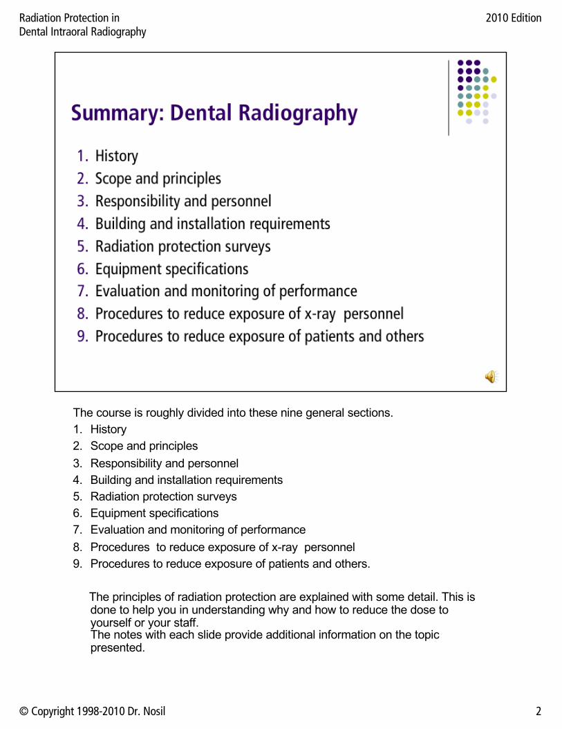
The course is roughly divided into these nine general sections.1. History2. Scope and principles 3. Responsibility and personnel4. Building and installation requirements5. Radiation protection surveys6. Equipment specifications7. Evaluation and monitoring of performance8. Procedures to reduce exposure of x-ray personnel9. Procedures to reduce exposure of patients and others.
The principles of radiation protection are explained with some detail. This is done to help you in understanding why and how to reduce the dose to yourself or your staff.The notes with each slide provide additional information on the topic presented.
Radiation Protection inDental Intraoral Radiography
2010 Edition
© Copyright 1998-2010 Dr. Nosil 2
Radiation Protection inDental Intraoral Radiography
2010 Edition
© Copyright 1998-2010 Dr. Nosil 3
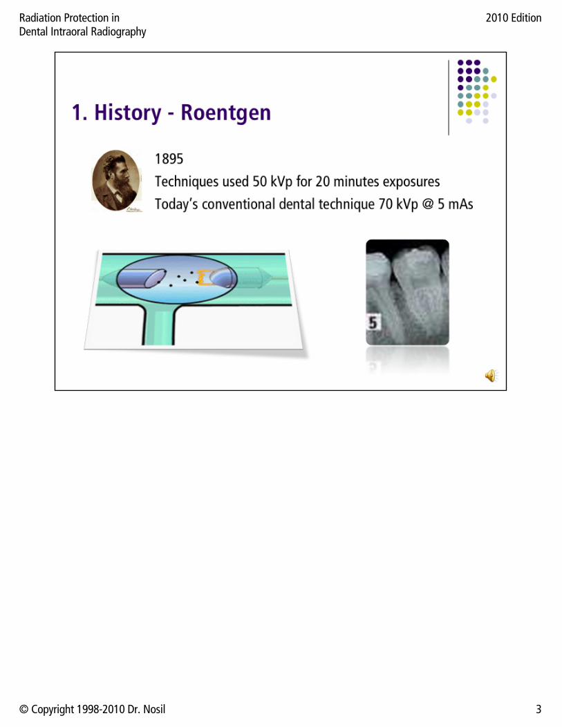
The previous basic physics and radiobiology lecture examined what happens at the atomic or cell level when x-rays impinge upon matter. We saw that atoms were ionized by the removal of one or more orbital electrons, with the result that ‘ion pairs' are produced. We also learned how cells can be altered by free radicals, and how effects on humans produced. This presentation will answer following questions:1. What does radiation safety mean to you?2. Three types of dental X-Ray units must be licensed. What are they?3. What are the four D's of radiation safety?4. What is the unit that describes the biological effect of radiation?5. In terms of these units, during a bitewing what is the exposure dose that the skin of the patient receives?6. In terms of these units, what is the regulatory limit for the annual exposure of radiation workers?7. Why is the size and shape of the intraoral applicator subject to regulation?8. Why is the method of processing of dental films important from a radiation protection viewpoint?9. What is the significance of focal spot size in dental radiography?
Radiation Protection inDental Intraoral Radiography
2010 Edition
© Copyright 1998-2010 Dr. Nosil 4

This document applies to the use of equipment specifically designed for radiography of the teeth or jaws including radiography using intra-oral image receptor radiography.
This Bermuda seminar has been prepared to provide specific recommendation to the dentist, dental hygienist, dental assistant, and other support personnel concerned with safety procedures and equipment performance.
The aim of radiation protection in dentistry is to obtain the desired clinical information with minimum radiation exposure to patients, dental personnel, and the public.
There are four main concerns when dealing with radiation hazards.1. First, patients should not be subjected to unnecessary dental
radiography.2. Second, patients need to be protected from unnecessary exposures.3. Third, it is essential that personnel in dental facilities be protected
from unnecessary exposure to radiation in the course of their work. 4. Finally, the public requires adequate protection.
Radiation Protection inDental Intraoral Radiography
2010 Edition
© Copyright 1998-2010 Dr. Nosil 5
We know that x-rays, in sufficient doses, may produce harmful effects in human beings. However, we do not know the size of the risk (or even if there is any risk at all) from small doses of x-rays such as those used in dental radiography. It is the consensus of dental radiologists that the dosage from dental x-ray exposure is not harmful.However, the absence of conclusive proof that establishes the absence of risk means we must assume that there is the potential of some risk from diagnostic exposure. Whenever we consider exposing patients to x-rays, the ALARA principle (As Low As Reasonably Achievable) applies. Any dose that can be reduced without major difficulty, great expense, or inconvenience should be reduced or eliminated. The benefits of dental radiography certainly outweigh the potential risks.
Radiation Protection inDental Intraoral Radiography
2010 Edition
© Copyright 1998-2010 Dr. Nosil 6
Dental X-ray equipment must only be operated by individuals who have been trained in the safe use of the equipment and the procedures being performed. An operator is any person who is entitled to carry out all, or part of the practical aspects associated with a dental radiographic examination. Practical aspects include:
(a) patient identification;(b) positioning the film, the patient and the x-ray tube head;(c) setting the exposure parameters;(d) pressing the exposure button to initiate the exposure;(e) processing films;(f) clinical evaluation of radiographs;(g) exposing test objects as part of the QA programme.
Non-clinical staff should not normally be asked to undertake the majority of operator’s duties. Some of the more straightforward operator’s duties may be undertaken by non-clinical staff, but these staff must have been appropriately trained for all of the tasks that they perform. A female operator should immediately notify her employer upon knowledge that she is pregnant.
Radiation Protection inDental Intraoral Radiography
2010 Edition
© Copyright 1998-2010 Dr. Nosil 7
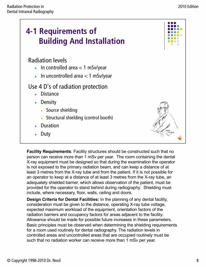
Facility Requirements: Facility structures should be constructed such that no person can receive more than 1 mSv per year. The room containing the dental X-ray equipment must be designed so that during the examination the operator is not exposed to the primary radiation beam, and can keep a distance of at least 3 metres from the X-ray tube and from the patient. If it is not possible for an operator to keep at a distance of at least 3 metres from the X-ray tube, an adequately shielded barrier, which allows observation of the patient, must be provided for the operator to stand behind during radiography. Shielding must include, where necessary, floor, walls, ceiling and doors.Design Criteria for Dental Facilities: In the planning of any dental facility, consideration must be given to the distance, operating X-ray tube voltage, expected maximum workload of the equipment, orientation factors of the radiation barriers and occupancy factors for areas adjacent to the facility. Allowance should be made for possible future increases in these parameters. Basic principles must be observed when determining the shielding requirements for a room used routinely for dental radiography. The radiation levels in controlled areas and uncontrolled areas that are occupied routinely must be such that no radiation worker can receive more than 1 mSv per year.
Radiation Protection inDental Intraoral Radiography
2010 Edition
© Copyright 1998-2010 Dr. Nosil 8

Determination of Shielding: To determine detailed shielding necessary for a dental facility, certain preliminary information is essential. It is recommended to contact the appropriate agency to enquire about shielding requirements and calculations. To determine shielding requirements, the following information is necessary:
1. What is the distance between the nearest point of the area to be shielded and the usual operational position of the X-ray tube?
2. Is the area to be designated as a controlled or uncontrolled area? (The area occupied by non-radiation workers is subject to the limit of 1 mSv per year.)
3. Will the intervening shield between the X-ray tube and the occupied area attenuate the direct radiation beam or stray radiation?
4. What will be the anticipated maximum workload of the dental X-ray unit? (The workload indicates the operational time of a dental X-ray unit in milliampere-seconds per week.)
5. What will be the maximum operational X-ray tube voltage?6. What are the Occupancy and Orientation Factors?
Radiation Protection inDental Intraoral Radiography
2010 Edition
© Copyright 1998-2010 Dr. Nosil 9

Examples of Building and Installation Requirements: Structural shielding of dental units operating up to above 70 kVp.
•Walls protecting dentists, dental hygienists and dental assistants - 2” thickness of gypsum board required.•Walls protecting receptionists, secretaries and other members of the public - 4” thickness of gypsum board required.
Room warning signs and signals: When a controlled area (i.e. > 1 mSv) extends to any entrance of the x-ray room, a warning notice should be provided on the outside of the door. This notice must conform to current legal requirements and include the basic ionising radiation symbol.
Radiation Protection inDental Intraoral Radiography
2010 Edition
© Copyright 1998-2010 Dr. Nosil 10

Film and chemical storage: Since radiographic films are sensitive to light, heat, humidity, chemical contamination, mechanical stress and X-radiation, they should be stored at temperatures in the range of 10°C to 21°C with humidity from 30% to 60%. It is best to follow the film manufacturer’s instruction. Film storage areas should be free of chemical fumes and X-radiation. Manufacturer’s recommendations should be followed in storing chemicals to avoid oxidization and any chemicals showing sign of oxidization or sedimentation must not be used.Darkroom conditions: The darkroom must be clean of dirt, dust, and free of spilled chemical residues. The darkroom must be light-tight and proper darkroom lighting used.
Radiation Protection inDental Intraoral Radiography
2010 Edition
© Copyright 1998-2010 Dr. Nosil 11
It is recommended that routine radiation protection inspections be carried out at intervals not exceeding two years to verify the dental X-ray equipment functions properly and according to applicable standards and legislative requirements, the dental X-ray equipment is installed in a safe environment and is used in a way which provides maximum radiation safety for patients and operators, the Quality Assurance program is properly implemented and maintained and that the maximum benefits are obtained from the program.However, annual testing is recommended if assessed representative patient doses exceed the recommended levels, image quality analysis indicates a failure to meet the accepted or the Survey from previous year identifies some other significant performance weakness.A record of maintenance, including any defects found and their repair, should be kept for each item of x-ray equipment and relevant auxiliary equipment. Following maintenance, the service engineer should provide a written report prior to handing the equipment back for clinical use. This should detail any changes that may affect radiation dose (to patient or staff) or image quality. The Medical Physicist should be consulted as necessary.
Radiation Protection inDental Intraoral Radiography
2010 Edition
© Copyright 1998-2010 Dr. Nosil 12
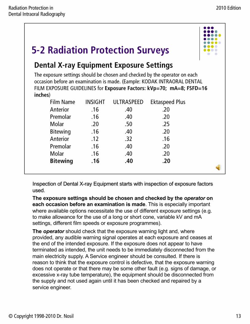
Inspection of Dental X-ray Equipment starts with inspection of exposure factors used.The exposure settings should be chosen and checked by the operator on each occasion before an examination is made. This is especially important where available options necessitate the use of different exposure settings (e.g. to make allowance for the use of a long or short cone, variable kV and mAsettings, different film speeds or exposure programmes).The operator should check that the exposure warning light and, where provided, any audible warning signal operates at each exposure and ceases at the end of the intended exposure. If the exposure does not appear to have terminated as intended, the unit needs to be immediately disconnected from the main electricity supply. A Service engineer should be consulted. If there is reason to think that the exposure control is defective, that the exposure warning does not operate or that there may be some other fault (e.g. signs of damage, or excessive x-ray tube temperature), the equipment should be disconnected from the supply and not used again until it has been checked and repaired by a service engineer.
Radiation Protection inDental Intraoral Radiography
2010 Edition
© Copyright 1998-2010 Dr. Nosil 13
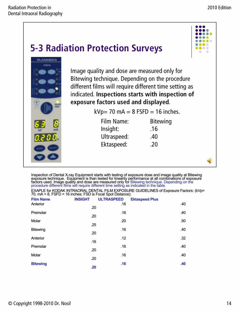
Inspection of Dental X-ray Equipment starts with testing of exposure dose and image quality at Bitewing exposure technique. Equipment is than tested for linearity performance at all combinations of exposure factors used. Image quality and dose are measured only for Bitewing technique. Depending on the procedure different films will require different time setting as indicated in the table.EXAPLE for KODAK INTRAORAL DENTAL FILM EXPOSURE GUIDELINES of Exposure Factors: (kVp= 70, mA = 8, FSFD = 16 inches; FSD is Focal Spot Distance):Film Name INSIGHT ULTRASPEED Ektaspeed PlusAnterior .16 .40
.20Premolar .16 .40
.20Molar .20 .50
.25Bitewing .16 .40
.20Anterior .12 .32
.16Premolar .16 .40
.20Molar .16 .40
.20Bitewing .16 .40
.20
Radiation Protection inDental Intraoral Radiography
2010 Edition
© Copyright 1998-2010 Dr. Nosil 14
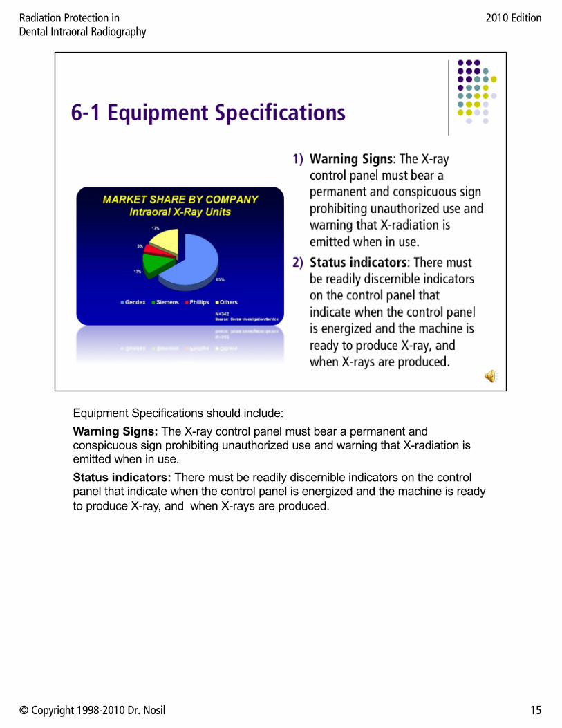
Equipment Specifications should include:Warning Signs: The X-ray control panel must bear a permanent and conspicuous sign prohibiting unauthorized use and warning that X-radiation is emitted when in use.Status indicators: There must be readily discernible indicators on the control panel that indicate when the control panel is energized and the machine is ready to produce X-ray, and when X-rays are produced.
Radiation Protection inDental Intraoral Radiography
2010 Edition
© Copyright 1998-2010 Dr. Nosil 15
Equipment Specifications should include:3. Indication of loading factors: For dental X-ray equipment having adjustable loading factors, the control panel must incorporate meters or other indicators that enable determination of the X-ray tube voltage, X-ray tube current and time, or combinations of these. For equipment having non-adjustable loading factors, permanent marks or labels may be used to indicate these parameters.4. Irradiation switch: There must be an irradiation switch to start and terminate X-ray production. This switch must require continuous pressure by the operator to produce X-rays. Where the irradiation switch is a footswitch it must be so constructed that operation of the X-ray tube cannot occur inadvertently. The irradiation switch must be at least 3 metres from the tube house.
Radiation Protection inDental Intraoral Radiography
2010 Edition
© Copyright 1998-2010 Dr. Nosil 16

Equipment Specifications should include:5. Controlling timer: An electronic timing device must be provided to automatically terminate the irradiation. The timer must be designed and constructed in such a way that it is not possible to energize the X-ray tube without automatic or manual resetting of the timer after each loading, irradiation cannot be started with the timer set at its zero or OFF position, and the production of X-rays is automatically terminated after a preset time, preset milliampere second value, a preset exposure or air kerma value.6. Beam filtration: The total filtration of the beam should be equivalent to not less than 1.5 mm aluminum for x-ray tube voltages up to and including 70 kV or 2.5 mm aluminum, (of which 1.5 mm should be permanent), for x-ray tube voltages above 70 kV.
Radiation Protection inDental Intraoral Radiography
2010 Edition
© Copyright 1998-2010 Dr. Nosil 17

Equipment Specifications should include:7. Irradiation reproducibility: For a series of 10 consecutive radiation
measurements taken at the same distance from the target, in the X-ray beam, within a time period of one hour, and where all variable controls for loading factors are adjusted to alternate settings and reset to the test setting before each measurement, the coefficient of variation of measurements is not greater than 0.05.
8. Mechanical stability: The X-ray tube must be securely fixed and correctly aligned within the tube housing. The X-ray tube housing must maintain its required position or movement without excessive drift or vibration during operation and must be supported by mechanical means. The drift shall be no greater than 0.5 cm.
Radiation Protection inDental Intraoral Radiography
2010 Edition
© Copyright 1998-2010 Dr. Nosil 18
Equipment Specifications should include:9. X-ray source assembly X-ray tube shielding: The X-ray tube must be
enclosed within a shielded housing. The housing must be constructed so that the leakage radiation, measured at a distance of one metre in any direction from the focal spot of the X-ray tube, does not exceed 0.25 mGy in one hour for any specified rating of the tube. The x-ray tube head should be marked to identify the nominal focal spot position.
10. X-ray tube voltage: The actual peak X-ray tube voltage should not deviate from the indicated or selected value by more than 5%, or by the value specified by the manufacturer. It must not be possible to set or operate the X-ray tube with the tube voltage below 50 kilovolts (peak).
Radiation Protection inDental Intraoral Radiography
2010 Edition
© Copyright 1998-2010 Dr. Nosil 19
Equipment Specifications should include:9. X-ray tube current: The actual X-ray tube current should not deviate from the
indicated or selected value by more than 5%, or by the value specified by the manufacturer, and be temperature compensated for normal operating conditions
10. Linearity: For any selected X-ray tube voltage linearity must be according to the specification in REGULATION DOCUMENTS.
11. Focal spot to skin distance: Must be more than 18 cm
Radiation Protection inDental Intraoral Radiography
2010 Edition
© Copyright 1998-2010 Dr. Nosil 20
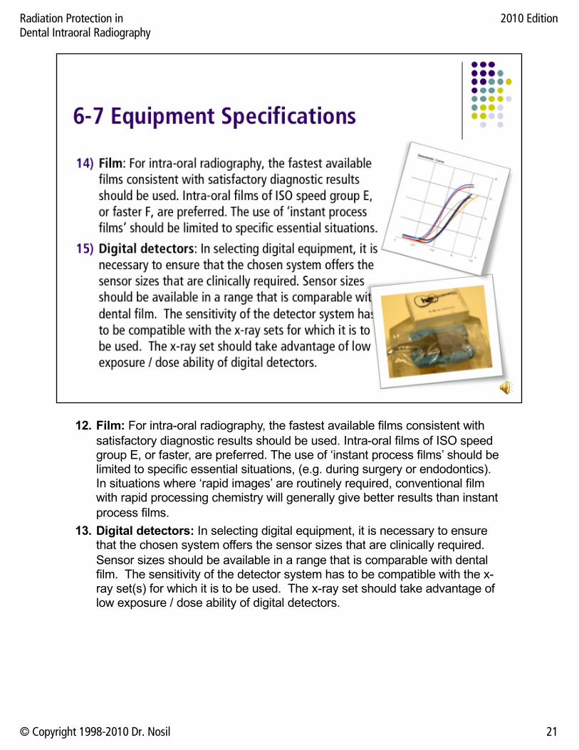
12. Film: For intra-oral radiography, the fastest available films consistent with satisfactory diagnostic results should be used. Intra-oral films of ISO speed group E, or faster, are preferred. The use of ‘instant process films’ should be limited to specific essential situations, (e.g. during surgery or endodontics). In situations where ‘rapid images’ are routinely required, conventional film with rapid processing chemistry will generally give better results than instant process films.
13. Digital detectors: In selecting digital equipment, it is necessary to ensure that the chosen system offers the sensor sizes that are clinically required. Sensor sizes should be available in a range that is comparable with dental film. The sensitivity of the detector system has to be compatible with the x-ray set(s) for which it is to be used. The x-ray set should take advantage of low exposure / dose ability of digital detectors.
Radiation Protection inDental Intraoral Radiography
2010 Edition
© Copyright 1998-2010 Dr. Nosil 21
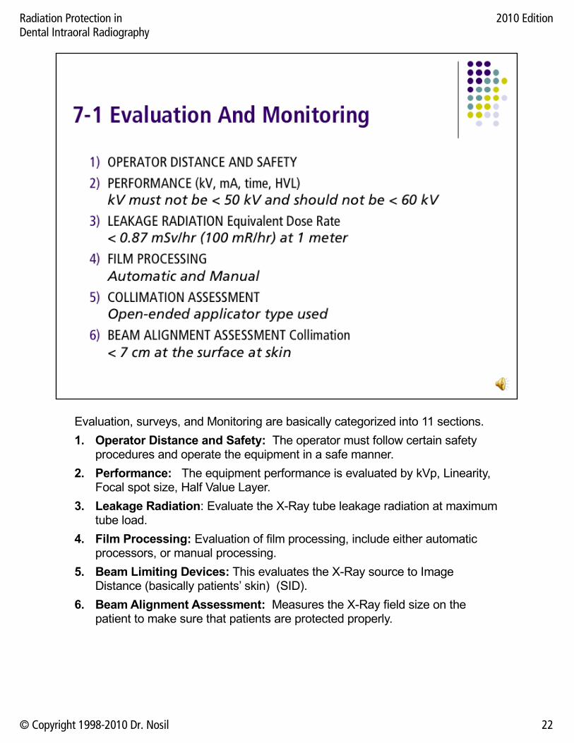
Evaluation, surveys, and Monitoring are basically categorized into 11 sections.1. Operator Distance and Safety: The operator must follow certain safety
procedures and operate the equipment in a safe manner. 2. Performance: The equipment performance is evaluated by kVp, Linearity,
Focal spot size, Half Value Layer.3. Leakage Radiation: Evaluate the X-Ray tube leakage radiation at maximum
tube load. 4. Film Processing: Evaluation of film processing, include either automatic
processors, or manual processing.5. Beam Limiting Devices: This evaluates the X-Ray source to Image
Distance (basically patients’ skin) (SID). 6. Beam Alignment Assessment: Measures the X-Ray field size on the
patient to make sure that patients are protected properly.
Radiation Protection inDental Intraoral Radiography
2010 Edition
© Copyright 1998-2010 Dr. Nosil 22
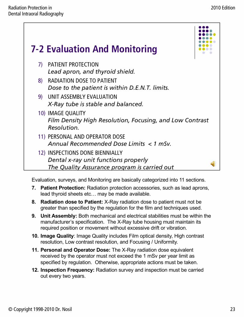
Evaluation, surveys, and Monitoring are basically categorized into 11 sections.7. Patient Protection: Radiation protection accessories, such as lead aprons,
lead thyroid sheets etc… may be made available.8. Radiation dose to Patient: X-Ray radiation dose to patient must not be
greater than specified by the regulation for the film and techniques used. 9. Unit Assembly: Both mechanical and electrical stabilities must be within the
manufacturer’s specification. The X-Ray tube housing must maintain its required position or movement without excessive drift or vibration.
10. Image Quality: Image Quality includes Film optical density, High contrast resolution, Low contrast resolution, and Focusing / Uniformity.
11. Personal and Operator Dose: The X-Ray radiation dose equivalent received by the operator must not exceed the 1 mSv per year limit as specified by regulation. Otherwise, appropriate actions must be taken.
12. Inspection Frequency: Radiation survey and inspection must be carried out every two years.
Radiation Protection inDental Intraoral Radiography
2010 Edition
© Copyright 1998-2010 Dr. Nosil 23
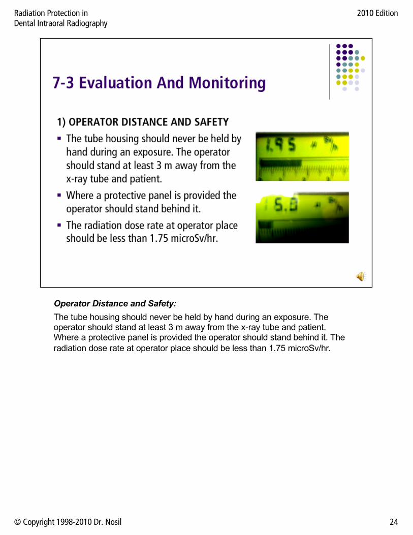
Operator Distance and Safety:The tube housing should never be held by hand during an exposure. The operator should stand at least 3 m away from the x-ray tube and patient. Where a protective panel is provided the operator should stand behind it. The radiation dose rate at operator place should be less than 1.75 microSv/hr.
Radiation Protection inDental Intraoral Radiography
2010 Edition
© Copyright 1998-2010 Dr. Nosil 24
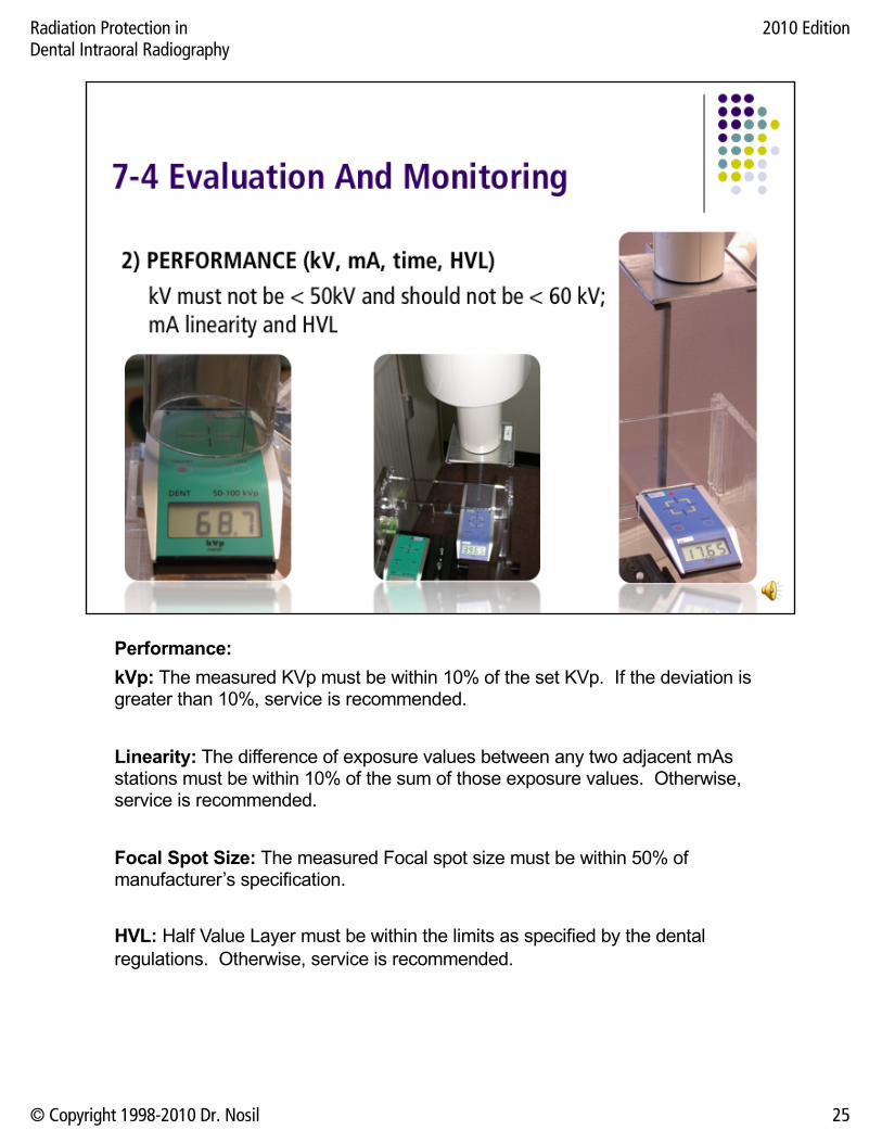
Performance:kVp: The measured KVp must be within 10% of the set KVp. If the deviation is greater than 10%, service is recommended.
Linearity: The difference of exposure values between any two adjacent mAsstations must be within 10% of the sum of those exposure values. Otherwise, service is recommended.
Focal Spot Size: The measured Focal spot size must be within 50% of manufacturer’s specification.
HVL: Half Value Layer must be within the limits as specified by the dental regulations. Otherwise, service is recommended.
Radiation Protection inDental Intraoral Radiography
2010 Edition
© Copyright 1998-2010 Dr. Nosil 25

Leakage Radiation:X-Ray tube leakage radiation rate must be no more than 0.25 mGy/hr at 1-meter distance in any direction. Otherwise, service is recommended.
This test is performed with the X-ray tube under maximum load.
Radiation Protection inDental Intraoral Radiography
2010 Edition
© Copyright 1998-2010 Dr. Nosil 26

Image Receptor Specifications: The performance of image receptor systems can be characterized by five parameters: speed, contrast, latitude, resolution, and noise. Intraoral film: Conventional intraoral film is direct exposure film, in that the latent image is produced by the direct exposure of film emulsion by the x-ray beam. Intraoral film speed is designated by speed as groups C through F. Whenever possible, the fastest speed of film should be used since patient exposure can be significantly reduced without compromising diagnostic quality.Film Storage: Film storage container must be adequately shielded so that no film receives more than 1.75 µGy (micro Grays) of radiation before use. For the majority of facilities, 1.5 mm of lead shielding will be more than adequate. Films should be stored in a cool, dry area.
Radiation Protection inDental Intraoral Radiography
2010 Edition
© Copyright 1998-2010 Dr. Nosil 27
Film processing: The film must be processed in chemically fresh developer, at proper temperature and for sufficient time to ensure that the silver in exposed silver halide crystals in the film emulsion is completely reduced, to achieve full development of the film. Other factors, such as cleanliness of the processing system, film immersion time, agitation, the efficiency of the rinsing, and temperature control can also affect the quality of the processed film. Manual film processing of conventional dental X-ray films is not recommended, especially for high workload facilities. Manual processing is acceptable for facilities where the workload is very low, only a few films per day.Processing recommendations: It is necessary to observe the following recommendations:(a) Manufacturers’ recommendations concerning the strength of solution, temperature and time must be strictly followed to ensure optimum development. An accurate thermometer is essential for adequate processing and, for manual processing; an accurate timer must be used.(b) Developing solutions must be replenished as necessary and must be changed regularly, as required.(c) Developing solutions must be monitored regularly. Developer must not be used when processing times become significantly longer than what is recommended by the manufacturers or the radiation dose necessary to obtain an acceptable film density has increased significantly.(d) Cleanliness is extremely important for reducing film artifacts in both manual and automatic film processing. Proper stainless steel processing tanks complete with water bath and lids must be used when manual processing is used. With automatic processors, the film transport mechanisms must be cleaned frequently.(e) Automatic film processors must be maintained regularly, in accordance with the manufacturers’ instructions, and the temperature and composition of the processing chemicals must be kept within the specified tolerances.
Radiation Protection inDental Intraoral Radiography
2010 Edition
© Copyright 1998-2010 Dr. Nosil 28

Darkroom recommendations:Manual processing of films requires the use of a proper, well-equipped darkroom. The following applies to all darkrooms:(a) The room must be light-tight.(b) The darkroom must be designed to ensure light-tightness when undeveloped films are being handled.(c) A warning light should be located outside the darkroom, at the entrance, to indicate when the room is in use.(d) Safelights equipped with bulbs of correct intensity must be provided above the work area within the darkroom. Safelights must have filters appropriate to meet the specifications of the film used and must be positioned at the proper distances from work areas. Safelight filters should be checked regularly since they may deteriorate with time or may crack.(e) The darkroom must be equipped with proper stainless steel processing tanks with water bath and lids, including an accurate thermometer and timing device.
Radiation Protection inDental Intraoral Radiography
2010 Edition
© Copyright 1998-2010 Dr. Nosil 29

Collimation Performance:The primary radiation beam must be collimated in size at the end of the applicator to a circle not more than 6 centimeters in diameter, or a rectangle of area not more than 38.5 cm2. Otherwise, service or replace is needed.The left picture depicts that all radiation comes from leakage of the tube, when the collimator is closed completely. Middle picture, when the collimator is open half way, shows the contributions of radiation from both leakage and scatter radiations.The picture on the right when the collimator is open all the way, shows the same contribution of radiation from leakage and increased contribution of scatter radiations.
Radiation Protection inDental Intraoral Radiography
2010 Edition
© Copyright 1998-2010 Dr. Nosil 30
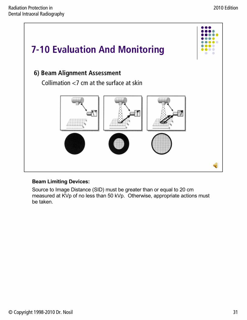
Beam Limiting Devices:Source to Image Distance (SID) must be greater than or equal to 20 cm measured at KVp of no less than 50 kVp. Otherwise, appropriate actions must be taken.
Radiation Protection inDental Intraoral Radiography
2010 Edition
© Copyright 1998-2010 Dr. Nosil 31

Patient and Public Protection: Radiation protection accessories, such as lead aprons, lead thyroid sheets etc… may be made available.
Radiation Protection inDental Intraoral Radiography
2010 Edition
© Copyright 1998-2010 Dr. Nosil 32
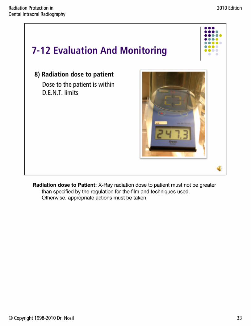
Radiation dose to Patient: X-Ray radiation dose to patient must not be greater than specified by the regulation for the film and techniques used. Otherwise, appropriate actions must be taken.
Radiation Protection inDental Intraoral Radiography
2010 Edition
© Copyright 1998-2010 Dr. Nosil 33
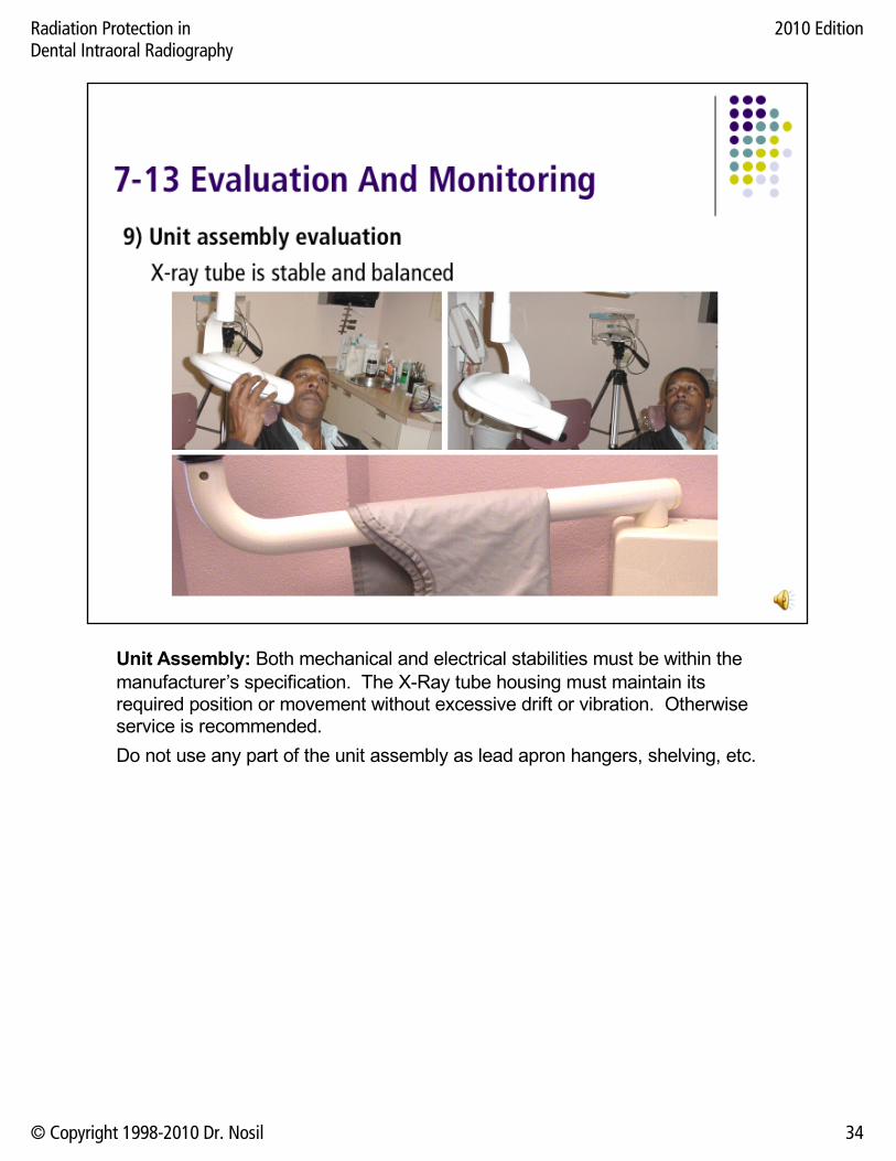
Unit Assembly: Both mechanical and electrical stabilities must be within the manufacturer’s specification. The X-Ray tube housing must maintain its required position or movement without excessive drift or vibration. Otherwise service is recommended.Do not use any part of the unit assembly as lead apron hangers, shelving, etc.
Radiation Protection inDental Intraoral Radiography
2010 Edition
© Copyright 1998-2010 Dr. Nosil 34

Image QualityFilm optical density: The optical density of the film (no air, using the NEXT phantom) must be between 1.4 and 1.6.High contrast resolution: The high contrast resolution must be no less than 5.9 lp/mm.Low contrast resolution: The low contrast resolution must be no less than 2 holes completely resolved.Focusing / Uniformity: The image must not be blurred, and no visible artifacts should be present.
Radiation Protection inDental Intraoral Radiography
2010 Edition
© Copyright 1998-2010 Dr. Nosil 35
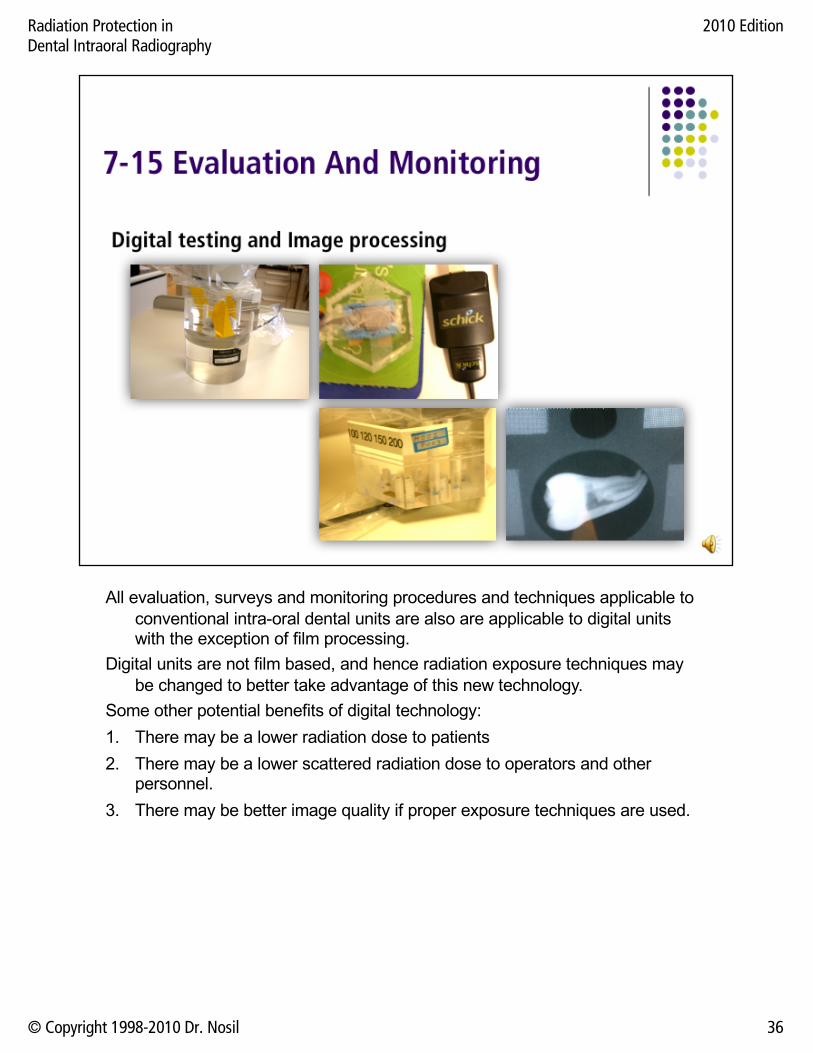
All evaluation, surveys and monitoring procedures and techniques applicable to conventional intra-oral dental units are also are applicable to digital units with the exception of film processing.
Digital units are not film based, and hence radiation exposure techniques may be changed to better take advantage of this new technology.
Some other potential benefits of digital technology:1. There may be a lower radiation dose to patients2. There may be a lower scattered radiation dose to operators and other
personnel.3. There may be better image quality if proper exposure techniques are used.
Radiation Protection inDental Intraoral Radiography
2010 Edition
© Copyright 1998-2010 Dr. Nosil 36
11. Personal and Operator Dose: The X-Ray radiation dose equivalent received by the operator must not exceed the 1 mSv per year limit as specified by regulation. Otherwise, appropriate actions must be taken.
All units should be inspected every two years.
Radiation Protection inDental Intraoral Radiography
2010 Edition
© Copyright 1998-2010 Dr. Nosil 37
Procedures to Reduce Radiation Exposure to PersonnelA room must not be used at the same time for more than one radiological investigation.All persons, other than the patient and those whose presence is essential, must leave the room when a radiographic examination is carried out.Personnel must always keep as far away from the primary radiation beam as practical. Direct radiation exposure to personnel must not occur. Deliberate irradiation of an individual for training purposes must never be allowed. The operation of X-ray tube should be controlled from the control panel located outside the radiography room or behind a protective barrier. In special circumstances, where the operator is required to control the loading while at the side of the patient, protective clothing must be worn.The dental film or digital detector should only be held by the patient when it cannot otherwise be kept in position. It should not normally be hand-held by anyone else. Exceptionally it may be held by someone other than the patient using a pair of forceps, or other appropriate holder, to avoid direct irradiation of the fingers, for example, when a child or a handicapped person cannot hold it themselves. In such cases advice should be sought from Radiation Safety Office.
Radiation Protection inDental Intraoral Radiography
2010 Edition
© Copyright 1998-2010 Dr. Nosil 38
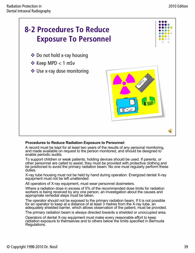
Procedures to Reduce Radiation Exposure to Personnel:A record must be kept for at least two years of the results of any personal monitoring, and made available on request to the person monitored, and should be designed to enable periodic audits. To support children or weak patients, holding devices should be used. If parents, or other personnel are called to assist, they must be provided with protective clothing and be positioned to avoid the primary radiation beam. No one must regularly perform these duties.X-ray tube housing must not be held by hand during operation. Energized dental X-ray equipment must not be left unattended.All operators of X-ray equipment, must wear personnel dosimeters.Where a radiation dose in excess of 5% of the recommended dose limits for radiation workers is being received by any one person, an investigation about the causes and appropriate remedial steps must be taken.The operator should not be exposed to the primary radiation beam. If it is not possible for an operator to keep at a distance of at least 3 metres from the X-ray tube, an adequately shielded barrier, which allows observation of the patient, must be provided.The primary radiation beam is always directed towards a shielded or unoccupied area.Operators of dental X-ray equipment must make every reasonable effort to keep radiation exposure to themselves and to others below the limits specified in Bermuda Regulations.
Radiation Protection inDental Intraoral Radiography
2010 Edition
© Copyright 1998-2010 Dr. Nosil 39
Procedures specific to intra-oral radiography: Whenever practicable, techniques using film holders incorporating beam-aiming devices should be adopted for bitewing and periapical radiography. If rectangular collimation is being used, a beam alignment device is essential for accurate alignment with the intra-oral film. Attention is drawn to the probable need for additional operator training in the use of film holders when moving from circular to rectangular collimation.When a choice of beam collimators is provided, the one most suited to the technique to be employed should be fitted, ideally just covering the film or image detector. The open end of the collimator should be placed as close as possible to the patient’s head to minimize the size of the incident x-ray beam. If it is desired to use a longer focus-to-skin distance (FSD), a longer collimator/director should be employed.
Radiation Protection inDental Intraoral Radiography
2010 Edition
© Copyright 1998-2010 Dr. Nosil 40
The dental practitioner will reduce unnecessary radiation exposure to the patient by eliminating examinations that are not clinically justified. Basic recommendations are:A radiographic examination should be for the purpose of obtaining diagnostic information about the patient to aid in a clinical evaluation of the patient and treatment when warranted.Routine or screening examinations, in which there is no prior clinical evaluation of the patient, should not be prescribed. It is considered a bad practice to radiograph patients unnecessarily, as in a standard survey, and this is especially deplored when done on children. It is also considered bad practice to take radiograms before a clinical examination by the dentist. These two practices constitute the largest potential abuse of radiology in dentistry.It should be determined whether there have been any previous radiographic examinations which would make further examination unnecessary or allow for an abbreviated radiographic examination. When a patient is transferred from one practitioner to another, any relevant radiograms should accompany the patient or should be requested from the previous dentist.The number of radiographic views required in an examination should be kept to the minimum practical, consistent with the clinical objectives of the examination.In prescribing radiographic examinations of pregnant or possibly pregnant women, full consideration should be taken of the consequences of fetal irradiation. Repeat radiographic examinations should not be prescribed simply because a radiogram may not be of the “best” diagnostic quality, but does provide the desired information.A patient’s clinical records should include details of all radiographic examinations carried out.
Radiation Protection inDental Intraoral Radiography
2010 Edition
© Copyright 1998-2010 Dr. Nosil 41
It is the responsibility of the operator and dental practitioner to be aware of this and to know how to carry out a prescribed examination with the lowest practical dose to the patient.The operator must not perform any radiographic examinations not prescribed by the dental practitioner responsible for the patient.Dental radiography must not be carried out at X-ray tube voltages below 50 kilovolts (peak) and should not be carried out at X-ray tube voltages below 60 kilovolts (peak).Dental X-ray equipment should be well maintained and its performance checked routinely. Accurate calibration of the equipment should also be carried out on a regular basis.The quality of radiograms should be monitored routinely, through a Quality Assurance program, to ensure that they satisfy diagnostic requirements with minimal radiation exposure to the patient.The fastest film or film-screen combination consistent with the requirements of the examination should be used. The film processing technique should ensure optimum development and should be in accordance with manufacturer’s recommendations.Dental X-ray films must be examined with a viewbox specifically designed for this purpose.The operator should not be exposed to the primary radiation beam and can keep a distance of at least 3 metres from the X-ray tube and from the patient. If it is not possible for an operator to keep at a distance of at least 3 metres from the X-ray tube, an adequately shielded barrier, which allows observation of the patient, must be provided for the operator to stand behind during radiography.The primary X-ray beam must be collimated to irradiate the minimum area necessary for the examination.The primary X-ray beam should be aligned and the patient’s head positioned in such a way that the beam is not directed at the patient’s gonads and is not unnecessarily irradiating the patient’s body.
Radiation Protection inDental Intraoral Radiography
2010 Edition
© Copyright 1998-2010 Dr. Nosil 42

Guidelines for Patient Protection: Protective EquipmentThe patient may be provided with a shielded apron, for gonad protection, and a thyroid shield, especially during occlusal radiographic examinations of the maxilla. The use of a thyroid shield is especially important in children. The shielded apron and thyroid shield should have a lead equivalence of at least 0.25 mm of lead. In panoramic radiography, since the radiation is also coming from the back of the patient, a conventional lead apron is not adequate and dual (front and back) lead aprons should be worn.Protective Equipment SpecificationsThere is no justification for the routine use of lead aprons for patients in dental radiography.Thyroid collars should be used in those few cases where the thyroid may be in the primary beam, based on advice from a fully qualified expert. Lead aprons only provide a practicable degree of protection in the case of the infrequently used vertex occlusal projection. Even in that case, the use of the lead apron could only be regarded as prudent for a female patient who is, or may be, pregnant.Protective aprons, having a lead equivalence of not less than 0.25 mm, should be provided for any adult who provides assistance by supporting a patient.When a lead apron is provided, it must be correctly stored (e.g. over a suitable hanger) and not folded. Its condition must be routinely checked including a visual inspection at annual intervals.
Radiation Protection inDental Intraoral Radiography
2010 Edition
© Copyright 1998-2010 Dr. Nosil 43
Guidelines for Patient ProtectionIt is generally agreed by experts in the scientific community that radiation exposure to patients from medical and dental radiographic sources can be reduced substantially with no decrease in the value of diagnostic information derived.Every effort should be made to reduce the number of radiograms and the number of persons examined radio-graphically, as well as to reduce the dose involved in a particular examination.It is essential that patients not be subjected to unnecessary radiological examinations and, when a radiological examination is required, it is essential that patients be protected from excessive radiation exposure during the examination.The dose to the patient must be kept to the lowest practical value, consistent with clinical objectives. To achieve this, techniques appropriate to the equipment available should be used. It is recommended the X-ray loading factors charts be established when using X-ray units that do not have preprogrammed anatomical feature settings. The loading factors chart must be established after optimizing the film processing procedure.
Radiation Protection inDental Intraoral Radiography
2010 Edition
© Copyright 1998-2010 Dr. Nosil 44

Dental radiography must not be carried out at X-ray tube voltages below 50 kilovolts (peak) and should not be carried out at X-ray tube voltages below 60 kilovolts (peak).Lower voltages can not penetrate the denser parts, which will be underexposed, and dose is increased.Higher voltages produce poorer contrast images because of more scattering, but the overall dose is lowered.
Radiation Protection inDental Intraoral Radiography
2010 Edition
© Copyright 1998-2010 Dr. Nosil 45
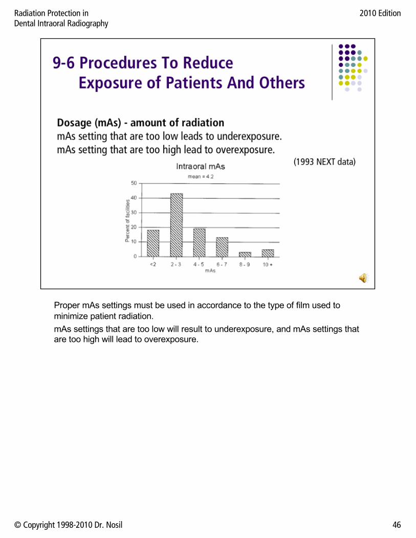
Proper mAs settings must be used in accordance to the type of film used to minimize patient radiation.mAs settings that are too low will result to underexposure, and mAs settings that are too high will lead to overexposure.
Radiation Protection inDental Intraoral Radiography
2010 Edition
© Copyright 1998-2010 Dr. Nosil 46
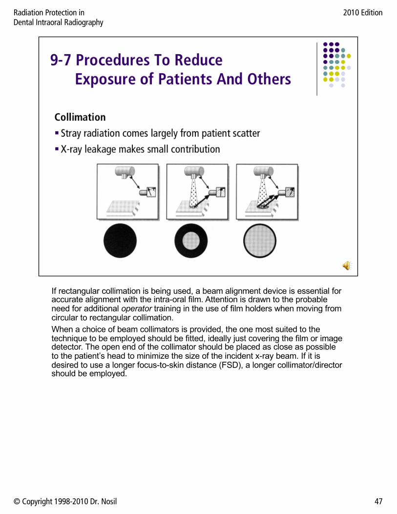
If rectangular collimation is being used, a beam alignment device is essential for accurate alignment with the intra-oral film. Attention is drawn to the probable need for additional operator training in the use of film holders when moving from circular to rectangular collimation.When a choice of beam collimators is provided, the one most suited to the technique to be employed should be fitted, ideally just covering the film or image detector. The open end of the collimator should be placed as close as possible to the patient’s head to minimize the size of the incident x-ray beam. If it is desired to use a longer focus-to-skin distance (FSD), a longer collimator/director should be employed.
Radiation Protection inDental Intraoral Radiography
2010 Edition
© Copyright 1998-2010 Dr. Nosil 47
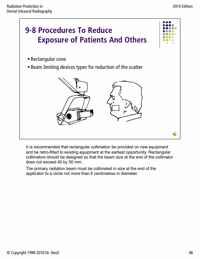
It is recommended that rectangular collimation be provided on new equipment and be retro-fitted to existing equipment at the earliest opportunity. Rectangular collimators should be designed so that the beam size at the end of the collimator does not exceed 40 by 50 mm.The primary radiation beam must be collimated in size at the end of the applicator to a circle not more than 6 centimetres in diameter.
Radiation Protection inDental Intraoral Radiography
2010 Edition
© Copyright 1998-2010 Dr. Nosil 48
A position-indicating device must be provided to limit the minimum focal spot to skin distance to not less than 18 centimeters. The applicator must be an open-ended type. Pointed cone or close-ended applicators must not be used.
Radiation Protection inDental Intraoral Radiography
2010 Edition
© Copyright 1998-2010 Dr. Nosil 49

Whatever the application, there are many aspects to consider when selecting dental X-Ray film including low radiation, infection control, speed, convenience and of course, a quality image. Kodak has a comprehensive range of products.Kodak Ektaspeed Plus dental film is a high speed (category E), high contrast, dental film which uses a patented T-grain emulsion to provide maximum detail. The high speed makes it ideally suited for long-cone radiographic techniques and modern X-Ray generating equipment designed for short exposures (the Kodak Dental X-Ray Beam Filter Kit is available for all other equipment). The fast speed also permits a significant reduction in patient exposure by up to 50%, while continuing to capture quality images. Kodak Ektaspeed Plus dental film is available in 3 packaging options: periapical, bite wing, and occlusal.
Radiation Protection inDental Intraoral Radiography
2010 Edition
© Copyright 1998-2010 Dr. Nosil 50

The film must be processed in chemically fresh developer, at proper temperature and for sufficient time to ensure that the silver in exposed silver halide crystals in the film emulsion is completely reduced, to achieve full development of the film. Other factors, such as cleanliness of the processing system, film immersion time, agitation, the efficiency of the rinsing, and temperature control can also affect the quality of the processed film.Manual film processing of conventional dental X-ray films is not recommended, especially for high workload facilities. Manual processing is acceptable for facilities where the workload is very low, only a few films per day.
Radiation Protection inDental Intraoral Radiography
2010 Edition
© Copyright 1998-2010 Dr. Nosil 51

The condition, including cleanliness of viewboxes should be checked regularly.Problems with improper illumination caused by the non-uniformity of light produced by fluorescent tube and the discoloration of the viewing surface should be corrected.It is best to use only one type of fluorescent tube within a facility. These tubes should be changed when signs of aging develop.
Radiation Protection inDental Intraoral Radiography
2010 Edition
© Copyright 1998-2010 Dr. Nosil 52
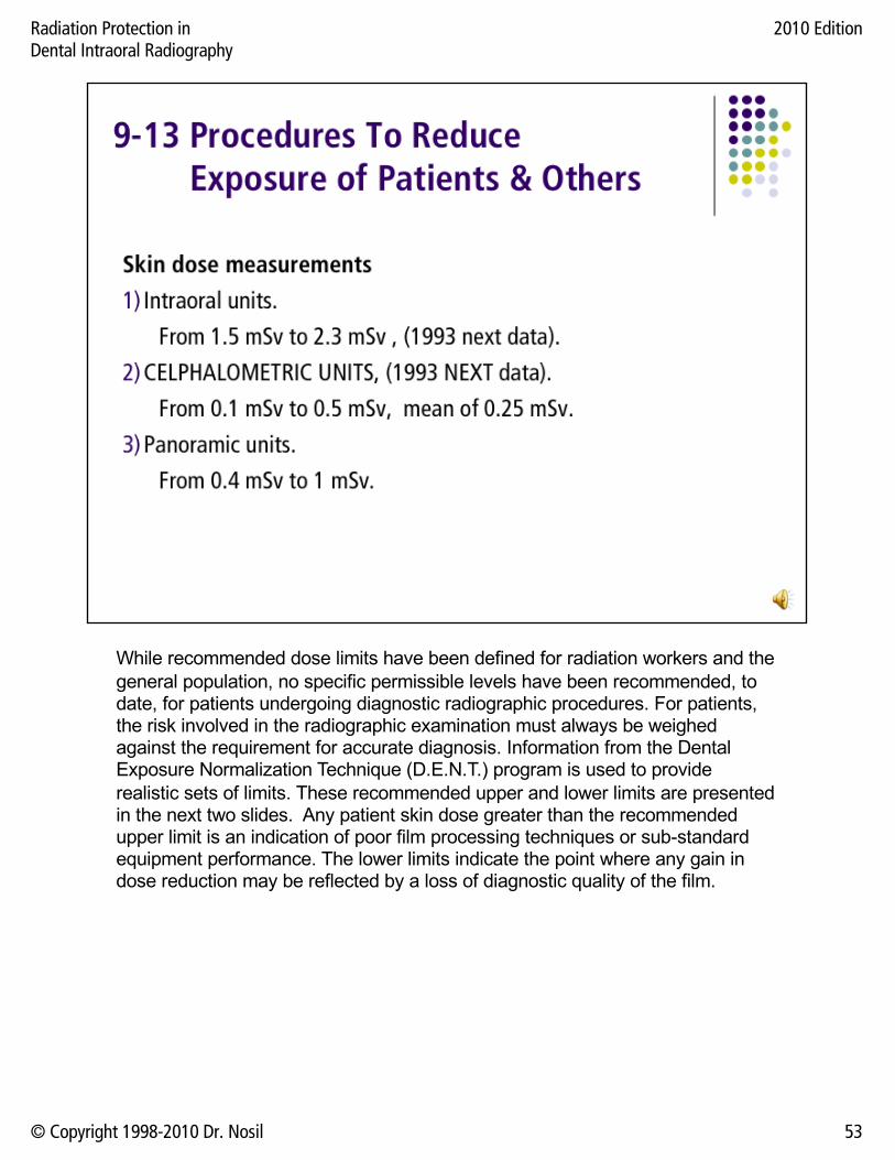
While recommended dose limits have been defined for radiation workers and the general population, no specific permissible levels have been recommended, to date, for patients undergoing diagnostic radiographic procedures. For patients, the risk involved in the radiographic examination must always be weighed against the requirement for accurate diagnosis. Information from the Dental Exposure Normalization Technique (D.E.N.T.) program is used to provide realistic sets of limits. These recommended upper and lower limits are presented in the next two slides. Any patient skin dose greater than the recommended upper limit is an indication of poor film processing techniques or sub-standard equipment performance. The lower limits indicate the point where any gain in dose reduction may be reflected by a loss of diagnostic quality of the film.
Radiation Protection inDental Intraoral Radiography
2010 Edition
© Copyright 1998-2010 Dr. Nosil 53
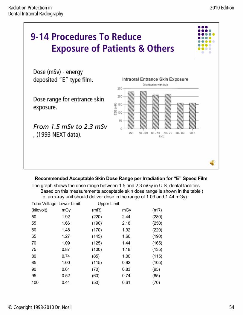
Recommended Acceptable Skin Dose Range per Irradiation for “E” Speed FilmThe graph shows the dose range between 1.5 and 2.3 mGy in U.S. dental facilities.
Based on this measurements acceptable skin dose range is shown in the table ( i.e. an x-ray unit should deliver dose in the range of 1.09 and 1.44 mGy).
Tube Voltage Lower Limit Upper Limit(kilovolt) mGy (mR) mGy (mR)50 1.92 (220) 2.44 (280)55 1.66 (190) 2.18 (250)60 1.48 (170) 1.92 (220)65 1.27 (145) 1.66 (190)70 1.09 (125) 1.44 (165)75 0.87 (100) 1.18 (135)80 0.74 (85) 1.00 (115)85 1.00 (115) 0.92 (105)90 0.61 (70) 0.83 (95)95 0.52 (60) 0.74 (85)100 0.44 (50) 0.61 (70)
Radiation Protection inDental Intraoral Radiography
2010 Edition
© Copyright 1998-2010 Dr. Nosil 54

Acceptable Skin Dose Range per Irradiation for “D” Speed Film: The table shows the dose range between 2.01 and 3.14 mGy acceptable at 70 kVp. This demonstrate dose increase compared to D film.X-ray Tube Voltage Lower Limit Upper Limit(kilovolt) mGy (mR) mGy (mR)50 3.49 (400) 4.80 (550)55 3.23 (370) 4.54 (520)60 2.79 (320) 4.15 (475)65 2.36 (270) 3.62 (415)70 2.01 (230) 3.14 (360)75 1.57 (180) 2.66 (305)80 1.40 (160) 2.27 (260)85 1.22 (140) 2.01 (230)90 1.05 (120) 1.83 (210)100 0.79 ( 90) 1.57 (180)
Radiation Protection inDental Intraoral Radiography
2010 Edition
© Copyright 1998-2010 Dr. Nosil 55
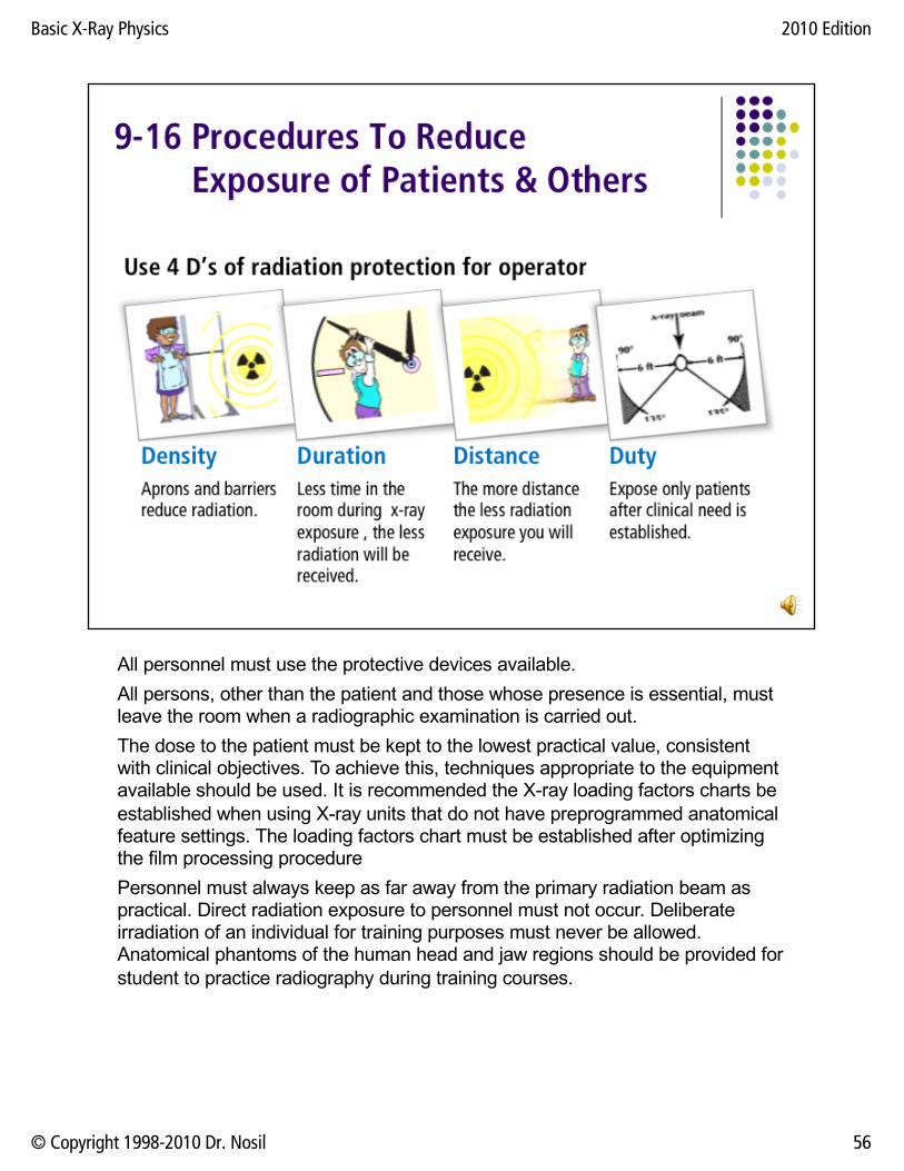
All personnel must use the protective devices available.All persons, other than the patient and those whose presence is essential, must leave the room when a radiographic examination is carried out.The dose to the patient must be kept to the lowest practical value, consistent with clinical objectives. To achieve this, techniques appropriate to the equipment available should be used. It is recommended the X-ray loading factors charts be established when using X-ray units that do not have preprogrammed anatomical feature settings. The loading factors chart must be established after optimizing the film processing procedure Personnel must always keep as far away from the primary radiation beam as practical. Direct radiation exposure to personnel must not occur. Deliberate irradiation of an individual for training purposes must never be allowed. Anatomical phantoms of the human head and jaw regions should be provided for student to practice radiography during training courses.
Basic X-Ray Physics 2010 Edition
© Copyright 1998-2010 Dr. Nosil 56
Please send answers to the questions below to the Bermuda MOH if you wish to be included on the list of certified radiation users:1. What does radiation safety mean to you?2. Three types of dental X-Ray units must be licensed. What are they?3. What are the four D's of radiation safety?4. What is the unit that describes the biological effect of radiation?5. In terms of these units, during a bitewing exposure the skin of the patient receives which of the following:
0.1 to 1.01.0 to 1010 to 100100 to 1000
6. In terms of these units, what is the regulatory limit for the annual exposure of radiation workers?7. Why is the size and shape of the intraoral applicator subject to regulation?8. Why is the method of processing of dental films important from a radiation protection viewpoint?9. What is the significance of focal spot size in dental radiography?
Radiation Protection inDental Intraoral Radiography
2010 Edition
© Copyright 1998-2010 Dr. Nosil 57

Radiation Protection inDental Intraoral Radiography
2010 Edition
© Copyright 1998-2010 Dr. Nosil 58