Surgical adjunctive procedures for accelerating orthodontic treatment
Upload
khangminh22Category
view
5download
0
Page 1/13
Intraoral and extraoral approach for surgicaltreatment of Eagle's syndrome: a retrospectivestudyJing Wang
West China Second University Hospital of Sichuan UniversityZhibin Wang
Huazhong University of Science and TechnologyKaiSheng Yan
First A�liated Hospital of Dalian Medical UniversityYan Liu ( [email protected] )
First A�liated Hospital of Dalian Medical University
Research Article
Keywords: Eagle’s syndrome, styloidectomy, three-dimensional computed tomography
Posted Date: February 23rd, 2021
DOI: https://doi.org/10.21203/rs.3.rs-222000/v1
License: This work is licensed under a Creative Commons Attribution 4.0 International License. Read Full License
Page 2/13
Abstract
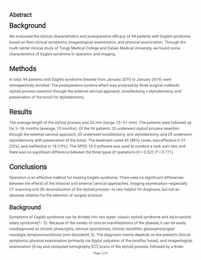
BackgroundWe evaluated the clinical characteristics and postoperative e�cacy of 94 patients with Eagle’s syndromebased on their clinical symptoms, imageological examination, and physical examination. Through themulti center clinical study of Tongji Medical College and Dalian Medical University, we found somecharacteristics of Eagle’s syndrome in operation and imaging.
MethodsIn total, 94 patients with Eagle’s syndrome (treated from January 2010 to January 2019) wereretrospectively enrolled. The postoperative curative effect was analysed by three surgical methods:styloid process resection through the external cervical approach, tonsillectomy + styloidectomy, andpreservation of the tonsil for styloidectomy.
ResultsThe average length of the styloid process was 33 mm (range, 25–61 mm). The patients were followed upfor 3–36 months (average, 15 months). Of the 94 patients, 20 underwent styloid process resectionthrough the external cervical approach, 45 underwent tonsillectomy and styloidectomy, and 29 underwentstyloidectomy with preservation of the tonsil. The treatment cured 45 (48%) cases, was effective in 31(33%), and ineffective in 18 (19%). The SPSS 19.0 software was used to conduct a rank sum test, andthere was no signi�cant difference between the three types of operations (H = 0.521, P = 0.771).
ConclusionsOperation is an effective method for treating Eagle’s syndrome. There were no signi�cant differencesbetween the effects of the intraoral and external cervical approaches. Imaging examination—especiallyCT scanning and 3D reconstruction of the styloid process—is very helpful for diagnosis, but not anabsolute criterion for the selection of surgery protocol.
BackgroundSymptoms of Eagle’s syndrome can be divided into two types: classic styloid syndrome and stylo-carotid-artery syndrome(1–3). Because of the variety of clinical manifestations of the disease, it can be easilymisdiagnosed as chronic pharyngitis, cervical spondylosis, chronic tonsillitis, glossopharyngealneuralgia, temporomandibular joint disorder(4, 5). The diagnosis mainly depends on the patient's clinicalsymptoms, physical examination (primarily via digital palpation of the tonsillar fossa), and imageologicalexamination (X-ray and computed tomography [CT] scans of the styloid process, followed by a three-

Page 3/13
dimensional [3D] reconstruction)(6, 7). In China, surgical treatment for this disease is generally performedby the otolaryngology department. However, the clinical symptoms of some patients are not solvedthrough surgery(8). Therefore, this retrospective study aimed to evaluate the surgical treatment of thisdisease through statistical analysis of various postoperative outcome measures, providing a reference forthe treatment of such patients in the future.
Methods1. Data and patients
A total of 94 patients treated at the First A�liated Hospital of Dalian Medical University (37 cases) andTongji Hospital (a�liated to Tongji Medical College of Huazhong University of Science and Technology,57 cases) from January 2010 to January 2019 were enrolled as research subjects. The patients wereprovided informed consent, and the relevant institutional review boards granted ethical clearance for thestudy. The selection criteria were as follows: 1) patients complained of either pharyngeal pain,pharyngeal foreign body sensation, unilateral neck shoulder radiation pain, or radiation ear pain; 2) abony eminence was palpable in the fossa; 3) either the 3D CT reconstruction of the styloid process or theanteroposterior, lateral X-ray �lm of the styloid process met the diagnostic criteria, and the length of thestyloid process was > 25 mm. Patients with severe cardiovascular diseases, neuropsychiatric diseases,and neuromuscular diseases were excluded. There were 43 men and 51 women with an average age of45 ± 8.06 years (range, 25–68 years). The disease course was 3–60 months. Of the 94 cases, 63 hadunilateral disease and 31 had bilateral diseases; 20 patients underwent styloid process resection throughthe external cervical approach, 45 underwent tonsillectomy as well as styloid process shortening, and 29underwent styloid process shortening along with tonsil preservation.
2. Imageological examinations
Imageological examination was performed with a Siemens 64-slice double helix machine (Siemens,Munich, Germany). The patient was put in a supine position with the mandible raised, such that theauditory orbital line was perpendicular to the scanning table. The measurement method used was asfollows(7, 9): based on the 3D CT reconstruction image, the length of the styloid process was measuredas the distance between its root and the distal end. On the anteroposterior image, a perpendicular linewas drawn parallel to the skull base, and the angle between the centre line of the styloid process and theperpendicular measurement line was noted as the inward angle(10). An acoustic orbital line (aperpendicular line connecting the upper edge of the outer ear hole and the lower edge of the orbit) wasdrawn on the reconstructed image, and the internal deviation angle was measured as the angle betweenthe perpendicular line and the centre line of the styloid process. If the length of the styloid process wasgreater than 25 mm(11), the styloid process was considered too long. In this case, the angle of inclination(inward or forward) was greater than 25° and was called the abnormal azimuth angle. Theanteroposterior, lateral X-ray �lm of the styloid process was examined as follows: for scanning, thepatient (either standing or reclining) was instructed to keep their head in a lateral position and slightly tilt

Page 4/13
their body. After scanning, the data was transmitted to a picture archiving and communication systemworkstation to measure the azimuth angle and the length of the styloid process using the same methodas before.
3. Surgical indications
At present, patients diagnosed with Eagle’s syndrome are not uncommon. Some patients with a longstyloid process undergo conservative treatments such as non-steroidal anti-in�ammatory drugs andglucocorticoids(12), although it has a short-term effect, can easily relapse. The inclusion criteria forsurgery were the same as the selection criteria for this study. All three criteria (1, 2, and 3) were considerednecessary conditions and were strictly observed, and the imaging data were carefully analysed. Althoughthe X-ray of the styloid process could show the long styloid process bone, the 3D reconstruction of thestyloid process was used as the �nal examination method in this study to determine surgical method.This is because the 3D CT scan of the styloid process is more accurate in displaying the details, andaccurate judgement of de�ection, interruption, and ossi�cation of the hyoid ligament(10).
4. Selection of the operation method
The patients were placed under general anaesthesia and the appropriate surgical method was selectedbased on their oropharyngeal palpation, 3D CT reconstruction of the styloid process, and speci�cselection criteria: 1) for patients in whom the styloid process bone could be touched directly duringintraoral palpation, or in whom the distal part of the styloid process could not be directly touched but theCT scan showed that the bone inclined toward the oropharynx and its distal part was relatively close tothe oropharynx cavity (which does not cause excessive injury in the parapharyngeal space), styloidprocess resection was selected for transoral surgery. Whether tonsillectomy was performed depended onwhether the patient’s tonsil was too large to affect the surgical incision; 2) for patients whose styloidprocess could not be touched through the mouth or could be touched under the jaw, or when the CT scanshowed that the inclination angle was not large, the external cervical approach was selected. Theselection of appropriate surgical methods can lower operation time, injury, postoperative pharyngalgia,and the probability of complications.
4.1. Bimanual examination
The index �nger of one hand was placed in the affected tonsillar fossa, and the other �nger was placedbehind or under the ipsilateral ramus of the mandible(13). Of 74 patients who underwent surgery usingthe intraoral approach, 49 patients could either touch the hard cord in the unilateral or bilateral tonsillarfossa, or felt tenderness, fullness, or other discomforts at the touchpoint; 10 patients could touch the hardcord behind or under the ramus of the mandible. The other 15 patients could not touch any abnormality.Speci�c surgical methods: 1) the tonsillar fossa approach: if the tonsil was too large and affected theexposure of the whole styloid process, tonsillectomy was performed �rst. Based on the position andcourse of the styloid process, a sickle knife was used to cut the mucosa of the palatoglossal orpalatopharyngeal arch in order to separate the superior pharyngeal muscle and expose the distal styloid
Page 5/13
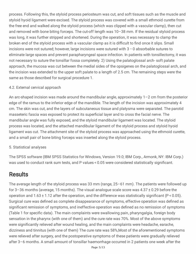
process. Following this, the styloid process periosteum was cut, and soft tissues such as the muscle andstyloid hyoid ligament were excised. The styloid process was covered with a small ethmoid curette fromthe free end and walked along the styloid process (which was clipped with a vascular clamp), then cutand removed with bone biting forceps. The cut-off length was 10–38 mm. If the residual styloid processwas long, it was further stripped and shortened. During the operation, it was necessary to clamp thebroken end of the styloid process with a vascular clamp as it is di�cult to �nd once it slips. Smallincisions were not sutured; however, large incisions were sutured with 3 − 0 absorbable sutures toeliminate large spaces and prevent parapharyngeal space infection. In patients with tonsillectomy, it wasnot necessary to suture the tonsillar fossa completely. 2) Using the palatoglossal arch- soft palateapproach, the mucosa was cut between the medial sides of the opsigenes on the palatoglossal arch, andthe incision was extended to the upper soft palate to a length of 2.5 cm. The remaining steps were thesame as those described for surgical procedure 1.
4.2. External cervical approach
An arc-shaped incision was made around the mandibular angle, approximately 1–2 cm from the posterioredge of the ramus to the inferior edge of the mandible. The length of the incision was approximately 4cm. The skin was cut, and the layers of subcutaneous tissue and platysma were separated. The parotidmasseteric fascia was exposed to protect its super�cial layer and to cross the facial nerve. Themandibular angle was fully exposed, and the styloid mandibular ligament was located. The styloidprocess was located, and the attached mandibular ligament of the styloid process and styloid hyoidligament was cut. The attachment site of the styloid process was approached using the ethmoid curette,and a small pair of bone biting forceps was inserted along the styloid process.
5. Statistical analyses
The SPSS software (IBM SPSS Statistics for Windows, Version 19.0; IBM Corp., Armonk, NY: IBM Corp.)was used to conduct rank sum tests, and P values < 0.05 were considered statistically signi�cant.
ResultsThe average length of the styloid process was 33 mm (range, 25–61 mm). The patients were followed upfor 3–36 months (average, 15 months). The visual analogue scale score was 4.37 ± 0.29 before theoperation and 1.63 ± 1.12 after the operation, and the difference was statistically signi�cant (P < 0.05).Surgical cure was de�ned as complete disappearance of symptoms, effective operation was de�ned assigni�cant remission of symptoms, and ineffective operation was de�ned as no remission of symptoms(Table 1 for speci�c data). The main complaints were swallowing pain, pharyngalgia, foreign bodysensation in the pharynx (with one of them) and the cure rate was 70%. Most of the above symptomswere signi�cantly relieved after wound healing, and the main complaints were headache, earache,dizziness and tinnitus (with one of them) The cure rate was 58%,Most of the aforementioned symptomswere relieved after surgery, and the postoperative symptoms of these patients were gradually relievedafter 3–6 months. A small amount of tonsillar haemorrhage occurred in 2 patients one week after the

Page 6/13
operation, but healed without special treatment, no parapharyngeal space infection or facial nerveparalysis. Persistent dry throat and pharyngeal foreign body sensation were reported in individual casesbut were tolerable without special intervention. The results of the rank sum tests were not statisticallysigni�cant (H = 0.521, P = 0.771).
Table 1The therapeutic effect of three surgical methods
THERAPEUTICEFFECT
EXTERNALCERVICALAPPROACH
TONSILLECTOMY + STYLOIDECTOMY
PRESERVATION OF THETONSIL FORSTYLOIDECTOMY
TOTAL
Cure 10 20 15 45(48%)
Effective 7 15 9 31(33%)
Ineffective 3 10 5 18(19%)
Total 20 45 29 94
Case ReportCase 1
a 54 years old female patient, complained of " pharyngalgia for half a year". This case was a typicalstyloid process truncation with preservation of tonsils through oral approach. Through CT, we couldclearly see the length, anteversion and inclination of styloid process, and judge that styloid process wastoward the oropharynx cavity. Palpation through tonsil fossa also further con�rms that styloid processwas too long, and bone of styloid process could be seen protruding to tonsil fossa. Such typical patientwas relatively rare. After operation, the wound in the mouth was very small and healed quickly. Thepatient's pharyngalgia was obviously improved. (Fig. 1)
Case 2
a 52 years old female patient, was admitted with the chief complaint of "foreign body sensation in throatfor one year". This case was a special type. The CT of the patient showed that the styloid process bonewas intact, and the styloid process was not touched in the tonsil fossa and neck. During the operation,the styloid process bone was not �rmly �xed. During the operation, the styloid process could swingslightly, and the thickness was uneven, the styloid process bone was discontinuous. One month after theoperation, the patient's symptoms improved. (Fig. 2)
Case 3
a 47 years old male patient, was admitted with " pharyngalgia for half a year", a case of extremely longstyloid process on both sides. CT accurately measured the true length of styloid process, which wasconsistent with the intraoperative �ndings. It could be seen that CT three-dimensional reconstruction has

Page 7/13
diagnostic value for the disease. Two weeks after operation, the symptoms improved signi�cantly.(Fig. 3)
Case 4
a 37 years old male patient, complained of " pharyngalgia for a year", a case of styloid process boneinterruption. Both styloid processes were interrupted in CT. After cutting the mucosa in the opening, thedistal styloid process was not �xed. During the operation, there was a cord like interruption at the junctionof the anterior and middle third of styloid process, and the bone was discontinuous. We found that if thestyloid process was obviously interrupted in CT, the interruption could be seen during the operation. If thestyloid process bone interruption was not shown by CT, bone interruption could occur in individual cases.The patient's symptoms relieved 3 months after operation. (Fig. 4)
DiscussionThe styloid process is a slender cylindrical bone located at the junction of the mastoid and petrous partof the temporal bone. It originates from the anteromedial side of the stylomastoid foramen(14). Thestyloid process and its associated muscles (the styloid hyoid muscle, styloid pharyngeal muscle, andstyloid tongue muscle) divide the parapharyngeal space into two parts: the anterior part (the anteriorparapharyngeal space) and the posterior part (the posterior parapharyngeal space). The anteriorparapharyngeal space is relatively small and contains ascending pharyngeal arteries and veins, whereasthe medial side is close to the superior pharyngeal constrictor and palatine tonsil. Because of thisanatomical relationship, abnormal anatomical morphology of the styloid process can cause pharyngealsymptoms such as pharyngalgia, foreign body sensation in the pharynx, and cough. The retro-parapharyngeal space is large and contains the internal carotid artery, veins, - cranial nerves, andcervical lymph nodes, cause headache, neck and shoulder pain, tinnitus. Abnormal styloid processdevelopment or ossi�cation of the styloid hyoid ligament can lead to a long styloid process (> 25 mm),abnormal angle, and abnormal shape. The normal inclination angle of the vertical line between thestyloid process and the plane of the skull base plane in normal adults is approximately 25° forward andinward; if the angle is > 40° or < 20°, it is considered abnormal(7). Because the anatomical variation of thenormal styloid process is relatively large, a long styloid process may not cause clinical symptoms.Therefore, both the length of the styloid process and the angle of the styloid process should beconsidered by clinicians. Abnormality in the styloid process shape and orientation or an abnormally longstyloid process may not have the corresponding clinical symptoms of styloid process syndrome.
In this study, 94 patients were enrolled, of whom most complained of pharyngalgia, foreign bodysensation in the pharynx, and neck pain. Among them, 3 patients were admitted to the hospital withotodynia as the main complaint. Most patients have been diagnosed with chronic pharyngitis, chronictonsillitis, or otitis before surgery, Eagle's syndrome can also cause these symptoms, which makes itdi�cult to differentiate from other diseases that can cause neck or facial pain and abnormal sensations.Special examination—especially tonsillar fossa and neck palpation—has become the key diagnosis in

Page 8/13
such cases. If the long styloid process can be touched or causes obvious tenderness, foreign bodysensation, and fullness, the diagnosis is con�rmed. Imaging technology, especially CT scanning and 3Dreconstruction of the styloid process, can clearly show the styloid process length, shape, internalde�ection angle, and ligament ossi�cation(7). These examinations allow relatively accurate observationand measurement, which is of great help in guiding surgery and clinical research.
This study found that there was no signi�cant difference in the e�cacy of intraoral and extraoralapproaches, but the postoperative pain lasted 5 days longer in patients who underwent surgery using theextraoral approach compared to those who underwent the intraoral approach. The average postoperativehospital stay of patients who underwent surgery with the intraoral approach was 3 days, shorter than thepatients who received the extraoral approach for 3 days. A small amount of tonsillar haemorrhageoccurred in 2 patients one week after the operation, but healed without special treatment, noparapharyngeal space infection, or facial nerve paralysis. Persistent dry throat and pharyngeal foreignbody sensation were reported in individual cases but were tolerable without special intervention. Wefound that imaging examination—especially CT scanning and 3D reconstruction of the styloid process—isvery helpful for diagnosis, but not an absolute criterion for the selection of surgery protocol. In somecases, although CT scans showed that the bone was continuous, it was found to be interrupted during theoperation, in these cases, the effect of operation were often not very good. In some cases, CT scansshowed that the styloid process was too long, but intraoperative exploration, especially in the case ofsu�cient exposure to the external cervical approach, the styloid process was not too long, which may berelated to the incomplete ossi�cation of styloid ligament. In this study, we set the styloid process lengthat > 25 mm; however, most other related studies set it at > 30 mm(15), the main consideration is that thelength cut-off is not universal; some patients have a styloid process length of 25–30 mm, but theirsymptoms are obvious, the hard styloid process can be felt on tonsillar fossa examination, and surgicaltreatment is also feasible. Therefore, imaging examinations can only be used as an important auxiliarytool in clinical practice and cannot become a de�nitive diagnosis.
Styloid process truncation has a clear curative effect on patients with Eagle's syndrome, and cansigni�cantly reduce the symptoms of pharyngalgia, swallowing pain, cervicodynia, earache, andpharyngeal foreign body sensation(6, 16). However, the symptoms of patients with tinnitus, headache,and dizziness are not signi�cantly relieved. The surgeon in this study is pro�cient in the above threesurgical methods, who presents no technical differences. However, some scholars believe that the lateralcervical region approach can ensure the safety of operations and reduce the probability of postoperativecomplications(17), it is also suggested that the styloid process truncation should be performed throughthe external cervical approach because it allows clear surgical vision and less damage to peripheral bloodvessels and nerves(13, 18). However, extensive facial anatomy and long operation times can easily causeepidermal nerve injuries, such as sensory abnormalities of the great auricular nerve and facial nervebranch injury(19). Open surgery should be selected when the styloid process grows towards the lateralcervical region, so as to avoid excessive operation in the parapharyngeal space, longer operation times,and higher operating costs. Through the surgical procedures used in this study, the wound in the mouthhad a small internal diameter and the recovery time was short (6 days after the operation), there was no

Page 9/13
incision or scar on the outside. Although the �eld of vision was smaller, the styloid process could be fullyexposed in most cases(20). These selection criteria can help reduce the risk of parapharyngeal spaceinfection and nerve injury(21). In general, it is very important to choose the appropriate surgical methodaccording to the experience of the operator and the different pathological characteristics of the patients.
ConclusionSurgery is an effective method for treating styloid process syndrome. However, in view of the imprecisecurative effect of surgery in some cases and postoperative discomfort (dry throat and foreign bodysensation in the pharynx), and the imaging examination was also inconsistent with the intraoperative�ndings, it is very important to strengthen the preoperative evaluation (length and angle judgement of thestyloid process via imaging, digital palpation), select surgical protocols according to the symptoms. Ourresults show that there were no signi�cant differences between the intraoral and extraoral surgicalmethods. However, based on the surgeon's pro�ciency in the corresponding surgical methods(22), theunnecessary operation should be avoided, the operation time should be minimized, and the adjacentanatomical structures should be avoided during surgery. Through ten years of retrospective study, wecollected a large number of cases, including many typical cases, and showed them to readers throughimages and statistical data. It's a �aw not to record the photos of the operation in the typical cases.
AbbreviationsCTcomputed tomography3Dthree-dimensional
DeclarationsEthics approval and consent to participate
This study was conducted in agreement with the Ethics Committee of Tongji Hospital A�liated to TongjiMedical College and The First A�liated Hospital of Dalian Medical University, All participants hadprovided a written informed consent
Consent for publication
All participants gave written informed consent for their personal and clinical data publication along withwritten consent for publication for any identifying images in this study, according to the China laws.
Availability of data and materials

Page 10/13
The datasets used and/or analyzed during the current study are available from the corresponding authorupon reasonable request.
Competing interests
None.
Funding
No funding was received for this research.
Authors' contributions
ZBW and YL: study conception and design, acquisition of data, YKS analyzes and processes data. JW:drafting of the manuscript. All authors have read and approved the manuscript.
Acknowledgements
Our thanks go to all participating department
Authors' information
1 Department of Otorhinolaryngology, The West China Second University Hospital of SichuanUniversity, Cheng Du 610041, China.
2 Department of Otorhinolaryngology, Tongji Hospital A�liated to Tongji Medical College of HuazhongUniversity of science and technology, Wu Han 430030 , China.
3 Department of Otorhinolaryngology, The First A�liated Hospital of Dalian Medical University, Dalian116000, China.
guidelines
All procedures were performed in accordance with relevant guidelines.
References1. Baldino G, Di Girolamo C, De Blasis G, Gori A. Eagle Syndrome and Internal Carotid Artery Dissection:
Description of Five Cases Treated in Two Italian Institutions and Review of the Literature. Ann VascSurg. 2020;67:565 e17- e24.
2. Zamboni P, Scerrati A, Menegatti E, Galeotti R, Lapparelli M, Traina L, et al. The eagle jugularsyndrome. BMC Neurol. 2019;19(1):333.
3. Aydin E, Quliyev H, Cinar C, Bozkaya H, Oran I. Eagle Syndrome Presenting with NeurologicalSymptoms. Turk Neurosurg. 2018;28(2):219–25.

Page 11/13
4. Krohn S, Brockmeyer P, Kubein-Meesenburg D, Kirschneck C, Buergers R. Elongated styloid process inpatients with temporomandibular disorders - Is there a link? Ann Anat. 2018;217:118–24.
5. Mahmoud NR, Ashour EM. Cervico-facial pain associated with Eagle's syndrome misdiagnosed ascranio-mandibular disorders. A retrospective study. J Craniomaxillofac Surg. 2020;48(10):1009–17.
�. Papadiochos I, Papadiochou S, Sarivalasis ES, Goutzanis L, Petsinis V. Treatment of Eagle syndromewith transcervical approach secondary to a failed intraoral attempt: Surgical technique and literaturereview. J Stomatol Oral Maxillofac Surg. 2017;118(6):353–8.
7. Kent DT, Rath TJ, Snyderman C. Conventional and 3-Dimensional Computerized Tomography inEagle's Syndrome, Glossopharyngeal Neuralgia, and Asymptomatic Controls. Otolaryngol Head NeckSurg. 2015;153(1):41–7.
�. Costantinides F, Vidoni G, Bodin C, Di Lenarda R. Eagle's syndrome: signs and symptoms. Cranio.2013;31(1):56–60.
9. Ayyildiz VA, Senel FA, Dursun A, Ozturk K. Morphometric examination of the styloid process by 3D-CTin patients with Eagle syndrome. Eur Arch Otorhinolaryngol. 2019;276(12):3453–9.
10. Burulday V, Akgul MH, Bayar Muluk N, Yagdiran B, Inal M. The importance of medial-lateral styloidprocess angulation/coronal plane angle in symptomatic eagle syndrome. Clin Anat. 2017;30(4):487–91.
11. Scavone G, Caltabiano DC, Raciti MV, Calcagno MC, Pennisi M, Musumeci AG, et al. Eagle'ssyndrome: a case report and CT pictorial review. Radiol Case Rep. 2019;14(2):141–5.
12. Maher T, Shankar H. Ultrasound-Guided Peristyloid Steroid Injection for Eagle Syndrome. Pain Pract.2017;17(4):554–7.
13. Pigache P, Fontaine C, Ferri J, Raoul G. Transcervical styloidectomy in Eagle's syndrome. Eur AnnOtorhinolaryngol Head Neck Dis. 2018;135(6):433–6.
14. Fusco DJ, Asteraki S, Spetzler RF. Eagle's syndrome: embryology, anatomy, and clinical management.Acta Neurochir (Wien). 2012;154(7):1119–26.
15. Bruno G, De Stefani A, Balasso P, Mazzoleni S, Gracco A. Elongated styloid process: Anepidemiological study on digital panoramic radiographs. J Clin Exp Dent. 2017;9(12):e1446-e52.
1�. Lou Z. Commentary on Surgical management of patients with Eagle syndrome. Am J Otolaryngol.2019;40(5):783.
17. Hardin FM, Xiao R, Burkey BB. Surgical management of patients with Eagle syndrome. Am JOtolaryngol. 2018;39(5):481–4.
1�. Ceylan A, Koybasioglu A, Celenk F, Yilmaz O, Uslu S. Surgical treatment of elongated styloid process:experience of 61 cases. Skull Base. 2008;18(5):289–95.
19. Sham E, Reddy TJ, Menon PS, Kumar V, Nathan AS, Mathews S, et al. Anterior Tonsillar FossaApproach to Elongated Styloid Process. Ann Maxillofac Surg. 2020;10(1):203–9.
20. Cheng C, She C, Zhang Q. The experience of treatment of coblation assisted surgical approach toEagle's syndrome. Am J Otolaryngol. 2017;38(3):301–4.

Page 12/13
21. Badhey A, Jategaonkar A, Anglin Kovacs AJ, Kadakia S, De Deyn PP, Ducic Y, et al. Eagle syndrome: Acomprehensive review. Clin Neurol Neurosurg. 2017;159:34–8.
22. Al Weteid AS, Miloro M. Transoral endoscopic-assisted styloidectomy: How should Eagle syndromebe managed surgically? Int J Oral Maxillofac Surg. 2015;44(9):1181–7.
Figures
Figure 1
A and B: CT examination shows the bilateral styloid process growing downward, inward, and forward.The length of the right styloid process is 44.1 mm, the inclination angle is 27.2°, the anteversion angle is29.6° to the superior margin of the axis, the lower segment of the right styloid process bends, and theinclination angle increases to the medial. C: During the operation, the right tonsil fossa is palpated, and ahard rod-like protrusion is found. D: The styloid process is completely truncated during the operation, andno ligament or soft tissue attachment is left around the styloid process. E and F: wound recovery on thethird and sixth days after the operation. There are small wounds in the mouth with a little whitemembrane attached to the surface, and the surrounding mucosa is intact without redness, swelling, orcongestion. The wound recovered to six days after the operation.

Page 13/13
Figure 2
A-C: CT examination of the neck, the styloid process on the left side is 28 mm in length, with aninclination of 37.3° and an anteversion of 9.3°. The CT scan shows that the styloid process is continuouswithout interruption; D: a strip-like interruption touches during the operation.
Figure 3
A–C: The length of the right styloid process is 58.8 mm and the inclination angle is 30.3°; the length ofthe left styloid process is 55.6 mm and the inclination angle is 26.7°, CT can clearly show the course andlength of styloid process. D: Bilateral styloid process was completely truncated during operation.
Figure 4
A–C: the left styloid process is 35.73 mm, and the right styloid process is 37.36 mm. Both styloidprocesses are interrupted in the CT scans. After cutting the internal mucosa, the distal styloid process isnot �xed. D: A cord-like interruption is present at the junction of the anterior and middle third of the styloidprocess, and the bone is discontinuous.
Copyright © 2022 FDOKUMEN