The Anticariogenic Effect of Sugar-Free Gum Containing CPP-ACP Nanocomplexes on Approximal Caries...
14
Fax +41 61 306 12 34 E-Mail [email protected] www.karger.com Original Paper Caries Res 2008;42:171–184 DOI: 10.1159/000128561 The Anticariogenic Effect of Sugar-Free Gum Containing CPP-ACP Nanocomplexes on Approximal Caries Determined Using Digital Bitewing Radiography M.V. Morgan a G.G. Adams a D.L. Bailey a C.E. Tsao a S.L. Fischman b E.C. Reynolds a a Cooperative Research Centre for Oral Health Science, School of Dental Science, Bio21 Institute, University of Melbourne, Parkville, Vic., Australia; b School of Dental Medicine, State University of New York at Buffalo, Buffalo, N.Y., USA modelling of the transition scores at the tooth surface level. There was a statistically significant difference in the frequen- cy distributions of the transition scores between the two gum groups (OR = 0.82, p = 0.03). For subjects chewing the CPP-ACP gum the odds of a surface experiencing caries pro- gression were 18% less than those of a surface experiencing caries progression for subjects chewing the control gum. In conclusion, the 54 mg CPP-ACP sugar-free gum significantly slowed progression and enhanced regression of approximal caries relative to a control sugar-free gum in a 24-month clin- ical trial. Copyright © 2008 S. Karger AG, Basel The caries-preventive effectiveness of sugar-free gum has been investigated in several studies over the past 25 years. A number of controlled trials, both randomized and non-randomized, have compared sugar-free gum with a non-gum-chewing control group and there is a growing body of evidence which supports the caries-pre- ventive nature of chewing sugar-free gum [Scheie and Fe- jerskov, 1998; Lingström et al., 2003; van Loveren, 2004]. The studies have generally affirmed that chewing xylitol- or sorbitol-based gums is caries-preventive when com- Key Words Approximal caries progression Casein phosphopeptide- amorphous calcium phosphate nanocomplexes Digital bitewing radiography Enamel remineralization Ordinal categorical data Randomized clinical trial Sugar-free chewing gum Abstract This study investigated, using digital bitewing radiography, the progression and regression of approximal caries in ado- lescent subjects chewing a sugar-free gum containing 54 mg CPP-ACP relative to the identical gum without CPP-ACP. 2,720 subjects from 29 schools were randomly assigned to one of the two gums and were instructed to chew their as- signed gum for 3 ! 10 min/day, with one session supervised on school days, over the 24-month study period. Standard- ized digital bitewing radiographs were taken at the baseline and 24-month clinical examinations for each subject. The ra- diographs, scored by a single examiner, were assessed for approximal surface dental caries at both the enamel and dentine level. Surface level transitions were scored using a transition matrix. Caries progression or regression was anal- ysed using proportional-odds ordered logistic regression Received: March 14, 2007 Accepted after revision: February 14, 2008 Published online: April 29, 2008 Eric C. Reynolds School of Dental Science, University of Melbourne 720 Swanston Street Parkville, Vic. 3010 (Australia) Tel. +61 3 9341 1547, Fax +61 3 9341 1597, E-Mail [email protected] © 2008 S. Karger AG, Basel 0008–6568/08/0423–0171$24.50/0 Accessible online at: www.karger.com/cre
Transcript of The Anticariogenic Effect of Sugar-Free Gum Containing CPP-ACP Nanocomplexes on Approximal Caries...

Fax +41 61 306 12 34E-Mail [email protected]
Original Paper
Caries Res 2008;42:171–184 DOI: 10.1159/000128561
The Anticariogenic Effect of Sugar-Free Gum Containing CPP-ACP Nanocomplexes on Approximal Caries Determined Using Digital Bitewing Radiography
M.V. Morgan a G.G. Adams a D.L. Bailey a C.E. Tsao a S.L. Fischman b
E.C. Reynolds a
a Cooperative Research Centre for Oral Health Science, School of Dental Science, Bio21 Institute,University of Melbourne, Parkville, Vic. , Australia; b School of Dental Medicine, State University ofNew York at Buffalo, Buffalo, N.Y. , USA
modelling of the transition scores at the tooth surface level. There was a statistically significant difference in the frequen-cy distributions of the transition scores between the two gum groups (OR = 0.82, p = 0.03). For subjects chewing the CPP-ACP gum the odds of a surface experiencing caries pro-gression were 18% less than those of a surface experiencing caries progression for subjects chewing the control gum. In conclusion, the 54 mg CPP-ACP sugar-free gum significantly slowed progression and enhanced regression of approximal caries relative to a control sugar-free gum in a 24-month clin-ical trial. Copyright © 2008 S. Karger AG, Basel
The caries-preventive effectiveness of sugar-free gum has been investigated in several studies over the past 25 years. A number of controlled trials, both randomized and non-randomized, have compared sugar-free gum with a non-gum-chewing control group and there is a growing body of evidence which supports the caries-pre-ventive nature of chewing sugar-free gum [Scheie and Fe-jerskov, 1998; Lingström et al., 2003; van Loveren, 2004]. The studies have generally affirmed that chewing xylitol- or sorbitol-based gums is caries-preventive when com-
Key Words
Approximal caries progression � Casein phosphopeptide-amorphous calcium phosphate nanocomplexes � Digital bitewing radiography � Enamel remineralization � Ordinal categorical data � Randomized clinical trial � Sugar-free chewing gum
Abstract
This study investigated, using digital bitewing radiography, the progression and regression of approximal caries in ado-lescent subjects chewing a sugar-free gum containing 54 mg CPP-ACP relative to the identical gum without CPP-ACP. 2,720 subjects from 29 schools were randomly assigned to one of the two gums and were instructed to chew their as-signed gum for 3 ! 10 min/day, with one session supervised on school days, over the 24-month study period. Standard-ized digital bitewing radiographs were taken at the baseline and 24-month clinical examinations for each subject. The ra-diographs, scored by a single examiner, were assessed for approximal surface dental caries at both the enamel and dentine level. Surface level transitions were scored using a transition matrix. Caries progression or regression was anal-ysed using proportional-odds ordered logistic regression
Received: March 14, 2007 Accepted after revision: February 14, 2008 Published online: April 29, 2008
Eric C. Reynolds School of Dental Science, University of Melbourne 720 Swanston Street Parkville, Vic. 3010 (Australia) Tel. +61 3 9341 1547, Fax +61 3 9341 1597, E-Mail [email protected]
© 2008 S. Karger AG, Basel0008–6568/08/0423–0171$24.50/0
Accessible online at:www.karger.com/cre
Morgan /Adams /Bailey /Tsao /Fischman /Reynolds
Caries Res 2008;42:171–184172
pared to not chewing gum [Isokangas et al., 1988; Kan-delman and Gagnon, 1990; Mäkinen et al., 1995, 1996; Beiswanger et al., 1998; Alanen et al., 2000; Machiul-skiene et al., 2001; Szöke et al., 2001; Peng et al., 2004].
A number of trials have attempted to demonstrate the caries-preventive effectiveness of one sugar-free gum over another, usually by comparing one sugar substitute with another. The most commonly reported comparison has been between xylitol and sorbitol. In general, there have been inconsistent results when attempts have been made to demonstrate the differences between one sugar-substituted gum over another. Whilst methodological weaknesses limit what can be inferred in terms of effi-cacy, the cumulative weight of evidence suggests that both xylitol-based and sorbitol-based chewing gums help reduce caries experience through salivary stimulation [Gales and Nguyen, 2000; Hayes, 2001; Maguire and Rugg-Gunn, 2003; van Loveren, 2004; Burt, 2006].
A new technology involving phosphopeptides isolated from the milk protein casein, complexed with calcium phosphate [referred to as casein-phosphopeptide amor-phous calcium-phosphate (CPP-ACP) nanocomplexes], has recently been shown to be efficacious in both theprevention and reversal of enamel subsurface lesions in caries models [Reynolds et al., 1995; Shen et al., 2001; Reynolds et al., 2003; Iijima et al., 2004]. Casein phospho-peptides (CPP) containing the sequence -Ser(P)-Ser(P)-Ser(P)-Glu-Glu- can stabilize calcium phosphate and prevent transformation to the insoluble phases [Cross et al., 2005]. The CPP bind to forming nanoclusters of cal-cium and phosphate ions in solution to form highly solu-ble and bioavailable nanocomplexes [Cross et al., 2005]. The anticariogenicity of the CPP-ACP nanocomplexes has been demonstrated in the rat caries model [Reynolds et al., 1995]. Further studies using human in situ caries models have shown that the CPP-ACP nanocomplexes could prevent enamel demineralization and promote enamel subsurface remineralization [Reynolds, 1998; Shen et al., 2001; Reynolds et al., 2003; Iijima et al., 2004]. In fact, recently it was shown that the CPP-ACP nano-complexes were superior to other forms of calcium phos-phate in remineralizing enamel subsurface lesions in situ [Reynolds et al., 2003]. This was attributed to the ability of the CPP to not only stabilize the calcium and phos-phate as bioavailable ions, but also to localize the calcium and phosphate ions at the tooth surface thereby produc-ing an effective concentration gradient into the subsur-face enamel to promote remineralization in situ [Reyn-olds et al., 2003]. Iijima et al. [2004] and Cai et al. [2007] have recently shown that enamel remineralized by CPP-
ACP nanocomplexes is relatively more resistant to acid challenge when compared with normal tooth enamel. This has been attributed to the CPP-ACP nanocomplexes promoting the formation of mineral that is less soluble in acid, particularly in the presence of fluoride ions, when compared with normal carbonated tooth enamel [Iijima et al., 2004]. However, this relative resistance to acid chal-lenge may also be attributable to the localization of the CPP-ACP at the tooth surface inhibiting enamel demin-eralization [Reynolds, 1998; Cai et al., 2007]. Combining the technologies of sugar-free chewing gum with supple-mental use of the CPP-ACP nanocomplexes has the po-tential to be an important adjunct in caries prevention. In fact, a number of in situ human studies have shown that sugar-free gum is a suitable delivery vehicle for the CPP-ACP as the studies showed release of the CPP-ACP in a bioavailable form which provided significantly greater enamel subsurface lesion remineralization than the con-trol sugar-free gum [Shen et al., 2001; Reynolds et al., 2003; Iijima et al., 2004; Cai et al., 2007].
An important methodological issue when evaluating new preventive or remineralizing technologies is deter-mining the level at which dental caries diagnosis is made. The clinical endpoint of a caries lesion with loss of enam-el integrity (cavitation), which is most frequently used in conventional caries studies, focuses on one end of the caries progression continuum at the expense of early car-ies initiation and progression [Biesbrock et al., 2004]. The reliance on cavitation as the primary endpoint and the failure to include early caries lesions in studies have re-sulted in poor results and outcomes for remineralization technologies [Pretty, 2006]. In low caries populations in particular, it is more appropriate to investigate the pro-gression and regression of initial caries lesions than dif-ferences in overall caries experience.
The DMFS (decayed, missing and filled surfaces) in-dex was developed in an era of high dental caries experi-ence, but is a coarse measure of disease experience in populations with optimal exposure to fluoride and in which low levels of dental caries development are antici-pated during the course of a clinical caries trial. Its major limitation is that the D component of the index relies on a binary classification of surfaces as either sound or de-cayed, regardless of which diagnostic threshold for caries is in use. This is at odds with the current understanding of cavitation as resulting from the failure of a normal de-mineralization/remineralization process that continual-ly affects every dental surface [Imrey and Kingman, 2004].

Anticariogenic Effect of a Gum Containing CPP-ACP
Caries Res 2008;42:171–184 173
In clinical studies of caries experience, the caries status of each tooth surface is determined for each subject at each examination, with examinations often repeated at several time points. Traditional methods for analysing the data obtained from these studies have involved calculating a summary measure, such as the DMF index or DMF incre-ment, for each subject and then comparing the summary measure between study groups. The use of subject-level summary measures for comparison of efficacy or effec-tiveness may mask important information, such as the dif-ferences in caries susceptibility or intervention effective-ness between the various tooth surfaces [Hannigan et al., 2001; Burnside et al., 2007]. Analysing these data at the tooth surface level allows both subject-level and surface-level risk factors to be incorporated into the analysis. How-ever, such analyses require that the clustering of tooth sur-faces within subjects be taken into account to ensure that valid inferences are made and erroneous false-positive conclusions avoided [Hujoel et al., 1990]. Current ap-proaches available to account for clustering include the ad-justment of � 2 test [Donner and Banting, 1988, 1989; Ahn et al., 2002], generalized estimating equations [Liang and Zeger, 1986; DeRouen et al., 1991], clustered survival anal-ysis [Hannigan et al., 2001], multilevel modelling [Burn-side et al., 2007] and survey sampling methods [Beck et al., 1997; LaVange et al., 2001; Imrey and Kingman, 2004].
The importance of bitewing radiographs as a supple-ment to the clinical diagnosis of approximal caries has been confirmed in literature reviews [Kidd and Pitts, 1990; Pitts, 1996]. Bitewing radiographs generally detect-ed more than 90% of the total number of approximal le-sions found when using both clinical and radiographic examinations, whereas clinical examination generally detected less than 50% of the total approximal lesions found (depending on the clinical diagnostic threshold used) [Pitts, 1996]. The additional diagnostic value of bitewing radiographs has been confirmed in recent stud-ies of populations with low caries experience [Hopcraft and Morgan, 2005; Llena-Puy and Forner, 2005].
A clinical study was undertaken with the objective of comparing the anticariogenicity of a CPP-ACP-contain-ing sugar-free (sorbitol) chewing gum with that of a con-trol sugar-free (sorbitol) chewing gum in a sample of ad-olescents employing usual oral hygiene practices. Sorbi-tol-containing sugar-free gums have been shown to prevent dental caries and, as such, a standard sorbitol-based sugar-free gum can be considered an active con-trol. This paper reports on the progression and regression of approximal caries determined from standardized dig-ital radiographs taken at baseline and 24 months.
Subjects and Methods
Test Products Sugar-free chewing gum was used as the mode of delivery for
the CPP-ACP nanocomplexes. Control subjects chewed the stan-dard sorbitol-based sugar-free gum, while intervention subjects chewed an identical gum containing CPP-ACP. The sugar-free gum was provided by Cadbury Schweppes Science and Technol-ogy (N.J., USA) and was composed of approximately 50% polyols (sorbitol and mannitol), 32% gum base, 12% glycerin and 6% sweeteners, colours and flavours. The concentration of CPP-ACP (Recaldent CAS 691364-49-5) in the chewing gum was 3% w/w, equivalent to 54 mg CPP-ACP per serving of gum, and was based on the efficacy results from human in situ CPP-ACP trials [Shen et al., 2001]. All subjects were requested to chew their gum 3 times per day for the duration of the study. The length of chewing time was 10 min per session. One supervised chewing session was un-dertaken each day at school during the school day. A teacher or student class monitor documented subject attendance at each su-pervised chewing session on supervised chewing session logs. The remaining sessions were unsupervised and subjects were asked to chew in their own time. To monitor these unsupervised chewing sessions, subjects were asked to complete a diary docu-menting their gum chewing habits. The diaries were collected and reviewed by the study team every 3 months. Subjects returning completed chewing diaries were awarded a gift voucher. For the duration of the study, each subject and household were supplied with an Australian Dental Association-approved fluoride tooth-paste (Colgate Triple Cool Stripe, 1,000 ppm NaF) and soft texture toothbrush. Toothbrushes were replaced and sufficient quantities of toothpaste were distributed every 3 months together with the new supplies of chewing gum.
Study Sample The study design was a 2-year, double-blind, parallel-group,
active sorbitol-based control, randomized clinical trial. The study population consisted of male and female year 7 students, aged be-tween 11.5 and 13.5 years, enrolled at secondary schools in the metropolitan area of Melbourne, Australia. Following agreement by a participating school, the parents/guardians of all year 7 stu-dents received an information brochure outlining the proposed clinical trial and the intention to measure the efficacy of a new preventive material for dental caries. An informed consent state-ment and a health and background questionnaire were also in-cluded. The statement and questionnaire were translated into five additional languages (Arabic, Cambodian, Chinese, Somali and Vietnamese). Students who returned the completed consent form and questionnaire were assessed for eligibility to undergo the baseline clinical and radiographic examinations. Subjects were included if they were in good general health and had at least eight permanent posterior teeth. Exclusion criteria included milk pro-tein allergies, phenylketonuria, presence of fixed orthodontic ap-pliances, chronic use of antibiotics or medications which affect salivary flow rates, and poor oral health as evidenced by rampant dental caries. Subjects were also excluded if they had previous significant exposure to radiation. Final eligibility for the study was determined following the baseline visual-tactile and radio-graphic examinations. Subjects who did not fulfil the entry crite-ria were notified, in writing, of their exclusion from the study. Twenty-nine schools were recruited progressively from April

Morgan /Adams /Bailey /Tsao /Fischman /Reynolds
Caries Res 2008;42:171–184174
2001 to September 2003, until the desired sample size was reached. Schools were recruited in three cohorts: subjects in cohort 1 were recruited from April to November in 2001, subjects in cohort 2 from February to May 2002, and subjects in cohort 3 from May to September 2003. Some schools participated in multiple cohorts. Within a population with a generally low risk of caries, schools were targeted where students were likely to have a higher inci-dence of dental caries. This was determined from incidence data gathered from the Victorian School Dental Service and a prior study conducted by the investigators [Campain et al., 2003]. Sin-gle-sex schools were excluded to reduce gender bias. The Mel-bourne metropolitan water supply has been fluoridated at ap-proximately 1.0 ppm since 1977.
Clinical Examinations Subjects received a visual-tactile examination at baseline, 12
months and 24 months in classrooms at the participating schools. The clinical diagnosis of dental caries was based on a modifica-tion of the traditional criteria described in Radike [1972]. White spot lesions were diagnosed on the gingival third of the buccal or lingual/palatal surfaces only. The presence of secondary caries and sealants was also recorded.
Radiographic Examinations Bitewing radiographs of the posterior teeth were taken at base-
line and at 24 months by registered dental personnel in accor-dance with a standardized procedure. Subjects were able to re-quest that copies of their radiographs be sent to their home ad-dress or their treating clinician. The radiographs were taken using a Belmont Belray 096 Dental X-ray unit (Takara Belmont Corp., Osaka, Japan, 70 kV, 10 mA, 0.1 s exposure) and a Dexis digital X-ray system (Dexis v3.0, Provision Dental Systems, Palo Alto, Calif., USA). Total radiation exposure (baseline and 24 months) was approximately 20 � Sv. A purpose-built mobile dental van al-lowed the radiographs to be taken on site at the schools. A rigid-connection aiming device was designed, using a modification of the system described by Pitts [1983]. The Dexis sensor holders were also modified by thickening the arm, inscribing locator ref-erence marks and the inclusion of a seven-step brass step wedge. This allowed a measure of standardization of the geometrical re-lationship of the sensor, the teeth and the X-ray beam between subjects and also between the baseline and 24-month examina-tions [Bailey et al., 2006].
Scoring of Radiographic Images All radiographic images were scored by a single dentist
(C.E.T.) over a 7-month period during 2005 in a dedicated room with controlled light level (20 lx) and monitor (Mitsubishi Dia-mond Professional 2070 SB-BK 22-inch cathode ray tube monitor) settings. Approximal surfaces, from the mesial of the first premo-lar to the distal of the second molar, were assessed for dental caries at both the enamel and dentine level using a modification of the scoring system described in Pitts [1984, 1985] ( table 1 ). Overlap was recorded only when it interfered with the diagnosis of car-ies – a surface that displayed an overlapping image of an adjacent surface greater than one quarter of the enamel width was scored as R 5 (unreadable overlap) unless another score could be recorded with confidence (excepting R 0 – sound). The radiographs were as-sessed without knowledge of the treatment group or any clinical diagnosis of caries; the reviewer was only informed of those teeth
that were deciduous or that had been scored unerupted, missing or excluded at the visual-tactile examination. Teeth were also ex-cluded from scoring in cases of severe hypomineralization or trau-ma into dentine. Prior to scoring, the radiographs were contrast-corrected using a histogram equalization technique from the NIH image analysis package ImageJ (http://rsb.info.nih.gov/ij). A cus-tom-designed database provided for a split screen interface to al-low image reading and data entry. Baseline and 24-month radio-graphs were read in succession. For the determination of intra-ex-aminer variability 30 complete sets of radiographs (baseline and 24-month) were assessed 3 times at random intervals during the course of the 7-month review period.
Randomization and Blinding Within each school, all subjects (unit of randomization) who
had fulfilled the entry criteria were stratified into two groups (caries-free/non-caries-free). Each school was randomized sepa-rately following the completion of the baseline clinical and radio-graphic examinations at the school. The study sponsor was noti-fied of the numbers in each group and they assigned the blocks of product codes to be used for the caries-free subjects and the non-caries-free subjects (as the study sponsor was based in the USA, the blinded packaged product had been shipped to Australia by air freight in advance of randomization). Within each stratum the
Table 1. Diagnostic codes and criteria for grading of approximal lesions
Code Category Diagnostic criteria
R0 sound no radiolucency or restoration visible
R1 outer-half enamel lesion
zone of increased radiolucency confined to outer half of the enamel (no minimum limit)
R2 inner-half enamel lesion
zone of increased radiolucency involving both inner and outer halves of the enam-el, including lesions extending up to but not beyond amelodentinal junction
R3 outer-half dentine lesion
zone of increased radiolucency penetrat-ing enamel and amelodentinal junction but confined to the outer half of thedentine
R4 inner-half dentine lesion
zone of increased radiolucency penetrat-ing into the inner half of dentine with or without apparent pulpal involvement
R5 unreadable overlap
unreadable due to extent of overlap
R6 secondary caries
zone of increased radiolucency associ-ated with a filled surface
R7 filled surface radiographic appearance consistent with a restoration
R8 not visible tooth not visible on radiograph

Anticariogenic Effect of a Gum Containing CPP-ACP
Caries Res 2008;42:171–184 175
product codes were then randomly assigned to the subjects by the study statistician (G.G.A.). The control and intervention chewing gums were identical in appearance, taste and smell. The subjects, the clinical examiners and those involved in distributing the test products were not aware of which chewing gum the subject was assigned. The investigators were provided with sealed code break envelopes, which could be used in a medical emergency. These were audited and returned unopened to the study sponsor at the completion of the study. The gum allocation was only divulged by the study sponsor upon completion of all data queries and the locking of the databases.
Ethics Participation in the study was voluntary and both subjects and
their parents/guardians gave written, informed consent. Prior to the commencement of subject recruitment, approval for the study was obtained from the University of Melbourne Human Research Ethics Committee and the Victorian Department of Education. The study was conducted in accordance with the good clinical practice guidelines of the Australian Therapeutic Goods Admin-istration [2000]. Adherence to the protocol requirements and ver-ification of data generation accuracy was achieved through mon-itoring and audit visits to the study site at periodic intervals dur-ing the study and at the completion of the study.
Caries Progression The efficacy variable of interest was the 24-month caries pro-
gression scores on approximal surfaces. Caries progression on ap-proximal surfaces was estimated from the radiographs using the scoring system described by Pitts [1985]. Following the reading and coding of the baseline and 24-month radiographs, the surface level transitions were scored according to the rules set out in the transition matrix ( table 2 ). Lesion regression was accommodated in that the appropriate negative scores were given when transi-tions to less severe grades of lesion occurred. Only approximal tooth surfaces from the distal of the first premolar to the mesial of the second molar were included in the caries progression anal-
ysis since the mesial of the first premolar and the distal of the second premolar were visible on less than 27% and 1% of radio-graphs, respectively. A surface scored R 5 or R 8 at either examina-tion excluded that surface from the transition score calculations. Surfaces were also not scored if the corresponding tooth had been assessed as missing (due to caries), missing (non-carious), crowned or excluded at the visual-tactile examination.
Compliance Overall compliance was calculated as the ratio of the number
of supervised chewing sessions attended by the subject to the number of supervised chewing sessions that a fully compliant subject would have attended over the 24-month period. The cal-culation of the number of sessions took into account weekends, school and public holidays where no supervised sessions were held. Because of difficulties associated with the non-return, com-pleteness and veracity of the subjects’ unsupervised chewing dia-ries, it was considered that the data obtained from the diaries were not sufficiently robust to be analysed and reported.
Sample Size For the sample size calculation it was assumed that in the con-
trol gum group 5% of surfaces would suffer caries progression, 94.5% would remain stable and 0.5% would regress. It was also assumed that the odds ratio for caries progression for the CPP-ACP gum group would be 0.8. Based on these assumptions the expected proportions of surfaces in the three categories for the CPP-ACP gum group were calculated as 4.04, 95.34 and 0.62%, respectively [Whitehead, 1993]. For a one-sided 5% significance level and 80% power the estimated number of surfaces required per group was approximately 5,150, assuming surfaces are inde-pendent [Whitehead, 1993]. To account for clustering of surfaces within subjects it was assumed that an average of 16 of the 24 sur-faces present on the radiographic images could be scored for car-ies progression and that the intra-class correlation was 0.10. With these assumptions the required number of surfaces was inflated by a factor of 2. 5 [1 + (16 – 1)0.10] to account for the clustering. Thus the required number of surfaces required was estimated to be approximately 12,875 surfaces per gum group, equivalent to approximately 805 subjects.
Statistical Analysis Unweighted kappa statistics [Cohen, 1960] were calculated to
assess intra-examiner agreement on the scoring of the radio-graphic images. Cross-tabulations of the caries prevalence at the baseline and 24-month examinations and of the caries progres-sion between the two examinations were calculated for each gum group. Odds ratios, 95% confidence intervals and p values are provided for all regression models fitted. A p value of less than 0.05 was regarded as being statistically significant. All statistical analyses were performed using STATA version 10.0 software (StataCorp LP, College Station, Tex., USA).
Surface Level Analyses A proportional odds ordered logistic regression model [Mc-
Cullagh, 1980] was used to compare the caries progression be-tween the two gum groups (model 1). Under the proportional odds model, the odds ratio is an effective one-parameter repre-sentation of a distributional shift between the score distributions of the treatment groups [Imrey and Kingman, 2004]. To assess the
Table 2. Score transition matrix used in the analysis of the radio-graphic data
Baseline radiographic score
24-month radiographic score
R0 R1 R2 R3 R4 R5 R6 R7 R8
R0 0 1 2 3 4 † 3 3 †R1 –1 0 1 2 3 † 2 2 †R2 –2 –1 0 1 2 † 2 1 †R3 –3 –2 –1 0 1 † 0 0 †R4 * * –2 –1 0 † 0 0 †R5 † † † † † † † † †R6 * * * * * † 0 0 †R7 * * * * * † 0 0 †R8 † † † † † † † † †
* = Transition impossible or extremely implausible; † = transi-tion impossible to interpret.
Morgan /Adams /Bailey /Tsao /Fischman /Reynolds
Caries Res 2008;42:171–184176
sensitivity of the scoring method, the transition scores were also collapsed into a three-category score [regression (–3 to –1), stable (0) and progression (1 to 4)] (model 2) and a dichotomous score [progression (1 to 4) versus no progression (–3 to 0)] (model 3). Models 1 and 2 were fitted using the svy ologit command and the proportional odds assumption tested using the brant command. Model 3 was fitted using the svy logit command. All surface level analyses were adjusted for the subject’s age and sex, the quadrant (upper-right, upper-left, lower-left, lower-right) and the surface (4d, 5m, 5d, 6m, 6d, 7m). Correlations between surfaces within the same subject’s mouth were adjusted for using a survey-sam-pling statistical approach (with schools as strata, subjects as the primary sampling unit and tooth surfaces clustered within sub-jects) to correct the standard errors for point estimates [Beck et al., 1997]. The results from the regression models fitted with the uncorrected variance estimates are provided for comparison pur-poses. Ordinal categories with counts less than 5 were combined with an adjacent category.
Subject Level Analysis For each subject the proportion of surfaces with new or pro-
gressed lesions was calculated. The proportions were compared between gum groups using the logistic command, adjusted for the subject’s age and sex (model 4).
Safety Safety was assessed through routine adverse event monitor-
ing. An adverse event is any untoward medical occurrence in a clinical investigation subject administered an investigational product and which does not necessarily have a causal relationship with the product [Therapeutic Goods Administration, 2000]. All subjects were followed up for any adverse events that occurred from their first examination up to 28 days after the completion of their involvement in the study. Adverse events were elicited from multiple sources including telephone calls; letters; e-mails; par-ticipating schools; gum resupplies; baseline, 12- and 24-month examinations; health update questionnaires and chewing diaries. All serious adverse events and adverse event withdrawals were reported to the Study Sponsor and the University of Melbourne Human Research Ethics Committee. All adverse events were cod-ed from the verbatim term according to the Medical Dictionary for Regulatory Activities (MedDRA) for System Organ Class and Preferred Term.
Results
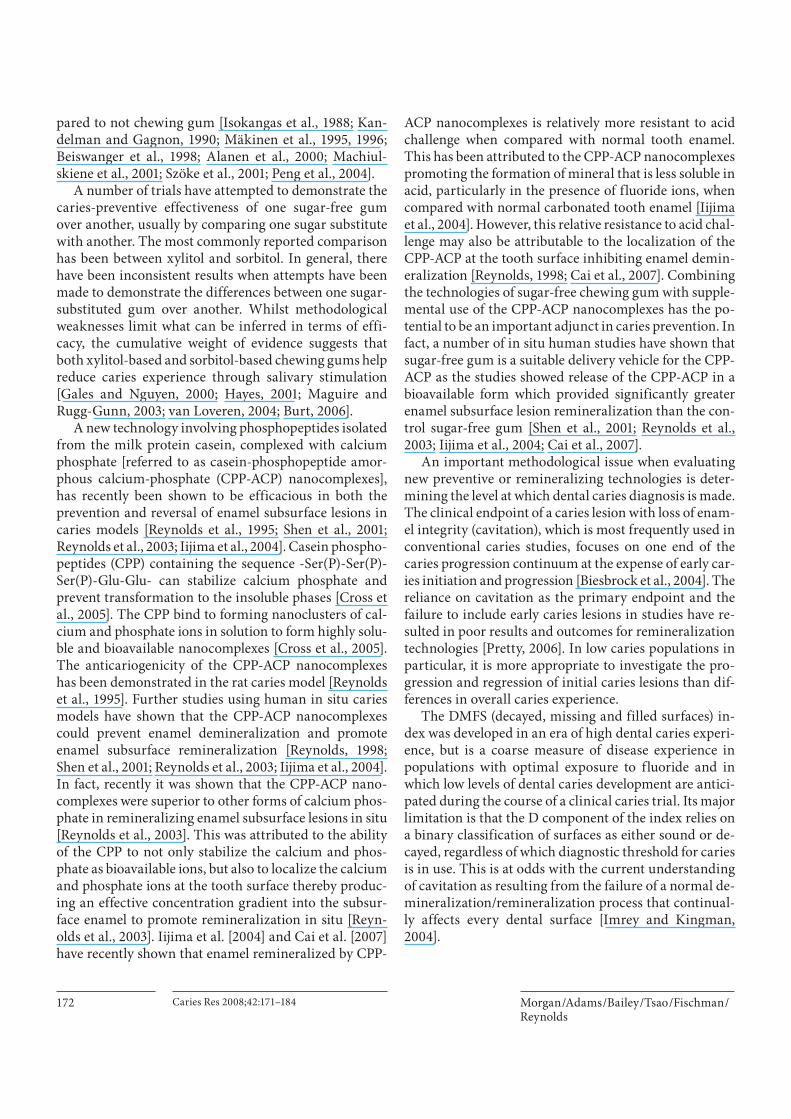
Of the 2,768 subjects screened for participation in the trial, 2,720 were randomized and 2,711 supplied with the study gums: 1,346 (49.7%) received the control gum and 1,365 (50.3%) received the gum containing CPP-ACP. Full subject disposition is presented in figure 1 . At the completion of the trial, 857 subjects chewing the control gum and 892 in the group chewing the CPP-ACP gum were evaluable per protocol and with both baseline and 24-month radiographs available. The most common rea-sons for subject non-completion were that the subject had
Table 3. Baseline demographic and dental characteristics
Demographic characteristic Control gum(n = 857)
CPP-ACP gum(n = 892)
AgeMean 8 SD 12.780.35 12.780.36Range 11.8413.5 11.5413.5
SexMale 449 (52.4%) 479 (53.7%)Female 408 (47.6%) 413 (46.3%)
Country of birthAustralia 737 (86.0%) 782 (87.7%)New Zealand/Pacific Islands 17 (2.0%) 12 (1.3%)Europe 19 (2.2%) 18 (2.0%)Africa and Middle East 14 (1.6%) 16 (1.8%)Asia 58 (6.8%) 51 (5.7%)Americas 0 (0.0%) 5 (0.6%)Unknown 12 (1.4%) 8 (0.9%)
Chewing gum usageNo 36 (4.2%) 33 (3.7%)Occasionally 582 (68.0%) 629 (70.7%)Regularly 237 (27.6%) 228 (25.6%)Unknown 2 (0.2%) 2 (0.2%)
Tooth brushingTwo or more times per day 391 (45.6%) 421 (47.2%)Once a day 390 (45.5%) 386 (43.3%)Less than once a day 56 (6.5%) 58 (6.5%)Rarely 19 (2.2%) 24 (2.7%)Unknown 1 (0.1%) 3 (0.3%)
Normally use fluoride toothpasteYes 771 (90.0%) 797 (89.3%)No 61 (7.1%) 63 (7.1%)Unknown 25 (2.9%) 32 (3.6%)
Family dentistYes 344 (40.1%) 382 (42.8%)No 500 (58.3%) 502 (56.3%)Unknown 13 (1.5%) 8 (0.9%)
Visits to dentistEvery 6 months 117 (13.7%) 152 (17.0%)Once a year 187 (21.8%) 180 (20.2%)Once every 2 years 19 (2.2%) 28 (3.1%)Less than once every 2 years 16 (1.9%) 14 (1.6%)When treatment required 135 (15.8%) 135 (15.1%)Never been 24 (2.8%) 22 (2.5%)Unknown 359 (41.9%) 361 (40.5%)
D1MFTa
Mean 8 SD 2.2282.76 2.1882.75Range 0419 0422
D1MFSa
Mean 8 SD 2.8083.85 2.7683.79Range 0430 0427
Data shown as number and percent (except for age, D1MFT, D1MFS).
a Based on the results of visual-tactile examinations supple-mented with radiographic examinations. D component included white spot lesions.

Anticariogenic Effect of a Gum Containing CPP-ACP
Caries Res 2008;42:171–184 177
Invited to participate (n = 4577)
Responded (n = 2978)
Assessed for eligibility-dental/radiographic examinations
(n = 2768)
Excluded (n = 210)Incomplete informed consent form or
questionnaire (n = 29)Did not meet inclusion criteria (n = 164)Refused to participate (n = 17)
Excluded (n = 48)Failed to attend screening (n = 17)Did not meet inclusion criteria
(n = 12)Moved schools (n = 10)Subject discontinued participation (n = 9)
Subjects randomized (n = 2720)
Allocated to Control Gum (n = 1351)Received gum (n = 1346)Did not receive gum (n = 5)
Moved schools (n = 5)
Allocated to CPP-ACP Gum (n = 1369)Received gum (n = 1365)Did not receive gum (n = 4)
Moved schools (n = 3)Incorrectly randomised (n = 1)
Completed (n = 894)
Analysed (Per-Protocol)(n = 857)
Excluded from Analysis(n = 37)
Protocol violations (n = 36)Did not satisfy entry criteria (n = 3)Chronic use of antibiotics during study (n = 1)Fixed orthodontics at 24-month examinations (n = 17)Missed 24-month clinical examination (n = 4)24-month examination outside visit window (n = 5)Non-compliance (n = 1)Missing baseline or 24-month radiographs (n = 5)
Incorrect replacement gum supplied (n = 1)
Lost to follow-up (n = 272)Moved schools (n = 186)School withdrawal (n = 86)
Discontinued intervention (n = 180)Adverse event (n = 23)Fixed orthodontic appliances (n = 65)Prolonged or repeated absences from school (n = 14)Subject discontinued participation (n = 73)Other (n = 5)
Completed (n = 926)
Analysed (Per-Protocol)(n = 892)
Excluded from Analysis(n = 34)
Protocol violations (n = 34)Did not satisfy entry criteria (n = 1)Fixed orthodontics at 24-month examinations (n = 21)Missed 24-month clinical examination (n = 3)24-month examination outside visit window (n = 5)Missing baseline or 24-month radiographs (n = 4)
Lost to follow-up (n = 255)Moved schools (n = 167)School withdrawal (n = 88)
Discontinued intervention (n = 184)Adverse event (n = 19)Fixed orthodontic appliances (n = 61)Prolonged or repeated absences from school (n = 14)Subject discontinued participation (n = 84)Other (n = 6)
Invited to participate (n = 4,577)
Responded (n = 2,978)
Assessed for eligibility-dental/radiographic examinations
(n = 2,768)
Excluded (n = 210)Incomplete informed consent form or
questionnaire (n = 29)Did not meet inclusion criteria (n = 164)Refused to participate (n = 17)
Excluded (n = 48)Failed to attend screening (n = 17)Did not meet inclusion criteria
(n = 12)Moved schools (n = 10)Subject discontinued participation (n = 9)
Subjects randomized (n = 2,720)
Allocated to Control Gum (n = 1,351)Received gum (n = 1,346)Did not receive gum (n = 5)
Moved schools (n = 5)
Allocated to CPP-ACP Gum (n = 1,369)Received gum (n = 1,365)Did not receive gum (n = 4)
Moved schools (n = 3)Incorrectly randomized (n = 1)
Fig. 1. Subject disposition.

Morgan /Adams /Bailey /Tsao /Fischman /Reynolds
Caries Res 2008;42:171–184178
transferred to a non-participating school, the subject’s school withdrew from the study, the subject received fixed orthodontic appliances or the subject discontinued participation for personal reasons.
Baseline Characteristics Table 3 summarizes the demographic and caries expe-
rience characteristics of subjects. There was no difference at baseline in the average age of subjects between the two gum groups. The proportions of males and females in each gum group were similar. Although over 86% of sub-jects were born in Australia, the study sample was ethni-cally diverse. Over 95% of subjects indicated that, prior to participating in the study, they chewed gum on a regu-lar or occasional basis. For both gum groups, over 90% of subjects brushed their teeth at least once a day, over 89% of subjects normally used fluoride toothpastes and just over 40% of subjects had a family dentist. The difference between gum groups in dental visitation was not statisti-cally significant. There were no statistically significant
differences in the baseline caries experience (DMFS/T) between the gum groups.
Scoring Reliability The three scorings of the reliability radiographs
showed excellent agreement with the intra-examiner kappa scores being 0.93 (initial vs. repeat 1), 0.92 (initial vs. repeat 2) and 0.93 (repeat 1 vs. repeat 2). The agree-ment between scorings on the extent of penetration of approximal lesions was evaluated from those surfaces that were scored R 0 , R 1 , R 2 , R 3 and R 4 at both readings. Table 4 summarizes the agreement and discrepancies in depth codes. The radiographic scores assigned agreed for greater than 97.7% of surfaces between the three scorings. The discrepancy was greater than one depth code in less than 1% of surfaces.
Baseline Caries Experience Table 5 summarizes the baseline radiographic approx-
imal caries experience of subjects. There was no statisti-
Table 4. Intra-examiner agreement on extent of penetration of lesions
Scoring Surfaces scored
Complete agreement
Discrepancy in depth code
1 2 3
Initial versus repeat 1 1,880 1,841 (97.9%) 29 (1.5%) 4 (0.2%) 6 (0.3%)Initial versus repeat 2 1,875 1,831 (97.7%) 33 (1.8%) 5 (0.3%) 6 (0.3%)Repeat 1 versus repeat 2 1,874 1,831 (97.7%) 28 (1.5%) 7 (0.4%) 8 (0.4%)
Table 5. Baseline radiographic approximal caries diagnosis
Baseline radiographic score
All surfaces Surfaces used in transition analysis
control guma CPP-ACP guma control gum CPP-ACP gum
R0 15,004 (96.51%) 15,717 (96.79%) 13,502 (96.33%) 14,097 (96.63%)R1 237 (1.52%) 231 (1.42%) 231 (1.65%) 222 (1.52%)R2 176 (1.13%) 155 (0.95%) 170 (1.21%) 151 (1.04%)R3 63 (0.41%) 65 (0.40%) 61 (0.44%) 64 (0.44%)R4 30 (0.19%) 26 (0.16%) 20 (0.14%) 15 (0.10%)R5 2,025 2,016 – –R6 6 (0.04%) 11 (0.07%) 4 (0.03%) 9 (0.06%)R7 31 (0.20%) 33 (0.20%) 29 (0.21%) 31 (0.21%)R8 2,996 3,154 – –
All 20,568 (100.00%) 21,408 (100.00%) 14,017 (100.00%) 14,589 (100.00%)
a Percentages exclude surfaces with unreadable overlap (R5) or not visible (R8).
Anticariogenic Effect of a Gum Containing CPP-ACP
Caries Res 2008;42:171–184 179
cally significant difference in the baseline caries experi-ence between the two gum groups. The average number of tooth surfaces present in the radiographic images was 20.6 8 4.7 in the control gum group and 20.6 8 4.5 in the CPP gum group. At baseline, 608 subjects (70.9%) in the control gum group and 630 subjects (70.6%) in the CPP-ACP gum group had no approximal lesions visible on the radiographs.
Compliance There was large variability in attendance at supervised
chewing sessions, both between schools and between subjects within school. Overall, subjects in the control gum group attended an average of 64.3 8 17.4% of super-vised chewing sessions and subjects in the CPP-ACP gum group attended an average of 63.5 8 17.7% of supervised chewing sessions.
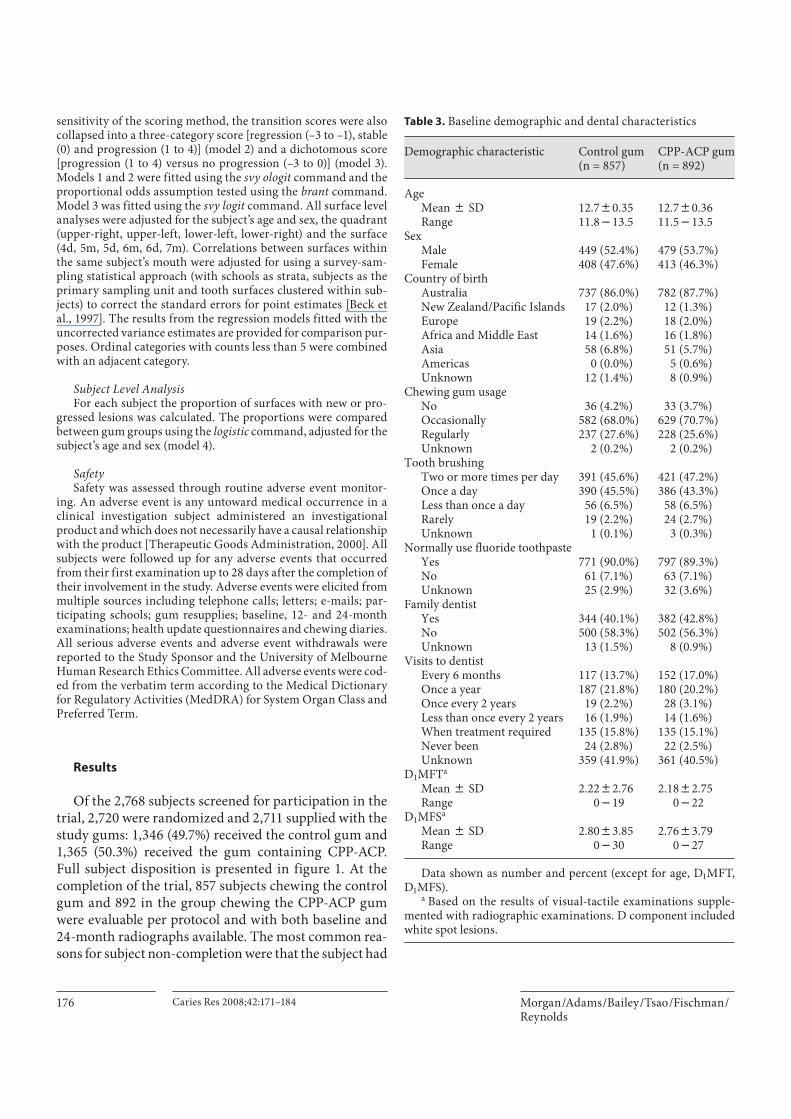
Approximal Caries Progression and Regression Table 6 displays the frequency distributions of the ra-
diographic scores from the baseline and 24-month radio-graphs for subjects in the control and CPP-ACP gum groups. At baseline, 2,025 (9.8%) and 2,015 (9.4%) sur-faces were recorded as having an unreadable overlap (R 5 )
for subjects in the control and CPP-ACP gum groups, re-spectively, whilst for the 24-month radiographs the total number of unreadable overlaps was 2,609 (12.7%) and 2,758 (12.9%), respectively. A total of 952 (4.6%) and 951 (4.4%) surfaces were coded as R 5 in both the baseline and 24-month radiographs. The majority of approximal sur-face carious lesions were detected radiographically, with only 72 of 522 (13.8%) and 113 of 1,381 (9.1%) untreated
Table 6. Distribution of radiographic scores at baseline and 24 months
Baseline radiographic score
24-month radiographic score
R0 R1 R2 R3 R4 R6 R7 all
Control gumR0 12,793 (94.75%) 398 (2.95%) 255 (1.89%) 23 (0.17%) 20 (0.15%) 4 (0.03%) 9 (0.07%) 13,502 (100.00%)R1 19 (8.23%) 88 (38.10%) 107 (46.32%) 11 (4.76%) 3 (1.30%) 3 (1.30%) 231 (100.00%)R2 7 (4.12%) 9 (5.29%) 96 (56.47%) 48 (28.24%) 5 (2.94%) 5 (2.94%) 170 (100.00%)R3 1 (1.64%) 37 (60.66%) 15 (24.59%) 1 (1.64%) 7 (11.48%) 61 (100.00%)R4 14 (70.00%) 6 (30.00%) 20 (100.00%)R6 4 (100.00%) 4 (100.00%)R7 3 (10.34%) 26 (89.66%) 29 (100.00%)
All 12,819 (91.45%) 495 (3.53%) 459 (3.27%) 119 (0.85%) 57 (0.41%) 12 (0.09%) 56 (0.40%) 14,017 (100.00%)
CPP-ACP gumR0 13,484 (95.65%) 330 (2.34%) 224 (1.59%) 36 (0.26%) 9 (0.06%) 3 (0.02%) 11 (0.08%) 14,097 (100.00%)R1 30 (13.51%) 84 (37.84%) 86 (38.74%) 18 (8.11%) 1 (0.45%) 3 (1.35%) 222 (100.00%)R2 9 (5.96%) 13 (8.61%) 74 (49.01%) 45 (29.80%) 4 (2.65%) 1 (0.66%) 5 (3.31%) 151 (100.00%)R3 1 (1.56%) 2 (3.13%) 31 (48.44%) 9 (14.06%) 5 (7.81%) 16 (25.00%) 64 (100.00%)R4 8 (53.33%) 1 (6.67%) 6 (40.00%) 15 (100.00%)R6 8 (88.89%) 1 (11.11%) 9 (100.00%)R7 3 (9.68%) 28 (90.32%) 31 (100.00%)
All 13,524 (92.70%) 427 (2.93%) 386 (2.65%) 130 (0.89%) 31 (0.21%) 21 (0.14%) 70 (0.48%) 14,589 (100.00%)
The rows represent the baseline radiological appearance of approximal surfaces and the columns their appearance at the 24-month follow-upexamination.
Table 7. Distribution of transition scores
Transition score
Control gum CPP-ACP gum Difference in percentages
–3 0 (0.00%) 1 (0.01%) +0.01–2 7 (0.05%) 9 (0.06%) +0.01–1 29 (0.21%) 45 (0.31%) +0.10
0 13,075 (93.28%) 13,749 (94.24%) +0.961 573 (4.09%) 476 (3.26%) –0.832 274 (1.95%) 249 (1.71%) –0.253 39 (0.28%) 51 (0.35%) +0.074 20 (0.14%) 9 (0.06%) –0.08
All 14,017 (100.00%) 14,589 (100.00%)

Morgan /Adams /Bailey /Tsao /Fischman /Reynolds
Caries Res 2008;42:171–184180
lesions being detected clinically at the baseline and 24-month examinations, respectively (for consistency with the visual-tactile examinations, radiographic scores R 0 and R 1 were considered sound).
The frequency distribution of the transition scores is summarized by gum group in table 7 . The number of tooth surfaces per subject for which transitions were scored ranged from 3 to 24 (average 16.4 8 5.1) in the
control gum group and from 4 to 24 (average 16.4 8 4.9) in the CPP-ACP gum group. The baseline caries experi-ence in transition surfaces was similar to the baseline car-ies experience summarized in table 5 . The CPP-ACP gum slowed progression of carious lesions (transition scores 1 to 4) compared to the control gum: 785 of 14,589 approx-imal surfaces (5.4%) experienced caries progression with the CPP-ACP gum compared to 906 of 14,017 approximal
Table 8. Ordered logistic and logistic regression models for approximal caries progression
Parameter estimatea
Standard error
Odds ratio (OR)b
95% CI p value
Surface level analysesOrdered logistic model (model 1)
Correlatedc, d –0.2020 0.0940 0.82 0.68, 0.98 0.03Independenced –0.2020 0.0496 0.82 0.74, 0.90 <0.001
Ordered logistic model (model 2) (regression/stable/progression)Correlatedc, d –0.2044 0.0940 0.82 0.68, 0.98 0.03Independenced –0.2044 0.0493 0.82 0.74, 0.90 <0.001
Logistic model (model 3) (no progression/progression)Correlatedc, d –0.1955 0.1001 0.82 0.67, 1.00 0.05Independenced –0.1955 0.0506 0.82 0.74, 0.91 <0.001
Subject level analysisLogistic model (model 4) (no progression/progression)
Independencee –0.2259 0.1019 0.80 0.65, 0.97 0.03
a Estimate of the difference in caries progression between gum groups (CPP-ACP gum – control gum) on log odds scale.
b An OR <1 implies that the odds of a surface progressing for subjects in the CPP-ACP gum group is less than the odds for a surface progressing for subjects in the control gum group.
c Standard errors adjusted for correlations between surfaces using survey sampling methods (subject-primary sampling unit, school-strata).
d Terms in model: age, sex, quadrant (upper right, upper left, lower left, lower right) and surface (4d, 5m, 5d, 6m, 6d, 7m).
e Model adjusted for the subject’s age and sex.
Table 9. Summary of treatment-emergent adverse events
Control gum(n = 1,346)
CPP-ACP gum(n = 1,365)
Subjects with any adverse event 829 (61.6%) 844 (61.8%)Subjects with serious adverse events 33 (2.5%) 30 (2.2%)Subjects with non-serious adverse events related to study gumsa 93 (6.9%) 90 (6.6%)Subjects with serious adverse events related to study gumsa 0 (0.0%) 0 (0.0%)Subjects who discontinued the study due to adverse events 23 (1.7%) 19 (1.4%)
Total number of adverse events recorded 1,769 1,830
a Considered by the investigators as possibly or probably related to the study gums.

Anticariogenic Effect of a Gum Containing CPP-ACP
Caries Res 2008;42:171–184 181
surfaces (6.5%) with the control gum, a reduction of 17%. The CPP-ACP gum also enhanced regression of carious lesions (transition scores –3 to –1) compared to the con-trol gum: 55 approximal surfaces (0.4%) experienced car-ies regression with the CPP-ACP gum compared to 36 approximal surfaces (0.3%) with the control gum. A greater percentage of approximal surfaces also remained unchanged with the CPP-ACP gum (94.2%) than with the control gum (93.3%).
Surface Level Analyses The differences in the transition score distributions
between the two gum groups were statistically significant ( table 8 , model 1: p = 0.03). The odds ratio was estimated as 0.82. Overall, the odds of a surface experiencing caries progression for subjects in the CPP-ACP gum group was 18% less than the odds of a surface experiencing caries progression for subjects in the control gum group. The odds of caries progression also increased with age (p = 0.03) and differed between surfaces (p ! 0.001), with sur-faces 5d, 6m, 6d and 7m having significantly more caries progressions than surfaces 4d and 5m. No significant dif-ferences in caries progression were found between males and females (p = 0.5) or between quadrants of the mouth (p = 0.2), and no gum interaction terms were significant. No departure from the proportional odds assumption was found (p = 0.2). The results from model 2 in table 8 were almost identical to the results above for the full mod-el (model 1). The odds ratio and 95% confidence interval for model 3 were also similar to those obtained from mod-
els 1 and 2. The major difference was that the odds ratio for caries progression on surface 6m was now 2.42 com-pared to 1.86 with the other models. This was not surpris-ing since 71 of 91 surfaces with negative transitions were from surface 6m and these were now coded as not pro-gressing. The standard errors estimated from the models that assumed independence were approximately half the standard errors estimated for the survey sampling models that adjusted for the correlation between tooth surfaces (the estimated intra-class correlation was 0.12).
Subject Level Analyses The average number of surfaces that experienced car-
ies progression was 1.42 surfaces per 24 surfaces in the control gum group and 1.23 surfaces per 24 surfaces in the CPP-ACP gum group. For 615 subjects (68.9%) in the CPP-ACP gum group compared with 548 subjects (63.9%) in the control gum group, all scorable approximal sur-faces either remained unchanged or experienced caries regression. The estimated OR for caries progression from the logistic regression was 0.80 (p = 0.03, table 8 , model 4). The odds of a surface experiencing caries progression for subjects in the CPP-ACP gum group was 20% less than the odds of a surface experiencing caries progres-sion for subjects in the control gum group.
Safety Table 9 summarizes the adverse events which oc-
curred after the first examination (treatment-emergent adverse events) for the randomized population. Table 10 shows the most common adverse events reported in ei-ther group, the non-serious adverse events most frequent-ly assessed as related to use of the study gums (nausea, headache and diarrhoea) and the most common reasons for adverse event discontinuation. All serious adverse events were assessed by the investigators as not being re-lated to the gum usage. For 3 subjects the serious adverse event (although not product-related) resulted in their dis-continuation from the study. The incidences of adverse events (serious and non-serious) were similar between the two study gums.
Discussion
The trial compared the effectiveness of a CPP-ACP gum over an active control. There is little scientific lit-erature which is directly comparable to this study where a control sugar-free gum was compared to a similar gum with an additional caries-preventive agent. All reason-
Table 10. Most common adverse events reported
MedDRa preferred term Control gum(n = 1,346)
CPP-ACP gum(n = 1,365)
Most common adverse events (i.e. those occurring in more than 5%)
Malaise 565 (42.0%) 567 (41.5%)Headache 122 (9.1%) 99 (7.3%)Nasopharyngitis 106 (7.9%) 99 (7.3%)Influenza 82 (6.1%) 104 (7.6%)
Most common treatment-related adverse events (i.e. those occurring in more than 1%)
Nausea 15 (1.1%) 23 (1.7%)Headache 25 (1.9%) 11 (0.8%)Diarrhoea 6 (0.4%) 13 (1.0%)
Most common adverse event discontinuation reasonsNausea 3 (0.2%) 9 (0.7%)Headache 6 (0.4%) 3 (0.2%)Diarrhoea 1 (0.1%) 3 (0.2%)

Morgan /Adams /Bailey /Tsao /Fischman /Reynolds
Caries Res 2008;42:171–184182
able attempts were made to test the CPP-ACP gum in a situation where it could reasonably be expected that den-tal caries might be unlikely to occur and in an environ-ment where dental caries development is low. Subjects from both groups were exposed to fluoridated drinking water and they, and their families, were provided with a constant supply of fluoridated toothpaste and tooth-brushes. All subjects were notified on an annual basis of their oral health status and copies of their radiographs supplied to their chosen dental care provider. In addition to the above preventive dental caries programme, sub-jects in both groups were supervised in the use of sugar-free chewing gum. To the extent that it was possible to control in a large clinical trial, the only point of difference in terms of dental caries prevention was the inclusion of CPP-ACP in the sugar-free gum used by one group of subjects.
Despite these intended stringencies in the trial design, chewing CPP-ACP afforded an increased preventive ef-fect beyond that achieved through good, basic oral care habits and the use of chewing sugar-free gum. Subjects chewing the CPP-ACP sugar-free gum demonstrated a statistically significant difference in radiographically di-agnosed approximal carious lesions compared with those chewing the control sugar-free gum. The level to which this result is clinically significant is worth considering. Given that the population in which the study was con-ducted can be considered ‘low risk’, the difference in ap-proximal caries development is indicative of the clinical strength of the effect. In addition, the ethnically diverse study population promotes the generalizability of the study results.
The results of this trial have been presented using data obtained from standardized digital radiographs. The as-sessment of the effectiveness of a preventive agent where incipient lesion detection is key (in an era of low dental caries and in populations of low risk) requires diagnostic criteria which are highly refined and reproducible. Whilst historically, reliance has been placed on measurements obtained from clinical examinations, for the purposes of early lesion detection such measurements can no longer be considered appropriate, especially in the current oral disease environment. A recent review of approximal car-ies diagnosis reported higher sensitivities for bitewing ra-diography than fibre-optic transillumination and visual-tactile assessment [Bader et al., 2001]. An additional ad-vantage of radiographs is that the images can be stored indefinitely and are able to be reviewed under standard-ized conditions at any time. Results of this study were restricted to approximal surfaces. From a clinical stand-
point the diagnosis of occlusal lesions using radiographs, particularly those confined to enamel, is poor [Wenzel and Fejerskov, 1992; Kidd et al., 2003; Hopcraft and Mor-gan, 2005]. The use of radiography is not new in dentist-ry, although its use has been limited in clinical trials be-cause of ethical issues regarding cumulative radiation dosage, together with practicalities of access and cost. The radiographic methodology used in this study (stan-dardized measurements, low dosage exposure and digital recording, and the use of a dedicated on-site radiography van) ensured that the effect of these issues was mini-mized. Standardized digital radiography procedures in conjunction with a scoring method that grades lesion depth provide potentially the most appropriate method of determining early carious lesion development in large-scale caries clinical trials and are particularly appropriate in a low caries population. This study also confirmed the value of bitewing radiographs for detecting approximal carious lesions in clinical trials. The additional diagnos-tic yield from the bitewing radiographs was substantial: greater than 6-fold and greater than 11-fold for the base-line and 24-month examinations, respectively.
Previous investigations [Lawrence et al., 1997; Law-rence and Sheiham, 1997; Chesters et al., 2002] using a similar radiography scoring system also reported good reliability (kappa scores of � 0.8). The use of transition scores to interpret the progression (and regression) of ap-proximal caries in longitudinal studies [Grondahl et al., 1977; Granath et al., 1980; Cook, 1984; Källestål and Holm, 1994; Lawrence and Sheiham, 1997] and in clinical trials [Chesters et al., 2002] has become an accepted ana-lytical methodology.
The results from the surface level analyses mirrored the results from the subject level analysis. An advantage of the surface level analyses was that they confirmed the differing susceptibility of approximal surfaces to caries progression found in other studies [Dummer et al., 1988; Mejàre et al., 1999; Hintze, 2001; Stenlund et al., 2003]. This paper also illustrated the need for statistical analysis to account for the clustering of surfaces within subjects that has been reported in other studies. The assumption of independence of tooth surfaces within a subject re-sulted in a twofold underestimation of the standard er-rors for the treatment difference and resulted in exagger-ated p values for the comparison between the gum groups. Dichotomizing the transition scores leads to a loss of ef-ficiency for the comparison between gum groups since caries regressions are discounted.
Several studies using an in situ remineralization mod-el have demonstrated that the CPP-ACP nanocomplexes

Anticariogenic Effect of a Gum Containing CPP-ACP
Caries Res 2008;42:171–184 183
delivered in a sugar-free gum, as used in this clinical tri-al, significantly remineralized enamel subsurface lesions [Shen et al., 2001; Reynolds et al., 2003; Iijima et al., 2004; Cai et al., 2007]. In the current clinical trial we observed not only a smaller number of approximal surfaces that progressed but also a greater number of surfaces (lesions) that regressed (remineralized) with the CPP-ACP gum compared with the control sugar-free gum. The regres-sions involved predominantly remineralization of enam-el lesions (e.g. inner-half enamel lesion to sound; outer-half enamel lesion to sound and inner-half enamel lesion to outer-half enamel lesion) but also a smaller number of dentine lesions that remineralized (e.g. outer-half den-tine lesion to inner-half enamel lesion). These clinical tri-al results therefore confirm the findings from the short-term in situ remineralization studies and demonstrate that the longer-term manifestation of enamel subsurface lesion remineralization and inhibition of demineraliza-tion by the CPP-ACP nanocomplexes is the significant
slowing of caries progression. In conclusion, chewing sugar-free gum containing CPP-ACP can be regarded as an additional caries prevention tool, over and above oth-er accepted preventive strategies such as water fluorida-tion or fluoridated toothpaste.
Acknowledgements
The authors would like to acknowledge the significant contri-butions to the design, execution and analysis of this study made by staff members from the Clinical Trial Group at the School of Dental Science, The University of Melbourne; Dr. D. Tancredi, Dr. D. Ming and staff members of Cadbury Schweppes Science and Technology, and the trial monitor, Mr. M. Boschenok. This study would not have been possible without the cooperation of the Vic-torian Department of Education and the principals, teachers, par-ents and students at the participating schools. Support for this study was provided by Cadbury Schweppes Science and Technol-ogy and the Australian National Health and Medical Research Council Development Grant No. 991501.
References
Ahn C, Jung SH, Donner A: Application of an adjusted � 2 statistic to site-specific data in observational dental services. J Clin Peri-odontol 2002; 29: 79–82.
Alanen P, Isokangas P, Gutmann K: Xylitol can-dies in caries prevention: results of a field study in Estonian children. Community Dent Oral Epidemiol 2000; 28: 218–224.
Bader JD, Shugars DA, Bonito AJ: Systematic re-views of selected dental caries diagnostic and management methods. J Dent Educ 2001; 65: 960–968.
Bailey DL, Adams GG, Tsao CE, Morgan MV: Standardisation of digital bitewing radio-graphs during a clinical caries trial (abstract 62). Caries Res 2006; 40: 324.
Beck JD, Lawrence HP, Koch GG: Analyticapproaches to longitudinal caries data in adults. Community Dent Oral Epidemiol 1997; 25: 42–51.
Beiswanger BB, Boneta AE, Mau MS, Katz BP, Proskin HM, Stookey GK: The effect of chewing sugar-free gum after meals on clin-ical caries incidence. J Am Dental Assoc 1998; 129: 1623–1626.
Biesbrock AR, Chesters RK, Ellwood RP, Smith SR: The challenges of validating diagnostic methods relative to a conventional two-year caries clinical trial. J Dent Res 2004; 83:C53–C55.
Burnside G, Pine CM, Williamson PR: The ap-plication of multilevel modelling to dental caries data. Stat Med 2007; 26: 4139–4149.
Burt BA: The use of sorbitol- and xylitol-sweet-ened chewing gum in caries control. J Am Dental Assoc 2006; 137: 190–196.
Cai F, Manton DJ, Shen P, Walker GD, Cross KJ, Yuan Y, Reynolds C, Reynolds EC: Effect of addition of citric acid and casein phospho-peptide-amorphous calcium phosphate to a sugar-free chewing gum on enamel reminer-alization in situ. Caries Res 2007; 41: 377–383.
Campain AC, Morgan MV, Evans RW, Ugoni A, Adams GG, Conn JA, Watson MJ: Sugar-starch combinations in food and the rela-tionship to dental caries in low-risk adoles-cents. Eur J Oral Sci 2003; 111: 316–325.
Chesters RK, Pitts NB, Matuliene G, Kvedariene A, Huntington E, Bendinskaite R, Balciu-niene I, Matheson JR, Nicholson JA, Gendvi-lyte A, Sabalaite R, Ramanauskiene J, Savage D, Mileriene J: An abbreviated caries clinical trial design validated over 24 months. J Dent Res 2002; 81: 637–640.
Cohen J: A coefficient of agreement for nominal scales. Educ Psychol Meas 1960; 20: 37–46.
Cook SR: A longitudinal radiographic study of caries progression in dental students. Aust Dent J 1984; 29: 315–320.
Cross KJ, Huq NL, Palamara JE, Perich JW, Reynolds EC: Physicochemical characteriza-tion of casein phosphopeptide-amorphous calcium phosphate nanocomplexes. J Biol Chem 2005; 280: 15362–15369.
DeRouen TA, Mancl L, Hujoel P: Measurement of associations in periodontal diseases using statistical methods for dependent data. J Periodontal Res 1991; 26: 218–229.
Donner A, Banting D: Analysis of site-specific data in dental statistics. J Dent Res 1988; 67: 1392–1395.
Donner A, Banting D: Adjustment of frequently used chi-square procedures for the effect of site-to-site dependencies in the analysis of dental data. J Dent Res 1989; 68: 1350–1354.
Dummer PMH, Addy M, Oliver SJ, Shaw WC: Changes in the distribution of decayed and filled tooth surfaces and the progression of approximal caries in children between the ages of 11–12 years and 15–16 years. Br Dent J 1988; 164: 277–282.
Gales MA, Nguyen TM: Sorbitol compared with xylitol in prevention of dental caries. Ann Pharmacother 2000; 34: 98–100.
Granath L, Kahlmeter A, Matsson L, Schroder U: Progression of proximal enamel caries in early teens related to caries activity. Acta Odontol Scand 1980; 38: 247–251.
Grondahl HG, Hollender L, Malmcrona E, Sund-quist B: Dental caries and restorations in teenagers. Swed Dent J 1977; 1: 45–50.
Hannigan A, O’Mullane DM, Barry D, Schafer F, Roberts AJ: A re-analysis of a caries clinical trial by survival analysis. J Dent Res 2001; 80: 427–431.
Hayes C: The effect of non-cariogenic sweeten-ers on the prevention of dental caries: a re-view of the evidence. J Dent Educ 2001; 65: 1105–1109.

Morgan /Adams /Bailey /Tsao /Fischman /Reynolds
Caries Res 2008;42:171–184184
Hintze H: Approximal caries prevalence in Dan-ish recruits and progression of caries in the late teens: a retrospective radiographic study. Caries Res 2001; 35: 27–35.
Hopcraft MS, Morgan MV: Comparison of ra-diographic and clinical diagnosis of approx-imal and occlusal dental caries in a young adult population. Community Dent OralEpidemiol 2005; 33: 212–218.
Hujoel PP, Loesche WJ, DeRouen TA: Assess-ment of relationships between site-specific variables. J Periodontol 1990; 61: 368–372.
Iijima Y, Cai F, Shen P, Walker G, Reynolds C, Reynolds EC: Acid resistance of enamel sub-surface lesions remineralized by a sugar-free chewing gum containing casein phos-phopeptide-amorphous calcium phosphate. Caries Res 2004; 38: 551–556.
Imrey PB, Kingman A: Analysis of clinical tri-als involving non-cavitated caries lesions. J Dent Res 2004; 83:C103–C108.
Isokangas P, Alanen P, Tiekso J, Mäkinen KK: Xylitol chewing gum in caries prevention: a field study in children. J Am Dental Assoc 1988; 117: 315–320.
Källestål C, Holm AK: Allocation of dental car-ies prevention in Swedish teenagers. Com-munity Dent Oral Epidemiol 1994; 22: 100–105.
Kandelman D, Gagnon G: A 24-month clinical study of the incidence and progression of dental caries in relation to consumption of chewing gum containing xylitol in school preventive programs. J Dent Res 1990; 69: 1771–1775.
Kidd EAM, Mejàre I, Nyvad B: Clinical and ra-diographic diagnosis; in Fejerskov O, Kidd EAM (eds): Dental Caries: The Disease and Its Clinical Management. Oxford, Blackwell, 2003, pp 111–128.
Kidd EAM, Pitts NB: A reappraisal of the value of the bitewing radiograph in the diagnosis of posterior approximal caries. Br Dent J 1990; 169: 195–200.
LaVange LM, Koch GG, Schwartz TA: Applying sample survey methods to clinical trials data. Stat Med 2001; 20: 2609–2623.
Lawrence HP, Benn DK, Sheiham A: Digital ra-diographic measurement of approximal car-ies progression in fluoridated and non-fluo-ridated area of Rio de Janeiro, Brazil. Community Dent Oral Epidemiol 1997; 25: 412–418.
Lawrence HP, Sheiham A: Caries progression in 12- to 16-year-old schoolchildren in fluori-dated and fluoride-deficient areas in Brazil. Community Dent Oral Epidemiol 1997; 25: 402–411.
Liang KY, Zeger SL: Longitudinal data analysis using generalized linear models. Biometrics 1986; 73: 13–22.
Lingström P, Holm AK, Mejàre I, Twetman S, Söder B, Norlund A, Axelsson S, Lagerlöf F, Nordenram G, Petersson LG, Dahlgren H, Källestål C: Dietary factors in the prevention of dental caries: a systematic review. Acta Odontol Scand 2003; 61: 331–340.
Llena-Puy C, Forner L: A clinical and radio-graphic comparison of caries diagnosed in approximal surfaces of posterior teeth in a low-risk population of 14-year-old children. Oral Health Prev Dent 2005; 3: 47–52.
Machiulskiene V, Nyvad B, Baelum V: Caries preventive effect of sugar-substituted chew-ing gum. Community Dent Oral Epidemiol 2001; 29: 278–288.
Maguire A, Rugg-Gunn AJ: Xylitol and caries prevention – is it a magic bullet? Br Dent J 2003; 194: 429–436.
Mäkinen KK, Bennett CA, Hujoel PP, Isokangas PJ, Isotupa KP, Pape Jr HR, Mäkinen PL: Xy-litol chewing gums and caries rates: a 40-month cohort study. J Dent Res 1995; 74: 1904–1913.
Mäkinen KK, Mäkinen PL, Pape HR Jr, Peldyak J, Hujoel PP, Isotupa KP, Isokangas PJ, Allen P, Bennett C: Conclusion and review of the ‘Michigan Xylitol Programme’ (1986–1995) for the prevention of dental caries. Int Dent J 1996; 46: 22–34.
McCullagh P: Regression models for ordinal data. J R Statist Soc B 1980; 42: 109–142.
Mejàre I, Källestål C, Stenlund H: Incidence and progression of approximal caries from 11 to 22 years of age in Sweden: a prospective ra-diographic study. Caries Res 1999; 33: 93–100.
Peng B, Petersen PE, Bian Z, Tai B, Jiang H: Can school-based oral health education and a sugar-free chewing gum program improve oral health? Results from a two-year study in PR China. Acta Odontol Scand 2004; 62: 328–332.
Pitts NB: The use of film holding, beam colli-mating and aiming devices in bitewing radi-ography: a suggested design for routine and research use. Dentomaxillofac Radiol 1983; 12: 77–82.
Pitts NB: Systems for grading approximal cari-ous lesions and overlaps diagnosed from bitewing radiographs: proposals for future standardization. Community Dent OralEpidemiol 1984; 12: 114–122.
Pitts NB: Score system for behaviour of radio-logically diagnosed approximal carious le-sions. Community Dent Oral Epidemiol 1985; 13: 268–272.
Pitts NB: The use of bitewing radiographs in the management of dental caries: scientific and practical considerations. Dentomaxillofac Radiol 1996; 25: 5–16.
Pretty IA: Caries detection and diagnosis: novel techniques. J Dent 2006; 34: 727–739.
Radike AW: Criteria for diagnosis of dental car-ies; in Conference on the Clinical Testing of Cariostatic Agents. American Dental Asso-ciation, 1972, pp 87–88.
Reynolds EC: Anticariogenic complexes of amorphous calcium phosphate stabilized by casein phosphopeptides: a review. Spec Care Dentist 1998; 18: 8–16.
Reynolds EC, Cai F, Shen P, Walker GD: Reten-tion in plaque and remineralization of enam-el lesions by various forms of calcium in a mouthrinse or sugar-free chewing gum. J Dent Res 2003; 82: 206–212.
Reynolds EC, Cain CJ, Webber FL, Black CL, Ri-ley PF, Johnson IH, Perich JW: Anticarioge-nicity of calcium phosphate complexes of tryptic casein phosphopeptides in the rat. J Dent Res 1995; 74: 1272–1279.
Scheie AA, Fejerskov OB: Xylitol in caries pre-vention: what is the evidence for clinical ef-ficacy? Oral Dis 1998; 4: 268–278.
Shen P, Cai F, Nowicki J, Vincent J, Reynolds EC: Remineralization of enamel subsurface le-sions by sugar-free chewing gum containing casein phosphopeptide-amorphous calcium phosphate. J Dent Res 2001; 80: 2066–2070.
Stenlund H, Mejàre I, Källestål C: Caries inci-dence rates in Swedish adolescents and young adults with particular reference to ad-jacent approximal tooth surfaces: a method-ological study. Community Dent Oral Epi-demiol 2003; 31: 361–367.
Szöke J, Bánóczy J, Proskin HM: Effect of after-meal sucrose-free gum-chewing on clinical caries. J Dent Res 2001; 80: 1725–1729.
Therapeutic Goods Administration: Note for guidance on good clinical practice (cpmp/ich/135/95) annotated with TGA comments. Canberra, Therapeutic Goods Administra-tion, 2000.
van Loveren C: Sugar alcohols: what is the evi-dence for caries-preventive and caries-thera-peutic effects? Caries Res 2004; 38: 286–293.
Wenzel A, Fejerskov O: Validity and diagnosis of questionable caries lesions in occlusal sur-faces of extracted third molars. Caries Res 1992; 26: 188–194.
Whitehead J: Sample size calculations for or-dered categorical data. Stat Med 1993; 12: 2257–2271.