Mechanism and Substrate Recognition of Human Holo ACP Synthase
19
Mechanism and Substrate Recognition of Human Holo ACP Synthase Gabor Bunkoczi 1,3 , Saloni Pasta 2,3 , Anil Joshi 2 , Xiaoqiu Wu 1 , Kathryn L. Kavanagh 1 , Stuart Smith 2∗∗ , and Udo Oppermann 1∗ 1 Structural Genomics Consortium, University of Oxford, Oxford OX3 7LD, UK. 2 Children's Hospital Oakland Research Institute, Oakland, CA 94609, USA. Summary Mammals utilize a single phosphopantetheinyl transferase for the posttranslational modification of at least three different apoproteins: the carrier protein components of cytosolic and mitochondrial fatty acid synthases and the aminoadipate semialdehyde reductase involved in lysine degradation. We determined the crystal structure of the human phosphopantetheinyl transferase, a eukaryotic phosphopantetheinyl transferase characterized, complexed with CoA and Mg 2+ , and in ternary complex with CoA and ACP. The involvement of key residues in ligand binding and catalysis was confirmed by mutagenesis and kinetic analysis. Human phosphopantetheinyl transferase exhibits an α/β fold and 2-fold pseudosymmetry similar to the Sfp phosphopantetheinyl transferase from Bacillus subtilis. Although the bound ACP exhibits a typical four-helix structure, its binding is unusual in that it is facilitated predominantly by hydrophobic interactions. A detailed mechanism is proposed describing the substrate binding and catalytic process. Keywords CHEMBIO; PROTEINS Introduction The posttranslational modification of a conserved serine residue in carrier proteins with a phosphopantetheinyl moiety transferred from coenzyme A through the enzyme phosphopantetheinyl transferase (PPT) (Figure 1) is vital to all living organisms. The resulting holo-carrier proteins play essential roles as components of fatty acid and polyketide synthases (acyl carrier proteins, ACPs) and nonribosomal peptide synthases (peptidyl carrier proteins, PCPs) [1]. In addition, phosphopantetheinylated carrier proteins are also involved in the metabolism of lysine and tetrahydrofolate [2–4]. In each of these systems, the reactive sulfhydryl of the flexible 20 Å-long phosphopantetheine arm provides a center for the transport of reaction intermediates between the various catalytic components of the pathway. Whereas microbial genomes contain more than one PPT [5–8], only one PPT has been identified in mammals. This enzyme exhibits a broad substrate specificity and is able to © 2007 Elsevier Ltd. ∗Corresponding author [email protected]. ∗∗Corresponding author [email protected]. 3 These authors contributed equally to the work. This document was posted here by permission of the publisher. At the time of deposit, it included all changes made during peer review, copyediting, and publishing. The U.S. National Library of Medicine is responsible for all links within the document and for incorporating any publisher-supplied amendments or retractions issued subsequently. The published journal article, guaranteed to be such by Elsevier, is available for free, on ScienceDirect. Sponsored document from Chemistry & Biology Published as: Chem Biol. 2007 November 26; 14(11): 1243–1253. Sponsored Document Sponsored Document Sponsored Document
-
Upload
independent -
Category
Documents
-
view
0 -
download
0
Transcript of Mechanism and Substrate Recognition of Human Holo ACP Synthase

Mechanism and Substrate Recognition of Human Holo ACPSynthase
Gabor Bunkoczi1,3, Saloni Pasta2,3, Anil Joshi2, Xiaoqiu Wu1, Kathryn L. Kavanagh1, StuartSmith2∗∗, and Udo Oppermann1∗1Structural Genomics Consortium, University of Oxford, Oxford OX3 7LD, UK.
2Children's Hospital Oakland Research Institute, Oakland, CA 94609, USA.
SummaryMammals utilize a single phosphopantetheinyl transferase for the posttranslational modification ofat least three different apoproteins: the carrier protein components of cytosolic and mitochondrialfatty acid synthases and the aminoadipate semialdehyde reductase involved in lysine degradation.We determined the crystal structure of the human phosphopantetheinyl transferase, a eukaryoticphosphopantetheinyl transferase characterized, complexed with CoA and Mg2+, and in ternarycomplex with CoA and ACP. The involvement of key residues in ligand binding and catalysis wasconfirmed by mutagenesis and kinetic analysis. Human phosphopantetheinyl transferase exhibits anα/β fold and 2-fold pseudosymmetry similar to the Sfp phosphopantetheinyl transferase from Bacillussubtilis. Although the bound ACP exhibits a typical four-helix structure, its binding is unusual in thatit is facilitated predominantly by hydrophobic interactions. A detailed mechanism is proposeddescribing the substrate binding and catalytic process.
KeywordsCHEMBIO; PROTEINS
IntroductionThe posttranslational modification of a conserved serine residue in carrier proteins with aphosphopantetheinyl moiety transferred from coenzyme A through the enzymephosphopantetheinyl transferase (PPT) (Figure 1) is vital to all living organisms. The resultingholo-carrier proteins play essential roles as components of fatty acid and polyketide synthases(acyl carrier proteins, ACPs) and nonribosomal peptide synthases (peptidyl carrier proteins,PCPs) [1]. In addition, phosphopantetheinylated carrier proteins are also involved in themetabolism of lysine and tetrahydrofolate [2–4]. In each of these systems, the reactivesulfhydryl of the flexible 20 Å-long phosphopantetheine arm provides a center for the transportof reaction intermediates between the various catalytic components of the pathway.
Whereas microbial genomes contain more than one PPT [5–8], only one PPT has beenidentified in mammals. This enzyme exhibits a broad substrate specificity and is able to
© 2007 Elsevier Ltd.∗Corresponding author [email protected]. ∗∗Corresponding author [email protected] authors contributed equally to the work.This document was posted here by permission of the publisher. At the time of deposit, it included all changes made during peer review,copyediting, and publishing. The U.S. National Library of Medicine is responsible for all links within the document and for incorporatingany publisher-supplied amendments or retractions issued subsequently. The published journal article, guaranteed to be such by Elsevier,is available for free, on ScienceDirect.
Sponsored document fromChemistry & Biology
Published as: Chem Biol. 2007 November 26; 14(11): 1243–1253.
Sponsored Docum
ent Sponsored D
ocument
Sponsored Docum
ent

phosphopantetheinylate the ACP components of the cytosolic and the mitochondrial FASsystems, as well as aminoadipate semialdehyde dehydrogenase, the human ortholog to the yeastlys5 gene [2, 9]. Subcellular fractionation studies indicate that the human PPT is located in thecytosolic compartment, implying that the mitochondrial ACP and aminoadipate semialdehydedehydrogenase undergo posttranslational modification prior to their import into mitochondria.Molecular cloning and biochemical characterization of human PPT revealed that this cytosolicenzyme is a Mg2+-requiring monomeric enzyme that exhibits a broad acceptor substratespecificity [9].
Based on sequence motifs and structural features, phosphopantetheine transferases areclassified in three different groups [6]. Group I is represented by bacterial transferases of about120 residues, acting on the ACP of bacterial FASs with Escherichia coli or Bacillus subtilisACPs as prototype members. Structure determination of the group I PPT (designated as holo-ACP synthase, AcpS) from B. subtilis and Streptococcus pneumoniae revealed a trimericarrangement of an α/β fold domain [10, 11]. The active site is located in the cleft between thedifferent subunits, resulting in three active sites per molecule. Group II PPTs are exemplifiedby the Sfp protein from B. subtilis, which catalyzes the 4′-phosphopantetheine transfer fromCoA to the nonribosomal, PCP domain of bacterial surfactin synthetase [7, 12, 13]. Sfp is amonomer of about 240 residues and exhibits a broader acceptor substrate specificity than groupI PPTs. The crystallographic analysis of Sfp [13] revealed a two-domain enzyme with intrinsicpseudo 2-fold symmetry, however, with a similar domain architecture as trimeric AcpS. The4′-phosphopantetheine donor is bound in a cleft formed by the two domains, and the structurerevealed basic principles of Mg2+ and CoA binding. Group III PPTs comprise transferases thatare an integral part of the heteromeric yeast and fungal FASs [14], which display a differentarchitecture than mammalian FAS systems [15–17]. The group III PPTs appear to act on theirapoACP substrates prior to assembly of the megasynthase complex since, in the context of thecomplete complex, the PPT structure is distorted and incapable of binding CoA [16].
The closest structurally characterized enzyme to human PPT is the group II Sfp enzyme fromB. subtilis (PDB ID: 1QR0 [13]). Here, we describe the structure determination of apo andsubstrate complexes and provide biochemical analysis of human PPT, to our knowledge thefirst higher eukaryotic representative of this class of enzymes. These data suggest a generalreaction mechanism for eukaryotic PPT and, as far as we know, furthermore provide for the firsttime structural evidence that type I and II ACPs distinguish their substrate molecules throughdistinct recognition mechanisms.
Results and DiscussionOverall Structure of Human PPT
Human PPT consists of two nearly identical domains connected by a short linker region. Thetwo halves superimpose on each other with an rmsd of 2.4 Å but are distinguished by twofeatures. First, the N- and C-terminal regions of the protein contain 16 and 42 residueextensions, respectively, that are unique to each half of the molecule. Both extensions are wellordered, and only the terminal four-five residues are indistinguishable from the noise in theelectron density maps. Second, although the regions encompassing residues 91–116 in the N-terminal domain and residues 207–239 in the C-terminal domain both fold into a pair of βsheets connected by a loop region, the extension in the C-terminal domain is significantlylonger (Figure 2).
The overall fold adopted by human PPT most closely resembles that of the B. subtilis Sfpprotein with the exception of the N- and C-terminal extensions that are unique to the humanenzyme. The C-terminal extension folds back to the N-terminal domain and possibly fixes therelative orientation of the domains so that it is optimal for catalysis. Both the human and B.
Bunkoczi et al. Page 2
Published as: Chem Biol. 2007 November 26; 14(11): 1243–1253.
Sponsored Docum
ent Sponsored D
ocument
Sponsored Docum
ent

subtilis enzymes contain a pair of positionally equivalent β sheets looped out from the corestructure in the C-terminal domain: residues 196–227 in PPT and 160–187 in Sfp. However,in Sfp this region is involved in an extended crystal contact and protrudes from the main bodyof the protein up to 45 Å. Reuter et al. [13] have speculated that this region may be involvedin apoprotein binding.
The functional domains can be defined with the help of the group I PPT AcpS, which consistsof a shorter chain that trimerises to form the functionally active protein. Superposition of thisstructure on the N- and C-terminal domains of human PPT (Figure 2) reveals that N-terminal13 and the C-terminal 52 residues can be regarded as extensions and that the linker betweenthe N- and C-terminal domain consists of a single residue. Another feature is the disorderedloop between residues 248–263, which starts after the superposition of the PPT C-terminaldomain and AcpS ends, suggesting that this may act as a flexible linker.
Structure of the Fatty Acid Synthase ACP Domain and Binding to PPTACPs are classified into two distinct groups, type I representing those that are part of themultifunctional polypeptides that constitute the cytosolic animal FAS and modular polyketidesynthases and type II that are associated with the freestanding enzyme systems involved infatty acid and polyketide synthesis in bacteria, plastids, and mitochondria. Although severaltype II ACP structures have been determined experimentally, only one type I structure has beensolved, that of the rat FAS, using NMR [18]. We have now determined the structure of humanPPT in complex with CoA and the ACP domain of human FAS (PDB ID: 2CG5). The serineacceptor residue for the phosphopantetheinyl moiety in ACP was mutated to alanine to facilitatecrystallization of a stable ternary complex of PPT, apoACP, and CoA. Thus, as far as we know,this study constitutes the first description of a type I ACP molecule in complex with both CoAand one of its cognate interacting proteins.
The structures of both type II ACPs and peptidyl carrier proteins superimpose onto that ofhuman cytosolic FAS ACP with rmsds generally <3 Å. Whereas the published type I rat ACPstructure determined by NMR (1N8L [18]) shows clear differences in the N-terminal loopsegments (rmsd >5 Å), a recently deposited rat type I ACP NMR structure (2PNG) is moresimilar to human cytosolic ACP with rmsd for α-carbons ∼2 Å. As the first structure (1N8L)is indicated as being a preliminary model, the differences with the human ACP in the N-terminal region can be disregarded.
Although sequence identities between different ACP forms are as low as 20%, all ACPs thathave been structurally characterized display a four-helix bundle arrangement in which theserine residue targeted for phosphopantetheinylation is located on the surface of the protein atthe N terminus of helix-α2 (Figure 3). ACP domains associated with type I fungal FASs deviateslightly from this pattern in that they consist of two four-helix subdomains, the second of whichcarries the postranslationally added phosphopantetheinyl moiety [16, 19].
The type I human ACP binds in the cleft between the two domains of the PPT and the essentialSer2156Ala points directly toward the β-phosphate of the bound CoA. The interaction betweenthe two proteins takes place on a large interface and is mostly of hydrophobic character. Threehydrophobic patches on the PPT surface interact with the ACP. The first is formed by PPTresidues Phe51-Ala54 and interacts with a hydrophobic island formed by ACP residuesIle2137, Val2143, and Leu2152. The second consists of the exposed side chain of Leu191 onthe PPT surface and Val2176 on the ACP. Possibly, the hydrophobic pantetheinyl of CoA alsointeracts with this patch as it is pointing toward this direction, but it cannot be located in theelectron density map and therefore has not been modeled. The third hydrophobic interfaceconsists of PPT residues Phe144-Met148, Phe173, and Trp177 interacting with ACP residuesLeu2157, Met2158, and Val2160. Elimination of this hydrophobic patch by replacement of all
Bunkoczi et al. Page 3
Published as: Chem Biol. 2007 November 26; 14(11): 1243–1253.
Sponsored Docum
ent Sponsored D
ocument
Sponsored Docum
ent

three amino acids with alanine residues decreased the Km for ACP by at least three orders ofmagnitude, consistent with a role for the hydrophobic patch in recognition by the PPT (datanot shown). A few polar interactions that involve hydrogen bonds and salt bridges can beobserved (ACP-PPT: Arg2138-Gly137, Asp2151-Gln50, Thr2165-Arg138, Glu2169-Arg138), but binding specificity seems to be determined by shape complementarity rather thanthrough the few specific interactions.
Although these polar interactions are largely mediated by residues located on ACP helix α2,which is categorized for type II ACPs as the “universal recognition helix” [20], the type ofrecognition clearly is different between type I and II ACPs. In type II ACPs, highly conservednegatively charged residues play a dominant role in mediating binding to their cognate proteins[20–22]. For example, the ability of the group I E. coli PPT to catalyze phosphopantetheinetransfer to heterologous type II ACPs involved in polyketide synthesis correlates closely withthe overall negative charge on the apo-ACP substrates [23], and the complex between the GroupI B. subtilis PPT and its cognate type II ACP is stabilized predominantly by hydrophilic contacts[11]. However, a comparison of the recognition surfaces of type II (E. coli) ACP and type I(human cytosolic FAS) ACPs reveals a considerably lower overall negative charge in thehuman ACP (Figure 3). Based on analysis of sequence alignments, this feature appears to bea characteristic of the ACP domains associated with both the type I fatty acid and polyketidesynthases. So, although the positioning of the human ACP in complex with its cognate PPT issimilar to that found in the type II ACP:AcpS complex (PDB ID: 1F80) and the same helix-α2 structural element appears to play a key role in this interaction, hydrophobic residues appearto make a stronger contribution in the type I systems.
Upon binding the ACP, the two domains of PPT rotate around a hinge consisting of residueGlu133, and close slightly (3.9 Å at the furthest point from the hinge, or approximately 10degrees). This motion is not expected to have a significant impact on catalysis since theconformation of active-site residues remains nearly identical, except for Glu138 that isinvolved in Mg binding; in the ACP complex, the lack of Mg supposedly lets the Glu138 sidechain rotate into a different position. In the PPT-CoA complex, the long C-terminal coil of theenzyme (residues 290–305) is highly ordered due to its participation in crystal packing withtwo other subunits in the crystal lattice. For the PPT-CoA-ACP complex, the packing withinthe crystal lattice is different, and this region is no longer visible in the electron density maps,indicating inherent flexibility. On the other hand, the position of the bound CoA is changedonly slightly on binding of ACP, and the phosphopantetheine moiety is now disordered,possibly oriented away from the active site to avoid collision with the bound ACP. Structuresof PPT with CoA bound show variable position of the pantetheine moiety, suggesting this partof the molecule does not have a constrained conformation [11] (PDB ID: 2jbz). This is also inagreement with the fact that modified CoAs such as acetyl-CoA can act as substrates for PPT.Surprisingly, in the PPT-CoA-ACP complex, no density was found for the Mg that issequestered in the active center of the PPT-CoA complex. This may be explained by a watermolecule that coordinates the Mg in the PPT-CoA structure, which is displaced by the Ala sidechain (substituted for the Ser2156) when complexed with ACP. The β-phosphate of CoA alsomoves closer to the acceptor Ser2156Ala and to the catalytic residue Glu181 (see below). Nosignificant motion can be detected among the side chains of active-site residues (except forGlu181, which is pushed slightly back by the movement of the β-phosphate of CoA), indicatingthat binding of CoA alone positions them suitably for catalysis.
CoA and Mg Binding to Human PPTIn the structure of PPT with Mg2+ and CoA (2C43), the ligands are found in the active siteformed in a shallow groove at the interface of the two domains (Figure 2A), similar to thebinding mode of these ligands determined for bacterial Sfp [13]. CoA is bound in a bent
Bunkoczi et al. Page 4
Published as: Chem Biol. 2007 November 26; 14(11): 1243–1253.
Sponsored Docum
ent Sponsored D
ocument
Sponsored Docum
ent

conformation with the pyrophosphate group at the apex of the bend, and clear density isobserved for all parts of the molecule including the phosphopantheteinyl moiety (Figure 2B).As observed in other CoA complex structures, the adenine is bound to the adenine ribose in anantiglycosidic torsion angle. The ribose is in a 3′-endo conformation, similar to theconformation in complex with B. subtilis Sfp [13]. The 3-phospho-adenosine portion of CoAis tightly bound through a combination of hydrophobic and hydrogen-bonding interactionscontributed by residues Lys89, Lys91, Asn108, Arg47, and Arg86 (Figure 4). The latter twobasic residues play a key role by forming salt bridges to the oxygen atoms of the 3′-phosphateof the adenine ribose. In Sfp, this role is played by Lys28 and Lys31, but although Lys31corresponds to Arg47 in PPT, the equivalent residue to Lys28 is in fact Glu44 and, since it isnegatively charged, cannot contribute to phosphate binding (Figure 2E). Since Glu44 is withinhydrogen-bonding distance to both Arg47 and Arg86, this change may be necessary to stabilizethe two highly charged Arg side chains. The residue corresponding to Arg86 is Thr70 in Sfpand, as the shorter side chain suggests, plays no role in CoA binding. In addition, the Nɛ2nitrogen of the imidazole side chain of His111 binds to the 3′-phosphate group. The α-phosphate group of the pyrophosphate moiety is held in place by interactions with the main-chain amide of His111 and the side-chain hydroxyl of Ser110, while the phosphopantheteinylgroup is bound through interactions with side and main chains of residues Glu181, Gly190,Leu191, and Leu195. Magnesium is coordinated by the α and β groups of the CoApyrophosphate, the carboxylates of Asp129 and Glu181, and one water molecule (Figure 4).Although the side chain of Gln112 is disordered, modeling indicates that it could participatein Mg coordination by substituting its side-chain amide oxygen for the ordered water. A sixthligand for the Mg is not present in the final crystallographic model; however, there is someresidual density that implies this position may be occupied by a poorly ordered water molecule.The metal cation is found in a solvent inaccessible area, shielded by protein and CoA moieties.As found in many other Mg binding proteins [24], PPT has, besides the inner coordinationsphere of the ligands described above, a hydrophobic outer shell that is provided by residuesIle130, Met131, Tyr174, and Trp177, thus creating an environment of high hydrophobiccontrast.
The cocrystallization experiments with PPT, CoA, Mg, and a Ser2156Ala mutant of humancytosolic-FAS ACP clearly showed that Mg was not present in this structure (PDB ID:2CG5) (Figure 3), unequivocally demonstrating the nonessential nature of Mg for CoA binding.As described above, the Ala2156 side chain pushes out the water molecule that is bound toGlu181 in the PPT-CoA structure. Elimination of the water causes Glu181 to adopt a differentside-chain conformation, which no longer contributes to Mg binding. This water wouldpresumably be replaced by the Ser-OH acceptor group in a productive PPT-CoA-ACPcomplex. Taken together, this indicates CoA binding is mainly dependent on conservedresidues binding to the 3′-adenosine phosphate (Arg47, Arg86, and His111) as well as to theα-phosphate (Lys185, Ser110, and His111) but not on binding of the divalent cation. Proteinligands critically involved in Mg binding are side chains of Glu181 and Asp129 as deducedfrom the structural and kinetic data.
Kinetic Analysis of Human PPTResidues likely involved in Mg and CoA binding were evaluated by determining the effectsof site-directed mutagenetic replacements on catalysis. Residues found to be highly conservedin the PPT family comprise in the human enzyme Arg47, Arg86, His111, Gln112, Asp129,Glu181, and Lys185. These residues were replaced as indicated in Table 1.
Kinetic analyses (Table 1) revealed that replacement of Glu181 (with Ala or Gln) or Asp129(with Ala) has a relatively small effect on Km for CoA but reduces the affinity for Mg2+ by20- to 40-fold and kcat by three orders of magnitude; thus, catalytic efficiency is reduced by
Bunkoczi et al. Page 5
Published as: Chem Biol. 2007 November 26; 14(11): 1243–1253.
Sponsored Docum
ent Sponsored D
ocument
Sponsored Docum
ent

four orders of magnitude. These findings, together with the crystallographic data are consistentwith a key role for Glu181 and Asp129 side chains in Mg2+ binding. Though only partiallydefined in the electron density maps, the crystallographic analysis also implies that the sidechain carbonyl of Gln112 could be involved in Mg2+ binding but replacement with Glu hadlittle effect on the Km for Mg2+, whereas kcat was decreased by an order of magnitude. Incontrast, replacement of Glu181 with Gln elevated the Km for Mg2+ ∼20-fold and decreasedkcat > 300-fold. Exchange of the Glu and Gln side chains at positions 112 and 181 in the doublemutant elevated the Km for Mg2+ > 200-fold and reduced kcat ∼3 orders of magnitude,emphasizing the importance of the position of the two side chains for proper coordination ofMg2+. Alanine replacement of three residues involved in CoA binding, Arg47, Arg86, orHis111, resulted in a lowered affinity for CoA. However, the affinity of these mutants forMg2+ was also lowered, suggesting that binding of CoA facilitates the subsequent binding ofMg2+. This inference is supported by the crystallographic data that show that Mg2+ is boundto CoA via both the α and β groups of the pyrophosphate and that in the crystal structure ofthe complex with ACP, CoA is present but Mg2+ is not. Importantly, kcat for the two argininemutants was significantly elevated, suggesting that, in the wild-type enzyme, the rate of releaseof the 3′,5′-ADP product may be limited by interaction of the two guanidinium moieties withthe 3′-phosphate. Replacement of Lys185 has no major effect on affinity for either CoA or Mgbut results in a reduction of kcat by three orders of magnitude, consistent with the primary roleof this residue in the protonation of the α-phosphate that accompanies pyrophosphate cleavage.
The Catalytic Mechanism of Human PPTBased on the structural and functional data obtained, we propose the following catalyticmechanism for human PPT (Figure 5): after initial CoA and Mg binding, ACP completes thecomplex. A proton is abstracted from the ACP-Ser-hydroxyl group by Glu181, facilitated bya helix-dipole effect from the α2 helix on ACP. The resulting ACP-Ser-hydroxylate carries outa nucleophilic attack on the β-phosphate of CoA, and after charge migration over the α-phosphate and protonation by Lys185, the pyrophosphate is cleaved, and the productsdissociate from the enzyme. A rate limiting step is likely to be the dissociation of the 3′,5′-ADP product from the enzyme, indicated by the significantly higher Vmax values obtained formutants Arg47Ala and Arg86Ala, which are involved in binding this portion of the CoAmolecule. Besides the structural data observed in the complexes, the role of Glu181 and Lys185as key acid/base catalysts is strongly supported by the observed significant loss of activity forthe Glu181Gln, Glu181Ala, and Lys185Ala mutant proteins. In addition to being a key catalyst,Lys185 is also involved in binding the α-phosphate, whereas Glu181 does not contribute toCoA but to Mg binding. The function of Asp129 is compatible with a role in Mg coordination(indicated by a loss of activity for the Ala substitution) but not in CoA binding. This proposedmechanism for human and presumably all other eukaryotic group II PPTs is similar to thesuggested Sfp mechanism [12] but significantly different from the sequence of catalytic stepsoutlined for the group I PPT AcpS, where a metal-ion-activated water molecule is proposed toabstract a proton from the acceptor Ser-hydroxyl group [11]. This is further supported by thesignificantly reduced pyrophosphate cleavage activity for group II PPTs (∼five orders ofmagnitude) in the absence of an acceptor molecule.
In summary, our study reveals fundamental differences in the active site of the group II humanPPT and the group I bacterial PPTs, not only in architecture, but also in the reactionmechanisms. The group I PPTs have a divalent cation coordinated by the CoA α-phosphate,an Asp and a Glu residue, and three water ligands, one of which is proposed to act as a catalyticbase [10, 11]. In the group II human PPT, the divalent cation is coordinated by the CoA α- andβ-phosphates, as well as two to three protein side chains and a water molecule. Uniquely inthe group II human PPT, a protein carboxylate side chain is directly involved in catalysis.Recently, a novel class of anthranilate compounds was reported as lead candidate for bacterial
Bunkoczi et al. Page 6
Published as: Chem Biol. 2007 November 26; 14(11): 1243–1253.
Sponsored Docum
ent Sponsored D
ocument
Sponsored Docum
ent

group I PPT targets [25], and screening of these compounds in a PPT binding assay did notindicate significant interactions with the human enzyme (data not shown), consistent with theobserved fundamental differences in ligand binding and reaction mechanisms between groupI and II PPTs. Finally, hydrophobic interactions appear to play an important role in stabilizingthe complex between the group II human PPT and its cognate type I ACP, whereas ionicinteractions are more critical in stabilizing complexes between the group I PPTs and theircognate type II ACPs.
SignificanceCytosolic FAS has been discussed as a potential target for the development of drugs forthe treatment of obesity and cancer [26, 27]. Thus, inhibitors of the human PPT thatwould block the formation of a functional holo-FAS might also be considered useful inthis regard. Surprisingly, however, mammals appear to use a single PPT for theposttranslational modification of the ACP domain of the cytosolic FAS and at least twoother proteins: the ACP component of a mitochondrial FAS [22, 28–32] that is involvedin the synthesis of the lipoyl moieties required for the posttranslational modification ofseveral key mitochondrial enzymes [33] and the aminoadipate semialdehydedehydrogenase involved in lysine degradation [2]. Therefore, anti-human-PPT agentswould be expected to impact lipid synthesis, mitochondrial metabolism and amino acidmetabolism and are unlikely to find therapeutic application. Nevertheless, group Iprokaryotic PPTs are being investigated as possible targets for the development of newantibacterial agents that could inhibit the production of bacterial lipopolysaccharidesand membrane lipids [8, 34]. So it will be important to establish that these agents exhibithigh selectivity and do not target the human PPT. Our study reveals fundamentaldifferences in the active site of the group II human PPT and the group I bacterial PPTs,not only in architecture but also in the mechanism of interaction with the CoA, divalentcation, and apoACP. This information could be exploited in the design of inhibitors thattarget specifically the bacterial group I PPTs.
Experimental ProceduresCloning, Expression, and Purification of Human PPT and the ACP Domain of FAS
A construct encoding human PPT (template was obtained from the MGC clone collection) wascloned by PCR into a pCOEX1 vector, resulting in an N-terminally His6-tagged variant witha TEV protease site. The construct was expressed in the phage-resistant E. coli expressionstrain BL21(DE3)-R3 in TB medium supplemented with 34 μg/ml chloramphenicol. High-level soluble protein production was achieved by IPTG induction (1 mM) at 25°C for 4 hr.Cells were disrupted in a high-pressure homogenizer followed by sonication, nucleic acidswere precipitated by addition of polyethyleneimine (0.15%), and the resulting cell lysate wascentrifuged for 20 min at 40,000 × g. The clarified supernatant was filtered and then subjectedto immobilized metal affinity chromatography (Ni-NTA resin, QIAGEN) and eluted in 250mM imidazole, 500 mM NaCl, 50 mM HEPES (pH 7.5), 5% glycerol. This was followed bya final gel filtration chromatographic step on a Superdex 200 HiLoad 26/60 column (GEHealthcare, Uppsala, Sweden). Protein was purified in 10 mM HEPES (pH 7.5), containing2mM TCEP, 5% (w/v) glycerol, and concentrated to 20 mg/ml by using a 30,000 kDa MWcutoff Amicon Ultra concentration device (Millipore, Bedford, MA), resulting in ahomogenous and pure protein preparation. The experimentally determined mass by ESI-TOFmass spectrometry (Agilent LC-MSD TOF) was in agreement to the predicted mass value. Aconstruct encoding residues 2117–2205 of the human FAS together with a tev-cleavable N-terminal His6 tag was expressed in E. coli, and the apoprotein purified as described previously[8]. The apoACP was used for crystallization without cleavage of the N-terminal tag.
Bunkoczi et al. Page 7
Published as: Chem Biol. 2007 November 26; 14(11): 1243–1253.
Sponsored Docum
ent Sponsored D
ocument
Sponsored Docum
ent
PPT MutagenesisPPT point mutations were generated with QuickChange XL site-directed mutagenesis kit(Stratagene, USA) and wild-type human PPT cDNA expression plasmid as DNA template.Appropriate mutant primers were designed and mutagenesis carried out according to themanufacturer's instructions. Authenticity of all mutants was confirmed by DNA sequencing.
Expression and Purification of PPTFor activity measurements, WT and mutants were expressed in E. coli BL21 (DE3) with TBmedium containing 34 μg/ml chloramphenicol in 4 × 500 ml cultures, each in 2 l flasks. Cellswere grown at 37°C to a density corresponding to A600nm of 2, transferred to 25°C, and thenexpression was induced for 4 hr by the addition of 1 mM IPTG. Cells were lysed in 50 mMTris-HCl (pH 7.5), 500 mM NaCl, 10 mM imidazole, 2 mM mercaptoethanol, 10% glycerol,containing protease inhibitors (leupeptin 5μg/ml, trans-epoxysuccinyl-LGB 10 μM, pepstatin1 μg/ml, and antitrypsin 5 μg/ml) with a high-pressure microfluidizer, and the extract wascentrifuged at 214,000 × g, 90 min. The supernatant was filtered through a 0.45 μ membrane,and the enzyme was purified on a column (6 ml) of NiNTA.
The bound PPT was eluted with 50 mM Tris-HCl (pH 7.5), 500 mM NaCl, 250 mM imidazole,2 mM mercapto EtOH, 10% glycerol, and dialyzed overnight against 50 mM Tris-HCl (pH7.5)/1 mM DTT/10% glycerol. Further purification was achieved by anion exchangechromatography on a column (6 ml) of Resource Q (Amersham Pharmacia) equilibrated with50 mM Tris-HCl (pH 7.5)/1 mM DTT/10% glycerol and eluted with a gradient of NaCl (0–0.25M) in the same buffer. The proteins were dialyzed against 50 mM Tris-HCl (pH 7.5)/0.1M NaCl/1 mM DTT/10% glycerol, homogeneity was confirmed by SDS-PAGE on 10%acrylamide gels, and protein concentration was determined from A280 nm by using extinctioncoefficients estimated from A280 nm with the ProParam tool athttp://www.expasy.ch/tools/protparam.html.
PPT KineticsMutant PPT activities were assayed at 37°C in 83 mM Tris-HCl buffer (pH 7) containing 100μM apo-ACP, MgCl2 and [1-14C]acetyl-CoA; final volume was 30 μl. The specificradioactivity of the [1-14C]acetyl-CoA substrate, the amount of enzyme used, and the durationof the incubation were varied, according to the activity of the particular mutant, to providesufficient yield of radiolabeled product and ensure that steady-state conditions alwaysprevailed. Reactions were stopped by the addition of trichloroacetic acid (final concentration,10%); bovine serum albumin (0.4 mg) was added as coprecipitant, and the precipitatecontaining [1-14C]acetyl-S-ACP was washed three times with trichloroacetic acid, dissolvedin 330 μl of 6 M guanidine hydrochloride and assayed for radioactivity by liquid scintillationspectrometry. One unit of activity corresponds to 1 nmol product formed per min. Kineticparameters are the mean ± SD values from six different methods, derived with EnzymeKinetics(Trinity Software). CoA pyrophosphatase activity was assayed in the absence of an ACPacceptor. Unreacted CoA was derivatized with DTNB and separated from the 3′,5′-ADPproduct by anion exchange chromatography on a HiTrap Q HR column eluted with a gradientof NaCl, 0–1 M in 20 mM BisTris (pH 7).
Crystallization and Structure DeterminationInitial crystals were obtained from several coarse screen conditions that contained around 20%PEG, 0.20 M salt and had slightly acidic pH. These were further optimized, and conditionswere sorted based on diffraction properties. Eventually, long rod-like crystals with averagedimensions 0.1 × 0.1 × 0.6 mm3 could be obtained from 14% PEG3350 and 0.05 M H3Cit/Na3Cit (pH 5.7). Since it was established that no suitable molecular replacement model exists
Bunkoczi et al. Page 8
Published as: Chem Biol. 2007 November 26; 14(11): 1243–1253.
Sponsored Docum
ent Sponsored D
ocument
Sponsored Docum
ent

and crystals from selenomethionine-labeled protein could not be obtained, a crystal was soakedin a cryoprotectant solution consisting of the mother liquor, 20% PEG400 and 0.5 M KBr, andflash frozen by plunging into liquid nitrogen. Datasets were collected at the PXII beamline atthe Swiss Light Source by using 0.9198 Å radiation (peak wavelength determined from afluorescence scan). Data were processed with XDS, and different scans were scaled togetherwith XSCALE. Data collection and refinement statistics are given in Table 2.
Strong anomalous signal was obtained, and bromide atoms were located with SHELXD. Afterphase calculation and density modification by SHELXE, ARP/wARP was started that resultedin an initial trace, which was completed manually and recycled for ARP/wARP. After severalcycles, a virtually complete trace could be obtained. Refinement was completed with refmac5.
Crystals of the PPT-CoA complex were obtained by cocrystallizing the protein with 5 mMCoA in the presence of 20 mM MgCl2. Initial screening was repeated and diffraction qualitycrystal were obtained from 2.0 M NaCl and 10% PEG6000. PPT was also cocrystallized withequimolar amount of Ser2156Ala ACP, in the presence of 2.5 mM coenzyme A and 12 mMMgCl2. Trigonal pyramids could be obtained from 0.4 M NH4H2PO4. Data for both complexeswere collected on a Rigaku FR-E rotating anode equipped with Osmic-HR multilayer opticsand a Rigaku HTC image plate detector. Datasets were processed with XDS, and the structureswere solved with molecular replacement by using the apoprotein as a search model. For theACP complex, ACP was built manually into the electron density. The structures were refinedwith refmac5.
Please note that the sequence numbering between the PDB files and the structure discussed inthis text is offset by +10.
Accession NumbersModels of the PPT apostructure, PPT-CoA complex, and PPT-CoA-ACP ternary complex weredeposited into the Protein Data Bank under accession numbers 2BYD, 2C43, and 2CG5,respectively.
Acknowledgments
Cloning of wild-type human PPT from the MGC collection by Carol Smee and Opher Gileadi as well as technicalassistance by Victoria Hozjan in purification of wild-type enzyme is gratefully acknowledged. We thank Tony Sturmfor performing the pyrophosphatase assays and Brian Marsden for help with the ICM program. The StructuralGenomics Consortium is a registered charity (number 1097737) that receives funds from the Canadian Institutes forHealth Research, the Canadian Foundation for Innovation, Genome Canada through the Ontario Genomics Institute,GlaxoSmithKline, Karolinska Institutet, the Knut and Alice Wallenberg Foundation, the Ontario Innovation Trust,the Ontario Ministry for Research and Innovation, Merck & Co., Inc., the Novartis Research Foundation, the SwedishAgency for Innovation Systems, the Swedish Foundation for Strategic Research, and the Wellcome Trust. S.S. issupported by grants DK16073 and GM069717 from the National Institutes of Health.
References1. Lambalot R.H. Walsh C.T. Cloning, overproduction, and characterization of the Escherichia coli holo-
acyl carrier protein synthase. J. Biol. Chem. 1995;270:24658–24661. [PubMed: 7559576]2. Praphanphoj V. Sacksteder K.A. Gould S.J. Thomas G.H. Geraghty M.T. Identification of the alpha-
aminoadipic semialdehyde dehydrogenase-phosphopantetheinyl transferase gene, the human orthologof the yeast LYS5 gene. Mol. Genet. Metab. 2001;72:336–342. [PubMed: 11286508]
3. Nishida H. Nishiyama M. What is characteristic of fungal lysine synthesis through the alpha-aminoadipate pathway? J. Mol. Evol. 2000;51:299–302. [PubMed: 11029074]
4. Donato H. Krupenko N.I. Tsybovsky Y. Krupenko S.A. 10-formyltetrahydrofolate dehydrogenaserequires 4′-phosphopantetheine prosthetic group for catalysis. J. Biol. Chem.. 2007in press. Publishedonline September 20, 2007
Bunkoczi et al. Page 9
Published as: Chem Biol. 2007 November 26; 14(11): 1243–1253.
Sponsored Docum
ent Sponsored D
ocument
Sponsored Docum
ent

5. Finking R. Solsbacher J. Konz D. Schobert M. Schafer A. Jahn D. Marahiel M.A. Characterization ofa new type of phosphopantetheinyl transferase for fatty acid and siderophore synthesis in Pseudomonasaeruginosa. J. Biol. Chem. 2002;277:50293–50302. [PubMed: 12381736]
6. Mootz H.D. Finking R. Marahiel M.A. 4′-phosphopantetheine transfer in primary and secondarymetabolism of Bacillus subtilis. J. Biol. Chem. 2001;276:37289–37298. [PubMed: 11489886]
7. Quadri L.E. Weinreb P.H. Lei M. Nakano M.M. Zuber P. Walsh C.T. Characterization of Sfp, a Bacillussubtilis phosphopantetheinyl transferase for peptidyl carrier protein domains in peptide synthetases.Biochemistry 1998;37:1585–1595. [PubMed: 9484229]
8. Takiff H.E. Baker T. Copeland T. Chen S.M. Court D.L. Locating essential Escherichia coli genes byusing mini-Tn10 transposons: the pdxJ operon. J. Bacteriol. 1992;174:1544–1553. [PubMed:1537799]
9. Joshi A.K. Zhang L. Rangan V.S. Smith S. Cloning, expression, and characterization of a human 4′-phosphopantetheinyl transferase with broad substrate specificity. J. Biol. Chem. 2003;278:33142–33149. [PubMed: 12815048]
10. Chirgadze N.Y. Briggs S.L. McAllister K.A. Fischl A.S. Zhao G. Crystal structure of Streptococcuspneumoniae acyl carrier protein synthase: an essential enzyme in bacterial fatty acid biosynthesis.EMBO J. 2000;19:5281–5287. [PubMed: 11032795]
11. Parris K.D. Lin L. Tam A. Mathew R. Hixon J. Stahl M. Fritz C.C. Seehra J. Somers W.S. Crystalstructures of substrate binding to Bacillus subtilis holo-(acyl carrier protein) synthase reveal a noveltrimeric arrangement of molecules resulting in three active sites. Structure 2000;8:883–895.[PubMed: 10997907]
12. Mofid M.R. Finking R. Essen L.O. Marahiel M.A. Structure-based mutational analysis of the 4′-phosphopantetheinyl transferases Sfp from Bacillus subtilis: carrier protein recognition and reactionmechanism. Biochemistry 2004;43:4128–4136. [PubMed: 15065855]
13. Reuter K. Mofid M.R. Marahiel M.A. Ficner R. Crystal structure of the surfactin synthetase-activatingenzyme sfp: a prototype of the 4′-phosphopantetheinyl transferase superfamily. EMBO J.1999;18:6823–6831. [PubMed: 10581256]
14. Fichtlscherer F. Wellein C. Mittag M. Schweizer E. A novel function of yeast fatty acid synthase.Subunit alpha is capable of self-pantetheinylation. Eur. J. Biochem. 2000;267:2666–2671. [PubMed:10785388]
15. Jenni S. Leibundgut M. Boehringer D. Frick C. Mikolasek B. Ban N. Structure of fungal fatty acidsynthase and implications for iterative substrate shuttling. Science 2007;316:254–261. [PubMed:17431175]
16. Lomakin I.B. Xiong Y. Steitz T.A. The crystal structure of yeast fatty acid synthase, a cellular machinewith eight active sites working together. Cell 2007;129:319–332. [PubMed: 17448991]
17. Maier T. Jenni S. Ban N. Architecture of mammalian fatty acid synthase at 4.5 A resolution. Science2006;311:1258–1262. [PubMed: 16513975]
18. Reed M.A. Schweizer M. Szafranska A.E. Arthur C. Nicholson T.P. Cox R.J. Crosby J. Crump M.P.Simpson T.J. The type I rat fatty acid synthase ACP shows structural homology and analogousbiochemical properties to type II ACPs. Org. Biomol. Chem. 2003;1:463–471. [PubMed: 12926246]
19. Leibundgut M. Jenni S. Frick C. Ban N. Structural basis for substrate delivery by acyl carrier proteinin the yeast fatty acid synthase. Science 2007;316:288–290. [PubMed: 17431182]
20. Zhang Y.M. Rao M.S. Heath R.J. Price A.C. Olson A.J. Rock C.O. White S.W. Identification andanalysis of the acyl carrier protein (ACP) docking site on beta-ketoacyl-ACP synthase III. J. Biol.Chem. 2001;276:8231–8238. [PubMed: 11078736]
21. Zhang Y.M. Marrakchi H. White S.W. Rock C.O. The application of computational methods toexplore the diversity and structure of bacterial fatty acid synthase. J. Lipid Res. 2003;44:1–10.[PubMed: 12518017]
22. Zhang Y.M. Wu B. Zheng J. Rock C.O. Key residues responsible for acyl carrier protein and beta-ketoacyl-acyl carrier protein reductase (FabG) interaction. J. Biol. Chem. 2003;278:52935–52943.[PubMed: 14527946]
23. Gehring A.M. Lambalot R.H. Vogel K.W. Drueckhammer D.G. Walsh C.T. Ability of Streptomycesspp. acyl carrier proteins and coenzyme A analogs to serve as substrates in vitro for E. coli holo-ACPsynthase. Chem. Biol. 1997;4:17–24. [PubMed: 9070424]
Bunkoczi et al. Page 10
Published as: Chem Biol. 2007 November 26; 14(11): 1243–1253.
Sponsored Docum
ent Sponsored D
ocument
Sponsored Docum
ent

24. Dudev T. Lim C. Principles governing Mg, Ca, and Zn binding and selectivity in proteins. Chem.Rev. 2003;103:773–788. [PubMed: 12630852]
25. Joseph-McCarthy D. Parris K. Huang A. Failli A. Quagliato D. Dushin E.G. Novikova E. SeverinaE. Tuckman M. Petersen P.J. Use of structure-based drug design approaches to obtain novelanthranilic acid acyl carrier protein synthase inhibitors. J. Med. Chem. 2005;48:7960–7969.[PubMed: 16335920]
26. De Schrijver E. Brusselmans K. Heyns W. Verhoeven G. Swinnen J.V. RNA interference-mediatedsilencing of the fatty acid synthase gene attenuates growth and induces morphological changes andapoptosis of LNCaP prostate cancer cells. Cancer Res. 2003;63:3799–3804. [PubMed: 12839976]
27. Loftus T.M. Jaworsky D.E. Frehywot G.L. Townsend C.A. Ronnett G.V. Lane M.D. Kuhajda F.P.Reduced food intake and body weight in mice treated with fatty acid synthase inhibitors. Science2000;288:2379–2381. [PubMed: 10875926]
28. Brody S. Oh C. Hoja U. Schweizer E. Mitochondrial acyl carrier protein is involved in lipoic acidsynthesis in Saccharomyces cerevisiae. FEBS Lett. 1997;408:217–220. [PubMed: 9187370]
29. Miinalainen I.J. Chen Z.J. Torkko J.M. Pirila P.L. Sormunen R.T. Bergmann U. Qin Y.M. HiltunenJ.K. Characterization of 2-enoyl thioester reductase from mammals. An ortholog of YBR026p/MRF1′p of the yeast mitochondrial fatty acid synthesis type II. J. Biol. Chem. 2003;278:20154–20161.[PubMed: 12654921]
30. Schneider R. Brors B. Burger F. Camrath S. Weiss H. Two genes of the putative mitochondrial fattyacid synthase in the genome of Saccharomyces cerevisiae. Curr. Genet. 1997;32:384–388. [PubMed:9388293]
31. Schneider R. Brors B. Massow M. Weiss H. Mitochondrial fatty acid synthesis: a relic ofendosymbiontic origin and a specialized means for respiration. FEBS Lett. 1997;407:249–252.[PubMed: 9175861]
32. Zhang L. Joshi A.K. Hofmann J. Schweizer E. Smith S. Cloning, expression, and characterization ofthe human mitochondrial beta-ketoacyl synthase. Complementation of the yeast CEM1 knock-outstrain. J. Biol. Chem. 2005;280:12422–12429. [PubMed: 15668256]
33. Witkowski A. Joshi A.K. Smith S. Coupling of the de novo fatty acid biosynthesis and lipoylationpathways in mammalian mitochondria. J. Biol. Chem. 2007;282:14178–14185. [PubMed: 17374604]
34. McAllister K.A. Peery R.B. Meier T.I. Fischl A.S. Zhao G. Biochemical and molecular analyses ofthe Streptococcus pneumoniae acyl carrier protein synthase, an enzyme essential for fatty acidbiosynthesis. J. Biol. Chem. 2000;275:30864–30872. [PubMed: 10903317]
35. Abagyan R.A. Totrov M.M. Kuznetsov D.A. Icm: a new method for protein modeling and design:applications to docking and structure prediction from the distorted native conformation active sitesworking together. J. Comput. Chem. 1994;15:488–506.
36. Totrov M. Abagyan R. Rapid boundary element solvation electrostatics calculations in foldingsimulations: successful folding of a 23-residue peptide. Biopolymers 2001;60:124–133. [PubMed:11455546]
37. Wallace A.C. Laskowski R.A. Thornton J.M. LIGPLOT: a program to generate schematic diagramsof protein-ligand interactions. Protein Eng. 1995;8:127–134. [PubMed: 7630882]
Bunkoczi et al. Page 11
Published as: Chem Biol. 2007 November 26; 14(11): 1243–1253.
Sponsored Docum
ent Sponsored D
ocument
Sponsored Docum
ent

Figure 1.Reaction Catalyzed by Human PPTA conserved Ser-OH nucleophile in the ACP acceptor protein is modified through modificationby the (β-) phosphopantetheinyl group of CoA to yield holo-ACP.
Bunkoczi et al. Page 12
Published as: Chem Biol. 2007 November 26; 14(11): 1243–1253.
Sponsored Docum
ent Sponsored D
ocument
Sponsored Docum
ent
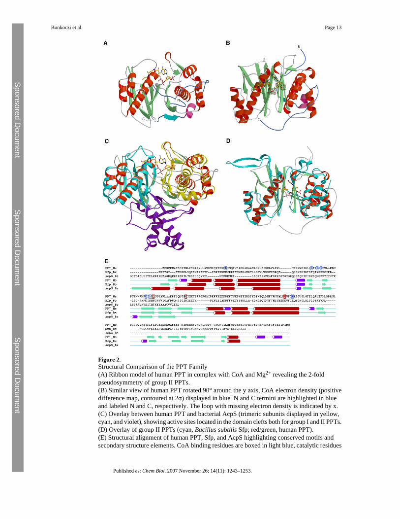
Figure 2.Structural Comparison of the PPT Family(A) Ribbon model of human PPT in complex with CoA and Mg2+ revealing the 2-foldpseudosymmetry of group II PPTs.(B) Similar view of human PPT rotated 90° around the y axis, CoA electron density (positivedifference map, contoured at 2σ) displayed in blue. N and C termini are highlighted in blueand labeled N and C, respectively. The loop with missing electron density is indicated by x.(C) Overlay between human PPT and bacterial AcpS (trimeric subunits displayed in yellow,cyan, and violet), showing active sites located in the domain clefts both for group I and II PPTs.(D) Overlay of group II PPTs (cyan, Bacillus subtilis Sfp; red/green, human PPT).(E) Structural alignment of human PPT, Sfp, and AcpS highlighting conserved motifs andsecondary structure elements. CoA binding residues are boxed in light blue, catalytic residues
Bunkoczi et al. Page 13
Published as: Chem Biol. 2007 November 26; 14(11): 1243–1253.
Sponsored Docum
ent Sponsored D
ocument
Sponsored Docum
ent

in red. One AcpS molecule is included in the alignment to highlight similar secondary structurefeatures. The structural alignment and the plot were generated with the ICM software package[35], extended strands are depicted in green, alpha helices in red, and 3/10 helices in purple.
Bunkoczi et al. Page 14
Published as: Chem Biol. 2007 November 26; 14(11): 1243–1253.
Sponsored Docum
ent Sponsored D
ocument
Sponsored Docum
ent

Figure 3.ACP Binding to Human PPT(A) Ribbon model of human PPT (green/red) with ACP (light brown). The mutated Ser→Alaacceptor residue of ACP is indicated in stick representation (arrow).(B) Detailed PPT-ACP interactions. ACP is shown as light brown ribbon model with interactingresidue side chains (labeled in blue). PPT residues are depicted as stick models (labeled inblack).(C and D) Local electrostatic potential of the α2 (recognition) helices of human ACP (C) andE. coli ACP (D), calculated with the REBEL module implemented in the ICM software package[36]. Red color indicates negative charge, blue color positive charges, ranging from −5 to +5kcal/e.u. charge units. The Ser acceptor positions are depicted at the lower left corner of theACP molecule. Arrows indicate positions of the hydrophobic recognition patches.
Bunkoczi et al. Page 15
Published as: Chem Biol. 2007 November 26; 14(11): 1243–1253.
Sponsored Docum
ent Sponsored D
ocument
Sponsored Docum
ent

Figure 4.Ligand Binding of Human PPT-CoA Complex(A) Detail of CoA and Mg binding to human PPT. The side chains of residues involved inbinding are drawn as sticks and labeled. The hypothetical model for the side chain of Gln112is represented in light blue (see text for detail).(B) Ligplot [37] representation of CoA and Mg binding with hydrogen bonding interactiondistances given in Ångstrom.
Bunkoczi et al. Page 16
Published as: Chem Biol. 2007 November 26; 14(11): 1243–1253.
Sponsored Docum
ent Sponsored D
ocument
Sponsored Docum
ent

Figure 5.Proposed Reaction Mechanism for Human PPTResidue E181 is the acid/base catalyst that abstracts a proton from Ser2156 of ACP, andcleavage of the pyrophosphate bond is achieved after charge migration and protonation of theα-phosphate likely achieved through Lys185, resulting in dissociation of the products fromPPT.
Bunkoczi et al. Page 17
Published as: Chem Biol. 2007 November 26; 14(11): 1243–1253.
Sponsored Docum
ent Sponsored D
ocument
Sponsored Docum
ent

Sponsored Docum
ent Sponsored D
ocument
Sponsored Docum
ent
Bunkoczi et al. Page 18
Table 1Kinetic Properties of Human PPT and Selected Active-Site Mutants
PPT Varied Ligand Km (mM) kcat (min−1) kcat/Km (min−1
mM−1)
WT Mg2+ 0.44 ± 0.04 14.4 ± 0.4 32.7D129A 4.6 ± 0.3 0.0042 ± 0.0005 0.001H111A 34.5 ± 5.6 7.3 ± 0.6 0.21R47A 3.3 ± 1.0 48.0 ± 6.6 14.5R86A 15 ± 3 72.0 ± 5.9 4.8K185A 1.1 ± 0.1 0.018 ± 0.0004 0.016E181A 18.3 ± 0.7 0.023 ± 0.0007 0.001E181Q 7.6 ± 4.6 0.039 ± 0.005 0.005Q112E 0.75 ± 0.24 1.32 ± 0.21 1.76E181Q, Q112E 104.1 ± 51 0.019 ± 0.004 0.0002WT C2-CoA 0.0249 ± 0.003 14.4 ± 0.73 578D129A 0.142 ± 0.035 0.0059 ± 0.0001 0.04H111A 0.095 ± 0.033 6.1 ± 1.3 64.2R47A 0.076 ± 0.0038 34.0 ± 1.2 447R86A 0.399 ± 0.041 58.4 ± 3.8 146K185A 0.0247 ± 0.0036 0.015 ± 0.0004 0.6E181A 0.0454 ± 0.0038 0.043 ± 0.0015 0.9E181Q 0.081 ± 0.0013 0.044 ± 0.0026 0.5Q112E 0.047 ± 0.023 2.2 ± 0.73 46.8E181Q, Q112E 0.093 ± 0.001 0.024 ± 0.004 0.2
Kinetic parameters for Mg2+ were determined at saturating concentrations of acetyl-CoA and vice versa.
Published as: Chem Biol. 2007 November 26; 14(11): 1243–1253.
Sponsored Docum
ent Sponsored D
ocument
Sponsored Docum
ent
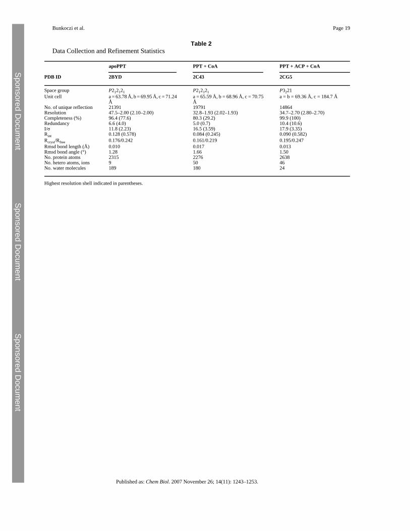
Bunkoczi et al. Page 19
Table 2Data Collection and Refinement Statistics
apoPPT PPT + CoA PPT + ACP + CoA
PDB ID 2BYD 2C43 2CG5
Space group P212121 P212121 P3221Unit cell a = 63.78 Å, b = 69.95 Å, c = 71.24
Åa = 65.59 Å, b = 68.96 Å, c = 70.75Å
a = b = 69.36 Å, c = 184.7 Å
No. of unique reflection 21391 19791 14864Resolution 47.5–2.00 (2.10–2.00) 32.8–1.93 (2.02–1.93) 34.7–2.70 (2.80–2.70)Completeness (%) 96.4 (77.6) 80.3 (29.2) 99.9 (100)Redundancy 6.6 (4.0) 5.0 (0.7) 10.4 (10.6)I/σ 11.8 (2.23) 16.5 (3.59) 17.9 (3.35)Rint 0.128 (0.578) 0.084 (0.245) 0.090 (0.582)Rcryst/Rfree 0.176/0.242 0.161/0.219 0.195/0.247Rmsd bond length (Å) 0.010 0.017 0.013Rmsd bond angle (°) 1.28 1.66 1.50No. protein atoms 2315 2276 2638No. hetero atoms, ions 9 50 46No. water molecules 189 180 24
Highest resolution shell indicated in parentheses.
Published as: Chem Biol. 2007 November 26; 14(11): 1243–1253.