Fast neutron radiography and tomography of wood
12
Fast Neutron Radiography and Tomography of Wood as Compared to Photon Based Technologies Kurt R. S. Osterloh 1 , Thomas Bücherl 2 , Andreas Hasenstab 3 , Christoph Rädel 1 , Uwe Zscherpel 1 , Dietmar Meinel 1 , Gerd Weidemann 1 , Jürgen Goebbels 1 , Uwe Ewert 1 1 Federal Institute for Materials Research and Testing (BAM), 12205 Berlin, Germany, phone: +49 (0) 30 8104 0, e-mail: [email protected] 2 Insitut für Radiochemie, Technische Universität München, Walther Meißner-Str. 3, 85748 Garching, Germany, phone: +49 (0) 89 289 14328, e-mail: [email protected] 3 LGA Bautechnik GmbH, Tillystraße 2, 90431 Nürnberg, Germany, phone: +49 (0) 911 655-55 61, e-mail: [email protected] Abstract It is a well known fact that neutron radiation has absorption properties complementary to photons. While dense materials are capable to absorbing both, X- and gamma rays, they are apparently transparent to neutrons. Vice verse, organic materials containing large amounts of hydrogen are well penetrated by photons, but not by neutrons. This property of being easily absorbed by light elements such as hydrogen makes them predestined to imaging e.g. the distribution of water within a specimen. The content of moisture is of concern in wood, particularly in lumber or in girders, since this could make them prone to rot. If wet and dry wood differs in its overall content of hydrogen rather than in its matrix of hydrocarbons, then this should result in an observable contrast in neutron radiographs. Usually, best radiographic results are obtainable with thermal neutrons. However, since wood by itself constitutes a hydrogen containing material, it absorbs them considerably from a certain thickness on; i.e, they would hardly penetrate e.g. a girder. This study demonstrates that it is feasible to use fast neutrons with a more efficient penetration capability for this purpose. Digital imaging technologies are essential for radiographs of wooden specimens since structural details appear rather faintly in the original images requiring some processing to display them clearly. Particularly large samples like trunks or girders leave poor signal to noise ratios. The noise has been compensated successfully by dedicated filtering. In addition, neutron radiographs are blotched with white spots caused by unavoidable ubiquitous radiation while the beam is open. Specially developed algorithms have been applied to remove selectively these flaws within the image while leaving other fine structures untouched, i.e. avoiding the loss of filigree like features by smoothing. Samples will be shown subjected both to neutron and X-ray radiography, and tomography as well. The differences between the results of both technologies will be discussed. Keywords: NDT, wood, water distribution, neutron radiography, large hydrogen containing specimens 1. Introduction Wood is a remarkable material since it has accompanied mankind from the very begin- ning. Weapons, tools and housing have been made, at least in part, from wood. In ad- dition, it was the first fuel for the fire. In this course, it was the source of charcoal which was essential for introducing metallurgy. Even today, we are surrounded by wooden furniture, encounter wood in artworks and enter buildings stabilised by wooden girders. Both, the preservation of our cultural heritage and the safety of buildings rely on the integrity of the wood contained. On the other hand, radiographic technologies are only sporadically applied for wooden specimens, though modern approaches such as digital radiography do have the potential to substantially contribute to detecting dam- ages in wood. Figure 1 provides both, the capabilities and prerequisites on one hand and the limitations, i.e. yet unresolved problems on the other hand for plausible and ef- DIR 2007 - International Symposium on Digital industrial Radiology and Computed Tomography, June 25-27, 2007, Lyon, France
-
Upload
independent -
Category
Documents
-
view
0 -
download
0
Transcript of Fast neutron radiography and tomography of wood

Fast Neutron Radiography and Tomography of Wood as Compared to Photon Based Technologies
Kurt R. S. Osterloh 1, Thomas Bücherl 2, Andreas Hasenstab 3, Christoph Rädel 1, Uwe
Zscherpel 1, Dietmar Meinel 1, Gerd Weidemann 1, Jürgen Goebbels 1, Uwe Ewert 1
1 Federal Institute for Materials Research and Testing (BAM), 12205 Berlin,
Germany, phone: +49 (0) 30 8104 0, e-mail: [email protected] 2 Insitut für Radiochemie, Technische Universität München,
Walther Meißner-Str. 3, 85748 Garching, Germany, phone: +49 (0) 89 289 14328, e-mail: [email protected]
3 LGA Bautechnik GmbH, Tillystraße 2, 90431 Nürnberg, Germany, phone: +49 (0) 911 655-55 61, e-mail: [email protected]
Abstract It is a well known fact that neutron radiation has absorption properties complementary to photons. While dense materials are capable to absorbing both, X- and gamma rays, they are apparently transparent to neutrons. Vice verse, organic materials containing large amounts of hydrogen are well penetrated by photons, but not by neutrons. This property of being easily absorbed by light elements such as hydrogen makes them predestined to imaging e.g. the distribution of water within a specimen.
The content of moisture is of concern in wood, particularly in lumber or in girders, since this could make them prone to rot. If wet and dry wood differs in its overall content of hydrogen rather than in its matrix of hydrocarbons, then this should result in an observable contrast in neutron radiographs. Usually, best radiographic results are obtainable with thermal neutrons. However, since wood by itself constitutes a hydrogen containing material, it absorbs them considerably from a certain thickness on; i.e, they would hardly penetrate e.g. a girder. This study demonstrates that it is feasible to use fast neutrons with a more efficient penetration capability for this purpose.
Digital imaging technologies are essential for radiographs of wooden specimens since structural details appear rather faintly in the original images requiring some processing to display them clearly. Particularly large samples like trunks or girders leave poor signal to noise ratios. The noise has been compensated successfully by dedicated filtering. In addition, neutron radiographs are blotched with white spots caused by unavoidable ubiquitous radiation while the beam is open. Specially developed algorithms have been applied to remove selectively these flaws within the image while leaving other fine structures untouched, i.e. avoiding the loss of filigree like features by smoothing. Samples will be shown subjected both to neutron and X-ray radiography, and tomography as well. The differences between the results of both technologies will be discussed. Keywords: NDT, wood, water distribution, neutron radiography, large hydrogen containing specimens 1. Introduction Wood is a remarkable material since it has accompanied mankind from the very begin-ning. Weapons, tools and housing have been made, at least in part, from wood. In ad-dition, it was the first fuel for the fire. In this course, it was the source of charcoal which was essential for introducing metallurgy. Even today, we are surrounded by wooden furniture, encounter wood in artworks and enter buildings stabilised by wooden girders. Both, the preservation of our cultural heritage and the safety of buildings rely on the integrity of the wood contained. On the other hand, radiographic technologies are only sporadically applied for wooden specimens, though modern approaches such as digital radiography do have the potential to substantially contribute to detecting dam-ages in wood. Figure 1 provides both, the capabilities and prerequisites on one hand and the limitations, i.e. yet unresolved problems on the other hand for plausible and ef-
DIR 2007 - International Symposium on Digital industrial Radiology and Computed Tomography, June 25-27, 2007, Lyon, France

ficient radiographic testing of wood. This includes also the aspect of wood as a natural material prone to kinds of damages which are not a subject to other materials.
Figure 1: Aspects of radiographic testing of wood.
In addition to quite common indications of damages that need to be detected like
cracks, cavities, abrasion or moisture (essential e.g. for corrosive processes), there are certain features that occur in naturally grown materials such as wood only. Certain in-sects such as bark beetles or termites are specialised in eating wood while no animal is known to eat steel or plastics for nutrition. Fungal growth is a fundamental problem since it might cause substantial damages such as up to 80% loss in mechanical strength without any indication visible on the surface. Current practice in localising such dam-ages e.g. in wooden girders within buildings is to ask an expert about the presence and extend of fungal infestation, to add 1.5 m at both ends to be on the safe side, to carve out the infested section and to replace it. There are no standards in testing or decision making whether a girder needs replacement or not. Secondary indications are changes in the regular histology given by the annual rings, that may be not visible on the surface too only, and the distribution of moisture since any fungus needs water to grow. A re-sult of neglect can be seen in Figure 2 a where the whole house appears to be irrepara-ble. Some damages appear quite visibly on the surface of wooden beams or trunks such as holes left from gnawing insects (Figure 2 b) or fruiting bodies of wood destroying fungi (Figure 2 c). In case of bark beetle, the core might be still keeping its strength whereas the appearance of fruiting bodies is a most serious indication. It should be noted that two types of heart rot seriously reduce the mechanical strength, the brown and the white rot. While the former just attacks the cellulose only the latter one has the capability to degrade the lignin too. A challenge is given for radiographic inspection if wooden parts are shielded by metal sheets (e. g. archaeological specimens, artworks, reinforced griders) which will be discussed in context with the application of neutron radiography.

However, this study can only be an initiative for further investigations since some problems will persist. Particularly the detection of structural (histological) changes could boost the application of radiographic inspection of wood, but this may need instrumentation capable for spectroscopic discrimination as it is realised in the dual energy technology for luggage inspection. In this context, the modification of the penetrating beam within the specimen will be an issue, like the beam hardening of X-rays. It has to be expected that fast neutrons are moderated within wood. This has to be considered in combination with the energy dependent detection efficiency of the applied detector system, but there are is currently nearly no details about this effect available. Moreover, the results obtained by any of the radiographic methods need an interpreta-tion in terms of biological findings such as fungal growth. Finally, if results could be achieved in laboratory facilities they would become of more practical interest if the method applied could be carried into the fields. In case of neutron radiography this would mean to find a portable source of adequate neutrons and a suitable image detec-tor.
b
a
c
Figure 2: Typical damages of wood
a) Consequences when girder and beams loose their strength after moistening, b) insect holes,
c) fruiting bodies of mould (late stage). 2. Material and methods The samples investigated radiographically by both, X-rays and neutrons, were two dam-aged pieces of a pine trunk (17 and 30 cm in diameter, the second one with a cavity left

from a woodpecker) and a girder with some damage on the surface. The specimen for tomography was a wooden beam of 10 x 10 cm width with a centrally drilled hole, 2.5 cm in diameter, stuffed with saw dust and further internal features such as a knot and a small inclusion (presumably resin).
An overview on the methods applied is already given in Figure 1. All results of radiographic experiments using X-rays were achieved with devices that potentially could be carried into the fields for on-site inspections: A portable X-ray flash generator (Golden Engineering XRS 3, 270 keV max. photon energy, 99 pulses) with a flat panel detector based on amorphous selenium (Agfa DirectRay am-Se) and a YXLON MCN 225 X-ray tube with variable voltage adjustment (40 – 225 kV) together with a small flat panel detector (Vidisco FOX Rayzor). The conditions applied for exposures to generate a dual energy image [1] were: 60 kV, 6 mA and high energy 160 kV, 6 mA, 3.5 mm Cu, respectively, 20 s each. Portable neutron generators have not been used in this study.
Fast neutron radiographs were produced at the NECTAR (Neutron Computer-ized Tomography and Radiography) facility [2] at the FRM II research reactor of the Technische Universität München. The energy spectrum of the fast neutrons in these ex-periments ranged from ca. 1 to 2 MeV with a maximum at about 1.6 MeV. The radio-graphs were measured using an fluoroscopic setup with an Andor DV434-BV CCD-camera at an exposure time of 1 min.
Image processing was achieved with own programs that included particular fil-tering for cleansing neutron radiographs from white spots caused by scattered radiation hitting the camera [3]. The tomographic data of both, the X-ray and the neutron tomo-graphy, were reconstructed with the Feldkamp algorithm [4]. The commercial program VGStudio Max [5] was used to present all tomographic results. 3. Results Both specimens of the pine tree have been subsequently subjected to X-ray and fast neutron radiography, the girder served for probing the magnitude of layer thickness of wood penetrable with fast neutrons. A previous preliminary experiment using thermal neutrons with even smaller specimens such as branches of some 3 to 5 cm in diameter have shown that wood is not transparent for neutrons with low kinetic energy. For all samples in this study fast neutrons were essential for a successful neutron radiography or tomography. 3.1 X-ray technologies The smaller section of the pine trunk was studied in detail with an X-ray tube with ad-justable voltage and a flat panel detector. Because of the limited panel size only the core part with the knots could be radiographed. The advantage of using digital radio-graphy was not only the capability of digital processing the resulting images but also to combine separate exposures taken under varied conditions as it is standard practice in dual-energy technologies. Figure 3 shows the results stepwise from the raw image taken at 60 kV (Figure 3 b), after high pass filtering and after advanced processing that included automatic noise compensation (Figure 3 c and d), i.e. without parameter set-tings by the user [6]. In a further step, the anisotropic distributions of the signal to noise

ratios in different direction due to the annual ring structure have been considered when compensating the noise in the filtering step (Figure 3 e). Particularly the filtered images with the noise compensation indicate a slightly altered structural pattern within the core areas. a
b
c
d
e
f
g
Figure 3: Infested section of a pine tree investigated with radiographic methods. a) specimen with flat panel detector (Vidisco FoxRayzor),
b) digital raw image, c) high pass filtering,
d) combined filtering with automatic noise attenuation, e) correction for anisotropic noise distribution,
f) Dual-Energy-exposition with combined filtering (d), g) view on top showing infested core regions in a darker colour.
These peculiarities become more evident when superimposing two exposures taken at different X-ray tube voltages and after colour coding the ratio between these two images. Particularly in the lower part, structures appear not paralleling the annual ring patterns. A view on the physical cross section of the specimen (Figure 3 g) reveals a change in the colour of the heart wood indicating rot. If the patterns seen in the radio-graphs are linked to the growth of the mould remains to be elucidated, but they ob-viously parallel the infested areas. 3.2 Neutron specific aspects a
b
c
d
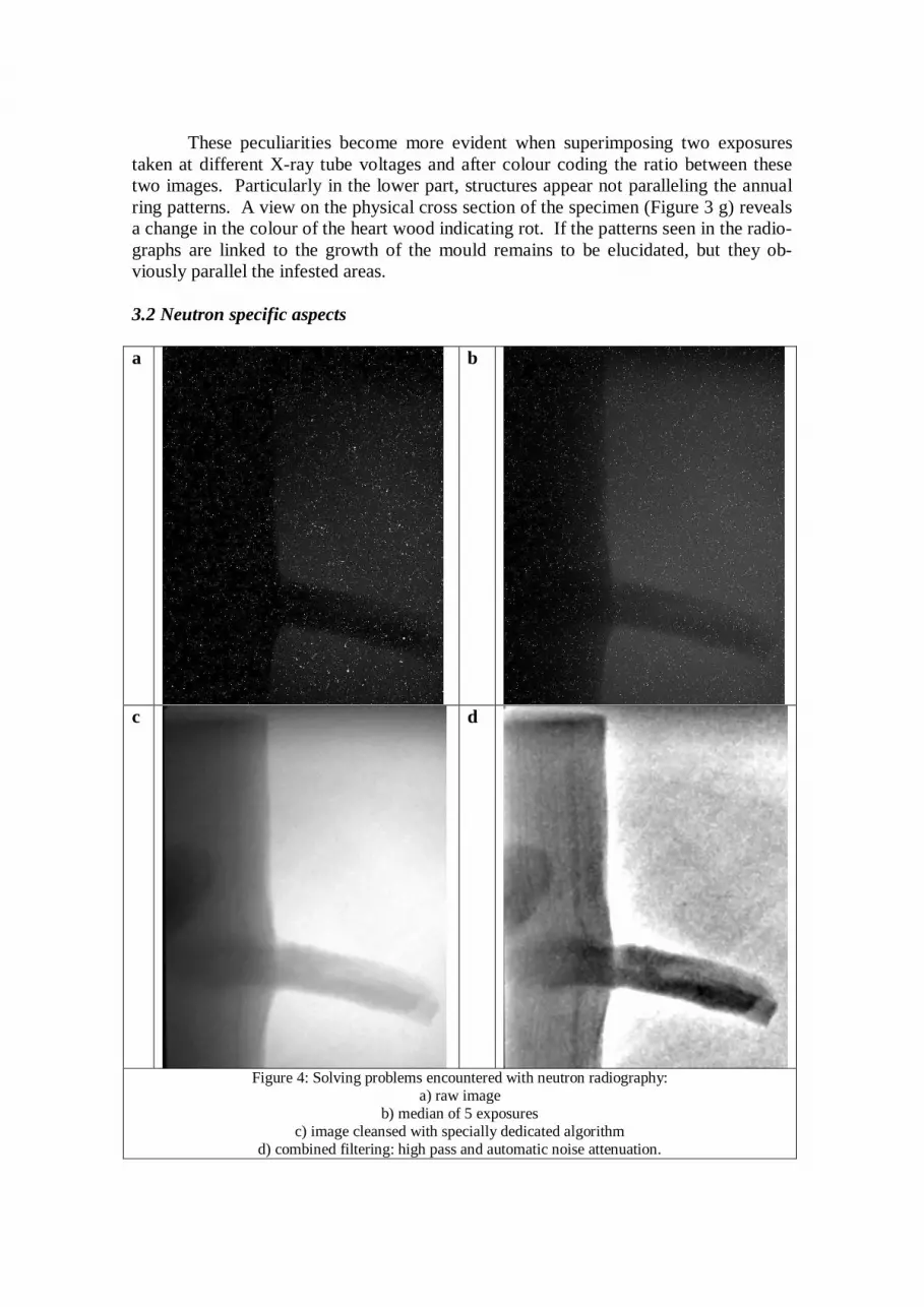
Figure 4: Solving problems encountered with neutron radiography:
a) raw image b) median of 5 exposures
c) image cleansed with specially dedicated algorithm d) combined filtering: high pass and automatic noise attenuation.

When subjecting the same specimen to fast neutron radiography it became apparent that some technical problems needed to be resolved, as shown in Figure 4. The raw image was spotted with white dots so that it was rather hard to realise the outline of the object. Even the median of five subsequent exposures did not provide a solution (Figures 4 a and b). This problem was resolved with an own algorithm specially developed to cleanse images from such white spots while leaving all other fine structures untouched (Figure 4 c), in difference to applying general filters to the entire image plane [2]. A combined filtering as it has been applied to the X-ray images revealed the annual ring structure and, in difference to the X-ray radiographs, a lane parallel to the annual rings more in the outer circuit of the trunk (Figure 4 d). It is not yet clear if this could be at-tributed to a water conduct so this structure also needs further elucidation. Since the panel size of the detector did not cover the full diameter of the speci-men, the neutron radiograph had to be made in two consecutive sections which had to be rearranged in a combined presentation as shown in Figure 5. Unfortunately, the structures seen in the X-ray radiograph paralleling the obviously rotten heart wood could not been found in the neutron image in the current setup.
Figure 5: Combination of partial images after shading correction and combined filtering (as above).
The other sample was the larger piece of pine trunk with the internal cavity.
Figure 6 shows the results of both radiographic technologies after combined filtering, i.e. high pass combined with automatic noise compensation. It was quite obvious that the neutron radiography (Figure 6 c) did not reach the image quality that was achieved with X-ray inspection (Figure 6 b). However, the neutron image showed darker lanes running along the annual ring structures not visible with X-rays in contrast to the sur-rounding matrix.

b
a
c
Figure 6: Comparison between X-ray and neutron radiography
a) specimen with the area of the images, b) X-ray radiograph after combined filtering,
c) neutron radiograph after combined filtering.
A third specimen was subjected to fast neutron radiography to probe the limits of penetration, and the results can be taken from Figure 7. A girder (Figure 7 a) was placed into the neutron beam in both ways, perpendicular and longitudinal to the broad side (Figure 7 b and c). Both, the annual ring structure and the damaged area on the sur-face of the broad side can be realised in both images. This experiment showed that wooden samples can be penetrated by fast neutrons at least up to a layer thickness of 30 cm.

b
a
c
Figure 7: Neutron radiograph of wooden girder, currently the thickest wooden specimen penetrated with
neutron radiation, a) specimen
b) transversal c) longitudinal direction (from the right side).

3.3 Tomography The tomographic results achieved are summarised in Figure 8. All image sections have been purposely chosen in a way that the major features of the interior are visible in a single view. These are the knot which is internally channelled, the annual rings in dif-ferent densities, the central hole with the saw dust and a small inclusion near the left front side presumably representing a resin lens. a
b
c
d
Figure 8: X-ray and fast neutron tomography: a) angular section through the reconstructed data set from X-ray tomography showing some obvious
intrinsic features (central hole stuffed with saw dust, knots, contrasting annual rings, inclusion at the left front edge),
b) same sample after adding water into the central hole, c) difference image of (a) and (b), d) neutron radiograph of the same specimen (added water dried out).

In case of the X-ray tomography (Figure 8 a - c) the appearance of water was studied by adding water to the centrally drilled hole stuffed with saw dust. Within Fig-ure 8, the panel a shows the specimen before, and panel b after pouring water into the hole. Panel c represents the difference between the two previous panels. Most of the water remained in the hole and some was drained into the space around the knot after two hours. The appearance of the rear surfaces in the difference image may not be nec-essarily due to moisture, swelling differences currently cannot be excluded. The neu-tron image is of lesser quality as the X-ray tomographs, but shows all details of the pre-vious ones. Though not yet quantitatively confirmed, the contrast between the dense and light annual ring structures appears to be more pronounced than in the X-ray results. Remarkably, the small inclusion near the front edge appears more prominent in the neutron tomography. No experiment was conducted with a metal shielding because this appeared to be trivial since the neutron beam has passed already several ten centimetres of lead filters when reaching the sample to reduce the gamma background contaminat-ing the fast neutron beam. 4. Conclusion The overview given in Figure 9 reconvenes to the scheme shown in the introduction (Figure 1) to demonstrate the achievements of radiography of wood either with a photon based technology (X-ray) or with particles (neutrons). It is quite obvious that fast neu-trons are essential to penetrate wood in thicker layers as shown here. All major features detected with one method became also visible with the other – as principally expected. However, there are some differences that might give some practical hints for meaning-ful future application of either technique.
Figure 9: Achievements and remaining problems with X-ray and neutron radiography
First of all, X-ray based technologies still deliver images of better quality than the one based on fast neutrons. However, there are some differences seen in the con-trast of some selected features as described above in detail. This justifies further inves-tigating whether the differences are based on varied moisture contents or local material variations such as resin inclusions. One advantage of fast neutron radiography or tomo-graphy did not become obvious on a first glance within this study: Wooden objects can be studied behind metal shielding where there is no chance with any photon based tech-nology.
Another aspect for practical applications is the instrumentation available for each technology. While X-ray application are advanced in a large scale from laboratory facilities to portable devices that easily can be taken to the site of inspection, neutron technologies are currently restricted to reactor facilities or portable sources with limited efficiency. Moreover, effective detection of neutrons, particularly the fast ones, is defi-nitely more difficult than that of any photon based technology (X- or γ-rays). To in-clude spectral aspects such as combining images taken at various energy levels appears to be a promising technology deserving further attention, particularly if small variation of material composition should be detected based not only in different elementary com-position but also with structural differences that may influence absorption e.g. by scat-tering. Absolutely essential for obtaining interpretable results is the application of digital imaging techniques that allow image processing as listed in Figure 9.
Again, numerous problems remain unresolved yet particularly with the fast neu-tron radiography. However, further developments are on their way as improving the detection technology as well as the availability of neutron sources outside of reactors. Since neutrons are capable to produce different images under different conditions than all the photon based methods, both technologies can well be regarded as complementary to each other. References 1. J. Beckmann, Y. Onel, K. Osterloh, U. Zscherpel, U. Ewert (2000), Dual-Energie-
Radiographie mit digitalen Speicherfoliensystemen zur mobilen Durchstrahlungs-prüfung, Deutsche Gesellschaft für Zerstörungsfreie Prüfung e.V.
Jahrestagung 2000, Innsbruck, 29.-31. Mai 2000, Berichtsband 73 (2000), Band 1, p. 595-607
2. T. Bücherl, Ch. Lierse von Gostomski, Radiography using Fission Neutrons, Pro-ceedings of Science, International Workshop on Fast Neutron Detectors and Ap-plications, April, 3-6, 2006, University of Cape Town, South Africa, http://pos.sissa.it/cgi-bin/reader/conf.cgi?confid=25.
3. K. Osterloh, T. Bücherl, C. Rädel, U. Zscherpel, U. Ewert (2007), Cleansing im-ages of dotted interferences while avoiding loss of contours (in preparation)
4. L.A. Feldkamp, L.C. Davis and J.W. Kress (1984), Practical cone-beam algorithm, J. Opt. Soc. Am. A 1, 612-619
5. VGStudio MAX 1.2, Volume Graphics GmbH, D-69123 Heidelberg, Germany, 2004, http://www.volumegraphics.com
6. K. Osterloh, U. Ewert, U. Zscherpel, O. Alekseychuk (2006), Verfahren zur parameterfreien Bildbearbeitung, patent application DE 2006 034 132.5-53