calcium signaling
31
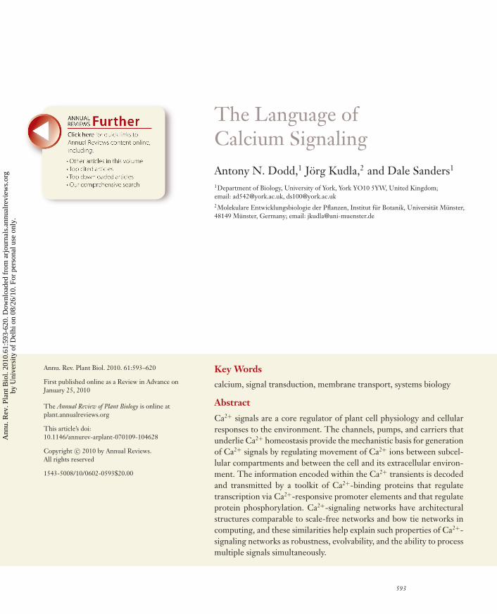
The Language of Calcium Signaling Antony N. Dodd, 1 J¨ org Kudla, 2 and Dale Sanders 1 1 Department of Biology, University of York, York YO10 5YW, United Kingdom; email: [email protected], [email protected] 2 Molekulare Entwicklungsbiologie der Pflanzen, Institut f ¨ ur Botanik, Universit ¨ at M ¨ unster, 48149 M ¨ unster, Germany; email: [email protected] Annu. Rev. Plant Biol. 2010. 61:593–620 First published online as a Review in Advance on January 25, 2010 The Annual Review of Plant Biology is online at plant.annualreviews.org This article’s doi: 10.1146/annurev-arplant-070109-104628 Copyright c 2010 by Annual Reviews. All rights reserved 1543-5008/10/0602-0593$20.00 Key Words calcium, signal transduction, membrane transport, systems biology Abstract Ca 2+ signals are a core regulator of plant cell physiology and cellular responses to the environment. The channels, pumps, and carriers that underlie Ca 2+ homeostasis provide the mechanistic basis for generation of Ca 2+ signals by regulating movement of Ca 2+ ions between subcel- lular compartments and between the cell and its extracellular environ- ment. The information encoded within the Ca 2+ transients is decoded and transmitted by a toolkit of Ca 2+ -binding proteins that regulate transcription via Ca 2+ -responsive promoter elements and that regulate protein phosphorylation. Ca 2+ -signaling networks have architectural structures comparable to scale-free networks and bow tie networks in computing, and these similarities help explain such properties of Ca 2+ - signaling networks as robustness, evolvability, and the ability to process multiple signals simultaneously. 593 Annu. Rev. Plant Biol. 2010.61:593-620. Downloaded from arjournals.annualreviews.org by University of Delhi on 08/26/10. For personal use only.
Transcript of calcium signaling
ANRV410-PP61-25 ARI 31 March 2010 17:4
The Language ofCalcium SignalingAntony N. Dodd,1 Jorg Kudla,2 and Dale Sanders1
1Department of Biology, University of York, York YO10 5YW, United Kingdom;email: [email protected], [email protected] Entwicklungsbiologie der Pflanzen, Institut fur Botanik, Universitat Munster,48149 Munster, Germany; email: [email protected]
Annu. Rev. Plant Biol. 2010. 61:593–620
First published online as a Review in Advance onJanuary 25, 2010
The Annual Review of Plant Biology is online atplant.annualreviews.org
This article’s doi:10.1146/annurev-arplant-070109-104628
Copyright c© 2010 by Annual Reviews.All rights reserved
1543-5008/10/0602-0593$20.00
Key Words
calcium, signal transduction, membrane transport, systems biology
Abstract
Ca2+ signals are a core regulator of plant cell physiology and cellularresponses to the environment. The channels, pumps, and carriers thatunderlie Ca2+ homeostasis provide the mechanistic basis for generationof Ca2+ signals by regulating movement of Ca2+ ions between subcel-lular compartments and between the cell and its extracellular environ-ment. The information encoded within the Ca2+ transients is decodedand transmitted by a toolkit of Ca2+-binding proteins that regulatetranscription via Ca2+-responsive promoter elements and that regulateprotein phosphorylation. Ca2+-signaling networks have architecturalstructures comparable to scale-free networks and bow tie networks incomputing, and these similarities help explain such properties of Ca2+-signaling networks as robustness, evolvability, and the ability to processmultiple signals simultaneously.
593
Ann
u. R
ev. P
lant
Bio
l. 20
10.6
1:59
3-62
0. D
ownl
oade
d fr
om a
rjou
rnal
s.an
nual
revi
ews.
org
by U
nive
rsity
of
Del
hi o
n 08
/26/
10. F
or p
erso
nal u
se o
nly.
ANRV410-PP61-25 ARI 31 March 2010 17:4
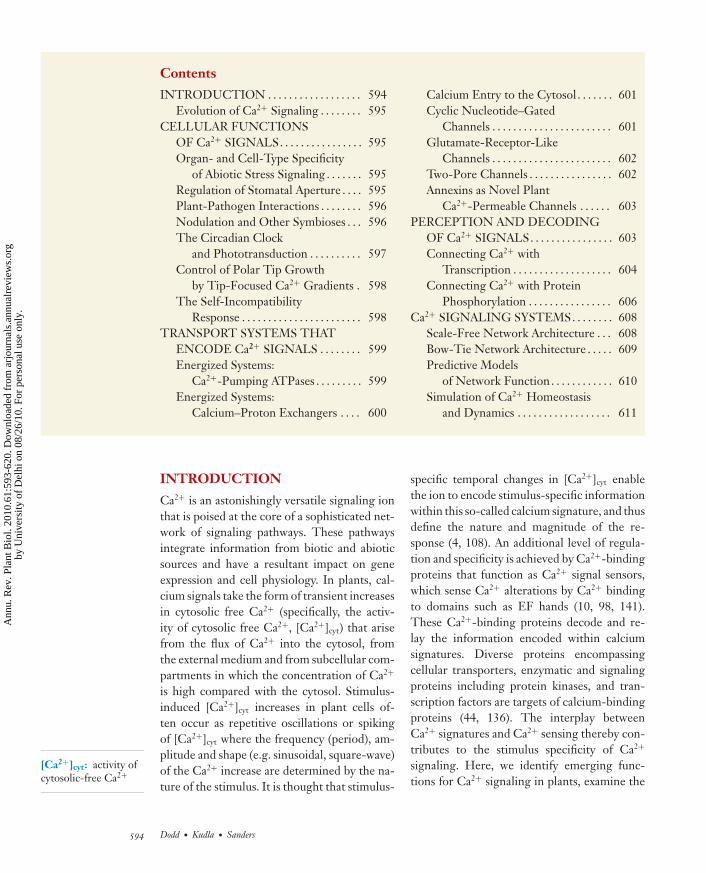
Contents
INTRODUCTION . . . . . . . . . . . . . . . . . . 594Evolution of Ca2+ Signaling . . . . . . . . 595
CELLULAR FUNCTIONSOF Ca2+ SIGNALS. . . . . . . . . . . . . . . . 595Organ- and Cell-Type Specificity
of Abiotic Stress Signaling . . . . . . . 595Regulation of Stomatal Aperture . . . . 595Plant-Pathogen Interactions . . . . . . . . 596Nodulation and Other Symbioses . . . 596The Circadian Clock
and Phototransduction . . . . . . . . . . 597Control of Polar Tip Growth
by Tip-Focused Ca2+ Gradients . 598The Self-Incompatibility
Response . . . . . . . . . . . . . . . . . . . . . . . 598TRANSPORT SYSTEMS THAT
ENCODE Ca2+ SIGNALS . . . . . . . . 599Energized Systems:
Ca2+-Pumping ATPases . . . . . . . . . 599Energized Systems:
Calcium–Proton Exchangers . . . . 600
Calcium Entry to the Cytosol . . . . . . . 601Cyclic Nucleotide–Gated
Channels . . . . . . . . . . . . . . . . . . . . . . . 601Glutamate-Receptor-Like
Channels . . . . . . . . . . . . . . . . . . . . . . . 602Two-Pore Channels . . . . . . . . . . . . . . . . 602Annexins as Novel Plant
Ca2+-Permeable Channels . . . . . . 603PERCEPTION AND DECODING
OF Ca2+ SIGNALS. . . . . . . . . . . . . . . . 603Connecting Ca2+ with
Transcription . . . . . . . . . . . . . . . . . . . 604Connecting Ca2+ with Protein
Phosphorylation . . . . . . . . . . . . . . . . 606Ca2+ SIGNALING SYSTEMS. . . . . . . . 608
Scale-Free Network Architecture . . . 608Bow-Tie Network Architecture . . . . . 609Predictive Models
of Network Function. . . . . . . . . . . . 610Simulation of Ca2+ Homeostasis
and Dynamics . . . . . . . . . . . . . . . . . . 611
[Ca2+]cyt: activity ofcytosolic-free Ca2+
INTRODUCTION
Ca2+ is an astonishingly versatile signaling ionthat is poised at the core of a sophisticated net-work of signaling pathways. These pathwaysintegrate information from biotic and abioticsources and have a resultant impact on geneexpression and cell physiology. In plants, cal-cium signals take the form of transient increasesin cytosolic free Ca2+ (specifically, the activ-ity of cytosolic free Ca2+, [Ca2+]cyt) that arisefrom the flux of Ca2+ into the cytosol, fromthe external medium and from subcellular com-partments in which the concentration of Ca2+
is high compared with the cytosol. Stimulus-induced [Ca2+]cyt increases in plant cells of-ten occur as repetitive oscillations or spikingof [Ca2+]cyt where the frequency (period), am-plitude and shape (e.g. sinusoidal, square-wave)of the Ca2+ increase are determined by the na-ture of the stimulus. It is thought that stimulus-
specific temporal changes in [Ca2+]cyt enablethe ion to encode stimulus-specific informationwithin this so-called calcium signature, and thusdefine the nature and magnitude of the re-sponse (4, 108). An additional level of regula-tion and specificity is achieved by Ca2+-bindingproteins that function as Ca2+ signal sensors,which sense Ca2+ alterations by Ca2+ bindingto domains such as EF hands (10, 98, 141).These Ca2+-binding proteins decode and re-lay the information encoded within calciumsignatures. Diverse proteins encompassingcellular transporters, enzymatic and signalingproteins including protein kinases, and tran-scription factors are targets of calcium-bindingproteins (44, 136). The interplay betweenCa2+ signatures and Ca2+ sensing thereby con-tributes to the stimulus specificity of Ca2+
signaling. Here, we identify emerging func-tions for Ca2+ signaling in plants, examine the
594 Dodd · Kudla · Sanders
Ann
u. R
ev. P
lant
Bio
l. 20
10.6
1:59
3-62
0. D
ownl
oade
d fr
om a
rjou
rnal
s.an
nual
revi
ews.
org
by U
nive
rsity
of
Del
hi o
n 08
/26/
10. F
or p
erso
nal u
se o
nly.
ANRV410-PP61-25 ARI 31 March 2010 17:4
membrane transport mechanisms that controlCa2+ movement within the cell thereby drivingCa2+ signaling, and explore how Ca2+-bindingproteins recognize specific Ca2+ increases inorder to translate these into specific cellular re-sponses. Finally, we consider the new types ofknowledge concerning cell signaling that haveemerged from studies of the integrated func-tioning of entire Ca2+ signaling networks.
Evolution of Ca2+ Signaling
Maintenance of [Ca2+]cyt at submicromolaractivities would have been an early con-straint during the evolution of cells. Inter-mediary metabolism based on energy con-tained within phosphoanhydride esters (e.g.phosphate-phosphate bonds within ATP) re-quires that free Ca2+ be sustained at a low levelotherwise precipitation of calcium salts ensuesas a result of the low solubility product of Ca2+
with Pi (139). This constraint drove the evo-lution of transport systems that export Ca2+
from the cytosol. In the earliest unicellular or-ganisms, calcium homeostasis would have re-quired export across the plasma membrane. Inall eukaryotes, sequestration systems have alsoevolved at endomembranes. Importantly, theevolution of transport systems that maintain ex-tremely low cytosolic free Ca2+ created a cel-lular environment for the evolution of a signal-ing mechanism that elevates cytosolic free Ca2+
very rapidly by capitalizing upon the enormouselectrochemical potential difference for Ca2+
across membrane systems that separate “stores”of Ca2+ from the cytosol (139).
CELLULAR FUNCTIONSOF Ca2+ SIGNALS
Ca2+ signals regulate a large number of abi-otic stress responses (108), as well as stomatalaperture (121), self-incompatibility during fer-tilization (49), interactions with pathogenic andsymbiotic microorganisms (103, 124), and thedevelopment of tip-growing structures such aspollen tubes and root hairs (63). Ca2+ signalsalso participate in light and circadian signaling(39, 72, 147, 149). We highlight recent advances
in the functions of Ca2+ signals that raise im-portant questions for future research.
Organ- and Cell-Type Specificityof Abiotic Stress Signaling
Ca2+ elevations with stimulus-specific proper-ties are evoked by extracellular sodium, osmoticstress, oxidative stress, low temperature, ozone,and mechanical cues (108). Dose-dependent re-lationships between the concentration of ex-ternal NaCl and NaCl-induced [Ca2+]cyt spikemagnitude in Arabidopsis roots (155) recentlydemonstrated that information concerning abi-otic stimulus strength is encoded within this[Ca2+]cyt signal. Stress-induced Ca2+ signalshave intriguing cell type–specific properties.[Ca2+]cyt oscillations with different propertiesoccur in the pericycle and endodermis af-ter challenge by NaCl or osmotic stress withmannitol. Mannitol causes repetitive [Ca2+]cyt
increases in the endodermis with a peak in[Ca2+]cyt every 20–30 s, compared with a sus-tained [Ca2+]cyt increase in the pericycle (76),but the outcome of the different calcium sig-natures in these cell types in terms of cellularacclimation to abiotic stress remains unknown.An alternative explanation for the different cell-specific Ca2+ signatures of the endodermis andpericycle is that these cell types experience dif-fering levels of exposure to the stress as a re-sult of their location within the root structure,and so the different Ca2+ signatures in the celltypes are caused by differing stimulus magni-tudes. Whether cell type–specific Ca2+ signalscause cell type–specific stress responses remainsan important and open question.
Regulation of Stomatal Aperture
Ca2+ signals are core regulators of stom-atal aperture (121). Information encoded in[Ca2+]cyt oscillations alone can program stom-atal aperture because artificially imposed os-cillations in guard cell [Ca2+]cyt close stom-ata, and the [Ca2+]cyt oscillation frequencyand amplitude determine steady-state aperture(4). The greatest degree of steady-state clo-sure is caused by artificial [Ca2+]cyt increases
www.annualreviews.org • Ca2+ Signaling 595
Ann
u. R
ev. P
lant
Bio
l. 20
10.6
1:59
3-62
0. D
ownl
oade
d fr
om a
rjou
rnal
s.an
nual
revi
ews.
org
by U
nive
rsity
of
Del
hi o
n 08
/26/
10. F
or p
erso
nal u
se o
nly.

ANRV410-PP61-25 ARI 31 March 2010 17:4
CAMTA:calmodulin-bindingtranscription factor(strictly, calmodulin-binding transcriptionactivator)
that have a period of 10 minutes and wheneach increase lasts for approximately 5 minutes(4), which is comparable with ABA-induced[Ca2+]cyt oscillations in guard cells that closestomata and have periods of 6 to 8 minutes.An emerging hypothesis is that preexposure ofguard cells to ABA and CO2 increases the sen-sitivity of Ca2+ sensors to subsequent Ca2+ in-creases (71, 148). For example, guard cell pre-exposure to ABA increases both the magnitudeof S-type anion efflux currents and the down-regulation of K+
in currents in response to Ca2+
(148). The authors (71) therefore propose thatspecific stimuli prime specific Ca2+ sensors sothat the sensor is more readily activated byan increase in [Ca2+]cyt. The mechanistic ba-sis for the priming of Ca2+ sensors by ABA andCO2 is unclear but could involve direct interac-tions between Ca2+ sensor(s) and an ABA/CO2-responsive protein or protein kinase, or alterna-tively the convergence of Ca2+-dependent andCa2+-independent components of ABA signal-ing downstream from Ca2+ increases. Stomatalclosure is attenuated but not prevented by sup-pression of ABA-induced [Ca2+]cyt oscillations(148). This does not necessarily indicate thatthere is a Ca2+-independent pathway to stom-atal closure, because if Ca2+ sensors are primed,resting [Ca2+]cyt might be sufficient to activateguard cell ion efflux. This is consistent with re-ports of ABA activation of anion channels in theabsence of [Ca2+]cyt increases (91).
Plant-Pathogen Interactions
Ca2+ signals are an early response by cells tothe presence of pathogenic and symbiotic mi-croorganisms. Surprisingly, defense responsesare both suppressed and activated by Ca2+ sig-nals, which suggests that Ca2+-responsive butantagonistic signaling mechanisms are present.Molecules with microbe-associated molecu-lar patterns (MAMPs) mobilize Ca2+ fromboth extracellular (apoplast) and intracellu-lar (vacuole/endoplasmic reticulum) stores ofCa2+ and cause alterations in nuclear [Ca2+].The increases in nuclear Ca2+ that are in-duced by specific MAMPs are prolonged
compared with those induced in the cytosol(e.g. harpin causes 5 min and 150 min in-creases in cytosolic and nuclear [Ca2+], re-spectively) and the Ca2+ increases measuredfrom cell cultures are sustained rather thanoscillatory (86). MAMP-induced [Ca2+] in-creases lead to the combined Ca2+-dependentactivation of mitogen-, salicylic acid (SA)–, and wound-activated protein kinases (86,103). In contrast to the general principle thatstimulus-induced Ca2+ increases can encodestimulus-specific information, it is proposedthat MAMP-specific patterns of [Ca2+]cyt in-crease are unusual because they do not en-code MAMP-specific information. This is pri-marily because prolonged [Ca2+]cyt increasesinduce similar defense responses irrespectiveof the elicitor (103 and references within). Incontrast to MAMP-induced Ca2+ inductionof defense responses, Ca2+ signals also sup-press SA-mediated acquisition of systemic ac-quired resistance (41). The positive regulatorof basal resistance and SA levels, ENHANCEDDISEASE SUSCEPTIBILITY1 (EDS1), is re-pressed following Ca2+/calmodulin binding tothe Ca2+/calmodulin-binding transcription ac-tivator (CAMTA) CAMTA3 (41). In combina-tion these studies suggest that the integrationof Ca2+ signaling with defense responses is ex-traordinarily complex and could incoroporateseveral independent signaling pathways.
Nodulation and Other Symbioses
Nitrogen-fixing bacteria in proximity tolegume roots secrete nodulation (Nod) fac-tors. Nod factors cause perinuclear [Ca2+]cyt
spiking in root epidermal cells, which initiatescellular internalization of rhizobia and rootnodule development (123). The core Nodfactor receptor comprises the NOD FACTORRECEPTOR1 (NFR1)/NFR5 heterodimerand is required for Nod factor–induced[Ca2+]cyt spiking and membrane depolariza-tion (123). Interactions occur between ABAand Nod factor signaling upstream of Ca2+
spiking (38), so it would be interesting todiscover whether inositol 1,4,5-trisphosphate
596 Dodd · Kudla · Sanders
Ann
u. R
ev. P
lant
Bio
l. 20
10.6
1:59
3-62
0. D
ownl
oade
d fr
om a
rjou
rnal
s.an
nual
revi
ews.
org
by U
nive
rsity
of
Del
hi o
n 08
/26/
10. F
or p
erso
nal u
se o
nly.
ANRV410-PP61-25 ARI 31 March 2010 17:4
(InsP3), which mobilizes Ca2+ and participatesin ABA signaling, is involved in this interaction.Nod factor–induced Ca2+ spikes result in Ca2+/calmodulin binding to the Ca2+/calmodulin–dependent protein kinase (CCaMK)DOESN’T MAKE INFECTIONS3 (DMI3)in Medicago truncatula (123). This activatesDMI3 which promotes early nodulation(ENOD) genes. ENOD genes are activatedby induction of (a) the GRAS-family domaintranscription factors NSP1 and NSP2 thattogether form a DNA-binding complex thatbinds the AATTT promoter motif, and (b) theERF-subfamily transcription factor ERN (65,123).
[Ca2+]cyt transients also occur in plant cellsduring the formation of symbiotic relationshipsbetween plants and other micro-organisms.[Ca2+]cyt transients occur during developmentof symbioses with other arbuscular-mycorrhizal(AM) fungi (82, 120) and after exposure tocell wall extracts (CWEs) from the growth-promoting fungus Piriformospora indica (158).These signals interact with, but are not iden-tical to, defense signaling because both P. in-dica CWEs and MAMPs increase expressionof defense-related MAP KINASE6 (MPK6),but CWEs do not cause downstream de-fense responses and instead increase the tran-script abundance of CYCLIC NUCLEOTIDEGATED CHANNEL10 (CNGC10), CNGC13,CALMODULIN-LIKE PROTEIN42 (CML42),and CML38 (158). This implies that Ca2+ sig-naling is a common feature of plant-microbeinteractions.
The Circadian Clockand Phototransduction
Ca2+ signals contribute to red light (RL), bluelight (BL), and UV-B signaling. Ca2+ signal-ing during phytochrome-mediated RL pho-totransduction has been reviewed elsewhere(147, 149) so here we focus on the involve-ment of Ca2+ in BL signaling. BL and RLcause brief (60 s) [Ca2+]cyt transients withoutapparent oscillations (13, 149) and the BLspectrum causes [Ca2+]cyt transient maxima
at the 440 nm and 470 nm wavelengths.BL causes Ca2+ influx through plasmamembrane (lanthanum-sensitive) voltage-gatedCa2+ channels but does not cause Ca2+ re-lease from the ER (thapsigargin-insensitive)(13). This Ca2+ increase is mediated by BL-activation of phototropin blue light photore-ceptors (PHOTs) rather than cryptochromeblue light photoreceptors (CRYs) (13, 58,59). BL Ca2+ signals are also required forBL inhibition of seedling growth (46). Sev-eral proteins might participate in sensing BL-induced Ca2+ increases. Ca2+-binding proteinsthat have been linked to BL include SHORTUNDER BLUE LIGHT1 (SUB1), an EF-hand–containing protein involved in control ofHY5-mediated seedling de-etiolation by phy-tochrome and cryptochrome (55). However,since SUB1 is involved in CRY and PHY sig-naling, but CRYs do not appear to mediate BL-induced Ca2+ increases (13), the involvement ofSUB1 with light induced Ca2+ signals might re-late primarily to the decoding of phytochrome-mediated Ca2+ alterations. PLASTID MOVE-MENT IMPAIRED1 (PMI1) has regions withhomology to binding domains for interactionwith C-domain Ca2+-binding proteins and isrequired for BL-induced chloroplast rearrange-ment (34). In contrast to higher plants, inthe bryophyte Physcomitrella patens BL-induced[Ca2+]cyt increases are mediated by both CRYsand PHOTs (156). This raises the intriguingpossibility that in higher plants CRYs mightcontribute to BL-induced Ca2+ signals, but thisis not always detected with the aequorin-basedtechnology used for Ca2+ measurements. Fu-ture comparative studies involving the mea-surement of BL-induced Ca2+ increases in sin-gle cells could expand our understanding ofwhether CRYs are associated with BL-inducedCa2+ signals in higher plants or whether thismechanism was lost during the evolution ofhigher plants.
Circadian oscillations of [Ca2+]cyt occurin continuous light and are controlled bythe molecular circadian oscillator (72, 168).Under constant light there is a sinusoidalvariation in [Ca2+]cyt over 24 h that is estimated
www.annualreviews.org • Ca2+ Signaling 597
Ann
u. R
ev. P
lant
Bio
l. 20
10.6
1:59
3-62
0. D
ownl
oade
d fr
om a
rjou
rnal
s.an
nual
revi
ews.
org
by U
nive
rsity
of
Del
hi o
n 08
/26/
10. F
or p
erso
nal u
se o
nly.
ANRV410-PP61-25 ARI 31 March 2010 17:4
to reach peak concentrations of 300–700 nMbetween the middle and end of the subjectiveday (72, 96). Similar [Ca2+]cyt variations occurunder light–dark cycles (96) but do not persistunder continuous darkness, requiring lightinput via PHYTOCHROME B (PHYB),CRYPTOCHROME1 (CRY1), and CRY2(168). Circadian rhythms of Ca2+ are thoughtto be positioned downstream of the calcium-mobilizing molecule cyclic ADP ribose(cADPR) (39). In light–dark cycles, there isa rhythm of ADP ribosyl cyclase activity inEuglena gracilis with peak activity during thelight period (106); in continuous light, circa-dian cADPR fluctuations occur in Arabidopsis(39). In Arabidopsis, perturbation of circadianCa2+ oscillations or cADPR signaling altercircadian clock function, which suggests thatCa2+/cADPR forms a feedback loop within theclock because the circadian clock is requiredfor circadian [Ca2+]cyt oscillations to occur(39, 168). As with many Ca2+ signals in plants,the downstream Ca2+-binding proteins thatdecode circadian [Ca2+]cyt oscillations areunidentified. It is not known whether circadian[Ca2+]cyt dynamics are distributed uniformlywithin the cytosol or are alternatively anaggregation of frequency- or amplitude-modulated [Ca2+]cyt spikes. The extensivecross talk between circadian timing and stresssignaling networks (39) indicates that it will beimportant in the future to discover whethercircadian [Ca2+]cyt oscillations participate inabiotic stress responses, particularly given thecontribution of cADPR-mediated Ca2+ releaseto abscisic acid signaling.
Control of Polar Tip Growthby Tip-Focused Ca2+ Gradients
Tip-focused Ca2+ gradients are important de-terminants of polarity in tip-growing cells suchas root hairs, pollen tubes, fungal hyphae, andalgal rhizoids (63). Here, we consider researchdevelopments in Ca2+ signaling during roothair extension because there have been sev-eral recent developments in our knowledge ofCa2+ signaling in this cell type. The NADPH
oxidase ROOT HAIR DEFECTIVE2 (RHD2,also AtRBOHC) is localized to the plasmamembrane of the growing tip of roothairs (152). RHD2 produces reactive oxygenspecies (ROS) that stimulate hyperpolarization-activated Ca2+ channels of unknown molecularidentity, leading to formation of a tip-focusedCa2+ gradient (47). This is thought to tar-get the cytoskeleton and secretory apparatusto the growing tip. Polarity is proposed tobe self-sustained though positive feedback, inwhich ROS-induced Ca2+ influx maintains el-evated Ca2+ at the root hair tip, which re-sults in synergistic activation of RHD2 byboth Ca2+ binding to two EF-hands on RHD2and Ca2+-dependent phosphorylation of twoserine residues on RHD2 (152). Consistentwith this hypothesis, there are oscillations inthe elevated tip-focused [Ca2+]cyt gradient andin root hair extension, separated by a lag ofapproximately 5 s (112). The oscillations areproposed to arise from a burst of root hairextension causing a pulse of Ca2+ influx thatsubsequently induces ROS production, furtherCa2+ influx and the next pulse of growth (112).This model is dependent upon the localiza-tion of RHD2 and relevant channels to thetip so that Ca2+ influx occurs only in this re-gion, but the mechanisms controlling this dis-tribution are unclear and this represents animportant area for future research. Annexins,which can generate Ca2+-permeable conduc-tances (85), are concentrated in tip-growingstructures (114) and are an attractive poten-tial regulator of root hair polarity. The RhoGTPase GDP dissociation inhibitor (RhoGDI)SUPERCENTIPEDE1 (SCN1) regulates tip-focused ROS production (18), and the RopGTPase ROP2 might do so as well (73).
The Self-Incompatibility Response
Ca2+ signals are involved in reproductiveself-incompatibility in poppy (Papaver rhoeas).To prevent inbreeding, self-produced pollenthat shares the same S-allele as the stigmais recognized by the growing pollen tubewhen it develops on the stigma. This causes
598 Dodd · Kudla · Sanders
Ann
u. R
ev. P
lant
Bio
l. 20
10.6
1:59
3-62
0. D
ownl
oade
d fr
om a
rjou
rnal
s.an
nual
revi
ews.
org
by U
nive
rsity
of
Del
hi o
n 08
/26/
10. F
or p
erso
nal u
se o
nly.
ANRV410-PP61-25 ARI 31 March 2010 17:4
the self-incompatibility (SI) response thatstops pollen tube growth and prevents self-fertilization. In poppy [Ca2+]cyt alterationsact as an early signal in prevention of self-fertilization and activate several inhibitorymechanisms. Within seconds of pollen tubechallenge with self- (i.e., incompatible) pistilS-proteins (PrsS), [Ca2+]cyt in the pollen tubeshank increases from ∼200 nM to 1.5 μMfor several minutes and the oscillatory tip-focused [Ca2+]cyt gradient dissipates (48). Arti-ficial [Ca2+]cyt elevation within the pollen tubemimics the SI response (49). The pollen SIdeterminant is the plasma membrane proteinPrpS and this interacts with the pistil self-determinant PrsS to trigger the SI responsewhen pollen PrpS interacts with pistil self-PrsS (164). The molecular identity of the Ca2+
channels that cause Ca2+ influx during theSI response remains to be discovered. TheSI response also involves Ca2+/calmodulin-dependent inhibitory hyperphosphorylation ofthe soluble inorganic pyrophosphatases Pr-p26.1a and Pr-p26.2b. This is proposed to causePi to increase during the SI response and so in-hibit pollen tube metabolism (33). The SI Ca2+
signal also promotes caspase (cysteine-asparticprotease)-like activity, which is a componentof mechanisms causing programmed cell death(153).
TRANSPORT SYSTEMS THATENCODE Ca2+ SIGNALS
Calcium transport systems have to maintainlow [Ca2+]cyt against a significant electrochem-ical potential difference for Ca2+ (i.e., not onlya concentration difference but also an elec-trical potential) and thus are energized (141).In plants energization is accomplished eitherthrough Ca2+ pumps powered by ATP hydrol-ysis or through Ca2+-H+ antiporters poweredby a proton-motive force (141). A conventionalview of these energized systems in plant cells isthat they provide the homeostatic backgroundagainst which Ca2+-release channels operatetransiently to elevate free [Ca2+]cyt (139). Open-ing of such channels should therefore provide
at least the initial spike for the elevation of[Ca2+]cyt. In general, this paradigm remains in-tact. However, as discussed in the next section,research involving mutants in genes encodingenergized transport systems has yielded intrigu-ing results demonstrating that energized trans-port of Ca2+ from the cytosol might providemuch more than a housekeeping backgroundto Ca2+ signaling.
Energized Systems:Ca2+-Pumping ATPases
ATP-dependent export of Ca2+ from the cy-tosol is accomplished by P-type ATPases ofthe P2 class. P2 Ca2+-pumping ATPases com-prise two distinct clades. In Arabidopsis theseare ER (endoplasmic reticulum)-type Ca2+-ATPases (ECAs) of the P2A group, and AUTO-INHIBITED Ca2+-ATPases (ACAs) of the P2B
group. Two major features distinguish ECAsfrom ACAs (17). First, an N-terminal cytosolicdomain present only in ACAs binds calmod-ulin that is bound to Ca2+, and this interactionactivates Ca2+ pumping (8). Second, there aredifferences in membrane-located residues thatare thought to be involved in Ca2+ binding.
This latter difference might account forthe fact that insertional mutants in at leasttwo of the four Arabidopsis ECA genes haveMn2+- as well as Ca2+-related phenotypes.Thus, while plants lacking the ER-localizedECA1 pump (166) and those lacking the Golgi-or endosome/post-Golgi-localized ECA3 (93,111) exhibit growth-sensitive phenotypes tolow Ca2+ concentrations, all mutants also ex-hibit phenotypes with respect to Mn2+. There-fore, this group of Ca2+-ATPases seems to beinvolved principally in delivery of cations to in-tracellular compartments where there is a re-quirement for secretion or as a cofactor for en-zymatic activity, rather than in cytosolic Ca2+
homeostasis and signaling per se.By contrast, ACA pumps, which comprise
a 10-member gene family in Arabidopsis, areemerging as potential key players in plant Ca2+
signaling. Hints at this role come from ex-pression profiling. For example, ACA12 and
www.annualreviews.org • Ca2+ Signaling 599
Ann
u. R
ev. P
lant
Bio
l. 20
10.6
1:59
3-62
0. D
ownl
oade
d fr
om a
rjou
rnal
s.an
nual
revi
ews.
org
by U
nive
rsity
of
Del
hi o
n 08
/26/
10. F
or p
erso
nal u
se o
nly.

ANRV410-PP61-25 ARI 31 March 2010 17:4
ACA13 transcripts are dramatically upregulatedby pathogen stress (17). Furthermore, tran-script abundance of the closely-related ACA8and ACA10 is differentially regulated by cold(142), whereas transcripts of ACA8 and ACA9are both acutely upregulated by ABA (21).
ACAs 8, 9, and 10 are plasma membrane lo-calized (16, 142, 143), and distinct roles in sig-naling and development have become apparent.T-DNA insertional mutants in the ACA9 geneexhibit partial male sterility, consistent with ex-pression primarily in pollen (143). This find-ing need not of itself indicate a key signalingrole for ACA9, despite the central function ofCa2+ in dictating pollen tube growth and guid-ance (63): It is possible that a general disrup-tion of cytosolic Ca2+ homeostasis in aca9 mu-tants impacts the phenotypes of reduced pollentube growth and discharge of sperm cells intoovules.
However, an intriguing phenotype of aca10mutants suggests that ACA pumps play criti-cal developmental roles. A compact inflores-cence (cif) phenotype in Arabidopsis is specific toaca10 mutants but not to aca8 or aca9 mutants(53). Nevertheless, overexpression of ACA8 inan aca10 background will complement the cifphenotype, suggesting that differences in ex-pression among members of this subgroup ofP2B ATPases can impact development.
ACA pumps are also subject to complexposttranslational regulation, including activa-tion by calmodulin, regulation by acidic phos-pholipids (15), and in the case of the ER-localized ACA2, phosphorylation (68). Thesemultiple modes of control, differential intracel-lular locations (e.g., ACA4 and ACA11 localizeto vacuoles; 52, 88), and differing tissue-specificexpression of ACAs are challenging in terms ofunderstanding the extent to which ACAs havespecific roles in Ca2+ signaling. In Physcomitrellaan ACA-type deletion of Ca2+-ATPase gene re-sults in enhanced sensitivity to NaCl and this isassociated with aberrant regulation of [Ca2+]cyt
(134). To date, there have been no such studiesin higher plants, making this a fruitful area forfurther investigation.
Energized Systems:Calcium–Proton Exchangers
With respect to P2 ATPases, calcium-protonexchangers are low-affinity cytosolic export sys-tems, coupled to the thermodynamically down-hill exchange flux of H+. It is likely thatthere are six bona fide calcium–proton exchang-ers, known as CATION EXCHANGER1 to-6 (CAX) (146), encoded in the Arabidopsisgenome. CAX transporters are members of theMajor Facilitator Superfamily and are predictedto have 11 transmembrane domains (TMDs).Where membrane location has been character-ized, in the cases of CAX1 to CAX4, the local-ization is vacuolar (146).
In Arabidopsis, CAX1 and CAX3 appearto be prominent in Ca2+ homeostasis. cax1mutants exhibit developmental reductions inlateral root length and number, a dramaticreduction in the length of the primary inflo-rescence (24), and increased capacity for coldacclimation associated with enhanced expres-sion of CBF/DREB1 (20). Although CAX1 ismore strongly expressed in shoot tissue andCAX3 more so in roots, cax1/cax3 double mu-tants have significantly more severe phenotypesthan either of the single mutants, including leaftip necrosis and ionic content (25). cax3 mu-tants are also more susceptible to salt stress(171).
CAX transporters, like ACA pumps, are sub-ject to posttranslational regulation in plantsthrough an autoinhibitory N-terminus (110)and possibly also through regulatory protein–protein interactions (23, 26). In combinationwith cax mutant phenotypes, this posttrans-lational regulation suggests an acute role forCAXs in Ca2+ homeostasis. A critical currentquestion revolves around the nature of the mu-tant phenotypes of these energized cytosolicexport systems: Are they merely disrupted inCa2+ homeostasis, as upregulation of alterna-tive Ca2+ transporters can imply in some mu-tants (24, 25), or is there a more fundamentalrole in Ca2+ signaling?
Whether energized Ca2+ transport sys-tems are more than background players in
600 Dodd · Kudla · Sanders
Ann
u. R
ev. P
lant
Bio
l. 20
10.6
1:59
3-62
0. D
ownl
oade
d fr
om a
rjou
rnal
s.an
nual
revi
ews.
org
by U
nive
rsity
of
Del
hi o
n 08
/26/
10. F
or p
erso
nal u
se o
nly.

ANRV410-PP61-25 ARI 31 March 2010 17:4
the Ca2+ signaling toolbox, and, like PCAI inPhyscomitrella, play an active role in shapingCa2+ signals, will only be established once mea-surements of [Ca2+]cyt have been performed inmutants lacking these transport systems.
Calcium Entry to the Cytosol
Passive return flow of Ca2+ down the electro-chemical potential generated by ATPases andCAXs is thought to be the primary driver forCa2+ signaling (141). This flux occurs throughion channels. For example, plasma membraneCa2+-permeable channels, which are in normalconditions relatively inactive, are activated byABA or by ROS in guard cells (117, 127), twostimuli that effect stomatal closure. ROS alsoactivate Ca2+-permeable channels at the plasmamembrane of root hairs during Ca2+-regulatedcell expansion (47). These ROS-related activa-tion events might involve extracellular ATP sig-naling (36). However, depending on the stimu-lus, Ca2+ signals can be generated across mem-branes other than the plasma membrane: Coldshock induces mobilization of vacuolar Ca2+
(79), while Nod factors generate [Ca2+]cyt sig-nals that emanate from the perinuclear regionER of legume root hairs (123).
Electrophysiological studies, particularlyduring the 1990s, established that a variety ofdifferent Ca2+-permeable channel types existin plants (reviewed in 35, 141). The plasmamembrane possesses Ca2+-permeable channelswith a range of voltage-dependencies: someare activated by membrane hyperpolarizationor by depolarization, and some are voltage-insensitive. In accord with the notion thatCa2+ can be mobilized across endomembranes,voltage-dependent channels also reside in thevacuolar membrane and ER. Additionally, elec-trophysiological approaches with intact vac-uoles and radiometric approaches using mem-brane vesicles have established the presenceof ligand-gated Ca2+-permeable pathways atendomembranes. Second messengers such asInsP3 and cADPR release Ca2+ from the vac-uolar lumen (e.g., 5). Both ligands also liber-ate Ca2+ from the ER (116, 119), along with
nicotinic acid adenine dinucleotide phosphate(NAADP; 118).
InsP3 participates in pollen tube orientation,salt and hyperosmotic stress signaling, ABA sig-naling and gravitropism, while cADPR medi-ates the activation of some defense genes andfunctions in ABA and circadian signaling (39,141). These findings confirm the physiologi-cal relevance of the two ligands and, in com-bination with results demonstrating that InsP3
and cADPR mobilize Ca2+, suggest the pres-ence of receptors that also function as Ca2+
channels, as is the case in mammalian cells.However, homologues of mammalian InsP3
receptor and ryanodine receptor (the recep-tor for cADPR) are not encoded by higherplant genomes, and the molecular identities ofthe InsP3- and cADPR-activated Ca2+-releasepathways in higher plants remain unknown.The absence of this molecular handle has im-peded progress in understanding the roles ofInsP3 and cADPR in plant Ca2+ signaling.
Until relatively recently, the molecular iden-tities of plasma membrane Ca2+-permeablechannels and those not controlled by ligands atendomembranes were unknown. Forward andreverse genetic approaches have now yieldedspecific information on the molecular identi-ties and physiological roles of some plant Ca2+-permeable channels (161).
Cyclic Nucleotide–Gated Channels
Arabidopsis possesses a large gene family of 20members that encode cyclic nucleotide-gatedchannels (CNGCs) (105). These channels havesix TMDs and a pore domain, and proba-bly assemble tetramerically to form the pore.Some plant CNGCs are Ca2+-permeable whenexpressed heterologously (90, 157), althoughsome are also permeable to monovalent ions(9, 89). Besides a cyclic nucleotide-binding do-main, CNGCs also bind calmodulin (144). AllCNGCs studied to date localize to the plasmamembrane (7, 54, 157).
Mutants in Arabidopsis CNGC2, CNGC4,CNGC11, and CNGC12 have aberrant regula-tion of pathogen defense responses (9, 29, 170),
www.annualreviews.org • Ca2+ Signaling 601
Ann
u. R
ev. P
lant
Bio
l. 20
10.6
1:59
3-62
0. D
ownl
oade
d fr
om a
rjou
rnal
s.an
nual
revi
ews.
org
by U
nive
rsity
of
Del
hi o
n 08
/26/
10. F
or p
erso
nal u
se o
nly.

ANRV410-PP61-25 ARI 31 March 2010 17:4
and cngc2 mutants additionally lack a cAMP-gated Ca2+ current at the plasma membrane ofguard cells (3). The manner in which CNGCsare activated during defense signaling has yet tobe determined. A more diverse role for mem-bers of the CNGC family has been indicatedin cngc18 mutants, which are defective in pollentube growth (50).
Glutamate-Receptor-Like Channels
Plants contain homologues of animalionotropic glutamate receptors that functionas nonselective cation channels at postsynapticmembranes. The GLUTAMATE RECEPTOR(GLR) gene family in Arabidopsis comprises 20members, each of which encodes a protein withthree predicted TMDs. The entire channel isprobably formed as a tetramer or pentamer(32).
Glutamate, as well as five other amino acidsand even glutathione, applied to Arabidopsisseedlings generates an inward current and a cy-tosolic Ca2+ spike, and both responses are at-tenuated in glr3.3 mutants (37, 132). Differentmembers of the GLR family respond differ-ently to activating ligands (150), but it is notclear which ligands are physiologically active.Nevertheless, exogenous glutamate and glycinehave roles in processes as diverse as hypocotylelongation (43) and the regulation of C and Nmetabolism (74).
For both GLR and CNGC ion channels,the potential for members of large genefamilies to form heteromultimeric complexesmight explain the diversity of plant plasmamembrane Ca2+-permeable channels that havebeen reported in electrophysiological studies.However, a note of caution is also necessary.Channels that depolarize a membrane whenactivated might contribute to a cytosolic Ca2+
response merely by activating some othervoltage-sensitive pathway. One example forwhich this is almost certainly the case relatesto perinuclear Ca2+ spiking during Nod-factorperception. CASTOR and POLLUX (in Lotus)and DMI1 (in Medicago) are nucleus-localizedchannels, and mutants lack Nod-factor-
induced Ca2+ spiking. However, all threechannels, when studied heterologously, havecharacteristics that suggest they are regulatorsof Ca2+-permeable channels but do not them-selves form the physiological pathway for Ca2+
release (22, 129). Indeed, perinuclear Ca2+
spiking in Medicago can be elicited in dmi1 mu-tants by mastoparan, a G-protein agonist (151).
Two-Pore Channels
The Arabidopsis genome contains a single mem-ber of the TWO-PORE CHANNEL (TPC )family, TPC1. The protein is predicted to have12 TMDs and incorporate two pore domains(hence the name), and is likely to form a homo-dimer. A cytosolic loop between TMDs 6 and7 includes two putative calcium-binding EF-hands and a 14-3-3 binding domain, and TMDs4 and 10 have positively charged residues thatsuggest the channel should be voltage gated(128).
TPC1 localizes in Arabidopsis to the vac-uolar membrane, and mutants lack activity ofthe so-called slow vacuolar (SV) channel (128)that dominates the vacuolar membrane conduc-tance at high [Ca2+]cyt (62). SV/TPC1 channelsappear to be expressed ubiquitously (62). TheTPC1 protein is unusually highly expressed fora channel protein, appearing in a number ofvacuolar proteomics analyses (19, 165). Elec-trophysiological studies demonstrate that thisCa2+-activated channel is Ca2+ permeable (6,162), suggesting that the channel provides apathway for Ca2+-induced Ca+ release. Thishas been confirmed in a careful study involvingpatch clamp electrophysiology combined withnoninvasive ion flux measurements using ion-selective microelectrodes (131).
Deletion mutants in TPC1 are defective intwo Ca2+ signaling pathways—Ca2+-inducedstomatal closure and ABA-delayed germination(128)—but not in a selective range of othertypes of Ca2+ signaling (135), demonstrat-ing the stimulus specificity of Ca2+-permeablechannel activation. A constitutively active mu-tant of TPC1 (fou2) exhibits jasmonate over-production (14). One way that TPC1 channel
602 Dodd · Kudla · Sanders
Ann
u. R
ev. P
lant
Bio
l. 20
10.6
1:59
3-62
0. D
ownl
oade
d fr
om a
rjou
rnal
s.an
nual
revi
ews.
org
by U
nive
rsity
of
Del
hi o
n 08
/26/
10. F
or p
erso
nal u
se o
nly.

ANRV410-PP61-25 ARI 31 March 2010 17:4
activation might occur at the vacuolar mem-brane is illustrated in Figure 1 (140, 162).
Annexins as Novel PlantCa2+-Permeable Channels
Annexin proteins purified from maize can func-tion as Ca2+-permeable channels when incor-porated into planar lipid bilayers and can alsoelicit elevations of [Ca2+]cyt when added to pro-toplasts (85). The purified protein also has per-oxidase activity, and it is suggested that annexinsmight form plasma membrane Ca2+-permeablechannels during stress responses.
PERCEPTION AND DECODINGOF Ca2+ SIGNALS
The increases in cytosolic and nuclear free cal-cium that occur during signal transduction aredetected by calcium sensor proteins. Proteinsinvolved in the perception and decoding ofCa2+ signals are present in the cytosol and nu-cleus, and are both free and attached to mem-branes. The large number of calcium sensorproteins with different Ca2+-binding character-istics, subcellular localizations and downstreamsignaling interactions is thought to provide atoolkit that enables the decoding of informa-tion within Ca2+ oscillations and spikes and theprocessing of this information into alterationsin cell function.
Calcium sensor proteins are classified aseither sensor relays or sensor responders(141). Sensor responder proteins such as Ca2+-dependent protein kinases (CDPKs) combinewithin a single protein (a) a sensing function,mediated by calcium-binding domains thatoften cause Ca2+-induced conformationalchanges, and (b) a response activity (e.g., kinaseactivity). In contrast, sensor relay proteinssuch as calmodulin also contain multiplecalcium-binding domains and usually undergoCa2+-induced conformational changes butlack other effector domains (Figure 2). Totransmit the Ca2+ signal, sensor relay proteins
H+
CAXs
ATP
ADP
Ca2+
+
NAADPP
InsP3
cADPR
+ +
+ +
+
ECAs
Ca2+
− −
Ca2+
Ca2+
Ca2+
Ca2+
TPC1 (SV)
NAADP-R
cADPR-R cADPR-R
InsP3R InsP
3R
Vacuolarmembrane
CytosolVacuole“limitless” Ca2+
ERlimited Ca2+
ER membrane
Figure 1Ca2+-induced Ca2+ release in plant cells involves the vacuole and ER. Thevacuole and ER store different quantities of Ca2+ and their membrane systemsare sensitive to different combinations of small molecules. Trigger Ca2+,released through ligand-activated Ca2+ channels, might activate the TPC1/SVchannel (a) directly through binding to the EF-hands, (b) indirectly throughmembrane depolarization resulting from cytosol-directed movement of Ca2+,or (c) through eliciting Ca2+-activated K+ channels (not shown). This dualform of activation would trigger regenerative Ca2+ release, for which somekind of negative feedback—perhaps through kinase inhibition—would berequired. Abbreviations: cADPR, cyclic adenosine diphosphate ribose;cADPR-R, receptor for cADPR (unknown in plants); CAX, Ca2+/H+antiporter (cation exchanger); ECA, ER-type Ca2+ ATPase; InsP3, inositol1,4,5-trisphosphate; InsP3R, receptor for inositol 1,4,5-trisphosphate(unknown in plants); NAADP, nicotinic acid adenine dinucleotide phosphate;NAADP-R, receptor for NAADP; TPC1, TWO-PORE CHANNEL1.
www.annualreviews.org • Ca2+ Signaling 603
Ann
u. R
ev. P
lant
Bio
l. 20
10.6
1:59
3-62
0. D
ownl
oade
d fr
om a
rjou
rnal
s.an
nual
revi
ews.
org
by U
nive
rsity
of
Del
hi o
n 08
/26/
10. F
or p
erso
nal u
se o
nly.
ANRV410-PP61-25 ARI 31 March 2010 17:4
NADPHoxidases
S-typeanion channels
CPK3/6 CDPK
Unknown
CIPK24
CIPK24
CIPK23
Diverse targetproteins
CDPK
CCaMK
Light-inducible genes
Cold-inducible genes
Na+
H+
K+
CAMTA1/3Na+
SOS1
Vacuole
Stimulus
AKT1
Nucleus
Ca2+
CBL1/9
CAM
CAM7
CBL10
CBL1/4
RSG
CAM
CAM
CDPKs
CAMsCBLs/CIPKs
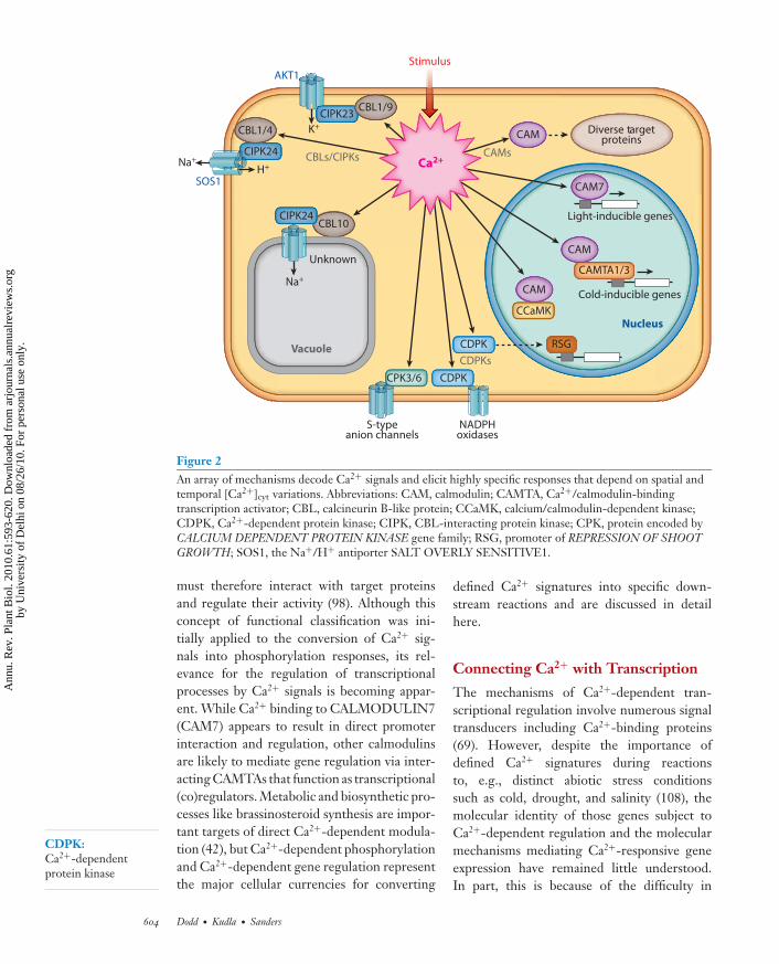
Figure 2An array of mechanisms decode Ca2+ signals and elicit highly specific responses that depend on spatial andtemporal [Ca2+]cyt variations. Abbreviations: CAM, calmodulin; CAMTA, Ca2+/calmodulin-bindingtranscription activator; CBL, calcineurin B-like protein; CCaMK, calcium/calmodulin-dependent kinase;CDPK, Ca2+-dependent protein kinase; CIPK, CBL-interacting protein kinase; CPK, protein encoded byCALCIUM DEPENDENT PROTEIN KINASE gene family; RSG, promoter of REPRESSION OF SHOOTGROWTH; SOS1, the Na+/H+ antiporter SALT OVERLY SENSITIVE1.
CDPK:Ca2+-dependentprotein kinase
must therefore interact with target proteinsand regulate their activity (98). Although thisconcept of functional classification was ini-tially applied to the conversion of Ca2+ sig-nals into phosphorylation responses, its rel-evance for the regulation of transcriptionalprocesses by Ca2+ signals is becoming appar-ent. While Ca2+ binding to CALMODULIN7(CAM7) appears to result in direct promoterinteraction and regulation, other calmodulinsare likely to mediate gene regulation via inter-acting CAMTAs that function as transcriptional(co)regulators. Metabolic and biosynthetic pro-cesses like brassinosteroid synthesis are impor-tant targets of direct Ca2+-dependent modula-tion (42), but Ca2+-dependent phosphorylationand Ca2+-dependent gene regulation representthe major cellular currencies for converting
defined Ca2+ signatures into specific down-stream reactions and are discussed in detailhere.
Connecting Ca2+ with Transcription
The mechanisms of Ca2+-dependent tran-scriptional regulation involve numerous signaltransducers including Ca2+-binding proteins(69). However, despite the importance ofdefined Ca2+ signatures during reactionsto, e.g., distinct abiotic stress conditionssuch as cold, drought, and salinity (108), themolecular identity of those genes subject toCa2+-dependent regulation and the molecularmechanisms mediating Ca2+-responsive geneexpression have remained little understood.In part, this is because of the difficulty in
604 Dodd · Kudla · Sanders
Ann
u. R
ev. P
lant
Bio
l. 20
10.6
1:59
3-62
0. D
ownl
oade
d fr
om a
rjou
rnal
s.an
nual
revi
ews.
org
by U
nive
rsity
of
Del
hi o
n 08
/26/
10. F
or p
erso
nal u
se o
nly.
ANRV410-PP61-25 ARI 31 March 2010 17:4
distinguishing stress-dependent Ca2+ re-sponses from stress-dependent but Ca2+-independent reactions (45, 115). The induc-tion of defined artificial Ca2+ transients bythe use of the calmodulin antagonists WP7and SKF-7171 allowed identification of 230Ca2+-responsive genes that were differentiallyexpressed 1 h post stimulus (75). Consideringthat this study covered only 25% of knownArabidopsis genes, these results suggest thatapproximately 3.3% of Arabidopsis genes aresubject to regulation by Ca2+. Many of theCa2+-regulated genes identified by this studywere known early stress-induced genes. Impor-tantly, this investigation established that knownABRE (abscisic acid–responsive element)-related cis-promoter elements are sufficient toconfer transcriptional regulation in responseto cytosolic Ca2+ signatures. Since ABREs arepresent in the promoter of C-REPEAT/DREBINDING FACTOR (CBF or DREB1) tran-scription factors that function as master regula-tors of abiotic stress responses (45), these find-ings point to a direct interconnection betweenCa2+ regulation of transcription and abioticstress responses but raise the question of howCa2+ signals are transduced to the transcriptionmachinery. The answer may be provided inpart by the function of CAMTA proteins.
CAMTAs are a family of eukaryotic Ca2+-dependent calmodulin-binding transcriptionfactors with six members in Arabidopsis (44).These transcription factors share a conserveddomain structure, including a C-terminalcalmodulin-binding domain that mediates in-teractions with calmodulin and an N-terminalCG-1 domain that mediates binding to DNAcis-elements (CAMTA binding sites), the latterincluding ABREs and additional CGCG ele-ments (44). CAMTA gene expression in Ara-bidopsis responds rapidly and transiently to var-ious stresses (e.g., cold, salinity) and hormones(e.g., ABA, jasmonic acid) (169), suggestingtheir involvement in multiple signal transduc-tion pathways that are critical for plant stresstolerance. First insights into the physiologicalfunction of plant CAMTA proteins were pro-vided by a reverse genetic analysis of Arabidopsis
CAMTA3 function that revealed a criticalrole for this protein in suppressing plant re-sponses to pathogens such as Pseudomonas sy-ringae and Botrytis cinerea (51). Importantly, arecent study (40) provided evidence for a di-rect link between Ca2+ signaling (via CAMTA1and CAMTA3) and cold tolerance in plants bydiscovering that these CAMTA proteins bindto regulatory elements (CAMTA binding sites)in the promoter of the DREB1c/CBF2 gene.Whilst cold induction of CBF2 and other cold-induced genes is reduced in a single camta3mutant, camta1/camta3 double mutants areimpaired in their cold acclimation to freezingtolerance (40).
These findings establish a role forCa2+/calmodulin-regulated CAMTA tran-scription factors in controlling the CBF regulonof cold-regulated genes and promoting freez-ing tolerance. Moreover, they suggest a modelin which CAMTAs may function directly inthe transduction of cold-induced cytosolicCa2+ signatures into the regulation of geneexpression through interaction with one ormore of the seven Arabidopsis Ca2+/calmodulinsensors (109). Further investigations will needto address exactly how changes in [Ca2+]cyt
lead to changes in nuclear transcription.Calmodulins are prototypical Ca2+ sensor
relay proteins, and their genomics, structuralproperties, and functional principles have beenreviewed comprehensively (98, 109). A sur-prising twist to our view about calmodulinfunction in plants was provided by a recentstudy of CALMODULIN7 (CAM7) from Ara-bidopsis (84). In Arabidopsis seven genes encodefour CAM isoforms, of which CAM1/CAM4differ by four amino acid substitutions fromCAM7, whereas CAM2/3/5 and CAM 6 differby one amino acid from CAM7 (109). Kush-waha et al. (84) established that CAM7, butnot CAM2/3/5, is a transcriptional regulatorthat interacts directly with promoters of sev-eral light-inducible genes. cam7 mutants did nothave photomorphogenic growth alterations,most likely due to overlapping functions, butcam7 mutants had reduced expression of light-inducible genes. Conversely, overexpression
www.annualreviews.org • Ca2+ Signaling 605
Ann
u. R
ev. P
lant
Bio
l. 20
10.6
1:59
3-62
0. D
ownl
oade
d fr
om a
rjou
rnal
s.an
nual
revi
ews.
org
by U
nive
rsity
of
Del
hi o
n 08
/26/
10. F
or p
erso
nal u
se o
nly.
ANRV410-PP61-25 ARI 31 March 2010 17:4
CIPK: CBL-interacting proteinkinase
of CAM7 caused an increase in expressionof light-inducible genes and hyperphotomor-phogenic growth. These findings suggest thatthe calcium sensor CAM7 translates cytosolicCa2+ signatures into gene expression throughDNA binding. However, future work needs toinvestigate whether this requires the interactionof CAM7 with additional (transcription factor)proteins and exactly how the interconnectionbetween cytosolic Ca2+ signatures and nucleargene regulation is achieved.
Connecting Ca2+ with ProteinPhosphorylation
Phosphorylation cascades regulated by kinasesand phosphatases are primary downstream in-terpreters of Ca2+ signals. Ca2+ transients areperceived and transmitted by Ca2+-dependentkinases and phosphatases. These proteins canalter biochemical function directly and rapidlythrough reversible phosphorylation, and alsocause alterations in gene expression by modu-lating transcription factor activity (139). Plantshave a unique repertoire of Ca2+-dependentprotein kinases that comprise the familiesof CCaMKs (Calcium-Calmodulin-DependentKinases), CDPKs (Calcium-Dependent ProteinKinases), and CIPKs (CBL-Interacting ProteinKinases) that form an intricate cellular networkfor decoding Ca2+ signals and regulating cel-lular processes, including ion homeostasis (seeFigure 2) (10, 141). While CDPKs andCCaMKs are typical sensor responders, theCIPKs are targets of Calcineurin B–like (CBL)sensor relay proteins (note that CCaMKs areabsent from the Arabidopsis genome).
The Arabidopsis genome encodes 34 CDPKsand 8 additional CDPK-related kinases (66).The biochemistry and regulation of CDPKshave been reviewed (60, 61, 100). Activa-tion of CDPKs is assumed to occur afterbinding of Ca2+ to the C-terminal EF-hand-containing regulatory domain, causing confor-mational changes that relieve the active siteof the kinase domain from masking by an au-toinhibitory domain. This process is paralleledby autophosphorylation of the CDPKs that
contributes to full activation of the kinases(100).
The first in vivo evidence for CDPK func-tion was obtained by suppression of NtCDPK2by viral-induced gene silencing (VIGS) in Nico-tiana benthamiana (137). CDPK-silenced plantshad a reduced and delayed hypersensitive re-sponse after race-specific Avr9 elicitation in agene-for-gene interaction, and lacked an ac-companying wilting phenotype. Remarkably,further analysis of NtCDPK2 function suggeststhat elevated CDPK signaling inhibits stress-induced MAPK activation, and this inhibi-tion requires ethylene synthesis and perception(101). This indicates that CDPK and MAPKpathways do not function independently andconcerted regulation of both pathways con-trols response specificity to biotic and abioticstress.
Reverse genetic analyses have substantiallyextended our knowledge on the physiolog-ical function of several CDPKs. ArabidopsisCALCIUM DEPENDENT PROTEIN KI-NASE3 (CPK3) and CPK6 function in ABAregulation of stomatal closure and modulateguard cell S-type anion channels (113). Inaddition, CPK4 and CPK11 are critical forABA responsiveness of guard cells and theyphosphorylate the ABA-responsive transcrip-tion factors ABSCISIC ACID RESPONSIVEELEMENT-BINDING FACTOR1 (ABF1)and ABF4 in vitro (172). Experiments in to-bacco revealed that CDPK1 regulates the tran-scription factor REPRESSION OF SHOOTGROWTH (RSG) in response to gibberellins(70), and work in potato suggests that severalCDPKs regulate ROS production by NADPHoxidases (80). Together, these findings indicatecritical roles for CDPK-mediated Ca2+ signal-ing in a very diverse array of processes. How-ever, it will be most important to discover themechanistic basis for how specific CDPKs con-tribute to the decoding of specific Ca2+ signa-tures (see Figure 2).
The CBL protein family and their inter-acting kinases (CIPKs) separate Ca2+-bindingfunctionality (sensor relay function) and kinaseactivity (response activity) into two flexible,
606 Dodd · Kudla · Sanders
Ann
u. R
ev. P
lant
Bio
l. 20
10.6
1:59
3-62
0. D
ownl
oade
d fr
om a
rjou
rnal
s.an
nual
revi
ews.
org
by U
nive
rsity
of
Del
hi o
n 08
/26/
10. F
or p
erso
nal u
se o
nly.

ANRV410-PP61-25 ARI 31 March 2010 17:4
combinable modules. This allows for the for-mation of a complex and dynamic Ca2+-decoding signaling network. Since the discov-ery of CBLs and CIPKs in Arabidopsis (83, 145),advances in our knowledge of the structuralfeatures, evolution, and functional principlesof this Ca2+-decoding system have been re-viewed (10, 11, 97–99, 163). CBL proteins havesignificant similarity to the regulatory B sub-unit of calcineurin and neuronal calcium sen-sor (NCS) proteins from animals and yeast (83).CBLs contain four Ca2+-binding EF-hand do-mains that are arranged with invariant spacing(81). CIPK-type kinases comprise a conservedN-terminal kinase domain with high similar-ity to yeast SNF1, and a C-terminal regula-tory domain that is separated from the kinasedomain by a variable junction domain. Withinthe rather divergent regulatory domain, theconserved NAF domain is required and suffi-cient for interaction with CBLs (1). Moreover, aprotein-phosphatase interaction (PPI) domainthat mediates CIPK interaction with PP2Cphosphatases is present in the C-terminus ofthese kinases (122). It is assumed that CBLbinding to the NAF domain of CIPKs releasesthe C-terminal (autoinhibitory) domain fromthe kinase domain, thereby transforming thekinase into an active state (56). Comprehen-sive bioinformatic analyses of both protein fam-ilies have identified 10 CBLs and 26 CIPKs inthe Arabidopsis genome, and 10 CBLs and 30CIPKs in rice (1, 81, 163). Single CBL and CIPKgenes are present in several species of greenalgae, while Physcomitrella contains four CBLsand seven CIPKs and the genome of the fernSelaginella moellendorfii has five CBLs and fiveCIPKs (11, 163). These findings suggest thatthe evolution of plants was accompanied by theevolution of complexity of the CBL and CIPKprotein families.
Spatial specificity is an important aspect ofcellular information processing. Localizationstudies of Arabidopsis CBL proteins revealedthat four CBLs are present at the plasma mem-brane, four are localized to the vacuolar mem-brane, and two are present in the cytoplasmand nucleus (12, 28, 31, 77, 163). This suggests
that CBL-CIPK complexes could function asfast responders to local Ca2+ release eventsfrom internal and external stores and that thespatial separation of distinct CBL-CIPK com-plexes contributes to spatial specificity in Ca2+
signaling. For the plasma membrane–localizedCBL1, dual lipid modification by myristoy-lation and S-acylation are required for bothits function and its localization to the plasmamembrane. CBL1 localization is achieved bya two-step targeting process in which initialmyristoylation results in localization to the en-doplasmatic reticulum (ER) and subsequentS-acylation is crucial for ER-to-plasma mem-brane trafficking (12).
Most CIPK-GFP fusion proteins have cy-tosolic and nuclear localization (11, 31, 163).However, CBL-CIPK interaction analyses us-ing bimolecular fluorescence complementation(BiFC) revealed that CIPKs are targeted to dif-ferent compartments of the cell by their re-spective interacting CBL proteins (12, 28, 31,160). For example, CIPK1 is targeted to theplasma membrane by CBL1 or CBL9 (28, 160)but upon interaction with CBL2 the resultingCBL2/CIPK1 complexes are exclusively vacuo-lar membrane-localized (12).
Initial insights into the physiological func-tion of CBLs and CIPKs came from for-ward genetic screens. The CBL calcium sen-sor SOS3 (AtCBL4) and the CIPK-type kinaseSOS2 (AtCIPK24) appear to be part of a Ca2+-regulated signaling pathway that specificallymediates salt stress adaptation by regulating theNa+/H+ antiporter SOS1 (see Figure 2) (57,94, 95). Recent studies revealed that the calciumsensor CBL10 also interacts with and activatesthe kinase CIPK24 (77, 133). CBL10/CIPK24complexes are vacuolar membrane-localized,thereby supporting the functional concept thatalternative complex formation of CIPK24 ki-nases with either CBL4 or CBL10 creates adual-function kinase with separate functionsat the plasma and vacuolar membranes (seeFigure 2). While CBL4/CIPK24 complexesmediate Na+ extrusion via the regulation of theH+/Na+ antiporter SOS1 at the plasma mem-brane, formation of CBL10/CIPK24 results in
www.annualreviews.org • Ca2+ Signaling 607
Ann
u. R
ev. P
lant
Bio
l. 20
10.6
1:59
3-62
0. D
ownl
oade
d fr
om a
rjou
rnal
s.an
nual
revi
ews.
org
by U
nive
rsity
of
Del
hi o
n 08
/26/
10. F
or p
erso
nal u
se o
nly.
ANRV410-PP61-25 ARI 31 March 2010 17:4
vacuolar Na+ sequestration by regulating un-known targets (see Figure 2).
Reverse genetics has greatly advancedour understanding of CBLs and CIPKs andrevealed crucial functions of distinct CBL pro-teins and CIPKs for mineral nutrition, as well asresponses to abiotic stresses and to ABA. Anal-ysis of a CIPK3 loss-of-function allele estab-lished that this kinase regulates ABA responsesduring seed germination and regulates ABA-induced gene expression (78). Two independentreverse genetic analyses of CBL1 functionrevealed that CBL1 is a central integrator ofresponses to drought, cold, and salinity (2, 27).While the mutant studies of CBL1 revealed anABA-independent function of this protein inseveral abiotic stress responses, loss of functionof the closely related Ca2+ sensor CBL9renders plants hypersensitive to ABA (125).Alternative complex formation between thekinase CIPK1 and either CBL1 or CBL9 me-diates ABA-dependent and ABA-independentresponses (31). CBL9 also appears to complexwith CIPK3 for modulating ABA responses(126).
The CBL/CIPK system also regulates K+
homeostasis. CIPK23 is targeted to the plasmamembrane and is activated by the two highlyrelated Ca2+ sensors CBL1 and CBL9 (28,167), and the complexes regulate the activityof the shaker-like K+ channel ARABIDOPSISK+ TRANSPORTER1 (AKT1). CIPK23 in-teracts exclusively with AKT1 and no other K+
transporters from Arabidopsis (87). Besides theregulation of K+ uptake in roots, the Ca2+-decoding CBL1/CBL9/CIPK23 module is in-volved in stomatal regulation under dehydrat-ing conditions (28).
The findings of all these studies indicate thatthe CBL-CIPK network is a central and criti-cal system for decoding Ca2+ signals in responseto a broad variety of stimuli. It is also becomingapparent that each CBL and each CIPK rep-resents a multifunctional signaling componentthat can undergo alternative protein interac-tions, determining the flow of information pro-cessing through this signaling system. There-fore, elucidating the mechanistic factors that
determine the “decision making” in this flexibleinteraction network will be of eminent impor-tance to further our understanding of Ca2+-decoding mechanisms.
Ca2+ SIGNALING SYSTEMS
Ca2+ signaling networks are complex and so-phisticated. Prediction of network function isoften nonintuitive due to the high degree of in-terconnectivity between network components.Here, we (a) consider systems-based investiga-tions of network architecture, and (b) discussthe modeling of [Ca2+] alterations during thegeneration and decoding of Ca2+ signals.
Scale-Free Network Architecture
Some signaling networks are proposed to haveproperties similar to scale-free networks incomputing. Scale-free networks have many in-terconnected nodes (i.e., signaling intermedi-ates). A small number of the nodes are veryhighly connected and called hubs (64). Scale-free networks are robust to node removal,are sensitive to hub removal, and can pro-cess multiple signals simultaneously (64). Inthe guard cell, the extensive connectivity ofCa2+ with other network components and de-pendency of stomatal closure upon stimulus-induced Ca2+ alterations imply that Ca2+ is ahub (64). The paradigm of scale-free networkarchitecture is not a formalism of network func-tion but provides a tool for development ofmathematical models, advances understandingof the evolutionary basis of the network, and al-lows identification of optimal manipulation tar-gets for research or agricultural purposes.
Knowledge of interconnectivities between alarge number of nodes (>1000) is necessary toconclude scale-free architecture (64). An Ara-bidopsis whole-genome network derived fromtranscriptome data has scale-free properties butis not completely scale-free (102). This net-work (102) reflects gene regulation and so didnot specifically incorporate Ca2+. Interestingly,the cold-regulated subnetwork includes knownand uncharacterized proteins linked to Ca2+
608 Dodd · Kudla · Sanders
Ann
u. R
ev. P
lant
Bio
l. 20
10.6
1:59
3-62
0. D
ownl
oade
d fr
om a
rjou
rnal
s.an
nual
revi
ews.
org
by U
nive
rsity
of
Del
hi o
n 08
/26/
10. F
or p
erso
nal u
se o
nly.
ANRV410-PP61-25 ARI 31 March 2010 17:4
signaling (102). Transcripts associated withCa2+ signaling also occupy separate and well-connected nodes within subnetworks associatedwith ER stress responses (CALRETICULIN2),biotic stress (CAX3), jasmonic acid/ethylenesignaling (Ca2+-binding TSK-ASSOCIATINGPROTEIN1), and salicylic acid metabolism andpathogen responses (calmodulin-binding pro-tein encoded by At1g73805) (102). When Ca2+
signaling is considered within gene networks,genes associated with Ca2+ signaling appearto occupy independent positions within sev-eral subnetworks. This might derive from thecapacity of Ca2+ signals to encode specificity,which allows these signals to occupy multiplepositions within the network. Scale-free net-work architecture is also a useful paradigm withwhich to consider smaller networks. For exam-ple, the CBL/CIPK Ca2+ sensor relay system(10) is proposed to have scale-free architecturebecause the majority of CBLs interact with asmall number of CIPKs, while a limited num-ber of hublike CBLs (e.g., CBL2) interact withmany CIPKs (10). This provides the capac-ity to process multiple signals simultaneouslythough formation of alternative CBL/CIPKcomplexes, depending on localization and con-centration of Ca2+ alterations and localizationof CBL/CIPKs (31, 160).
Bow-Tie Network Architecture
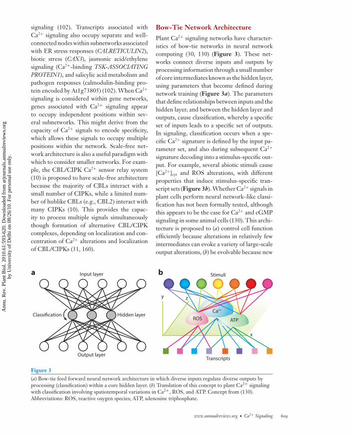
Plant Ca2+ signaling networks have character-istics of bow-tie networks in neural networkcomputing (30, 130) (Figure 3). These net-works connect diverse inputs and outputs byprocessing information through a small numberof core intermediates known as the hidden layer,using parameters that become defined duringnetwork training (Figure 3a). The parametersthat define relationships between inputs and thehidden layer, and between the hidden layer andoutputs, cause classification, whereby a specificset of inputs leads to a specific set of outputs.In signaling, classification occurs when a spe-cific Ca2+ signature is defined by the input pa-rameter set, and also during subsequent Ca2+
signature decoding into a stimulus-specific out-put. For example, several abiotic stimuli cause[Ca2+]cyt and ROS alterations, with differentproperties that induce stimulus-specific tran-script sets (Figure 3b). Whether Ca2+ signals inplant cells perform neural network–like classi-fication has not been formally tested, althoughthis appears to be the case for Ca2+ and cGMPsignaling in some animal cells (130). This archi-tecture is proposed to (a) control cell functionefficiently because alterations in relatively fewintermediates can evoke a variety of large-scaleoutput alterations, (b) be evolvable because new
Transcripts
Stimuli
Classification
x
y z
a bInput layer
Hidden layer
Output layer
Ca2+
ROS ATP
Figure 3(a) Bow-tie feed forward neural network architecture in which diverse inputs regulate diverse outputs byprocessing (classification) within a core hidden layer. (b) Translation of this concept to plant Ca2+ signalingwith classification involving spatiotemporal variations in Ca2+, ROS, and ATP. Concept from (130).Abbreviations: ROS, reactive oxygen species; ATP, adenosine triphosphate.
www.annualreviews.org • Ca2+ Signaling 609
Ann
u. R
ev. P
lant
Bio
l. 20
10.6
1:59
3-62
0. D
ownl
oade
d fr
om a
rjou
rnal
s.an
nual
revi
ews.
org
by U
nive
rsity
of
Del
hi o
n 08
/26/
10. F
or p
erso
nal u
se o
nly.
ANRV410-PP61-25 ARI 31 March 2010 17:4
inputs and outputs integrate readily with thetoolbox of core molecules, and (c) have fragilityto failure of the core mechanism and suscep-tibility to hijacking (30). This final propertymight explain the evolutionary recruitment ofroot hair Ca2+ signaling into initiation of rootnodule formation.
Predictive Modelsof Network Function
The dynamic interactions within many signal-ing networks are well characterized, but quan-titative descriptions of network function are in-frequent (67). Here, we consider simulations ofnetworks that include Ca2+ signaling.
One model for ABA-induced stomatal clo-sure has generated new questions concerningCa2+ signaling in the guard cell (92). In thismodel interactions of signaling intermediatesare described with Boolean logic, with the ul-timate outcome that the pore is either open orclosed (92). The most highly connected compo-nents are [Ca2+]cyt (12 interconnections), pHcyt
(9 interconnections), and plasma membrane de-polarization (9 interconnections). Within thisnetwork there are at least two semiindepen-dent pathways connecting ABA perception withstomatal closure, involving changes in [Ca2+]cyt
and pHcyt, respectively (92). One hypothesisformed from this model is that stomata areclosed by ABA-induced [Ca2+]cyt increases, but[Ca2+]cyt increases are not a prerequisite forstomatal closure (92). Testing this hypothesis
AM
Nod
a
b
5 min
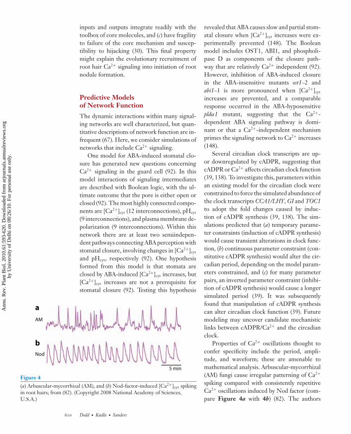
Figure 4(a) Arbuscular-mycorrhizal (AM), and (b) Nod-factor-induced [Ca2+]cyt spikingin root hairs; from (82). (Copyright 2008 National Academy of Sciences,U.S.A.)
revealed that ABA causes slow and partial stom-atal closure when [Ca2+]cyt increases were ex-perimentally prevented (148). The Booleanmodel includes OST1, ABI1, and phospholi-pase D as components of the closure path-way that are relatively Ca2+ independent (92).However, inhibition of ABA-induced closurein the ABA-insensitive mutants ost1–2 andabi1–1 is more pronounced when [Ca2+]cyt
increases are prevented, and a comparableresponse occurred in the ABA-hyposensitivepldα1 mutant, suggesting that the Ca2+-dependent ABA signaling pathway is domi-nant or that a Ca2+-independent mechanismprimes the signaling network to Ca2+ increases(148).
Several circadian clock transcripts are up-or downregulated by cADPR, suggesting thatcADPR or Ca2+ affects circadian clock function(39, 138). To investigate this, parameters withinan existing model for the circadian clock wereconstrained to force the simulated abundance ofthe clock transcripts CCA1/LHY, GI and TOC1to adopt the fold changes caused by induc-tion of cADPR synthesis (39, 138). The sim-ulations predicted that (a) temporary parame-ter constraints (induction of cADPR synthesis)would cause transient alterations in clock func-tion, (b) continuous parameter constraint (con-stitutive cADPR synthesis) would alter the cir-cadian period, depending on the model param-eters constrained, and (c) for many parameterpairs, an inverted parameter constraint (inhibi-tion of cADPR synthesis) would cause a longersimulated period (39). It was subsequentlyfound that manipulation of cADPR synthesiscan alter circadian clock function (39). Futuremodeling may uncover candidate mechanisticlinks between cADPR/Ca2+ and the circadianclock.
Properties of Ca2+ oscillations thought toconfer specificity include the period, ampli-tude, and waveform; these are amenable tomathematical analysis. Arbuscular-mycorrhizal(AM) fungi cause irregular patterning of Ca2+
spiking compared with consistently repetitiveCa2+ oscillations induced by Nod factor (com-pare Figure 4a with 4b) (82). The authors
610 Dodd · Kudla · Sanders
Ann
u. R
ev. P
lant
Bio
l. 20
10.6
1:59
3-62
0. D
ownl
oade
d fr
om a
rjou
rnal
s.an
nual
revi
ews.
org
by U
nive
rsity
of
Del
hi o
n 08
/26/
10. F
or p
erso
nal u
se o
nly.

ANRV410-PP61-25 ARI 31 March 2010 17:4
investigated mathematically whether AM-induced Ca2+ spikes are unpredictable (stochas-tic) or deterministic but with chaotic prop-erties, and concluded from multiple measuresthat both AM- and Nod-induced Ca2+ spikeshave recurrent properties. The variable pat-terning of AM-induced Ca2+ spikes is there-fore likely due to chaotic rather than stochasticproperties of the system (82). This suggests thatthe transport processes causing the oscillationshave chaotic regulatory properties that allowslight differences in the stimulus to evoke highlydifferentiated output responses (82), a conclu-sion that may help explain how many distinctCa2+ signatures can be caused by a small set ofchannels.
Simulation of Ca2+ Homeostasisand Dynamics
Classical simulations of stimulus-induced Ca2+
oscillations in animal cells parameterize two ormore Ca2+ stores, a Ca2+ release mechanism,cytosolic Ca2+-scavenging Ca2+ ATPases, andmembrane leak currents (154). These modelssimulate Ca2+ oscillations with properties sim-ilar to Ca2+ oscillations in animal cells. Moresophisticated models incorporate featuressuch as the ligand binding kinetics of Ca2+
release channels (154). Important differencesbetween plant and animal cells require incor-poration into simulations. cADPR and InsP3
are thought to activate separate Ca2+ stores inanimal cells, whereas common cADPR- andInsP3-sensitive Ca2+ stores exist in plant cellsbecause the vacuolar membrane is cADPR- andInsP3-sensitive (5), whereas ER membranesare InsP3-, cADPR-, and NAADP-sensitive(see Figure 1) (5, 116, 118, 119). The notionof an ER store that contains finite quantitiesof Ca2+ that functions in combination witha vacuolar store containing virtually limitlessCa2+ was incorporated within a simulationof ABA-induced [Ca2+]cyt oscillations (159).The study concluded that [Ca2+]cyt oscillations
with comparable properties are evoked byABA concentrations that span several ordersof magnitude because the different kinetics ofCa2+ scavenging Ca2+ ATPases and Ca2+/H+
antiporters mean that at high [ABA], Ca2+/H+
antiporters remove Ca2+ from the cytosol andallow [Ca2+]cyt oscillations; whereas, at low[ABA], [Ca2+]cyt oscillations can occur withonly Ca2+-ATPase activity (see Figure 1)(159). The long period of [Ca2+]cyt oscillationsin plant cells compared with animal cells mightarise from differences in the rate of activation byInsP3 of InsP3-sensitive Ca2+ channels (strictly,relief from Ca2+ inhibition of the channelby InsP3), and different Ca2+ signatures mayarise from variations in this parameter (159).The simulation assumed that Ca2+ release wasentirely mediated by cADPR and InsP3-gatedchannels, so future simulations incorporat-ing regulatory kinetics of TPC1-mediatedCa2+ release (128) would be informative (seeFigure 1). The contribution of extracellularCa2+ influx to stimulus-induced Ca2+ signals,which is important in at least those guard cellsthat are relatively insensitive to ABA (104),could provide further information concerningthe contribution of each Ca2+ store to Ca2+
oscillations.Four-dimensional simulation of Ca2+ waves
has been performed using as a template the3D geometry of membrane systems in animalcells from tomographic electron microscopy(107). Simulations predicted that regions ofclose membrane proximity participate in for-mation of signaling microdomains. Applicationto plant cells seems appropriate given the dis-tinctive arrangement of a large central vacuolesurrounded by a relatively small-volume, ER-containing cytosol, which provides a very dif-ferent topology for the propagation of Ca2+
signals compared with animal cells. We suggestthat, in future, computational approaches willbe essential for advancing our understandingof the complex intracellular language of Ca2+
signaling.
www.annualreviews.org • Ca2+ Signaling 611
Ann
u. R
ev. P
lant
Bio
l. 20
10.6
1:59
3-62
0. D
ownl
oade
d fr
om a
rjou
rnal
s.an
nual
revi
ews.
org
by U
nive
rsity
of
Del
hi o
n 08
/26/
10. F
or p
erso
nal u
se o
nly.
ANRV410-PP61-25 ARI 31 March 2010 17:4
SUMMARY POINTS
1. Many energized transporters previously thought to function only in Ca2+ homeostasismay have signaling roles.
2. Specific Ca2+ signatures are decoded by a complex toolkit of Ca2+-binding proteins thattranslate the information encoded within Ca2+ signals into phosphorylation events anddefined transcriptional responses.
3. Ca2+-signaling networks could have architectural and functional properties that are com-parable to scale-free networks and bow-tie networks.
FUTURE ISSUES
1. Understand the transport and signaling functions of specific gene products that are likelyto form multisubunit Ca2+-permeable channels.
2. Identify genes encoding ligand-gated channels to establish the mechanistic basis forcADPR, InsP3 and NAADP signaling in plant cells and the evolutionary basis for thesemechanisms, particularly given the absence of animal cADPR and InsP3 receptors fromthe plant genome.
3. Measure [Ca2+]cyt in Ca2+ pump and Ca2+ exchanger mutants to position these proteinswithin the Ca2+ signaling network.
4. Understand the biological significance of the spatial distribution of Ca2+ signals withinsingle plant cells during signaling.
5. Develop predictive models for Ca2+ signals that incorporate the kinetics and regulationof specific gene products.
6. Understand how circadian Ca2+ oscillations are decoded: (a) What is the identity ofthe Ca2+ sensor protein(s) that are involved in this pathway? (b) What are the down-stream mechanisms that convert the signals arising from activation of these proteins intoalterations in gene expression or enzyme activity?
7. Uncover the functional interconnections between the systems that decode Ca2+ signalsand other cell signaling systems such as PP2C-type phosphatases, receptor-like kinases,and MAPK modules.
DISCLOSURE STATEMENT
The authors are not aware of any affiliations, memberships, funding, or financial holdings thatmight be perceived as affecting the objectivity of this review.
ACKNOWLEDGMENTS
We apologize to researchers whose work is not cited due to space limitations. We thank Dr.Lorella Navazio for critical reading of our manuscript. A.N.D. is grateful to the Royal Societyfor the award of a University Research Fellowship. J. K. thanks Dr. Hillel Fromm for helpfuldiscussions about CAMTA proteins and Dr. Kenji Hashimoto for help with Figure 2. Work in
612 Dodd · Kudla · Sanders
Ann
u. R
ev. P
lant
Bio
l. 20
10.6
1:59
3-62
0. D
ownl
oade
d fr
om a
rjou
rnal
s.an
nual
revi
ews.
org
by U
nive
rsity
of
Del
hi o
n 08
/26/
10. F
or p
erso
nal u
se o
nly.
ANRV410-PP61-25 ARI 31 March 2010 17:4
the J. K. laboratory was supported by the D.F.G., the Alexander von Humboldt foundation, theD. A. A. D., the G. I. F., and the H. F. S. P.
LITERATURE CITED
1. Albrecht V, Ritz O, Linder S, Harter K, Kudla J. 2001. The NAF domain defines a novel protein-proteininteraction module conserved in Ca2+-regulated kinases. EMBO J. 20:1051–63
2. Albrecht V, Weinl S, Blazevic D, D’Angelo C, Batistic O, et al. 2003. The calcium sensor CBL1 integratesplant responses to abiotic stresses. Plant J. 36:457–70
3. Ali R, Ma W, Lemtiri-Chlieh F, Tsaltas D, Leng Q, et al. 2007. Death Don’t Have No Mercy and NeitherDoes Calcium: Arabidopsis CYCLIC NUCLEOTIDE GATED CHANNEL2 and Innate Immunity.Plant Cell 19:1081–95
4. Allen GJ, Chu SP, Harrington CL, Schumacher K, Hoffman T, et al. 2001. A defined range of guardcell calcium oscillation parameters encodes stomatal movements. Nature 411:1053–57
5. Allen GJ, Muir SR, Sanders D. 1995. Release of Ca2+ from individual plant vacuoles by both InsP(3)and cyclic ADP-ribose. Science 268:735–37
6. Allen GJ, Sanders D. 1995. Calcineurin, a type 2B protein phosphatase, modulates the Ca2+-permeableslow vacuolar ion-channel of stomatal guard-cells. Plant Cell 7:1473–83
7. Arazi T, Kaplan B, Fromm H. 2000. A high-affinity calmodulin-binding site in a tobacco plasma-membrane channel protein coincides with a characteristic element of cyclic nucleotide-binding domains.Plant Mol. Biol. 42:591–601
8. Baekgaard L, Fuglsang AT, Palmgren MG. 2005. Regulation of plant plasma membrane H+- and Ca2+-ATPases by terminal domains. J. Bioenerg. Biomembr. 37:369–74
9. Balague C, Lin BQ, Alcon C, Flottes G, Malmstrom S, et al. 2003. HLM1, an essential signalingcomponent in the hypersensitive response, is a member of the cyclic nucleotide-gated channel ion channelfamily. Plant Cell 15:365–79
10. Batistic O, Kudla J. 2004. Integration and channeling of calcium signaling through the CBL calciumsensor/CIPK protein kinase network. Planta 219:915–24
11. Batistic O, Kudla J. 2009. Plant calcineurin B–like proteins and their interacting protein kinases. Biochim.Biophys. Acta 1793:985–92
12. Batistic O, Sorek N, Schultke S, Yalovsky S, Kudla J. 2008. Dual fatty acyl modification determines thelocalization and plasma membrane targeting of CBL/CIPK Ca2+ signaling complexes in Arabidopsis.Plant Cell 20:1346–62
13. Baum G, Long JC, Jenkins GI, Trewavas AJ. 1999. Stimulation of the blue light phototropic receptorNPH1 causes a transient increase in cytosolic Ca2+. Proc. Natl. Acad. Sci. USA 96:13554–59
14. Bonaventure G, Gfeller A, Proebsting WM, Hortensteiner S, Chetelat A, et al. 2007. A gain-of-functionallele of TPC1 activates oxylipin biogenesis after leaf wounding in Arabidopsis. Plant J. 49:889–98
15. Bonza MC, Luoni L, De Michelis MI. 2001. Stimulation of plant plasma membrane Ca2+-ATPaseactivity by acidic phospholipids. Physiol. Plantarum 112:315–20
16. Bonza MC, Morandini P, Luoni L, Geisler M, Palmgren MG, De Michelis MI. 2000. AtACA8 encodesa plasma membrane–localized calcium-ATPase of Arabidopsis with a calmodulin-binding domain at theN terminus. Plant Physiol. 123:1495–505
17. Boursiac Y, Harper JF. 2007. The origin and function of calmodulin regulated Ca2+ pumps in plants.J. Bioenerg. Biomembr. 39:409–14
18. Carol RJ, Takeda S, Linstead P, Durrant MC, Kakesova H, et al. 2005. A RhoGDP dissociation inhibitorspatially regulates growth in root hair cells. Nature 438:1013–16
19. Carter C, Pan S, Zouhar J, Avila EL, Girke T, Raikhel NV. 2004. The vegetative vacuole proteome ofArabidopsis thaliana reveals predicted and unexpected proteins. Plant Cell 16:3285–303
20. Catala R, Santos E, Alonso JM, Ecker JR, Martinez-Zapater JM, Salinas J. 2003. Mutations in theCa2+/H+ transporter CAX1 increase CBF/DREB1 expression and the cold-acclimation response inArabidopsis. Plant Cell 15:2940–51
www.annualreviews.org • Ca2+ Signaling 613
Ann
u. R
ev. P
lant
Bio
l. 20
10.6
1:59
3-62
0. D
ownl
oade
d fr
om a
rjou
rnal
s.an
nual
revi
ews.
org
by U
nive
rsity
of
Del
hi o
n 08
/26/
10. F
or p
erso
nal u
se o
nly.
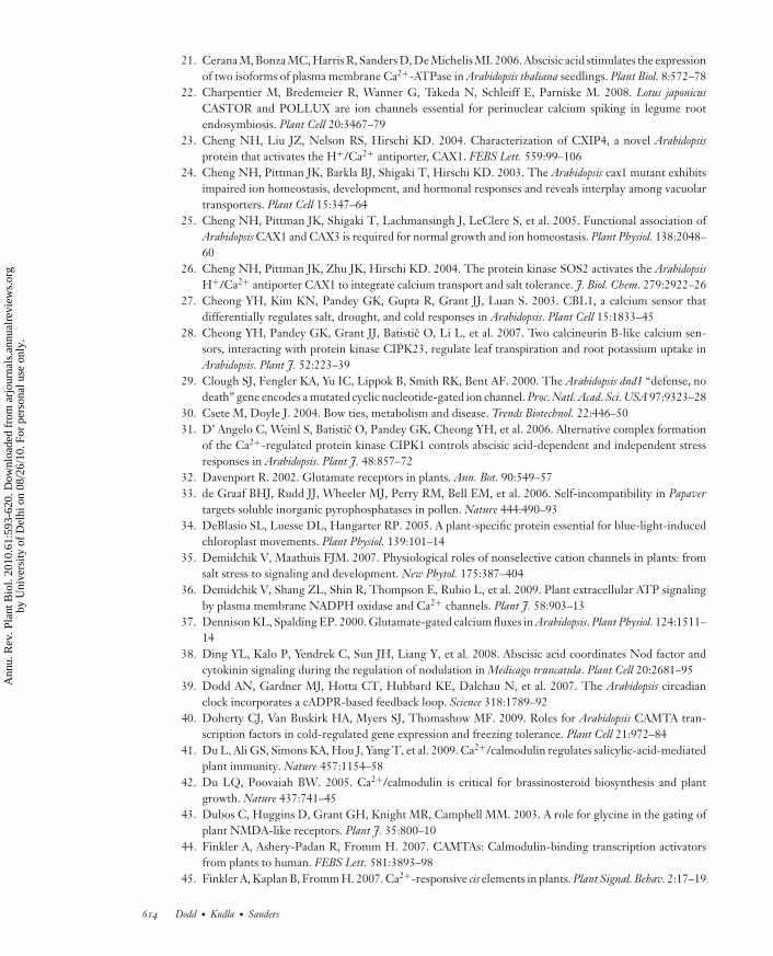
ANRV410-PP61-25 ARI 31 March 2010 17:4
21. Cerana M, Bonza MC, Harris R, Sanders D, De Michelis MI. 2006. Abscisic acid stimulates the expressionof two isoforms of plasma membrane Ca2+-ATPase in Arabidopsis thaliana seedlings. Plant Biol. 8:572–78
22. Charpentier M, Bredemeier R, Wanner G, Takeda N, Schleiff E, Parniske M. 2008. Lotus japonicusCASTOR and POLLUX are ion channels essential for perinuclear calcium spiking in legume rootendosymbiosis. Plant Cell 20:3467–79
23. Cheng NH, Liu JZ, Nelson RS, Hirschi KD. 2004. Characterization of CXIP4, a novel Arabidopsisprotein that activates the H+/Ca2+ antiporter, CAX1. FEBS Lett. 559:99–106
24. Cheng NH, Pittman JK, Barkla BJ, Shigaki T, Hirschi KD. 2003. The Arabidopsis cax1 mutant exhibitsimpaired ion homeostasis, development, and hormonal responses and reveals interplay among vacuolartransporters. Plant Cell 15:347–64
25. Cheng NH, Pittman JK, Shigaki T, Lachmansingh J, LeClere S, et al. 2005. Functional association ofArabidopsis CAX1 and CAX3 is required for normal growth and ion homeostasis. Plant Physiol. 138:2048–60
26. Cheng NH, Pittman JK, Zhu JK, Hirschi KD. 2004. The protein kinase SOS2 activates the ArabidopsisH+/Ca2+ antiporter CAX1 to integrate calcium transport and salt tolerance. J. Biol. Chem. 279:2922–26
27. Cheong YH, Kim KN, Pandey GK, Gupta R, Grant JJ, Luan S. 2003. CBL1, a calcium sensor thatdifferentially regulates salt, drought, and cold responses in Arabidopsis. Plant Cell 15:1833–45
28. Cheong YH, Pandey GK, Grant JJ, Batistic O, Li L, et al. 2007. Two calcineurin B-like calcium sen-sors, interacting with protein kinase CIPK23, regulate leaf transpiration and root potassium uptake inArabidopsis. Plant J. 52:223–39
29. Clough SJ, Fengler KA, Yu IC, Lippok B, Smith RK, Bent AF. 2000. The Arabidopsis dnd1 “defense, nodeath” gene encodes a mutated cyclic nucleotide-gated ion channel. Proc. Natl. Acad. Sci. USA 97:9323–28
30. Csete M, Doyle J. 2004. Bow ties, metabolism and disease. Trends Biotechnol. 22:446–5031. D’ Angelo C, Weinl S, Batistic O, Pandey GK, Cheong YH, et al. 2006. Alternative complex formation
of the Ca2+-regulated protein kinase CIPK1 controls abscisic acid-dependent and independent stressresponses in Arabidopsis. Plant J. 48:857–72
32. Davenport R. 2002. Glutamate receptors in plants. Ann. Bot. 90:549–5733. de Graaf BHJ, Rudd JJ, Wheeler MJ, Perry RM, Bell EM, et al. 2006. Self-incompatibility in Papaver
targets soluble inorganic pyrophosphatases in pollen. Nature 444:490–9334. DeBlasio SL, Luesse DL, Hangarter RP. 2005. A plant-specific protein essential for blue-light-induced
chloroplast movements. Plant Physiol. 139:101–1435. Demidchik V, Maathuis FJM. 2007. Physiological roles of nonselective cation channels in plants: from
salt stress to signaling and development. New Phytol. 175:387–40436. Demidchik V, Shang ZL, Shin R, Thompson E, Rubio L, et al. 2009. Plant extracellular ATP signaling
by plasma membrane NADPH oxidase and Ca2+ channels. Plant J. 58:903–1337. Dennison KL, Spalding EP. 2000. Glutamate-gated calcium fluxes in Arabidopsis. Plant Physiol. 124:1511–
1438. Ding YL, Kalo P, Yendrek C, Sun JH, Liang Y, et al. 2008. Abscisic acid coordinates Nod factor and
cytokinin signaling during the regulation of nodulation in Medicago truncatula. Plant Cell 20:2681–9539. Dodd AN, Gardner MJ, Hotta CT, Hubbard KE, Dalchau N, et al. 2007. The Arabidopsis circadian
clock incorporates a cADPR-based feedback loop. Science 318:1789–9240. Doherty CJ, Van Buskirk HA, Myers SJ, Thomashow MF. 2009. Roles for Arabidopsis CAMTA tran-
scription factors in cold-regulated gene expression and freezing tolerance. Plant Cell 21:972–8441. Du L, Ali GS, Simons KA, Hou J, Yang T, et al. 2009. Ca2+/calmodulin regulates salicylic-acid-mediated
plant immunity. Nature 457:1154–5842. Du LQ, Poovaiah BW. 2005. Ca2+/calmodulin is critical for brassinosteroid biosynthesis and plant
growth. Nature 437:741–4543. Dubos C, Huggins D, Grant GH, Knight MR, Campbell MM. 2003. A role for glycine in the gating of
plant NMDA-like receptors. Plant J. 35:800–1044. Finkler A, Ashery-Padan R, Fromm H. 2007. CAMTAs: Calmodulin-binding transcription activators
from plants to human. FEBS Lett. 581:3893–9845. Finkler A, Kaplan B, Fromm H. 2007. Ca2+-responsive cis elements in plants. Plant Signal. Behav. 2:17–19
614 Dodd · Kudla · Sanders
Ann
u. R
ev. P
lant
Bio
l. 20
10.6
1:59
3-62
0. D
ownl
oade
d fr
om a
rjou
rnal
s.an
nual
revi
ews.
org
by U
nive
rsity
of
Del
hi o
n 08
/26/
10. F
or p
erso
nal u
se o
nly.
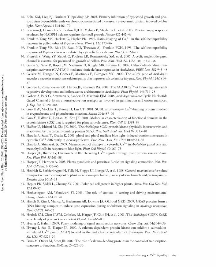
ANRV410-PP61-25 ARI 31 March 2010 17:4
46. Folta KM, Lieg EJ, Durham T, Spalding EP. 2003. Primary inhibition of hypocotyl growth and pho-totropism depend differently on phototropin-mediated increases in cytoplasmic calcium induced by bluelight. Plant Physiol. 133:1464–70
47. Foreman J, Demidchik V, Bothwell JHF, Mylona P, Miedema H, et al. 2003. Reactive oxygen speciesproduced by NADPH oxidase regulate plant cell growth. Nature 422:442–46
48. Franklin-Tong VE, Hackett G, Hepler PK. 1997. Ratio-imaging of Cai2+ in the self-incompatibility
response in pollen tubes of Papaver rhoeas. Plant J. 12:1375–8649. Franklin-Tong VE, Ride JP, Read ND, Trewavas AJ, Franklin FCH. 1993. The self incompatibility
response of Papaver rhoeas is mediated by cytosolic free calcium. Plant J. 4:163–7750. Frietsch S, Wang YF, Sladek C, Poulsen LR, Romanowsky SM, et al. 2007. A cyclic nucleotide-gated
channel is essential for polarized tip growth of pollen. Proc. Natl. Acad. Sci. USA 104:14531–3651. Galon Y, Nave R, Boyce JM, Nachmias D, Knight MR, Fromm H. 2008. Calmodulin-binding tran-
scription activator (CAMTA) 3 mediates biotic defense responses in Arabidopsis. FEBS Lett. 582:943–4852. Geisler M, Frangne N, Gomes E, Martinoia E, Palmgren MG. 2000. The ACA4 gene of Arabidopsis
encodes a vacuolar membrane calcium pump that improves salt tolerance in yeast. Plant Physiol. 124:1814–27
53. George L, Romanowsky SM, Harper JF, Sharrock RA. 2008. The ACA10 Ca2+-ATPase regulates adultvegetative development and inflorescence architecture in Arabidopsis. Plant Physiol. 146:716–28
54. Gobert A, Park G, Amtmann A, Sanders D, Maathuis FJM. 2006. Arabidopsis thaliana Cyclic NucleotideGated Channel 3 forms a nonselective ion transporter involved in germination and cation transport.J. Exp. Bot. 57:791–800
55. Guo HW, Mockler T, Duong H, Lin CT. 2001. SUB1, an Arabidopsis Ca2+-binding protein involvedin cryptochrome and phytochrome coaction. Science 291:487–90
56. Guo Y, Halfter U, Ishitani M, Zhu JK. 2001. Molecular characterization of functional domains in theprotein kinase SOS2 that is required for plant salt tolerance. Plant Cell 13:1383–99
57. Halfter U, Ishitani M, Zhu JK. 2000. The Arabidopsis SOS2 protein kinase physically interacts with andis activated by the calcium-binding protein SOS3. Proc. Natl. Acad. Sci. USA 97:3735–40
58. Harada A, Sakai T, Okada K. 2003. phot1 and phot2 mediate blue light–induced transient increases incytosolic Ca2+ differently in Arabidopsis leaves. Proc. Natl. Acad. Sci. USA 100:8583–88
59. Harada A, Shimazaki K. 2009. Measurement of changes in cytosolic Ca2+ in Arabidopsis guard cells andmesophyll cells in response to blue light. Plant Cell Physiol. 50:360–73
60. Harper JE, Breton G, Harmon A. 2004. Decoding Ca2+ signals through plant protein kinases. Annu.Rev. Plant Biol. 55:263–88
61. Harper JF, Harmon A. 2005. Plants, symbiosis and parasites: A calcium signaling connection. Nat. Rev.Mol. Cell Biol. 6:555–66
62. Hedrich R, Barbierbrygoo H, Felle H, Flugge UI, Luttge U, et al. 1988. General mechanisms for solutetransport across the tonoplast of plant vacuoles—a patch-clamp survey of ion channels and proton pumps.Botanica Acta 101:7–13
63. Hepler PK, Vidali L, Cheung AY. 2001. Polarized cell growth in higher plants. Annu. Rev. Cell Dev. Biol.17:159–87
64. Hetherington AM, Woodward FI. 2003. The role of stomata in sensing and driving environmentalchange. Nature 424:901–8
65. Hirsch S, Kim J, Munoz A, Heckmann AB, Downie JA, Oldroyd GED. 2009. GRAS proteins form aDNA binding complex to induce gene expression during nodulation signaling in Medicago truncatula.Plant Cell 21:545–57
66. Hrabak EM, Chan CWM, Gribskov M, Harper JF, Choi JH, et al. 2003. The Arabidopsis CDPK-SnRKsuperfamily of protein kinases. Plant Physiol. 132:666–80
67. Huang Z, Hahn J. 2009. Fuzzy modeling of signal transduction networks. Chem. Eng. Sci. 64:2044–5668. Hwang I, Sze H, Harper JF. 2000. A calcium-dependent protein kinase can inhibit a calmodulin-
stimulated Ca2+ pump (ACA2) located in the endoplasmic reticulum of Arabidopsis. Proc. Natl. Acad.Sci. USA 97:6224–29
69. Ikura M, Osawa M, Ames JB. 2002. The role of calcium-binding proteins in the control of transcription:structure to function. BioEssays 24:625–36
www.annualreviews.org • Ca2+ Signaling 615
Ann
u. R
ev. P
lant
Bio
l. 20
10.6
1:59
3-62
0. D
ownl
oade
d fr
om a
rjou
rnal
s.an
nual
revi
ews.
org
by U
nive
rsity
of
Del
hi o
n 08
/26/
10. F
or p
erso
nal u
se o
nly.
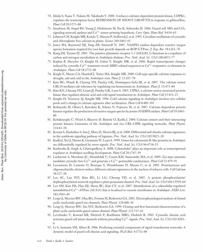
ANRV410-PP61-25 ARI 31 March 2010 17:4
70. Ishida S, Yuasa T, Nakata M, Takahashi Y. 2008. A tobacco calcium-dependent protein kinase, CDPK1,regulates the transcription factor REPRESSION OF SHOOT GROWTH in response to gibberellins.Plant Cell 20:3273–88
71. Israelsson M, Siegel RS, Young J, Hashimoto M, Iba K, Schroeder JI. 2006. Guard cell ABA and CO2
signaling network updates and Ca2+ sensor priming hypothesis. Curr. Opin. Plant Biol. 9:654–6372. Johnson CH, Knight MR, Kondo T, Masson P, Sedbrook J, et al. 1995. Circadian oscillations of cytosolic
and chloroplastic free calcium in plants. Science 269:1863–6573. Jones MA, Raymond MJ, Yang ZB, Smirnoff N. 2007. NADPH oxidase-dependent reactive oxygen
species formation required for root hair growth depends on ROP GTPase. J. Exp. Bot. 58:1261–7074. Kang JM, Turano FJ. 2003. The putative glutamate receptor 1.1 (AtGLR1.1) functions as a regulator of
carbon and nitrogen metabolism in Arabidopsis thaliana. Proc. Natl. Acad. Sci. USA 100:6872–7775. Kaplan B, Davydov O, Knight H, Galon Y, Knight MR, et al. 2006. Rapid transcriptome changes
induced by cytosolic Ca2+ transients reveal ABRE-related sequences as Ca2+-responsive cis elements inArabidopsis. Plant Cell 18:2733–48
76. Kiegle E, Moore CA, Haseloff J, Tester MA, Knight MR. 2000. Cell-type-specific calcium responses todrought, salt and cold in the Arabidopsis root. Plant J. 23:267–78
77. Kim BG, Waadt R, Cheong YH, Pandey GK, Dominguez-Solis JR, et al. 2007. The calcium sensorCBL10 mediates salt tolerance by regulating ion homeostasis in Arabidopsis. Plant J. 52:473–84
78. Kim KN, Cheong YH, Grant JJ, Pandey GK, Luan S. 2003. CIPK3, a calcium sensor-associated proteinkinase that regulates abscisic acid and cold signal transduction in Arabidopsis. Plant Cell 15:411–23
79. Knight H, Trewavas AJ, Knight MR. 1996. Cold calcium signaling in Arabidopsis involves two cellularpools and a change in calcium signature after acclimation. Plant Cell 8:489–503
80. Kobayashi M, Ohura I, Kawakita K, Yokota N, Fujiwara M, et al. 2007. Calcium-dependent proteinkinases regulate the production of reactive oxygen species by potato NADPH oxidase. Plant Cell 19:1065–80
81. Kolukisaoglu U, Weinl S, Blazevic D, Batistic O, Kudla J. 2004. Calcium sensors and their interactingprotein kinases: Genomics of the Arabidopsis and rice CBL-CIPK signaling networks. Plant Physiol.134:43–58
82. Kosuta S, Hazledine S, Sun J, Miwa H, Morris RJ, et al. 2008. Differential and chaotic calcium signaturesin the symbiosis signaling pathway of legumes. Proc. Natl. Acad. Sci. USA 105:9823–28
83. Kudla J, Xu Q, Harter K, Gruissem W, Luan S. 1999. Genes for calcineurin B–like proteins in Arabidopsisare differentially regulated by stress signals. Proc. Natl. Acad. Sci. USA 96:4718–23
84. Kushwaha R, Singh A, Chattopadhyay S. 2008. Calmodulin7 plays an important role as transcriptionalregulator in Arabidopsis seedling development. Plant Cell 20:1747–59
85. Laohavisit A, Mortimer JC, Demidchik V, Coxon KM, Stancombe MA, et al. 2009. Zea mays annexinsmodulate cytosolic free Ca2+ and generate a Ca2+-permeable conductance. Plant Cell 21:479–93
86. Lecourieux D, Lamotte O, Bourque S, Wendehenne D, Mazars C, et al. 2005. Proteinaceous andoligosaccharidic elicitors induce different calcium signatures in the nucleus of tobacco cells. Cell Calcium38:527–38
87. Lee SC, Lan WZ, Kim BG, Li LG, Cheong YH, et al. 2007. A protein phosphorylation/dephosphorylation network regulates a plant potassium channel. Proc. Natl. Acad. Sci. USA 104:15959–64
88. Lee SM, Kim HS, Han HJ, Moon BC, Kim CY, et al. 2007. Identification of a calmodulin-regulatedautoinhibited Ca2+-ATPase (ACA11) that is localized to vacuole membranes in Arabidopsis. FEBS Lett.581:3943–49
89. Leng Q, Mercier RW, Hua BG, Fromm H, Berkowitz GA. 2002. Electrophysiological analysis of clonedcyclic nucleotide-gated ion channels. Plant Physiol. 128:400–10
90. Leng Q, Mercier RW, Yao WZ, Berkowitz GA. 1999. Cloning and first functional characterization of aplant cyclic nucleotide-gated cation channel. Plant Physiol. 121:753–61
91. Levchenko V, Konrad KR, Dietrich P, Roelfsema MRG, Hedrich R. 2005. Cytosolic abscisic acidactivates guard cell anion channels without preceding Ca2+ signals. Proc. Natl. Acad. Sci. USA 102:4203–8
92. Li S, Assmann SM, Albert R. 2006. Predicting essential components of signal transduction networks: Adynamic model of guard cell abscisic acid signaling. PLoS Biol. 4:1732–48
616 Dodd · Kudla · Sanders
Ann
u. R
ev. P
lant
Bio
l. 20
10.6
1:59
3-62
0. D
ownl
oade
d fr
om a
rjou
rnal
s.an
nual
revi
ews.
org
by U
nive
rsity
of
Del
hi o
n 08
/26/
10. F
or p
erso
nal u
se o
nly.
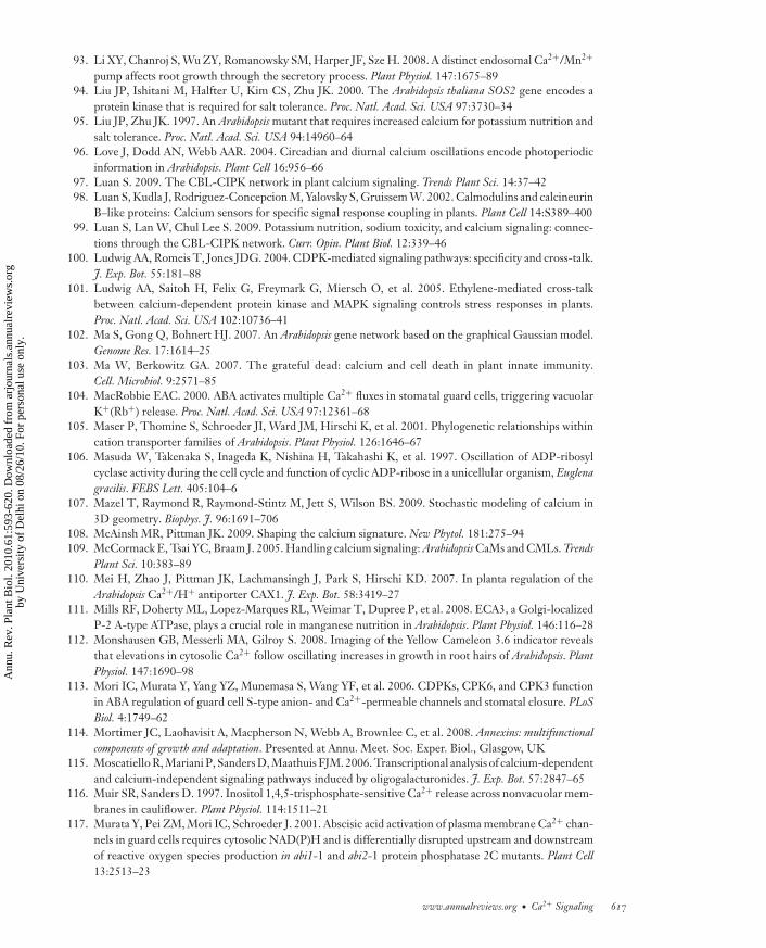
ANRV410-PP61-25 ARI 31 March 2010 17:4
93. Li XY, Chanroj S, Wu ZY, Romanowsky SM, Harper JF, Sze H. 2008. A distinct endosomal Ca2+/Mn2+
pump affects root growth through the secretory process. Plant Physiol. 147:1675–8994. Liu JP, Ishitani M, Halfter U, Kim CS, Zhu JK. 2000. The Arabidopsis thaliana SOS2 gene encodes a
protein kinase that is required for salt tolerance. Proc. Natl. Acad. Sci. USA 97:3730–3495. Liu JP, Zhu JK. 1997. An Arabidopsis mutant that requires increased calcium for potassium nutrition and
salt tolerance. Proc. Natl. Acad. Sci. USA 94:14960–6496. Love J, Dodd AN, Webb AAR. 2004. Circadian and diurnal calcium oscillations encode photoperiodic
information in Arabidopsis. Plant Cell 16:956–6697. Luan S. 2009. The CBL-CIPK network in plant calcium signaling. Trends Plant Sci. 14:37–4298. Luan S, Kudla J, Rodriguez-Concepcion M, Yalovsky S, Gruissem W. 2002. Calmodulins and calcineurin
B–like proteins: Calcium sensors for specific signal response coupling in plants. Plant Cell 14:S389–40099. Luan S, Lan W, Chul Lee S. 2009. Potassium nutrition, sodium toxicity, and calcium signaling: connec-
tions through the CBL-CIPK network. Curr. Opin. Plant Biol. 12:339–46100. Ludwig AA, Romeis T, Jones JDG. 2004. CDPK-mediated signaling pathways: specificity and cross-talk.
J. Exp. Bot. 55:181–88101. Ludwig AA, Saitoh H, Felix G, Freymark G, Miersch O, et al. 2005. Ethylene-mediated cross-talk
between calcium-dependent protein kinase and MAPK signaling controls stress responses in plants.Proc. Natl. Acad. Sci. USA 102:10736–41
102. Ma S, Gong Q, Bohnert HJ. 2007. An Arabidopsis gene network based on the graphical Gaussian model.Genome Res. 17:1614–25
103. Ma W, Berkowitz GA. 2007. The grateful dead: calcium and cell death in plant innate immunity.Cell. Microbiol. 9:2571–85
104. MacRobbie EAC. 2000. ABA activates multiple Ca2+ fluxes in stomatal guard cells, triggering vacuolarK+(Rb+) release. Proc. Natl. Acad. Sci. USA 97:12361–68
105. Maser P, Thomine S, Schroeder JI, Ward JM, Hirschi K, et al. 2001. Phylogenetic relationships withincation transporter families of Arabidopsis. Plant Physiol. 126:1646–67
106. Masuda W, Takenaka S, Inageda K, Nishina H, Takahashi K, et al. 1997. Oscillation of ADP-ribosylcyclase activity during the cell cycle and function of cyclic ADP-ribose in a unicellular organism, Euglenagracilis. FEBS Lett. 405:104–6
107. Mazel T, Raymond R, Raymond-Stintz M, Jett S, Wilson BS. 2009. Stochastic modeling of calcium in3D geometry. Biophys. J. 96:1691–706
108. McAinsh MR, Pittman JK. 2009. Shaping the calcium signature. New Phytol. 181:275–94109. McCormack E, Tsai YC, Braam J. 2005. Handling calcium signaling: Arabidopsis CaMs and CMLs. Trends
Plant Sci. 10:383–89110. Mei H, Zhao J, Pittman JK, Lachmansingh J, Park S, Hirschi KD. 2007. In planta regulation of the
Arabidopsis Ca2+/H+ antiporter CAX1. J. Exp. Bot. 58:3419–27111. Mills RF, Doherty ML, Lopez-Marques RL, Weimar T, Dupree P, et al. 2008. ECA3, a Golgi-localized
P-2 A-type ATPase, plays a crucial role in manganese nutrition in Arabidopsis. Plant Physiol. 146:116–28112. Monshausen GB, Messerli MA, Gilroy S. 2008. Imaging of the Yellow Cameleon 3.6 indicator reveals
that elevations in cytosolic Ca2+ follow oscillating increases in growth in root hairs of Arabidopsis. PlantPhysiol. 147:1690–98
113. Mori IC, Murata Y, Yang YZ, Munemasa S, Wang YF, et al. 2006. CDPKs, CPK6, and CPK3 functionin ABA regulation of guard cell S-type anion- and Ca2+-permeable channels and stomatal closure. PLoSBiol. 4:1749–62
114. Mortimer JC, Laohavisit A, Macpherson N, Webb A, Brownlee C, et al. 2008. Annexins: multifunctionalcomponents of growth and adaptation. Presented at Annu. Meet. Soc. Exper. Biol., Glasgow, UK
115. Moscatiello R, Mariani P, Sanders D, Maathuis FJM. 2006. Transcriptional analysis of calcium-dependentand calcium-independent signaling pathways induced by oligogalacturonides. J. Exp. Bot. 57:2847–65
116. Muir SR, Sanders D. 1997. Inositol 1,4,5-trisphosphate-sensitive Ca2+ release across nonvacuolar mem-branes in cauliflower. Plant Physiol. 114:1511–21
117. Murata Y, Pei ZM, Mori IC, Schroeder J. 2001. Abscisic acid activation of plasma membrane Ca2+ chan-nels in guard cells requires cytosolic NAD(P)H and is differentially disrupted upstream and downstreamof reactive oxygen species production in abi1-1 and abi2-1 protein phosphatase 2C mutants. Plant Cell13:2513–23
www.annualreviews.org • Ca2+ Signaling 617
Ann
u. R
ev. P
lant
Bio
l. 20
10.6
1:59
3-62
0. D
ownl
oade
d fr
om a
rjou
rnal
s.an
nual
revi
ews.
org
by U
nive
rsity
of
Del
hi o
n 08
/26/
10. F
or p
erso
nal u
se o
nly.

ANRV410-PP61-25 ARI 31 March 2010 17:4
118. Navazio L, Bewell MA, Siddiqua A, Dickinson GD, Galione A, Sanders D. 2000. Calcium release fromthe endoplasmic reticulum of higher plants elicited by the NADP metabolite nicotinic acid adeninedinucleotide phosphate. Proc. Natl. Acad. Sci. USA 97:8693–98
119. Navazio L, Mariani P, Sanders D. 2001. Mobilization of Ca2+ by cyclic ADP-ribose from the endoplasmicreticulum of cauliflower florets. Plant Physiol. 125:2129–38
120. Navazio L, Moscatiello R, Genre A, Novero M, Baldan B, et al. 2007. A diffusible signal from arbuscularmycorrhizal fungi elicits a transient cytosolic calcium elevation in host plant cells. Plant Physiol. 144:673–81
121. Ng CKY, McAinsh MR, Gray JE, Hunt L, Leckie CP, et al. 2001. Calcium-based signaling systems inguard cells. New Phytol. 151:109–20
122. Ohta M, Guo Y, Halfter U, Zhu JK. 2003. A novel domain in the protein kinase SOS2 mediates interactionwith the protein phosphatase 2C AB12. Proc. Natl. Acad. Sci. USA 100:11771–76
123. Oldroyd GED, Downie JA. 2008. Coordinating nodule morphogenesis with rhizobial infection inlegumes. Annu. Rev. Plant Biol. 59:519–46
124. Oldroyd GED, Harrison MJ, Paszkowski U. 2009. Reprogramming plant cells for endosymbiosis. Science324:753–54
125. Pandey GK, Cheong YH, Kim KN, Grant JJ, Li LG, et al. 2004. The calcium sensor calcineurin B–likeCBL9 modulates abscisic acid sensitivity and biosynthesis in Arabidopsis. Plant Cell 16:1912–24
126. Pandey GK, Grant JJ, Cheong YH, Kim BG, Li LG, Luan S. 2008. Calcineurin-B-like protein CBL9interacts with target kinase CIPK3 in the regulation of ABA response in seed germination. Mol. Plant1:238–48
127. Pei ZM, Murata Y, Benning G, Thomine S, Klusener B, et al. 2000. Calcium channels activated byhydrogen peroxide mediate abscisic acid signaling in guard cells. Nature 406:731–34
128. Peiter E, Maathuis FJM, Mills LN, Knight H, Pelloux J, et al. 2005. The vacuolar Ca2+-activated channelTPC1 regulates germination and stomatal movement. Nature 434:404–8
129. Peiter E, Sun J, Heckmann AB, Venkateshwaran M, Riely BK, et al. 2007. The Medicago truncatula DMI1protein modulates cytosolic calcium signaling. Plant Physiol. 145:192–203
130. Polouliakh N, Nock R, Nielsen F, Kitano H. 2009. G-protein coupled receptor signaling architectureof mammalian immune cells. PLoS One 4:e4189
131. Pottosin I, Wherrett T, Shabala S. 2009. SV channels dominate the vacuolar Ca2+ release during intra-cellular signaling. FEBS Lett. 583:921–26
132. Qi Z, Stephens NR, Spalding EP. 2006. Calcium entry mediated by GLR3.3, an Arabidopsis glutamatereceptor with a broad agonist profile. Plant Physiol. 142:963–71
133. Quan RD, Lin HX, Mendoza I, Zhang YG, Cao WH, et al. 2007. SCABP8/CBL10, a putative calciumsensor, interacts with the protein kinase SOS2 to protect Arabidopsis shoots from salt stress. Plant Cell19:1415–31
134. Qudeimat E, Faltusz AMC, Wheeler G, Lang D, Brownlee C, et al. 2008. A P-IIB-type Ca2+-ATPaseis essential for stress adaptation in Physcomitrella patens. Proc. Natl. Acad. Sci. USA 105:19555–60
135. Ranf S, Wunnenberg P, Lee J, Becker D, Dunkel M, et al. 2008. Loss of the vacuolar cation channel,AtTPC1, does not impair Ca2+ signals induced by abiotic and biotic stresses. Plant J. 53:287–99
136. Reddy VS, Reddy ASN. 2004. Proteomics of calcium-signaling components in plants. Phytochemistry65:1745–76
137. Romeis T, Ludwig AA, Martin R, Jones JDG. 2001. Calcium-dependent protein kinases play an essentialrole in a plant defense response. EMBO J. 20:5556–67
138. Sanchez JP, Duque P, Chua NH. 2004. ABA activates ADPR cyclase and cADPR induces a subset ofABA-responsive genes in Arabidopsis. Plant J. 38:381–95
139. Sanders D, Brownlee C, Harper JF. 1999. Communicating with calcium. Plant Cell 11:691–706140. Sanders D, Johannes E, Hedrich R. 1990. Opening plant calcium channels. Nature 344:593–94141. Sanders D, Pelloux J, Brownlee C, Harper JF. 2002. Calcium at the crossroads of signaling. Plant Cell
14:S401–17142. Schiott M, Palmgren MG. 2005. Two plant Ca2+ pumps expressed in stomatal guard cells show opposite
expression patterns during cold stress. Physiol. Plantarum 124:278–83
618 Dodd · Kudla · Sanders
Ann
u. R
ev. P
lant
Bio
l. 20
10.6
1:59
3-62
0. D
ownl
oade
d fr
om a
rjou
rnal
s.an
nual
revi
ews.
org
by U
nive
rsity
of
Del
hi o
n 08
/26/
10. F
or p
erso
nal u
se o
nly.
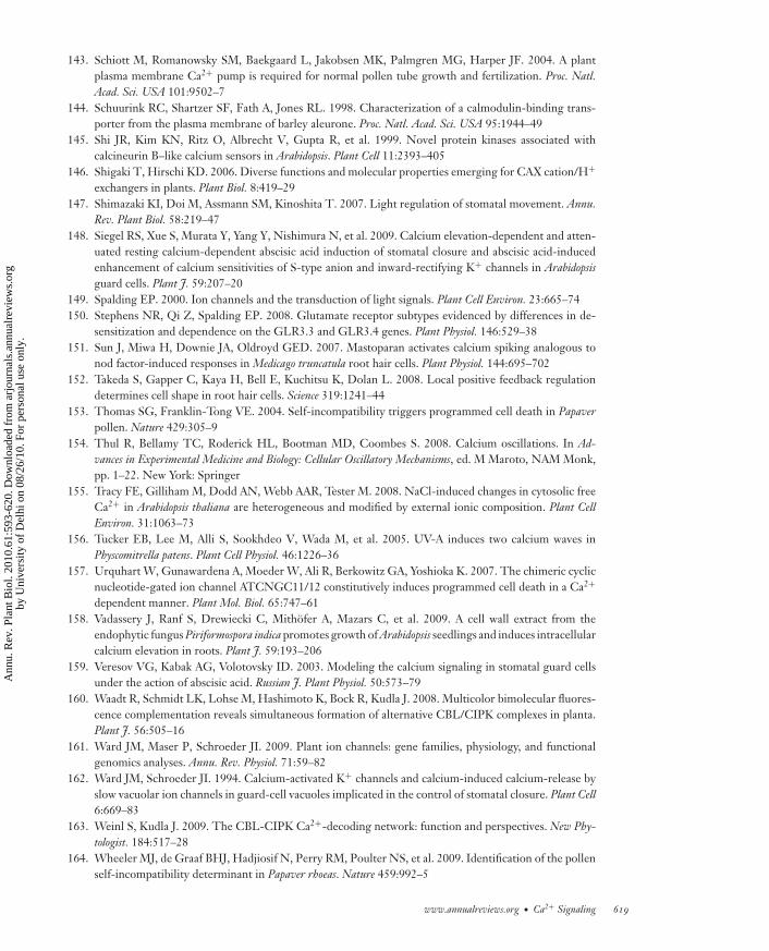
ANRV410-PP61-25 ARI 31 March 2010 17:4
143. Schiott M, Romanowsky SM, Baekgaard L, Jakobsen MK, Palmgren MG, Harper JF. 2004. A plantplasma membrane Ca2+ pump is required for normal pollen tube growth and fertilization. Proc. Natl.Acad. Sci. USA 101:9502–7
144. Schuurink RC, Shartzer SF, Fath A, Jones RL. 1998. Characterization of a calmodulin-binding trans-porter from the plasma membrane of barley aleurone. Proc. Natl. Acad. Sci. USA 95:1944–49
145. Shi JR, Kim KN, Ritz O, Albrecht V, Gupta R, et al. 1999. Novel protein kinases associated withcalcineurin B–like calcium sensors in Arabidopsis. Plant Cell 11:2393–405
146. Shigaki T, Hirschi KD. 2006. Diverse functions and molecular properties emerging for CAX cation/H+
exchangers in plants. Plant Biol. 8:419–29147. Shimazaki KI, Doi M, Assmann SM, Kinoshita T. 2007. Light regulation of stomatal movement. Annu.
Rev. Plant Biol. 58:219–47148. Siegel RS, Xue S, Murata Y, Yang Y, Nishimura N, et al. 2009. Calcium elevation-dependent and atten-
uated resting calcium-dependent abscisic acid induction of stomatal closure and abscisic acid-inducedenhancement of calcium sensitivities of S-type anion and inward-rectifying K+ channels in Arabidopsisguard cells. Plant J. 59:207–20
149. Spalding EP. 2000. Ion channels and the transduction of light signals. Plant Cell Environ. 23:665–74150. Stephens NR, Qi Z, Spalding EP. 2008. Glutamate receptor subtypes evidenced by differences in de-
sensitization and dependence on the GLR3.3 and GLR3.4 genes. Plant Physiol. 146:529–38151. Sun J, Miwa H, Downie JA, Oldroyd GED. 2007. Mastoparan activates calcium spiking analogous to
nod factor-induced responses in Medicago truncatula root hair cells. Plant Physiol. 144:695–702152. Takeda S, Gapper C, Kaya H, Bell E, Kuchitsu K, Dolan L. 2008. Local positive feedback regulation
determines cell shape in root hair cells. Science 319:1241–44153. Thomas SG, Franklin-Tong VE. 2004. Self-incompatibility triggers programmed cell death in Papaver
pollen. Nature 429:305–9154. Thul R, Bellamy TC, Roderick HL, Bootman MD, Coombes S. 2008. Calcium oscillations. In Ad-
vances in Experimental Medicine and Biology: Cellular Oscillatory Mechanisms, ed. M Maroto, NAM Monk,pp. 1–22. New York: Springer
155. Tracy FE, Gilliham M, Dodd AN, Webb AAR, Tester M. 2008. NaCl-induced changes in cytosolic freeCa2+ in Arabidopsis thaliana are heterogeneous and modified by external ionic composition. Plant CellEnviron. 31:1063–73
156. Tucker EB, Lee M, Alli S, Sookhdeo V, Wada M, et al. 2005. UV-A induces two calcium waves inPhyscomitrella patens. Plant Cell Physiol. 46:1226–36
157. Urquhart W, Gunawardena A, Moeder W, Ali R, Berkowitz GA, Yoshioka K. 2007. The chimeric cyclicnucleotide-gated ion channel ATCNGC11/12 constitutively induces programmed cell death in a Ca2+
dependent manner. Plant Mol. Biol. 65:747–61158. Vadassery J, Ranf S, Drewiecki C, Mithofer A, Mazars C, et al. 2009. A cell wall extract from the
endophytic fungus Piriformospora indica promotes growth of Arabidopsis seedlings and induces intracellularcalcium elevation in roots. Plant J. 59:193–206
159. Veresov VG, Kabak AG, Volotovsky ID. 2003. Modeling the calcium signaling in stomatal guard cellsunder the action of abscisic acid. Russian J. Plant Physiol. 50:573–79
160. Waadt R, Schmidt LK, Lohse M, Hashimoto K, Bock R, Kudla J. 2008. Multicolor bimolecular fluores-cence complementation reveals simultaneous formation of alternative CBL/CIPK complexes in planta.Plant J. 56:505–16
161. Ward JM, Maser P, Schroeder JI. 2009. Plant ion channels: gene families, physiology, and functionalgenomics analyses. Annu. Rev. Physiol. 71:59–82
162. Ward JM, Schroeder JI. 1994. Calcium-activated K+ channels and calcium-induced calcium-release byslow vacuolar ion channels in guard-cell vacuoles implicated in the control of stomatal closure. Plant Cell6:669–83
163. Weinl S, Kudla J. 2009. The CBL-CIPK Ca2+-decoding network: function and perspectives. New Phy-tologist. 184:517–28
164. Wheeler MJ, de Graaf BHJ, Hadjiosif N, Perry RM, Poulter NS, et al. 2009. Identification of the pollenself-incompatibility determinant in Papaver rhoeas. Nature 459:992–5
www.annualreviews.org • Ca2+ Signaling 619
Ann
u. R
ev. P
lant
Bio
l. 20
10.6
1:59
3-62
0. D
ownl
oade
d fr
om a
rjou
rnal
s.an
nual
revi
ews.
org
by U
nive
rsity
of
Del
hi o
n 08
/26/
10. F
or p
erso
nal u
se o
nly.
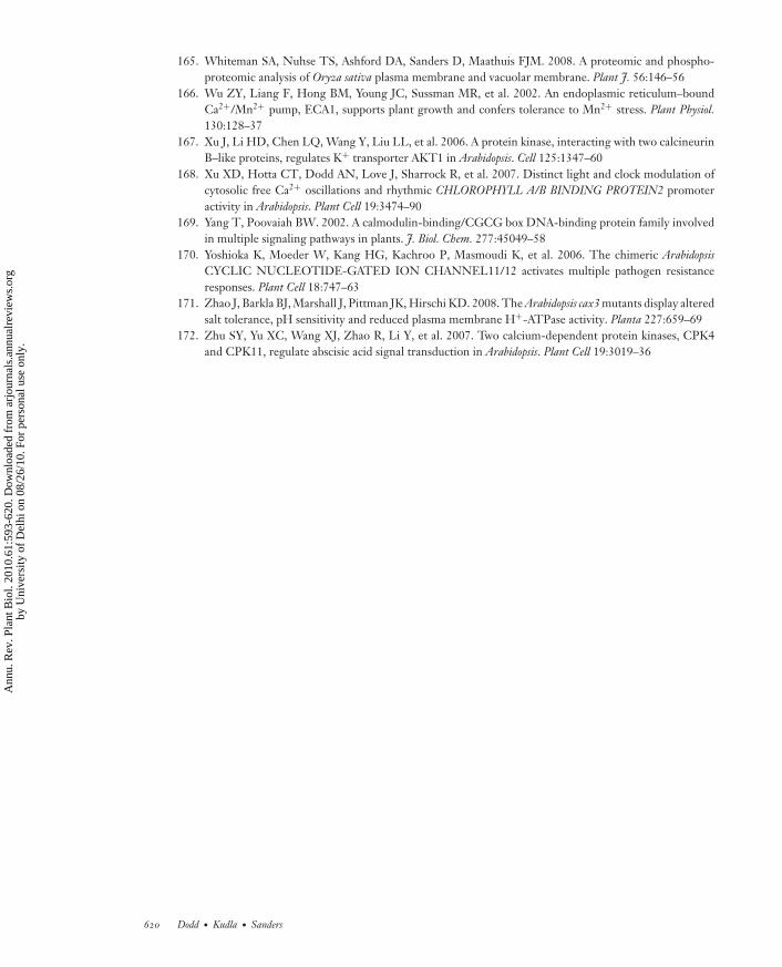
ANRV410-PP61-25 ARI 31 March 2010 17:4
165. Whiteman SA, Nuhse TS, Ashford DA, Sanders D, Maathuis FJM. 2008. A proteomic and phospho-proteomic analysis of Oryza sativa plasma membrane and vacuolar membrane. Plant J. 56:146–56
166. Wu ZY, Liang F, Hong BM, Young JC, Sussman MR, et al. 2002. An endoplasmic reticulum–boundCa2+/Mn2+ pump, ECA1, supports plant growth and confers tolerance to Mn2+ stress. Plant Physiol.130:128–37
167. Xu J, Li HD, Chen LQ, Wang Y, Liu LL, et al. 2006. A protein kinase, interacting with two calcineurinB–like proteins, regulates K+ transporter AKT1 in Arabidopsis. Cell 125:1347–60
168. Xu XD, Hotta CT, Dodd AN, Love J, Sharrock R, et al. 2007. Distinct light and clock modulation ofcytosolic free Ca2+ oscillations and rhythmic CHLOROPHYLL A/B BINDING PROTEIN2 promoteractivity in Arabidopsis. Plant Cell 19:3474–90
169. Yang T, Poovaiah BW. 2002. A calmodulin-binding/CGCG box DNA-binding protein family involvedin multiple signaling pathways in plants. J. Biol. Chem. 277:45049–58
170. Yoshioka K, Moeder W, Kang HG, Kachroo P, Masmoudi K, et al. 2006. The chimeric ArabidopsisCYCLIC NUCLEOTIDE-GATED ION CHANNEL11/12 activates multiple pathogen resistanceresponses. Plant Cell 18:747–63
171. Zhao J, Barkla BJ, Marshall J, Pittman JK, Hirschi KD. 2008. The Arabidopsis cax3 mutants display alteredsalt tolerance, pH sensitivity and reduced plasma membrane H+-ATPase activity. Planta 227:659–69
172. Zhu SY, Yu XC, Wang XJ, Zhao R, Li Y, et al. 2007. Two calcium-dependent protein kinases, CPK4and CPK11, regulate abscisic acid signal transduction in Arabidopsis. Plant Cell 19:3019–36
620 Dodd · Kudla · Sanders
Ann
u. R
ev. P
lant
Bio
l. 20
10.6
1:59
3-62
0. D
ownl
oade
d fr
om a
rjou
rnal
s.an
nual
revi
ews.
org
by U
nive
rsity
of
Del
hi o
n 08
/26/
10. F
or p
erso
nal u
se o
nly.
AR410-FM ARI 6 April 2010 15:25
Annual Review ofPlant Biology
Volume 61, 2010Contents
A Wandering Pathway in Plant Biology: From Wildflowers toPhototropins to Bacterial VirulenceWinslow R. Briggs � � � � � � � � � � � � � � � � � � � � � � � � � � � � � � � � � � � � � � � � � � � � � � � � � � � � � � � � � � � � � � � � � � � � � � � � � � � � � � 1
Structure and Function of Plant PhotoreceptorsAndreas Moglich, Xiaojing Yang, Rebecca A. Ayers, and Keith Moffat � � � � � � � � � � � � � � � � � � � � �21
Auxin Biosynthesis and Its Role in Plant DevelopmentYunde Zhao � � � � � � � � � � � � � � � � � � � � � � � � � � � � � � � � � � � � � � � � � � � � � � � � � � � � � � � � � � � � � � � � � � � � � � � � � � � � � � � � � � � �49
Computational Morphodynamics: A Modeling Framework toUnderstand Plant GrowthVijay Chickarmane, Adrienne H.K. Roeder, Paul T. Tarr, Alexandre Cunha,
Cory Tobin, and Elliot M. Meyerowitz � � � � � � � � � � � � � � � � � � � � � � � � � � � � � � � � � � � � � � � � � � � � � � � � � � � � �65
Female Gametophyte Development in Flowering PlantsWei-Cai Yang, Dong-Qiao Shi, and Yan-Hong Chen � � � � � � � � � � � � � � � � � � � � � � � � � � � � � � � � � � � � � � �89
Doomed Lovers: Mechanisms of Isolation and Incompatibility in PlantsKirsten Bomblies � � � � � � � � � � � � � � � � � � � � � � � � � � � � � � � � � � � � � � � � � � � � � � � � � � � � � � � � � � � � � � � � � � � � � � � � � � � � � 109
Chloroplast RNA MetabolismDavid B. Stern, Michel Goldschmidt-Clermont, and Maureen R. Hanson � � � � � � � � � � � � � � 125
Protein Transport into ChloroplastsHsou-min Li and Chi-Chou Chiu � � � � � � � � � � � � � � � � � � � � � � � � � � � � � � � � � � � � � � � � � � � � � � � � � � � � � � � � � � 157
The Regulation of Gene Expression Required for C4 PhotosynthesisJulian M. Hibberd and Sarah Covshoff � � � � � � � � � � � � � � � � � � � � � � � � � � � � � � � � � � � � � � � � � � � � � � � � � � � � 181
Starch: Its Metabolism, Evolution, and Biotechnological Modificationin PlantsSamuel C. Zeeman, Jens Kossmann, and Alison M. Smith � � � � � � � � � � � � � � � � � � � � � � � � � � � � � � � 209
Improving Photosynthetic Efficiency for Greater YieldXin-Guang Zhu, Stephen P. Long, and Donald R. Ort � � � � � � � � � � � � � � � � � � � � � � � � � � � � � � � � � � � 235
HemicellulosesHenrik Vibe Scheller and Peter Ulvskov � � � � � � � � � � � � � � � � � � � � � � � � � � � � � � � � � � � � � � � � � � � � � � � � � � � 263
Diversification of P450 Genes During Land Plant EvolutionMasaharu Mizutani and Daisaku Ohta � � � � � � � � � � � � � � � � � � � � � � � � � � � � � � � � � � � � � � � � � � � � � � � � � � � 291
v
Ann
u. R
ev. P
lant
Bio
l. 20
10.6
1:59
3-62
0. D
ownl
oade
d fr
om a
rjou
rnal
s.an
nual
revi
ews.
org
by U
nive
rsity
of
Del
hi o
n 08
/26/
10. F
or p
erso
nal u
se o
nly.
AR410-FM ARI 6 April 2010 15:25
Evolution in Action: Plants Resistant to HerbicidesStephen B. Powles and Qin Yu � � � � � � � � � � � � � � � � � � � � � � � � � � � � � � � � � � � � � � � � � � � � � � � � � � � � � � � � � � � � � � 317
Insights from the Comparison of Plant Genome SequencesAndrew H. Paterson, Michael Freeling, Haibao Tang, and Xiyin Wang � � � � � � � � � � � � � � � � 349
High-Throughput Characterization of Plant Gene Functions by UsingGain-of-Function TechnologyYouichi Kondou, Mieko Higuchi, and Minami Matsui � � � � � � � � � � � � � � � � � � � � � � � � � � � � � � � � � � � � 373
Histone Methylation in Higher PlantsChunyan Liu, Falong Lu, Xia Cui, and Xiaofeng Cao � � � � � � � � � � � � � � � � � � � � � � � � � � � � � � � � � � � � 395
Genetic and Molecular Basis of Rice YieldYongzhong Xing and Qifa Zhang � � � � � � � � � � � � � � � � � � � � � � � � � � � � � � � � � � � � � � � � � � � � � � � � � � � � � � � � � � 421
Genetic Engineering for Modern Agriculture: Challenges andPerspectivesRon Mittler and Eduardo Blumwald � � � � � � � � � � � � � � � � � � � � � � � � � � � � � � � � � � � � � � � � � � � � � � � � � � � � � � � 443
Metabolomics for Functional Genomics, Systems Biology, andBiotechnologyKazuki Saito and Fumio Matsuda � � � � � � � � � � � � � � � � � � � � � � � � � � � � � � � � � � � � � � � � � � � � � � � � � � � � � � � � � 463
Quantitation in Mass-Spectrometry-Based ProteomicsWaltraud X. Schulze and Bjorn Usadel � � � � � � � � � � � � � � � � � � � � � � � � � � � � � � � � � � � � � � � � � � � � � � � � � � � � 491
Metal Hyperaccumulation in PlantsUte Kramer � � � � � � � � � � � � � � � � � � � � � � � � � � � � � � � � � � � � � � � � � � � � � � � � � � � � � � � � � � � � � � � � � � � � � � � � � � � � � � � � � � 517
Arsenic as a Food Chain Contaminant: Mechanisms of Plant Uptakeand Metabolism and Mitigation StrategiesFang-Jie Zhao, Steve P. McGrath, and Andrew A. Meharg � � � � � � � � � � � � � � � � � � � � � � � � � � � � � 535
Guard Cell Signal Transduction Network: Advances in UnderstandingAbscisic Acid, CO2, and Ca2+ SignalingTae-Houn Kim, Maik Bohmer, Honghong Hu, Noriyuki Nishimura,
and Julian I. Schroeder � � � � � � � � � � � � � � � � � � � � � � � � � � � � � � � � � � � � � � � � � � � � � � � � � � � � � � � � � � � � � � � � � � � 561
The Language of Calcium SignalingAntony N. Dodd, Jorg Kudla, and Dale Sanders � � � � � � � � � � � � � � � � � � � � � � � � � � � � � � � � � � � � � � � � � � 593
Mitogen-Activated Protein Kinase Signaling in PlantsMaria Cristina Suarez Rodriguez, Morten Petersen, and John Mundy � � � � � � � � � � � � � � � � � 621
Abscisic Acid: Emergence of a Core Signaling NetworkSean R. Cutler, Pedro L. Rodriguez, Ruth R. Finkelstein, and Suzanne R. Abrams � � � � 651
Brassinosteroid Signal Transduction from Receptor Kinases toTranscription FactorsTae-Wuk Kim and Zhi-Yong Wang � � � � � � � � � � � � � � � � � � � � � � � � � � � � � � � � � � � � � � � � � � � � � � � � � � � � � � � � 681
vi Contents
Ann
u. R
ev. P
lant
Bio
l. 20
10.6
1:59
3-62
0. D
ownl
oade
d fr
om a
rjou
rnal
s.an
nual
revi
ews.
org
by U
nive
rsity
of
Del
hi o
n 08
/26/
10. F
or p
erso
nal u
se o
nly.

AR410-FM ARI 6 April 2010 15:25
Directional Gravity Sensing in GravitropismMiyo Terao Morita � � � � � � � � � � � � � � � � � � � � � � � � � � � � � � � � � � � � � � � � � � � � � � � � � � � � � � � � � � � � � � � � � � � � � � � � � � 705
Indexes
Cumulative Index of Contributing Authors, Volumes 51–61 � � � � � � � � � � � � � � � � � � � � � � � � � � � 721
Cumulative Index of Chapter Titles, Volumes 51–61 � � � � � � � � � � � � � � � � � � � � � � � � � � � � � � � � � � � � 726
Errata
An online log of corrections to Annual Review of Plant Biology articles may be found athttp://plant.annualreviews.org
Contents vii
Ann
u. R
ev. P
lant
Bio
l. 20
10.6
1:59
3-62
0. D
ownl
oade
d fr
om a
rjou
rnal
s.an
nual
revi
ews.
org
by U
nive
rsity
of
Del
hi o
n 08
/26/
10. F
or p
erso
nal u
se o
nly.