Store-operated calcium entry in neuroglia
9
Neurosci Bull February 1, 2014, 30(1): 125–133. http://www.neurosci.cn DOI: 10.1007/s12264-013-1343-x 125 ·Review· Store-operated calcium entry in neuroglia Alexei Verkhratsky 1,2 , Vladimir Parpura 3,4 1 Faculty of Life Sciences, The University of Manchester, Manchester, M13 9PT, UK 2 Achucarro Center for Neuroscience, IKERBASQUE, Basque Foundation for Science, 48011, Bilbao, Spain; and Department of Neurosciences, University of the Basque Country UPV/EHU, 48940, Leioa, Spain 3 Department of Neurobiology, Center for Glial Biology in Medicine, Atomic Force Microscopy & Nanotechnology Laboratories, Civitan International Research Center, Evelyn F. McKnight Brain Institute, University of Alabama, Birmingham, AL 35294, USA 4 Department of Biotechnology, University or Rijeka, 51000 Rijeka, Croatia Corresponding authors: Alexei Verkhratsky and Vladimir Parpura. E-mail: [email protected], vlad@ uab.edu © Shanghai Institutes for Biological Sciences, CAS and Springer-Verlag Berlin Heidelberg 2014 Neuroglial cells are homeostatic neural cells. Generally, they are electrically non-excitable and their activation is associated with the generation of complex intracellular Ca 2+ signals that define the “Ca 2+ excitability” of glia. In mammalian glial cells the major source of Ca 2+ for this excitability is the lumen of the endoplasmic reticulum (ER), which is ultimately (re)filled from the extracellular space. This occurs via store-operated Ca 2+ entry (SOCE) which is supported by a specific signaling system connecting the ER with plasmalemmal Ca 2+ entry. Here, emptying of the ER Ca 2+ store is necessary and sufficient for the activation of SOCE, and without Ca 2+ influx via SOCE the ER store cannot be refilled. The molecular arrangements underlying SOCE are relatively complex and include plasmalemmal channels, ER Ca 2+ sensors, such as stromal interaction molecule, and possibly ER Ca 2+ pumps (of the SERCA type). There are at least two sets of plasmalemmal channels mediating SOCE, the Ca 2+ -release activated channels, Orai, and transient receptor potential (TRP) channels. The molecular identity of neuroglial SOCE has not been yet identified unequivocally. However, it seems that Orai is predominantly expressed in microglia, whereas astrocytes and oligodendrocytes rely more on TRP channels to produce SOCE. In physiological conditions the SOCE pathway is instrumental for the sustained phase of the Ca 2+ signal observed following stimulation of metabotropic receptors on glial cells. Keywords: calcium signaling; astrocyte; oligodendrocyte; microglia; store-operated calcium entry; TRP; STIM; Orai Store-Operated Calcium Entry: The Concept The spatio-temporal organization of intracellular Ca 2+ signals, which regulate a wide variety of cellular functions, results from tightly coordinated fluxes of Ca 2+ through intracellular and plasmalemmal membranes. These fluxes are mediated by several families of Ca 2+ -permeable membrane channels, and Ca 2+ transporters powered by ATP hydrolysis (Ca 2+ pumps) or by transmembrane ionic concentration gradients (Ca 2+ exchangers). The relative contribution of different types of Ca 2+ fluxes depends on the physiological context and defines the remarkable versatility of the resulting Ca 2+ signals. Plasmalemmal Ca 2+ channels can be activated by different factors. In many excitable cells, plasmalemmal Ca 2+ influx mainly occurs through ligand-gated Ca 2+ channels (also known as ionotropic receptors) activated by extracellular transmitters such as glutamate or acetylcholine
-
Upload
manchester -
Category
Documents
-
view
7 -
download
0
Transcript of Store-operated calcium entry in neuroglia

Neurosci Bull February 1, 2014, 30(1): 125–133. http://www.neurosci.cnDOI: 10.1007/s12264-013-1343-x 125
·Review·
Store-operated calcium entry in neuroglia Alexei Verkhratsky1,2, Vladimir Parpura3,4 1Faculty of Life Sciences, The University of Manchester, Manchester, M13 9PT, UK2Achucarro Center for Neuroscience, IKERBASQUE, Basque Foundation for Science, 48011, Bilbao, Spain; and
Department of Neurosciences, University of the Basque Country UPV/EHU, 48940, Leioa, Spain3Department of Neurobiology, Center for Glial Biology in Medicine, Atomic Force Microscopy & Nanotechnology
Laboratories, Civitan International Research Center, Evelyn F. McKnight Brain Institute, University of Alabama, Birmingham,
AL 35294, USA 4Department of Biotechnology, University or Rijeka, 51000 Rijeka, Croatia
Corresponding authors: Alexei Verkhratsky and Vladimir Parpura. E-mail: [email protected], [email protected]
© Shanghai Institutes for Biological Sciences, CAS and Springer-Verlag Berlin Heidelberg 2014
Neuroglial cells are homeostatic neural cells. Generally, they are electrically non-excitable and their activation is associated with the generation of complex intracellular Ca2+ signals that defi ne the “Ca2+ excitability” of glia. In mammalian glial cells the major source of Ca2+ for this excitability is the lumen of the endoplasmic reticulum (ER), which is ultimately (re)fi lled from the extracellular space. This occurs via store-operated Ca2+ entry (SOCE) which is supported by a specific signaling system connecting the ER with plasmalemmal Ca2+ entry. Here, emptying of the ER Ca2+ store is necessary and suffi cient for the activation of SOCE, and without Ca2+ infl ux via SOCE the ER store cannot be refi lled. The molecular arrangements underlying SOCE are relatively complex and include plasmalemmal channels, ER Ca2+ sensors, such as stromal interaction molecule, and possibly ER Ca2+ pumps (of the SERCA type). There are at least two sets of plasmalemmal channels mediating SOCE, the Ca2+-release activated channels, Orai, and transient receptor potential (TRP) channels. The molecular identity of neuroglial SOCE has not been yet identifi ed unequivocally. However, it seems that Orai is predominantly expressed in microglia, whereas astrocytes and oligodendrocytes rely more on TRP channels to produce SOCE. In physiological conditions the SOCE pathway is instrumental for the sustained phase of the Ca2+ signal observed following stimulation of metabotropic receptors on glial cells.
Keywords: calcium signaling; astrocyte; oligodendrocyte; microglia; store-operated calcium entry; TRP; STIM; Orai
Store-Operated Calcium Entry: The Concept
The spatio-temporal organization of intracellular Ca2+ signals, which regulate a wide variety of cellular functions, results from tightly coordinated fluxes of Ca2+ through intracellular and plasmalemmal membranes. These fl uxes are mediated by several families of Ca2+-permeable membrane channels, and Ca2+ transporters powered by ATP hydrolysis (Ca2+ pumps) or by transmembrane ionic
concentration gradients (Ca2+ exchangers). The relative contribution of different types of Ca2+ fl uxes depends on the physiological context and defi nes the remarkable versatility of the resulting Ca2+ signals.
Plasmalemmal Ca2+ channels can be activated by different factors. In many excitable cells, plasmalemmal Ca2+ influx mainly occurs through ligand-gated Ca2+ channels (also known as ionotropic receptors) activated by extracellular transmitters such as glutamate or acetylcholine

Neurosci Bull February 1, 2014, 30(1): 125–133126
and through voltage-gated Ca2+ channels gated by changes in membrane potential. In some excitable cells, activation of voltage-gated Ca2+ channels triggers massive release of Ca2+ from intracellular stores; the trigger mechanisms rely either on direct coupling between plasmalemmal Ca2+ channels and type 1 ryanodine receptors localized in the endoplasmic reticulum (ER) membrane (this mechanism underlies electro-mechanical coupling in skeletal muscle) or on an initial fl ux of Ca2+ ions that is positively amplifi ed by Ca2+-dependent opening of type 2 and 3 ryanodine receptors (this pathway dominates in cardiomyocytes and is expressed in neuronal and secretory cells). In electrically non-excitable cells, the predominant mechanism for Ca2+ signal generation involves a specific transduction system in which activation of metabotropic receptors results in synthesis of the second messenger inositol 1,4,5 trisphosphate (InsP3), which subsequently activates Ca2+ channels (generally known as InsP3 receptors) expressed in the ER membrane that results in Ca2+ release. The ER Ca2+ source is, however, finite and can be depleted, this depletion being a trigger for opening of the distinct set of plasmalemmal Ca2+ channels that mediate a store-operated Ca2+ entry (SOCE).
The concept of a specifi c signaling system connecting the ER and plasmalemmal Ca2+ entry pathways emerged in the late 1970s[1-3] and was formalized by Jim Putney within the next decade[4,5]. This concept postulated that: (1) the emptying of the ER Ca2+ store is necessary and suffi cient for the activation of SOCE; (2) without Ca2+ infl ux via SOCE the ER store cannot be refilled; and (3) in physiological conditions the SOCE pathway is instrumental for the sustained phase of the Ca2+ signal observed following stimulation of metabotropic receptors. Initially, the SOCE pathway was named “capacitative Ca2+ entry”[5], due to certain similarities with the electrical circuitry comprising a resistor (the Ca2+ channel) and a capacitor (the Ca2+ store). Incidentally, Jim Putney also suggested that the transduction mechanism required a direct link between the ER membrane and the plasmalemma, this suggestion being confi rmed in recent experiments.
Molecular Physiology of SOCE
The molecular arrangements underlying SOCE are relatively complex and include plasmalemmal channels, ER
Ca2+ sensors, and possibly ER Ca2+ pumps (of the SERCA type). All this machinery is localized at ER-to-plasma membrane (PM) junctions that provide the morphological substrate for the ER-PM link (see Fig.1 and[6-10]). At least two sets of plasmalemmal channels mediate SOCE, the Ca2+-release activated channels mediating the current known as ICRAC
[11-13] and transient receptor potential (TRP) channels[14-16]. It is now generally accepted that the ICRAC channel is formed by a family of homologous proteins, named Orai (after the Greek goddesses, who were the keepers of heaven’s gate[17]). Three of these proteins (Orai 1 to 3) have been identifi ed in vertebrates, with each protein being able to form the plasmalemmal channel[17-19]. The Ca2+ sensor monitoring intra(luminal)-ER Ca2+ concentration has been associated with the stromal interaction molecule (STIM), of which two homologues, STIM1 and STIM2, are expressed in vertebrates[20-22]. Co-expression of Orai and STIM1 results in the appearance of a large ICRAC
[23]. The Ca2+-sensing properties of STIM1 are associated with two EF-hand Ca2+ binding motifs localized at the N-terminus that faces the lumen of the ER. At ER Ca2+ levels above ~400 μmol/L, the EF motifs are assembled with the so-called sterile α-motif (SAM) domain. Decrease in the ER Ca2+ content results in dissociation of Ca2+ from this EF-hand motif and unfolds the EF-SAM complex, which triggers the oligomerization of STIM1 monomers and initiates the migration of STIM1 oligomers close to the plasmalemma where they form distinct puncta, the loci of their interaction with and activation of Orai Ca2+ channels (see for example[8] for details and further references). Replenishment of the ER store with Ca2+ triggers the opposite process, which results in dissociation of the STIM1 complex from Orai and inactivation of ICRAC. The clusters of STIM1 and Orai also co-localize with SERCA pumps; this complex apparently ensures the preferential uptake of SOCE-originated Ca2+ into the ER lumen. The functional activity of the entire complex is also controlled by mitochondria that may regulate the amplitude of the ICRAC and the degree of Ca2+ accumulation by the ER and mitochondria[24,25].
TRP channels, however, may also contribute to the SOCE mechanism. In particular, STIM1 can interact with several TRPC isoforms and activate TRPC1 and TRPC3 channels[7,26-28]. The TRP-mediated currents are of course very different from ICRAC in their gating, conductance and permeation; TRP channels are cation-permeable and do not have strict Ca2+ selectivity.

Alexei Verkhratsky, et al. SOCE in neuroglia 127
Store-Operated Ca2+ Entry in Neuroglia
Neuroglial cells (which include highly heterogeneous astrocytes, oligodendrocytes, NG2 cells and microglia in the central nervous system, and myelinating and non-myelinating Schwann cells, satellite glia, enteric glia and olfactory ensheathing cells in the peripheral nervous system) are homeostatic neural cells[29-31]. Generally, neuroglial cells are electrically non-excitable and their activation is associated with the generation of complex intracellular Ca2+ signals and propagating Ca2+ waves that define the “Ca2+ excitability” of glia[32,33]. As in other non-excitable cells, glial Ca2+ signaling is primarily associated with ER Ca2+ release[33]; consequently neuroglial cells are in full possession of the SOCE mechanism.
Glial (and particularly astroglial) glutamate receptors have generated specifi c interest in the last decade, mostly because of the role of glutamate as the principal excitatory
neurotransmitter in the CNS. Several types of ionotropic and metabotropic receptors have been discovered in glial cells; these include AMPA and NMDA ionotropic receptors[34-36] and several types of metabotropic receptors of which mGluR5 has been considered to govern ER-originating Ca2+ signaling[37,38]. Recently however, it was found that the expression of glutamate receptors undergoes substantial remodeling during ageing and maturation[39,40].AstrocytesThe SOCE mechanism is ubiquitously expressed in astroglia (Fig. 2). Stimulation of astrocytes in vitro or in situ with metabotropic agonists (such as ATP, noradrenalin, histamine or endothelin) triggers a biphasic [Ca2+]i response with a plateau phase almost entirely dependent on extracellular Ca2+ (see e.g.[41-45]); this dependency refl ects functional expression of the SOCE pathway. Similarly, depletion of the ER Ca2+ store in astrocytes with SERCA blockers triggers Ca2+ entry through SOCE[46,47]. Astroglial
Fig. 1. Schematic representation of the store-operated calcium entry pathway. See text for explanation.
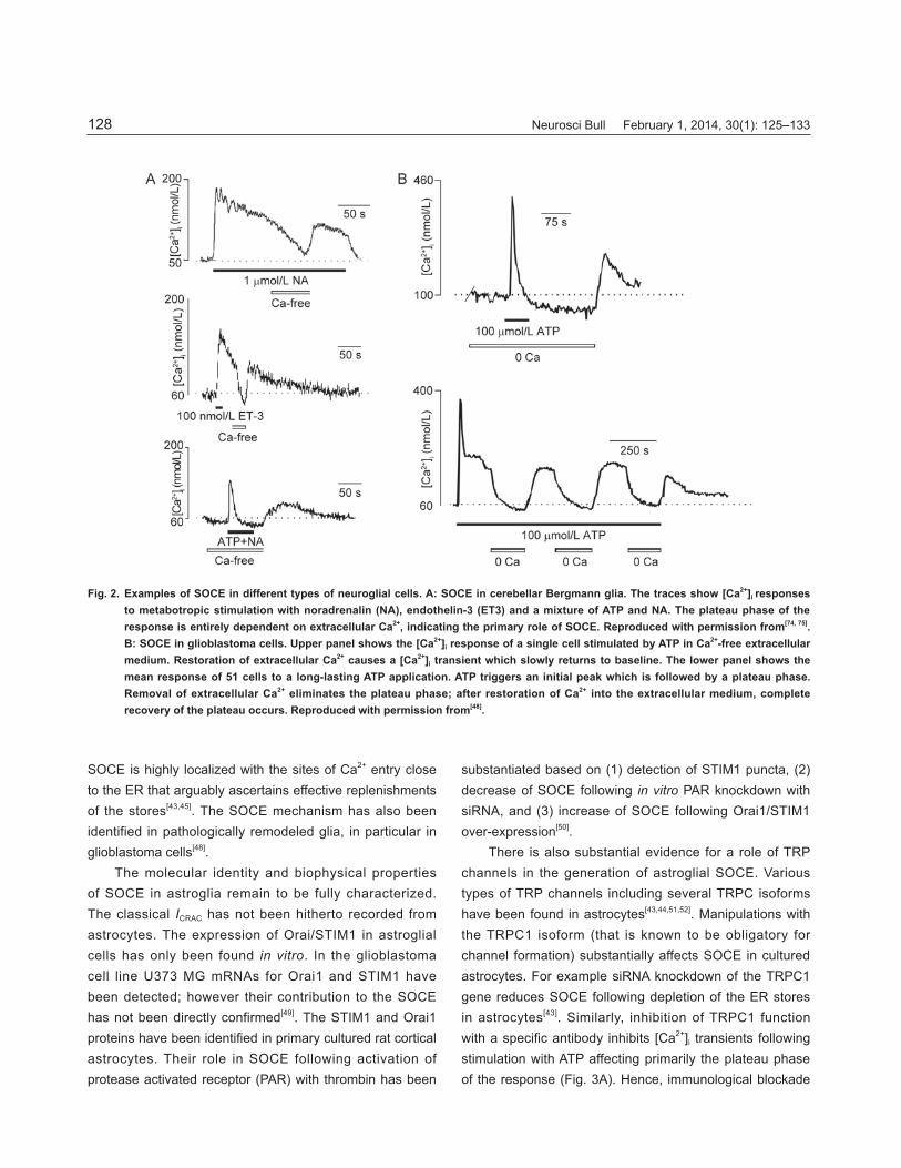
Neurosci Bull February 1, 2014, 30(1): 125–133128
SOCE is highly localized with the sites of Ca2+ entry close to the ER that arguably ascertains effective replenishments of the stores[43,45]. The SOCE mechanism has also been identified in pathologically remodeled glia, in particular in glioblastoma cells[48].
The molecular identity and biophysical properties of SOCE in astroglia remain to be fully characterized. The classical ICRAC has not been hitherto recorded from astrocytes. The expression of Orai/STIM1 in astroglial cells has only been found in vitro. In the glioblastoma cell line U373 MG mRNAs for Orai1 and STIM1 have been detected; however their contribution to the SOCE has not been directly confirmed[49]. The STIM1 and Orai1 proteins have been identifi ed in primary cultured rat cortical astrocytes. Their role in SOCE following activation of protease activated receptor (PAR) with thrombin has been
substantiated based on (1) detection of STIM1 puncta, (2) decrease of SOCE following in vitro PAR knockdown with siRNA, and (3) increase of SOCE following Orai1/STIM1 over-expression[50].
There is also substantial evidence for a role of TRP channels in the generation of astroglial SOCE. Various types of TRP channels including several TRPC isoforms have been found in astrocytes[43,44,51,52]. Manipulations with the TRPC1 isoform (that is known to be obligatory for channel formation) substantially affects SOCE in cultured astrocytes. For example siRNA knockdown of the TRPC1 gene reduces SOCE following depletion of the ER stores in astrocytes[43]. Similarly, inhibition of TRPC1 function with a specifi c antibody inhibits [Ca2+]i transients following stimulation with ATP affecting primarily the plateau phase of the response (Fig. 3A). Hence, immunological blockade
Fig. 2. Examples of SOCE in different types of neuroglial cells. A: SOCE in cerebellar Bergmann glia. The traces show [Ca2+]i responses to metabotropic stimulation with noradrenalin (NA), endothelin-3 (ET3) and a mixture of ATP and NA. The plateau phase of the response is entirely dependent on extracellular Ca2+, indicating the primary role of SOCE. Reproduced with permission from[74, 75]. B: SOCE in glioblastoma cells. Upper panel shows the [Ca2+]i response of a single cell stimulated by ATP in Ca2+-free extracellular medium. Restoration of extracellular Ca2+ causes a [Ca2+]i transient which slowly returns to baseline. The lower panel shows the mean response of 51 cells to a long-lasting ATP application. ATP triggers an initial peak which is followed by a plateau phase. Removal of extracellular Ca2+ eliminates the plateau phase; after restoration of Ca2+ into the extracellular medium, complete recovery of the plateau occurs. Reproduced with permission from[48].

Alexei Verkhratsky, et al. SOCE in neuroglia 129
of TRPC1 results in ~30% reduction of the ATP-induced transient Ca2+ elevation, along with full abolishment of the [Ca2+]i transient plateau. Similarly, the mechanically-induced Ca2+ response in astrocytes is greatly reduced by acute immunological inhibition of TRPC1 protein, which also inhibits Ca2+-dependent glutamate release from astrocytes[44] (Fig. 3B, C). Importantly, store-operated
activation of TRP channels results in a substantial Na+ infl ux which may link the activation of metabotropic receptors and ER Ca2+ release with astroglial Na+ signaling[53]. Oligodendroglia and NG2 cellsVery little experimental evidence on SOCE in cells of the oligodendroglial lineage has been accumulated. Store-operated Ca2+ influx is detectable in oligodendrocyte
Fig. 3. The role of TRPC1 in SOCE generation in cultured astroglia. A: TRPC1 plays a role in the intracellular Ca2+ elevation elicited by receptor activation in astrocytes. Application of ATP (100 μmol/L) to astrocytes from rat visual cortex results in a biphasic intracellular Ca2+ response: an initial transient elevation and a sustained (plateau) elevation. Intracellular Ca2+ measurements were obtained using the Ca2+ indicator fluo-3. If TRPC1-containing channels are blocked by incubating cells with an antibody against TRPC1, the sustained (plateau) Ca2+ elevation, reporting on SOCE, is abolished. Vertical dashed line indicates the initial point of a sustained plateau Ca2+ response, of which the cumulative values are shown in the bar graph. B–C: TRPC1 plays a role in mechanically-elicited intracellular Ca2+ responses in astrocytes and the resulting Ca2+-dependent glutamate release from these cells. Mechanical stimulation causes cytoplasmic Ca2+ elevation in astrocytes, as recorded using the Ca2+ indicator X-rhod-1 (B). Glutamate release from astrocytes, reported by an increase in extracellular NADH fl uorescence, is induced by mechanical stimulation (C). Both responses (Ca2+ and glutamate) are reduced when astrocytes are incubated with TRPC1 antibody. Arrows indicate the time of mechanical stimulation. Points and bars in (A–C) indicate mean ± SEM. Asterisks indicate a signifi cant change compared with the control group (Student’s t-test; *P <0.05, **P <0.01). Modifi ed from[44].

Neurosci Bull February 1, 2014, 30(1): 125–133130
precursor cells (OPCs) in culture where it is involved in the regulation of proliferation. It should be noted that TRPC1 is also important for astrocytic proliferation in cell culture[43]. Nonetheless, the SOCE in these OPCs is positively modulated by golli protein, which belongs to the basic myelin protein family[54]. This golli protein has been reported to interact with STIM1 proteins[55] as well as with TRPC1 channels, the latter being also shown to mediate SOCE in OPCs[56].
MicrogliaThe SOCE mechanism is widespread in microglia; stimulation of various types of metabotropic receptors (such as P2Y purinoceptors, PARs, platelet-activating factor receptors (PAFRs), complement fragment receptors, endothelin receptors, and lysophosphatidic acid receptors) triggers transient ER Ca2+ release, which is followed by a long-lasting plateau of elevated Ca2+ that is almost entirely mediated by SOCE (see for example[41,57-59] and[60] for further references). SOCE in microglia demonstrates a peculiar phenomenon of long-lasting activation following strong stimulation of metabotropic receptors. This phenomenon was initially found in cultured microglia stimulated with supramaximal doses of ATP or UTP that activate P2Y2/4 purinoceptors and result in full depletion of the ER Ca2+ store. This induces a persistent activation of SOCE that lasts for tens of minutes (Fig. 4). During this period, the ER signaling is inhibited because neither metabotropic stimulation nor exposure to thapsigargin, a SERCA blocker, is able to produce an additional [Ca2+]i signal[61]. Long-lasting SOCE activation also occurs in microglial cells stimulated with phorbol ester, and with brain-derived neurotrophic factor[62,63]. Similar mechanism may also account for the long-lasting elevation of [Ca2+]i in lipopolysaccharide (endotoxin)-treated cells; this [Ca2+]i elevation is accompanied by suppressed Ca2+ release from the ER[64]. The long-lasting elevation in [Ca2+]i that can be mediated by chronically activated SOCE has been suggested to be fundamentally important for regulating various aspects of microglial activation[64]. The long-lasting [Ca2+]i increase is found in microglia treated with another activating agent, interferon-γ[65], in microglia treated with toxic β-amyloid fragment(25-35)[66] and also in microglial cells isolated and cultured from the human post-mortem brains from Alzheimer’s disease patients[67].
Whether such a chronic increase in [Ca2+]i is mediated by activation of SOCE, remains, however, unclear. Furthermore, in microglial cells isolated from Alzheimer’s post-mortem brains, the SOCE following PAFR stimulation is substantially reduced, although the duration of the ATP-induced response is much increased[67]. The sustained, chronic SOCE activation is also speculated to be related to chronic activation of microglia in neuropsychiatric disorders[68]. Moreover, microglial SOCE has been reported to be regulated by mitochondria because inhibition of mitochondrial peripheral benzodiazepine receptors with the agent PK11195 reduces Ca2+ entry following the activation of PAFRs[69].
The molecular and biophysical identity of microglial SOCE has been studied in cultured cells. A current very much resembling ICRAC, with an amplitude of ~1.3 pA/pF, has been recorded in mouse microglia in vitro. The amplitude of this current is reduced to 0.5 pA/pF 48 h after activation of microglia by treatment with lipopolysaccharide[70]. Similarly, ICRAC (with very high Ca2+-selectivity of PCa/PNa > 1 000) has been detected in cultured rat neonatal microglia. These cells also express mRNA for all three Orais and it has been
Fig. 4. Long-lasting activation of SOCE in microglial cells. Following depletion of the intracellular Ca2+ stores by metabotropic stimulation of glial cells in the absence of extracellular Ca2+, the readmission of external Ca2+ induces a large rebound increase in [Ca2+]i. This rebound is followed by a return of [Ca2+]i to a new steady-state level, higher than the initial resting level. This chronic activation of SOCE renders the ER store non-responsive, and subsequent stimulation by ATP in Ca2+-free conditions fails to elicit Ca2+ release (observations by Toescu & Verkhratsky, and[61]).

Alexei Verkhratsky, et al. SOCE in neuroglia 131
suggested that the Orai1/STIM complex is responsible for ICRAC generation[71].
Microglial cells also express a wide variety of TRP channels. RT-PCR of cultured rat microglia reveals the following rank of mRNA expression TRPM7 > TRPC6 > TRPM2 > TRPC1 > TRPC3 ≥ TRPC4 > TRPC7 > TRPC5 > TRPC2[71]. High levels of expression of mRNA for TRPM2 have been detected in rat microglial cells in vitro and these channels are supposed to be activated by hydrogen peroxide and intracellular injection of ADP-ribose[72]. Primary cultured rat microglial cells have also been found to express TRPV1 channels as demonstrated by RT-PCR, Western blot analysis and immunocytochemistry[73]. Nonetheless, there are no clear indications that TRP channels mediate SOCE in microglia.
Conclusions
The SOCE mechanism is functionally expressed in all types of CNS neuroglial cells. The molecular identity of the neuroglial SOCE has not yet been identifi ed unequivocally, although it seems that the STIM/Orai-composed ICRAC is predominantly expressed in microglia, whereas astrocytes and oligodendrocytes rely more on TRP channels to produce SOCE.
ACKNOWLEDGEMENTS
The authors’ research was supported by an Alzheimer’s Research Trust (UK) Programme Grant (ART/PG2004A/1) to A.V. and by a National Science Foundation grant (CBET 0943343) to V.P.
Received date: 2013-01-13; Accepted date: 2013-02-14
REFERENCES
Parod RJ, Putney JW Jr. The role of calcium in the receptor [1] mediated control of potassium permeability in the rat lacrimal gland. J Physiol 1978, 281: 371–381.Putney JW Jr. Muscarinic, alpha-adrenergic and peptide [2] receptors regulate the same calcium infl ux sites in the parotid gland. J Physiol 1977, 268: 139–149.Casteels R, Droogmans G. Exchange characteristics of the [3] noradrenaline-sensitive calcium store in vascular smooth muscle cells or rabbit ear artery. J Physiol 1981, 317: 263–279.Putney JW Jr. Capacitative calcium entry revisited. Cell [4]
Calcium 1990, 11: 611–624.Putney JW Jr. A model for receptor-regulated calcium entry. [5] Cell Calcium 1986, 7: 1–12.Alonso MT, Manjarres IM, Garcia-Sancho J. Privileged [6] coupling between Ca2+ entry through plasma membrane store-operated Ca2+ channels and the endoplasmic reticulum Ca2+ pump. Mol Cell Endocrinol 2011, 353: 37–44.Cahalan MD. STIMulating store-operated Ca[7] 2+ entry. Nat Cell Biol 2009, 11: 669–677.Soboloff J, Rothberg BS, Madesh M, Gill DL. STIM proteins: [8] dynamic calcium signal transducers. Nat Rev Mol Cell Biol 2012, 13: 549–565.Carrasco S, Meyer T. STIM proteins and the endoplasmic [9] reticulum-plasma membrane junctions. Annu Rev Biochem 2011, 80: 973–1000.Lewis RS. The molecular choreography of a store-operated [10] calcium channel. Nature 2007, 446: 284–287.Hoth M, Penner R. Depletion of intracellular calcium stores [11] activates a calcium current in mast cells. Nature 1992, 355: 353–356.Parekh AB, Penner R. Store depletion and calcium influx. [12] Physiol Rev 1997, 77: 901–930.Parekh AB. Store-operated CRAC channels: function in [13] health and disease. Nat Rev Drug Discov 2010, 9: 399–410.Salido GM, Sage SO, Rosado JA. TRPC channels and store-[14] operated Ca2+ entry. Biochim Biophys Acta 2009, 1793: 223–230.Worley PF, Zeng W, Huang GN, Yuan JP, Kim JY, Lee MG[15] , et al. TRPC channels as STIM1-regulated store-operated channels. Cell Calcium 2007, 42: 205–211.Ambudkar IS, Ong HL, Liu X, Bandyopadhyay BC, Cheng KT. [16] TRPC1: the link between functionally distinct store-operated calcium channels. Cell Calcium 2007, 42: 213–223.Feske S, Gwack Y, Prakriya M, Srikanth S, Puppel SH, [17] Tanasa B, et al. A mutation in Orai1 causes immune deficiency by abrogating CRAC channel function. Nature 2006, 441: 179–185.Vig M, Peinelt C, Beck A, Koomoa DL, Rabah D, Koblan-[18] Huberson M, et al. CRACM1 is a plasma membrane protein essential for store-operated Ca2+ entry. Science 2006, 312: 1220–1223.Zhang SL, Yeromin AV, Zhang XH, Yu Y, Safrina O, Penna [19] A, et al. Genome-wide RNAi screen of Ca2+ infl ux identifi es genes that regulate Ca2+ release-activated Ca2+ channel activity. Proc Natl Acad Sci U S A 2006, 103: 9357–9362.Zhang SL, Yu Y, Roos J, Kozak JA, Deerinck TJ, Ellisman [20] MH, et al. STIM1 is a Ca2+ sensor that activates CRAC channels and migrates from the Ca2+ store to the plasma membrane. Nature 2005, 437: 902–905.Liou J, Kim ML, Heo WD, Jones JT, Myers JW, Ferrell JE [21]

Neurosci Bull February 1, 2014, 30(1): 125–133132
Jr, et al. STIM is a Ca2+ sensor essential for Ca2+-store-depletion-triggered Ca2+ influx. Curr Biol 2005, 15: 1235–1241.Dziadek MA, Johnstone LS. Biochemical properties and [22] cellular localisation of STIM proteins. Cell Calcium 2007, 42: 123–132.Mercer JC, Dehaven WI, Smyth JT, Wedel B, Boyles RR, [23] Bird GS, et al. Large store-operated calcium selective currents due to co-expression of Orai1 or Orai2 with the intracellular calcium sensor, Stim1. J Biol Chem 2006, 281: 24979–24990.Watson R, Parekh AB. Mitochondrial regulation of CRAC [24] channel-driven cellular responses. Cell Calcium 2012, 52: 52–56.Kopach O, Kruglikov I, Pivneva T, Voitenko N, Verkhratsky [25] A, Fedirko N. Mitochondria adjust Ca2+ signaling regime to a pattern of stimulation in salivary acinar cells. Biochim Biophys Acta 2011, 1813: 1740–1748.Zeng W, Yuan JP, Kim MS, Choi YJ, Huang GN, Worley [26] PF, et al. STIM1 gates TRPC channels, but not Orai1, by electrostatic interaction. Mol Cell 2008, 32: 439–448.Huang GN, Zeng W, Kim JY, Yuan JP, Han L, Muallem S[27] , et al. STIM1 carboxyl-terminus activates native SOC, Icrac and TRPC1 channels. Nat Cell Biol 2006, 8: 1003–1010.Yuan JP, Zeng W, Huang GN, Worley PF, Muallem S. STIM1 [28] heteromultimerizes TRPC channels to determine their function as store-operated channels. Nat Cell Biol 2007, 9: 636–645.Kettenmann H, Ransom BR. Neuroglia. Oxford, New York: [29] Oxford University Press, 2013: 864.Parpura V, Heneka MT, Montana V, Oliet SH, Schousboe [30] A, Haydon PG, et al. Glial cells in (patho)physiology. J Neurochem 2012, 121: 4–27.Verkhratsky A, Parpura V, Rodriguez JJ. Where the thoughts [31] dwell: the physiology of neuronal-glial “diffuse neural net”. Brain Res Rev 2011, 66: 133–151.Verkhratsky A, Orkand RK, Kettenmann H. Glial calcium: [32] homeostasis and signaling function. Physiol Rev 1998, 78: 99–141.Verkhratsky A, Rodriguez JJ, Parpura V. Calcium signalling in [33] astroglia. Mol Cell Endocrinol 2012, 353: 45–56.Lalo U, Pankratov Y, Parpura V, Verkhratsky A. Ionotropic [34] receptors in neuronal-astroglial signalling: what is the role of “excitable” molecules in non-excitable cells. Biochim Biophys Acta 2011, 1813: 992–1002.Parpura V, Verkhratsky A. Astrocytes revisited: concise [35] historic outlook on glutamate homeostasis and signaling. Croat Med J 2012, 53: 518–528.Verkhratsky A. Physiology of neuronal-glial networking. [36] Neurochem Int 2010, 57: 332–343.
Verkhratsky A, Kirchhoff F. Glutamate-mediated neuronal-[37] glial transmission. J Anat 2007, 210: 651–660.Parpura V, Verkhratsky A. Astroglial amino acid-based [38] transmitter receptors. Amino Acids 2013, 44(4): 1151–1158Lalo U, Palygin O, North RA, Verkhratsky A, Pankratov Y. [39] Age-dependent remodelling of ionotropic signalling in cortical astroglia. Aging Cell 2011, 10: 392–402.Sun W, McConnell E, Pare JF, Xu Q, Chen M, Peng W[40] , et al. Glutamate-dependent neuroglial calcium signaling differs between young and adult brain. Science 2013, 339: 197–200.Moller T, Kann O, Prinz M, Kirchhoff F, Verkhratsky A, [41] Kettenmann H. Endothelin-induced calcium signaling in cultured mouse microglial cells is mediated through ETB receptors. Neuroreport 1997, 8: 2127–2131.Kirischuk S, Tuschick S, Verkhratsky A, Kettenmann H. [42] Calcium signalling in mouse Bergmann glial cells mediated by a1-adrenoreceptors and H1 histamine receptors. Eur J Neurosci 1996, 8: 1198–1208.Golovina VA. Visualization of localized store-operated [43] calcium entry in mouse astrocytes. Close proximity to the endoplasmic reticulum. J Physiol 2005, 564: 737–749.Malarkey EB, Ni Y, Parpura V. Ca[44] 2+ entry through TRPC1 channels contributes to intracellular Ca2+ dynamics and consequent glutamate release from rat astrocytes. Glia 2008, 56: 821–835.Pivneva T, Haas B, Reyes-Haro D, Laube G, Veh RW, Nolte C[45] , et al. Store-operated Ca2+ entry in astrocytes: different spatial arrangement of endoplasmic reticulum explains functional diversity in vitro and in situ. Cell Calcium 2008, 43: 591–601.Lo KJ, Luk HN, Chin TY, Chueh SH. Store depletion-induced [46] calcium influx in rat cerebellar astrocytes. Br J Pharmacol 2002, 135: 1383–1392.Singaravelu K, Lohr C, Deitmer JW. Regulat ion of [47] store-operated calcium entry by calcium-independent phospholipase A2 in rat cerebellar astrocytes. J Neurosci 2006, 26: 9579–9592.Hartmann J, Verkhratsky A. Relations between intracellular [48] Ca2+ stores and store-operated Ca2+ entry in primary cultured human glioblastoma cells. J Physiol 1998, 513 (Pt 2): 411–424.Barajas M, Andrade A, Hernandez-Hernandez O, Felix R, [49] Arias-Montano JA. Histamine-induced Ca2+ entry in human astrocytoma U373 MG cells: evidence for involvement of store-operated channels. J Neurosci Res 2008, 86: 3456–3468.Moreno C, Sampieri A, Vivas O, Pena-Segura C, Vaca L. [50] STIM1 and Orai1 mediate thrombin-induced Ca2+ infl ux in rat cortical astrocytes. Cell Calcium 2012, 52: 457–467.Grimaldi M, Maratos M, Verma A. Transient receptor potential [51] channel activation causes a novel form of [Ca 2+]I oscillations

Alexei Verkhratsky, et al. SOCE in neuroglia 133
and is not involved in capacitative Ca2+ entry in glial cells. J Neurosci 2003, 23: 4737–4745.Pizzo P, Burgo A, Pozzan T, Fasolato C. Role of capacitative [52] calcium entry on glutamate-induced calcium infl ux in type-I rat cortical astrocytes. J Neurochem 2001, 79: 98–109.Kirischuk S, Parpura V, Verkhratsky A. Sodium dynamics: [53] another key to astroglial excitability? Trends Neurosci 2012, 35: 497–506.Paez PM, Fulton DJ, Spreuer V, Handley V, Campagnoni [54] CW, Campagnoni AT. Regulation of store-operated and voltage-operated Ca2+ channels in the proliferation and death of oligodendrocyte precursor cells by golli proteins. ASN Neuro 2009, 1. pii: e00003.Walsh CM, Doherty MK, Tepikin AV, Burgoyne RD. Evidence [55] for an interaction between Golli and STIM1 in store-operated calcium entry. Biochem J 2010, 430: 453–460.Paez PM, Fulton D, Spreuer V, Handley V, Campagnoni AT. [56] Modulation of canonical transient receptor potential channel 1 in the proliferation of oligodendrocyte precursor cells by the golli products of the myelin basic protein gene. J Neurosci 2011, 31: 3625–3637.Moller T, Kann O, Verkhratsky A, Kettenmann H. Activation [57] of mouse microglial cells affects P2 receptor signaling. Brain Res 2000, 853: 49–59.Moller T, Nolte C, Burger R, Verkhratsky A, Kettenmann H. [58] Mechanisms of C5a and C3a complement fragment-induced [Ca2+]i signaling in mouse microglia. J Neurosci 1997, 17: 615–624.Wang X, Bae JH, Kim SU, McLarnon JG. Platelet-activating [59] factor induced Ca2+ signaling in human microglia. Brain Res 1999, 842: 159–165.Kettenmann H, Hanisch UK, Noda M, Verkhratsky A. [60] Physiology of microglia. Physiol Rev 2011, 91: 461–553.Toescu EC, Moller T, Kettenmann H, Verkhratsky A. Long-[61] term activation of capacitative Ca2+ entry in mouse microglial cells. Neuroscience 1998, 86: 925–935.Mizoguchi Y, Monji A, Kato T, Seki Y, Gotoh L, Horikawa H[62] , et al. Brain-derived neurotrophic factor induces sustained elevation of intracellular Ca2+ in rodent microglia. J Immunol 2009, 183: 7778–7786.Moller T. Calcium signaling in microglial cells. Glia 2002, 40: [63] 184–194.Hoffmann A, Kann O, Ohlemeyer C, Hanisch UK, [64] Kettenmann H. Elevation of basal intracellular calcium as a central element in the activation of brain macrophages (microglia): suppression of receptor-evoked calcium signaling
and control of release function. J Neurosci 2003, 23: 4410–4419.Franciosi S, Choi HB, Kim SU, McLarnon JG. Interferon-[65] gamma acutely induces calcium infl ux in human microglia. J Neurosci Res 2002, 69: 607–613.Korotzer AR, Whittemore ER, Cotman CW. Differential [66] regulation by beta-amyloid peptides of intracellular free Ca2+ concentration in cultured rat microglia. Eur J Pharmacol 1995, 288: 125–130.McLarnon JG, Choi HB, Lue LF, Walker DG, Kim SU. [67] Perturbations in calcium-mediated signal transduction in microglia from Alzheimer’s disease patients. J Neurosci Res 2005, 81: 426–435.Mizoguchi Y, Monji A, Kato TA, Horikawa H, Seki Y, Kasai M[68] , et al. Possible role of BDNF-induced microglial intracellular Ca2+ elevation in the pathophysiology of neuropsychiatric disorders. Mini Rev Med Chem 2011, 11: 575–581.Hong SH, Choi HB, Kim SU, McLarnon JG. Mitochondrial [69] ligand inhibits store-operated calcium influx and COX-2 production in human microglia. J Neurosci Res 2006, 83: 1293–1298.Beck A, Penner R, Fleig A. Lipopolysaccharide-induced [70] down-regulation of Ca2+ release-activated Ca2+ currents (ICRAC) but not Ca2+-activated TRPM4-like currents (ICAN) in cultured mouse microglial cells. J Physiol 2008, 586: 427–439.Ohana L, Newell EW, Stanley EF, Schlichter LC. The Ca[71] 2+ release-activated Ca2+ current (ICRAC) mediates store-operated Ca2+ entry in rat microglia. Channels (Austin) 2009, 3: 129–139.Kraft R, Grimm C, Grosse K, Hoffmann A, Sauerbruch S, [72] Kettenmann H, et al. Hydrogen peroxide and ADP-ribose induce TRPM2-mediated calcium infl ux and cation currents in microglia. Am J Physiol Cell Physiol 2004, 286: C129–137.Kim SR, Kim SU, Oh U, Jin BK. Transient receptor potential [73] vanilloid subtype 1 mediates microglial cell death in vivo and in vitro via Ca2+-mediated mitochondrial damage and cytochrome c release. J Immunol 2006, 177: 4322–4329.Kirischuk S, Matiash V, Kulik A, Voitenko N, Kostyuk P, [74] Verkhratsky A. Activation of P2-purino-, a1-adreno and H1-histamine receptors triggers cytoplasmic calcium signalling in cerebellar Purkinje neurons. Neuroscience 1996, 73: 643–647.Tuschick S, Kirischuk S, Kirchhoff F, Liefeldt L, Paul M, [75] Verkhratsky A, et al. Bergmann glial cells in situ express endothelinB receptors linked to cytoplasmic calcium signals. Cell Calcium 1997, 21: 409–419.