Anti-Hypertensive Activity of Some Selected Unani Formulations
Upload
tatainteractiveCategory
view
0download
0
J. Funct. Biomater. 2013, 4, 209-311; doi:10.3390/jfb4040209
Journal of
Functional
Biomaterials ISSN 2079-4983
www.mdpi.com/journal/jfb/
Review
Self-Setting Calcium Orthophosphate Formulations
Sergey V. Dorozhkin
Kudrinskaja sq. 1-155, Moscow 123242, Russia; E-Mail: [email protected];
Tel. +7-499-255-4460
Received: 10 September 2013; in revised form: 18 October 2013 / Accepted: 21 October 2013 /
Published: 12 November 2013
Abstract: In early 1980s, researchers discovered self-setting calcium orthophosphate
cements, which are bioactive and biodegradable grafting bioceramics in the form of a
powder and a liquid. After mixing, both phases form pastes, which set and harden forming
either a non-stoichiometric calcium deficient hydroxyapatite or brushite. Since both of
them are remarkably biocompartible, bioresorbable and osteoconductive, self-setting
calcium orthophosphate formulations appear to be promising bioceramics for bone
grafting. Furthermore, such formulations possess excellent molding capabilities, easy
manipulation and nearly perfect adaptation to the complex shapes of bone defects,
followed by gradual bioresorption and new bone formation. In addition, reinforced
formulations have been introduced, which might be described as calcium orthophosphate
concretes. The discovery of self-setting properties opened up a new era in the medical
application of calcium orthophosphates and many commercial trademarks have been
introduced as a result. Currently such formulations are widely used as synthetic bone
grafts, with several advantages, such as pourability and injectability. Moreover, their
low-temperature setting reactions and intrinsic porosity allow loading by drugs,
biomolecules and even cells for tissue engineering purposes. In this review, an insight into
the self-setting calcium orthophosphate formulations, as excellent bioceramics suitable for
both dental and bone grafting applications, has been provided.
Keywords: calcium orthophosphates; hydroxyapatite; self-setting; self-hardening;
cements; concretes; bioceramics; bone grafts; scaffolds; tissue engineering
OPEN ACCESS

J. Funct. Biomater. 2013, 4 210
1. Introduction
According to the statistics, approximately half of the population sustains at least one bone fracture
during their lifetime [1] and, as a result, surgery might be necessary. Luckily, among the surgical
procedures available, minimally invasive techniques are able to offer special benefits for patients such
as fewer associated injuries, quicker recovery and less pain. In addition, shorter hospital stays are
needed, often allowing outpatient treatments that cheapen the expenses [2]. However, these techniques
require biomaterials able to be implanted through small (the smaller, the better) incisions, e.g., by
means of syringes with needles and/or laparoscopic devices. To fulfill such requirements, the potential
implants should be in a liquid or an injectable state, e.g., as pastes. On the other hand, since all types of
the calcified tissues are in the solid state, the bone repairing biomaterials should be solid as well.
Therefore, potential bone grafts applicable to the minimally invasive surgery must combine
injectability with hardness. Such formulations are known as self-setting (self-hardening, self-curing)
formulations because, together with an initial softness and injectability, they possess an ability to
solidify in the appropriate period, giving strength to the implantation sites. Since the inorganic part of
the mammalian calcified tissues is composed of calcium orthophosphates of biological origin [3],
self-setting formulations based on calcium orthophosphates appear to be excellent candidates for bone
repairing [4,5]. The list of all known calcium orthophosphates, including their chemical formulae,
standard abbreviations and the major properties, is summarized in Table 1 [6].
Although the entire subject of calcium orthophosphates has been investigated since 1770s [7,8],
historically, Kingery appears to be the first, who contributed to their self-setting abilities. Namely, in
1950, he published a paper on the chemical interactions between oxides and/or hydroxides of various
metals (including CaO) with H3PO4, in which he mentioned that some of the reaction products were
set [9]. However, the calcium orthophosphate formulations were just a very small section of that study.
Afterwards, self-setting abilities of some calcium orthophosphates formulations were described in the
early 1970s by Driskell et al. [10]. However, that study was not noticed. Then, in early 1980s,
scientists from the American Dental Association LeGeros et al. [11], as well as Brown and
Chow [12–15] published results of their studies. Since that, this subject became known as calcium
phosphate cements (commonly referred to as CPC) [16], and, due to their suitability for repair,
augmentation and regeneration of bones, such formulations were also named as calcium phosphate
bone cements (occasionally referred to as CPBC) [17–20]. In order to stress the fact, that these
formulations consist either entirely or essentially from calcium orthophosphates, this review is limited
to consideration of calcium orthophosphate-based compositions only. The readers interested in
self-setting formulations based on other types of calcium phosphates are requested to read the original
publications [20,21].

J. Funct. Biomater. 2013, 4 211
Table 1. Existing calcium orthophosphates and their major properties [6].
Ca/P
molar ratio
Compounds and
their typical abbreviations Chemical formula
Solubility at
25 °C, −log(Ks)
Solubility at
25 °C, g/L
pH stability range in aqueous
solutions at 25 °C
0.5 Monocalcium phosphate monohydrate
(MCPM) Ca(H2PO4)2·H2O 1.14 ~18 0.0–2.0
0.5 Monocalcium phosphate anhydrous
(MCPA or MCP) Ca(H2PO4)2 1.14 ~17 [c]
1.0 Dicalcium phosphate dihydrate (DCPD),
mineral brushite CaHPO4·2H2O 6.59 ~0.088 2.0–6.0
1.0 Dicalcium phosphate anhydrous
(DCPA or DCP), mineral monetite CaHPO4 6.90 ~0.048 [c]
1.33 Octacalcium phosphate (OCP) Ca8(HPO4)2(PO4)4·5H2O 96.6 ~0.0081 5.5–7.0
1.5 α-Tricalcium phosphate (α-TCP) α-Ca3(PO4)2 25.5 ~0.0025 [a]
1.5 β-Tricalcium phosphate (β-TCP) β-Ca3(PO4)2 28.9 ~0.0005 [a]
1.2–2.2 Amorphous calcium phosphates (ACP) CaxHy(PO4)z·nH2O, n = 3–4.5;
15%–20% H2O
[b] [b] ~5–12 [d]
1.5–1.67 Calcium-deficient hydroxyapatite
(CDHA or Ca-def HA) [e]
Ca10−x(HPO4)x(PO4)6−x(OH)2−x
(0 < x < 1) ~85 ~0.0094 6.5–9.5
1.67 Hydroxyapatite (HA, HAp or OHAp) Ca10(PO4)6(OH)2 116.8 ~0.0003 9.5–12
1.67 Fluorapatite (FA or FAp) Ca10(PO4)6F2 120.0 ~0.0002 7–12
1.67 Oxyapatite (OA, OAp or OXA) [f] Ca10(PO4)6O ~69 ~0.087 [a]
2.0 Tetracalcium phosphate (TTCP or TetCP),
mineral hilgenstockite Ca4(PO4)2O 38–44 ~0.0007 [a]
[a] These compounds cannot be precipitated from aqueous solutions; [b] Cannot be measured precisely. However, the following values were found: 25.7 ± 0.1 (pH = 7.40),
29.9 ± 0.1 (pH = 6.00), 32.7 ± 0.1 (pH = 5.28). The comparative extent of dissolution in acidic buffer is: ACP >> α-TCP >> β-TCP > CDHA >> HA > FA; [c] Stable at
temperatures above 100 °C; [d] Always metastable; [e] Occasionally, it is called ―precipitated HA (PHA)‖ and [f] Existence of OA remains questionable.

J. Funct. Biomater. 2013, 4 212
Due to a good bioresorbability, all self-setting calcium orthophosphate formulations belong to the
second generation of bone substituting biomaterials [22]. These formulations are blends of amorphous
and/or crystalline calcium orthophosphate powder(s) with an aqueous solution, which might be
distilled water [11–15], phosphate buffer solution (PBS) [16], aqueous solutions of sodium
orthophosphates [23–30], ammonium orthophosphates [31], H3PO4 [32–37], citric acid [24,38] and its
salts [39], sodium silicate [40–42], soluble magnesium orthophosphates [43], chitosan lactate in lactic
acid [44], etc. Due to the presence of other ions in a number of solutions, some of such formulations
are set with formation of ion-substituted calcium orthophosphates. After the calcium orthophosphate
powder(s) and the solution have been mixed together, a viscous and moldable paste is formed that sets
to a firm mass within a few minutes. When the paste becomes sufficiently stiff, it can be placed into a
defect as a substitute for the damaged part of bone, where it hardens in situ within the operating
theatre. The proportion of solid to liquid or the powder-to-liquid (P/L) ratio is a very important
characteristic because it determines both bioresorbability and rheological properties. As the paste is set
and hardened at room or body temperature, direct application in healing of bone defects became a new
and innovative treatment modality by the end of the XX-th century. Moreover, self-setting calcium
orthophosphate formulations can be injected directly into the fractures and bone defects, where they
intimately adapt to the bone cavity regardless its shape. More to the point, they were found to promote
development of osteoconductive pathways, possess sufficient compressive strengths, be non-cytotoxic,
create chemical bonds to the host bones, restore contour and have both the chemical composition and
X-ray diffraction patterns similar to those of bone [45]. Finally, but yet importantly, the self-setting
calcium orthophosphate formulations are osteotransductive, i.e., after implantation, the hardened
formulations are replaced by a new bone tissue [46–48].
Since the hardened calcium orthophosphates reproduce the composition, structure, morphology and
crystallinity of bone crystals, the initial self-setting formulations might be considered as biomimetic
ones [49,50]. The aim of such formulations is to disturb bone functions and properties as little as
possible and, until a new bone has been grown, to behave temporary in a manner similar to that of
bone. Therefore, they provide surgeons with a unique ability of manufacturing, shaping and implanting
the bioactive bone substitute biomaterials on a patient-specific base in real time in the surgery room.
Implanted bone tissues also take benefits from the self-setting formulations that give, in an acceptable
clinical time, a suitable mechanical strength for a shorter tissue functional recovery. Thus, the major
advantages of the self-setting calcium orthophosphate formulations include a fast setting time, an
excellent moldability, an outstanding biocompatibility and an easy manipulation; therefore, they are
more versatile in handling characteristics than prefabricated granules or blocks. Besides, like any other
type of calcium orthophosphate bioceramics, the self-setting formulations provide an opportunity for
bone grafting using alloplastic materials, which are unlimited in quantity and provide no risk of
infectious diseases [51–53].
Since self-setting calcium orthophosphate formulations have been developed for using as implanted
biomaterials for parenteral application, for their chemical composition one might employ all ionic
compounds of oligoelements occurring naturally in a human body. The list of possible additives
includes (but is not limited to) the following cations: Na+, K
+, Mg
2+, Ca
2+, Sr
2+, Zn
2+, H
+ and anions:
PO43−
, HPO42−
, H2PO4−, P2O7
4−, CO3
2−, HCO3
−, SO4
2−, HSO4
−, Cl
−, OH
−, F
−, silicates [46]. Therefore,
mixed-type self-setting formulations consisting of calcium orthophosphates and other calcium salts,

J. Funct. Biomater. 2013, 4 213
such as calcium sulfate [54–63], calcium pyrophosphate [64–66], calcium polyphosphates [67,68],
calcium carbonates [16,26,28,30,50,69–71], calcium oxide [72–77], calcium hydroxide [78–80],
calcium aluminates [43,81,82], calcium silicates [83–89], etc., are available. In addition, other
chemicals such as Sr-containing compounds [19,90–93], Mg-containing compounds [93–100],
Zn-containing compounds [101,102], etc., might be added to calcium orthophosphates as well.
Furthermore, the self-setting formulations might be prepared from various types of ion substituted
calcium orthophosphates such as Ca2KNa(PO4)2, NaCaPO4, Na3Ca6(PO4)5 (so called ―calcium
alkaline orthophosphates‖) [103–107], magnesium substituted calcium-deficient hydroxyapatite (CDHA),
strontium substituted CDHA, etc. [108–113]. More to the point, self-setting formulations might be
prepared in the reaction-setting mixture of Ca(OH)2–KH2PO4 system [114], as well as by treatment of
calcium carbonate or calcium hydroxide with orthophosphate solutions [115]. In addition, if a
self-setting formulation consisting of calcium orthophosphates only is set in a chemically reactive
environment (e.g., in presence of CO2), ion-substituted calcium orthophosphates, such as carbonate
apatite, are formed [116]. Finally, self-setting calcium orthophosphate-based formulations possessing
special properties, such as magnetic ones due to incorporation of iron oxides [117,118] have been
developed as well. However, with a few important exceptions, the ion-substituted formulations have
not been considered in this review, while the interested readers are suggested to study the
aforementioned publications.
The purpose of this review is to evaluate the chemistry, physical, mechanical and biomedical
properties of the available self-setting calcium orthophosphate formulations with the specific reference
to their applications in surgery and dentistry.
2. General Information and Knowledge
According to Wikipedia, the free encyclopedia: ―In the most general sense of the word, cement is a
binder, a substance that sets and hardens independently and can bind other materials together‖. The
name ―cement‖ goes back to the Romans who used the term ―opus caementitium‖ to describe masonry,
which resembled concrete and was made from crushed rock with burnt lime as binder. The volcanic
ash and pulverized brick additives, which were added to the burnt lime to obtain a hydraulic binder,
were later referred to as cementum, cimentum, cäment and cement‖ [119]. Thus, calcium
orthophosphate cement appears to be a generic term to describe chemical formulations in the ternary
system Ca(OH)2–H3PO4–H2O which can experience a transformation from a liquid or a pasty state to a
solid state and in which the end-product of the chemical reactions is a calcium orthophosphate.
The first self-setting calcium orthophosphate formulation consisted of the equimolar mixture of
TTCP and dicalcium phosphate (DCPA or DCPD) which was mixed with water at a P/L ratio of 4:1;
the paste hardened in about 30 min and formed CDHA. These highly viscous and non-injectable pastes
could be molded and, therefore, were used mainly as a contouring material in craniofacial surgery.
Later studies revealed some differences between TTCP + DCPD and TTCP + DCPA formulations.
Namely, due to a higher solubility of DCPD (Table 1 and Figure 1), TTCP + DCPD mixtures set faster
than TTCP + DCPA ones. Besides, injectability of TTCP + DCPD formulations is better [120–122].
In 1990s, it was established that there were about 15 different binary combinations of calcium
orthophosphates, which gave self-setting pastes upon mixing with water or aqueous solutions. The

J. Funct. Biomater. 2013, 4 214
list of these combinations is available in literature [123–125]. From these basic systems, secondary
self-setting formulations could be derived containing additional or even non-reactive
compounds [17,46,74,123,126–139]. Concerning their viscosity, both pasties [140–145] and
putties [146] of a very high viscosity [146–149] are known.
Figure 1. (a): a 3D version of the classical solubility phase diagrams for the ternary system
Ca(OH)2–H3PO4–H2O. Reprinted from [150] with permission. (b,c): solubility phase
diagrams in two-dimensional graphs, showing two logarithms of the concentrations of (a)
calcium and (b) orthophosphate ions as a function of the pH in solutions saturated with
various salts. Reprinted from [151] with permission.
(a)
(b) (c)
According to the classical solubility data of calcium orthophosphates (Figure 1), depending upon
the pH value of a self-setting paste, after hardening all formulations can form only two major
end-products: a precipitated poorly crystalline HA or CDHA at pH > 4.2 and DCPD (also called
―brushite‖) at pH < 4.2 [152]. Here one should notice, that in the vast majority cases, terms
―a precipitated poorly crystalline HA‖ and ―CDHA‖ are undistinguishable and might be considered as
synonyms [6], while the term ―brushite‖ was coined to honor an American mineralogist George Jarvis
Brush (1831–1912), who was a professor at Yale University, USA. However, in the real self-setting

J. Funct. Biomater. 2013, 4 215
formulations, the pH-border of 4.2 might be shifted to higher pH values. Namely, DCPD might be
crystallized at the solution pH up to ~6, while CDHA normally is not formed at pH below 6.5–7 (Table 1).
In early 1990s, depending on the type of calcium orthophosphate formed after the setting,
five groups of the self-setting formulations were thought to exist: DCPD, CDHA, HA, ACP and
OCP [125,153], while currently only two cement groups remain. Namely, the results of the only study
on an ACP-forming formulation demonstrated that it was rapidly converted into CDHA [137]; thus, it
belongs to apatite-forming formulations. With the OCP-forming formulations [154–157] the situation
looks as this. Contrary to the reports of late 1980s [154] and early 1990s [155], in recent papers either
simultaneous formation of OCP and CHDA has been detected [157] or no phase analysis has been
performed [156]. Strong experimental evidences of the existence of a transient OCP phase during
setting were found in still another study; however, after a few hours, the OCP phase disappeared
giving rise to the final CDHA phase [41]. Finally, according to the aforementioned, CDHA and HA are
synonyms. Thus, all existing self-setting calcium orthophosphate formulations are divided into two
major groups: apatite-forming formulations and brushite-forming ones. This in fact is a predictable
situation since HA is the least soluble calcium orthophosphate at pH > 4.2 and brushite is the least
soluble one at pH < 4.2 (Figure 1). The final hardened product of the formulations is of the paramount
importance because it determines the solubility and, therefore, in vivo bioresorbability. Since the
chemical composition of mammalian bones is similar to an ion-substituted CDHA, apatite-forming
formulations have been more extensively investigated. Nevertheless, many research papers on
brushite-forming formulations have been published as well.
All self-setting calcium orthophosphate formulations are made of an aqueous solution and fine
powders of one or several calcium orthophosphate(s). Here, dissolution of the initial calcium
orthophosphate(s) (quickly or slowly depending on the chemical composition and solution pH) and
mass transport appear to be the primary functions of an aqueous environment, in which the dissolved
reactants form a supersaturated (very far away from the equilibrium) microenvironment with regard to
precipitation of the final product(s) [158,159]. The relative stability and solubility of various calcium
orthophosphates (see Table 1) is the major driving force of the setting reactions occurred. Therefore,
mixing of a dry powder with an aqueous solution induces various chemical transformations, in which
crystals of the initial calcium orthophosphate(s) rapidly dissolve(s) and precipitate(s) into crystals of
CDHA (precipitated HA) or DCPD with possible formation of intermediate precursor phases (e.g.,
ACP [30,137] and OCP [41,154–157]). During precipitation, the newly formed crystals grow and form
a web of intermingling microneedles or microplatelets of CDHA or DCPD, thus provide a mechanical
rigidity to the hardened cements. In other words, entanglement of the newly formed crystals is the
major reason of setting (Figure 2). For the majority of apatite-forming formulations, water is not a
reactant in the setting reactions; it is just a medium for reactions to occur. Therefore, the quantity of
water, actually needed for setting of such formulations, is very small [22,158,160]. However, for the
brushite-forming formulations, water always participates in the chemical transformations because it is
necessary for DCPD formation. Due to this reason, the brushite-forming formulations are always
hydraulic, while usually this term is not associated with the apatite-forming ones.

J. Funct. Biomater. 2013, 4 216
Figure 2. A typical microstructure of calcium orthophosphate formulation after hardening.
The mechanical stability is provided by the physical entanglement of crystals. Reprinted
from [161] with permission.
Setting of calcium orthophosphate formulations is a continuous process that always starts with
dissolution of the initial compounds in an aqueous system. This supplies ions of calcium and
orthophosphate into the solution, where they chemically interact and precipitate in the form of either
the final products or precursor phases, which causes the cement setting [13,162,163]. This was
confirmed by Ishikawa and Asaoka, who showed that when TTCP and DCPA powders were mixed in
double-distilled water, both powders were dissolved. The dissolved calcium and orthophosphate ions
in the solution were then precipitated in the form of CDHA on the surface of unreacted powders [164].
Since the physical state of the precipitates can be either a gel or a conglomerate of crystals, the
hardening mechanism is either a sol-gel transition of ACP [30,137] or entanglement of the precipitated
crystals of CDHA or DCPD [46]. Thus, all types of hardened formulations possess an intrinsic
porosity within the nano/submicron size ranges (Figure 2). For example, for the classical Brown-Chow
cement formulation, after the initial setting, petal or needle-like crystals enlarge epitaxially and are
responsible for the adherence and interlocking of the crystalline grains, which result in hardening.
After ~2 h, the newly formed crystals become rod-like, resulting from higher crystallinity with the
observation of more material at the inter-particle spaces. During this period, the setting reactions
proceeded at a near-constant rate, suggesting that the reaction rate was limited by factors that are
unrelated to the amounts of the starting materials and the reaction products present in the system. Such
factors could be related to the surface area of DCPA or TTCP or to the diffusion distances over which
the calcium and orthophosphate ions should migrate to form CDHA [165–167]. At ~24 h, the crystals
are completely formed, being very compacted in some areas of high density, and well separated in
areas with more porosity [130,135,136].
The chemical reactions occurring during setting of calcium orthophosphate formulations depend on
their chemical composition. However, it can be stated that only two major chemical types of the setting
reaction are possible. The first type occurs according to the classical rules of the acid-base interaction,
i.e., a relatively acidic calcium orthophosphate reacts with a relatively basic one to produce a relatively

J. Funct. Biomater. 2013, 4 217
neutral compound. The first cement by Brown and Chow is a typical example of this type because
TTCP (basic) reacts with DCPA (slightly acidic) in an aqueous suspension to form a poorly crystalline
precipitated HA (slightly basic) [13,14]:
2Ca4(PO4)2O + 2CaHPO4 = Ca10(PO4)6(OH)2 (1)
Initially, it was believed that DCPA and TTCP reacted upon mixing with water to form the
stoichiometric HA [12–15]. However, further investigations have shown that only the first nuclei
consist of a nearly stoichiometric HA, whereas further growth of these nuclei occurs in the form of
CDHA [168]. Besides, there is a study demonstrating that the initially formed stoichiometric HA
further interacts with remaining DCPD to form CDHA [169].
According to Equation (1), formation of precipitated HA releases neither acidic nor basic
by-products. Thus, the liquid phase of the formulation remains at a near constant pH of ~7.5 for the
TTCP + DCPD and ~8.0 for the TTCP + DCPA mixtures, respectively [165–167]. Various deviations
from the stoichiometry of chemical Equation (1) were studied in details and various types of CDHA
with Ca/P ionic ratio within 1.5–1.67 were found as the final product [170]. The effect of mixing ratio
and pH on the reaction between TTCP and DCPA is well described elsewhere [171]. Furthermore, the
influence of Ca/P ionic ratio of TTCP on the properties of the TTCP + DCPD cement was studied as
well [172].
A blend proposed by Lema < ître et al., [173,174] is another example of the acid-base interaction in
which β-TCP (almost neutral) reacts with MCPM (acidic) to form DCPD (slightly acidic):
β-Ca3(PO4)2 + Ca(H2PO4)2·H2O + 7H2O = 4CaHPO4·2H2O (2)
In chemical Equation (2) MCPM might easily be substituted by H3PO4 [32–36] or MCPA,
while β-TCP might be replaced by α-TCP [175,176], CDHA [177,178], HA [179,180] or even
Ca(OH)2 [29,35] and CaO. For example:
Ca9(HPO4)(PO4)5(OH) + 3H3PO4 + 17H2O = 9CaHPO4·2H2O (3)
Furthermore, self-setting formulations based on mixtures of ACP + α-TCP [181],
ACP + DCPD [182,183], DCPA + α-TCP [176], OCP + TTCP [184], OCP + α-TCP [185,186] and
unspecified ―partially crystallized calcium phosphate‖ (presumably, CDHA) + DCPA [187–189] as the
initial reagents, are also available. In addition, multiphase self-setting compositions such as
α-TCP + TTCP + DCPA [190] and DCPA + α-TCP + β-TCP + CDHA [191] have been developed
as well.
The second type of the setting reaction might be defined as hydrolysis of metastable calcium
orthophosphates in aqueous media. As the result, both the initial components and final products have
the same Ca/P ionic ratio. Due to the fact, that only one calcium orthophosphate is used; the solid part
of such formulations might be called as a single-phase (or single-component) cement powder [192].
Self-setting formulations made of ACP + an aqueous solution [193,194], α-TCP + an aqueous
solution [23,24,27,195–203], β-TCP + an aqueous solution [199,204], DCPA + an aqueous
solution [40], CDHA + an aqueous solution [41], OCP + an aqueous solution [42], TTCP + an aqueous
solution [43,205,206] or γ-radiated TTCP + an aqueous solution [207–209] are the typical examples;
the majority of them are re-crystallized to CDHA during setting:

J. Funct. Biomater. 2013, 4 218
CaxHy(PO4)z·n H2O + H2O → Ca10-x(HPO4)x(PO4)6-x(OH)2-x + n H2O (4)
3(α- or β-)Ca3(PO4)2 + H2O = Ca9(HPO4)(PO4)5(OH) (5)
3Ca4(PO4)2O + 3H2O = Ca9(HPO4)(PO4)5(OH) + 3Ca(OH)2 (6)
As seen from the amount of publications, α-TCP is the most popular compound to produce
self-setting single-phase calcium orthophosphate formulations.
An interesting study was performed on the microstructures, mechanical and setting properties of
calcium orthophosphate formulations with variable Ca/P ratio within 1.29 < Ca/P < 1.77 [210]. The
results showed that: (a) only the reactant with Ca/P = 1.50 was monophasic and consisted of α-TCP,
which transformed during the setting into CDHA; (b) reactants with Ca/P < 1.50 were composed of
calcium pyrophosphate, α-TCP and β-TCP blends, while those with Ca/P > 1.50 were composed of
α-TCP, HA and TTCP blends; (c) formulations with Ca/P ratio other than 1.50 had longer setting and
lower hardening properties; (d) the formulations’ reactivity was clearly affected by the Ca/P ratio of
the starting reactant; (e) depending on the Ca/P ratio of the starting reactant, the hardened formulations
developed different crystal microstructures with specific features [210]. Similarly, a self-setting
formulation might be prepared from the thermal decomposition products of HA [211].
The experimental details on TTCP hydrolysis under a near-constant composition condition might be
found elsewhere [212]. The details on α-TCP hydrolysis are also available. The results indicated that
setting of α-TCP was initially controlled by surface dissolution; therefore, it depended on the surface
area of the reactants [213–216]. Hydrolysis of DCPD to CDHA was studied as well [217]. Addition of
~2 wt% of a precipitated poorly crystalline HA (i.e., CDHA) as a seed to α-TCP powder phase
might be useful to accelerate the kinetics of reaction (5) [218]. The aforementioned information is
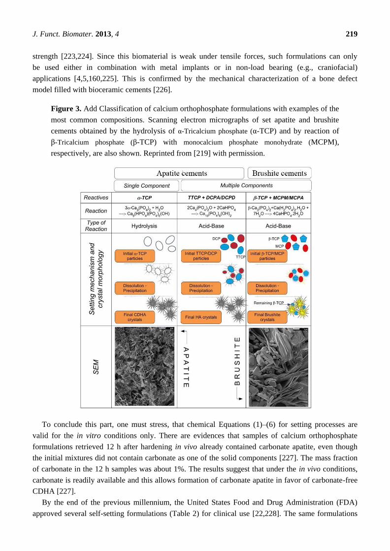
summarized in Figure 3 [219].
Further, there is a single-phase formulation consisting of K- and Na- containing CDHA (with the
Ca/P ionic ratio of 1.64 ± 0.02) that sets and hardens after mixing with an aqueous solution of sodium
citrate and sodium orthophosphate [220]. After setting, this formulation gives rise to formation of a
weak cement (the compressive strength of 15 ± 3 MPa) consisting of the ion-substituted CDHA again
(presumably, with smaller Ca/P ionic ratio), mimicking the bone mineral. Unfortunately, neither the
setting reaction nor the setting mechanism of this cement has been disclosed [220].
The hydration process of calcium orthophosphate formulations is slightly exothermic and undergoes
five periods: initiating period, induction period, accelerating period, decelerating period and
terminating period [221]. For the classical Brown-Chow formulation, the activation energy of the
hydration reaction is 176 kJ/mol [222]. The rate of heat liberation during the solidification of calcium
orthophosphate formulations is low. The results of adiabatic experiments showed that the temperature
rise arrived at the highest value of 37 °C 3 h later, which would cause no harm to surrounding
tissues [221]. The results showed that the hardening process of that formulation was initially controlled
by dissolution of the reactants in a 4 h period and subsequently by diffusion through the product layer
of CDHA around the grains [136]. In general, setting of calcium orthophosphate formulations occurs
mostly within the initial ~6 h, yielding an ~80% conversion to the final products with the volume
almost constant during setting (i.e., shrinkage is small). However, after hardening, the formulations
always form brittle bioceramics with the tensile strength of 5 to 20 times lower than the compression
J. Funct. Biomater. 2013, 4 219
strength [223,224]. Since this biomaterial is weak under tensile forces, such formulations can only
be used either in combination with metal implants or in non-load bearing (e.g., craniofacial)
applications [4,5,160,225]. This is confirmed by the mechanical characterization of a bone defect
model filled with bioceramic cements [226].
Figure 3. Add Classification of calcium orthophosphate formulations with examples of the
most common compositions. Scanning electron micrographs of set apatite and brushite
cements obtained by the hydrolysis of α-Tricalcium phosphate (α-TCP) and by reaction of
β-Tricalcium phosphate (β-TCP) with monocalcium phosphate monohydrate (MCPM),
respectively, are also shown. Reprinted from [219] with permission.
To conclude this part, one must stress, that chemical Equations (1)–(6) for setting processes are
valid for the in vitro conditions only. There are evidences that samples of calcium orthophosphate
formulations retrieved 12 h after hardening in vivo already contained carbonate apatite, even though
the initial mixtures did not contain carbonate as one of the solid components [227]. The mass fraction
of carbonate in the 12 h samples was about 1%. The results suggest that under the in vivo conditions,
carbonate is readily available and this allows formation of carbonate apatite in favor of carbonate-free
CDHA [227].
By the end of the previous millennium, the United States Food and Drug Administration (FDA)
approved several self-setting formulations (Table 2) for clinical use [22,228]. The same formulations
J. Funct. Biomater. 2013, 4 220
also received a Conformite Europene (CE) mark for certain maxillofacial indications and for use as
bone-void fillers in the specific non-load-bearing orthopedic indications [160]. The major properties of
these formulations are available in literature [22]. An extended list of the available formulations is
presented in Table 3 [149], while even more formulations are in experimental stages. Other lists of the
commercially available injectable bone cements with their chemical composition (when obtainable)
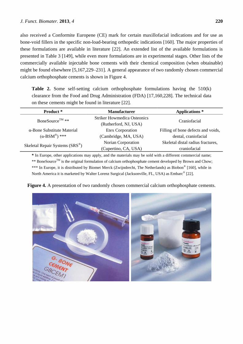
might be found elsewhere [5,167,229–231]. A general appearance of two randomly chosen commercial
calcium orthophosphate cements is shown in Figure 4.
Table 2. Some self-setting calcium orthophosphate formulations having the 510(k)
clearance from the Food and Drug Administration (FDA) [17,160,228]. The technical data
on these cements might be found in literature [22].
Product * Manufacturer Applications *
BoneSourceTM ** Striker Howmedica Osteonics
(Rutherford, NJ, USA) Craniofacial
α-Bone Substitute Material
(α-BSM®) ***
Etex Corporation
(Cambridge, MA, USA)
Filling of bone defects and voids,
dental, craniofacial
Skeletal Repair Systems (SRS®) Norian Corporation
(Cupertino, CA, USA)
Skeletal distal radius fractures,
craniofacial
* In Europe, other applications may apply, and the materials may be sold with a different commercial name;
** BoneSourceTM is the original formulation of calcium orthophosphate cement developed by Brown and Chow;
*** In Europe, it is distributed by Biomet Merck (Zwijndrecht, The Netherlands) as Biobon® [160], while in
North America it is marketed by Walter Lorenz Surgical (Jacksonville, FL, USA) as Embarc® [22].
Figure 4. A presentation of two randomly chosen commercial calcium orthophosphate cements.

J. Funct. Biomater. 2013, 4 221
Table 3. A list of the commercial self-setting calcium orthophosphate formulations with
the producer, product name, composition (when available) and main end-product. The
end-product of the reactions can be either an apatite (CDHA, carbonate apatite, etc.) or
brushite (=DCPD) [149].
Producer Commercial name Composition Product
aap Implantate
(GER) OsteoCem®
Powder: calcium orthophosphates
(details unknown); Solution: unknown apatite
Berkeley Advanced
Biomaterials (U.S.)
Cem-OsteticTM
Powder: calcium orthophosphates
(details unknown); Solution: water apatite
Tri-OsteticTM Powder: calcium orthophosphates
(details unknown); Solution: water apatite
Biomatlante (FR) MCPC Powder: mainly α-TCP, ACP, BCP (HA + β-TCP);
Solution: phosphate buffered solution apatite
Biomet (U.S.)
Interpore (U.S.) Calcibon®
Powder: α-TCP (61%), DCPA (26%), CaCO3 (10%),
CDHA (3%); Solution: H2O, Na2HPO4 apatite
Walter Lorenz
Surgical (GER) MimixTM
Powder: TTCP, α-TCP, trisodium citrate; Solution:
citric acid aqueous solution apatite
Quick Set MimixTM
Powder: Calcium orthophosphate powders, trisodium
citrate; Solution: citric acid aqueous solution apatite
Calcitec (U.S.) Osteofix Powder: calcium orthophosphate and calcium oxide
powders; Solution: phosphate buffer apatite
ETEX (U.S.)
α-BSM®; Embarc;
Biobon
Powder: ACP (50%), DCPD (50%);
Solution: un-buffered aqueous saline solution apatite
β-BSM®
Composition: could not be found
(it has apparently a higher compressive strength
and better injectability than α-BSM®)
apatite
γ-BSM® Composition: could not be found (putty consistency) apatite
OssiPro Composition: could not be found; the cement is
claimed to be macroporous after hardening apatite
CarriGen
Composition: synthetic calcium orthophosphate,
sodium carboxymethylcellulose,
sodium bicarbonate and sodium carbonate
apatite
Graftys (FR)
Graftys® HBS
Powder: α-TCP (78%), DCPD (5%), MCPM (5%),
CDHA (10%), hydroxypropylmethylcellulose (2%);
Solution: 5% Na2HPO4 aqueous solution
apatite
Graftys® Quickset
Composition: calcium orthophosphate salts,
hydroxypropylmethylcellulose and
orthophosphate-based aqueous solution
apatite
Kasios (FR)
Jectos Eurobone® Powder: β-TCP (98%), Na2P2O7 (2%);
Solution: H2O, H3PO4 (3.0 M), H2SO4 (0.1 M) brushite
Jectos+ Composition: could not be found
(likely to be close to that of Jectos) brushite

J. Funct. Biomater. 2013, 4 222
Table 3. Cont.
Producer Commercial name Composition Product
Kyphon (U.S.) KyphOsTM
Powder: β-TCP (77%), Mg3(PO4)2 (14%),
MgHPO4 (4.8%), SrCO3 (3.6%);
Solution: H2O, (NH4)2HPO4 (3.5 M)
apatite
Merck (GER)
Biomet (U.S.) Biocement D
Powder: 58% α-TCP, 24% DCPA, 8.5% CaCO3,
8.5% CDHA; Solution: 4 wt% Na2HPO4 in water apatite
Mitsubishi
Materials (J)
Biopex®
Powder: α-TCP (75%), TTCP (20%–18%),
DCPD (5%), HA (0%–2%)
Solution: H2O, Na succinate (12%–13%),
Na chondroitin sulfate (5%–5.4%)
apatite
Biopex®-R
Powder: α-TCP, TTCP, DCPD, HA, Mg3(PO4)2,
NaHSO3; Solution: H2O, Na succinate,
Na chondroitin sulfate
Apatite
Produits Dentaires
SA (CH)
CalciphOs (CH)
VitalOs4
Solution 1: β-TCP (1.34 g), Na2H2P2O7 (0.025 g),
H2O, salts (0.05 M PBS solution, pH 7.4);
Solution 2: MCPM (0.78 g), CaSO4·2H2O (0.39 g),
H2O, H3PO4 (0.05 M)
Brushite
Shanghai Rebone
Biomaterials Co
(CN)
Rebone Powder: TTCP, DCPA; Solution: H2O Apatite
Skeletal Kinetics
(U.S.)
CallosTM Composition: α-TCP, CaCO3, MCPM;
Solution: sodium silicate Apatite
Callos InjectTM Composition: α-TCP and unknown compounds
(likely to be close to that of CallosTM) Apatite
OsteoVation
EX Inject
Probably similar to Callos InjectTM
(Product produced by S.K. but sold by OsteoMed) Apatite
Stryker (U.S.)
Leibinger (GER) BoneSourceTM
Powder: TTCP (73%), DCPD (27%); Solution: H2O,
mixture of Na2HPO4 and NaH2PO4 Apatite
Stryker (U.S.) HydroSetTM Powder: TTCP, DCPD, trisodium citrate; Solution:
H2O, polyvynilpyrrolidone, Na orthophosphate Apatite
DePuy Synthes
(U.S.)
Norian® SRS
Norian® CRS
Powder: α-TCP (85%), CaCO3 (12%), MCPM (3%);
Solution: H2O, Na2HPO4 Apatite
Norian® SRS Fast
Set Putty Norian®
CRS Fast Set Putty
Composition: could not be found
(likely to be close to that of Norian SRS/CRS) Apatite
Norian Drillable Composition: calcium orthophosphate powder,
bioresorbable fibers and Na hyaluronate solution Apatite
ChronOSTM Inject
Powder: β-TCP (73%), MCPM (21%),
MgHPO4·3H2O (5%), MgSO4 (< 1%), Na2H2P2O7
(< 1%); Solution: H2O, Na hyaluronate (0.5%)
Brushite
Teknimed (FR)
Cementek® Powder: α-TCP, TTCP, Na glycerophosphate;
Solution: H2O, Ca(OH)2, H3PO4 Apatite
Cementek® LV Powder: α-TCP, TTCP, Na glycerophosphate,
dimethylsiloxane; Solution: H2O, Ca(OH)2, H3PO4 Apatite

J. Funct. Biomater. 2013, 4 223
3. Two Major Types of the Self-Setting Calcium Orthophosphate Formulations
3.1. Apatite-Forming Formulations
As indicated by its name, apatite-forming formulations have a poorly crystalline precipitated HA
and/or CDHA as the final product of setting reactions [chemical Equations (1) and (4)–(6)], although
traces of un-reacted starting compounds can be present [130]. Self-setting FA-forming formulations
are also known; they can be prepared by the same way but in the presence of soluble
F−-ions [37,232,233]. Due to the initial presence of carbonates, such commercial formulations as
Norian SRS®
and Biocement D®
(Table 3) form a non-stoichiometric carbonate apatite or dahllite
[Ca8.8(HPO4)0.7(PO4)4.5(CO3)0.7(OH)1.3] as the end-product [69,234]. As both CDHA and carbonate
apatite are formed in an aqueous environment and have a low crystallinity, they appear to be rather
similar to the biological apatite of bones and teeth. These properties are believed to be responsible for
their excellent in vivo resorption characteristics. Conventional apatite-forming formulations contain
TCP and/or TTCP phases in their powder components [230], while a single component formulation
consisting of K- and Na- containing CDHA is also available [220]. The reactivity of TCP-based
apatite-forming formulations was found to vary as a function of TCP crystal phase, crystallinity and
particle size [235,236]. Generally, a higher reactivity is observed with a thermodynamically less stable
phase (from β-TCP to α-TCP and further to ACP) and with a smaller particle size [199]. Nominally, it
might be stated that formation of apatites through self-setting reactions is a sort of a biomimetic
process because it occurs in physiological environment and at body temperature [53]; however, both
the crystallization kinetics and a driving force are very far away from the biomimeticity. A unique
feature of the hardened apatite-forming formulations is that the force linking the newly formed crystals
(of both CDHA and carbonate apatite) is weak; therefore, the crystals can be easily detached from the
bulk of hardened formulations, especially after dissolution has partly occurred. When this happens,
osteoclasts and other cells can easily ingest the apatite crystals [237].
Immediately after implantation, any formulation becomes exposed to blood and other tissue fluids
that delay the setting time. Intrinsic setting time for apatite-forming formulations has been extensively
studied and it appears to be rather long. For example, for the original formulation by Brown and Chow
it ranges from 15 to 22 min [13,14]. This may result in procedural complications. To remedy this, the
amount of liquid might be reduced to a possible minimum. In such cases, all apatite-forming
formulations look like viscous and easily moldable pastes, which tend to be difficult to inject. Besides
playing with the P/L ratio, the setting time can also be reduced by using additives to the liquid phase
(which is distilled water in the Brown-Chow formulation [13,14]). The list of possible additives
includes H3PO4, MCPM and other soluble orthophosphates. These additives promote dissolution of the
initial solids by lowering the solution pH. In such cases, a setting time in the range of 10–15 min can
be obtained [193–201,238]. The influence of soluble orthophosphates (e.g., Na2HPO4 or NaH2PO4) on
the setting time is explained by the fact that dissolution of DCPA and formation of CDHA during
setting occur in a linear fashion, thus avoiding early formation of CDHA. This is important because
too early formation of CDHA might engulf un-reacted DCPA, which slows down DCPA dissolution
and thus the setting kinetics becomes slower, while the presence of sodium orthophosphates prevents
DCPA particles from being isolated [239]. Particle size [218,240,241], temperature and initial

J. Funct. Biomater. 2013, 4 224
presence of HA powders as seeds in the solid phase are other factors that influence the setting
time [13,14,53,235,236]; however, in vitro studies demonstrated that these parameters did not affect
significantly [130]. On the other hand, particle size reduction was found to result in a significant
decrease in both initial and final setting times [218,240,241], an acceleration of the hardening
rate [218] and hydration kinetics of the hardening formulation [241]. In general, smaller crystals or
particles result in a higher supersaturation degrees achieved in the self-setting pastes, which favors
crystal nucleation and results in the precipitation of greater many and smaller needle-like crystals,
instead of the larger plate-like crystals formed when bigger particles are used (Figure 5) [219]. These
different microstructures give rise to different pore size distributions in the set formulations (bottom
part of Figure 5). Besides, the crystallite sizes of the final products can be strongly reduced by
increasing the specific surface of the starting powders, which allows developing formulations with
tailored structures at the micro and nano-scale levels [218]. Unfortunately, an unclear correlation was
found between the particle dimensions of the initial calcium orthophosphates and mechanical
properties of the hardened products: namely, a significant increase in compressive strength and storage
modulus was reported for some formulations [240,241] but a minor effect on compressive strength was
discovered for other ones [218]. This inconsistence is not surprising because the manufacturing
methods used to produce test samples varied from one author to the other. Therefore, the only
remaining fact is that the hardened formulations are brittle and hence worthless for load-bearing
applications [4,5].
Figure 5. A schematic drawing of the influence of the particle dimensions on the
properties of self-setting formulations. Reprinted from [219] with permission.
Setting process of the most types of apatite-forming formulations occurs according to just one
chemical reaction [see chemical Equations (1) and (4)–(6)] and at near the physiological pH, which
J. Funct. Biomater. 2013, 4 225
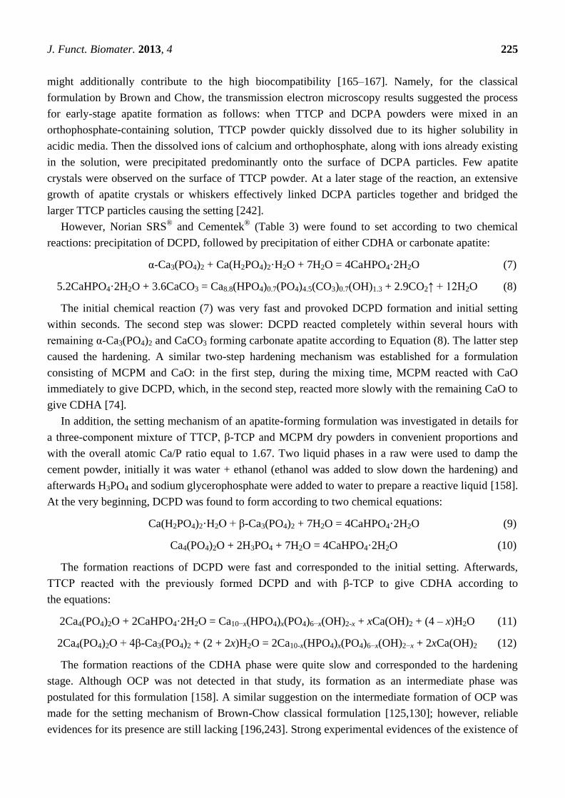
might additionally contribute to the high biocompatibility [165–167]. Namely, for the classical
formulation by Brown and Chow, the transmission electron microscopy results suggested the process
for early-stage apatite formation as follows: when TTCP and DCPA powders were mixed in an
orthophosphate-containing solution, TTCP powder quickly dissolved due to its higher solubility in
acidic media. Then the dissolved ions of calcium and orthophosphate, along with ions already existing
in the solution, were precipitated predominantly onto the surface of DCPA particles. Few apatite
crystals were observed on the surface of TTCP powder. At a later stage of the reaction, an extensive
growth of apatite crystals or whiskers effectively linked DCPA particles together and bridged the
larger TTCP particles causing the setting [242].
However, Norian SRS®
and Cementek®
(Table 3) were found to set according to two chemical
reactions: precipitation of DCPD, followed by precipitation of either CDHA or carbonate apatite:
α-Ca3(PO4)2 + Ca(H2PO4)2·H2O + 7H2O = 4CaHPO4·2H2O (7)
5.2CaHPO4·2H2O + 3.6CaCO3 = Ca8.8(HPO4)0.7(PO4)4.5(CO3)0.7(OH)1.3 + 2.9CO2↑ + 12H2O (8)
The initial chemical reaction (7) was very fast and provoked DCPD formation and initial setting
within seconds. The second step was slower: DCPD reacted completely within several hours with
remaining α-Ca3(PO4)2 and CaCO3 forming carbonate apatite according to Equation (8). The latter step
caused the hardening. A similar two-step hardening mechanism was established for a formulation
consisting of MCPM and CaO: in the first step, during the mixing time, MCPM reacted with CaO
immediately to give DCPD, which, in the second step, reacted more slowly with the remaining CaO to
give CDHA [74].
In addition, the setting mechanism of an apatite-forming formulation was investigated in details for
a three-component mixture of TTCP, β-TCP and MCPM dry powders in convenient proportions and
with the overall atomic Ca/P ratio equal to 1.67. Two liquid phases in a raw were used to damp the
cement powder, initially it was water + ethanol (ethanol was added to slow down the hardening) and
afterwards H3PO4 and sodium glycerophosphate were added to water to prepare a reactive liquid [158].
At the very beginning, DCPD was found to form according to two chemical equations:
Ca(H2PO4)2·H2O + β-Ca3(PO4)2 + 7H2O = 4CaHPO4·2H2O (9)
Ca4(PO4)2O + 2H3PO4 + 7H2O = 4CaHPO4·2H2O (10)
The formation reactions of DCPD were fast and corresponded to the initial setting. Afterwards,
TTCP reacted with the previously formed DCPD and with β-TCP to give CDHA according to
the equations:
2Ca4(PO4)2O + 2CaHPO4·2H2O = Ca10−x(HPO4)x(PO4)6−x(OH)2-x + xCa(OH)2 + (4 – x)H2O (11)
2Ca4(PO4)2O + 4β-Ca3(PO4)2 + (2 + 2x)H2O = 2Ca10-x(HPO4)x(PO4)6−x(OH)2−x + 2xCa(OH)2 (12)
The formation reactions of the CDHA phase were quite slow and corresponded to the hardening
stage. Although OCP was not detected in that study, its formation as an intermediate phase was
postulated for this formulation [158]. A similar suggestion on the intermediate formation of OCP was
made for the setting mechanism of Brown-Chow classical formulation [125,130]; however, reliable
evidences for its presence are still lacking [196,243]. Strong experimental evidences of the existence of

J. Funct. Biomater. 2013, 4 226
a transient OCP phase during setting were found in still another study; however, that system contained
sodium silicates [41]. In all cases, OCP was suggested to appear as an intermediate because it was a
faster forming phase than CDHA. This hypothesis is based upon the classical studies performed by
Brown et al. [244–246], about the precursor phase formation during chemical crystallization of apatites
in aqueous solutions.
Solubility of the hardened apatite-forming formulations in aqueous solutions is expected to be
rather similar to that of bone mineral. This means that they are relatively insoluble at neutral pH and
increasingly soluble as pH drops down; this is an important characteristic of normal bone mineral that
facilitates controlled dissolution by osteoclasts [234].
To conclude this part, one should mention, that in 2000 the U.S. bone substitute market for Norian
SRS®
accounted for ~15% of the total sales, followed by BoneSourceTM
at ~13%, and α-BSM®
at
~8.5% [160].
3.2. Brushite-Forming Formulations
As indicated by its name, DCPD is the major product of the setting reaction for brushite-forming
formulations [chemical Equations (2) and (3)], although traces of the un-reacted starting compounds
can be present. Mirtchi and Lemaître [173] and independently Bajpai et al. [32] introduced this type of
the cements in 1987. Up to now, several formulations have been already proposed, e.g.,
β-TCP + MCPM [173,174], β-TCP + H3PO4 [32–34] and TTCP + MCPM + CaO [247]. The full list of
brushite-forming formulations is available in a topical review on the subject [248]. As seen from the
chemical composition, all types of the brushite-forming formulations are set by the acid-base
interaction only. As DCPD can only be precipitated at the solution pH < 6 (Table 1), the pastes of the
self-setting brushite-forming formulations are always acidic during hardening [34,249]. For example,
during setting of a β-TCP + MCPM formulation, the formulation pH varies from very acidic pH values
of ~2.5, to almost neutral pH values of ~6.0 [34]. Replacing MCPM by H3PO4 renders the paste very
acidic for the initial ~30 s but then the pH profile follows that obtained with MCPM. It is important to
notice, that β-TCP + H3PO4 formulations have several advantages over β-TCP + MCPM ones, namely:
(i) easier and faster preparation; (ii) a better control of the chemical composition and reactivity;
(iii) improved physico-chemical properties, such as longer setting times and larger tensile strengths
due to a higher homogeneity. However, the use of H3PO4 might impair the biocompatibility of the
formulations, due to low pH values during setting [34].
As the solubility of calcium orthophosphates generally decreases with increasing of their basicity
(Table 1 and Figure 1), the setting time of brushite-forming formulations much depends on the
solubility of a basic phase: the higher its solubility, the faster the setting time. Therefore, the setting
time of formulations made of MCPM + a basic calcium orthophosphate increases in the order:
HA > β-TCP > α-TCP [4,5]. For example, HA + MCPM mixtures have a setting time of several
minutes, β-TCP + MCPM mixtures—of 30 to 60 s and α-TCP + MCPM mixtures—of a few
seconds [173,174]. Furthermore, if brushite-forming formulations contain an excess of a basic phase,
the equilibrium pH will be given by the intersection of the solubility isotherms of the basic phase
with that of DCPD. For example, the equilibrium pH values of β-TCP + MCPM, HA + MCPM and
TTCP + MCPM mixtures were found to be 5.9, 4.2 and 7.6, respectively [4,5]. Follow-up of the

J. Funct. Biomater. 2013, 4 227
chemical composition by 31
P solid-state nuclear magnetic resonance (NMR) technique enabled to show
that the chemical setting process for β-TCP + MCPM formulation reached the end after ~20 min [250].
Nevertheless, despite this initial high reactivity, the hardening reaction of brushite-forming
formulations typically lasts one day until completion [235,236]. Additives that inhibit the crystal
growth of DCPD have successfully been used to increase the setting time of β-TCP + MCPM
mixtures [251]. Interestingly, contrary to apatite-forming formulations, the brushite-forming ones can be
initially liquid and still set within a short period of time [4,5].
By itself, brushite is remarkably biocompatible and bioresorbable [249]. Due to both a better
solubility of DCPD if compared to that of CDHA (Table 1 and Figure 1) and metastability of DCPD
under physiological conditions [252], after implantation brushite-forming formulations are faster
degradable than apatite-forming ones [253–255]. They are quickly resorbed in vivo and suffered from a
rapid decrease in strength (although the mechanical properties of the healing bone increase as bone
ingrowth occurs [51]). Short setting times, low mechanical strength and limited injectability seem to
prevent brushite-forming formulations from a broader clinical application. However, the major reason
why they are not more widespread is probably not related to the mechanical issues but just to a later
arrival on the market. Use of sodium citrate or citric acid as setting retardants is an option to get more
workable and less viscous pastes of brushite-forming formulations [38,256–259]. Similar effect might
be achieved by addition of chondroitin 4-sulfate [260] and glycolic acid [261]. For the formulations
with H3PO4 as the initial reactant [chemical Equation (3)], acid deficient formulations were also found
to improve the workability. In this case, the setting reaction might be described by the following
chemical equation [259]:
3.7β-Ca3(PO4)2 + H3PO4 + 27.8H2O = 3CaHPO4·2H2O + 2.7β-Ca3(PO4)2 + 21H2O (13)
Although several studies revealed that too much of DCPD in a given volume was not detrimental to
the biological properties of brushite-forming formulations [51,234,247], occasionally, when large
quantities of them were used, a certain degree of tissue inflammation during the first weeks of in vivo
implantation were reported [255,259,262]. Further investigations indicated that the inflammatory could
be due to a partial transformation of DCPD into CDHA with release of orthophosphoric acid [263]:
(10 − x)CaHPO4·2H2O = Ca10−x(HPO4)x(PO4)6−x(OH)2−x + (4 – x)H3PO4 + (18 – x)H2O (14)
Transformation of DCPD into CDHA occurs via two successive processes: dissolution and
precipitation [264] and can be retarded by adding magnesium ions to the formulations, thus reducing
the possibility of inflammation [4,5]. The aforementioned case of acid deficient formulations
[chemical Equation (13)] is the second option, because it reduces the amount of un-reacted acid [259]
with an option to consume liberating in chemical Equation (14) H3PO4 by the excess of β-TCP.
Implantation of previously set brushite-forming formulations might be the third option, because a solid
bioceramics was found to be better tolerated than paste implants. Besides, more bone was formed at
the solid implant contact and the solid material degraded not so rapidly [265]. For the hardened
brushite formulations, a linear degradation rate of 0.25 mm/week was reported [266]. This rapid
degradation rate might lead to formation of an immature bone. Adding β-TCP granules to the
self-setting pastes could solve this problem because the granules might act as bone anchors and
encourage formation of a mature bone [266,267].

J. Funct. Biomater. 2013, 4 228
To finalize this topic, one should briefly mention on a possibility to precipitate DCPA (monetite)
instead of DCPD. It is well known, that DCPA might be crystallized under the same conditions as
DCPD but either from aqueous solutions at elevated (>~90 °C) temperatures or at ambient conditions
but in water-deficient environments [249,268]. Therefore, monetite-forming self-setting calcium
orthophosphate formulations could exist. Indeed, there are several publications, in which formation of
monetite instead of brushite has been detected as the final product [29,35,269–272]. In addition,
monetite might be formed during a prolonged storage of dry powders of brushite-forming formulations
in normal laboratory atmosphere (~60% relative humidity) [273]. Therefore, one might claim on an
incipient topic of self-setting monetite-forming calcium orthophosphate formulations, which has a
potential to be developed in a league of its own.
Additional details on the self-setting brushite-forming formulations might be found in a recent
review on the subject [248].
4. Various Properties
4.1. Setting and Hardening
Generally, self-setting calcium orthophosphate formulations must set slowly enough to provide
sufficient time to a surgeon to perform implantation but fast enough to prevent delaying the operation.
Ideally, good mechanical properties should be reached within minutes after initial setting. Two main
experimental approaches are used to study the setting process: a batch approach and a continuous
approach. In the batch approach, the setting reaction is stopped at various times and the resulting
samples are analyzed to determine, e.g., the composition and compressive strength of the
samples [235,236]. There are currently two standardized methods to apply this approach, namely,
Gillmore needles method (ASTM C266-89) [274] and Vicat needle method (ASTM C191-92) [275].
The idea of both methods is to examine visually the sample surfaces to decide whether the formulation
has already set, i.e., if no mark can be seen on the surface after indentation. Besides, the setting process
might be monitored in real time by non-destructive methods (the continuous approach), e.g., using
pulse-echo ultrasound technique [276,277], isothermal differential scanning calorimetry [198,199,278–284]
and alternating current (AC) impedance spectroscopy [285]. For example, calorimetry measurements
suggested that in Equation (2) the endothermic MCPM dissolution and the highly exothermic β-TCP
dissolution occurred simultaneously, followed by the exothermic crystallization of DCPD [282].
Thus, brushite-forming formulations usually warm upon final setting [278]. Moreover, acid-base
reactions (1)–(3) can be and have been analyzed by measuring the pH evolution of diluted pastes [235].
In addition, non-destructive methods of Fourier-transform infrared spectroscopy [40,41,43,283,286],
solid state NMR [250], X-ray diffraction [40,43,66,175,287] and energy dispersive X-ray
diffraction [40–43,288–290] might be applied as well. The latter techniques proved to be powerful
even though they have limitations such as the time required for each measurement (250 s for an X-ray
diffraction scan is a problem for fast setting reactions). In addition, the analysis is often located at the
sample surfaces where evaporation and thermal effects can modify the reaction rates if compared to
those in the bulk. Furthermore, the continuous approaches are indirect, which markedly complicates an
interpretation of the collected data, particularly in complex formulations [235].
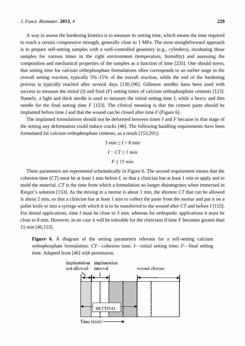
J. Funct. Biomater. 2013, 4 229
A way to assess the hardening kinetics is to measure its setting time, which means the time required
to reach a certain compressive strength, generally close to 1 MPa. The most straightforward approach
is to prepare self-setting samples with a well-controlled geometry (e.g., cylinders), incubating those
samples for various times in the right environment (temperature, humidity) and assessing the
composition and mechanical properties of the samples as a function of time [235]. One should stress,
that setting time for calcium orthophosphate formulations often corresponds to an earlier stage in the
overall setting reaction, typically 5%–15% of the overall reaction, while the end of the hardening
process is typically reached after several days [130,196]. Gillmore needles have been used with
success to measure the initial (I) and final (F) setting times of calcium orthophosphate cements [123].
Namely, a light and thick needle is used to measure the initial setting time I, while a heavy and thin
needle for the final setting time F [153]. The clinical meaning is that the cement paste should be
implanted before time I and that the wound can be closed after time F (Figure 6).
The implanted formulations should not be deformed between times I and F because in that stage of
the setting any deformation could induce cracks [46]. The following handling requirements have been
formulated for calcium orthophosphate cements, as a result [153,291]:
3 min ≤ I < 8 min
I − CT ≥ 1 min
F ≤ 15 min
These parameters are represented schematically in Figure 6. The second requirement means that the
cohesion time (CT) must be at least 1 min before I, so that a clinician has at least 1 min to apply and to
mold the material. CT is the time from which a formulation no longer disintegrates when immersed in
Ringer’s solution [153]. As the mixing in a mortar is about 1 min, the shortest CT that can be allowed
is about 2 min, so that a clinician has at least 1 min to collect the paste from the mortar and put it on a
pallet knife or into a syringe with which it is to be transferred to the wound after CT and before I [153].
For dental applications, time I must be close to 3 min, whereas for orthopedic applications it must be
close to 8 min. However, in no case it will be tolerable for the clinicians if time F becomes greater than
15 min [46,153].
Figure 6. A diagram of the setting parameters relevant for a self-setting calcium
orthophosphate formulation: CT—cohesion time; I—initial setting time; F—final setting
time. Adapted from [46] with permission.

J. Funct. Biomater. 2013, 4 230
4.2. Phase Mixing
In the clinical situation, self-setting calcium orthophosphate formulations can be either applied by
the fingertips of a surgeon or injected from a syringe to the defect area of a bone. The first type of the
application requires formulation of high-viscous self-setting pastes and putties, which can be applied
manually as dough, while the second type requires formulation of low-viscosity compositions, which
can be applied by injection from a syringe [153]. Currently, injection appears to be the preferred
method between these two major options. Thus, a compromise must be found between a high viscosity
leading to too high injection forces and a low viscosity increasing the risk of extravasations. Thus,
viscosity values in the range of 100–2000 Pa·s are generally considered to be adequate [292].
In any case, before using a surgeon needs to have a powder and a liquid be mixed properly and
thoroughly (to avoid the powder/liquid encapsulation) within the prescribed time. This process must be
performed in a sterile environment. Therefore, a mixing procedure is very important because prior to
be injected, a self-setting paste must be transferred from a mixing chamber into a syringe. Ideally,
this should be done without trapping air bubbles by the formulation [293]. Earlier, most calcium
orthophosphate formulations were manually mixed with aqueous solutions using a mortar and either a
pestle or a spatula. That time, some concerns were raised about an insufficient and inhomogeneous
mixing thus compromising the implant strength, as well as on inconsistencies between operators
causing unpredictable variations in graft performance [294]. Mechanical mixing (e.g., by either an
electrically powered mixing machine of Norian SRS/CRS®
(Figure 7) or Mini-malax®
mixing system
for Cementek®
, produced by Teknimed S.A., City, Country) is the modern approach. It allows mixing
the pastes within 60–80 s and enables a rapid and reliable filling of the application syringe [230].
Besides, a powder and a solution might be placed into a syringe and mixed inside a shaker to produce a
consistent self-setting paste of the desired viscosity [293]. A mechanical mixing was found to decrease
both the mean viscosity of the curing pastes and variability in the viscosity at a given time [295].
However, it did not improve the mechanical strength of the hardened formulations [4,5].
Of the commercial formulations, listed in Table 2, Norian SRS®
is sold as a reactant pack
containing two components: a mixture of dry powders (MCPM + α-TCP + CaCO3) and a liquid
(aqueous solution of Na2HPO4). The components are mixed in the operating room. The paste that is
formed is malleable and injectable for ~5 min; it hardens within ~10 min after injection [22,234].
However, data are available that out of 4.5 mL Norian SRS®
cement paste ~3 mL is injectable only,
whereas up to 1.5 mL of the paste might remain uninjectable from the syringe [46]. This phenomenon
is prescribed to the formulation rheology and its interaction with the hydraulic forces of the syringe.
α-BSM®
(Table 2) is also a two-component system; it is prepared from a mixture of ACP and DCPD
powders and a saline solution [193]. Biopex®
consists of four different calcium orthophosphates:
75 wt% α-TCP, 18 wt% TTCP, 5 wt% DCPD and 2 wt% HA (Table 3). The aqueous solution contains
12 wt% sodium succinate and 5 wt% sodium chondroitin sulfate [296]. Effects of liquid phase on
the basic properties of Biopex®
were investigated. When mixed with neutral sodium hydrogen
orthophosphate or succinic acid disodium salt solution, the initial setting times of the cement were
19.4 ± 0.55 and 11.8 ± 0.45 min, respectively. These setting times were much shorter than that of
distilled water, 88.4 ± 0.55 min [297]. Biopex®
is mixed with a spatula inside a syringe that can be
J. Funct. Biomater. 2013, 4 231
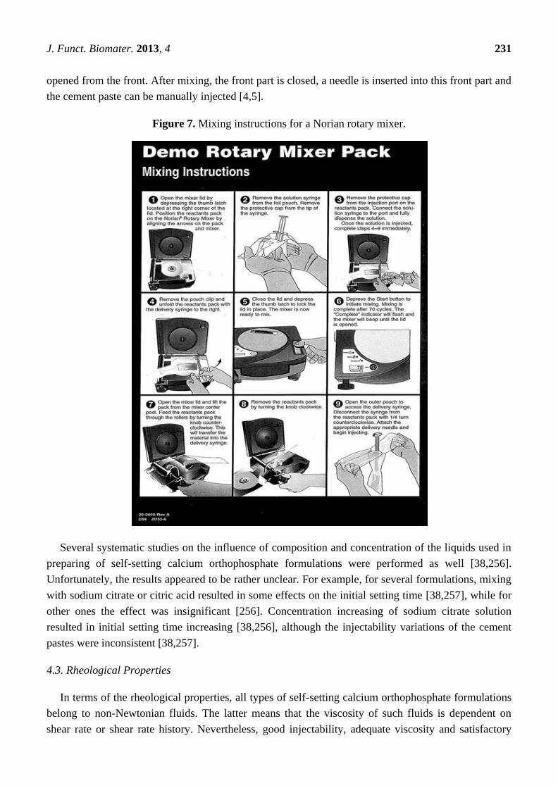
opened from the front. After mixing, the front part is closed, a needle is inserted into this front part and
the cement paste can be manually injected [4,5].
Figure 7. Mixing instructions for a Norian rotary mixer.
Several systematic studies on the influence of composition and concentration of the liquids used in
preparing of self-setting calcium orthophosphate formulations were performed as well [38,256].
Unfortunately, the results appeared to be rather unclear. For example, for several formulations, mixing
with sodium citrate or citric acid resulted in some effects on the initial setting time [38,257], while for
other ones the effect was insignificant [256]. Concentration increasing of sodium citrate solution
resulted in initial setting time increasing [38,256], although the injectability variations of the cement
pastes were inconsistent [38,257].
4.3. Rheological Properties
In terms of the rheological properties, all types of self-setting calcium orthophosphate formulations
belong to non-Newtonian fluids. The latter means that the viscosity of such fluids is dependent on
shear rate or shear rate history. Nevertheless, good injectability, adequate viscosity and satisfactory

J. Funct. Biomater. 2013, 4 232
cohesion are required for the successful biomedical applications [298,299]. Among them, injectability
is defined as an ability of a formulation to be extruded through a small hole of a long needle
(e.g., 2 mm diameter and 10 cm length) [300,301] (other needles are also applied [302,303]); and for
certain applications, injectability is even a prerequisite. It is measured by the weight percentage of the
formulation that could be injected without demixing from a standard syringe by either a hand or a force
of 100 N maximum (Figure 8). The numerical values are calculated by the following equation [304]:
Inj = (WF − WA)/(WF − WE) × 100%
where Inj is the percentage injectability; WE is the weight of the empty syringe; WF is the weight of the
full syringe and WA is the weight of the syringe after the injection.
Usually, injectability of calcium orthophosphate formulations are varied inversely with their
viscosity, the P/L ratio, as well as the time after starting the mixing of liquid and powder [72,301,305].
In addition, powder reactivity was shown to influence the injectability. Namely, significant differences
were observed between the injection behaviors of the non-hardening β-TCP pastes and self-hardening
α-TCP pastes, α-TCP being less injectable than β-TCP and requiring higher injection loads. What is
more, the parameters affecting powder reactivity were shown also to affect injectability. Thus, whereas
powder calcination resulted in increased injectability, an addition of setting accelerants tended to
reduce the injectability [304]. Furthermore, injectability is improved with smaller particle sizes, with
shorter and larger diameter cannula, as well as at smaller flow rates [300]. Moreover, particle shape of
the powder is also expected to have effects on the injectability. Namely, powders with spherical shape
or round particles are easy to roll and thus good handling properties and injectability are found when
pastes are prepared from such materials. Besides, it should be noted that the paste could become fluid
with less amount of liquid phase since no captured liquid exists in the case of spherical powder [306].
Figure 8. A schematic representation of the experimental setup used to quantify the
injectability of the calcium orthophosphate formulations. Reprinted from [305]
with permission.

J. Funct. Biomater. 2013, 4 233
Unfortunately, when a self-setting formulation, which is a biphasic mixture of a finely divided
ceramic (powder, granules) and a liquid, is submitted to a pressure gradient, the liquid may flow faster
than the solid, resulting in local changes of the paste composition. Specifically, the paste present in the
region of the highest pressure (e.g., close to the plunger of a syringe) may become so depleted in liquid
that the biphasic mixture in this zone is not longer a paste but a wet powder [300,302]. Contrarily, the
paste in the zone of the lowest pressure (e.g., at the cannula tip) is enriched in liquid. Since these
effects are dynamic, the size of the zone depleted in liquid (wet powder) increases during injection,
eventually reaching the tip of the injection device and plugging it. The phenomenon, in which the
pressure applied to the paste provokes a phase separation after a certain injection time, is generally
referred as filter pressing, phase separation or phase migration [149] (see the aforementioned example
for Norian SRS®
[46], in which a thick mass remained inside a syringe).
Possible mechanisms underlying the limited injectability of self-setting calcium orthophosphate
formulations have been discussed in literature [303,307]. In the case of demixing, the exact
composition of the extruded part of the paste becomes unknown. Moreover, due to a deviation from
the initial P/L ratio, it becomes unclear whether the setting behavior and the mechanical and
histological properties of the extruded part are still clinically acceptable. Therefore, a good cohesion of
the paste is necessary in order to avoid these problems [308].
Cohesion (= cohesiveness, ―non-decay‖) is the ability of a paste to keep its geometrical integrity in
an aqueous solution [149]. It is evaluated by measuring the amount of solid particles released from the
formulation prior to its final setting. For self-setting formulations, a bad cohesion may prevent setting
and may lead to negative in vivo reactions due to the release of microparticles [309]. Since a high
cohesion is the result of strong attractive forces among the particles, factors enhancing van der Waals
forces (attractive) and decreasing electrostatic forces (repulsive) can be used to improve cohesion [149].
For example, an appropriate cohesion was achieved when no disintegration of the paste was observed
in the fluid [153,308]. This can be accomplished by keeping a high viscosity for self-setting pastes [22]
or using cohesion promoters (e.g., 1% aqueous solution of sodium alginate [200,310,311], as well as
other chemicals [200,312–314]). Some calcium orthophosphate formulations fulfill both criteria,
e.g., Norian SRS®
, but others fulfill only one or even none of these requirements. For example,
BoneSourceTM
[127] and Cementek®
(Table 3) are not injectable and blood must be kept away from
the implanting site until setting [4,5]. A poor cohesion has been associated to a poor biocompatibility
that might lead to inflammatory reactions [309]. Further details on the cohesion properties of various
calcium orthophosphate pastes are available in literature [308].
Viscosity is a measure of the resistance of a fluid, which is being deformed by either shear stress or
tensile stress. Generally, the viscosity in the range of 100–1000 Pa·s appears to be ideal [315] and,
if possible, a self-setting formulation should have a constant viscosity in the indicated range.
Unfortunately, viscosity of self-setting formulations is not a constant value, which, after a decrease in
the first seconds after mixing, increases considerably during curing, eventually leading to hardening.
Furthermore, viscosity should be high enough to prevent extravasation; therefore, it is very important
to define an adequate injection window [315].

J. Funct. Biomater. 2013, 4 234
4.4. Properties Improving
As written above, the properties of the existing self-setting calcium orthophosphate formulations are
not ideal. Several ways can be adopted to improve them. The first approach consists of injectability
improvement. There are several options for this. Firstly, the injection device can be modified. For
example, shorter cannulas with a larger diameter, as well as smaller injection rates favor a good
injectability. The last option is not so straightforward: for example, Habib et al. have shown that large
injection rates are not detrimental to injectability because of the shear-thinning behavior of many
calcium orthophosphate cements [303]; Secondly, an external energy might be applied. For example,
injectability was improved by ultrasonication, which was believed to result from a reduction in the
injection force versus the filtration force as a result of a lesser reduction in the particle interaction and
the paste flowability [316]; Thirdly, the formulation composition can also be adapted. Namely, a
decrease of the particle size, the P/L ratio and the plastic limit was found to contribute to a better
injectability [300,305]. For example, injectability was found to be unaffected by P/L ratio within the
range of 3.85–4.50 g/mL but drops by nearly 100% between P/L ratio of 4.50 and 5.00 g/mL [38].
However, a decrease in P/L ratio leads to a decrease in the mechanical properties of the self-setting
formulations and cohesion might be destroyed. Furthermore, both the initial and final setting times
decreased markedly with the P/L ratio increasing [256,317]. Therefore, variations in the P/L ratio
appear to be valid to a certain extent only. That is why the manufacturer of Biopex®
suggests using a
P/L ratio of 2.8 or 3.3 g/mL.
Particle size decreasing of calcium orthophosphate crystals is the second approach for the
injectability improvement. For example, α-BSM®
is well injectable because it consists of small
crystals. Even though small particles require a larger amount of mixing liquid to obtain a paste,
injectability and cohesion of such formulations are generally very good [4,5]. An indirect approach is
to add calcium orthophosphate crystals those act as spacers between other particles. For example,
DCPA is added to the formulation of Biocement D®
to improve injectability [4,5]. Similarly, there is
an apatite-forming formulation containing spherical particles of TTCP to improve injectability [318].
Using various chemical additives is the second way to improve the properties of the self-setting
formulations [319]. For example, water demand of can be reduced by ionically modifying the liquid
component, e.g., by adding nontoxic sodium salts of α-hydroxy di- and tri- acids [320,321]. A list of
additives, that have been already studied, includes fluidificants, air-entraining agents, porogens,
workability-improvement agents, setting time controllers and reinforcing additives [182,231,322].
Besides, various radiopacifiers might be used to simplify an un-invasive in vivo monitoring of the
implanted cements [323–327]. The main role of fluidificants is to reduce a mixing time of the
formulations. Citric acid is an example of this reagent; it retards the dissolution-precipitation reactions,
decreases the compressive strength during initial setting but increases its strength in the final stages of
hardening [257]. Furthermore, data are available, that citric acid decreases the setting time and
improves the mechanical properties of the hardened formulations [328]. Adding of surfactants to
the self-setting formulations was found to have two different meanings: they might act as both
air-entraining agents by lowering the surface tension [329,330] and interaction modifiers by shifting
the isoelectric point [331].

J. Funct. Biomater. 2013, 4 235
In addition, studies are available, in which self-setting calcium orthophosphate formulations were
modified by various bioorganic compounds in attempts to influence the bone healing process [332–335].
For example, there is a study, in which a self-setting formulation was set in the presence of
cocarboxylase, glucuronic acid, tartaric acid, α-glucose-1-phosphate, L-arginine, L-aspartic acid and
L-lysine, respectively, with the aim to influence formation and growth of CDHA crystals through the
functional groups of these biomolecules [335]. Except for glucuronic acid, all these modifications were
found to result in the formation of smaller and more agglomerated CDHA particles, which had a
positive impact on the biological performance indicated by first experiments with the human osteoblast
cell line hFOB 1.19. Moreover, initial adhesion of human bone marrow-derived mesenchymal stem
cells was improved on the formulations containing cocarboxylase, arginine and aspartic acid.
Furthermore, cell proliferation was enhanced on the formulations modified with cocarboxylase and
arginine whereas osteogenic differentiation remained unaffected. Besides, the formulations with
arginine and aspartic acid, but not with cocarboxylase, led to a higher BMP-2 binding [335].
Since a good adhesion to bones and other structures allows better transmission of forces at the
formulation-bone interfaces, a proper adhesion between the set formulations and bones is very
important for many surgical procedures. Chemical additives might also improve adhesive properties of
self-setting calcium orthophosphate formulations. For example, it was observed that brushite-forming
formulations set with pyrophosphoric acid in the liquid phase had an increased adherence to various
surfaces such as bone, alumina, sintered HA and stainless steel [336].
Porosity is a very important property to provide good in vivo bioresorption of implanted
biomaterials. Thus, various air-entraining agents and porogens are commonly used to induce
macroporosity of self-setting calcium orthophosphate formulations, ideally, without affecting their
normal setting. For example, crystals of mannitol, CH2OH(CHOH)4CH2OH, were tested as an
air-entraining agent; however, both loss of workability during mixing and severe depreciation of
mechanical properties were discovered simultaneously [337–342]. Other porogenic agents were also
tested to create porosity. The examples include: hydrogen peroxide in the liquid phase [343] and/or
iced [344], crystals of NaHCO3 and Na2HPO4 [345], calcium sulfate [58], calcite [247] and
NaCl [346,347], poly(D,L-lactic-co-glycolic acid) microparticles [348–355], microspheres of
pectin [356], and gelatin [357,358], vesicants [359], cetyltrimethyl ammonium bromide [360],
polytrimethylene carbonate [361], sucrose granules, as well as some immiscible liquids. These
additives could be applied on pre-set formulations only, while the solubility degree of the solid
porogens during setting influences both the content and dimensions of the macroporosity. After
hardening, dissolution of the remaining soluble porogens in either water or body fluids produces
macropores with the dimensions and shapes of the dissolved crystals. One important limitation that can
be envisaged from this route is the need to add a large amount of porogenic agents to guarantee pore
interconnectivity, thus compromising not only the excellent biocompatibility and bioactivity of
self-setting calcium orthophosphate formulations but also their injectability. Another shortcoming is a
lack of strength of the resulting bioceramics, especially if particulates dissolve quickly, greatly limiting
its applications. An innovative approach that aims at overcoming the lack of interconnectivity and
initial strength consists in using resorbable fibers [362–370]. These fibers have the function of initial
reinforcing, providing the needed short-term strength and toughness, and gradually dissolving
afterwards, leaving behind macropores suitable for bone ingrowth. One interesting advantage of long

J. Funct. Biomater. 2013, 4 236
fibers over particulates and short fibers is the fact that once resorbed they can form interconnected
pores inside the solid structure facilitating bone tissue regeneration [371].
One more approach to create porosity consists in adding solid NaHCO3 to the starting powder and
using two different liquids: first, a basic liquid to form the paste, and later an acid liquid to obtain
CO2 bubbles to create porosity [372]. Besides, pore forming CO2 bubbles appear at hardening of
apatite-forming formulations, consisting of an acidic calcium orthophosphate, such as MCPM or
DCPD, and either CaCO3 [30,50,69–71] or NaHCO3 [373–375]. Furthermore, addition of an
effervescent porogen formulation comprised from NaHCO3 (54.52%) and citric acid monohydrate
(45.48%) has been suggested [376]. More to the point, the liquid phase of a formulation might be
initially foamed and subsequently mixed with the self-setting powders. In this case, the setting
reactions transform the liquid foam into a solid, which ideally maintains the geometry, size and shape
of the bubbles (Figure 9). Thus, the liquid foam acts as a template for the macroporosity of the solid
foam [343,377–379]. In addition, several other porosity creation techniques for self-setting calcium
orthophosphate formulations are known and, for further details on the subject, the interested readers
are referred to an excellent review [371].
Figure 9. A schematic drawing of calcium-deficient hydroxyapatite (CDHA) foams
preparation. Initially a liquid was formed by mechanical agitation of an aqueous solution of
a soluble surfactant. Then, the foam was mixed with α-TCP powder, producing a foamed
paste, which was either cast or directly injected into the moulds. The setting reaction
produced hydrolysis of α-TCP to CDHA, which resulted in foam hardening. Reprinted
from [378] with permission.
The major examples of workability-improvement agents, which are added to the self-setting
formulations, include water-soluble polymers. Specifically, polysaccharides [120,132,380–383],
gelatin [317,384–390] and polyacrylic acid [391–393] are of an interest due to their biocompatibility
and good rheological properties. Only small amounts (a few weight %) are needed to dramatically
increase the viscosity of the pastes. Besides, the pastes become more cohesive and highly resistant to
washout immediately after mixing. For example, a 5 wt% sodium chondroitin sulfate solution is used
as mixing liquid in Biopex®
[4,5]. In the case of gelatin, more than a 50% improvement of the
compressive strength was detected [386]. The gelatin-containing formulations after setting were found

J. Funct. Biomater. 2013, 4 237
to exhibit reduced crystallinity, much smaller CDHA crystals and a more compact microstructure; all
these phenomena might be accounted for the improved mechanical properties [387]. In addition, the
presence of gelatin improved mechanical properties of the formulations; in particular, the formulations
containing 2 wt% gelatin were found to harden in an acceptable time and were recommended for
clinical applications [390]. In some cases addition of a gelling agent might cause an increase in
hardening time [394] but this was remedied by the use of a sodium orthophosphate solution as the
liquid phase [166,167]. Most polysaccharide solutions are thixotropic, i.e., the viscosity of the solution
decreases as the shear rate increases. Certain polysaccharides, such as sodium alginate, pectize in
contact with calcium ions. This property can be used to make putty-like cement pastes [22]. However,
only few polysaccharides are accepted for parenteral use [4,5]. Nevertheless, the use of gelling agents
widened a possible application of the self-setting calcium orthophosphate formulations because such
formulations can be used even when complete homeostasis is difficult.
Of two families of the self-setting formulations, brushite-forming ones react generally much
faster than apatite ones. As a result, to satisfy the clinical requirements (Figure 6), setting time of
brushite-forming formulations has to be prolonged, whereas that of apatite-forming ones has to be
shortened [4,5]. According to the aforementioned, setting reactions of any self-setting calcium
orthophosphate formulation consists of three successive stages: (1) dissolution of reactants to saturate
the mixing liquids by calcium and orthophosphate ions; (2) nucleation of crystals from the
supersaturated solutions; (3) growth of crystals. Therefore, experimental approaches to modify the
setting kinetics are to be targeted to these three stages. The available approaches have been
summarized in Table 4 [235]. Furthermore, seven strategies have been described to decrease the
setting time of calcium orthophosphate formulations [236]. They are: (i) mean particle size decreasing
of the initial powders; (ii) the P/L ratio increasing; (iii) pH drop of the mixing liquid to increase
calcium orthophosphate solubility and hence accelerate the chemical transformations; (iv) a nucleating
phase addition, such as a nano-sized HA powder; (v) adding orthophosphate and/or calcium ions into
the mixing liquid to accelerate the setting reaction according to the common-ion effect; (vi) solubility
reducing of the reaction end-product, for example, by adding fluoride ions into the mixing liquid;
(vii) solubility increasing of the starting material by amorphization, e.g., by prolonged milling. For
further details on these strategies and approaches, as well as for application examples, the interested
readers are referred to the original publications [235,236].
Various setting time controllers (accelerators and retardants) are used to influence the setting time.
They include sodium hydrogen pyrophosphate (Na2H2P2O7) and magnesium sulfate (according to other
studies, ions of citrate, sulfate and pyrophosphate are necessary [251,395]), which are added in
amounts <1 wt% [396]. Application of biocompatible α-hydroxylated organic acids (glycolic, lactic,
malic, tartaric and citric acids) and their calcium and sodium salts for modification of both rheological
and setting properties of calcium orthophosphate formulations is well described elsewhere [397,398].
Besides, aqueous solutions of sodium orthophosphates [120,239,273,353,399–401] and gelatinized
starch [402] are also known as setting time accelerators. An extensive list of the compounds, which
might be suitable as accelerators, retarders, additives or reactants in calcium orthophosphate cement
formulations, might be found in literature [123]. Interestingly that in some cases a simple thermal
treatment of the initial reagents (in that particular case, α-TCP powder) at ~500 °C could extend the

J. Funct. Biomater. 2013, 4 238
initial part of the setting reaction from a few minutes to a few hours hence providing a potential
approach to better control the setting process [403,404].
Table 4. List of strategies and approaches to modify reactivity of the self-setting calcium
orthophosphate formulations [235].
Strategy Approach Sub-approaches
1. Dissolution
rate
1.1. Change contact area between reagent
and mixing liquid
1.1.1. Change milling duration
1.1.2. Use nano- or micron-sized powders
1.2. Change solubility in the mixing liquid 1.2.1. Use more/less soluble phase
1.2.2. Change of reaction pH
1.3. Change saturation of the mixing liquid
1.4. Use dissolution inhibitors in the
mixing liquid
1.5. Modify reagent surface 1.5.1. Chemical change (pre-reaction)
1.5.2. Physical change (dissolution pits)
2. Nucleation
rate
2.1. Use crystallization nuclei
2.2. Change the saturation of the reaction
product in the mixing liquid
2.2.1. Change of saturation
2.2.2. Change of end-product solubility
2.3. Use nucleation inhibitors
3. Growth
rate
3.1. Change the saturation of the reaction
product in the mixing liquid
3.1.1. Change of saturation
3.1.2. Change of end-product solubility
3.2. Use crystal growth inhibitors
The subject of the reinforcing additives is discussed in details below in Section 7.
Concerning storage stability and shelf life, the factors, significantly influencing those properties for
the initial dry powders of calcium orthophosphate formulations, were found to be temperature,
humidity and a mixing regime of the powders. Various storage conditions appeared to be effective in
prolonging the stability of dry brushite-forming formulations. In the order of effectiveness, they were
ranged: adding solid citric acid retardant > dry argon atmosphere ≈ gentle mixing (minimal mechanical
energy input) >> low temperature [273]. Finally, the self-setting formulations must be sterilized before
a clinical use. A detailed description of the sterilization techniques might be found elsewhere [405].
5. Bioresorption and Replacement of the Self-Setting Formulations by Bones
Due to the excellent bioresorbability of DCPD and CDHA, a newly forming woven bone might
substitute the hardened calcium orthophosphate formulations. Namely, the implants made of hardened
BoneSourceTM
(an apatite-forming formulation) were found to be partly resorbed and replaced by
natural bone, depending upon the size of the cranial defect [127]. Replacement of BoneSourceTM
by
bone with a minimal invasion of connective tissue was detected in another study, while ChronOSTM
Inject (a brushite-forming formulation) samples exhibited a higher rate of connective tissue formation
and an insufficient osseointegration [406]. α-BSM®
was evaluated in a canine femoral slot model. New
bone was found to form in 3 weeks via an osteoconductive pathway. After 4 weeks, only ~1.7% of the
implanted material was observed. The hybrid bone possessed the strength of normal, unoperated bone
after 12 weeks. In 26 weeks, the boundary between old and new bones was virtually indistinguishable,

J. Funct. Biomater. 2013, 4 239
with only ~0.36% of the implant recognizable [193]. Neither influence on general health, limb specific
function and pain, nor associated complications with α-BSM®
application were found past 2 years in
another study [407]. Norian SRS®
was evaluated in canine tibial and femoral metaphyseal defects. The
hardened formulation appeared to be gradually remodeled over time, with blood vessels penetrating
through it. However, some amounts of Norian SRS®
were detected in the medullary area as long as
78 weeks after being implanted in dog femurs [49]. An interesting study on the in vitro resorption of
three apatite-forming formulations (conventional, fast-setting and anti-washout) by osteoclasts if
compared with a similar resorption of sintered HA and a cortical bone revealed an intermediate
behavior of the formulations: they were resorbed slower than bone but faster than HA [408].
Furthermore, bone neo-formation was seen 7 seven days after implantation of a self-setting α-TCP
formulation [409]. The biodegradation rate of the formulations might be influenced by ionic
substitutions in calcium orthophosphates [410]. Evidences of the direct contact of bone and a
hardened calcium orthophosphate formulation without soft tissue interposition might be found in
literature [411,412].
Different studies reported on both bioresorption and the progress of bone formation around
hardened calcium orthophosphate formulations which in certain cases demonstrated both
osteoconductive and osteoinductive properties [413]. However, there are studies in which the
osteoinductive properties of self-setting calcium orthophosphate formulations were not confirmed [414].
Besides, inflammatory reactions were noticed when the formulation did not set [309]. Since the
solubility of a non-stoichiometric CDHA is higher than that of stoichiometric HA, α- and β-TCP
(Table 1), while the particle dimensions of a precipitated CDHA is smaller than that of sintered
calcium orthophosphates, the biodegradability of apatite-forming formulations is always better than
that of dense bioceramics made of sintered stoichiometric calcium orthophosphates. For example,
histologically, at 2 weeks, spicules of living bone with normal bone marrow and osteocytes in lacunae
could be seen in implanted formulations. At 8 weeks, the formulation was almost totally surrounded by
mature bone. At this stage, no resorption was observed [415]. Only ~30% decrease of the implanted
amount of Norian SRS®
was reported after 24 months in a rabbit femur [416]. Moreover, several
differences could be expected depending on the formulation type. For example, as the product of
BoneSourceTM
and Cementek®
is a crystalline CDHA, both commercial formulations are expected to
resorb slower than other apatite-forming formulations. Indeed no resorption of BoneSourceTM
was
observed after several years implantation; though some resorption of Biobone®
was detected.
However, porosity appears to be the main biodegradability factor at play: the more porous (for cells)
hardened formulation degrades faster than the less porous one [417]. For example, as Biobone®
is
more porous than BoneSourceTM
, the discovered diversity could be due to the differences in
porosity [4,5]. The latter conclusion is confirmed by the results of other studies: a positive influence of
the porosity on resorption rates was found [311]. The interested readers are referred to a study on the
suitability of hardened and porous calcium orthophosphate formulations as scaffolds for bone
regeneration, using a rabbit model [418].
The bioresorption properties of bioceramics are generally believed to relate to the solubility of their
constitutive phases. The implanted calcium orthophosphates might be bioresorbed by two possible
mechanisms, namely: an active resorption, mediated by the cellular activity of macrophages,
osteoclasts and other types of living cells (so called phagocytosis or literally ―cell-eating‖) [419–421]

J. Funct. Biomater. 2013, 4 240
and a passive resorption due to either dissolution [6] or chemical hydrolysis (brushite-forming
formulations only) [180,259] in the body fluids. Dissolution might be both chemical and physical. The
former occurs with calcium orthophosphates of a low solubility (those with Ca/P ratio > ~1.3) in acidic
environments, while the latter occurs with calcium orthophosphates of a high solubility (those with
Ca/P ratio < ~1.3). For example, for MCPM, MCPA, DCPD and DCPA the solubility product are
several times higher than the corresponding ion concentrations in the surrounding body fluids;
therefore, they might be physically dissolved in vivo, which is not the case for α-TCP, β-TCP, CDHA,
HA, FA, OA and TTCP since the surrounding body fluids are already supersaturated with regard to
these compounds. Therefore, biodegradation of the latter materials is only possible by osteoclastic
bone remodeling and is limited to surface degradation since cells cannot penetrate the microporous
ceramic structure. Osteoclastic cells resorb calcium orthophosphates with Ca/P ratio > ~1.3 by
providing a local acidic environment which results in chemical dissolution. In order to investigate two
bioresorption mechanisms separately, experiments should be performed by incubating the samples in a
cell culture medium without cells to study the passive resorption, whereas the active resorption should
be determined during cell culturing on the sample surfaces [422]. Unfortunately, the factors concerning
the biodegradation of calcium orthophosphate biomaterials have not been completely elucidated yet.
The chemical composition, physical characteristics and crystal structures certainly play an important
role in their biological behavior. In addition, biodegradation may be influenced by the investigational
conditions, such as experimental models, implantation sites and animal species [420].
The data are available that macrophages and giant cells decompose quickly resorbed calcium
orthophosphates (e.g., brushite-forming formulations) [255], while slowly (from months to years)
resorbed apatite-forming formulations are decomposed by osteoclast-type cells [47,237,423]. Clearly,
a fast resorption of brushite-forming formulations can only be achieved if the resorption occurs before
conversion DCPD to CDHA according to Equation (14) [64]. Both types of the resorption mechanisms
(active + passive) might occur almost simultaneously, if a hardened formulation consists of two
different calcium orthophosphates, e.g., from DCPD and β-TCP. For example, the biphasic
brushite-forming ChronOSTM
Inject was found to resorb by dissolution with cement disintegration and
particle formation followed by the phagocytosis of the cement particles through macrophages [424].
Similar formulation was found to be degraded through a dissolution process associated with a cellular
process. The observations suggested that cell activities could be influenced by a small particle size,
without close correlation between the particle size and the cell activities but with a correlation between
particle concentration and the cell activities [420]. To get further details on this topic, the interested
readers are referred to an interesting review on the cellular degradation mechanisms of calcium
orthophosphate bioceramics [425].
The summary of studies on brushite-forming formulations implantation in various animal models
and defect locations is available in literature [259]. Generally, in the same animal model, a degradation
rate decreases with a sample size increases, as does DCPD to CDHA conversion time. Data are
available that hardened brushite-forming formulations experience an initial linear degradation rate of
~0.25 mm/week [266], which slightly overwhelms the bone regeneration capacity, resulting in small
bone-material gaps and a reduction in mechanical properties [51]. The compositional changes of
brushite-forming formulations after implantation in sheep is well described elsewhere [396,426].

J. Funct. Biomater. 2013, 4 241
The kinetics of passive resorption depends on porosity of the samples, ionic substitutions in calcium
orthophosphates (when applicable), crystallinity and pH at the tissue interfaces. The active resorption
is due to cellular activity; however, it is also related to the passive one. Namely, the solution pH near
macrophages and osteoclasts can drop to ~5 by excretion of lactic acid, which increases the solubility
(Figure 1), whereas near osteoblasts (bone forming cells) solution pH can become as high as 8.5 by
excretion of ammonia [46]. Dissolution chemistry of CDHA (therefore, of hardened apatite-forming
formulations) in acidic media [calcium orthophosphates are almost insoluble in alkaline solutions
(Figure 1)] might be described as a slightly modified sequence of four successive chemical
equations [427,428]:
Ca10−x(HPO4)x(PO4)6−x(OH)2−x + (2–x)H+ = Ca10−x(HPO4)x(PO4)6−x(H2O)2−x
(2−x)+ (15)
Ca10−x(HPO4)x(PO4)6−x(H2O)2−x(2−x)+
= 3Ca3(PO4)2 + (1 – x)Ca2+
+ (2 – x)H2O (16)
Ca3(PO4)2 + 2H+ = Ca
2+ + 2CaHPO4 (17)
CaHPO4 + H+ = Ca
2+ + H2PO4
− (18)
Obviously, the dissolution chemistry of DCPD (therefore, of hardened brushite-forming formulations)
in acidic media is described by Equation (18). One should stress, that in Equation (18) water is omitted
for simplicity. Therefore, dissolution of DCPA is written instead.
Nevertheless, the situation with biodegradation mechanisms appears to be more difficult than it
was expected before. Namely, in a special study brushite-forming MCPM/HA and MCPM/β-TCP
formulations were compared to test the hypothesis that DCPD chemistry affected both degradation
properties and cytocompatibility of the self-setting formulations [429]. Using simple in vitro models
the authors found that brushite-forming MCPM/β-TCP formulations degraded primarily by
DCPD dissolution, which was associated with a slight pH drop and relatively low mass loss.
Cytocompatibility testing revealed no significant change in cell viability relative to the negative
control for all of the MCPM/β-TCP formulations. In contrast, the brushite-forming MCPM/HA
formulations were prone to undergo rapid conversion of DCPD to CDHA, resulting in a sharp pH drop
and extensive mass loss. A stoichiometric excess of HA in initial formulations was found to accelerate
the conversion process and significant cytotoxicity was observed. Presumably, the initial excess of HA
promoted DCPD → CDHA transformation. The authors concluded that, although the product of the
setting reaction was the same, brushite-forming formulations produced from MCPM/HA and
MCPM/β-TCP differed significantly in their degradation properties and cytocompatibility [429].
The mechanism of bone healing caused by self-setting calcium orthophosphate formulations is very
multifactorial because the surface of the formulations is rapidly colonized by cells. Several types of
these cells degrade calcium orthophosphates by either phagocytotic mechanisms (fibroblasts,
osteoblasts, monocytes/macrophages) or an acidic mechanism with a proton pump to reduce the pH of
the microenvironment and resorb the hardened bioceramics (osteoclasts) [425,430]. Various
mesenchymal cells located at the implantation sites can induce solubilization of calcium
orthophosphates. Upon the cells arrival, various active enzymes, such as acid phosphatase, are secreted
that causes dissolution of the hardened cements [431–433]. Much more biology, than chemistry and
material science altogether, is involved into this very complex process and many specific details still

J. Funct. Biomater. 2013, 4 242
remain unknown. Nevertheless, the entire process of bone defect healing by self-setting calcium
orthophosphate formulation might be schematically represented by Figure 10 [434].
Figure 10. A schematic drawing of bone defect regeneration by means of a self-setting
calcium orthophosphate formulation: (a) filling of a bone defect with a viscous
formulation; (b) formulation setting with formation of a end product (CDHA or DCPD);
(c) colonization by cells; (d) resorption of CDHA or DCPD by osteoclasts and bone
formation by osteoblasts; (e) bone regeneration. Reprinted from [434] with permission.
It is well known that various polypeptides and growth factors present in bone matrix might be
adsorbed onto HA [435–437] and modulate the local milieu of cells. This is supported by many
purification protocols of growth factors and bone morphogenetic proteins/osteogenins involving HA
chromatography [438,439]. However, osteoblasts are not found in direct contact with calcium
orthophosphates. A complex proteinaceous layer, usually osteoid, directly contacts the osteoblasts.
After implantation of self-setting calcium orthophosphate formulations, mitogenic events could occur
either during the initial mesenchyma1 cell contact or after osteoid degradation by osteoblast
collagenase. In a dense, mineralized biomaterials such as hardened calcium orthophosphate
formulations, which provide a barrier to the free diffusion of circulating hormones, growth factors, and
cytokines, it is questionable whether the local responses at the periphery of the material regulate
osteoconduction [22]. The tissue response to injectable calcium orthophosphate formulations is well
described in literature [373,408,423,440,441]. Recent histological and mechanical evaluation in a
sheep vertebral bone void model is available elsewhere [442]. The interested readers are also advised
to get through a paper on the in vitro biodegradation of hardened brushite-forming formulations by a
macrophage cell-line [152].
To conclude this part, one should note that self-setting calcium orthophosphate formulations are
able to provide short-term biologically desirable properties and then be replaced by a new bone, which
is very important [443]. In general, the growth rate of a newly forming bone depends on age, sex and
general metabolic health of the recipient as well as on the anatomic site, porosity, bulk site,

J. Funct. Biomater. 2013, 4 243
crystallinity, chemical composition (brushite or apatite), particle sizes and P/L ratio of the mixture.
Considering all these factors, it might take from 3 to 36 months for different formulations to be
completely resorbed and replaced by bones [228]. However, additional sound scientific data to
determine the exact degree of biodegradability are still needed, viz. animal studies performed in a
critical-size defect model. One must stress that the resorption kinetics should be balanced with the rate
of new bone formation to avoid collapse at the fracture site, which might occur if the resorption is too
fast. Interestingly that to advance self-setting calcium orthophosphate formulations as bioabsorbable
bone replaceable materials, it is essential to utilize the patient’s own blood in combination with the
formulations [444].
6. The Mechanical Properties
6.1. Nonporous Formulations
As in most clinical applications self-setting calcium orthophosphate formulations are applied in
direct contact with human trabecular bones, it may be stated as a mechanical requirement that the
strength of the formulations must be at least as high as that of trabecular bones, which is close to
10 MPa [445]. Due to a combination of different forces that may include bending, torsion, tension
and compression, three-dimensional (3D) complex load is normally applied to human bones.
Unfortunately, ordinary calcium orthophosphate cements are strong enough at compression only [223].
In theory, after setting, they can reach the mechanical properties comparable to those of calcium
orthophosphate blocks with the same porosity. However, in practice, their strength is lower than that of
bones, teeth or sintered calcium orthophosphate bioceramics [167].
Two types of mechanical assessments are usually performed with the hardened self-setting calcium
orthophosphate formulations: compressive strength and tensile strength tests. Compressive strength
measurements are performed on cylindrical samples with an aspect ratio of 2 until fracture occurs
(Figure 11) [446]. On the other hand, direct tensile strength is difficult to measure in such brittle
materials. Therefore, in many studies the alternative method of measuring the diametric tensile
strength has been used, despite the fact that this technique gives results that underestimate the true
tensile strength by a factor of 85% [447].
Figure 11. Pictorial representation of specimens post critical loading for (a) the control and
(b) the same formulation reinforced by 5 wt% of bovine collagen fibers. Reprinted
from [446] with permission.

J. Funct. Biomater. 2013, 4 244
Having the ceramic origin, the set products of all calcium orthophosphate formulations are brittle,
have both a low impact resistance and a low tensile strength (within 1–10 MPa), whereas the
compression strength varies within 10 to 100 MPa [164,223,224]. The latter value exceeds the
maximum compression strength of human trabecular bones. Furthermore, at 12 weeks after
implantation the compressive strength of the hardened formulations was found to be still significantly
higher (60–70 MPa) than that of normal bone [51]. In general, hardened brushite-forming formulations
are slightly weaker than hardened apatite-forming ones. Namely, a tensile strength of ~10 MPa and a
compressive strength of ~60 MPa were obtained for brushite-forming formulations [448]. In
comparison, apatite-forming ones can reach a tensile strength of ~16 MPa [449] and a compressive
strength of ~83 MPa [450]. However, due to the inherent brittleness of ceramics, those values are close
to be meaningless. Namely, the indication of a mean compressive strength of, say, 50 MPa measured
on well-prepared (e.g., under vibrations and pressure) and perfectly shaped samples does not inform
the readers with which probability this formulation will fail in situ under a cyclic load of, e.g., 10 MPa.
Furthermore, a comparison of the compressive strength of hardened formulations with that of cancellous
bone is not very helpful either because cancellous bone is much less brittle than ceramics [149].
Moreover, the mechanical properties of hardened calcium orthophosphate formulations are not
narrowly distributed around a mean value (as for metals), but widespread over a very large range of
values, which strongly reduces their clinical application [451]. In vivo, the difference between the
hardened apatite- and brushite-forming formulations boosts: namely, the mechanical properties of the
former were found to increase [401], whereas those of the latter decreased [51]. This is attributed to a
higher solubility of DCPD when compared with that of CDHA (Table 1). However, the mechanical
properties of the hardened formulations may vary with implantation time. For example, animal
studies indicated that the mechanical properties of apatite-forming formulations tended to increase
continually [401], in contrast to those of brushite-forming ones, which initially decreased and
again increased when bone was growing [51]. Furthermore, shear and tensile forces play a very
important role. Thus, these parameters should also be considered, for example, using the Mohr circle
approach [447]. Besides, it is difficult to compare the mechanical properties of different formulations.
For example, the following numeric values of the compression strength and setting time were obtained:
(i) Norian SRS®
(~50% porosity): 33 ± 5 MPa and 8.5 ± 0.5 min; (ii) Cementek®
: 8 ± 2 MPa and
17 ± 1 min; (iii) Biocement D®
(~40% porosity): 83 ± 4 MPa and 6.5 ± 0.5 min; (iv) α-BSM®
(~80%
porosity): 4 ± 1 MPa and 19 ± 1 min, respectively [450]. Among them, Biocement D®
has the highest
compressive strength but the lowest porosity and a high compressive strength does not necessarily
mean that Biocement D®
is the least breakable implant [4]. Additional details on the major properties
of Norian SRS®
are available elsewhere [231,452]. Besides, the interested readers are suggested to get
through the mechanical characterization of a bone defect model filled with ceramic cements [226].
To improve the mechanical properties of the self-setting calcium orthophosphate formulations,
addition of water-soluble polymers might be considered. For example, in early 1990s,
Miyazaki et al. [453,454] used a number of polymers, including polyacrylic acid and polyvinyl alcohol
to improve the properties of a TTCP + DCPD formulation. They noted marked increases (up to threefold)
in mechanical properties but with an unacceptable reduction of workability and setting time. Later, another
research group reported similar results using sodium alginate and sodium polyacrylate [455]. Afterwards,
other researchers added several polyelectrolytes, polyethylene oxide and a protein bovine serum

J. Funct. Biomater. 2013, 4 245
albumin into α-BSM®
cement pastes to create calcium orthophosphate-polymer biocomposites [456].
Biocomposites of α-BSM® with polycations (polyethylenimine and polyallylamine hydrochloride)
exhibited compressive strengths up to six times greater than that of pure α-BSM®
material.
Biocomposites of α-BSM®
with bovine serum albumin developed compressive strengths twice that of
the original α-BSM®
[456]. Similar strengthening effect was achieved by addition of some commercial
superplasticizers [457]. The results showed that small additions, i.e., 0.5 vol%, in the aqueous liquid
phase improved the maximum compressive strength (35 MPa) of Biocement-H© by 71%, i.e., till ~60 MPa.
Moreover, the addition of high amounts of superplasticizers, i.e., 50 vol%, allowed for a significant
increasing of the P/L ratio from 3.13 to 3.91 g/mL, without affecting the maximum strength and/or the
workability [457]. This effect was explained by an inhibiting effect of the aforementioned additives on
the crystal growth kinetics of newly forming crystals of calcium orthophosphates, which resulted in
smaller crystallites and, hence, a denser and more interdigitated microstructure. However, the
increased strength was attributed mainly to the polymer’s capacity to bridge between multiple
crystallites (thus forming a more cohesive composite) and to absorb energy through a plastic flow [456].
Other factors affecting strength are the materials used in the solid phase, particle sizes, incorporation of
fillers (see Section 7 for details), the P/L ratio and various additives to the liquid phase [130].
The strength of the cement-prosthesis interface might be studied by a pullout test. The details are
available elsewhere [81].
6.2. Porous Formulations
As presence of pores simplifies for cracks to run throughout the ceramic mass, the mechanical
properties of the hardened formulations were found to decrease exponentially with the porosity
increase [458]. In theory, self-setting calcium orthophosphate formulations can be made with almost
any porosity. However, for most commercial formulations, the pores are of 8–12 μm in diameter and,
after setting, porosity occupies about 40%–50% of the entire volume [459]. To reduce the porosity of
hardened formulations, pressure can be applied [167,460,461]. Usually, the pore dimensions in
hardened formulations are too small to allow a fast bone ingrowth. Thus, there is a lack of
macroporosity. Besides, unless special efforts have been performed, the available pores are not
interconnected. Due to these reasons, after injection, osteoclastic cells are able to degrade the hardened
calcium orthophosphates layer-by-layer only, starting at the bone/implant interface throughout its inner
part (in other words, from the outside to the inside). This is the main drawback of the classical
self-setting formulations when compared to calcium orthophosphate ceramic scaffolds with an open
macroporosity [4,5].
Since strength is reciprocally proportional to porosity [417], the former might be adjusted by
varying the P/L ratio in the hardening mixtures. At high P/L (i.e., low L/P) ratios the space between
particles in self-setting pastes decreases. Considering that precipitation of new crystals takes place
surrounding the initial powder particles, this leads to a more compact structure of the crystal
agglomerates (Figure 12) [219]. Elevated compression strength would be applicable in cranioplasty for
regions requiring significant soft-tissue support. For small bone defects, such as root canal fillings,
formulations of low compression strength might be used [160]. Concerning the tensile strength of
self-setting calcium orthophosphate formulations, as a rule of thumb, it appears to increase two-fold

J. Funct. Biomater. 2013, 4 246
with each 10 vol% decrease of the porosity, i.e., 5, 10, 20, 40 and 80 MPa for 80%, 70%, 60%, 50%
and 40% porosity, respectively [4,5]. The effect of porosity on the compressive modulus of self-setting
calcium orthophosphate formulations is available as Figure 4 in [461]. Ishikawa and Asaoka showed a
linear relation (R2 = 0.94) between the natural logarithm of diametral tensile strength and porosity of
self-setting calcium orthophosphate formulations where porosity was controlled by compaction
pressure (up to 173 MPa) [164]. Besides, an empirical relationship between strength, S, and porosity,
P, has been introduced in another study:
S = S0·e−bP
where S0 is the theoretical strength at P = 0 (fully dense) and b is an empirical constant [462].
Figure 12. A schematic drawing of the influence of the L/P ratio on the properties of
self-setting formulations. Reprinted from [219] with permission.
As the porosity is mainly due to an excess of water used in the self-setting formulations, attempts
were made to reduce the amount of water. However, the amount of water determines the rheological
properties of self-setting pastes: a decrease in water content leads to a large increase in viscosity,
eventually leading to non-flowable pastes. As calcium orthophosphate formulations are set at an
almost constant volume, the final porosity can be predicted from the initial composition [4,5]. A
shrinkage degree of ~1% causes no restrictions on clinical use [221]. Studies on the in vivo evaluation
of an injectable macroporous calcium orthophosphate formulations revealed a higher bioresorption rate
due to both a higher surface contact with body fluids (which increases dissolution) and enhancing
cellular activity due to particle degradation [311,373].
Besides the addition of porogens [337–361], the porosity level of the self-setting calcium
orthophosphate formulations might be controlled to a certain extent by adjusting particle sizes and
the P/L ratio. When the P/L ratio is high, the porosity of the hardened formulations is low [4,5].
J. Funct. Biomater. 2013, 4 247
According to calculations, the tensile strength of the formulations with zero porosity could be as high
as 103 MPa [164]. However, a high density and a lack of pores decreases bioresorbability because a
newly forming bone appears to be unable to grow into the implant; it might grow only simultaneously
with dissolution of the hardened formulations. Thus, porosity of self-setting calcium orthophosphate
formulations is a very important factor for their biodegradability [4,5].
7. Reinforced Formulations and Concretes
Being aware on the excellent bioresorbability of DCPD and CDHA, researchers are focused
on attempts to overcome the mechanical weakness of the self-setting calcium orthophosphate
formulations by using different fillers, fibers and reinforcing additives that give rise to formation of
various multiphasic biocomposites [128,129,133,225,296,459,462–471]. Even carbon nanotubes have
been successfully tested to reinforce the self-setting formulations [29,472–474]. Although the
biomaterials community does not use this term (just 1 paper has been published [475]), a substantial
amount of such formulations might be defined as calcium orthophosphate concretes. According to
Wikipedia, the free encyclopedia: ―Concrete is a construction material that consists of a cement
(commonly Portland cement), aggregates (generally gravel and sand) and water. It solidifies and
hardens after mixing and placement due to a chemical process known as hydration. The water reacts
with the cement, which bonds the other components together, eventually creating a stone-like
material‖ [476]. The idea behind the concretes is simple: if a strong filler is present in the matrix, it
might stop crack propagation. In such formulations, the load is transferred through the matrix to the
fillers by shear deformation at the matrix/filler interfaces. Both fillers and matrix are assumed to work
altogether providing a synergism needed to make an effective composite. However, adding fillers
always reduced porosity, which negatively influenced the ability of the concretes to allow bone
ingrowth into the pores. Hence, denser formulations have slower resorption rates and thus a slower
bone substitution [164]. Moreover, due to the presence of fillers, injectability and other rheological
properties of the reinforced formulations and concretes frequently appear to be worse than the same
properties of the ordinary formulations. Thus, it is difficult to increase strength of the self-setting
formulations without having a negative influence on the other properties.
The reinforced formulations and concretes can be prepared from both apatite-forming and
brushite-forming formulations. For example, in an attempt to improve the mechanical properties of
calcium orthophosphate formulations, a group of investigators prepared concretes by adding human
cadaveric femur bone chips in amounts of 25%, 50% and 75% (w/w) to α-BSM®
cement [464]. The
mechanical tests revealed that the specimens of pure cement exhibited a relatively high stiffness but a
low ductility. However, for the concretes an increasing of bone content was found to result in the
elastic modulus decreasing and the ductility increasing; however, the ultimate strength showed only
small changes with no apparent trend [464]. A concrete of Biopex®
cement with allografts taken from
femurs and tibiae of rabbits is also available. Unfortunately, nothing is written on the mechanical
properties improvement but, surprisingly, by the addition of allografts, the hydrolysis process of
Biopex®
was significantly changed [296]. By adding polymers, other researchers succeeded in
improving the mechanical strength of the formulations up to ~30 MPa; however, both the kinetics of
CDHA formation and, thus, the bioactivity were decreased [134,477]. Xu et al. [478] reported that

J. Funct. Biomater. 2013, 4 248
incorporation of long carbon fibers at a volume fraction of 5.7% increased the flexural strength about
4 times and work of fracture ~100 times, if compared to un-reinforced formulations. In another study,
DCPD-forming formulations were reinforced by poly(propylene fumarate) and, if compared with
non-reinforced controls, flexural strength improved from 1.80 ± 0.19 to 16.1 ± 1.7 MPa, flexural
modulus increased from 1073 ± 158 to 1304 ± 110 MPa, maximum displacement during testing
increased from 0.11 ± 0.04 to 0.51 ± 0.09 mm and work of fracture improved from
2.7 ± 0.8 to 249 ± 82 J/m2 [479]. The reinforcement mechanisms were found to be crack bridging and
fiber pullout, while fiber length and volume fraction were key microstructural parameters that
determined the concrete properties [480]. Although addition of polypropylene, nylon and carbon fibers
was found to reduce the compression strength of a double-setting calcium orthophosphate formulation
due to increased porosity, it strongly increased the fracture toughness and tensile strength, relative to
the values for the un-reinforced formulations [465]. A knitted two-dimensionally oriented polyglactin
fiber-mesh was found to be effective in improving load-bearing behavior of self-setting formulations
for potential structural repair of bone defects [225]. To make the material stronger, fast setting and
anti-washout, chitosan might be added [202,389,453,480–490]. Furthermore, anti-washout properties
might appear by adding sodium alginate [491]. Calcium orthophosphate concretes containing SiO2 and
TiO2 particles showed a significant (~80–100 MPa) increase in the compressive strength, whilst no
change in the mechanical behavior was observed when ZrO2 particles were added [492]. Additional
examples of the properties improving comprise addition of calcium silicates [83], calcium
carbonate [71,492], polypeptide copolymers [493], fibrin glue [494] and collagen [446,495–503].
Figure 11 shows specimens of an hardened unreinforced calcium orthophosphate formulation (a) and
the same formulation reinforced by 5 wt% of bovine collagen fibers (b) after compression loading up
to ~15% level of strain. The characteristic brittle behavior of the set unreinforced formulation can be
observed, as the specimen exhibited catastrophic failure after critical loading and subsequently broke
into fragments. Observing a typical compression specimen of the set reinforced formulation after
testing clearly displays that the failure mechanism was very different, as the specimen maintains a
degree of cohesive structure and remains capable of supporting a load [446]. Additionally, strength
improvement was found when DCPA and TiO2 crystals were used as fillers for mechanically activated
α-TCP formulations [504].
The blending of fibers with the self-setting pastes or precursor powders can be carried out using
different structures of the fibrous materials, as shown in Figure 13 [505] and the effects of varying
fiber type, fiber length and volume fraction of fiber-reinforced calcium orthophosphate formulations
were investigated [481,506]. Four fiber types were studied: aramid, carbon, E-glass and polyglactin.
Fiber length ranged within 3–200 mm and fiber volume fraction ranged within 1.9%–9.5%. The results
indicated that self-setting formulations were substantially strengthened via fiber reinforcement. Aramid
contributed to the largest increase in strength, followed by carbon, E-glass and polyglactin. Fiber
length, fiber volume fraction and fiber strength were found to be key microstructural parameters that
controlled the mechanical properties of the concretes [481,506]. Fiber reinforcement of porous
formulations (mannitol was used as a porogen) was performed as well [507]. Namely, reinforcement
by aramid fibers (volume fraction of 6%) was found to improve the properties of a calcium
orthophosphate concrete with the strength increasing threefold at 0% mannitol, sevenfold at 30%
mannitol and nearly fourfold at 40% mannitol. Simultaneously, the work of fracture increased by

J. Funct. Biomater. 2013, 4 249
nearly 200 times, however the modulus was not changed as a result of fiber reinforcement [507].
Addition of 20 wt% of acrylamide and 1 wt% ammonium polyacrylate to the liquid increased the
compressive and tensile strength of α-TCP formulation by 149% and 69% (55 and 21 MPa),
respectively due to a dual setting mechanism (i.e., both inorganic and organic phases were set
simultaneously) [508]. Other types of the dual setting formulations containing calcium
orthophosphates are known as well [509–511]. A positive influence of polyamide fibers [512] and
bioactive glass [513–516] is also known. Interestingly that reinforcement of self-setting formulations
might be performed by infiltration of a preset composition by a reactive polymer and then
cross-linking the polymer in situ [517].
Figure 13. Different ways of fiber disposition in the fiber-reinforced calcium
orthophosphate self-setting formulations. As seen from this figure, fibers can be introduced
as short staple ones (a) or as long fibers forming a random bundle (b). The random bundles
can also be cut into small pieces and dispersed inside the self-setting matrix (c). If fibers
are spun into yarns, the latter can also be cut and introduced randomly into the formulation
[(a), (b)], oriented (e) or may be woven or knitted into laminar textile structures (d).
Reprinted from [505] with permission.
The reinforcement of self-setting calcium orthophosphate formulations by resorbable or
biodegradable fillers responds to a different strategy. In this case, the rationale is to provide strength
augmentation at the initial stages and, subsequently to filler degradation, to facilitate bone ingrowth
into the macropores [362–370]. For example, the initial strength of a concrete was threefold higher
than that of the unreinforced control [362]. The work of fracture (toughness) was found to increase by
two orders of magnitude for other biocomposites of calcium orthophosphates with resorbable fibers,
such as Vicryl (polyglactin 910) (Ethicon, Somerville, NJ, USA) [363] and a mesh of copolymer of
polyglycolic and polylactic acids [367]. The addition of fillers with higher bioresorption rate than the
hardened calcium orthophosphate matrix allows creating macropores to favor cell colonization,
angiogenesis and eventually fostering bone regeneration. Ideally, the loss of strength produced by filler
degradation should be compensated by the formation of new bone. One important advantage of long
fibers over particulates and short fibers is the fact that once resorbed they form a network of
interconnected channels inside the set structures, which could facilitate bone ingrowth into
implants [166,167,362,367]. For example, interconnected macropores were formed in a hardened
formulation at 84 days’ immersion in a physiological solution [367]. One should note that, apart from
the mechanical properties of the reinforcing materials, the structure of the incorporated fibers, regular

J. Funct. Biomater. 2013, 4 250
or random, appears to be crucial for the resulting flexural strength and modulus of elasticity [365]. A
higher strength might help extending the use of calcium orthophosphate formulations to larger
stress-bearing repairs, while the macropores might facilitate tissue ingrowth and integration of the
hardened formulations with adjacent bones. To extend this idea further, several types of fibers with
different rates of bioresorbability might be simultaneously incorporated into self-setting formulations.
Besides the aforementioned, it is important to mention on the reinforced formulations and
concretes, after hardening consisting of calcium orthophosphates only [266,267,424,518–523]. The
first biphasic concrete consisting of a hardened DCPD matrix filled with β-TCP granules was
introduced in 1992 [519]. Further development of this formulation was well described in other
papers [266,424]; unfortunately, neither mechanical nor rheological properties of that concrete were
disclosed. Nevertheless, the results of still another study showed that, by addition of 20 wt% the
as-prepared β-TCP aggregates, the compressive strength of the self-setting concrete was increased by
about 70%, while the paste itself still maintained injectable, while the heat release in the hydration
process decreased by ~25% [522].
At physiological pH, the in vitro solubility of DCPD is approximately 100 times higher than that of
β-TCP (Table 1 and Figure 1); roughly, the same order of magnitude applies for the in vivo resorption
kinetics of these calcium orthophosphates. Thus, a new bone is formed in the space left after resorption
of the DCPD matrix, while β-TCP granules act as guiding structures. This feature of the cement can be
considered an inverse scaffolding effect [524]. Another group of investigators invented a formulation
that incorporated as major powder components α-TCP, ACP and BCP (HA + β-TCP in various
HA/β-TCP ratios) [463]. It was believed that after setting such a formulation could provide a porous
bioceramics in vivo due to preferential dissolution of a better soluble ACP component compared to the
other calcium orthophosphates in the matrix. Further, this combination was extended to a multiphase
concrete composition consisting of 70% w/w settable matrix (mixture of 45% α-TCP, 5% MCPM and
25% of ion-substituted ACP) with the average particle dimensions of 15 µm and 30% BCP
(HA + β-TCP) granules (ranging between 80 and 200 µm) as a filler [518]. The role of BCP granules
is quite interesting: after implantation of a formulation without BCP granules, the quality of newly
formed bone was not identical to the host bone, while implantation of a concrete with BCP granules
resulted in formation of a new bone identical to the host bone. The reason of this phenomenon is not
clear yet; but, perhaps, it correlates with similar results for β-TCP granules, which act as bone anchors
and encourage formation of a mature bone [266,267]. Other ACP-containing formulations were
elaborated as well [525].
Effects of added α-TCP and β-TCP were investigated to shed light on the setting reactions of
apatite-forming formulations consisting of TTCP and DCPA [521]. Added β-TCP showed no
reactivity, and thus resulted in extended setting time and decreased mechanical strength. In contrast,
α-TCP dissolved to supply calcium and orthophosphate ions after initial apatite crystal formation by
the chemical reaction (1). Although setting time was delayed because α-TCP was involved only in the
latter reaction of apatite cement, larger apatite crystals were formed due to its addition. Due to larger
apatite crystal formation, the mechanical strength of the α-TCP-added formulations increased by
approximately 30%, as compared to α-TCP-free ones [521]. In another study, HA whiskers were used
as the reinforcement phase to prepare concretes and the maximum strength was achieved when HA
whiskers were added in amount of 4 wt% [523]. Besides, self-setting calcium orthophosphate

J. Funct. Biomater. 2013, 4 251
formulations might be reinforced by calcium polyphosphate fibers [526,527]. Additional details on this
topic might be found in special reviews [505,528].
To conclude this part, one should briefly mention on the reverse situation: there are bone concretes
made of various polymeric cements, reinforced by calcium orthophosphate powders or granules to
establish a compromise between the desired mechanical and biological properties [529–536]. The
calcium orthophosphates presented in such formulations act as fillers, which are necessary to both
improve the mechanical properties and impart bioactivity; however, they do not participate in the
hardening mechanisms. For example, the higher the amount of HA was in bioactive acrylic bone
cements, the higher were the compressive and tensile moduli. Furthermore, as the percentage of HA
increased to 20 wt%, the heterogeneity of the material was higher [534]. Polymerization of monomers
is primarily responsible for setting of such types of biocomposites and concretes. However, that is
another story.
8. Biomedical and Clinical Applications
Injectable and self-setting calcium orthophosphate formulations have been introduced as adjuncts to
internal fixation for treating selected fractures. Different studies have already shown that they are
highly biocompatible and osteoconductive materials, which can stimulate tissue regeneration [22,537].
The main purpose of calcium orthophosphate cements, concretes, pastes and putties is to fill voids in
metaphyseal bone, thereby reducing the need for bone graft, although such formulations might also
improve the holding strength around metal devices in osteoporotic bone. Bone augmentation (i.e., a
reinforcement of osteoporotic bone through injection) appears to be a very promising application field
of the self-setting calcium orthophosphate formulations. Such procedures ease the fixation of screws in
mechanically poor bone (for example for osteosynthesis) and decrease pains associated with unstable
vertebrae. The combination of a self-setting nature, biocompatibility, lack of any by-products and a
great potential for replacement by bones make calcium orthophosphate cements, concretes, pastes and
putties very promising materials for clinical and medical applications. In addition, they can easily be
used by bone remodeling cells for reconstruction of damaged parts of bones [126,127,255,441,538–540].
The ability to be molded in place also is a very important property because these formulations can
easily be delivered into the desired place and can be fitted perfectly with bone defects [127]. Besides,
some formulations were found to possess an antimicrobial activity [72,75,77,84,541], as well as
promote osteoblast cell adhesion and gene expression in vitro [542].
Numerous studies reported optimistic results on the clinical application of the self-setting calcium
orthophosphate formulations. For example, the data on cytocompatibility and early osteogenic
characteristics are available in literature [543]. The ratio of the cases determined to be ―effective‖ or
―better‖ among the 74 cases we found to be 97.3% [544]. Besides, the results of intra-articular
degradation and resorption kinetics of these formulations revealed no signs of pronounced acute or
chronic inflammation [545]. Injected Norian SRS®
cement was mainly found as a single particle,
anterior to the cruciate ligaments. Synovial tissues surrounded the cement within 4 weeks and signs of
superficial resorption were found [545]. However, disintegration or washout of self-setting calcium
orthophosphate formulations has been reported as a potential clinical problem [164,238]. Perhaps, this
problem could be solved by putting pressure on the paste during the setting period. In addition, sodium

J. Funct. Biomater. 2013, 4 252
alginate might be added; however, the mechanical properties (strength) of this formulation are still
poor [132].
According to the available information, the earliest attempts for biomedical applications of
the self-setting calcium orthophosphate formulations occurred in 1984 and were related to
dentistry [546,547]. However, those were in vitro studies, while the earliest animal studies were
performed in 1987 [32]. Afterwards, in 1991, a TTCP + DCPA cement was investigated histologically
by implanting disks within the heads of nine cats [548]. Simultaneously, another research group
evaluated the tissue reactions to this cement in the teeth of monkeys [549]. Some important examples
of medical applications of the self-setting calcium orthophosphate formulations are given below.
8.1. Dental Applications
A group of investigators extracted all mandibular premolar teeth from beagles [550]. After one
month of healing, alveolar bone was reduced to make space for a previously fabricated calcium
orthophosphate cement block. One more month later, 8-mm HA implants were placed in such a
manner that the apical half was embedded into alveolar bone and the coronal half in the calcium
orthophosphate cement block. The investigators observed that the cement block was gradually replaced
by bone and histopathologic features of the cement area were similar to that of natural bone. Moreover,
the coronal half of the implants, previously surrounded by the calcium orthophosphate cement, was
firmly attached by natural bone [550]. In another study, the same researchers used fluorescent labeling
analysis and electron microanalysis to measure the extent of new bone formation and elemental
(Ca, P, Mg) distribution [551]. The results indicated the presence of newly formed bone at ~1 month
after surgery and similar elemental distributions in the calcium orthophosphate cement and natural
bone areas at ~6 months after surgery [228].
A self-setting calcium orthophosphate was injected as a bone filler for gaps around oral implants
placed on the medial femoral condyles of six goats and found excellent bone formation around the
graft material. Unfortunately, the degradation rate of the formulation appeared to be very slow and no
resorption was observed [552]. In another study, a self-setting formulation was placed on artificially
created periodontal defects but no significant difference was found between the hardened formulation
and control. However, the formulation acted as a scaffold for bone formation and provided
histocompatible healing of periodontal tissues [553]. Still other investigators used a self-setting
formulation for direct pulp capping [554,555] and compared it to calcium hydroxide. Both materials
were found to be equally capable of producing a secondary dentin at ~24 weeks [555]. Positive results
were obtained in other studies [156,556]. Besides, self-setting calcium orthophosphate formulations
were tried as root canal fillers [75,557,558], for pulpotomy [559] and restoration of enamel carious
cavities [37]. Finally, self-setting calcium orthophosphate formulations can be used as adjunctive
supportive agents for dental implants [560]. Further details on the dental applications of calcium
orthophosphates might be found in a topical review [561].
8.2. Oral, Maxillofacial and Craniofacial Applications
Bone regeneration in oral, maxillofacial and craniofacial surgery can be divided in two main
types of procedures: bone augmentation and bone defect healing. The use of self-setting calcium

J. Funct. Biomater. 2013, 4 253
orthophosphate formulations for such purposes seems logical, as there is little or no stress generated
under these conditions. Moreover, the ability to mold the material at placement is an enormous
advantage from a cosmetics standpoint [228]. For example, BoneSourceTM
is indicated for the repair of
neurosurgical burr holes, contiguous craniotomy cuts and other cranial defects with a surface area
no larger than 25 cm2 per a defect. In addition, it may be used in the sinus region for facial
augmentation [127,562] and the formulation can be supported by metal hardware [127]. In dogs,
BoneSourceTM
was employed to supplement the supraorbital ridge and to augment skull base
defects [563]. Another group performed trials to ascertain the inflammation around the site and the
degree of loss of the implanted BoneSourceTM
. The material was found to be osteoconductive with
both periosteal and endosteal bone formation [564]. Still another group presented excellent results
using the material combined with an underlying resorbable mesh in calvarian defects of Yorkshire
pigs. They found progressive bone ingrowths in all defects at 180 days, with nearly complete
replacement by host bone [368]. Besides, excellent results for over 100 human patients were reported
when a self-setting calcium orthophosphate formulation was used in cranial defects. The success
rate after 6 years was 97% [118]. Furthermore, self-setting calcium orthophosphate formulations are
used in orbital reconstructions [141,143,565]. The results of still other medical trials are available
elsewhere [248,566–580].
To conclude this part, one should stress that complications still occur: namely, two cases of
apatite-forming cement resorption and subsequent seroma formation have been reported for
patients who had undergone retrosigmoid craniotomy [581]. Furthermore, another study describes
complications occurred with 17 patients who underwent secondary forehead cranioplasty with Norian®
CRS [582]. Of 17 patients, 10 (59%) ultimately had infectious complications. Infection occurred on a
mean of 17.3 months after surgery and of the 10 patients with complications, 9 required surgical
debridement and subsequent delayed reconstruction. The authors concluded that although
apatite-forming cements could yield excellent aesthetic results, their use in secondary reconstruction
yielded unacceptably high infection rates leading to discontinuation of their use in this patient
population [582].
8.3. Orthopedic Applications
Self-setting calcium orthophosphate formulations have successfully been used for treatment of the
distal radius fracture [234,583,584]. Besides, other successful attempts have been made to use these
formulations for calcaneal fractures [585], hip fractures [586,587], augmentation of osteoporotic
vertebral bodies [588], distal radius fractures [589], tibial plateau fractures [50,589–594], restoration of
pedicle screw fixation [595,596], reinforcement of thoracolumbar burst fractures [597], cancellous
bone screws [598,599], vertebral body fillings [600], in wrist arthrodesis [601] and for fixation of
titanium implants [602]. A study on a cement augmentation of the femoral neck defect might be found
elsewhere [603]. Considering their properties, self-setting calcium orthophosphate formulations might
potentially be applied to reinforce osteoporotic vertebral bodies [588,604]. Further details and
additional examples on this topic are available elsewhere [248,605–607]. Besides, the self-setting
formulations appear to be a reliable subchondral replacement biomaterial when the bone defect is
adjacent to the articular cartilage [608].

J. Funct. Biomater. 2013, 4 254
8.4. Vertebroplasty and Kyphoplasty
Vertebroplasty and kyphoplasty are two surgical procedures that recently have been introduced
to medically manage of osteoporosis-induced vertebral compression fractures. Particularly, both
procedures aim to augment the weakened vertebral body, stabilize it and/or restore it to as much of its
normal height and functional state as possible. Both procedures involve injection of self-setting
calcium orthophosphate pastes into the fractured vertebral body, which resulted in a faster
healing [118,231,608–616]. Furthermore, prophylactic injections of such formulations also have
been performed.
8.5. Drug Delivery
In general, a potential substrate to be used as a drug carrier must have the ability to incorporate a
drug, retain it in a specific target site and deliver it progressively with time in the surrounding tissues.
Therefore, a certain level of porosity is mandatory. Additional advantages are provided if the
biomaterial is injectable, biodegradable, sets at ambient temperature, has both near neutral pHs and a
large surface area [52,53]. These properties make self-setting calcium orthophosphate formulations to
be very attractive candidates as drug carriers for therapeutic peptides [617], antibiotics [26,618–629],
anticancer [630–634] and anti-inflammatory agents [635,636], cytokines [637], hormones [638], bone
morphogenetic proteins [488,639–643] and other biologically active compounds [644–651]. For
example, a ―growth factor cement‖ has been reported [652]. In that study, a combination of bone
morphogenetic protein-2 (BMP-2), transforming growth factor-beta (TGF-β1), platelet-derived growth
factor and basic fibroblast growth factor (bFGF) was used in a calcium orthophosphate cement for
treatment of peri-implant defects in a dog model. The findings indicated a significant effect of the
―growth factor cement‖ on increased bone-to-implant contact and amount of bone per surface area if
compared with both the cement-only and no-cement treatment groups [652]. Similar data were found
for a combination of a self-setting formulation with an exogenous nerve growth factor [653]. Even
more complicated combination of deproteinized osteoarticular allografts integrated with a calcium
orthophosphate cement and recombinant human vascular endothelial cell growth factor plus
recombinant human BMP-2 (rhBMP-2) has been studied as well [654]. The drug delivery properties of
the self-setting calcium orthophosphate formulations might be influenced by crystal morphology,
porosity and microstructure [655].
For the self-setting formulations, the first issue that has to be considered and which will determine a
drug distribution and its interaction with the matrix is the incorporation method of the drug. In
principle, drugs (as well as hormones, cells and other biomedical or biological compounds) might be
incorporated into both a liquid and a powder phases before phase mixing, as well as into the
self-setting formulations obtained after both phases have been mixed. This process is schematically
shown in Figure 14 [219]. After setting, the drugs appear to be distributed within a porous solid matrix.
According to a topical review on the subject [219], there are 3 options of drug existence inside the
matrix: (a) dissolved in the remaining liquid phase within the existing pores among the newly formed
inorganic crystals; (b) adsorbed or chemically bound on the surface of the crystals; or (c) in a solid
form inside pores (Figure 15).

J. Funct. Biomater. 2013, 4 255
Figure 14. A schematic drawing of the possible ways of drug (denoted as D) incorporation
into the self-setting formulations. Prior phase mixing a drug can either be distributed within
the powder phase (a) or solubilized in the liquid phase (b). Drug loading can also be made
after setting by droplet addition (c) or by imbibition (immersion) in the drug-containing
solution (d). The procedures (c) and (d) do not allow injection since they require
formulation pre-setting. Reprinted from [219] with permission.
Figure 15. A schematic drawing of the different ways a drug can be found within a solid
matrix: (a) as individual molecules dissolved in the remaining liquid within the pores;
(b) adsorbed or chemically bound to the crystals surface; (c) in a solid form, as drug
crystals or aggregates. Reprinted from [219] with permission.
Studies on drug release are the second most important topic on drugs incorporation into the
self-setting formulations [238,623–626,656,657]. This process is regulated by the microstructure of the
set formulations (i.e., their porosity), as well as by presence or absence of additives able to influence
the movement of drug molecules within the solid matrix. For example, it was observed that hardened
formulations with very low porosity showed much slower drug release patterns than those with higher

J. Funct. Biomater. 2013, 4 256
porosities [624]. Moreover, drugs that inhibit the setting reactions and reduce the porosity have a
slower rate of release. This phenomenon has been observed with gentamicin sulfate. The presence of
sulfate ions in this drug inhibits brushite crystal growth, resulting in a finer solid microstructure with
lower porosity that slows down drug release [619]. In another study, a group of investigators added
flomoxef sodium to a self-setting formulation and found that the release of antibiotic could be easily
controlled in vivo by adjusting the content of sodium alginate [238]. In vitro elution of vancomycin
from a hardened cement has been studied as well [657]. Concerning possible mechanisms of the drug
releases, a topical review on the subject [219] describes three reasonable scenarios: (a) if the rate of the
matrix degradation is slower than drug diffusion, drug release is controlled by diffusion of the drug
through the liquid permeating the set formulation (might be valid for apatite-forming formulations);
(b) if the rate of matrix degradation is faster than drug diffusion, the former controls drug release
(might be valid for brushite-forming formulations) and (c) in some cases, an apatite layer can be
formed on the surface after implantation, this hindering the diffusion of the drug to the surrounding
tissue (Figure 16). Finally, various types of surface coatings might be applied as well to slow down the
kinetics of drug release.
Figure 16. A schematic drawing of the different ways a drug release from hardened
formulations. Reprinted from [219] with permission.
Studies on drug adsorption appear to be the third most important topic. In general, adsorption of any
type of bioorganic molecules is related to chemical interactions between their functional groups and
the calcium orthophosphate matrix; the strength of this interaction will influence the release pattern of
drugs. Accordingly, bioorganic substances with adherent functional groups show a slow release
pattern, whereas those that do not adhere well to calcium orthophosphate matrices will be more rapidly
released. For instance, vancomycin release from 3D printed brushite matrices is complete within
1–2 days, while only 25% of tetracycline loaded on the same matrix is released after 5 days
incubation [658]. Therefore, in order to obtain adequate release patterns, adsorption of bioorganic
molecules to the self-setting matrix needs to be tuned. This can be done by either selecting the most
appropriate drug for the matrix or modifying the self-setting matrix itself. For instance, doping
brushite-forming formulations with Sr was found to reduce the antibiotic adsorption capacity, resulting
in an increase in the fraction of drug released and in a faster release rate [659].The laboratory studies
on drugs incorporation into the self-setting calcium orthophosphate formulations cover different
aspects. Firstly, it is necessary to verify that addition of a drug does not influence the setting reaction

J. Funct. Biomater. 2013, 4 257
not only in terms of the setting and hardening mechanisms but also with respect to the rheological
behavior and injectability; Secondly, it is necessary to determine the in vitro kinetics of drug release;
Thirdly, the drug delivery properties of the formulation must be studied in vivo. Finally, but still
importantly, the clinical performance of the drug delivery system must be evaluated as well [52,53].
For example, recombinant human transforming growth factor β1 (rhTGF-β1) was added to a calcium
orthophosphate cement [660–663]. This resulted in formation of a bioactivated formulation that could
be used as a bone filler and for the replacement of bone [660]. It appeared that after 8 weeks the
addition of growth factors stimulated and increased bone formation (50% volume) and bone contact
(65%) in comparison to control calvarian defects in an animal study. Besides, the growth factor group
reduced the remaining volume of the cement by 20% [661]. Examples of rhBMP-2 release from a
loaded porous calcium orthophosphate cement might be found elsewhere [663,664], while an
experimental study on a self-setting formulation impregnated with dideoxy-kanamycin B is also
available [665]. In addition, self-setting calcium orthophosphate formulations might be loaded
by other bioactive and/or biological compounds, such as nucleic acids [666]. Further details and
additional examples are well described elsewhere [46,52,53,219]. Thus, the possibility of using
injectable and self-setting calcium orthophosphate formulations as drug-delivery systems offers an
attractive and efficient solution for the treatment of various bone diseases, e.g., tumours, osteoporosis
and osteomyelitis, which normally require long and painful therapies.
8.6. Brief Conclusions on the Biomedical Applications
To conclude the biomedical part, one should stress that despite several encouraging results, not
every surgeon’ expectation has been met yet [665]. First, self-setting calcium orthophosphate
formulations are not superior to autografts, despite offering primary stability against compressive
loading [667,668]. One of the main concerns of clinicians is to reach higher rates of bioresorption, an
improvement of bone reconstruction and to a lesser extent, higher mechanical resistance [50]. Besides,
clinical application of the self-setting formulations in comminuted fractures revealed penetration of the
viscous paste into the joint space [669–671]. The interested readers are referred to a paper on cement
leakage during vertebroplasty [672]. To date, cadaveric studies have already shown that using of the
self-setting formulations with conventional metal fixation in certain fractures of the distal radius, tibial
plateau, proximal femur and calcaneus can produce better stability, stiffness and strength than metal
fixation alone. Early clinical results have revealed a reduced time to full load bearing when the
formulations were used for augmentation of tibial plateau and calcaneal fractures, more rapid gain of
strength and range of motion when used in distal radius fractures and improved stability in certain hip
fractures [539,583]. However, surgeons reported on difficulties in filling the vertebral bodies (a bad
injectability of present formulations) and other problems, such as filter pressing and decohesion,
observed during vertebral body injection that resulted in bone instability due to low mechanical
strength as well as long setting times of the cements [673]. This happens due to not only poor
mechanical properties of the self-setting formulations but also some difficulties of filling vertebral
bodies. In order to maintain a good cohesion and reduce filter pressing, the calcium orthophosphate
formulations need to be more viscous (hence, less injectable) [4,5]. For example, they might be
modified by addition of polysaccharides [120,132,380–383] and/or gelatin [317,384–389].

J. Funct. Biomater. 2013, 4 258
Another type of concerns has been raised that the use of self-setting calcium orthophosphate
formulations for the augmentation of fractured and osteoporotic bones might aggravate cardiovascular
deterioration in the event of pulmonary cement embolism by stimulating coagulation [674]. To
investigate these potential problems, 2.0 mL of either calcium orthophosphate or
polymethylmethacrylate (PMMA) cement were injected intravenously in 14 sheep. Intravenous
injection of calcium orthophosphate cement resulted in a more severe increase in pulmonary arterial
pressure and decrease in arterial blood pressure compared to the PMMA cement. Disintegration of the
calcium orthophosphate cement seemed to be the reason for more severe reaction that represents a risk
of cardiovascular complications. The authors concluded that further research efforts should aim at
improving cohesion of self-setting calcium orthophosphate formulations in an aqueous environment
for future clinical applications such as vertebral body augmentation [674].
The third type of concerns is related to inflammation and other adverse reactions from the
surrounding tissues. Although such cases are rare, all of them must be considered in differential
diagnosis of the side effects [581,582,675–678]. For example, there was a patient, who experienced an
allergic reaction to Biopex®
[677]. A patch test was performed and a positive reaction to magnesium
orthophosphate was obtained. Since Biopex®
contains magnesium orthophosphate, that case was
diagnosed as an allergic reaction. Two publications [581,582] have been described above in
Section 8.2. In addition, there are cases, such as cochlear implantation surgery [678], in which
self-setting calcium orthophosphate formulations appear to be unsuitable.
To conclude the biomedical part of this review, one should mention that, although the long-term
outcomes are still poorly documented, currently there are no doubts concerning a very great potential
of the clinical applications of self-setting calcium orthophosphate formulations for healing of bone and
dental defects. For example, a bioresorbable calcium orthophosphate cement was once found to be a
better choice, at least in terms of the prevention of subsidence, than autogenous iliac bone graft for the
treatment of subarticular defects associated with unstable tibial plateau fractures [679]. Furthermore,
BoneSourceTM
was found to be safe and effective when used to fill traumatic metaphyseal bone voids
and appeared to be at least as good as autograft for treatment of these defects [680]. However, in
other studies, autologous cancellous grafts were demonstrated to lead to a significantly better bone
regeneration compared to the application of calcium orthophosphate granules produced from a self-setting
calcium orthophosphate formulation after 6 weeks [681]. As this text is intended to be read mainly by
chemists and materials researchers, the biological, medical and clinical aspects of self-setting calcium
orthophosphate formulations have not been discussed in many details. For additional biomedical
details, the interested readers are referred to other papers and reviews [22,46,52,53,160,539,544,667].
9. Non-Biomedical Applications
Since a non-biomedical topic is beyond the general subject of current review, this section is brief. In
literature, there are some reports on brushite cement-based biosensors, one for phenol detection by
combing the cement with the enzyme tyrosinase [682] and another for the detection of glucose using
the enzyme glucose oxidase [683]. Both biosensors have faster signaling and higher sensitivity than
traditional biosensor systems based on polymeric or clay matrices, opening up many possibilities for
the future development of these devices.

J. Funct. Biomater. 2013, 4 259
10. Recent Achievements and Future Developments
As the self-setting calcium orthophosphate formulations represent an intriguing group of new
biomaterials for bone augmentation and reconstruction, there is a great potential for further
improvement of their properties, in which the ideal characteristics (Table 5) should be approached by
manipulations with the chemical composition, powder particle size and distribution, as well as by
means of various additives. Several commercial formulations have been already approved for the
clinical applications (Tables 2 and 3). New compositions of both apatite- and brushite-forming
self-setting formulations are expected to appear in the market soon. The forthcoming commercial
products will need to be improved in order to take the advantage of a variety of possibilities offered by
the self-setting properties. New formulations will include: (i) injectable and open macroporous
compositions to optimize their osteoconduction [317]; (ii) formulations containing only one calcium
orthophosphate (single-phase powders) [220] and (iii) drug-loaded and hormone-loaded formulations
for the treatment of bone diseases [46,52,53]. Furthermore, incorporation of autologous or allogenic
osteo-progenitor cells into the self-setting formulations will be favorable [684–686]. Obviously, the
first two directions deal with both chemistry and material science, while the last two directions are
more related to biology and medicine.
Concerning the material point of view, an innovative approach of so-called ready-to-use self-setting
formulations was introduced relatively recently. The concept was shown to work with both
single-phase calcium orthophosphate powders and mixtures of several components on both brushite-
and apatite-forming formulations. For example, the ready-to-use formulations can be obtained by
stabilizing the calcium orthophosphate reactants as separated liquid or pasty components, with at least
one of them containing an aqueous liquid, which is needed to initiate the setting reactions after mixing.
Usually, such formulations consist of two injectable pastes to be mixed together and injected at the
time of implantation [687,688]. The preparation process is fast and reproducible since two liquid
phases can be mixed more homogeneously than powder with liquid as performed for conventional
self-setting formulations. This strategy allows usage of dual chamber syringes equipped with a mixing
device (e.g., by a static twin-chambered mixer incorporated in the injection cannula that allows
injection of the paste immediately after mixing), meaning reduced paste processing/handling time,
lesser contamination risks, enhanced reproducibility and immediate injection of the mixture into the
bone defects [689]. In such formulations, a wide range of possibilities appears by changing the calcium
orthophosphate components. Furthermore, such formulations can also be modulated by adjoining
different additives as setting retardants, polymeric adjuvants, visco-enhancing agents, suspension
stabilizers, osteoinductive agents, radio-opaque fillers or macropore-forming agents [371,434].
Nevertheless, this approach is limited to acid-base formulations only [230].

J. Funct. Biomater. 2013, 4 260
Table 5. Major advantages and disadvantages of the self-setting calcium orthophosphate
formulations [52,53,228].
Advantages Disadvantages
1. Self-setting ability in vivo.
2. Good injectability that allows cement implantation
by minimally invasive surgical techniques,
which are less damageable than the traditional
surgical techniques.
3. Good osteoconductivity and occasional
osteoinductivity: the initial biological properties of
the hardened cements are similar to those of
CDHA or brushite.
4. Can be replaced by newly formed bone after a period
of time (osteotransductivity).
5. Moldability: the perfect fit to the implant site, which
assures good bone-material contact, even in
geometrically complex defects.
6. Excellent biocompatibility and bioactivity.
7. No toxicity.
8. Low cost.
9. Ease of preparation and handling.
10. Setting at body temperature.
11. Form chemical bonds to the host bone.
12. Clinically safe materials in their powder components.
13. Can be used to deliver antibiotics, anti-inflammatory
drugs, growth factors, morphogenic proteins, etc.
at local sites, which are able to stimulate certain
biological responses. *
1. Mechanical weakness: limited use due to
potential collapse of material followed by
soft tissue formation instead of bone
formation (loaded areas). Until cements with
adequate shear strength are available, most
complex fractures that can be repaired with
cement also will require metal supports.
2. Can be washed out from surgical defect if
excess of blood.
3. Lack of macroporosity (especially
interconnected pores), which prevents fast
bone ingrowth and the cements degrade
layer-by-layer from the outside to the
inside only.
4. The in vivo biodegradation of many
formulations is slower than the growth rate of
a newly forming bone.
* Further studies are necessary.
Another preparation approach of the ready-to-use self-setting formulations comprises a
water-reactive paste such as a mixture of TTCP and DCPD powders dispersed in a nonaqueous but
water-miscible liquid (e.g., glycerol, polyethylene glycol, N-methyl-2-pyrrolidone) + a gelling agent
(e.g., hydroxypropylmethylcellulose, carboxymethylcellulose, chitosan, sodium alginate) + a hardening
accelerator (e.g., MCPM, Na2HPO4, tartaric, malic, malonic, citric or glycolic acids) to form a stable
paste that can be directly injected into bone defects [690–697]. In literature, this type of self-setting
pastes is called ―premixed calcium phosphate cements‖ (occasionally referred to as PCPC), in which
the paste preparation is done under defined conditions, while the pastes remain stable during storage
and harden only after placement into the defect. The pastes can be obtained of different consistencies,
from low viscosity ones to putty-like plastic pastes [146–148]. Setting occurs in vivo upon a contact
with body fluids or in vitro in a physiological solution. This approach eliminates the powder-liquid
mixing stage during surgery, which might improve the formulation performance. Besides, it allows
shortening the surgical time and the risk of operator-induced error is considerably reduced.
Unfortunately, the setting reaction of the premixed formulations is difficult to control and the
mechanical properties of the hardened calcium orthophosphates are poor. Besides, such formulations

J. Funct. Biomater. 2013, 4 261
must be protected from the environmental moisture during storage [698,699]. Furthermore, little
attention has been paid to the problem that the presence of water impurities in the non-aqueous liquid
and/or the powdered solid can compromise the stability of the paste. The composition of some
premixed calcium orthophosphate formulations might be found in literature [371].
The earliest premixed self-setting calcium orthophosphate formulations were formed apatite as the
final product, had a setting time of longer than 1 h and a low mechanical strength [690]. Afterwards,
improved formulations have been developed. They exhibited a rapid setting when immersed in a
physiological solution, yielding a hardened bioceramics with a higher mechanical strength, approached
the reported strengths of sintered porous HA implants and cancellous bone [691–693]. Brushite-forming
premixed self-setting formulations have been introduced as well [687,688,698,700–702]; they have
shorter setting times then the aforementioned apatite-forming ones. In addition, studies appeared on
preparation of the premixed monetite-forming formulations [269,270], as well as on premixed
macroporous calcium orthophosphate scaffolds reinforced by slow-dissolving fibers (in other words,
premixed macroporous concretes) [369]. Furthermore, antimicrobial properties might be provided as
well [703,704].
The third approach to manufacture the ready-to-use self-setting calcium orthophosphate
formulations applies very low temperatures [36]. According to this approach, powder and liquid
components of the self-setting formulations are mixed and the prepared pastes are immediately frozen.
Thus, premixed frozen calcium orthophosphate ―slabs‖ are obtained, which are stored in freezers or
even in liquid nitrogen. By freezing, the setting reactions are slowed down or even inhibited (this
depends on the temperature) but when the formulations have to be applied, the ―slabs‖ are defrosted
and the softened pastes are molded by hands at ambient temperatures. When frozen and stored at
t = −80 °C or less, significant degradation in compression strength did not occur for the duration of the
study (28 days). Interestingly, that in the case of the brushite-forming formulations prepared from a
combination of β-TCP with 2 M H3PO4 solution, freezing the paste had the effect of increasing mean
compressive strength fivefold (from 4 to 20 MPa), which was accompanied by a reduction in the
setting rate of the cement. This strength improvement was attributed to a modification of crystal
morphology and a reduction in damage caused to the cement matrix during manipulation [36].
A lack of macropores is a substantial disadvantage of many current self-setting calcium
orthophosphate formulations [311]. As a result, biodegradation takes place layer-by-layer on the
surface, from outside to inside. To solve this problem, various types of porogens are used [337–361].
Using a hydrophobic liquid instead of soluble particles could be an alternative. At the turn of the
millennium, an open macroporous structure was obtained using a mixture of oil and a self-setting
paste [705]; however, since than no research papers on this subject have been published. Besides, by
means of surfactants, air bubbles might be incorporated into the bulk of the formulations [330].
Unfortunately, the mechanical strength and porosity are conflicting requirements. As porosity of the
calcium orthophosphate formulations appears to be of paramount importance to achieve an excellent
bioresorbability, other experimental approaches have to be developed [706].
In the case of calcium orthophosphate reinforced formulations and concretes, future studies could
combine in one formulation porogens and biodegradable fibers of different shapes and dissolution rates
to form after in vivo hardening scaffolds with sustained strength. In such a system, one porogen is
quickly dissolved, which creates macropores to start a bone ingrowth process, while the second type of

J. Funct. Biomater. 2013, 4 262
fibers provides the required strength to the implant. After significant bone ingrowth into the initial
pores increased the implant strength, the second set of fibers would then be dissolved to create
additional macropores for bone ingrowth [362]. Such complicated formulations have already been
developed. For example, chitosan, sodium orthophosphate and hydroxypropylmethylcellulose were
used to render calcium orthophosphate formulations fast setting and resistant to washout, while
absorbable fibers and mannitol porogen were incorporated for strength and macropores, respectively.
Both strength and fracture resistance of this concrete were substantially increased and approached
those values for sintered porous HA implants [707]. Turning on a bit of imagination, one might predict
development of polymeric forms of drugs (already available [708,709]), hormones, growth factors, etc.
(e.g., prepared by either incorporation into or cross-linking with either water-soluble or bioresorbable
polymers). Coupled with reinforcing biodegradable fibers and porogens, such types of ―healing fibers‖
might be added to self-setting calcium orthophosphate formulations, which not only will accelerate the
remedial process, but also will allow simultaneous improvement in both their strength and injectability.
In addition, graded structures are possible. For example, a layered structure was designed by
combining a macroporous layer of calcium orthophosphate cement with a strong fiber-reinforced
calcium orthophosphate concrete layer. The rationale for such construction was for the macroporous
layer to accept tissue ingrowth, while the fiber-reinforced strong layer would provide the needed
early-strength [710].
Stability (insolubility) in normal physiological fluid environment and resorbability under acidic
conditions produced by osteoclasts appears to be among the most important in vivo characteristics of
modern types of calcium orthophosphate bioceramics. For some clinical applications, such as
cranioplasty, a relatively slow resorption and replacement by bone is quite acceptable, whereas in other
applications, such as periodontal bone defects repair, sinus lift, etc., the ability of the hardened
formulations to be replaced quickly by bone is crucial. Experimental results suggest that a number of
parameters of the self-setting calcium orthophosphate formulations, such as the Ca/P ionic ratio,
carbonate content, ionic substitution, crystallinity, etc. might affect the dissolution characteristics in
slightly acidic solutions. This gives an opportunity to formulate compositions, possessing different
resorption rates, which is suited for different biomedical applications [166,167].
Furthermore, the discovery of self-setting calcium orthophosphate formulations has already opened
up new perspectives in synthesis of bioceramic scaffolds, possessing sufficient mechanical properties
and suitable for tissue engineering purposes [339,343,344,462]. In the past, strong scaffolds could only
be manufactured by the sintering route at elevated temperatures [711]. Therefore, until recently it
was impossible to produce resorbable preset low-temperature hydrated 3D bioceramics for various
applications, e.g., scaffolds and granules, from low-temperature calcium orthophosphates, such as
ACP, DCPA, DCPD, OCP and CDHA. Currently, using the appropriate techniques (e.g., 3D powder
printing [658,712–714], open macroporous 3D scaffolds [189,338,371,377–379,462,714–717] and/or
other objects [718,719] consisting of the aforementioned low-temperature phases (currently, excluding
ACP and OCP) can be produced via a cementitious reaction; thus, dramatically widening the
biomedical applications of low-temperature calcium orthophosphates. This type of bioceramics could
be very promising for tissue engineering applications and, among them, CDHA is of a special interest
due to its chemical similarity to bone material and a large specific surface area.
J. Funct. Biomater. 2013, 4 263
Nevertheless one should stress, that the most promising direction of the future developments of the
self-setting calcium orthophosphate formulations is obviously seen in their functionalization by
incorporation of (or impregnation by) various hormones, growth factors, drugs, other bioorganic
compounds, as well as incorporation of living cells and/or other tiny biological objects [720–732]. For
example, silk fibroin can regular the mineralization process and bond with HA to form fibroin/HA
nanodimensional biocomposites with increased gelation properties and, thus, it can be used as an
additive to improve cohesion of calcium orthophosphate formulations and decrease a risk of
cardiovascular complications in its application in veterbroplasty and kyphoplasty [727].
While the simplicity in the processing of self-setting formulations encourages the incorporation of
cells, the principal difficulty remains to ensure cell survival. The harsh environment in terms of pH and
high ionic strength together with the high stiffness achieved upon hardening can be thought as the
principal threats for cell endurance. The initial attempts have already been performed but without a
great success yet. For example, researchers have already found that unset calcium orthophosphate
formulations might have toxic effects when placed on cell monolayers, while the set formulations are
biocompatible for the same type of cells (MC3T3-E1 osteoblast-like cells were tested). A gel
encapsulation in alginate beads was found to be a possible solution to protect living cells for seeding
into self-setting pastes [685,733]. In vitro cytotoxic effect of α-TCP-based self-setting formulation was
also observed [734]. In light of these results, the encapsulation approach [352] could potentially be
used to seed a patient’s ex vivo expanded stem cells into the formulations to create osteoinductive bone
grafts those could be used to treat that patient. However, this becomes more related to tissue
engineering and biology, rather than to chemistry and material science. A first possibility would be
designing self-setting formulations that have setting reactions close to the physiological pH or by
adding additives into the self-setting pastes able to neutralize the acidic or basic ions released during
the chemical reactions.
In addition, besides the aforementioned chemical, material and biomedical improvements of the
self-setting calcium orthophosphate formulations, one should not forget on a better design of both the
mixing equipment and delivery (injection) techniques. As an example, the interested readers are
referred to a new cannula to ease cement injection during vertebroplasty [735]; however, this subject is
beyond the scope of current review.
Finally, one should not forget on the recent progress in self-setting formulations used as
construction materials. Due to the ceramic nature, industrial concretes are very sensitive to crack
formation because of their limited tensile strength. Therefore, self-healing formulations are
developed [736–739]; presumably, some of these principles and approaches might be applied for the
biomedical formulations as well.
11. Conclusions
Thus, among the diverse range of bone replacing biomaterials, self-setting calcium orthophosphate
formulations undoubtedly represent a distinct group because they are relatively simple biomaterials
formed by combining of a calcium orthophosphate mixture with an aqueous solution. However, they
symbolize an important breakthrough in the field of bone repair bioceramics, since they offer the
possibility of obtaining thermally unstable calcium orthophosphates in a monolithic form at room or

J. Funct. Biomater. 2013, 4 264
body temperature by means of a cementation reaction. This particular fabrication technique implies
that the self-setting formulations are moldable and therefore can adapt easily to the bone cavity
providing a good fixation and the optimum tissue-biomaterial contact, necessary for stimulating bone
ingrowth into them and their subsequent osteotransduction [46].
Unfortunately, the perfect grafting material does not exist. The self-setting calcium orthophosphate
formulations are not an exception to this statement. While possessing excellent biological properties
(osteoconduction and, occasionally, osteoinduction), adequate setting time, excellent moldability and
the capability to deliver different bone-enhancing proteins/antibiotics at a local level, unfortunately,
the biomaterial lacks adequate mechanical properties for applications other than non-loaded surgical
sites (see Table 5 for other details). Nevertheless, even in its present state, the self-setting calcium
orthophosphate formulations appear to be suitable for a number of applications. They can be injected
into osteoporotic bone to reinforce it or can be used to make granules and blocks out of
low-temperature calcium orthophosphates. Several types of the self-setting formulations are now on
the market (Tables 2 and 3), while scaffolds made of low-temperature calcium orthophosphates are
being tested. The use of slightly different chemical compositions and various dopants affects both the
setting time and tensile strength that enables further improvements. In addition, new trials are
conducted with the reinforced formulations and concretes, which represent additional attempts to
improve the existing products.
It is anticipated that the use of self-setting calcium orthophosphate formulations will enable a faster
and more aggressive rehabilitation, as the strength of the hardened concretes makes it possible to allow
full weight-bearing earlier than when bone graft is used. Although, preliminary clinical trials have
already confirmed the great potential of this novel therapeutic product, the self-setting calcium
orthophosphate formulations need to be improved further; in particular, their bioresorption needs to be
accelerated as well as their injectability and mechanical properties need to get better. Besides, extra
clinical studies are required to define the most appropriate indications and limitations of calcium
orthophosphate formulations for fracture repair.
In the author’s humble opinion, mentioning the James M. Anderson’s opinion on the history of
biomaterials field would be the best way to conclude this subject. According to Anderson, within
1950–1975 researchers studied biomaterials, within 1975–2000 they studied biomaterials and since
2000 the time for BIOmaterials has been coming [740]. Here, the capital letters emphasis the major
direction of the research efforts in the complex subject of biomaterials. As the real history of
self-setting calcium orthophosphate formulations started only in 1983, the aforementioned periods
were shifted along the time scale. Certainly, the biomaterials-epoch for the self-setting formulations is
over (every possible combination of calcium orthophosphates has been already studied and tested),
while the biomaterials-era (where cells are the key factor) is just at the very beginning. Most likely,
current state-of-the-art of the self-setting calcium orthophosphate formulations corresponds to the final
stages of the biomaterials-phase with an approximately equal contribution of the biological and
materials directions. Therefore, still there is much room for versatile ideas and approaches.
Conflicts of Interest
The author declares no conflict of interest.

J. Funct. Biomater. 2013, 4 265
References
1. Brinker, M.R.; O’Connor, D.P. The incidence of fractures and dislocations referred for
orthopaedic services in a capitated population. J. Bone Joint Surg. Am. 2004, 86, 290–297.
2. Smith, Z.A.; Fessler, R.G. Paradigm changes in spine surgery—Evolution of minimally invasive
techniques. Nat. Rev. Neurol. 2012, 8, 443–450.
3. Glimcher, M.J. Bone: Nature of the calcium phosphate crystals and cellular, structural, and
physical chemical mechanisms in their formation. Rev. Mineral. Geochem. 2006, 64, 223–282.
4. Bohner, M. Calcium orthophosphates in medicine: From ceramics to calcium phosphate cements.
Injury 2000, 31, 37–47.
5. Bohner, M. Physical and chemical aspects of calcium phosphates used in spinal surgery.
Eur. Spine J. 2001, 10, S114–S121.
6. Dorozhkin, S.V. Calcium Orthophosphates: Applications in Nature, Biology, and Medicine;
Pan Stanford: Singapore, Singapore, 2012; p. 850.
7. Dorozhkin, S.V. Calcium orthophosphates and human beings. A historical perspective from the
1770s until 1940. Biomatter 2012, 2, 53–70.
8. Dorozhkin, S.V. A detailed history of calcium orthophosphates from 1770s till 1950. Mater. Sci.
Eng. C 2013, 33, 3085–3110.
9. Kingery, W.D. II. Cold setting properties. J. Am. Ceram. Soc. 1950, 33, 242–246.
10. Driskell, T.D.; Heller, A.L.; Koenigs, J.F. Dental Treatments. U.S. Patent No. 3913229,
21 October 1975.
11. LeGeros, R.Z.; Chohayeb, A.; Shulman, A. Apatitic calcium phosphates: Possible dental
restorative materials. J. Dent. Res. 1982, 61, 343.
12. Brown, W.E.; Chow, L.C. A new calcium phosphate setting cement. J. Dent. Res. 1983, 62, 672.
13. Brown, W.E.; Chow, L.C. A New Calcium Phosphate Water-Setting Cement. In Cements
Research Progress; Brown, P.W., Ed.; American Ceramic Society: Westerville, OH, USA, 1986;
pp. 351–379.
14. Brown, W.E.; Chow, L.C. Dental Restorative Cement Pastes. U.S. Patent No. 4518430, 21 May 1985.
15. Gruninger, S.E.; Siew, C.; Chow, L.C.; O’Young, A.; Tsao, N.K.; Brown, W.E. Evaluation of the
biocompatibility of a new calcium phosphate setting cement. J. Dent. Res. 1984, 63, 200.
16. Cheng, H.C.; Chu, K.T.; Teng, N.C.; Tsai, H.L.; Ou, K.L.; Ou, S.F. The effect of pH value on
phase transformation of calcium phosphate cement. Int. J. Appl. Ceram. Technol. 2013,
doi:10.1111/ijac.12020.
17. Driessens, F.C.M.; Planell, J.A.; Gil, F.J. Calcium Phosphate Bone Cements. In Encyclopedic
Handbook of Biomaterials and Bioengineering, Part B, Applications; Wise, D.L., Trantolo, D.J.,
Altobelli, D.E., Yaszemski, M.J., Gresser, J.D., Schwarz, E.R., Eds.; Marcel Dekker: New York,
NY, USA, 1995; Volume 2, pp. 855–877.
18. Tofighi, A. Calcium phosphate bone cement (CPBC): Development, commercialization and
future challenges. Key Eng. Mater. 2012, 493–494, 349–354.
19. Schumache, M.; Henß, A.; Rohnke, M.; Gelinsky, M. A novel and easy-to-prepare strontium (II)
modified calcium phosphate bone cement with enhanced mechanical properties. Acta Biomater.
2013, 9, 7536–7544.
J. Funct. Biomater. 2013, 4 266
20. Bolarinwa, A.; Gbureck, U.; Purnell, P.; Bold, M.; Grover, L.M. Cement casting of calcium
pyrophosphate based bioceramics. Adv. Appl. Ceram. 2010, 109, 291–295.
21. Grover, L.M.; Wright, A.J.; Gbureck, U.; Bolarinwa, A.; Song, J.; Liu, Y.; Farrar, D.F.; Howling, G.;
Rose, J.; Barralet, J.E. The effect of amorphous pyrophosphate on calcium phosphate cement
resorption and bone generation. Biomaterials 2013, 34, 6631–6637.
22. Schmitz, J.P.; Hollinger, J.O.; Milan, S.B. Reconstruction of bone using calcium phosphate bone
cements: A critical review. J. Oral Maxillofac. Surg. 1999, 57, 1122–1126.
23. Espanol, M.; Perez, R.A.; Montufar, E.B.; Marichal, C.; Sacco, A.; Ginebra, M.P. Intrinsic
porosity of calcium phosphate cements and its significance for drug delivery and tissue
engineering applications. Acta Biomater. 2009, 5, 2752–2762.
24. Cardoso, H.A.I.; Motisuke, M.; Zavaglia, C.A.C. The influence of three additives on the setting
reaction kinetics and mechanical strength evolution of α-tricalcium phosphate cements.
Key Eng. Mater. 2012, 493–494, 397–402.
25. Varma, N.P.; Garai, S.; Sinha, A. Synthesis of injectable and cohesive nano hydroxyapatite
scaffolds. J. Mater. Sci. Mater. Med. 2012, 23, 913–919.
26. Rabiee, S.M. Development of hydroxyapatite bone cement for controlled drug release via
tetracycline hydrochloride. Bull. Mater. Sci. 2013, 36, 171–174.
27. Matsuya, S.; Maruta, M.; Tsuru, K.; Ishikawa, K. Preparation of carbonate apatite cement based
on α-TCP. Key Eng. Mater. 2013, 529–530, 197–201.
28. Cahyanto, A.; Maruta, M.; Tsuru, K.; Matsuya, S.; Ishikawa, K. Basic properties of carbonate
apatite cement consisting of vaterite and dicalcium phosphate anhydrous. Key Eng. Mater. 2013,
529–530, 192–196.
29. Boroujeni, N.M.; Zhou, H.; Luchini, T.J.F.; Bhaduri, S.B. Development of multi-walled
carbon nanotubes reinforced monetite bionanocomposite cements for orthopedic applications.
Mater. Sci. Eng. C 2013, 33, 4323–4330.
30. Chen, S.Y.; Ou, S.F.; Teng, N.C.; Kung, C.M.; Tsai, H.L.; Chu, K.T.; Ou, K.L. Phase
transformation on bone cement: Monocalcium phosphate monohydrate into calcium-deficient
hydroxyapatite during setting. Ceram. Int. 2013, 39, 2451–2455.
31. Chen, C.K.; Ju, C.P.; Lin, J.H.C. Setting solution concentration effect on properties of a
TTCP/DCPA-derived calcium phosphate cement. J. Mater. Sci. Mater. Med. 2012, 23, 2109–2114.
32. Bajpai, P.; Fuchs, C.; McCullum, D. Development of Tricalcium Orthophosphate Ceramic
Cement. In Quantitative Characterization and Performance of Porous Implants for Hard Tissue
Applications; Lemons, J., Ed.; ASTM STP 953. American Society for Testing and Materials:
Philadelphia, PA, USA, 1987; pp. 377–388.
33. Bohner, M.; Lema tre, J.; Ring, T.A. Effects of sulfate, pyrophosphate and citrate ions on the
physiochemical properties of cements made of β-tricalcium phosphate-phosphoric acid-water
mixtures. J. Am. Ceram. Soc. 1996, 79, 1427–1434.
34. Bohner, M.; van Landuyt, P.; Merkle, H.P.; Lema tre, J. Composition effects on the pH of a
hydraulic calcium orthophosphate cement. J. Mater. Sci. Mater. Med. 1997, 8, 675–681.

J. Funct. Biomater. 2013, 4 267
35. Desai, T.R.; Bhaduri, S.B.; Tas, A.C. A Self-Setting, Monetite (CaHPO4) Cement for Skeletal
Repair. In Advances in Bioceramics and Biocomposites II, Ceramic Engineering and Science
Proceedings; Wereszczak, A., Lara-Curzio, E., Mizuno, M., Eds.; John Wiley & Sons, Inc.:
Hoboken, NJ, USA, 2008; Volume 27, pp. 61–69.
36. Grover, L.M.; Hofmann, M.P.; Gbureck, U.; Kumarasami, B.; Barralet, J.E. Frozen delivery of
brushite calcium phosphate cements. Acta Biomater. 2008, 4, 1916–1923.
37. Wei, J.; Wang, J.; Liu, X.; Ma, J.; Liu, C.; Fang, J.; Wei, S. Preparation of fluoride substituted
apatite cements as the building blocks for tooth enamel restoration. Appl. Surf. Sci. 2011, 257,
7887–7892.
38. Gbureck, U.; Barralet, J.E.; Spatz, K.; Grover, L.M.; Thull, R. Ionic modification of calcium
phosphate cement viscosity. Part I: Hypodermic injection and strength improvement of apatite
cement. Biomaterials 2004, 25, 2187–2195.
39. Cama, G.; Barberis, F.; Capurro, M.; di Silvio, L.; Deb, S. Tailoring brushite for in situ setting
bone cements. Mater. Chem. Phys. 2011, 130, 1139–1145.
40. Generosi, A.; Rau, J.V.; Komlev, V.S.; Albertini, V.R.; Fedotov, A.Y.; Barinov, S.M.
Anomalous hardening behavior of a calcium phosphate bone cement. J. Phys. Chem. B 2010,
114, 973–979.
41. Rau, J.V.; Generosi, A.; Komlev, V.S.; Fosca, M.; Barinov, S.M.; Albertini, V.R. Real-time
monitoring of the mechanism of poorly crystalline apatite cement conversion in the presence of
chitosan, simulated body fluid and human blood. Dalton Trans. 2010, 39, 11412–11423.
42. Fosca, M.; Komlev, V.S.; Fedotov, A.Y.; Caminiti, R.; Rau, J.V. Structural study of octacalcium
phosphate bone cement conversion in vitro. ACS Appl. Mater. Interfaces 2012, 4, 6202–6210.
43. Smirnov, V.V.; Rau, J.V.; Generosi, A.; Albertini, V.R.; Ferro, D.; Barinov, S.M. Elucidation of
real-time hardening mechanisms of two novel high-strength calcium phosphate bone cements.
J. Biomed. Mater. Res. B Appl. Biomater. 2010, 93, 74–83.
44. Paduraru, G.D.; Aelenei, N.; Luca, D.; Cimpoeşu, N. New brushite cements analysis.
Optoelectron. Adv. Mater. Rapid Commun. 2011, 5, 465–468.
45. Driessens, F.C.M.; Boltong, M.G.; Khairoun, I.; de Maeyer, E.A.P.; Ginebra, M.P.; Wenz, R.;
Planell, J.A.; Verbeeck, R.M.H. Applied Aspects of Calcium Phosphate Bone Cement. In
Biomaterials Engineering and Devices: Human Applications; Wise, D.L., Trantolo, D.J.,
Lewandrowski, K.U., Gresser, J.D., Cattaneo, M.V., Eds.; Humana Press: Totowa, NJ, USA,
2000; Volume 2, pp. 253–260.
46. Driessens, F.C.M.; Planell, J.A.; Boltong, M.G.; Khairoun, I.; Ginebra, M.P. Osteotransductive
bone cements. Proc. Inst. Mech. Eng. H J. Eng. Med. 1998, 212, 427–435.
47. Frankenburg, E.P.; Goldstein, S.A.; Bauer, T.W.; Harris, S.A.; Poser, R.D. Biomechanical and
histological evaluation of a calcium phosphate cement. J. Bone Joint Surg. Am. 1998, 80,
1112–1124.
48. Frayssinet, P.; Gineste, L.; Conte, P.; Fages, J.; Rouquet, N. Short-term implantation effects of a
DCPD-based calcium phosphate cement. Biomaterials 1998, 19, 971–977.
49. Rey, C.; Tofighi, A.; Mounic, S.; Combes, C.; Lee, D. Biomimetism and Calcium Phosphate
Cements. In Actualités en Biomatériaux; Mainard, D., Louis, J.P., Eds.; Editions Romillat: Paris,
France, 2002; Volume 6, pp. 27–37.

J. Funct. Biomater. 2013, 4 268
50. Combes, C.; Bareille, R.; Rey, C. Calcium carbonate-calcium phosphate mixed cement
compositions for bone reconstruction. J. Biomed. Mater. Res. A 2006, 79A, 318–328.
51. Ikenaga, M.; Hardouin, P.; Lema tre, J.; Andrianjatovo, H.; Flautre, B. Biomechanical
characterization of a biodegradable calcium phosphate hydraulic cement: A comparison with
porous biphasic calcium phosphate ceramics. J. Biomed. Mater. Res. 1998, 40, 139–144.
52. Ginebra, M.P.; Traykova, T.; Planell, J.A. Calcium phosphate cements: Competitive drug carriers
for the musculoskeletal system? Biomaterials 2006, 27, 2171–2177.
53. Ginebra, M.P.; Traykova, T.; Planell, J.A. Calcium phosphate cements as bone drug delivery
systems: A review. J. Control. Release 2006, 113, 102–110.
54. Bohner, M. New hydraulic cements based on α-tricalcium phosphate – calcium sulfate dihydrate
mixtures. Biomaterials 2004, 25, 741–749.
55. Fernández, E.; Vlad, M.D.; Gel, M.M.; Lopez, J.; Torres, R.; Cauich, J.V.; Bohner, M.
Modulation of porosity in apatitic cements by the use of α-tricalcium phosphate-calcium sulphate
dihydrate mixtures. Biomaterials 2005, 26, 3395–3404.
56. Hu, G.; Xiao, L.; Fu, H.; Bi, D.; Ma, H.; Tong, P. Degradable and bioactive scaffold of calcium
phosphate and calcium sulphate from self-setting cement for bone regeneration. J. Porous Mater.
2010, 17, 605–613.
57. Hu, G.; Xiao, L.; Fu, H.; Bi, D.; Ma, H.; Tong, P. Study on injectable and degradable cement of
calcium sulphate and calcium phosphate for bone repair. J. Mater. Sci. Mater. Med. 2010, 21,
627–634.
58. Nilsson, M.; Fernández, E.; Sarda, S.; Lidgren, L.; Planell, J.A. Characterization of a novel
calcium phosphate/sulphate bone cement. J. Biomed. Mater. Res. 2002, 61, 600–607.
59. Vlad, M.D.; Şindilar, E.V.; Mariñoso, M.L.; Poeatǎ, I.; Torres, R.; López, J.; Barracó, M.;
Fernández, E. Osteogenic biphasic calcium sulphate dihydrate/iron-modified α-tricalcium
phosphate bone cement for spinal applications: In vivo study. Acta Biomater. 2010, 6, 607–616.
60. Ju, C.P.; Hung, S.H.; Chen, C.K.; Chen, W.L.; Lee, J.W.; Lin, R.M.; Chen, W.C.; Chern, J.H.L.
Immersion-induced changes in structure and properties of a TTCP/DCPA/CSH cement.
Mater. Chem. Phys. 2011, 130, 303–308.
61. Zhou, W.; Xue, Y.; Ji, X.; Yin, G.; Zhang, N.; Ren, Y. A novel injectable and degradable
calcium phosphate/calcium sulfate bone cement. Afr. J. Biotechnol. 2011, 10, 19449–19457.
62. Lin, J.H.C.; Hung, S.H.; Chen, W.L.; Chen, C.K.; Lin, J.L.; Ju, C.P. Properties of
TTCP/DCPA/CSH cement immersed in Hanks’ solution. J. Med. Biol. Eng. 2012, 32, 201–204.
63. Zima, A.; Paszkiewicz, Z.; Siek, D.; Czechowska, J.; Ślósarczyk, A. Study on the new bone
cement based on calcium sulfate and Mg, CO3 doped hydroxyapatite. Ceram. Int. 2012, 38,
4935–4942.
64. Grover, L.M.; Gbureck, U.; Wright, A.J.; Tremaynec, M.; Barralet, J.E. Biologically mediated
resorption of brushite cement in vitro. Biomaterials 2006, 27, 2178–2185.
65. Grover, L.M.; Gbureck, U.; Wright, A.J.; Barralet, J.E. Cement formulations in the calcium
phosphate H2O–H3PO4–H4P2O7 system. J. Am. Ceram. Soc. 2005, 88, 3096–3103.
66. Grover, L.M.; Gbureck, U.; Young, A.M.; Wright, A.J.; Barralet, J.E. Temperature dependent
setting kinetics and mechanical properties of β-TCP–pyrophosphoric acid bone cement.
J. Mater. Chem. 2005, 46, 4955–4962.

J. Funct. Biomater. 2013, 4 269
67. Oh, K.S.; Jeong, Y.K.; Yu, J.P.; Chae, S.K.; Kim, H.Y.; Lee, H.Y.; Jeun, S.S. Preparation and
in vivo studies of β-TCP based bone cement containing polyphosphate. Key Eng. Mater. 2005,
284–286, 93–96.
68. Lilley, K.J.; Gbureck, U.; Wright, A.J.; Knowles, J.C.; Farrar, D.F.; Barralet, J.E. Brushite
cements from polyphosphoric acid, calcium phosphate systems. J. Am. Ceram. Soc. 2007, 90,
1892–1898.
69. Fernández, E.; Planell, J.A.; Best, S.M. Precipitation of carbonated apatite in the cement system
α-Ca3(PO4)2–Ca(H2PO4)2–CaCO3. J. Biomed. Mater. Res. 1999, 47, 466–471.
70. Calafiori, A.R.; di Marco, G.; Martino, G.; Marotta, M. Preparation and characterization of
calcium phosphate biomaterials. J. Mater. Sci. Mater. Med. 2007, 18, 2331–2338.
71. Kon, M.; Hirakata, L.M.; Miyamoto, Y.; Kasahara, H.; Asaoka, K. Strengthening of calcium
phosphate cement by compounding calcium carbonate whiskers. Dent. Mater. J. 2005, 24,
104–110.
72. Serraj, S.; Michailesco, P.; Margerit, J.; Bernard, B.; Boudeville, P. Study of a hydraulic calcium
phosphate cement for dental applications. J. Mater. Sci. Mater. Med. 2002, 13, 125–131.
73. Nurit, L.; Margerit, J.; Terol, A.; Boudeville, P. pH-metric study of the setting reaction of
monocalcium phosphate monohydrate/calcium oxide-based cements. J. Mater. Sci. Mater. Med.
2002, 13, 1007–1014.
74. Boudeville, P.; Serraj, S.; Leloup, J.M.; Margerit, J.; Pauvert, B.; Terol, A. Physical properties
and self-setting mechanism of calcium phosphate cements from calcium bis-dihydrogenophosphate
monohydrate and calcium oxide. J. Mater. Sci. Mater. Med. 1999, 10, 99–109.
75. Michaïlesco, P.; Kouassi, M.; Briak, H.E.; Armynot, A.; Boudeville, P. Antimicrobial activity
and tightness of a DCPD–CaO-based hydraulic calcium phosphate cement for root canal filling.
J. Biomed. Mater. Res. Part B Appl. Biomater. 2005, 74, 760–767.
76. Briak, H.E.; Durand, D.; Nurit, J.; Munier, S.; Pauvert, B.; Boudeville, P. Study of a hydraulic
dicalcium phosphate dihydrate/calcium oxide-based cement for dental applications. J. Biomed.
Mater. Res. Appl. Biomater. 2002, 63, 447–453.
77. Briak, H.E.; Durand, D.; Boudeville, P. Study of a hydraulic DCPA/CaO-based cement for dental
applications. J. Mater. Sci. Mater. Med. 2008, 19, 737–744.
78. Takagi, S.; Chow, L.C.; Ishikawa, K. Formation of hydroxyapatite in new calcium phosphate
cements. Biomaterials 1998, 19, 1593–1599.
79. Yang, Q.; Troczynski, T.; Liu, D.M. Influence of apatite seeds on the synthesis of calcium
phosphate cement. Biomaterials 2002, 23, 2751–2760.
80. Hsu, H.C.; Chiu, C.Y.; Tuan, W.H.; Lee, H.Y. Structural stability of calcium phosphate cement
during aging in water. Mater. Sci. Eng. C 2008, 28, 429–433.
81. Roemhildt, M.L.; McGee, T.D.; Wagner, S.D. Novel calcium phosphate composite bone cement,
strength and bonding properties. J. Mater. Sci. Mater. Med. 2003, 14, 137–141.
82. Roemhildt, M.L.; Wagner, S.D.; McGee, T.D. Characterization of a novel calcium phosphate
composite bone cement: Flow, setting, and aging properties. J. Mater. Sci. Mater. Med. 2006, 17,
1127–1132.
83. Wang, X.; Ye, J.; Wang, Y.; Chen, L. Self-setting properties of a β-dicalcium silicate reinforced
calcium phosphate cement. J. Biomed. Mater. Res. Part B Appl. Biomater. 2007, 82B, 93–99.

J. Funct. Biomater. 2013, 4 270
84. Huan, Z.; Chang, J. Novel tricalcium silicate/monocalcium phosphate monohydrate composite
bone cement. J. Biomed. Mater. Res. Part B Appl. Biomater. 2007, 82B, 352–359.
85. Huan, Z.; Chang, J. Calcium-phosphate-silicate composite bone cement, self-setting properties
and in vitro bioactivity. J. Mater. Sci. Mater. Med. 2009, 20, 833–841.
86. Huan, Z.; Chang, J. Novel bioactive composite bone cements based on the β-tricalcium
phosphate–monocalcium phosphate monohydrate composite cement system. Acta Biomater.
2009, 5, 1253–1264.
87. Shen, Q.; Sun, J.; Wu, J.; Liu, C.; Chen, F. An in vitro investigation of the mechanical-chemical
and biological properties of calcium phosphate/calcium silicate/bismutite cement for dental pulp
capping. J. Biomed. Mater. Res. Part B Appl. Biomater. 2010, 94, 141–148.
88. Morejón-Alonso, L.; Ferreira, O.J.B.; Carrodeguas, R.G.; dos Santos, L.A. Bioactive composite
bone cement based on α-tricalcium phosphate/tricalcium silicate. J. Biomed. Mater. Res. Part B
Appl. Biomater. 2012, 100B, 94–102.
89. Zhou, S.; Ma, J.; Shen, Y.; Haapasalo, M.; Ruse, N.D.; Yang, Q.; Troczynski, T. In vitro studies
of calcium phosphate silicate bone cements. J. Mater. Sci. Mater. Med. 2013, 24, 355–364.
90. Guo, D.; Xu, K.; Zhao, X.; Han, Y. Development of a strontium-containing hydroxyapatite bone
cement. Biomaterials 2005, 26, 4073–4083.
91. Wang, X.; Ye, J. Variation of crystal structure of hydroxyapatite in calcium phosphate cement by
the substitution of strontium ions. J. Mater. Sci. Mater. Med. 2008, 19, 1183–1186.
92. Pina, S.; Torres, P.M.; Goetz-Neunhoeffer, F.; Neubauer, J.; Ferreira, J.M.F. Newly developed
Sr-substituted α-TCP bone cements. Acta Biomater. 2010, 6, 928–935.
93. Pina, S.; Torres, P.M.C.; Ferreira, J.M.F. Injectability of brushite-forming Mg-substituted and
Sr-substituted α-TCP bone cements. J. Mater. Sci. Mater. Med. 2010, 21, 431–438.
94. Wu, F.; Su, J.C.; Wei, J.; Guo, H.; Liu, C.S. Injectable bioactive calcium-magnesium phosphate
cement for bone regeneration. Biomed. Mater. 2008, 3, 044105:1–044105:7.
95. Wu, F.; Wei, J.; Guo, H.; Chen, F.P.; Hong, H.; Liu, C.S. Self-setting bioactive calcium-magnesium
phosphate cement with high strength and degradability for bone regeneration. Acta Biomater.
2008, 4, 1873–1884.
96. Pina, S.; Olhero, S.M.; Gheduzzi, S.; Miles A.W.; Ferreira, J.M.F. Influence of setting liquid
composition and liquid-to-powder ratio on properties of a Mg-substituted calcium phosphate
cement. Acta Biomater. 2009, 5, 1233–1240.
97. Klammert, U.; Reuther, T.; Blank, M.; Reske, I.; Barralet, J.E.; Grover, L.M.; Kübler, A.C.;
Gbureck, U. Phase composition, mechanical performance and in vitro biocompatibility of
hydraulic setting calcium magnesium phosphate cement. Acta Biomater. 2010, 6, 1529–1535.
98. Alkhraisat, M.H.; Cabrejos-Azama, J.; Rodríguez, C.R.; Jerez, L.B.; Cabarcos, E.L. Magnesium
substitution in brushite cements. Mater. Sci. Eng. C 2013, 33, 475–481.
99. Jia, J.; Zhou, H.; Wei, J.; Jiang, X.; Hua, H.; Chen, F.; Wei, S.; Shin, J.W.; Liu, C. Development
of magnesium calcium phosphate biocement for bone regeneration. J. R. Soc. Interface 2010, 7,
1171–1180.
100. Lu, J.; Wei, J.; Yan, Y.; Li, H.; Jia, J.; Wei, S.; Guo, H.; Xiao, T.; Liu, C. Preparation and
preliminary cytocompatibility of magnesium doped apatite cement with degradability for bone
regeneration. J. Mater. Sci. Mater. Med. 2011, 22, 607–615.

J. Funct. Biomater. 2013, 4 271
101. Pina, S.; Vieira, S.I.; Rego, P.; Torres, P.M.C.; da Cruz e Silva, O.A.B.; da Cruz e Silva, E.F.;
Ferreira, J.M.F. Biological responses of brushite-forming Zn- and ZnSr- substituted beta-tricalcium
phosphate bone cements. Eur. Cell Mater. 2010, 20, 162–177.
102. Pina, S.; Vieira, S.I.; Torres, P.M.C.; Goetz-Neunhoeffer, F.; Neubauer, J.; da Cruz e Silva, O.A.B.;
da Cruz e Silva, E.F.; Ferreira, J.M.F. In vitro performance assessment of new brushite-forming
Zn- and ZnSr-substituted β-TCP bone cements. J. Biomed. Mater. Res. Part B Appl. Biomater.
2010, 94, 414–420.
103. Doi, Y.; Shimizu, Y.; Moriwaki, Y.; Aga, M.; Iwanaga, H.; Shibutani, T.; Yamamoto, K.;
Iwayama, Y. Development of a new calcium phosphate cement that contains sodium calcium
phosphate. Biomaterials 2001, 22, 847–854.
104. Gbureck, U.; Knappe, O.; Grover, L.M.; Barralet, J.E. Antimicrobial potency of alkali ion
substituted calcium phosphate cements. Biomaterials 2005, 26, 6880–6886.
105. Gbureck, U.; Thull, R.; Barralet, J.E. Alkali ion substituted calcium phosphate cement formation
from mechanically activated reactants. J. Mater. Sci. Mater. Med. 2005, 16, 423–427.
106. Dombrowski, F.; Hoffmann, R.; Ploska, U.; Marx, H.; Berger, G. Investigations on degradable
and figuline calcium alkaline phosphate cements with multimodal particle size distribution.
Key Eng. Mater. 2012, 493–494, 355–360.
107. Dombrowski, F.; Marx, H.; Ploska, U.; Nicolaides, D.; Stiller, M.; Knabe, C.; Berger, G.
Solubility and ingrowth behaviour of degradable and figuline calcium alkaline phosphate
cements. Key Eng. Mater. 2012, 493–494, 387–390.
108. Lilley, J.; Gbureck, U.; Knowles, J.C.; Farrar, D.F.; Barralet, J.E. Cement from magnesium
substituted hydroxyapatite. J. Mater. Sci. Mater. Med. 2005, 16, 455–460.
109. Ni, G.X.; Lu, W.W.; Tang, B.; Ngan, A.H.W.; Chiu, K.Y.; Cheung, K.M.C.; Li, Z.Y.;
Luk, K.D.K. Effect of weight-bearing on bone-bonding behavior of strontium-containing
hydroxyapatite bone cement. J. Biomed. Mater. Res. Part A 2007, 83, 570–576.
110. Alkhraisat, M.H.; Mariño, F.T.; Rodr guez, C.R.; Jerez, L.B.; Cabarcos, E.L. Combined effect of
strontium and pyrophosphate on the properties of brushite cements. Acta Biomater. 2008, 4,
664–670.
111. Yao, Z.P.; Liu, W.G.; Ni, G.X. Biology characteristics and clinical application of strontium
substituted hydroxyapatite bone cement. J. Clin. Rehabil. Tissue Eng. Res. 2008, 12, 7151–7154.
112. Pina, S.; Ferreira, J.M.F. Brushite-forming Mg-, Zn- and Sr-substituted bone cements for clinical
applications. Materials 2010, 3, 519–535.
113. Ni, G.X.; Lin, J.H.; Chiu, P.K.Y.; Li, Z.Y.; Lu, W.W. Effect of strontium-containing
hydroxyapatite bone cement on bone remodeling following hip replacement. J. Mater. Sci.
Mater. Med. 2010, 21, 377–384.
114. Fadeeva, I.V.; Barinov, S.M.; Komlev, V.S.; Fedotov, D.A.; Durisin, J.; Medvecky, L. Apatite
formation in the reaction-setting mixture of Ca(OH)2–KH2PO4 system. J. Biomed. Mater. Res. A
2004, 70A, 303–308.
115. Tas, A.C. Use of vaterite and calcite in forming calcium phosphate cement scaffolds.
Ceram. Eng. Sci. Proc. 2008, 28, 135–150.
116. Cahyanto, A.; Tsuru, K.; Ishikawa, K. Carbonate apatite formation during the setting reaction of
apatite cement. Ceram. Eng. Sci. Proc. 2013, 33, 7–10.

J. Funct. Biomater. 2013, 4 272
117. Fernández, E.; Vlad, M.D.; Hamcerencu, M.; Darie, A.; Torres, R.; Lopez, J. Effect of iron on
the setting properties of α-TCP bone cements. J. Mater. Sci. 2005, 40, 3677–3682.
118. Vlad, M.D.; del Valle, L.J.; Poeata, I.; Barracó, M.; López, J.; Torres, R.; Fern ndez, E.
Injectable iron-modified apatitic bone cement intended for kyphoplasty, cytocompatibility study.
J. Mater. Sci. Mater. Med. 2008, 19, 3575–3583.
119. Cement. Available online: http://en.wikipedia.org/wiki/Cement (accessed on 15 September 2013).
120. Burguera, E.F.; Xu, H.H.K.; Weir, M.D. Injectable and rapid-setting calcium phosphate bone
cement with dicalcium phosphate dihydrate. J. Biomed. Mater. Res. Part B Appl. Biomater. 2006,
77, 126–134.
121. Burguera, E.F.; Guitian, F.; Chow, L.C. A water setting tetracalcium phosphate-dicalcium
phosphate dihydrate cement. J. Biomed. Mater. Res. A 2004, 71, 275–282.
122. Kim, S.Y.; Jeon, S.H. Setting properties, mechanical strength and in vivo evaluation of calcium
phosphate-based bone cements. J. Ind. Eng. Chem. 2012, 18, 128–136.
123. Driessens, F.C.M.; Boltong, M.G.; Bermudez, O.; Planell, J.A. Formulation and setting times of
some calcium orthophosphate cements, a pilot study. J. Mater. Sci. Mater. Med. 1993, 4, 503–508.
124. Chow, L.C.; Markovic, M.; Takagi, S. Calcium Phosphate Cements. In Cements Research
Progress; Struble, L.J., Ed.; American Ceramic Society: Westerville, OH, USA, 1998; Chapter 7,
pp. 215–238.
125. Driessens, F.C.M.; Boltong, M.G.; Bermudez, O.; Planell, J.A.; Ginebra, M.P.; Fernández, E.
Effective formulations for the preparation of calcium phosphate bone cements. J. Mater. Sci.
Mater. Med. 1994, 5, 164–170.
126. Kurashina, K.; Hirano, M.; Kotani, A.; Klein, C.P.A.T.; de Groot, K. In vivo study of
calcium phosphate cements, implantation of an α-tricalcium phosphate/dicalcium phosphate
dibasic/tetracalcium phosphate monoxide cement paste. Biomaterials 1997, 18, 539–543.
127. Friedman, C.D.; Costantino, P.D.; Takagi, S.; Chow, L.C. BoneSourceTM
hydroxyapatite cement,
a novel biomaterial for craniofacial skeletal tissue engineering and reconstruction. J. Biomed.
Mater. Res. 1998, 43, 428–432.
128. Khairoun, I.; Boltong, M.G.; Driessens, F.C.M.; Planell, J.A. Effect of calcium carbonate on the
compliance of apatitic calcium phosphate bone cement. Biomaterials 1997, 18, 1535–1539.
129. Fernández, E.; Gil, F.J.; Best, S.M.; Ginebra, M.P.; Driessens, F.C.M.; Planell, J.A. Improvement
of the mechanical properties of new calcium phosphate bone cements in the CaHPO4-α-Ca3(PO4)2
system, compressive strength and microstructural development. J. Biomed. Mater. Res. 1998, 41,
560–567.
130. Fukase, Y.; Eanes, E.D.; Takagi, S.; Chow, L.C.; Brown, W.E. Setting reactions and compressive
strengths of calcium phosphate cements. J. Dent. Res. 1990, 69, 1852–1856.
131. Xie, L.; Monroe, E.A. Calcium phosphate dental cements. Mat. Res. Soc. Symp. Proc. 1991, 179,
25–39.
132. Ishikawa, K.; Miyamoto, Y.; Kon, M.; Nagayama, M.; Asaoka, K. Non-decay type fast-setting
calcium orthophosphate cement composite with sodium alginate. Biomaterials 1995, 16, 527–532.
133. Xu, H.H.K.; Quinn, J.B.; Takagi, S.; Chow, L.C. Processing and properties of strong and
non-rigid calcium phosphate cement. J. Dent. Res. 2002, 81, 219–224.

J. Funct. Biomater. 2013, 4 273
134. Lee, Y.K.; Lim, B.S.; Kim, C.W. Mechanical properties of calcium phosphate based dental
filling and regeneration materials. J. Oral Rehabil. 2003, 30, 418–425.
135. Ginebra, M.P.; Fernández, E.; de Mayer, E.A.P.; Verbeeck, R.M.H.; Boltong, M.G.; Ginebra, J.;
Driessens, F.C.M.; Planell, J.A. Setting reaction and hardening of an apatitic calcium phosphate
cement. J. Dent. Res. 1997, 76, 905–912.
136. Liu, C.; Shen, W.; Gu, Y.; Hu, L. Mechanism of the hardening process for a hydroxyapatite
cement. J. Biomed. Mater. Res. 1997, 35, 75–80.
137. Driessens, F.C.M.; de Mayer, E.A.P.; Fernández, E.; Boltong, M.G.; Berger, G.; Verbeeck, R.M.H.;
Ginebra, M.P.; Planell, J.A. Amorphous calcium phosphate cements and their transformation into
calcium deficient hydroxyapatite. Bioceramics 1996, 9, 231–234.
138. Lema tre, J. Injectable calcium phosphate hydraulic cements: New developments and potential
applications. Inn. Tech. Biol. Med. 1995, 16, 109–120.
139. Neira, I.S.; Kolen’ko, Y.V.; Lebedev, O.I.; van Tendeloo, G.; Gupta, H.S.; Matsushita, N.;
Yoshimura, M.; Guitián, F. Rational synthesis of a nanocrystalline calcium phosphate cement
exhibiting rapid conversion to hydroxyapatite. Mater. Sci. Eng. C 2009, 29, 2124–2132.
140. Kawakami, T.; Antoh, M.; Hasegawa, H.; Yamagishi, T.; Ito, M.; Eda, S. Experimental study on
osteoconductive properties of a chitosan-bonded hydroxyapatite self-hardening paste.
Biomaterials 1992, 13, 759–763.
141. Tañag, M.A.; Yano, K.; Hosokawa, K. Orbital floor reconstruction using calcium phosphate
cement paste: An animal study. Plast. Reconstr. Surg. 2004, 114, 1826–1831.
142. Hatoko, M.; Tada, H.; Tanaka, A.; Yurugi, S.; Niitsuma, K.; Iioka, H. The use of calcium
phosphate cement paste for the correction of the depressed nose deformity. J. Craniofac. Surg.
2005, 16, 327–331.
143. Tañag, M.A.; Madura, T.; Yano, K.; Hosokawa, K. Use of calcium phosphate cement paste in
orbital volume augmentation. Plast. Reconstr. Surg. 2006, 117, 1186–1193.
144. Meng, D.; Xie, Q.F.; Xiao, J.J. Effects of two calcium phosphate cement pastes on osteoblasts
during solidification. J. Clin. Rehabil. Tissue Eng. Res. 2009, 13, 471–474.
145. Chen, F.; Liu, C.; Wei, J.; Chen, X.; Zhao, Z.; Gao, Y. Preparation and characterization of injectable
calcium phosphate cement paste modified by polyethylene glycol-6000. Mater. Chem. Phys.
2011, 125, 818–824.
146. Ishikawa, K.; Miyamoto, Y.; Takechi, M.; Toh, T.; Kon, M.; Nagayama, M.; Asaoka, K.
Non-decay type fast-setting calcium phosphate cement: Hydroxyapatite putty containing an
increased amount of sodium alginate. J. Biomed. Mater. Res. 1997, 36, 393–399.
147. Ishikawa, K.; Miyamoto, Y.; Takechi, M.; Ueyama, Y.; Suzuki, K.; Nagayama, M.; Matsumura, T.
Effects of neutral sodium hydrogen phosphate on setting reaction and mechanical strength of
hydroxyapatite putty. J. Biomed. Mater. Res. 1999, 44, 322–329.
148. Momota, Y.; Miyamoto, Y.; Ishikawa, K.; Takechi, M.; Yuasa, T.; Tatehara, S.; Nagayama, M.
Effects of neutral sodium hydrogen phosphate on the setting property and hemostatic ability of
hydroxyapatite putty as a local hemostatic agent for bone. J. Biomed. Mater. Res. Part B Appl.
Biomater. 2004, 69, 99–103.
149. Bohner, M. Design of ceramic-based cements and putties for bone graft substitution. Eur. Cell
Mater. 2010, 20, 1–12.

J. Funct. Biomater. 2013, 4 274
150. Chow, L.C. Next generation calcium phosphate-based biomaterials. Dent. Mater. J. 2009, 28,
1–10.
151. Ishikawa, K. Bone substitute fabrication based on dissolution-precipitation reactions. Materials
2010, 3, 1138–1155.
152. Xia, Z.; Grover, L.M.; Huang, Y.; Adamopoulos, I.E.; Gbureck, U.; Triffitt, J.T.; Shelton, R.M.;
Barralet, J.E. In vitro biodegradation of three brushite calcium phosphate cements by a
macrophage cell-line. Biomaterials 2006, 27, 4557–4565.
153. Khairoun, I.; Boltong, M.G.; Driessens, F.C.M.; Planell, J.A. Limited compliance of some
apatitic calcium phosphate bone cements with clinical requirements. J. Mater. Sci. Mater. Med.
1998, 9, 667–671.
154. Monma, H.; Makishima, A.; Mitomo, M.; Ikegami, T. Hydraulic properties of the tricalcium
phosphate–dicalcium phosphate mixture. J. Ceram. Soc. Jpn. 1988, 96, 878–880.
155. Bermudez, O.; Boltong, M.G.; Driessens, F.C.M.; Planell, J.A. Development of an octacalcium
phosphate cement. J. Mater. Sci. Mater. Med. 1994, 5, 144–146.
156. Sena, M.; Yamashita, Y.; Nakano, Y.; Ohgaki, M.; Nakamura, S.; Yamashita, K.; Takagi, Y.
Octacalcium phosphate-based cement as a pulp-capping agent in rats. Oral Surg. Oral Med. Oral
Pathol. Oral Radiol. Endod. 2004, 97, 749–755.
157. Markovic, M.; Chow, L.C. An octacalcium phosphate forming cement. J. Res. Natl. Inst.
Stand. Technol. 2010, 115, 257–265.
158. Lacout, J.; Mejdoubi, E.; Hamad, M. Crystallization mechanisms of calcium orthophosphate
cement for biological uses. J. Mater. Sci. Mater. Med. 1996, 7, 371–374.
159. Song, Y.; Feng, Z.; Wang, T. In situ study on the curing process of calcium phosphate bone
cement. J. Mater. Sci. Mater. Med. 2007, 18, 1185–1193.
160. Weiss, D.D.; Sachs, M.A.; Woodard, C.R. Calcium phosphate bone cements: A comprehensive
review. J. Long Term Eff. Med. Implant. 2003, 13, 41–47.
161. Bohner, M. Resorbable biomaterials as bone graft substitutes. Mater. Today 2010, 13, 24–30.
162. Fernández, E.; Gil, F.J.; Ginebra, M.P.; Driessens, F.C.M.; Planell, J.A.; Best, S.M. Calcium
phosphate bone cements for clinical applications. Part I: Solution chemistry. J. Mater. Sci.
Mater. Med. 1999, 10, 169–176.
163. Hatim, Z.; Freche, M.; Keribech, A.; Lacout, J.L. The setting mechanism of a phosphocalcium
biological cement. Ann. Chim. Sci. Mater. 1998, 23, 65–68.
164. Ishikawa, K.; Asaoka, K. Estimation of ideal mechanical strength and critical porosity of calcium
phosphate cement. J. Biomed. Mater. Res. 1995, 29, 1537–1543.
165. Chow, L.C. Development of self-setting calcium phosphate cements. J. Ceram. Soc. Jpn. 1991,
99, 954–964.
166. Chow, L.C. Calcium phosphate cements: Chemistry, properties and applications. Mater. Res.
Soc. Symp. Proc. 2000, 599, 27–37.
167. Chow, L.C. Calcium Phosphate Cements. In Octacalcium Phosphate; Chow, L.C., Eanes, E.D.,
Eds.; Monographs in Oral Science. Karger: Basel, Switzerland, 2001; Volume 18, pp. 148–163.
168. Brown, P.W.; Fulmer, M.T. Kinetics of hydroxyapatite formation at low temperature. J. Am.
Ceram. Soc. 1991, 74, 934–940.

J. Funct. Biomater. 2013, 4 275
169. TenHuisen, K.S.; Brown, P.W. The formation of hydroxyapatite-ionomer cements at 38 °C.
J. Dent. Res. 1994, 3, 598–606.
170. Ishikawa, K.; Takagi, S.; Chow, L.C.; Suzuki, K. Reaction of calcium phosphate cements with
different amounts of tetracalcium phosphate and dicalcium phosphate anhydrous. J. Biomed.
Mater. Res. 1999, 46, 504–510.
171. Matsuya, S.; Takagi, S.; Chow, L.C. Effect of mixing ratio and pH on the reaction between
Ca4(PO4)2O and CaHPO4. J. Mater. Sci. Mater. Med. 2000, 11, 305–311.
172. Burguera, E.F.; Guitian, F.; Chow, L.C. Effect of the calcium to phosphate ratio of tetracalcium
phosphate on the properties of calcium phosphate bone cement. J. Biomed. Mater. Res. Part A
2008, 85, 674–683.
173. Lema tre, J.; Mirtchi, A.A.; Mortier, A. Calcium phosphate cements for medical use: State of the
art and perspectives of development. Silic. Ind. 1987, 9–10, 141–146.
174. Mirtchi, A.A.; Lema tre, J.; Terao, N. Calcium phosphate cements: Study of the β-tricalcium
phosphate—Monocalcium phosphate system. Biomaterials 1989, 10, 475–480.
175. Fernández, E.; Gil, F.J.; Best, S.M.; Ginebra, M.P.; Driessens, F.C.M.; Planell, J.A. The cement
setting reaction in the CaHPO4-α-Ca3(PO4)2 system: An X-ray diffraction study. J. Biomed.
Mater. Res. 1998, 42, 403–406.
176. Fernández, E.; Gil, F.J.; Ginebra, M.P.; Driessens, F.C.M.; Planell, J.A.; Best, S.M. Production
and characterisation of new calcium phosphate bone cements in the CaHPO4-α-Ca3(PO4)2
system: pH, workability and setting times. J. Mater. Sci. Mater. Med. 1999, 10, 223–230.
177. Barralet, J.E.; Lilley, K.J.; Grover, L.M.; Farrar, D.F.; Ansell, C.; Gbureck, U. Cements from
nanocrystalline hydroxyapatite. J. Mater. Sci. Mater. Med. 2004, 15, 407–411.
178. Lilley, K.J.; Gbureck, U.; Wright, A.J.; Farrar, D.F.; Barralet, J.E. Cement from nanocrystalline
hydroxyapatite: Effect of calcium phosphate ratio. J. Mater. Sci. Mater. Med. 2005, 16,
1185–1190.
179. Alge, D.L.; Cruz, G.S.; Goebel, W.S.; Chu, T.M.G. Characterization of dicalcium phosphate
dihydrate cements prepared using a novel hydroxyapatite-based formulation. Biomed. Mater.
2009, 4, 025016, doi:10.1088/1748-6041/4/2/025016.
180. Alge, D.L.; Goebel, W.S.; Chu, T.M.G. In vitro degradation and cytocompatibility of
dicalcium phosphate dihydrate cements prepared using the monocalcium phosphate
monohydrate/hydroxyapatite system reveals rapid conversion to HA as a key mechanism.
J. Biomed. Mater. Res. Part B Appl. Biomater. 2012, 100, 595–602.
181. Wang, X.; Ye, J.; Wang, Y.; Wu, X.; Bai, B. Control of crystallinity of hydrated products in a
calcium phosphate bone cement. J. Biomed. Mater. Res. Part A 2007, 81, 781–790.
182. Wang, X.; Ye, J.; Wang, H. Effects of additives on the rheological properties and injectability of
a calcium phosphate bone substitute material. J. Biomed. Mater. Res. Part B Appl. Biomater.
2006, 78, 259–264.
183. Tofighi, A.; Schaffer, K.; Palazzolo, R. Calcium phosphate cement (CPC): A critical development
path. Key Eng. Mater. 2008, 361–363, 303–306.
184. De Maeyer, E.A.P.; Verbeeck, R.M.H.; Vercruysse, C.W.J. Conversion of octacalcium
phosphate in calcium phosphate cements. J. Biomed. Mater. Res. 2000, 52, 95–106.

J. Funct. Biomater. 2013, 4 276
185. Nakano, Y.; Ohgaki, M.; Nakamura, S.; Takagi, Y.; Yamashita, K. In vitro and in vivo
characterization and mechanical properties of α-TCP/OCP settings. Bioceramics 1999, 12,
315–318.
186. Nakano, Y. Preparation and characterization of porous octacalcium phosphate setting improved
by α-tricalcium phosphate additive. J. Dent. Mater. 2000, 19, 65–76.
187. Wang, X.; Ye, J.; Wang, Y. Hydration mechanism of a novel PCCP + DCPA cement system.
J. Mater. Sci. Mater. Med. 2008, 19, 813–816.
188. Wang, X.; Ye, J. Exothermal behavior during the hydration of the PCCP + DCPA system
cement. Mater. Sci. Forum 2009, 610–613, 1255–1258.
189. He, F.; Ye, J. Bi-layered calcium phosphate cement-based composite scaffold mimicking natural
bone structure. Sci. Technol. Adv. Mater. 2013, 14, 045010:1–045010:11.
190. Kim, Y.B.; Lee, B.M.; Lee, M.C.; Noh, I.; Lee, S.J.; Kim, S.S. Preparation and characterization
of calcium phosphate cement of α-tricalcium phosphate-tetracalcium phosphate-dicalcium
phosphate system incorporated with poly(γ-glutamic acid). Macromol. Res. 2013, 21, 892–898.
191. Lopez-Heredia, M.A.; Bongio, M.; Bohner, M.; Cuijpers, V.; Winnubst, L.A.; van Dijk, N.;
Wolke, J.G.; van den Beucken, J.J.; Jansen, J.A. Processing and in vivo evaluation of multiphasic
calcium phosphate cements with dual tricalcium phosphate phases. Acta Biomater. 2012, 8,
3500–3508.
192. Zoulgami, M.; Lucas, A.; Briard, P.; Gaudé, J. A self-setting single-component calcium
phosphate cement. Biomaterials 2001, 22, 1933–1937.
193. Knaack, D.; Goad, M.E.; Aiolova, M.; Rey, C.; Tofighi, A.; Chakravarthy, P.; Lee, D.D.
Resorbable calcium phosphate bone substitute. J. Biomed. Mater. Res. 1998, 43, 399–409.
194. Tofighi, A.; Mounic, S.; Chakravarthy, P.; Rey, C.; Lee, D. Setting reactions involved in
injectable cements based on amorphous calcium phosphate. Key Eng. Mater. 2001, 192–195,
769–772.
195. Monma, H.; Kanazawa, T. Hydration of α-tricalcium phosphate. J. Ceram. Soc. Jpn. 2000, 108,
575–580.
196. Fernández, E.; Ginebra, M.P.; Boltong, M.G.; Driessens, F.C.M.; Ginebra, J.; de Maeyer, E.A.P.;
Verbeeck, R.M.H.; Planell, J.A. Kinetic study of the setting reaction of a calcium phosphate bone
cement. J. Biomed. Mater. Res. 1996, 32, 367–374.
197. Gbureck, U.; Barralet, J.E.; Radu, L.; Klinger, H.G.; Thull, R. Amorphous α-tricalcium
phosphate, preparation and aqueous setting reaction. J. Am. Ceram. Soc. 2004, 87, 1126–1132.
198. Bohner, M.; Malsy, A.K.; Camire, C.L.; Gbureck, U. Combining particle size distribution and
isothermal calorimetry data to determine the reaction kinetics of α-tricalcium phosphate–water
mixtures. Acta Biomater. 2006, 2, 343–348.
199. Brunner, T.J.; Grass, R.N.; Bohner, M.; Stark, W.J. Effect of particle size, crystal phase and
crystallinity on the reactivity of tricalcium phosphate cements for bone reconstruction. J. Mater.
Chem. 2007, 38, 4072–4078.
200. Alves, H.L.R.; dos Santos, L.A.; Bergmann, C.P. Injectability evaluation of tricalcium phosphate
bone cement. J. Mater. Sci. Mater. Med. 2008, 19, 2241–2246.

J. Funct. Biomater. 2013, 4 277
201. Jack, V.; Buchanan, F.J.; Dunne, N.J. Particle attrition of α-tricalcium phosphate, effect on
mechanical, handling, and injectability properties of calcium phosphate cements. Proc. Inst.
Mech. Eng. H J. Eng. Med. 2008, 222, 19–28.
202. Oh, S.A.; Lee, G.S.; Park, J.H.; Kim, H.W. Osteoclastic cell behaviors affected by the
α-tricalcium phosphate based bone cements. J. Mater. Sci. Mater. Med. 2010, 21, 3019–3027.
203. Ishikawa, K.; Tsuru, K.; Pham, T.K.; Maruta, M.; Matsuya, S. Fully-interconnected pore forming
calcium phosphate cement. Key Eng. Mater. 2012, 493–494, 832–835.
204. Gbureck, U.; Grolms, O.; Barralet, J.E.; Grover, L.M.; Thull, R. Mechanical activation and
cement formation of β-tricalcium phosphate. Biomaterials 2003, 24, 4123–4131.
205. Gbureck, U.; Barralet, J.E.; Hofmann, M.P.; Thull, R. Nanocrystalline tetracalcium phosphate
cement. J. Dent. Res. 2004, 83, 425–428.
206. Gbureck, U.; Barralet, J.E.; Hofmann, M.P.; Thull, R. Mechanical activation of tetracalcium
phosphate. J. Am. Ceram. Soc. 2004, 87, 311–313.
207. Tsai, C.H.; Ju, C.P.; Lin, J.H.C. Morphology and mechanical behavior of TTCP-derived calcium
phosphate cement subcutaneously implanted in rats. J. Mater. Sci. Mater. Med. 2008, 19,
2407–2415.
208. Tsai, C.H.; Lin, R.M.; Ju, C.P.; Lin, J.H.C. Bioresorption behavior of tetracalcium
phosphate-derived calcium phosphate cement implanted in femur of rabbits. Biomaterials 2008,
29, 984–993.
209. Tsai, C.H.; Lin, J.H.C.; Ju, C.P. γ-radiation-induced changes in structure and properties of
tetracalcium phosphate and its derived calcium phosphate cement. J. Biomed. Mater. Res. Part B
Appl. Biomater. 2007, 80, 244–252.
210. Vlad, M.D.; Gómez, S.; Barracó, M.; López, J.; Fernández, E. Effect of the calcium to
phosphorus ratio on the setting properties of calcium phosphate bone cements. J. Mater. Sci.
Mater. Med. 2012, 23, 2081–2090.
211. Koshino, T.; Kubota, W.; Morii, T. Bone formation as a reaction to hydraulic hydroxyapatite
thermal decomposition product used as bone cement in rabbits. Biomaterials 1995, 16, 125–128.
212. Chow, L.C.; Markovic, M.; Frukhtbeyn, S.A.; Takagi, S. Hydrolysis of tetracalcium phosphate
under a near-constant composition condition—Effects of pH and particle size. Biomaterials
2005, 26, 393–401.
213. TenHuisen, K.S.; Brown, P.W. Formation of calcium-deficient hydroxyapatite from α-tricalcium
phosphate. Biomaterials 1998, 19, 2209–2217.
214. Ginebra, M.P.; Fernández, E.; Driessens, F.C.M.; Planell, J.A. Modeling of the hydrolysis of
α-TCP. J. Am. Ceram. Soc. 1999, 82, 2808–2812.
215. Durucan, C.; Brown, P.W. α-tricalcium phosphate hydrolysis to hydroxyapatite at and near
physiological temperature. J. Mater. Sci. Mater. Med. 2000, 11, 365–371.
216. Durucan, C.; Brown, P.W. Kinetic model for α-tricalcium phosphate hydrolysis. J. Am. Ceram. Soc.
2002, 85, 2013–2018.
217. Fulmer, M.T.; Brown, P.W. Hydrolysis of dicalcium phosphate dihydrate to hydroxyapatite.
J. Mater. Sci. Mater. Med. 1998, 9, 197–202.

J. Funct. Biomater. 2013, 4 278
218. Ginebra, M.P.; Driessens, F.C.M.; Planell, J.A. Effect of the particle size on the micro and
nanostructural features of a calcium phosphate cement: A kinetic analysis. Biomaterials 2004,
25, 3453–3462.
219. Ginebra, M.P.; Canal, C.; Espanol, M.; Pastorino, D.; Montufar, E.B. Calcium phosphate
cements as drug delivery materials. Adv. Drug Deliv. Rev. 2012, 64, 1090–1110.
220. Tas, A.C. Porous, biphasic CaCO3-calcium phosphate biomedical cement scaffolds from calcite
(CaCO3) powder. Int. J. Appl. Ceram. Technol. 2007, 4, 152–163.
221. Liu, C.; Huang, Y.; Chen, J. The physicochemical properties of the solidification of calcium
phosphate cement. J. Biomed. Mater. Res. Part B Appl. Biomater. 2004, 69, 73–78.
222. Liu, C.; Gai, W.; Pan, S.; Liu, Z. The exothermal behavior in the hydration process of calcium
phosphate cement. Biomaterials 2003, 24, 2995–3003.
223. Charrière, E.; Terrazzoni, S.; Pittet, C.; Mordasini, P.; Dutoit, M.; Lema tre, J.; Zysset, P.
Mechanical characterization of brushite and hydroxyapatite cements. Biomaterials 2001, 22,
2937–2945.
224. Morgan, H.; Dauskardt, R.H. Notch strength insensitivity of self-setting hydroxyapatite bone
cements. J. Mater. Sci. Mater. Med. 2003, 14, 647–653.
225. Von Gonten, A.S.; Kelly, J.R.; Antonucci, J.M. Load-bearing behavior of a simulated
craniofacial structure fabricated from a hydroxyapatite cement and bioresorbable fiber-mesh.
J. Mater. Sci. Mater. Med. 2000, 11, 95–100.
226. Gisep, A.; Kugler, S.; Wahl, D.; Rahn, B. The mechanical characterization of a bone defect
model filled with ceramic cements. J. Mater. Sci. Mater. Med. 2004, 15, 1065–1071.
227. Takagi, S.; Chow, L.C.; Markovic, M.; Friedman, C.D.; Costantino, P.D. Morphological and
phase characterizations of retrieved calcium phosphate cement implants. J. Biomed. Mater. Res.
Appl. Biomater. 2001, 58, 36–41.
228. Ambard, A.J.; Mueninghoff, L. Calcium phosphate cement: Review of mechanical and biological
properties. J. Prosthodont. 2006, 15, 321–328.
229. Kenny, S.M.; Buggy, M. Bone cements and fillers: A review. J. Mater. Sci. Mater. Med. 2003,
14, 923–938.
230. Bohner, M.; Gbureck, U.; Barralet, J.E. Technological issues for the development of more
efficient calcium phosphate bone cements: A critical assessment. Biomaterials 2005, 26,
6423–6429.
231. Lewis, G. Injectable bone cements for use in vertebroplasty and kyphoplasty, state-of-the-art
review. J. Biomed. Mater. Res. Part B Appl. Biomater. 2006, 76, 456–468.
232. Takagi, S.; Frukhtbeyn, S.; Chow, L.C.; Sugawara, A.; Fujikawa, K.; Ogata, H.; Hayashi, M.;
Ogiso, B. In vitro and in vivo characteristics of fluorapatite-forming calcium phosphate cements.
J. Res. Natl. Inst. Stand. Technol. 2010, 115, 267–276.
233. Wei, J.; Wang, J.; Shan, W.; Liu, X.; Ma, J.; Liu, C.; Fang, J.; Wei, S. Development of
fluorapatite cement for dental enamel defects repair. J. Mater. Sci. Mater. Med. 2011, 22,
1607–1614.
234. Constantz, B.R.; Ison, I.C.; Fulmer, M.T.; Poser, R.D.; Smith, S.T.; van Wagoner, M.; Ross, J.;
Goldstein, S.A.; Jupiter, J.B.; Rosenthal, D.I. Skeletal repair by in situ formation of the mineral
phase of bone. Science 1995, 267, 1796–1799.

J. Funct. Biomater. 2013, 4 279
235. Bohner, M. Reactivity of calcium phosphate cements. J. Mater. Chem. 2007, 38, 3980–3986.
236. Bohner, M.; Brunner, T.J.; Stark, W.J. Controlling the reactivity of calcium phosphate cements.
J. Mater. Chem. 2008, 18, 5669–5675.
237. Yuan, H.; Li, Y.; de Bruijn, J.D.; de Groot, K.; Zhang, X. Tissue responses of calcium phosphate
cement, a study in dogs. Biomaterials 2000, 21, 1283–1290.
238. Takechi, M.; Miyamoto, Y.; Ishikawa, K.; Toh, T.; Yuasa, T.; Nagayama, M.; Suzuki, K. Initial
histological evaluation of anti-washout type fast-setting calcium phosphate cement following
subcutaneous implantation. Biomaterials 1998, 19, 2057–2063.
239. Fulmer, M.T.; Brown, P.W. Effects of Na2HPO4 and NaH2PO4 on hydroxyapatite formation.
J. Biomed. Mater. Res. 1993, 27, 1095–1102.
240. Otsuka, M.; Matsuda, Y.; Suwa, Y.; Fox, J.L.; Higuchi, W.I. Effect of particle size of metastable
calcium phosphates on mechanical strength of a novel self-setting bioactive calcium phosphate
cement. J. Biomed. Mater. Res. 1995, 29, 25–32.
241. Liu, C.; Shao, H.; Chen, F.; Zheng, H. Effects of granularity of raw materials on the hydration
and hardening process of calcium phosphate cement. Biomaterials 2003, 24, 4103–4113.
242. Chen, W.C.; Lin, J.H.C.; Ju, C.P. Transmission electron microscopic study on setting mechanism
of tetracalcium phosphate/dicalcium phosphate anhydrous-based calcium phosphate cement.
J. Biomed. Mater. Res. 2003, 64, 664–671.
243. Fernández, E.; Gil, F.J.; Ginebra, M.P.; Driessens, F.C.M.; Planell, J.A.; Best, S.M. Calcium
phosphate bone cements for clinical applications. Part II: Precipitate formation during setting
reactions. J. Mater. Sci. Mater. Med. 1999, 10, 177–183.
244. Brown, W.E. Crystal growth of bone mineral. Clin. Orthop. Rel. Res. 1966, 44, 205–220.
245. Tung, M.S.; Brown, W.E. An intermediate state in hydrolysis of amorphous calcium phosphate.
Calcif. Tissue Int. 1983, 35, 783–790.
246. Brown, W.E.; Eidelman, N.; Tomazic, B.B. Octacalcium phosphate as a precursor in biomineral
formation. Adv. Dent. Res. 1987, 1, 306–313.
247. Constantz, B.R.; Barr, B.M.; Ison, I.C.; Fulmer, M.T.; Baker, J.; McKinney L.A.; Goodman S.B.;
Gunasekaren, S.; Delaney, D.C.; Ross, J.; et al. Histological, chemical and crystallographic
analysis of four calcium phosphate cements in different rabbit osseous sites. J. Biomed. Mater.
Res. Appl. Biomater. 1998, 43, 451–461.
248. Tamimi, F.; Sheikh, Z.; Barralet, J. Dicalcium phosphate cements: Brushite and monetite.
Acta Biomater. 2012, 8, 474–487.
249. Elliott, J.C. Structure and Chemistry of the Apatites and Other Calcium Orthophosphates;
Elsevier: Amsterdam, The Netherlands, 1994; p. 404.
250. Legrand, A.P.; Sfihi, H.; Lequeux, N.; Lema tre, J. 31
P solid-state NMR study of the chemical
setting process of a dual-paste injectable brushite cements. J. Biomed. Mater. Res. Part B Appl.
Biomater. 2009, 91, 46–54.
251. Bohner, M.; Merkle, H.P.; van Landuyt, P.; Trophardy, G.; Lema tre, J. Effect of several
additives and their admixtures on the physico-chemical properties of a calcium phosphate
cement. J. Mater. Sci. Mater. Med. 2000, 11, 111–116.
252. Vereecke, G.; Lema tre, J. Calculation of the solubility diagrams in the system
Ca(OH)2–H3PO4–KOH–HNO3–CO2–H2O. J. Cryst. Growth 1990, 104, 820–832.

J. Funct. Biomater. 2013, 4 280
253. Klein, C.P.; de Groot, K.; Driessen, A.A.; van der Lubbe, H.B. Interaction of biodegradable
β-whitlockite ceramics with bone tissue, an in vivo study. Biomaterials 1985, 6, 189–192.
254. Liu, C.; Shen, W.; Chen, J. Solution property of calcium phosphate cement hardening body.
Mater. Chem. Phys. 1999, 58, 78–83.
255. Apelt, D.; Theiss, F.; El-Warrak, A.O.; Zlinszky, K.; Bettschart-Wolfisberger, R.; Bohner, M.;
Matter, S.; Auer, J.A.; von Rechenberg, B. In vivo behavior of three different injectable hydraulic
calcium phosphate cements. Biomaterials 2004, 25, 1439–1451.
256. Barralet, J.E.; Grover, L.M.; Gbureck, U. Ionic modification of calcium phosphate cement
viscosity. Part II: Hypodermic injection and strength improvement of brushite cement.
Biomaterials 2004, 25, 2197–2203.
257. Sarda, S.; Fernández, E.; Nilsson, M.; Balcells, M.; Planell, J.A. Kinetic study of citric acid
influence on calcium phosphate bone cements as water-reducing agent. J. Biomed. Mater. Res.
2002, 61, 653–659.
258. Qi, X.; Ye, J.; Wang, Y. Improved injectability and in vitro degradation of a calcium phosphate
cement containing poly(lactide-co-glycolide) microspheres. Acta Biomater. 2008, 4, 1837–1845.
259. Grover, L.M.; Knowles, J.C.; Fleming, G.J.P.; Barralet, J.E. In vitro ageing of brushite calcium
phosphate cement. Biomaterials 2003, 24, 4133–4141.
260. Mariño, F.T.; Mastio, J.; Rueda, C.; Blanco, L.; Cabarcos, E.L. Increase of the final setting time
of brushite cements by using chondroitin 4-sulfate and silica gel. J. Mater. Sci. Mater. Med.
2007, 18, 1195–1201.
261. Mariño, F.T.; Torres, J.; Hamdan, M.; Rodríguez, C.R.; Cabarcos, E.L. Advantages of using
glycolic acid as a retardant in a brushite forming cement. J. Biomed. Mater. Res. Part B Appl.
Biomater. 2007, 83, 571–579.
262. Flautre, B.; Delecourt, C.; Blary, M.; van Landuyt, P.; Lema tre, J.; Hardouin, P. Volume effect
on biological properties of a calcium phosphate hydraulic cement, experimental study on sheep.
Bone 1999, 25, S35–S39.
263. Bohner, M. pH variations of a solution after injecting brushite cements. Key Eng. Mater. 2001,
192–195, 813–816.
264. Xie, J.; Riley, C.; Chittur, K. Effect of albumin on brushite transformation to hydroxyapatite.
J. Biomed. Mater. Res. 2001, 57, 357–365.
265. Frayssinet, P.; Roudier, M.; Lerch, A.; Ceolin, J.L.; Depres, E.; Rouquet, N. Tissue reaction
against a self-setting calcium phosphate cement set in bone or outside the organism. J. Mater.
Sci. Mater. Med. 2000, 11, 811–815.
266. Ohura, K.; Bohner, M.; Hardouin, P.; Lema tre, J.; Pasquier, G.; Flautre, B. Resorption of and
bone formation from new β-tricalcium phosphate – monocalcium phosphate cements: An in vivo
study. J. Biomed. Mater. Res. 1996, 30, 193–200.
267. Flautre, B.; Maynou, C.; Lema tre, J.; van Landuyt, P.; Hardouin, P. Bone colonization of
β-TCP granules incorporated in brushite cements. J. Biomed. Mater. Res. Appl. Biomater. 2002,
63, 413–417.
268. Tas, A.C. Monetite (CaHPO4) synthesis in ethanol at room temperature. J. Am. Ceram. Soc.
2009, 92, 2907–2912.

J. Funct. Biomater. 2013, 4 281
269. Åberg, J.; Engqvist, H. Non-Aqueous, Hydraulic Cement Useful for Producing Hardened
Cement, as Biomaterials Composition Comprises Non-Aqueous Mixture of Brushite or
Monetite-Forming Calcium Phosphate Powder Composition, and Nonaqueous Water-Miscible
Liquid. WO2010055483 A2, 12 November 2008.
270. Åberg, J.; Brisby, H.; Henriksson, H.B.; Lindahl, A.; Thomsen, P.; Engqvist, H. Premixed acidic
calcium phosphate cement: Characterization of strength and microstructure. J. Biomed. Mater.
Res. Part B Appl. Biomater. 2010, 93, 436–441.
271. Cama, G.; Gharibi, B.; Sait, M.S.; Knowles, J.C.; Lagazzo, A.; Romeed, S.; di Silvio, L.; Deb, S.
A novel method of forming micro- and macroporous monetite cements. J. Mater. Chem. B 2013,
1, 958–969.
272. Şahin, E.; Çiftçioğlu, M. Monetite promoting effect of NaCl on brushite cement setting kinetics.
J. Mater. Chem. B 2013, 1, 2943–2950.
273. Gbureck, U.; Dembski, S.; Thull, R.; Barralet, J.E. Factors influencing calcium phosphate cement
shelf-life. Biomaterials 2005, 26, 3691–3697.
274. American Society for Testing and Materials. Standard Test Method for Time of Setting of
Hydraulic Cement Paste by Gillmore Needles; ASTM C266-89; American Society for Testing
and Materials: Philadelphia, PA, USA, 1993; pp. 189–191.
275. American Society for Testing and Materials. Standard Test Method for Time of Setting of
Hydraulic Cement Paste by Vicat Needle; ASTM C191-92; American Society for Testing and
Materials: Philadelphia, PA, USA, 1993; pp. 158–160.
276. Nilsson, M.; Carlson, J.; Fernández, E.; Planell, J.A. Monitoring the setting of calcium-based
bone cements using pulse-echo ultrasound. J. Mater. Sci. Mater. Med. 2002, 13, 1135–1141.
277. Carlson, J.; Nilsson, M.; Fernández, E.; Planell, J.A. An ultrasonic pulse-echo technique for
monitoring the setting of CaSO4-based bone cement. Biomaterials 2003, 24, 71–77.
278. Hofmann, M.P.; Nazhat, S.N.; Gbureck, U.; Barralet, J.E. Real-time monitoring of the setting
reaction of brushite-forming cement using isothermal differential scanning calorimetry.
J. Biomed. Mater. Res. Part B Appl. Biomater. 2006, 79, 360–364.
279. Martin, R.I.; Brown, P.W. The effects of magnesium on hydroxyapatite formation in vitro from
CaHPO4 and Ca4(PO4)2O at 37.4 °C. Calcif. Tissue Int. 1997, 60, 538–546.
280. Brunner, T.J.; Bohner, M.; Dora, C.; Gerber, C.; Stark, W.J. Comparison of amorphous TCP
nanoparticles to micron-sized α-TCP as starting materials for calcium phosphate cements.
J. Biomed. Mater. Res. Part B Appl. Biomater. 2007, 83, 400–407.
281. Gao, W.Y.; Wang, Y.W.; Dong, L.M.; Yu, Z.W. Thermokinetic analysis of the hydration process
of calcium phosphate cement. J. Therm. Anal. Calorim. 2006, 85, 785–789.
282. Bohner, M.; Gbureck, U. Thermal reactions of brushite cements. J. Biomed. Mater. Res. Part B
Appl. Biomater. 2008, 84, 375–385.
283. Hofmann, M.P.; Young, A.M.; Nazhat, S.N.; Gbureck, U.; Barralet, J.E. Setting kinetics
observation of a brushite cement by FTIR and DSC. Key Eng. Mater. 2006, 309–311, 837–840.
284. Mohn, D.; Doebelin, N.; Tadier, S.; Bernabei, R.E.; Luechinger, N.A.; Stark, W.J.; Bohner, M.
Reactivity of calcium phosphate nanoparticles prepared by flame spray synthesis as precursors
for calcium phosphate cements. J. Mater. Chem. 2011, 21, 13963–13972.

J. Funct. Biomater. 2013, 4 282
285. Liu, C.; Huang, Y.; Zheng, H. Study of the hydration process of calcium phosphate cement by
AC impedance spectroscopy. J. Am. Ceram. Soc. 1999, 82, 1052–1057.
286. Hofmann, M.P.; Young, A.M.; Gbureck, U.; Nazhat, S.N.; Barralet, J.E. FTIR-monitoring of a
fast setting brushite bone cement: Effect of intermediate phases. J. Mater. Chem. 2006, 16,
3199–3206.
287. Hsu, H.C.; Tuan, W.H.; Lee, H.Y. In-situ observation on the transformation of calcium
phosphate cement into hydroxyapatite. Mater. Sci. Eng. C 2009, 29, 950–954.
288. Rau, J.V.; Generosi, A.; Smirnov, V.V.; Ferro, D.; Rossi, A.V.; Barinov, S.M. Energy dispersive
X-ray diffraction study of phase development during hardening of calcium phosphate bone
cements with addition of chitosan. Acta Biomater. 2008, 4, 1089–1094.
289. Generosi, A.; Smirnov, V.V.; Rau, J.V.; Rossi, A.V.; Ferro, D.; Barinov, S.M. Phase
development in the hardening process of two calcium phosphate bone cements: An energy
dispersive X-ray diffraction study. Mater. Res. Bull. 2008, 43, 561–571.
290. Rau, J.V.; Fosca, M.; Komlev, V.S. In situ time-resolved energy dispersive X-ray diffraction
studies of calcium phosphate based bone cements. Key Eng. Mater. 2013, 541, 115–120.
291. Ginebra, M.P.; Fernández, E.; Driessens, F.C.M.; Boltong, M.G.; Muntasell, J.; Font, J.;
Planell, J.A. The effects of temperature on the behaviour of an apatitic calcium phosphate
cement. J. Mater. Sci. Mater. Med. 1995, 6, 857–860.
292. Baroud, G.; Bohner, M.; Heini, P.; Steffen, T. Injection biomechanics of bone cements used in
vertebroplasty. Biomed. Mater. Eng. 2004, 14, 487–504.
293. Leung, K.S.; Siu, W.S.; Li, S.F.; Qin, L.; Cheung, W.H.; Tam, K.F.; Po, P.; Lui, Y. An in vitro
optimized injectable calcium phosphate cement for augmenting screw fixation in osteopenic
goats. J. Biomed. Mater. Res. Part B Appl. Biomater. 2006, 78, 153–160.
294. Eames, W.B.; Monroe, S.D.; Roan, J.D.; Oneal, S.J. Proportioning and mixing of cements: A
comparison of working times. Oper. Dent. 1977, 2, 97–104.
295. Baroud, G.; Matsushita, C.; Samara, M.; Beckman, L.; Steffen, T. Influence of oscillatory mixing
on the injectability of three acrylic and two calcium phosphate bone cements for vertebroplasty.
J. Biomed. Mater. Res. Part B Appl. Biomater. 2004, 68, 105–111.
296. Nomoto, T.; Haraguchi, K.; Yamaguchi, S.; Sugano, N.; Nakayama, H.; Sekino, T.; Niihara, K.
Hydrolyses of calcium phosphates-allografts composite in physiological solutions. J. Mater. Sci.
Mater. Med. 2006, 17, 379–385.
297. Oda, M.; Takeuchi, A.; Lin, X.; Matsuya, S.; Ishikawa, K. Effects of liquid phase on basic
properties of α-tricalcium phosphate-based apatite cement. Dent. Mater. J. 2008, 27, 672–677.
298. Sarda, S.; Fernández, E.; Llorens, J.; Martinez, S.; Nilsson, M.; Planell, J.A. Rheological
properties of an apatitic bone cement during initial setting. J. Mater. Sci. Mater. Med. 2001, 12,
905–909.
299. Liu, C.; Shao, H.; Chen, F.; Zheng, H. Rheological properties of concentrated aqueous injectable
calcium phosphate cement slurry. Biomaterials 2006, 27, 5003–5013.
300. Bohner, M.; Baroud, G. Injectability of calcium phosphate pastes. Biomaterials 2005, 26,
1553–1563.
301. Khairoun, I.; Boltong, M.G.; Driessens, F.C.M.; Planell, J.A. Some factors controlling the
injectability of calcium phosphate bone cements. J. Mater. Sci. Mater. Med. 1998, 9, 425–428.
J. Funct. Biomater. 2013, 4 283
302. Burguera, E.F.; Xu, H.H.K.; Sun, L. Injectable calcium phosphate cement: Effects of
powder-to-liquid ratio and needle size. J. Biomed. Mater. Res. Part B Appl. Biomater. 2008, 84,
493–502.
303. Habib, M.; Baroud, G.; Gitzhofer, F.; Bohner, M. Mechanisms underlying the limited
injectability of hydraulic calcium phosphate paste. Acta Biomater. 2008, 4, 1465–1471.
304. Montufar, E.B.; Maazouz, Y.; Ginebra, M.P. Relevance of the setting reaction to the injectability
of tricalcium phosphate pastes. Acta Biomater. 2013, 9, 6188–6198.
305. Baroud, G.; Cayer, E.; Bohner, M. Rheological characterization of concentrated aqueous
beta-tricalcium phosphate suspensions: The effect of liquid-to-powder ratio, milling time and
additives. Acta Biomater. 2005, 1, 357–363.
306. Ishikawa, K. Effects of spherical tetracalcium phosphate on injectability and basic properties of
apatitic cement. Key Eng. Mater. 2003, 240–242, 369–372.
307. Habib, M.; Baroud, G.; Gitzhofer, F.; Bohner, M. Mechanisms underlying the limited
injectability of hydraulic calcium phosphate paste. Part ІІ: Particle separation study.
Acta Biomater. 2010, 6, 250–256.
308. Bohner, M.; Doebelin, N.; Baroud, G. Theoretical and experimental approach to test the cohesion
of calcium phosphate pastes. Eur. Cell Mater. 2006, 12, 26–35.
309. Miyamoto, Y.; Ishikawa, K.; Takechi, M.; Toh, T.; Yuasa, T.; Nagayama, M.; Suzuki, K.
Histological and compositional evaluations of three types of calcium phosphate cements when
implanted in subcutaneous tissue immediately after mixing. J. Biomed. Mater. Res. Appl. Biomater.
1999, 48, 36–42.
310. Bermudez, O.; Boltong, M.G.; Driessens, F.C.M.; Planell, J.A. Compressive strength and
diametral tensile strength of some calcium-orthophosphate cements, a pilot study. J. Mater. Sci.
Mater. Med. 1993, 4, 389–393.
311. Del Valle, S.; Miňo, N.; Muňoz, F.; Gonz lez, A.; Planell, J.A.; Ginebra, M.P. In vivo evaluation
of an injectable macroporous calcium phosphate cement. J. Mater. Sci. Mater. Med. 2007, 18,
353–361.
312. Khairoun, I.; Driessens, F.C.M.; Boltong, M.G.; Planell, J.A.; Wenz, R. Addition of cohesion
promoters to calcium orthophosphate cements. Biomaterials 1999, 20, 393–398.
313. Alkhraisat, M.H.; Rueda, C.; Mariño, F.T.; Torres, J.; Jerez, L.B.; Gbureck, U.; Cabarcos, E.L.
The effect of hyaluronic acid on brushite cement cohesion. Acta Biomater. 2009, 5, 3150–3156.
314. Alkhraisat, M.H.; Rueda, C.; Jerez, L.B.; Mariño, F.T.; Torres, J.; Gbureck, U.; Cabarcos, E.L.
Effect of silica gel on the cohesion, properties and biological performance of brushite cement.
Acta Biomater. 2010, 6, 257–265.
315. Low, K.L.; Tan, S.H.; Zein, S.H.S.; Roether, J.A.; Mouriño, V.; Boccaccini, A.R. Calcium
phosphate-based composites as injectable bone substitute materials. J. Biomed. Mater. Res. Part
B Appl. Biomater. 2010, 94, 273–286.
316. Habib, M.; Baroud, G.; Galea, L.; Bohner, M. Evaluation of the ultrasonication process for
injectability of hydraulic calcium phosphate pastes. Acta Biomater. 2012, 8, 1164–1168.
317. Bigi, A.; Bracci, B.; Panzavolta, S. Effect of added gelatin on the properties of calcium
phosphate cement. Biomaterials 2004, 25, 2893–2899.
J. Funct. Biomater. 2013, 4 284
318. Ishikawa, K.; Matsuya, S.; Nakagawa, M.; Udoh, K.; Suzuki, K. Basic properties of apatite
cement containing spherical tetracalcium phosphate made with plasma melting method. J. Mater.
Sci. Mater. Med. 2004, 15, 13–17.
319. Wang, X.; Ye, J.; Wang, Y. Effect of additives on the morphology of the hydrated product and
physical properties of a calcium phosphate cement. J. Mater. Sci. Technol. 2008, 24, 285–288.
320. Barralet, J.E.; Hofmann, M.; Grover, L.M.; Gbureck, U. High strength apatitic cement by
modification with α-hydroxy acid salts. Adv. Mater. 2003, 15, 2091–2095.
321. Barralet, J.E.; Duncan, C.O.; Dover, M.S.; Bassett, D.C.; Nishikawa, H.; Monaghan, A.;
Gbureck, U. Cortical bone screw fixation in ionically modified apatite cements. J. Biomed.
Mater. Res. Part B Appl. Biomater. 2005, 73, 238–243.
322. Ginebra, M.P.; Boltong, M.G.; Fernández, E.; Planell, J.A.; Driessens, F.C.M. Effect of various
additives and temperature on some properties of an apatitic calcium phosphate cement. J. Mater.
Sci. Mater. Med. 1995, 6, 612–616.
323. Acarturk, O.; Lehmicke, M.; Aberman, H.; Toms, D.; Hollinger, J.O.; Fulmer, M.T. Bone
healing response to an injectable calcium phosphate cement with enhanced radiopacity.
J. Biomed. Mater. Res. Part B Appl. Biomater. 2008, 86, 56–62.
324. Wang, X.; Ye, J.; Wang, Y. Influence of a novel radiopacifier on the properties of an injectable
calcium phosphate cement. Acta Biomater. 2007, 3, 757–763.
325. Chen, F.; Liu, C.; Mao, Y. Bismuth-doped injectable calcium phosphate cement with improved
radiopacity and potent antimicrobial activity for root canal filling. Acta Biomater. 2010, 6,
3199–3207.
326. Romieu, G.; Garric, X.; Munier, S.; Vert, M.; Boudeville, P. Calcium-strontium mixed phosphate
as novel injectable and radio-opaque hydraulic cement. Acta Biomater. 2010, 6, 3208–3215.
327. Åberg, J.; Henriksson, H.B.; Engqvist, H.; Palmquist, A.; Brantsing, C.; Lindahl, A.; Thomsen, P.;
Brisby, H. Biocompatibility and resorption of a radiopaque premixed calcium phosphate cement.
J. Biomed. Mater. Res. Part A 2012, 100, 1269–1278.
328. Watanabe, M.; Tanaka, M.; Sakurai, M.; Maeda, M. Development of calcium phosphate cement.
J. Eur. Ceram. Soc. 2006, 26, 549–552.
329. Bercier, A.; Gonçalves, S.; Lignon, O.; Fitremann, J. Calcium phosphate bone cements including
sugar surfactants: Part one—Porosity, setting times and compressive strength. Materials 2010, 3,
4695–4709.
330. Sarda, S.; Nilsson, M.; Balcells, M.; Fernández, E. Influence of surfactant molecules as
air-entraining agent for bone cement macroporosity. J. Biomed. Mater. Res. Part A 2003, 65,
215–221.
331. Friberg, J.; Fernández, E.; Sarda, S.; Nilsson, M.; Ginebra, M.P.; Martinez, S.; Planell, J.A.
An experimental approach to the study of the rheology behavior of synthetic bone calcium
phosphate cements. Key Eng. Mater. 2001, 192–195, 777–780.
332. Reinstorf, A.; Hempel, U.; Olgemöller, F.; Domaschke, H.; Schneiders, W.; Mai, R.; Stadlinger, B,
Rösen-Wolff, A.; Rammelt, S.; Gelinsky, M.; et al. O-phospho-L-serine modified calcium
phosphate cements—Material properties, in vitro and in vivo investigations. Materwiss. Werkst.
2006, 37, 491–503.

J. Funct. Biomater. 2013, 4 285
333. Lode, A.; Reinstorf, A.; Bernhardt, A.; Wolf-Brandstetter, C.; König, U.; Gelinsky, M. Heparin
modification of calcium phosphate bone cements for VEGF functionalization. J. Biomed. Mater.
Res. Part A 2008, 86, 749–759.
334. Mai, R.; Lux, R.; Proff, P.; Lauer, G.; Pradel, W.; Leonhardt, H.; Reinstorf, A.; Gelinsky, M.;
Jung, R.; Eckelt, U.; et al. O-phospho-L-serine: A modulator of bone healing in calcium-phosphate
cements. Biomed. Tech. 2008, 53, 229–233.
335. Vater, C.; Lode, A.; Bernhardt, A.; Reinstorf, A.; Nies, B.; Gelinsky, M. Modifications of a
calcium phosphate cement with biomolecules—Influence on nanostructure, material, and
biological properties. J. Biomed. Mater. Res. A 2010, 95A, 912–923.
336. Grover, L.M.; Gbureck, U.; Farrar, D.F.; Barralet, J.E. Adhesion of a novel calcium phosphate
cement to cortical bone and several common biomaterials. Key Eng. Mater. 2006, 309–311,
849–852.
337. Markovic, M.; Takagi, S.; Chow, L.C. Formation of macropores in calcium phosphate cements
through the use of mannitol crystals. Key Eng. Mater. 2001, 192–195, 773–776.
338. Tajima, S.; Kishi, Y.; Oda, M.; Maruta, M.; Matsuya, S.; Ishikawa, K. Fabrication of biporous
low-crystalline apatite based on mannitol dissolution from apatite cement. Dent. Mater. J. 2006,
25, 616–620.
339. Xu, H.H.K.; Weir, M.D.; Burguera, E.F.; Fraser, A.M. Injectable and macroporous calcium
phosphate cement scaffold. Biomaterials 2006, 27, 4279–4287.
340. Cama, G.; Barberis, F.; Botter, R.; Cirillo, P.; Capurro, M.; Quarto, R.; Scaglione, S.; Finocchio, E.;
Mussi, V.; Valbusa, U. Preparation and properties of macroporous brushite bone cements.
Acta Biomater. 2009, 5, 2161–2168.
341. Vazquez, D.; Takagi, S.; Frukhtbeyn, S.; Chow, L.C. Effects of addition of mannitol crystals on
the porosity and dissolution rates of a calcium phosphate cement. J. Res. Natl. Inst. Stand. Technol.
2010, 115, 225–232.
342. Shimogoryo, R.; Eguro, T.; Kimura, E.; Maruta, M.; Matsuya, S.; Ishikawa, K. Effects of added
mannitol on the setting reaction and mechanical strength of apatite cement. Dent. Mater. J. 2009,
28, 627–633.
343. Almirall, A.; Larrecq, G.; Delgado, J.A.; Martínez, S.; Planell, J.A.; Ginebra, M.P. Fabrication of
low temperature macroporous hydroxyapatite scaffolds by foaming and hydrolysis of an α-TCP
paste. Biomaterials 2004, 25, 3671–3680.
344. Barralet, J.E.; Grover, L.; Gaunt, T.; Wright, A.J.; Gibson, I.R. Preparation of macroporous
calcium phosphate cement tissue engineering scaffold. Biomaterials 2002, 23, 3063–3072.
345. Takagi, S.; Chow, L.C. Formation of macropores in calcium phosphate cement implants.
J. Mater. Sci. Mater. Med. 2001, 12, 135–139.
346. Tas, A.C. Preparation of porous apatite granules from calcium phosphate cement. J. Mater. Sci.
Mater. Med. 2008, 19, 2231–2239.
347. Tas, A.C. Preparation of self-setting cement-based micro- and macroporous granules of
carbonated apatitic calcium phosphate. Ceram. Eng. Sci. Proc. 2006, 27, 49–60.
348. Simon, C.G., Jr.; Khatri, C.A.; Wight, S.A.; Wang, F.W. Preliminary report on the
biocompatibility of a moldable, resorbable, composite bone graft consisting of calcium phosphate
cement and poly(lactide-co-glycolide) microspheres. J. Orthop. Res. 2002, 20, 473–482.

J. Funct. Biomater. 2013, 4 286
349. Ruhe, P.Q.; Hedberg, E.L.; Padron, N.T.; Spauwen, P.H.M.; Jansen, J.A.; Mikos, A.G.
Biocompatibility and degradation of poly(D,L-lactic-co-glycolic acid)/calcium phosphate cement
composites. J. Biomed. Mater. Res. Part A 2005, 74, 533–544.
350. Habraken, W.J.E.M.; Wolke, J.G.C.; Mikos, A.G.; Jansen, J.A. Injectable PLGA
microsphere/calcium phosphate cements, physical properties and degradation characteristics.
J. Biomater. Sci. Polym. Ed. 2006, 17, 1057–1074.
351. Link, D.P.; van den Dolder, J.; Jurgens, W.J.F.M.; Wolke, J.G.C.; Jansen, J.A. Mechanical
evaluation of implanted calcium phosphate cement incorporated with PLGA microparticles.
Biomaterials 2006, 27, 4941–4947.
352. Habraken, W.J.E.M.; Wolke, J.G.C.; Mikos, A.G.; Jansen, J.A. PLGA microsphere/calcium
phosphate cement composites for tissue engineering, in vitro release and degradation
characteristics. J. Biomater. Sci. Polym. Ed. 2008, 19, 1171–1188.
353. Link, D.P.; van den Dolder, J.; van den Beucken, J.J.J.P.; Cuijpers, V.M.; Wolke, J.G.C.;
Mikos, A.G.; Jansen, J.A. Evaluation of the biocompatibility of calcium phosphate
cement/PLGA microparticle composites. J. Biomed. Mater. Res. Part A 2008, 87, 760–769.
354. Lanao, R.P.F.; Leeuwenburgh, S.C.; Wolke, J.G.; Jansen, J.A. In vitro degradation rate of
apatitic calcium phosphate cement with incorporated PLGA microspheres. Acta Biomater. 2011,
7, 3459–3468.
355. Lopez-Heredia, M.A.; Sariibrahimoglu, K.; Yang, W.; Bohner, M.; Yamashita, D.; Kunstar, A.;
van Apeldoorn, A.A.; Bronkhorst, E.M.; Lanao, R.P.F.; Leeuwenburgh, S.C.G.; et al. Influence
of the pore generator on the evolution of the mechanical properties and the porosity and
interconnectivity of a calcium phosphate cement. Acta Biomater. 2012, 8, 404–414.
356. Fullana, S.G.; Ternet, H.; Freche, M.; Lacout, J.L.; Rodriguez, F. Controlled release properties
and final macroporosity of a pectin microspheres-calcium phosphate composite bone cement.
Acta Biomater. 2010, 6, 2294–2300.
357. Li, M.; Liu, X.; Liu, X.; Ge, B.; Chen, K. Creation of macroporous calcium phosphate cements
as bone substitutes by using genipin-crosslinked gelatin microspheres. J. Mater. Sci. Mater. Med.
2009, 20, 925–934.
358. Habraken, W.J.E.M.; de Jonge, L.T.; Wolke, J.G.C.; Yubao, L.; Mikos, A.G.; Jansen, J.A.
Introduction of gelatin microspheres into an injectable calcium phosphate cement. J. Biomed.
Mater. Res. A 2008, 87A, 643–655.
359. Tang, P.F.; Li, G.; Wang, J.F.; Zheng, Q.J.; Wang, Y. Development, characterization, and
validation of porous carbonated hydroxyapatite bone cement. J. Biomed. Mater. Res. Part B
Appl. Biomater. 2009, 90, 886–893.
360. Wang, X.P.; Ye, J.D.; Li, X.; Dong, H. Production of in-situ macropores in an injectable
calcium phosphate cement by introduction of cetyltrimethyl ammonium bromide. J. Mater. Sci.
Mater. Med. 2008, 19, 3221–3225.
361. Habraken, W.J.E.M.; Zhang, Z.; Wolke, J.G.C.; Grijpma, D.W.; Mikos, A.G.; Feijen, J.; Jansen, J.A.
Introduction of enzymatically degradable poly(trimethylene carbonate) microspheres into an
injectable calcium phosphate cement. Biomaterials 2008, 29, 2464–2476.
362. Xu, H.H.K.; Simon, C.G., Jr. Self-hardening calcium phosphate composite scaffold for bone
tissue engineering. J. Orthop. Res. 2004, 22, 535–543.

J. Funct. Biomater. 2013, 4 287
363. Burguera, E.F.; Xu, H.H.K.; Takagi, S.; Chow, L.C. High early strength calcium phosphate bone
cement: Effects of dicalcium phosphate dihydrate and absorbable fibers. J. Biomed. Mater. Res.
Part A 2005, 75, 966–975.
364. Xu, H.H.K.; Quinn, J.B. Calcium phosphate cement containing resorbable fibers for short-term
reinforcement and macroporosity. Biomaterials 2002, 23, 193–202.
365. Gorst, N.J.S.; Perrie, Y.; Gbureck, U.; Hutton, A.L.; Hofmann, M.P.; Grover, L.M.; Barralet, J.E.
Effects of fiber reinforcement on the mechanical properties of brushite cement. Acta Biomater.
2006, 2, 95–102.
366. Zuo, Y.; Yang, F.; Wolke, J.G.C.; Li, Y.; Jansen, J.A. Incorporation of biodegradable electrospun
fibers into calcium phosphate cement for bone regeneration. Acta Biomater. 2010, 6, 1238–1247.
367. Xu, H.H.K.; Simon, C.G., Jr. Self-hardening calcium phosphate cement-mesh composite:
Reinforcement, macropores, and cell response. J. Biomed. Mater. Res. Part A 2004, 69, 267–278.
368. Losee, J.E.; Karmacharya, J.; Gannon, F.H.; Slemp, A.E.; Ong, G.; Hunenko, O.; Gorden, A.D.;
Bartlett, S.P.; Kirschner, R.E. Reconstruction of the immature craniofacial skeleton with a
carbonated calcium phosphate bone cement, interaction with bioresorbable mesh. J. Craniofac. Surg.
2003, 14, 117–124.
369. Xu, H.H.K.; Carey, L.E.; Simon, C.G., Jr. Premixed macroporous calcium phosphate cement
scaffold. J. Mater. Sci. Mater. Med. 2007, 18, 1345–1353.
370. Vasconcellos, L.A.; dos Santos, L.A. Calcium phosphate cement scaffolds with PLGA fibers.
Mater. Sci. Eng. C 2013, 33, 1032–1040.
371. Ginebra, M.P.; Espanol, M.; Montufar, E.B.; Perez, R.A.; Mestres, G. New processing
approaches in calcium phosphate cements and their applications in regenerative medicine.
Acta Biomater. 2010, 6, 2863–2873.
372. Del Real, R.P.; Wolke, J.G.C.; Vallet-Regi, M.; Jansen, J.A. A new method to produce
macropores in calcium phosphate cements. Biomaterials 2002, 23, 3673–3680.
373. Del Real, R.P.; Ooms, E.; Wolke, J.G.C.; Vallet-Regi, M.; Jansen, J.A. In vivo bone response to
porous calcium phosphate cement. J. Biomed. Mater. Res. Part A 2003, 65, 30–36.
374. Hesaraki, S.; Moztarzadeh, F.; Sharifi, D. Formation of interconnected macropores in apatitic
calcium phosphate bone cement with the use of an effervescent additive. J. Biomed. Mater. Res.
Part A 2007, 83, 80–87.
375. Hesaraki, S.; Zamanian, A.; Moztarzadeh, F. The influence of the acidic component of the
gas-foaming porogen used in preparing an injectable porous calcium phosphate cement on its
properties, acetic acid versus citric acid. J. Biomed. Mater. Res. Part B Appl. Biomater. 2008, 86,
208–216.
376. Hesaraki, S.; Moztarzadeh, F.; Solati-Hashjin, M. Phase evaluation of an effervescent-added
apatitic calcium phosphate bone cement. J. Biomed. Mater. Res. Part B Appl. Biomater. 2006,
79, 203–209.
377. Ginebra, M.P.; Delgado, J.A.; Harr, I.; Almirall, A.; del Valle, S.; Planell, J.A. Factors affecting
the structure and properties of an injectable self-setting calcium phosphate foam. J. Biomed.
Mater. Res. Part A 2007, 80, 351–361.

J. Funct. Biomater. 2013, 4 288
378. Montufar, E.B.; Aguirre, A.; Gil, C.; Engel, E.; Traykova, T.; Planell, J.A.; Ginebra, M.P.
Foamed surfactant solution as a template for self-setting injectable hydroxyapatite scaffolds for
bone regeneration. Acta Biomater. 2010, 6, 876–885.
379. Montufar, E.B.; Traykova, T.; Planell, J.A.; Ginebra, M.P. Comparison of a low molecular
weight and a macromolecular surfactant as foaming agents for injectable self setting
hydroxyapatite foams: Polysorbate 80 versus gelatin. Mater. Sci. Eng. C 2011, 31, 1498–1504.
380. Andrianjatovo, H.; Lemaître, J. Effects of polysaccharides on the cement properties in the
monocalcium phosphate/β-tricalcium phosphate system. Innov. Tech. Biol. Med. 1995, 16,
140–147.
381. Cherng, A.; Takagi, S.; Chow, L.C. Effects of hydroxypropylmethylcellulose and other gelling
agents on the handling properties of calcium phosphate cement. J. Biomed. Mater. Res. 1997, 35,
273–277.
382. Yokoyama, A.; Matsuno, H.; Yamamoto, S.; Kawasaki, T.; Kohgo, T.; Uo, M.; Watari, F.;
Nakasu, M. Tissue response to a newly developed calcium phosphate cement containing succinic
acid and carboxymethyl-chitin. J. Biomed. Mater. Res. Part A 2003, 64, 491–501.
383. Jyoti, M.A.; Thai, V.V.; Min, Y.K.; Lee, B.T.; Song, H.Y. In vitro bioactivity and
biocompatibility of calcium phosphate cements using hydroxy-propyl-methyl-cellulose (HPMC).
Appl. Surf. Sci. 2010, 257, 1533–1539.
384. Bigi, A.; Torricelli, P.; Fini, M.; Bracci, B.; Panzavolta, S.; Sturba, L.; Giardino, R.
A biomimetic gelatin-calcium phosphate bone cement. Int. J. Artif. Organs 2004, 27, 664–673.
385. Bigi, A.; Panzavolta, S.; Sturba, L.; Torricelli, P.; Fini, M.; Giardino, R. Normal and osteopenic
bone-derived osteoblast response to a biomimetic gelatin – calcium phosphate bone cement.
J. Biomed. Mater. Res. Part A 2006, 78, 739–745.
386. Fujishiro, Y.; Takahashi, K.; Sato, T. Preparation and compressive strength of α-tricalcium
phosphate/gelatin gel composite cement. J. Biomed. Mater. Res. 2001, 54, 525–530.
387. Bigi, A.; Panzavolta, S.; Rubini, K. Setting mechanism of a biomimetic bone cement.
Chem. Mater. 2004, 16, 3740–3745.
388. Panzavolta, S.; Torricelli, P.; Sturba, L.; Bracci, B.; Giardino, R.; Bigi, A. Setting properties and
in vitro bioactivity of strontium-enriched gelatin-calcium phosphate bone cements. J. Biomed.
Mater. Res. Part A 2008, 84, 965–972.
389. Xu, L.X.; Shi, X.T.; Wang, Y.P.; Shi, Z.L. Performance of calcium phosphate bone cement using
chitosan and gelatin as well as citric acid as hardening liquid. J. Clin. Rehabil. Tissue Eng. Res.
2008, 12, 6381–6384.
390. Shie, M.Y.; Chen, D.C.H.; Wang, C.Y.; Chiang, T.Y.; Ding, S.J. Immersion behavior of
gelatin-containing calcium phosphate cement. Acta Biomater. 2008, 4, 646–655.
391. Majekodunmi, A.O.; Deb, S.; Nicholson, J.W. Effect of molecular weight and concentration of
poly(acrylic acid) on the formation of a polymeric calcium phosphate cement. J. Mater. Sci.
Mater. Med. 2003, 14, 747–752.
392. Majekodunmi, A.O.; Deb. S. Poly(acrylic acid) modified calcium phosphate cements, the effect
of the composition of the cement powder and of the molecular weight and concentration of the
polymeric acid. J. Mater. Sci. Mater. Med. 2007, 18, 1883–1888.

J. Funct. Biomater. 2013, 4 289
393. Chen, W.C.; Ju, C.P.; Wang, J.C.; Hung, C.C.; Lin, J.H.C. Brittle and ductile adjustable cement
derived from calcium phosphate cement/polyacrylic acid composites. Dent. Mater. 2008, 24,
1616–1622.
394. Komath, M.; Varma, H.K. Development of a fully injectable calcium phosphate cement for
orthopedic and dental applications. Bull. Mater. Sci. 2003, 26, 415–422.
395. Chavez, G.S.C.; Alge, D.L.; Chu, T.M.G. Additive concentration effects on dicalcium phosphate
dihydrate cements prepared using monocalcium phosphate monohydrate and hydroxyapatite.
Biomed. Mater. 2011, 6, 065007, doi:10.1088/1748-6041/6/6/065007.
396. Bohner, M.; Theiss, F.; Apelt, D.; Hirsiger, W.; Houriet, R.; Rizzoli, G.; Gnos, E.; Frei, C.;
Auer, J.A.; von Rechenberg, B. Compositional changes of a dicalcium phosphate dihydrate
cement after implantation in sheep. Biomaterials 2003, 24, 3463–3474.
397. Leroux, L.; Hatim, Z.; Freche, M.; Lacout, J.L. Effects of various adjuvants (lactic acid, glycerol
and chitosan) on the injectability of a calcium phosphate cement. Bone 1999, 25, S31–S34.
398. Barralet, J.E.; Tremayne, M.J.; Lilley, K.J.; Gbureck, U. Chemical modification of calcium
phosphate cements with α-hydroxy acids and their salts. Chem. Mater. 2005, 17, 1313–1319.
399. Driessens, F.C.M.; Boltong, M.G.; de Maeyer, E.A.P.; Verbeeck, R.M.H.; Wenz, R. Effect of
temperature and immersion on the setting of some calcium phosphate cements. J. Mater. Sci.
Mater. Med. 2000, 11, 453–457.
400. Ishikawa, K.; Takagi, S.; Chow, L.C.; Ishikawa, Y. Properties and mechanisms of fast-setting
calcium phosphate cements. J. Mater. Sci. Mater. Med. 1995, 6, 528–533.
401. Miyamoto, Y.; Ishikawa, K.; Fukao, K.; Sawada, M.; Nagayama, M.; Kon, M.; Asaoka, K. In vivo
setting behavior of fast-setting calcium phosphate cement. Biomaterials 1995, 16, 855–860.
402. Kawai, T.; Fujisawa, N.; Suzuki, I.; Ohtsuki, C.; Matsushima, Y.; Unuma, H. Control of setting
behavior of calcium phosphate paste using gelatinized starch. J. Ceram. Soc. Jpn. 2010, 118,
421–424.
403. Bohner, M.; Luginbühl, R.; Reber, C.; Doebelin, N.; Baroud, G.; Conforto, E. A physical
approach to modify the hydraulic reactivity of α-tricalcium phosphate powder. Acta Biomater.
2009, 5, 3524–3535.
404. Egli, R.J.; Gruenenfelder, S.; Doebelin, N.; Hofstetter, W.; Luginbuehl, R.; Bohner, M. Thermal
treatments of calcium phosphate biomaterials to tune the physico-chemical properties and modify
the in vitro osteoclast response. Adv. Eng. Mater. 2011, 13, B102–B107.
405. Takechi, M.; Miyamoto, Y.; Momota, Y.; Yuasa, T.; Tatehara, S.; Nagayama, M.; Ishikawa, K.
Effects of various sterilization methods on the setting and mechanical properties of apatite
cement. J. Biomed. Mater. Res. Part B Appl. Biomater. 2004, 69, 58–63.
406. Schneider, G.; Blechschmidt, K.; Linde, D.; Litschko, P.; Körbs, T.; Beleites, E. Bone
regeneration with glass ceramic implants and calcium phosphate cements in a rabbit cranial
defect model. J. Mater. Sci. Mater. Med. 2010, 21, 2853–2859.
407. Johal, H.S.; Buckley, R.E.; Le, I.L.D.; Leighton, R.K. A prospective randomized controlled trial
of a bioresorbable calcium phosphate paste (α-BSM) in treatment of displaced intra-articular
calcaneal fractures. J. Trauma Inj. Infect. Crit. Care 2009, 67, 875–882.
408. Yuasa, T.; Miyamoto, Y.; Ishikawa, K.; Takechi, M.; Nagayama, M.; Suzuki, K. In vitro
resorption of three apatite cements with osteoclasts. J. Biomed. Mater. Res. 2001, 54, 344–350.

J. Funct. Biomater. 2013, 4 290
409. Puricelli, E.; Corsetti, A.; Ponzoni, D.; Martins, G.L.; Leite, M.G.; Santos, L.A. Characterization
of bone repair in rat femur after treatment with calcium phosphate cement and autogenous bone
graft. Head Face Med. 2010, 6, 10, doi:10.1186/1746-160X-6-10.
410. Zhaoa, X.; Lib, F.; Lic, S. Degradation characteristic of strontium-containing calcium phosphate
cement in vivo. Adv. Mater. Res. 2010, 105–106, 553–556.
411. Khairoun, I.; Magne, D.; Gauthier, O.; Bouler, J.M.; Aguado, E.; Daculsi, G.; Weiss, P. In vitro
characterization and in vivo properties of a carbonated apatite bone cement. J. Biomed. Mater. Res.
2002, 60, 633–642.
412. Mao, K.; Yang, Y.; Li, J.; Hao, L.; Tang, P.; Wang, Z.; Wen, N.; Du, M.; Wang, J.; Wang, Y.
Investigation of the histology and interfacial bonding between carbonated hydroxyapatite cement
and bone. Biomed. Mater. 2009, 4, 045003, doi:10.1088/1748-6041/4/4/045003.
413. Sanzana, E.S.; Navarro, M.; Macule, F.; Suso, S.; Planell, J.A.; Ginebra, M.P. Of the in vivo
behavior of calcium phosphate cements and glasses as bone substitutes. Acta Biomater. 2008, 4,
1924–1933.
414. Bodde, E.W.H.; Cammaert, C.T.R.; Wolke, J.G.C.; Spauwen, P.H.M.; Jansen, J.A. Investigation
as to the osteoinductivity of macroporous calcium phosphate cement in goats. J. Biomed. Mater.
Res. Part B Appl. Biomater. 2007, 83, 161–168.
415. Miyamoto, Y.; Ishikawa, K.; Takeshi, M.; Toh, T.; Yoshida, Y.; Nagayama, M.; Kon, M.;
Asaoka, K. Tissue response to fast-setting calcium phosphate cement in bone. J. Biomed.
Mater. Res. 1997, 37, 457–464.
416. Young, S.; Holde, M.; Gunasekaran, S.; Poser, R.; Constantz, B.R. The Correlation of
Radiographic, MRI and Histological Evaluations over Two Years of a Carbonated Apatite
Cement in a Rabbit Model. In Proceedings of the 44th Annual Meeting, Orthopedic Research
Society: New Orleans, LA, USA, 16–19 March 1998; p. 846.
417. Feng, B.; Guolin, M.; Yuan, Y.; Changshen, L.; Zhen, W.; Jian, L. Role of macropore size in the
mechanical properties and in vitro degradation of porous calcium phosphate cements.
Mater. Lett. 2010, 64, 2028–2031.
418. Kroese-Deutman, H.C.; Wolke, J.G.C.; Spauwen, P.H.M.; Jansen, J.A. Closing capacity of
cranial bone defects using porous calcium phosphate cement implants in a rabbit animal model.
J. Biomed. Mater. Res. Part A 2006, 79, 503–511.
419. Bourgeois, B.; Laboux, O.; Obadia, L.; Gauthier, O.; Betti, E.; Aguado, E.; Daculsi, G.;
Bouler, J.M. Calcium-deficient apatite: A first in vivo study concerning bone ingrowth.
J. Biomed. Mater. Res. Part A 2003, 65, 402–408.
420. Lu, J.; Descamps, M.; Dejou, J.; Koubi, G.; Hardouin, P.; Lema tre, J.; Proust, J.P. The
biodegradation mechanism of calcium phosphate biomaterials in bone. J. Biomed. Mater. Res.
Appl. Biomater. 2002, 63, 408–412.
421. Wenisch, S.; Stahl, J.P.; Horas, U.; Heiss, C.; Kilian, O.; Trinkaus, K.; Hild, A.; Schnettler, R.
In vivo mechanisms of hydroxyapatite ceramic degradation by osteoclasts, fine structural
microscopy. J. Biomed. Mater. Res. Part A 2003, 67, 713–718.
422. Grossardt, C.; Ewald, A.; Grover, L.M.; Barralet, J.E.; Gbureck, U. Passive and active in vitro
resorption of calcium and magnesium phosphate cements by osteoclastic cells. Tissue Eng. A
2010, 16, 3687–3695.

J. Funct. Biomater. 2013, 4 291
423. Ooms, E.M.; Wolke, J.G.C.; van der Waerden, J.P.; Jansen, J.A. Trabecular bone response to
injectable calcium phosphate (Ca-P) cement. J. Biomed. Mater. Res. 2002, 61, 9–18.
424. Theiss, F.; Apelt, D.; Brand, B.; Kutter, A.; Zlinszky, K.; Bohner, M.; Matter, S.; Frei, C.;
Auer, J.A.; von Rechenberg, B. Biocompatibility and resorption of a brushite calcium phosphate
cement. Biomaterials 2005, 26, 4383–4394.
425. Heymann, D.; Pradal, G.; Benahmad, M. Cellular mechanisms of calcium phosphate degradation.
Histol. Histopathol. 1999, 14, 871–877.
426. Penel, G.; Leroy, N.; van Landuyt, P.; Flautre, B.; Hardouin, P.; Lema tre, J.; Leroy, G. Raman
microspectrometry studies of brushite cement: In vivo evolution in a sheep model. Bone 1999,
25, 81S–84S.
427. Dorozhkin, S.V. Inorganic chemistry of the dissolution phenomenon, the dissolution mechanism
of calcium apatites at the atomic (ionic) level. Comments Inorg. Chem. 1999, 20, 285–299.
428. Dorozhkin, S.V. Dissolution mechanism of calcium apatites in acids: A review of literature.
World J. Methodol. 2012, 2, 1–17.
429. Alge, D.L.; Goebel, W.S.; Chu, T.M.G. Effects of DCPD cement chemistry on degradation
properties and cytocompatibility: Comparison of MCPM/β-TCP and MCPM/HA formulations.
Biomed. Mater. 2013, 8, 025010, doi:10.1088/1748-6041/8/2/025010.
430. Knabe, C.; Driessens, F.C.M.; Planell, J.A.; Gildenhaar, R.; Berger, G.; Reif, D.; Fitzner, R.;
Radlanski, R.J.; Gross, U. Evaluation of calcium phosphates and experimental calcium phosphate
bone cements using osteogenic cultures. J. Biomed. Mater. Res. 2000, 52, 498–508.
431. Teitelbaum, S.L. Bone resorption by osteoclasts. Science 2000, 289, 1504–1508.
432. Mostov, K.; Werb, Z. Journey across the osteoclast. Science 1997, 276, 219–220.
433. Rodan, G.A.; Martin, T.J. Therapeutic approaches to bone diseases. Science 2000, 289,
1508–1514.
434. Sugawara, A.; Asaoka, K.; Ding, S.J. Calcium phosphate-based cements: Clinical needs and
recent progress. J. Mater. Chem. B 2013, 1, 1081–1089.
435. Midy, V.; Hollande, E.; Rey, C.; Dard, M.; Plouët, J. Adsorption of vascular endothelial growth
factor to two different apatitic materials and its release. J. Mater. Sci. Mater. Med. 2001, 12,
293–298.
436. Hossain, M.; Irwin, R.; Baumann, M.J.; McCabe, L.R. Hepatocyte growth factor (HGF)
adsorption kinetics and enhancement of osteoblast differentiation on hydroxyapatite surfaces.
Biomaterials 2005, 26, 2595–2602.
437. Sun, L.; Berndt, C.C.; Gross, K.A.; Kucuk, A. Material fundamentals and clinical performance of
plasma-sprayed hydroxyapatite coatings, a review. J. Biomed. Mater. Res. Part B Appl. Biomater.
2001, 58, 570–592.
438. Renault, F.; Chabriere, E.; Andrieu, J.P.; Dublet, B.; Masson, P.; Rochu, D. Tandem purification
of two HDL-associated partner proteins in human plasma, paraoxonase (PON1) and phosphate
binding protein (HPBP) using hydroxyapatite chromatography. J. Chromatogr. B 2006, 836,
15–21.
439. Yoshitake, T.; Kobayashi, S.; Ogawa, T.; Okuyama, T. Hydroxyapatite chromatography of
guanidine denatured proteins: 1. guanidine containing phosphate buffer system. Chromatography
2006, 27, 19–26.

J. Funct. Biomater. 2013, 4 292
440. Ooms, E.M.; Egglezos, E.A.; Wolke, J.G.C.; Jansen, J.A. Soft-tissue response to injectable
calcium phosphate cements. Biomaterials 2003, 24, 749–757.
441. Ooms, E.M.; Wolke, J.G.C.; van de Heuvel, M.T.; Jeschke, B.; Jansen, J.A. Histological
evaluation of the bone response to calcium phosphate cement implanted in cortical bone.
Biomaterials 2003, 24, 989–1000.
442. Kobayashi, N.; Ong, K.; Villarraga, M.; Schwardt, J.; Wenz, R.; Togawa, D.; Fujishiro, T.;
Turner, A.S.; Seim, H.B., III.; Bauer, T.W. Histological and mechanical evaluation of self-setting
calcium phosphate cements in a sheep vertebral bone void model. J. Biomed. Mater. Res. Part A
2007, 81, 838–846.
443. Wen, C.Y.; Qin, L.; Lee, K.M.; Chan, K.M. The use of brushite calcium phosphate cement for
enhancement of bone-tendon integration in an anterior cruciate ligament reconstruction rabbit
model. J. Biomed. Mater. Res. Part B Appl. Biomater. 2009, 89, 466–474.
444. Musha, Y.; Umeda, T.; Yoshizawa, S.; Shigemitsu, T.; Mizutani, K.; Itatani, K. Effects of blood
on bone cement made of calcium phosphate: Problems and advantages. J. Biomed. Mater. Res.
Part B Appl. Biomater. 2010, 92, 95–101.
445. Fernández, E.; Ginebra, M.P.; Bermudez, O.; Boltong, M.G.; Driessens, F.C.M.; Planell, J.A.
Dimensional and thermal behaviour of calcium phosphate cements during setting compared to
PMMA bone cements. J. Mater. Sci. Lett. 1995, 14, 4–5.
446. O’Hara, R.M.; Orr, J.F.; Buchanan, F.J.; Wilcox, R.K.; Barton, D.C.; Dunne, N.J. Development
of a bovine collagen-apatitic calcium phosphate cement for potential fracture treatment through
vertebroplasty. Acta Biomater. 2012, 8, 4043–4052.
447. Pittet, C.; Lema tre, J. Mechanical characterization of brushite cements: A Mohr circles approach.
J. Biomed. Mater. Res. Appl. Biomater. 2000, 53, 769–780.
448. Andrianjatovo, H.; Jose, F.; Lema tre, J. Effect of β-TCP granulometry on setting time and
strength of calcium orthophosphate hydraulic cements. J. Mater. Sci. Mater. Med. 1996, 7,
34–39.
449. Ishikawa, K.; Takagi, S.; Chow, L.C.; Ishikawa, Y.; Eanes, E.D.; Asaoka, K. Behavior of a
calcium orthophosphate cement in simulated blood plasma in vitro. Dent. Mater. 1994, 10,
26–32.
450. Driessens, F.C.M. Chemistry and Applied Aspects of Calcium Orthophosphate Bone Cements. In
Proceedings of Concepts and Clinical Applications of Ionic Cements, 15th European Conference
on Biomaterials, Bordeaux, France, 8 September 1999.
451. Yamamoto, H.; Niwa, S.; Hori, M.; Hattori, T.; Sawai, K.; Aoki, S.; Hirano, M.; Takeuchi, H.
Mechanical strength of calcium phosphate cement in vivo and in vitro. Biomaterials 1998, 19,
1587–1591.
452. Morgan, E.F.; Yetkinler, D.N.; Constantz, B.R.; Dauskardt, R.H. Mechanical properties of
carbonated apatite bone mineral substitute: Strength, fracture and fatigue behaviour. J. Mater.
Sci. Mater. Med. 1997, 8, 559–570.
453. Miyazaki, K.; Horibe, T.; Antonucci, J.M.; Takagi, S.; Chow, L.C. Polymeric calcium phosphate
cements, analysis of reaction products and properties. Dent. Mater. 1993, 9, 41–45.
454. Miyazaki, K.; Horibe, T.; Antonucci, J.M.; Takagi, S.; Chow, L.C. Polymeric calcium phosphate
cements: Setting reaction modifiers. Dent. Mater. 1993, 9, 46–50.

J. Funct. Biomater. 2013, 4 293
455. Dos Santos, L.A.; de Oliveira, L.C.; Rigo, E.C.S.; Carrodeguas, R.G.; Boschi, A.O.;
de Arruda, A.C.F. Influence of polymeric additives on the mechanical properties of α-tricalcium
phosphate cement. Bone 1999, 25, 99S–102S.
456. Mickiewicz, R.A.; Mayes, A.M.; Knaack, D. Polymer–calcium phosphate cement composites for
bone substitutes. J. Biomed. Mater. Res. 2002, 61, 581–592.
457. Fernández, E.; Sarda, S.; Hamcerencu, M.; Vlad, M.D.; Gel, M.; Valls, S.; Torres, R.; López, J.
High-strength apatitic cement by modification with superplasticizers. Biomaterials 2005, 26,
2289–2296.
458. Takahashi, T.; Yamamoto, M.; Ioku, K.; Goto, S. Relationship between compressive strength and
pore structure of hardened cement pastes. Adv. Cem. Res. 1997, 9, 25–30.
459. Costantino, P.D.; Friedman, C.D.; Jones, K.; Chow, L.C.; Sisson, G.A. Experimental
hydroxyapatite cement cranioplasty. Plast. Reconstr. Surg. 1992, 90, 174–185.
460. Chow, L.C.; Hirayama, S.; Takagi, S.; Parry, E. Diametral tensile strength and compressive
strength of a calcium phosphate cement, effect of applied pressure. J. Biomed. Mater. Res.
Appl. Biomater. 2000, 53, 511–517.
461. Barralet, J.E.; Gaunt, T.; Wright, A.J.; Gibson, I.R.; Knowles, J.C. Effect of porosity reduction
by compaction on compressive strength and microstructure of calcium phosphate cement.
J. Biomed. Mater. Res. Appl. Biomater. 2002, 63, 1–9.
462. Zhang, Y.; Xu, H.H.K.; Takagi, S.; Chow, L.C. In situ hardening hydroxyapatite-based scaffold
for bone repair. J. Mater. Sci. Mater. Med. 2006, 17, 437–445.
463. Khairoun, I.; LeGeros, R.Z.; Daculsi, G.; Bouler, J.M.; Guicheux, J.; Gauthier, O. Macroporous,
Resorbable and Injectable Calcium Phosphate-Based Cements (MCPC) for Bone Repair:
Augmentation, Regeneration and Osteoporosis Treatment. U.S. Patent No. 7351280, 1 April 2008.
464. Speirs, A.D.; Oxland, T.R.; Masri, B.A.; Poursartip, A.; Duncan, C.P. Calcium phosphate cement
composites in revision hip arthroplasty. Biomaterials 2005, 26, 7310–7318.
465. Dos Santos, L.A.; Carrodeguas, R.G.; Boschi, A.O.; de Arruda, A.C.F. Fiber-enriched
double-setting calcium phosphate bone cement. J. Biomed. Mater. Res. Part A 2003, 65, 244–250.
466. Gbureck, U.; Spatz, K.; Thull, R. Improvement of mechanical properties of self-setting calcium
phosphate bone cements mixed with different metal oxides. Materwiss. Werkst. 2003, 34,
1036–1040.
467. Zhang, Y.; Xu, H.H.K. Effects of synergistic reinforcement and absorbable fiber strength on
hydroxyapatite bone cement. J. Biomed. Mater. Res. Part A 2005, 75, 832–840.
468. Buchanan, F.; Gallagher, L.; Jack, V.; Dunne, N. Short-fibre reinforcement of calcium phosphate
bone cement. Proc. Inst. Mech. Eng. H: J. Eng. Med. 2007, 221, 203–212.
469. Guo, H.; Wei, J.; Song, W.; Zhang, S.; Yan, Y.; Liu, C.; Xiao, T. Wollastonite nanofiber-doped
self-setting calcium phosphate bioactive cement for bone tissue regeneration. Int. J. Nanomed.
2012, 7, 3613–3624.
470. Srakaew, N.; Rattanachan, S.T. Effect of apatite wollastonite glass ceramic addition on brushite
bone cement containing chitosan. Adv. Mater. Res. 2012, 506, 106–109.
471. Wu, T.Y.; Zhou, Z.B.; He, Z.W.; Ren, W.P.; Yu, X.W.; Huang, Y. Reinforcement of a new
calcium phosphate cement with RGD-chitosan-fiber. J. Biomed. Mater. Res. A 2013,
doi:10.1002/jbm.a.34669.

J. Funct. Biomater. 2013, 4 294
472. Wang, X.; Ye, J.; Wang, Y.; Chen, L. Reinforcement of calcium phosphate cement by
bio-mineralized carbon nanotube. J. Am. Ceram. Soc. 2007, 90, 962–964.
473. Chew, K.K.; Low, K.L.; Zein, S.H.S.; McPhail, D.S.; Gerhardt, L.C.; Roether, J.A.; Boccaccini, A.R.
Reinforcement of calcium phosphate cement with multi-walled carbon nanotubes and bovine
serum albumin for injectable bone substitute applications. J. Mech. Behav. Biomed. Mater. 2011,
4, 331–339.
474. Low, K.L.; Tan, S.H.; Zein, S.H.S.; McPhail, D.S.; Boccaccini, A.R. Optimization of the
mechanical properties of calcium phosphate/multi-walled carbon nanotubes/bovine serum
albumin composites using response surface methodology. J. Mater. Des. 2011, 32, 3312–3319.
475. Vélez, D.; Arita, I.H.; García-Garduño, M.V.; Castaño, V.M. Synthesis and characterization of a
hydroxyapatite-zinc oxide-polyacrylic acid concrete. Mater. Lett. 1994, 19, 309–315.
476. Concrete. Available online: http://en.wikipedia.org/wiki/Concrete (accessed on 15 September 2013).
477. Dickens-Venz, S.H.; Takagi, S.; Chow, L.C.; Bowen, R.L.; Johnston, A.D.; Dickens, B. Physical
and chemical properties of resin-reinforced calcium phosphate cements. Dent. Mater. 1994, 10,
100–106.
478. Xu, H.H.K.; Eichmiller, F.C.; Barndt, P.R. Effects of fiber length and volume fraction on the
reinforcement of calcium phosphate cement. J. Mater. Sci. Mater. Med. 2001, 12, 57–65.
479. Alge, D.L.; Bennett, J.; Treasure, T.; Voytik-Harbin, S.; Goebel, W.S.; Chu, T.M.G.
Poly(propylene fumarate) reinforced dicalcium phosphate dihydrate cement composites for bone
tissue engineering. J. Biomed. Mater. Res. Part A 2012, 100, 1792–1802.
480. Takagi, S.; Chow, L.C.; Hirayama, S.; Eichmiller, F.C. Properties of elastomeric calcium
phosphate cement-chitosan composites. Dent. Mater. 2003, 19, 797–804.
481. Xu, H.H.K.; Quinn, J.B.; Takagi, S.; Chow, L.C. Synergistic reinforcement of in situ hardening
calcium phosphate composite scaffold for bone tissue engineering. Biomaterials 2004, 25,
1029–1037.
482. Yokoyama, A.; Yamamoto, S.; Kawasaki, T.; Kohgo, T.; Nakasu, M. Development of calcium
phosphate cement using chitosan and citric acid for bone substitute materials. Biomaterials 2002,
23, 1091–1101.
483. Xu, H.H.K.; Simon, C.G., Jr. Fast setting calcium phosphate-chitosan scaffold: Mechanical
properties and biocompatibility. Biomaterials 2005, 26, 1337–1348.
484. Sun, L.; Xu, H.H.K.; Takagi, S.; Chow, L.C. Fast setting calcium phosphate cement – chitosan
composite, mechanical properties and dissolution rates. J. Biomater. Appl. 2007, 21, 299–316.
485. Pan, Z.H.; Jiang, P.P.; Fan, Q.Y.; Ma, B.; Cai, H.P. Mechanical and biocompatible influences of
chitosan fiber and gelatin on calcium phosphate cement. J. Biomed. Mater. Res. Part B Appl.
Biomater. 2007, 82, 246–252.
486. Liu, H.; Li, H.; Cheng, W.; Yang, Y.; Zhu, M.; Zhou, C. Novel injectable calcium
phosphate/chitosan composites for bone substitute materials. Acta Biomater. 2006, 2, 557–565.
487. Pan, Z.H.; Cai, H.P.; Jiang, P.P.; Fan, Q.Y. Properties of a calcium phosphate cement
synergistically reinforced by chitosan fiber and gelatin. J. Polym. Res. 2006, 13, 323–327.
488. Weir, M.D.; Xu, H.H.K. High-strength, in situ-setting calcium phosphate composite with protein
release. J. Biomed. Mater. Res. Part A 2008, 85, 388–396.

J. Funct. Biomater. 2013, 4 295
489. Lian, Q.; Li, D.C.; He, J.K.; Wang, Z. Mechanical properties and in-vivo performance of calcium
phosphate cement-chitosan fibre composite. Proc. Inst. Mech. Eng. H: J. Eng. Med. 2008, 222,
347–353.
490. Wang, X.; Chen, L.; Xiang, H.; Ye, J. Influence of anti-washout agents on the rheological
properties and injectability of a calcium phosphate cement. J. Biomed. Mater. Res. Part B Appl.
Biomater. 2007, 81, 410–418.
491. Tanaka, S.; Kishi, T.; Shimogoryo, R.; Matsuya, S.; Ishikawa, K. Biopex acquires anti-washout
properties by adding sodium alginate into its liquid phase. Dent. Mater. J. 2003, 22, 301–312.
492. Sariibrahimoglu, K.; Leeuwenburgh, S.C.G.; Wolke, J.G.C.; Yubao, L.; Jansen, J.A. Effect of
calcium carbonate on hardening, physicochemical properties, and in vitro degradation of
injectable calcium phosphate cements. J. Biomed. Mater. Res. Part A 2012, 100, 712–719.
493. Lin, J.; Zhang, S.; Chen, T.; Liu, C.; Lin, S.; Tian, X. Calcium phosphate cement reinforced by
polypeptide copolymers. J. Biomed. Mater. Res. Part B Appl. Biomater. 2006, 76, 432–439.
494. Lopez-Heredia, M.A.; Pattipeilohy, J.; Hsu, S.; Grykien, M.; van der Weijden, B.;
Leeuwenburgh, S.C.G.; Salmon, P.; Wolke, J.G.C.; Jansen, J.A. Bulk physicochemical,
interconnectivity, and mechanical properties of calcium phosphate cements-fibrin glue
composites for bone substitute applications. J. Biomed. Mater. Res. Part A 2013, 101, 478–490.
495. Miyamoto, Y.; Ishikawa, K.; Takechi, M.; Toh, T.; Yuasa, T.; Nagayama, M.; Suzuki, K. Basic
properties of calcium phosphate cement containing atelocollagen in its liquid or powder phases.
Biomaterials 1998, 19, 707–715.
496. Knepper-Nicolai, B.; Reinstorf, A.; Hofinger, I.; Flade, K.; Wenz, R.; Pompe, W. Influence of
osteocalcin and collagen I on the mechanical and biological properties of Biocement D®
.
Biomol. Eng. 2002, 19, 227–231.
497. Hempel, U.; Reinstorf, A.; Poppe, M.; Fischer, U.; Gelinsky, M.; Pompe, W.; Wenzel, K.W.
Proliferation and differentiation of osteoblasts on Biocement D®
modified with collagen type I
and citric acid. J. Biomed. Mater. Res. Part B Appl. Biomater. 2004, 71, 130–143.
498. Reinstorf, A.; Ruhnow, M.; Gelinsky, M.; Pompe, W.; Hempel, U.; Wenzel, K.W.; Simon, P.
Phosphoserine—A convenient compound for modification of calcium phosphate bone cement
collagen composites. J. Mater. Sci. Mater. Med. 2004, 15, 451–455.
499. Otsuka, M.; Kuninaga, T.; Otsuka, K.; Higuchi, W.I. Effect of nanostructure on biodegradation
behaviors of self-setting apatite/collagen composite cements containing vitamin K2 in rats.
J. Biomed. Mater. Res. Part B Appl. Biomater. 2006, 79, 176–184.
500. Moreau, J.L.; Weir, M.D.; Xu, H.H.K. Self-setting collagen-calcium phosphate bone cement:
Mechanical and cellular properties. J. Biomed. Mater. Res. Part A 2009, 91, 605–613.
501. Otsuka, M.; Nakagawa, H.; Ito, A.; Higuchi, W.I. Effect of geometrical structure on drug release
rate of a three-dimensionally perforated porous apatite/collagen composite cement. J. Pharm. Sci.
2010, 99, 286–292.
502. Dunne, N.; O’Gara, R.; Buchanan, F.; Orr, J. Effect of liquid/powder ratio on the setting,
handling and mechanical properties of collagen-apatitic cements. Key Eng. Mater. 2012,
493–494, 415–421.
503. Perez, R.A.; Ginebra, M.P. Injectable collagen/α-tricalcium phosphate cement: Collagen-mineral
phase interactions and cell response. J. Mater. Sci. Mater. Med. 2013, 24, 381–393.

J. Funct. Biomater. 2013, 4 296
504. Gbureck, U.; Spatz, K.; Thull, R.; Barralet, J.E. Rheological enhancement of mechanically
activated α-tricalcium phosphate cements. J. Biomed. Mater. Res. Part B Appl. Biomater. 2005,
73, 1–6.
505. Canal, C.; Ginebra, M.P. Fibre-reinforced calcium phosphate cements: A review. J. Mech.
Behav. Biomed. Mater. 2011, 4, 1658–1671.
506. Xu, H.H.K.; Eichmiller, F.C.; Giuseppetti, A.A. Reinforcement of a self-setting calcium
phosphate cement with different fibers. J. Biomed. Mater. Res. 2000, 52, 107–114.
507. Xu, H.H.K.; Quinn, J.B.; Takagi, S.; Chow, L.C.; Eichmiller, F.C. Strong and macroporous
calcium phosphate cement: Effects of porosity and fiber reinforcement on mechanical properties.
J. Biomed. Mater. Res. 2001, 57, 457–466.
508. Dos Santos, L.A.; Carrodeguas, R.G.; Boschi, A.O.; de Arruda, A.C.F. Dual-setting calcium
phosphate cement modified with ammonium polyacrylate. Artif. Organs 2003, 27, 412–418.
509. Rigo, E.C.S.; dos Santos, L.A.; Vercik, L.C.O.; Carrodeguas, R.G.; Boschi, A.O. α-tricalcium
phosphate- and tetracalcium phosphate/dicalcium phosphate-based dual setting cements. Lat. Am.
Appl. Res. 2007, 37, 267–274.
510. Barounian, M.; Hesaraki, S.; Kazemzadeh, A. Development of strong and bioactive calcium
phosphate cement as a light-cure organic-inorganic hybrid. J. Mater. Sci. Mater. Med. 2012, 23,
1569–1581.
511. Christel, T.; Kuhlmann, M.; Vorndran, E.; Groll, J.; Gbureck, U. Dual setting α-tricalcium
phosphate cements. J. Mater. Sci. Mater. Med. 2013, 24, 573–581.
512. Dos Santos, L.A.; de Oliveira, L.C.; da Silva Rigo, E.C.; Carrodéguas, R.G.; Boschi, A.O.;
de Arruda, A.C.F. Fiber reinforced calcium phosphate cement. Artif. Organs 2000, 24, 212–216.
513. Liu, C.S.; Chen, C.W.; Ducheyne, P. In vitro surface reaction layer formation and
dissolution of calcium phosphate cement-bioactive glass composites. Biomed. Mater. 2008, 3,
034111:1–034111:11.
514. Renno, A.C.M.; van de Watering, F.C.J.; Nejadnik, M.R.; Crovace, M.C.; Zanotto, E.D.;
Wolke, J.G.C.; Jansen, J.A.; van den Beucken, J.J.J.P. Incorporation of bioactive glass in calcium
phosphate cement: An evaluation. Acta Biomater. 2013, 9, 5728–5739.
515. Renno, A.C.M.; Nejadnik, M.R.; van de Watering, F.C.J.; Crovace, M.C.; Zanotto, E.D.;
Hoefnagels, J.P.M.; Wolke, J.G.C.; Jansen, J.A.; van den Beucken, J.J.J.P. Incorporation of
bioactive glass in calcium phosphate cement: Material characterization and in vitro degradation.
J. Biomed. Mater. Res. Part A 2013, 101, 2365–2373.
516. Yu, L.; Li, Y.; Zhao, K.; Tang, Y.; Cheng, Z.; Chen, J.; Zang, Y.; Wu, J.; Kong, L.; Liu, S.; et al.
A novel injectable calcium phosphate cement-bioactive glass composite for bone regeneration.
PLoS One 2013, 8, e62570, doi:10.1371/journal.pone.0062570.
517. Alge, D.L.; Chu, T.M.G. Calcium phosphate cement reinforcement by polymer infiltration and
in situ curing: A method for 3D scaffold reinforcement. J. Biomed. Mater. Res. Part A 2010, 94,
547–555.
518. Julien, M.; Khairoun, I.; LeGeros, R.Z.; Delplace, S.; Pilet, P.; Weiss, P.; Daculsi, G.; Bouler, J.M.;
Guicheux, J. Physico-chemical-mechanical and in vitro biological properties of calcium
phosphate cements with doped amorphous calcium phosphates. Biomaterials 2007, 28, 956–965.

J. Funct. Biomater. 2013, 4 297
519. Lema tre, J.; Munting, E.; Mirtchi, A.A. Setting, hardening and resorption of calcium phosphate
hydraulic cements. Rev. Stomatol. Chir. Maxillofac. 1992, 93, 163–165.
520. Müller, F.A.; Gbureck, U.; Kasuga, T.; Mizutani, Y.; Barralet, J.E.; Lohbauer, U.
Whisker-reinforced calcium phosphate cements. J. Am. Ceram. Soc. 2007, 90, 3694–3697.
521. Nakagawa, A.; Matsuya, S.; Takeuchi, A.; Ishikawa, K. Comparison of the effects of added
α- and β-tricalcium phosphate on the basic properties of apatite cement. Dent. Mater. J. 2007, 26,
342–347.
522. Gu, T.; Shi, H.; Ye, J. Reinforcement of calcium phosphate cement by incorporating with
high-strength β-tricalcium phosphate aggregates. J. Biomed. Mater. Res. Part B Appl. Biomater.
2012, 100, 350–359.
523. Zhao, P.; Zhao, S.; Zhao, T.; Ren, X.; Wang, F.; Chen, X. Hydroxyapatite whisker effect on
strength of calcium phosphate bone cement. Adv. Mater. Res. 2012, 534, 30–33.
524. Gisep, A.; Wieling, R.; Bohner, M.; Matter, S.; Schneider, E.; Rahn, B. Resorption patterns of
calcium-phosphate cements in bone. J. Biomed. Mater. Res. Part A 2003, 66, 532–540.
525. Van den Vreken, N.M.F.; Pieters, I.Y.; Declercq, H.A.; Cornelissen, M.J.; Verbeeck, R.M.H.
Characterization of calcium phosphate cements modified by addition of amorphous calcium
phosphate. Acta Biomater. 2010, 6, 617–625.
526. Zhou, L.; Yan, J.L.; Hu, C.J. Degradation of bone repairing composite of calcium polyphosphate
fiber, calcium phospate cement and micromorselized bone in vitro. J. Clin. Rehabil. Tissue
Eng. Res. 2007, 11, 33–36.
527. Xu, L.X.; Shi, X.T.; Wang, Y.P.; Shi, Z.L. Mechanical effect of calcium polyphosphate fiber on
reinforcing calcium phosphate bone cement composites. J. Clin. Rehabil. Tissue Eng. Res. 2009,
13, 7474–7476.
528. Krüger, R.; Groll, J. Fiber reinforced calcium phosphate cements—On the way to degradable
load bearing bone substitutes? Biomaterials 2012, 33, 5887–5900.
529. Harper, E.J.; Behiri, J.C.; Bonfield, W. Flexural and fatigue properties of a bone cement based
upon polyethylmethacrylate and hydroxyapatite. J. Mater. Sci. Mater. Med. 1995, 6, 799–803.
530. Harper, E.J. Bioactive bone cements. Proc. Inst. Mech. Eng. H J. Eng. Med. 1998, 212, 113–120.
531. Shinzato, S.; Kobayashi, M.; Mousa, W.F.; Kamimura, M.; Neo, M.; Kitamura, Y.; Kokubo, T.;
Nakamura, T. Bioactive polymethylmethacrylate-based bone cement: Comparison of glass beads,
apatite- and wollastonite-containing glass-ceramic, and hydroxyapatite fillers on mechanical and
biological properties. J. Biomed. Mater. Res. 2000, 51, 258–272.
532. Harper, E.J.; Braden, M.; Bonfield, W. Mechanical properties of hydroxyapatite reinforced
poly(ethylmethacrylate) bone cement after immersion in a physiological solution, Influence of a
silane coupling agent. J. Mater. Sci. Mater. Med. 2000, 11, 491–497.
533. Xu, H.H.K.; Quinn, J.B. Whisker-reinforced bioactive composites containing calcium phosphate
cement fillers: Effects of filler ratio and surface treatments on mechanical properties. J. Biomed.
Mater. Res. 2001, 57, 165–174.
534. Espigares, I.; Elvira, C.; Mano, J.F.; Vázquez, B.; san Román, J.; Reis, R.L. New partially
degradable and bioactive acrylic bone cements based on starch blends and ceramic fillers.
Biomaterials 2002, 23, 1883–1895.

J. Funct. Biomater. 2013, 4 298
535. Pek, Y.S.; Kurisawa, M.; Gao, S.; Chung, J.E.; Ying, J.Y. The development of a nanocrystalline
apatite reinforced crosslinked hyaluronic acid-tyramine composite as an injectable bone cement.
Biomaterials 2009, 30, 822–828.
536. Lopez-Heredia, M.A.; Sa, Y.; Salmon, P.; de Wijn, J.R.; Wolke, J.G.C.; Jansen, J.A. Bulk
properties and bioactivity assessment of porous polymethylmethacrylate cement loaded with
calcium phosphates under simulated physiological conditions. Acta Biomater. 2012, 8, 3120–3127.
537. Claes, L.; Höllen, I.; Ignatius, A. Resorbable bone cements. Orthopäde 1997, 26, 459–462.
538. Jansen, J.A.; de Ruijter, J.E.; Schaeken, H.G.; van der Waerden, J.P.C.; Planell, J.A.;
Driessens, F.C.M. Evaluation of tricalciumphosphate/hydroxyapatite cement for tooth
replacement, an experimental animal study. J. Mater. Sci. Mater. Med. 1995, 6, 653–657.
539. Larsson, S.; Bauer, T.W. Use of injectable calcium phosphate cement for fracture fixation: A
review. Clin. Orthop. Relat. Res. 2002, 395, 23–32.
540. Oshtory, R.; Lindsey, D.P.; Giori, N.J.; Mirza, F.M. Bioabsorbable tricalcium phosphate bone
cement strengthens fixation of suture anchors. Clin. Orthop. Relat. Res. 2010, 468, 3406–3412.
541. Gbureck, U.; Knappe, O.; Hofmann, N.; Barralet, J.E. Antimicrobial properties of nanocrystalline
tetracalcium phosphate cements. J. Biomed. Mater. Res. B Appl. Biomater. 2007, 83B, 132–137.
542. Sethuraman, S.; Nair, L.S.; El-Amin, S.; Nguyen, M.T.N.; Greish, Y.E.; Bender, J.D.;
Brown, P.W.; Allcock, H.R.; Laurencin, C.T. Novel low temperature setting nanocrystalline
calcium phosphate cements for bone repair: Osteoblast cellular response and gene expression
studies. J. Biomed. Mater. Res. Part A 2007, 82, 884–891.
543. Link, D.P.; van den Dolder, J.; Wolke, J.G.C.; Jansen, J.A. The cytocompatibility and early
osteogenic characteristics of an injectable calcium phosphate cement. Tissue Eng. 2007, 13,
493–500.
544. Oda, H.; Nakamura, K.; Matsushita, T.; Yamamoto, S.; Ishibashi, H.; Yamazaki, T.; Morimoto, S.
Clinical use of a newly developed calcium phosphate cement (XSB-671D). J. Orthop. Sci. 2006,
11, 167–174.
545. Braun, C.; Rahn, B.; Fulmer, M.T.; Steiner, A.; Gisep, A. Intra-articular calcium Phosphate
cement, its fate and impact on joint tissues in a rabbit model. J. Biomed. Mater. Res. Part B
Appl. Biomater. 2006, 79, 151–158.
546. Krell, K.V.; Wefel, J.S. A calcium phosphate cement root canal sealer—Scanning electron
microscopic analysis. J. Endod. 1984, 10, 571–576.
547. Krell, K.V.; Madison, S. Comparison of apical leakage in teeth obturated with a calcium
phosphate cement or Grossman’s cement using lateral condensation. J. Endod. 1985, 8, 336–339.
548. Costantino, P.; Friedman, C.; Jones, K.; Chow, L.C.; Pelzer, H.; Sisson, G. Hydroxyapatite
cement. I. Basic chemistry and histologic properties. Arch. Otolaryngol. Head Neck Surg. 1991,
117, 379–384.
549. Hong, Y.C.; Wang, J.T.; Hong, C.Y.; Brown, W.E.; Chow, C.Y. The periapical tissue reactions
to a calcium phosphate cement in the teeth of monkeys. J. Biomed. Mater. Res. 1991, 25,
485–498.
550. Sugawara, A.; Fujikawa, K.; Kusama, K.; Nishiyama, M.; Murai, S.; Takagi, S.; Chow, L.C.
Histopathologic reaction of calcium phosphate cement for alveolar ridge augmentation.
J. Biomed. Mater. Res. 2002, 61, 47–52.

J. Funct. Biomater. 2013, 4 299
551. Fujikawa, K.; Sugawara, A.; Kusama, K.; Nishiyama, M.; Murai, S.; Takagi, S.; Chow, L.C.
Fluorescent labeling analysis and electron probe microanalysis for alveolar ridge augmentation
using calcium phosphate cement. Dent. Mater. J. 2002, 21, 296–305.
552. Comuzzi, L.; Ooms, E.; Jansen, J.A. Injectable calcium phosphate cement as a filler for bone
defects around oral implants: An experimental study in goats. Clin. Oral Implants Res. 2002, 13,
304–311.
553. Shirakata, Y.; Oda, S.; Kinoshita, A.; Kikuchi, S.; Tsuchioka, H.; Ishikawa, I. Histocompatible
healing of periodontal defects after application of injectable calcium phosphate bone cement. A
preliminary study in dogs. J. Periodontol. 2002, 73, 1043–1053.
554. Lee, S.K.; Lee, S.K.; Lee, S.I.; Park, J.H.; Jang, J.H.; Kim, H.W.; Kim, E.C. Effect of calcium
phosphate cements on growth and odontoblastic differentiation in human dental pulp cells.
J. Endod. 2010, 36, 1537–1542.
555. Chaung, H.M.; Hong, C.H.; Chiang, C.P.; Lin, S.K.; Kuo, Y.S.; Lan, W.H.; Hsieh, C.C.
Comparison of calcium phosphate cement mixture and pure calcium hydroxide as direct
pulp-capping agents. J. Formos. Med. Assoc. 1996, 95, 545–550.
556. Zhang, W.; Walboomers, X.F.; Jansen, J.A. The formation of tertiary dentin after pulp capping
with a calcium phosphate cement, loaded with PLGA microparticles containing TGF-β1.
J. Biomed. Mater. Res. Part A 2008, 85, 439–444.
557. Sugawara, A.; Chow, L.C.; Takagi, S.; Chohayeb, H. In vitro evaluation of the sealing ability of
a calcium phosphate cement when used as a root canal sealer-filler. J. Endod. 1990, 16, 162–165.
558. Noetzel, J.; Özer, K.; Reisshauer, B.H.; Anil, A.; Rössler, R.; Neumann, K.; Kielbassa, A.M.
Tissue responses to an experimental calcium phosphate cement and mineral trioxide aggregate as
materials for furcation perforation repair, a histological study in dogs. Clin. Oral Investig. 2006,
10, 77–83.
559. Tagaya, M.; Goto, H.; Iinuma, M.; Wakamatsu, N.; Tamura, Y.; Doi, Y. Development of
self-setting Te-Cp/α-TCP cement for pulpotomy. Dent. Mater. J. 2005, 24, 555–561.
560. Arisan, V.; Anil, A.; Wolke, J.G.; Özer, K. The effect of injectable calcium phosphate cement on
bone anchorage of titanium implants: An experimental feasibility study in dogs. Int. J. Oral
Maxillofac. Surg. 2010, 39, 463–468.
561. Dorozhkin, S.V. Calcium orthophosphates in dentistry. J. Mater. Sci. Mater. Med. 2013, 24,
1335–1363.
562. Aral, A.; Yalçn, S.; Karabuda, Z.C.; Anιl, A.; Jansen, J.A.; Mutlu, Z. Injectable calcium
phosphate cement as a graft material for maxillary sinus augmentation: An experimental pilot
study. Clin. Oral Implants Res. 2008, 19, 612–617.
563. Sliindo, M.L.; Costantino, P.D.; Friedman, C.D.; Chow, L.C. Facial skeletal augmentation using
hydroxyapatite cement cranioplasty. Arch. Otolaryngol. Head Neck Surg. 1993, 119, 185–190.
564. Bifano, C.A.; Edgin, W.A.; Colleton, C.; Bifano, S.L.; Constantino, P.D. Preliminary evaluation
of hydroxyapatite cement as an augmentation device in the edentulous atrophic canine mandible.
Oral Surg. 1998, 85, 512–516.
565. Ciprandi, M.T.O.; Primo, B.T.; Gassen, H.T.; Closs, L.Q.; Hernandez, P.A.G.; Silva, A.N., Jr.
Calcium phosphate cement in orbital reconstructions. J. Craniofac. Surg. 2012, 23, 145–148.

J. Funct. Biomater. 2013, 4 300
566. Friedman, C.D.; Constantino, P.D.; Jones, K.; Chow, L.C.; Pelzer, H.; Sisson, G. Hydroxyapatite
cement. II. Obliteration and reconstruction of the cat frontal sinus. Arch. Otolaryngol. Head
Neck Surg. 1991, 117, 385–389.
567. Sinikovic, B.; Kramer, F.J.; Swennen, G.; Lubbers, H.T.; Dempf, R. Reconstruction of orbital
wall defects with calcium phosphate cement: Clinical and histological findings in a sheep model.
Int. J. Oral Maxillofac. Surg. 2007, 36, 54–61.
568. Smartt, J.M.; Karmacharya, J.; Gannon, F.H.; Ong, G.; Jackson, O.; Bartlett, S.P.; Poser, R.D.;
Kirschner, R.E. Repair of the immature and mature craniofacial skeleton with a carbonated
calcium phosphate cement: Assessment of biocompatibility, osteoconductivity and remodeling
capacity. Plast. Reconstr. Surg. 2005, 115, 1642–1650.
569. Reddi, S.P.; Stevens, M.R.; Kline, S.N.; Villanueva, P. Hydroxyapatite cement in craniofacial
trauma surgery, indications and early experience. J. Craniomaxillofac. Trauma 1999, 5, 7–12.
570. Friedman, C.D.; Costantino, P.D.; Synderman, C.H.; Chow, L.C.; Takagi, S. Reconstruction of
the frontal sinus and frontofacial skeleton with hydroxyapatite cement. Arch. Facial Plast. Surg.
2000, 2, 124–129.
571. Kuemmerle, J.M.; Oberle, A.; Oechslin, C.; Bohner, M.; Frei, C.; Boecken, I.; von Rechenberg, B.
Assessment of the suitability of a new brushite calcium phosphate cement for cranioplasty—An
experimental study in sheep. J. Craniomaxillofac. Surg. 2005, 33, 37–44.
572. Luaces-Rey, R.; Garc a-Rozado, A.; Crespo-Escudero, J.L.; Seijas, B.P.; Arenaz-B a, J.;
López-Cedr n, J.L. Use of carbonated calcium phosphate bone cement and resorbable plates for
the treatment of frontal sinus fractures: Two case reports. J. Plast. Reconstr. Aesthet. Surg. 2009,
62, 272–273.
573. Tamimi, F.; Torres, J.; Cabarcos, E.L.; Bassett, D.C.; Habibovic, P.; Luceron, E.; Barralet, J.E.
Minimally invasive maxillofacial vertical bone augmentation using brushite based cements.
Biomaterials 2009, 30, 208–216.
574. Lee, D.W.; Kim, J.Y.; Lew, D.H. Use of rapidly hardening hydroxyapatite cement for facial
contouring surgery. J. Craniofac. Surg. 2010, 21, 1084–1088.
575. Singh, K.A.; Burstein, F.D.; Williams, J.K. Use of hydroxyapatite cement in pediatric
craniofacial reconstructive surgery: Strategies for avoiding complications. J. Craniofac. Surg.
2010, 21, 1130–1135.
576. Bambakidis, N.C.; Munyon, C.; Ko, A.; Selman, W.R.; Megerian, C.A. A novel method of
translabyrinthine cranioplasty using hydroxyapatite cement and titanium mesh: A technical
report. Skull Base 2010, 20, 157–161.
577. Abe, T.; Anan, M.; Kamida, T.; Fujiki, M. Surgical technique for anterior skull base
reconstruction using hydroxyapatite cement and titanium mesh. Acta Neurochir. 2009, 151,
1337–1338.
578. Sanada, Y.; Fujinaka, T.; Yoshimine, T.; Kato, A. Optimal reconstruction of the bony defect after
frontotemporal craniotomy with hydroxyapatite cement. J. Clin. Neurosci. 2011, 18, 280–282.
579. Araki, K.; Tomifuji, M.; Suzuki, H.; Shiotani, A. Vocal fold injection with calcium phosphate
cement (BIOPEX). Jpn. J. Logop. Phoniatr. 2012, 53, 187–193.

J. Funct. Biomater. 2013, 4 301
580. Chung, S.B.; Nam, D.H.; NamPark, K.; Kim, J.H.; Kong, D.S. Injectable hydroxyapatite cement
patch as an on-lay graft for the sellar reconstructions following endoscopic endonasal approach.
Acta Neurochir. 2012, 154, 659–664.
581. Benson, A.G.; Djalilian, H.R. Complications of hydroxyapatite bone cement reconstruction of
retrosigmoid craniotomy: Two cases. Ear Nose Throat J. 2009, 88, E1–E4.
582. Wong, R.K.; Gandolfi, B.M.; St-Hilaire, H.; Wise, M.W.; Moses, M. Complications of
hydroxyapatite bone cement in secondary pediatric craniofacial reconstruction. J. Craniofac. Surg.
2011, 22, 247–251.
583. Liverneaux, P. Osteoporotic distal radius curettage-filling with an injectable calcium phosphate
cement. A cadaveric study. Eur. J. Orthop. Surg. Traumatol. 2005, 15, 1–6.
584. Liverneaux, P.; Vernet, P.; Robert, C.; Diacono, P. Cement pinning of osteoporotic distal radius
fractures with an injectable calcium phosphate bone substitute, report of 6 cases. Eur. J. Orthop.
Surg. Traumatol. 2006, 16, 10–16.
585. Thordarson, D.; Hedman, T.; Yetkinler, D.; Eskander, E.; Lawrence, T.; Poser, R. Superior
compressive strength of a calcaneal fracture construct augmented with remodelable cancellous
bone cement. J. Bone Joint Surg. Am. 1999, 81, 239–246.
586. Stankewich, C.J.; Swiontkowski, M.F.; Tencer, A.F.; Yetkinler, D.N.; Poser, R.D. Augmentation
of femoral neck fracture fixation with an injectable calcium-phosphate bone mineral cement.
J. Orthop. Res. 1996, 14, 786–793.
587. Goodman, S.; Bauer, T.; Carter, D.; Casteleyn, P.P.; Goldstein, S.A.; Kyle, R.F.; Larsson, S.;
Stankewich, C.J.; Swiontkowski, M.F.; Tencer, A.F.; et al. Norian SRS®
cement augmentation in
hip fracture treatment. Clin. Orthop. Relat. Res. 1998, 348, 42–50.
588. Bai, B.; Jazrawi, L.; Kummer, F.; Spivak, J. The use of an injectable, biodegradable calcium
orthophosphate bone substitute for the prophylactic augmentation of osteoporotic vertebrae and
the management of vertebral compression fractures. Spine 1999, 24, 1521–1526.
589. Ryf, C.; Goldhahn, S.; Radziejowski, M.; Blauth, M.; Hanson, B. A new injectable brushite
cement: First results in distal radius and proximal tibia fractures. Eur. J. Trauma Emerg. Surg.
2009, 35, 389–396.
590. Horstmann, W.G.; Verheyen, C.C.P.M.; Leemans, R. An injectable calcium phosphate cement as
a bone-graft substitute in the treatment of displaced lateral tibial plateau fractures. Injury 2003,
34, 141–144.
591. Simpson, D.; Keating, J.F. Outcome of tibial plateau fractures managed with calcium phosphate
cement. Injury 2004, 35, 913–918.
592. Welch, R.D.; Zhang, H.; Bronson, D.G. Experimental tibial plateau fractures augmented with
calcium phosphate cement or autologous bone graft. J. Bone Joint Surg. Am. 2003, 85, 222–231.
593. Keating, J.F.; Hajducka, C.L.; Harper, J. Minimal internal fixation and calcium-phosphate
cement in the treatment of fractures of the tibial plateau. J. Bone Joint Surg. Br. 2003, 85,
68–73.
594. Yin, X.; Li, J.; Xu, J.; Huang, Z.; Rong, K.; Fan, C. Clinical assessment of calcium phosphate
cement to treat tibial plateau fractures. J. Biomater. Appl. 2013, 28, 199–206.
595. Moore, D.; Maitra, R.; Farjo, L.; Graziano, G.; Goldstein, S. Restoration of pedicle screw
fixation with an in situ setting calcium orthophosphate cement. Spine 1997, 22, 1696–1705.

J. Funct. Biomater. 2013, 4 302
596. Cho, W.; Wu, C.; Erkan, S.; Kang, M.M.; Mehbod, A.A.; Transfeldt, E.E. The effect on the
pullout strength by the timing of pedicle screw insertion after calcium phosphate cement
injection. J. Spinal Disord. Tech. 2011, 24, 116–120.
597. Mermelstein, L.E.; McLain, R.F.; Yerby, S.A. Reinforcement of thoracolumbar burst fractures
with calcium phosphate cement. Spine 1998, 23, 664–671.
598. Mermelstein, L.E.; Chow, L.C.; Friedman, C.; Crisco, J. The reinforcement of cancellous bone
screws with calcium orthophosphate cement. J. Orthop. Trauma 1996, 10, 15–20.
599. Stadelmann, V.A.; Bretton, E.; Terrier, A.; Procter, P.; Pioletti, D.P. Calcium phosphate cement
augmentation of cancellous bone screws can compensate for the absence of cortical fixation.
J. Biomech. 2010, 43, 2869–2874.
600. Daculsi, G.; Durand, M.; Hauger, O.; Seris, E.; Borget, P.; LeGeros, R.; Le Huec, J.C. Self
hardening macroporous biphasic calcium phosphate bone void filler for bone reconstruction;
animal study and human data. Key Eng. Mater. 2012, 493–494, 709–713.
601. Liverneaux, P.; Khallouk, R. Calcium phosphate cement in wrist arthrodesis: Three cases.
J. Orthop. Sci. 2006, 11, 289–293.
602. Ooms, E.M.; Wolke, J.G.C.; van der Waerden, J.P.C.M.; Jansen, J.A. Use of injectable calcium
phosphate cement for the fixation of titanium implants: An experimental study in goats.
J. Biomed. Mater. Res. Part B Appl. Biomater. 2003, 66, 447–456.
603. Strauss, E.J.; Pahk, B.; Kummer, F.J.; Egol, K. Calcium phosphate cement augmentation of the
femoral neck defect created after dynamic hip screw removal. J. Orthop. Trauma 2007, 21,
295–300.
604. Schildhauer, T.A.; Bennett, A.P.; Wright, T.M.; Lane, J.M.; O’Leary, P.F. Intravertebral body
reconstruction with an injectable in situ-setting carbonated apatite: Biomechanical evaluation of a
minimally invasive technique. J. Orthop. Res. 1999, 17, 67–72.
605. Jansen, J.A.; Ooms, E.; Verdonschot, N.; Wolke, J.G.C. Injectable calcium phosphate cement for
bone repair and implant fixation. Orthop. Clin. North Am. 2005, 36, 89–95.
606. Maestretti, G.; Cremer, C.; Otten, P.; Jakob, R.P. Prospective study of standalone balloon
kyphoplasty with calcium phosphate cement augmentation in traumatic fractures. Eur. Spine J.
2007, 16, 601–610.
607. Van der Stok, J.; Weinans, H.; Kops, N.; Siebelt, M.; Patka, P.; van Lieshout, E.M. Properties of
commonly used calcium phosphate cements in trauma and orthopaedic surgery. Injury 2013, 44,
1368–1374.
608. Hisatome, T.; Yasunaga, Y.; Ikuta, Y.; Fujimoto, Y. Effects on articular cartilage of subchondral
replacement with polymethylmethacrylate and calcium phosphate cement. J. Biomed. Mater. Res.
2002, 59, 490–498.
609. Lim, T.H.; Brebach, G.T.; Renner, S.M.; Kim, W.J.; Kim, J.G.; Lee, R.E.; Andersson, G.B.;
An, H.S. Biomechanical evaluation of an injectable calcium phosphate cement for vertebroplasty.
Spine 2002, 27, 1297–1302.
610. Belkoff, S.M.; Mathis, J.M.; Jasper, L.E.; Deramond, H. An ex vivo biomechanical evaluation of
a hydroxyapatite cement for use with vertebroplasty. Spine 2001, 26, 1542–1546.

J. Funct. Biomater. 2013, 4 303
611. Heini, P.F.; Berlemann, U.; Kaufmann, M.; Lippuner, K.; Fankhauser, C.; van Landuyt, P.
Augmentation of mechanical properties in osteoporotic vertebral bones—A biomechanical
investigation of vertebroplasty efficacy with different bone cements. Eur. Spine J. 2001, 10,
164–171.
612. Tomita, S.; Kin, A.; Yazu, M.; Abe, M. Biomechanical evaluation of kyphoplasty and
vertebroplasty with calcium phosphate cement in a simulated osteoporotic compression fracture.
J. Orthop. Sci. 2003, 8, 192–197.
613. Libicher, M.; Hillmeier, J.; Liegibel, U.; Sommer, U.; Pyerin, W.; Vetter, M.; Meinzer, H.P.;
Grafe, I.; Meeder, P.; Nöldge, G.; et al. Osseous integration of calcium phosphate in osteoporotic
vertebral fractures after kyphoplasty: Initial results from a clinical and experimental pilot study.
Osteoporos. Int. 2006, 17, 1208–1215.
614. Khanna, A.J.; Lee, S.; Villarraga, M.; Gimbel, J.; Steffey, D.; Schwardt, J. Biomechanical
evaluation of kyphoplasty with calcium phosphate cement in a 2-functional spinal unit vertebral
compression fracture model. Spine J. 2008, 8, 770–777.
615. Zhu, X.S.; Zhang, Z.M.; Mao, H.Q.; Geng, D.C.; Wang, G.L.; Gan, M.F.; Yang, H.L.
Biomechanics of calcium phosphate cement in vertebroplasty. J. Clin. Rehabil. Tissue Eng. Res.
2008, 12, 8071–8074.
616. Nakano, M.; Hirano, N.; Zukawa, M.; Suzuki, K.; Hirose, J.; Kimura, T.; Kawaguchi, Y.
Vertebroplasty using calcium phosphate cement for osteoporotic vertebral fractures: Study of
outcomes at a minimum follow-up of two years. Asian Spine J. 2012, 6, 34–42.
617. Otsuka, M.; Matsuda, Y.; Suwa, Y.; Fox, J.L.; Higuchi, W.I. A novel skeletal drug-delivery
system using a self-setting calcium orthophosphate cement. 3. Physicochemical properties and
drug-release rate of bovine insulin and bovine albumin. J. Pharm. Sci. 1994, 83, 255–258.
618. Yu, D.; Wong, J.; Matsuda, Y.; Fox, J.L.; Higuchi, W.I.; Otsuka, M. Self-setting hydroxyapatite
cement: A novel skeletal drug-delivery system for antibiotics. J. Pharm. Sci. 1992, 81, 529–531.
619. Bohner, M.; Lema tre, J.; van Landuyt, P.; Zambelli, P.; Merkle, H.P.; Gander, B.
Gentamicin-loaded hydraulic calcium orthophosphate bone cement as antibiotic delivery system.
J. Pharm. Sci. 1997, 86, 565–572.
620. Bohner, M.; Lema tre, J.; Merkle, H.P.; Gander, B. Control of gentamicin release from a calcium
phosphate cement by admixed poly(acrylic acid). J. Pharm. Sci. 2000, 89, 1262–1270.
621. Ratier, A.; Freche, M.; Locout, J.L.; Rodriguez, F. Behaviour of an injectable calcium phosphate
cement with added tetracycline. Int. J. Pharm. 2004, 274, 261–268.
622. Kisanuki, O.; Yajima, H.; Umeda, T.; Takakura, Y. Experimental study of calcium phosphate
cement impregnated with dideoxy-kanamycin B. J. Orthop. Sci. 2007, 12, 281–288.
623. McNally, A.; Sly, K.; Lin, S.; Bourges, X.; Daculsi, G. Release of antibiotics from macroporous
injectable calcium phosphate cement. Key Eng. Mater. 2008, 361–363, 359–362.
624. Hofmann, M.P.; Mohammed, A.R.; Perrie, Y.; Gbureck, U.; Barralet, J.E. High-strength
resorbable brushite bone cement with controlled drug-releasing capabilities. Acta Biomater.
2009, 5, 43–49.
625. Tamimi, F.; Torres, J.; Bettini, R.; Ruggera, F.; Rueda, C.; López-Ponce, M.; Cabarcos, E.L.
Doxycycline sustained release from brushite cements for the treatment of periodontal diseases.
J. Biomed. Mater. Res. Part A 2008, 85, 707–714.
J. Funct. Biomater. 2013, 4 304
626. Young, A.M.; Ng, P.Y.J.; Gbureck, U.; Nazhat, S.N.; Barralet, J.E.; Hofmann, M.P.
Characterization of chlorhexidine-releasing, fast-setting, brushite bone cements. Acta Biomater.
2008, 4, 1081–1088.
627. Hesaraki, S.; Nemati, R. Cephalexin-loaded injectable macroporous calcium phosphate bone
cement. J. Biomed. Mater. Res. Part B Appl. Biomater. 2009, 89, 342–352.
628. Van Staden, A.D.; Dicks, L.M.T. Calcium orthophosphate-based bone cements (CPCs):
Applications, antibiotic release and alternatives to antibiotics. J. Appl. Biomater. Funct. Mater.
2012, 1, 2–11.
629. Canal, C.; Pastorino, D.; Mestres, G.; Schuler, P.; Ginebra, M.P. Relevance of microstructure for
the early antibiotic release of fresh and pre-set calcium phosphate cements. Acta Biomater. 2013,
9, 8403–8412.
630. Sakamoto, Y.; Ochiai, H.; Ohsugi, I.; Inoue, Y.; Yoshimura, Y.; Kishi, K. Mechanical strength
and in vitro antibiotic release profile of antibiotic-loaded calcium phosphate bone cement.
J. Craniofac. Surg. 2013, 24, 1447–1450.
631. Otsuka, M.; Matsuda, Y.; Suwa, Y.; Fox, J.L.; Higuchi, W.I. A novel skeletal drug delivery
system using a self-setting calcium orthophosphate cement. 5. Drug release behavior from a
heterogeneous drug-loaded cement containing an anticancer drug. J. Pharm. Sci. 1994, 83,
1565–1568.
632. Tahara, Y.; Ishii, Y. Apatite cement containing cis-diamminedichloroplatinum implanted in
rabbit femur for sustained release of the anticancer drug and bone formation. J. Orthop. Sci.
2001, 6, 556–565.
633. Tani, T.; Okada, K.; Takahashi, S.; Suzuki, N.; Shimada, Y.; Itoi, E. Doxorubicin-loaded calcium
phosphate cement in the management of bone and soft tissue tumors. In Vivo 2006, 20, 55–60.
634. Tanzawa, Y.; Tsuchiya, H.; Shirai, T.; Nishida, H.; Hayashi, K.; Takeuchi, A.; Kawahara, M.;
Tomita, K. Potentiation of the antitumor effect of calcium phosphate cement containing
anticancer drug and caffeine on rat osteosarcoma. J. Orthop. Sci. 2011, 16, 77–84.
635. Otsuka, M.; Matsuda, Y.; Suwa, Y.; Fox, J.L.; Higuchi, W.I. A novel skeletal drug delivery
system using a self-setting calcium orthophosphate cement. 2. Physicochemical properties and
drug release rate of the cement-containing indomethacin. J. Pharm. Sci. 1994, 83, 611–615.
636. Panzavolta, S.; Torricelli, P.; Bracci, B.; Fini, M.; Bigi, A. Alendronate and pamidronate calcium
phosphate bone cements, setting properties and in vitro response of osteoblast and osteoclast
cells. J. Inorg. Biochem. 2009, 103, 101–106.
637. Le Nihouannen, D.; Hacking, S.A.; Gbureck, U.; Komarova, S.V.; Barralet, J.E. The use of
RANKL-coated brushite cement to stimulate bone remodeling. Biomaterials 2008, 29, 3253–3259.
638. Li, D.X.; Fan, H.S.; Zhu, X.D.; Tan, Y.F.; Xiao, W.Q.; Lu, J.; Xiao, Y.M.; Chen, J.Y.;
Zhang, X.D. Controllable release of salmon-calcitonin in injectable calcium phosphate cement
modified by chitosan oligosaccharide and collagen polypeptide. J. Mater. Sci. Mater. Med. 2007,
18, 2225–2231.
639. Kamegai, A.; Shimamura, N.; Naitou, K.; Nagahara, K.; Kanematsu, N.; Mori, M. Bone
formation under the influence of bone morphogenetic protein/self-setting apatite cement
composite as delivery system. Biomed. Mater. Eng. 1994, 4, 291–307.

J. Funct. Biomater. 2013, 4 305
640. Fei, Z.; Hu, Y.; Wu, D.; Wu, H.; Lu, R.; Bai, J.; Song, H. Preparation and property of a novel
bone graft composite consisting of rhBMP-2 loaded PLGA microspheres and calcium phosphate
cement. J. Mater. Sci. Mater. Med. 2008, 19, 1109–1116.
641. Ruh , P.Q.; Kroese-Deutman, H.C.; Wolke, J.G.C.; Spauwen, P.H.M.; Jansen, J.A. Bone
inductive properties of rhBMP-2 loaded porous calcium phosphate cement implants in cranial
defects in rabbits. Biomaterials 2004, 25, 2123–2132.
642. Bodde, E.W.H.; Boerman, O.C.; Russel, F.G.M.; Mikos, A.G.; Spauwen, P.H.M.; Jansen, J.A.
The kinetic and biological activity of different loaded rhBMP-2 calcium phosphate cement
implants in rats. J. Biomed. Mater. Res. Part A 2008, 87, 780–791.
643. Perrier, M.; Lu, Y.; Nemke, B.; Kobayashi, H.; Peterson, A.; Markel, M. Acceleration of
second and fourth metatarsal fracture healing with recombinant human bone morphogenetic
protein-2/calcium phosphate cement in horses. Vet. Surg. 2008, 37, 648–655.
644. Lopez-Heredia, M.A.; Kamphuis, B.G.J.; Thüne, P.C.; Öner, C.F.; Jansen, J.A.; Walboomers, F.X.
An injectable calcium phosphate cement for the local delivery of paclitaxel to bone. Biomaterials
2011, 32, 5411–5416.
645. Schnitzler, V.; Fayon, F.; Despas, C.; Khairoun, I.; Mellier, C.; Rouillon, T.; Massiot, D.;
Walcarius, A.; Janvier, P.; Gauthier, O.; et al. Investigation of alendronate-doped apatitic
cements as a potential technology for the prevention of osteoporotic hip fractures: Critical
influence of the drug introduction mode on the in vitro cement properties. Acta Biomater. 2011,
7, 759–770.
646. Irbe, Z.; Loca, D.; Vempere, D.; Berzina-Cimdina, L. Controlled release of local anesthetic from
calcium phosphate bone cements. Mater. Sci. Eng. C 2012, 32, 1690–1694.
647. Thein-Han, W.; Liu, J.; Xu, H.H.K. Calcium phosphate cement with biofunctional agents and
stem cell seeding for dental and craniofacial bone repair. Dent. Mater. 2012, 28, 1059–1070.
648. Otsuka, M.; Hamada, H.; Otsuka, K.; Ohshima, H. Dissolution medium responsive simvastatin
release from biodegradable apatite cements drug delivery system—The therapeutically effect and
their histology in osteoporosis rats. Key Eng. Mater. 2012, 493–494, 684–688.
649. Ko, C.L.; Chen, W.C.; Chen, J.C.; Wang, Y.H.; Shih, C.J.; Tyan, Y.C.; Hung, C.C.; Wang, J.C.
Properties of osteoconductive biomaterials: Calcium phosphate cement with different ratios of
platelet-rich plasma as identifiers. Mater. Sci. Eng. C 2013, 33, 3537–3544.
650. Forouzandeh, A.; Hesaraki, S.; Zamanian, A. The releasing behavior and in vitro osteoinductive
evaluations of dexamethasone-loaded porous calcium phosphate cements. Ceram. Int. 2013,
in press.
651. Perez, R.A.; Kim, T.H.; Kim, M.; Jang, J.H.; Ginebra, M.P.; Kim, H.W. Calcium phosphate
cements loaded with basic fibroblast growth factor: Delivery and in vitro cell response.
J. Biomed. Mater. Res. Part A 2013, 101, 923–931.
652. Meraw, S.J.; Reeve, C.M.; Lohse, C.M.; Sioussat, T.M. Treatment of perimplant defects with
combination growth factor cement. J. Periodontol. 2000, 71, 8–13.
653. Liu, H.; Zang, X.F.; Zhao, Z.P.; Wang, J.L.; Mi, L. Co-transplantation of exogenous nerve
growth factor and calcium phosphate cement composite for repairing rabbit radial bone defects.
J. Clin. Rehabil. Tissue Eng. Res. 2008, 12, 8037–8041.
J. Funct. Biomater. 2013, 4 306
654. Qu, X.Y.; Jiang, D.M.; Li, M.; Zhang, D.W.; Qin, J.Q.; Liu, C.K. Deproteinized osteoarticular
allografts integrated with calcium phosphate cement and recombinant human vascular
endothelial cell growth factor plus recombinant human bone morphogenetic protein-2, an
immunological study. J. Clin. Rehabil. Tissue Eng. Res. 2008, 12, 8067–8070.
655. Yu, T.; Ye, J.; Gao, C.; Yu, L.; Wang, Y. Synthesis and drug delivery property of calcium
phosphate cement with special crystal morphology. J. Am. Ceram. Soc. 2010, 93, 1241–1244.
656. Stallmann, H.P.; de Roo, R.; Faber, C.; Amerongen, A.V.N.; Wuisman, P.I.J.M. In vivo release
of the antimicrobial peptide hLFi-11 from calcium phosphate cement. J. Orthop. Res. 2008, 26,
531–538.
657. Sasaki, T.; Ishibashi, Y.; Katano, H.; Nagumo, A.; Toh, S. In vitro elution of vancomycin from
calcium phosphate cement. J. Arthroplast. 2005, 20, 1055–1059.
658. Gbureck, U.; Vorndran, E.; Muller, F.A.; Barralet, J.E. Low temperature direct 3D printed
bioceramics and biocomposites as drug release matrices. J. Control. Release 2007, 122, 173–180.
659. Alkhraisat, M.H.; Rueda, C.; Cabrejos-Azama, J.; Lucas-Aparicio, J.; Mariño, F.T.;
García-Denche, J.T.; Jerez, L.B.; Gbureck, U.; Cabarcos, E.L. Loading and release of
doxycycline hyclate from strontium-substituted calcium phosphate cement. Acta Biomater. 2010,
6, 1522–1528.
660. Blom, E.J.; Klein-Nulend, J.; Wolke, J.G.C.; van Waas, M.A.J.; Driessens, F.C.M.; Burger, E.H.
Transforming growth factor-β1 incorporation in a calcium phosphate bone cement, Material
properties and release characteristics. J. Biomed. Mater. Res. 2002, 59, 265–272.
661. Blom, E.J.; Klein-Nulend, J.; Yin, L.; van Waas, M.A.J.; Burger, E.H. Transforming growth
factor-β1 incorporated in calcium phosphate cement stimulates osteotransductivity in rat calvarial
bone defects. Clin. Oral Implants Res. 2001, 12, 609–616.
662. Link, D.P.; van den Dolder, J.; van den Beucken, J.J.; Wolke, J.G.C.; Mikos, A.G.; Jansen, J.A.
Bone response and mechanical strength of rabbit femoral defects filled with injectable CaP
cements containing TGF-β1 loaded gelatin microparticles. Biomaterials 2008, 29, 675–682.
663. Habraken, W.J.E.M.; Boerman, O.C.; Wolke, J.G.C.; Mikos, A.G.; Jansen, J.A. In vitro growth
factor release from injectable calcium phosphate cements containing gelatin microspheres.
J. Biomed. Mater. Res. Part A 2009, 91, 614–622.
664. Ruhé, P.Q.; Boerman, O.C.; Russel, F.G.M.; Mikos, A.G.; Spauwen, P.H.M.; Jansen, J.A. In vivo
release of rhBMP-2 loaded porous calcium phosphate cement pretreated with albumin. J. Mater.
Sci. Mater. Med. 2006, 17, 919–927.
665. Naito, K.; Obayashi, O.; Mogami, A.; Itoi, A.; Kaneko, K. Fracture of the calcium phosphate
bone cement which used to enchondroma of the hand: A case report. Eur. J. Orthop.
Surg. Traumatol. 2008, 18, 405–408.
666. Ito, T.; Koyama, Y.; Otsuka, M. DNA complex-releasing system by injectable self-setting apatite
cement. J. Gene Med. 2012, 14, 251–261.
667. Blattert, T.R.; Delling, G.; Weckbach, A. Evaluation of an injectable calcium phosphate cement
as an autograft substitute for transpedicular lumbar interbody fusion: A controlled, prospective
study in the sheep model. Eur. Spine J. 2003, 12, 216–223.

J. Funct. Biomater. 2013, 4 307
668. Cavalcanti, S.C.; Santos, S.C.; Pereira, C.L.; Mazzonetto, R.; de Moraes, M.; Moreira, R.W.F.
Histological and histomorphometric analyses of calcium phosphate cement in rabbit calvaria.
J. Craniomaxillofac. Surg. 2008, 36, 354–359.
669. Sanchez-Sotelo, J.; Munuera, L.; Madero, R. Treatment of fractures of the distal radius with a
remodellable bone cement: A prospective, randomised study using Norian SRS®
. J. Bone Joint
Surg. Br. 2000, 82, 856–863.
670. Lobenhoffer, P.; Gerich, T.; Witte, F.; Tscherne, H. Use of an injectable calcium phosphate bone
cement in the treatment of tibial plateau fractures: A prospective study of twenty-six cases with
twenty-month mean follow-up. J. Orthop. Trauma 2002, 16, 143–149.
671. Cassidy, C.; Jupiter, J.B.; Cohen, M.; Delli-Santi, M.; Fennell, C.; Leinberry, C.; Husband, J.;
Ladd, A.; Seitz, W.R.; Constantz, B.R. Norian SRS®
cement compared with conventional
fixation in distal radial fractures, a randomized study. J. Bone Joint Surg. Am. 2003, 85,
2127–2137.
672. Schmidt, R.; Cakir, B.; Mattes, T.; Wegener, M.; Puhl, W.; Richter, M. Cement leakage during
vertebroplasty, an underestimated problem? Eur. Spine J. 2005, 14, 466–473.
673. Vlad, M.D.; Torres, R.; López, J.; Barracó, M.; Moreno, J.A.; Fernández, E. Does mixing affect
the setting of injectable bone cement? An ultrasound study. J. Mater. Sci. Mater. Med. 2007, 18,
347–352.
674. Krebs, J.; Aebli, N.; Goss, B.G.; Sugiyama, S.; Bardyn, T.; Boecken, I.; Leamy, P.J.; Ferguson, S.J.
Cardiovascular changes after pulmonary embolism from injecting calcium phosphate cement.
J. Biomed. Mater. Res. Part B Appl. Biomater. 2007, 82, 526–532.
675. Poetker, D.M.; Pytynia, K.B.; Meyer, G.A.; Wackym, P.A. Complication rate of transtemporal
hydroxyapatite cement cranioplasties: A case series review of 76 cranioplasties. Otol. Neurotol.
2004, 25, 604–609.
676. Ridenour, J.S.; Poe, D.S.; Roberson, D.W. Complications with hydroxyapatite cement in mastoid
cavity obliteration. Otolaryngol. Head Neck Surg. 2008, 139, 641–645.
677. Mizowaki, T.; Miyake, S.; Yoshimoto, Y.; Matsuura, Y.; Akiyama, S. Allergy of calcium
phosphate cement material following skull reconstruction: A case report. Neurol. Surg. 2013, 41,
323–327.
678. Gaskin, J.A.; Murphy, J.; Marshall, A.H. Complications of hydroxyapatite bone cement use in
cochlear implantation? Cochlear Implant. Int. 2013, 14, 174–177.
679. Russell, T.A.; Leighton, R.K. Comparison of autogenous bone graft and endothermic calcium
phosphate cement for defect augmentation in tibial plateau fractures. A multicenter, prospective,
randomized study. J. Bone Joint Surg. Am. 2008, 90, 2057–2061.
680. Dickson, K.F.; Friedman, J.; Buchholz, J.G.; Flandry, F.D. The use of BoneSourceTM
hydroxyapatite cement for traumatic metaphyseal bone void filling. J. Trauma 2002, 53,
1103–1108.
681. Jungbluth, P.; Hakimi, M.; Grassmann, J.P.; Schneppendahl, J.; Kessner, A.; Sager, M.;
Hakimi, A.R.; Becker, J.; Windolf, J.; Wild, M. The progress of early phase bone healing using
porous granules produced from calcium phosphate cement. Eur. J. Med. Res. 2010, 15, 196–203.

J. Funct. Biomater. 2013, 4 308
682. Lopez, M.S.P.; Tamimi, F.; Lopez-Cabarcos, E.; Lopez-Ruiz, B. Highly sensitive amperometric
biosensor based on a biocompatible calcium phosphate cement. Biosens. Bioelectron. 2009, 24,
2574–2579.
683. Lopez, M.S.P.; Lopez-Ruiz, B. A sensitive glucose biosensor based on brushite, a biocompatible
cement. Electroanalysis 2011, 23, 280–286.
684. Yoshikawa, T.; Suwa, Y.; Ohgushi, H.; Tamai, S.; Ichijima, K. Self-setting hydroxyapatite
cement as a carrier for bone-forming cells. Biomed. Mater. Eng. 1996, 6, 345–351.
685. Simon, C.G., Jr.; Guthrie, W.F.; Wang, F.W. Cell seeding into calcium phosphate cement.
J. Biomed. Mater. Res. Part A 2004, 68, 628–639.
686. Xu, H.H.K.; Weir, M.D.; Simon, C.G., Jr. Injectable and strong nano-apatite scaffolds for
cell/growth factor delivery and bone regeneration. Dent. Mater. 2008, 24, 1212–1222.
687. Lema tre, J.; Pittet, C.; Brendlen, D. Pasty or Liquid Multiple Constituent Compositions for
Injectable Calcium Phosphate Cements. U.S. Patent No. 7407542, 8 May 2008.
688. Chow, L.C.; Takagi, S. Dual-Phase Cement Precursor Systems for Bone Repair. U.S. Patent
Application No. 20070092580, 26 April 2007.
689. Heinemann, S.; Rössler, S.; Lemm, M.; Ruhnow, M.; Nies, B. Properties of injectable ready-to-use
calcium phosphate cement based on water-immiscible liquid. Acta Biomater. 2013, 9, 6199–6207.
690. Takagi, S.; Chow, L.C.; Hirayama, S.; Sugawara, A. Premixed calcium phosphate cement pastes.
J. Biomed. Mater. Res. Part B Appl. Biomater. 2003, 67, 689–696.
691. Carey, L.E.; Xu, H.H.K.; Simon, C.G., Jr.; Takagi, S.; Chow, L.C. Premixed rapid-setting
calcium phosphate composites for bone repair. Biomaterials 2005, 26, 5002–5014.
692. Xu, H.H.K.; Carey, L.E.; Simon, C.G., Jr.; Takagi, S.; Chow, L.C. Premixed calcium phosphate
cements: Synthesis, physical properties, and cell cytotoxicity. Dent. Mater. 2007, 23, 433–441.
693. Shimada, Y.; Chow, L.C.; Takagi, S.; Tagami, J. Properties of injectable apatite-forming
premixed cements. J. Res. Natl. Inst. Stand. Technol. 2010, 115, 233–241.
694. Sugawara, A.; Fujikawa, K.; Hirayama, S.; Takagi, S.; Chow, L.C. In vivo characteristics of
premixed calcium phosphate cements when implanted in subcutaneous tissues and periodontal
bone defects. J. Res. Natl. Inst. Stand. Technol. 2010, 115, 277–290.
695. Rajzer, I.; Castaño, O.; Engel, E.; Planell, J.A. Injectable and fast resorbable calcium phosphate
cement for body-setting bone grafts. J. Mater. Sci. Mater. Med. 2010, 21, 2049–2056.
696. Wu, F.; Ngothai, Y.; Wei, J.; Liu, C.; O’Neill, B.; Wu, Y. Premixed, injectable PLA-modified
calcium deficient apatite biocement (cd-AB) with washout resistance. Colloids Surf. B Biointerfaces
2012, 92, 113–120.
697. Chen, F.; Mao, Y.; Liu, C. Premixed injectable calcium phosphate cement with excellent
suspension stability. J. Mater. Sci. Mater. Med. 2013, 24, 1627–1637.
698. Han, B.; Ma, P.W.; Zhang, L.L.; Yin, Y.J.; Yao, K.D.; Zhang, F.J.; Zhang, Y.D.; Li, X.L.; Nie, W.
β-TCP/MCPM-based premixed calcium phosphate cements. Acta Biomater. 2009, 5, 3165–3177.
699. Chow, L.C.; Takagi, S. Premixed Self-Hardening Bone Graft Pastes. U.S. Patent Application
No. 20060263443, 23 November 2006.
700. Aberg, J.; Henriksson, H.B.; Engqvist, H.; Palmquist, A.; Lindahl, A.; Thomsen, P.; Brisby, H.
In vitro and in vivo evaluation of an injectable premixed calcium phosphate cement; cell viability
and immunological response from rat. Int. J. Nano Biomater. 2011, 3, 203–221.
J. Funct. Biomater. 2013, 4 309
701. Engstrand, J.; Åberg, J.; Engqvist, H. Influence of water content on hardening and handling of a
premixed calcium phosphate cement. Mater. Sci. Eng. C 2013, 33, 527–531.
702. Åberg, J.; Engstrand, J.; Engqvist, H. Influence of particle size on hardening and handling of a
premixed calcium phosphate cement. J. Mater. Sci. Mater. Med. 2013, 24, 829–835.
703. Akashi, A.; Matsuya, Y.; Unemori, M.; Akamine, A. Release profile of antimicrobial agents from
α-tricalcium phosphate cement. Biomaterials 2001, 22, 2713–2717.
704. Ewald, A.; Hösel, D.; Patel, S.; Grover, L.M.; Barralet, J.E.; Gbureck, U. Silver-doped calcium
phosphate cements with antimicrobial activity. Acta Biomater. 2011, 7, 4064–4070.
705. Bohner, M. Calcium phosphate emulsions: Possible applications. Key Eng. Mater. 2001,
192–195, 765–768.
706. Troczynski, T. A concrete solution. Nat. Mater. 2004, 3, 13–14.
707. Xu, H.H.K.; Takagi, S.; Quinn, J.B.; Chow, L.C. Fast-setting calcium phosphate scaffolds with
tailored macropore formation rates for bone regeneration. J. Biomed. Mater. Res. Part A 2004,
68, 725–734.
708. Ginebra, M.P.; Rilliard, A.; Fernández, E.; Elvira, C.; san Roman, J.; Planell, J.A. Mechanical
and rheological improvement of a calcium phosphate cement by the addition of a polymeric drug.
J. Biomed. Mater. Res. 2001, 57, 113–118.
709. García-Fernández, L.; Halstenberg, S.; Unger, R.E.; Aguilar, M.R.; Kirkpatrick, C.J.; san Román, J.
Anti-angiogenic activity of heparin-like polysulfonated polymeric drugs in 3D human cell
culture. Biomaterials 2010, 31, 7863–7872.
710. Xu, H.H.K.; Burguera, E.F.; Carey, L.E. Strong, macroporous and in situ-setting calcium
phosphate cement-layered structures. Biomaterials 2007, 28, 3786–3796.
711. Andriotis, O.; Katsamenis, O.L.; Mouzakis, D.E.; Bouropoulos, N. Preparation and
characterization of bioceramics produced from calcium phosphate cements. Cryst. Res. Technol.
2010, 45, 239–243.
712. Gbureck, U.; Hozel, T.; Klammert, U.; Wurzler, K.; Muller, F.A.; Barralet, J.E. Resorbable
dicalcium phosphate bone substitutes prepared by 3D powder printing. Adv. Funct. Mater. 2007,
17, 3940–3945.
713. Habibovic, P.; Gbureck, U.; Doillon, C.J.; Bassett, D.C.; van Blitterswijk, C.A.; Barralet, J.E.
Osteoconduction and osteoinduction of low-temperature 3D printed bioceramic implants.
Biomaterials 2008, 29, 944–953.
714. Lode, A.; Meissner, K.; Luo, Y.; Sonntag, F.; Glorius, S.; Nies, B.; Vater, C.; Despang, F.;
Hanke, T.; Gelinsky, M. Fabrication of porous scaffolds by three-dimensional plotting of a pasty
calcium phosphate bone cement under mild conditions. J. Tissue Eng. Regen. Med. 2013,
doi:10.1002/term.1563.
715. Steffen, T.; Stoll, T.; Arvinte, T.; Schenk, R.K. Porous tricalcium phosphate and transforming
growth factor used for anterior spine surgery. Eur. Spine J. 2001, 10, S132–S140.
716. Guo, H.; Su, J.; Wei, J.; Kong, H.; Liu, C. Biocompatibility and osteogenicity of degradable
Ca-deficient hydroxyapatite scaffolds from calcium phosphate cement for bone tissue engineering.
Acta Biomater. 2009, 5, 268–278.
717. Guo, H.; Wei, J.; Kong, H.; Liu, C.; Pan, K. Biocompatibility and osteogenesis of calcium
phosphate cement scaffolds for bone tissue engineering. Adv. Mater. Res. 2008, 47–50, 1383–1386.

J. Funct. Biomater. 2013, 4 310
718. Park, J.H.; Lee, G.S.; Shin, U.S.; Kim, H.W. Self-hardening microspheres of calcium phosphate
cement with collagen for drug delivery and tissue engineering in bone repair. J. Am. Ceram. Soc.
2011, 94, 351–354.
719. Moseke, C.; Bayer, C.; Vorndran, E.; Barralet, J.E.; Groll, J.; Gbureck, U. Low temperature
fabrication of spherical brushite granules by cement paste emulsion. J. Mater. Sci. Mater. Med.
2012, 23, 2631–2637.
720. Weir, M.D.; Xu, H.H.K.; Simon, C.G., Jr. Strong calcium phosphate cement-chitosan-mesh
construct containing cell-encapsulating hydrogel beads for bone tissue engineering. J. Biomed.
Mater. Res. Part A 2006, 77, 487–496.
721. Xu, J.H.; Tan, W.Q.; Lin, J. Repair of madibular bone defect by combining calcium phosphate
cement with bone morphogenetic protein composite as a bone graft material. Chin. J. Biomed. Eng.
2007, 26, 153–156.
722. Niikura, T.; Tsujimoto, K.; Yoshiya, S.; Tadokoro, K.; Kurosaka, M.; Shiba, R.
Vancomycin-impregnated calcium phosphate cement for methicillin-resistant staphylococcus
aureus femoral osteomyelitis. Orthopedics 2007, 30, 320–321.
723. Lode, A.; Wolf-Brandstetter, C.; Reinstorf, A.; Bernhardt, A.; König, U.; Pompe, W.; Gelinsky, M.
Calcium phosphate bone cements, functionalized with VEGF: Release kinetics and biological
activity. J. Biomed. Mater. Res. Part A 2007, 81, 474–483.
724. Yoshikawa, M.; Toda, T. In vivo estimation of periapical bone reconstruction by chondroitin
sulfate in calcium phosphate cement. J. Eur. Ceram. Soc. 2004, 24, 521–531.
725. Wang, J.L.; Mi, L.; Hou, G.H.; Zheng, Z. Repair of radial defects using calcium phosphate
cements/poly lactic-co-glycolic acid materials combined with mesenchymal stem cells in rabbits.
J. Clin. Rehabil. Tissue Eng. Res. 2008, 12, 8001–8005.
726. Zhao, L.; Weir, M.D.; Xu, H.H.K. Human umbilical cord stem cell encapsulation in calcium
phosphate scaffolds for bone engineering. Biomaterials 2010, 31, 3848–3857.
727. Ding, T.; Yang, H.; Maltenfort, M.; Xie, R. Silk fibroin added to calcium phosphate cement to
prevent severe cardiovascular complications. Case Rep. Clin. Pract. Rev. 2010, 16, 23–26.
728. Panzavolta, S.; Torricelli, P.; Bracci, B.; Fini, M.; Bigi, A. Functionalization of biomimetic
calcium phosphate bone cements with alendronate. J. Inorg. Biochem. 2010, 104, 1099–1106.
729. Xu, H.H.K.; Zhao, L.; Detamore, M.S.; Takagi, S.; Chow, L.C. Umbilical cord stem cell seeding
on fast-resorbable calcium phosphate bone cement. Tissue Eng. A 2010, 16, 2743–2753.
730. Li, M.; Liu, X.; Liu, X.; Ge, B. Calcium phosphate cement with BMP-2-loaded gelatin
microspheres enhances bone healing in osteoporosis: A pilot study. Clin. Orthop. Relat. Res.
2010, 468, 1978–1985.
731. Weir, M.D.; Xu, H.H.K. Human bone marrow stem cell-encapsulating calcium phosphate
scaffolds for bone repair. Acta Biomater. 2010, 6, 4118–4126.
732. Chen, W.; Zhou, H.; Tang, M.; Weir, M.D.; Bao, C.; Xu, H.H.K. Gas-foaming calcium
phosphate cement scaffold encapsulating human umbilical cord stem cells. Tissue Eng. A 2012,
18, 816–827.
733. Zhao, L.; Weir, M.D.; Xu, H.H.K. An injectable calcium phosphate-alginate hydrogel-umbilical
cord mesenchymal stem cell paste for bone tissue engineering. Biomaterials 2010, 31, 6502–6510.

J. Funct. Biomater. 2013, 4 311
734. Dos Santos, L.A.; Carrodéguas, R.G.; Rogero, S.O.; Higa, O.Z.; Boschi, A.O.; de Arruda, A.C.
Alpha-tricalcium phosphate cement: ―In vitro‖ cytotoxicity. Biomaterials 2002, 23, 2035–2042.
735. Baroud, G.; Steffen, T. A new cannula to ease cement injection during vertebroplasty. Eur. Spine J.
2005, 14, 474–479.
736. Joseph, C.; Gardner, D.; Jefferson, T.; Isaacs, B.; Lark, B. Self-healing cementitious materials: A
review of recent work. Proc. Inst. Civ. Eng. Constr. Mater. 2011, 164, 29–41.
737. Wu, M.; Johannesson, B.; Geiker, M. A review: Self-healing in cementitious materials and
engineered cementitious composite as a self-healing material. Constr. Build. Mater. 2012, 28,
571–583.
738. Mihashi, H.; Nishiwaki, T. Development of engineered self-healing and self-repairing
concrete-state-of-the-art report. J. Adv. Concr. Technol. 2012, 10, 170–184.
739. Van Tittelboom, K.; de Belie, N. Self-healing in cementitious materials—A review. Materials
2013, 6, 2182–2217.
740. Anderson, J.M. The future of biomedical materials. J. Mater. Sci. Mater. Med. 2006, 17,
1025–1028.
© 2013 by the authors; licensee MDPI, Basel, Switzerland. This article is an open access article
distributed under the terms and conditions of the Creative Commons Attribution license
(http://creativecommons.org/licenses/by/3.0/).
Copyright © 2022 FDOKUMEN