Calcium dysregulation in heart diseases: Targeting calcium ...
23
Pharmacological Research 177 (2022) 106119 Available online 5 February 2022 1043-6618/© 2022 Elsevier Ltd. All rights reserved. Calcium dysregulation in heart diseases: Targeting calcium channels to achieve a correct calcium homeostasis Giampaolo Morciano a, b, * , Alessandro Rimessi a , Simone Patergnani a , Veronica A.M. Vitto a , Alberto Danese a , Asrat Kahsay a , Laura Palumbo a , Massimo Bonora a , Mariusz R. Wieckowski c , Carlotta Giorgi a , Paolo Pinton a, b, * a Laboratory for Technologies of Advanced Therapies (LTTA), Section of Experimental Medicine, Department of Medical Sciences, University of Ferrara, 44121 Ferrara, Italy b Maria Cecilia Hospital, GVM Care & Research, 48033 Cotignola, RA, Italy c Laboratory of Mitochondrial Biology and Metabolism. Nencki Institute of Experimental Biology, Polish Academy of Sciences, 02-093 Warsaw, Poland A R T I C L E INFO Keywords: Calcium channels Mitochondria Sarcoplasmic reticulum Heart disease Therapy ABSTRACT Intracellular calcium signaling is a universal language source shared by the most part of biological entities inside cells that, all together, give rise to physiological and functional anatomical units, the organ. Although prefer- entially recognized as signaling between cell life and death processes, in the heart it assumes additional relevance considered the importance of calcium cycling coupled to ATP consumption in excitation-contraction coupling. The concerted action of a plethora of exchangers, channels and pumps inward and outward calcium fluxes where needed, to convert energy and electric impulses in muscle contraction. All this without realizing it, thousands of times, every day. An improper function of those proteins (i.e., variation in expression, mutations onset, dysre- gulated channeling, differential protein-protein interactions) being part of this signaling network triggers a short circuit with severe acute and chronic pathological consequences reported as arrhythmias, cardiac remodeling, heart failure, reperfusion injury and cardiomyopathies. By acting with chemical, peptide-based and pharmaco- logical modulators of these players, a correction of calcium homeostasis can be achieved accompanied by an amelioration of clinical symptoms. This review will focus on all those defects in calcium homeostasis which occur in the most common cardiac diseases, including myocardial infarction, arrhythmia, hypertrophy, heart failure and cardiomyopathies. This part will be introduced by the state of the art on the proteins involved in calcium homeostasis in cardiomyocytes and followed by the therapeutic treatments that to date, are able to target them and to revert the pathological phenotype. 1. Intracellular calcium handling and calcium cycling 1.1. Calcium from plasma membrane Plasma membrane (PM, or sarcolemma in the cardiac muscle cells) is a selectively permeable structure which finely regulate the inner content of the cell by the concerted action of numerous systems of passive and active transport. More, the sarcolemma connects the basement mem- brane to other muscle cells and actively contribute to excitation and conduction of neuronal impulses. At PM, among all membrane-spanning proteins, there are a wide diversity of calcium (Ca 2+ ) channel types that diffuse Ca 2+ ions down its electromechanical gradient from the extra- cellular space to the cytoplasm; these channels are mostly classified on the basis of their activation mechanism. Voltage-gated Ca 2+ channels (VGCCs) are transmembrane proteins activated by membrane depolarization; their activation allows Ca 2+ influx in the cell, they are essential for the induction of physiological processes in the heart [1]. This is a peculiarity of excitable cells; car- diomyocytes, mainly express the L-type Ca 2+ channels (LTCCs) Ca V1.2 with major roles in excitation-contraction coupling (ECC) [2]. Conversely, in non-excitable cells the main pathway for Ca 2+ influx is performed by store-operated calcium channels (SOCCs) [3] that * Corresponding authors at: Laboratory for Technologies of Advanced Therapies (LTTA), Section of Experimental Medicine, Department of Medical Sciences, University of Ferrara, 44121 Ferrara, Italy. E-mail addresses: [email protected] (G. Morciano), [email protected] (P. Pinton). Contents lists available at ScienceDirect Pharmacological Research journal homepage: www.elsevier.com/locate/yphrs https://doi.org/10.1016/j.phrs.2022.106119 Received 22 December 2021; Received in revised form 1 February 2022; Accepted 3 February 2022
-
Upload
khangminh22 -
Category
Documents
-
view
4 -
download
0
Transcript of Calcium dysregulation in heart diseases: Targeting calcium ...

Pharmacological Research 177 (2022) 106119
Available online 5 February 20221043-6618/© 2022 Elsevier Ltd. All rights reserved.
Calcium dysregulation in heart diseases: Targeting calcium channels to achieve a correct calcium homeostasis
Giampaolo Morciano a,b,*, Alessandro Rimessi a, Simone Patergnani a, Veronica A.M. Vitto a, Alberto Danese a, Asrat Kahsay a, Laura Palumbo a, Massimo Bonora a, Mariusz R. Wieckowski c, Carlotta Giorgi a, Paolo Pinton a,b,*
a Laboratory for Technologies of Advanced Therapies (LTTA), Section of Experimental Medicine, Department of Medical Sciences, University of Ferrara, 44121 Ferrara, Italy b Maria Cecilia Hospital, GVM Care & Research, 48033 Cotignola, RA, Italy c Laboratory of Mitochondrial Biology and Metabolism. Nencki Institute of Experimental Biology, Polish Academy of Sciences, 02-093 Warsaw, Poland
A R T I C L E I N F O
Keywords: Calcium channels Mitochondria Sarcoplasmic reticulum Heart disease Therapy
A B S T R A C T
Intracellular calcium signaling is a universal language source shared by the most part of biological entities inside cells that, all together, give rise to physiological and functional anatomical units, the organ. Although prefer-entially recognized as signaling between cell life and death processes, in the heart it assumes additional relevance considered the importance of calcium cycling coupled to ATP consumption in excitation-contraction coupling. The concerted action of a plethora of exchangers, channels and pumps inward and outward calcium fluxes where needed, to convert energy and electric impulses in muscle contraction. All this without realizing it, thousands of times, every day. An improper function of those proteins (i.e., variation in expression, mutations onset, dysre-gulated channeling, differential protein-protein interactions) being part of this signaling network triggers a short circuit with severe acute and chronic pathological consequences reported as arrhythmias, cardiac remodeling, heart failure, reperfusion injury and cardiomyopathies. By acting with chemical, peptide-based and pharmaco-logical modulators of these players, a correction of calcium homeostasis can be achieved accompanied by an amelioration of clinical symptoms.
This review will focus on all those defects in calcium homeostasis which occur in the most common cardiac diseases, including myocardial infarction, arrhythmia, hypertrophy, heart failure and cardiomyopathies. This part will be introduced by the state of the art on the proteins involved in calcium homeostasis in cardiomyocytes and followed by the therapeutic treatments that to date, are able to target them and to revert the pathological phenotype.
1. Intracellular calcium handling and calcium cycling
1.1. Calcium from plasma membrane
Plasma membrane (PM, or sarcolemma in the cardiac muscle cells) is a selectively permeable structure which finely regulate the inner content of the cell by the concerted action of numerous systems of passive and active transport. More, the sarcolemma connects the basement mem-brane to other muscle cells and actively contribute to excitation and conduction of neuronal impulses. At PM, among all membrane-spanning proteins, there are a wide diversity of calcium (Ca2+) channel types that
diffuse Ca2+ ions down its electromechanical gradient from the extra-cellular space to the cytoplasm; these channels are mostly classified on the basis of their activation mechanism.
Voltage-gated Ca2+ channels (VGCCs) are transmembrane proteins activated by membrane depolarization; their activation allows Ca2+
influx in the cell, they are essential for the induction of physiological processes in the heart [1]. This is a peculiarity of excitable cells; car-diomyocytes, mainly express the L-type Ca2+ channels (LTCCs) CaV1.2 with major roles in excitation-contraction coupling (ECC) [2]. Conversely, in non-excitable cells the main pathway for Ca2+ influx is performed by store-operated calcium channels (SOCCs) [3] that
* Corresponding authors at: Laboratory for Technologies of Advanced Therapies (LTTA), Section of Experimental Medicine, Department of Medical Sciences, University of Ferrara, 44121 Ferrara, Italy.
E-mail addresses: [email protected] (G. Morciano), [email protected] (P. Pinton).
Contents lists available at ScienceDirect
Pharmacological Research
journal homepage: www.elsevier.com/locate/yphrs
https://doi.org/10.1016/j.phrs.2022.106119 Received 22 December 2021; Received in revised form 1 February 2022; Accepted 3 February 2022

Pharmacological Research 177 (2022) 106119
2
generate the store-operated calcium entry (SOCE), activated in response to endoplasmic reticulum (ER)-Ca2+ stores depletion and following stimulation of PM receptors that couple to phosphatidyl inositol-bisphosphate (PIP2) hydrolysis and inositol-triphosphate (IP3) generation [4]. The knowledge about presence and function of SOCCs and molecular partners in cardiomyocytes has undergone a remarkable evolution: in principle, they were excluded from this cell type; later, they have been reported to be expressed only in the embryonal car-diomyocyte with a progressive downregulation until shutdown in adult and differentiated cells. Nevertheless, latest discoveries highlight not only the presence of SOCE mechanism in cardiomyocytes but also its involvement in disease [5].
Molecular components of SOCE are the mammalian transient re-ceptor potential canonical (TRPC) family of Ca2+ permeable channels (TRPC1–TRPC7), Stromal Interaction Molecule (STIM1) and ORAI Cal-cium Release-Activated Calcium Modulator 1 (ORAI1). The lowering of ER-Ca2+ concentrations stimulate STIM1 oligomerization and the interaction with ORAI1 and/or TRPC. Remarkably, TRPC and ORAI1 generate different patterns of Ca2+ motifs that are decoded for the regulation of precise cellular functions [6]. Several types of TRP exist; in this review we take into consideration the canonical family, considered its main contribution in cardiac diseases discussed below. TRPC3 is the most abundant form in the heart [7]; it was found at the sarcolemma of pacemaker cells and it triggers CaMKII activation by channeling Ca2+; also, it provides for the phosphorylation of several proteins including PLB and RyR2 and thus increasing sarcoplasmic reticulum (SR) Ca2+
release [8]. There exist also evidence about TRCP3 involvement in sinoatrial node pacemaker activity trough SOCE and its kinase activity [8].
Postulated to be part of the SOCE pathway [9], Acid-sensing ion channel (ASIC) allows the passage of cations through the PM; ASICs generally sense changes in extracellular acidity (pH<7) and in car-diomyocytes they generate intracellular Ca2+ transients [10]. Since their expression is significant high in in neonatal cells and very low in adult cardiomyocytes, a role in development is supposed [11]. Widely expressed in neurons, they were found also in cardiomyocytes with pivotal roles in development and cardioprotection [11,12].
P2X receptors (P2X7Rs) are PM trimeric assemblies, defined as ATP- gated channels inasmuch they bind ATP for ionic permeation through the PM. There are seven P2XR subunits with very different trafficking properties and, consequently, different membrane subcellular distribu-tions (PM, ER, endosomes and lysosomes) [13]. Their activation allow for Na+ and Ca2+ influx into the SR and cytosol, contributing to stim-ulate sarcomere contraction [14]. ATP-induced Ca2+ mobilization can also induce pathological states [15]. ATP is therefore one of the most important elements in controlling intracellular Ca2+ and this is also reflected in the extrusion of this important second messenger from the intracellular to the extracellular environment.
The Na+/Ca2+ exchanger (NCX) is a PM ion transporter that works in two directions. Normally, the exchanger transports one Ca2+ ion in the extracellular environment and three Na+ ions into the cell and this is known as the Ca2+ exit, or "forward" mode. However, under certain conditions, the exchanger can reverse and transport Ca2+ ions into the cell putting into action what is called the “Ca2+ entry mode” [16].
PM Ca2+ ATPase (PMCA) belongs to the superfamily of P-type ATPases and have a peculiar feature that make them unique as they are activated by calmodulin (CaM), which binds at a C-terminal extension that functions as a pump autoinhibitory domain. PMCA undergo conformational changes during the reaction cycle for which energy is provided by ATP forming a high energy acyl phosphate to permit the pumps to transfer Ca2+ across the membrane against the Ca2+ ion gradient [17]. In mammals there exist 4 different isoforms (PMCA1–4) where the most represented in the heart are PMCA 1 and PMCA4 [18]. Overall, consequently to indirect findings which attributed to NCX and SR the greater ability to remove “useless” cytosolic Ca2+, it is believed that PMCA contributes only minimally to Ca2+ cycling in the heart.
However, increasingly evidence due to the discovery of mutations in the genes encoding PMCAs found among a wide variety of ethnics groups, show its their differential expression or activity produces severe defects in cardiac function [18,19].
1.2. Contacts with sarcoplasmic reticulum: a functional role
Sarcolemma invaginates to face some highly specific region of the SR (called junctional SR, jSR) with transversal tubules (TT). Here, the most percentage of CaV1.2 are located on TT and are very close to Ryanodine Receptors (RyRs). In jSR is located a large amount of Ca2+ which is bound to calsequestrin (CSQ), a protein able to store most of Ca2+
needed for rapid and frequent contractions [20]. In turn, SR perform contacts also with mitochondria. While playing
different biological roles and being two distinct organelles, mitochon-dria and the SR are not completely independent structures; specifically, the dynamic and tight association between both organelles give rise to a microdomain called mitochondria-associated ER/SR membranes (MAMs) [21]. MAMs play a crucial role in many signaling pathways, providing an excellent platform for the SR-mitochondria crosstalk and, in this way, allowing the rapid exchange of biological molecules to maintain cellular health. It has been described, especially in recent years, as the correct signaling between mitochondria and SR guaranteed by the passage of the second messenger Ca2+, is a biological event of fundamental importance for the maintenance of normal physiological functions; the perturbation of this delicate balance leads to pathologies [22].
Focusing on SR-mitochondria contact sites, the SR locally canalize Ca2+ signals through the RyRs to Voltage Dependent Anion-selective Channel (VDAC) on the mitochondrial side after both electrical and chemical cell stimulation. SR is the major intracellular Ca2+ storage organelle; on its lumen Ca2+ is accumulated via active Ca2+ transport mediated by ER Ca2+ ATPases (SERCA) followed by intraluminal Ca2+
buffering by calnexin, calreticulin and CSQ [20]. The cardiac specific isoform of SERCA in the heart is SERCA2a. RyRs are the most important Ca2+ outflow channels on the SR surface, mediating Ca2+ release to the cytoplasm following the functional interaction of agonists on the PM receptors and intracellular second messenger IP3. In mammals exists three different isoforms of IP3R (IP3R1, − 2 and − 3) that are ubiqui-tously expressed [23] though in cardiomyocytes they are 50-fold less present [24] than the RyRs (RyR1 primarily expressed in skeletal mus-cles, − 2 in cardiac muscle and − 3 mostly in hippocampal neurons) [25]. A huge number of proteins modulate Ca2+ efflux from the ER/SR, at MAMs, through interactions with IP3Rs. An emblematic example is represented by the sigma-1 receptor (S1R), a protein defined as a Ca2+-sensitive chaperone that exerts a protective function in cells in various ways, including the modulation of MAMs Ca2+ signaling [26]. In 2020, a systematic review and metanalysis on S1R function in cardiac pathologies, ascribed to it cardioprotective roles by triggering the Akt-eNOS pathway and the modulation of intracellular Ca2+ signaling [26].
The ER Ca2+ content preserves the ability to regulate the channel opening. While being IP3 and Ca2+ essential for IP3R channel activation, other physiological ligands are not essential but can finely modulate channel Ca2+ sensitivity; for example, the IP3R modulation by ATP is biphasic. ATP micromolar concentrations exerts a stimulatory effect of IP3Rs while inhibits channel opening in the millimolar range [27]. Thanks to SR proximity to mitochondria, that juxtapose at a distance that can range from ~10 to ~25 nm [28], Ca2+ is taken up into mito-chondria through VDACs. Established proteins involved in maintaining the structure of MAMs which guarantee a correct tethering between the two organelles are the Vesicle-associated membrane protein-associated protein B/C (VAPB)- Protein tyrosine phosphatase interacting protein 51 (PTPIP51) complex [29] and Mitofusins (Mfn1-Mfn2) complex.
Thanks to the growing evidence that is increasingly characterizing MAMs, it is becoming more and more evident that the SR-mitochondria
G. Morciano et al.

Pharmacological Research 177 (2022) 106119
3
interfaces have a crucial role in Ca2+ homeostasis regulation and are fundamental for different functional outcomes, such as cell metabolism or induction of cell death [22].
Summarizing the route of Ca2+ waves, what happens following an action potential along the sarcolemma and the TT of each car-diomyocyte, is the Ca2+ flow from CaV1.2 to stimulate RyR2 at jSR (forming diads), in turn releasing the ion from the SR and, at the same time, the accumulation by SERCA2a. In this way Ca2+ transiently reside in the cytosol and finely regulates ECC.
1.3. Mitochondrial calcium entry
The mitochondrial Ca2+ uptake plays a pivotal role in fundamental cellular processes, it modulates the metabolism, the Ca2+ homeostasis, cell fate and autophagy [30].
Ca2+ transiently enters the mitochondrial matrix driven by the electronegative mitochondrial membrane potential (MMP), which fa-vors the Ca2+ transit across the permeable outer mitochondrial mem-brane (OMM) and impermeable inner mitochondrial membrane (IMM) relying on the activity of selective channels [31]. In basal condition, the mitochondrial Ca2+ uptake is limited, it rapidly rises when the cytosolic Ca2+ concentration ([Ca2+]) reaches orders of micromolar in close proximity to mitochondria. This is due to the dynamic apposition be-tween the main intracellular Ca2+ stores, such as the SR, Golgi or lyso-somes, and the mitochondria that permit to generate Ca2+ hot spots, microdomains of high [Ca2+] formed, for example at TT-SR and MAMs by which born the mitochondrial Ca2+ signals [32].
Ca2+ presumably across the OMM mediating an inert diffusion via VDAC. VDAC is the most abundant protein at the OMM, which also al-lows the exchange of ATP and ADP as well as electron transport chain (ETC) substrates pyruvate, malate, succinate, and NADH [33–35]. When open, the channel is typically anion-selective but becomes cation-selective when the channel assumes a half-open state, favoring the diffusion of Ca2+ and other cationic metabolites [36]. Three indi-vidual genes codify for the three mammalian isoforms of VDAC (VDAC1, − 2 and − 3), respectively. The three isoforms display similarities in both structure and function, but each VDAC isoform presents a tissue-specific expression pattern and plays a distinct role in mitochondrial processes, including metabolism or apoptosis. In general, VDAC1 and VDAC2 show a higher protein expression level than VDAC3 with exception in the testis [37]. Among the three isoforms, only VDAC2 is lethal in embry-onic knockout model while VDAC1 and VDAC3 knockout mice present only bioenergetic defects and/or infertility [38–40]. All three isoforms are expressed in the heart.
To date, it is still unknown whether VDAC really plays an active role in Ca2+ regulation or it is only an inert Ca2+ diffusion pore. Tom40 could be a further candidate for the Ca2+-transfer across the OMM. Component of the Translocase of Outer Membrane (TOM complex), this channel shows high cation selectivity, resulting a more favorable candidate for active Ca2+-transport respect to the anion-selective VDAC [41]. VDAC is distributed at the level of SR-mitochondria contact sites and it is inserted in the multi-protein complex containing the ER-IP3R1 and the adaptor protein GRP75, responsible of SR-mitochondria Ca2+ microdomains [42]. Experiments performed in VDACs knockout cells showed that the IP3-induced Ca2+-transfer to mitochondria was impaired while the overexpression of individual isoform increased the mitochondrial Ca2+
uptake, without affecting the number of SR-mitochondria contact sites [43]. These data indicate that VDAC is important for Ca2+ transfer to mitochondria because increase the permeability of OMM to Ca2+. In particular, VDAC2 and VDAC3 had a slightly higher effect on mito-chondrial Ca2+ uptake respect to VDAC1, being the last more anionic and thus less Ca2+ selective among the three isoforms [44]. Mediating immunoprecipitation assay it has been validated that VDAC1, but not VDAC2 or VDAC3, is responsible of Ca2+ transfer to mitochondria from lysosomes, via Transient Receptor Potential Mucolipin 1 (TRPML1) channel at the mitochondria-lysosomes contact sites [45]. All these data
suggest that the distinct role of each isoform dependents by their dif-ferential regulation and interaction with specific protein partners. Only VDAC1 and VDAC3 seem to have high specificity for the skeletal muscle. Their influence the Ca2+-transfer to mitochondria forming specific multiprotein complex with Ca2+ channels express in several organelles or binding cytosolic and/or OMM proteins, such as the cytosolic free dimeric tubulin and PD-associated protein a-synuclein or the OMM protein Translocator Protein 18 kDa (TSPO) [44,46,47].
The permeability of IMM to Ca2+ is more stringent, the Ca2+-trans-port across this membrane is mediated by a highly Ca2+ selective channel, the mitochondrial calcium uptake (MCU) complex [48]. MCU complex is present in almost all mammalian tissues, it exists in a large multiprotein complex of ~500 kDa, composed by the pore forming MCU subunits (known as CCDC109A), the mitochondrial calcium uptake proteins MICU1, MICU2 and MICU3, the essential MCU regulator (EMRE) and additional partners, including the MCU dominant negative subunit, MCUb, and the MCU regulator, MCUR1 [30]. In these last years, it has been determined the structure of human MCU complex in presence or in absence of Ca2+, using cryo-electron microscopy [48]. In presence of Ca2+, the Ca2+ activated open state of the channel is compatible with a V-shaped MCU-EMRE subcomplex dimer that bridges a heterotetramer MICU1-MICU2 complex while in absence of Ca2+ the channel is inhibited by two MICU1-MICU2 dimers. In resting condition, a single heterodimer of MICU1-MICU2 is sufficient to gate an MCU-EMRE tetramer, where MICU1 shuts the mitochondrial Ca2+ uniport covering the aspartate ring on the MCU pore entrance of the MCU-EMRE sub-complex. Upon Ca2+ increase, the ion binds the MICU1-MICU2 hetero-dimer promoting a conformational change that weaken the interactions between MICU1 and MCU, leading to Ca2+-activation state of the uni-porter. The heterodimer MICU1-MICU2 appears to be more stable in absence of Ca2+, but upon Ca2+ elevation, the heterodimer MICU1-MICU2 further dimerizes to form the heterotetramer mediating Ca2+ binding and stabilization of MICU2 [49]. The MCU complex ac-tivity is regulated by intracellular Ca2+ concentration, as described until now, but it may be also influenced by the levels of Ca2+ into the mito-chondrial matrix [50]. The mitochondrial Ca2+ concentration ([Ca2+]m) influences the MCU Ca2+ current in a biphasic manner, where the minimum MCU Ca2+ current is about 400 nM, which constitutes the point of “maximal suppression” while the minimal point is at normal resting (about 100 nM), and that this point changes when is altered the composition of MCU complex, suggesting that exist a coupled of Ca2+
regulatory mechanism that act at level of intermembrane space and matrix, respectively.
MCU complex features depend on the tissue, in fact recent findings showed that the number of Ca2+ activate open MCU channels is regu-lated by intracellular Ca2+ concentration in skeletal muscle but not in the heart, indicating that the MCU complex presents a functional di-versity specific for the different tissues [51]. Indispensable for MCU complex Ca2+ transport is the contribution of the EMRE binding to MCU [52]. The interaction between EMRE and MICU1 helps to shut MCU pore in absence of Ca2+, and upon Ca2+ elevation prevents the dissociation of MICU1 from the subcomplex [48]. To maximize the uniporter’s expo-sure to intracellular Ca2+ hot spots, the uniporter can dimerize, favoring its distribution towards IMM and OMM contact sites. This spatial arrangement should facilitate the effective inter-organelle Ca2+ transfer [48].
1.4. Mitochondrial calcium efflux
The recovery of Ca2+ homeostasis into mitochondrial matrix is guarantee by efficient Ca2+-efflux pathway, which the molecular iden-tification is occurred only in the last decade and albeit still open to further investigations. Findings demonstrated that PM NCX isoforms are also expressed on mitochondria where contribute to Ca2+ and Na+
handling [53]. In mammalian exist three NCX isoforms, named NCX1, NCX2 and NCX3, differently redistributed in the tissues. NCX1 is a
G. Morciano et al.

Pharmacological Research 177 (2022) 106119
4
ubiquitous isoform while NCX2 is mainly expressed in the brain and NCX3 is essentially found in skeletal muscles and brain [54].
Their catalyze the bidirectional and rheogenic exchange of three Na+
and one Ca2+ ions across the membranes, primarily working in forward mode, where their operate the Ca2+-efflux from matrix and Na+-influx from cytosol [55]. When the membrane potential changes and/or the mitochondrial Na+ or Ca2+ concentration is altered, the NCX activity may change direction, operating in reverse mode, where the Ca2+ is imported to mitochondria while Na+ is extruded.
The first evidence that documented the existence of a mitochondrial Na+-dependent Ca2+ efflux was published in the 1974, where Carafoli et al. showed that Na+ and Li+, but not K+, Mg2+ and Rb+, were able to evoke an efficient mitochondrial Ca2+ release in isolated cardiac mito-chondria [56,57]. In the years findings have continuously supported that PM NCX isoforms localized also within mitochondria, contributing to mitochondrial Ca2+ handling in different cell types [58]. The pio-neering Carafoli’s findings was supported by Garlid et al. that in the 1992 identified an additional mitochondrial Ca2+ efflux system in heart beef mitochondria, a 110 kDa Na+/Ca2+ antiporter, successively iden-tified as Na+/Ca2+/Li+ exchanger (NCLX) [59,60]. Encoded by Slc8b1, NCLX is expressed at the IMM and catalyzes Li+/Ca2+ exchange as well as Na+/Ca2+ exchange at similar rate, transporting Ca2+ outside the matrix [59,61]. The two identified mitochondrial Ca2+ efflux systems may cooperate in the Ca2+ handling, both expressed within IMM, there can form hetero or homomeric complexes. Modulating the activity or expression of one exchanger is possible to interfere with the activity of the other [62]. These exchangers exhibit similar distinctive features, including the ionic regulation of their activity, potentiated by K+, their activity is inhibited by Ni2+, Mg2+, Ba2+ and La3+ [63–65]. Also, the H+
can significantly regulate the activity of both exchangers [66,67]. Structurally, both have two transmembrane hydrophobic regions sepa-rated by a hydrophilic loop, in which are contained the catalytic regions involved in ions transport [68,69]. Differences are emerged in lipid and kinases regulation, where lipid molecules may affect function and localization of NCX but it not clear whether the same effects are
addressed to NCLX; or about Protein Kinase A (PKA) that typically stimulates NCLX activity while for NCX activity the effects are contro-versial [66,70,71]. A peculiar difference between NCX and NCLX is due to variations in ion-coordinating regions involved in exchange activity. NCX is allosterically regulated by Ca2+ mediating the binding with Calcium Binding Domain (CBD) 1 and CBD2 while in NCLX seems lack the CBD domains and the Ca2+ ion should act as an indirect modulator [66]. Future findings will aim to perform the molecular characterization of these exchangers, shedding light on the real scenario of mitochondrial Ca2+-efflux that involves the tandem, NCX and NCLX, in Ca2+ handling.
Taken together, all these systems that allow Ca2+ to enter and escape from the cell constitute an essential toolkit for the maintenance of Ca2+
homeostasis (Fig. 1) and their perturbation often leads to imbalances and various pathologies.
2. Calcium dysregulations in cardiac diseases
2.1. The importance of the calcium cycling and the bioenergetics in the heart
The heart is one of the highest energy-demanding organs as it beats continuously throughout the entire course of a lifetime and heavily reliant on mitochondria which provide the most energy to the heart [72]. This is evident as mitochondria occupy ~30% of the car-diomyocyte’s volume and supply > 90% of the ATP required for cardiac contraction [73]. In addition, they are suited close proximity Ca2+
release, such as SR and SERCA2a pumps which actually requires an adequate ATP/ADP ratio in the cytoplasm [74]. Thus, the coupling of Ca2+ dynamics with mitochondrial bioenergetic is crucial for the func-tioning of cardiomyocytes both in health and disease conditions. The cardiac contraction (systole) is activated by an intracellular Ca2+ tran-sient ([Ca2+]i) which triggers cross bridge cycling (binding of the pro-tein complex to actin-binding sites followed by myosin head to bridge the gap) under load and ATP consumption by the myosin ATPases. Ca2+, by traveling across the sarcolemma, TT, SR and cytosol, is thus an
Fig. 1. Calcium cycling and calcium handling in the heart. The figure depicts all channels, pumps and exchangers involved in cardiomyocyte calcium homeostasis. On the left, a transversal section of cardiac tissue was imaged in confocal microscopy which highlights in green the sarcomeric alpha actin, in blue the nuclei and in red the ATP synthase dimers. On the right, three “levels” of calcium regulation are showed: the sarcolemma and TT with NCX, TRPCs, PMCA, Orai1 and LTCCs; the SR with SERCA2a, RyRs and IP3Rs; the mitochondrial level with VDAC, MCU complex, NCX and NCLX. Calcium fluxes coupled with ATP consumption transduce electrical impulses in mechanical force to establish the ECC.
G. Morciano et al.

Pharmacological Research 177 (2022) 106119
5
excellent signal for the cell to control contractile behavior of the heart. Also, it can enter the mitochondrial matrix and influence the behavior of key enzymes. Studies have shown that [Ca2+]m is known to exert a complex activation of several dehydrogenases of tricarboxylic acid (TCA) cycle indirectly, increasing membrane potential [75,76] and to directly drive ATP production through an ATP synthase inhibitory fac-tor, termed Ca2+ binding inhibitor which has been described to disso-ciate into monomers upon Ca2+ binding, leading to the activation of the ATP synthase [77]. Thus, rises in mitochondrial matrix Ca2+ regulates the respiration capacity [78]. A series of studies demonstrated Ca2+
sensitivities of the proteins and complexes associated with the ETC. Studies on skeletal muscle show the dose-response effect of Ca2+ sensi-tivity for all three proton pumps (Complexes I, III, and IV) in the ETC [79]. In addition to potentially regulating the ETC proton pumps, the effects of Ca2+ on oxidative phosphorylation were investigated in iso-lated porcine heart. Ca2+ has been indicated to critically regulate the F1F0 ATP synthase through increased ATP production and adenylate translocase activity [80]. One novel protein, S100A1, a Ca2+ sensing protein of the EF-hand family and its interaction with mitochondrial F1-ATPase, found to affect F1-ATPase activity and cellular ATP produc-tion. In particular, cardiomyocytes that overexpress S100A1 exhibited a higher ATP content than control cells, whereas knockdown of S100A1 expression decreased ATP levels. Consistently, ATP synthase activity is reduced in cardiomyocytes from S100A1 knockout mice [81]. Further-more, [Ca2+]c fluctuations are relayed to the mitochondria by MCU, closely coupling cytoplasm’s Ca2+ dynamic to the cells energetic de-mands [82]. Moreover, the selection of energetic substrates in car-diomyocytes is a fundamental and fatty acids (FA) are known to be the main source of energy when the heart is at rest and during fasting pe-riods. Most of the acetyl CoA that enters the TCA cycle comes from the β-oxidation of free FA within the mitochondria. Furthermore, Ca2+
activation of TCA cycle dehydrogenases controls NADH production, which in turn influences cellular detoxification (anti-oxidant) capacity and mitochondrial reactive oxygen species (ROS) production. In relation to energy production, ROS signaling triggers mitochondrial matrix Ca2+
sparks which also include MCU and NCX [83]. During diastole, to reach an optimal muscle relaxation, [Ca2+]i
should be removed and should return to resting levels. In this, proteins involved in Ca2+ removal from the cytosol are of fundamental impor-tance and include in order of importance SERCA2a (ATP-dependent), NCX and PMCA (ATP-dependent) (Fig. 1).
2.2. Calcium dysregulation in myocardial infarction
Myocardial Infarction (MI) is responsible for over 15% of mortality each year. The etiologies leading to the partial or complete block of the coronary circulation are manifold but they share the presence of com-mon pathophysiological evolving features: the myocardial ischemia and the reperfusion injury (RI) [84,85].
Indeed, the occlusion of coronary vessels determines the arrest of either whole or partial blood flow to the heart inducing the ischemic condition, an imbalance of the ratio between oxygen demand and sup-ply. After a brief period of ischemia (about 15–20 s), the only energy source of the heart becomes the anaerobic glycolysis. The limited amount of ATP produced by this process is not sufficient to satisfy the elevated ATP demand of the cardiac tissue [86]. Therefore, car-diomyocytes which do not adapt, die. The first line of clinical and me-chanical intervention against ischemia is reperfusion, in which an early and fast restoration of the blood flow is induced [87]. However, this second phase can aggravate the damage of cardiac tissue, worsening the patient’s condition as consequence [88]. During reperfusion, the oxidative phosphorylation of mitochondria reaches levels comparable to the pre-ischemic condition within seconds, meanwhile the myocardium does not reacquire the normal contractile functions immediately [89]. This postischemic condition is named stunned myocardium, in which the excessive H+ accumulated during the aerobic glycolysis in the
ischemic attack is transported outside the cardiac cell. One of the most known Ca2+ dysregulated mechanisms in MI is the
progressive intracellular Ca2+ overload priming during ischemia, and increasing with a more extent during the reperfusion phase [90]. In ischemia, to normalize the intracellular pH that has become acid, H+ is inefficiently exchanged with Na+ through the H+/Na+ exchanger (HNX). Again, intracellular Na+ increases as the Na+/K+ is inhibited in the absence of ATP; the bidirectional NCX at the sarcolemma is active, but works in reverse mode causing cytosolic Ca2+ overload. At reper-fusion, the sudden nutrient availability and oxygen restore the oxidative phosphorylation causing a burst in ROS generation and further Ca2+
accumulation due to the recovering of MMP. Oxidative stress exacerbate Ca2+ fluxes by the hyperactivation of the
opening of the RYR2 channels inducing Ca2+ leak from SR [91]. This pattern is dependent on the oxidation status of the RyR2 channel, this state would regulate the release threshold of Ca2+ [91]. Ca2+ released from the SR accumulates into the cytosol where can activate phospho-lipase A and protein kinase C, which mediate the destruction of cell membrane, with consequent release of toxic substance such as prosta-glandins, ROS and leukotrienes. Additionally, [Ca2+]c increase activates calpains, endonucleases, kinases and caspases, potentiating the activa-tion of cell death events [90]. Among them, a particular role has been assigned for the CaM kinase type II (CaMKII) family. Indeed, it has been demonstrated that an increase in [Ca2+]i may activate the isoform C, which is responsible to phosphorylate proteins of the apoptotic cascade [92] and those from which depend Ca2+ homeostasis and pathological consequences of MI (i.e., tissue remodeling, fibrosis, arrhythmia). An example is given by the impairment of the functioning of RyR2 channels, thereby provoking Ca2+ leakage from the SR that can contribute to cardiomyocyte contractile dysfunction and apoptosis [92]. At the same time, also the isoform B may be activated by Ca2+ overload. In this case, CaMKIIδB is responsible for maladaptive changes of the heart after MI, like hypertrophy that can lead to heart failure (HF) [93].
Intracellular Ca2+ during reperfusion also accumulates in the mito-chondrial compartment via MCU. Consequently, mitochondria are sub-jected to an overload of Ca2+ that results in mPTP opening, mitochondrial dysfunctions and cardiomyocyte death [94,95].
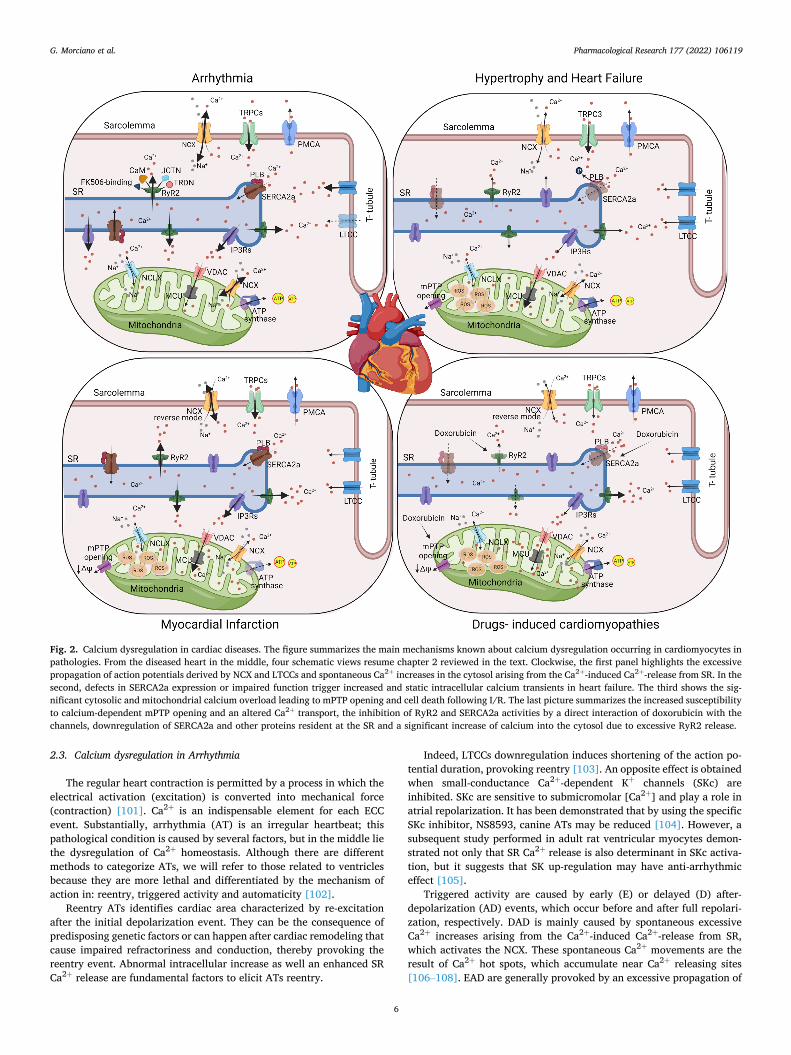
Indeed, it has been investigated that up-regulation of MCU promotes downregulation of mitochondrial functioning and activation of apoptotic process throughout activation of the Ca2+-dependent thiol- protease family calpains that, in turn, alter the functioning of the Optic Atrophy Protein 1 (OPA1), a master regulator of mitochondrial fusion [96]. Consistently, downregulation of expression of OPA1 was found in both humans with coronary disease and rat model of HF [97], and mice harboring OPAdelTTAG mutations showed altered Ca2+ dynamics and increased infarct size than WT littermates [98]. In detail, car-diomyocytes from left ventricle with OPA1 deletion showed lower amplitude of Ca2+ transients, decreased SR Ca2+ uptake, increased cytosolic Ca2+ removal by NCX and impaired mitochondrial Ca2+ up-take. This has significant repercussion on the heart which results more sensitive to I/R and the number of the arrhythmias occurrence. Furthermore, genetic overexpression of OPA1 as well as the genetic ablation of its protease, OMA1, exert cardioprotective effects in in vivo ischemia reperfusion (I/R) model [99]. In contrast to OPA1, genetic ablation of mitofusins (MFNs), other regulator of mitochondrial fusion, have been shown to protective against I/R [100]. These unexpected effects may be explained by the fact that MFNs have pleiotropic non-fusion effects, including the preservation of ER/SR-mitochondria juxtaposition, which are fundamental for the Ca2+ transfer into mito-chondria. Indeed, the ablation of MFNs reduced the proximity of mito-chondria and SR protected from Ca2+ overload during I/R [100], thereby suggesting a close relationship between SR-mitochondria Ca2+
homeostasis and mitochondrial dynamics during I/R (Fig. 2).
G. Morciano et al.
Pharmacological Research 177 (2022) 106119
6
2.3. Calcium dysregulation in Arrhythmia
The regular heart contraction is permitted by a process in which the electrical activation (excitation) is converted into mechanical force (contraction) [101]. Ca2+ is an indispensable element for each ECC event. Substantially, arrhythmia (AT) is an irregular heartbeat; this pathological condition is caused by several factors, but in the middle lie the dysregulation of Ca2+ homeostasis. Although there are different methods to categorize ATs, we will refer to those related to ventricles because they are more lethal and differentiated by the mechanism of action in: reentry, triggered activity and automaticity [102].
Reentry ATs identifies cardiac area characterized by re-excitation after the initial depolarization event. They can be the consequence of predisposing genetic factors or can happen after cardiac remodeling that cause impaired refractoriness and conduction, thereby provoking the reentry event. Abnormal intracellular increase as well an enhanced SR Ca2+ release are fundamental factors to elicit ATs reentry.
Indeed, LTCCs downregulation induces shortening of the action po-tential duration, provoking reentry [103]. An opposite effect is obtained when small-conductance Ca2+-dependent K+ channels (SKc) are inhibited. SKc are sensitive to submicromolar [Ca2+] and play a role in atrial repolarization. It has been demonstrated that by using the specific SKc inhibitor, NS8593, canine ATs may be reduced [104]. However, a subsequent study performed in adult rat ventricular myocytes demon-strated not only that SR Ca2+ release is also determinant in SKc activa-tion, but it suggests that SK up-regulation may have anti-arrhythmic effect [105].
Triggered activity are caused by early (E) or delayed (D) after-depolarization (AD) events, which occur before and after full repolari-zation, respectively. DAD is mainly caused by spontaneous excessive Ca2+ increases arising from the Ca2+-induced Ca2+-release from SR, which activates the NCX. These spontaneous Ca2+ movements are the result of Ca2+ hot spots, which accumulate near Ca2+ releasing sites [106–108]. EAD are generally provoked by an excessive propagation of
Fig. 2. Calcium dysregulation in cardiac diseases. The figure summarizes the main mechanisms known about calcium dysregulation occurring in cardiomyocytes in pathologies. From the diseased heart in the middle, four schematic views resume chapter 2 reviewed in the text. Clockwise, the first panel highlights the excessive propagation of action potentials derived by NCX and LTCCs and spontaneous Ca2+ increases in the cytosol arising from the Ca2+-induced Ca2+-release from SR. In the second, defects in SERCA2a expression or impaired function trigger increased and static intracellular calcium transients in heart failure. The third shows the sig-nificant cytosolic and mitochondrial calcium overload leading to mPTP opening and cell death following I/R. The last picture summarizes the increased susceptibility to calcium-dependent mPTP opening and an altered Ca2+ transport, the inhibition of RyR2 and SERCA2a activities by a direct interaction of doxorubicin with the channels, downregulation of SERCA2a and other proteins resident at the SR and a significant increase of calcium into the cytosol due to excessive RyR2 release.
G. Morciano et al.

Pharmacological Research 177 (2022) 106119
7
action potentials derived by NCX and LTCCs and by Na+ and K+ cur-rents, which permit Ca2+ to enter [109,110]. Furthermore, EAD are also associated with an excessive phosphorylation activity of CaMKII and an over activation of LTCCs, thus accelerating the recovery after their shutdown [111].
Automaticity consists of spontaneous depolarizations during the diastolic phase.
At demonstration that intracellular Ca2+ handling can affect different cardiac ATs, mutations in genes encoding Ca2+ transporters, channels and regulatory proteins have been discovered in different inherited ATs syndromes. For example, catecholaminergic polymorphic ventricular tachycardia (CPVT), one of the most-deadly arrhythmias known, manifests mutations in RyR2 [112,113]. These mutations cause in increased SR Ca2+ leak, thereby causing frequent Ca2+ releasing events that activate NCX and, finally, induce ventricular ATs [114] (Fig. 2). Mutations were also found for CaM. In this case, the mutations impair the Ca2+ binding of CaM and result in a compromised interaction between the protein and RyR2 [115]. Since RyR2 exists as a macro-molecular complex and interacts with other members including CaM, FK506-binding protein-12.6 (FKBP12.6 or Calstabin2), junctin (JCTN) and triadin (TRDN) [116], also the onset of mutations in each of these components, such as TRDN [117] carry out ATs phenotypes. All muta-tional profiles are already reviewed in [118].
Lastly, also TRPC proteins take part in Ca2+ dysregulation-mediated ATs; they have been reported as overexpressed in patients affected by atrial fibrillation, especially the first and third isoform [119], they also may contribute to DAD events [120].
2.4. Calcium dysregulation in hypertrophy and heart failure
Cardiac hypertrophy (CH) is the process of heart remodeling in response to either increased workload (i.e., intended as physiological adaptation following chronic and sustained physical exercise or during pregnancy) or pressure overload (i.e., as consequence of hypertension and valvular diseases [121]). Macroscopically, physiologic CH differs from pathological hypertrophy because it induces changes that are reversible, and which involves also the ventricular chamber enlarge-ment besides the walls thickness. Moreover, the cardiac structure re-mains normal and cardiac function is enhanced. From a molecular point of view, several pathways regulate CH and they, in part, differ between physiology and pathology as reviewed in [122]. Also, CH in pregnancy significantly differ from that occurs after exercise due to hormones contribution [123]. Regarding Ca2+ signaling, in physiologic CH there is an adaptation signaling pathway aimed to sustain enhanced cardiac function and workload; indeed, [Ca2+]i are increased and SR Ca2+ up-take optimized [124]. A concomitant upregulation of RyR2 and SERCA2 proteins are reported in the mild and adaptative stages of CH.
Otherwise, physiologic CH can result in a diseased phenotype where the intracellular Ca2+ overload is reported to be involved in this tran-sition; increased and prolonged [Ca2+]i lead to the activation of pro- hypertrophic factors which, in turn, further stimulate CH. In this sce-nario, CaM and calcineurin are the mainly involved Ca2+ signaling proteins and once they become activated, they trigger NFAT nuclear translocation and its activation [125,126]. Also their expression is re-ported to increase in CH pathological states [126–128]. These changes in Ca2+ homeostasis due to Ca2+ overload are responsible for mito-chondrial dysfunctions, anomalies in cardiac contraction and cellular damage. In turn, developed pathologic CH supports VEGF secretion through mechanical stretch and in a NFkB-dependent manner [129].
After chronic insults, deep changes in thickness and elasticity of the walls of the heart degenerate in HF, a condition in which the heart fails to efficiently pump the blood to the systemic circulation. Indeed, CH is the main risk factor for HF and for these reasons they share also several similarities in terms of molecular pathways become impaired. In the literature are reported several dysregulations of Ca2+ signaling accom-panying this process of maladaptation: they refer to several proteins
involved and mainly focused on the impaired Ca2+ sequestration by SR. The diastolic dysfunction and the correlated disturbance in ventricle
relaxation are one aspect that characterize some types of HF [130]. As mentioned in the introductive paragraphs, the diastole phase which is responsible for a correct ventricle filling [131], is guaranteed by a rapid reduction of cytosolic Ca2+ channeled in the SR of the cardiomyocyte. In CH and HF this does not occur; the most accredited hypothesis is that reduced levels of SERCA2a and not functional ATPase activity of the remaining expressed protein, impair the correct Ca2+ homeostasis with consequent increased stasis of [Ca2+]i. Indeed, in failing hearts, severe metabolic derangements occur [72] and these prompt a real energy crisis. Since the functioning of some proteins involved in Ca2+ homeo-stasis is ATP-dependent, their activity result to be impaired and the contractile heart function as well. The decline of the respiratory chain function and ATP depletion prevent the above mentioned Ca2+ removal from the cytosol which is essential in diastole for muscle relaxation before the next heartbeat [132]. A similar scenario is caused by the impaired action of PMCA and actomyosin, the other two ATP consuming proteins. It follows the absence of transients and a diminished contrac-tility of the heart [133,134].
Nevertheless, other mechanistic insights are suggested in the litera-ture. From the analysis of tissues of failing myocardium, it emerged a key role for an interactor of SERCA2a, phospholamban (PLB). PLB has the peculiarity to inhibit the Ca2+ transport trough the pump by decreasing its affinity for the bivalent ion when it is dephosphorylated. On the contrary, when PLB is phosphorylated, it allows Ca2+ spread. In failing hearts, the phosphorylation status of PLB is significantly reduced as the kinetics of SERCA2a Ca2+ transport [135,136]. At the center of this regulatory pathway, the Serine (Ser) 16 and Threonine (Thr) 17, both phosphorylated by PKA and by CaMKII, respectively [137].
Further investigations on genetic profile in humans predisposing to HF phenotype, highlighted the importance of the mutational profile of PLB in the inhibition of SERCA2a and thus in adverse events accompa-nying HF. The most known are two missense variations R9C and the R14del [138,139]. The first one is able to block the PKA-mediated phosphorylation of the wild type PLB; the second one consisting in the deletion of arginine 14 on PLB failed the interaction PLB-SERCA2a. An additional mutation Leu39stop, identified in humans with a hereditary form of HF, entails severe HF with consequent need of heart transplant in young age when expressed in homozygosity [140]. If expressed in heterozygosity, signs of CH have been described. Here, low levels of PLB expressed were redirected to the PM causing Ca2+ dysregulation.
Histidine-rich calcium (HRC) binding protein is recognized as an additional negative regulator of SR Ca2+ uptake interacting with SER-CA2a. This 170 kDa protein is able to localize at SR thanks to its amino- terminal domain, while an histidine-rich sequence is supposed to have Ca2+ binding properties although there isn’t a real Ca2+ binding motif [141,142]. Its overexpression in the heart is reported to decrease Ca2+
uptake into SR with consequent impairment of muscle relaxation and contractility [143].
Restoring [Ca2+]i for a correct ECC depends in a lower percentage also by NCX. However, literature investigating its expression in tissues from failing hearts, reported controversial results. On one hand, an increased expression seems act as compensatory mechanism to help the cardiac contractility during HF, but it inevitably results in Na+ overload [144]; on the other hand, no appreciable differences have been detected in similar experiments [145].
In a scenario where [Ca2+]i are increased and static and that SR is unable to accumulate Ca2+, mitochondria may be in the position to buffer Ca2+ and suffer from these oscillations [146]. Indeed, Ca2+
overload prompts mPTP opening together to oxidative stress inducing apoptosis and cardiomyocytes loss [147] (Fig. 2).
Increased activity of TRPCs is reported to additionally contribute to CH and its remodeling. They are able to inward great amount of Ca2+
inside cells and stimulate hypertrophy and fibrosis via the calcineurin- NFAT axis [148–150]. TRPC3 in particular, mediates CH also by
G. Morciano et al.

Pharmacological Research 177 (2022) 106119
8
modulating CaV1.2 activity and the reverse mode of action of NCX resulting in more Ca2+ entry [151].
2.5. Calcium dysregulation in drug-induced cardiomyopathies
In agreement with Elliot’s classification established in 2008 [152], cardiomyopathies (CMs) are classified in dilated (DCM), hypertrophic (HCM), restrictive (RCM), arrhythmogenic (ACM) and those remained unclassified. It is clear CMs consist of a heterogenous group of disorders joined together by an improper functionality of the heart due to ultra-structural abnormalities. Despite some criteria in their classification have changed during last years, our priority is not the discussion of what differences exist among them but what types of Ca2+ signaling dysre-gulation occur and contribute to the maladapted phenotype of drug-induced CMs.
Indeed, it is known that CMs may be the side effect of chronic assumption of some classes of drugs and unhealthy lifestyle which cause a progressive and prolonged state of cardiotoxicity. Drug-induced CMs are potentially reversible but in most cases leads to HF.
Unhealthy lifestyle, like the abuse of alcohol consumption, may induce adverse effects in the adult heart and it is considered a risk factor for CMs also in women during pregnancy. To unveil Ca2+ dysregulation, alcohol-dependent cardiac toxic effects have been studied in culture with cardiomyocytes treated with ethanol [153]. In the study, 69% of Ca2+ transients analyzed in cardiomyocytes treated with ethanol were abnormal. As cause of this phenotype, a deep change of protein expression profile has been observed; among the interested proteins, Annexin 6 was downregulated by 40% and ATP1A2 by 29% [153]. Both proteins are involved in Ca2+ transport: the first one modulating Ca2+
influx through the LTCCs and its release from SR by RyR2 and NCX [154]; the second protein is part of the Na+ pump that can regulate intracellular Ca2+ and muscle contractility [155].
A problem currently considered of great importance is the car-diotoxicity of some classes of drugs in the long-term treatment of oncologic patients, such as anthracyclines. A general consensus is their use in therapy despite they are responsible for progressive accumulation of several mitochondrial dysfunctions [156], mostly mediated by a strong oxidant action. Although in principle they are potentially reversible changes, often they concur to the onset of severe CMs.
An example of cardiotoxicity from anthracyclines is given by doxo-rubicin. Patients experiencing chronic exposure to doxorubicin show the following dysregulations regarding Ca2+ signaling: i) increased suscep-tibility to Ca2+-dependent mPTP opening and an altered Ca2+ transport: these effects are dose-dependent [157]; ii) inhibition of RyR2 and SERCA2a activities by a direct interaction of doxorubicin with the channels: this action is oxidative stress-mediated and already in the nanomolar range has a deleterious effect [158]; iii) ROS- and Mitogen Activated Protein Kinase (MAPK) axis- mediated downregulation of SERCA2a mRNA in hearts treated with doxorubicin [159]; iv) a large-scale mRNA downregulation of Ca2+-dependent proteins resident at SR, not only limited to SERCA2a but also including RyR2, PLB and CSQ have been found in a rabbit model of doxorubicin-induced CM [160] and v) [Ca2+] increase into the cytosol due to excessive RyR2 release [161]. All these changes prompt a sever Ca2+ homeostasis perturbation inside cells which are related in concurring several clinical symptoms, cell death (Fig. 2) [162].
Other links between drug-induced CMs and Ca2+ dysregulation are described also for other drugs such as arsenic trioxide [163], mitoxan-trone [164], abuse of cocaine [165], methamphetamine [166], despite mechanistic insights are not completely addressed.
2.6. References to the calcium-mediated PTPC
One of the best-characterized effectors of Ca2+-mediated cardiac stress is the mPTP. This is a supramolecular entity assembled at the interface between the inner and outer mitochondrial membrane
responsible for abrupt increase of permeability of the inner mitochon-drial membrane, also known as mitochondrial permeability transition (MPT). In response to high [Ca2+]m, mPTP opens allowing the deregu-lated exchange of small solutes (up to 1.5 kDa) between the mitochon-drial matrix and cytosol, along their electrochemical gradients. The mPTP is believed to open with 2 different configurations: one at low conductance, believed to operate mostly in physiological conditions to allow the equilibration of Ca2+, mitochondrial pH and ROS; the second at high conductance, which causes immediate and complete dissipation of the ΔΨm, osmotic swelling of the mitochondrial matrix and inhibition of oxidative phosphorylation [167]. When a few mitochondria under-gone MPT do not cause major cellular alterations [168] because they can be efficiently removed by the autophagy machinery and are proposed to participate in cell physiology (for a further discussion see [169]). On opposite, widespread MPT initiates cell death via regulated necrosis or apoptosis [170]. Regulated necrosis is characterized by MPT-mediated block of mitochondrial energy production with the consequent arrest of its dependent activities [171]. Conversely, MPT-driven apoptosis mainly depends on the release of mitochondrial intermembrane pro-teins, especially cytochrome c, apoptosis-inducing factor, mitochondrion-associated, 1 (AIFM1, best known as AIF), and diablo, an IAP-binding mitochondrial protein (DIABLO, also known as Smac) [172].
A role for mPTP has been demonstrated in several cardiac conditions, foremost reperfusion injury [94]. The most accepted model is that during cardiac ischemia mitochondria uptake Ca2+ and produces ROS (a strong sensitizer of mPTP) but MPT is blocked by the simultaneous accumulation of mPTP endogenous inhibitors as ADP and acidic matrix pH. At the reperfusion phase, the re-flow of oxygen favors a ROS burst and a mild restoration of pH and ADP levels which ultimately cause the massive opening of mPTP. Thanks to the identification of cyclosporine A (CsA) as a strong inhibitor of MPT, this mechanism has been proven as responsible for most of the necrotic area (and possibly a large fraction of apoptosis) in the myocardia undergoing reperfusion injury [95].
Pieces of evidence link mPTP also to reperfusion induced arrhythmia. Exposure to the mitochondrial uncoupler FCCP induces arrythmias in explanted mouse hearths. FCCP causes dissipation of mitochondrial membrane potential which in turns favors the opening of the mPTP. It results the inability of IMM to hold a Ca2+ gradient between mitochondrial matrix and cytosol. The alteration of [Ca2+]c in response to FCCP is believed a major determinant in FCCP-induced arrythmias. Genetic inhibition of mPTP (via deletion of the gene Ppif coding for Cyclophilin D, CypD, the target of CsA) results in significant resistance of explanted hearth to the insurgence of arrythmias [173]. Contrasting investigation have tested the pharmacological inhibition of mPTP by CsA on reperfusion induced arrhythmias on different animal models. Arteaga and co-workers originally reported a significant effect of CsA on rat heart which were not confirmed by other investigation in rat [174] guinea pig [175] and rabbit [176]. Overall, these piece of evidence calls for additional investigation, tough should be noted that obtaining and experimental design able to discriminated the impact of mPTP in reperfusion damage or reperfusion-induced arrythmia is challenging.
A similar mechanism is proposed for drug-induced cardiotoxicity. Compounds as doxorubicin [177,178], naproxen, diclofenac, celecoxib [179] and sorafenib [180] are proposed to elevate ROS production and affect Ca2+ homeostasis in cardiomyocytes, ultimately leading to mPTP opening and cell death. The exposure to mPTP inhibitors (as CsA) or antioxidants have been indeed shown to protect against drug-induced cardiotoxicity in different animal models. The MPT is also proposed to play a role in the necrotic cell death that is present in chronic HF. Car-diomyocytes isolated from dogs with chronic HF displayed increased opening of the mPTP, decreased mitochondrial membrane potential, and decreased mitochondrial cytochrome c oxidase and respiration. All the mitochondrial phenotypes were reverted by the administration of CsA [181,182].
However, a mechanism for mPTP opening in HF is not clear.
G. Morciano et al.

Pharmacological Research 177 (2022) 106119
9
Tamoxifen-dependent conditional deletion of the mitochondrial NCX cause the rapid onset on CH and HF in mouse heart by causing excessive accumulation of mitochondrial Ca2+ and mPTP opening [183]. In addition, the mitochondrial NAD+-dependent deacetylase SIRT3, was demonstrated to target CypD - thus exerting a regulatory activity on mPTP and prevents age-related CH [184]. Despite this evidence, mice knock out for Ppif displayed propensity in develop HF, CH, fibrosis, and reduction in myocardial function in response to pressure overload stimulation [185]. This phenotype is proposed to be mediated by a chronic remodeling of energy metabolism (possibly mediated by mPTP) rather than a direct regulation of cell death.
3. Approaches to correct calcium dysregulation: potentially many targets in cardiac diseases
Since the 90 s, it is becoming increasingly clear that targeting intracellular Ca2+ can be a valid strategy for the treatment of several diseases, including in the heart. The knowledge of the previous mentioned studies has led to the development of regulators, more or less selective, of exchangers, pumps and channels with the aim to revert the pathologic phenotype and clinical symptoms of most cardiac diseases (Table 1). Moreover, the recent identification of the genetic component of the mitochondrial uniporter and other mechanistic insights about Ca2+ cycling allowed the better understanding the biological roles of Ca2+ regulation and to create more selective molecules that regulate its activity [186–192].
3.1. Targeting MCU
From the topics covered by this review it is clear that sustained mitochondrial Ca2+ overload is toxic for both tissues and cells in heart diseases, especially in I/R injury (IRI) where Ca2+ accumulation occur both in ischemia and reperfusion. Thus, although the inhibition of the MCU activity to treat this mitochondrial disfunction may be considered an innovative therapeutic approach, it is necessary to take note of a series of apparent controversial results which don’t always do theory the practice [193]. For example, it is reported that constitutive cardiac MCU-deficient animal models are not protected by IRI [188,192,194] despite mitochondria are protected from Ca2+ entry. These findings claim that the role of mitochondrial Ca2+ in IRI is extremely complex and whether on one hand sustained Ca2+ overload is harmful, also the complete and chronic inhibition of Ca2+ uptake may be deleterious or assumes compensative adaptations. In support with this opinion, con-ditional knockout of the MCU or its transitory chemical inhibition, conferred the expected protective effects [195].
To date, several groups have developed molecules capable of reducing Ca2+ uptake into mitochondria through the inhibition of MCU. The most known and widely used is ruthenium red (RuRed), synthesized in 1892 [196]. RuRed is a substance that blocks Ca2+ uptake by MCU inhibition without affecting Ca2+ efflux and mitochondrial respiration [197–199]. The most important paper using RuRed against heart disease was by Grover et al. in which the treatment of perfused rat hearts with the compound in the micromolar range improved cardiac contractile function and oxygen efficiency at reperfusion after ischemia [200]. Despite its great potential, RuRed has shown low cellular permeability together with a poor selectivity for MCU and several off-target biological effects.
Subsequently, starting from RuRed another compound has been formulated and was called Ruthenium 360 (Ru360) [201,202]. The in-hibition of MCU by Ru360 was highly selective and, unlike its prede-cessor, it did not induce changes in SR Ca2+ release, both actomyosin ATPase and NCX activity, LTCCs current and cytosolic Ca2+ dynamics [203]. The use of Ru360 as an intravenous single bolus (quantified in 15–50 nmol/kg) lasting 30 min before ischemia in rats, significantly reduced reperfusion-induced arrhythmias, mitochondrial damage and improved cardiac performance [201].
Recently, Woods et al. have synthesized and characterized the bioactivity of a new ruthenium-based MCU inhibitor, Ru265. This compound is structurally similar to Ru360 and would overcome it thanks to an increased selectivity, cell permeability associated to low toxicity detection [204]. It has been used as drug to prevent hypo-xia/reoxygenation (H/R) in rat neonatal cardiomyocytes with a signif-icant preservation from MMP lowering and mPTP opening [204].
Kon et al. have recently identified a novel cell permeable inhibitor of MCU, called DS16570511 [205]. Among the described effects ascribed to DS16570511 inhibition of mitochondrial Ca2+ uptake, the most common are increased heart contractility and reduction of RI when used in the range 3–30 µM. Its inhibition results reversible simply after washout.
However, two small compounds have been recently discovered through the screening of a library of about 44.000 substances for their capacity to regulate mitochondrial Ca2+ uptake. These compounds, called MCU-i4 and MCU-i11, directly bind a specific domain of MICU1, fundamental for the gating activity of MCU complex. Their interaction with MICU1 in muscle fibers has decreased Ca2+ influx altering the growth of myotubes [206]. MCU-i4, unlike -i11, impacts mitochondrial depolarization thus future investigations should be focused on the sec-ond one.
Santo-Domingo et al. have demonstrated that KB-R7943, a com-pound originally developed as inhibitor of the NCX, protects against myocardial I/R injury through the inhibition of MCU [207]. Inhibition of MCU by KB-R7943 may block the mitochondrial calcium uptake, the matrix overload and the subsequent opening of the mPTP, contributing to cardioprotective activity [207]. On the other hand, it has been seen that KB-R7943 inhibits Ca2+-induced mPTP opening as primary and more direct function [208].
These data highlight that there is the need for new studies associated to the difficulty of finding permeable molecules that can inhibit the MCU, selectively, and without altering biological functions.
3.2. Targeting SERCA2a
As previously reviewed, SERCA2a is one of the main proteins to be dysregulated in CH and HF. Its function is impaired by an improper protein expression, an inhibitory action of molecular interactors and gene mutations. Malfunction of this protein is responsible for non- physiological [Ca2+]i transients which have repercussion in muscle relaxation during diastole and thus heart pumping. For these reasons, the recovery of their function may improve cardiac contraction and clinical conditions of patients affected.
According to experimental researches made in the field, the resto-ration of the functions of the SERCA2a can be carried out by either acting directly or indirectly, by targeting some of its regulators.
3.2.1. Direct ways The most accredited way for the maintenance or the recovery of
SERCA2a levels, especially for translational applications in humans, is gene therapy, which is applied from 25 years in the cardiovascular field and attracting interest for always new clinical trials. In the 90’s, for the first time, an adenoviral expression (AV) system is used in ventricular cardiomyocytes of HF patients, to directly increase SERCA2a levels [209]. The effect has been replicated also in cardiomyocytes from different species [210]. By this method, Del Monte et al., have shown that the overexpression of SERCA2a in isolated failing human car-diomyocytes has restored the [Ca2+]i, muscle relaxation and increased cardiac contractility [209]. Moreover, several studies have further verified the beneficial effects of SERCA2a gene transfer on cardiac performance also in animal models of HF, improving myocardial excit-ability and preventing ventricular arrhythmia in pigs [211,212], dia-stolic dysfunction in swine [213] and dose-dependent functional benefit in ovine models [214]. It has been shown that Adeno-Associated Virus (AAV)-based vectors, compared to AV technique, are safer and more
G. Morciano et al.

Pharmacological Research 177 (2022) 106119
10
Table 1 Existing modulators of intracellular calcium to counteract cardiac diseases.
Modulators Protein target Main experimental model Molecular effects Physiologic effects
Ruthenium Red MCU Cells, perfused animal hearts of I/R
Inhibition of mitochondrial Ca2+ load, changes in SR Ca2+ release;
Improved cardiac contractility
Ru360 Cells, whole body laboratory animals of I/R
Inhibition of mitochondrial Ca2+ load Reduced RI, prevention of arrhythmias and mitochondrial damage (MMP lowering and mPTP opening), improved cardiac performance, increased heart contractility
Ru265 DS16570511
MCU-i4 MICU1 Cells and muscle tissues Inhibition of mitochondrial Ca2+ load, impairment of MMP
N/A
MCU-i11 Inhibition of mitochondrial Ca2+ load KB-R7943 MCU Cells, perfused animal
hearts of I/R Inhibition of mitochondrial Ca2+ load, inhibition of Ca2+-induced mPTP opening Inhibition of the reverse mode of NCX
Protective effects following I/R by improving LV function, by reducing ventricular fibrillation and hypercontracture of cardiomyocytes
NCX
AV/SERCA2a SERCA2a Cells, large animal models of HF
Restore SERCA2a expression, restore of Ca2+
transients Muscle relaxation, increased cardiac contractility, improved myocardial excitability, prevention of arrhythmia
AAV/SERCA2a SERCA2a Cells, large animal models of HF, humans
Improved vascular reactivity, restored cardiac ejection fraction, blood flow, eNOS expression, decreased apoptosis
AAV9/PLB shRNA PLB Cells, large animal models of HF
PLB inhibition/silencing Improved systolic and diastolic function, normalization of dilation and hypertrophy, reduced fibrosis
Antisense RNA AV/S16E PLB phosphorylation Aptamers Hydralazine SERCA2a Patients with HF Demethylation of the promoter region of
SERCA2a Ca2+ homeostasis correction
Glucocorticoids PLB Patients with I/R PLB phosphorylation, Unaltered Ca2+ transients, reduced calpain activation
Improved myocardial function after cardiopulmonary bypass
Istaroxime PLB Large animal models of HF, patients with HF
PLB inhibition, decrease of Na2+/K+-ATPase activity
Decreased heart rate, cardiac contractility, improved pulmonary capillary wedge pressure Na2+/K+
Resveratrol SIRT1 Laboratory animals Increased SERCA2a activity and Ca2+transient Improved contractile amplitude, reduced relengthening time and superoxide generation PI3 kinase-
Akt-SERCA2a Losartan/Enalapril SERCA2a Patients Prevented downregulation of SERCA2a, RyR2,
PLB and CSQ Partial improvement in LV function
RAS N106 SUMO1 Cells, mouse model of HF SUMOylation of SERCA2a Improved hemodynamic performance and reduced
mortality among the animals with HF antagomiR-25 miR-25 Mice models of HF SERCA2a restoring Improved cardiac function and survival Diltiazem Calcium
antagonist Patients
JTV519 RyR2 Large animals with AT Decreased Ca2+ leak from SR Improved cardiac function RyR2/ Calstabin2 complex
Stabilization of the RyR2/Calstabin2 complex to prevent Ca2+ release
S107 RyR2/ Calstabin2 complex
Humans with AT, HF and CPVT
Prevention of the Ca2+ leakage from the SR
Dantrolene CaM/RyR2 complex
Humans with CPVT, treatment of ATs following I/R in large animal models
Stabilization of CaM/RyR2 complex, inhibition of cytosolic Ca2+ sparks in diastole
Prevention of DAD, antiarrhythmic
Flecainide RyR2 Patients Correction of aberrant RyR2 activity, blocking the open state of RyR2, decreased spontaneous Ca2+ leak
Completely prevention ov CPVT
Tetracaine and derivatives (EL1–9), EL20
RyR2 Laboratory animals Inhibition of RyR2 and Ca2+ leak Prevented the induction of ventricular tachycardia without affecting heart rate or cardiac contractility
Benzothiazepines LTCCs Patients with HCM, CHF, CH, angina pectoris and cardiac ATs
Ca2+ channel blockers Decreased traits of hypertrophy, AT and improved contractility Dihydropyridines
Phenylalkylamines SEA0400 NCX Cells, large animal models
of I/R Inhibition of NCX and Ca2+-induced cell death Recovery of LV function after reperfusion and
decreased of infarct size and incidence of ventricular fibrillations.
SN-6 YM-244769 GSK255B + GSK503A TRCP3 Cell models of CH TRCPs inhibitors, antagonize either the
molecular signaling inducing CH or reduced the phosphorylation status of many proteins involved in Ca2+ handling
N/A TRCP6
Pyr-3 TRCP3 Cells TRCP3 inhibitor, inhibition of Ca2+ influx, modulation of NFAT
Reduced hypertrophy
C31 TRPC1/4/5 complex
Cells N/A N/A
KN-93 CaMKII Cells Inhibition of Ca2+ currents N/A LTCCs IP3Rs CaM
AS105 CaMKII Cells, laboratory animals Inhibition of CaMKII, reduced SR Ca2+ leak GS-680
(continued on next page)
G. Morciano et al.

Pharmacological Research 177 (2022) 106119
11
successfully alter gene expression in cardiac tissue, in addition to reducing inflammation [215,216]. Among more than 100 wild-type AAV serotypes, some have been demonstrated to have distinct features on cardiomyocyte transduction [215]. AAV-1 vector was the most used and studied. In several congestive heart failure (CHF) animal models it has been seen that the intracoronary injection of AAV carrying SERCA2a acutely improves vascular reactivity. Also, long-term overexpression of SERCA2a by the same method, significantly restored cardiac ejection fraction, blood flow, eNOS expression in coronary arteries and decreased myocardial apoptosis [213,216–218]. Interestingly, SERCA2a gene transfer by improving Ca2+ signaling, directly influenced vascular endothelial and smooth muscle cell function [219]. Finally, AAVs/-SERCA2a infusion in animal models of HF, have improved the myocardial and coronary artery Ca2+ handling and consequently the cardiac function.
On the basis the data obtained in preclinical models, Calcium Upregulation by Percutaneous Administration of Gene Therapy in Car-diac Disease -Trial (CUPID) was established as first clinical trial in humans, to test the efficiency of recombinant AAV1/SERCA2a in HF [220,221]. The purpose of the clinical trial was intended of monitoring safety and efficacy of high doses of AAV1/SERCA2a administration as therapeutic approach. The result of the Phase I and Phase IIa trials showed reduced adverse cardiac events [220,221]. However, the double-blind randomized and placebo-controlled phase IIb clinical trial, named CUPID2, failed to show significant improvement in ventricular remodeling. Consequently, CUPID2 were terminated prematurely [222]. Despite the failure of these clinical trials, the field of cardiac gene therapy has gained valuable knowledge on AAV gene delivery in the heart and on safety in advanced HF population [223]. In recent years is emerging a new gene delivery method using other viral vectors mainly based on lentivirus. In a rat model of HF, it has been shown that lenti-virus can integrate the gene of interest in the host genome with long-term effect of gene transduction [224–227]. In addition, in a model of I/R, the overexpression of SERCA2a reduced the incidence of the ventricular arrhythmia and improved hemodynamics [228–230].
3.2.2. Indirect ways PLB is a SR membrane protein which modulates SR Ca2+ uptake,
contractility and relaxation by SERCA2a inhibition [231]. In its dephosphorylated state, it lowers the affinity of SERCA for Ca2+, thereby inhibiting the uptake of the latter [231]. On the contrary, its phos-phorylation relieves PLB-mediated inhibition of SERCA2a, thereby increasing the activity of the channel.
Several studies have demonstrated that to increase SERCA2a activity in treating HF, either increase PLB phosphorylation or its knockdown might be a promising therapeutic approach [232]. Indeed, all studies in large animals reported similar data output describing enhanced [Ca2+]i, improved systolic and diastolic LV function, the normalization of dila-tion and hypertrophy and reduced cardiac fibrosis; all clinical stages that act synergistically to reverse HF phenotype. Methods used are multiple and include the depletion of PLB through either AAV9-mediated shRNA [233] or antisense RNA directed to PLB mRNA [234], the use of an AV-mediated phosphorylated mutant of PLB which is constitutively active [235] and cell-permeable aptamers [236]. Nevertheless, attention should be maintained in the use of some methods due to the possible presence of side effects [237].
In addition to this, several drugs may act indirectly on SERCA2a activity. Some of these are: Hydralazine, glucocorticoids, Istaroxime and resveratrol. Hydralazine is a drug already used in the treatment of pa-tients affected by HF, it acts indirectly being an inhibitor of DNA methylation [238]. Demethylation of the promoter region of SERCA2a increases its mRNA levels and leads to a correct Ca2+ homeostasis [238]. Glucocorticoids have been shown to restore the phosphorylation status of PLB together to SERCA2a when used as treatment in IRI or cardiac arrest [239].
Istaroxime is utilized for the treatment of acute HF syndromes, it increases function of SERCA2a by acting on PLB inhibition and de-creases of Na2+/K+-ATPase activity [240,241]. The results are clear and various; in principle administered in dogs, they have highlighted the absence of arrhythmic events associated with improved heart function [242]. In addition, also humans benefit from this [243] as evidenced by a clinical trial in patients with HF which has shown that the adminis-tration of Istaroxime decreases heart rate, improves pulmonary capillary wedge pressure besides demonstrating safety and beneficial effects on cardiac contractility [244,245]. Likewise, other drugs like Losartan and Enalapril can prevent the downregulation of SERCA2a protein and the mRNA levels through blockage of the renin–angiotensin system (RAS) and an angiotensin II receptor blocker (ARBs) [246]. Moreover, the administration of Resveratrol in diabetic mice has shown an increase in SERCA2a expression and an improvement of the cardiac function through activation of SIRT1 [247,248]. In addition to this, in diabetic rat cardiomyocytes, the treatment with insulin-like growth factor 1 (IGF-1) through activation of the PI3 kinase-Akt-SERCA2a signaling pathway enables the increase the SERCA2a levels [249]. Lastly, in a rat model of chronic HF also the anti-inflammatory agent named Oxymatrine, im-proves the SERCA2a expression and cardiac function [250].
However, recent studies have focused the attention on possible SERCA2a post-translational modifications. Studies have shown that in cardiomyocytes the activity and levels of SERCA2a are modulated through SUMO1; SUMOylation of SERCA2a is reduced in HF and with it also the protein amount [251]. Kho et al., have developed a molecule that induced the activation of SUMO1, called N106, that leads to the SUMOylation of SERCA2a with consequent stimulation of the activity of the latter [252]. In addition to this, also the gene therapy through AAV9 gene transfer the SUMO1 levels are increased, leading to of SERCA2a normal levels. Consequently, induces an improved hemodynamic per-formance and a reduced mortality among the animals with HF. Likewise, it has been proven that increased expression of S100A1, a Ca2+ binding protein involved in intracellular Ca2+ regulation, improves the activity of both SERCA2a and RyR2, restoring the correct [Ca2+]i transients and Ca2+ at the SR [253]. This is translated into a long-term reversal of LV dysfunction with improvements of contractile function in a pre-clinical model of ischemic CM. These data indicate that S100A1 can be another target for therapy of the human HF [254,255].
Furthermore recent studies have discovered that many microRNAs (miRs) are dysregulated with HF [256,257]. In particular, experiments in cardiomyocytes of both mice and humans, have detected high levels of miR-25 that delays Ca2+ re-uptake kinetics with consequently increased HF phenotypes. Wahlquist et al. have demonstrated that the use of antagomiR-25 in a mouse model improves cardiac function and survival through restoring SERCA2a activity [258].
Table 1 (continued )
Modulators Protein target Main experimental model Molecular effects Physiologic effects
Reduced arrhythmia, increased contractility in failing myocardium, improved cardiac parameters linked to contractility
RA306
AIP Large animal with CH and ATs
Interaction and block of CaMKII N/A AC3-I CN19o Cells
G. Morciano et al.

Pharmacological Research 177 (2022) 106119
12
3.3. Targeting RyR2
RyR2 plays a central role in the dysregulation of Ca2+ homeostasis found mainly in ATs [259]. Indeed, the pathological leak of Ca2+ from RyR2 is at the basis of arrhythmogenesis.
Several groups of research have developed compounds and drugs with the potential of regulating RyR2 channel activity, to maintain the normal SR Ca2+ release during systole and inhibit it during diastole [260].
From the study of the pluriannual use of Diltiazem in clinic, a 1,5- benzothiazepine-derivate and Ca2+ antagonist [261], led to the dis-covery of further additional benzothiazepine derivatives with potential therapeutic properties for the treatment of ATs. Among them, JTV519 is a 1,4-benzothiazepine compound that shows its cardioprotective effects against atrial fibrillation through the finely regulation of RyR2 proper-ties without altering the capacity to the propagation of Ca2+ waves [262]. This piece of evidence on the molecular mechanism adopted by JTV519 in preventing the onset of ATs in cardiac diseases, seem not to be the only one existing. It has been seen that in dogs, after four weeks of chronic right ventricular pacing, the administration of JTV519 controlled RyR2 opening by restoring Calstabin2 levels and stabilized the RyR2/FKBP12.6 complex, improving cardiac function and decreasing SR Ca2+ with recorded reduced RyR2 hyperphosphorylation [263]. Subsequently, Wehrens et al. have shown that JTV519 stabilizes the closed state of RyR2 by promoting FKBP12.6 binding. The same group also demonstrated that in Calstabin2 knockout mice, JTV519 had no effects, highlighting the importance of Calstabin2 for the antiar-rhythmic effect induced by this compound [264]. In recent years, in 2016, it has been reported an additional property for this drug, such as its partial and mild block of SERCA2a just enough to prevent SR Ca2+
overload and related harmful events in IRI; thus, as consequence, it has repercussion also on SR Ca2+ release modulation [265].
A second 1,4-benzothiazepine derivative is S107; it has the same function of JTV519, or preventing the Ca2+ leakage from the SR, by promoting the Calstabin2-RyR2 binding. S107 has high specificity for RyR2 and no off-target activity [266]. But the real innovation con-cerning this novel drug is the ability to create a complex RyR2-Calstabin2 and thus blocking pathological Ca2+ leakage and serious ATs also in the presence of a mutated form of RyR2, which is the main responsible for CPVT [266,267]. These compounds may have great promise for treating patients with CPVT and not only, indeed, clinical trials studies for the treatment of HF and ATs are in phase 2. In addition to this, S107 can also prevent muscle weakness in aging, stress-induced cognitive dysfunction, Duchenne muscular dystrophy and the progres-sion of HF [268–271].
Dantrolene is a hydantoin derivative that is clinically used for the treatment of malignant hyperthermia, a condition caused by mutations in the skeletal RyR1 [272]. Its mechanism of action involves CaM-dependent stabilization of the interaction between the amino ter-minal and central domains of RyR1, required for the closed state of the RyR Ca2+ channel [273]. Kobayashi et al. have shown that Dantrolene has a role against CPVT by binding also the mutated form of RyR2, specifically to domain 601–620 and resulting in the inhibition of cyto-solic Ca2+ sparks in diastole and DAD [274]. However, it has been demonstrated by using the knock-in mouse model with a human CPVT-associated RyR2 mutation (R2474S) that pretreatment with Dantrolene inhibited CPVT-related tachycardia, induced by either epinephrine or exercise [275]. Subsequently, studies have been carried out on the therapeutic antiarrhythmic potential of Dantrolene in the treatment of CPVT. Dantrolene was administered intravenously and it reduced the number of premature ventricular complexes with antiar-rhythmic effects in a subgroup of CPVT1 patients with specific RyR2 mutations [276]. These data showed comparable results even in ex-periments conducted in pluripotent stem cells (iPSC)-derived sponta-neously beating cardiomyocytes generated from the same patients, proving once again the antiarrhythmic effects of Dantrolene [276].
Dantrolene has found useful also for the treatment of ATs following I/R in several animal models [277,278]. All these evidences underline that to attenuate Ca2+-mediated arrhythmias associated with HF, this hydantoin derivative can be a possible therapy. Currently, a new clinical trial is in the recruitment phase to test the efficacy of Dantrolene in CPVT patients [279].
Flecainide is a trifluoroethoxybenzamide, an oral class Ic antiar-rhythmic drug, already approved for medical use in the United States since 1984. Flecainide known as a Na+ channel blocker, is used in pa-tients who do not have structural heart disease, for the treatment of atrial fibrillation/atrial flutter, paroxysmal supraventricular tachy-cardia, atrioventricular nodal reentrant tachycardia, AV reentrant tachycardia [280,281]. Recent data have shown both in iPSC-cardiomyocytes and in mouse models of CPVT that Flecainide acts either reducing or removing aberrant RyR2 activity, blocking the open state of RyR2 and decreasing spontaneous Ca2+ leak, thus preventing ventricular tachycardia [282,283]. Confirming its efficacy in CPVT is not only due by the blockage of the Na+ channel. In addition to this, treatment of two patients with Flecainide, who remained symptomatic with conventional drug therapy, has prevented CPVT with great success. Hence, these data provide clear evidence on the antiarrhythmic effec-tiveness of Flecainide in inhibiting defective RyR2 Ca2+ release channels in humans, identifying it as a possible currently available drug for fight CPVT [283].
A last class of drugs considered efficient in reducing spontaneous and pathological Ca2+ release are Tetracaine and derivatives (EL1–9) [284] for the treatment of ventricular tachycardia; they showed great affinity for RyR2 and few off-target side effects. The last one identified by Word et al., EL20, used at low nanomolar concentrations and also efficacy in cardiomyocytes harboring the R176Q variant from a CPVT patient [285].
3.4. Targeting LTCCs
LTCCs are the main mediators of VGCC-mediated Ca2+ influx into cardiac cells and are fundamental in certain mechanisms and electrical properties of the heart. These channels, thus are considered as an important target for the treatment of several diseases [286–288]. In the heart, the most expressed isoforms are CaV1.2 and CaV1.3 and their alteration is related to the onset of cardiac diseases related to the impairment of ECC and inotropy [289].
It is known that mutations in LTCC genes induce the dysfunctions of Ca2+ channels, resulting in the abnormal excitations of cardiomyocytes leading to the onset of cardiac ATs such as LQTS (Long QT syndrome) [290] and Brugada syndrome [291].
For decades antagonists of LTCCs have been used for the treatment of CH and I/R. However, in patients with HF, LTCC Ca2+ blockers are employed with great caution. In addition to this, the knockdown of LTCCs induces the secondary CH signaling through a compensatory in-crease in Ca2+ by RyR2 leak [292].
However, over the years, several organic molecules have been identified as modulators of LTCCs by blocking the inward Ca2+ current. These modulators bind to three separate receptor sites on LTCCs and can be grouped into three groups: benzothiazepines, dihydropyridines (DHP) and phenylalkylamines [286,293,294]. All binding sites are sit-uated within the IIIS5, IIIS6, and IVS6 transmembrane segments and near to the pore [295]. These substances are used to treat several dis-eases, including HCM, CHF, CH, angina pectoris and cardiac ATs [296–299]. In particular, the non-dihydropyridines Ca2+ channel blockers that includes benzothiazepines and phenylalkylamines are used also for the treatment of ATs, but should not be used in patients with HF and atrial fibrillation united with preexcitation. Several studies have demonstrated that the use of non-dihydropyridines increase mortality and incidence of HF, due by their negative inotropic effects [300,301]. Indeed, to prevent further depression of cardiac function the non-dihydropyridines, are used only when β-blockers alone are
G. Morciano et al.

Pharmacological Research 177 (2022) 106119
13
unsuccessful [302]. In addition to this, several studies have shown that in arrhythmic
conditions the combination of different Ca2+ channel blockers can be a valid therapeutic treatment. Interestingly, in patients affected by CPVT, it has been reported that by adding verapamil to existing β-blocker therapy, the incidence of exercise-induced arrhythmias was reduced when compared to β-blocker therapy alone [303].
Despite the physiological role of the T-type Ca2+ channel in the human heart is still little documented, it is known they are present in the sinoatrial node, atrioventricular node and Purkinje fibers and are involved in the pacemaker activity. Mangoni et al., have highlighting the importance of the channel in cardiac rhythm, through CaV3.1 knockout mice [304]. In this animal, the genetic inactivation of the CaV3.1 result in a significant slowing of the heart rate and atrioventricular conduction [304].
3.5. Targeting NCX and PMCA
In addition to SERCA2a, both PMCA and NCX are important regu-lators of [Ca2+]i intervening in Ca2+ removal when it exceeds, for instance after contraction. NCX has the adjunctive peculiarity to work in a bidirectional way creating inward and outward currents of Ca2+ and Na+, depending on the intracellular concentrations of the ions exchanged and on membrane potential. Thus, both activators and in-hibitors of this channel may result in cardioprotective effects. During I/R when it functions in reverse mode by increasing Ca2+ influx, its inhibi-tion may result useful; otherwise, in HF Ca2+ extrusion may result impaired caused by high [Na+], in this scenario, stimulators of its ac-tivity may provide benefit.
There exist many NCX inhibitors, but most of them are not selective (i.e., Amiodarone, Bepridil, Aprindine) as they modulate also Na+/K+
and LTCCs currents and they are already reviewed in [305], although they retain good antiarrhythmic properties.
The first, but less selective inhibitor, is KB-R7943 developed by Watano T. [306]. Its administration has showed protective effects following I/R by improving LV function, by reducing ventricular fibril-lation and hypercontracture of cardiomyocytes [307,308]. KB-R7943 in not considered too selective because it results also a potent MCU in-hibitor; thus, it is difficult to discriminate if its cardioprotection following I/R (at a concentration of 10 µM) is dependent on the inhi-bition of mitochondrial Ca2+ uptake or on the modulation of NCX, or both; although that dose should not influence the exchanger as claim some reports [207].
To date, the most potent and selective compounds targeting NCX are named as SEA0400, SN-6 and YM-244769. Overall, in the heart, SEA0400 is considered to have a higher potency in the inhibition of NCX compared to SN-6, which result similar to KB-R7943 [305]. At the same time, also its selectivity is greater than SN-6 which in turn, is more se-lective of KB-R7943. Cardioprotective effects of SA0400 are multiple and range from cell cultures to large animals. Generally, its treatment led to Ca2+-induced cell death inhibition [309], recovery of LV function after reperfusion and a decrease of the infarct size [310], and the inci-dence of ventricular fibrillations. To conclude, the newest YM-244769, firstly described in the brain and targeting preferentially NCX3 [311], showed protective action under H/R both in neuronal diseases and in the heart [312].
On the other hand, although NCX stimulators own cardioprotective features, they share many molecular mechanisms of protection which often fall back so far into the modulation of Ca2+ homeostasis [313].
Concerning PMCAs, PMCA1 and PMCA4 are primarily reported to be expressed in the heart. These proteins are considered minor contributors for Ca2+ efflux from the cell, thus they have always been of secondary importance to study in diseases and as target of cardioprotection. Nevertheless, it is reported the existence of interesting mutational pro-files of the PMCA1 gene concerning cardiac disease. Genome wide as-sociation studies (GWAS) performed in several cohorts of the whole
world population and taking into consideration different ethnicities, identified at least 10 SNPs in correspondence of intronic regions of the gene encoding for PMCA1. As consequence, they are significantly associated to hypertension, pre-eclampsia, salt sensitivity and leading to coronary artery diseases, myocardial infarction and also calcification. These SNPs are the following: rs1401982 [314–316] associated to artery stenosis, hypertension and arterial stiffness; rs2070759 [317,318] associated to hypertension in both Chinese and Japanese population; rs2681472 [319–321] responsible for hypertension and arterial stiff-ness; rs2681492 [322,323] involved in high blood pressure in people from Africa; rs7136259 [324–326] associated to premature coronary disease and atherosclerosis in multi-ethnic populations; rs10858911 [327], rs11105354 [328–331] with increased risk of cardiovascular disease; rs11105378 [317,332] with increased susceptibility to hyper-tension; the same for both rs12817819 [333] and rs17249754 [319,320, 333]. Besides what reported above, more than 50 additional papers reported same findings on the association of PMCA1 to hypertension and coronary artery disease like MI. This evidence involve loss of function of PMCA1 or reduced amount of the protein at the PM that has, as direct effect, the increase in the expression of LTCCs and blood pressure, triggering CH and heart remodeling [334,335]. Overall, no reports classify these mutational profiles as deadly themselves, but it is clear that they significantly increase the onset of hypertension and premature coronary artery disease with all related consequences. Also PMCA4 plays key roles in heart physiology by controlling cardiac contractility [336]. In a scenario of established I/R, its overexpression alleviates all maladaptation of the heart included CH and HF [337]. However, to date there are no drugs able to induce PMCA overexpression.
3.6. Targeting TRP channels
Each of TRPCs has an individual role in cardiovascular pathologies, being differentially expressed in the diseased heart. On the basis of recent considerations made on TRPC targeting to prevent CH, fibrosis, heart remodeling and HF, there exist several pitfalls that obstacles the use of this target in human clinical trials, when compared to other strategies for other proteins. In particular, still very few drugs targeting these channels have been discovered where the majority are not selec-tive, they do not exist cardiac-specific isoforms of these channels thus their targeting may involve severe off-target effects [8].
However, here we report what is known about advanced studies in cells. Deletion of the first isoform of TRCPs is able to deactivate the calcineurin-NFAT signaling axis in the progression of CH [338]. About TRCP3 there exists few inhibitors like GSK255B [339] which blocks the channel but does not led to any protective effect when administered alone. While, the synergistic inhibition with TRPC6 with GSK503A, antagonize the signaling inducing CH or reduced the phosphorylation status of many proteins involved in Ca2+ handling [340]. The second inhibitor Pyr-3, a pyrazole compound, mediate a plethora of effects ranging from Ca2+ influx, modulation of NFAT to reduce hypertrophy; Pyr-3 seems to acquire more selective skills [341]. Recently, it has been developed a selective compound 31, c31, that working in the picomolar range of concentrations it inhibits the assembly of TRPC1/4/5 complex, but no cardioprotective effects have yet been investigated [342].
3.7. Targeting CAMKII
CaMKII is a kinase reported as mainly involved in intracellular Ca2+- dependent changes occurring during HF, CH and ATs and caused by its intense activity of phosphorylation towards proteins involved in Ca2+
handling. It has many targets (i.e., PLB, RyR2, SERCA2a) and, overall, it triggers SR Ca2+ leaking and the opening of LTCCs. It is clear that its inhibition constitutes the way to preserve physiological functions.
There are two types of CaMKII inhibitors: those grouped in the pharmacological area and those based on peptides structures. In the 90’s, one of the first CaMKII pharmacological inhibitor was KN-93
G. Morciano et al.

Pharmacological Research 177 (2022) 106119
14
[343]; if its use in basic research was spread, it had no future in clinical practice. Indeed, side effects targeting multiple proteins as LTCCs, IP3Rs and calmodulin have been described several times [344–346].
The second known pharmacological inhibitor is AS105 [347]; by working 1000-fold less concentrated than the first one, it successfully reduced SR Ca2+ leak in cardiomyocytes from mice with HF. Given the very low dose of use, no negative effects on ECC and SR Ca2+ loading were recorded.
Again, another similar binding molecule (which results in a ATP- competitive binding) is GS-680 [348]. It has been improved by Wag-ner S et al. with a strong selection against cardiac CaMKII and with beneficial effects reducing arrhythmia and increasing contractility in failing myocardium. Also, this compound worked efficiently in the low nanomolar range. Of the same class, RA306 was described for the first time by Beauverger P. and colleagues in 2020 [349]; by treating diseased mice with oral administrations, after few weeks it improved cardiac parameters linked to contractility.
Among the peptide-based inhibitors, autocamtide-2-related inhibi-tory peptide (AIP) [350] and autocamtide-3 derived inhibitory peptide (AC3-I) [351] interact with CaMKII and blocked its canonical activation. This resulted in a strong enzyme inhibition in several animal models providing evidence once again on the importance to target this protein to counteract pathological hypertrophy and lethal arrhythmias [352].
Across the end of 90’s and the early 2000’s, by using the catalytic domain of CaMKII as bait [353,354], proteins with inhibitory function against the enzyme have been unveiled; they were resident in the brain, but absent in the heart. For this reason, the first effort was an efficient delivery in the heart by the use of AAV vectors and by targeting the CaMKII once active and limiting I/R consequences and ATs [111,355]. Taking advantage from growing biomolecular methods, it was created a mitochondria-targeted inhibitory protein [356]. Its administration at reperfusion relieved CaMKII activation and thus its positive loop on MCU-mediated mitochondrial Ca2+ load and mPTP opening. Further studies in improving these peptides achieved a great result by creating the smallest sequence of 19 aa (named as CN19o) sufficient to exert all cardioprotective effects in the picomolar range [357].
4. Conclusions
From the literature reviewed above, it emerges that the knowledge of the tasks accomplished by each of Ca2+-dependent proteins in the heart physiology and how they become differentially impaired in diseases is essential to draw a successfully targeted therapy. Indeed, Ca2+ cycling involves a finely tuned and dynamic spatio-temporal waves to achieve a proper contraction-relaxation with high frequency, thousands of times, every day. Overall, there is a good understanding about Ca2+ dysregu-lation in cardiac diseases; now, the efforts should be more directed to-wards drugs developing and in translating results from preclinical models to patients.
Also, proper drug delivery methods are needed to increase bioavailability, to better target only specific cardiac protein isoforms and minimize off-target effects.
Declaration of interest
The authors have no conflicts of interest.
Acknowledgements
The Signal Transduction Laboratory is supported by the Italian As-sociation for Cancer Research (IG-23670 to P.P. and IG-19803 to C.G.), A-ROSE, Progetti di Rilevante Interesse Nazionale (PRIN2017E5L5P3 to P.P and PRIN20177E9EPY to C.G.), the Italian Ministry of Health (GR- 2013-02356747 to C.G. and GR-2019-12369862 to G.M.), the European Research Council (853057-InflaPML to C.G.) and local funds from the University of Ferrara to C.G. and P.P. P.P. is grateful to C. degli
Scrovegni for her continuous support. M.R.W. was supporte0d by the Polish National Science Centre (Grant: UMO-2014/15/B/NZ1/00490).
References
[1] W.A. Catterall, Voltage-gated calcium channels, Cold Spring Harb. Perspect. Biol. 3 (2011), a003947, https://doi.org/10.1101/cshperspect.a003947.
[2] F. Hofmann, V. Flockerzi, S. Kahl, J.W. Wegener, L-type CaV1.2 calcium channels: from in vitro findings to in vivo function, Physiol. Rev. 94 (2014) 303–326, https://doi.org/10.1152/physrev.00016.2013.
[3] M. Prakriya, R.S. Lewis, Store-operated calcium channels, Physiol. Rev. 95 (2015) 1383–1436, https://doi.org/10.1152/physrev.00020.2014.
[4] M.D. Bootman, K. Rietdorf, Tissue specificity: store-operated Ca2+ entry in cardiac myocytes, Adv. Exp. Med Biol. 993 (2017) 363–387, https://doi.org/ 10.1007/978-3-319-57732-6_19.
[5] P. Rosenberg, H. Zhang, V.G. Bryson, C. Wang, SOCE in the cardiomyocyte: the secret is in the chambers, Pflug. Arch. 473 (2021) 417–434, https://doi.org/ 10.1007/s00424-021-02540-3.
[6] G.M. Salido, I. Jardín, J.A. Rosado, The TRPC ion channels: association with Orai1 and STIM1 proteins and participation in capacitative and non-capacitative calcium entry, Adv. Exp. Med Biol. 704 (2011) 413–433, https://doi.org/ 10.1007/978-94-007-0265-3_23.
[7] M.Y. Asghar, K. Tornquist, Transient receptor potential canonical (TRPC) channels as modulators of migration and invasion, Int J. Mol. Sci. 21 (2020), E1739, https://doi.org/10.3390/ijms21051739.
[8] T. Hof, S. Chaigne, A. Recalde, L. Salle, F. Brette, R. Guinamard, Transient receptor potential channels in cardiac health and disease, Nat. Rev. Cardiol. 16 (2019) 344–360, https://doi.org/10.1038/s41569-018-0145-2.
[9] N.L. Jernigan, M.L. Paffett, B.R. Walker, T.C. Resta, ASIC1 contributes to pulmonary vascular smooth muscle store-operated Ca(2+) entry, Am. J. Physiol. Lung Cell Mol. Physiol. 297 (2009) L271–285, https://doi.org/10.1152/ ajplung.00020.2009.
[10] X. Liu, C. Liu, J. Ye, S. Zhang, K. Wang, R. Su, Distribution of acid sensing ion channels in axonal growth cones and presynaptic membrane of cultured hippocampal neurons, Front Cell Neurosci. 14 (2020) 205, https://doi.org/ 10.3389/fncel.2020.00205.
[11] X.-H. Zhang, T. Saric, N.Z. Mehrjardi, S. Hamad, M. Morad, Acid-sensitive ion channels are expressed in human induced pluripotent stem cell-derived cardiomyocytes, Stem Cells Dev. 28 (2019) 920–932, https://doi.org/10.1089/ scd.2018.0234.
[12] M.A. Redd, S.E. Scheuer, N.J. Saez, Y. Yoshikawa, H.S. Chiu, L. Gao, M. Hicks, J. E. Villanueva, Y. Joshi, C.Y. Chow, G. Cuellar-Partida, J.N. Peart, L.E. See Hoe, X. Chen, Y. Sun, J.Y. Suen, R.J. Hatch, B. Rollo, D. Xia, M.A.H. Alzubaidi, S. Maljevic, G.A. Quaife-Ryan, J.E. Hudson, E.R. Porrello, M.Y. White, S. J. Cordwell, J.F. Fraser, S. Petrou, M.E. Reichelt, W.G. Thomas, G.F. King, P. S. Macdonald, N.J. Palpant, Therapeutic inhibition of acid-sensing ion channel 1a recovers heart function after ischemia-reperfusion injury, Circulation 144 (2021) 947–960, https://doi.org/10.1161/CIRCULATIONAHA.121.054360.
[13] F.Di Virgilio, P2X receptors and inflammation, Curr. Med. Chem. 22 (2015) 866–877, https://doi.org/10.2174/0929867322666141210155311.
[14] Q. Mei, B.T. Liang, P2 purinergic receptor activation enhances cardiac contractility in isolated rat and mouse hearts, Am. J. Physiol. Heart Circ. Physiol. 281 (2001) H334–341, https://doi.org/10.1152/ajpheart.2001.281.1.H334.
[15] I.S. Gurung, A. Kalin, A.A. Grace, C.L.-H. Huang, Activation of purinergic receptors by ATP induces ventricular tachycardia by membrane depolarization and modifications of Ca2+ homeostasis, J. Mol. Cell Cardiol. 47 (2009) 622–633, https://doi.org/10.1016/j.yjmcc.2009.08.001.
[16] G.J. Jeffs, B.P. Meloni, A.J. Bakker, N.W. Knuckey, The role of the Na(+)/Ca(2+) exchanger (NCX) in neurons following ischaemia, J. Clin. Neurosci. 14 (2007) 507–514, https://doi.org/10.1016/j.jocn.2006.07.013.
[17] J. Krebs, The plasma membrane calcium pump (PMCA): regulation of cytosolic Ca2+, genetic diversities and its role in sub-plasma membrane microdomains, Adv. Exp. Med Biol. 981 (2017) 3–21, https://doi.org/10.1007/978-3-319- 55858-5_1.
[18] X. Wu, B. Chang, N.S. Blair, M. Sargent, A.J. York, J. Robbins, G.E. Shull, J. D. Molkentin, Plasma membrane Ca2+-ATPase isoform 4 antagonizes cardiac hypertrophy in association with calcineurin inhibition in rodents, J. Clin. Invest. 119 (2009) 976–985, https://doi.org/10.1172/JCI36693.
[19] Y. Wang, C. Wilson, E.J. Cartwright, M. Lei, Plasma membrane Ca2+ -ATPase 1 is required for maintaining atrial Ca2+ homeostasis and electrophysiological stability in the mouse, J. Physiol. 595 (2017) 7383–7398, https://doi.org/ 10.1113/JP274110.
[20] N.A. Beard, D.R. Laver, A.F. Dulhunty, Calsequestrin and the calcium release channel of skeletal and cardiac muscle, Prog. Biophys. Mol. Biol. 85 (2004) 33–69, https://doi.org/10.1016/j.pbiomolbio.2003.07.001.
[21] A. Danese, S. Marchi, V.A.M. Vitto, L. Modesti, S. Leo, M.R. Wieckowski, C. Giorgi, P. Pinton, Cancer-related increases and decreases in calcium signaling at the endoplasmic reticulum-mitochondria interface (MAMs), Rev. Physiol. Biochem. Pharm. (2020), https://doi.org/10.1007/112_2020_43.
[22] G. Santulli, G. Monaco, V. Parra, G. Morciano, Editorial: mitochondrial remodeling and dynamic inter-organellar contacts in cardiovascular physiopathology, Front. Cell Dev. Biol. 9 (2021), 679725, https://doi.org/ 10.3389/fcell.2021.679725.
[23] A. Bartok, D. Weaver, T. Golenar, Z. Nichtova, M. Katona, S. Bansaghi, K. J. Alzayady, V.K. Thomas, H. Ando, K. Mikoshiba, S.K. Joseph, D.I. Yule,
G. Morciano et al.
Pharmacological Research 177 (2022) 106119
15
G. Csordas, G. Hajnoczky, IP3 receptor isoforms differently regulate ER- mitochondrial contacts and local calcium transfer, Nat. Commun. 10 (2019) 3726, https://doi.org/10.1038/s41467-019-11646-3.
[24] M.C. Moschella, A.R. Marks, Inositol 1,4,5-trisphosphate receptor expression in cardiac myocytes, J. Cell Biol. 120 (1993) 1137–1146, https://doi.org/10.1083/ jcb.120.5.1137.
[25] J.T. Lanner, D.K. Georgiou, A.D. Joshi, S.L. Hamilton, Ryanodine receptors: structure, expression, molecular details, and function in calcium release, Cold Spring Harb. Perspect. Biol. 2 (2010), a003996, https://doi.org/10.1101/ cshperspect.a003996.
[26] R. Lewis, J. Li, P.J. McCormick, C.L.-H. Huang, K. Jeevaratnam, Is the sigma-1 receptor a potential pharmacological target for cardiac pathologies? A systematic review, Int J. Cardiol. Heart Vasc. 26 (2020), 100449, https://doi.org/10.1016/j. ijcha.2019.100449.
[27] I. Bezprozvanny, B.E. Ehrlich, ATP modulates the function of inositol 1,4,5-tri-sphosphate-gated channels at two sites, Neuron 10 (1993) 1175–1184, https:// doi.org/10.1016/0896-6273(93)90065-y.
[28] G. Csordas, C. Renken, P. Varnai, L. Walter, D. Weaver, K.F. Buttle, T. Balla, C. A. Mannella, G. Hajnoczky, Structural and functional features and significance of the physical linkage between ER and mitochondria, J. Cell Biol. 174 (2006) 915–921, https://doi.org/10.1083/jcb.200604016.
[29] R. Stoica, K.J. De Vos, S. Paillusson, S. Mueller, R.M. Sancho, K.-F. Lau, G. Vizcay- Barrena, W.-L. Lin, Y.-F. Xu, J. Lewis, D.W. Dickson, L. Petrucelli, J.C. Mitchell, C. E. Shaw, C.C.J. Miller, ER-mitochondria associations are regulated by the VAPB- PTPIP51 interaction and are disrupted by ALS/FTD-associated TDP-43, Nat. Commun. 5 (2014) 3996, https://doi.org/10.1038/ncomms4996.
[30] S. Marchi, C. Giorgi, L. Galluzzi, P. Pinton, Ca2+ fluxes and cancer, Mol. Cell 78 (2020) 1055–1069, https://doi.org/10.1016/j.molcel.2020.04.017.
[31] A. Rimessi, G. Pedriali, B. Vezzani, A. Tarocco, S. Marchi, M.R. Wieckowski, C. Giorgi, P. Pinton, Interorganellar calcium signaling in the regulation of cell metabolism: a cancer perspective, Semin Cell Dev. Biol. 98 (2020) 167–180, https://doi.org/10.1016/j.semcdb.2019.05.015.
[32] R. Rizzuto, A.W. Simpson, M. Brini, T. Pozzan, Rapid changes of mitochondrial Ca2+ revealed by specifically targeted recombinant aequorin, Nature 358 (1992) 325–327, https://doi.org/10.1038/358325a0.
[33] T. Rostovtseva, M. Colombini, ATP flux is controlled by a voltage-gated channel from the mitochondrial outer membrane, J. Biol. Chem. 271 (1996) 28006–28008, https://doi.org/10.1074/jbc.271.45.28006.
[34] M.Y. Liu, M. Colombini, Regulation of mitochondrial respiration by controlling the permeability of the outer membrane through the mitochondrial channel, VDAC, Biochim. Biophys. Acta 1098 (1992) 255–260, https://doi.org/10.1016/ s0005-2728(05)80344-5.
[35] M. Zizi, M. Forte, E. Blachly-Dyson, M. Colombini, NADH regulates the gating of VDAC, the mitochondrial outer membrane channel, J. Biol. Chem. 269 (1994) 1614–1616.
[36] E. Pavlov, S.M. Grigoriev, L.M. Dejean, C.L. Zweihorn, C.A. Mannella, K. W. Kinnally, The mitochondrial channel VDAC has a cation-selective open state, Biochim. Biophys. Acta 1710 (2005) 96–102, https://doi.org/10.1016/j. bbabio.2005.09.006.
[37] A. Messina, S. Reina, F. Guarino, V. De Pinto, VDAC isoforms in mammals, Biochim. Biophys. Acta 1818 (2012) 1466–1476, https://doi.org/10.1016/j. bbamem.2011.10.005.
[38] E.H.Y. Cheng, T.V. Sheiko, J.K. Fisher, W.J. Craigen, S.J. Korsmeyer, VDAC2 inhibits BAK activation and mitochondrial apoptosis, Science 301 (2003) 513–517, https://doi.org/10.1126/science.1083995.
[39] C.P. Baines, R.A. Kaiser, T. Sheiko, W.J. Craigen, J.D. Molkentin, Voltage- dependent anion channels are dispensable for mitochondrial-dependent cell death, Nat. Cell Biol. 9 (2007) 550–555, https://doi.org/10.1038/ncb1575.
[40] M.J. Sampson, W.K. Decker, A.L. Beaudet, W. Ruitenbeek, D. Armstrong, M. J. Hicks, W.J. Craigen, Immotile sperm and infertility in mice lacking mitochondrial voltage-dependent anion channel type 3, J. Biol. Chem. 276 (2001) 39206–39212, https://doi.org/10.1074/jbc.M104724200.
[41] K. Hill, K. Model, M.T. Ryan, K. Dietmeier, F. Martin, R. Wagner, N. Pfanner, Tom40 forms the hydrophilic channel of the mitochondrial import pore for preproteins [see comment], Nature 395 (1998) 516–521, https://doi.org/ 10.1038/26780.
[42] G. Szabadkai, K. Bianchi, P. Varnai, D. De Stefani, M.R. Wieckowski, D. Cavagna, A.I. Nagy, T. Balla, R. Rizzuto, Chaperone-mediated coupling of endoplasmic reticulum and mitochondrial Ca2+ channels, J. Cell Biol. 175 (2006) 901–911, https://doi.org/10.1083/jcb.200608073.
[43] D. De Stefani, A. Bononi, A. Romagnoli, A. Messina, V. De Pinto, P. Pinton, R. Rizzuto, VDAC1 selectively transfers apoptotic Ca2+ signals to mitochondria, Cell Death Differ. 19 (2012) 267–273, https://doi.org/10.1038/cdd.2011.92.
[44] W.M. Rosencrans, V.M. Aguilella, T.K. Rostovtseva, S.M. Bezrukov, α-Synuclein emerges as a potent regulator of VDAC-facilitated calcium transport, Cell Calcium 95 (2021), 102355, https://doi.org/10.1016/j.ceca.2021.102355.
[45] W. Peng, Y.C. Wong, D. Krainc, Mitochondria-lysosome contacts regulate mitochondrial Ca2+ dynamics via lysosomal TRPML1, Proc. Natl. Acad. Sci. USA 117 (2020) 19266–19275, https://doi.org/10.1073/pnas.2003236117.
[46] P.A. Gurnev, T.K. Rostovtseva, S.M. Bezrukov, Tubulin-blocked state of VDAC studied by polymer and ATP partitioning, FEBS Lett. 585 (2011) 2363–2366, https://doi.org/10.1016/j.febslet.2011.06.008.
[47] M.W. McEnery, A.M. Snowman, R.R. Trifiletti, S.H. Snyder, Isolation of the mitochondrial benzodiazepine receptor: association with the voltage-dependent anion channel and the adenine nucleotide carrier, Proc. Natl. Acad. Sci. USA 89 (1992) 3170–3174, https://doi.org/10.1073/pnas.89.8.3170.
[48] M. Fan, J. Zhang, C.-W. Tsai, B.J. Orlando, M. Rodriguez, Y. Xu, M. Liao, M.- F. Tsai, L. Feng, Structure and mechanism of the mitochondrial Ca2+ uniporter holocomplex, Nature 582 (2020) 129–133, https://doi.org/10.1038/s41586-020- 2309-6.
[49] K.J. Kamer, Z. Grabarek, V.K. Mootha, High-affinity cooperative Ca2+ binding by MICU1-MICU2 serves as an on-off switch for the uniporter, EMBO Rep. 18 (2017) 1397–1411, https://doi.org/10.15252/embr.201643748.
[50] H. Vais, R. Payne, U. Paudel, C. Li, J.K. Foskett, Coupled transmembrane mechanisms control MCU-mediated mitochondrial Ca2+ uptake, Proc. Natl. Acad. Sci. USA 117 (2020) 21731–21739, https://doi.org/10.1073/ pnas.2005976117.
[51] A.P. Wescott, J.P.Y. Kao, W.J. Lederer, L. Boyman, Voltage-energized Calcium- sensitive ATP Production by Mitochondria, Nat. Metab. 1 (2019) 975–984, https://doi.org/10.1038/s42255-019-0126-8.
[52] Y. Sancak, A.L. Markhard, T. Kitami, E. Kovacs-Bogdan, K.J. Kamer, N.D. Udeshi, S.A. Carr, D. Chaudhuri, D.E. Clapham, A.A. Li, S.E. Calvo, O. Goldberger, V. K. Mootha, EMRE is an essential component of the mitochondrial calcium uniporter complex, Science 342 (2013) 1379–1382, https://doi.org/10.1126/ science.1242993.
[53] S. Magi, S. Piccirillo, A. Preziuso, S. Amoroso, V. Lariccia, Mitochondrial localization of NCXs: balancing calcium and energy homeostasis, Cell Calcium 86 (2020), 102162, https://doi.org/10.1016/j.ceca.2020.102162.
[54] P. Altmann, P. Wagenbichler, A. Schaller, A casuistic report on the Gruber or Meckel syndrome, Hum. Genet. 38 (1977) 357–362, https://doi.org/10.1007/ BF00402165.
[55] M.P. Blaustein, W.J. Lederer, Sodium/calcium exchange: its physiological implications, Physiol. Rev. 79 (1999) 763–854, https://doi.org/10.1152/ physrev.1999.79.3.763.
[56] E. Carafoli, R. Tiozzo, G. Lugli, F. Crovetti, C. Kratzing, The release of calcium from heart mitochondria by sodium, J. Mol. Cell Cardiol. 6 (1974) 361–371, https://doi.org/10.1016/0022-2828(74)90077-7.
[57] E. Carafoli, K. Malmstrom, E. Sigel, M. Crompton, The regulation of intracellular calcium, Clin. Endocrinol. 5 (Suppl) (1976) 49S–59S, https://doi.org/10.1111/ j.1365-2265.1976.tb03815.x.
[58] A. Minelli, P. Castaldo, P. Gobbi, S. Salucci, S. Magi, S. Amoroso, Cellular and subcellular localization of Na+-Ca2+ exchanger protein isoforms, NCX1, NCX2, and NCX3 in cerebral cortex and hippocampus of adult rat, Cell Calcium 41 (2007) 221–234, https://doi.org/10.1016/j.ceca.2006.06.004.
[59] R. Palty, W.F. Silverman, M. Hershfinkel, T. Caporale, S.L. Sensi, J. Parnis, C. Nolte, D. Fishman, V. Shoshan-Barmatz, S. Herrmann, D. Khananshvili, I. Sekler, NCLX is an essential component of mitochondrial Na+/Ca2+ exchange, Proc. Natl. Acad. Sci. USA 107 (2010) 436–441, https://doi.org/10.1073/ pnas.0908099107.
[60] W. Li, Z. Shariat-Madar, M. Powers, X. Sun, R.D. Lane, K.D. Garlid, Reconstitution, identification, purification, and immunological characterization of the 110-kDa Na+/Ca2+ antiporter from beef heart mitochondria, J. Biol. Chem. 267 (1992) 17983–17989.
[61] P. Jadiya, D.W. Kolmetzky, D. Tomar, A. Di Meco, A.A. Lombardi, J.P. Lambert, T.S. Luongo, M.H. Ludtmann, D. Pratico, J.W. Elrod, Impaired mitochondrial calcium efflux contributes to disease progression in models of Alzheimer’s disease, Nat. Commun. 10 (2019) 3885, https://doi.org/10.1038/s41467-019- 11813-6.
[62] X. Ren, D.A. Nicoll, G. Galang, K.D. Philipson, Intermolecular cross-linking of Na +-Ca2+ exchanger proteins: evidence for dimer formation, Biochemistry 47 (2008) 6081–6087, https://doi.org/10.1021/bi800177t.
[63] L.H. Hayat, M. Crompton, The effects of Mg2+ and adenine nucleotides on the sensitivity of the heart mitochondrial Na+-Ca2+ carrier to extramitochondrial Ca2+. A study using arsenazo III-loaded mitochondria, Biochem. J. 244 (1987) 533–538, https://doi.org/10.1042/bj2440533.
[64] E. Ligeti, J. Bodnar, E. Karoly, E. Lindner, Ni2+, a new inhibitor of mitochondrial calcium transport, Biochim. Biophys. Acta 656 (1981) 177–182, https://doi.org/ 10.1016/0005-2787(81)90084-8.
[65] G.L. Lukacs, A. Fonyo, The Ba2+ sensitivity of the Na+-induced Ca2+ efflux in heart mitochondria: the site of inhibitory action, Biochim. Biophys. Acta 858 (1986) 125–134, https://doi.org/10.1016/0005-2736(86)90298-1.
[66] M. Kostic, I. Sekler, Functional properties and mode of regulation of the mitochondrial Na+/Ca2+ exchanger, NCLX, Semin. Cell Dev. Biol. 94 (2019) 59–65, https://doi.org/10.1016/j.semcdb.2019.01.009.
[67] L. Annunziato, G. Pignataro, G.F. Di Renzo, Pharmacology of brain Na+/Ca2+exchanger: from molecular biology to therapeutic perspectives, Pharm. Rev. 56 (2004) 633–654, https://doi.org/10.1124/pr.56.4.5.
[68] X. Cai, J. Lytton, The cation/Ca(2+) exchanger superfamily: phylogenetic analysis and structural implications, Mol. Biol. Evol. 21 (2004) 1692–1703, https://doi.org/10.1093/molbev/msh177.
[69] S. Roy, K. Dey, M. Hershfinkel, E. Ohana, I. Sekler, Identification of residues that control Li+ versus Na+ dependent Ca2+ exchange at the transport site of the mitochondrial NCLX, Biochim. Biophys. Acta Mol. Cell Res. 1864 (2017) 997–1008, https://doi.org/10.1016/j.bbamcr.2017.01.011.
[70] L. Reilly, J. Howie, K. Wypijewski, M.L.J. Ashford, D.W. Hilgemann, W. Fuller, Palmitoylation of the Na/Ca exchanger cytoplasmic loop controls its inactivation and internalization during stress signaling, FASEB J. 29 (2015) 4532–4543, https://doi.org/10.1096/fj.15-276493.
[71] A. Yaradanakul, S. Feng, C. Shen, V. Lariccia, M.-J. Lin, J. Yang, T.M. null Kang, P. Dong, H.L. Yin, J.P. Albanesi, D.W. Hilgemann, Dual control of cardiac Na+Ca2+ exchange by PIP(2): electrophysiological analysis of direct and indirect
G. Morciano et al.

Pharmacological Research 177 (2022) 106119
16
mechanisms, J. Physiol. 582 (2007) 991–1010, https://doi.org/10.1113/ jphysiol.2007.132712.
[72] G. Morciano, V.A.M. Vitto, E. Bouhamida, C. Giorgi, P. Pinton, Mitochondrial bioenergetics and dynamism in the failing heart, Life 11 (2021) 436, https://doi. org/10.3390/life11050436.
[73] I. Fernandez-Ruiz, Imaging: Mitochondria shine light on heart function, Nat. Rev. Cardiol. 14 (2017) 633, https://doi.org/10.1038/nrcardio.2017.158.
[74] J. Piquereau, M. Novotova, D. Fortin, A. Garnier, R. Ventura-Clapier, V. Veksler, F. Joubert, Postnatal development of mouse heart: formation of energetic microdomains, J. Physiol. 588 (2010) 2443–2454, https://doi.org/10.1113/ jphysiol.2010.189670.
[75] J. Gambardella, B. Trimarco, G. Iaccarino, G. Santulli, New insights in cardiac calcium handling and excitation-contraction coupling, Adv. Exp. Med. Biol. 1067 (2018) 373–385, https://doi.org/10.1007/5584_2017_106.
[76] N. Traaseth, S. Elfering, J. Solien, V. Haynes, C. Giulivi, Role of calcium signaling in the activation of mitochondrial nitric oxide synthase and citric acid cycle, Biochim. Biophys. Acta 1658 (2004) 64–71, https://doi.org/10.1016/j. bbabio.2004.04.015.
[77] L.S. Jouaville, P. Pinton, C. Bastianutto, G.A. Rutter, R. Rizzuto, Regulation of mitochondrial ATP synthesis by calcium: evidence for a long-term metabolic priming, Proc. Natl. Acad. Sci. USA 96 (1999) 13807–13812, https://doi.org/ 10.1073/pnas.96.24.13807.
[78] R. Moreno-Sanchez, B.A. Hogue, R.G. Hansford, Influence of NAD-linked dehydrogenase activity on flux through oxidative phosphorylation, Biochem J. 268 (1990) 421–428, https://doi.org/10.1042/bj2680421.
[79] B. Glancy, W.T. Willis, D.J. Chess, R.S. Balaban, Effect of calcium on the oxidative phosphorylation cascade in skeletal muscle mitochondria, Biochemistry 52 (2013) 2793–2809, https://doi.org/10.1021/bi3015983.
[80] P.R. Territo, V.K. Mootha, S.A. French, R.S. Balaban, Ca(2+) activation of heart mitochondrial oxidative phosphorylation: role of the F(0)/F(1)-ATPase, Am. J. Physiol. Cell Physiol. 278 (2000) C423–435, https://doi.org/10.1152/ ajpcell.2000.278.2.C423.
[81] M. Boerries, P. Most, J.R. Gledhill, J.E. Walker, H.A. Katus, W.J. Koch, U. Aebi, C.-A. Schoenenberger, Ca2+ -dependent interaction of S100A1 with F1-ATPase leads to an increased ATP content in cardiomyocytes, Mol. Cell Biol. 27 (2007) 4365–4373, https://doi.org/10.1128/MCB.02045-06.
[82] Y. Kirichok, G. Krapivinsky, D.E. Clapham, The mitochondrial calcium uniporter is a highly selective ion channel, Nature 427 (2004) 360–364, https://doi.org/ 10.1038/nature02246.
[83] Y. Yan, J. Liu, C. Wei, K. Li, W. Xie, Y. Wang, H. Cheng, Bidirectional regulation of Ca2+ sparks by mitochondria-derived reactive oxygen species in cardiac myocytes, Cardiovasc Res 77 (2008) 432–441, https://doi.org/10.1093/cvr/ cvm047.
[84] R. Pavasini, F. D’Ascenzo, G. Campo, Moving forward from statistical to clinical considerations regarding complete revascularization, Eur. Heart J. 41 (2020) 2225, https://doi.org/10.1093/eurheartj/ehaa351.
[85] S. Biscaglia, V. Guiducci, A. Santarelli, I. Amat Santos, F. Fernandez-Aviles, V. Lanzilotti, F. Varbella, L. Fileti, R. Moreno, F. Giannini, I. Colaiori, M. Menozzi, A. Redondo, M. Ruozzi, E. Gutierrez Ibanes, J.L. Díez Gil, E. Maietti, G. Biondi Zoccai, J. Escaned, M. Tebaldi, E. Barbato, D. Dudek, A. Colombo, G. Campo, Physiology-guided revascularization versus optimal medical therapy of nonculprit lesions in elderly patients with myocardial infarction: Rationale and design of the FIRE trial, Am. Heart J. 229 (2020) 100–109, https://doi.org/ 10.1016/j.ahj.2020.08.007.
[86] C.J.A. Ramachandra, S. Hernandez-Resendiz, G.E. Crespo-Avilan, Y.-H. Lin, D. J. Hausenloy, Mitochondria in acute myocardial infarction and cardioprotection, EBioMedicine 57 (2020), 102884, https://doi.org/10.1016/j. ebiom.2020.102884.
[87] S. Leonardi, C. Montalto, G. Casella, D. Grosseto, A. Repetto, M. Portolan, F. Fortuni, F. Ottani, M. Galvani, L.S. Cardelli, S. De Servi, A. Rubboli, G.M. De Ferrari, L. Oltrona Visconti, G. Campo, Clinical governance programme in patients with acute coronary syndrome: design and methodology of a quality improvement initiative, Open Heart 7 (2020), e001415, https://doi.org/ 10.1136/openhrt-2020-001415.
[88] E. Tonet, D. Bernucci, G. Morciano, G. Campo, Pharmacological protection of reperfusion injury in ST-segment elevation myocardial infarction. Gone with the wind? Post. Kardiol. Inter. 14 (2018) 5–8, https://doi.org/10.5114/ aic.2018.74349.
[89] M. Dambrova, C.J. Zuurbier, V. Borutaite, E. Liepinsh, M. Makrecka-Kuka, Energy substrate metabolism and mitochondrial oxidative stress in cardiac ischemia/ reperfusion injury, Free Radic. Biol. Med. 165 (2021) 24–37, https://doi.org/ 10.1016/j.freeradbiomed.2021.01.036.
[90] G. Morciano, C. Giorgi, M. Bonora, S. Punzetti, R. Pavasini, M.R. Wieckowski, G. Campo, P. Pinton, Molecular identity of the mitochondrial permeability transition pore and its role in ischemia-reperfusion injury, J. Mol. Cell Cardiol. 78 (2015) 142–153, https://doi.org/10.1016/j.yjmcc.2014.08.015.
[91] H.M.M. Waddell, J.Z. Zhang, K.J. Hoeksema, J.J. McLachlan, J.C. McLay, P. P. Jones, Oxidation of RyR2 has a biphasic effect on the threshold for store overload-induced calcium release, Biophys. J. 110 (2016) 2386–2396, https:// doi.org/10.1016/j.bpj.2016.04.036.
[92] M.A. Salas, C.A. Valverde, G. Sanchez, M. Said, J.S. Rodriguez, E.L. Portiansky, M. A. Kaetzel, J.R. Dedman, P. Donoso, E.G. Kranias, A. Mattiazzi, The signalling pathway of CaMKII-mediated apoptosis and necrosis in the ischemia/reperfusion injury, J. Mol. Cell Cardiol. 48 (2010) 1298–1306, https://doi.org/10.1016/j. yjmcc.2009.12.015.
[93] M. Weinreuter, M.M. Kreusser, J. Beckendorf, F.C. Schreiter, F. Leuschner, L. H. Lehmann, K.P. Hofmann, J.S. Rostosky, N. Diemert, C. Xu, H.C. Volz, A. Jungmann, A. Nickel, C. Sticht, N. Gretz, C. Maack, M.D. Schneider, H.- J. Grone, O.J. Müller, H.A. Katus, J. Backs, CaM Kinase II mediates maladaptive post-infarct remodeling and pro-inflammatory chemoattractant signaling but not acute myocardial ischemia/reperfusion injury, EMBO Mol. Med 6 (2014) 1231–1245, https://doi.org/10.15252/emmm.201403848.
[94] M. Bonora, M.R. Wieckowski, D.A. Sinclair, G. Kroemer, P. Pinton, L. Galluzzi, Targeting mitochondria for cardiovascular disorders: therapeutic potential and obstacles, Nat. Rev. Cardiol. 16 (2019) 33–55, https://doi.org/10.1038/s41569- 018-0074-0.
[95] G. Morciano, G. Pedriali, M. Bonora, R. Pavasini, E. Mikus, S. Calvi, M. Bovolenta, M. Lebiedzinska-Arciszewska, M. Pinotti, A. Albertini, M.R. Wieckowski, C. Giorgi, R. Ferrari, L. Galluzzi, G. Campo, P. Pinton, A naturally occurring mutation in ATP synthase subunit c is associated with increased damage following hypoxia/reoxygenation in STEMI patients, Cell Rep. 35 (2021), 108983, https://doi.org/10.1016/j.celrep.2021.108983.
[96] L. Guan, Z. Che, X. Meng, Y. Yu, M. Li, Z. Yu, H. Shi, D. Yang, M. Yu, MCU Up- regulation contributes to myocardial ischemia-reperfusion Injury through calpain/OPA-1-mediated mitochondrial fusion/mitophagy Inhibition, J. Cell Mol. Med. 23 (2019) 7830–7843, https://doi.org/10.1111/jcmm.14662.
[97] L. Chen, Q. Gong, J.P. Stice, A.A. Knowlton, Mitochondrial OPA1, apoptosis, and heart failure, Cardiovasc. Res. 84 (2009) 91–99, https://doi.org/10.1093/cvr/ cvp181.
[98] S. Le Page, M. Niro, J. Fauconnier, L. Cellier, S. Tamareille, A. Gharib, A. Chevrollier, L. Loufrani, C. Grenier, R. Kamel, E. Sarzi, A. Lacampagne, M. Ovize, D. Henrion, P. Reynier, G. Lenaers, D. Mirebeau-Prunier, F. Prunier, Increase in Cardiac Ischemia-Reperfusion Injuries in Opa1+/- Mouse Model, PLoS One 11 (2016), e0164066, https://doi.org/10.1371/journal.pone.0164066.
[99] X. Xiao, Y. Hu, P.M. Quiros, Q. Wei, C. Lopez-Otín, Z. Dong, OMA1 mediates OPA1 proteolysis and mitochondrial fragmentation in experimental models of ischemic kidney injury, Am. J. Physiol. Ren. Physiol. 306 (2014) F1318–1326, https://doi.org/10.1152/ajprenal.00036.2014.
[100] A.R. Hall, N. Burke, R.K. Dongworth, S.B. Kalkhoran, A. Dyson, J.M. Vicencio, G. W. Dorn, D.M. Yellon, D.J. Hausenloy, Hearts deficient in both Mfn1 and Mfn2 are protected against acute myocardial infarction, Cell Death Dis. 7 (2016), e2238, https://doi.org/10.1038/cddis.2016.139.
[101] D.M. Bers, Cardiac excitation-contraction coupling, Nature 415 (2002) 198–205, https://doi.org/10.1038/415198a.
[102] J.N. Weiss, A. Garfinkel, H.S. Karagueuzian, T.P. Nguyen, R. Olcese, P.-S. Chen, Z. Qu, Perspective: a dynamics-based classification of ventricular arrhythmias, J. Mol. Cell Cardiol. 82 (2015) 136–152, https://doi.org/10.1016/j. yjmcc.2015.02.017.
[103] U. Schotten, M. Duytschaever, J. Ausma, S. Eijsbouts, H.-R. Neuberger, M. Allessie, Electrical and contractile remodeling during the first days of atrial fibrillation go hand in hand, Circulation 107 (2003) 1433–1439, https://doi.org/ 10.1161/01.cir.0000055314.10801.4f.
[104] X.-Y. Qi, J.G. Diness, B.J.J.M. Brundel, X.-B. Zhou, P. Naud, C.-T. Wu, H. Huang, M. Harada, M. Aflaki, D. Dobrev, M. Grunnet, S. Nattel, Role of small-conductance calcium-activated potassium channels in atrial electrophysiology and fibrillation in the dog, Circulation 129 (2014) 430–440, https://doi.org/10.1161/ CIRCULATIONAHA.113.003019.
[105] D. Terentyev, J.A. Rochira, R. Terentyeva, K. Roder, G. Koren, W. Li, Sarcoplasmic reticulum Ca2+ release is both necessary and sufficient for SK channel activation in ventricular myocytes, Am. J. Physiol. Heart Circ. Physiol. 306 (2014) H738–746, https://doi.org/10.1152/ajpheart.00621.2013.
[106] K. Acsai, G. Antoons, L. Livshitz, Y. Rudy, K.R. Sipido, Microdomain [Ca2+] near ryanodine receptors as reported by L-type Ca2+ and Na+/Ca2+ exchange currents, J. Physiol. 589 (2011) 2569–2583, https://doi.org/10.1113/ jphysiol.2010.202663.
[107] C.R. Weber, K.S. Ginsburg, K.D. Philipson, T.R. Shannon, D.M. Bers, Allosteric regulation of Na/Ca exchange current by cytosolic Ca in intact cardiac myocytes, J. Gen. Physiol. 117 (2001) 119–131, https://doi.org/10.1085/jgp.117.2.119.
[108] C.R. Weber, V. Piacentino, K.S. Ginsburg, S.R. Houser, D.M. Bers, Na(+)-Ca(2+) exchange current and submembrane [Ca(2+)] during the cardiac action potential, Circ. Res. 90 (2002) 182–189, https://doi.org/10.1161/hh0202.103940.
[109] N. Chiamvimonvat, Y. Chen-Izu, C.E. Clancy, I. Deschenes, D. Dobrev, J. Heijman, L. Izu, Z. Qu, C.M. Ripplinger, J.I. Vandenberg, J.N. Weiss, G. Koren, T. Banyasz, E. Grandi, M.C. Sanguinetti, D.M. Bers, J.M. Nerbonne, Potassium currents in the heart: functional roles in repolarization, arrhythmia and therapeutics, J. Physiol. 595 (2017) 2229–2252, https://doi.org/10.1113/JP272883.
[110] H.S. Karagueuzian, A. Pezhouman, M. Angelini, R. Olcese, Enhanced late Na and Ca currents as effective antiarrhythmic drug targets, Front. Pharm. 8 (2017) 36, https://doi.org/10.3389/fphar.2017.00036.
[111] O.M. Koval, X. Guan, Y. Wu, M.-L. Joiner, Z. Gao, B. Chen, I.M. Grumbach, E. D. Luczak, R.J. Colbran, L.-S. Song, T.J. Hund, P.J. Mohler, M.E. Anderson, CaV1.2 beta-subunit coordinates CaMKII-triggered cardiomyocyte death and afterdepolarizations, Proc. Natl. Acad. Sci. USA 107 (2010) 4996–5000, https:// doi.org/10.1073/pnas.0913760107.
[112] S.G. Priori, C. Napolitano, N. Tiso, M. Memmi, G. Vignati, R. Bloise, V. Sorrentino, G.A. Danieli, Mutations in the cardiac ryanodine receptor gene (hRyR2) underlie catecholaminergic polymorphic ventricular tachycardia, Circulation 103 (2001) 196–200, https://doi.org/10.1161/01.cir.103.2.196.
[113] T.M. Bauer, E. Murphy, Role of mitochondrial calcium and the permeability transition pore in regulating cell death, Circ. Res 126 (2020) 280–293, https:// doi.org/10.1161/CIRCRESAHA.119.316306.
G. Morciano et al.

Pharmacological Research 177 (2022) 106119
17
[114] K. Kujala, J. Paavola, A. Lahti, K. Larsson, M. Pekkanen-Mattila, M. Viitasalo, A. M. Lahtinen, L. Toivonen, K. Kontula, H. Swan, M. Laine, O. Silvennoinen, K. Aalto-Setala, Cell model of catecholaminergic polymorphic ventricular tachycardia reveals early and delayed afterdepolarizations, PLoS One 7 (2012), e44660, https://doi.org/10.1371/journal.pone.0044660.
[115] M. Nyegaard, M.T. Overgaard, M.T. Søndergaard, M. Vranas, E.R. Behr, L. L. Hildebrandt, J. Lund, P.L. Hedley, A.J. Camm, G. Wettrell, I. Fosdal, M. Christiansen, A.D. Børglum, Mutations in calmodulin cause ventricular tachycardia and sudden cardiac death, Am. J. Hum. Genet. 91 (2012) 703–712, https://doi.org/10.1016/j.ajhg.2012.08.015.
[116] H. Chen, G. Valle, S. Furlan, A. Nani, S. Gyorke, M. Fill, P. Volpe, Mechanism of calsequestrin regulation of single cardiac ryanodine receptor in normal and pathological conditions, J. Gen. Physiol. 142 (2013) 127–136, https://doi.org/ 10.1085/jgp.201311022.
[117] N. Chopra, T. Yang, P. Asghari, E.D. Moore, S. Huke, B. Akin, R.A. Cattolica, C. F. Perez, T. Hlaing, B.E.C. Knollmann-Ritschel, L.R. Jones, I.N. Pessah, P.D. Allen, C. Franzini-Armstrong, B.C. Knollmann, Ablation of triadin causes loss of cardiac Ca2+ release units, impaired excitation-contraction coupling, and cardiac arrhythmias, Proc. Natl. Acad. Sci. USA 106 (2009) 7636–7641, https://doi.org/ 10.1073/pnas.0902919106.
[118] A.P. Landstrom, D. Dobrev, X.H.T. Wehrens, Calcium signaling and cardiac arrhythmias, Circ. Res 120 (2017) 1969–1993, https://doi.org/10.1161/ CIRCRESAHA.117.310083.
[119] Y.-H. Zhang, H.-J. Wu, H. Che, H.-Y. Sun, L.-C. Cheng, X. Li, W.-K. Au, H.-F. Tse, G.-R. Li, Functional transient receptor potential canonical type 1 channels in human atrial myocytes, Pflug. Arch. 465 (2013) 1439–1449, https://doi.org/ 10.1007/s00424-013-1291-3.
[120] Y.-H. Zhang, H.-Y. Sun, K.-H. Chen, X.-L. Du, B. Liu, L.-C. Cheng, X. Li, M.-W. Jin, G.-R. Li, Evidence for functional expression of TRPM7 channels in human atrial myocytes, Basic Res. Cardiol. 107 (2012) 282, https://doi.org/10.1007/s00395- 012-0282-4.
[121] G. Morciano, S. Patergnani, G. Pedriali, P. Cimaglia, E. Mikus, S. Calvi, A. Albertini, C. Giorgi, G. Campo, R. Ferrari, P. Pinton, Impairment of mitophagy and autophagy accompanies calcific aortic valve stenosis favoring cell death and the severity of disease, Cardiovasc. Res. (2021) cvab267, https://doi.org/ 10.1093/cvr/cvab267.
[122] J.R. McMullen, G.L. Jennings, Differences between pathological and physiological cardiac hypertrophy: novel therapeutic strategies to treat heart failure, Clin. Exp. Pharm. Physiol. 34 (2007) 255–262, https://doi.org/10.1111/j.1440- 1681.2007.04585.x.
[123] E. Chung, J. Heimiller, L.A. Leinwand, Distinct cardiac transcriptional profiles defining pregnancy and exercise, PLoS One 7 (2012), e42297, https://doi.org/ 10.1371/journal.pone.0042297.
[124] B.M.R. Carvalho, R.A. Bassani, K.G. Franchini, J.W.M. Bassani, Enhanced calcium mobilization in rat ventricular myocytes during the onset of pressure overload- induced hypertrophy, Am. J. Physiol. Heart Circ. Physiol. 291 (2006) H1803–1813, https://doi.org/10.1152/ajpheart.01345.2005.
[125] J. Nie, Q. Duan, M. He, X. Li, B. Wang, C. Zhou, L. Wu, Z. Wen, C. Chen, D. W. Wang, K.M. Alsina, X.H.T. Wehrens, D.W. Wang, L. Ni, Ranolazine prevents pressure overload-induced cardiac hypertrophy and heart failure by restoring aberrant Na+ and Ca2+ handling, J. Cell Physiol. 234 (2019) 11587–11601, https://doi.org/10.1002/jcp.27791.
[126] T. Zhang, L.S. Maier, N.D. Dalton, S. Miyamoto, J. Ross, D.M. Bers, J.H. Brown, The deltaC isoform of CaMKII is activated in cardiac hypertrophy and induces dilated cardiomyopathy and heart failure, Circ. Res. 92 (2003) 912–919, https:// doi.org/10.1161/01.RES.0000069686.31472.C5.
[127] S. Haq, G. Choukroun, H. Lim, K.M. Tymitz, F. del Monte, J. Gwathmey, L. Grazette, A. Michael, R. Hajjar, T. Force, J.D. Molkentin, Differential activation of signal transduction pathways in human hearts with hypertrophy versus advanced heart failure, Circulation 103 (2001) 670–677, https://doi.org/ 10.1161/01.cir.103.5.670.
[128] J.D. Molkentin, J.R. Lu, C.L. Antos, B. Markham, J. Richardson, J. Robbins, S. R. Grant, E.N. Olson, A calcineurin-dependent transcriptional pathway for cardiac hypertrophy, Cell 93 (1998) 215–228, https://doi.org/10.1016/s0092-8674(00) 81573-1.
[129] A. Leychenko, E. Konorev, M. Jijiwa, M.L. Matter, Stretch-induced hypertrophy activates NFkB-mediated VEGF secretion in adult cardiomyocytes, PLoS One 6 (2011), e29055, https://doi.org/10.1371/journal.pone.0029055.
[130] A.F. Leite-Moreira, Current perspectives in diastolic dysfunction and diastolic heart failure, Heart 92 (2006) 712–718, https://doi.org/10.1136/ hrt.2005.062950.
[131] I. Hay, J. Rich, P. Ferber, D. Burkhoff, M.S. Maurer, Role of impaired myocardial relaxation in the production of elevated left ventricular filling pressure, Am. J. Physiol. Heart Circ. Physiol. 288 (2005) H1203–1208, https://doi.org/10.1152/ ajpheart.00681.2004.
[132] P.A. Gorski, D.K. Ceholski, R.J. Hajjar, Altered myocardial calcium cycling and energetics in heart failure–a rational approach for disease treatment, Cell Metab. 21 (2015) 183–194, https://doi.org/10.1016/j.cmet.2015.01.005.
[133] M. Arai, T. Suzuki, R. Nagai, Sarcoplasmic reticulum genes are upregulated in mild cardiac hypertrophy but downregulated in severe cardiac hypertrophy induced by pressure overload, J. Mol. Cell Cardiol. 28 (1996) 1583–1590, https:// doi.org/10.1006/jmcc.1996.0149.
[134] R. Stüdeli, S. Jung, P. Mohacsi, S. Perruchoud, P. Castiglioni, P. Wenaweser, G. Heimbeck, M. Feller, R. Hullin, Diastolic dysfunction in human cardiac allografts is related with reduced SERCA2a gene expression, Am. J. Transpl. 6 (2006) 775–782, https://doi.org/10.1111/j.1600-6143.2006.01241.x.
[135] U. Schmidt, R.J. Hajjar, C.S. Kim, D. Lebeche, A.A. Doye, J.K. Gwathmey, Human heart failure: cAMP stimulation of SR Ca(2+)-ATPase activity and phosphorylation level of phospholamban, Am. J. Physiol. 277 (1999) H474–480, https://doi.org/10.1152/ajpheart.1999.277.2.H474.
[136] R.H. Schwinger, G. Münch, B. Bolck, P. Karczewski, E.G. Krause, E. Erdmann, Reduced Ca(2+)-sensitivity of SERCA 2a in failing human myocardium due to reduced serin-16 phospholamban phosphorylation, J. Mol. Cell Cardiol. 31 (1999) 479–491, https://doi.org/10.1006/jmcc.1998.0897.
[137] E.G. Kranias, R.J. Hajjar, Modulation of cardiac contractility by the phospholamban/SERCA2a regulatome, Circ. Res. 110 (2012) 1646–1660, https:// doi.org/10.1161/CIRCRESAHA.111.259754.
[138] J.P. Schmitt, M. Kamisago, M. Asahi, G.H. Li, F. Ahmad, U. Mende, E.G. Kranias, D.H. MacLennan, J.G. Seidman, C.E. Seidman, Dilated cardiomyopathy and heart failure caused by a mutation in phospholamban, Science 299 (2003) 1410–1413, https://doi.org/10.1126/science.1081578.
[139] K. Haghighi, T. Pritchard, J. Bossuyt, J.R. Waggoner, Q. Yuan, G.-C. Fan, H. Osinska, A. Anjak, J. Rubinstein, J. Robbins, D.M. Bers, E.G. Kranias, The human phospholamban Arg14-deletion mutant localizes to plasma membrane and interacts with the Na/K-ATPase, J. Mol. Cell Cardiol. 52 (2012) 773–782, https:// doi.org/10.1016/j.yjmcc.2011.11.012.
[140] K. Haghighi, F. Kolokathis, L. Pater, R.A. Lynch, M. Asahi, A.O. Gramolini, G.- C. Fan, D. Tsiapras, H.S. Hahn, S. Adamopoulos, S.B. Liggett, G.W. Dorn, D. H. MacLennan, D.T. Kremastinos, E.G. Kranias, Human phospholamban null results in lethal dilated cardiomyopathy revealing a critical difference between mouse and human, J. Clin. Invest. 111 (2003) 869–876, https://doi.org/10.1172/ JCI17892.
[141] S.L. Hofmann, J.L. Goldstein, K. Orth, C.R. Moomaw, C.A. Slaughter, M.S. Brown, Molecular cloning of a histidine-rich Ca2+-binding protein of sarcoplasmic reticulum that contains highly conserved repeated elements, J. Biol. Chem. 264 (1989) 18083–18090.
[142] S.L. Hofmann, M.S. Brown, E. Lee, R.K. Pathak, R.G. Anderson, J.L. Goldstein, Purification of a sarcoplasmic reticulum protein that binds Ca2+ and plasma lipoproteins, J. Biol. Chem. 264 (1989) 8260–8270.
[143] C.S. Park, S. Chen, H. Lee, H. Cha, J.G. Oh, S. Hong, P. Han, K.S. Ginsburg, S. Jin, I. Park, V.P. Singh, H.-S. Wang, C. Franzini-Armstrong, W.J. Park, D.M. Bers, E. G. Kranias, C. Cho, D.H. Kim, Targeted ablation of the histidine-rich Ca(2+)- binding protein (HRC) gene is associated with abnormal SR Ca(2+)-cycling and severe pathology under pressure-overload stress, Basic Res. Cardiol. 108 (2013) 344, https://doi.org/10.1007/s00395-013-0344-2.
[144] M. Flesch, R.H. Schwinger, F. Schiffer, K. Frank, M. Südkamp, F. Kuhn-Regnier, G. Arnold, M. Bohm, Evidence for functional relevance of an enhanced expression of the Na(+)-Ca2+ exchanger in failing human myocardium, Circulation 94 (1996) 992–1002, https://doi.org/10.1161/01.cir.94.5.992.
[145] R.H. Schwinger, J. Wang, K. Frank, J. Müller-Ehmsen, K. Brixius, A. A. McDonough, E. Erdmann, Reduced sodium pump alpha1, alpha3, and beta1- isoform protein levels and Na+,K+-ATPase activity but unchanged Na+-Ca2+exchanger protein levels in human heart failure, Circulation 99 (1999) 2105–2112, https://doi.org/10.1161/01.cir.99.16.2105.
[146] V. Robert, P. Gurlini, V. Tosello, T. Nagai, A. Miyawaki, F. Di Lisa, T. Pozzan, Beat-to-beat oscillations of mitochondrial [Ca2+] in cardiac cells, EMBO J. 20 (2001) 4998–5007, https://doi.org/10.1093/emboj/20.17.4998.
[147] H. Tsutsui, S. Kinugawa, S. Matsushima, Oxidative stress and mitochondrial DNA damage in heart failure, Circ. J. 72 (Suppl A) (2008) A31–37, https://doi.org/ 10.1253/circj.cj-08-0014.
[148] T. Ohba, H. Watanabe, M. Murakami, Y. Takahashi, K. Iino, S. Kuromitsu, Y. Mori, K. Ono, T. Iijima, H. Ito, Upregulation of TRPC1 in the development of cardiac hypertrophy, J. Mol. Cell Cardiol. 42 (2007) 498–507, https://doi.org/10.1016/j. yjmcc.2006.10.020.
[149] E.W. Bush, D.B. Hood, P.J. Papst, J.A. Chapo, W. Minobe, M.R. Bristow, E. N. Olson, T.A. McKinsey, Canonical transient receptor potential channels promote cardiomyocyte hypertrophy through activation of calcineurin signaling, J. Biol. Chem. 281 (2006) 33487–33496, https://doi.org/10.1074/jbc.M605536200.
[150] G. Oguri, T. Nakajima, Y. Yamamoto, N. Takano, T. Tanaka, H. Kikuchi, T. Morita, F. Nakamura, T. Yamasoba, I. Komuro, Effects of methylglyoxal on human cardiac fibroblast: roles of transient receptor potential ankyrin 1 (TRPA1) channels, Am. J. Physiol. Heart Circ. Physiol. 307 (2014) H1339–1352, https://doi.org/ 10.1152/ajpheart.01021.2013.
[151] M. Goel, C.-D. Zuo, W.G. Sinkins, W.P. Schilling, TRPC3 channels colocalize with Na+/Ca2+ exchanger and Na+ pump in axial component of transverse-axial tubular system of rat ventricle, Am. J. Physiol. Heart Circ. Physiol. 292 (2007) H874–883, https://doi.org/10.1152/ajpheart.00785.2006.
[152] P. Elliott, B. Andersson, E. Arbustini, Z. Bilinska, F. Cecchi, P. Charron, O. Dubourg, U. Kühl, B. Maisch, W.J. McKenna, L. Monserrat, S. Pankuweit, C. Rapezzi, P. Seferovic, L. Tavazzi, A. Keren, Classification of the cardiomyopathies: a position statement from the european society of cardiology working group on myocardial and pericardial diseases, Eur. Heart J. 29 (2008) 270–276, https://doi.org/10.1093/eurheartj/ehm342.
[153] R. Liu, F. Sun, P. Forghani, L.C. Armand, A. Rampoldi, D. Li, R. Wu, C. Xu, Proteomic profiling reveals roles of stress response, Ca2+ transient dysregulation, and novel signaling pathways in alcohol-induced cardiotoxicity, Alcohol Clin. Exp. Res. 44 (2020) 2187–2199, https://doi.org/10.1111/acer.14471.
[154] R.G. Matteo, C.S. Moravec, Immunolocalization of annexins IV, V and VI in the failing and non-failing human heart, Cardiovasc. Res. 45 (2000) 961–970, https://doi.org/10.1016/s0008-6363(99)00409-5.
G. Morciano et al.

Pharmacological Research 177 (2022) 106119
18
[155] L. Liu, J. Wu, D.J. Kennedy, Regulation of cardiac remodeling by cardiac Na(+)/K (+)-ATpase isoforms, Front. Physiol. 7 (2016) 382, https://doi.org/10.3389/ fphys.2016.00382.
[156] J. Alexandre, J.J. Moslehi, K.R. Bersell, C. Funck-Brentano, D.M. Roden, J.- E. Salem, Anticancer drug-induced cardiac rhythm disorders: Current knowledge and basic underlying mechanisms, Pharm. Ther. 189 (2018) 89–103, https://doi. org/10.1016/j.pharmthera.2018.04.009.
[157] L.E. Solem, L.J. Heller, K.B. Wallace, Dose-dependent increase in sensitivity to calcium-induced mitochondrial dysfunction and cardiomyocyte cell injury by doxorubicin, J. Mol. Cell Cardiol. 28 (1996) 1023–1032, https://doi.org/ 10.1006/jmcc.1996.0095.
[158] A.D. Hanna, A. Lam, S. Tham, A.F. Dulhunty, N.A. Beard, Adverse effects of doxorubicin and its metabolic product on cardiac RyR2 and SERCA2A, Mol. Pharm. 86 (2014) 438–449, https://doi.org/10.1124/mol.114.093849.
[159] M. Arai, A. Yoguchi, T. Takizawa, T. Yokoyama, T. Kanda, M. Kurabayashi, R. Nagai, Mechanism of doxorubicin-induced inhibition of sarcoplasmic reticulum Ca(2+)-ATPase gene transcription, Circ. Res. 86 (2000) 8–14, https://doi.org/ 10.1161/01.res.86.1.8.
[160] M. Arai, K. Tomaru, T. Takizawa, K. Sekiguchi, T. Yokoyama, T. Suzuki, R. Nagai, Sarcoplasmic reticulum genes are selectively down-regulated in cardiomyopathy produced by doxorubicin in rabbits, J. Mol. Cell Cardiol. 30 (1998) 243–254, https://doi.org/10.1006/jmcc.1997.0588.
[161] S.-Y. Kim, S.-J. Kim, B.-J. Kim, S.-Y. Rah, S.M. Chung, M.-J. Im, U.-H. Kim, Doxorubicin-induced reactive oxygen species generation and intracellular Ca2+increase are reciprocally modulated in rat cardiomyocytes, Exp. Mol. Med. 38 (2006) 535–545, https://doi.org/10.1038/emm.2006.63.
[162] D. Ramaccini, V. Montoya-Uribe, F.J. Aan, L. Modesti, Y. Potes, M.R. Wieckowski, I. Krga, M. Glibetic, P. Pinton, C. Giorgi, M.L. Matter, Mitochondrial function and dysfunction in dilated cardiomyopathy, Front Cell Dev. Biol. 8 (2020), 624216, https://doi.org/10.3389/fcell.2020.624216.
[163] X. Zhao, T. Feng, H. Chen, H. Shan, Y. Zhang, Y. Lu, B. Yang, Arsenic trioxide- induced apoptosis in H9c2 cardiomyocytes: implications in cardiotoxicity, Basic Clin. Pharm. Toxicol. 102 (2008) 419–425, https://doi.org/10.1111/j.1742- 7843.2007.00150.x.
[164] L.G. Rossato, V.M. Costa, V. Vilas-Boas, M. de Lourdes Bastos, A. Rolo, C. Palmeira, F. Remiao, Therapeutic concentrations of mitoxantrone elicit energetic imbalance in H9c2 cells as an earlier event, Cardiovasc Toxicol. 13 (2013) 413–425, https://doi.org/10.1007/s12012-013-9224-0.
[165] F.A. Lattanzio, D. Tiangco, C. Osgood, S. Beebe, J. Kerry, B.Y. Hargrave, Cocaine increases intracellular calcium and reactive oxygen species, depolarizes mitochondria, and activates genes associated with heart failure and remodeling, Cardiovasc. Toxicol. 5 (2005) 377–390, https://doi.org/10.1385/ct:5:4:377.
[166] S.K. Shenouda, K.C. Lord, E. McIlwain, P.A. Lucchesi, K.J. Varner, Ecstasy produces left ventricular dysfunction and oxidative stress in rats, Cardiovasc. Res. 79 (2008) 662–670, https://doi.org/10.1093/cvr/cvn129.
[167] M. Bonora, S. Patergnani, D. Ramaccini, G. Morciano, G. Pedriali, A.E. Kahsay, E. Bouhamida, C. Giorgi, M.R. Wieckowski, P. Pinton, Physiopathology of the permeability transition pore: molecular mechanisms in human pathology, Biomolecules 10 (2020), E998, https://doi.org/10.3390/biom10070998.
[168] X. Lu, J.Q. Kwong, J.D. Molkentin, D.M. Bers, Individual cardiac mitochondria undergo rare transient permeability transition pore openings, Circ. Res. 118 (2016) 834–841, https://doi.org/10.1161/CIRCRESAHA.115.308093.
[169] M. Bonora, C. Giorgi, P. Pinton, Molecular mechanisms and consequences of mitochondrial permeability transition, Nat. Rev. Mol. Cell Biol. (2021), https:// doi.org/10.1038/s41580-021-00433-y.
[170] G. Morciano, M. Bonora, G. Campo, G. Aquila, P. Rizzo, C. Giorgi, M. R. Wieckowski, P. Pinton, Mechanistic role of mPTP in ischemia-reperfusion injury, Adv. Exp. Med. Biol. 982 (2017) 169–189, https://doi.org/10.1007/978- 3-319-55330-6_9.
[171] L. Galluzzi, I. Vitale, S.A. Aaronson, J.M. Abrams, D. Adam, P. Agostinis, E. S. Alnemri, L. Altucci, I. Amelio, D.W. Andrews, M. Annicchiarico-Petruzzelli, A. V. Antonov, E. Arama, E.H. Baehrecke, N.A. Barlev, N.G. Bazan, F. Bernassola, M. J.M. Bertrand, K. Bianchi, M.V. Blagosklonny, K. Blomgren, C. Borner, P. Boya, C. Brenner, M. Campanella, E. Candi, D. Carmona-Gutierrez, F. Cecconi, F.K.- M. Chan, N.S. Chandel, E.H. Cheng, J.E. Chipuk, J.A. Cidlowski, A. Ciechanover, G.M. Cohen, M. Conrad, J.R. Cubillos-Ruiz, P.E. Czabotar, V. D’Angiolella, T. M. Dawson, V.L. Dawson, V. De Laurenzi, R. De Maria, K.-M. Debatin, R. J. DeBerardinis, M. Deshmukh, N. Di Daniele, F. Di Virgilio, V.M. Dixit, S. J. Dixon, C.S. Duckett, B.D. Dynlacht, W.S. El-Deiry, J.W. Elrod, G.M. Fimia, S. Fulda, A.J. García-Saez, A.D. Garg, C. Garrido, E. Gavathiotis, P. Golstein, E. Gottlieb, D.R. Green, L.A. Greene, H. Gronemeyer, A. Gross, G. Hajnoczky, J. M. Hardwick, I.S. Harris, M.O. Hengartner, C. Hetz, H. Ichijo, M. Jaattela, B. Joseph, P.J. Jost, P.P. Juin, W.J. Kaiser, M. Karin, T. Kaufmann, O. Kepp, A. Kimchi, R.N. Kitsis, D.J. Klionsky, R.A. Knight, S. Kumar, S.W. Lee, J. J. Lemasters, B. Levine, A. Linkermann, S.A. Lipton, R.A. Lockshin, C. Lopez-Otín, S.W. Lowe, T. Luedde, E. Lugli, M. MacFarlane, F. Madeo, M. Malewicz, W. Malorni, G. Manic, J.-C. Marine, S.J. Martin, J.-C. Martinou, J.P. Medema, P. Mehlen, P. Meier, S. Melino, E.A. Miao, J.D. Molkentin, U.M. Moll, C. Munoz- Pinedo, S. Nagata, G. Nunez, A. Oberst, M. Oren, M. Overholtzer, M. Pagano, T. Panaretakis, M. Pasparakis, J.M. Penninger, D.M. Pereira, S. Pervaiz, M. E. Peter, M. Piacentini, P. Pinton, J.H.M. Prehn, H. Puthalakath, G.A. Rabinovich, M. Rehm, R. Rizzuto, C.M.P. Rodrigues, D.C. Rubinsztein, T. Rudel, K.M. Ryan, E. Sayan, L. Scorrano, F. Shao, Y. Shi, J. Silke, H.-U. Simon, A. Sistigu, B. R. Stockwell, A. Strasser, G. Szabadkai, S.W.G. Tait, D. Tang, N. Tavernarakis, A. Thorburn, Y. Tsujimoto, B. Turk, T. Vanden Berghe, P. Vandenabeele, M. G. Vander Heiden, A. Villunger, H.W. Virgin, K.H. Vousden, D. Vucic, E.
F. Wagner, H. Walczak, D. Wallach, Y. Wang, J.A. Wells, W. Wood, J. Yuan, Z. Zakeri, B. Zhivotovsky, L. Zitvogel, G. Melino, G. Kroemer, Molecular mechanisms of cell death: recommendations of the nomenclature committee on cell death 2018, Cell Death Differ. 25 (2018) 486–541, https://doi.org/10.1038/ s41418-017-0012-4.
[172] S.W.G. Tait, D.R. Green, Mitochondria and cell death: outer membrane permeabilization and beyond, Nat. Rev. Mol. Cell Biol. 11 (2010) 621–632, https://doi.org/10.1038/nrm2952.
[173] R. Gordan, N. Fefelova, J.K. Gwathmey, L.-H. Xie, Involvement of mitochondrial permeability transition pore (mPTP) in cardiac arrhythmias: evidence from cyclophilin D knockout mice, Cell Calcium 60 (2016) 363–372, https://doi.org/ 10.1016/j.ceca.2016.09.001.
[174] J. Dow, A. Bhandari, R.A. Kloner, The mechanism by which ischemic postconditioning reduces reperfusion arrhythmias in rats remains elusive, J. Cardiovasc Pharm. Ther. 14 (2009) 99–103, https://doi.org/10.1177/ 1074248408329606.
[175] F.G. Akar, M.A. Aon, G.F. Tomaselli, B. O’Rourke, The mitochondrial origin of postischemic arrhythmias, J. Clin. Invest 115 (2005) 3527–3535, https://doi.org/ 10.1172/JCI25371.
[176] D.A. Brown, M.A. Aon, F.G. Akar, T. Liu, N. Sorarrain, B. O’Rourke, Effects of 4’- chlorodiazepam on cellular excitation-contraction coupling and ischaemia- reperfusion injury in rabbit heart, Cardiovasc. Res. 79 (2008) 141–149, https:// doi.org/10.1093/cvr/cvn053.
[177] R. Dhingra, M. Guberman, I. Rabinovich-Nikitin, J. Gerstein, V. Margulets, H. Gang, N. Madden, J. Thliveris, L.A. Kirshenbaum, Impaired NF-κB signalling underlies cyclophilin D-mediated mitochondrial permeability transition pore opening in doxorubicin cardiomyopathy, Cardiovasc. Res. 116 (2020) 1161–1174, https://doi.org/10.1093/cvr/cvz240.
[178] K.B. Wallace, V.A. Sardao, P.J. Oliveira, Mitochondrial determinants of doxorubicin-induced cardiomyopathy, Circ. Res. 126 (2020) 926–941, https:// doi.org/10.1161/CIRCRESAHA.119.314681.
[179] A. Salimi, M.R. Neshat, P. Naserzadeh, J. Pourahmad, Mitochondrial permeability transition pore sealing agents and antioxidants protect oxidative stress and mitochondrial dysfunction induced by naproxen, diclofenac and celecoxib, Drug Res. 69 (2019) 598–605, https://doi.org/10.1055/a-0866-9356.
[180] W. Ma, M. Liu, F. Liang, L. Zhao, C. Gao, X. Jiang, X. Zhang, H. Zhan, H. Hu, Z. Zhao, Cardiotoxicity of sorafenib is mediated through elevation of ROS level and CaMKII activity and dysregulation of calcium homoeostasis, Basic Clin. Pharm. Toxicol. 126 (2020) 166–180, https://doi.org/10.1111/bcpt.13318.
[181] V.G. Sharov, A. Todor, S. Khanal, M. Imai, H.N. Sabbah, Cyclosporine A attenuates mitochondrial permeability transition and improves mitochondrial respiratory function in cardiomyocytes isolated from dogs with heart failure, J. Mol. Cell Cardiol. 42 (2007) 150–158, https://doi.org/10.1016/j. yjmcc.2006.09.013.
[182] V.G. Sharov, A.V. Todor, M. Imai, H.N. Sabbah, Inhibition of mitochondrial permeability transition pores by cyclosporine A improves cytochrome C oxidase function and increases rate of ATP synthesis in failing cardiomyocytes, Heart Fail Rev. 10 (2005) 305–310, https://doi.org/10.1007/s10741-005-7545-1.
[183] T.S. Luongo, J.P. Lambert, P. Gross, M. Nwokedi, A.A. Lombardi, S. Shanmughapriya, A.C. Carpenter, D. Kolmetzky, E. Gao, J.H. van Berlo, E. J. Tsai, J.D. Molkentin, X. Chen, M. Madesh, S.R. Houser, J.W. Elrod, The mitochondrial Na+/Ca2+ exchanger is essential for Ca2+ homeostasis and viability, Nature 545 (2017) 93–97, https://doi.org/10.1038/nature22082.
[184] A.V. Hafner, J. Dai, A.P. Gomes, C.-Y. Xiao, C.M. Palmeira, A. Rosenzweig, D. A. Sinclair, Regulation of the mPTP by SIRT3-mediated deacetylation of CypD at lysine 166 suppresses age-related cardiac hypertrophy, Aging 2 (2010) 914–923, https://doi.org/10.18632/aging.100252.
[185] J.W. Elrod, R. Wong, S. Mishra, R.J. Vagnozzi, B. Sakthievel, S.A. Goonasekera, J. Karch, S. Gabel, J. Farber, T. Force, J.H. Brown, E. Murphy, J.D. Molkentin, Cyclophilin D controls mitochondrial pore-dependent Ca(2+) exchange, metabolic flexibility, and propensity for heart failure in mice, J. Clin. Invest 120 (2010) 3680–3687, https://doi.org/10.1172/JCI43171.
[186] R. Rizzuto, D. De Stefani, A. Raffaello, C. Mammucari, Mitochondria as sensors and regulators of calcium signalling, Nat. Rev. Mol. Cell Biol. 13 (2012) 566–578, https://doi.org/10.1038/nrm3412.
[187] J.K. Foskett, B. Philipson, The mitochondrial Ca(2+) uniporter complex, J. Mol. Cell Cardiol. 78 (2015) 3–8, https://doi.org/10.1016/j.yjmcc.2014.11.015.
[188] X. Pan, J. Liu, T. Nguyen, C. Liu, J. Sun, Y. Teng, M.M. Fergusson, I.I. Rovira, M. Allen, D.A. Springer, A.M. Aponte, M. Gucek, R.S. Balaban, E. Murphy, T. Finkel, The physiological role of mitochondrial calcium revealed by mice lacking the mitochondrial calcium uniporter, Nat. Cell Biol. 15 (2013) 1464–1472, https://doi.org/10.1038/ncb2868.
[189] Y. Wu, T.P. Rasmussen, O.M. Koval, M.-L.A. Joiner, D.D. Hall, B. Chen, E. D. Luczak, Q. Wang, A.G. Rokita, X.H.T. Wehrens, L.-S. Song, M.E. Anderson, The mitochondrial uniporter controls fight or flight heart rate increases, Nat. Commun. 6 (2015) 6081, https://doi.org/10.1038/ncomms7081.
[190] K.M. Holmstrom, X. Pan, J.C. Liu, S. Menazza, J. Liu, T.T. Nguyen, H. Pan, R. J. Parks, S. Anderson, A. Noguchi, D. Springer, E. Murphy, T. Finkel, Assessment of cardiac function in mice lacking the mitochondrial calcium uniporter, J. Mol. Cell Cardiol. 85 (2015) 178–182, https://doi.org/10.1016/j.yjmcc.2015.05.022.
[191] J.Q. Kwong, J.D. Molkentin, Physiological and pathological roles of the mitochondrial permeability transition pore in the heart, Cell Metab. 21 (2015) 206–214, https://doi.org/10.1016/j.cmet.2014.12.001.
[192] T.P. Rasmussen, Y. Wu, M.A. Joiner, O.M. Koval, N.R. Wilson, E.D. Luczak, Q. Wang, B. Chen, Z. Gao, Z. Zhu, B.A. Wagner, J. Soto, M.L. McCormick, W. Kutschke, R.M. Weiss, L. Yu, R.L. Boudreau, E.D. Abel, F. Zhan, D.R. Spitz, G.
G. Morciano et al.

Pharmacological Research 177 (2022) 106119
19
R. Buettner, L.-S. Song, L.V. Zingman, M.E. Anderson, Inhibition of MCU forces extramitochondrial adaptations governing physiological and pathological stress responses in heart, Proc. Natl. Acad. Sci. USA 112 (2015) 9129–9134, https://doi. org/10.1073/pnas.1504705112.
[193] K. Marta, P. Hasan, M. Rodríguez-Prados, M. Paillard, G. Hajnoczky, Pharmacological inhibition of the mitochondrial Ca2+ uniporter: Relevance for pathophysiology and human therapy, J. Mol. Cell Cardiol. 151 (2021) 135–144, https://doi.org/10.1016/j.yjmcc.2020.09.014.
[194] M. Nichols, P.A. Elustondo, J. Warford, A. Thirumaran, E.V. Pavlov, G. S. Robertson, Global ablation of the mitochondrial calcium uniporter increases glycolysis in cortical neurons subjected to energetic stressors, J. Cereb. Blood Flow. Metab. 37 (2017) 3027–3041, https://doi.org/10.1177/ 0271678X16682250.
[195] Z. Yu, R. Chen, M. Li, Y. Yu, Y. Liang, F. Han, S. Qin, X. Chen, Y. Su, J. Ge, Mitochondrial calcium uniporter inhibition provides cardioprotection in pressure overload-induced heart failure through autophagy enhancement, Int J. Cardiol. 271 (2018) 161–168, https://doi.org/10.1016/j.ijcard.2018.05.054.
[196] J.H. Luft, Ruthenium red and violet. I. Chemistry, purification, methods of use for electron microscopy and mechanism of action, Anat. Rec. 171 (1971) 347–368, https://doi.org/10.1002/ar.1091710302.
[197] C.L. Moore, Specific inhibition of mitochondrial Ca++ transport by ruthenium red, Biochem. Biophys. Res. Commun. 42 (1971) 298–305, https://doi.org/ 10.1016/0006-291x(71)90102-1.
[198] F.D. Vasington, P. Gazzotti, R. Tiozzo, E. Carafoli, The effect of ruthenium red on Ca 2+ transport and respiration in rat liver mitochondria, Biochim. Biophys. Acta 256 (1972) 43–54, https://doi.org/10.1016/0005-2728(72)90161-2.
[199] C.S. Rossi, F.D. Vasington, E. Carafoli, The effect of ruthenium red on the uptake and release of Ca 2+ by mitochondria, Biochem. Biophys. Res. Commun. 50 (1973) 846–852, https://doi.org/10.1016/0006-291x(73)91322-3.
[200] G.J. Grover, S. Dzwonczyk, P.G. Sleph, Ruthenium red improves postischemic contractile function in isolated rat hearts, J. Cardiovasc. Pharm. 16 (1990) 783–789, https://doi.org/10.1097/00005344-199011000-00014.
[201] G. de, J. García-Rivas, K. Carvajal, F. Correa, C. Zazueta, Ru360, a specific mitochondrial calcium uptake inhibitor, improves cardiac post-ischaemic functional recovery in rats in vivo, Br. J. Pharm. 149 (2006) 829–837, https:// doi.org/10.1038/sj.bjp.0706932.
[202] G. de Jesús García-Rivas, A. Guerrero-Hernandez, G. Guerrero-Serna, J. S. Rodríguez-Zavala, C. Zazueta, Inhibition of the mitochondrial calcium uniporter by the oxo-bridged dinuclear ruthenium amine complex (Ru360) prevents from irreversible injury in postischemic rat heart, FEBS J. 272 (2005) 3477–3488, https://doi.org/10.1111/j.1742-4658.2005.04771.x.
[203] M.A. Matlib, Z. Zhou, S. Knight, S. Ahmed, K.M. Choi, J. Krause-Bauer, R. Phillips, R. Altschuld, Y. Katsube, N. Sperelakis, D.M. Bers, Oxygen-bridged dinuclear ruthenium amine complex specifically inhibits Ca2+ uptake into mitochondria in vitro and in situ in single cardiac myocytes, J. Biol. Chem. 273 (1998) 10223–10231, https://doi.org/10.1074/jbc.273.17.10223.
[204] J.J. Woods, N. Nemani, S. Shanmughapriya, A. Kumar, M. Zhang, S.R. Nathan, M. Thomas, E. Carvalho, K. Ramachandran, S. Srikantan, P.B. Stathopulos, J. J. Wilson, M. Madesh, A selective and cell-permeable mitochondrial calcium uniporter (MCU) inhibitor preserves mitochondrial bioenergetics after hypoxia/ reoxygenation injury, ACS Cent. Sci. 5 (2019) 153–166, https://doi.org/10.1021/ acscentsci.8b00773.
[205] N. Kon, M. Murakoshi, A. Isobe, K. Kagechika, N. Miyoshi, T. Nagayama, DS16570511 is a small-molecule inhibitor of the mitochondrial calcium uniporter, Cell Death Discov. 3 (2017) 17045, https://doi.org/10.1038/ cddiscovery.2017.45.
[206] G. Di Marco, F. Vallese, B. Jourde, C. Bergsdorf, M. Sturlese, A. De Mario, V. Techer-Etienne, D. Haasen, B. Oberhauser, S. Schleeger, G. Minetti, S. Moro, R. Rizzuto, D. De Stefani, M. Fornaro, C. Mammucari, A high-throughput screening identifies MICU1 targeting compounds, Cell Rep. 30 (2020) 2321–2331.e6, https://doi.org/10.1016/j.celrep.2020.01.081.
[207] J. Santo-Domingo, L. Vay, E. Hernandez-Sanmiguel, C.D. Lobaton, A. Moreno, M. Montero, J. Alvarez, The plasma membrane Na+/Ca2+ exchange inhibitor KB-R7943 is also a potent inhibitor of the mitochondrial Ca2+ uniporter, Br. J. Pharm. 151 (2007) 647–654, https://doi.org/10.1038/sj.bjp.0707260.
[208] B.M. Wiczer, R. Marcu, B.J. Hawkins, KB-R7943, a plasma membrane Na(+)/Ca(2 +) exchanger inhibitor, blocks opening of the mitochondrial permeability transition pore, Biochem Biophys. Res. Commun. 444 (2014) 44–49, https://doi. org/10.1016/j.bbrc.2014.01.009.
[209] F. null del Monte, S.E. Harding, U. Schmidt, T. Matsui, Z.B. Kang, G.W. Dec, J. K. Gwathmey, A. Rosenzweig, R.J. Hajjar, Restoration of contractile function in isolated cardiomyocytes from failing human hearts by gene transfer of SERCA2a, Circulation 100 (1999) 2308–2311, https://doi.org/10.1161/01.cir.100.23.2308.
[210] R.J. Hajjar, J.X. Kang, J.K. Gwathmey, A. Rosenzweig, Physiological effects of adenoviral gene transfer of sarcoplasmic reticulum calcium ATPase in isolated rat myocytes, Circulation 95 (1997) 423–429, https://doi.org/10.1161/01. cir.95.2.423.
[211] L.J. Motloch, M. Cacheux, K. Ishikawa, C. Xie, J. Hu, J. Aguero, K.M. Fish, R. J. Hajjar, F.G. Akar, Primary effect of SERCA 2a gene transfer on conduction reserve in chronic myocardial infarction, J. Am. Heart Assoc. 7 (2018), e009598, https://doi.org/10.1161/JAHA.118.009598.
[212] F. Prunier, Y. Kawase, D. Gianni, C. Scapin, S.B. Danik, P.T. Ellinor, R.J. Hajjar, F. Del Monte, Prevention of ventricular arrhythmias with sarcoplasmic reticulum Ca2+ ATPase pump overexpression in a porcine model of ischemia reperfusion, Circulation 118 (2008) 614–624, https://doi.org/10.1161/ CIRCULATIONAHA.108.770883.
[213] Y. Kawase, H.Q. Ly, F. Prunier, D. Lebeche, Y. Shi, H. Jin, L. Hadri, R. Yoneyama, K. Hoshino, Y. Takewa, S. Sakata, R. Peluso, K. Zsebo, J.K. Gwathmey, J.- C. Tardif, J.-F. Tanguay, R.J. Hajjar, Reversal of cardiac dysfunction after long- term expression of SERCA2a by gene transfer in a pre-clinical model of heart failure, J. Am. Coll. Cardiol. 51 (2008) 1112–1119, https://doi.org/10.1016/j. jacc.2007.12.014.
[214] M.J. Byrne, J.M. Power, A. Preovolos, J.A. Mariani, R.J. Hajjar, D.M. Kaye, Recirculating cardiac delivery of AAV2/1SERCA2a improves myocardial function in an experimental model of heart failure in large animals, Gene Ther. 15 (2008) 1550–1557, https://doi.org/10.1038/gt.2008.120.
[215] M.Y. Rincon, T. VandenDriessche, M.K. Chuah, Gene therapy for cardiovascular disease: advances in vector development, targeting, and delivery for clinical translation, Cardiovasc. Res. 108 (2015) 4–20, https://doi.org/10.1093/cvr/ cvv205.
[216] T.J. Samuel, R.P. Rosenberry, S. Lee, Z. Pan, Correcting calcium dysregulation in chronic heart failure using SERCA2a gene therapy, Int J. Mol. Sci. 19 (2018), E1086, https://doi.org/10.3390/ijms19041086.
[217] W. Xin, X. Lu, X. Li, K. Niu, J. Cai, Attenuation of endoplasmic reticulum stress- related myocardial apoptosis by SERCA2a gene delivery in ischemic heart disease, Mol. Med. 17 (2011) 201–210, https://doi.org/10.2119/molmed.2010.00197.
[218] L. Hadri, R. Bobe, Y. Kawase, D. Ladage, K. Ishikawa, F. Atassi, D. Lebeche, E. G. Kranias, J.A. Leopold, A.-M. Lompre, L. Lipskaia, R.J. Hajjar, SERCA2a gene transfer enhances eNOS expression and activity in endothelial cells, Mol. Ther. 18 (2010) 1284–1292, https://doi.org/10.1038/mt.2010.77.
[219] L. Lipskaia, L. Hadri, J.J. Lopez, R.J. Hajjar, R. Bobe, Benefit of SERCA2a gene transfer to vascular endothelial and smooth muscle cells: a new aspect in therapy of cardiovascular diseases, Curr. Vasc. Pharm. 11 (2013) 465–479, https://doi. org/10.2174/1570161111311040010.
[220] B.E. Jaski, M.L. Jessup, D.M. Mancini, T.P. Cappola, D.F. Pauly, B. Greenberg, K. Borow, H. Dittrich, K.M. Zsebo, R.J. Hajjar, Calcium up-regulation by percutaneous administration of gene therapy in cardiac disease (CUPID) trial investigators, calcium upregulation by percutaneous administration of gene therapy in cardiac disease (CUPID Trial), a first-in-human phase 1/2 clinical trial, J. Card. Fail 15 (2009) 171–181, https://doi.org/10.1016/j.cardfail.2009.01.013.
[221] M. Jessup, B. Greenberg, D. Mancini, T. Cappola, D.F. Pauly, B. Jaski, A. Yaroshinsky, K.M. Zsebo, H. Dittrich, R.J. Hajjar, Calcium upregulation by percutaneous administration of gene therapy in cardiac disease (CUPID) investigators, calcium upregulation by percutaneous administration of gene therapy in cardiac disease (CUPID): a phase 2 trial of intracoronary gene therapy of sarcoplasmic reticulum Ca2+-ATPase in patients with advanced heart failure, Circulation 124 (2011) 304–313, https://doi.org/10.1161/ CIRCULATIONAHA.111.022889.
[222] J.-S. Hulot, J.-E. Salem, A. Redheuil, J.-P. Collet, S. Varnous, P. Jourdain, D. Logeart, E. Gandjbakhch, C. Bernard, S.N. Hatem, R. Isnard, P. Cluzel, C. Le Feuvre, P. Leprince, N. Hammoudi, F.M. Lemoine, D. Klatzmann, E. Vicaut, M. Komajda, G. Montalescot, A.-M. Lompre, R.J. Hajjar, AGENT-HF Investigators, Effect of intracoronary administration of AAV1/SERCA2a on ventricular remodelling in patients with advanced systolic heart failure: results from the AGENT-HF randomized phase 2 trial, Eur. J. Heart Fail 19 (2017) 1534–1541, https://doi.org/10.1002/ejhf.826.
[223] J.-S. Hulot, K. Ishikawa, R.J. Hajjar, Gene therapy for the treatment of heart failure: promise postponed, Eur. Heart J. 37 (2016) 1651–1658, https://doi.org/ 10.1093/eurheartj/ehw019.
[224] K. Niwano, M. Arai, N. Koitabashi, A. Watanabe, Y. Ikeda, H. Miyoshi, M. Kurabayashi, Lentiviral vector-mediated SERCA2 gene transfer protects against heart failure and left ventricular remodeling after myocardial infarction in rats, Mol. Ther. 16 (2008) 1026–1032, https://doi.org/10.1038/mt.2008.61.
[225] S. Fleury, E. Simeoni, C. Zuppinger, N. Deglon, L.K. von Segesser, L. Kappenberger, G. Vassalli, Multiply attenuated, self-inactivating lentiviral vectors efficiently deliver and express genes for extended periods of time in adult rat cardiomyocytes in vivo, Circulation 107 (2003) 2375–2382, https://doi.org/ 10.1161/01.CIR.0000065598.46411.EF.
[226] L. Naldini, U. Blomer, P. Gallay, D. Ory, R. Mulligan, F.H. Gage, I.M. Verma, D. Trono, In vivo gene delivery and stable transduction of nondividing cells by a lentiviral vector, Science 272 (1996) 263–267, https://doi.org/10.1126/ science.272.5259.263.
[227] H. Miyoshi, M. Takahashi, F.H. Gage, I.M. Verma, Stable and efficient gene transfer into the retina using an HIV-based lentiviral vector, Proc. Natl. Acad. Sci. USA 94 (1997) 10319–10323, https://doi.org/10.1073/pnas.94.19.10319.
[228] S. Sakata, D. Lebeche, N. Sakata, Y. Sakata, E.R. Chemaly, L.F. Liang, T. Tsuji, Y. Takewa, F. del Monte, R. Peluso, K. Zsebo, D. Jeong, W.J. Park, Y. Kawase, R. J. Hajjar, Restoration of mechanical and energetic function in failing aortic- banded rat hearts by gene transfer of calcium cycling proteins, J. Mol. Cell Cardiol. 42 (2007) 852–861, https://doi.org/10.1016/j.yjmcc.2007.01.003.
[229] S. Sakata, D. Lebeche, Y. Sakata, N. Sakata, E.R. Chemaly, L.F. Liang, P. Padmanabhan, N. Konishi, M. Takaki, F. del Monte, R.J. Hajjar, Mechanical and metabolic rescue in a type II diabetes model of cardiomyopathy by targeted gene transfer, Mol. Ther. 13 (2006) 987–996, https://doi.org/10.1016/j. ymthe.2006.01.002.
[230] L. Lipskaia, E.R. Chemaly, L. Hadri, A.-M. Lompre, R.J. Hajjar, Sarcoplasmic reticulum Ca(2+) ATPase as a therapeutic target for heart failure, Expert Opin. Biol. Ther. 10 (2010) 29–41, https://doi.org/10.1517/14712590903321462.
[231] D.H. MacLennan, E.G. Kranias, Phospholamban: a crucial regulator of cardiac contractility, Nat. Rev. Mol. Cell Biol. 4 (2003) 566–577, https://doi.org/ 10.1038/nrm1151.
G. Morciano et al.

Pharmacological Research 177 (2022) 106119
20
[232] N.-A.D. Ablorh, D.D. Thomas, Phospholamban phosphorylation, mutation, and structural dynamics: a biophysical approach to understanding and treating cardiomyopathy, Biophys. Rev. 7 (2015) 63–76, https://doi.org/10.1007/ s12551-014-0157-z.
[233] L. Suckau, H. Fechner, E. Chemaly, S. Krohn, L. Hadri, J. Kockskamper, D. Westermann, E. Bisping, H. Ly, X. Wang, Y. Kawase, J. Chen, L. Liang, I. Sipo, R. Vetter, S. Weger, J. Kurreck, V. Erdmann, C. Tschope, B. Pieske, D. Lebeche, H.- P. Schultheiss, R.J. Hajjar, W.C. Poller, Long-term cardiac-targeted RNA interference for the treatment of heart failure restores cardiac function and reduces pathological hypertrophy, Circulation 119 (2009) 1241–1252, https:// doi.org/10.1161/CIRCULATIONAHA.108.783852.
[234] J. Li, B.H. Jia, J. Sun, X.L. Lou, S.J. Hu, Phospholamban antisense RNA improves SR Ca2+-ATPase activity and left ventricular function in STZ-induced diabetic rats, Biomed. Environ. Sci. 26 (2013) 577–583, https://doi.org/10.3967/0895- 3988.2013.07.009.
[235] D.M. Kaye, A. Preovolos, T. Marshall, M. Byrne, M. Hoshijima, R. Hajjar, J. A. Mariani, S. Pepe, K.R. Chien, J.M. Power, Percutaneous cardiac recirculation- mediated gene transfer of an inhibitory phospholamban peptide reverses advanced heart failure in large animals, J. Am. Coll. Cardiol. 50 (2007) 253–260, https://doi.org/10.1016/j.jacc.2007.03.047.
[236] H. Sakai, Y. Ikeda, T. Honda, Y. Tanaka, K. Shiraishi, M. Inui, A cell-penetrating phospholamban-specific RNA aptamer enhances Ca2+ transients and contractile function in cardiomyocytes, J. Mol. Cell Cardiol. 76 (2014) 177–185, https://doi. org/10.1016/j.yjmcc.2014.09.006.
[237] L.T. Bish, M.M. Sleeper, C. Reynolds, J. Gazzara, E. Withnall, G.E. Singletary, G. Buchlis, D. Hui, K.A. High, G. Gao, J.M. Wilson, H.L. Sweeney, Cardiac gene transfer of short hairpin RNA directed against phospholamban effectively knocks down gene expression but causes cellular toxicity in canines, Hum. Gene Ther. 22 (2011) 969–977, https://doi.org/10.1089/hum.2011.035.
[238] Y.-H. Kao, C.-C. Cheng, Y.-C. Chen, C.-C. Chung, T.-I. Lee, S.-A. Chen, Y.-J. Chen, Hydralazine-induced promoter demethylation enhances sarcoplasmic reticulum Ca2+ -ATPase and calcium homeostasis in cardiac myocytes, Lab Invest. 91 (2011) 1291–1297, https://doi.org/10.1038/labinvest.2011.92.
[239] J.M. Pearl, D.M. Plank, K.M. McLean, C.J. Wagner, J.Y. Duffy, Glucocorticoids improve calcium cycling in cardiac myocytes after cardiopulmonary bypass, J. Surg. Res. 167 (2011) 279–286, https://doi.org/10.1016/j.jss.2009.05.001.
[240] G.G. Mattera, P. Lo Giudice, F.M.P. Loi, E. Vanoli, J.-P. Gagnol, F. Borsini, P. Carminati, Istaroxime: a new luso-inotropic agent for heart failure, Am. J. Cardiol. 99 (2007) 33A–40A, https://doi.org/10.1016/j.amjcard.2006.09.004.
[241] M. Ferrandi, P. Barassi, F. Tadini-Buoninsegni, G. Bartolommei, I. Molinari, M. G. Tripodi, C. Reina, M.R. Moncelli, G. Bianchi, P. Ferrari, Istaroxime stimulates SERCA2a and accelerates calcium cycling in heart failure by relieving phospholamban inhibition, Br. J. Pharm. 169 (2013) 1849–1861, https://doi.org/ 10.1111/bph.12278.
[242] H.N. Sabbah, M. Imai, D. Cowart, A. Amato, P. Carminati, M. Gheorghiade, Hemodynamic properties of a new-generation positive luso-inotropic agent for the acute treatment of advanced heart failure, Am. J. Cardiol. 99 (2007) 41A–46A, https://doi.org/10.1016/j.amjcard.2006.09.005.
[243] R. Micheletti, F. Palazzo, P. Barassi, G. Giacalone, M. Ferrandi, A. Schiavone, B. Moro, O. Parodi, P. Ferrari, G. Bianchi, Istaroxime, a stimulator of sarcoplasmic reticulum calcium adenosine triphosphatase isoform 2a activity, as a novel therapeutic approach to heart failure, Am. J. Cardiol. 99 (2007) 24A–32A, https://doi.org/10.1016/j.amjcard.2006.09.003.
[244] M. Gheorghiade, J.E.A. Blair, G.S. Filippatos, C. Macarie, W. Ruzyllo, J. Korewicki, S.I. Bubenek-Turconi, M. Ceracchi, M. Bianchetti, P. Carminati, D. Kremastinos, G. Valentini, H.N. Sabbah, HORIZON-HF Investigators, Hemodynamic, echocardiographic, and neurohormonal effects of istaroxime, a novel intravenous inotropic and lusitropic agent: a randomized controlled trial in patients hospitalized with heart failure, J. Am. Coll. Cardiol. 51 (2008) 2276–2285, https://doi.org/10.1016/j.jacc.2008.03.015.
[245] J.K. Ghali, W.B. Smith, G. Torre-Amione, W. Haynos, B.K. Rayburn, A. Amato, D. Zhang, D. Cowart, G. Valentini, P. Carminati, M. Gheorghiade, A phase 1-2 dose-escalating study evaluating the safety and tolerability of istaroxime and specific effects on electrocardiographic and hemodynamic parameters in patients with chronic heart failure with reduced systolic function, Am. J. Cardiol. 99 (2007) 47A–56A, https://doi.org/10.1016/j.amjcard.2006.09.006.
[246] X. Guo, D. Chapman, N.S. Dhalla, Partial prevention of changes in SR gene expression in congestive heart failure due to myocardial infarction by enalapril or losartan, Mol. Cell Biochem. 254 (2003) 163–172, https://doi.org/10.1023/a: 1027321130997.
[247] T. Bai, X. Hu, Y. Zheng, S. Wang, J. Kong, L. Cai, Resveratrol protects against lipopolysaccharide-induced cardiac dysfunction by enhancing SERCA2a activity through promoting the phospholamban oligomerization, Am. J. Physiol. Heart Circ. Physiol. 311 (2016) H1051–H1062, https://doi.org/10.1152/ ajpheart.00296.2016.
[248] M. Sulaiman, M.J. Matta, N.R. Sunderesan, M.P. Gupta, M. Periasamy, M. Gupta, Resveratrol, an activator of SIRT1, upregulates sarcoplasmic calcium ATPase and improves cardiac function in diabetic cardiomyopathy, Am. J. Physiol. Heart Circ. Physiol. 298 (2010) H833–843, https://doi.org/10.1152/ajpheart.00418.2009.
[249] F.L. Norby, L.E. Wold, J. Duan, K.K. Hintz, J. Ren, IGF-I attenuates diabetes- induced cardiac contractile dysfunction in ventricular myocytes, Am. J. Physiol. Endocrinol. Metab. 283 (2002) E658–666, https://doi.org/10.1152/ ajpendo.00003.2002.
[250] S.-T. Hu, Y. Tang, Y.-F. Shen, H.-H. Ao, J. Bai, Y.-L. Wang, Y.-J. Yang, Protective effect of oxymatrine on chronic rat heart failure, J. Physiol. Sci. 61 (2011) 363–372, https://doi.org/10.1007/s12576-011-0154-y.
[251] C. Kho, A. Lee, D. Jeong, J.G. Oh, A.H. Chaanine, E. Kizana, W.J. Park, R.J. Hajjar, SUMO1-dependent modulation of SERCA2a in heart failure, Nature 477 (2011) 601–605, https://doi.org/10.1038/nature10407.
[252] C. Kho, A. Lee, D. Jeong, J.G. Oh, P.A. Gorski, K. Fish, R. Sanchez, R.J. DeVita, G. Christensen, R. Dahl, R.J. Hajjar, Small-molecule activation of SERCA2a SUMOylation for the treatment of heart failure, Nat. Commun. 6 (2015) 7229, https://doi.org/10.1038/ncomms8229.
[253] P. Most, S.T. Pleger, M. Volkers, B. Heidt, M. Boerries, D. Weichenhan, E. Loffler, P.M.L. Janssen, A.D. Eckhart, J. Martini, M.L. Williams, H.A. Katus, A. Remppis, W.J. Koch, Cardiac adenoviral S100A1 gene delivery rescues failing myocardium, J. Clin. Invest. 114 (2004) 1550–1563, https://doi.org/10.1172/JCI21454.
[254] S.T. Pleger, P. Most, M. Boucher, S. Soltys, J.K. Chuprun, W. Pleger, E. Gao, A. Dasgupta, G. Rengo, A. Remppis, H.A. Katus, A.D. Eckhart, J.E. Rabinowitz, W. J. Koch, Stable myocardial-specific AAV6-S100A1 gene therapy results in chronic functional heart failure rescue, Circulation 115 (2007) 2506–2515, https://doi. org/10.1161/CIRCULATIONAHA.106.671701.
[255] P. Most, W.J. Koch, S100A1: a calcium-modulating inotropic prototype for future clinical heart failure therapy, Future Cardiol. 3 (2007) 5–11, https://doi.org/ 10.2217/14796678.3.1.5.
[256] S. Ikeda, S.W. Kong, J. Lu, E. Bisping, H. Zhang, P.D. Allen, T.R. Golub, B. Pieske, W.T. Pu, Altered microRNA expression in human heart disease, Physiol. Genom. 31 (2007) 367–373, https://doi.org/10.1152/physiolgenomics.00144.2007.
[257] S. Leptidis, H. El Azzouzi, S.I. Lok, R. de Weger, S. Olieslagers, S. Olieslagers, N. Kisters, G.J. Silva, S. Heymans, E. Cuppen, E. Berezikov, L.J. De Windt, P. da Costa Martins, A deep sequencing approach to uncover the miRNOME in the human heart, PLoS One 8 (2013), e57800, https://doi.org/10.1371/journal. pone.0057800.
[258] C. Wahlquist, D. Jeong, A. Rojas-Munoz, C. Kho, A. Lee, S. Mitsuyama, A. van Mil, W.J. Park, J.P.G. Sluijter, P.A.F. Doevendans, R.J. Hajjar, M. Mercola, Inhibition of miR-25 improves cardiac contractility in the failing heart, Nature 508 (2014) 531–535, https://doi.org/10.1038/nature13073.
[259] M.D. McCauley, X.H.T. Wehrens, Targeting ryanodine receptors for anti- arrhythmic therapy, Acta Pharm. Sin. 32 (2011) 749–757, https://doi.org/ 10.1038/aps.2011.44.
[260] P. Connell, T.A. Word, X.H.T. Wehrens, Targeting pathological leak of ryanodine receptors: preclinical progress and the potential impact on treatments for cardiac arrhythmias and heart failure, Expert Opin. Ther. Targets 24 (2020) 25–36, https://doi.org/10.1080/14728222.2020.1708326.
[261] M. Chaffman, R.N. Brogden, Diltiazem. A review of its pharmacological properties and therapeutic efficacy, Drugs 29 (1985) 387–454, https://doi.org/10.2165/ 00003495-198529050-00001.
[262] M. Sacherer, S. Sedej, P. Wakuła, M. Wallner, M.A. Vos, J. Kockskamper, P. Stiegler, M. Sereinigg, D. von Lewinski, G. Antoons, B.M. Pieske, F.R. Heinzel, CONTICA investigators, JTV519 (K201) reduces sarcoplasmic reticulum Ca2+
leak and improves diastolic function in vitro in murine and human non-failing myocardium, Br. J. Pharm. 167 (2012) 493–504, https://doi.org/10.1111/ j.1476-5381.2012.01995.x.
[263] M. Yano, S. Kobayashi, M. Kohno, M. Doi, T. Tokuhisa, S. Okuda, M. Suetsugu, T. Hisaoka, M. Obayashi, T. Ohkusa, M. Kohno, M. Matsuzaki, FKBP12.6- mediated stabilization of calcium-release channel (ryanodine receptor) as a novel therapeutic strategy against heart failure, Circulation 107 (2003) 477–484, https://doi.org/10.1161/01.cir.0000044917.74408.be.
[264] X.H.T. Wehrens, S.E. Lehnart, S.R. Reiken, S.-X. Deng, J.A. Vest, D. Cervantes, J. Coromilas, D.W. Landry, A.R. Marks, Protection from cardiac arrhythmia through ryanodine receptor-stabilizing protein calstabin2, Science 304 (2004) 292–296, https://doi.org/10.1126/science.1094301.
[265] Y.L. Darcy, P.L. Diaz-Sylvester, J.A. Copello, K201 (JTV519) is a Ca2+-dependent blocker of SERCA and a partial agonist of ryanodine receptors in striated muscle, Mol. Pharm. 90 (2016) 106–115, https://doi.org/10.1124/mol.115.102277.
[266] S.E. Lehnart, M. Mongillo, A. Bellinger, N. Lindegger, B.-X. Chen, W. Hsueh, S. Reiken, A. Wronska, L.J. Drew, C.W. Ward, W.J. Lederer, R.S. Kass, G. Morley, A.R. Marks, Leaky Ca2+ release channel/ryanodine receptor 2 causes seizures and sudden cardiac death in mice, J. Clin. Invest 118 (2008) 2230–2245, https:// doi.org/10.1172/JCI35346.
[267] K. Sasaki, T. Makiyama, Y. Yoshida, Y. Wuriyanghai, T. Kamakura, S. Nishiuchi, M. Hayano, T. Harita, Y. Yamamoto, H. Kohjitani, S. Hirose, J. Chen, M. Kawamura, S. Ohno, H. Itoh, A. Takeuchi, S. Matsuoka, M. Miura, N. Sumitomo, M. Horie, S. Yamanaka, T. Kimura, Patient-specific human induced pluripotent stem cell model assessed with electrical pacing validates S107 as a potential therapeutic agent for catecholaminergic polymorphic ventricular tachycardia, PLoS One 11 (2016), e0164795, https://doi.org/10.1371/journal. pone.0164795.
[268] J. Shan, M.J. Betzenhauser, A. Kushnir, S. Reiken, A.C. Meli, A. Wronska, M. Dura, B.-X. Chen, A.R. Marks, Role of chronic ryanodine receptor phosphorylation in heart failure and β-adrenergic receptor blockade in mice, J. Clin. Invest. 120 (2010) 4375–4387, https://doi.org/10.1172/JCI37649.
[269] D.C. Andersson, M.J. Betzenhauser, S. Reiken, A.C. Meli, A. Umanskaya, W. Xie, T. Shiomi, R. Zalk, A. Lacampagne, A.R. Marks, Ryanodine receptor oxidation causes intracellular calcium leak and muscle weakness in aging, Cell Metab. 14 (2011) 196–207, https://doi.org/10.1016/j.cmet.2011.05.014.
[270] A.M. Bellinger, S. Reiken, C. Carlson, M. Mongillo, X. Liu, L. Rothman, S. Matecki, A. Lacampagne, A.R. Marks, Hypernitrosylated ryanodine receptor calcium release channels are leaky in dystrophic muscle, Nat. Med. 15 (2009) 325–330, https://doi.org/10.1038/nm.1916.
[271] X. Liu, M.J. Betzenhauser, S. Reiken, A.C. Meli, W. Xie, B.-X. Chen, O. Arancio, A. R. Marks, Role of leaky neuronal ryanodine receptors in stress-induced cognitive
G. Morciano et al.

Pharmacological Research 177 (2022) 106119
21
dysfunction, Cell 150 (2012) 1055–1067, https://doi.org/10.1016/j. cell.2012.06.052.
[272] T. Krause, M.U. Gerbershagen, M. Fiege, R. Weisshorn, F. Wappler, Dantrolene–a review of its pharmacology, therapeutic use and new developments, Anaesthesia 59 (2004) 364–373, https://doi.org/10.1111/j.1365-2044.2004.03658.x.
[273] S. Kobayashi, M.L. Bannister, J.P. Gangopadhyay, T. Hamada, J. Parness, N. Ikemoto, Dantrolene stabilizes domain interactions within the ryanodine receptor, J. Biol. Chem. 280 (2005) 6580–6587, https://doi.org/10.1074/jbc. M408375200.
[274] S. Kobayashi, M. Yano, T. Suetomi, M. Ono, H. Tateishi, M. Mochizuki, X. Xu, H. Uchinoumi, S. Okuda, T. Yamamoto, N. Koseki, H. Kyushiki, N. Ikemoto, M. Matsuzaki, Dantrolene, a therapeutic agent for malignant hyperthermia, markedly improves the function of failing cardiomyocytes by stabilizing interdomain interactions within the ryanodine receptor, J. Am. Coll. Cardiol. 53 (2009) 1993–2005, https://doi.org/10.1016/j.jacc.2009.01.065.
[275] S. Kobayashi, M. Yano, H. Uchinoumi, T. Suetomi, T. Susa, M. Ono, X. Xu, H. Tateishi, T. Oda, S. Okuda, M. Doi, T. Yamamoto, M. Matsuzaki, Dantrolene, a therapeutic agent for malignant hyperthermia, inhibits catecholaminergic polymorphic ventricular tachycardia in a RyR2(R2474S/+) knock-in mouse model, Circ. J. 74 (2010) 2579–2584, https://doi.org/10.1253/circj.cj-10-0680.
[276] K. Penttinen, H. Swan, S. Vanninen, J. Paavola, A.M. Lahtinen, K. Kontula, K. Aalto-Setala, Antiarrhythmic effects of dantrolene in patients with catecholaminergic polymorphic ventricular tachycardia and replication of the responses using iPSC models, PLoS One 10 (2015), e0125366, https://doi.org/ 10.1371/journal.pone.0125366.
[277] E.O. Balam Ortiz, K. Carvajal, D. Cruz, Protective effect of dantrolene in post- ischemic reperfusion myocardial damage, Arch. Inst. Cardiol. Mex. 69 (1999) 311–319.
[278] J.A. Boys, A.H. Toledo, R. Anaya-Prado, F. Lopez-Neblina, L.H. Toledo-Pereyra, Effects of dantrolene on ischemia-reperfusion injury in animal models: a review of outcomes in heart, brain, liver, and kidney, J. Invest. Med. 58 (2010) 875–882.
[279] S. Kobayashi, T. Wakeyama, S. Ono, Y. Ikeda, M. Omura, T. Oda, Y. Hisamatsu, K. Seki, A. Satoh, M. Hiromoto, S. Akashi, K. Uchida, M. Harada, Y. Furutani, Y. Nakamura, M. Kohno, S. Kawamura, M. Obayashi, H. Michishige, M. Yano, A multicenter, randomized, double-blind, controlled study to evaluate the efficacy and safety of dantrolene on ventricular arrhythmia as well as mortality and morbidity in patients with chronic heart failure (SHO-IN trial): rationale and design, J. Cardiol. 75 (2020) 454–461, https://doi.org/10.1016/j. jjcc.2019.08.020.
[280] G.K. Andrikopoulos, S. Pastromas, S. Tzeis, Flecainide: current status and perspectives in arrhythmia management, World J. Cardiol. 7 (2015) 76–85, https://doi.org/10.4330/wjc.v7.i2.76.
[281] K. Arunachalam, T. Alzahrani, Flecainide, in: StatPearls, StatPearls Publishing, Treasure Island (FL), 2021. 〈http://www.ncbi.nlm.nih.gov/books/NBK542291/〉. accessed December 10, 2021.
[282] F.A. Hilliard, D.S. Steele, D. Laver, Z. Yang, S.J. Le Marchand, N. Chopra, D. W. Piston, S. Huke, B.C. Knollmann, Flecainide inhibits arrhythmogenic Ca2+waves by open state block of ryanodine receptor Ca2+ release channels and reduction of Ca2+ spark mass, J. Mol. Cell Cardiol. 48 (2010) 293–301, https:// doi.org/10.1016/j.yjmcc.2009.10.005.
[283] H. Watanabe, N. Chopra, D. Laver, H.S. Hwang, S.S. Davies, D.E. Roach, H.J. Duff, D.M. Roden, A.A.M. Wilde, B.C. Knollmann, Flecainide prevents catecholaminergic polymorphic ventricular tachycardia in mice and humans, Nat. Med. 15 (2009) 380–383, https://doi.org/10.1038/nm.1942.
[284] N. Li, Q. Wang, M. Sibrian-Vazquez, R.C. Klipp, J.O. Reynolds, T.A. Word, L. Scott, G. Salama, R.M. Strongin, J.J. Abramson, X.H.T. Wehrens, Treatment of catecholaminergic polymorphic ventricular tachycardia in mice using novel RyR2-modifying drugs, Int J. Cardiol. 227 (2017) 668–673, https://doi.org/ 10.1016/j.ijcard.2016.10.078.
[285] T.A. Word, A.P. Quick, C.Y. Miyake, M.K. Shak, X. Pan, J.J. Kim, H.D. Allen, M. Sibrian-Vazquez, R.M. Strongin, A.P. Landstrom, X.H.T. Wehrens, Efficacy of RyR2 inhibitor EL20 in induced pluripotent stem cell-derived cardiomyocytes from a patient with catecholaminergic polymorphic ventricular tachycardia, J. Cell Mol. Med. (2021), https://doi.org/10.1111/jcmm.16521.
[286] G.W. Zamponi, J. Striessnig, A. Koschak, A.C. Dolphin, The physiology, pathology, and pharmacology of voltage-gated calcium channels and their future therapeutic potential, Pharm. Rev. 67 (2015) 821–870, https://doi.org/10.1124/ pr.114.009654.
[287] D.M. Bers, Calcium cycling and signaling in cardiac myocytes, Annu Rev. Physiol. 70 (2008) 23–49, https://doi.org/10.1146/annurev.physiol.70.113006.100455.
[288] I. Bidaud, A. Mezghrani, L.A. Swayne, A. Monteil, P. Lory, Voltage-gated calcium channels in genetic diseases, Biochim. Biophys. Acta 1763 (2006) 1169–1174, https://doi.org/10.1016/j.bbamcr.2006.08.049.
[289] L. Tang, T.M. Gamal El-Din, T.M. Swanson, D.C. Pryde, T. Scheuer, N. Zheng, W. A. Catterall, Structural basis for inhibition of a voltage-gated Ca2+ channel by Ca2+ antagonist drugs, Nature 537 (2016) 117–121, https://doi.org/10.1038/ nature19102.
[290] D.J. Tester, M.J. Ackerman, Genetics of long QT syndrome, Methodist Debakey Cardiovasc. J. 10 (2014) 29–33, https://doi.org/10.14797/mdcj-10-1-29.
[291] E. Burashnikov, R. Pfeiffer, H. Barajas-Martinez, E. Delpon, D. Hu, M. Desai, M. Borggrefe, M. Haissaguerre, R. Kanter, G.D. Pollevick, A. Guerchicoff, R. Laino, M. Marieb, K. Nademanee, G.-B. Nam, R. Robles, R. Schimpf, D.D. Stapleton, S. Viskin, S. Winters, C. Wolpert, S. Zimmern, C. Veltmann, C. Antzelevitch, Mutations in the cardiac L-type calcium channel associated with inherited J-wave syndromes and sudden cardiac death, Heart Rhythm 7 (2010) 1872–1882, https://doi.org/10.1016/j.hrthm.2010.08.026.
[292] S.A. Goonasekera, K. Hammer, M. Auger-Messier, I. Bodi, X. Chen, H. Zhang, S. Reiken, J.W. Elrod, R.N. Correll, A.J. York, M.A. Sargent, F. Hofmann, S. Moosmang, A.R. Marks, S.R. Houser, D.M. Bers, J.D. Molkentin, Decreased cardiac L-type Ca2+ channel activity induces hypertrophy and heart failure in mice, J. Clin. Invest. 122 (2012) 280–290, https://doi.org/10.1172/JCI58227.
[293] D.B. Tikhonov, B.S. Zhorov, Structural model for dihydropyridine binding to L- type calcium channels, J. Biol. Chem. 284 (2009) 19006–19017, https://doi.org/ 10.1074/jbc.M109.011296.
[294] J. Striessnig, M. Grabner, J. Mitterdorfer, S. Hering, M.J. Sinnegger, H. Glossmann, Structural basis of drug binding to L Ca2+ channels, Trends Pharm. Sci. 19 (1998) 108–115, https://doi.org/10.1016/s0165-6147(98)01171- 7.
[295] G.H. Hockerman, B.Z. Peterson, B.D. Johnson, W.A. Catterall, Molecular determinants of drug binding and action on L-type calcium channels, Annu Rev. Pharm. Toxicol. 37 (1997) 361–396, https://doi.org/10.1146/annurev. pharmtox.37.1.361.
[296] C.T. January, L.S. Wann, J.S. Alpert, H. Calkins, J.E. Cigarroa, J.C. Cleveland, J. B. Conti, P.T. Ellinor, M.D. Ezekowitz, M.E. Field, K.T. Murray, R.L. Sacco, W. G. Stevenson, P.J. Tchou, C.M. Tracy, C.W. Yancy, American College of Cardiology/American Heart Association Task Force on Practice Guidelines, 2014 AHA/ACC/HRS guideline for the management of patients with atrial fibrillation: a report of the American College of Cardiology/American Heart Association Task Force on Practice Guidelines and the Heart Rhythm Society, J. Am. Coll. Cardiol. 64 (2014) e1–76, https://doi.org/10.1016/j.jacc.2014.03.022.
[297] M. Lei, L. Wu, D.A. Terrar, C.L.-H. Huang, Modernized classification of cardiac antiarrhythmic drugs, Circulation 138 (2018) 1879–1896, https://doi.org/ 10.1161/CIRCULATIONAHA.118.035455.
[298] Y. Mori, G. Mikala, G. Varadi, T. Kobayashi, S. Koch, M. Wakamori, A. Schwartz, Molecular pharmacology of voltage-dependent calcium channels, Jpn J. Pharm. 72 (1996) 83–109, https://doi.org/10.1254/jjp.72.83.
[299] W.J. Elliott, C.V.S. Ram, Calcium channel blockers, J. Clin. Hypertens. Greenwich 13 (2011) 687–689, https://doi.org/10.1111/j.1751-7176.2011.00513.x.
[300] Effect of verapamil on mortality and major events after acute myocardial infarction (the Danish Verapamil Infarction Trial II–DAVIT II), Am J Cardiol. 66 (1990) 779–785. https://doi.org/10.1016/0002–9149(90)90351-z.
[301] R.E. Goldstein, S.J. Boccuzzi, D. Cruess, S. Nattel, Diltiazem increases late-onset congestive heart failure in postinfarction patients with early reduction in ejection fraction. The Adverse Experience Committee; and the Multicenter Diltiazem Postinfarction Research Group, Circulation 83 (1991) 52–60, https://doi.org/ 10.1161/01.cir.83.1.52.
[302] T. Godfraind, Discovery and development of calcium channel blockers, Front. Pharm. 8 (2017) 286, https://doi.org/10.3389/fphar.2017.00286.
[303] R. Rosso, J.M. Kalman, O. Rogowski, S. Diamant, A. Birger, S. Biner, B. Belhassen, S. Viskin, Calcium channel blockers and beta-blockers versus beta-blockers alone for preventing exercise-induced arrhythmias in catecholaminergic polymorphic ventricular tachycardia, Heart Rhythm 4 (2007) 1149–1154, https://doi.org/ 10.1016/j.hrthm.2007.05.017.
[304] M.E. Mangoni, A. Traboulsie, A.-L. Leoni, B. Couette, L. Marger, K. Le Quang, E. Kupfer, A. Cohen-Solal, J. Vilar, H.-S. Shin, D. Escande, F. Charpentier, J. Nargeot, P. Lory, Bradycardia and slowing of the atrioventricular conduction in mice lacking CaV3.1/alpha1G T-type calcium channels, Circ. Res. 98 (2006) 1422–1430, https://doi.org/10.1161/01.RES.0000225862.14314.49.
[305] Y. Watanabe, Y. Koide, J. Kimura, Topics on the Na+/Ca2+ exchanger: pharmacological characterization of Na+/Ca2+ exchanger inhibitors, J. Pharm. Sci. 102 (2006) 7–16, https://doi.org/10.1254/jphs.fmj06002x2.
[306] T. Iwamoto, T. Watano, M. Shigekawa, A novel isothiourea derivative selectively inhibits the reverse mode of Na+/Ca2+ exchange in cells expressing NCX1, J. Biol. Chem. 271 (1996) 22391–22397, https://doi.org/10.1074/ jbc.271.37.22391.
[307] A. Nakamura, K. Harada, H. Sugimoto, F. Nakajima, N. Nishimura, Effects of KB- R7943, a novel Na+/Ca2+ exchange inhibitor, on myocardial ischemia/ reperfusion injury, Nihon Yakurigaku Zasshi 111 (1998) 105–115, https://doi. org/10.1254/fpj.111.105.
[308] Y. Ladilov, S. Haffner, C. Balser-Schafer, H. Maxeiner, H.M. Piper, Cardioprotective effects of KB-R7943: a novel inhibitor of the reverse mode of Na +/Ca2+ exchanger, Am. J. Physiol. 276 (1999) H1868–1876, https://doi.org/ 10.1152/ajpheart.1999.276.6.H1868.
[309] K. Takahashi, T. Takahashi, T. Suzuki, M. Onishi, Y. Tanaka, A. Hamano- Takahashi, T. Ota, K. Kameo, T. Matsuda, A. Baba, Protective effects of SEA0400, a novel and selective inhibitor of the Na+/Ca2+ exchanger, on myocardial ischemia-reperfusion injuries, Eur. J. Pharm. 458 (2003) 155–162, https://doi. org/10.1016/s0014-2999(02)02732-2.
[310] M. Yoshiyama, Y. Nakamura, T. Omura, T. Hayashi, Y. Takagi, T. Hasegawa, H. Nishioka, K. Takeuchi, H. Iwao, J. Yoshikawa, Cardioprotective effect of SEA0400, a selective inhibitor of the Na(+)/Ca(2+) exchanger, on myocardial ischemia-reperfusion injury in rats, J. Pharm. Sci. 95 (2004) 196–202, https:// doi.org/10.1254/jphs.fpj03101x.
[311] T. Iwamoto, S. Kita, YM-244769, a novel Na+/Ca2+ exchange inhibitor that preferentially inhibits NCX3, efficiently protects against hypoxia/reoxygenation- induced SH-SY5Y neuronal cell damage, Mol. Pharm. 70 (2006) 2075–2083, https://doi.org/10.1124/mol.106.028464.
[312] K. Yamashita, Y. Watanabe, S. Kita, T. Iwamoto, J. Kimura, Inhibitory effect of YM-244769, a novel Na+/Ca2+ exchanger inhibitor on Na+/Ca2+ exchange current in guinea pig cardiac ventricular myocytes, Naunyn Schmiede Arch. Pharm. 389 (2016) 1205–1214, https://doi.org/10.1007/s00210-016-1282-y.
G. Morciano et al.

Pharmacological Research 177 (2022) 106119
22
[313] Y. Watanabe, Cardiac Na+/Ca2+ exchange stimulators among cardioprotective drugs, J. Physiol. Sci. 69 (2019) 837–849, https://doi.org/10.1007/s12576-019- 00721-5.
[314] D. An, J. Zhang, X. Tang, P. Gao, Y. Li, Y. Wang, D. Zhu, Association of ATP2B1 common variants with asymptomatic intracranial and extracranial large artery stenosis in hypertension patients, Clin. Exp. Hypertens. 41 (2019) 323–329, https://doi.org/10.1080/10641963.2018.1481421.
[315] Y. Wang, Y. Zhang, Y. Li, X. Zhou, X. Wang, P. Gao, L. Jin, X. Zhang, D. Zhu, Common variants in the ATP2B1 gene are associated with hypertension and arterial stiffness in Chinese population, Mol. Biol. Rep. 40 (2013) 1867–1873, https://doi.org/10.1007/s11033-012-2242-3.
[316] H.-J. Lee, J.-O. Kang, S.-M. Kim, S.-M. Ji, S.-Y. Park, M.E. Kim, B. Jigden, J.E. Lim, S.-Y. Hwang, Y.-H. Lee, B. Oh, Gene silencing and haploinsufficiency of Csk increase blood pressure, PLoS One 11 (2016), e0146841, https://doi.org/ 10.1371/journal.pone.0146841.
[317] J. Xu, H. Qian, S. Hu, L. Liu, M. Zhou, M. Feng, J. Su, L. Ji, Gender-specific association of ATP2B1 variants with susceptibility to essential hypertension in the han chinese population, Biomed. Res. In.t 2016 (2016), 1910565, https://doi. org/10.1155/2016/1910565.
[318] Y. Tabara, K. Kohara, Y. Kita, N. Hirawa, T. Katsuya, T. Ohkubo, Y. Hiura, A. Tajima, T. Morisaki, T. Miyata, T. Nakayama, N. Takashima, J. Nakura, R. Kawamoto, N. Takahashi, A. Hata, M. Soma, Y. Imai, Y. Kokubo, T. Okamura, H. Tomoike, N. Iwai, T. Ogihara, I. Inoue, K. Tokunaga, T. Johnson, M. Caulfield, P. Munroe, Global Blood Pressure Genetics Consortium, S. Umemura, H. Ueshima, T. Miki, Common variants in the ATP2B1 gene are associated with susceptibility to hypertension: the Japanese Millennium Genome Project, Hypertension 56 (2010) 973–980, https://doi.org/10.1161/HYPERTENSIONAHA.110.153429.
[319] H.K. Sombie, J.K. Kologo, D. Tchelougou, S.Y. Ouedraogo, A.K. Ouattara, T. R. Compaore, B.M. Nagalo, A.P. Sorgho, I. Nagabila, S.T. Soubeïga, F.W. Djigma, A.T. Yonli, P. Zabsonre, H. Millogo, J. Simpore, Positive association between ATP2B1 rs17249754 and essential hypertension: a case-control study in Burkina Faso, West Africa, BMC Cardiovasc. Disord. 19 (2019) 155, https://doi.org/ 10.1186/s12872-019-1136-x.
[320] X.-M. Sun, M. Yang, C.-X. Jiang, Association of ATP2B1 gene polymorphism with incidence of eclampsia, Eur. Rev. Med. Pharm. Sci. 23 (2019) 10609–10616, https://doi.org/10.26355/eurrev_201912_19756.
[321] S.T. Nurnberg, H. Zhang, N.J. Hand, R.C. Bauer, D. Saleheen, M.P. Reilly, D. J. Rader, From loci to biology: functional genomics of genome-wide association for coronary disease, Circ. Res. 118 (2016) 586–606, https://doi.org/10.1161/ CIRCRESAHA.115.306464.
[322] S.A. Lule, A.J. Mentzer, B. Namara, A.G. Muwenzi, B. Nassanga, D. Kizito, H. Akurut, L. Lubyayi, J. Tumusiime, C. Zziwa, F. Akello, D. Gurdasani, M. Sandhu, L. Smeeth, A.M. Elliott, E.L. Webb, A genome-wide association and replication study of blood pressure in Ugandan early adolescents, Mol. Genet Genom. Med. 7 (2019), e00950, https://doi.org/10.1002/mgg3.950.
[323] J. Kayima, J. Liang, Y. Natanzon, J. Nankabirwa, I. Ssinabulya, J. Nakibuuka, A. Katamba, H. Mayanja-Kizza, A. Miron, C. Li, X. Zhu, Association of genetic variation with blood pressure traits among East Africans, Clin. Genet 92 (2017) 487–494, https://doi.org/10.1111/cge.12974.
[324] Y. Guo, M.T. Garcia-Barrio, L. Wang, Y.E. Chen, Experimental biology for the identification of causal pathways in atherosclerosis, Cardiovasc Drugs Ther. 30 (2016) 1–11, https://doi.org/10.1007/s10557-016-6644-7.
[325] J.D. Vargas, A. Manichaikul, X.-Q. Wang, S.S. Rich, J.I. Rotter, W.S. Post, J. F. Polak, M.J. Budoff, D.A. Bluemke, Detailed analysis of association between common single nucleotide polymorphisms and subclinical atherosclerosis: The Multi-ethnic Study of Atherosclerosis, Data Brief. 7 (2016) 229–242, https://doi. org/10.1016/j.dib.2016.01.048.
[326] X. Liu, M. Zhang, H.-W. Shan, X.-T. Song, S.-Z. Lyu, Association of single nucleotide polymorphism rs2076185 in chromosome 6P24.1 with premature coronary artery diseases in Chinese Han population, J. Geriatr. Cardiol. 13 (2016) 138–144, https://doi.org/10.11909/j.issn.1671-5411.2016.02.008.
[327] Y. Wang, C. Huang, Y. Fang, Q. Yang, R. Li, Flexible semiparametric analysis of longitudinal genetic studies by reduced rank smoothing, J. R. Stat. Soc. Ser. C. Appl. Stat. 61 (2012) 1–24, https://doi.org/10.1111/j.1467-9876.2011.01016.x.
[328] S. Kulkarni, M. Lenin, R. Ramesh, S.C.W. Delphine, K. Velu, Evaluation of Single- nucleotide polymorphisms of transcription factor 7-Like 2 and ATP2B1 genes as cardiovascular risk predictors in chronic kidney disease, Int. J. Appl. Basic Med. Res. 9 (2019) 221–225, https://doi.org/10.4103/ijabmr.IJABMR_92_19.
[329] G. Shi, E. Boerwinkle, A.C. Morrison, C.C. Gu, A. Chakravarti, D.C. Rao, Mining gold dust under the genome wide significance level: a two-stage approach to analysis of GWAS, Genet. Epidemiol. 35 (2011) 111–118, https://doi.org/ 10.1002/gepi.20556.
[330] T. Johnson, T.R. Gaunt, S.J. Newhouse, S. Padmanabhan, M. Tomaszewski, M. Kumari, R.W. Morris, I. Tzoulaki, E.T. O’Brien, N.R. Poulter, P. Sever, D. C. Shields, S. Thom, S.G. Wannamethee, P.H. Whincup, M.J. Brown, J.M. Connell, R.J. Dobson, P.J. Howard, C.A. Mein, A. Onipinla, S. Shaw-Hawkins, Y. Zhang, G. Davey Smith, I.N.M. Day, D.A. Lawlor, A.H. Goodall, Cardiogenics Consortium, F.G. Fowkes, G.R. Abecasis, P. Elliott, V. Gateva, Global BPgen Consortium, P. S. Braund, P.R. Burton, C.P. Nelson, M.D. Tobin, P. van der Harst, N. Glorioso, H. Neuvrith, E. Salvi, J.A. Staessen, A. Stucchi, N. Devos, X. Jeunemaitre, P.- F. Plouin, J. Tichet, P. Juhanson, E. Org, M. Putku, S. Sober, G. Veldre, M. Viigimaa, A. Levinsson, A. Rosengren, D.S. Thelle, C.E. Hastie, T. Hedner, W. K. Lee, O. Melander, B. Wahlstrand, R. Hardy, A. Wong, J.A. Cooper, J. Palmen, L. Chen, A.F.R. Stewart, G.A. Wells, H.-J. Westra, M.G.M. Wolfs, R. Clarke, M. G. Franzosi, A. Goel, A. Hamsten, M. Lathrop, J.F. Peden, U. Seedorf, H. Watkins, W.H. Ouwehand, J. Sambrook, J. Stephens, J.-P. Casas, F. Drenos, M.V. Holmes,
M. Kivimaki, S. Shah, T. Shah, P.J. Talmud, J. Whittaker, C. Wallace, C. Delles, M. Laan, D. Kuh, S.E. Humphries, F. Nyberg, D. Cusi, R. Roberts, C. Newton-Cheh, L. Franke, A.V. Stanton, A.F. Dominiczak, M. Farrall, A.D. Hingorani, N.J. Samani, M.J. Caulfield, P.B. Munroe, Blood pressure loci identified with a gene-centric array, Am. J. Hum. Genet. 89 (2011) 688–700, https://doi.org/10.1016/j. ajhg.2011.10.013.
[331] D. Levy, G.B. Ehret, K. Rice, G.C. Verwoert, L.J. Launer, A. Dehghan, N.L. Glazer, A.C. Morrison, A.D. Johnson, T. Aspelund, Y. Aulchenko, T. Lumley, A. Kottgen, R.S. Vasan, F. Rivadeneira, G. Eiriksdottir, X. Guo, D.E. Arking, G.F. Mitchell, F.U. S. Mattace-Raso, A.V. Smith, K. Taylor, R.B. Scharpf, S.-J. Hwang, E.J. G. Sijbrands, J. Bis, T.B. Harris, S.K. Ganesh, C.J. O’Donnell, A. Hofman, J. I. Rotter, J. Coresh, E.J. Benjamin, A.G. Uitterlinden, G. Heiss, C.S. Fox, J.C. M. Witteman, E. Boerwinkle, T.J. Wang, V. Gudnason, M.G. Larson, A. Chakravarti, B.M. Psaty, C.M. van Duijn, Genome-wide association study of blood pressure and hypertension, Nat. Genet. 41 (2009) 677–687, https://doi. org/10.1038/ng.384.
[332] J. Li, J. Shi, W. Huang, J. Sun, Y. Wu, Q. Duan, J. Luo, L.A. Lange, P. Gordon- Larsen, S.L. Zheng, W. Yuan, Y. Wang, B.M. Popkin, Z. Mo, J. Xu, S. Du, K. L. Mohlke, E.M. Lange, Variant near FGF5 has stronger effects on blood pressure in chinese with a higher body mass index, Am. J. Hypertens. 28 (2015) 1031–1037, https://doi.org/10.1093/ajh/hpu263.
[333] V. Fontana, C.W. McDonough, Y. Gong, N.M. El Rouby, A.C.C. Sa, K.D. Taylor, Y.- D.I. Chen, J.G. Gums, A.B. Chapman, S.T. Turner, C.J. Pepine, J.A. Johnson, R. M. Cooper-DeHoff, Large-scale gene-centric analysis identifies polymorphisms for resistant hypertension, J. Am. Heart Assoc. 3 (2014), e001398, https://doi.org/ 10.1161/JAHA.114.001398.
[334] Y. Okuyama, N. Hirawa, M. Fujita, A. Fujiwara, Y. Ehara, K. Yatsu, K. Sumida, M. Kagimoto, M. Katsumata, Y. Kobayashi, S. Saka, S. Umemura, K. Tamura, The effects of anti-hypertensive drugs and the mechanism of hypertension in vascular smooth muscle cell-specific ATP2B1 knockout mice, Hypertens. Res. 41 (2018) 80–87, https://doi.org/10.1038/hr.2017.92.
[335] R. Little, M. Zi, S.K. Hammad, L. Nguyen, A. Njegic, S. Kurusamy, S. Prehar, A. L. Armesilla, L. Neyses, C. Austin, E.J. Cartwright, Reduced expression of PMCA1 is associated with increased blood pressure with age which is preceded by remodelling of resistance arteries, Aging Cell 16 (2017) 1104–1113, https://doi. org/10.1111/acel.12637.
[336] T.M.A. Mohamed, D. Oceandy, M. Zi, S. Prehar, N. Alatwi, Y. Wang, M. A. Shaheen, R. Abou-Leisa, C. Schelcher, Z. Hegab, F. Baudoin, M. Emerson, M. Mamas, G. Di Benedetto, M. Zaccolo, M. Lei, E.J. Cartwright, L. Neyses, Plasma membrane calcium pump (PMCA4)-neuronal nitric-oxide synthase complex regulates cardiac contractility through modulation of a compartmentalized cyclic nucleotide microdomain, J. Biol. Chem. 286 (2011) 41520–41529, https://doi. org/10.1074/jbc.M111.290411.
[337] A.M. Sadi, T. Afroze, M.A. Siraj, A. Momen, C. White-Dzuro, D. Zarrin-Khat, S. Handa, K. Ban, M.G. Kabir, M.G. Trivieri, R. Gros, P. Backx, M. Husain, Cardiac- specific inducible overexpression of human plasma membrane Ca2+ ATPase 4b is cardioprotective and improves survival in mice following ischemic injury, Clin. Sci. 132 (2018) 641–654, https://doi.org/10.1042/CS20171337.
[338] M. Seth, Z.-S. Zhang, L. Mao, V. Graham, J. Burch, J. Stiber, L. Tsiokas, M. Winn, J. Abramowitz, H.A. Rockman, L. Birnbaumer, P. Rosenberg, TRPC1 channels are critical for hypertrophic signaling in the heart, Circ. Res 105 (2009) 1023–1030, https://doi.org/10.1161/CIRCRESAHA.109.206581.
[339] D.G. Washburn, D.A. Holt, J. Dodson, J.J. McAtee, L.R. Terrell, L. Barton, S. Manns, A. Waszkiewicz, C. Pritchard, D.J. Gillie, D.M. Morrow, E.A. Davenport, I.M. Lozinskaya, J. Guss, J.B. Basilla, L.K. Negron, M. Klein, R.N. Willette, R. E. Fries, T.C. Jensen, X. Xu, C.G. Schnackenberg, J.P. Marino, The discovery of potent blockers of the canonical transient receptor channels, TRPC3 and TRPC6, based on an anilino-thiazole pharmacophore, Bioorg. Med Chem. Lett. 23 (2013) 4979–4984, https://doi.org/10.1016/j.bmcl.2013.06.047.
[340] K. Seo, P.P. Rainer, V. Shalkey Hahn, D.-I. Lee, S.-H. Jo, A. Andersen, T. Liu, X. Xu, R.N. Willette, J.J. Lepore, J.P. Marino, L. Birnbaumer, C.G. Schnackenberg, D.A. Kass, Combined TRPC3 and TRPC6 blockade by selective small-molecule or genetic deletion inhibits pathological cardiac hypertrophy, Proc. Natl. Acad. Sci. USA 111 (2014) 1551–1556, https://doi.org/10.1073/pnas.1308963111.
[341] S. Kiyonaka, K. Kato, M. Nishida, K. Mio, T. Numaga, Y. Sawaguchi, T. Yoshida, M. Wakamori, E. Mori, T. Numata, M. Ishii, H. Takemoto, A. Ojida, K. Watanabe, A. Uemura, H. Kurose, T. Morii, T. Kobayashi, Y. Sato, C. Sato, I. Hamachi, Y. Mori, Selective and direct inhibition of TRPC3 channels underlies biological activities of a pyrazole compound, Proc. Natl. Acad. Sci. USA 106 (2009) 5400–5405, https://doi.org/10.1073/pnas.0808793106.
[342] H.N. Rubaiy, M.J. Ludlow, M. Henrot, H.J. Gaunt, K. Miteva, S.Y. Cheung, Y. Tanahashi, N. Hamzah, K.E. Musialowski, N.M. Blythe, H.L. Appleby, M. A. Bailey, L. McKeown, R. Taylor, R. Foster, H. Waldmann, P. Nussbaumer, M. Christmann, R.S. Bon, K. Muraki, D.J. Beech, Picomolar, selective, and subtype-specific small-molecule inhibition of TRPC1/4/5 channels, J. Biol. Chem. 292 (2017) 8158–8173, https://doi.org/10.1074/jbc.M116.773556.
[343] M. Sumi, K. Kiuchi, T. Ishikawa, A. Ishii, M. Hagiwara, T. Nagatsu, H. Hidaka, The newly synthesized selective Ca2+/calmodulin dependent protein kinase II inhibitor KN-93 reduces dopamine contents in PC12h cells, Biochem. Biophys. Res Commun. 181 (1991) 968–975, https://doi.org/10.1016/0006-291x(91) 92031-e.
[344] M.E. Anderson, A.P. Braun, Y. Wu, T. Lu, Y. Wu, H. Schulman, R.J. Sung, KN-93, an inhibitor of multifunctional Ca++/calmodulin-dependent protein kinase, decreases early afterdepolarizations in rabbit heart, J. Pharm. Exp. Ther. 287 (1998) 996–1006.
G. Morciano et al.

Pharmacological Research 177 (2022) 106119
23
[345] C.N. Johnson, R. Pattanayek, F. Potet, R.T. Rebbeck, D.J. Blackwell, R. Nikolaienko, V. Sequeira, R. Le Meur, P.B. Radwanski, J.P. Davis, A.V. Zima, R. L. Cornea, S.M. Damo, S. Gyorke, A.L. George, B.C. Knollmann, The CaMKII inhibitor KN93-calmodulin interaction and implications for calmodulin tuning of NaV1.5 and RyR2 function, Cell Calcium 82 (2019), 102063, https://doi.org/ 10.1016/j.ceca.2019.102063.
[346] J.T. Smyth, A.L. Abbott, B. Lee, I. Sienaert, N.N. Kasri, H. De Smedt, T. Ducibella, L. Missiaen, J.B. Parys, R.A. Fissore, Inhibition of the inositol trisphosphate receptor of mouse eggs and A7r5 cells by KN-93 via a mechanism unrelated to Ca2+/calmodulin-dependent protein kinase II antagonism, J. Biol. Chem. 277 (2002) 35061–35070, https://doi.org/10.1074/jbc.M202928200.
[347] S. Neef, A. Steffens, P. Pellicena, J. Mustroph, S. Lebek, K.R. Ort, H. Schulman, L. S. Maier, Improvement of cardiomyocyte function by a novel pyrimidine-based CaMKII-inhibitor, J. Mol. Cell Cardiol. 115 (2018) 73–81, https://doi.org/ 10.1016/j.yjmcc.2017.12.015.
[348] S. Lebek, A. Ploßl, M. Baier, J. Mustroph, D. Tarnowski, C.M. Lücht, S. Schopka, B. Florchinger, C. Schmid, Y. Zausig, N. Pagratis, B. Marchand, D.O. Koltun, W. K. Hung, S. Ahmadyar, L. Belardinelli, L.S. Maier, S. Wagner, The novel CaMKII inhibitor GS-680 reduces diastolic SR Ca leak and prevents CaMKII-dependent pro-arrhythmic activity, J. Mol. Cell Cardiol. 118 (2018) 159–168, https://doi. org/10.1016/j.yjmcc.2018.03.020.
[349] P. Beauverger, M.-L. Ozoux, G. Begis, V. Glenat, V. Briand, M.-C. Philippo, C. Daveu, G. Tavares, S. Roy, A. Corbier, P. Briand, O. Dorchies, A.-L. Bauchet, E. Nicolai, O. Duclos, D. Tamarelle, M.-P. Pruniaux, A.J. Muslin, P. Janiak, Reversion of cardiac dysfunction by a novel orally available calcium/calmodulin- dependent protein kinase II inhibitor, RA306, in a genetic model of dilated cardiomyopathy, Cardiovas.c Re.s 116 (2020) 329–338, https://doi.org/ 10.1093/cvr/cvz097.
[350] A. Ishida, I. Kameshita, S. Okuno, T. Kitani, H. Fujisawa, A novel highly specific and potent inhibitor of calmodulin-dependent protein kinase II, Biochem.
Biophys. Res Commun. 212 (1995) 806–812, https://doi.org/10.1006/ bbrc.1995.2040.
[351] A.P. Braun, H. Schulman, A non-selective cation current activated via the multifunctional Ca(2+)-calmodulin-dependent protein kinase in human epithelial cells, J. Physiol. 488 (Pt 1) (1995) 37–55, https://doi.org/10.1113/jphysiol.1995. sp020944.
[352] R. Zhang, M.S.C. Khoo, Y. Wu, Y. Yang, C.E. Grueter, G. Ni, E.E. Price, W. Thiel, S. Guatimosim, L.-S. Song, E.C. Madu, A.N. Shah, T.A. Vishnivetskaya, J. B. Atkinson, V.V. Gurevich, G. Salama, W.J. Lederer, R.J. Colbran, M.E. Anderson, Calmodulin kinase II inhibition protects against structural heart disease, Nat. Med. 11 (2005) 409–417, https://doi.org/10.1038/nm1215.
[353] B.H. Chang, S. Mukherji, T.R. Soderling, Characterization of a calmodulin kinase II inhibitor protein in brain, Proc. Natl. Acad. Sci. USA 95 (1998) 10890–10895, https://doi.org/10.1073/pnas.95.18.10890.
[354] B.H. Chang, S. Mukherji, T.R. Soderling, Calcium/calmodulin-dependent protein kinase II inhibitor protein: localization of isoforms in rat brain, Neuroscience 102 (2001) 767–777, https://doi.org/10.1016/s0306-4522(00)00520-0.
[355] P.D. Swaminathan, A. Purohit, S. Soni, N. Voigt, M.V. Singh, A.V. Glukhov, Z. Gao, B.J. He, E.D. Luczak, M.A. Joiner, W. Kutschke, J. Yang, J.K. Donahue, R. M. Weiss, I.M. Grumbach, M. Ogawa, P.-S. Chen, I. Efimov, D. Dobrev, P. J. Mohler, T.J. Hund, M.E. Anderson, Oxidized CaMKII causes cardiac sinus node dysfunction in mice, J. Clin. Invest. 121 (2011) 3277–3288, https://doi.org/ 10.1172/JCI57833.
[356] M.-L.A. Joiner, O.M. Koval, J. Li, B.J. He, C. Allamargot, Z. Gao, E.D. Luczak, D. D. Hall, B.D. Fink, B. Chen, J. Yang, S.A. Moore, T.D. Scholz, S. Strack, P. J. Mohler, W.I. Sivitz, L.-S. Song, M.E. Anderson, CaMKII determines mitochondrial stress responses in heart, Nature 491 (2012) 269–273, https://doi. org/10.1038/nature11444.
[357] S.J. Coultrap, K.U. Bayer, Improving a natural CaMKII inhibitor by random and rational design, PLoS One 6 (2011), e25245, https://doi.org/10.1371/journal. pone.0025245.
G. Morciano et al.