Properties of a ternary calcium sulfoaluminate–calcium sulfate–fly ash cement
Structures of apicomplexan calcium-dependent protein kinasesreveal mechanism of activation by calcium
Amy K. Wernimont1, Jennifer D. Artz1, Patrick Finerty Jr.1, Y. Lin1, Mehrnaz Amani1,Abdellah Allali-Hassani1, Guillermo Senisterra1, Masoud Vedadi1, Wolfram Tempel1, FarrellMackenzie1, Irene Chau1, Sebastian Lourido2, L. David Sibley2, and Raymond Hui1,3
1Structural Genomics Consortium, University of Toronto, MaRS South Tower, Suite 732, 101College St., Toronto, Ontario, Canada M5G 1L72Department of Molecular Microbiology, Washington University School of Medicine, 660 S. EuclidAvenue, St Louis, Missouri 63110, USA. 2Donald Danforth Plant Science Center, 975 N. WarsonRoad, St Louis, Missouri 63132, USA
AbstractCalcium-dependent protein kinases (CDPKs) play pivotal roles in the calcium-signaling pathwayin plants, ciliates and apicomplexan parasites, and comprise a CaMK-like kinase domain regulatedby a calcium-binding domain in the C-terminus. To understand this intramolecular mechanism ofactivation, we solved the structures of the autoinhibited (apo) and activated (calcium-bound)conformations of CDPKs from the apicomplexan parasites Toxoplasma gondii andCryptosporidium parvum. In the apo form, the C-terminal CDPK activation domain (CAD)resembles a calmodulin protein with an unexpected long helix in the N-terminus that inhibits thekinase domain in the same manner as CaMKII. Calcium binding triggers the reorganization of theCAD into a highly intricate fold, leading to its relocation around the base of the kinase domain to asite remote from the substrate-binding site. This large conformational change constitutes a distinctmechanism in calcium signal transduction pathways.
Apicomplexan parasites are a diverse group of protozoan parasites, several of which causeimportant human and animal diseases. They are united by a conserved set of specializedapical organelles – e.g. the rhoptry and micronemes – and a shared mechanism of actin-based motility that is essential for active penetration of their host cells1. Calcium controls anumber of essential pathways in these parasites including protein secretion, activation ofmotility, cell invasion and egress2. Similar to other eukaryotes, apicomplexan organismsemploy EF-hand containing proteins as calcium sensors, including a group of calcium-dependent protein kinases (CDPK) also found in plants and ciliates but not in animals orfungi. The canonical CDPK comprises a calcium-binding domain of 4 EF-hands attached tothe C-terminus of a kinase domain that is highly homologous to calmodulin-dependentkinases (CaMK). Whereas CaMKs are autoinhibited by a C-terminal helix3, CDPKs are
3Correspondence and requests for materials should be addressed to R.H. ([email protected]).
Author Contributions Y.L. and M.A. purified, crystallized the proteins and performed mass spec analyses. A.K.W. and W.T.collected and processes x-ray data. A.K.W., J.D.A. and R.H. designed the experiments and analyzed the results. A.K.W. refined andanalyzed the structural models. F.M. cloned the constructs. S.L. and L.D.S. identified the entry clone for TgCDPK1. L.D.S. providedfunctional analysis. P.F., A.A., G.S. and I.C. conducted various assays on the proteins and analyzed the results. M.V. was alsoinvolved in analysis of assay results.
The authors declare no competing financial interests.
Accession codes. Protein Data Bank: Coordinates for a-TgCDPK1, i-TgCDPK1, i-TgCDPK1 and a-CpCDPK1 have been depositedwith the accession codes 3HX4, 3KU2, 3HZT and 3IGO respectively.
NIH Public AccessAuthor ManuscriptNat Struct Mol Biol. Author manuscript; available in PMC 2013 June 07.
Published in final edited form as:Nat Struct Mol Biol. 2010 May ; 17(5): 596–601. doi:10.1038/nsmb.1795.
NIH
-PA Author Manuscript
NIH
-PA Author Manuscript
NIH
-PA Author Manuscript
regulated by their calcium-binding domain. Specifically, in response to transient increases inthe level of cellular calcium, CDPKs undergo conformational changes that activate theirability to regulate the other proteins by means of phosphorylation. So far, these serine/threonine kinases have been implicated in specific functions and development of distinctstages in the complex life cycles of apicomplexan parasites, based on chemical and geneticexperiments4; however, the precise roles played by individual CDPKs remain largelyundefined in both plants and parasites.
CaMKs and CDPKs are not the only kinases with integral regulatory domains. Proteinkinase C (PKC) features an amino-terminal regulatory domain that includes an inhibitorypseudosubstrate motif and activates the kinase domain upon binding diacylglycerol andcalcium. Rhoptry kinases in Toxoplasma gondii have an N-terminal arm blocking the ATP-binding site until it is autophosphorylated at specific positions5. Abl and Src tyrosine kinasesare regulated by interaction between the kinase domain and the N-terminal SH2 and SH3domains6,7. Despite the availability of information on these and other kinases, variousstudies on CDPKs8,9 suggest they are regulated by a distinctive mechanism, which remainsa subject of speculation.
To shed light on this mechanism of activation, we present the structures of three CDPKsfrom the apicomplexan parasites Toxoplasma gondii and Cryptosporidium parvum, namelyTgCDPK1, CpCDPK1 and TgCDPK3 (naming scheme follows Billker et al4). The first twoproteins are respectively the Toxoplasma and Cryptosporidium orthologues of PlasmodiumCDPK44,9,10, previously implicated in malarial gamete formation10. The third protein in ourstudy, namely TgCDPK3, is an orthologue of PfCDPK1, which has been found tophosphorylate substrates involved in host invasion11. All three proteins feature the canonicalarrangement of a kinase domain followed by 4 EF-hands (sequence alignment and keymotifs shown in Supplementary Figure 1), making them representative models for themajority of CDPKs in plants and parasites.
ResultsCharacterization of the Effect of Calcium on CDPKs
We tested the activity of TgCDPK1, CpCDPK1 and TgCDPK3 as a function of calciumusing the PK-LDH assay12 with Syntide 2 (a common CaMK substrate with the sequencePLARTLSVAGLPGKK) as a peptide substrate9,13. All exhibited a level of phosphorylationactivity increasing with Ca2+ concentration, with activation constants determined to be 10,14 and 14 μM respectively (Figure 1a).
To study whether calcium-induced activation is reversible and requires continuous presenceof Ca2+, we conducted a “calcium in-out-in” experiment. The proteins were incubated inCaCl2, MgCl2 and ATP for an hour to allow for activation and potentialautophosphorylation. Subsequently, an aliquot of each was taken to confirm full activity.The remaining activated samples were treated with EDTA and extensively dialyzed toremove Ca2+: this resulted in lower levels of activity, with the two Toxoplasma CDPKspartially active whereas the Cryptosporidium enzyme showed only a basal level of function.Subsequently, CaCl2 was added again to the samples, with all three almost completelyrestored to their maximum level of activity (Figure 1b). These results suggest that, under theconditions tested, some but not all CDPKs can be fully inactivated by removing calciumalone.
Structural BiologyTo study the inactivated state of CDPKs, we obtained crystal structures of TgCDPK1 andTgCDPK3, respectively at 2.3 and 2.0Å (Table 1), with the kinase domain (KD) and EF-
Wernimont et al. Page 2
Nat Struct Mol Biol. Author manuscript; available in PMC 2013 June 07.
NIH
-PA Author Manuscript
NIH
-PA Author Manuscript
NIH
-PA Author Manuscript
hands intact but without adding calcium. Both structures, henceforth referred to as i-TgCDPK1 and i-TgCDPK3 to denote their inactivated enzymatic state (Figures 2a and 2b),feature a canonical bilobed kinase domain (KD) and a regulatory domain dubbed the CDPKactivation domain (CAD). The CAD starts a few residues downstream of a highly conservedHXW motif (X being most often proline including in TgCDPK1 and TgCDPK3). The firstpart of the CAD emanates from the base of the KD in the form of a long helix (named CH1herein and shown in cyan in Figure 2), extending between the C-termini of helices αD andαF (following common kinase nomenclature). Beyond CH1, the CAD spans the N-EF-lobeand a second long helix dubbed CH2, and ends after the C-EF-lobe. It shares above 30%sequence identity with CaM as well as human troponin C (TnC) and largely resembles thedumbbell shape of the closed form of these two EF-hand containing proteins but with anextra long helix added (Supplementary Figures 2 and 3). Past studies of the regulatorydomain of CDPKs included only a C-terminal portion of CH1 and characterized the first halfas an independent junction domain9,14; however, this mostly amphipathic helix is an integralcomponent in its entirety and defines the overall CAD as a novel member of the EF-handcontaining family. As described further below, additional structural evidence supports thisdistinction.
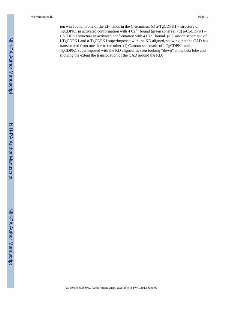
The CH1 helix can be divided into three segments. The first is the autoinhibitory regionblocking substrate-binding site of the KD and extends a basic residue (Lys338 in i-TgCDPK1, which is used as the reference protein in the description of the inactivated CDPKunless otherwise specified) to interact with a conserved acidic cluster made up of E135 andD138 on helix αD (Figure 3a). The side-chain of E135 is turned towards the CH1 helix andprevents it from fulfilling its function of positioning the ATP ribose in the active site pocket.This Lys-Glu-Asp triad (with Arg being a frequent substitute for Lys) and the orientation ofCH1 are conserved in parasitic and plant CDPKs, and are also characteristic of thepseudosubstrate inhibition mode observed in CaMKII3. The residues Lys338 and Leu339mark the transition into the second segment, which also ends in a KL pair and contributeshydrophobic residues, namely Leu339, Met347 and Leu351, to the latter’s hydrophobiccore. All three residues are conserved in CDPKs, with Leu339 and Met347 also conservedin different isoforms of CaMKII and participating in key hydrophobic interaction withcalmodulin15. Finally, the last segment of the CH1 helix serves as the E-helix of the first EF-hand, namely E1 (Supplementary Figure 1 and Figures 2a and 2b), with Leu360, Phe364 andMet367 engaged in the hydrophobic core of the N-EF-lobe.
From the N-EF-lobe onward, the inactivated form of the CAD largely resembles CaM instructure and organization (Supplementary Figures 2 and 3). The CH2 helix emerges fromthe second EF-hand to run almost anti-parallel to CH1 but is comparatively shorter by thelength of the autoinhibitory segment. It can also be divided into three segments: the F-helixof the second EF-hand (F2) and the E-helix of third EF-hand (E3) form the bookends, with alinker in the middle that is similar to but shorter than that found in CaM and TnC.Interestingly, in i-TgCDPK1 and i-TgCDPK3, both CH1 and CH2 are slightly bent, with thetwo helices bowed towards each other in a manner presaging this region’s potentialconformational change.
In addition to the Lys-Glu-Asp triad, there are at least three other regions implicated inbinding the KD and CAD in the inactivated state. First, Leu323, Ile327, Ile330 and Phe333(conserved as identical or similarly hydrophobic residues in other parasitic CDPKs) on CH1are buried in the amphipathic cleft in the C-terminal lobe of the KD, anchoring the longhelix in place (Figure 3d). In addition, the N-lobe of the KD interacts with an insert in the N-EF-lobe of the CAD (Figures 3a and 3b) – an insert found in a number of CDPKs but notCaM or TnC (see Supplementary Figure 1). Finally, both the activation segment of the KD(between the DFG and APE motifs) and the helix αC are in their respective inactive
Wernimont et al. Page 3
Nat Struct Mol Biol. Author manuscript; available in PMC 2013 June 07.
NIH
-PA Author Manuscript
NIH
-PA Author Manuscript
NIH
-PA Author Manuscript

positions, with the activation segment thus locked by an isoleucine (Ile212) extended into ahydrophobic pocket in the interface between CH1 and the C-EF-lobe (Figure 3b). Both theisoleucine and the residues forming the pocket are conserved amongst many apicomplexanand plant CDPKs Along with the hydrophobic cores in the two EF-lobes, these three regionsstabilize the inactivated CDPK conformation.
We also obtained the calcium-bound structures of TgCDPK1 and CpCDPK1, respectivelyresolved at 1.9 and 2.1 Å, with the KD and CAD intact (Table 1). They represent theactivated conformation of CDPKs and are therefore referred to as a-TgCDPK1 and a-CpCDPK1 below. In comparison to i-TgCDPK1 and i-TgCDPK3, it is easily noticeable thatthe entire CAD is translocated to a new position roughly 135° clockwise from its inactivatedposition about the KD (Figures 2e and 2f). Unforeseen in previous studies of CDPKs, thismovement liberates the residues required for activity and substrate recognition. Furthermore,with the CAD out of the way, helix αD is now positioned much closer to helix αF, closingthe cleft at the base of the KD where the CH1 is anchored in the autoinhibited structure(Figures 3e).
Calcium binding also induces the CAD to undergo substantial refolding. In both the a-TgCDPK1 and a-CpCDPK1 structures, all four EF-hands coordinate Ca2+ (Figures 2c and2d), resulting in the opening of these subdomains, exposure of hydrophobic surfaces andsubsequent refolding of the entire activation domain. The CH1 and CH2 helices are nolonger anti-parallel. Instead, they are partially unwound and bent at the boundaries betweenthe above defined segments, and intricately intertwined around each other and engaged inhydrophobic interaction with the two calcium-loaded EF-lobes (Figures 2c and 2d).Interestingly, the autoinhibitory region in the CH1 helix, previously found in hydrophobicinteraction with the C-EF-lobe in the autoinhibited state, is now involved in similarinteraction with the N-EF-lobe. This highlights the range of the CAD rearrangement, whichis more extensive than that seen in calmodulin and made possible because the involvementof the entire CH1 helix makes available more surfaces for interaction in comparison to otherEF-hand containing domains.
Driven out of its autoinhibitory position, the CAD finds itself lodged on a different face ofthe KD, secured by a different set of contacts (Figure 3c). The N-EF-lobe, which lines upnext to the N-lobe of the KD in the inactivated state, is now next to the C-lobe instead, withsalt bridges formed between the backbone nitrogens of 424TS425 and the side-chain ofAsp107, which is just downstream of helix αC. All three residues are highly conserved withthe TS pair situated on the tail end of the second EF-hand (DFDKNGYIEYSE), whereincalcium coordination has imposed a particular geometry to expose the backbone nitrogens tothe surface. In another contact between the domains, the E3 helix in the C-EF-lobe extendsArg445 (highly conserved as Arg, Lys or Gln in apicomplexan CDPKs but not in plantCDPKs) into a negatively charged pocket on the KD surface to form salt bridges with Asp70and Glu116 (with the aspartate conserved amongst parasites and plants but the glutamateconserved only in parasites). Additional backbone interactions are found between 349SY350
and 113KL114. This KL motif is conserved throughout parasitic CDPKs as well as someplant CDPKs (with lysine replaceable by arginine and leucine by isoleucine); however, theSK pair is only preserved for a small subset of parasitic CDPKs and not at all in plants.The 338KL339 pair in the pseudosubstrate segment on CH1 is also engaged, with Leu339again occupied in the hydrophobic core of the C-EF-lobe. Interestingly, Lys338 forms ahydrogen bond with the backbone oxygen of Leu32, which is part of a N-terminal stretch(30 residues from the beginning of the full length protein and shown in green in Figure 3c)latching first onto a hydrophobic ridge and then between the second EF-hand motif and theC-terminal EF-hands (Figure 3c). This N-terminal region is missing in the a-CpCDPK1
Wernimont et al. Page 4
Nat Struct Mol Biol. Author manuscript; available in PMC 2013 June 07.
NIH
-PA Author Manuscript
NIH
-PA Author Manuscript
NIH
-PA Author Manuscript
structure; however, sequence alignment suggests it is conserved in this kinase and someother apicomplexan CDPKs.
DiscussionThe extent of the structural rearrangement in CDPKs resulting from binding calcium is,insofar as we know, unprecedented amongst kinases (Figure 4a), surpassing the movementof the SH2 and SH3 domains in tyrosine kinases. The CAD is also capable of more twistsand turns than other proteins containing EF-hands such as CaM16 (Figures 4b and 4c) in partbecause it is a larger domain with on average about 200 amino acids, compared to 148 forcalmodulin (Supplementary Figure 2). The additional length is located primarily in the CH1helix, as well as inserts specific to each protein. As in CaM and TnC, the activated CAD isheld together by extensive hydrophobic interaction, featuring methionine and, moreprominently in CDPKs, leucine. More importantly, unlike CaM and TnC, the purpose of theCAD rearrangement is not to engage a substrate protein. Instead, we propose that thebending of CH1 and CH2 helices and their entanglement with the EF-hands drive the wholedomain away from the KD, severing the above-described KD-CAD interfaces, including andspecifically the autoinhibitory triad of Lys-Glu-Asp. This process makes Glu135 available tointeract with ATP, allows the activation loop to assume the active orientation, removesocclusion from the substrate binding state and frees the helix αC to approach the nucleotide-binding site. Other steps may still be required for complete activation; however, ourenzymatic results as well as other published results indicate that calcium alone can activatemany CDPKs.
CDPKs have other interesting sequence features in addition to the EF-hands. Specifically,the (K/R)L pair is a recurring motif amongst parasite and plant CDPKs, as well as withinindividual CDPKs (Supplementary Figure 1). In the N-lobe of the KD, 113KL114 (numberingbased on TgCDPK1) is engaged in interaction with the CAD in the activated structure.Interestingly, their interaction partner is the 349SK350 pair, with Lys350 being partof 350KL351. The remaining pair, 338KL339, is also the most significant, with Lys338 as thekey residue in the autoinhibitory mode and L339 participating in hydrophobic interactionwith the C-EF-lobe. Ranjan et al9 observed that in PfCDPK4, replacement of this leucine(Leu360 in PfCDPK4) with an alanine resulted in loss of activity, thus confirming thesignificance of this hydrophobic interaction and the CH1 helix as a whole.
Although CDPKs adopt the pseudosubstrate inhibition feature, they do not share all otherimportant mechanistic features of CaMKs. First, there is no evidence in structure orsequence suggesting that CDPKs form an oligomeric complex similar to that adopted byCaMKII3. Second, unlike CaMKI and CaMKIV, phosphorylation by another kinase has sofar not been found necessary for CDPK activation as seen in previous reports9,17 and ourown assays; however, the number of detailed biochemical studies on individual CDPKs stillprovide insufficient data for family-wide generalization. On the other hand,autophosphorylation is often necessary for activation of many kinases. In PfCDPK4, forexample, autophosphrylation of Thr234 has been observed to be essential9. This threonine ispart of a IGTAYY motif in the activation loop that is highly conserved in both parasitic andplant CDPKs. Therefore, autophosphorylation of the active loop may be a commonrequirement amongst CDPKs. Specific to CaMKIIs is autophosphorylation of Thr286located at the base of the autoinhibitory helix, which takes place to maintain constitutiveactivity after initial activation. In another study18, it was observed that this key threoninewas not conserved amongst plant and Plasmodium CDPKs; however, we note that in mostCDPKs, there are various serines and threonines situated in other positions between theHXW motif at the base of the C-lobe of the KD and the N-terminus of the CH1 helix.Covalent attachment of a phosphate group to anyone of these residues may potentially
Wernimont et al. Page 5
Nat Struct Mol Biol. Author manuscript; available in PMC 2013 June 07.
NIH
-PA Author Manuscript
NIH
-PA Author Manuscript
NIH
-PA Author Manuscript
prevent a CDPK’s return to the inactivated state. Furthermore, autophosphorylation sites inthe CAD have been identified in Arabidopsis and Plasmodium CDPKs18, some of whichmay have the capacity to lock a kinase in the activated conformation even in the absence ofcalcium. Our “calcium in-out-in” experiments were designed to investigate this possibility,but produced mixed results – under the conditions tested, only CpCDPK1 can be toggledbetween the active and inactive states solely by manipulation of calcium levels. ForTgCDPK1 and TgCDPK3, there are two possible interpretations: EDTA may be inadequateas a chelating agent or these two enzymes were modified by the process of calciumactivation such that they were unable to fully return to their inactivated state. Additionalresearch is required to determine whether some or most CDPKs share CaMK’s ability tobecome constitutively active.
Based on sequence alignment, many of the structural features discussed herein, includingpseudosubstrate inhibition and some of the KD-CAD interactions in both states, can bepredicted to be conserved in other apicomplexan CDPKs as well as canonical CDPKs atlarge. What is unconserved is a small gatekeeper residue - glycine in TgCDPK1 andCpCDPK1, and serine in Plasmodium CDPK4. This is rare in native kinases and creates ahydrophobic pocket in the nucleotide-binding site (Supplementary Figures 1 and 4), whichcan be exploited for selective chemical inhibition 19,20.
Study of CaMKs has shown that their mechanistic behavior and function are highlyrelated21, 22. It follows that structural and mechanistic discrepancies between CaMKs andCDPKs are also driven by functional divergence. Based on findings to date, CDPKs can becharacterized as self-contained calcium sensors enaged in calcium pathways simpler than inanimals in which extraneous proteins such as calmodulin and CaMKK are necessary tocontrol the action of CaMKs. The selective use of CDPKs by ciliates and apicomplexans isparticularly interesting, given that their genomes do not contain PKC and have relativelyfew CaMKs. These two groups are the dominant calcium-mediated kinases in other protists,most notably trypanosomatids23 which do not share the apicomplexan method for hostinvasion. Future research will uncover specific mechanistic behavior of individual CDPKsand reveal how plants and parasites use the direct calcium-sensing properties of thesekinases to their advantage.
MethodsProtein Expression
Information for all vectors used are available at http://www.sgc.utoronto.ca/SGC-WebPages/toronto-vectors.php. Using a full-length synthetic template purchased from Codon Devices(Cambridge, MA, USA) and reference sequence for the gene 541.m00134 in the ToxoDBdatabase (www.toxodb.org), we subcloned a construct of TgCDPK3 truncated at the N-terminus (L72-H537) and appended with an N-terminal His6tag including an integrated TEVcleavage site (mhhhhhhssgrenlyfq*g) into the pET15-MHL vector. A similarly taggedconstruct of CpCDPK1 (T71-E538) was subcloned from Cryptosporidium parvum strainIowa genomic DNA (obtained from MR4, Manassas, VA, USA) also into the pET15-MHLvector. The full length TgCDPK1 cDNA was cloned into the same expression vector andalso appended in the same way as the others. We expressed and purified all constructs aspreviously described24 using the Lex bioreactor system (Harbinger Biotechnology andEngineering Corp., Markham, ON, Canada) and BL21(DE3)-V2R-pACYC LamP as theexpression host, which includes a plasmid for co-expression of lambda phosphatase tosuppress protein phosphorylation. EDTA was excluded from the purification protocol. Allprotein samples eluted as apparent monomers from a Superdex S200 gel filtration column(GE Life Sciences, Baie d’Urfe, QC, Canada). The identities of the purified constructs were
Wernimont et al. Page 6
Nat Struct Mol Biol. Author manuscript; available in PMC 2013 June 07.
NIH
-PA Author Manuscript
NIH
-PA Author Manuscript
NIH
-PA Author Manuscript
verified by mass spectroscopic analysis, which also confirmed the absence ofphosphorylation.
CrystallographyTgCDPK3 crystals emerged in 20% PEG 3350 and 0.2M KF with the addition of 3mM 5-[(Z)-(5-chloro-2-oxo-1,2-dihydro-3H-indol-3-ylidene)-methyl]-N-[2-(diethylamino)ethyl]-2,4-dimethyl-1H-pyrrole-3-carboxamide (SU11652 from EMDChemicals, Gibbstown, NJ, USA), and were flash frozen in N2(l) with 25% glycerol as acryoprotectant. For CpCDPK1, the conditions yielding the final crystals were 20% PEG3350 and 0.2M diammonium tartrate in the presence of 2mM AMPPNP, 4mM MgCl2, 2mMCaCl2, and 6mM TCEP. These crystals were flash cooled in N2(l) with 30% glycerol as acryoprotectant. For the i-TgCDPK1 structure, crystals grew at 20 °C in 20% PEG 3350,0.2M KF, 2mM AMPPNP, 4mM MgCl2, 6.25mM TCEP. Crystals were flash cooled inN2(l) with 35% Peg 3350 as a cryoprotectant. For the activated form, TgCDPK1 wascrystallized at 20 °C in 26% PEG 3350, 0.2M Li2SO4, and Tris buffer (pH 8.0) in thepresence of 2mM AMPPNP, 4mM MgCl2, 2mM CaCl2, and 6mM TCEP. Crystals wereflash cooled in N2(l) with mother liquor supplemented with 30% glycerol.
We collected data for TgCDPK3 and a-TgCDPK1 at the APS beam line 19ID (http://www.sbc.anl.gov). Data for CpCDPK1 were collected on a rotating anode FR-E+Superbright with a RAxis IV++ (Rigaku, The Woodlands, TX, USA). Data for i-TgCDPK1were collected on a Rigaku rotating anode FR-E Superbright with a Rigaku Saturn A200 ccddetector. We processed all data using HKL-2000 (HKL Research Inc., Charlottesville, USA;www.hkl-xray.com) and solved all the structures first by molecular replacement usingPhaser25 and the TgCDPK3 kinase domain structure as the reference model (3DXN). TheCAD domain was manually built in iterative stages using Coot and Refmac5 within theCCP4 program suite26. Final refinement for i-TgCDPK1 was done using the programBUSTER-TNT (Global Phasing Ltd., Cambridge, UK; www.globalphasing.com). Dataprocessing and refinement statistics are summarized in Table 1. All structuralrepresentations in this paper were prepared with Pymol (Delano Scientific LLC, Palo Alto,USA; http://www.pymol.org) and espript (http://espript.ibcp.fr)
Calcium dependence assaysKinase activity was measured using an NADH-coupled ATPase assay (using pyruvatekinase (PK) and lactate dehydrogenase (LDH)) in a 96-well format based on the method ofKilanitsa and adapted for use with high ATP concentrations27. After incubating ATP (1mM) with the assay components (typically including 1 mM Syntide-2, 600 mM NADH, 200mM PEP, LDH/PK mix from Sigma (with 6 units of LDH/mL), 10 mM MgCl2, and CaCl2,as indicated) for 10 minutes, the reaction was initiated with the addition of 125 nM enzyme.Initial rates were calculated (for 3 to 10 minutes) and triplicate data were analyzed usingSigmaPlot 9.
For the “calcium-in-out-in” assay, each CDPK (1 μM) was incubated with 5mM CaCl2,5mM MgCl2 and 1mM ATP in 25mM Tris, 200mM NaCl at pH 7.5. The mixture wasincubated for 1 hour at room temperature. After incubation, 20mM EDTA was added, andthe mixture was dialyzed overnight in 4 L of 25mM Tris, 200mM NaCl, pH 7.5 at 4°C.After testing for activity, 100 μM CaCl2 was added and all the samples were tested foractivity again. In summary, the different samples were assayed after initial incubation, afterEDTA treatment and again after the second addition of calcium, all using the PK-LDHcoupled assay with 1mM ATP and 1 mM Syntide-2.
Wernimont et al. Page 7
Nat Struct Mol Biol. Author manuscript; available in PMC 2013 June 07.
NIH
-PA Author Manuscript
NIH
-PA Author Manuscript
NIH
-PA Author Manuscript
Supplementary MaterialRefer to Web version on PubMed Central for supplementary material.
AcknowledgmentsWe thank John Christodoulou at University College London, Oliver Billker at the Sanger Institute, Stefan Knapp,Tom Heightman, Matthieu Schapira, Cheryl Arrowsmith and Aled Edwards at the Structural Genomics Consortium(SGC) for insightful discussions and/or constructive review of this manuscript. We also thank Jocelyne Lew,Ashley Hutchinson, Abbasali Hassanali and Helen Ren at the SGC for their efforts in cloning and expressing theproteins in this study. The Structural Genomics Consortium (SGC) is a registered charity (number 1097737) thatreceives funds from the Canadian Institutes for Health Research, the Canadian Foundation for Innovation, GenomeCanada through the Ontario Genomics Institute, GlaxoSmithKline, the Knut and Alice Wallenberg Foundation, theOntario Innovation Trust, the Ontario Ministry for Research and Innovation, Merck & Co. Inc., the NovartisResearch Foundation, the Petrus and Augusta Hedlund’s Foundation, the Swedish Agency for Innovation Systems,the Swedish Foundation for Strategic Research, and the Wellcome Trust.
References1. Sibley LD. Invasion strategies of intracellular parasites. Science. 2004; 304:248. [PubMed:
15073368]
2. Sibley LD. Intracellular parasite invasion strategies. Science. 2004; 304(5668):248. [PubMed:15073368] Nagamune K, Moreno SN, Chini EN, Sibley LD. Calcium regulation and signaling inapicomplexan parasites. Subcell Biochem. 2008; 47:70. [PubMed: 18512342]
3. Rosenberg OS, et al. Structure of the autoinhibited kinase domain of CaMKII and SAXS analysis ofthe holoenzyme. Cell. 2005; 123(5):849. [PubMed: 16325579]
4. Billker O, Lourido S, Sibley LD. Calcium-dependent signaling and kinases in apicomplexanparasites. Cell Host Microbe. 2009; 5(6):612. [PubMed: 19527888]
5. Qiu W, et al. Novel structural and regulatory features of rhoptry secretory kinases in Toxoplasmagondii. EMBO J. 2009; 28(7):969. [PubMed: 19197235]
6. Cowan-Jacob SW, et al. The crystal structure of a c-Src complex in an active conformation suggestspossible steps in c-Src activation. Structure. 2005; 13(6):861. [PubMed: 15939018]
7. Nagar B, et al. Structural basis for the autoinhibition of c-Abl tyrosine kinase. Cell. 2003; 112(6):859. [PubMed: 12654251]
8. Chandran V, et al. Structure of the regulatory apparatus of a calcium-dependent protein kinase(CDPK): a novel mode of calmodulin-target recognition. J Mol Biol. 2006; 357(2):400. [PubMed:16430916] Christodoulou J, Malmendal A, Harper JF, Chazin WJ. Evidence for differing roles foreach lobe of the calmodulin-like domain in a calcium-dependent protein kinase. J Biol Chem. 2004;279(28):29092. [PubMed: 15126505] Harmon AC, Putnam-Evans C, Cormier MJ. A Calcium-Dependent but Calmodulin-Independent Protein Kinase from Soybean. Plant Physiol. 1987; 83(4):830. [PubMed: 16665348]
9. Ranjan R, Ahmed A, Gourinath S, Sharma P. Dissection of mechanisms involved in the regulationof plasmodium falciparum calcium dependent protein kinase 4 (PfCDPK4). J Biol Chem. 2009
10. Billker O, et al. Calcium and a calcium-dependent protein kinase regulate gamete formation andmosquito transmission in a malaria parasite. Cell. 2004; 117(4):503. [PubMed: 15137943]
11. Green JL, et al. The motor complex of Plasmodium falciparum: phosphorylation by a calcium-dependent protein kinase. J Biol Chem. 2008; 283(45):30980. [PubMed: 18768477]
12. Dolle C, Ziegler M. Application of a coupled enzyme assay to characterize nicotinamide ribosidekinases. Anal Biochem. 2009; 385(2):377. [PubMed: 19027704]
13. Harmon AC, Yoo BC, McCaffery C. Pseudosubstrate inhibition of CDPK, a protein kinase with acalmodulin-like domain. Biochemistry. 1994; 33(23):7278. [PubMed: 8003491]
14. Yoo BC, Harmon AC. Intramolecular binding contributes to the activation of CDPK, a proteinkinase with a calmodulin-like domain. Biochemistry. 1996; 35(37):12029. [PubMed: 8810907]
15. Meador WE, Means AR, Quiocho FA. Modulation of calmodulin plasticity in molecularrecognition on the basis of x-ray structures. Science. 1993; 262(5140):1718. [PubMed: 8259515]
Wernimont et al. Page 8
Nat Struct Mol Biol. Author manuscript; available in PMC 2013 June 07.
NIH
-PA Author Manuscript
NIH
-PA Author Manuscript
NIH
-PA Author Manuscript
16. Chattopadhyaya R, Meador WE, Means AR, Quiocho FA. Calmodulin structure refined at 1.7 Aresolution. J Mol Biol. 1992; 228(4):1177. [PubMed: 1474585]
17. Vitart V, et al. Intramolecular activation of a Ca(2+)-dependent protein kinase is disrupted byinsertions in the tether that connects the calmodulin-like domain to the kinase. Biochemistry.2000; 39(14):4004. [PubMed: 10747788]
18. Hegeman AD, et al. A phyloproteomic characterization of in vitro autophosphorylation in calcium-dependent protein kinases. Proteomics. 2006; 6(12):3649. [PubMed: 16758442]
19. Knight ZA, Shokat KM. Features of selective kinase inhibitors. Chem Biol. 2005; 12(6):621.[PubMed: 15975507]
20. Ojo KK, et al. A unique variation of the ATP binding site makes Toxoplasma gondii calcium-dependent protein kinase 1 a drug target for selective kinase inhibitors. Nature Structural andMolecular Biology. 2010
21. Braun AP, Schulman H. The multifunctional calcium/calmodulin-dependent protein kinase: fromform to function. Annu Rev Physiol. 1995; 57:417. [PubMed: 7778873]
22. Hanson PI, Meyer T, Stryer L, Schulman H. Dual role of calmodulin in autophosphorylation ofmultifunctional CaM kinase may underlie decoding of calcium signals. Neuron. 1994; 12(5):943.[PubMed: 8185953]
23. Parsons M, Worthey EA, Ward PN, Mottram JC. Comparative analysis of the kinomes of threepathogenic trypanosomatids: Leishmania major, Trypanosoma brucei and Trypanosoma cruzi.BMC Genomics. 2005; 6:127. [PubMed: 16164760]
24. Vedadi M, et al. Genome-scale protein expression and structural biology of Plasmodiumfalciparum and related Apicomplexan organisms. Mol Biochem Parasitol. 2007; 151(1):100.[PubMed: 17125854]
25. Terwilliger T. SOLVE and RESOLVE: automated structure solution, density modification andmodel building. J Synchrotron Radiat. 2004; 11(Pt 1):49. [PubMed: 14646132]
26. Emsley P, Cowtan K. Coot: model-building tools for molecular graphics. Acta Crystallogr D BiolCrystallogr. 2004; 60(Pt 12 Pt 1):2126. [PubMed: 15572765]
27. Kiianitsa K, Solinger JA, Heyer WD. NADH-coupled microplate photometric assay for kineticstudies of ATP-hydrolyzing enzymes with low and high specific activities. Anal Biochem. 2003;321(2):266. [PubMed: 14511695]
Wernimont et al. Page 9
Nat Struct Mol Biol. Author manuscript; available in PMC 2013 June 07.
NIH
-PA Author Manuscript
NIH
-PA Author Manuscript
NIH
-PA Author Manuscript
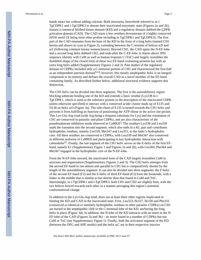
Figure 1. Effect of Ca2+on CDPKs(a) Using the LDH/PK assay and Syntide 2 as a substrate, all three enzymes were tested foractivity in the presence of MgCl2 and ATP with increasing Ca2+ concentration. ForTgCDPK1 (black), Km was found to be 10.2±1.6 μM. For TgCDPK3 (green), Km found tobe 14.0±2.0 μM. For CpCDPK1 (red), Km was found to be 14.1±0.4 μM. (b) Calciumdependence experiment: Each CDPK (1 μM) was incubated with CaCl2, MgCl2 and ATPfor 1 hour for activation and autophosphorylation. Afterwards, each sample was treated withEDTA and thoroughly dialyzed to remove Ca2+. Two samples (labeled TgCDPK1-E,TgCDPK3-E, in the bargraph) demonstrated partial activity while CpCDPK1-Edemonstrated only a basal level of activity. Calcium was added to all three samples again foractivation, with all found to be fully active (TgCDPK1-E-Ca, TgCDPK3-E-Ca andCpCDPK1-E-Ca in the bargraph).
Wernimont et al. Page 10
Nat Struct Mol Biol. Author manuscript; available in PMC 2013 June 07.
NIH
-PA Author Manuscript
NIH
-PA Author Manuscript
NIH
-PA Author Manuscript
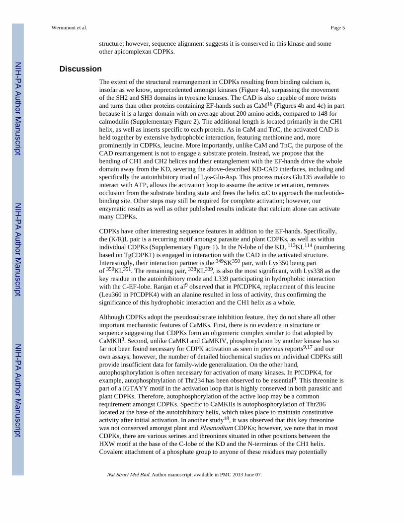
Figure 2. Crystal structures of apicomplexan CDPKsThe first four structures (a, b, c and d) are oriented by aligning their kinase domains. Thekinase domain (KD) is shown in grey in all cases, with the CDPK activation domain (CAD)shown in color. The CH1 and CH2 helices are highlighted in cyan and pale magentarespectively, with these helices broken into three segments in the activated conformation (a-CpCDPK1 and a-TgCDPK1). The N-terminal latch in TgCDPK1 is shown in green.Calcium ions are shown as green spheres, with those hidden in the protein structuresindicated by green arrows. (a) i-TgCDPK1 – structure of TgCDPK1 in inactivatedconformation. A metal ion was observed in three of the four EF hands. The coordination ofeach did not match that of Ca2+ or Mg2+. The pseudosubstrate autoinhibitory triad ishighlighted. (b) i-TgCDPK3 – structure of TgCDPK3 in inactivated conformation. A Mg2+
Wernimont et al. Page 11
Nat Struct Mol Biol. Author manuscript; available in PMC 2013 June 07.
NIH
-PA Author Manuscript
NIH
-PA Author Manuscript
NIH
-PA Author Manuscript
ion was found in one of the EF-hands in the C-terminus. (c) a-TgCDPK1 – structure ofTgCDPK1 in activated conformation with 4 Ca2+ bound (green spheres). (d) a-CpCDPK1 –CpCDPK1 structure in activated conformation with 4 Ca2+ bound. (e) Cartoon schematic ofi-TgCDPK1 and a-TgCDPK1 superimposed with the KD aligned, showing that the CAD hastranslocated from one side to the other. (f) Cartoon schematic of i-TgCDPK1 and a-TgCDPK1 superimposed with the KD aligned, as seen looking “down” at the beta lobe andshowing the extent the translocation of the CAD around the KD.
Wernimont et al. Page 12
Nat Struct Mol Biol. Author manuscript; available in PMC 2013 June 07.
NIH
-PA Author Manuscript
NIH
-PA Author Manuscript
NIH
-PA Author Manuscript
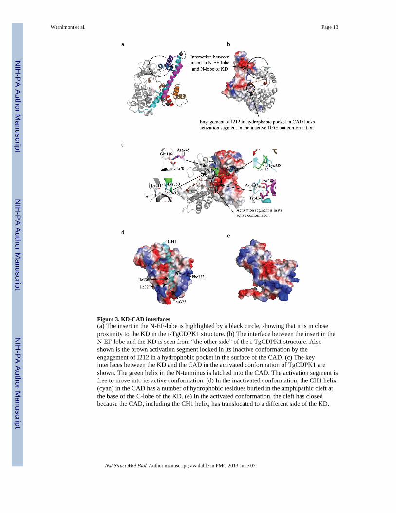
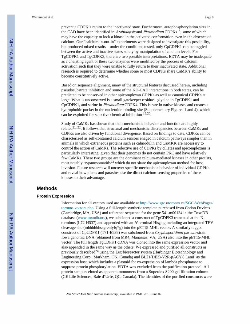
Figure 3. KD-CAD interfaces(a) The insert in the N-EF-lobe is highlighted by a black circle, showing that it is in closeproximity to the KD in the i-TgCDPK1 structure. (b) The interface between the insert in theN-EF-lobe and the KD is seen from “the other side” of the i-TgCDPK1 structure. Alsoshown is the brown activation segment locked in its inactive conformation by theengagement of I212 in a hydrophobic pocket in the surface of the CAD. (c) The keyinterfaces between the KD and the CAD in the activated conformation of TgCDPK1 areshown. The green helix in the N-terminus is latched into the CAD. The activation segment isfree to move into its active conformation. (d) In the inactivated conformation, the CH1 helix(cyan) in the CAD has a number of hydrophobic residues buried in the amphipathic cleft atthe base of the C-lobe of the KD. (e) In the activated conformation, the cleft has closedbecause the CAD, including the CH1 helix, has translocated to a different side of the KD.
Wernimont et al. Page 13
Nat Struct Mol Biol. Author manuscript; available in PMC 2013 June 07.
NIH
-PA Author Manuscript
NIH
-PA Author Manuscript
NIH
-PA Author Manuscript
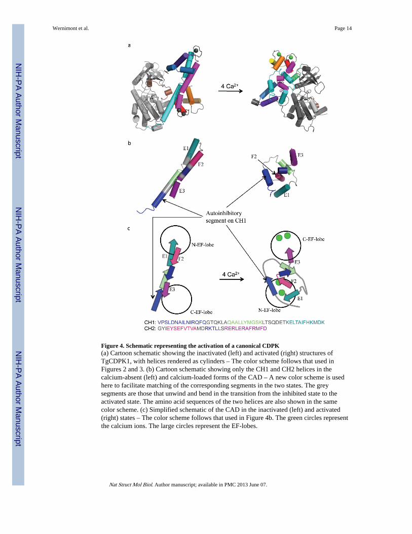
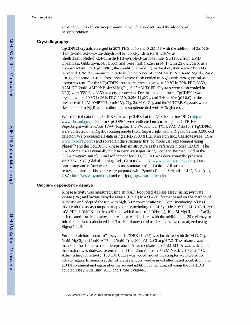
Figure 4. Schematic representing the activation of a canonical CDPK(a) Cartoon schematic showing the inactivated (left) and activated (right) structures ofTgCDPK1, with helices rendered as cylinders – The color scheme follows that used inFigures 2 and 3. (b) Cartoon schematic showing only the CH1 and CH2 helices in thecalcium-absent (left) and calcium-loaded forms of the CAD – A new color scheme is usedhere to facilitate matching of the corresponding segments in the two states. The greysegments are those that unwind and bend in the transition from the inhibited state to theactivated state. The amino acid sequences of the two helices are also shown in the samecolor scheme. (c) Simplified schematic of the CAD in the inactivated (left) and activated(right) states – The color scheme follows that used in Figure 4b. The green circles representthe calcium ions. The large circles represent the EF-lobes.
Wernimont et al. Page 14
Nat Struct Mol Biol. Author manuscript; available in PMC 2013 June 07.
NIH
-PA Author Manuscript
NIH
-PA Author Manuscript
NIH
-PA Author Manuscript
NIH
-PA Author Manuscript
NIH
-PA Author Manuscript
NIH
-PA Author Manuscript
Wernimont et al. Page 15
Table 1
Crystallographic table for all four protein structures
i-TgCDPK1 a-TgCDPK1 i-TgCDPK3 a-CpCDPK1
Data collection
Space group P21 P212121 P21 P21
Cell dimensions
a, b, c (Å) 48.01, 72.91, 65.02 49.24, 95.61, 101.61 71.05, 43.76, 84.82 60.48, 55.51, 81.71
α, β, γ (°) 90.00, 96.26, 90.00 90.00, 90.00, 90.00 90.00, 96.95, 90.00 90.00, 105.44, 90.00
Resolution (Å) 20-2.3 (2.34-2.30) 50-1.9 (1.97-1.90) 30-2.0(2.03-2.00) 50-2.25(2.33-2.25)
Rsym or Rmerge 5.9 (39.5) 6.4 (84.2) 4.8 (45.7) 8.9 (87.8)
I / σI 18.1 (1.2) 34.4 (1.2) 26.6 (2.7) 16.6 (1.7)
Completeness (%) 99.5 (94.4) 99.8 (98.1) 99.7 (99.6) 100 (99.9)
Redundancy 3.5 (2.4) 6.1 (3.7) 3.6 (3.3) 3.6 (3.5)
Refinement
Resolution (Å) 20.0 - 2.3 50.0 – 1.95 50.0 – 2.0 50.0 – 2.25
No. reflections 19811 35714 33611 23815
Rwork / Rfree 0.201/0.238 0.209/0.257 0.212/0.253 0.201/0.255
No. atoms
Protein 3592 3768 3481 3645
Ligand/ion 106 60 78 86
Water 78 195 203 125
B-factors
Protein 55.08 24.1 42.12 28.405
Ligand/ion 72.057 45.22 39.75 66.61
Water 48.16 27.52 45.93 32.98
R.m.s. deviations
Bond lengths (Å) 0.01 0.01 0.01 0.019
Bond angles (°) 1.1 1.2 1.2 1.9
N.B. For each structure, a single crystal was used.
Nat Struct Mol Biol. Author manuscript; available in PMC 2013 June 07.
Copyright © 2022 FDOKUMEN