Controlling TIME: How MNK Kinases Function to Shape ... - MDPI
19
Cancers 2020, 12, 2096; doi:10.3390/cancers12082096 www.mdpi.com/journal/cancers Review Controlling TIME: How MNK Kinases Function to Shape Tumor Immunity Thao N.D. Pham 1,2, *, Christina Spaulding 1,2 and Hidayatullah G. Munshi 1,2,3, * 1 Department of Medicine, Feinberg School of Medicine, Northwestern University, Chicago, IL 60611, USA; [email protected] 2 Jesse Brown VA Medical Center, Chicago, IL 60612, USA 3 Robert H. Lurie Comprehensive Cancer Center, Chicago, IL 60611, USA * Correspondence: [email protected] (T.N.D.P.); [email protected] (H.G.M.); Tel.: +312-503-0312 (T.N.D.P.); +312-503-2301 (H.G.M.) Received: 11 June 2020; Accepted: 26 July 2020; Published: 28 July 2020 Abstract: A number of studies have clearly established the oncogenic role for MAPK-interacting protein kinases (MNK) in human malignancies. Modulation of MNK activity affects translation of mRNAs involved in cancer development, progression, and resistance to therapies. As a result, there are ongoing efforts to develop and evaluate MNK inhibitors for cancer treatment. However, it is important to recognize that MNK activity also plays an important role in regulating the innate and adaptive immune systems. A better understanding of the role of MNK kinases and MNK-mediated signals in regulating the immune system could help mitigate undesired side effects while maximizing therapeutic efficacy of MNK inhibitors. Here, we provide a systematic review on the function of MNK kinases and their substrates in immune cells. Keywords: MNK kinases; eIF4E; hnRNP A1; Sprouty; macrophages; neutrophils; lymphocytes; tumor immune microenvironment (TIME), immune responses 1. Introduction 1.1. MAPK-Interacting Serine/Threonine-Protein Kinases The MAPK-interacting kinases 1 and 2, or MNK1 and MNK2, are serine/threonine protein kinases that were identified as substrates for MAPK/ERK and p38 MAPK by two different groups [1,2]. Phosphorylation of MNK kinases by MAPKs allows MNKs to interact with and phosphorylate a number of downstream effectors, including eukaryotic initiation factor 4E (eIF4E) [3], heterogeneous nuclear ribonucleoprotein A1 (hnRNP A1) [4], and Sprouty 1,2 [5,6]. These proteins, in turn, participate in a myriad of signals that culminate in distinct biological responses. It should be noted that MNK1 and MNK2 can exhibit substrate specificity [7], and therefore can elicit responses that are unique to each kinase. MNK kinases play important roles in cancer development and resistance to therapeutics. Targeting MNK kinases has the potential to delay tumor growth and reverse drug resistance. For example, blockade of MNK activity enhances sensitivity to BET inhibitors [8] and gemcitabine [9] in pancreatic cancer, mTOR inhibitors in leukemia [10] and lung cancer [11], and trastuzumab in breast cancer [12]. More recently, it was demonstrated in the highly metastatic Myc Tg ;Kras G12D hepatocellular carcinoma model that targeting MNK kinases can reprogram the tumor microenvironment, allowing for increased T-cell infiltration and function [13]. These studies emphasize the need for a better understanding of how MNK kinases and their effectors function to affect different components of the tumor microenvironment. Several of these MNK effectors have been implicated in regulating the immune compartment.
-
Upload
khangminh22 -
Category
Documents
-
view
3 -
download
0
Transcript of Controlling TIME: How MNK Kinases Function to Shape ... - MDPI

Cancers 2020, 12, 2096; doi:10.3390/cancers12082096 www.mdpi.com/journal/cancers
Review
Controlling TIME: How MNK Kinases Function to
Shape Tumor Immunity
Thao N.D. Pham 1,2,*, Christina Spaulding 1,2 and Hidayatullah G. Munshi 1,2,3,*
1 Department of Medicine, Feinberg School of Medicine, Northwestern University, Chicago, IL 60611, USA;
[email protected] 2 Jesse Brown VA Medical Center, Chicago, IL 60612, USA 3 Robert H. Lurie Comprehensive Cancer Center, Chicago, IL 60611, USA
* Correspondence: [email protected] (T.N.D.P.); [email protected] (H.G.M.); Tel.:
+312-503-0312 (T.N.D.P.); +312-503-2301 (H.G.M.)
Received: 11 June 2020; Accepted: 26 July 2020; Published: 28 July 2020
Abstract: A number of studies have clearly established the oncogenic role for MAPK-interacting
protein kinases (MNK) in human malignancies. Modulation of MNK activity affects translation of
mRNAs involved in cancer development, progression, and resistance to therapies. As a result, there
are ongoing efforts to develop and evaluate MNK inhibitors for cancer treatment. However, it is
important to recognize that MNK activity also plays an important role in regulating the innate and
adaptive immune systems. A better understanding of the role of MNK kinases and MNK-mediated
signals in regulating the immune system could help mitigate undesired side effects while
maximizing therapeutic efficacy of MNK inhibitors. Here, we provide a systematic review on the
function of MNK kinases and their substrates in immune cells.
Keywords: MNK kinases; eIF4E; hnRNP A1; Sprouty; macrophages; neutrophils; lymphocytes;
tumor immune microenvironment (TIME), immune responses
1. Introduction
1.1. MAPK-Interacting Serine/Threonine-Protein Kinases
The MAPK-interacting kinases 1 and 2, or MNK1 and MNK2, are serine/threonine protein
kinases that were identified as substrates for MAPK/ERK and p38 MAPK by two different groups
[1,2]. Phosphorylation of MNK kinases by MAPKs allows MNKs to interact with and phosphorylate
a number of downstream effectors, including eukaryotic initiation factor 4E (eIF4E) [3],
heterogeneous nuclear ribonucleoprotein A1 (hnRNP A1) [4], and Sprouty 1,2 [5,6]. These proteins,
in turn, participate in a myriad of signals that culminate in distinct biological responses. It should be
noted that MNK1 and MNK2 can exhibit substrate specificity [7], and therefore can elicit responses
that are unique to each kinase. MNK kinases play important roles in cancer development and
resistance to therapeutics. Targeting MNK kinases has the potential to delay tumor growth and
reverse drug resistance. For example, blockade of MNK activity enhances sensitivity to BET inhibitors
[8] and gemcitabine [9] in pancreatic cancer, mTOR inhibitors in leukemia [10] and lung cancer [11],
and trastuzumab in breast cancer [12]. More recently, it was demonstrated in the highly metastatic
MycTg;KrasG12D hepatocellular carcinoma model that targeting MNK kinases can reprogram the
tumor microenvironment, allowing for increased T-cell infiltration and function [13]. These studies
emphasize the need for a better understanding of how MNK kinases and their effectors function to
affect different components of the tumor microenvironment. Several of these MNK effectors have
been implicated in regulating the immune compartment.

Cancers 2020, 12, 2096 2 of 19
1.1.1. Eukaryotic Initiation Factor 4E (eIF4E)
eIF4E is the most well-studied and the best-characterized of the MNK substrates. To date, MNK
kinases are the only known physiological kinases for eIF4E [3]. The protein is necessary for cap-
dependent translation of a subset of mRNAs that typically have long 5′-untranslated regions (5′-
UTRs), high G/C content, and complex hairpin structures [14]. The process of mRNA translation is
initiated by the binding of the m7G cap structure at the 5′-terminus to the trimeric eIF4F complex.
This complex consists of eIF4E, the scaffolding protein eIF4G, and the RNA helicase eIF4A [15,16].
The physical interaction between eIF4G and eIF4E, which is via a highly conserved motif, is crucial
for the recruitment of other initiation factors and ribosomal proteins for protein synthesis [15,16]. In
addition, eIF4G also serves as a docking site for MNK kinases to phosphorylate eIF4E, as it brings
MNK kinases and eIF4E into proximity to each other [17]. As a result, there have been efforts at
targeting this eIF4G:eIF4E interaction, in addition to MNK kinases, to modulate eIF4E
phosphorylation and translation process.
Deletion of MNK kinases results in impairment of eIF4E phosphorylation and eIF4E-mediated
translation [3,18]. Even a modest increase in eIF4E expression was sufficient to cause cellular
transformation [19], while expression of a non-phosphorylatable eIF4E mutant in mice resulted in
delayed tumor development [20] and metastasis [21]. Like eIF4E, constitutively active (T332D), but
not the kinase-dead (T2A2), MNK1 kinase accelerated lymphomagenesis [20]. These and other
studies have identified key targets of eIF4E that are essential for cancer cell proliferation, survival,
and migration, such as cyclin D1 and D3, c-Myc, MDM2, VEGF, Survivin, and Bcl-2 [22].
1.1.2. Heterogeneous Nuclear Ribonucleoprotein A1 (hnRNP A1)
Heterogeneous nuclear ribonucleoproteins (hnRNPs) are the largest and most well-studied
family of RNA-binding proteins with more than 20 members [23,24]. hnRNP A1 belongs to the
subfamily A/B and is arguably the best known member among the hnRNPs [23,24]. hnRNP A1 can
promote or inhibit translation of select genes by binding to AU-rich elements (AREs), for example
AUGGGA(U) or UAGA(G), residing within the 3′-UTR of the transcripts [23,24]. hnRNP A1 was first
identified as a MNK substrate in T lymphocytes [4]. In both in vitro and in vivo settings, hnRNP A1
is phosphorylated by MNK kinases at residues 192 and 310/311/312, which is abrogated upon
selective MNK inhibition [4]. The molecular mechanisms by which hnRNP A1 supports malignant
transformation include regulation of cell survival, alteration of cell cycle, invasion and metastasis,
altered metabolism and stress adaptation [23,24]. To date, inhibition of hnRNP A1 expression and
activity can be achieved with both natural and synthetic compounds [25–28]. Accordingly, we and
other groups have shown that inhibition of hnRNP A1 synergizes with targeted anti-cancer therapies
to further suppress tumor cell proliferation and survival [27,28].
1.1.3. Sprouty 1/2
Sprouty proteins are evolutionary conserved regulators of the ERK/MAPK signaling pathway.
They inhibit activation of ERK in response to a wide range of trophic factors [29,30]. In mammals,
there are four Sprouty homologs (Spry1‐4), with Spry2 exhibiting highest evolutionary conservation
[31]. Spry2 protein was first reported to be a MNK substrate by DaSilva and colleagues [5]. Growth
factor-induced serine phosphorylation of Spry2 increased the protein stability, which is mediated by
MNK1 at Ser112 and Ser121 residues [5]. Later on, it was demonstrated that stabilization and
upregulation of the Spry1 protein, following activation of type I interferon (IFN) signaling, is also
mediated by MNK kinases [6]. As expression of Spry1/2 is inhibitory to IFN-mediated signaling [6],
MNK-mediated upregulation of Sprouty proteins might be a potential mechanism accounting for
IFN-resistance in malignant cells.
1.2. Cellular Heterogeneity of Tumor Immune Microenvironment
Tumor immune microenvironment (TIME) is a dynamic system and consists of different cellular
and non-cellular elements (reviewed in [32]). It is becoming clear that both the innate (macrophages,

Cancers 2020, 12, 2096 3 of 19
myeloid derived suppressor cells, neutrophils, dendritic cells, and natural killer cells) and adaptive
(T and B lymphocytes) immune systems are essential components of TIME and that their crosstalk
plays a major role in dictating anti-tumor responses (reviewed in [32,33]). Each cell type has been
evaluated and associated with different prognosis and therapeutic responses in patients (reviewed
in [32,33]).
1.2.1. Innate Immunity
The innate immune compartment includes cell types such as macrophages, neutrophils, natural
killer cells, and mast cells [33]. Among these, the antigen-presenting cells, namely macrophages and
dendritic cells, can recognize, process, and present non-self antigens to lymphocytes [34]. Depending
on the pathogen identity, the non-self antigens can be coupled with either major histocompatibility
complex (MHC) class I or MHC class II molecules (reviewed in [34]). The engagement of MHC
molecules with T-cell antigen receptors determines T-cell differentiation and function as discussed
later in this review (reviewed in [35]).
Macrophages
Macrophages are the most abundant tumor-infiltrating leukocytes [36,37]. Depending on local
cues, infiltrating macrophages can assume either an anti (M1)- or pro (M2)-tumorigenic phenotype
(tumor-associated macrophages, TAMs) (reviewed in [38]). It is noteworthy that the commonly used
M1/M2 terminology refers to two extreme states of macrophage polarization, and does not by any
means capture the wide range of functional plasticity in macrophages. The M1 phenotype can be
induced by type I cytokines like TNFα and IFNγ, while type II cytokines, such as interleukin (IL)-4,
IL-10, IL-13, and transforming growth factor beta (TGFβ), can promote macrophage polarization
toward the M2 state. M1 and M2 polarization can also be induced by colony-stimulating factor 2
(CSF2 or GM-CSF) and colony-stimulating factor 1 (CSF1 or M-CSF), respectively (reviewed in [39]).
M2 macrophages, referred to as TAMs from hereon, inhibit anti-tumor immune responses through
multiple mechanisms, most notably, through depletion of amino acids and production of
immunosuppressive metabolites, such as indolamine-2,3-dioxygenase (IDO) and arginase 1 (ARG1).
TAMs can also suppress T-cell function through direct engagement of T-cell inhibitory proteins (e.g.,
B7-1/CD80, B7-2/CD86, programmed death-ligand 1 (PD-L1) and 2 (PD-L2)) and apoptotic receptors
(e.g., FasL and TRAIL). Finally, through the production of IL-10 and TGFβ, TAMs can inhibit the
activation, differentiation, proliferation, and function of effector T-cells, as well as promote the
expansion of suppressive T-cells (reviewed in [38]).
Pathways that regulate infiltration, survival, and immunosuppressive activity of TAMs have
been identified and characterized in preclinical models. Targeting these pathways with CSF1 receptor
(CSF1R) inhibitors, PI3Kγ inhibitors, or mTOR inhibitors suppresses TAM recruitment and survival
and limits tumor progression as a result (reviewed in [38]). Notably, targeting CSF1R can also re-
polarize the remaining TAMs towards the M1 phenotype and increase their anti-tumorigenic activity
[40,41].
Myeloid-Derived Suppressor Cells
Myeloid-derived suppressor cells (MDSCs), which express CD11b and Gr1, refer to a group of
myeloid cells that suppress immune responses (reviewed in [42]). These cells are rare in healthy
individuals and are only found in pathological conditions. Under physiological conditions, GM-CSF
can drive myelopoiesis while M-CSF promotes the differentiation of macrophages (reviewed in [42]).
In pathological conditions, such as cancer, increased expression of these factors results in increased
generation of MDSCs. Although MDSCs can suppress a number of different cells of the immune
system, the main targets of MDSCs are T-cells. The factors implicated in MDSC-mediated immune
suppression include ARG1 and nitric oxide synthase 2 (NOS2). ARG1 and NOS2 directly interfere
with T-cell proliferation, activity, and survival by metabolizing L-arginine, a major T-cell metabolite.
Cancers 2020, 12, 2096 4 of 19
In addition, MDSCs can promote the development of regulatory T-cells through secretion of
immunosuppressive cytokines (reviewed in [42,43]).
Different tumor types display different levels of MDSC infiltration. When MDSCs are present,
there is a strong association between high numbers of MDSCs and resistance to anti-cancer therapies
(reviewed in [42,43]). In recent years, various preclinical studies and clinical trials targeting MDSCs
have demonstrated promising results. For instance, it has been demonstrated that inhibitors to
JAK/STAT or VEGFA block MDSC development, and agents such as all-trans retinoic acid (ATRA),
vitamins A and D3, and IL-12 are able to modulate MDSC differentiation. Additionally, agents aimed
at inhibiting MDSC recruitment to the tumors, such as anti-glycan, anti-IL-17, anti-CXCR2, anti-
galectin-3, are also being studied (reviewed in [43]).
Tumor-Associated Neutrophils
Neutrophils make up a substantial proportion of the immune infiltrate in many cancer types.
Neutrophils are elevated in the circulation and accumulate in peripheral organs during tumor
progression (reviewed in [44]). Similar to macrophages, cancer-related neutrophils can undergo a
spectrum of functional plasticity in response to local cues found in the TIME. Among the cytokines
that can program neutrophil polarization, TGFβ, granulocyte colony-stimulating factor (G-CSF or
CSF3), and IFNβ are the best-studied molecules. While IFNβ signals to promote the anti-tumor (N1)
phenotype, TGFβ and G-CSF are known to promote tumor- and metastasis-promoting (N2)
phenotypes in neutrophils [44,45]. Interestingly, N1/N2 neutrophils may be associated with different
stages of tumor development: at early stages the neutrophils remain at the edges of the tumor and
are predominantly N1, but as the tumor progresses, the neutrophils infiltrate into the tumor and
display an N2 phenotype [46]. The exact cues that turn on this phenotypic switch remains unknown.
Mature neutrophils upregulate several receptors, including Toll-like receptor (TLR) 4 and CXCR2.
Ligands of CXCR2 (such as CXCL1, CXCL2, CXCL5, and CXCL8) are expressed within the tumor
microenvironment by cancer, stromal, and immune cells to recruit neutrophils (reviewed in [44]).
Developing and mature neutrophils also rely on G-CSF for proliferation, mobilization, and function.
N2 neutrophils strongly upregulate markers CCL17, ARG1, and MMP9, which support their
immunosuppressive function (reviewed in [44]).
Several studies have suggested that expression of PD-L1+ on neutrophils is indicative of their
immunosuppressive function. Intra- and peri-tumoral neutrophils are found to have higher
expression levels of PD-L1 compared to circulating neutrophils [47]. In addition, PD-L1+ neutrophils
suppress effector T-cell functions and contribute to tumor progression. Treatment with anti-PD-L1
was found to sufficiently reinvigorate T-cells and control tumor growth. In addition, targeting
CXCR2, which is critical for neutrophil recruitment, decreases neutrophil accumulation, reduces
metastases, improves T-cell infiltration, and enhances the efficacy of anti-PD-1 therapy (reviewed in
[44]).
1.2.2. Adaptive Immunity
T Lymphocytes
T lymphocytes include TCRαβ T-cells, regulatory T-cells (Treg) (described further below), the
less common TCRγδ T-cells, and T-cells expressing invariant receptors (reviewed in [48]). The two
types of TCRαβ-bearing T-cells that have been largely focused on are the conventional CD4+ and
CD8+ T-cells. Naïve T-cells can be differentiated into CD4+ T helper cells or CD8+ cytotoxic T-cells
when their T‐cell antigen receptor (TCR) engages with MHC class II and I molecules, respectively
(reviewed in [34,48]).
CD8+ T-cells directly kill infected or damaged cells by using lytic enzymes, such as perforin and
granzymes (e.g., granzyme A, B). However, various mechanisms of immune evasion have been
described and demonstrated. These mechanisms include, among others, secretion of
immunosuppressive cytokines (IL-10, TGFβ) by immunosuppressive cells and cancer cells

Cancers 2020, 12, 2096 5 of 19
themselves (reviewed in [33]). These cells can also induce T-cell exhaustion via direct engagement
with inhibitory receptors on T-cells, such as PD-1, LAG-3, and TIM3 to name a few (reviewed in [33]).
As discussed earlier, it is now becoming clear that most, if not all, components of the innate immune
system can be re-educated by the tumor to become pro-tumorigenic and T-cell suppressive.
Immunotherapy, designed to overcome these inhibitory signals, can reinvigorate T-cells and enhance
their cytotoxic activity against tumor cells.
CD4+ T-cells can also infiltrate neoplastic tissues and have central roles in shaping pro- and anti-
tumor immune responses (reviewed in [49]). Naïve CD4+ T-cells can differentiate into Tregs or one of
the several CD4+ T helper subsets depending on their milieu at the time of TCR engagement to MHC
class II (reviewed in [34]). In addition to their requirement for optimal CD8+ T-cell priming and
memory responses, T helper cells are also essential for B-cell development in the germinal center [50].
T helper 1 (Th1) and T helper 2 (Th2) cells were the first two classes of CD4+ T helper cells to be
discovered and characterized. While Th1 cells upregulate immune-stimulatory cytokines such as IL-
2, IFNγ, and TNFα; Th2 cells are known to secrete immunosuppressive cytokines, such as IL-4, IL-6,
and IL-10 (reviewed in [51]). Accordingly, the presence of Th1 cells and Th1 cytokines within the
TIME is associated with improved prognosis in several human cancers, whereas Th2 T-cell
polarization has been implicated as a poor prognosis indicator for several malignancies (reviewed in
[51]). Several other distinct T helper subsets, such as Th17, Th9, and Th22, have also been identified
and shown to have opposing roles in tumorigenesis, highlighting the extensive heterogeneity of T
helper populations (reviewed in [51]).
Regulatory T Cells (Tregs)
Tregs have an important role in regulating immune responses against both foreign and self-
antigens, thereby preventing autoimmunity and preserving immune homeostasis (reviewed in [52]).
Defined as a subset of CD4+ T-cells, Tregs express CD4 as well as CD25 (α chain of the high-affinity
IL-2 receptor). High expression of CD25 allows Tregs to enhance their uptake of IL-2 and, as a result,
deplete this important cytokine from effector T-cells. Expression of the transcription factor FOXP3 is
another defining feature of murine Tregs, and is required to maintain their immunosuppressive
features. Experimental depletion of FOXP3 in Tregs results in deadly lymphoproliferative
autoimmunity, whereas ectopic expression of FOXP3 is sufficient to reprogram conventional CD4+ T-
cells to have an anti-inflammatory Treg phenotype. Tregs suppress T-cell activity through secreting
immunosuppressive cytokines, such as TGFβ, IL-10, and IL-35, and induce T-cell apoptosis via
secretion of perforin and granzymes (reviewed in [52]).
B-cells
B-cells are another important component of the adaptive immune system. These cells produce
antibodies against foreign antigens and are responsible for long‐lasting immunological protection. B-
cells mature in the bone marrow and are activated within the secondary lymphoid organs (e.g.,
spleen, lymph nodes) via their interaction with T-cells. After binding to foreign antigens, B-cells
present them to helper T-cells as part of a complex with MHC II molecules [50]. This interaction
enables T-cells to upregulate signaling molecules, such as CD40L, IL-2, -4, -5, that are important for
B-cell proliferation and production of antibodies [50]. While being less abundant in tumors compared
to T-cells [53,54], the presence and function of B-cells have been studied as an important prognostic
factor in select human cancers [55–57]. Lately, tumor-infiltrating B-cells have gained attraction for
their diverse functions in tumor immunity, for example by promoting Treg transformation,
suppressing effector T-cell functions, and even by differentiating into regulatory B-cells, a new
immunomodulatory cell subset.
2. Regulation of Different Immune Compartments by MNK Kinases and Their Effectors
Immune cells can tightly control their gene expression at both the transcriptional and post-
transcriptional levels. However, many signaling events in innate and adaptive immunity require

Cancers 2020, 12, 2096 6 of 19
gene expression changes that are too fast for de novo transcription or alternative pre-mRNA
processing. In this case, changes in the stability or translation of pre-existing mRNAs can allow for
more rapid and dynamic responses. As described earlier, MNK kinases and their substrates can
modulate translation and stability of select mRNAs following appropriate cues. Here, we review how
MNK-mediated signals can regulate proliferation, differentiation, and function of innate and
adaptive immune cell types.
2.1. Innate Immunity
2.1.1. Macrophages
Compared to neutrophils and suppressive myeloid cells, studies on MNK substrates have been
performed more often in macrophages. Pharmacological inhibition of MNK kinases attenuated
agonist-induced production of the pro-inflammatory cytokines TNFα and IL-6 in mouse monocytes
as well as in bone marrow-derived macrophages [58]. In addition, the treated cells were found to
upregulate IL-10, a potent immunosuppressive molecule [58]. These early results suggest an
important role of MNK kinases in regulating pro-inflammatory responses by macrophages.
Subsequent studies have provided more evidence to support a role of MNK kinases and their
substrates in the survival and function of macrophages (Figure 1).
Figure 1. Different roles of MNK substrates in innate immune cells. Compared to neutrophils and
suppressive myeloid cells, more studies on MNK substrates have been performed in macrophages.
MNK substrates can modulate both survival and function of these innate cell types. Currently, there
is a lack of studies in cell types such as natural killer cells or dendritic cells (not shown).
eIF4E
Expression levels of phosphorylated eIF4E usually increase in response to viral infection [59].
TLR agonists, such as lipopolysaccharide (LPS), can induce MNK-mediated phosphorylation and
activation of eIF4E [3]. Compared to wild type, mouse monocytes in which MNK-mediated
phosphorylation of eIF4E is blocked are less efficient at translating inflammatory cytokines such as
TNFα or IL-6 in response to TLR agonists [60]. These observations suggest a functional role of eIF4E
in macrophage-mediated immunity.
In pathological conditions, such as cancer, macrophages can be guided to polarize into an anti-
inflammatory, immunosuppressive phenotype (reviewed in [38,39]). The polarization of
macrophages into cells with specific functional characteristics is tightly controlled by transcription
factors. IFN regulatory factor-8 (IRF8) is an IFNγ-inducible transcription factor intimately involved

Cancers 2020, 12, 2096 7 of 19
in the differentiation and function of macrophages [61]. It has been shown that activation of Notch1
and TLR4 leads to enhanced translation of IRF8 via MAPK-MNK1-MNK2 and downstream
phosphorylation of eIF4E. IRF8, in turn, induces transcription of M1 macrophage-associated genes,
such as Il12a, Il12b, and Nos2 [62]. Accordingly, inhibition of MAPK (p38 and ERK1/2) or MNK1
decreases IRF8 translation and subsequent reduction of M1 genes in an eIF4E-dependent manner
[62]. Simultaneously, expression of select M2-identified genes are downregulated upon
phosphorylation and activation of the MNK-eIF4E pathway [62]. In addition to maintaining the M1
phenotype, IRF8 expression in macrophages also governs an anti-metastatic program. A recent study
by Twum et al. demonstrated increased lung metastases in mice with IRF8-deficient macrophages
[63]. Mechanistically, IRF8 deficiency in macrophages resulted in a gene expression program
enriched for metastasis-associated genes [63]. Together, these studies expand our current knowledge
on IRF8 and eIF4E-mediated regulation of IRF8 in the host defense against cancer.
hnRNP A1
Similar to eIF4E, hnRNP A1 may function to maintain the M1 and suppress the M2 phenotype
in macrophages. A main target of hnRNP A1 in macrophages is osteopontin (OPN) [64]. OPN is a
glycophosphoprotein with an exposed arginine-glycine-aspartate (RGD)-containing domain that is
expressed by various cell types, including macrophages [65]. OPN is upregulated by tumor-
infiltrating macrophages, and is associated with infiltration of these cells within tumor specimens
[66]. In a murine model of glioblastoma, depletion of OPN resulted in significantly diminished
percentages of CD11b+CD206+ macrophages in the spleen and CD11b+F4/80+ macrophages in the
brain [67]. Further analysis revealed that OPN expression is required to maintain an M2 gene
signature [67]. Compared to undifferentiated monocytes and M1 macrophages, M2 macrophages
display the highest level of OPN, and depletion of OPN significantly reduces the expression of
prominent M2 genes [67]. It has been demonstrated hnRNP A1 binding to the OPN promoter
significantly reduced its promoter activity. Accordingly, displacement of hnRNP A1 is required for
promoter activity and transcription of the Opn gene [64]. This study establishes Opn as a
transcriptional target of hnRNP A1 [64] and suggests that hnRNP A1 may have an underappreciated
role in mediating the M1/M2 balance in macrophages.
hnRNP A1 is well-known for its ability to modulate mRNA stability and translation by binding
to AREs, which are often found in the 3’-UTRs of mRNAs [23,24,68,69]. Some of these mRNAs, such
as Gm-csf, have important immune-modulatory functions. Increased Gm-csf mRNA turnover is
associated with a proviral insertion that enhances hnRNP A1 binding to its AREs [69], indicating a
positive role of hnRNP A1 in regulating Gm-csf mRNA stability. Like OPN, GM-CSF promotes the
M1, pro-inflammatory macrophage phenotype [39]. As the cytokine can be secreted by a variety of
cell types, including macrophages, T-cells, cancer cells, and fibroblasts, it remains to be investigated
which particular cellular subset is more likely to depend on hnRNP A1 to drive macrophage
polarization. Studies evaluating relative expression of hnRNP A1 in distinct cell types will be helpful
in addressing this.
Spry1/2
A growing body of evidence demonstrates that type I IFN can inhibit alternative macrophage
activation [70,71]. For instance, loss of type I IFN signaling increases the expression of genes
associated with the M2 phenotype while decreasing the expression of genes associated with M1
macrophages in M. tuberculosis-infected lungs [70]. As Spry1 and Spry2 proteins are downstream
effectors of IFN-mediated signaling [6], it is conceivable that their activity may play a role in
macrophage polarization and function. In fact, depletion of Spry2 in macrophages reduced the
expression of M1 genes (Nos2, Il6). Significantly, Spry2 knockdown also promoted the expression of
genes characteristic of M2 genes (Il10, Arg1, Chil3) [72]. While these data suggest Spry2 may have a
potential role in regulating the M1/M2 polarization in macrophages, future studies are required to
characterize the extent to which Spry2null macrophages suppress effector T-cell function.
Interestingly, in a model of allergic asthma, Spry2 deletion significantly increased the number of

Cancers 2020, 12, 2096 8 of 19
macrophages [73]. As M2 cells are the major effector macrophages in allergic asthma [74], it is
conceivable that Spry2 deletion increased the abundance of this population. Future detailed
characterization of Spry2 function in TAMs will provide insights on how to modulate macrophage
polarization and function to achieve anti-tumor immunity.
2.1.2. MDSCs
eIF4E
The function of eIF4E in MDSCs has recently been evaluated. Using a preclinical model of
melanoma, the authors found that compared to eIF4EWT mice, mice that harbor MNK-resistant,
eIF4ES209A exhibited significantly delayed tumor growth, reduced metastasis, and increased survival
[75]. Cytokine profiling revealed a pro-MDSC cytokine signature in the eIF4EWT tumor, indicating a
phosphorylated eIF4E-linked immunosuppression [75]. In support of the immune suppressive
cytokine signature associated with phosphorylated eIF4E expression, eIF4EWT melanomas showed a
significant increase in MDSCs and fewer cytotoxic CD8+ T-cells, compared to melanomas with
phosphorylation-deficient eIF4E [75]. Finally, pharmacologic inhibition of phosphorylated eIF4E
reduced MDSC abundance and immunosuppressive function, and synergized with anti-PD-1
immunotherapy to delay tumor growth and metastasis [75]. These data collectively suggest that
phosphorylated eIF4E expression in MDSCs is important for their survival and function. It should be
noted that systemic depletion of eIF4E expression or blocking MNK-induced eIF4E phosphorylation
is sufficient to block tumorigenesis [8–12]. This recent study hence proposes that MDSC-specific
eIF4E phosphorylation may be the major determinant of tumoral immunosuppression in melanoma.
It is not clear if this phenomenon remains true for other tumor types, especially in those that are more
heavily infiltrated by immunosuppressive cell types other than MDSCs.
hnRNP A1
Besides the known role of hnRNP A1 in regulating select cytokines [23,24], MDSC-specific
hnRNP A1 expression and function are not very well-studied. Several proteins that are important for
macrophage function and polarization are also relevant in MDSCs, but whether they are subject to
similar regulation is not clear. For example, recent work has identified IRF8 as a previously
unrecognized negative regulator of MDSCs [76]. IRF8 expression is downregulated in the tumor-
induced MDSC subset. Furthermore, downregulation of IRF8, facilitated by G-CSF and GM-CSF, was
found to accelerate MDSC generation and function [76]. CD11b+Gr-1+ MDSC cells that are IRF8-
deficient appear to be more immunosuppressive and pro-tumorigenic [76]. This study again
highlights the role of IRF8 in promoting myeloid differentiation and maturation. While it remains to
be determined if IRF8 is a downstream target of MNK signaling pathway in MDSCs as in
macrophages [77], it is noted that both G-csf and Gm-csf harbor multiple AREs in their 3′UTRs, which
can be targeted by hnRNP A1 [69]. OPN, a transcriptional target of hnRNP A1 in macrophages [64],
has been shown to promote MDSC expansion via the STAT3 pathway [78] and suppress anti-tumor
immunity by promoting extramedullary myelopoiesis [79]. Future studies are needed to evaluate if
and how MNK kinases mediate expression of IRF8 and OPN in MDSCs through hnRNP A1.
2.1.3. Tumor-Associated Neutrophils
eIF4E
Compared to other cell types, neutrophils have a significantly shorter lifespan in the
bloodstream as they frequently undergo spontaneous apoptosis [80]. In inflammatory conditions,
such as within the tumor microenvironment, they survive much longer. Some neutrophil
physiological agonists, such as LPS, TNFα, can also delay their spontaneous apoptosis and prolong
their survival [80]. It is becoming clear that MNK activity is required for this delayed apoptosis. While
inhibition of MNK activity does not affect apoptosis or necrosis per se, the inhibition results in
increased neutrophil cell death following treatment with agonists [81]. Furthermore, eIF4E
Cancers 2020, 12, 2096 9 of 19
phosphorylation promotes neutrophil accumulation and survival in mouse tumors [21,82]. In fact,
neutrophils expressing mutant eIF4ES209A, which cannot undergo MNK-mediated phosphorylation,
were found to have lower levels of anti-apoptotic proteins, such as MCL1 and BCL2 [21]. In addition,
phosphorylated eIF4E-driven survival of neutrophils is required for their ability to accumulate in the
tumor and promote metastasis [21,82].
The possibility that neutrophil differentiation is dependent on eIF4E phosphorylation has also
been evaluated. In response to G-CSF, myeloid progenitor cells can quickly differentiate to
neutrophils ex vivo. However, compared to wild-type, progenitor cells with phosphorylation-
deficient eIF4E significantly lose their ability to differentiate [82]. While there is no direct evidence to
suggest that mature neutrophils deficient in phosphorylated eIF4E are less immunosuppressive than
their counterparts, the accumulation of immature neutrophils, following eIF4E inhibition, may
significantly promote an immunosuppressive TIME.
Spry1/2
Studies on how Spry1 and Spry2 function in neutrophils are scarce. In one study, it was
demonstrated that tamoxifen-induced deletion of Spry2 in neutrophils resulted in defective
activation of Src family kinases and multiple downstream pathways including ERK1/2 and Akt [83].
Understanding that the PI3K/Akt signaling pathway is critical to several neutrophil function,
including chemotaxis, degranulation, and O2- production [84], this observation suggests that Spry2
expression is essential to maintain an inflammatory phenotype in neutrophils. However, the exact
mechanism underlying how Spry2 modulates neutrophil function is currently not known.
2.2. Adaptive Immunity
Besides an early observation that Mnk1/2 double deficiency does not affect the development of
conventional αβ T-cells [73], studies that directly analyze the role of MNK kinases in lymphocytes
are limited. However, there is more evidence supporting an essential role for MNK substrates,
including eIF4E and Sprouty proteins in effector T-cells. While eIF4E is more important for the
differentiation and proliferation of different T-cell lineages, Sprouty proteins seem to play a major
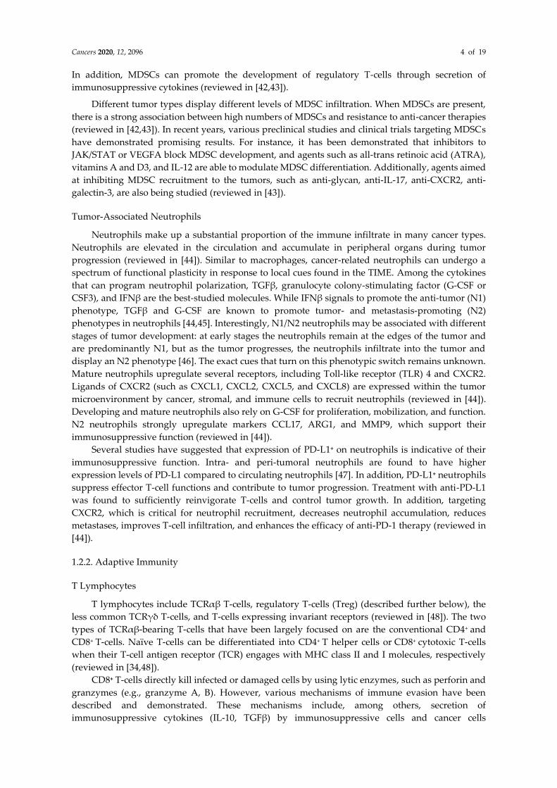
role in their effector functions (Figure 2).
Figure 2. Diverse roles of MNK effectors in T-cell function and differentiation. MNK effectors have
been widely studied in adaptive immune cells. While hnRNP A1 and Spry proteins play important
roles in functional T-cells, eIF4E expression and activity are essential for T-cell differentiation and B-
cell maturation.

Cancers 2020, 12, 2096 10 of 19
eIF4E
The T-cell chemokine RANTES/CCL5 is transcriptionally regulated by RFLAT-1 (RANTES for
late-activated T lymphocytes-1) [85]. RFLAT-1 expression is in turn regulated through cap-
dependent, eIF4E-mediated translation [86]. Overexpression of eIF4E increases RFLAT-1 protein,
while inhibition of either MNK kinases or eIF4E phosphorylation reduces RFLAT-1 production
respectively [86]. This mechanism is thought to enable T-cells to rapidly alter their abundance of
RANTES in response to various pro-inflammatory factors. RANTES-deficient CD8+ T-cells have poor
cytokine production, and also express higher amounts of inhibitory receptors consistent with more
severe exhaustion [87]. As such, one can assume that inhibition of MNK activity or eIF4E
phosphorylation may result in significantly reduced RANTES expression and suboptimal T-cell-
mediated immune responses as a result.
eIF4E may also play a role in T-cell differentiation. GATA-3 is a transcription factor both
necessary and sufficient for T helper type 2 (TH2) cell differentiation [88]. In addition to TCR
engagement, mTORC1-dependent increase in the synthesis of GATA-3 protein is required [89]. While
the precise mechanism of GATA-3 regulation by mTORC1 has not been elucidated, it is speculated
that the effects observed are mediated by a mechanism dependent on increased eIF4E
phosphorylation and activity [90]. Consistent with the proposed role for eIF4E in T-cell
differentiation, inhibition of eIF4E in FOXP3− CD4+ T cells ex vivo increases FOXP3 protein, indicating
that suppression of eIF4E bolsters the differentiation of FOXP3- CD4+ T-cells into FOXP3+ Tregs [91].
Similarly, impairment of eIF4E expression and/or activity in Th17 CD4+ T-cells decreases IL-17 and
increases FOXP3, which restrains Th17 cell differentiation and promotes the development of CD4+ T-
cells expressing FOXP3. These results strongly support the relevance of translational control in CD4+
T-cell fate decision.
Finally, the involvement of eIF4E in B-cell differentiation and proliferation has been explored.
Follicular helper T (TFH) cells are a subset of CD4+ T-cells and are essential for the activation of B-cells
in the germinal center (reviewed in [92]). These TFH cells express high levels of inducible T-cell
costimulator (ICOS), which is required for their initial generation and function. ICOS also has a
recognized role in maintaining the T cell–B cell interactions inside the germinal center (reviewed in
[92]). Notably, it has recently been reported that ICOS regulates Il4 mRNA translation via the eIF4E
pathway. Stimulation of ICOS results in increased polysome formation on Il4 mRNA and increased
IL-4 production [93]. This supply of IL-4 from TFH is thought to promote B-cell activation during the
course of their physical engagement [50,93]. Results from this study not only provide new insights
on the contribution of ICOS to the effector function of TFH but also suggest that inhibition of eIF4E
phosphorylation and activity could impede the delivery of IL-4 to cognate B-cells, leading to
suboptimal humoral responses.
hnRNP A1
Involvement of the MNK-hnRNP A1 axis in T-cell lymphocytes has largely been attributed to
its cytokine regulation. Expression of many cytokines is regulated at the posttranscriptional level. For
example, posttranscriptional regulatory control of TNFα expression is through AREs located in the
3′-UTR of the mRNA [4]. It has been shown that TNFα expression is activated by both the MAPK/ERK
and p38 MAPK pathway via the MNK- hnRNP A1 axis [4]. Blockade of either MNK expression or
activity inhibits TNFα production in T-cells, whereas MNK overexpression enhances TNFα
expression [4]. Mechanistically, MNK-mediated phosphorylation of hnRNP A1 relieves its binding
to TNFα-ARE, allowing for translation and expression of the protein [4]. A large number of immune-
modulatory cytokines are regulated posttranscriptionally through AREs and hence, it is conceivable
that they may also be bound and regulated by hnRNP A1. Very early studies demonstrated hnRNP
A1 binding to the ARE-rich regions of Il2 and Gm-csf mRNAs [69,94], but the functional readouts
were not investigated.
Like macrophages, T-cells also express high levels of OPN, which increase even further
following activation by the T-cell mitogen, Concanavalin A (Con A) [95]. Stimulation of OPN, also
known as the early T cell‐activation antigen‐1 gene (Eta‐1), in human T-cells results in IL-12

Cancers 2020, 12, 2096 11 of 19
production in a CD40L- and IFNγ‐dependent manner [96]. These findings suggest an important role
of OPN in early Th1 responses. As hnRNP A1 already functions in T-cells [4], it is of interest to see if
hnRNP A1 can regulate OPN in T-cells as it does in macrophages.
Spry1/2
The engagement between TCR and MHC complexes not only leads to T-cell activation, but also
upregulates inhibitory signals that restrain excessive T-cell activation to prevent autoimmunity and
maintain homeostasis. Such inhibitory mechanisms, such as CTLA-4 and PD-1, have been explored
in the context of cancer to improve T-cell function and immune responses [97,98]. Efforts to define
novel intracellular inhibitory molecules identified Spry proteins [99]. While loss of Spry1 does not
affect CD4+ or CD8+ T-cell development, Spry1 loss enhances their effector function with increased
IL-2, IFNγ, granzyme B secretion [99,100], and proliferation [101]. Mice in which Spry1 is selectively
deleted in T-cells demonstrate enhanced responses to tumor vaccines and subsequently more robust
tumor rejection compared to control mice [100]. Mechanistically, upon TCR engagement, both Spry1
and Spry2 are upregulated [99,101,102] and inhibit the activation of linker for activated T-cells (LAT)
and phospholipase C-γ (PLC-γ) [99,101]. Through LAT and PLC-γ, Spry1 and Spry2 inhibit
activation of MAPK signaling and NFκB, NFAT, and AP-1 transcription factors, thereby limiting T-
cell activation and proliferation [99,101,103]. Collectively, these studies demonstrate that Spry1 and
Spry2 molecules act as negative regulators of TCR signaling. In fact, in a chronic infection setting
such as HIV, virus-specific T-cells display high levels of Spry2, and inhibition of Spry2 expression
enhances the HIV-specific polyfunctional response independently of the PD-1 pathway [104].
In addition to enhancing the survival of effector CD8+ T-cells, it was recently demonstrated that
concurrent loss of Spry1 and Spry2 promotes the generation of memory CD8+ T-cells with faster recall
capacity and protective immunity [102]. Mechanistically, this is a result of decreased phosphorylation
and expression of Akt and Foxo1, leading to increased activity of transcription factors essential for
the development of memory T-cells, namely T-bet, TCF-1, and Eomesodermin. The identification of
Spry1/2 as intracellular inhibitors that function to limit T-cell proliferation, survival, and memory
formation, suggests that they may be attractive targets to improve tumor immunotherapy. In
agreement with this notion, Spry1-/- T cells were shown to have more potent cytolytic activity against
lymphoma and melanoma tumor cells [100].
3. Conclusion
MNK kinases were first recognized for their role in promoting tumor progression. In cancer cells,
MNK kinases mediate expression of multiple genes that promote growth, proliferation, resistance to
apoptosis, and resistance to therapy [8–12]. Correspondingly, inhibition of MNK kinases activity can
cause proliferative arrest and apoptosis in cancer cells [9,105,106]. Notably, multiple studies have also
demonstrated that MNK kinases mediate expression of multiple pro-inflammatory cytokines,
suggesting a potential role in immune cells. Except for neutrophils, it appears that depletion of MNK
kinases, while having some major effects on the function or phenotype of immune cells, has no
negative effect on their survival.
Attempts to target MNK kinases (and their downstream substrates) led to the development and
testing of several compounds, with earlier compounds failing to advance in clinical trials due to overt
toxicities [107]. In recent years, new compounds, such as eFT-508, have come to light. Having passed
phase 1 trials with a promising safety profile [108], it is now being evaluated in several clinical phase
II trials, including microsatellite stable solid tumors. In a preclinical study with the MycTg;KrasG12D
hepatocellular carcinoma model, treatment of tumor-bearing mice with eFT-508 led to a significant
increase in the number of cytotoxic CD8+ T-cells [13]. These observations raise a question about the
potential roles of MNK kinases and their mediated signals in other immune compartments in
addition to cancer cells. Ongoing trials evaluating eFT-508 in combination with PD-1/PD-L1 immune
checkpoint inhibitors will provide insights on the extent to which targeting MNK signaling
reprograms TIME. Here, we have provided a summary of what is known about how different MNK
effectors function in the various immune compartments, including effector T-cells (Table 1). It is
Cancers 2020, 12, 2096 12 of 19
becoming clear that while all working downstream of MNK kinases, these effectors may produce
varying, or even opposing effects in a given immune cell type. Advances in tumor profiling and
single-cell sequencing will allow for a more accurate evaluation on the global effect of MNK
inhibitors, and may even provide insights on the tumor types that are more likely to benefit from
MNK inhibitor treatment. In addition to MNK inhibitors, targeting eIF4G:eIF4E has also been
explored as an approach to control eIF4E phosphorylation and activity. For example, small-molecule
inhibitors of this interaction have been demonstrated to balance protein synthesis and exhibit activity
against cancer cells [14,109–111].
The advent of immunotherapy has significantly transformed the treatment landscape for select
human cancers. As a result, efforts to combine immune-based regimens with other therapies for
enhanced anti-tumor responses are also being pursued. Recent work by the Sonenberg lab
demonstrates that eIF4E activity promotes the survival of pro-metastatic neutrophils and contributes
to tumor metastasis [82]. Depletion of eIF4E phosphorylation by the MNK inhibitor merestinib
blocked neutrophil survival and metastatic progression. In addition, they showed that neutrophils
can inhibit T-cell proliferation and function. These and other data suggest that immunotherapeutic
drugs, including inhibitors of CTLA-4 and PD-1/PD-L1, are particularly promising candidates to
combine with MNK inhibitors. As a result, understanding how MNK kinases and their substrates
function in all the different TIME compartments, including immune cells, is critical and will guide
the optimization of these new combination regimens.
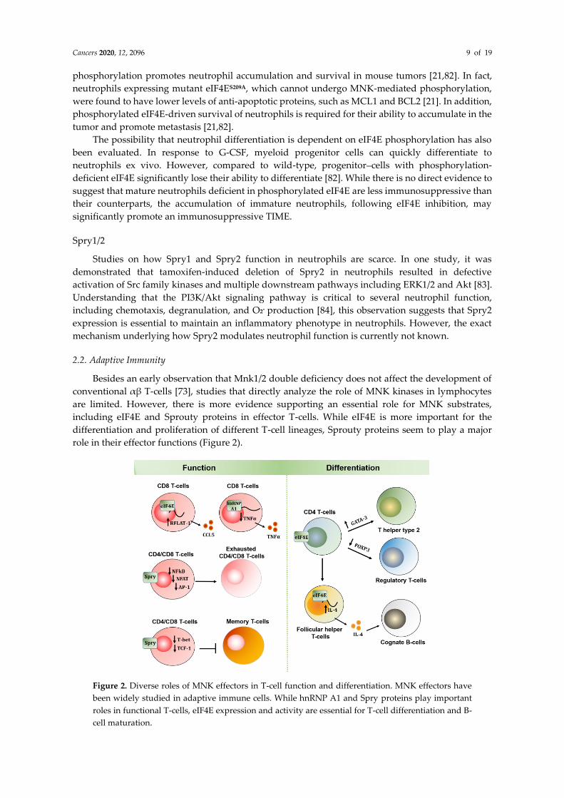
Table 1. Summary on different roles of MNK substrates in various immune compartments.
Inn
ate
imm
un
e ce
lls
eIF4E hnRNP A1 Sprouty 2
Macrophages
i) Promotes an M1, pro-
inflammatory phenotype by
regulating IRF8 [62,63]
ii) Stimulates translation
of pro-inflammatory
cytokines TNFα, IL-6 [58]
i) Inhibits
transcription and
expression of OPN,
which is required for
M2 phenotype [64,67]
ii) Increases stability
of Gm-csf mRNAs
[69,112]
i) Suppresses
expression of M2-
associated genes (Il10,
Arg1, Chil3) [72]
ii) Induces expression
of M1-associated genes
(Nos2, Il6) [72]
Myeloid-
derived
suppressor
cells
(MDSCs)
i) May promote MDSC
survival and
immunosuppressive
phenotype [75]
i) OPN can promote
MDSC expansion via
the STAT3 pathway
[64,76]
Neutrophils
i) Promotes neutrophil
survival by upregulating
anti-apoptotic proteins
(MCL1, BCL2) [21,82] ii) Promotes neutrophil-
driven metastasis [21,82] iii) Induces myeloid-
neutrophil differentiation in
response to G-CSF [82]
i) Sustains activation
of Src family and
downstream pathways
ERK1/2 and Akt [83,84]

Cancers 2020, 12, 2096 13 of 19
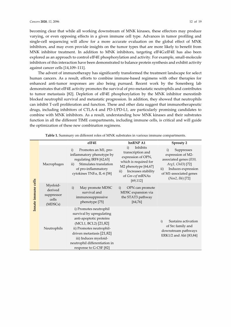
Ad
ap
tiv
e im
mu
ne
cel
ls
T lymphocytes
i) Induces cap-dependent
translation of RFLAT-1,
which transcribes RANTES
mRNAs [86,88]
ii) Induces expression of
GATA-3 for TH2
differentiation [88–90] iii) Inhibits expression of
FOXP3 in CD4+ T-cells
[90,91,113]
iv) Facilitates B-cell
differentiation by inducing
CD4+ T-cell production of
IL-4 [93]
i) Inhibits TNFα
translation and
expression by binding
to TNFα 3′-UTR [4]
i) Inhibits expression
of IL-2, IFNγ, and
Granzyme B [99,100]
ii) Inhibits T-cell
activation and
proliferation by
suppressing NFκB,
NFAT, and AP-1
[99,101,103]
iii) Inhibits formation
of memory T-cells
through the activity of
Akt and FoxO1 [102]
IRF8: IFN regulatory factor-8; G-CSF: granulocyte colony-stimulating factor; GM-CSF: granulocyte-
macrophage colony-stimulating factor; RFLAT-1: RANTES for late-activated T lymphocytes-1; OPN:
osteopontin; UTR: unstranlated region.
Author Contributions: Conceptualization and literature review, T.N.D.P. and H.G.M.; writing—original draft
preparation, T.N.D.P.; writing—editing, H.G.M. and C.S.; funding acquisition, T.N.D.P. and H.G.M. All
authors have read and agreed to the published version of the manuscript.
Funding: This work was supported by NIH/NCI grant R01CA217907 (to H.G.M.), a Merit award I01BX002922
(to H.G.M.) from the Department of Veterans Affairs, and NIH/NCI training grant T32CA070085 (to T.N.D.P).
Conflicts of Interest: The authors declare no conflict of interest. The funders had no role in the writing of the
manuscript, or in the decision to publish the results.
References
1. Fukunaga, R.; Hunter, T. MNK1, a new MAP kinase-activated protein kinase, isolated by a novel
expression screening method for identifying protein kinase substrates. EMBO J. 1997, 16, 1921–1933,
doi:10.1093/emboj/16.8.1921.
2. Waskiewicz, A.J.; Flynn, A.; Proud, C.G.; Cooper, J.A. Mitogen-activated protein kinases activate the
serine/threonine kinases Mnk1 and Mnk2. EMBO J. 1997, 16, 1909–1920, doi:10.1093/emboj/16.8.1909.
3. Ueda, T.; Watanabe-Fukunaga, R.; Fukuyama, H.; Nagata, S.; Fukunaga, R. Mnk2 and Mnk1 are essential
for constitutive and inducible phosphorylation of eukaryotic initiation factor 4E but not for cell growth or
development. Mol. Cell Biol. 2004, 24, 6539–6549, doi:10.1128/MCB.24.15.6539-6549.2004.
4. Buxade, M.; Parra, J.L.; Rousseau, S.; Shpiro, N.; Marquez, R.; Morrice, N.; Bain, J.; Espel, E.; Proud, C.G.
The Mnks are novel components in the control of TNF alpha biosynthesis and phosphorylate and regulate
hnRNP A1. Immunity 2005, 23, 177–189, doi:10.1016/j.immuni.2005.06.009.
5. DaSilva, J.; Xu, L.; Kim, H.J.; Miller, W.T.; Bar-Sagi, D. Regulation of sprouty stability by Mnk1-dependent
phosphorylation. Mol. Cell Biol. 2006, 26, 1898–1907, doi:10.1128/MCB.26.5.1898-1907.2006.
6. Buxade, M.; Morrice, N.; Krebs, D.L.; Proud, C.G. The PSF.p54nrb complex is a novel Mnk substrate that
binds the mRNA for tumor necrosis factor alpha. J. Biol. Chem. 2008, 283, 57–65, doi:10.1074/jbc.M705286200.
7. Pham, T.N.D.; Kumar, K.; De Cant, B.T.; Shang, M.; Munshi, S.Z.; Matsangou, M.; Ebine, K.; Munshi, H.G.
Induction of MNK Kinase-dependent eIF4E Phosphorylation by Inhibitors Targeting BET Proteins Limits
Efficacy of BET Inhibitors. Mol. Cancer Ther. 2019, 18, 235–244, doi:10.1158/1535-7163.MCT-18-0768.
8. Adesso, L.; Calabretta, S.; Barbagallo, F.; Capurso, G.; Pilozzi, E.; Geremia, R.; Delle Fave, G.; Sette, C.
Gemcitabine triggers a pro-survival response in pancreatic cancer cells through activation of the
MNK2/eIF4E pathway. Oncogene 2013, 32, 2848–2857, doi:10.1038/onc.2012.306.
9. Altman, J.K.; Glaser, H.; Sassano, A.; Joshi, S.; Ueda, T.; Watanabe-Fukunaga, R.; Fukunaga, R.; Tallman,
M.S.; Platanias, L.C. Negative regulatory effects of Mnk kinases in the generation of chemotherapy-induced
antileukemic responses. Mol. Pharmacol. 2010, 78, 778–784, doi:10.1124/mol.110.064642.
10. Wang, X.; Yue, P.; Chan, C.B.; Ye, K.; Ueda, T.; Watanabe-Fukunaga, R.; Fukunaga, R.; Fu, H.; Khuri, F.R.;
Sun, S.Y. Inhibition of mammalian target of rapamycin induces phosphatidylinositol 3-kinase-dependent

Cancers 2020, 12, 2096 14 of 19
and Mnk-mediated eukaryotic translation initiation factor 4E phosphorylation. Mol. Cell Biol. 2007, 27,
7405–7413, doi:10.1128/MCB.00760-07.
11. Astanehe, A.; Finkbeiner, M.R.; Krzywinski, M.; Fotovati, A.; Dhillon, J.; Berquin, I.M.; Mills, G.B.; Marra,
M.A.; Dunn, S.E. MKNK1 is a YB-1 target gene responsible for imparting trastuzumab resistance and can
be blocked by RSK inhibition. Oncogene 2012, 31, 4434–4446, doi:10.1038/onc.2011.617.
12. Xu, Y.; Poggio, M.; Jin, H.Y.; Shi, Z.; Forester, C.M.; Wang, Y.; Stumpf, C.R.; Xue, L.; Devericks, E.; So, L.; et al.
Translation control of the immune checkpoint in cancer and its therapeutic targeting. Nat. Med. 2019, 25,
301–311, doi:10.1038/s41591-018-0321-2.
13. Moerke, N.J.; Aktas, H.; Chen, H.; Cantel, S.; Reibarkh, M.Y.; Fahmy, A.; Gross, J.D.; Degterev, A.; Yuan, J.;
Chorev, M.; et al. Small-molecule inhibition of the interaction between the translation initiation factors
eIF4E and eIF4G. Cell 2007, 128, 257–267, doi:10.1016/j.cell.2006.11.046.
14. Gingras, A.C.; Raught, B.; Sonenberg, N. eIF4 initiation factors: Effectors of mRNA recruitment to ribosomes
and regulators of translation. Annu. Rev. Biochem 1999, 68, 913–963, doi:10.1146/annurev.biochem.68.1.913.
15. Siddiqui, N.; Sonenberg, N. Signalling to eIF4E in cancer. Biochem. Soc. Trans. 2015, 43, 763–772,
doi:10.1042/BST20150126.
16. Proud, C.G. Mnks, eIF4E phosphorylation and cancer. Biochim. Biophys. Acta 2015, 1849, 766–773,
doi:10.1016/j.bbagrm.2014.10.003.
17. Hsieh, A.C.; Costa, M.; Zollo, O.; Davis, C.; Feldman, M.E.; Testa, J.R.; Meyuhas, O.; Shokat, K.M.; Ruggero, D.
Genetic dissection of the oncogenic mTOR pathway reveals druggable addiction to translational control
via 4EBP-eIF4E. Cancer Cell 2010, 17, 249–261, doi:10.1016/j.ccr.2010.01.021.
18. Rousseau, D.; Kaspar, R.; Rosenwald, I.; Gehrke, L.; Sonenberg, N. Translation initiation of ornithine
decarboxylase and nucleocytoplasmic transport of cyclin D1 mRNA are increased in cells overexpressing
eukaryotic initiation factor 4E. Proc. Natl. Acad. Sci. USA 1996, 93, 1065–1070, doi:10.1073/pnas.93.3.1065.
19. Wendel, H.G.; Silva, R.L.; Malina, A.; Mills, J.R.; Zhu, H.; Ueda, T.; Watanabe-Fukunaga, R.; Fukunaga, R.;
Teruya-Feldstein, J.; Pelletier, J.; et al. Dissecting eIF4E action in tumorigenesis. Genes Dev. 2007, 21, 3232–
3237, doi:10.1101/gad.1604407.
20. Robichaud, N.; del Rincon, S.V.; Huor, B.; Alain, T.; Petruccelli, L.A.; Hearnden, J.; Goncalves, C.; Grotegut,
S.; Spruck, C.H.; Furic, L.; et al. Phosphorylation of eIF4E promotes EMT and metastasis via translational
control of SNAIL and MMP-3. Oncogene 2015, 34, 2032–2042, doi:10.1038/onc.2014.146.
21. Graff, J.R.; Konicek, B.W.; Carter, J.H.; Marcusson, E.G. Targeting the eukaryotic translation initiation factor
4E for cancer therapy. Cancer Res. 2008, 68, 631–634, doi:10.1158/0008-5472.CAN-07-5635.
22. Jean-Philippe, J.; Paz, S.; Caputi, M. HNRNP A1: The Swiss army knife of gene expression. Int. J. Mol. Sci.
2013, 14, 18999–19024, doi:10.3390/ijms140918999.
23. Roy, R.; Huang, Y.; Seckl, M.J.; Pardo, O.E. Emerging roles of hnRNPA1 in modulating malignant
transformation. Wiley Interdiscip. Rev. RNA 2017, 8, doi:10.1002/wrna.1431.
24. Carabet, L.A.; Leblanc, E.; Lallous, N.; Morin, H.; Ghaidi, F.; Lee, J.; Rennie, P.S.; Cherkasov, A. Computer-
Aided Discovery of Small Molecules Targeting the RNA Splicing Activity of hnRNP A1 in Castration-
Resistant Prostate Cancer. Molecules 2019, 24, 763, doi:10.3390/molecules24040763.
25. Ko, C.C.; Chen, Y.J.; Chen, C.T.; Liu, Y.C.; Cheng, F.C.; Hsu, K.C.; Chow, L.P. Chemical proteomics
identifies heterogeneous nuclear ribonucleoprotein (hnRNP) A1 as the molecular target of quercetin in its
anti-cancer effects in PC-3 cells. J. Biol. Chem. 2014, 289, 22078–22089, doi:10.1074/jbc.M114.553248.
26. Pham, T.N.D.; Stempel, S.; Shields, M.A.; Spaulding, C.; Kumar, K.; Bentrem, D.J.; Matsangou, M.; Munshi,
H.G. Quercetin Enhances the Anti-Tumor Effects of BET Inhibitors by Suppressing hnRNPA1. Int. J. Mol.
Sci. 2019, 20, 4293, doi:10.3390/ijms20174293.
27. Tummala, R.; Lou, W.; Gao, A.C.; Nadiminty, N. Quercetin Targets hnRNPA1 to Overcome Enzalutamide
Resistance in Prostate Cancer Cells. Mol. Cancer Ther. 2017, 16, 2770–2779, doi:10.1158/1535-7163.MCT-17-0030.
28. Gross, I.; Bassit, B.; Benezra, M.; Licht, J.D. Mammalian sprouty proteins inhibit cell growth and differentiation
by preventing ras activation. J. Biol. Chem. 2001, 276, 46460–46468, doi:10.1074/jbc.M108234200.
29. Impagnatiello, M.A.; Weitzer, S.; Gannon, G.; Compagni, A.; Cotten, M.; Christofori, G. Mammalian
sprouty-1 and -2 are membrane-anchored phosphoprotein inhibitors of growth factor signaling in
endothelial cells. J. Cell Biol. 2001, 152, 1087–1098, doi:10.1083/jcb.152.5.1087.
30. Minowada, G.; Jarvis, L.A.; Chi, C.L.; Neubuser, A.; Sun, X.; Hacohen, N.; Krasnow, M.A.; Martin, G.R.
Vertebrate Sprouty genes are induced by FGF signaling and can cause chondrodysplasia when
overexpressed. Development 1999, 126, 4465–4475.

Cancers 2020, 12, 2096 15 of 19
31. Sharma, B.; Joshi, S.; Sassano, A.; Majchrzak, B.; Kaur, S.; Aggarwal, P.; Nabet, B.; Bulic, M.; Stein, B.L.;
McMahon, B.; et al. Sprouty proteins are negative regulators of interferon (IFN) signaling and IFN-
inducible biological responses. J. Biol. Chem. 2012, 287, 42352–42360, doi:10.1074/jbc.M112.400721.
32. Binnewies, M.; Roberts, E.W.; Kersten, K.; Chan, V.; Fearon, D.F.; Merad, M.; Coussens, L.M.; Gabrilovich,
D.I.; Ostrand-Rosenberg, S.; Hedrick, C.C.; et al. Understanding the tumor immune microenvironment
(TIME) for effective therapy. Nat. Med. 2018, 24, 541–550, doi:10.1038/s41591-018-0014-x.
33. Gajewski, T.F.; Schreiber, H.; Fu, Y.X. Innate and adaptive immune cells in the tumor microenvironment.
Nat. Immunol. 2013, 14, 1014–1022, doi:10.1038/ni.2703.
34. Ramachandra, L.; Simmons, D.; Harding, C.V. MHC molecules and microbial antigen processing in
phagosomes. Curr. Opin. Immunol. 2009, 21, 98–104, doi:10.1016/j.coi.2009.01.001.
35. Luckheeram, R.V.; Zhou, R.; Verma, A.D.; Xia, B. CD4(+)T cells: Differentiation and functions. Clin. Dev.
Immunol. 2012, 2012, 925135, doi:10.1155/2012/925135.
36. Condeelis, J.; Pollard, J.W. Macrophages: Obligate partners for tumor cell migration, invasion, and
metastasis. Cell 2006, 124, 263–266, doi:10.1016/j.cell.2006.01.007.
37. Qian, B.Z.; Pollard, J.W. Macrophage diversity enhances tumor progression and metastasis. Cell 2010, 141,
39–51, doi:10.1016/j.cell.2010.03.014.
38. Pathria, P.; Louis, T.L.; Varner, J.A. Targeting Tumor-Associated Macrophages in Cancer. Trends Immunol.
2019, 40, 310–327, doi:10.1016/j.it.2019.02.003.
39. Hamilton, T.A.; Zhao, C.; Pavicic, P.G., Jr.; Datta, S. Myeloid colony-stimulating factors as regulators of
macrophage polarization. Front. Immunol. 2014, 5, 554, doi:10.3389/fimmu.2014.00554.
40. Pyonteck, S.M.; Akkari, L.; Schuhmacher, A.J.; Bowman, R.L.; Sevenich, L.; Quail, D.F.; Olson, O.C.; Quick,
M.L.; Huse, J.T.; Teijeiro, V.; et al. CSF-1R inhibition alters macrophage polarization and blocks glioma
progression. Nat. Med. 2013, 19, 1264–1272, doi:10.1038/nm.3337.
41. Zhu, Y.; Knolhoff, B.L.; Meyer, M.A.; Nywening, T.M.; West, B.L.; Luo, J.; Wang-Gillam, A.; Goedegebuure,
S.P.; Linehan, D.C.; De Nardo, D.G. CSF1/CSF1R blockade reprograms tumor-infiltrating macrophages and
improves response to T-cell checkpoint immunotherapy in pancreatic cancer models. Cancer Res. 2014, 74,
5057–5069, doi:10.1158/0008-5472.CAN-13-3723.
42. Veglia, F.; Perego, M.; Gabrilovich, D. Myeloid-derived suppressor cells coming of age. Nat. Immunol. 2018,
19, 108–119, doi:10.1038/s41590-017-0022-x.
43. Fleming, V.; Hu, X.; Weber, R.; Nagibin, V.; Groth, C.; Altevogt, P.; Utikal, J.; Umansky, V. Targeting
Myeloid-Derived Suppressor Cells to Bypass Tumor-Induced Immunosuppression. Front. Immunol. 2018,
9, 398, doi:10.3389/fimmu.2018.00398.
44. Coffelt, S.B.; Wellenstein, M.D.; de Visser, K.E. Neutrophils in cancer: Neutral no more. Nat. Rev. Cancer
2016, 16, 431–446, doi:10.1038/nrc.2016.52.
45. Fridlender, Z.G.; Sun, J.; Kim, S.; Kapoor, V.; Cheng, G.; Ling, L.; Worthen, G.S.; Albelda, S.M. Polarization of
tumor-associated neutrophil phenotype by TGF-beta: “N1” versus “N2” TAN. Cancer Cell 2009, 16, 183–194,
doi:10.1016/j.ccr.2009.06.017.
46. Mishalian, I.; Bayuh, R.; Levy, L.; Zolotarov, L.; Michaeli, J.; Fridlender, Z.G. Tumor-associated neutrophils
(TAN) develop pro-tumorigenic properties during tumor progression. Cancer Immunol. Immunother. 2013,
62, 1745–1756, doi:10.1007/s00262-013-1476-9.
47. He, G.; Zhang, H.; Zhou, J.; Wang, B.; Chen, Y.; Kong, Y.; Xie, X.; Wang, X.; Fei, R.; Wei, L.; et al.
Peritumoural neutrophils negatively regulate adaptive immunity via the PD-L1/PD-1 signalling pathway
in hepatocellular carcinoma. J. Exp. Clin. Cancer Res. 2015, 34, 141, doi:10.1186/s13046-015-0256-0.
48. Godfrey, D.I.; Uldrich, A.P.; McCluskey, J.; Rossjohn, J.; Moody, D.B. The burgeoning family of
unconventional T cells. Nat. Immunol. 2015, 16, 1114–1123, doi:10.1038/ni.3298.
49. Bevan, M.J. Helping the CD8(+) T-cell response. Nat. Rev. Immunol. 2004, 4, 595–602, doi:10.1038/nri1413.
50. Crotty, S. A brief history of T cell help to B cells. Nat. Rev. Immunol. 2015, 15, 185–189, doi:10.1038/nri3803.
51. Liudahl, S.M.C.L. To Help or To Harm: Dynamic Roles of CD4+ T Helper Cells in Solid Tumor
Microenvironment. In Immunotoxicology, Immunopathology, and Immunotherapy; MA, H., Ed.; Elsevier Inc:
Cambridge, MA, USA, 2018; Volume 1.
52. Romano, M.; Fanelli, G.; Albany, C.J.; Giganti, G.; Lombardi, G. Past, Present, and Future of Regulatory T Cell
Therapy in Transplantation and Autoimmunity. Front. Immunol. 2019, 10, 43, doi:10.3389/fimmu.2019.00043.

Cancers 2020, 12, 2096 16 of 19
53. Bindea, G.; Mlecnik, B.; Tosolini, M.; Kirilovsky, A.; Waldner, M.; Obenauf, A.C.; Angell, H.; Fredriksen, T.;
Lafontaine, L.; Berger, A.; et al. Spatiotemporal dynamics of intratumoral immune cells reveal the immune
landscape in human cancer. Immunity 2013, 39, 782–795, doi:10.1016/j.immuni.2013.10.003.
54. Chevrier, S.; Levine, J.H.; Zanotelli, V.R.T.; Silina, K.; Schulz, D.; Bacac, M.; Ries, C.H.; Ailles, L.; Jewett,
M.A.S.; Moch, H.; et al. An Immune Atlas of Clear Cell Renal Cell Carcinoma. Cell 2017, 169, 736–749,
doi:10.1016/j.cell.2017.04.016.
55. Castino, G.F.; Cortese, N.; Capretti, G.; Serio, S.; Di Caro, G.; Mineri, R.; Magrini, E.; Grizzi, F.; Cappello, P.;
Novelli, F; et al. Spatial distribution of B cells predicts prognosis in human pancreatic adenocarcinoma.
Oncoimmunology 2016, 5, e1085147, doi:10.1080/2162402X.2015.1085147.
56. Erdag, G.; Schaefer, J.T.; Smolkin, M.E.; Deacon, D.H.; Shea, S.M.; Dengel, L.T.; Patterson, J.W.; Slingluff, C.L.,
Jr. Immunotype and immunohistologic characteristics of tumor-infiltrating immune cells are associated with
clinical outcome in metastatic melanoma. Cancer Res. 2012, 72, 1070–1080, doi:10.1158/0008-5472.CAN-11-3218.
57. Kroeger, D.R.; Milne, K.; Nelson, B.H. Tumor-Infiltrating Plasma Cells Are Associated with Tertiary
Lymphoid Structures, Cytolytic T-Cell Responses, and Superior Prognosis in Ovarian Cancer. Clin. Cancer
Res. 2016, 22, 3005–3015, doi:10.1158/1078-0432.CCR-15-2762.
58. Rowlett, R.M.; Chrestensen, C.A.; Nyce, M.; Harp, M.G.; Pelo, J.W.; Cominelli, F.; Ernst, P.B.; Pizarro, T.T.; Sturgill,
T.W.; Worthington, M.T. MNK kinases regulate multiple TLR pathways and innate proinflammatory cytokines
in macrophages. Am. J. Physiol. Gastrointest. Liver Physiol. 2008, 294, G452–459, doi:10.1152/ajpgi.00077.2007.
59. Montero, H.; Garcia-Roman, R.; Mora, S.I. EIF4E as a control target for viruses. Viruses 2015, 7, 739–750,
doi:10.3390/v7020739.
60. Wan, Y.; Xiao, H.; Affolter, J.; Kim, T.W.; Bulek, K.; Chaudhuri, S.; Carlson, D.; Hamilton, T.; Mazumder,
B.; Stark, G.R.; et al. Interleukin-1 receptor-associated kinase 2 is critical for lipopolysaccharide-mediated
post-transcriptional control. J. Biol. Chem. 2009, 284, 10367–10375, doi:10.1074/jbc.M807822200.
61. Tamura, T.; Kurotaki, D.; Koizumi, S. Regulation of myelopoiesis by the transcription factor IRF8. Int. J.
Hematol. 2015, 101, 342–351, doi:10.1007/s12185-015-1761-9.
62. Xu, H.; Zhu, J.; Smith, S.; Foldi, J.; Zhao, B.; Chung, A.Y.; Outtz, H.; Kitajewski, J.; Shi, C.; Weber, S.; et al.
Notch-RBP-J signaling regulates the transcription factor IRF8 to promote inflammatory macrophage
polarization. Nat. Immunol. 2012, 13, 642–650, doi:10.1038/ni.2304.
63. Twum, D.Y.; Colligan, S.H.; Hoffend, N.C.; Katsuta, E.; Cortes Gomez, E.; Hensen, M.L.; Seshadri, M.;
Nemeth, M.J.; Abrams, S.I. IFN regulatory factor-8 expression in macrophages governs an antimetastatic
program. JCI Insight 2019, 4, doi:10.1172/jci.insight.124267.
64. Gao, C.; Guo, H.; Mi, Z.; Wai, P.Y.; Kuo, P.C. Transcriptional regulatory functions of heterogeneous nuclear
ribonucleoprotein-U and -A/B in endotoxin-mediated macrophage expression of osteopontin. J. Immunol.
2005, 175, 523–530, doi:10.4049/jimmunol.175.1.523.
65. Shevde, L.A.; Das, S.; Clark, D.W.; Samant, R.S. Osteopontin: An effector and an effect of tumor metastasis.
Curr. Mol. Med. 2010, 10, 71–81, doi:10.2174/156652410791065381.
66. Atai, N.A.; Bansal, M.; Lo, C.; Bosman, J.; Tigchelaar, W.; Bosch, K.S.; Jonker, A.; De Witt Hamer, P.C.;
Troost, D.; McCulloch, C.A.; et al. Osteopontin is up-regulated and associated with neutrophil and
macrophage infiltration in glioblastoma. Immunology 2011, 132, 39–48, doi:10.1111/j.1365-2567.2010.03335.x.
67. Wei, J.; Marisetty, A.; Schrand, B.; Gabrusiewicz, K.; Hashimoto, Y.; Ott, M.; Grami, Z.; Kong, L.Y.; Ling, X.;
Caruso, H.; et al. Osteopontin mediates glioblastoma-associated macrophage infiltration and is a potential
therapeutic target. J. Clin. Invest. 2019, 129, 137–149, doi:10.1172/JCI121266.
68. Barreau, C.; Paillard, L.; Osborne, H.B. AU-rich elements and associated factors: Are there unifying
principles? Nucleic Acids Res. 2005, 33, 7138–7150, doi:10.1093/nar/gki1012.
69. Hamilton, B.J.; Burns, C.M.; Nichols, R.C.; Rigby, W.F. Modulation of AUUUA response element binding
by heterogeneous nuclear ribonucleoprotein A1 in human T lymphocytes. The roles of cytoplasmic location,
transcription, and phosphorylation. J. Biol. Chem. 1997, 272, 28732–28741, doi:10.1074/jbc.272.45.28732.
70. Moreira-Teixeira, L.; Sousa, J.; McNab, F.W.; Torrado, E.; Cardoso, F.; Machado, H.; Castro, F.; Cardoso, V.;
Gaifem, J.; Wu, X.; et al. Type I IFN Inhibits Alternative Macrophage Activation during Mycobacterium
tuberculosis Infection and Leads to Enhanced Protection in the Absence of IFN-gamma Signaling. J.
Immunol. 2016, 197, 4714–4726, doi:10.4049/jimmunol.1600584.
71. Park, S.H.; Kang, K.; Giannopoulou, E.; Qiao, Y.; Kang, K.; Kim, G.; Park-Min, K.H.; Ivashkiv, L.B. Type I
interferons and the cytokine TNF cooperatively reprogram the macrophage epigenome to promote
inflammatory activation. Nat. Immunol. 2017, 18, 1104–1116, doi:10.1038/ni.3818.

Cancers 2020, 12, 2096 17 of 19
72. Atomura, R.; Sanui, T.; Fukuda, T.; Tanaka, U.; Toyoda, K.; Taketomi, T.; Yamamichi, K.; Akiyama, H.;
Nishimura, F. Inhibition of Sprouty2 polarizes macrophages toward an M2 phenotype by stimulation with
interferon gamma and Porphyromonas gingivalis lipopolysaccharide. Immun. Inflamm. Dis. 2016, 4, 98–110,
doi:10.1002/iid3.99.
73. Gorentla, B.K.; Krishna, S.; Shin, J.; Inoue, M.; Shinohara, M.L.; Grayson, J.M.; Fukunaga, R.; Zhong, X.P. Mnk1
and 2 are dispensable for T cell development and activation but important for the pathogenesis of experimental
autoimmune encephalomyelitis. J. Immunol. 2013, 190, 1026–1037, doi:10.4049/jimmunol.1200026.
74. Saradna, A.; Do, D.C.; Kumar, S.; Fu, Q.L.; Gao, P. Macrophage polarization and allergic asthma. Transl.
Res. 2018, 191, 1–14, doi:10.1016/j.trsl.2017.09.002.
75. Huang, F.; Goncalves, C.; Guo, Q.; Rémy-Sarrazin, J.; Emond, A.; Yang, W.; Plourde, D.; Bartish, M.; Su, J.;
ZHan, Y.; et al. Abstract A53: Phosphorylation of eIF4E promotes phenotype switching and MDSC-mediated
immunosuppression in melanoma. Cancer Immunol. Res. 2020, 8, doi:10.1158/2326-6074.TUMIMM19-A53.
76. Waight, J.D.; Netherby, C.; Hensen, M.L.; Miller, A.; Hu, Q.; Liu, S.; Bogner, P.N.; Farren, M.R.; Lee, K.P.;
Liu, K.; et al. Myeloid-derived suppressor cell development is regulated by a STAT/IRF-8 axis. J. Clin. Invest.
2013, 123, 4464–4478, doi:10.1172/JCI68189.
77. Salem, S.; Langlais, D.; Lefebvre, F.; Bourque, G.; Bigley, V.; Haniffa, M.; Casanova, J.L.; Burk, D.; Berghuis,
A.; Butler, K.M.; et al. Functional characterization of the human dendritic cell immunodeficiency associated
with the IRF8(K108E) mutation. Blood 2014, 124, 1894–1904, doi:10.1182/blood-2014-04-570879.
78. Sangaletti, S.; Tripodo, C.; Sandri, S.; Torselli, I.; Vitali, C.; Ratti, C.; Botti, L.; Burocchi, A.; Porcasi, R.;
Tomirotti, A.; et al. Osteopontin shapes immunosuppression in the metastatic niche. Cancer Res. 2014, 74,
4706–4719, doi:10.1158/0008-5472.CAN-13-3334.
79. Kim, E.K.; Jeon, I.; Seo, H.; Park, Y.J.; Song, B.; Lee, K.A.; Jang, Y.; Chung, Y.; Kang, C.Y. Tumor-derived
osteopontin suppresses antitumor immunity by promoting extramedullary myelopoiesis. Cancer Res. 2014,
74, 6705–6716, doi:10.1158/0008-5472.CAN-14-1482.
80. McCracken, J.M.; Allen, L.A. Regulation of human neutrophil apoptosis and lifespan in health and disease.
J. Cell Death 2014, 7, 15–23, doi:10.4137/JCD.S11038.
81. Schott, J.; Reitter, S.; Philipp, J.; Haneke, K.; Schafer, H.; Stoecklin, G. Translational regulation of specific
mRNAs controls feedback inhibition and survival during macrophage activation. PLoS Genet. 2014, 10,
e1004368, doi:10.1371/journal.pgen.1004368.
82. Robichaud, N.; Hsu, B.E.; Istomine, R.; Alvarez, F.; Blagih, J.; Ma, E.H.; Morales, S.V.; Dai, D.L.; Li, G.;
Souleimanova, M.; et al. Translational control in the tumor microenvironment promotes lung metastasis:
Phosphorylation of eIF4E in neutrophils. Proc. Natl. Acad. Sci. USA 2018, 115, E2202–E2209,
doi:10.1073/pnas.1717439115.
83. Gorentla BK, A.R. The Adapter Protein Sprouty 2 (Spry 2) Differentially Regulates Lymphoid and Myeloid
Cell Function and Is Important for Allergic Asthma. J. Allergy Clin. Immunol. 2015, 135, AB283.
84. Chen, J.; Tang, H.; Hay, N.; Xu, J.; Ye, R.D. Akt isoforms differentially regulate neutrophil functions. Blood
2010, 115, 4237–4246, doi:10.1182/blood-2009-11-255323.
85. Song, A.; Chen, Y.F.; Thamatrakoln, K.; Storm, T.A.; Krensky, A.M. RFLAT-1: A new zinc finger
transcription factor that activates RANTES gene expression in T lymphocytes. Immunity 1999, 10, 93–103,
doi:10.1016/s1074-7613(00)80010-2.
86. Nikolcheva, T.; Pyronnet, S.; Chou, S.Y.; Sonenberg, N.; Song, A.; Clayberger, C.; Krensky, A.M. A
translational rheostat for RFLAT-1 regulates RANTES expression in T lymphocytes. J. Clin. Invest. 2002, 110,
119–126, doi:10.1172/JCI15336.
87. Crawford, A.; Angelosanto, J.M.; Nadwodny, K.L.; Blackburn, S.D.; Wherry, E.J. A role for the chemokine
RANTES in regulating CD8 T cell responses during chronic viral infection. PLoS Pathog. 2011, 7, e1002098,
doi:10.1371/journal.ppat.1002098.
88. Zheng, W.; Flavell, R.A. The transcription factor GATA-3 is necessary and sufficient for Th2 cytokine gene
expression in CD4 T cells. Cell 1997, 89, 587–596, doi:10.1016/s0092-8674(00)80240-8.
89. Cook, K.D.; Miller, J. TCR-dependent translational control of GATA-3 enhances Th2 differentiation. J.
Immunol. 2010, 185, 3209–3216, doi:10.4049/jimmunol.0902544.
90. Piccirillo, C.A.; Bjur, E.; Topisirovic, I.; Sonenberg, N.; Larsson, O. Translational control of immune
responses: From transcripts to translatomes. Nat. Immunol. 2014, 15, 503–511, doi:10.1038/ni.2891.
Cancers 2020, 12, 2096 18 of 19
91. Bjur, E.; Larsson, O.; Yurchenko, E.; Zheng, L.; Gandin, V.; Topisirovic, I.; Li, S.; Wagner, C.R.; Sonenberg,
N.; Piccirillo, C.A. Distinct translational control in CD4+ T cell subsets. PLoS Genet. 2013, 9, e1003494,
doi:10.1371/journal.pgen.1003494.
92. Qi, H. T follicular helper cells in space-time. Nat. Rev. Immunol. 2016, 16, 612–625, doi:10.1038/nri.2016.94.
93. Gigoux, M.; Lovato, A.; Leconte, J.; Leung, J.; Sonenberg, N.; Suh, W.K. Inducible costimulator facilitates T-
dependent B cell activation by augmenting IL-4 translation. Mol. Immunol. 2014, 59, 46–54,
doi:10.1016/j.molimm.2014.01.008.
94. Henics, T.; Sanfridson, A.; Hamilton, B.J.; Nagy, E.; Rigby, W.F. Enhanced stability of interleukin-2 mRNA
in MLA 144 cells. Possible role of cytoplasmic AU-rich sequence-binding proteins. J. Biol. Chem. 1994, 269,
5377–5383.
95. Patarca, R.; Freeman, G.J.; Singh, R.P.; Wei, F.Y.; Durfee, T.; Blattner, F.; Regnier, D.C.; Kozak, C.A.; Mock,
B.A.; Morse, H.C., 3rd; et al. Structural and functional studies of the early T lymphocyte activation 1 (Eta-
1) gene. Definition of a novel T cell-dependent response associated with genetic resistance to bacterial
infection. J. Exp. Med. 1989, 170, 145–161, doi:10.1084/jem.170.1.145.
96. O’Regan, A.W.; Hayden, J.M.; Berman, J.S. Osteopontin augments CD3-mediated interferon-gamma and
CD40 ligand expression by T cells, which results in IL-12 production from peripheral blood mononuclear
cells. J. Leukoc. Biol. 2000, 68, 495–502.
97. Sharma, P.; Allison, J.P. The future of immune checkpoint therapy. Science 2015, 348, 56–61,
doi:10.1126/science.aaa8172.
98. Wei, S.C.; Levine, J.H.; Cogdill, A.P.; Zhao, Y.; Anang, N.A.S.; Andrews, M.C.; Sharma, P.; Wang, J.; Wargo,
J.A.; Pe′er, D.; et al. Distinct Cellular Mechanisms Underlie Anti-CTLA-4 and Anti-PD-1 Checkpoint
Blockade. Cell 2017, 170, 1120–1133, e1117, doi:10.1016/j.cell.2017.07.024.
99. Choi, H.; Cho, S.Y.; Schwartz, R.H.; Choi, K. Dual effects of Sprouty1 on TCR signaling depending on the
differentiation state of the T cell. J. Immunol. 2006, 176, 6034–6045, doi:10.4049/jimmunol.176.10.6034.
100. Collins, S.; Waickman, A.; Basson, A.; Kupfer, A.; Licht, J.D.; Horton, M.R.; Powell, J.D. Regulation of CD4(+)
and CD8(+) effector responses by Sprouty-1. PLoS ONE 2012, 7, e49801, doi:10.1371/journal.pone.0049801.
101. Akbulut, S.; Reddi, A.L.; Aggarwal, P.; Ambardekar, C.; Canciani, B.; Kim, M.K.; Hix, L.; Vilimas, T.; Mason,
J.; Basson, M.A.; et al. Sprouty proteins inhibit receptor-mediated activation of phosphatidylinositol-
specific phospholipase C. Mol. Biol. Cell 2010, 21, 3487–3496, doi:10.1091/mbc.E10-02-0123.
102. Shehata, H.M.; Khan, S.; Chen, E.; Fields, P.E.; Flavell, R.A.; Sanjabi, S. Lack of Sprouty 1 and 2 enhances
survival of effector CD8(+) T cells and yields more protective memory cells. Proc. Natl. Acad. Sci. USA 2018,
115, E8939–E8947, doi:10.1073/pnas.1808320115.
103. Lee, J.S.; Lee, J.E.; Oh, Y.M.; Park, J.B.; Choi, H.; Choi, C.Y.; Kim, I.H.; Lee, S.H.; Choi, K. Recruitment of
Sprouty1 to immune synapse regulates T cell receptor signaling. J. Immunol. 2009, 183, 7178–7186,
doi:10.4049/jimmunol.0803799.
104. Chiu, Y.L.; Shan, L.; Huang, H.; Haupt, C.; Bessell, C.; Canaday, D.H.; Zhang, H.; Ho, Y.C.; Powell, J.D.;
Oelke, M.; et al. Sprouty-2 regulates HIV-specific T cell polyfunctionality. J. Clin. Invest. 2014, 124, 198–208,
doi:10.1172/JCI70510.
105. Joshi, S.; Platanias, L.C. Mnk kinase pathway: Cellular functions and biological outcomes. World J. Biol.
Chem. 2014, 5, 321–333, doi:10.4331/wjbc.v5.i3.321.
106. Wen, Q.; Wang, W.; Luo, J.; Chu, S.; Chen, L.; Xu, L.; Zang, H.; Alnemah, M.M.; Ma, J.; Fan, S. CGP57380
enhances efficacy of RAD001 in non-small cell lung cancer through abrogating mTOR inhibition-induced
phosphorylation of eIF4E and activating mitochondrial apoptotic pathway. Oncotarget 2016, 7, 27787–27801,
doi:10.18632/oncotarget.8497.
107. Hou, J.; Lam, F.; Proud, C.; Wang, S. Targeting Mnks for cancer therapy. Oncotarget 2012, 3, 118–131,
doi:10.18632/oncotarget.453.
108. Reich, S.H.; Sprengeler, P.A.; Chiang, G.G.; Appleman, J.R.; Chen, J.; Clarine, J.; Eam, B.; Ernst, J.T.; Han,
Q.; Goel, V.K.; et al. Structure-based Design of Pyridone-Aminal eFT508 Targeting Dysregulated
Translation by Selective Mitogen-activated Protein Kinase Interacting Kinases 1 and 2 (MNK1/2) Inhibition.
J. Med. Chem. 2018, 61, 3516–3540, doi:10.1021/acs.jmedchem.7b01795.
109. Chen, L.; Aktas, B.H.; Wang, Y.; He, X.; Sahoo, R.; Zhang, N.; Denoyelle, S.; Kabha, E.; Yang, H.; Freedman,
R.Y.; et al. Tumor suppression by small molecule inhibitors of translation initiation. Oncotarget 2012, 3, 869–
881, doi:10.18632/oncotarget.598.

Cancers 2020, 12, 2096 19 of 19
110. Descamps, G.; Gomez-Bougie, P.; Tamburini, J.; Green, A.; Bouscary, D.; Maiga, S.; Moreau, P.; Le Gouill,
S.; Pellat-Deceunynck, C.; Amiot, M. The cap-translation inhibitor 4EGI-1 induces apoptosis in multiple
myeloma through Noxa induction. Br. J. Cancer 2012, 106, 1660–1667, doi:10.1038/bjc.2012.139.
111. Fan, S.; Li, Y.; Yue, P.; Khuri, F.R.; Sun, S.Y. The eIF4E/eIF4G interaction inhibitor 4EGI-1 augments TRAIL-
mediated apoptosis through c-FLIP Down-regulation and DR5 induction independent of inhibition of cap-
dependent protein translation. Neoplasia 2010, 12, 346–356, doi:10.1593/neo.10144.
112. Ushach, I.; Zlotnik, A. Biological role of granulocyte macrophage colony-stimulating factor (GM-CSF) and
macrophage colony-stimulating factor (M-CSF) on cells of the myeloid lineage. J. Leukoc. Biol. 2016, 100,
481–489, doi:10.1189/jlb.3RU0316-144R.
113. Ricciardi, S.; Manfrini, N.; Alfieri, R.; Calamita, P.; Crosti, M.C.; Gallo, S.; Muller, R.; Pagani, M.; Abrignani, S.;
Biffo, S. The Translational Machinery of Human CD4(+) T Cells Is Poised for Activation and Controls the
Switch from Quiescence to Metabolic Remodeling. Cell Metab. 2018, 28, 961, doi:10.1016/j.cmet.2018.09.010.
© 2020 by the authors. Licensee MDPI, Basel, Switzerland. This article is an open access
article distributed under the terms and conditions of the Creative Commons Attribution
(CC BY) license (http://creativecommons.org/licenses/by/4.0/).