calcium disorder,tsamwisi chem forget
66
Disorders of Calcium, Magnesium and Phosphate Metabolism By TSAMWISI .F 11/09/2022
Transcript of calcium disorder,tsamwisi chem forget

Disorders of Calcium, Magnesium and
Phosphate MetabolismBy TSAMWISI .F
11/09/2022
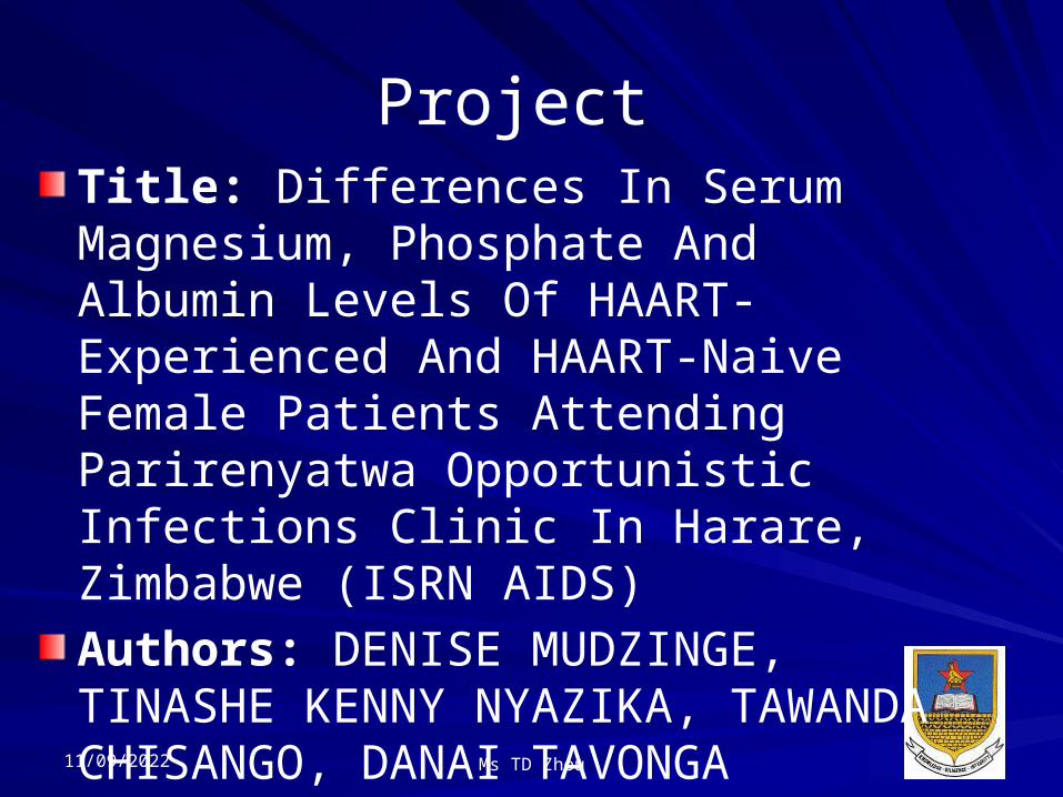
Project Title: Differences In Serum Magnesium, Phosphate And Albumin Levels Of HAART-Experienced And HAART-Naive Female Patients Attending Parirenyatwa Opportunistic Infections Clinic In Harare, Zimbabwe (ISRN AIDS)Authors: DENISE MUDZINGE, TINASHE KENNY NYAZIKA, TAWANDA CHISANGO, DANAI TAVONGA ZHOU
11/09/2022 Ms TD Zhou

BackgroundAntiretroviral Therapy inhibits HIV replication, maintains health and preserves life. However, both antiretroviral therapy and HIV infection have been reported to have short and long term effects on bone metabolism.
11/09/2022 Ms TD Zhou

Materials and MethodsStudy design: Cross-Sectional StudySetting: Parirenyatwa Group of Hospitals Opportunistic Infections ClinicStudy Participants: – 40 HIV Infected, on HAART– 40 HIV infected, not yet oh HAARTRecruitment:– Consent Process– Sample Collection– Processing (thawing, machine used, tests done)
11/09/2022

ResultsTables and figuresMg, Phosp and Alb levels were significantly higher in the Therapy-naïve than in Therapy-experienced. There was no statistically significant difference in Calcium levels of the two groups of women.
11/09/2022 Ms TD Zhou
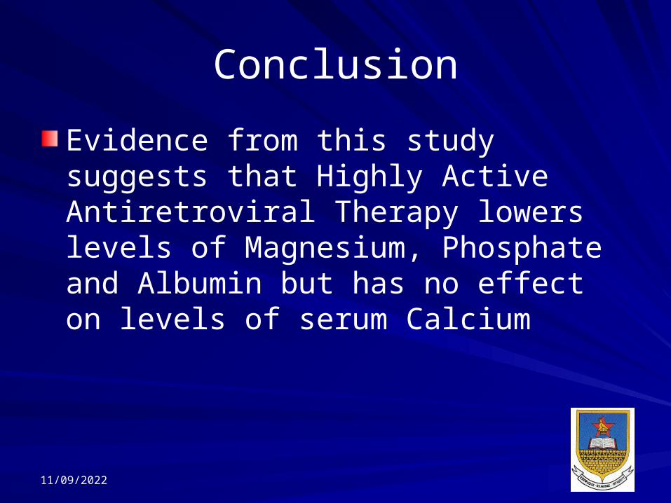
ConclusionEvidence from this study suggests that Highly Active Antiretroviral Therapy lowers levels of Magnesium, Phosphate and Albumin but has no effect on levels of serum Calcium
11/09/2022
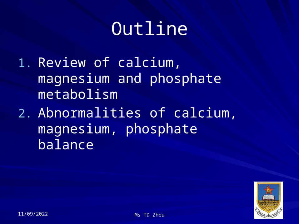
Outline1. Review of calcium,
magnesium and phosphate metabolism
2. Abnormalities of calcium, magnesium, phosphate balance
11/09/2022 Ms TD Zhou
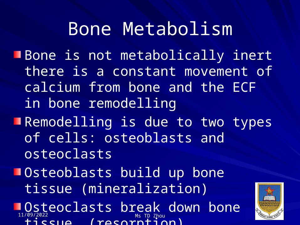
Bone MetabolismBone is not metabolically inert there is a constant movement of calcium from bone and the ECF in bone remodellingRemodelling is due to two types of cells: osteoblasts and osteoclastsOsteoblasts build up bone tissue (mineralization)Osteoclasts break down bone tissue (resorption)
11/09/2022 Ms TD Zhou

Calcium99% present in skeleton (reservoir)Serum calcium 2.15-2.6 mmol/LFunctions of calcium – Intracellular signalling– Coagulation– Bone mineralization– Plasma membrane potential
11/09/2022

Calcium MetabolismForms– Free – 50%– Bound – protein (Albumin) – 40%– Complexed – 10%

Calcium MetabolismThe following affect levels of Ca– Albumin levels ([Ca] is adjusted for albumin)
– Acid base status– PTH– Vitamin D– Calcitonin (inhibits osteoclastic activity)
– Phosphate – Magnesium
11/09/2022 Ms TD Zhou

Corrected Calcium Derived a when the albumin is abnormal.To make up for change in total calcium due to the change in albumin-bound calcium, and gives an estimate of what the calcium level would be if the albumin were within normal ranges.
11/09/2022 Ms TD Zhou

Corrected Calcium Corrected calcium (mg/dL) = measured total Ca (mg/dL) + 0.8 (4.0 - serum albumin [g/dL]), where 4.0 represents the average albumin level in g/dL
11/09/2022
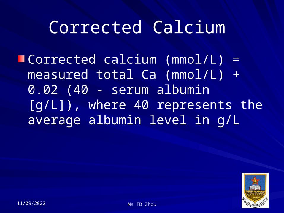
Corrected Calcium Corrected calcium (mmol/L) = measured total Ca (mmol/L) + 0.02 (40 - serum albumin [g/L]), where 40 represents the average albumin level in g/L
11/09/2022 Ms TD Zhou

Corrected Calcium
AKA Adjusted CaAdvantages– Accounts for changes in alb conc– To calculate the expected Ca conc if the alb were normal
Limitations– Interpret with caution when H+ status abnormal
– Not valid when alb is very low eg <20

Calcium Homeostasis
Skeleton
Intestine
Kidneys
Parathyroid gland
Ca++
Vitamin D

Major Mediators of Calcium and Phosphate Balance
Parathyroid hormone (PTH)
Calcitriol (active form of vitamin D3)
11/09/2022
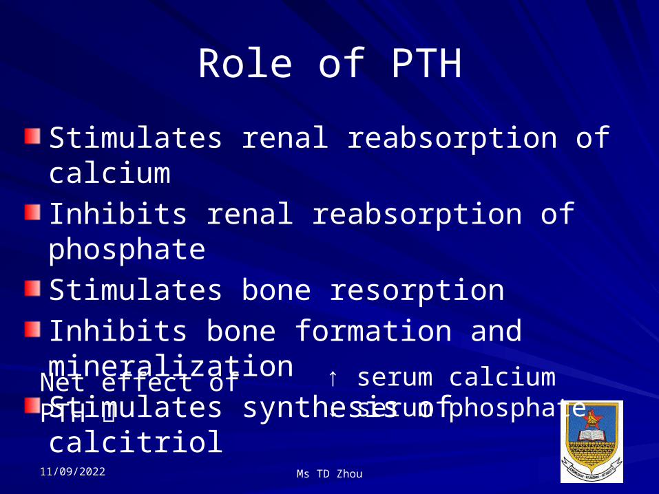
Role of PTHStimulates renal reabsorption of calciumInhibits renal reabsorption of phosphateStimulates bone resorptionInhibits bone formation and mineralizationStimulates synthesis of calcitriol
Net effect of PTH
↑ serum calcium↓ serum phosphate
11/09/2022 Ms TD Zhou

Regulation of PTH
Low serum [Ca+2] Increased PTH secretion
High serum [Ca+2] Decreased PTH secretion
11/09/2022

Role of CalcitriolStimulates GI absorption of both calcium and phosphateStimulates renal reabsorption of both calcium and phosphateStimulates bone resorption
Net effect of calcitriol
↑ serum calcium↑ serum phosphate
11/09/2022 Ms TD Zhou

Regulation of Calcitriol
11/09/2022 Ms TD Zhou

Overview of Calcium-Phosphate Regulation
11/09/2022

Different Forms of Calcium
At any one time, most of the calcium in the body exists as the mineral hydroxyapatite, Ca10(PO4)6(OH)2.
Calcium in the plasma:45% in ionized form (the physiologically
active form)45% bound to proteins (predominantly
albumin)10% complexed with anions (citrate,
sulfate, phosphate)
To estimate the physiologic levels of ionized calcium in states of hypoalbuminemia:
[Ca+2]Corrected = [Ca+2]Measured + [ 0.8 (4 – Albumin) ]
11/09/2022 Ms TD Zhou

Overview of Calcium Balance
11/09/2022

Etiologies of HypercalcemiaIncreased GI Absorption
Milk-alkali syndromeElevated calcitriol
Vitamin D excessExcessive dietary intakeGranuomatous diseases
Elevated PTHHypophosphatemia
Increased Loss From BoneIncreased net bone resorption
Elevated PTHHyperparathyroidism
MalignancyOsteolytic metastasesPTHrP secreting tumor
Increased bone turnoverPaget’s disease of boneHyperthyroidism
Decreased Bone Mineralization
Elevated PTHAluminum toxicity
Decreased Urinary Excretion
Thiazide diureticsElevated calcitriolElevated PTH
11/09/2022

Etiologies of HypocalcemiaDecreased GI Absorption
Poor dietary intake of calciumImpaired absorption of calcium
Vitamin D deficiency Poor dietary intake of
vitamin D Malabsorption
syndromesDecreased conversion of vit. D to
calcitriol Liver failure Renal failure Low PTH Hyperphosphatemia
Decreased Bone Resorption/Increased MineralizationLow PTH (aka hypoparathyroidism)PTH resistance (aka pseudohypoparathyroidism)Vitamin D deficiency / low calcitriolHungry bones syndromeOsteoblastic metastases
Increased Urinary Excretion Low PTH s/p thyroidectomy s/p I131 treatment
Autoimmune hypoparathyroidism PTH resistance Vitamin D deficiency / low calcitriol
11/09/2022

HypercalcemiaIncreased flux of Ca2+ into the extracellular fluid (ECF) from skeleton, kidney or intestineLethargyNauseaVomitingBones, moans, groans and stonesPolyuria Symptoms dependent on rate of increaseCa > 3mmol/L stones
Presentations
Groups 1-4
11/09/2022

Questions?

Causes of HypercalcemiaContamination Primary hyperparathyroidismMalignancy (skeletal involvement/PTHrP)Endocrine disorders hyper/hypothyroidism/acute adrenal insufficiencyFHH (familial hypocalciuric hypercalcemia)
• Renal failure• Idiopathic hyperCa of infancy
• Granulomatous disorders (eg TB)
• Diuretics• Lithium Toxicity• Aluminium toxicity
• Milk alkali syndrome
95%
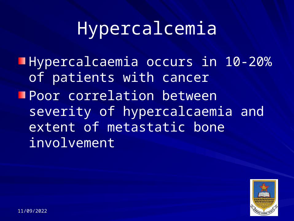
HypercalcemiaHypercalcaemia occurs in 10-20% of patients with cancerPoor correlation between severity of hypercalcaemia and extent of metastatic bone involvement
11/09/2022

HypercalcemiaPatients with mild asymptomatic hypercalcemia (<3mmol/L) may stay healthy for years without operation but increased risk of osteoporosis and renal failure. Should be reassessed regularly. High fluid intake to discourage renal calculus. Surgery is recommended if ca > 3mmol/L or marked hypercalciuria/complications present, < 50yrs11/09/2022

HypercalcemiaParathyroid hormone related protein–PTHrP binds to PTH receptor1,25 VitD (Calcitriol)In secondary/tertiary hyperPTH, chronic renal disease or vit D deficiency hypocalcemia). The body responds by producing more PTH hypercalcemia.
11/09/2022

HyperthyroidismThyrotoxicosis – Hypercalcaemia due to increased osteoclast activity10% of sarcoidosis develop hypercalcemia due to increased 1 hydroxylation of 25OHD by macrophages in sarcoid granulomas
11/09/2022 Ms TD Zhou
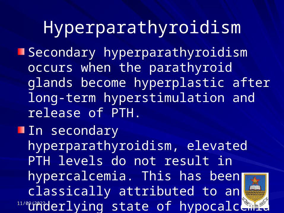
HyperparathyroidismSecondary hyperparathyroidism occurs when the parathyroid glands become hyperplastic after long-term hyperstimulation and release of PTH. In secondary hyperparathyroidism, elevated PTH levels do not result in hypercalcemia. This has been classically attributed to an underlying state of hypocalcemia in those with chronic renal failure (CRF).
11/09/2022

HyperparathyroidismWith long-term hyperstimulation, the glands function autonomously and produce high levels of PTH even after correction of chronic hypocalcemia.Tertiary hyperparathyroidism refers to hypercalcemia caused by autonomous parathyroid function after long-term hyperstimulation.
11/09/2022

HypercalcemiaHyperPTH usually detected by biochemical screeningPhosphate may be normal or lowPlasma ALP is raised in 20-30% of cases.
11/09/2022

Hyperparathyroidism• PTH Inappropriate to calcium level
• Raised calcium with raised/normal PTH
• ? Primary• ?Secondary/Tertiary• Primary - usually due to parathyroid adenoma (single/multiple)
• Multiple - ? MEN (Multiple endocrine neoplasm)
• Treatment• High fluid intake
• Surgery • Watch and wait
• Side effects• Osteoporosis• Renal failure• Stones

Investigations in Hypercalcemia
Bone profileRenal functionPTH (>3 pmol/L inappropriate for hyperCa)? Primary HyperPTH or FHHUrinary fractional calcium excretion– Fasting urine calcium x serum creatinine
Urine creatinine< 25 umol/L FHH> 30 umol/L PHPT

Case51 year old woman investigated after pain in kidneys shown on radiological examination to be due to Ca-containing stones. Serum Calcium 2.95 mmol/LPhosphate 0.7 mmol/LPTH 10 pmol/LBone radiographs normalSerum urea, albumin ALP normal
Make a diagnosis based on the results above

HintsHyperPTH may present in many ways including stones – due to hypercalciuria. 10% have clinical evidence of bone disease at presentation. – Many patients with hyperPTH detected by biochemical screening.
– Plasma phosphate may be normal or raised, particularly when there is renal damage.
– The plasma ALP is raised in 20-30% of cases.
– Inappropriateness of PTH to calcium level.
11/09/2022
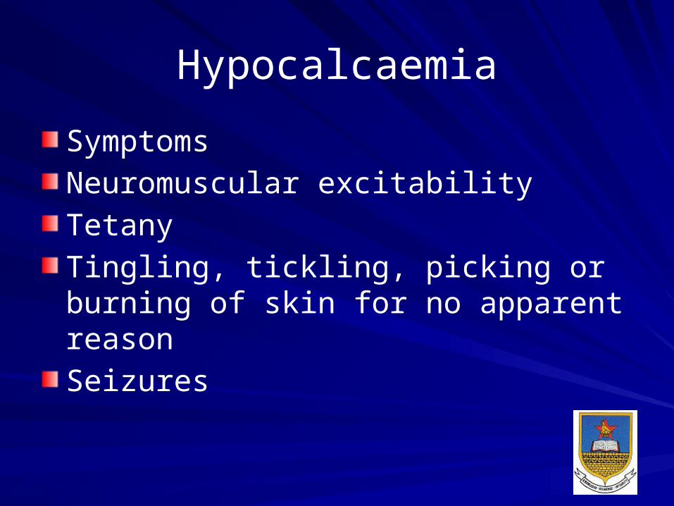
HypocalcaemiaSymptomsNeuromuscular excitabilityTetanyTingling, tickling, picking or burning of skin for no apparent reasonSeizures

Causes of hypocalcaemiaContaminationHypoalbuminaemia Chronic renal failure Magnesium deficiencyHypoparathyroidism (/pseudo)Vitamin D deficiency (or resistance)Acute haemorrhagic and edematous pancreatitisHungry bone syndrome

Causes of hypocalcemiaHungry bone syndrome – healing phase of bone disease of hypercalcaemia caused by increase bone resorption eg treated hyperparathyroidism (surgical), hyperthyroidism, and haematological malignancies. Removal of the stimulus of bone resorption results in rapid uptake of calcium into bone.
11/09/2022
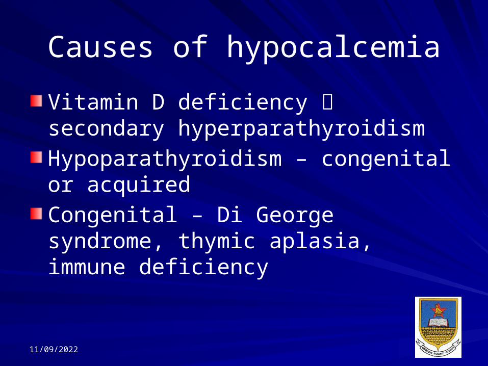
Causes of hypocalcemiaVitamin D deficiency secondary hyperparathyroidismHypoparathyroidism – congenital or acquiredCongenital – Di George syndrome, thymic aplasia, immune deficiency
11/09/2022

Causes of hypocalcemiaPseudo hyperparathyroidismPTH effects mediated through formation of cyclic AMPType 1 pseudo hyperparathyroidism activation of adenylate cyclase defective cAMP not formedType 2 cAMP formed, responses to it are blocked
11/09/2022

Chronic Renal failure Phosphate Protein 1, 25 Vit D Skeletal resistance to Vitamin D

Investigations in Hypocalcemia
Bone profileRenal functionMgVitamin D? History (eg surgery to neck)? PTHCaE – fasting urine calcium and creatinine and concurrent serum creatinine

Phosphate Metabolism85% present in skeletonSerum inorganic phosphate 0.84-1.45 mmol/L10% protein bound, 35% complexed, rest freeBalance maintained primarily by kidneys
FunctionsIntegrity of boneOxygen deliveryMuscle contractionRole in ATP (energy), nucleotides, NADP, cell membranes, gene transcription, cell growth

Hyperphosphataemia
Decreased renal excretion– Reabsorption
hypoPTHAcromegaly
Transcellular shift– Lactic acidosis– Respiratory acidosis
– DKA
Increased intake– Oral or IV– P containing laxatives/enemas
– Vit D intoxication
Cell Lysis– Rhabdomyolysis– Intravascular haemolysis
– Cytotoxic therapy– Leukaemia– Lymphoma
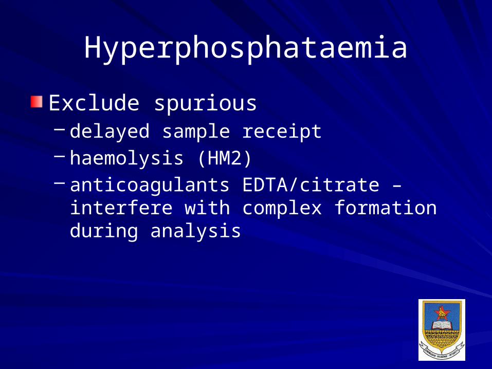
HyperphosphataemiaExclude spurious– delayed sample receipt– haemolysis (HM2)– anticoagulants EDTA/citrate – interfere with complex formation during analysis

HypophosphataemiaCommonMuscle weaknessRespiratory failureDecreased myocardial outputSevere hypoP haemolysisRickets/osteomalacia (chronic defy)Wernicke’s encephalopathy

Hypophosphataemia
Intracellular shiftGlucoseInsulinResp alkalosisRefeeding
Decreased absorption
• Increased loss• Vomiting• Diarrhoea• Phosphate binding antacids
• Decreased absorption
• Malabsorption syndrome
• VitD defy• Poor diet
Lowered renal P threshold
• Primary hyperPTH• Renal tubular defects
• Familial hypophospataemia
• Fanconi’s

Investigations? History? Contamination ? Repeat Bone profileRenal functionMg? Vitamin D (?Ca)? PTH (?Ca)

Magnesium Metabolism55% present in skeleton1% of total body Mg extracellularSerum Mg 0.7-1.0 mmol/L
FunctionsCofactor for enzymesRequired for ATP (MgATP)GlycolysisCell replicationProtein biosynthesisPTH increases renal tubular reabs of MgHomeostasis maintained - control of excretion
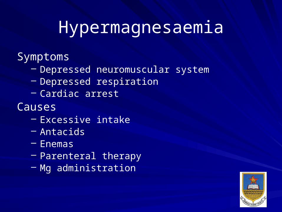
HypermagnesaemiaSymptoms
– Depressed neuromuscular system– Depressed respiration– Cardiac arrest
Causes– Excessive intake– Antacids– Enemas– Parenteral therapy– Mg administration

HypomagnesemiaCommon in in-patientsUsu assoc with hypoK and hypoPIncreased neuromuscular excitabilityCauses impaired PTH secretionPTH end organ resistanceOral K not retained if patient also Mg deficient
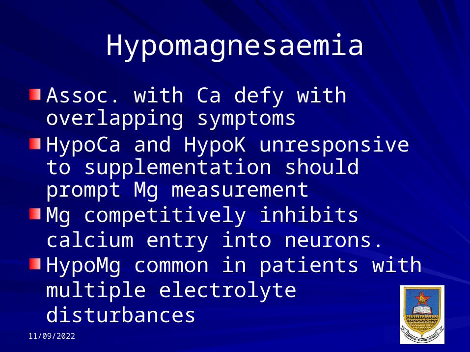
HypomagnesaemiaAssoc. with Ca defy with overlapping symptomsHypoCa and HypoK unresponsive to supplementation should prompt Mg measurementMg competitively inhibits calcium entry into neurons.HypoMg common in patients with multiple electrolyte disturbances
11/09/2022

HypomagnesaemiaGI
• Prolonged nasogastric suction
• Malabsorption• Bowel resection• Diarrhoea• Fistulas• Acute pancreatitis• Decreased intake• Chronic vomiting
Redistribution• Diabetic ketoacidosis DKA
• Hungry bone disease
Renal loss• Chronic total parenteral nutrition (TPN)
• Osmotic diuresis (DM/mannitol)
• Hypercalcaemia• Alcohol• Drugs – diuretics/aminoglycosides/cisplatin/cardiac glycosides
• Metabolic acidosis (DKA/ETOH/starvation)
• Renal disease

Overview of Phosphate Balance
11/09/2022 Ms TD Zhou

Etiologies of Hyperphosphatemia
Increased GI IntakeFleet’s Phospho-Soda
Decreased Urinary ExcretionRenal FailureLow PTH (hypoparathyroidism)
s/p thyroidectomys/p I131 treatment for Graves disease of
thyroid cancerAutoimmune hypoparathyroidism
Cell LysisRhabdomyolysisTumor lysis syndrome
11/09/2022

Etiologies of Hypophosphatemia
Decreased GI AbsorptionDecreased dietary intake (rare in isolation)Diarrhea / Malabsorption Phosphate binders (calcium acetate, Al & Mg containing
antacids)
Decreased Bone Resorption / Increased Bone Mineralization Vitamin D deficiency / low calcitriolHungry bones syndromeOsteoblastic metastases
Increased Urinary ExcretionElevated PTH (as in primary hyperparathyroidism)Vitamin D deficiency / low calcitriolFanconi syndrome
Internal Redistribution (due to acute stimulation of glycolysis)
Refeeding syndrome (seen in starvation, anorexia, and alcholism)
During treatment for DKA
11/09/2022

Case 1Mrs. T is a 59 year old woman with a past medical history significant for hypertension who comes for a routine clinic visit. She initially states that she has no symptomatic complaints, but later in the interview describes chronic fatigue and a mildly depressed mood. Her exam is unremarkable. Labs are as follows:
Calcium (total) – 11.9 mg/dL (normal ~ 8.5-10.2 mg/dL)Phosphate – 1.8 mg/dL (normal ~ 2.0-4.3 mg/dL)Albumin – 3.8 g/dL (normal ~ 3.5-5.0 g/dL)PTH – 124 pg/mL (normal ~ 10-60 pg/mL)Creatinine – 1.2 mg/dL
11/09/2022 Ms TD Zhou

Case 2Mr. G is a 40 year old man with a history of alcoholism. He had not seen a doctor for 15 years before police brought him to the ER after finding him confused and disheveled behind a local convenience store. In the ER, he was thought to be confused simply due to intoxication, but was admitted for mild alcoholic hepatitis and marked malnutrition. His mental status cleared up about 8 hours after admission. During morning rounds on hospital day #2, he complained of feeling fatigued and weak. Later that day, the nurses find him seizing. The seizures stop with low dose IV diazepam. Stat labs are sent:
Sodium – 136 meq/LPotassium – 3.2 meq/LCalcium (total) – 6.8 mg/dL (normal ~ 8.5-10.2 mg/dL)
Phosphate – 0.7 mg/dL (normal ~ 2.0-4.3 mg/dL)Albumin – 1.8 g/dL (normal ~ 3.5-5.0 g/dL)Creatinine – 1.3 mg/dLCK – 3500 U/L
11/09/2022

Case 3Mr. H is a 74 year old man with a past history significant for hypertension and COPD from smoking 2 packs per day for the last 40 years. He presented to an urgent pulmonary clinic appointment with 2 months of increased cough and 5 days of “mild” hemoptysis. Upon further obtaining further history, he reports feeling fatigued, nauseous, and chronically thirsty for several weeks. His exam is significant for bilateral rhonchi (no change from baseline lung exam) and absent reflexes. Stat labs are ordered from clinic:
Sodium – 138 meq/L CBC, PT/PTT – WNL Potassium – 3.7 meq/L PTH - PendingMagnesium – 1.8 mg/dL Albumin – 2.2 g/dL Calcium (total) – 13.1 mg/dL Phosphate – 1.3 mg/dLCreatinine – 2.8 mg/dL (baseline creatinine = 1.1)
11/09/2022 Ms TD Zhou

Case 4Miss L is a 16 year old woman with no significant past medical history, who is brought to the ER by her mother after she noted her to be acting bizarrely for the past several weeks. Thought to be actively psychotic, a psychiatry consult is asked to see the patient, who recommends checking routine labs:
Sodium – 142 meq/L Urine tox. screen – NegativePotassium – 4.1 meq/L Urine pregnancy - NegativeMagnesium – 2.3 mg/dLCalcium (total) – 6.9 mg/dLPhosphate – 4.4 mg/dLAlbumin – 4.2 g/dLCreatinine – 0.8 mg/dL
11/09/2022