Ruthenium metallopharmaceuticals Coord Chem Rev 2003
25
Ruthenium metallopharmaceuticals Michael J. Clarke Merkert Chemistry Center, Boston College, 2609 Beacon Street, Chestnut Hill, MA 02467-3860, USA Received 11 October 2001; accepted 18 January 2002 Abstract The well-developed synthetic chemistry of ruthenium, particularly with ammine, amine and imine ligands, provides for many approaches to innovative new metallopharmaceuticals. Advantages of utilizing ruthenium am(m)ine complexes in drug development include, (1) reliable preparations of stable complexes with predictable structures; (2) the ability to tune ligand affinities, electron transfer and substitution rates, and reduction potentials; and (3) an increasing knowledge of the biological effects of ruthenium complexes. Many Ru(II) and Ru(III) am(m)ine complexes selectively bind to imine sites in biomolecules. Collectively, these lend ruthenium complexes to redox-activation and photodynamic approaches to therapy as well as the development of radio- Contents Abstract ......................................................................... 209 1. Introduction .................................................................... 210 2. Anticancer compounds .............................................................. 211 2.1 Activation by reduction ......................................................... 211 2.2 Active Ru(II) complexes ......................................................... 211 2.3 Transferrin transport ........................................................... 212 2.4 Covalent DNA binding ......................................................... 213 2.5 DNA damage generated by covalently bound Ru ........................................... 215 2.6 Modulation of DNA binding by glutathione ............................................. 216 2.7 Cytotoxicities of Ru complexes ..................................................... 217 2.8 Complexes with EDTA-type ligands .................................................. 218 2.9 Complexes with R 2 SO .......................................................... 218 2.10 Photodynamic therapy .......................................................... 219 3. Ruthenium red and Ru360: inhibitors of Ca 2 utilization .......................................... 222 4. Nitric oxide complexes .............................................................. 223 5. Immunosuppressants ............................................................... 227 6. Radiopharmaceuticals .............................................................. 227 7. Conclusion ..................................................................... 228 Acknowledgements ................................................................... 229 References ........................................................................ 229 Abbreviations: Ade, A, adenine; 5?AMP, adenosine-5?-monophosphate; bpy, 2,2?-bipyridyl; bzac, 1-phenylbutane-1,3-diketonate; CD, circular dichroism; cdta, 1,2-cyclo -hexanediaminotetraacetate; chrysi, 5,6-chrysenequinone diimine; CT-DNA, calf thymus DNA; Cyt, C, cytosine; 5?-CMP, cytidine-5?-monophosphate; Cp, cyclo -pentadienide; cyclam, 1,4,8,11-tetraazo-cyclo -tetradecane; dppz, dipyrido[3,2-a :2?,3?-c] phenazine; en, ethylenediammine; 9EtGua, 9-ethylguanine; GSH, glutathione (g-glutamate /cysteine /glycine); 5?-dGMP, deoxyguanosine-5?-monophosphate; Gua, G, guanine; dGuo, guanosine; dGuo, dG, 2?-deoxyguanosine; Hyp, hypoxanthine; Icyt, isocytidine; ICR, imidazolium trans -tetrachloro- bisimidazoleruthenium(III); Imm, imidazole; Ind, indazole; Isn, isonicotinamide; Me, methyl; Et, ethyl; mgp, Methylguanidinium phenanthroline; 7MeGua, 7-methylguanine; 1MeGuo, 7-methylguanosine; 7MeHyp, 7-methylhypoxanthine; 6MeICyt, 6-methylisocytosine; 1,3Me 2 Xan , deprotonated 1,3-dimethylxanthine; MM2 molecular mechanics 2; NAMI, Na[trans -(Me 2 SO)(Im)Cl 4 Ru]; NAMI-A, ImH[trans - (Me 2 SO)(Im)Cl 4 Ru]; NHE, normal hydrogen electrode; 8-OG, 8-oxo-guanine; 8-Oguo, 8-oxo-guanosine; PIH, pyridoxal isonicotinoyl hydrazone; quin, 8-hydroxyquinoline; Ox, oxalate; pdta, 1,2-propylenediamminetetraacetate; phen, 1,10-phenanthroline; py, pyridine; Pyr, pyridine derivative; SSCE, standard saturated calomel electrode; 5?-TMP, thymidine-5?-monophosphate; 5?dTMP, deoxythymidine-5?-monophosphate; tpy, 2,2ƒ:6ƒ,2ƒ- terpyridine; Tf, transferrin; TfR, transferrin receptor; trien, triethylenetetraammine. Tel.: /1-617-552-3624; fax: /1-617-552-2705 E-mail address: [email protected] (M.J. Clarke). Coordination Chemistry Reviews 236 (2003) 209 /233 www.elsevier.com/locate/ccr 0010-8545/02/$ - see front matter # 2002 Elsevier Science B.V. All rights reserved. PII:S0010-8545(02)00312-0
Transcript of Ruthenium metallopharmaceuticals Coord Chem Rev 2003

Ruthenium metallopharmaceuticals
Michael J. Clarke �Merkert Chemistry Center, Boston College, 2609 Beacon Street, Chestnut Hill, MA 02467-3860, USA
Received 11 October 2001; accepted 18 January 2002
Abstract
The well-developed synthetic chemistry of ruthenium, particularly with ammine, amine and imine ligands, provides for many
approaches to innovative new metallopharmaceuticals. Advantages of utilizing ruthenium am(m)ine complexes in drug development
include, (1) reliable preparations of stable complexes with predictable structures; (2) the ability to tune ligand affinities, electron
transfer and substitution rates, and reduction potentials; and (3) an increasing knowledge of the biological effects of ruthenium
complexes. Many Ru(II) and Ru(III) am(m)ine complexes selectively bind to imine sites in biomolecules. Collectively, these lend
ruthenium complexes to redox-activation and photodynamic approaches to therapy as well as the development of radio-
Contents
Abstract . . . . . . . . . . . . . . . . . . . . . . . . . . . . . . . . . . . . . . . . . . . . . . . . . . . . . . . . . . . . . . . . . . . . . . . . . 209
1. Introduction . . . . . . . . . . . . . . . . . . . . . . . . . . . . . . . . . . . . . . . . . . . . . . . . . . . . . . . . . . . . . . . . . . . . 210
2. Anticancer compounds . . . . . . . . . . . . . . . . . . . . . . . . . . . . . . . . . . . . . . . . . . . . . . . . . . . . . . . . . . . . . . 211
2.1 Activation by reduction . . . . . . . . . . . . . . . . . . . . . . . . . . . . . . . . . . . . . . . . . . . . . . . . . . . . . . . . . 211
2.2 Active Ru(II) complexes . . . . . . . . . . . . . . . . . . . . . . . . . . . . . . . . . . . . . . . . . . . . . . . . . . . . . . . . . 211
2.3 Transferrin transport . . . . . . . . . . . . . . . . . . . . . . . . . . . . . . . . . . . . . . . . . . . . . . . . . . . . . . . . . . . 212
2.4 Covalent DNA binding . . . . . . . . . . . . . . . . . . . . . . . . . . . . . . . . . . . . . . . . . . . . . . . . . . . . . . . . . 213
2.5 DNA damage generated by covalently bound Ru . . . . . . . . . . . . . . . . . . . . . . . . . . . . . . . . . . . . . . . . . . . 215
2.6 Modulation of DNA binding by glutathione . . . . . . . . . . . . . . . . . . . . . . . . . . . . . . . . . . . . . . . . . . . . . 216
2.7 Cytotoxicities of Ru complexes . . . . . . . . . . . . . . . . . . . . . . . . . . . . . . . . . . . . . . . . . . . . . . . . . . . . . 217
2.8 Complexes with EDTA-type ligands . . . . . . . . . . . . . . . . . . . . . . . . . . . . . . . . . . . . . . . . . . . . . . . . . . 218
2.9 Complexes with R2SO . . . . . . . . . . . . . . . . . . . . . . . . . . . . . . . . . . . . . . . . . . . . . . . . . . . . . . . . . . 218
2.10 Photodynamic therapy . . . . . . . . . . . . . . . . . . . . . . . . . . . . . . . . . . . . . . . . . . . . . . . . . . . . . . . . . . 219
3. Ruthenium red and Ru360: inhibitors of Ca2� utilization . . . . . . . . . . . . . . . . . . . . . . . . . . . . . . . . . . . . . . . . . . 222
4. Nitric oxide complexes . . . . . . . . . . . . . . . . . . . . . . . . . . . . . . . . . . . . . . . . . . . . . . . . . . . . . . . . . . . . . . 223
5. Immunosuppressants . . . . . . . . . . . . . . . . . . . . . . . . . . . . . . . . . . . . . . . . . . . . . . . . . . . . . . . . . . . . . . . 227
6. Radiopharmaceuticals . . . . . . . . . . . . . . . . . . . . . . . . . . . . . . . . . . . . . . . . . . . . . . . . . . . . . . . . . . . . . . 227
7. Conclusion . . . . . . . . . . . . . . . . . . . . . . . . . . . . . . . . . . . . . . . . . . . . . . . . . . . . . . . . . . . . . . . . . . . . . 228
Acknowledgements . . . . . . . . . . . . . . . . . . . . . . . . . . . . . . . . . . . . . . . . . . . . . . . . . . . . . . . . . . . . . . . . . . . 229
References . . . . . . . . . . . . . . . . . . . . . . . . . . . . . . . . . . . . . . . . . . . . . . . . . . . . . . . . . . . . . . . . . . . . . . . . 229
Abbreviations: Ade, A, adenine; 5?AMP, adenosine-5?-monophosphate; bpy, 2,2?-bipyridyl; bzac, 1-phenylbutane-1,3-diketonate; CD, circular
dichroism; cdta, 1,2-cyclo -hexanediaminotetraacetate; chrysi, 5,6-chrysenequinone diimine; CT-DNA, calf thymus DNA; Cyt, C, cytosine; 5?-CMP,
cytidine-5?-monophosphate; Cp, cyclo -pentadienide; cyclam, 1,4,8,11-tetraazo-cyclo -tetradecane; dppz, dipyrido[3,2-a :2?,3?-c] phenazine; en,
ethylenediammine; 9EtGua, 9-ethylguanine; GSH, glutathione (g-glutamate�/cysteine�/glycine); 5?-dGMP, deoxyguanosine-5?-monophosphate;
Gua, G, guanine; dGuo, guanosine; dGuo, dG, 2?-deoxyguanosine; Hyp, hypoxanthine; Icyt, isocytidine; ICR, imidazolium trans -tetrachloro-
bisimidazoleruthenium(III); Imm, imidazole; Ind, indazole; Isn, isonicotinamide; Me, methyl; Et, ethyl; mgp, Methylguanidinium phenanthroline;
7MeGua, 7-methylguanine; 1MeGuo, 7-methylguanosine; 7MeHyp, 7-methylhypoxanthine; 6MeICyt, 6-methylisocytosine; 1,3Me2Xan�,
deprotonated 1,3-dimethylxanthine; MM2 molecular mechanics 2; NAMI, Na[trans -(Me2SO)(Im)Cl4Ru]; NAMI-A, ImH[trans -
(Me2SO)(Im)Cl4Ru]; NHE, normal hydrogen electrode; 8-OG, 8-oxo-guanine; 8-Oguo, 8-oxo-guanosine; PIH, pyridoxal isonicotinoyl hydrazone;
quin, 8-hydroxyquinoline; Ox, oxalate; pdta, 1,2-propylenediamminetetraacetate; phen, 1,10-phenanthroline; py, pyridine; Pyr, pyridine derivative;
SSCE, standard saturated calomel electrode; 5?-TMP, thymidine-5?-monophosphate; 5?dTMP, deoxythymidine-5?-monophosphate; tpy, 2,2ƒ:6ƒ,2ƒ-terpyridine; Tf, transferrin; TfR, transferrin receptor; trien, triethylenetetraammine.
� Tel.: �/1-617-552-3624; fax: �/1-617-552-2705
E-mail address: [email protected] (M.J. Clarke).
Coordination Chemistry Reviews 236 (2003) 209�/233
www.elsevier.com/locate/ccr
0010-8545/02/$ - see front matter # 2002 Elsevier Science B.V. All rights reserved.
PII: S 0 0 1 0 - 8 5 4 5 ( 0 2 ) 0 0 3 1 2 - 0

pharmaceuticals containing one of several radionuclides of ruthenium. Ruthenium red and the related Ru360 strongly inhibit
calcium ion uptake in the mitochondria. A number of ruthenium compounds with anticancer activity appear to penetrate tumors
through a transferrin-mediated process and bind to cellular DNA following intracellular activation by reduction. Ruthenium
complexes exhibit both nitric oxide release and scavenging functions that can affect vasodilation and synapse firing. Simple
ruthenium complexes are unusually effective in suppressing the immune response by inhibiting T cell proliferation.
# 2002 Elsevier Science B.V. All rights reserved.
Keywords: ruthenium; metallopharmaceutical; immunosuppressant; anticancer; transferring; DNA; photodynamic; nitric oxide;
radiopharmaceutical
1. Introduction
Along with an increased understanding of metallo-
protein function and some excellent models of metal ion
active sites [1], recent advances in understanding how
naturally-occurring metal ions are delivered to these
active sites and how metal ions are involved in some
diseases indicate new roles for metal ions in therapeutic
strategies [2]. The current array of successful metallo-
pharmaceuticals, which include the platinum anticancer
drugs [3�/9], radiodiagnostic agents that contain 99mTc
and other radionuclides [10,11], Gd(III) MRI agents
[12,13], ß-emitting radiotherapeutic compounds [14],
complexes involving vanadium [15�/17] as an insulin
mimic and Cr(III) as an intermediary in activating
insulin receptors [18,19], all indicate the utility of
complexes as both therapeutic and diagnostic agents.
Most of these metal-containing pharmaceuticals have
been developed in academic laboratories or by relatively
small or ‘start-up’ pharmaceutical enterprises [15,20�/
22]. The very success of pharmaceutical chemists in
synthesizing broad ranges of carbon-based compounds
tends to eliminate less common elements from their
synthetic programs. The lack of experience of traditional
medicinal chemists and pharmacologists in dealing with
biologically active metal complexes poses a substantial
activation energy barrier to their identifying active metal
complexes and shepherding them to the clinic. These
factors coupled with a tendency of pharmaceutical
houses and government screening programs to view
transition metal ions as toxic ‘heavy metals’ (despite the
body’s utilization of gram quantities of Fe, Cu and Zn)
retards the development of metallopharmaceuticals. On
the other hand, this also provides enterprising transition
metal chemists with opportunities to pioneer the devel-
opment of exciting new drugs.
While the statistical success of metal-based com-
pounds in reaching the clinic through the NIH antic-
ancer screening program is about the same as for
carbon-based compounds (1 in 6000 tested), the move-
ment of new transition metal chemotherapeutic agents
toward the clinic has been slow. Keppler has pointed out
the inherent bias in testing antitumor metal compounds
in cell and animal systems, which are sensitive to
cisplatin, and the difficulty in formulating metal com-
plexes, particularly those with low solubility [23].The synthetic chemistry of the transition metal,
ruthenium is well developed, particularly with ammine,
amine and imine ligands, and provides for many
approaches to innovative new metallopharmaceuticals.
Due to strong ligand-field stabilization, the more
common oxidation states (Ru(II), Ru(III), and
Ru(IV)) in aqueous solution are usually octahedral
and are often fairly inert to ligand substitution. The
drug-like effects of ruthenium red, which has been used
as a cytological stain for over a century, have long been
known. Advantages of utilizing ruthenium am(m)ine
complexes in drug development include, (1) reliable
methods of synthesizing stable complexes with predict-
able structures; (2) the ability to tune ligand affinities,
electron transfer and substitution rates, and reduction
potentials; and (3) an increasing knowledge of the
biological effects of ruthenium complexes.
Collectively these lend ruthenium complexes to redox-
activation and photodynamic approaches to therapy as
well as the development of radiopharmaceuticals con-
taining one of several radionuclides of ruthenium [24�/
32]. Finally, many am(m)ine complexes of Ru(II) and
Ru(III) complexes of ruthenium tend to selectively bind
to imine sites in biomolecules, which (as opposed to
amine sites) do not protonate at neutral pH, thereby
leaving their nitrogen lone pairs available for metal ion
coordination. Consequently, ruthenium complexes often
selectively coordinate histidyl imidazole nitrogens on
proteins [33�/35] and the N7 site on the imidazole ring of
purine nucleotides, and so can take advantage of the
properties of proteins, oligonucleotides and nucleic acids
to target specific tissues [36]. Thiolato complexes are
also possible, but these are often kinetically unstable
[37], particularly in air [38]. Complexes with flavins and
pterins are also known [39�/41], but tend to be photo-
chemically unstable.
M.J. Clarke / Coordination Chemistry Reviews 236 (2003) 209�/233210

2. Anticancer compounds
2.1. Activation by reduction
Due to the octahedral structure of Ru(II) and Ru(III)
complexes as opposed to the square-planar geometry of
Pt(II), ruthenium antitumor complexes probably func-
tion in a manner differently than cisplatin, which
appears to bend DNA by crosslinking adjacent Gua
thereby causing a class of DNA binding proteins toadhere to the site [4,5,8]. In what has become known as
the ‘activation by reduction’ hypothesis, we suggested
that Ru(III) complexes may serve as prodrugs that are
activated by reduction in vivo to coordinate more
rapidly to biomolecules [42�/44]. Since tumors rapidly
utilize oxygen and other nutrients and the development
of new blood vessels (known as neovascularization or
angiogenesis) often fails to keep pace with tumorgrowth, there is usually a lower O2 content (hypoxia)
in tumor cells [45�/49]. Consequently, cancer cells
depend more on glycolysis for energy and generate an
excess of lactic acid, which lowers the pH in tumor cells
[50]. Due to these metabolic differences, the relative
electrochemical potential inside tumors is generally
lower than in the surrounding normal tissue, particu-
larly at the center of the tumor [51]. These differences intumor relative to normal cell metabolism should favor
the production of Ru(II) relative to Ru(III) in tumors,
compared with normal tissue.
In the absence of p-bonding effects, the lower charge
on Ru(II) would cause it to be more actively substituting
than Ru(III) in a similar coordination environment. As
reduction of Ru(III) to Ru(II) fills the dp (t2g) orbitals,
p-donor ligands that coordinate firmly to Ru(III) are no
longer able to do so with Ru(II) and bind less strongly.
In the case of Ru(II) am(m)ine complexes, acido ligands
are lost fairly rapidly (k�/1�/10 s�1) [52,53]. Thiscoupled with the expected higher [Ru(II)]/[Ru(III)] ratio
in tumor cells should lead to increased intracellular
binding and hence somewhat selective tumor toxicity.
Glutathione (GSH) and a number of redox proteins are
capable of reducing Ru(III) complexes in vivo [54].
Single-electron-transfer proteins, which exist in both the
mitochondrial electron-transfer chain and in microso-
mal electron-transfer systems, can also reduce Ru(III)with the microsomal proteins being the more efficient
for [Cl(NH3)5Ru]2� [54]. In addition, ammineruthe-
nium complexes can also be reduced by transmembrane
electron transport systems, so that it is not necessary for
the complexes to enter cells in order to be reduced [55].
Oxidation of Ru(II) back to Ru(III) can occur by
molecular oxygen [56], cytochrome oxidase [56�/58],
and other oxidants, but this is relatively less likely tooccur in hypoxic tumor cells. The effect of hypoxia in
increasing DNA binding and thereby enhancing the
toxicity of the anticancer agents cis -[Cl2(NH3)4Ru]Cl
(CCR) and (ImH) trans -[(Im)2Cl4Ru] (ICR) against
HeLa cells has been clearly demonstrated (see Fig. 1)
[59]. There is also evidence that the first ruthenium
compound to enter clinical trials, Na{trans -
[Cl4(DMSO)(Im)Ru]} (NAMI), is activated by reduc-tion [60,61].
2.2. Active Ru(II) complexes
While the modest antitumor activity of Ru(II) com-
plexes such as cis -Cl2(DMSO)4Ru were studied rela-
tively early [62,63], significant activity has been seen
only recently with Ru(II) complexes stabilized with
heteroaromatic ligands [64�/66]. The isomer of cis -[Cl2(azpy)2Ru] (azpy�/2-(phenylazo)pyridine) with C2
symmetry exhibits substantial activity against several
cell lines [67,68]. Possible reasons for this are, (1) the
decrease in the rate of chloride aquation due the p-
acceptor effect of the imine ligands increasing the
effective charge on the metal ion so that the hydrolysis
rates are in the range of cisplatin; (2) increased hydro-
phobic or intercalative interactions with DNA, whichmay facilitate covalent binding; (3) geometric effects
exerted by the ligands, which may facilitate (or inhibit)
protein binding to the nucleic acid.
Arene ligands stabilize Ru(II) and provide a hydro-
phobic face that may enhance recognition and transport
through cell membranes and at least one such complex
inhibits topoisomerase II, which is essential in cell
division. Topoisomerase II (DNA gyrase) activity isinhibited by what is reported to be [Cl2(Me2-
SO )(C6H6)Ru(II)], but not by [(saldox)2Ru(II)] (sic ),
where sal�/salicylaldoximate [69]. Topoisomerase II
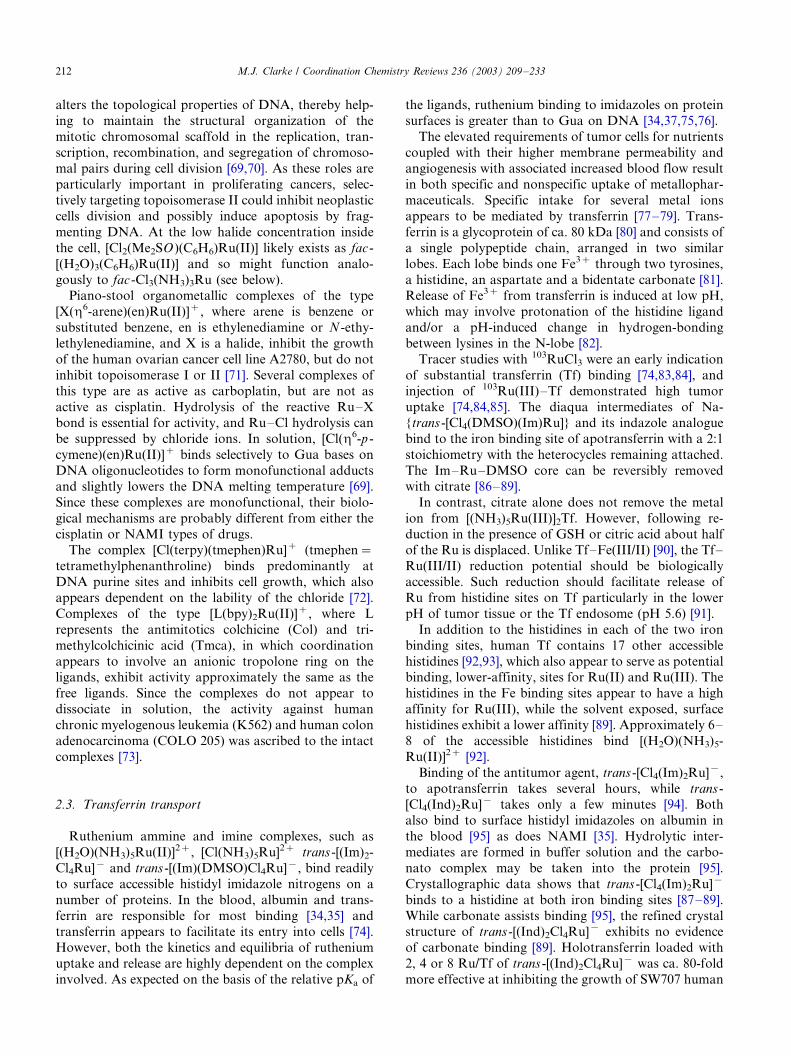
Fig. 1. DNA binding for ruthenium anticancer complexes as a
function of hypoxia. Correlation between the amount of Ru bound
to DNA at [Ru]�/10 mM and log[PO2]. Circle points, cis -
[Cl2(NH3)4Ru]Cl (CCR); square points, [ImH]trans -[(Im)2Cl4Ru]
(ICR). Curves are fitted to exponential lines [59].
M.J. Clarke / Coordination Chemistry Reviews 236 (2003) 209�/233 211
alters the topological properties of DNA, thereby help-
ing to maintain the structural organization of the
mitotic chromosomal scaffold in the replication, tran-
scription, recombination, and segregation of chromoso-mal pairs during cell division [69,70]. As these roles are
particularly important in proliferating cancers, selec-
tively targeting topoisomerase II could inhibit neoplastic
cells division and possibly induce apoptosis by frag-
menting DNA. At the low halide concentration inside
the cell, [Cl2(Me2SO )(C6H6)Ru(II)] likely exists as fac -
[(H2O)3(C6H6)Ru(II)] and so might function analo-
gously to fac -Cl3(NH3)3Ru (see below).Piano-stool organometallic complexes of the type
[X(h6-arene)(en)Ru(II)]�, where arene is benzene or
substituted benzene, en is ethylenediamine or N -ethy-
lethylenediamine, and X is a halide, inhibit the growth
of the human ovarian cancer cell line A2780, but do not
inhibit topoisomerase I or II [71]. Several complexes of
this type are as active as carboplatin, but are not as
active as cisplatin. Hydrolysis of the reactive Ru�/Xbond is essential for activity, and Ru�/Cl hydrolysis can
be suppressed by chloride ions. In solution, [Cl(h6-p -
cymene)(en)Ru(II)]� binds selectively to Gua bases on
DNA oligonucleotides to form monofunctional adducts
and slightly lowers the DNA melting temperature [69].
Since these complexes are monofunctional, their biolo-
gical mechanisms are probably different from either the
cisplatin or NAMI types of drugs.The complex [Cl(terpy)(tmephen)Ru]� (tmephen�/
tetramethylphenanthroline) binds predominantly at
DNA purine sites and inhibits cell growth, which also
appears dependent on the lability of the chloride [72].
Complexes of the type [L(bpy)2Ru(II)]�, where L
represents the antimitotics colchicine (Col) and tri-
methylcolchicinic acid (Tmca), in which coordination
appears to involve an anionic tropolone ring on theligands, exhibit activity approximately the same as the
free ligands. Since the complexes do not appear to
dissociate in solution, the activity against human
chronic myelogenous leukemia (K562) and human colon
adenocarcinoma (COLO 205) was ascribed to the intact
complexes [73].
2.3. Transferrin transport
Ruthenium ammine and imine complexes, such as
[(H2O)(NH3)5Ru(II)]2�, [Cl(NH3)5Ru]2� trans -[(Im)2-
Cl4Ru]� and trans -[(Im)(DMSO)Cl4Ru]�, bind readily
to surface accessible histidyl imidazole nitrogens on a
number of proteins. In the blood, albumin and trans-
ferrin are responsible for most binding [34,35] and
transferrin appears to facilitate its entry into cells [74].However, both the kinetics and equilibria of ruthenium
uptake and release are highly dependent on the complex
involved. As expected on the basis of the relative pKa of
the ligands, ruthenium binding to imidazoles on protein
surfaces is greater than to Gua on DNA [34,37,75,76].
The elevated requirements of tumor cells for nutrients
coupled with their higher membrane permeability andangiogenesis with associated increased blood flow result
in both specific and nonspecific uptake of metallophar-
maceuticals. Specific intake for several metal ions
appears to be mediated by transferrin [77�/79]. Trans-
ferrin is a glycoprotein of ca. 80 kDa [80] and consists of
a single polypeptide chain, arranged in two similar
lobes. Each lobe binds one Fe3� through two tyrosines,
a histidine, an aspartate and a bidentate carbonate [81].Release of Fe3� from transferrin is induced at low pH,
which may involve protonation of the histidine ligand
and/or a pH-induced change in hydrogen-bonding
between lysines in the N-lobe [82].
Tracer studies with 103RuCl3 were an early indication
of substantial transferrin (Tf) binding [74,83,84], and
injection of 103Ru(III)�/Tf demonstrated high tumor
uptake [74,84,85]. The diaqua intermediates of Na-{trans -[Cl4(DMSO)(Im)Ru]} and its indazole analogue
bind to the iron binding site of apotransferrin with a 2:1
stoichiometry with the heterocycles remaining attached.
The Im�/Ru�/DMSO core can be reversibly removed
with citrate [86�/89].
In contrast, citrate alone does not remove the metal
ion from [(NH3)5Ru(III)]2Tf. However, following re-
duction in the presence of GSH or citric acid about halfof the Ru is displaced. Unlike Tf�/Fe(III/II) [90], the Tf�/
Ru(III/II) reduction potential should be biologically
accessible. Such reduction should facilitate release of
Ru from histidine sites on Tf particularly in the lower
pH of tumor tissue or the Tf endosome (pH 5.6) [91].
In addition to the histidines in each of the two iron
binding sites, human Tf contains 17 other accessible
histidines [92,93], which also appear to serve as potentialbinding, lower-affinity, sites for Ru(II) and Ru(III). The
histidines in the Fe binding sites appear to have a high
affinity for Ru(III), while the solvent exposed, surface
histidines exhibit a lower affinity [89]. Approximately 6�/
8 of the accessible histidines bind [(H2O)(NH3)5-
Ru(II)]2� [92].
Binding of the antitumor agent, trans -[Cl4(Im)2Ru]�,
to apotransferrin takes several hours, while trans -[Cl4(Ind)2Ru]� takes only a few minutes [94]. Both
also bind to surface histidyl imidazoles on albumin in
the blood [95] as does NAMI [35]. Hydrolytic inter-
mediates are formed in buffer solution and the carbo-
nato complex may be taken into the protein [95].
Crystallographic data shows that trans -[Cl4(Im)2Ru]�
binds to a histidine at both iron binding sites [87�/89].
While carbonate assists binding [95], the refined crystalstructure of trans -[(Ind)2Cl4Ru]� exhibits no evidence
of carbonate binding [89]. Holotransferrin loaded with
2, 4 or 8 Ru/Tf of trans -[(Ind)2Cl4Ru]� was ca. 80-fold
more effective at inhibiting the growth of SW707 human
M.J. Clarke / Coordination Chemistry Reviews 236 (2003) 209�/233212
colon cancer cells in culture on a molar basis than the
Ru complex alone [96]. Efficacy was independent of
ruthenium loading and decreased when Ru-apotransfer-
rin was used [96]. While the binding of ruthenium toboth iron-binding and surface accessible histidyl imida-
zoles was taken to be quantitative in cytotoxic studies
with trans -[L2Cl4Ru]� [96], the estimated net affinities
of pentammineruthenium ions for imidazole sites on Tf
are only 102�/105 M�1 [97].
Binding the ammineruthenium complexes to Tf and
Fe2Tf had relatively little effect on the amount of
nuclear cellular DNA binding at the ruthenium IC50
concentrations [97]. On the other hand, an order of
magnitude higher DNA binding was required to achieve
50% inhibition of growth compared with those of cis -
[Cl2(NH3)4Ru]Cl and (ImH)trans -[(Im)2Cl4Ru] against
HeLa cells ([Ru]DNA/[P]DNA�/0.3�/10�3 and 0.4�/
10�3, respectively, 24 h incubation) [59]. This difference
in the amount of [Ru]DNA/[P]DNA required for IC50 cell
toxicity may be related to the increase in the IC50 valuesfor cis -[Ru(Cl2(NH3)4]Cl between the HeLa (3.5 mM)
[59] and Jurkat Tag (190 mM) cell lines [97]. On the other
hand, the IC50 toxicity of (ImH)trans -[(Im)2Cl4Ru]
remained essentially the same between the two cell lines
(2.0 mM, HeLa and 1.3 mM, Jurkat); but also required
an order of magnitude higher [Ru]DNA/[P]DNA (0.20,
HeLa and 2.2�/4.9, Jurkat) [59,97]. Since the amount of
Ru bound to nuclear Jurkat cell DNA required for 50%inhibition of growth was in the same range for all
complexes studied, but was achieved at much lower
media concentrations of (ImH)trans -[(Im)2Cl4Ru], the
difference in toxicity between cell lines may have to do
with a cellular uptake and release mechanism in Jurkat
cells that better utilizes the higher affinity and reversible
release of the imidazole complex by Tf.
In contrast to the quantitative removal of the metalfrom Cl3(Im)2Ru�/Tf by citrate at low pH [87], neither
Ru(III) ammine complex dissociated from Tf under
these conditions. When a 100-fold excess of citric acid
(pH 3.5, under argon over Zn�/Hg amalgam, 24 h,
23 8C) was used to remove Ru from apotransferrin
complexes under reducing conditions, ca. 50% of the
original amount of [(NH3)5Ru�/] and 36% of the original
amount of cis -[(NH3)4Ru�/] were removed.An 80-fold increase (per mole of Ru) in toxicity is also
seen for the apotransferrin form of [(NH3)5Ru(III)]2Tf
against Jurkat Tag cells; however, no increase in activity
was observed for cis -[(NH3)4Ru(III)] coordinated to
either apo- or holotransferrin [98]. Inhibition of HeLa
cell growth in tissue culture increased with added Tf for
both cis -[Cl2(NH3)4Ru]Cl and (ImH)trans -[(Im)2Cl4Ru]
[59]. Since they are preferentially taken into tumor cells,Ru�/Tf complexes may provide a new family of less toxic
and more effective antitumor agents. The enhanced
toxicity of trans- [(Im)2Cl4Ru]� relative to cis -
[Cl2(NH3)4Ru(III)] against some cell lines may be due
to the former’s greater uptake by Tf in the blood and
more efficient release in the low-pH Tf-receptor vesicle
inside the cell, rather than greater DNA binding for the
same concentration within the cell or generating moreeffective DNA lesions. While efficient uptake of Ru by
Tf and intracellular dissociation from it appears to be
necessary, it is also possible that the differences in
cytoxicity and DNA binding between the pentaammine
and trans -imidazole complexes among cell lines results
from metabolic differences other than differences in the
Ru uptake through the Tf receptor [97].
2.4. Covalent DNA binding
Relatively ‘soft’ transition metal ions such as Ru(II)
and Ru(III) tend to bind to nitrogen sites on DNA
bases. In the case of [L(NH3)5RuIII], where L is a purine,various N-bound types of linkage isomers have been
isolated and spectroscopically [36,99�/103] and structu-
rally characterized (see Fig. 2). Linkage isomers are
restricted by the sugar in nucleosides, so that N7
coordination is most frequently observed in purine
nucleosides. When this restriction is lifted nearly all
linkage isomers are obtained. The rate of the pH-
dependent linkage isomerization between the N3 andN9 sites of Hyp (see Fig. 2) follows the rate law �/d[N3]/
dt�/(k1[H�]�/koKa)/([H�]�/Ka)[N3], where [N3] is the
concentration of [HypkN3(NH3)5Ru(III)], k1�/1.25�/
10�4 s�1, ko�/2.2�/10�6 s�1, and pKa 4.82 [99,101].
Stable exocyclic linkage isomers have been obtained
with cytosine, adenine (Ade, A) and related derivatives
[100,102,104,105]. The single exception appears to be
coordination to the exocyclic N2 of Gua.Since cationic metal complexes have an electrostatic
attraction to polyanionic nucleic acids, the rate of
nucleic acid binding of GkN7 sites by [(H2O)-
(NH3)5Ru(II)]2� proceeds fairly rapidly and is strongly
dependent on ionic-strength [76]. In DNA, a second
reactive phase probably has to do with coordination of
interior sites exposed upon separation of the nucleic acid
strands [75]. The affinity binding constants of[(H2O)(NH3)5Ru(II)]2� are 5100 and 7800 M�1 for
helical and single-stranded CT-DNA, respectively [75].
Binding to RNA is somewhat lower, probably because
the additional sugar oxygen has a modest effect on the
basicity of the purine N7 [76].
Unlike [(H2O)(NH3)5Ru(II)]2� [75], covalent binding
of trans -[(H2O)(py)(NH3)4Ru(II)]2� to DNA is Gk7-
specific [106], with KG�/1�/104 M�1. Pyridine ligands(Pyr) slow DNA binding by trans -[(H2O)(Pyr)-
(NH3)4Ru(II)]2� relative to [(H2O)(NH3)5Ru(II)]2�
and favor the Ru(III/II) reduction by about 150 mV
1 The indicator k, as in Gk7, denotes the coordinating atom when
linkage isomers are possible.
M.J. Clarke / Coordination Chemistry Reviews 236 (2003) 209�/233 213

[106]. The air oxidation of [(py)(NH3)4Ru(II)]n �/DNA
to [(py)(NH3)4Ru(III)]n �/DNA at pH 6 occurs with a
pseudo first-order rate constant of 5.6�/10�4 s�1 at
m�/0.1 and 25 8C [106]. Stabilization of Ru(II) by
pyridine ligands also promotes the disproportionation
of Ru(III) to the corresponding complexes of Ru(II)
and, presumably, Ru(IV), which facilitates both hydro-
lysis and autoxidation for both monomers and DNA
[103]. Surprisingly, the rate limiting step in the hydrox-
ide-dependent disproportionation pathway is not elec-
tron transfer between Ru(III)’s, but deprotonation of an
ammine.
In the anticancer complex, trans -[Cl4(Im)2Ru]- (ICR),
aquation occurs stepwise by sequential loss of two
chlorides at an initial rate of 9.6�/10�6 s�1 at 25 8Cand 5.26�/10�5 s�1 at 37 8C [107]. The formation of
blue-green (lmax�/585 nm) precipitates in serum and
physiological buffer suggest that hydrolyzed forms
anate to form carbonato or carboxylato species [95].
After hydrolysis, ICR may cross link Gk7 sites on DNA
with histidyl imidazoles on proteins [108]. The surpris-
ingly high reduction potential of trans -[Cl4(Im)2Ru]�
(�/0.24 V) may allow in vivo reduction [107], which
would cause the chlorides to dissociate more rapidly,
and an activation by reduction mechanism has been
postulated [108,109]. It may be that a hydrido inter-
mediate is responsible for the high E8, which may
account for the rapid reaction between trans -
[Cl4(Im)2Ru]� and GSH [108].
In complexes such as [(CytkN4)�(NH3)5Ru(III)]2�,
the Ru(III) coordinates to the exocyclic amine and
rotates around the N4�/C4 axis. Since such coordination
forces the unusual imine tautomeric form, the relative
stability of the two rotational isomers is a function of
pH, with the rotamer shown in Fig. 3 being stabilized
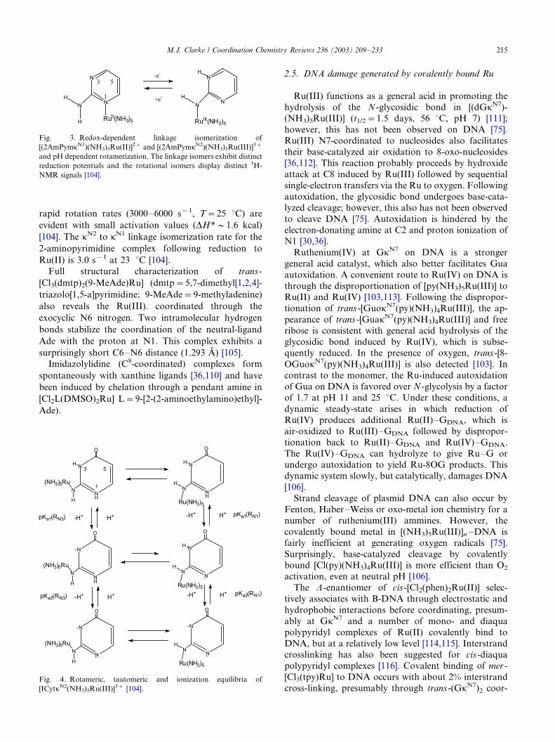
above the pKa (�/3.2) by hydrogen bonding to N3. Figs.
3 and 4 illustrate the types of redox-dependent linkage
isomerization and pH-dependent rotamerization that
can occur in [L(NH3)5Ru(III)], where L is a ligand with
a ring imine nitrogen adjacent to an exocyclic ammine,
such as 2-aminopyrimidineadenine (as illustrated in Fig.
3), Ade, Cyt, or isocytosine (as illustrated in Fig. 4). Due
to the difference in distance between the metal ion and
the ionization site, the rotamers in cytidine and adeno-
sine complexes exhibit different pKa. The interconver-
sion may be facilitated by proton dissociation and very
Fig. 2. Structures of a) [(Hypk7)(NH3)5Ru]3� showing the typical N7 coordination of many transition metal ions at the N7 of a purine [101]; b)
[(7MeGuak7)(NH3)5Ru]3�, depicting one of the linkage isomers present when N9 is also available for bonding [36]; c) cis -[Cl2(1,3,7Me3Xank8)-
(NH3)3Ru]3� illustrating an unusual carbon-bound form of caffeine bound to ruthenium; and d) [(1MeCytkN4)Cl(NH3)Ru]2� portraying binding to
an ionized exocyclic amine.
M.J. Clarke / Coordination Chemistry Reviews 236 (2003) 209�/233214
rapid rotation rates (3000�/6000 s�1, T�/25 8C) are
evident with small activation values (DH��/1.6 kcal)
[104]. The kN2 to kN1 linkage isomerization rate for the
2-aminopyrimidine complex following reduction to
Ru(II) is 3.0 s�1 at 23 8C [104].
Full structural characterization of trans -
[Cl3(dmtp)2(9-MeAde)Ru] (dmtp�/5,7-dimethyl[1,2,4]-
triazolo[1,5-a]pyrimidine; 9-MeAde�/9-methyladenine)
also reveals the Ru(III). coordinated through the
exocyclic N6 nitrogen. Two intramolecular hydrogen
bonds stabilize the coordination of the neutral-ligand
Ade with the proton at N1. This complex exhibits a
surprisingly short C6�/N6 distance (1.293 A) [105].
Imidazolylidine (C8-coordinated) complexes form
spontaneously with xanthine ligands [36,110] and have
been induced by chelation through a pendant amine in
[Cl2L(DMSO)2Ru] L�/9-[2-(2-aminoethylamino)ethyl]-
Ade).
2.5. DNA damage generated by covalently bound Ru
Ru(III) functions as a general acid in promoting the
hydrolysis of the N -glycosidic bond in [(dGkN7)-(NH3)5Ru(III)] (t1/2�/1.5 days, 56 8C, pH 7) [111];
however, this has not been observed on DNA [75].
Ru(III) N7-coordinated to nucleosides also facilitates
their base-catalyzed air oxidation to 8-oxo-nucleosides
[36,112]. This reaction probably proceeds by hydroxide
attack at C8 induced by Ru(III) followed by sequential
single-electron transfers via the Ru to oxygen. Following
autoxidation, the glycosidic bond undergoes base-cata-lyzed cleavage; however, this also has not been observed
to cleave DNA [75]. Autoxidation is hindered by the
electron-donating amine at C2 and proton ionization of
N1 [30,36].
Ruthenium(IV) at GkN7 on DNA is a stronger
general acid catalyst, which also better facilitates Gua
autoxidation. A convenient route to Ru(IV) on DNA is
through the disproportionation of [py(NH3)5Ru(III)] toRu(II) and Ru(IV) [103,113]. Following the dispropor-
tionation of trans -[GuokN7(py)(NH3)4Ru(III)], the ap-
pearance of trans -[GuakN7(py)(NH3)4Ru(III)] and free
ribose is consistent with general acid hydrolysis of the
glycosidic bond induced by Ru(IV), which is subse-
quently reduced. In the presence of oxygen, trans -[8-
OGuokN7(py)(NH3)4Ru(III)] is also detected [103]. In
contrast to the monomer, the Ru-induced autoxidationof Gua on DNA is favored over N -glycolysis by a factor
of 1.7 at pH 11 and 25 8C. Under these conditions, a
dynamic steady-state arises in which reduction of
Ru(IV) produces additional Ru(II)�/GDNA, which is
air-oxidized to Ru(III)�/GDNA followed by dispropor-
tionation back to Ru(II)�/GDNA and Ru(IV)�/GDNA.
The Ru(IV)�/GDNA can hydrolyze to give Ru�/G or
undergo autoxidation to yield Ru-8OG products. Thisdynamic system slowly, but catalytically, damages DNA
[106].
Strand cleavage of plasmid DNA can also occur by
Fenton, Haber�/Weiss or oxo-metal ion chemistry for a
number of ruthenium(III) ammines. However, the
covalently bound metal in [(NH3)5Ru(III)]n �/DNA is
fairly inefficient at generating oxygen radicals [75].
Surprisingly, base-catalyzed cleavage by covalentlybound [Cl(py)(NH3)4Ru(III)] is more efficient than O2
activation, even at neutral pH [106].
The L -enantiomer of cis -[Cl2(phen)2Ru(II)] selec-
tively associates with B-DNA through electrostatic and
hydrophobic interactions before coordinating, presum-
ably at GkN7 and a number of mono- and diaqua
polypyridyl complexes of Ru(II) covalently bind to
DNA, but at a relatively low level [114,115]. Interstrandcrosslinking has also been suggested for cis -diaqua
polypyridyl complexes [116]. Covalent binding of mer -
[Cl3(tpy)Ru] to DNA occurs with about 2% interstrand
cross-linking, presumably through trans -(GkN7)2 coor-
Fig. 3. Redox-dependent linkage isomerization of
[(2AmPymkN1)(NH3)5Ru(II)]2� and [(2AmPymkN2)(NH3)5Ru(III)]3�
and pH dependent rotamerization. The linkage isomers exhibit distinct
reduction potentials and the rotational isomers display distinct 1H-
NMR signals [104].
Fig. 4. Rotameric, tautomeric and ionization equilibria of
[ICytkN2(NH3)5Ru(III)]3� [104].
M.J. Clarke / Coordination Chemistry Reviews 236 (2003) 209�/233 215

dination, which may be responsible for its antitumor
activity [117]. A series of complexes with thiazole and
phosphine ligands has been prepared for testing and the
structure of mer -[Cl3(thz)3Ru], (thz�/1,3-thiazole) hasbeen determined [118,119].
The complexes trans -[Cl(SO2)(NH3)4Ru]� [120], mer -
[Cl3(Me2SO)3Ru], trans -[Cl4(Me2SO)2Ru]�, and mer ,-
cis -[Cl3(H2O)2(Me2SO)Ru] all produce DNA inter-
strand crosslinks [121]. Adjacent intrastrand GkN7�/
GkN7 crosslinks with cis -ruthenium ions may be possi-
ble, but are sterically more crowded by the octahedral
geometry [65,122�/124]. For example, trans -[Cl4-(Me2SO)2Ru]� reacts with d(GpG) to yield a macro-
cyclic chelate with the likely formulation, cis -
[d(GkN7pGkN7)Cl(H2O)(Me2SO)2Ru(II)], in which the
sugars are in anti configurations and the Gua are
destacked in a head-to-head arrangement [125].
The Ru�/Pt dinuclear complex, [{cis ,fac -(RuCl2(Me2-
SO)3)}m-NH2(CH2)4NH2-{cis -(Pt(NH3)Cl2)}], rapidly
loses Me2SO and chloride from the ruthenium centerand crosslinks DNA repair proteins to DNA [126,127].
The DNA lesion responsible for efficient DNA-protein
cross-linking is most probably a DNA�/DNA inter-
strand crosslink by the platinum end of the molecule
[127]. Analogous complexes of the type [(bpy)2M-
(dpb)PtCl2]Cl2 (where M�/Ru(II) or Os(II), and
dpb�/2,3-bis(2-pyridyl)benzoquinoxaline) also form
both intrastrand DNA crosslinks, due to the cis -Cl2Pt(II) moiety, and interstrand crosslinks, which are
probably made through the second metal center
[128,129]. Complexes of the type, [(bpy)2Ru-
(dpb)PtCl2]Cl2 covalently bind to DNA through the Pt
but may provide for photochemical effects through the
Ru [129].
2.6. Modulation of DNA binding by glutathione
Glutathione (g-glutamate�/cysteine�/glycine�/GSH)
[130] is present in cells at concentrations of 0.1�/10
mM, but is readily oxidized to the disulfide (GSSG,
E8?�/�/0.246 V vs. NHE) [131]. GSH helps protect cells
from: reactive oxygen intermediates, UV radiation, and
heavy metal toxicity [132]. Decreased GSH levels have
been implicated in cytochrome-c escape from the
mitochondria, which is a triggering event in apoptosis[133]. GSH reduces some metal ions, such as CrO4
2� and
Pt(IV) anticancer drugs [134,135], to species that co-
ordinate or otherwise react with DNA [136�/140]. On
the other hand, GSH binding to Pt(II) inhibits DNA
binding and appears to contribute to cisplatin resistance
in tumor cells [141�/143]. GSH (0.1 M, pH 6, apparently
in air) rapidly reduces the anticancer complex trans -
[Cl4(Im)2Ru]� (E8�/�/0.24 V), which then dissociatesits imidazole ligands within 1 h GSH coordination of
trans -[Cl4(Im)2Ru]� followed by electron transfer has
been assumed [108].
The aerobic reaction of GSH with [Cl(NH3)5-
Ru(III)]2� is first order in [GSH] and yields only
[OH(NH3)5Ru(III)]2� and GSSG. Since GSH only
slowly reduces [Cl(NH3)5Ru]2� under physiological
conditions (t1/2�/�/10 min) and the Ru(II) product is
readily oxidized by air, this mode of activating Ru to
bind to biopolymers by reduction may not be important
in tissues under normal oxygen tensions, but may be in
the hypoxic environment of tumors [54,59]. Since
oxygen also effectively prevents GSH coordination,
this could circumvent some thiol-based resistance to
rutheniumammine anticancer agents.
The anaerobic reaction of GSH with [Cl(NH3)5Ru]2�
proceeds through reduction of Ru(III) by GSH to give
[H2O(NH3)5Ru(II)]2�, followed by coordination to
produce [GSH(NH3)5Ru(II)]2� and then oxidation by
[OH(NH3)5Ru(III)]2� or GSSG to yield [GS(NH3)5-
Ru(III)]� (Fig. 5) [38]. The reduction potential of
[(GS)(NH3)5Ru(III)] is pH dependent with: E�/E8�/0.59 log{Ka/([H�]�/Ka)}, where E8�/�/0.44 V and
pKa�/7.1.
Depending on its relative concentration, GSH both
facilitates and inhibits ruthenium binding to DNA. At
[GSH]/[Ru(III)]5/1, the coordination of [Cl(NH3)5-
Ru(III)]2� to DNA is facilitated by GSH reduction to
the more substitution-labile [H2O(NH3)5Ru(II)]2�.
However, at [GSH]/[Ru(III)]]/1, DNA binding is
inhibited by GSH, which coordinates the Ru(II) and
facilitates oxidation back to Ru(III) because of the low
E8 of [GS(NH3)5Ru(III)]�. Consistent with this is the
Fig. 5. MM2 energy-minimized structure for the peptide portion of
[(GS)(NH3)Ru(III)]� illustrating the probable wrapping of the
carboxylates around the metal cation [38,353,354].
M.J. Clarke / Coordination Chemistry Reviews 236 (2003) 209�/233216

increased toxicity of [Cl(NH3)5Ru]2� to Jurkat T-cells,
when GSH levels are suppressed [38].
Inhibition of DNA binding by GSH is most evident at
GkN7 and GSH removes most of the metal ion fromGkN7 sites on DNA. It is less effective in preventing
binding or removing the metal from AkN6 and CkN4
sites owing to the lower Ru(III,II) reduction potential
[100], when the metal ion is attached to the exocyclic
ammine of these ligands [102]. The ability of Ade to
provide strong p-binding sites for both Ru(II) (N1) and
Ru(III) (ionized N6) may account for its maintaining
ruthenium binding even at high [GSH]. Such altering ofDNA binding at physiological concentrations of GSH
may have a significant effect on the mechanism of
ruthenium antitumor compounds by favoring A and C
binding over G [38], but speciation of nuclear DNA with
respect to Ru-binding has not been determined.
Since GSH partially removes ammineruthenium(III)
ions from Tf, NH3 from [(NH3)6Ru]3� and pyridine (py)
from [py(NH3)5Ru]3� to form [GS(NH3)5Ru(III)]� [38],it may be involved in the metabolism of many types of
ruthenium pharmaceuticals.
2.7. Cytotoxicities of Ru complexes
There appears to be a correlation between cytotoxi-
city and DNA binding for the representative ruthenium
am(m)ine anticancer compounds, cis -[Cl2(NH3)4-
Ru(III)]Cl2 and (HIm)[trans -[(Im)2Cl4Ru(III)] in cellcultures [59]. Also consistent with DNA binding in
vivo, a number of ammine, amine and heterocyclic
complexes of ruthenium exhibit: inhibition of DNA
replication [43], mutagenic activity and induction of the
SOS repair mechanism [144], binding to nuclear DNA
[59,145], and reduction of RNA synthesis [146]. EDTA-
type complexes of Ru(III) and even Ru(IV) have shown
anticancer activity, apparently through DNA binding[147,148]. The compound mer -[Ru(terpy)Cl3] (tpy�/
2,2?:6ƒ,2ƒ-terpyridine) exhibits antitumor activity mid-
way between those of cisplatin and carboplatin in the
L1210 cell line, which possibly involves interstrand G�/
G crosslinks [117].
More recently, Ru(II) complexes have shown good
activity. In particular, the two isomers of cis -[Cl2(az-
py)2Ru] (azpy�/2-(phenylazo)pyridine) are remarkably
different in their cytotoxicities with the a-atropisomer
(with C2 symmetry, see Fig. 6) being far more active
than the ß-isomer or the (trans -head-to-head)) g-isomer
[67,68]. As with cis -[Cl2(bpy)2Ru], steric constraintshinder coordination and only monoguanine adducts
have been characterized to date [67]. However in
contrast to the inactive cis -[Cl2(bpy)2Ru] [149], the
more flexible azapy analogue allows for more orienta-
tions for a coordinated Gua, which may allow for
binding of more than one DNA base. Indeed, the
inactivity of b-[Cl2(azpy)2Ru] may also be due to greater
steric constraints relative to the a-isomer. It is possiblethat the details of rotation between the phenyl rings on
the azpy ligands and the rotational flexibility of the Gua
or other DNA base ligands is important to under-
standing the differences in toxicity among the otherwise
structurally similar Ru(II) complexes with polypyridine
ligands [67,68].
While some monoacido complexes, such as
[CH3CH2CO2(NH3)5Ru](ClO4)2 are active, multichlorocompounds such as cis -[Cl2(NH3)4Ru]Cl, fac -
[Cl3(NH3)3Ru] [44,150�/152] and (HIm)trans -[Cl4(Im)2-
Ru] [153,154] exhibit the best activity against primary
tumors.
Solubility can be enhanced by increasing the number
of chlorides, and trans -complexes of the type
(LH)[Cl4L2Ru] (where L�/imidazole or indazole),
which show activity against a number of cancer celllines [155�/157] are particularly effective against color-
ectal tumors [107,109,153,158�/161]. These and other
multiacido ruthenium(III) complexes, particularly di-
through tetrachloro complexes, appear to be trans-
ported in the blood by Tf and albumin (HSA), with
the major portion (80%) binding to the latter
[74,83,84,87,88]. Albumin can bind up to five (hydro-
lyzed) units of [Cl4L2Ru]- [94], which disrupts theprotein a-helical structure. Quenching of the HSA Trp
214 fluorescence is consistent with Ru binding to the
nearby His 242 so as to alter the local structure and
expose Trp 214 to water [162]. Similarly, a substantial
reduction of heme and bilirubin binding is attributed to
Ru�/histidine coordination at or near the HSA�/heme
binding site. Solubility can be increased by utilizing
dialkylsulfoxide (R2SO) analogs, as in complexes suchas trans -[Cl2(Me2SO)4Ru], [Cl3(Me2SO)2BRu] (B�/imi-
dazole or indazole) and Natrans -[Cl4(R2SO)2Ru], where
R�/methyl and tetramethylene [60,121,163].
Mixed-valent, m-carboxylato complexes of the type, m-
[(RCO2)4ClRu2] (R�/CH3, CH3CH2) [160], are active
against P388 lymphocytic leukemia [160], possibly by
binding to DNA along the lines of the structurally similar
rhodium complexes. Complexes with m-N ,N ?-diphenyl-formamidinate and m-(fluoroanilino)pyridinates have
also been prepared [164�/166]. The compound m-
[(F3CCO2)4(F3CCO2)Ru2] forms cis -[m-(F3CCO2)4m-
(9EtGua)Ru(II)2(CH3OH)2]2�, where 9EtGua�/9-Fig. 6. The a-isomer of cis -[Cl2(azpy)2Ru] (azpy�/2-(phenylazo)pyr-
idine) [67,68].
M.J. Clarke / Coordination Chemistry Reviews 236 (2003) 209�/233 217
ethylguanine, in which the Gua bridge between the two
Ru(II) atoms in a N7�/O6 head-to-tail fashion [167].
Preparations of ‘ruthenium red’ (see below) bind to
polyanions such as plant pectins and the protectivemucopolysaccharide coat on some tumor cells [168].
Consequently, Ru-red concentrates in tumors [169] and
inhibits tumor growth, perhaps by inhibiting Ca2�
transport [170,171]. The complex m-O-[(H2O)2-
(bpy)2Ru(III)]24� coordinates to DNA at relatively
low levels ([Ru]DNA/[P]DNA�/0.02) with low stereose-
lectivity, which may favor the LL isomer. Coordination
at this level also stabilizes the thermal melting of DNAby about 8 8C and may involve interstrand crosslinks
[116].
Some nitrosylruthenium(II) species may be active by
releasing toxic nitric oxide upon reduction in vivo [172�/
174]. The binuclear complex {[Ru(NO)Cl3]2(tpada)}
(tpada�/N ,N ,N ?,N ?-tetrakis(2-pyridylmethyl)adip-
amide) contains two DNA-coordinating, photo-labile
Ru(II) centers, and a groove-spanning tether moiety[175].
2.8. Complexes with EDTA-type ligands
Ruthenium complexes with polyaminopolycarboxylic
chelating ligands constitute a relatively new group of
anticancer compounds [176,177]. The complex
[(H2O)(edta)Ru(II)]2� coordinates to both the N7
(30%) and N3 (70%) sites on 5?-GMP, but the Ru(III)form binds only at N7. [(5?-GMP)(edta)Ru(III)]n� has a
reduction potential of 0.01 V (22 8C), but ionizes a
proton from N1 at a pKa of 7.2, which should cause its
reduction potential to decreases at higher pH [178].
In [Cl2(cdta)Ru(IV)], where cdta�/1,2-cyclo -hexane-
diaminotetraacetate, the chlorides are cis to one another
and the carboxylates appear to be labile. The Ru(IV,III)
reduction potential occurs at 0.78 V, while that for theRu(III,II) couple is at �/0.01 V [177], so that these
complexes may belong to the class of multi-acido
ruthenium(III) complexes, whose activity involves trans-
port by Tf [34]. The compound, cis -[Cl2(pdta)Ru(III)],
where pdta�/1,2-propylendiaminetetraacetate, also
shows good antitumor activity, possibly by cross-linking
Guas on DNA; and a model complex, [(Gua)2(pdta)Ru-
(III)], has been isolated. The solid state structure of cis -[Cl2(pdta)Ru(III)] reveals coordination by two nitrogens
and two monodentate carboxylates [148]. In solution,
the chlorides dissociate to produce a number of reactive
Ru(III) species; however, the metal ion maintains its
oxidation state as well as the pdta ligand [179]. The
complex rapidly binds to albumin, apotransferrin or
diferric Tf to produce relatively stable adducts in which
(pdta)Ru(III) is probably bound to histidines on theprotein surface [34]. Cis -[Cl2(pdta)Ru(III)] damages
nuclear DNA, inhibits DNA recognition and DNA lysis
by restriction enzymes [180], alters the conformation of
pHV14 DNA [180], stimulates NADPH oxidase and a
respiratory burst in phagocytic neutrophils, and elicits
phosphorylation of tyrosine residues [147,148].
The complex [(hedta)Ru(II)]� binds to the usual N3position of pyrimidines, but can also bind in a h2-
fashion to C5�/C6 [181,182]. A distribution between h1
binding at both N1 and N3 of pyrimidine, which can be
either stereochemically rigid or fluxional, as well as h2-
binding is observed [183]. The complex [Ru2(II)-
(ttha)(DMU)2]2� (ttha�/triethylenetetraminehexaace-
tate; DMU�/1,3-dimethyluracil) suggests an interstrand
cross link between uracils [181]. As h2-coordinationacross C5�/C6 raises the Ru(III/II) reduction potential
to �/0.5 V, the h2-Ru(II)�/DNA adducts may be stable
in vivo. The complex {[Ru(II)(hedta)]2(tpada)}2�
(tpda�/N ,N ,N ?,N ?-tetrakis(2-pyridyl)adipamide) is
being investigated as a DNA crosslinking agent as an
extended bridge between DNA and proteins [184].
2.9. Complexes with R2SO
The compound cis -[Cl2(Me2SO)4Ru] exhibits only
marginal antitumor activity, but was the origin of efforts
in the investigation of other diaklysufoxide ruthenium
complexes as anticancer agents. Covalent binding of this
compound to DNA does not significantly affect the
conformation of B-DNA but does increase its thermal
stability. Comparisons with the inactive trans complex
may also be instructive. Trans -[Cl2(Me2SO)4Ru] bindsmuch more rapidly to DNA with greater changes in its
CD spectra, which are attributed to disruption of the
DNA structure due to crosslinking [185]. While indivi-
dual guanosines bind reversibly to trans -[Cl2(Me2-
SO)4Ru] [186] and 5?-dGMP forms an N7-PO4 chelate
rather than a bis-5?-dGMP complex [65], NMR evidence
suggests the formulation of cis -[d(Gk7pGk7)Cl(H2O)-
(Me2SO)2Ru(II)] [125], in which the sugars are in anti
configurations and the Gua are destacked in a head-to-
head arrangement similar to that of cisplatin [125]. Both
the cis and trans isomers induce the B to Z transition in
poly(dGdC), with the trans complex being much more
effective. DNA extracted from cells, which were sepa-
rately treated with each isomer, showed a five-fold
higher content of Ru in the trans case [64,187,188].
Since metastatic cancer is particularly difficult totreat, the antimetastatic activity of the ruthenium
dimethylsulfoxide complexes, particularly Natrans -
[Cl4(Me2SO)(Im)Ru] (NAMI) represents an important
development. Such complexes could be particularly
useful in minimizing the growth of undetected micro-
metastases following surgery or radiotherapy [189,190].
While structurally similar to (ImH)trans -[Cl4(Im)2Ru]
(ICR, E8�/�/0.24 V), NAMI has a significantly higherRu(III/II) reduction potential (0.235 V) [107,153,191]
owing to the p-acceptor effect of the S-bound DMSO,
which also exerts a kinetic trans -effect. Relatively high
M.J. Clarke / Coordination Chemistry Reviews 236 (2003) 209�/233218
concentrations (�/100 mM) are needed to produce a
cytotoxic effect, which depends upon the lipophilicity of
the complex and the presence of serum and plasma
proteins [192]. Of those tested, the most lipophiliccomplex, Natrans -[RuCl4(TMSO)Iq] (TMSO�/tetra-
methylensulfoxide; Iq�/isoquinoline), causes DNA
fragmentation similar to cisplatin.[193]. While preferen-
tially binding to G�/C rich regions, NAMI is much less
effective than cisplain at altering DNA conformation,
affecting DNA electrophoretic mobility, and inhibiting
DNA recognition and cleavage by restriction enzymes
[180,194]. Comparison with cisplatin and other similarcytotoxic ruthenium complexes indicate that the anti-
metasatic action of NAMI does not result from direct
cytotoxic effects [193]. NAMI can be administered
orally [163,192] and is active against a broad range of
tumors including Lewis lung carcinoma, B16 melanoma,
and MCa mammary carcinoma [195].
NAMI appears to increase resistance to the formation
of metastases [192,196,197], but not through an en-hanced antigenicity or an immunological response
[189,198]. While NAMI binds to DNA and forms
DNA�/protein crosslinks, it does not significantly form
DNA interstrand crosslinks (less than 1%) [189,199].
While the antimetastatic action of NAMI does not
appear to involve DNA binding, 80�/90% of the com-
plex in solution binds to CT-DNA within 24 h at 37 8C.
Such binding stabilizes DNA in low salt media (0.01 MNaClO4), but alterations in the CD spectrum suggest
unwinding of the DNA. Binding also inhibits the B to Z
transition in poly(Dg�/dC) and significantly inhibits
DNA and RNA polymerases, with termination occur-
ring preferentially at Gua residues [189].
At levels that cause a dramatic reduction in lung
metastases, NAMI greatly alters the ratio between the
mRNAs of MMP-2 (a metalloproteinase capable ofdegrading the extracellular matrix) and TIMP-2, the
specific tissue inhibitor of this enzyme [200,201]. This
corresponds with a pronounced increase of extracellular
matrix components in the tumor parenchyma and
around tumor blood vessels, which probably hinders
metastasis formation and blood flow to the tumor [202].
Overall, NAMI appears to down regulate type-IV
collagenolytic activity and the metastatic potential ofMCa mammary carcinoma [198]. Combining NAMI
with 5-fluoruracil achieved better results in mice against
the solid metastasizing MCa mammary carcinoma and
lymphocytic leukemia P388 [203].
NAMI can undergo a number of hydrolysis and
electron transfer reactions under physiological condi-
tions. Loss of DMSO and imidazole following chloride
dissociation results in polyoxo complexes of ruthenium.At 25 8C, hydrolysis of the first chloride occurs within
an hour, while the second takes more than twice as long.
At physiological pH, trans -[Cl4(Im)(Me2SO)Ru]� is
more labile to substitution than trans -[Cl4(Im)2Ru]�
(t1/2�/19.7 h at 25 8C) [107]. Chloride loss for the
former is catalyzed by reduction to Ru(II), which is
expected to occur under physiological conditions and is
enhanced in vitro by traces of biological reductants,such as ascorbic acid or cysteine [60,61]. Consequently, a
redox-catalytic (activation by reduction) mechanism is
suspected.
NAMI-A, (ImH)trans -[Cl4(Me2SO)(Im)Ru], is a
more stable and reproducible solid than NAMI, while
exhibiting similar pharmacological properties [204,205].
It selectively interferes with the growth of Lewis lung,
MCa mammary carcinoma and TS/A adenocarcinomametastases already settled in rodent lungs in a manner
that is independent of the stage of the metastatic growth
[206] and not simply related to a larger concentration in
the lungs than in other tissues [207]. When administered
intraperitoneally, NAMI-A is rapidly distributed to the
body and rapidly cleared from the blood by the kidneys
[207], with only 10% of the original dose remaining in
the blood after 5 min [189]. It significantly increases thepercentage of CD8� cells at three dose levels, while
CD4� cells increased only at the lowest dose and remain
unchanged at medium and high doses. NAMI-A sig-
nificantly increases the thickness of the connective tissue
of the tumor capsule and around tumor blood vessels,
and impairs MMP-2, possibly at the level of its gene
and/or its inhibitor TIMP-2 [189]. NAMI-A appears to
be less toxic than cisplatin, does not modify cell growth,and causes a transient cell cycle arrest of tumor cells in
the premitotic G2/M phase; whereas, cisplatin causes a
dose-dependent disruption of cell cycle phases and
reduces cell proliferation [206].
2.10. Photodynamic therapy
In photodynamic therapy (PDT) light is used to kill
undesired cells in the body. The activity of PDT agentsdepends on their ability to associate with biopolymers or
aggregates such as cell membranes or DNA. Cell
membranes can be disrupted by the conversion of light
energy to heat by nonradiative conversion, whereby
photothermal sensitizer molecules produce localized
hyperthermal damage. Damage to DNA can also occur
by photoinduced electron transfer between the excited
state of the PDT molecule and DNA, thereby damagingthe cell’s ability to function. Alternatively, light absorp-
tion by a photosensitizing molecule can lead to energy
transfer to activate another molecule, such as the
conversion of O2 to the excited singlet state [32,208�/
210]. Photoinduced ligand substitution is also possible
whereby a metal complex would either release a
biologically active molecule or bind to nucleic acids or
protein active sites [210,211].To be useful in PDT, the ground state complex should
be stable and generally nontoxic. The complex should
also have well described photophysical properties with
M.J. Clarke / Coordination Chemistry Reviews 236 (2003) 209�/233 219

an efficient photochemical conversion to the biologically
active form. Since mammalian tissue is highly absorbant
in the higher energy part of the visible spectrum, PDT
complexes must absorb strongly at the relatively longwavelengths (640�/850 nm) that penetrate tissues fairly
well [210].
Metal complexes of phthalocyanines, naphthalocya-
nines, porphyrins and texaphyrns absorb at long wave-
lengths and often show tumor localization. Since
complexes of these molecules with paramagnetic ions
often have short triplet lifetimes and decay by non-
radiative pathways releasing their energy as heat, theycan act as photothermal sensitizers [210].
Diamagnetic Ru(II) phthalocyanines are thought to
react by direct electron transfer to the photoexcited state
(type I mechanism) [212,213]. A series of complexes of
the type trans -[Ru(II)(nc)X2], where nc�/2,3-naphtha-
locyanine, prepared from trans -[Ru(II)(nc)(PhCN)2]
(PhCN�/benzonitrile) that have fairly strongly absorb-
ing Q-bands (710�/760 nm, o�/105 M�1 cm�1) exhibitactivity against HeLa cells at micromolar concentrations
[214,215]. The greatest phototoxicity and relatively low
chemotoxicity was observed when aromatic nitrogen
heterocycles (nicotinic acid, pyrazinic acid, 3-pyridine-
sulfonic acid) were coordinated in the axial positions to
enhance solubility [215].
In the case of anticancer photodynamic therapy,
DNA is usually the target. Metal complexes can interactwith DNA by, (1) electrostatic binding between a
cationic metal complex and the DNA polyanion
[216,217]; (2) noncovalent hydrophobic surface binding;
(3) intercalating between DNA bases, and 4) covalent
binding [218]. Any of these types of binding may also be
incorporated into shape-specific binding to particular
nucleic acid substructures [219]. Due to their intense
metal-to-ligand charge transfer transitions and long-lived excited states, ruthenium(II) complexes with N -
heterocyclic ligands are particularly attractive as poten-
tial PDT agents [32,220�/222], not only for DNA, but
also for specific DNA or RNA substructures
[219,223,224]. Polypyridyl and related ligand complexes
of Ru(II) are usually activated by a metal to ligand
charge transfer excitation, which leads to a triplet
MLCT state (3MLCT) [211]. The 3MLCT can convertby the following methods with the corresponding rate
constants, (1) radiative emission of light (kr) around 600
nm; (2) radiationless deactivation as heat (knr); and (3)
thermal conversion to a higher energy, metal-centered
triplet (3MC). The lifetime (t) of the 3MLCT is thus
controlled by the rates of these three processes [211].
1=t � kr�knr�kMCe�DE=RT
The 3MLCT state, which is simultaneously a betteroxidant and reductant than the ground state, often leads
to an electron transfer reaction. In contrast, the 3MC
state often involves populating a higher energy d-orbital
with antibonding character thereby weakening one or
more of the Ru�/N bonds resulting in ligand substitution
[211,221]. The ‘flash-quench’ approach involves oxidiz-
ing the (usually 3MLCT) excited state by an added
oxidant, such as [(NH3)6Ru]3�, to prepare a more
powerful ground state oxidant such as [(bpy)3Ru]3�.
Gua, which is the most easily oxidized DNA base, is
most commonly affected [225,226]. However, oxidation
of the other bases as well as the sugar is also possible
[227]. Different products are observed depending on
whether deoxyguanosine (dG) is oxidized by electron
transfer to the photoexcited complex (Type I) or by
oxidation by 1O2 (Type II) [228,229]. Single-electron
oxidation tends to give 2-aminoimidazolone and oxazo-
lone products, which are piperidine labile and lead to
DNA cleavage [230]. Singlet oxygen attack initially
forms 8-oxoguanine and 4-hydroxy-8-oxoguanine pro-
ducts but further reaction with 1O2 yields the oxalone
and additional 4-hydroxy-8-oxoguanine (see Fig. 7).
Photoinduced single-electron oxidation of 8OG can
also yield the imidazolone [229]. Stacked G’s are
predicted to be thermodynamically more easily oxidized
than single G’s and single-electron oxidation takes place
preferentially at the 5?-G of 5?GG-3? sequences
[231,232].
Table 1 summarizes the affinities of a number of
ruthenium complexes for noncovalent association with
DNA and indicates which appear to bind through
intercalation. Single strand cleavage resulting from the
photoexcitation of [(bpy)3Ru]2� or [(phen)3Ru]2�,
which bind to the surface of DNA, in the presence of
air results primarily from the formation of 1O2 [233].
Fig. 7. Type I oxidation products of deoxyguanosine following
photoinduced single-electron oxidation, and Type II oxidation pro-
ducts initially following oxidation by singlet oxygen.
M.J. Clarke / Coordination Chemistry Reviews 236 (2003) 209�/233220

For [(bpy)3Ru]2� the quantum yield increases from
1.2�/10�6 under anaerobic conditions to 6.6�/10�6 in
air [32]. On the other hand, the excited states of a
number of complexes of the type [L(bpy)2Ru]2�, where
L�/hat, tap and bpz (see Fig. 8) and similar ligands
react primarily by electron-transfer rather than by
generating singlet oxygen [234]. Covalently bound
DNA adducts involving the exocyclic amine of Gua
also occurs for some complexes whose excited states
oxidize guanine. This probably occurs by radical reac-
tions involving the reduced (radical ligand) ruthenium
complex and oxidized guanine radicals [32].
When [dip(tap)2Ru]2� was tethered to oligodeoxy-
ribonucleotides at the 5? position of a thymidine,
photoinduced electron transfer from the guanines on
the complimentary strand occurred. Photooxidation of
Gua is probably involved in the formation of photo-
product(s) that irreversibly crosslink the two strands
[235]. Flash-quenched generated [dppz(phen)2Ru]3�
bound to DNA produces Gua radicals that are capable
of crosslinking to tyrosine [236] and tryptophan [237]
radicals that may also be generated by long range
electron transfer in DNA, which implies that DNA�/
protein crosslinked proteins may be introduced by
PDT [238].
The dinuclear complex, m-hat[(phen)2Ru]24�, exhibits
the same photoreactivity as the mononuclear hat com-
plex, but forms strong ion pairs with nucleotides and
interacts more strongly with single- than double
stranded DNA. Analogous complexes with dppz are
significantly better intercalators, but have shorter ex-
cited life times than [(bpy)3Ru]2� and so generate less1O2 [219]. A complex in which two [(bpy)3Ru]2� ions
are tethered exhibited an affinity for DNA ca. 100-fold
greater than that of the monomer and also yielded more
efficient DNA strand cleavage [239].
While [(phen)2(prehat)Ru]2�, which does not emit in
water, luminesces upon intercalation, the luminescence
of [(tap)2(prehat)Ru]2� is quenched by binding to DNA
or in the presence of GMP. This strongly suggests
photoinduced electron transfer from the Gua residues of
Table 1
Binding constants for trisbidentate complexes of Ru(II)
Complex K Site size Enantio-selectivity Likely intercalator Reference
[Ru(bpy)3]2� 7�/102 6�/12 no no [344]
[Ru(bpy)(tpy)(H2O]2� 6.6�/102 no [345]
[Ru(bpy)2phen]2� 7�/102 10�/14 D no [344]
[Ru(bpy)2dip]2� 1.7�/103 12�/18 D yes [344]
[Ru(bpy)2ppz]2� 5.5�/103 3�/4 yes partial [346,347]
[Ru(bpy)2qpy]2� 1.3�/104 2�/3 no yes [346]
[Ru(bpy)2Me2qpy]4� 2.8�/104 3 yes yes [346]
[Ru(phen)3]2� 3�/104 3�/4 D no(?) [348]
[Ru(NH3)4dppz]2� 1.24�/105 yes [349]
[Ru(bpy)2phi]2� 1.6�/105 4 yes yes [344]
[Ru(tpy)dppz(H2O)]2� 7.3�/105 yes [345]
{(dpp)[(NH3)4Ru(II)]2}4� 8.3�/105 6 yes [350]
[Ru(phen)2dppz]2� �/108 2 yes yes [351,352]
Enantioselectivity is indicated as to whether binding by the D or L is preferred. If the reference indicates enantioselective binding, but does not
indicate which isomer a ‘yes’ is indicated.
Fig. 8. Structures of N-heteroaromatic ligands.
M.J. Clarke / Coordination Chemistry Reviews 236 (2003) 209�/233 221

GMP or DNA to the excited tap complex indicating it
as a good DNA photoreagent [240]. Bis intercalation of
dimeric complexes that must thread through the DNA
helix or in which the linker has to sling itself around
opening base pairs in order to thread has been suggested
as a way of increasing the affinity of PDT complexes for
DNA and extending their residence lifetime on this
biopolymer [241,242]. While threading does not always
occur in tethered dimers [242], the complex m-
(cpdppz)2[(phen)2Ru]24�, in which two chiral mono-
intercalator complexes of [dppz(phen)2Ru]2� are teth-
ered to each other via the dppz moieties and an aliphatic
diamide linker, does thread through DNA [241]. Bind-
ing titrations indicate that the size of the binding site is
about four base pairs, consistent with a nearest-neigh-
bor-exclusion binding model, in which the two inter-
calated dppz subunits of the complex are separated by
two base pairs. The linear dichroism spectrum of the
meso (L ,D) stereoisomer bound to DNA was an average
of those of the two opposite enantiomers (L ,L ) and
(D,D) suggesting that the binding geometry of each
subunit depends solely upon its absolute configuration
and not upon any diastereomeric intramolecular inter-
actions between the Ru(phen)2 moieties.
A supramolecular tetraruthenated tetrapyridylpor-
phyrin, m-[meso -5,10,15,20-(py)4porphyrin][Cl(bpy)2R-
u(II)]4 (TRP), introduces single-strand breaks into
DNA in the presence of light. This occurs by both
Type I and Type II mechanisms (see Fig. 7) but with 8-
oxoguanosine predominating as the product through the
formation of singlet oxygen [243]. In model studies using
dG alone, the Type II/Type I product ratio was 2.3[209].
Ogawa has attempted to utilize a synthetic ‘leucine
zipper’ (bZIP) DNA-binding peptide to carry a ruthe-
nium photosensitizer to DNA [244]. A photoactive
ruthenium site, [Ru(bpy)2(phen-IA)], where IA�/iodoa-
cetate, was attached to a surface-exposed cysteine
residue of the bZIP peptide. While the free metal
complex causes Gua-specific damage and the synthetic
Ru-bZIP peptide recognizes and binds the consensus
AP-1 DNA element, it does not damage DNA. It may
be that DNA photodamage does not occur because the
Ru binds at a point on the peptide that places the metal
within the coiled-coil region of the protein [244].
The complex [(bpz)3Ru(II)]2� (bpz�/trisbipyrazyl)
photolytically damages both single- and double-
stranded oligonucleotides by oxidative mechanisms
involving both excited state electron transfer and
production of singlet oxygen, both of which are
enhanced by Cu/Zn superoxide dismutase [245].
A unique ‘photodynamic’ approach is to use the
Mossbauer absorption of g-rays by ruthenium com-
plexes bound to DNA to induce Auger electrons to
damage the nucleic acid [246].
3. Ruthenium red and Ru360: inhibitors of Ca2�
utilization
Since preparations of the mixed-valent complex
ruthenium red (Ru-red, see Fig. 9A), [(NH3)5Ru-
(III)ORu(IV)(NH3)4Ru(III)(NH3)5]6� [247], have been
used as a cytological stain for over a century, its
biological properties are well known [149,248,249]. The
affinity of Ru-red for mucopolysaccharides provides for
the radioscintigraphic visualization of tumors, which
often generate a protective coating consisting largely of
hyaluronic acid [168]. Ruthenium red binds to a number
of calcium-binding proteins and interfere’s with meta-
bolism involving Ca2� [250�/255] and is also an
antagonist for capsaicin [256,257]. Ruthenium red
injected into rat brains concentrates in neuronal somas
located near the injected areas. In the case of the CA1
region of the hippocampus, there is remarkable damage
(vacuolization) of the pyramidal neurons followed by
cell loss and disruption of the CA1 cell layer. The
damage caused by selective penetration of ruthenium
red into neuronal bodies results in hyperactivity of
glutamatergic neurotransmission that leads to pro-
nounced changes in motor function [258].
Nanomolar levels of Ru-red strongly inhibit respira-
tion-driven Ca2� uptake in mitochondria [259], which
has led to increasing interest in Ru-red as a drug
[170,260�/262]. Ru-red’s ability to inhibit calcium ion
uptake by the mitochondria has also facilitated the
Fig. 9. Structure of (A) ruthenium red [247,355] and (B) Ru360 [266].
M.J. Clarke / Coordination Chemistry Reviews 236 (2003) 209�/233222
characterization of Ca2� utilization in this organelle
[263,264].
Commercial preparations of ruthenium red often
contain substantial impurities. The dimeric impurity,[X(NH3)4Ru(III)ORu(IV)(NH3)4X]3� (Ru360, X�/
Cl� or OH� see Fig. 9B) [265], is often responsible
for most of the inhibition of Ca2� uptake in mitochon-
dria [259,266]. Ru360 specifically blocks uptake of Ca2�
into the mitochondria of cardiac myocytes; but it does
not exhibit antitumor activity in cell culture screens
[266]. Ru360’s tripositive charge, hydrogen bonding
capability, and ability to readily deform probably allowit to bind to a variety of anionic carboxylate sites on
proteins including Ca2� receptors. Decomposition of
the hydroxo capped complex at physiological pH also
yields a substance that irreversibly binds to polyanionic
materials.
The bridging Ru�/O bond in Ru360 is quite short
(1.8240(6) A), while the Ru�/Oformate bond length
(2.033(3) A) is typical for a Ru�/O single bond. Thecrystallographic equivalence of the two Ru atoms,
together with magnetic (meff�/1.15 ß) and electrochemi-
cal evidence (E18�/1.17 V, E28�/0.05 V; DE8�/1.12 V)
indicate strong electronic coupling between the mixed-
valent Ru’s. The apical formates are easily replaced by
chlorides in HCl. Aquation rates of both the formato
and chloro complexes are pH dependent [266].
Since Ru360 is at least an order of magnitude morepotent than ruthenium red and specifically inhibits the
uptake of Ca2� by the mitochondria (Ru360 IC50�/
0.184 nM; ruthenium red IC50�/6.85 nM), it should
be preferred for probing the effects of Ca2� in this
organelle [252,253]. 103Ru360 binds to isolated mito-
chondria with a high affinity (Kd�/0.34 nM) [267]. The
inhibition of the calcium ion uniporter by Ru360 is
noncompetitive with Ca2� (Ki�/9.89 nM) [263,265].The binding of Ru360 to the uniporter protein has been
reported as �/5 pmol mg�1 protein in one study [263]
and 0.08 pmol mg�1 in another [267]. Ruthenium red
and Ru360 are mutually exclusive inhibitors of the
mitochondrial uniporter and appear to involve the same,
low-affinity Ca2� binding sites [263,268]. Since La3�
bound to the high affinity Ca2� site was not displaced
by Ru360, it has been speculated that the binding site inthe uniporter for both Ru-red and Ru360 involves a
negatively charged phosolipid membrane region, which
may be closely associated with the uniporter inhibitor-
binding site [263]. Ru360 has also been used as an
affinity ligand in an effort to isolate the calcium ion
uniporter protein [268].
The ability of Ru360 to bind to similar, if not the
same sites, as ruthenium red is probably due the twoions being somewhat similar in their cationic charge
density, ability to hydrogen bond through their ammine
ligands, and capability to readily deform along the Ru�/
O�/Ru axis. These properties should allow both type of
ions to bind to a variety of anionic carboxylate sites on
proteins including low-affinity Ca2� receptors as well as
anionic phospholipids in the membrane. The smaller size
of Ru360 and its ability to covalently bind to amino acidcarboxylate groups through its apical sites may account
for its greater biochemical activity relative to ruthenium
red. Ru360 is probably also responsible for at least a
portion of ruthenium red’s ability to stain for electron
microscopy.
4. Nitric oxide complexes
Nitric oxide (NO) is involved in a wide range of
physiological processes [172,269�/271]. When produced
by macrophages to kill invasive tumor cells, bacteria and
fungi [272], activation is caused by interferon-g, endo-
toxins (bacterial cell wall lipopolysaccharides that elicit
inflammatory responses), or T cells [272,273]. The toxic
effect appears to derive from inhibition of enzyme
activity through NO binding to nonheme protein iron-centers and release of intracellular iron from targeted
cells [274�/276]. In toxic-shock syndrome, excessive
macrophage NO-synthase (NOS) activity leads to arter-
ial expansion and cardiovascular collapse [277].
Nitric oxide synthase (NOS) catalyzes a net 5e�
oxidation of a terminal nitrogen of arginine to yield
citrulline and nitric oxide. There are three major classes
of NOS, endothelial, neuronal and immunological [269].To some degree all have inducible and constitutive
forms and all are NADPH-dependent cystolic oxyge-
nases that incorporate oxygen from O2 into both
products [271,278,279]. NOS contains both a P450-
type domain and a P450-reductase domain. As a P450
type enzyme, it is activated by a 2e-reduction in which
an oxygen from O2 is added to arginine to produce N -
hydroxy-arginine as an intermediate. The second stepinvolves a tetrahydrobiopterin cofactor [280], which
appears to facilitate the odd-electron oxidation to yield
NO+ [281,282].
Sodium nitroprusside, Na2[NO(CN)5Fe] �/2H2O,
which is clinically used to rapidly lower blood pressure,
is activated by reduction in vivo to [Fe(CN)5NO]3� in
which an antibonding orbital becomes populated to
facilitate the loss of NO [283]. Consequently, thebiological reduction of metallonitrosyls is already a
proven method of delivering NO to a desired tissue in
a clinical setting. Since varying levels of NO at specific
sites through the controlled binding/release of NO to/
from metallopharmaceuticals would be beneficial in a
number of biomedical applications, it is important to be
able modulate the rate of NO association/dissociation
while minimizing the occurrence of side reactionsinvolving the metal.
NO, functioning as a neuromodulator in the hippo-
campus of the brain, plays a fundamental role in the
M.J. Clarke / Coordination Chemistry Reviews 236 (2003) 209�/233 223

basic neuronal processes involved in short-term memory
and neuronal development and thereby in learning and
experience [284,285]. As these abilities are among the
very essences defining humanity, understanding them atthe molecular level is of fundamental importance.
Memory begins with an event that potentiates a network
of synapses to fire again upon the same or similar
stimuli. Since this potentiation must be relatively long-
lasting, even for short-term memory, this process is
called long-term potentiation (LTP). LTP involves the
growth of new synapes between neurons and usually
involves either a strong signal or simultaneous activa-tion by many inputs [286]. Since learning tends to occur
in situations that are, (a) new or striking; (b) reinforced
through many senses; or (c) repetitive, there are
probably parallel mechanisms for potentiating neural
networks to fire (i.e. thoughts to occur) under the
appropriate stimuli. The precise role of NO has been
controversial [287], as LTP and memory occur in
animals not possessing the hippocampal NOS gene[288]. Curiously, these animals also exhibit extremely
aggressive behavior. Consequently, NO seems to be
involved as a diffusive agent coordinating nearby
neurons as well as playing a role in the post/presynaptic
LTP feedback [289]. As LTP requires erasure to form
new memories, it may be that NO is involved in the
inverse process, long-term-depression (LDP) [290].
Communication across hippocampal synapses occursby vesicular release of neurotransmitters, particularly
glutamate. The glutamate travels from the presynaptic
neuron into the synapse and then arrives at (among
others) both an NMDA receptor and a non-NMDA
receptor on the postsynaptic membrane. On binding
glutamate, the non-NMDA receptor opens ion channels
that allow Na� to rush in, thereby depolarizing the
membrane. This depolarization opens a Ca2�-channelassociated with the NMDA receptor. Calmodulin binds
the incoming Ca2� and then activates NOS directly and
indirectly through stimulating several Ca2�-calmodulin
dependent kinases (a-CaMKII, protein kinase C and
tyrosine kinase) [291�/293], which can also be involved
in LTP independent of NOS. NO released from NOS
[294,295] diffuses out to neighboring synapses and back
to the presynaptic neuron, where it activates guanylatecyclase, initiating biochemical changes that stimulate
additional glutamate synthesis and release [275,285]. NO
thereby closes a feedback loop, which is initiated by
exceeding an activity-dependent threshold and whose
result is potentiating the neuron to fire again through
added synaptical junctions [296].
As the noninnocence of the nitrosyl group, it can be
coordinated as NO�, NO+ or NO�, in which the M�/
N�/O bond angle can vary from linear to markedly bent.
At least in Fe and Ru nitrosyl complexes, the p�-nitrosyl
orbital generally lies between the metal dp (t2g) and s�(eg) orbitals [297�/299]. In the case of nitropursside,
ZINDO (INDO/1) calculations suggest the HOMO’s are
primarily cyano rather than metal in character [300].
Reduction of these complexes populates a p molecular
orbital with both p�-nitrosyl and metal dp character
[301]. This probably decreases the p-bonding interaction
with the metal, but leaves the s-bond intact. In the case
of nitroprusside the trans cyano is preferentially lost.
This, in turn, lowers the energy of the metal dz2 -orbital,
so that it lies below that of the p�-NO. Consequently,
this s� orbital now becomes partially populated and all
the s-bonds become labilized, leading to loss of both
NO and CN�.
While a number of workers have pointed out that the
relative stability of ruthenium(II) nitrosyl complexes
provides a good model for the systematic study of
various nitrosyl association and dissociation pathways
[172], this approach has been most actively pursued by
Franco, Tfouni and coworkers in Brazil. The trans -
[(NO)L(NH3)4Ru]3� and related trans -[(NO)L-
(cyclam)Ru]n� systems (Figs. 10 and 11) are generally
soluble in water with the equatorial nitrogen ligands
being inert to substitution [302,303]. The related com-
plexes trans -[(NO)L(cyclam)Ru] (L�/Cl� and OH�,
dioxocyclam�/1,4,8,11-tetraaza-cyclo -tetradecane-5,7-
dione) have been recently prepared and characterized by
NMR, IR and ESI-MS techniques [304].
Fig. 10. Structure of the NO-releasing complex, trans -
[(NO)(P(OCH2CH3)(NH3)4Ru(II)] [321].
Fig. 11. Structure of the ruthenium nitrosyl vasodilator complex
trans -[Cl(NO)cyclamRu]2� with hydrogens omitted for clarity [321].
M.J. Clarke / Coordination Chemistry Reviews 236 (2003) 209�/233224
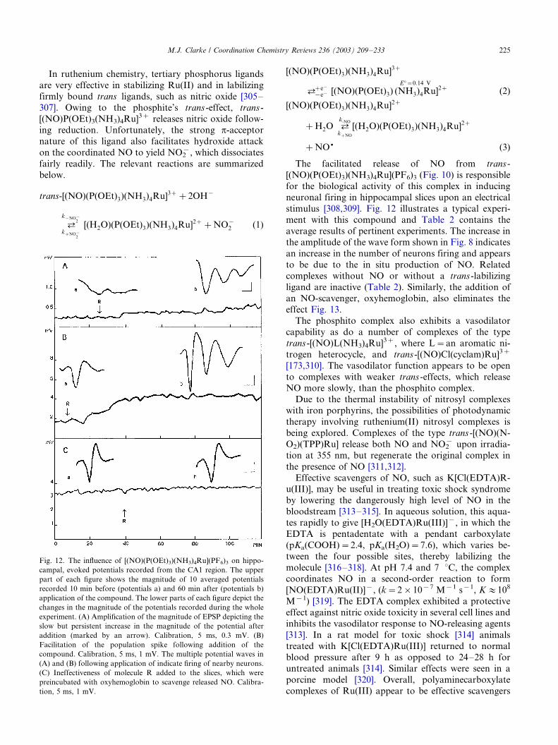
In ruthenium chemistry, tertiary phosphorus ligands
are very effective in stabilizing Ru(II) and in labilizing
firmly bound trans ligands, such as nitric oxide [305�/
307]. Owing to the phosphite’s trans -effect, trans -[(NO)P(OEt)3(NH3)4Ru]3� releases nitric oxide follow-
ing reduction. Unfortunately, the strong p-acceptor
nature of this ligand also facilitates hydroxide attack
on the coordinated NO to yield NO2�, which dissociates
fairly readily. The relevant reactions are summarized
below.
trans-[(NO)(P(OEt)3)(NH3)4Ru]3��2OH�
?k�NO�
2
k�NO�2
[(H2O)(P(OEt)3)(NH3)4Ru]2��NO�2 (1)
[(NO)(P(OEt)3)(NH3)4Ru]3�
?�e�
�e� [(NO)(P(OEt)3) (NH3)4RuE��0:14 V
]2� (2)
[(NO)(P(OEt)3)(NH3)4Ru]2�
�H2O ?k-NO
k�NO
[(H2O)(P(OEt)3)(NH3)4Ru]2�
�NO+ (3)
The facilitated release of NO from trans -
[(NO)(P(OEt)3)(NH3)4Ru](PF6)3 (Fig. 10) is responsible
for the biological activity of this complex in inducing
neuronal firing in hippocampal slices upon an electrical
stimulus [308,309]. Fig. 12 illustrates a typical experi-ment with this compound and Table 2 contains the
average results of pertinent experiments. The increase in
the amplitude of the wave form shown in Fig. 8 indicates
an increase in the number of neurons firing and appears
to be due to the in situ production of NO. Related
complexes without NO or without a trans -labilizing
ligand are inactive (Table 2). Similarly, the addition of
an NO-scavenger, oxyhemoglobin, also eliminates theeffect Fig. 13.
The phosphito complex also exhibits a vasodilator
capability as do a number of complexes of the type
trans -[(NO)L(NH3)4Ru]3�, where L�/an aromatic ni-
trogen heterocycle, and trans -[(NO)Cl(cyclam)Ru]3�
[173,310]. The vasodilator function appears to be open
to complexes with weaker trans -effects, which release
NO more slowly, than the phosphito complex.Due to the thermal instability of nitrosyl complexes
with iron porphyrins, the possibilities of photodynamic
therapy involving ruthenium(II) nitrosyl complexes is
being explored. Complexes of the type trans -[(NO)(N-
O2)(TPP)Ru] release both NO and NO2� upon irradia-
tion at 355 nm, but regenerate the original complex in
the presence of NO [311,312].
Effective scavengers of NO, such as K[Cl(EDTA)R-u(III)], may be useful in treating toxic shock syndrome
by lowering the dangerously high level of NO in the
bloodstream [313�/315]. In aqueous solution, this aqua-
tes rapidly to give [H2O(EDTA)Ru(III)]�, in which the
EDTA is pentadentate with a pendant carboxylate
(pKa(COOH)�/2.4, pKa(H2O)�/7.6), which varies be-
tween the four possible sites, thereby labilizing the
molecule [316�/318]. At pH 7.4 and 7 8C, the complexcoordinates NO in a second-order reaction to form
[NO(EDTA)Ru(II)]�, (k�/2�/10�7 M�1 s�1, K :/108
M�1) [319]. The EDTA complex exhibited a protective
effect against nitric oxide toxicity in several cell lines and
inhibits the vasodilator response to NO-releasing agents
[313]. In a rat model for toxic shock [314] animals
treated with K[Cl(EDTA)Ru(III)] returned to normal
blood pressure after 9 h as opposed to 24�/28 h foruntreated animals [314]. Similar effects were seen in a
porcine model [320]. Overall, polyaminecarboxylate
complexes of Ru(III) appear to be effective scavengers
Fig. 12. The influence of [(NO)(P(OEt)3)(NH3)4Ru](PF6)3 on hippo-
campal, evoked potentials recorded from the CA1 region. The upper
part of each figure shows the magnitude of 10 averaged potentials
recorded 10 min before (potentials a) and 60 min after (potentials b)
application of the compound. The lower parts of each figure depict the
changes in the magnitude of the potentials recorded during the whole
experiment. (A) Amplification of the magnitude of EPSP depicting the
slow but persistent increase in the magnitude of the potential after
addition (marked by an arrow). Calibration, 5 ms, 0.3 mV. (B)
Facilitation of the population spike following addition of the
compound. Calibration, 5 ms, 1 mV. The multiple potential waves in
(A) and (B) following application of indicate firing of nearby neurons.
(C) Ineffectiveness of molecule R added to the slices, which were
preincubated with oxyhemoglobin to scavenge released NO. Calibra-
tion, 5 ms, 1 mV.
M.J. Clarke / Coordination Chemistry Reviews 236 (2003) 209�/233 225

of NO in a variety of biological models and may provide
an alternative therapeutic strategy to NOS inhibitors for
the treatment of NO-mediated diseases [313].
The administration of trans -[(NO)Cl(cyclam)-
Ru(II)]2� reduces blood pressure in Wistar rats, espe-
cially in hypertensive rats. Notably, the slower rate of
release of NO+ from this complex following reduction
produces a hypotensive effect 20 times longer than that
generated by sodium nitroprusside in either normoten-
sive or hypertensive rats. The rate of NO+ release is
(k�NO) is 2.2�/10�3 s�1 at 35 8C following chemical
reduction (E8?�/0.10 V vs. NHE) of trans -
[(NO)Cl(cyclam)Ru(II)]2� [321]. The continuous infu-
sion of trans -[Cl(CF3SO3)(cyclam)Ru(III)]� blocks the
hypotensive effect of the trans -[(NO)Cl(cyclam)-
Ru(II)]2� probably by scavenging the NO+ released
Table 2
Influence of trans -[(NO)(P(OEt)3)(NH3)4Ru](PF6)3 and related control complexes on the hippocampal population spike [309]
Compound Concentration (mM) E 8 a N % Increase in P.S.
trans -[(NO)(P(OEt)3)(NH3)4Ru](PF6)3 1�/2.5 0.14 14 1789/46
trans -[(H2O)(P(OEt)3)(NH3)4Ru](PF6)2 0.25�/0.75 b 0.69 8 2.09/9.3
trans -[(P(OEt)3)2(NH3)4Ru](PF6)2 1�/2.5 0.89 7 21.59/21.4
[(NO)(NH3)5Ru]Cl3 1�/2.5 �/0.012 9 309/18
trans -[(NO)(P(OEt)3)(NH3)4Ru](PF6)3�/oxyhemoglobin 1�/2.5 0.14 5 4.29/2.3
NaNO2 0.2�/0.3 9 inhibition
trans -[(H2O)(P(OEt)3)(NH3)4Ru](PF6)2 1.0 0.69 16 749/11
�/NaNO2 1�/2.8
a Relative to SHE.b Concentration limited by solubility.
E 8, reduction potential; P.S., population spike.
Fig. 13. Structures of ruthenium immunosupressant complexes: (A) trans -[(Im)2(NH3)4Ru]3� [356], (B) cis -[(Im)2(NH3)4Ru]2�, (C) [(1MeIm)6-
Ru]2� [324]. Hydrogen atoms omitted from C for clarity.
M.J. Clarke / Coordination Chemistry Reviews 236 (2003) 209�/233226
after reduction. Taken together, these results suggests
that ruthenium cyclam complexes could be used as a
‘NO-buffer’ system to control physiological levels of
available NO [322].
5. Immunosuppressants
Since the most widely used immunosuppressive agent,
cyclosporin A, interferes with a Ca-dependent step in the
immune response [323] and also prevents the Ca2�-
dependent opening of a megachannel permeabilitytransition pore in the inner mitochondrial membrane,
it is conceivable that other Ca2� blocking agents, such
as ruthenium red and its related complexes, might
interfere with the immune response. Procept Inc. found
that ruthenium red inhibited the antigen-induced pro-
liferation of T cells with an IC50 of 35 nM, which is
comparable to that of cyclosporin A [249]. Subsequent
studies revealed that simple, substitution-inert Ru(II/III)complexes with nitrogen ligands (NH3, imidazole, py)
can be potent immunosuppressants [324�/326]. For
example, nanomolar concentrations of [4Mepy(NH3)5-
Ru(III)] (PRO-2844) and cis -[(Im)2(NH3)4Ru(III)]
(PRO-2846) strongly inhibit antigen-stimulated T lym-
phocyte proliferation with few or no acute toxic side
effects. While showing nM activity against antigen-
stimulated T cells, the compounds are notably less toxic(IC50�/10 mM) against a Jurkat cell line [326]. Since
these Ru drugs are mechanistically distinct from the
clinically used agents, cyclosporin A and FK506, and
may function through an electron-transfer pathway
[324], they may point the way toward an exciting new
class of immunosuppressive drugs [326,327].
The reported active Ru-immunosuppressant agents,
so far have involved six, small N-coordinated ligands(N6-coordinate) [324,326]. Among these compounds,
there is a window of activity between �/50 and �/400
mV, which not only indicates a redox process in their
mechanism, but also suggests why only Ru complexes
have thus far been reported to be active [327]. If electron
transfer is involved, this should be facile and take place
within a biologically accessible range of reduction
potentials. As has been well documented in studies ofRu-anticancer agents, simple Ru complexes with nitro-
gen ligands fulfill this requirement [149]. It seems likely
that the small size of the active complexes (among the
largest are [(1MeIm)6Ru]3�) allows them to approach
electron-transfer sites inside proteins. The higher activ-
ity of those complexes with aromatic ligands, is in
keeping with Gray’s studies indicating that aromatic
ligands penetrate deeper into electron-transfer sites inproteins [328]. Also consistent with this is the reduction
of a highly active immunosuppressant agent,
[py(NH3)5Ru]3� by HeLa cells [98].
Given the ‘redox window’ and the high activity of
these complexes, it is apparent that they must function
catalytically, i.e. by continually short-circuiting one or
more redox processes by accepting an electron from onemolecule and passing it to another. It is possible that
oxygen serves as the oxidant, which would bypass much
of the mitochondrial respiratory redox chain and
produce toxic superoxide. While active complexes such
as [py(NH3)5Ru]2� are so slowly oxidized by O2 [56],
that it would seem that another biological oxidant might
be more likely, autoxidation is accelerated if a ligand is
lost thereby allowing O2 to interact in an inner-spherefashion, or if a catalyst, such as Fe(II/III) is present. It
may be that a reductant at least as reducing as
ubiquinone in the mitochondrial respiratory redox chain
and/or an oxidant around cyt-oxidase or O2 are involved
in redox cycling of the Ru(II/III) to short-circuit the
production of ATP.
Given hydrolysis rates for a number of N6 Ru(II)
complexes, ligand loss is a very real possibility over thetime spans (1 day�/1 week) of tissue culture experiments
and would certainly be expected to occur over the long
periods required for drug administration needed to
suppress rejection of organ transplants [329]. The
reaction of GSH with [(4-picoline)(NH3)5Ru]3� and
[(NH3)6Ru]3� also produces some [GS(NH3)5-
Ru(III)]2�, which is indicative of a reduced intermediate
that eliminates nitrogen ligands, and suggests that GSHis conceivably involved in the ruthenium immunosup-
pressant activity. Ligand loss would give rise to com-
plexes with substitutable sites of the type discussed
under anticancer agents that would be expected to bind
to DNA, interfere with nucleic acid metabolism, and
would probably be mutagenic.
6. Radiopharmaceuticals
Metal containing radiopharmaceuticals can be classi-
fied into two broad types, (1) Those that find their target
by virtue of the properties of aqua metal ion or the metal
ion complex itself; and (2) those that localize in the
desired tissue by virtue of being attached to a targeting
protein or polypeptide. Localizing a- or ß-emitting
radionuclides in a tumor can be used to kill the tumorcells. Utilization of a g-emitting radionuclides, such as
the widely used 99mTc, allow for diagnostic organ
imaging. Radioscintigraphy refers to the visualization
of organs that have concentrated a g-emitting radio-
nuclide [10]. The g-rays are detected by a photodetector
and converted into a visible image by computer methods
in real time. This provides not only for visualizing the
organ, but observing its function as well. For example,the clearance of a radioactive compound from the blood
by the liver can be seen both as a decrease in activity in
the blood and an increase in activity in the liver,
M.J. Clarke / Coordination Chemistry Reviews 236 (2003) 209�/233 227

followed by the progression of activity into the gall
bladder and finally the intestine.
The radiophysical properties of 97Ru are nearly ideal
for some types of radiodiagnostic imaging. It decays byelectron capture with a 2.88 days half-life and emits a
216 keV g-ray that can be used in common radio-
scintigraphic instruments. Unfortunately, 97Ru is not
commercially available. An alternative isotope, 103Ru is
commercially available, but emits a 497 keV g-ray,
which is of only fair utility in radioscintigraphy, and
has a longer (39.35 days) half-life. On the other hand, its
longer half-life and lower specific activity can be anadvantage in developing synthetic methods [330].
A corollary of the activation-by-reduction hypothesis
for chemotherapy involving Ru(III) ammine complexes
is that they should concentrate in tumors as a result of
reduction and intracellular binding. Indeed, a number of
ruthenium ammine complexes exhibit good tumor
uptake [330,331]. Unfortunately, they also remain in
the blood through binding to albumin and Tf. RuCl3exhibits some nonspecific binding to erythrocytes [332].
The coordination of ruthenium to Tf can be used to
enhance tumor uptake, since neoplastic cells, especially
those in rapidly growing tumors, have a high iron
requirement and display a large number of Tf receptors
on their membrane surfaces. The approach of using a
protein to localize radioruthenium in a tumor was
demonstrated by Srivastava, Larson, et al. who showedthat the uptake of 103Ru-labelled Tf by EMT-6 sarcoma
in mice was almost twice as high as that of a widely used
tumor-imaging agent, 67Ga- citrate [74,330,333].
Effective radio-imaging agents for the liver have been
developed using radioruthenium(III) with iminodiace-
tato ligands containing lipophilic groups that facilitate
uptake by the liver and biliary tract [334�/336]. Several
lipophilic ruthenocene derivatives are excreted in bothurine and bile following hydroxylation in the liver and
formation of a glucuronide conjugate [337] and some
ruthenocene derivatives have been designed to localize
in the adrenals and other organs [338,339]. Rutheno-
cene-haloperidol exhibited a high affinity for the lung,
but not the brain, in rats [340]. The complex
[(pyal)(NH3)5Ru(III)]3� (pyal�/ß-(4-pyridyl)-a-ala-
nine) displayed good uptake by the pancreas [341].Ruthenium complexes with phosphate and phospho-
nates tend to localize in bone [84]. Ruthenium red [247]
binds in vitro and in vivo to cell surfaces high in acidic
glycoproteins and has been used to image tumors
[169,171,342]. The complex [(BLM)(NH3)5103Ru], where
BLM�/bleomycin, a clinically used antitumor antibiotic
that tends to localize in tumors, yielded a tissue
distribution in normal and tumor-bearing mice essen-tially the same as BLM itself [330,343].
Aside from uptake owing to nonspecific causes such
as increased blood perfusion and higher cell permeabil-
ity in tumors, overall it is likely that tumor accumulation
of simple ammineruthenium complexes proceeds by two
pathways. First, rapid tumor uptake proceeds through
activation of the Ru(III)-prodrug toward binding by
reduction in the tumor. Since small ions are excreted
fairly readily by the kidneys, this mode of binding
should decrease rapidly with time. Second, Tf mediated
tumor binding, which may occur for many days follow-
ing injection.
7. Conclusion
The versatile synthetic chemistry of ruthenium opens
up a wide range of complexes with a variety of ligands
that could provide an arsenal of compounds for clinical
uses. Ruthenium compounds appear to penetrate tu-
mors well and bind to cellular DNA. While the initial
DNA binding site of many ruthenium complexes is the
same as that of cisplatin, i.e. the N7 of Gua, their
antitumor mechanisms are different. In the case of the
ruthenium compound now in clinical trials, NAMI, its
antimetastatic activity is independent of DNA coordi-
nation, and may depend on the regulation of a matrix
proteinase. Better understanding Tf mediated Ru trans-
port into tumors could lead to more effective and
probably less toxic agents. Use of the radionuclide,97Ru, could have considerable advantages over 99mTc in
that its longer half life allows for more complicated
synthetic methods, purification and quality control
while still providing excellent radioscintigraphic images.
The substantial body of work on the photochemistry of
Ru(II) complexes with aromatic nitrogen ligands, parti-
cularly those that intercalate into DNA, offer the
potential for novel photodynamic approaches to cancer
therapy.
Particularly exciting is the unusual ability of simple
ruthenium complexes to suppress the immune response
by inhibiting the proliferation of T cells. While the initial
set of complexes tested are likely to give rise to others
that would bind to DNA and possibly be mutagenic, the
idea of short-circuiting electron transfer pathways
synergetically coupled with other subcellular interac-
tions is one with rich possibilities for controlling the
division and multiplication of selected cell types.
The ability to tune both the uptake and release of the
ubiquitous intercellular messenger molecule, NO, by
manipulating other ligands on Ru lays out a definite
way in which coordination chemists can contribute not
only to new classes of drugs to affect a variety of
biological functions, but also possibly to the develop-
ment of a molecular tool chest that can be used to probe
NO function in the same way that ruthenium red has
been used to probe that of Ca2�.
M.J. Clarke / Coordination Chemistry Reviews 236 (2003) 209�/233228
Acknowledgements
Funding for this work was provided by NIH Grant
GM26390.
References
[1] R.H. Holm, P. Kennepohl, E.I. Solomon, Chem. Rev. 96 (1996)
2239.
[2] S.J. Lippard, J.M. Berg, Curr. Opin. Chem. Biol. 4 (2000) 137.
[3] Q. He, C.H. Liang, S.J. Lippard, Proc. Natl. Acad. Sci. USA 97
(2000) 5768.
[4] A. Gelasco, S.J. Lippard, Top. Biol. Inorg. Chem. 1 (1999) 1.
[5] E.R. Jamieson, S.J. Lippard, Chem. Rev. 99 (1999) 2467.
[6] A.T. Yarnell, S. Oh, D. Reinberg, S.J. Lippard, J. Biol. Chem.
276 (2001) 25736.
[7] C.J. Ziegler, A.P. Silverman, S.J. Lippard, J. Biol. Inorg. Chem.
5 (2000) 774.
[8] N. Farrell, Y. Qu, J.D. Roberts, Top. Biol. Inorg. Chem. 1
(1999) 99.
[9] J. Zehnulova, J. Kasparkova, N. Farrell, V. Brabec, J. Biol.
Chem. 276 (2001) 22191.
[10] M.J. Clarke, L. Podbielski, Coord. Chem. Rev. 78 (1987) 253.
[11] A.B. Packard, J.F. Kronauge, M.W. Brechbiel, Top. Biol. Inorg.
Chem. 2 (1999) 45.
[12] P. Caravan, J.J. Ellison, T.J. McMurry, R.B. Lauffer, Chem.
Rev. (Washington, DC) 99 (1999) 2293.
[13] M.F. Tweedle, K. Kumar, Top. Biol. Inorg. Chem. 2 (1999) 1.
[14] M.J. Heeg, S.S. Jurisson, Acc. Chem. Res. 32 (1999) 1053.
[15] C. Orvig, M.J. Abrams, in: J. Michl, J.A. Gladysz, K.L. Rowlen
(Eds.), Chemical Reviews, American Chemical Society, Wa-
shington, DC, 1999.
[16] K.H. Thompson, C. Orvig, Dalton (2000) 2885.
[17] M. Melchior, S.J. Rettig, B.D. Liboiron, K.H. Thompson, V.G.
Yuen, J.H. McNeill, C. Orvig, Inorg. Chem. 40 (2001) 4686.
[18] R.A. Anderson, J. Am. Coll. Nutr. 17 (1998) 548.
[19] J.B. Vincent, Acc. Chem. Res. 33 (2000) 503.
[20] M.J. Clarke, P.J. Sadler, Top. Biol. Inorg. Chem. 1 (1999) 200.
[21] M.J. Clarke, P.J. Sadler, Top. Biol. Inorg. Chem. 2 (1999) 216.
[22] Z.J. Guo, P.J. Sadler, Angewandte Chemie-Int. Ed. 38 (1999)
1513.
[23] T. Pieper, K. Borsky, B.K. Keppler, Top. Bioinorg. Chem. 1
(1999) 171.
[24] M. Hartmann, B.K. Kepler, Comm. Inorg. Chem. 16 (1995) 339.
[25] N. Farrell, Transition Metal Complexes as Drugs and Che-
motherapeutic Agents, Kluwer Academic, Boston, 1989.
[26] B.K. Keppler, Metal Complexes in Cancer Chemotherapy,
VCH, Weinheim, Germany, 1993.
[27] M.J. Clarke, Ruthenium and Other Non-Platinum Metal Com-
plexes in Cancer Chemotherapy, Springer-Verlag, Heidelberg,
Germany, 1989.
[28] M.J. Clarke, S. Limauro, in: G. Berthon (Ed.), Handbook on
Metal�/Ligand Interactions in Biological Fluids, Marcel Dekker,
New York, 1995, pp. 1010�/1018.
[29] M.J. Clarke, M. Stubbs, Met. Ions Biol. Syst. 32 (1996) 727.
[30] M.J. Clarke, in: S.J. Isied (Ed.), Electron Transfer Reactions,
American Chemical Society Symposium Series, Washington,
D.C., 1997, pp. 349�/365.
[31] H. Ali, J.E. van Lier, Chem. Rev. 99 (1999) 2379.
[32] J.M. Kelly, C.-M. Moucheron, A. Kirsch-De Mesmaeker,
Struct. Bond. 92 (1999) 163.
[33] H.B. Gray, J.R. Winkler, Ann. Rev. Biochem. 65 (1996) 537.
[34] L. Messori, F.G. Vilchez, R. Vilaplana, F. Piccioli, E. Alessio, B.
Keppler, Met. Based Drugs 7 (2000) 335.
[35] L. Messori, P. Orioli, D. Vullo, E. Alessio, E. Iengo, Eur. J.
Biochem. 267 (2000) 1206.
[36] V.M. Bailey, K.J. LaChance-Galang, P.E. Doan, M.J. Clarke,
Inorg. Chem. 36 (1997) 1873.
[37] C.G. Kuehn, H. Taube, J. Am. Chem. Soc. 98 (1976) 689.
[38] D. Frasca, M.J. Clarke, J. Am. Chem. Soc. 121 (1999) 8523.
[39] M.J. Clarke, M.G. Dowling, Inorg. Chem. 20 (1981) 3506.
[40] M.J. Clarke, M.G. Dowling, in: V. Massey, C. Williams (Eds.),
Proceedings of the Seventh International Flavin Symposium,
Elsevier, New York, 1982, pp. 579�/583.
[41] A. Abelleira, R.D. Galang, M.J. Clarke, Inorg. Chem. 29 (1990)
633.
[42] M.J. Clarke, F. Zhu, D. Frasca, Chem. Rev. 99 (1999) 2511.
[43] A.D. Kelman, M.J. Clarke, S.D. Edmonds, H.J. Peresie, J. Clin.
Hematol. Oncol. 7 (1977) 274.
[44] M.J. Clarke, in: B.K. Keppler (Ed.), Metal Complexes in Cancer
Chemotherapy, VCH, Weinheim, 1993, pp. 129�/157.
[45] B.D. Palmer, W.R. Wilson, S.M. Pullen, J. Med. Chem. 33
(1990) 112.
[46] G.K. Snyder, J. Appl. Physiol. 65 (1988) 2332.
[47] R.G. Steen, Am. J. Roentgenol. 157 (1991) 243.
[48] P. Okunieff, E.P. Dunphy, P. Vaupel, Adv. Exp. Med. Biol. 345
(1994) 485.
[49] J.E. Biskupiak, K.A. Krohn, J. Nucl. Med. (1993) 411.
[50] J.L. Wike-Hooley, J. Haveman, H.S. Reinhold, Radiother.
Oncol. 2 (1984) 343.
[51] D. Miklavcic, G. Sersa, S. Novakovic, J. Bioelect. 9 (1990) 133.
[52] J.A. Marchant, T. Matsubara, P.C. Ford, Inorg. Chem. 16
(1977) 2160.
[53] G.M. Coleman, J.W. Gesler, E.A. Shirley, J.R. Kuempel, Inorg.
Chem. 12 (1973) 1036.
[54] M.J. Clarke, S. Bitler, D. Rennert, M. Buchbinder, A.D.
Kelman, J. Inorg. Biochem. 12 (1980) 79.
[55] J.F. Laliberte, I.L. Sun, F.L. Crane, M.J. Clarke, J. Bioenerg.
Biomembr. 19 (1987) 69.
[56] D.M. Stanbury, O. Haas, H. Taube, Inorg. Chem. 19 (1980) 518.
[57] D.M. Stanbury, D. Gaswick, G.M. Brown, H. Taube, Inorg.
Chem. 22 (1983) 1975.
[58] D.M. Stanbury, W.A. Mulac, J.C. Sullivan, H. Taube, Inorg.
Chem. 19 (1980) 3735.
[59] D. Frasca, J. Ciampa, J. Emerson, R.S. Umans, M.J. Clarke,
Metal-Based Drugs 3 (1996) 197.
[60] G. Mestroni, E. Alessio, G. Sava, S. Pacor, M. Coluccia, A.
Boccarelli, Metal-Based Drugs 1 (1994) 41.
[61] G. Mestroni, E. Alessio, G. Sava, S. Pacor, M. Coluccia, in: B.K.
Keppler (Ed.), Metal Complexes in Cancer Chemotherapy,
VCH, Weinheim, 1993, pp. 157�/185.
[62] C. Monti-Bragadin, L. Ramani, L. Samer, G. Mestroni, G.
Zassinovich, Antimicrob. Agents Chemother. 7 (1975) 825.
[63] G. Sava, S. Zorzet, T. Giraldi, G. Mestroni, G. Zassinovich, Eur.
J. Cancer Clin. Oncol. 20 (1984) 841.
[64] G. Sava, S. Pacor, S. Zorzat, E. Alessio, G. Mestroni,
Pharmacol. Res. 21 (1989) 617.
[65] E. Alessio, Y. Xuu, S. Cauci, G. Mestroni, F. Quadrifoglio, P.
Viglino, L.G. Marzilli, J. Am. Chem. Soc. 111 (1989) 7068.
[66] G. Mestroni, E. Alessio, M. Calligaris, W.M. Attia, F. Quad-
rifoglio, S. Cauci, G. Sava, S. Zorzet, S. Pacor, C. Monti-
Bragadin, M. Tamaro, L. Dolzani, Progress in Clinical Bio-
chemistry and Medicine, Springer-Verlag, Berlin, 1989.
[67] A.C.G. Hotze, A.H. Velders, F. Ugozzoli, M. Biagini-Cingi,
A.M. Manotti-Lanfredi, J.G. Haasnoot, J. Reedijk, Inorg.
Chem. 39 (2000) 3838.
[68] A.H. Velders, H. Kooijman, A.L. Spek, J.G. Haasnoot, D. De
Vos, J. Reedijk, Inorg. Chem. 39 (2000) 2966.
[69] Y.N.V. Gopal, D. Jayaraju, A.K. Kondapi, Biochemistry 38
(1999) 4382.
[70] L. Stryer, Biochemistry, Freeman, New York, 1995.
M.J. Clarke / Coordination Chemistry Reviews 236 (2003) 209�/233 229

[71] R.E. Morris, R.E. Aird, P.d.S. Murdoch, H.M. Chen, J.
Cummings, N.D. Hughes, S. Parsons, A. Parkin, G. Boyd,
D.I. Jodrell, P.J. Sadler, J. Med. Chem. 44 (2001) 3616, J.
Cummings, R.E. Aird, R.E. Morris, H.M. Chen, P.d.S. Mur-
doch, P.J. Sadler, J.F. Smyth, D.I. Jodrell, Clin. Cancer Res. 6
(2000) 142.
[72] C.-C. Cheng, W.-L. Lee, J.-G. Su, C.-L. Liu, J. Chin. Chem. Soc.
(Taipei) 47 (2000) 213.
[73] J. Helms, B.L. Huang, R. Price, B.P. Sullivan, B.A. Sullivan,
Inorg. Chem. 34 (1995) 5335.
[74] P. Som, Z.H. Oster, K. Matsui, G. Gugliemi, B. Persson, M.L.
Pellettieri, S.C. Srivastava, P. Richards, H.L. Atkins, A.B. Brill,
Eur. J. Nucl. Med. 8 (1983) 491.
[75] M.J. Clarke, B. Jansen, K.A. Marx, R. Kruger, Inorg. Chim.
Acta 124 (1986) 13.
[76] M. McNamara, M.J. Clarke, Inorg. Chim. Acta 195 (1992) 175.
[77] W.R. Harris, Struct. Bond. 92 (1998) 121.
[78] F. Kratz, M.T. Schutte, Cancer J. 11 (1998) 60.
[79] H. Sun, H. Li, P.J. Sadler, Chem. Rev. 99 (1999) 2817.
[80] H.S. Li, P.J. Sun, Eur. J. Bioichem. 242 (1996) 387.
[81] S.E. Bailey, R.W. Garratt, G. Richard, Biochemistry 27 (1988)
5804.
[82] R.T.A. MacGillivray, M.C. Bewley, C.A. Smith, Q.-Y. He, A.B.
Mason, R.C. Woodworth, E.N. Baker, Biochemistry 39 (2000)
1211.
[83] S.C. Srivastava, P. Richards, G.E. Meinken, S.M. Larson, Z.
Grunbaum, in: R.P. Spencer (Ed.), Radiopharmaceuticals,
Grune & Stratton, Inc, New York, 1981, p. 207.
[84] S.C. Srivastava, L.F. Mausner, M.J. Clarke, in: M.J. Clarke
(Ed.), Ruthenium and other Non-Platinum Metal Complexes in
Cancer Chemotherapy, Springer-Verlag, Heidleberg, 1989, pp.
111�/150.
[85] A. Anto, I. Anto, T. Hiraki, K. Hisada, Int. J. Rad. Appl. 15
(1988) 133.
[86] L. Messori, F. Kratz, E. Alessio, Met.-Based Drugs 3 (1996) 1.
[87] F. Kratz, M. Hartmann, B. Keppler, L. Messori, J. Biol. Chem.
269 (1994) 2581.
[88] F. Kratz, B.K. Keppler, L. Messori, C. Smith, E.N. Baker,
Metal-Based Drugs 1 (1994) 169.
[89] C.A. Smith, A.J. Sutherland-Smith, B.K. Keppler, F. Kratz,
E.N. Baker, J. Biol. Inorg. Chem. 1 (1996) 424.
[90] D.C. Kraiter, O. Zak, P. Aisen, A.L. Crumbliss, Inorg. Chem. 37
(1998) 964.
[91] F. Kratz, L. Messori, J. Inorg. Biochem. 49 (1993) 79.
[92] D.M. Martin, N.D. Chasteen, J.K. Grady, Biochim. Biophys.
Acta 1076 (1991) 252.
[93] P. Aisen, in: G.L. Eichhorn (Ed.), Inorganic Biochemistry,
Elsevier, Amsterdam, 1973, pp. 280�/305.
[94] F. Kratz, N. Mulinacci, L. Messori, I. Bertini, B.K. Keppler, in:
J. Anastassopoulou, P. Collery, J.-C. Etienne, T. Theophanides
(Eds.), Metal Ions in Biology and Medicine, John Libbey
Limited Eurotext, Paris, 1992, pp. 69�/74.
[95] B.K. Keppler, in: B.K. Keppler (Ed.), Metal Complexes in
Cancer Chemotherapy, VCH, Weinheim, Germany, 1993, pp.
187�/220.
[96] F. Kratz, B. Keppler, M. Hartmann, L. Messori, R. Berger,
Met.-Based Drugs 3 (1996) 15.
[97] D.R. Frasca, L.E. Gehrig, M.J. Clarke, J. Inorg. Biochem. 83
(2001) 139.
[98] D.R. Frasca, Ph.D. thesis, Boston College, 2000.
[99] M.J. Clarke, Inorg. Chem. 16 (1977) 738.
[100] M.J. Clarke, J. Am. Chem. Soc. 100 (1978) 5068.
[101] M.E. Kastner, K.F. Coffey, M.J. Clarke, S.E. Edmonds, K.
Eriks, J. Am. Chem. Soc. 103 (1981) 5747.
[102] V.M. Bailey, M.J. Clarke, Inorg. Chem. 36 (1997) 1611.
[103] K. LaChance-Galang, M. Zhao, M.J. Clarke, Inorg. Chem. 35
(1996) 6021.
[104] K.J. LaChance-Galang, I. Maldonado, M.L. Gallagher, W. Jian,
A. Prock, J. Chaklos, R.D. Galang, M.J. Clarke, Inorg. Chem.
40 (2001) 485.
[105] A.H. Velders, B. Van der Geest, H. Kooijman, A.L. Spek, J.G.
Haasnoot, J. Reedijk, Eur. J. Inorg. Chem. (2001) 369.
[106] M. Zhao, M.J. Clarke, J. Biol. Inorg. Chem. 4 (1999) 325.
[107] O.M. Dhubhghaill, W. Hagen, B.K. Keppler, K.-G. Lipponer, J.
Sadler, J. Chem. Soc. Dalton Trans. (1994) 3305.
[108] M. Hartmann, K.-G. Lipponer, B.K. Keppler, Inorg. Chim.
Acta 267 (1998) 137.
[109] B.K. Keppler, M. Hartmann, Metal-Based Drugs 1 (1994) 145.
[110] M.J. Clarke, H. Taube, J. Am. Chem. Soc. 97 (1975) 1397�/1403,
C. Price, M.R.J. Elsegood, W. Clegg, N.H. Reese, A. Houlton,
Angew. Chem., Intl. Ed. 36 (1997) 172�/1764.
[111] M.J. Clarke, P.E. Morrissey, Inorg. Chim. Acta 80 (1984) L69.
[112] K.C. Gariepy, M.A. Curtin, M.J. Clarke, J. Am. Chem. Soc. 111
(1989) 4947.
[113] D.P. Rudd, H. Taube, Inorg. Chem. 10 (1971) 1543.
[114] J.K. Barton, E. Lolis, J. Am. Chem. Soc. 107 (1985) 708.
[115] N. Grover, N. Gupta, H.H. Thorp, J. Am. Chem. Soc. 114
(1992) 3390.
[116] N. Grover, T.W. Welch, T.A. Fairley, M. Cory, H.H. Thorp,
Inorg. Chem. 33 (1994) 3544.
[117] P.M. van Vliet, M.S. Sarinten, S.M.S. Toekimin, J.G. Haasnoot,
J. Reedijk, O. Novakova, O. Vrana, V. Brabec, Inorg. Chim.
Acta 231 (1995) 57.
[118] C. Pifferi, R. Cini, J. Chem. Soc. Dalton Trans. (1998) 2679.
[119] C. Pifferi, R. Cini, Acta Crystallogr. Sect. C-Cryst. Struct.
Commun. 56 (2000) E439.
[120] S. Isied, H. Taube, Inorg. Chem. 15 (1976) 3070.
[121] S. Pacor, G. Sava, V. Ceschia, F. Bregant, G. Mestroni, E.
Alessio, Chem. Biol. Interact. 78 (1991) 223.
[122] L.G. Marzilli, M. Iwamoto, E. Alessio, L. Hansen, M. Calligaris,
J. Am. Chem. Soc. 116 (1994) 815.
[123] E. Alessio, E. Zangrando, R. Roppa, L. Marzilli, Inorg. Chem.
37 (1998) 2458.
[124] M. Iwamoto, E. Alessio, L.G. Marzilli, Inorg. Chem. 35 (1996)
2384.
[125] G. Esposito, S. Cauci, F. Fogolari, E. Alessio, M. Scocchi, F.
Quadrifoglio, P. Viglino, Biochemistry 31 (1992) 7094.
[126] N. Farrell, Y. Qu, Inorg. Chem. 34 (1995) 3573.
[127] B. Van Houten, S. Illenye, Y. Qu, N. Farrell, Biochemistry 32
(1993) 11794.
[128] M. Milkevitch, B.W. Shirley, K.J. Brewer, Inorg. Chim. Acta
264 (1997) 249.
[129] M. Milkevitch, H. Storrie, E. Brauns, K.J. Brewer, B.W. Shirley,
Inorg. Chem. 36 (1997) 4534.
[130] A. Corazza, I. Harvey, P. Sadler, Eur. J. Biochem. 236 (1996)
697.
[131] D.L. Rabenstein, K.K. Millis, K.H. Weaver, J. Org. Chem. 58
(1993) 4144.
[132] T.M. Buttke, P.A. Sandstrom, Immunol. Today 15 (1994) 7.
[133] S. Coppola, L. Ghibelli, Biochem. Soc. Trans. 28 (2000) 56.
[134] L. Chen, P.F. Lee, S.Y. Wong, J.D. Ranford, J.J. Vittal, J.
Chem. Soc. Dalton Trans. (1999) 1209.
[135] K. Lemma, J. Berglund, N. Farrell, L.I. Elding, J. Biol. Inorg.
Chem. 5 (2000) 300.
[136] H.J. Berkovits, R.A. Floyd, K.E. Wetterhahn, J. Aiyar, Environ.
Health Perspect. EI0 (1991) 53.
[137] K.E. Wetterhahn, S.L. Brauer, J. Am. Chem. Soc. 113 (1991)
3001.
[138] A. Eastman, Biochem. Pharmacol. 36 (1987) 4177.
[139] N.A. Kratochwil, Z. Guo, P.D.S. Murdoch, J.A. Parkinson, P.
Bednarski, P. Sadler, J. Am. Chem. Soc. 120 (1998) 8253.
[140] J. Reedijk, Chem. Rev. 99 (1999) 2499.
[141] T. Ishikawa, F. Ali-Osman, J. Biol. Chem. 268 (1993) 20116.
M.J. Clarke / Coordination Chemistry Reviews 236 (2003) 209�/233230

[142] Y. Chen, Z. Guo, J.A. Parkinson, P.J. Sadler, J. Chem. Soc.
Dalton Trans. (1998) 3577.
[143] J.-M. Teuben, J. Reedijk, J. Biol. Inorg. Chem. 5 (2000) 463.
[144] R.E. Yasbin, C.R. Matthews, M.J. Clarke, Chemico-Biol.
Interact. 30 (1980) 355.
[145] K.A. Marx, R. Kruger, M.J. Clarke, Molec. Cell. Biochem. 86
(1989) 155.
[146] K.A. Marx, C. Seery, P. Malloy, Mol. Cell. Biochem. 90 (1989)
37.
[147] M. Carballo, R. Vilaplana, G. Marquez, M. Conde, F.J. Bedoya,
F. Gonzalez-Vilchez, F. Sobrino, Biochem. J. 328 (1997) 559.
[148] R. Vilaplana, M.A. Romero, M. Quiros, J.M. Salas, F.
Gonzalez-Vilchez, Metal-Based Drugs 2 (1995) 211.
[149] M.J. Clarke, Met. Ions Biol. Syst. 11 (1980) 231.
[150] F. Bottomley, Can. J. Chem. 55 (1977) 2788.
[151] J.R. Durig, J. Danneman, W.D. Behnke, E.E. Mercer, Chem.-
Biol. Interact. 13 (1976) 287.
[152] M.J. Clarke, Prog. Clin. Biochem. Med. 10 (1989) 25.
[153] B.K. Keppler, W. Rupp, U.W. Juhl, H. Endres, R. Niebl, W.
Balzer, Inorg. Chem. 26 (1987) 4366.
[154] T. Pieper, B.K. Keppler, Analusis 26 (1998) M84.
[155] A. Galeano, M.R. Berger, B.K. Keppler, Arzneimittel-For-
schung/Drug Research 42-1 (1992) 821.
[156] E.D. Kreuser, B.K. Keppler, W.E. Berdel, A. Piest, E. Thiel,
Semin. Oncol. 19 (1992) 73.
[157] H. Depenbrock, S. Schmelcher, R. Peter, B.K. Keppler, G.
Weirich, T. Block, J. Rastetter, A.R. Hanauske, Eur. J. Cancer
33 (1997) 2404.
[158] M.H. Seelig, M.R. Berger, B.K. Keppler, J. Cancer Res. Clin.
Oncol. 118 (1992) 195.
[159] M.R. Berger, F.T. Garzon, D. Schmal, B.K. Keppler, Antic-
ancer Res. 9 (1989) 761.
[160] B.K. Keppler, M. Henn, U.M. Juhl, M.R. Berger, R. Niebel,
F.E. Wagner, in: M.J. Clarke (Ed.), Ruthenium and Other Non-
Platinum Metal Complexes in Cancer Chemotherapy, Springer-
Verlag, Heidelberg, 1989, pp. 41�/70.
[161] B.K. Keppler, W. Balzer, V. Seifried, Arzneim.-Forsch. 37
(1987) 770.
[162] L. Trynda-Lemiesz, B.K. Keppler, H.K. Koslowski, J. Inorg.
Biochem. 73 (1999) 123.
[163] G. Sava, S. Pacor, G. Mestroni, E. Alessio, Clin. Exp. Metastasis
10 (1992) 273.
[164] J.L. Bear, B. Han, K.M. Kadish, Inorg. Chem. 35 (1996) 3012.
[165] J.L. Bear, Y. Li, K.M. Kadish, Inorg. Chem. 35 (1996) 3053.
[166] J.L. Bear, Y. Li, K.M. Kadish, Inorg. Chem. 36 (1997) 5449.
[167] K.R. Dunbar, J.H. Matonic, V.P. Saharan, C.A. Crawford, G.
Christou, J. Am. Chem. Soc. 116 (1994) 2201.
[168] M.A. Oberc-Greenwood, L.M. Muul, M.K. Gately, P.L. Korn-
blith, B.W. Smith, J. Neurooncol. 3 (1986) 387.
[169] L.H. Anghileri, Strahlentherapie 149 (1975) 173.
[170] T.W. Hurley, Am. J. Physiol. 23 (1988) 621.
[171] L.J. Anghileri, C. Marchal, M. Matrat, M.C. Crone-Escanya,
Neoplasma 33 (1986) 603.
[172] M.J. Clarke, J.B. Gaul, Struct. Bonding 81 (1993) 147.
[173] S.S.S. Borges, C.U. Davanzo, E.E. Castellano, J. Zukerman-
Schpector, S.C. Silva, D.W. Franco, Inorg. Chem. 37 (1998)
2670.
[174] M.G. Gomes, C.U. Davanzo, S.C. Silva, L.G.F. Lopes, P.S.
Santos, D.W. Franco, J. Chem. Soc. Dalton Trans. (1998) 601.
[175] J.M. Slocik, R.A. Kortes, R.E. Shepherd, Met.-Based Drugs 7
(2000) 67.
[176] S.R. Vilaplana, M.G. Basallote, C. Ruiz-Valero, E. Gutierrez, F.
Gonzalez-Vilchez, J. Chem. Soc. Chem. Commun. (1991) 100.
[177] R.A. Vilaplana, F. Gonzalez-Vilchez, C. Ruiz-Valero, Inorg.
Chim. Acta 224 (1994) 15.
[178] D. Chatterjee, M.S. Ward, R.E. Shepherd, Inorg. Chim. Acta
285 (1999) 170.
[179] F. Gonzalez-Vilchez, R. Vilaplana, G. Blasco, L. Messori, J.
Inorg. Biochem. 71 (1998) 45.
[180] E. Gallori, C. Vettori, E. Alessio, F. Gonzalez-Vilchez, R.
Vilaplana, P. Orioli, A. Casini, L. Messori, Arch. Biochem.
Biophys. 376 (2000) 156.
[181] R.E. Shepherd, in: S. Isied (Eds.) Electron Transfer Reactions,
American Chemical Society Symposium Series Washington, DC,
1997, pp. 367�/399.
[182] R.E. Shepherd, Y. Chen, F.-T. Lin, Inorg. Chem. 36 (1997) 818.
[183] Y. Chen, F.-T. Lin, R.E. Shepherd, Inorg. Chim. Acta 268
(1998) 287.
[184] R.E. Shepherd, Y. Chen, R.A. Kortes, M.S. Ward, Inorg. Chim.
Acta 303 (2000) 30.
[185] G. Mestroni, E. Alessio, M. Calligaris, W.M. Attia, F. Quad-
rifoglio, S. Cauci, G. Sava, S. Zorzet, S. Pacor, C. Monti-
Bragadin, M. Tamaro, L. Dolzani, in: M.J. Clarke (Ed.),
Ruthenium and Other Non-Platinum Metal Complexes in
Cancer Chemotherapy, Springer-Verlag, Heidelberg, 1989, pp.
74�/88.
[186] S. Cauci, P. Viglino, G. Esposito, J. Inorg. Biochem. 43 (1991) 739.
[187] E. Alessio, G. Mestroni, G.A. Nardin, M. Wahib, M. Calligaria,
G. Sava, S. Zorzet, Inorg. Chem. 27 (1988) 4099.
[188] E. Alessio, W. Attia, M. Calligaris, S. Cauci, L. Dolzani, G.
Mestroni, C. Monti-Bragadin, G. Nardin, F. Quadrifoglio, G.
Sava, Dev. Oncol 54 (1988) 617.
[189] G. Sava, E. Alessio, E. Bergamo, G. Mestroni, Top. Biol. Inorg.
Chem. 1 (1999) 143.
[190] G. Sava, S. Pacor, M. Coluccia, M. Mariggio, M. Cocchietto, E.
Alessio, G. Mestroni, Drug Invest. 8 (1994) 150.
[191] E. Alessio, G. Balducci, A. Lutman, G. Mestroni, M. Calligaris,
W.M. Attia, Inorg. Chim. Acta 203 (1992) 205.
[192] G. Sava, S. Pacor, A. Bergamo, M. Cocchietto, G. Mestroni, E.
Alessio, Chem. Biol. Interact. 95 (1995) 109.
[193] I. Capozzi, K. Clerici, M. Cocchietto, G. Salerno, A. Bergamo,
G. Sava, Chem. Biol. Interact. 113 (1998) 51.
[194] L. Messori, A. Casini, D. Vullo, S.G. Haroutiunian, E.B.
Dalian, P. Orioli, Inorg. Chim. Acta 303 (2000) 283.
[195] G. Sava, S. Pacor, G. Mestroni, E. Alessio, Anti-Cancer Drugs 3
(1992) 25.
[196] R. Gagliardi, G. Sava, S. Pacor, G. Mestroni, E. Alessio, Clin.
Exp. Metastasis (DFC) 12 (1994) 93.
[197] A. Bergamo, M. Cocchietto, I. Capozzi, G. Mestroni, E. Alessio,
G. Sava, Anticancer Drugs 7 (1996) 697.
[198] G. Sava, G. Salerno, A. Bergamo, M. Cocchietto, R. Gagliardi,
E. Alessio, G. Mestroni, Metal Based Drugs 3 (1996) 67.
[199] A. Barca, B. Pani, M. Tamaro, E. Russo, Mut. Res. Fundam.
Mole. Mechan. Mutagenesis 423 (1999) 171.
[200] E. Morgunova, A. Tuuttila, U. Bergmann, M. Isupov, Y.
Lindqvist, G. Schneider, K. Tryggvason, Science 284 (1999)
1667.
[201] M. Whittaker, C.D. Floyd, P.D. Brown, J.H. Gearing, Chem.
Rev. 99 (1999) 2735.
[202] G. Mestroni, E. Alessio, G. Sava, PCT Int. Appl. (1996) WO
9800431 A1 980108, WO 97-EP3401, 970630, IT 96-MI1359
960702.
[203] M. Coluccia, G. Sava, G. Salerno, A. Bergamo, S. Pacor, G.
Mestroni, E. Alessio, Metal-Based Drugs 2 (1995) 195.
[204] G. Sava, R. Gagliardi, M. Cocchietto, K. Clerici, I. Capozzi, M.
Marrella, E. Alessio, G. Mestroni, R. Milanino, Pathol. Oncol.
Res. 4 (1998) 30.
[205] S. Geremia, E. Alessio, F. Todone, Inorg. Chim. Acta 253 (1996)
87.
[206] A. Bergamo, R. Gagliardi, V. Scarcia, A. Furlani, E. Alessio, G.
Mestroni, G. Sava, J. Pharmacol. Exp. Ther. 289 (1999) 559.
[207] G. Sava, K. Clerici, I. Capozzi, M. Cocchietto, R. Gagliardi, E.
Alessio, G. Mestroni, A. Perbellini, Anticancer Drugs 10 (1999)
129.
M.J. Clarke / Coordination Chemistry Reviews 236 (2003) 209�/233 231

[208] M.J. Abrams, Plat. Met. Rev. 39 (1995) 14.
[209] J.L. Ravanat, J. Cadet, K. Araki, H.E. Toma, M.H.G.
Medeiros, P. Di Mascio, Photochem. Photobiol. 68 (1998) 698.
[210] G. Stochel, A. Wanat, E. Kulis, Z. Stasicka, Coord. Chem. Rev.
171 (1998) 203.
[211] C. Moucheron, A. Kirsch-De Mesmaeker, J.M. Kelly, J.
Photochem. Photobiol. B 40 (1997) 91.
[212] P. Charlesworth, T.G. Truscott, R.C. Brooks, B.C. Wilson, J.
Photochem. Photobiol. B 26 (1994) 277.
[213] G.E. Bossard, M.J. Abrams, M.C. Darkes, J.F. Vollano, R.C.
Brooks, Inorg. Chem. 34 (1995) 1524.
[214] M.J. Abrams, Plat. Met. Rev 39 (1995) 14.
[215] J.F. Vollano, G.E. Bossard, S.A. Martellucci, M.C. Darkes, M.J.
Abrams, R.C. Brooks, J. Photochem. Photobiol. B 37 (1997)
230.
[216] G.S. Manning, Q. Rev. Biophys. 11 (1978) 179.
[217] G.S. Manning, J. Ray, J. Biomol. Struct. Dyn. 16 (1998) 461.
[218] G. Yang, J.Z. Wu, L. Wang, L.N. Ji, X. Tian, J. Inorg. Biochem.
66 (1997) 141.
[219] K.E. Erkkila,D.T.Odom,J.K.Barton,Chem.Rev. 99 (1999)2777.
[220] R.A. Krause, Struct. Bonding (Berlin) 67 (1987) 1.
[221] E. Tfouni, Coord. Chem. Rev. 196 (2000) 281.
[222] J.S. Bridgewater, L.M. Vogler, S.M. Molnar, K.J. Brewer,
Inorg. Chim. Acta 208 (1993) 179.
[223] A. Kirsch-De Mesmaeker, J.-P. Lecomte, J.M. Kelly, Top. Curr.
Chem. 177 (1996) 25.
[224] C.S. Chow, J.K. Barton, J. Am. Chem. Soc. 112 (1990) 2839.
[225] S.K. Tyagi, G. Dryhurst, J. Electroanal. Chem. 216 (1987) 137.
[226] I. Saito, M. Takayama, H. Sugiyama, K. Nakatani, J. Am.
Chem. Soc. 117 (1995) 6406.
[227] B. Armitage, Chem. Rev. 98 (1998) 1171.
[228] K. Kino, I. Saito, H. Sugiyama, J. Am. Chem. Soc. 120 (1998) 7373.
[229] H. Ikeda, I. Saito, J. Am. Chem. Soc. 121 (1999) 10836.
[230] J. Cadet, M. Berger, G.W. Buchko, P.C. Joshi, S. Raoul, J.-L.
Ravanat, J. Am. Chem. Soc. 116 (1994) 7403.
[231] H. Sugiyama, I. Saito, J. Am. Chem. Soc. 118 (1996) 7063.
[232] S. Kawanishi, S. Oikawa, M. Murata, H. Tsukitome, I. Saito,
Biochemistry 38 (1999) 16733.
[233] C.V. Kumar, J.K. Barton, N.J. Turro, J. Am. Chem. Soc. 107
(1985) 5518.
[234] J.-P. Lecomte, A. Kirsch-De Mesmaeker, J.M. Kelly, Inorg.
Chem. 34 (1995) 6481.
[235] I. Ortmans, S. Content, N. Boutonnet, A. Kirsch-De Mesmae-
ker, W. Bannwarth, J.F. Constant, E. Defrancq, J. Lhomme,
Chem. Eur. J. 5 (1999) 2712.
[236] H.-A. Wagenknecht, E.D.A. Stemp, J.K. Barton, Biochemistry
39 (2000) 5483.
[237] H.-A. Wagenknecht, E.D.A. Stemp, J.K. Barton, J. Am. Chem.
Soc. 122 (2000) 1.
[238] K.L. Nguyen, M. Steryo, K. Kurbanyan, K.M. Nowitzki, S.M.
Butterfield, S.R. Ward, E.D.A. Stemp, J. Am. Chem. Soc. 122
(2000) 3585.
[239] F. O’Reilly, J. Kelly, A. Kirsch-De Mesmaeker, Chem. Com-
mun. 9 (1996) 1013.
[240] C. Moucheron, A. Kirsch-De Mesmaeker, J. Phys. Org. Chem.
11 (1998) 577.
[241] B. Oenfelt, P. Lincoln, B. Norden, J. Am. Chem. Soc. 121 (1999)
10846.
[242] D.W. Dixon, N.B. Thornton, T. Netzel, Inorg. Chem. 38 (1999)
5526.
[243] J. Onuki, A.V. Ribas, M.H.G. Medeiros, K. Araki, H.E. Toma,
Photochem. Photobiol. 63 (1996) 272.
[244] R.C. Lasey, S.S. Banerji, M.Y. Ogawa, Inorg. Chim. Acta 300-
302 (2000) 822.
[245] E. Gicquel, N. Paillous, P. Vicendo, Photochem. Photobiol. 72
(2000) 583.
[246] F.M. Macdonnell, Biochemistry 34 (1995) 12871.
[247] M.A.A.F. d. C.T. Carrondo, W.P. Griffith, J.P. Hall, A.C.
Skapski, Biochem. Biophys. Acta 627 (1980) 332�/334.
[248] Y. Hirabayashi, T. Sakagami, K. Yamada, Acta Histochem.
Cytochem. 23 (1990) 165.
[249] D.S. Dwyer, K. Esenther, US Patent 5,238,689 (1996)
[250] J.H. Charuk, C.A. Pirraglia, R.A. Reithmeier, Anal. Biochem.
188 (1990) 123.
[251] S.S. Smaili, J.T. Russell, Cell Calcium 26 (1999) 121.
[252] R.A. Bassani, T.R. Shannon, D.M. Bers, Cell Calcium 23 (1998)
433.
[253] Z. Zhou, M.A. Matlib, D.M. Bers, J. Physiol. 507 (1998) 379.
[254] I. Velasco, R. Tapia, J. Neurosci. Res. 60 (2000) 543.
[255] M.T. Taylor, D.W. Bonhaus, Neuropharmacology 39 (2000) 1267.
[256] R. Amann, C.A. Maggi, Life Sci. 49 (1991) 849.
[257] A. Dray, Life Sci. 51 (1992) 1759.
[258] E. Belmar, G. Garcia-Ugalde, R. Tapia, Mol. Chem. Neuro-
pathol. 26 (1995) 285.
[259] K.C. Reed, F.L. Bygrave, Biochem. J. 140 (1974) 143.
[260] B. Antoniu, D.H. Kim, M. Morii, N. Ikemoto, Biochim.
Biophys. Acta 816 (1985) 9.
[261] M. Chiesi, R. Schwaller, G. Galviello, Biochem. Biophys. Res.
Commun. 154 (1988) 1.
[262] P.M. Vassilev, M.P. Kanazirska, L.J. Charamella, N.V. Dimi-
trov, H.T. Tien, Cancer Res. 47 (1987) 519.
[263] C. Zazueta, M.E. Sosa-Torres, F. Correa, A. Garza-Ortiz, J.
Bioenerg. Biomembr. 31 (1999) 551.
[264] T. Pozzan, R. Rizzuto, P. Volpe, J. Meldolesi, Physiol. Rev. 74
(1994) 595.
[265] W.-L. Ying, J. Emerson, M.J. Clarke, D.R. Sanadi, Biochem-
istry 30 (1991) 4949.
[266] J. Emerson, M.J. Clarke, W.-L. Ying, D.R. Sanadi, J. Am.
Chem. Soc. 115 (1993) 11799.
[267] M.A. Matlib, Z. Zhou, S. Knight, S. Ahmed, K.M. Choi, J.
Krause-Bauer, R. Phillips, R. Altschuld, Y. Kaatsube, N.
Sperelakis, D.M. Bers, J. Biol. Chem. 273 (1998) 10223.
[268] C. Zazueta, G. Zafra, G. Vera, C. Sanchez, E. Chavez, J.
Bioenerg. Biomembr. 30 (1998) 489.
[269] J.M. Fukuto, D.A. Wink, Met. Ions Biol. Syst. 36 (1999) 547.
[270] D.A. Wink, M. Feelisch, Y. Vodovotz, J. Fukuto, M.B.
Grisham, React. Oxygen Species Biol. Syst. (1999) 245.
[271] J.W. Denninger, M.A. Marletta, Biochim. Biophys. Acta 1411
(1999) 334.
[272] M.A. Marletta, P.S. Yoon, R. Iyengar, C.D. Leaf, J.S. Wishnok,
Biochemistry 27 (1988) 8706.
[273] S. Moncada, R.M.J. Palmer, E.A. Higgs, Pharmacol. Rev. 43
(1991) 109.
[274] J.B. Hibbs, R.R. Taintor, Z. Varvin, Biochem. Biophys. Res.
Commun. 157 (1988) 87.
[275] J. Garthwaite, TINS 14 (1991) 60.
[276] S. Moncada, E.A. Higgs, Nitric Oxide from L-Arginine: a
Bioregulatory System: Proceedings of a Symposium Held at
the Royal Society, London, 14�/15 September, 1989, Elsevier,
Amsterdam, 1990.
[277] A. Zembowicz, J.R. Vane, Proc. Natl. Acad. Sci. USA 89 (1992)
2051.
[278] A.M. Leone, R.M.J. Palmer, R.G. Knowles, P.L. Francis, D.S.
Ashton, S. Moncada, J. Biol. Chem. 266 (1991) 23790.
[279] M.A. Marletta, J. Biol. Chem. 268 (1993) 12231.
[280] T.L. Poulos, H. Li, C.S. Raman, Curr. Opin. Chem. Biol. 3
(1999) 131.
[281] A.R. Hurshman, C. Krebs, D.E. Edmondson, B.H. Huynh,
M.A. Marletta, Biochemistry 38 (1999) 15689.
[282] C.S. Raman, H. Li, P. Martasek, V. Kral, B.S.S. Masters, T.L.
Poulos, Cell (Cambridge, MA) 95 (1998) 939.
[283] C. Glidewell, I.L. Johnson, Inorg. Chim. Acta 132 (1987) 145.
[284] D.S. Bredt, S.H. Snyder, Neuron 8 (1992) 3.
[285] S.H. Snyder, D.S. Bredt, Scientific American (1992) 68.
M.J. Clarke / Coordination Chemistry Reviews 236 (2003) 209�/233232

[286] R. Nowak, J. NIH Res. 4 (1992) 49.
[287] C.L.M. Bon, J. Garthwaite, Neuropharmacology 40 (2001) 501�/
507. R.D. Hawkins, S. Hyeon, O. Arancio, Prog. Brain Res. 118
(1998) 153�/172.
[288] T.J. O’Dell, P.L. Huang, T.M. Dawson, J.L. Dinerman, S.H.
Snyder, E.R. Kandel, M.C. Fishman, Science 265 (1994) 542.
[289] K. Mori, H. Togashi, M. Matsumoto, M. Yoshioka, Acta
Physiol. Scand. 169 (2000) 79.
[290] I.I. Abramets, Y.V. Kuznetsov, I.M. Samoi’lovich, Neurophy-
siology 33 (2001) 86�/93.
[291] A.J. Silva, C.F. Stevens, S. Tonegaws, Y. Wang, Science 257
(1992) 201.
[292] J.C. Hirsch, F. Crepel, Synapse 10 (1992) 173.
[293] D.S. Bredt, C.D. Ferris, S.H. Snyder, J. Biol. Chem. 267 (1992)
10976.
[294] D.S. Bredt, P.M. Hwang, S.H. Snyder, Nature 347 (1990) 768.
[295] D.S. Bredt, P.M. Hwang, C.E. Glatt, C. Lowenstein, R.R. Reed,
S.H. Snyder, Nature 351 (1991) 714.
[296] N. Toni, P.A. Buchs, I. Nikonenko, C.R. Bron, D. Muller,
Nature 402 (1999) 421.
[297] B.F.G. Johnson, J.A. McCleverty, Prog. Inorg. Chem. 7 (1966)
277.
[298] P.T. Manoharan, H.B. Gray, J. Am. Chem. Soc. 87 (1965) 3340.
[299] R. Eisenberg, C.D. Meyer, Acc. Chem. Res. 8 (1975) 26.
[300] F. Bottomley, F. Grein, J. Chem. Soc., Dalton Trans. (1980)
1359.
[301] S.C. Da Silva, D.W. Franco, Spectrochim. Acta Part A 55A
(1999) 1515.
[302] P.C. Ford, R.D. Foust, Jr., R.E. Clarke, Inorg. Chem. 9 (1970)
1933.
[303] H. Taube, Comments Inorg. Chem. 1 (1981).
[304] J.M. Slocik, M.S. Ward, R.E. Shepherd, Inorg. Chim. Acta 317
(2001) 290.
[305] D.W. Franco, H. Taube, Inorg. Chem. 17 (1978) 571.
[306] D.W. Franco, Inorg. Chim. Acta 48 (1981) 1.
[307] J.C. Nascimento-Filho, B.S. Lima-Neto, J.B. Lima, D.W.
Franco, J. Mol. Catalysis 90 (1994) 257.
[308] L.G.F. Lopes, E.E. Castellano, J. Zukerman-Schpector, A.G.
Ferreira, C.U. Davanzo, M.J. Clarke, A. Wieraszko, D.W.
Franco, unpublished results (2000).
[309] A. Wieraszko, M.J. Clarke, D.R. Lang, L.G.F. Lopes, D.W.
Franco, Life Sci. 68 (2001) 1535.
[310] M.H. Krieger, A. Souza-Torsoni, F.G. Marcondes, D.W.
Franco, J.C. Toledo, E. Tfouni, A.A. Ferro, M. Haun, M.
Sumitami, Hypertension 33 (1999) 159.
[311] K.M. Miranda, X. Bu, I. Lorkovic, P.C. Ford, Inorg. Chem. 36
(1997) 4838.
[312] I.M. Lorkovic, K.M. Miranda, P.C. Ford, J. Am. Chem. Soc.
120 (1998) 11674.
[313] S.P. Fricker, Met. Ions Biol. Syst. 36 (1999) 665.
[314] S.P. Fricker, E. Slade, N.A. Powell, O.J. Vaughan, G.R.
Henderson, B.A. Murrer, I.L. Megson, S.K. Bisland, F.W.
Flitney, Br. J. Pharmacol 122 (1997) 1441.
[315] Y. Chen, F.-T. Lin, R.E. Shepherd, Inorg. Chem. 38 (1999) 973.
[316] T. Matsubara, C. Creutz, J. Am. Chem. Soc. 100 (1978) 6255.
[317] T. Matsubara, C. Creutz, Inorg. Chem. 18 (1979) 1956.
[318] H.C. Bajaj, R. Van Eldik, Inorg. Chem. 29 (1990) 2855.
[319] N.A. Davies, M.T. Wilson, E. Slade, S.P. Fricker, B.A. Murrer,
N.A. Powell, G.R. Henderson, Chem. Commun. (1997) 47.
[320] A.G. Baggs, S. Fricker, M. Abrams, C. Lee, M.P. Fink, Surg.
Forum 48 (1997) 84.
[321] D.R. Lang, J.A. Davis, L.G.F. Lopes, A.A. Ferro, L.C.G.
Vasconcellos, D.W. Franco, E. Tfouni, A. Wieraszko, M.J.
Clarke, Inorg. Chem. 39 (2000) 2294.
[322] F.G. Marcondes, A.A. Ferro, A. Souza-Torsoni, M. Sumitami,
M.J. Clarke, D.W. Franco, E. Tfouni, M.H. Krieger, Life
Sciences (2002) accepted for publication.
[323] P.J. Belshaw, S.L. Schreiber, J. Am. Chem. Soc. 119 (1997) 1805.
[324] M. Clarke, V.M. Bailey, P. Doan, C. Hiller, K.J. LaChance-
Galang, H. Daghlian, S. Mandal, C. Bastos, D. Lang, Inorg.
Chem. 35 (1996) 4896.
[325] T.D. Ocain, C.M. Bastos, K.A. Gordon, R.D. Granstein, J.C.
Jenson, B. Jones, D.J. McAuliffe, J.R. Newcomb, Transplant
Proc. 28 (1996) 3032.
[326] C.M. Bastos, K.A. Gordon, T.D. Ocain, Bioorg. Med. Chem.
Lett. 8 (1998) 147.
[327] J.D. Franolic, C.M. Bastos, T.D. Ocain, K.A. Gordon, in:
Abstracts of Papers. 212th National Meeting of the American
Chemical Society, Orlando, FL, Fall, American Chemical
Society, Washington, D.C., 1996, pp. INOR 327.
[328] D. Cummins, H.B. Gray, J. Am. Chem. Soc. 99 (1977) 5158.
[329] R.E. Shepherd, H. Taube, Inorg. Chem. 12 (1973) 1392.
[330] S.C. Srivastava, L.F. Mausner, M.J. Clarke, Prog. Clin.
Biochem. Med. 10 (1989) 111.
[331] S.C. Srivastava, P. Richards, G.E. Meinken, P. Som, H.L.
Atkins, S.M. Larson, A. Grunbaum, J.S. Rasey, M.G. Dowling,
M.J. Clarke, in: J.A. Sorensen (Ed.), Radiopharm. 2, Proc. Int.
Symp., Second ed., Society of Nuclear Medicine, New York,
1979, pp. 265�/274.
[332] T. Marik, B. Bibr, M. Kselikova, J. Petz, M. Becicova, Radio-
biol. Radiother. 27 (1986) 359.
[333] S.C. Srivastava, P. Richards, G.E. Meinken, S.M. Larson, Z.
Grunbaum, Radiopharm. Struct.-Act. Relat., [Proc. Symp.]
(1981) 207.
[334] E.R. Schachner, M.C. Gil, H.L. Atkins, P. Som, S.C. Srivastava,
J. Badia, D.F. Sacker, R.G. Fairchild, P. Richards, J. Nucl.
Med. 22 (1981) 352.
[335] E.R. Schachner, M.C. Gil, P. Som, Z.H. Oster, H.L. Atkins, G.
Subramanian, J. Badia, S.C. Srivastava, P. Richards, S. Treves,
Nucl. Med. Commun. 4 (1983) 94.
[336] I. Zanzi, S.C. Srivastava, G.E. Meinken, W. Robeson, L.F.
Mausner, R.G. Fairchild, D. Margouleff, Nucl. Med. Biol. 16
(1989) 397.
[337] A.J. Taylor, M. Wenzel, Biochem. J. 172 (1978) 77.
[338] R. Herken, M. Wenzel, Eur. J. Nucl. Med. 10 (1985) 56.
[339] M. Wenzel, I.H. Park, Int. J. Appl. Radiat. Isot. 37 (1986) 491.
[340] M. Wenzel, Y. Wu, Appl. Radiat. Isot. 39 (1988) 1237.
[341] C.D. Meyer, M.A. Davis, C.J. Periana, J. Med. Chem. 26 (1983)
737.
[342] R.V. Iozzo, J. Biol. Chem. 262 (1987) 1888.
[343] R. Margalit, H.B. Gray, L. Podbielski, M.J. Clarke, Chem-Biol.
Interact. 59 (1986) 231.
[344] A.M. Pyle, J.P. Rehmann, R. Meshoyrer, J. Am. Chem. Soc. 111
(1989) 3051.
[345] W. Kalsbeck, H.H. Thorp, J. Am. Chem. Soc. 115 (1993) 7146.
[346] A.D. Baker, R.J. Morgan, T.C. Strekas, J. Am. Chem. Soc. 113
(1991) 1411.
[347] S.A. Tysoe, R.J. Morgan, A.D. Baker, T.C. Strekas, J. Chem.
Phys. 97 (1993) 1707.
[348] S. Satyanarayana, J.C. Dabrowiak, J.B. Chaires, Biochemistry
31 (1992) 9319.
[349] R.B. Nair, E.S. Teng, C.J. Murphy, Inorg. Chem. 37 (1998) 139.
[350] D.L. Carlson, D.H. Huchital, E.J. Mantilla, R.D. Sheardy, W.R.
Murphy, J. Am. Chem. Soc. 115 (1993) 6424.
[351] C. Hiort, P. Lincoln, B. Norden, J. Am. Chem. Soc. 115 (1993)
3448.
[352] R.B. Nair, C.J. Murphy, J. Inorg. Biochem. 69 (1998) 129.
[353] M. Rubenstein, S. Rubenstein, Chem3D Pro, Cambridge
Scientific Computing, Inc, Cambridge, MA, 1997.
[354] N.L. Allinger, J. Am. Chem. Soc. 99 (1977) 8127.
[355] J.E. Earley, P.P. Smith, T. Fealey, J.V. Silverton, Inorg. Chem.
10 (1971) 1943.
[356] K.J. LaChance-Galang, P.E. Doan, M.J. Clarke, U. Rao, A.
Yamano, B. Hoffman, J. Am. Chem. Soc. 117 (1995) 3529.
M.J. Clarke / Coordination Chemistry Reviews 236 (2003) 209�/233 233