Substitution and redox chemistry of ruthenium complexes
200
il r¿ iJ SUBSTITUTION AND REDOX CHEMISTRY OF RUTHENIUM COMPLEXES Paul Stuaft Moritz, B. Sc. (Hons) A Thesis submitted for the Degree of Doctor of Philosophy. The Department of Physical and lnorganic Chemistry, The University of Adelaide. JUNE 1987 lìr.+:-c{,¡l I /tz lZ]' by *
-
Upload
khangminh22 -
Category
Documents
-
view
0 -
download
0
Transcript of Substitution and redox chemistry of ruthenium complexes
ilr¿
iJ
SUBSTITUTION AND REDOX CHEMISTRY OF
RUTHENIUM COMPLEXES
Paul Stuaft Moritz, B. Sc. (Hons)
A Thesis submitted for the Degree of Doctor of Philosophy.
The Department of Physical and lnorganic Chemistry,
The University of Adelaide.
JUNE 1987lìr.+:-c{,¡l I /tz lZ]'
by
*
STATEMENT.
This Thesis conta¡ns no materialwhich has been accepted for the award of any other
Degree or Diploma in any University and, to the best of my knowledge and belief,
contains no material previously published or written by another person, except
where due reference is made in the text. I give consent that, if this Thesis is
accepted for the award of the Degree of Doctor of Philosophy, it may be made
available for photocopying and, if applicable, loan.
Moritz.

SUMMARY
The coordination chemislry of ruthenium is domínated by the oxidation states, +2
and +3. Within these oxidation states, the ammine complexes form a large and welt-
characterized group. This thesis reports on the chemistry of lhe hitherto neglected
triammine complexes, with particular reference to their redox chemistry, and the
possible formation of Ru(lV) triammine complexes with terminal oxo ligands. The
chemistry of the +4 oxidation state is further explored through the formation of
stable chelate complexes.
The salt, [Ru(NH3)3(OH2)31(CFgSO3)3, was prepared by hydrotysis of Ru(NHs)sCts
in triflic acid solution. lts spectra, electrochemistry and substitution reactions are
similar to those of the well-known hexa-, penta-, and tetraammine complexes. At
freshly polished platinum and glassy carbon electrodes, a quasi reversible redox
wave was detected, corresponding to a proton-coupled reduction involving the
pu3+7pu2+ couple. At anodically-activated glassy carbon electrodes, an additional,
irreversible cV wave was delected,which was attributed to the pug2+7gugHr3+
couple.
The uv/vis spectrum of the Ru(lll) complex was also pH-dependent, and the pK"
values associated with two proton disssociation steps were measured. Solvolysis of
the aqua ligands by organic solvents was studied electrochemically and
spectroscopically. Coordination of methanol is favoured by the +3 oxidation state,
and acetonitrile can coordinate to either the +3 or +2 oxidation state, with the
reaction occurring more rapdily on the +2 state. Dimethylsulphoxide can coordinate
to either oxidation state, through a s-bound, r-bonding interaction. Acetone
appears to coordinate through the carbonyl oxygen alom to the +3 state, and to the
+2 state lhrough an î2 æ-interaction involving both atoms of the carbonyl group.
Substitution reactions were carried out with a variety of ligands to give three types of
complexes:
(1) substituted triammineaquaruthenium(lll) complexes, where the ligands are uni- or
dinegative bidentate ligands, such as diketonates and dicarboxylic acids.
(2) substituted triammineruthenium(ll) complexes with mono- and bidentate r¡-
acceptor ligands such as pyridines and nitriles.
(3) triammineruthenium(ll) complexes with tripodal, tridentate ligands containing
three pyridine molecules bound to a central heteroatom.
The spectra and electrochemistry of these complexes showed features similar to
those of the analogous Ru(lll) and Ru(ll) tetra- and pentaammine complexes. ln all
three cases, the complexes exhibited reversible or quasi reversible electron
lransfers attributable to the gu3+7pu2+ couple. ln the cases where an aqua ligand
was present, pH-dependent electron transfers were evident. At anodically-activated
glassy carbon electrodes, irreversible waves attributable to the RuOHr3+¡gug2+
oxidation were also observed. ln the case of the complex,
IRu(NH3)3(C2Od(OH2)]+, this latter redox couple was also quasi-reversible at a
highly polished glassy carbon electrode.
The chemistry of the triammine series was further extended by the isolation and
characterization of a triammine nitrosyl complex, and the reaction of this complex to
give dinitrogen complexes.
Complexes with dinegative, tridentate ligands based on aromatic hydrazones, were
formed by the reaction of Rucl3.3H20 with two equivalents of the appropriate
protonated ligand in the presence of base. Depending on the ligand used, either a
Ru(lll) or Ru(lV) complex was obtained. The spectra, magnetochemistry, and
electrochemistry of these complexes were studied. The oxidation state of the
complex, and the E172 potential of the gu4+¡gu3+ couple were shown to be
dependent on the electronic properties of the ligand. ln some cases, two reversible
electron lransfer reactions could be detected in DMSO, corresponding to lhe
gu4+79u3+ and Ru3+/Ru2+ couples.
ACKNOWLEDGEMENTS.
I acknowledge and thank my supervisor, Dr. Alex Diamantis. without his
encouragement, guidance and friendship this work would not have come to fruition.
I would also like to thank Dr. B. J. steel, who was always willing to discuss
electrochemical aspects of my work, and Dr. J. H. Coates, who acted as my
Supervisor during Dr. Díamantis's absence on study leave. Dr. F. R. Keene of
James Cook University kindly supplied ligands and preprints of his publications, and
Dr. G. A. Healh, formerly of Edinburgh University, provided a copy of some recent,
unpublished, results.
Thanks are also due to Dr. E. R. T. Tiekink, who determined the crystal struclures,
Mr. T. Blumenthal and Mr. M. Liddell, who recorded the mass spectra and Ms.
Andrea Hounslow who recorded the NMR spectra. Mr. Michael Hounslow provided
invaluable advice on the finer points of Macintosh word processing.
Dr. Md. Abdus Salam, Tom Horr, Robert Jones (who proof read this Thesis) and Mary
Manikas patiently humoured me while we shared the laboratory. They, and the rest
of my friends and fellow students, gave much me much support and many hours of
f ru itfu I conversation.
I also thank the Technical, Laboratory and Secretarial staff of the Department of
Physical and lnorganic Chemistry.
The financial assistance of a Commonwealth Postgraduate Research Award and a
short term University of Adelaide Scholarship is acknowledged. .
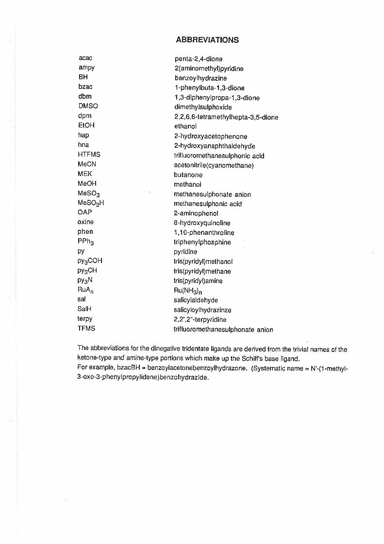
acac
ampy
BH
bzacdbm
DMSO
dpm
EtOH
hap
hna
HTFMS
MeCN
MEK
MeOH
MeSO3
MeSOsH
OAP
oxinephen
PPh3
py
py3OOH
pyscH
PYgN
RuAn
sal
SalH
terpy
TFMS
ABBREVIATIONS
penta-2,4-dione
2(aminomethyl)pyridine
benzoylhydrazine
1 -phenylbuta-1,3-dione
1,3-diphenylpropa-1,3-dione
dimethylsulphoxide
2,2,6, 6-tetramethylhepta-3,5-dioneethanol
2-hydroxyacetophenone
2 -hy dr oxy an ap ht h ald e h yd e
trif luoromethanesulphonic acidaceto nitrile(cyanomethane)
butanone
methanol
methanesulphonate anionmethanesulphonic acid2-aminophenol
8-hydroxyquinoline
1 ,10-phenanthrolinetriphenylphosphinepyridine
tris(pyridyl) methanol
tris(pyridyl)methane
tris(pyridyl)amine
Ru(NHs)n
salicylaldehyde
salicyloylhydrazinze
2,2',2"-lerpyridine
trif luoromethanesulphonate anio n
The abbreviations for the dinegative tridentate ligands are derived from the trivial names of theketone-type and amine-type portions which make up the schiff's base ligand.For example, bzacBH = benzoylacetonebenzoylhydrazone. (Systematic name = ¡rl'-(1-methyl-3-oxo-3-phenylpropyl idene) benzo hyd razide.
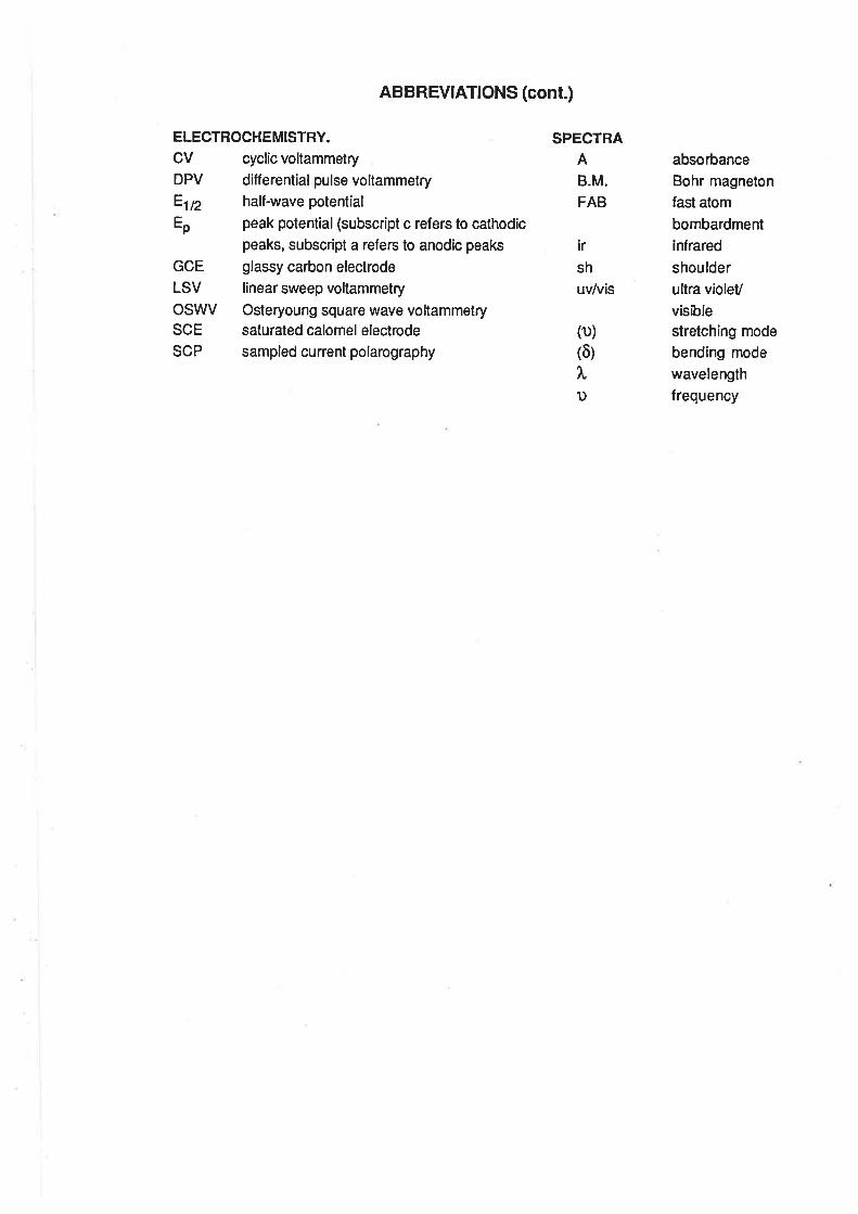
ABBREVIATIONS (cont.)
ELECTROCHEMISTRY.
CV cyclic voltammetry
DPV differentialpulse voltammetryÊln half-wave potential
Ep peak potential (subscript c refers to cathodicpeaks, subscript a refers to anodic peaks
GCE glassy carbon electrode
LSV linear sweep voltammetry
OSWV Osteryoung square wave voltammetrySCE saturated calomel electrode
SCP sampled current polarography
SPECTRAA
B.M.
FAB
ir
sh
uv/vis
(D)
(õ)
ì.1)
absorbance
Bohr magneton
fast atom
bombardment
infrared
shoulderultra violet/
visiblestretching mode
bending mode
wavelength
frequency
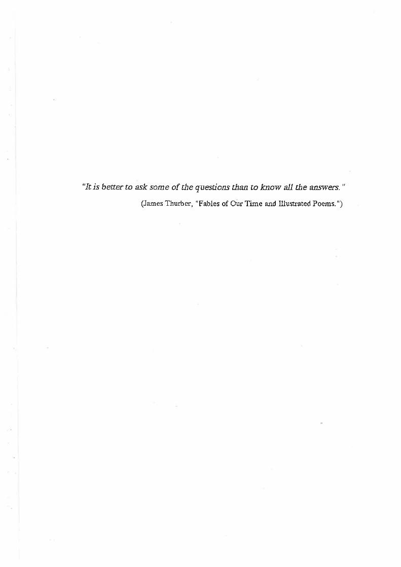
"It is beter to ask soÍne of the questions than to know all the an.sril'ers."
(James Thurter, "Fables of Our Time a¡rd Illustrated Poems. ")
TABLE OF CONTENTS.
STATEMENT
SUMMARY
ACKNOWLEDGEMENTS
ABBREVIATIONS
CHAPTER 1. ¡NTRODUCT¡ON.
1.1 Aims of the Project.
1.2 Factors Affecting the Stability of Ruthenium Oxidation States
1.4. Oxoruthenium(lV) Complexes With N-Donor Ligands.
1.4. Ruthenium(lV) Complexes With Bidentate Ligands.
1.5. Ruthenium(lV) Complexes With Chloro Ligands.
1.6. Ruthenium(lV)edtaComplexes.
1 .7. Tridentate Dinegative Ligands.
1.8. ElectrochemicalTechniquesandMaterials.
References.
CHAPTER 2. AMMINE AQUA RUTHENIUM COMPLEXES.
2.1. lntroduction.
2.2. lsolation of Triamminetriaquaruthenium(lll).
2.3 Acid-BaseProperties.
2.4 Electronic Spectra.
2.5. ElectrochemicalStudies.
2.6. Redox Chemislry.
2.7. Electrochemistry in Organic Solvents.
References.
CHAPTER 3. SUBSTITUTED TRIAMMINERUTHENIUM COMPLEXES.
3.1. lntroduction.
3.2. Ruthenium(lll)Complexes.
Preparation of Complexes
Electronic Spectra
Vibrational Spectra
The Electrochemistry of IRu(NHs)s(LL)(OH2)]2+ Complexes.
PAGE
1
2
5
8
10
11
12
12
17
20
21
21
24
25
31
33
37
40
42
42
43
45
46

The Electrochemistry of IRu(NH3)s(C2Od(OH2)l+.
Discussion of Electrochemical Results.
Comparison of E¡¡2with pK".
Electrolytic Reduction of Ru(lll) Complexes.
Description of the Crystal Structure of [Ru (NH3)3(acac) (OH2)](S206).2H2O.
3.3. Ruthenium(ll)Complexes.
Preparation of Complexes
Electronic Spectra
Vibrational Spectra
Electrochemical Studies
3.4. Triammine(tripod)ruthenium(ll) Complexes.
Preparation of Complexes
Electronic Spectra
Vibrational Spectra
Electrochemical Studies
Description of the Crystal Structure of [Ru(NH3)3{(py)3COH)]812.
3.5. Conclusions.
References.
CHAPTER 4. RUTHENIUM COMPLEXES WITH TRIDENTATE LIGANDS.
4.1. lntroduction.
4.2. Preparationof bis-(Tridentate) RutheniumComplexes.
4.3. ElectrochemicalStudies.
4.4. Mass Spectra.
4.5. VibrationalSpectra.
4.6. MagneticProperties.
4.7. Electronic Spectra.
4.8. Conclusion.
References.
CHAPTER 5. DINITROGEN AND NITROSYL COMPLEXES.
5.1. lntroduction.
5.2. Preparation and Characterizationof NitrosylComplexes.
5.3. lsolation of Triammineruthenium Dinitrogen Complexes.
5.4. Conclusion.
References.
52
53
56
57
58
59
59
61
63
65
68
68
69
71
71
71
73
75
80
80
B2
86
BB
8B
90
91
92
94
96
101
106
108
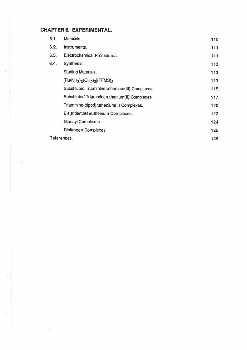
CHAPTER 6. Ð(PERIMENTAL.
6.1. Materials.
6.2. lnstruments.
6.3. ElectrochemicalProcedures.
' 6.4. Synthesis.
Starting Materials.
IRu(NHs)3(oH2)s](TFMS)g
Substituted Triammineruthenium(lll) Complexes.
Substituted Triammineruthenium(ll) Complexes.
Triammine(tripod) ruthenium(ll) Complexes.
Bis(tridentate)ruthenium Complexes.
Nitrosyl Complexes
Dinitrogen Complexes
Relerences.
110
111
111
113
113
113
115
117
120
122
124
125
128
Ghapter 1.
INTRODUCTION.
1.1. Aims of the Prolect.
Ruthenium is capable of forming compounds in at least 9 different oxidation states from 0 to
+8, but the most prevalent states are +2, +3 and +4. 11] However, there is a large difference in the
number of compounds formed in each of these oxidation states. ln a recent comprehensive work
on the chemistry of ruthenium [2], the chapters dealing with the oxidation state +2, +3 and +4 were
respectively 549, 180 and 62 pages long. While the discussion of the chemistry of Ru(ll) also dealt
with a large number of organometallic compounds, where the concept of oxidation state is more
formalthan in compounds with more eleclronegative donor atoms, there is stillobviously a much
larger number of compounds of Ru(ll) known than there is of Ru(lll), and in tum more of Ru(lll) than
of Ru(lV).
Additionally, the chemistry of Ru(lV) is dominated by multinuclear species. There are relatively
few mononuclear coordination complexes known in this oxidation state, [2a] whereas the
mononuclear complexes of Ru(ll) and Ru(lll) are, by far, the most prevalent.
ln recent times there has been a growing interest in the area of higher oxidation state
complexes of ruthenium, and many of the new complexes which have been synthesized are in the
+4 oxidation state. Meyer's group has demonstrated the existence of a relatively stable series of
oxoruthenium(lV) complexes containing diimine ligands [3-7], and also, in one case, ammine
ligands. [8] Poon's group has been able to obtain oxoruthenium complexes containing macrocyclic
tertiary amine ligands,[9-13] and Chakravorty's group has demonstrated the existence of a range of
Ru(lV) complexes containing N-donor ligands. [14-18] Finally, severalgroups have studied the
existence of Ru(lV) diketonate complexes. [19-23]
The aim of this project was to study the formation of simple coordination compounds
containing the oxoruthenium(lV), Ru=O2+, group. The hitherto little-known triammine series of
complexes was chosen as the group which offered the best scope to vary the coordination
environment by substitution at two coordinalion sites while maintaining the presence of an oxo
ligand. lt was proposed to form the oxoruthenium group by proton-coupled oxidation of a RuOH23+
group. A previous study has shown that it is possible to generate [Ru(NH3)5(O)]2+ by
electrochemical oxidation of [Ru(NHs)s(OHz)]3* at an anodically activated glassy carbon
voltammetric electrode. [8]
¡.
2
It was also intended to use the aqua ligand in the substituted triammineaqua ruthenium(ll)
complexes as a site for reaction with dinitrogen gas, and so form a series of substituted triammine
dinitrogen complexes. lt was hoped that any increased electron density on Ru(ll) which might be
induced by the substituting ligand would enhance the binding of the dinitrogen ligand to the metal
centre.
As an adjunct to this wod<, studies were also made of complex formation with a series of
dinegative, tridentate ligands based on aroyl hydrazones. These ligands have been shown to
stabilize vanadium(lV) in the absence of the vanadyl(V=g¡ group. 1Z4,2Sl
1.2. Factors Affecting the Stability of Ruthenium Oxidation States.
The relative stability of a complex in either of two oxidation states can be judged from the
reduction potential for the electron transfer between the two states. For the reaction:
Ox +e- + Red (1.r)
with a reduction potential, E, the lree energy change upon electron transfer will be given by the
equation:
Ao = _nFE (12)
lf a series of 6 coordinate complexes is studied, in which only one ligand is varied, (e.g.
IRu(NH3)5L]) then the entropy contributions to the free energy change associated with electron
transfer will be much the same for each complex in the series. The variation in E as the ligand is
changed may then be related to the contribution of the d electrons to lhe overall free energy
change which would be expected to be associated with the enthalpy term. Similarly, in the same
series of complexes, the variations in the equilibrium constant, K, for the attachment of the ligand
would be expected to reflect the effect of the ligand in altering the electronic energy of the
complex.
The ruthenium oxidation states, +2, +3, and +4 are d6, d5 and d4 systems respectively. The
complexes in the +2 and +3 states are most often low-spin mononuclear species while those of the
+4 state are sometimes low-spin mononuclear complexes, but more often diamagnetic,multinuclear
species. lâa,2b,2cl
The d¡ electrons of Ru(ll) participate in æ-backbonding to a greater extent than those in d6
complexes in higher oxídatíon states such as Co(lll) and Rh(lll). [26] Because of this, the Ru(ll)
complexes tend to contain ligands capable of strong fi-acceptance. This is the case with ligands
such as dinitrogen, carbon monoxide, organonitriles and pyridine ligands.
3
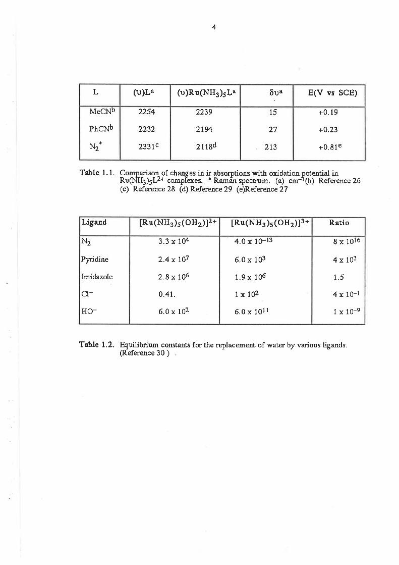
Clearly, when the æ-accepting ability of the ligand is quite good, it might be expected that the
Ru(ll) complex will be better stabilized to oxidation. lf the reduction in the ir stretching frequency of
a ligand X-Y, (D)XY, is taken as a measure of the ligand's ability to act as a æ-acceptor, then some
correlation might be expected between the reduction potential and the reduction in (u)X-Y. This is
exactly what is observed in the limited series shown in Table 1.1. lt can be seen that the higher
potential required to oxidize the terminaldinitrogen complex [27] ,compared with those of the two
organonitrile complexes [26], is consistent with the much greater reduction in (u)X-Y for the
dinitrogen ligand than for the nitriles. 128,291 As there is also some o-donor ability in the nitrile
ligands, the above c,omparison is made on the assumption that the æ-accepting ability of the nitriles
is the major contribution to the Ru(lI)-NCR bond.
A similar trend can be seen in the formation constant data displayed in Table 1.2. ll can be
seen that when the ligand is a good lt-acceptor, such as N2, the formalion constant for the Ru(ll)
complex is very much larger than that for the Ru(lll) complex with the same ligand. At the same time,
the formation constant for Ru(lll) complexes with poor n-acceptor ligands, such as Cl-, are very
much larger than those for the Ru(ll) complexes with the same ligands. [30]
As illustrated by the prevalence of Ru(ll) nitrile pyridine and polypyridine complexes, Ru(ll) is
not stabilized solely by zc-acceptor ligands, but also by ligands which have some o-donor ability.
Also, besides those ligands which are both æ-acceptors and o-donors, there are also compounds
of moderate stability which have purely o-donor ligands. These compounds include those with
ammine ligands, e.g. IRu(NHs)e]2* and [Ru(NHg)s(OHz)]z* [26], but such complexes do not favour
further substitution by n-donors. Hence, within the ammine series, there are no known examples
of complexes with ¡c-donors such as diketonates or carboxylates. Similarly, there are no examples
in which the Ru(ll) ion is complexed entirely by caÈoxylate ligands, and the only fully characterized
example of a tris diketonate complex of Ru(ll) involves trifluoropentanedionate as the ligand, which
has some fl-acceplor ability due to the electron withdrawing eff ects of the fluorine atoms. [31]
There are Ru(ll) complexes containing ligands such as diketonates and carboxylates where the co-
ligands are good fi-acceptors such as phosphines and carbon monoxide. lâe,2Íl
To conclude, the +2 oxidation state of ruthenium is stable with both o-donor and r-acceptor
ligands, and in.some cases one ligand provides a suitable combination of both properties. ru-
donors will only coordinate to Ru(ll) when n-acceptors are present.
The +3 oxidation state is also capable of existing with ligands which are both æ-acceptors and
o-donors, such as nitriles and pyr¡dyls. Within the pentaammine and tetraammine series, the nitrile
and pyridine ligands form complexes which are quite stable and the Ru(lll)/Ru(ll) electrochemical
4
+0.19
+0.23
+0.81e
15
27
213
2239
2t94
2n8d
2254
2232
233tc
MeCNb
PhCNb
Nz*
E(V vs SCE)ôua(u)R.u(NHs)sL"(1))L"L
Table 1. 1. Comparison of chanses in ir absorotioru with oxidation oRug.lua¡rLz+ complexes. * Ramän ï)ectrum. (a) cm-1(c) Reference 28 (d) Reference 29 (e)Reference2T
otential in(b) Reference 26
8 x 10¹⁶
4x 10³
1.5
4 x 10-l
1 x 10-e
4.0 x 10⁻¹³
6.0 x 10³
1.9 x 10⁶
1x10²
6.0 x 10¹¹
3.3 x 10⁴
2.4 x 10⁷
2.8 x 10⁶
0.4t.
6.0 x 10²
N2
Pyridine
Imidazole
cl-
HO-
R.atio[Ru (NH3 )s(oHz)]3 *IRu(NH3)s(oHz)]2*Ligao:d
Table L.2. Quilibrium constanfs for the replacement of water by various ligands.(Reference 30 )
5
couples are reversible. However, in the case of Ru(lll) complexes, the major contribution to metal-
ligand bond formation comes from the ability of the ligand to form a o-bond. This is illustrated by the
case of the nitrile ligands where the frequency of the ir absorption due to (ù)CN increases on
complex formation as a result of the loss of delocalization of electron density from the nitrogen lone
pair into the C-N antibonding orbitals, without concomitant back donation from the metalcentre.
[261
The importance of o-donation from the ligand to the Ru(lll) centre is further exemplified by the
existence of a large number of ammine carboxylates and diketonates, as well as the tris carboxylate
and diketonate complexes. [2d, 19,21-231
The coordination chemistry of Ru(lV) is dominated by bridged, multinuclear complexes. [2a]
The bridging ligands are quite often very strong æ-donors such as oxo and nitride ions. Also there
are a number of complexes containing phosphinimate, R3P=N-, ligands.[32] ln the aqueous
chemistry of Ru(lV), also, there are many examples of bridged multinuclear complexes. These are
mostly the aqua halo complexes, and may consist of up to four ruthenium ions, often with these
ions existing in several ditferent oxidation states within the complex.
The existence of the large number of bridged complexes of Ru(lV) is a direct consequence of
the ability of some ligands to stabilize this oxidation state. The very strong æ-donors, such as 02-
and N3- are also ligands which tend to act as very good bridging groups. [33] The existence of the
stable hexachlororuthenium(lV) ion provides good evidence that the Cl- ligand is also a sufficiently
strong æ-donor to stabilize the Ru(lV) centre. [34]
Recent electrochemical studies on the tris diketonate Ru(tll) complexes have also indicated
that these ligands are capable of stabilizing the +4 oxidation state. [20-23] With these ligands, and
perhaps other strong ft-donors, it may be possible to isolate other stable mononuclear Ru(lV)
complexes in the ammineruthenium series.
1.3. Oxoruthenium(lV) Complexes With N-Donor Ligands.
There are three classes of N-donor ligands under consideration. The polypyridyl groups
employed by Meyer's group, the macrocylic tertiary amines used by Poon's group and the
arylazopyridine ligands used by Chakravorty.
1.3.1. Polypyridyl Ligands. lnterest in this cfass of compounds stems from the ability of
tRu(trpy)(bpy)O1zl2* and similar complexes to acl as catalytic oxygen atom transfer agents. [3] ln
aqueous solution IRu(trpy)(bpy¡O{2lz+ is capable of undergoing two one electron oxidations with
coupled proton transfer.
6
[Ru(trpy)(bpyXO)]2* + e- + Hl-
lRu(trpyXbpy)OH¡z+ * F + H+
[Ru(trpy)(bpy)OH¡z+
IRu(trpy)(bpylCr$zlz*
(1.3)
(1.4)
(1.s)
The E1¡2values are +0.61 V vs SCE and +0.48 V vs SCE ,respectively, at pH 7. ln the presence of
organic molecules such as ethanol, propan-2ol or ethanal, the organic molecule reduces the
Ru(lV) oxo complex to the Ru(ll) aqua complex, and the organic molecule is oxidized. For propan-2-
ol, the overall reaction is:
tRu(trpyXbpyxo)12* + CH3CH(OH)CH3 + [Ru(rrpyXbpy)Oïzl2* + CH3C(O)CH3 (i.s)
Under conditíons of electrolytic oxidation of the Ru(ll) complex, the whole cycle could be made
catalytic and, over 100 redox cycles, 10-20% of the alcohol was oxidized. [3] The electrochemistry
of [Ru(trpy)(bpy)OH2]2+ was investigated over a wide range of pH values, and the pH dependence
of the E172 values was consistent with the pK" values measured by pH titration of the Ru(ll) and
Ru(lll)complexes. [4]
The electrochemistry of the IRu(bpy)z(py)o]2+/[Ru(bpy)2(py)oHz]2+ system was consistent
with the existence of two reversible electron transfer steps, which were pH dependent. At pH 7,
the E172 value for the Ru(lV)/Ru(lll) couple was +0.53 V, and for the Ru(lll)/Ru(ll) couple, +0.42Y.
[Ru(bpy)z(py)O]2* was isolated as its perchlorate salt, and the presence of the oxoruthenium
group, Ru=O2+, was confirmed by isotopic labelling with 018. ln the ir spectrum, (u)Ru=O16 was
observed alTgzcm-1 and (r)Ru=O18 al752cm-1. [5]
The complex crs [Ru(bpy)zQïz)z]2+ was shown to be capable of four one electron/one
proton oxidations to give a dioxoruthenium (Vl) complex according to reactions 1.6 to 1.9. [6]
øs[Ru(þy)2(O)zl2* + e-+H+ øs IRu(bpy)2(OHXO)]2+ (1.6)
crs[Ru(þy)2(OHXO)]2+ + e- + H+ cr's [Ru(bpy)2(oH2XO)]2+ (.7)
crs [Ru(bpy)2(OHj(O)]2+ + e- + H+ cis [Ru(bpy)2(oH2xoH)]2+ (1.8)
cis [Ru(bpy)2(OH2XOH)]2+ + e- + H+ ø.s [Ru (bpy) z(oH z) zl2,
-
._\
.._\
Ihe E1¡2 values for these reaclions were reported as +1.07, +0.94, +0.76 and +0.53 V
respectively, at pH 4.
*Henceforth, all E values willbe quoted vs the SCE unless otherwise stated.

7
Oxidation of cis [Ru(bpylzQ1z)zl2+ with 3 equivalents of Ce4+ ion resu]ted in the Ru(V)
complex cris [Ru(bpy)2(OHXO)]2+, the perchlorate sah of which had an ir absorption at 857 cm-1
which was assigned to the (u)RuO vibration. The perchlorate salt of cis [Ru(bpy')z(Olzl2+,obtained
by the reaction of excess Ce'î+ ion with criç [Ru(bpy)2(Oïz)zl2*, showed two bands attributable to
(r)RuO in the ir spectrum, at 860 cm-1 and 835 cm-1.
A phosphineoxoruthenium(lV) complex has also been isolated. [7] By oxidation of crs
tRu(bpy)2(OH2)PEtsl2+ with Ce4, it was possible to obtain cis [Ru(bpy)2(O)PEt3l2+. This
compound also had a wellbehaved electrochemistry and, at pH 7, it was possible to observe the
redox couples 1 .10 and 1 .1 1 with E172 values ol +0.72 V and +0.46 V respectively.
tRu(þy)lo)PEtu¡2+ + e- + H+ + [Ru(bpy)2(OH)Pft3]2+ (1.10)
tRu(þy)2(oH)PEtsl2+ + e- + H+ + [Ru(bpy)2(OH2)PEts]2+ (1.11)
The presence of the oxo group was confirmed by the presence of an ir absorption at 790 cm-1.
The Ru(lV) complex was also shown to be capable of oxidizing a variety of organic and inorganic
substrates. [7]
1.3.2. Macrocyclic Tediary Amine Ligands. ln 1984, Poon and co-workers reported that reaction of
trans [Ru(tmc)Cl2]Cl (tmc = 1,4,8,11-tetramethyl-1 ,4,8,1 1{etraazacyclotetradecane) with 30%
hydrogen peroxide gave a complex which they formulated as trans [Ru(tmc)(H2O)(O)]2+. [9] As ¡ts
perchlorate salt, this compound had an intense ir absorption at 855 cm-1. However, this product
was later positively identified as the Ru(Vl) complex lrans [Ru(trncl(O)zl2*. [10] The cyclic
voltammogram of this complex showed one two electron and two one electron waves, which were
assigned to the electron transfer couples shown as 1.12 to 1.14:
frans[Ru(tnrcXO)z]2* + 2e- +2H+ == frans[Ru(tmcXHzOXO)]2+ (1.12)
frans[Ru(tmc)(H2O)(O)]2+ + e- +2H+ == Íans[Ru(tmc)(H2O)2]+ (1.r3)
frarsIRu(tnrc)(HzO)ds +e- == Íans[Ru(tmc)(Hzo)zl2* (1.14)
fhe E1¡2values were +0.66 V, +0.36 V and +0.15 V, respectively, in 0.1 mol dm-3 HClo4.
The dioxoruthenium complex was shown to be capable of reacting with PPh3 in acetonitrile to
give trans [Ru(tmc)(MeCNXO)]2+. [11] The crystal structure of the PFs- salt revealed a Ru=O bond
distance of 1.765(5) A. The (o)RuO absorption in the ir spectrum could not be positively identified
because of absorptions due to the tmc ligand in the region 7OO-B2O cm-1. The other product of

8
the reaction was O=PPhg, indicating that the reduction of the Ru(Vl) complex was accompanied by
an oxo atom transferto the phosphine. There was also a report of the complex trans
[Ru(tmc)(CIXO)]*, with a Ru=O bond length of 1.765(7) A, and having a quasi-reversible one
electron reduction couple in acetonitrile at +1.1 V vs cp2Fe+lcp2Fe. [12] This complex was capable
of actíng as an electrocatalytic agent for the oxidation of benzyl alcohol.
ln later work, the same workers were able lo show that the Ru=O distance in the trans
dioxoruthenium(Vl) complexes was insensitive to the ring size of the macrocycle. [13]
1.3.3. Arylazopyridine Ligands. The existence of well-defined proton-coupled electron lransfer
reaclions involving Ru-OHr2+ ând Ru=O2+ compounds has also been demonstrated for complexes
containing arylazopyridine ligands. For [Ru(paplzþyl}Hdz+ (pap = phenylazopyridine), the two
electron redox couple 1.15 has been observed.[14]
lRu(pæ)z(pyxo)12* + Ze- + zH+ + [Ru(pap)2(py)oHz]2+ (1.15)
E1¡2lor the couple is +1.20 V in the pH range 1-4. The Ru(lV) complex was shown to be capable of
oxidizing H2O to 02.
Later, it was demonstrated that the hydroxoaqua and dihydroxo complexes, such as
IRu(pap)2(OH2)(OH)]2+and [Ru(pap)z(OH)z]* are capable of one electron oxidation to the
corresponding Ru(lV) complex. [15] e.g. E1¡2lor couple 1 .16 is +1 .89 V, and for couple 1 .17 is
+1.53 V
[Ruv(pap)2(Oti2xoH)]s + e- + [Rul¡l1pap)2(OH2XOH)]2+ (1.16)
lnulvlpap¡21o+)¿z+ + e- + [Rulll(pap)2(OH)z]* F.17)
It is apparent from these data lhat the hydroxo ligand is not as good at stabilizing the Ru(lV)
oxidation slate as the oxo ligand.
1.4. Ruthenium(lV) Gomplexes With Bidentate Ligands
1.4.1. Arylazopyridine Ligands. The diaqua complexes, [Ru(pap)2(OH2)212*, react with bidentate
ligands such as acac-, bpy, en and triazene-1-oxides to give complexes of the type,
[Ru(pap)2(Ll)¡n+ ¡n solution. [16] ln the case of the complex, [Ru(tap)2(acac)]+ (tap =
tolylazopyridine), the Ru(ll)/Ru(lll) oxidation occurs at +1.34 V and the Ru(lll)/Ru(lV) oxidation wave
was observed at about +2 V in the CV. However, it was not possible to isolate the oxidized species
I
1.4.2. RuN3O3 Complexes. A sedes of complexes, [RuLg], (L = [X-C6Ha-N-N=lrt19¡Rþ has been
synthesized. [17] ln acetonitrile, these complexes displayed a quasi-reversible wave in the region
-0.060 V to -1.20 V, assigned to the Ru(lll)/Ru(ll) couple, and a reversible wave in the region +0.20
V to + 0.70 V, assigned to the Ru(lV)/Ru(lll) couple.
Electrolysis of several of the complexes was reported, and the uv/vis spectra of the oxidized
species were determined and compared with those of the Ru(lll) complexes. None of the Ru(lV)
complexes were isolated. On electrolytic reduction of the complexes in acetonitrile, the reduced
species were observed to decompose. For both the Ru(lV)/Ru(lll) and Ru(lll)/Ru(¡l) couples there
was a good correlation between the E172 values and the Hammett constant lorthe sustituent X.
When two of the triazene-1-oxide ligands in the above complexes were replaced by a
bipyridine ligand, to give [Ru(bpy)21]+, the Ru(lll)/Ru(ll) couple was shifted to a potential in the
range +0.16 V to +0.44 V. [18] A further oxidation wave in the CV was observed in the region
+1.40 V to +1.70V, but there was some uncertainty about whether this oxidation was metal or
ligand-based.
1.4.3. Ru(diket)3 Complexes. The one electron oxidation of Ru(lll) diketonate complexes in non-
aqueous solutions has been the subject ol much study. [19-231 Fackler and co-worker
investigated the redox chemistry of the M(acac)3 complexes for a number of metals. [20] ln the
case of the ruthenium complex, it was found that there was a reversible oxidation in the CV at +1.05
V vs the Ag/AgCl electrode in a number of organic solvents. Although the oxidized species
showed no tendency to undergo lurther reaction on the voltammetric time scale, it proved to be
unstable over the longer time period required for electrosynthesis. ln N, N'-dimethylformamide, the
oxidation process was irreversible, and there was no evidence of the return cathodic wave after
oxidation.
ln two studies on the electrochemistry of the Ru(lll) complexes with the ligands [R-C(O)-C(R)-
C(O)-R']-, the substituents R and R" were varied with R' =H [21] and the substituent R'was varied
with R = R" = CHg.l22l For R'= H, the E172 values for oxidation and for reduction were plotted
against the sum of the Hammett constants of the substituents. The E1¡2values for reduction lie
along a straight line, while the E1¿2 values for oxidation appear to lie along two horizontal lines. ln
the second case, it was found that the oxidation and reduction potentials could be expressed as a
function of the sum of the Hammett constants il lhe para- position value was used for R', and the
meta- position values were used for R and R".
A later study was concerned with the redox chemistry of a greater number of tris diketonate
complexes than the previous work. [23] ll was found that both the reduction potential and oxidation
potential of the Ru(lll) complexes varied linearly with the Talt parameters, and with almost ider¡tical
10
slopes. The reduction potentials were in the range --0.67 V lo +0.27 V and the oxidation potentials
were in the range +1.09 V to +1.88 V vs Ag/AgCl.
The uv/vis spectra of Ru(diket)3 complexes were recorded in dichloromethane, and the
OTTLE technique was used to study the spectra of the Ru(ll) and Ru(lV) complexes. [23] lt was
found that all spectra contained intra- ligand absorptions in addition to absorptions assigned to
charge transfer transitíons. When the Ru(lll) complexes were subjected to reductive electrolysis,
the intensity of the intra-ligand bands increased markedly, sometimes with a shift in the absorption
maximum, and one of the LMCT bands disappeared while that at lower energy increased in
intensity, possibly due to MLCT. Under conditions of oxidative electrolysis, the intensity of the
intraligand bands did not increase by a great amount, but the absorption maximum shifted to a
longer wavelength as an intense absorption was induced at wavelengths shorter than 250 nm (the
absorption cutoff for the solvent). One charge transfer band disappeared and the low intensity
band at lower energy was shifted to a longer wavelength, with a higher absorbance. For example, in
the case of Ru(acac)3, the Ru(lll)complex had absorptions at 275 nm (A = 0.49 in the
spectroelectrochemicalcell),347 nm (A = 0.21) and 504 nm (A = 0.03). After reduction, there were
bands a1275 nm (A = 0.92), 347 nm ( a shoulder with A = 0.012) and 504 nm (A = 0.38). After
oxidation, there were bands aI284 nm (A = 0.4Íl), 347 nm (A = 0.05) and 700 nm (A = 0.13). [23]
1.5. Ruthenium(lV) Gomplexes With Ghloro Ligands.
1.5.1. Macrocyclic TeÉiary Amine Ligand Complexes. By electrochemical oxidation of the
complexes, frans [Ru(tmc)zXzl+ (X = Cl, Br, or NCO), Poon and co-workers were able to form the
Ru(lV) complexes, trans [Ru(tm c)Xzl2* . [35] ln acetonitrile, trans [Ru(tmc)Cl2]+ had a cyclic
voltammogram whích consisted of two quasi-reversible waves at -0.56 V and +1.21 V vs the
cp2Fe+lcp2Fe couple. (i.e. -0.25 V and +1.54 V vs SCE. [36]) These waves were assigned to the
Ru(lll)/Ru(ll) and Ru(lV)/Ru(lll) couples respectively. lt was possible to follow the efectrochemical
formation ol trans IRu(tmc)Cl2l2, by spectroscopic means, but the controlled-potentíatoxidation at
+1.30 V showed that the oxidative current did not decay to a background level, but remained at a
constant value as oxidation proceeded. lt was proposed that some of the electrochemically
generated Ru(lV) complex reacted with a non-electroactive species to regenerate the starting
material.
The large difference in the oxidation potentials of trans [Ru(tmc)2Ol2l+ and trans
[Ru(tmc)(H2O)z]3* (as shown in Table 1.3) is a good illustration of the ability of strong ¡r-donor
ligands to stabilize Ru(lV). Cl- is a weaker n-donor ligand than O2-, and it can be seen that it lacks
sufficient æ-donating powerto be able to stabilize the +4 oxidation state as well as O2-. fne
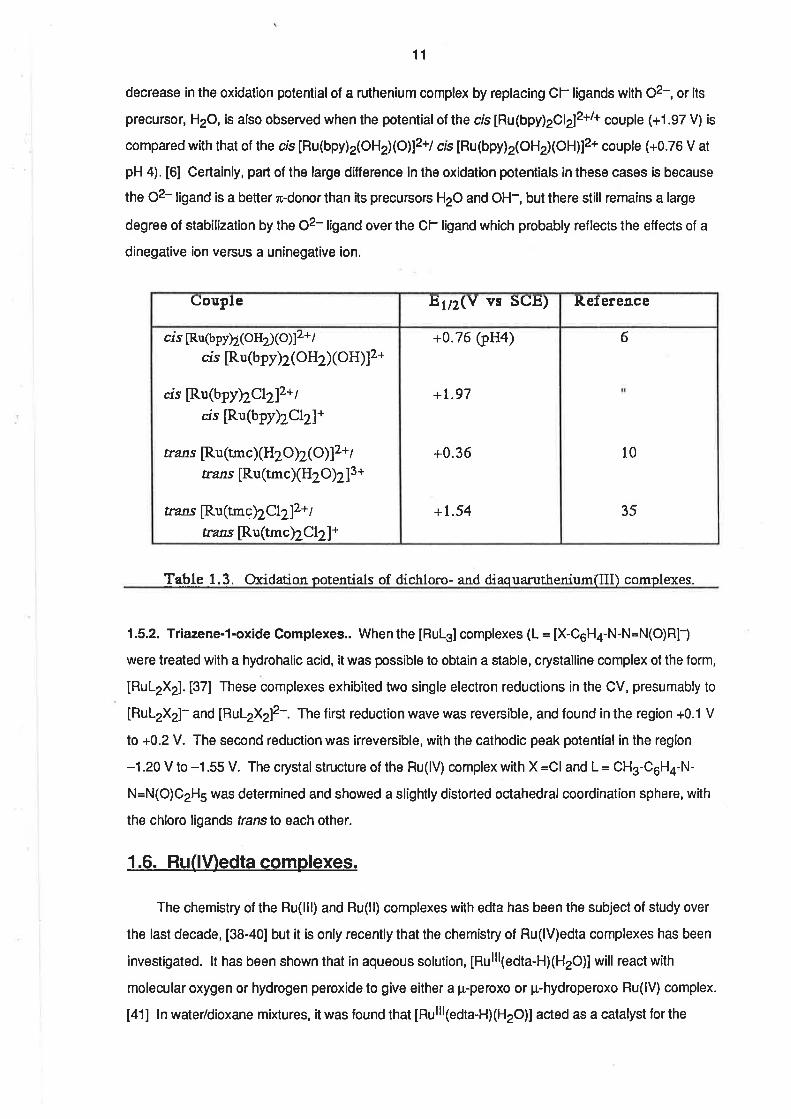
11
decrease in the oxidation potentialof a ruthenium complex by replacing Cl- ligands with O2-, or its
precursor, H2O, is also observed when the potential of the cis [Ru(bpy)29l2l2+l+ couple (+1.97 V) is
compared with that of the cis [Ru(bpy)2(OH2XO)]2+/ crs [Ru(bpy)2(OH2XOH)]2+ couple (+0.76 V at
pH 4). [6] Certainly, part of the large difference in the oxidation potentials in these cases is because
the 02- ligand is a better æ-donorlhan its precursors H2O and OH-, but there still remains a large
degree of stabilization by the 02- ligand over the Cl- ligand which probably ref lects the etfects of a
dinegative ion versus a uninegative ion.
6
10
35
+0.76 (pH4)
+t.97
+0.36
+1.54
cis [Ru(bpyþ(oH2xo)]2+ /
crs [Ru(bpy)2(OH2)(oH)12+
as [Ru(bpyþCL2lz+tas [Ru(bpyhClz]*
rrans [Ru (tmc)(H2 Oþ ( O) ]2+ I
rrans [Ru(tmc)(H2O)2]3+
rrans [Ru(tmcþCL2]z+tazns [Ru(tmcþCI2]+
Ref erenceEtn(Y vs SCE)Couple
Table 1.3. Oxidationpotentials of dichloro- and diaquaruthenium(Ill) complexes.
1.5.2. Triazene-1-oxide Complexes.. When the [RuL3] complexes (L = [X-C6H4-N-N=N(O)R]-)
were treated with a hydrohalic acid, it was possible to obtain a stable, crystalline complex of the form,
[RuL2X2]. [37] These complexes exhibited two single electron reductions in the CV, presumably to
[RuL2X2]- and [RuL2X2]2-. The first reduction wave was reversible, and found in the region +0.1 V
to +0.2 V. The sec¡nd reduction was irreversible, with the cathodic peak potential in lhe region
-1.20 V to -1.55 V. The crystal structure of the Ru(lV) complex with X =Cl and L = CH3-C6H¿-N-
N=N(O)CzH5 was determined and showed a slightly distorted octahedralcoordination sphere, with
the chloro lígands translo each other.
1.6. Ru(lV)edta complexes.
The chemistry of the Ru(lll) and Ru(ll) complexes with edta has been the subject of study over
the last decade, [38-40] but it is only recently that the chemistry of Ru(lV)edta complexes has been
investigated. lt has been shown that in aqueous solution, lnulllledta-HXHzO)lwill react with
molecular oxygen or hydrogen peroxide to give either a p-peroxo or ¡r-hydroperoxo Ru(lV) complex.
[41] ln water/dioxane mixtures, it was found that lnulllledta-HXHzO)] acted as a catalyst for the
12
oxidation of PPh3 by oxygen. [42] lt was proposed that the oxidation reaction proceeded through
the intermediates lnulvleOta¡1pphs)12o2 and [Ruv=O(edta)(pph3)].
1.7. Tridentate Dinegative Ligands.
The demonstrated ability of the oxo ligand to stabilize the Ru(lV) ion, parallels the importance
of the oxo ligand in stabilizing the vanadium (lV) oxidation state. The vanadyl, V-O2+, group is
almost ubiquitous in the chemistry of vanadium(lV). However, some recent studies have
demonslrated that other r-donor ligands are capable of stabilizing this oxidation stale.
ln 1975, Diamantis ar¡d co-workers reported the synthesis of a non-vanadyl V(lV) complex
containing a tridentate dinegative ligand. [2a] The ligand, penta-2,4-dionebenzoylhydrazonate,
was formed in situ in the react¡on between V(O)(acac)2 and benzoylhydrazíne. Later work by the
same group showed that it was possible to synthesize a large number of analogous complexes
using a variety of diketones and aroylhydrazones to pre-form the ligand prior to reacting it with a
vanadium compound. [25]
ln the light of this information it is reasonable to expect that the ligands which have proved to
be sufficiently strong fi-donors to stabilize V(lV) may also stabilize Ru(lV). For this reason, attempts
were made to synthesize ruthenium(lV) complexes with these ligands.
1.8. Electrochemical Techniques and Materials
1.8.1. Fundamentals. Voltammetry is concerned with the current-potential relationship in an
electrochemical cell and, in particular, with the current-lime response at a solid electrode at a
controlled potential. lf the potential is held, or stepped, to a potential at which charge transfer
occurs between the oxidized and reduced forms of a redox couple, a faradaic current willflow.
Reversible charge transfer reactions are those forwhich the rate of charge transfer is fast compared
with the time scale of the experiment. When the rate of electron transfer is slow compared with the
time scale of the experiment, the charge transfer is deemed irreversible.
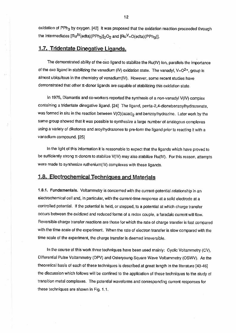
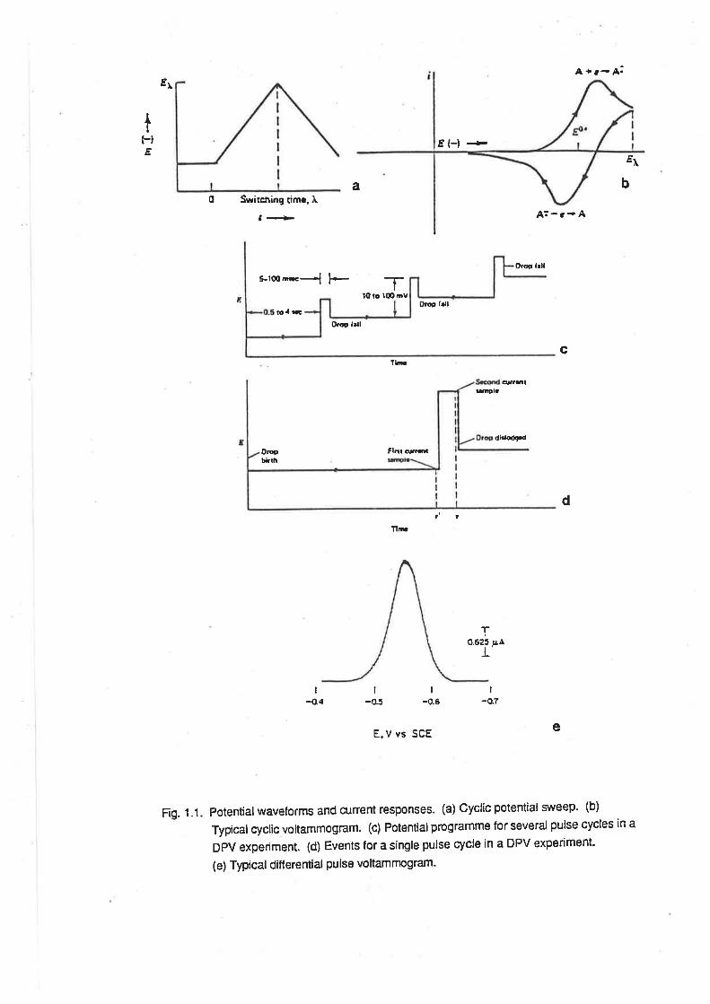
ln the course of this work three techniques have been used mainly: Cyclic Voltammetry (CV),
Differential Pulse Voltammetry (DPV) and Osteryoung Square Wave Voltammetry (OSWV). As the
theoretical basis of each of these techniques is described at great length in the literature [43-46]
the discussion which follows will be confined to the application of these techniques to the study of
transition metal complexes. The potential waveforms and corresponding current responses for
these techniques are shown in Fig. 1.1.
A rr- A.
t(-lÊ
á
El-l -àEr
ba0 Swirc¡ing rimc. tr
¡61-
*rmæl l+ -r
A:-c- A
Oroo llll
Orq frll
lO lo læ Dv
f¡d
P¡6p lall
c
q¡Dlaoarït
didodçd
OrootÉrh
Fl¡¡smr
llr
d
ï0.625 pa
E,V vs SCE
Potentialwaveforms and current responses. (a) Cyclic potential si¡'eep' (b)
Typical cyclic voltammogram. (c) Potential programme for several pulse cycles ¡n a
DPV experiment. (d) Events for a single pulse cycle in a DPV experiment-
(e) Typical ditf erential pulse vo ltamrnogram.
I
-07I
-o.6I
-o5I
{14
e
Fig. 1.1
STEP FREOUENCY
S.!V AMPLITUOE
SAMPLINGPOINTS
(W¡OTH ¡
QU¡ET T¡ME
.o.t(xtÞt. nEil¡-n¡r
t cEErt- t.tll -
Erllr-Î|
Fig. 1.1.(cont.) (f) Potential programme for Osteryoung square wave voltammetry.
(g) Typical Osteryoung square wave voltammogram.
E
tt
g
13
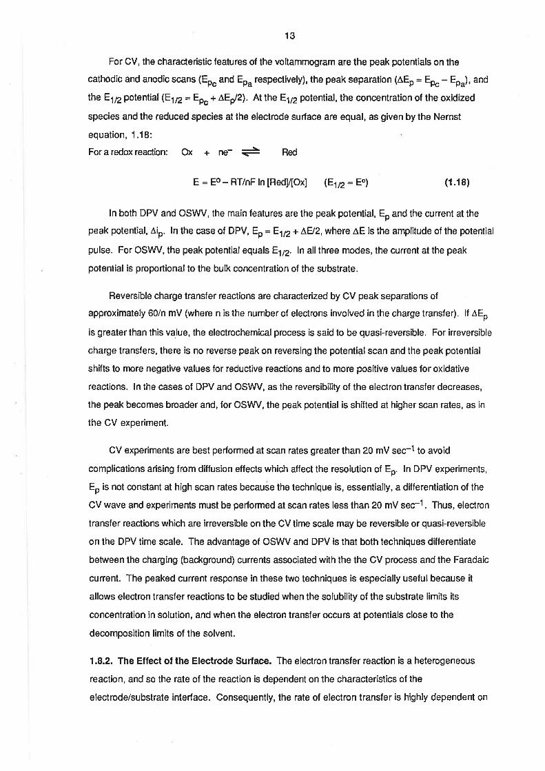
For CV, the characteristic features of the voltammogram are the peak potentials on the
cathodic and anodic scans (Eo" and Eo" respectively), the peak separation (ÂEo - Epc Eo"), and
lheEl¡2potential (Etn= EO"+ÂEy'2). Atthe ElT2potential,theconcentrationof theoxidized
species and the reduced species at the electrode surface are equal, as given by the Nernst
equation, 1.18:
Foraredoxreaction: ox + ne- + Red
E = Eo - RT/nF ln [Red![Ox] (Etp= Eo) (1.18)
ln both DPV and OSWV, the main features are the peak potential, Eo and the current at the
peak potential, Âio. ln the case of DPV, Ep= E.ttz+ LH2, where ÂE is the amplitude of the potential
pulse. For OSWV, the peak potential equals E1¡2. ln allthree modes, the current at the peak
potential is proportionalto the bulk concentration of the substrate.
Reversible charge transfer reactions are characterized by CV peak separations of
approximately 60/n mV (where n is the number of electrons involved in the charge transfer). lf lEo
is greater than this value, the electrochemical process is said to be quasi-reversible. For irreversible
charge transfers, there is no reverse peak on reversing the potential scan and the peak potential
shifts to more negative values for reductive reactions and to more positive values for oxidative
reactions. ln the cases of DPV and OS\Â/V, as the reversibility of the electron transfer decreases,
the peak becomes broader and, for OSWV, the peak potent¡al is shifted at higher scan rates, as in
the CV experiment.
CV experiments are best performed at scan rates greater than 20 mV sec-1 to avoid
complications arising from dillusion eflects which affect the resolution of Eo. ln DPV experiments,
Eo is not constant at high scan rates because the techn¡que is, essentially, a dilferentiation of the
CV wave and experiments must be performed at scan rates less than 20 mV sec-1. Thus, electron
transfer reactions which are irreversible on the CV time scale may be reversible or quasi-reversible
on the DPV time scale. The advantage of OSWV and DPV is that both techniques differentiate
between the charging (background) currents associated with the the CV process and the Faradaic
cunent. The peaked current response in these two techniques is especially usefulbecause it
allows electron transfer reactions to be studied when the solubility of the substrate limits its
concentration in solution, and when the electron transfer occurs at potentials close to the
decomposition limits of the solvent.
1.8.2. The Effect of the Electrode Surface. The electron transfer reaction is a heterogeneous
reaction, and so the rate of the reaction is dependent on the characteristics of the
electrode/substrate interface. Consequently, the rate ol electron transfer is highly dependent on
14
the state of the electrode surface. Generally, to obtain reproducible results, it is necessary for the
solid electrode surface to be highly potished. ln the case of glassy carbon electrodes, other pre-
treatment procedures have been used in order to enhance the electron transfer, such as coating
the electrode with a film which reversibly binds the substrate. [44
Another such pre-treatment has been the application of high anodic potentials to the working
electrode. This procedure has been lound to increase the anodic current for oxidative electron
transfers, and to improve the reversibility of the response. Anodic activation has also facilitated the
observation of reactions not normally observed on platinum or untreated glassy carbon electrodes.
[48-50]
Meyer's group has studied the surface of anodically activated glassy carbon using X-ray
photoelectron spectroscopy. [49] Their studies revealed that the activated electrodes had a much
higher surface oxygen content than the unactivated electrodes. Most of the oxygen seemed to be
present in either phenoUalcohol or quinone/carbonyl groups. Confirmation of this assignment
comes from another study in which glassy carbon electrodes modified by the adsorption of
quinone/hydroquinone groups on the electrode surface, improved the reversibility of the oxidation
of ascorbic acid. [51]
Other workers have established that similar surface groups are to be found on electrodes that
have been polished to a high degree with alumina having a particle size of 0.05 pm. [52] These
authors reported that the electrode was reproducibly activated towards the oxidation of a variety of
substrates, and that the electrode had surface groups similar to those on the anodically activated
electrodes. lt was noted that the small resídual amounts of alumina on the surface were unlikely to
be the cause of the activation, as had been observed previously. [53]
There has been much discussion concerning the mechanism by which the anodic activation
procedure enhances the electron transler at the eleclrode. Engstrom [48] has proposed that the
activation must involve either the oxidation of the surface to produce functionalities capable of
catalysing the electrochemical reaction or the oxidative removal of impurities from the surface. lt has
been proposed that the act¡vat¡on process occur either directly by electrochemical oxidation of the
electrode surface or indirectly by oxygen produced during anodization. lt has also been argued
that the increased oxygen content at the surface of the electrode acted by ahering surface charge
density or by the creation of catalytically active sites. [52]
ln studying the electrochemicaloxidation of [Ru(NH3)sOHz]3* and [Ru(bpy)z(OHz)]3*, Meyer
has argued that the surface groups formed by the anodization procedure would be capable of
mediating the coupled proton transfer. [49] lt was argued that the presence of these surface
15
groups increases the rate of electron transfer by maintaining an equilibrium concentralion of
protons at the electrode surface. The existence of a strong deuterium isotope effect in the
oxidation of cis [Ru(bpy)z(OHz)]3+ by hydrogen peroxide is evidence that proton transfer is the rate
limiting step in the oxidation reaction. [54]
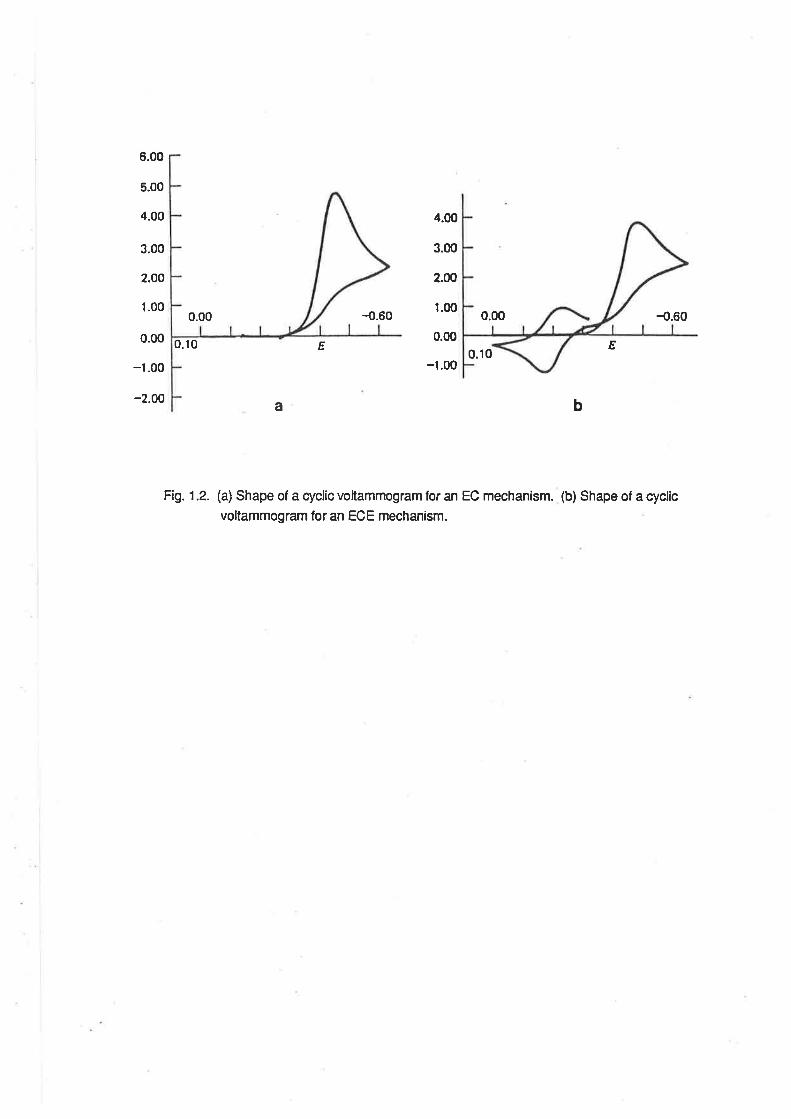
f .8.3. Chemical Reac'tion After Elec'trcn Transfer. ln discussing reversible CV experiments, it is
assumed that the product of the charge transfer reaction will be long-lived. However, if the product
of electron transfer undergoes some chemical reaction, viz:
Ox+ne- + Red
Red + z (1.1s)
there may be none of the species, Red, in the vicinity of the electrode when, after the scan
direction has been reversed, the potential of the cell passes through the value at which electron
transfer occurs. Thus, even though the electron transfer may satisfy the criteria of electrochemical
reversibility, the reverse electrochemical process will not be observed. The CV will resemble that
for an irreversible charge transfer. This situation is refened to as the "EC" (Electrochemicalfollowed
by Chemical reaction) mechanism of electron transfer. [aab] Fig 1.2. shows the shape of an EC CV
wave.
lf the product of the chemical reaction of Red is also capable of undergoing electron transfer
reactions, the peak potential associated with electron transfer to or from Z may be detected if it falls
within the potential limits of the CV experiment. This situation is referred lo as the 'ECErr
(Electrochemicalfollowed by Chemicalfollowed by Electrochemical reaction)mechanism. [aab] Fig.
1.2 shows the shape of an ECE CV wave.
EC and ECE mechanisms are also discernible in the DPV and OSWV modes, usually giving
rise to peak potentials and current maxima which are scan rate and direction dependent. lf the rate
of the reaction of Red is comparable to the voltammetric time scale, the relative heights of the
various peaks in the voltammogram will be scan rate-dependent. There is an extensive treatment of
the manner in which kinetic data may be obtained lrom CV experiments involving EC and ECE
mechanisms in Chapter 11 of relercnce M.
1.8.4. Proton-Coupled Electron Transfer. For a redox reaction in which electron transfer is
accompanied by proton transfer, e.g.
Ox+ne- +mH+ + Red (1.20)
6.00
5.00
4.00
3.00
2.00
1.00
0.00
-1.00
-2.00
0.00 4.60
4.00
3.00
2.00
1.00
0.00
-1.00
0.00 {).60
E0.10 E0.10
b
Fi1.1.2. (a) Shape of a cyclic voltammogram for an EC mechanism. (b) Shape of a cyclic
voltammogram for an ECE mechanism.
a
16
rearrangement of the Nernst equation gives the relationship,
E=E1t2- m/n x .059 pH. (1.211
where E is in volts.
For a one electron reduction, the value of the constant will be 59 mV for m = 1, 118 mV for m =
2 and so on. ll E1¡2for a reaction is determined over a range of pH values , andEl¡2 plotted as a
function of pH, it is possible to determine the ratio of protons to electrons in the electron transfer
reaction from the slope of the graph. (Such a graph of E172 vs pH is called a Pourbaix Diagram. [55])
By electrolysing a solution of the electroactive species at a large surface area electrode, it is
possible to determine the charge transferred for a given amount of the substrate and so determine
the number of electrons and protons involved in the charge transfer reaction.
17
REFERENCES
(1) "Handbook of Chemistry and Physics", 63rd Edition, CRC Press, Boca Raton, USA,
1982, p 835.
Ø E. A. Seddon, K. R. Seddon, "The Chemistryof Ruthenium", Flse\rier, Amsterdarn,
te8a. (a) p e1. (b) p.lss. (c) p 341. (Qpzaz. (e) p 55e. (f) p s63
(3) B. A. Moyer, M. S. Thompson, T. J. Meyer, J. Amæ. Chem. Soc.,lO2,
23 10(1980)
(4) K. J. Takeuchi, M. S. Thompson, D. W. Pipes, T. J. Meyer,hory.Chem.,23,184s(1e84)
(5) B. A. Moyer, T.J. Meyer, hory. Chem.,20,436(1981)
(6) K. J. Takeuchi, G. J. Samuels, S. W. Gersten, J. A. Gilbert, T. J. Meyer, Inorg
Chem.,22, 1407(1983)
(?) M. E. Marmion, K. J. Takeuchi, J. Amer. Chem. Soc., 108, 510(1986)
(8) A. A. Diamarris, W. R. Mulphy, T. J. Meyet, Inotg. Chem.,23,3230(1984)
(9) C. M. Che, T. W. Tang, C-K. Poon, J. Chem. Soc., Chem. Comm.,64I(1984)
(10) C. M. Che, K-Y. Wong, C-K. Poon,Ioory.Chem.,24, L797(1985)
(11) C. M. Che, K-Y. Wong, T. C. rW.Mak, J. Chem. Soc., Chem. Comm.,546(1985)
(12) C. M. Che, K-Y. Wong, T. C. W.Mak, J. Chem. Soc. , Chem. Comm.,988(1985)
(13) C. M. Che, K-Y. Wotg,T. C. W.Mak, J. Chem. Soc., Chem. Comm.,986(1985)
(I4) S. Goswami, A. R. Chakravany, A. Chakravo¡y, J. Chem. Soc., Chem.
Comm.,1288(1982)
(15) S. Goswami, A. R. Chakavany, A. Chakravorty,Ioory.Chem.,2z,602(1983)
(16) S. Goswami, R. Mukherjee, A. Chaknvorty, Inotg. Chem.,22,28?5(1983)
(I7) R. Mukherjee, A. Chaktavony, J. Chem. Soc., DaltonTrans., 955(1983)
(18) R. Mukherjee, A. Chakravorly, J. Chem. Soc., DaltonTrans.,219T(1983)
(19) G. S. Patterson, R. H. Holm, Inory. Chem.,11,2785(1972)
18
(20) J. H. Tocher, J. P. Fackler, Inotg. Chim. Acta, 102,211(1985)
(21) Y. Takeuchi, A. Endo, K. Shimizu, G. P. Sæo, J Electroanal. Chem.,l85,18s(1e8s)
QZ) A.Endo, K. Shimizu, G. P. Sato, Chem. Lett., 581(1985)
Q3) G. A. Heath, K. R. Seddon, J. B. A. F. Smeulders, Unpublished results.
(24) A. A. Diamantis, M. R. Snow, J. A.Yaruo, J. Chem. Soc., Chem.
Comm.,264(1976)
(25) Md. A. Salam, Ph. D. Thesis, The University of Adelaide, 1986.
(26) P. C. Ford, Coord. Chem. Rev., 5,75(1970)
(27) C. M. Elson, J. Gulens, I. J. Itzkovitch, J. A. Page, J. Chem. Soc., D.,875(1970)
(28) F. A. Cotton, G. Wilkinson, "Advanced tnorganic Chemistry", 3rd Edition, J. Wiley
& Son, NewYork, 1972, p 71O
(29) J. Chatt, G. J. Leigh, N. ThaÍkarajan, J. Chem. Soc., 4., 3169(1971)
(30) H. Taube, Coord. Chem. Rev., 26,75(1978)
(31) J. G. Gordon, M. J. O'Connor, R. H. Holm, Inorg. Chim. Acta,5,381(1971)
(32) D. Pawson, W. P. Crriffith, J. Chem. Soc., DaltonTrans.,4lT(1975), a¡d
references tfrerein.
(33) B. L. Haymore, Coord. Chem. Rev., 31, 123(1980)
(34) J. L. Howe, J. Amer. Chem. Soc.,49,2381(192?)
(35) C. M. Che, K-Y. Wong, C-K. Poon,Ioory.Chem.,25,1809(1986)
(36) G. J. Janz, R. P. T. Tompkiru (Eds.), "Nonaqueous Electrolytes Handbook",Vol. 2,
Academic Press, New York, 1973, p 472
(37) S. Battacharya, A. Chakravo(ty, F. A. Cotton, R. Mukhoj"", W. Schwotzæ, Inotg,
Chem.,23, 1709(1984)
(38) T. Matzubara, C. Creutz, hory.Chem., 18, 1956(1979)
(39) A. A. Diamantis, J. V. Dubrawski,hotg.Chem.,2O,ll42(I981)
19
(40) A. A. Diama¡:ris, J. V. Dubrawski,Inory. Chem.,22, 1934(1983)
(41) M. M. Taqui-Khan, A. Hussain, G. Rama¡chandraiah, M. A. Moiz, hory.Chem.,
25, 3023(1986)
(42) M. M. Taqui-Khan, M. R. H. Siddiqui, A. Hussain, M. A. Moiz, Ioory. Chem.,Z5,
276s(re86)
(43) A. Weissberger, B. W. Rossiær (Eds.), "Techniques of Chemistry", Vol I, Part II, J.
Wiley & Sons, Toronto, 1971.
(44) A. J. Bard, L. R. Faulkner, "Electrochemical Methods", J. Wiley & Sons, Toro¡to,
1e80. (a) p 1e0. (b) p 430480
(45) J. G. Osteryoung, R. A. Osteryoung, Anal. Chem., 57, lOtA(1985)
(46) R. S. Nicholson, Anal. Chem.,37, t35l(1965)
(47) K. D. Snell, A. G. Keenan, Chem. Soc. Rev.,8,259(1979)
(48) R. C. Engstrom, Anal. Chem.,54,2310(1982)
(49) G. E. Cabaniss, A. A. Diama¡tis, W. R. Mutphy, L. W. Lirrton, T. J. Meyer, -I.
Amer. Chem. Soc., 107, 1845(1985)
(50) J. C. Hoogvliet, C. M. B. van den Beld, C. J. van der Poel, W. P. van Ben-nekom, J.
Electoanal. Chem., 201, 1 1(1986)
(51) H. Gomathi, G. P. Rao, J. Electoanl. Chem., 190, 85(1985)
(52) G. N. Kamau, W. S. Willis, J. F. Rusling, AnaL Chem., 57, 545(1985)
(53) J. Zak,T. Kuwana, J. Antet. Chem. Soc., L04,5514(1982)
(54) S. W. Gersten, J. A. Glbert, T. J. Meyer, J. Amer. Chem. Soc.,104,6872G982)
(55) B. Douglas, D. H. McDaniel, J. J. Alexander, "Concepts and Models of Inorganic
Chemistry.", J. Wiley & Sons, New York, (1983), p495.
Chapter 2.
AMMINEAQUA RUTHENIUM COMPLEXES.
2.1. lntroduction.
The pentaammine and tetraammine complexes of ruthenium have been the subjects of
considerable interest since the pioneering work of Gleu, and the ammineaqua complexes are
important intermediates in the synthesis of further substituted species. [1] Hydrolysis of
[Ru(NH3)5Cl¡2+ salts yields pentaammineaquaruthenium(lll), [Ru(NH3)sOHzì3*, according to
reaction 2.1:
[Ru(NHj5Ctl2+ t+ [Ru(NH3)5oH2]e + cr- (2.r)+ Hzo
For acid hydrolysis, the rate constant, kl= 3.1x10⁻⁶ sec-1 at 35oC, and the base hydrolysis is about
106 times faster.[2] Base hydrolysis is the conventional method of synthesis of [Ru(NHs)sOHz]3*
salts via the hydroxo complex, [Ru(NH3)5OH]2+. Tn. same result may also be achieved using
reductive catalysis, which utilizes lhe enhanced lability of Ru(ll) complexes towards hydrolysis,
according to reaction scheme 2.2:
[Ru(NH.¡uCl]z+ + HzO + reducranr -+ [Ru(NH3)5Cl]+
[Ru(NH3)5Cl]+ + HzO + [Ru(NH3)5OH2]2+ + Ct- (2.21
k1 for the hydrolysis step has been found to be of the order of 101 sec-1 . [3] ln a solvent f rom
which oxygen has not been excluded, the Ru(ll) complexes are rapidly oxidized to the Ru(lll) ion
Ê1¡2lor this reductíon of the chloro complex is -0.24V vs SCE in acídic medium. [4]
The tetraamminediaquaruthenium complexes are less well known than the pentaammíneaqua,
but the cis and frans isomers of the Ru(ll) and Ru(lll) ions may be formed by the catalytic reduction
process described above. E1/z= -0.14V for the couple, cis [Ru(NHg)¿(OHz)]3*t2+.15) This same
complex may be synthesized from [Ru(NHs)tCzO¿], by hydrolysis in concentrated HTFMS. [6]
Reactions of the above complexes which have been studied include substitution by halide
and carboxylate ions [7, 8], organonitriles [9] and pyridines [10, 1 1], and electron transfer
reactions. t12l tRu(NHs)sOHzl2* was the lirst species from which a dinitrogen complex was
synthesized by the direct reaction with nitrogen gas. [13] A dinitrogen complex was also obtained
when [Ru(NH3)a(OH2)2]2* was reacted with nitrogen. [14]
21
Despite the abundance of pentaammine and tetraammine ruthenium complexes, very few
triammine ruthenium complexes have been identified. Ru(NHg)3C13 was synthesized by Gleu [1c]
and, more recently, Bottomley was able to synthesize thís complex from
[(NHj5RuC13Ru(NH3)5]C12. [15] The triammine trichloride is insoluble in all common solvents, a fact
which is attributed to the existence of hydrogen-bonded chains of octahedra of Ru(NH3)sCl3 in the
solid.[15b] Ru(NHj3Cl3 is the logical starting point for the synthesis of triammine complexes and it
is almost certain that its insolubility is the reason that so few triammine complexes are known.
2.2. lsolation of Triamminetriaquaruthenium(lll)
Pentaammine solvento complexes of ruthenium [16] and cobalt [17] have been prepared by the
acid hydrolysis of the appropriate chloro complex in concentrated, anhydrous triflic acid. The use of
anhydrous conditions, and the distillation of HCI gas lrom lhe reaction mixture resulted in
pentaamminetriflato complexes as the original products. However, in the presence Ag+ ion, in
concentrated aqueous solutions of triflic acid, Ru(NHg)sOls was readily hydrolysed to the title
complex under conditions of mild heating.(Temperature between 45oC and 70oC). The
preparative reaction is:
Ru(NH3)3C13 + 3H2o+3Ag+ + [Ru(NH3)3(oHz)g]3* + 3AgCt (2.3)
The complex was isolated as its triflate sah after filtering off the solid silver chloride,
concentrating the filtrate and then cooling it overnight. More than 98% of the theoretical amount of
siMer chloride can be separated, indicating that conversion of starting materialto product is virtually
complete. The overall yield of the product is 90%. Similar results were obtained when ptoluenesulphonic acid was used, but the overall yield was lower, due to contamination of the solid
with precipitated acid.
That the reaction product was a lriammine complex was confirmed by the formation of
Ru(NH3)3C13 on heatíng it in concentrated hydrochloric acid, and identified by its ir spectrum. lf
other non-coordinating organic acids, such as trifluoroacetic or methanesulphonic, were used in
the hydrolysis reaction, no precipitate was obtained after concentrating lhe filtered reaction mixture
Treatment of the concentrated filtrate with concentrated hydrochloric acid did not result in the
precipitation of Ru(NHg)3C13. ln lhese cases it would seem that the ammine ligands were also
replaced, probably due to protonation by the acid.
2.3. Acid-Base Properties.
The pK" of coordinated water is generally lower than that of the free molecule because of
changes in its bonding. For the complex, [Ru(NH3)5OHr]t*, the pK. value for the acid-base
22
equilibrium shown as reaction 2.4 has been found to be 3.9. [18]
tRu(NH3)5ol-þls [Ru(NHs)5CFtf+ + H+ (2.4)
Having several potential acidic protons, [Ru(NH3)s(OHz)g]3* would be expected to be
involved in more than one acid dissociation reaction. By potentiometric titration with 0.1 moldm-3
sodium hydroxide solution, the pK" values for the dissociation reactions 2.5 and 2.6 were
measured.
IRu(NH3)3(oH2) [Ru(NH3)3(OH2)2(OH)]2+ + H* (pKar = 9]7t.04) (2.5)
IRu(NH3)3(oH2) 2(oH)]2+ [Ru(NH3)3(OH2)(OH)2]+ + H+(PKaz = 6.93+.04) (2.6)
The pH of the solution rose to about 12.5 after the second inflection in the pH vs titre curve,
precluding estimation of the third proton-dissociation reaction which, consequently, must be very
high.
Because the properties of tetraamminediaquaruthenium(lll) were also of interest, the acid-base
properties of that complex were studied. By pH titration, two acid dissociations were detected,
corresponding to reactions 2.7 and 2.8.
Ka
Kat,3+ ---,3l-
Kaz
K"t
IRu(NH3)a(OHz)zl3* [Ru(NH3)a(OH2XOH)]2+ + H+ (pKar = 3.96t.0a) (2.7)
K^z
IRu(NH3)a(OH2XOH)]2+ [Ru(NH3)4(OH)2]+ + H+ (pKaz = 7.09+.04) (2.8)
These data are displayed in Table 2.1, where they are compared with the pK" values
previously determined for similar complexes. The main determinant of the pK" is the oxidation
state of the ruthenium ion. The f irst pK" for the Ru(lll) species is in the range 3.7 to 4.0, and those
of the Ru(ll) complexes are greater than 10. lt should be noted that the pK" also depends on lhe
ionic strength of the solution in which the measurement is made. [19a]. For the pH titrations, the
complexes were dissolved in 0.1 mol dm-3 sodium perchlorate solution in order to allow
comparison between the pK" values determined by titration and those determined in the course of
the electrochemical experimenls.
23
This work
18
2T
This work
4
4, r9a
19b
3.77
6.93
3.96
7.09
3.9
13.1(p: 1)
12.3(p : 0.1
2s1(11s0)340(1 14)
2e6(1600)
2s4(t200)337(r07)
2e6Q6s0)
268(734)
2esQL00)
22sQ480)
260sh (3s0)44s(60)
261(s30)43s(s4)
268(640)
390sh(54)
Ru(III) comolexes.æ[Ru(NH3)3(oHz)s]3"
[Ru(NH3)3(oHr2(oÐ]2+
[Ru(ÀIH3)a(OHÐz]3*
[Ru(NH3)a(oHr(oH)]2+
[Ru(NH3)5(oHz)]3*
[Ru(NH3)5(oH¡12+
[Ru(oH)6]r+
Ru(II) complexes.
[Ru(NH3)3(oHts]2*
[Ru(NH3)a(oHÐz]2*
[Ru(\IH3)5(oHz)]2*
R.eferencePKax-"*(e)"Compound
TableZ.l. Spectral and pKu data for ammineaqua ruthenium complexes. (a) l, inflm, E in M-l cm-l
24
2.4. Electronic Spectra.
The uv/vis spectrum of [Ru(NHg)g(OHe)g]3*was determined in aqueous solutions, and also in
methanol. ln aqueous solutions (Fig. 2.1) the spectrum was dependent on pH, as would be
expected given the acid-base properties described above. ln 0.1 mol6¡-,3 perchloric acid, an
absorption maximum was observed at 251 nm (e =1 150 M-1 cm-1 ), and there was a shoulder to this
peak at 340nm (e = 114 M-1 cm-1). At pH 5, the absorption maximum occurred at 296nm(e = 1600
M-1 cm-1). Above pH 7, the spectrum was initially almost identicalwith that at pH 5 (Àmax = 296nm,
e = 1760), but over a period of lime, two additional absorptions were observed. The first was
apparent as a shoulder at about 360nm, and reached its maximum intensity during the first 1.5
hours. The second peak, at 420nm, became more intense over a longer time scale (8 hours or
more). lt is apparent that the spectrum of [Ru(NH3)3(OH2XOH)21+ is almost identical with that of
IRu(NH3)3(OHr2(OH)]2+. However, over time, reactions occur in solution, possibly leading to the
formation of oxo-bridged multinuclear complexes as occurs with other ammineruthenium
complexes in basic conditions. [20]
The spectrum of [Ru(NHg)¿(OHz)z]3+ was also determined. (Fig.2.2l ln 0.1 mol dm€
perchloric acid there was an absorption maximum at 254nm (e = 1200 M-1 cm-1), and a shouldei to
this peak at 337nm (e = 107 M-1 cm-1). At pH 5, the absorption maximum occured at 296nm (e =
2650 M-1 cm-1).
The salt, [Ru(NH{3(OH2)3](TFMS)3, is soluble in many organic solvents, including alcohols. ln
methanol, the spectrum of [Ru(NH3)s(OHz)s]3* was time-dependent, as shown in Fig. 2.3. About 5
minutes after dissolution, ín addition to the peak at 250 nm, there was a small peak at about 305nm,
which formed without an apparent isosbestic point. Over a 3 hour period, these peaks were
replaced by another at 420nm, with a single isosbestic point at 300 nm. The peak at 420 nm
eventually attained an intensity corresponding to e = 2010. This result is indicative of the rapid
lormation of a comlex containing one or two methanol ligands, followed by the slow substiution of a
further ligand. The final product is apparently [Ru(NH3)3(MeOH2)3]3+, because there are no peaks
in the region 250 to 260 nm, where ammine aqua complexes are expected to have
absorptions.This result was significant in that it implied that ligand substitution reactions on alcoholic
solutions of [Ru(NHs)g(OHz)s]3* would be feasible.
The highest energy peak in each spectrum described above was assigned to a LMCT
transition by comparison with the spectrum of [Ru(OH2)6]3+, where the band a1225 nm has been
so assigned. [21] This assignment is supported by the observation that when one of the aqua
ligands is deprotonated, the band is shifted to lower energy. The OH- ligand is a better electron
donor than H2O, so the energy of a charge transfer from an hydroxo ligand will be of lower energy
It
t.
t.
c
¿Nt.tI
2
rt5
t.
t.$t. ¿lt.t ¡N¡. t lu.t 6il.t ru.t
N.
nt.t ¡Nt.l {il. 5Nr 6ll.r ¡ll.
Fig. 2.1. uvtuis spectra of: (a) [Ru(Nft)3(oHdsls. c = 4.0 x tOJ rnot dnr3, 0.2 cm ceils.(b) 1.0 cm cells. (c) [Ru(NHg)g(OHdz(OH)]2*,0.1 rnoldnr.3 MES butfeçc = 1.5x 1F n¡oldnr3,1.0cmceils. (d)lRu(NHg)g(OHd(OH)2J+, c= 1.1 x tNmol dm{, spectra recorded at t hour íntervals, 1.0 cm cells.
¡¿r tn,
d
I
t.
r¡5
a
¡il.t ¿61. l¡t.t
Frg.2.2. uv/vis spectra of: (a) [Fu(NHg)¿(OHdz]S. c = 5.0 x tO-a nþl dnrs, 1.0 cm ceils.(c) [Ru(NHg)¿(OHd(OH)12+, pH 6.5, c = 1.2 x 1N nþtdnÉ, 1.0 cm ceils.
$
2-
t.
b
tlt. I¡8t.0¡t¡.12¡t.¡t.¡¡l.t ¡8t.t t1t.¡
\A I
5ü
0ü
tü
ttI
Í[
4¿
ABË
IL
I
ü
it
ü, u[
I[ü. t5Ð]t,ü. fiül5[14
ìü ü,15 t,{üü,0
Fig.2.3. wtuis speclrum of [Ru(NH3)3(OHdgls in methanot. c = 1.0 x i0€ mot-drn-9,1.0 cm cells.
25
than one from an aqua ligand. Given their low molar absorptivities, the lower energy absorptions are
probably due to d-d transistions. lt can be seen from the data in Table 2.1 that, in both acid and
base, the spectra of the penta-, tetra- and tdammine complexes are similar, and are relatively
insensitive to the distribution between ammine and aqua ligands. In the case of complexes contain-
ing at least one hydroxo ligand, the spectra are almost identical regardless of the number of ammine
lígands. This probably arises because the better electron donating ability of the hydroxo ligand
tends to override subtle variations in the donating abilities of the aqua and ammine ligands. Similar-
ly, the ruthenium(ll) complexes have spectra in which î,r", values are virtually identical, regardless
of the distributíon between ammine and aqua ligands, due to the smaller crystal field splitting of the
Ru(ll) ion compared wíth the Ru(lll) ion. lt would appearthat, as is the case with pK" values, the
oxidation state of the ruthenium centre is the major determinant of the features of the spectrum.
The band assigned to a LMCT absorption in the spectrum of the complex after solvolysis in
methanol is at lower energy than the bands in the hydroxo complexes. This is surprising because it
would be expected that this band would be at higher energy than the comparable bands in the
aqua complexes, because methanol is not as good an electron donor as water. One explanation is
that the band is due to LMCT from a coordinated methoxo ion. This explanation was supported by
the observation that the yellow colour of the complex in methanol (10 crn3 lett standing lor 24
hours) faded significantly when three drops of methanesulphonic acid were added to the solution.
ln the spectrum of the resulting solution, the peak a|420 nm had disappeared, being replaced by a
broad shoulder at 265 nm. This peak probably represents a combination of the absorptions at 251
nm in the original complex and the transient peak seen at 305 nm in methanol. lt is possible that the
slow appearance of the third absorption in the spectrum in methanol is due lo the formation of a
methoxo complex, perhaps with the last coordinated aqua/hydroxo ligand on the complex
providing the base necessary to deprotonate the methanol.
2.5. Electrochemical Studies.
2.5.1. Tetraamminediaquaruthenium(lll). At a freshly-polished glassy carbon electrode, cyclic
voltammograms of aqueous solutions of [Ru(NHj +(OH.z)zl* consisted of a single, quasi-reversible
wave due to the Ru(lll)/Ru(ll) redox couple.(F¡g.2.4) The E1p potentialof the wave was in the
region -0.10v to -0.60V, and was dependent on pH. (Fig. 2.5.) e.g. in 0.1 mol dm4 HCIO4,
Ejp=-0.14 V, in accord with previous measurements, and at pH 5, E1n= {.20 V. These data,
along with electrochemicat data for other ammine aqua ruthenium complexes are displayed in Table
2.2. fhe regions of pH-dependence were as follows:
t.o o5 -o.5E(vl
Fig.2.4. Voltammograms of [Ru(NH3)a(OHj2]3* ¡n 0.08 mol dm4 HTFMS. (a) Cyclic
voltammogram at an unactivated G.C.E. (b) Differential pulse voltammogram at an
unactivated G.C.E. (c) Cyclic voltammogram at an activated G.C.E- (d) Differential
pulse voltamlnogram at an activated G.C.E.
Io
Ep(v)
1.0
0.8
0.6
o.4
0.2
0.0
-0.2
-0.4
-0.62345 7 I I 106
pH
Fig.2.5- Pourbaix diagrams lortetraamminediaquaruthenium couples. (a) Ru(lll)/Ru(ll)
couple. (b) Ru(lV)/Ru(lll) couple.
a
b
aaa
a ao
O¡o
26
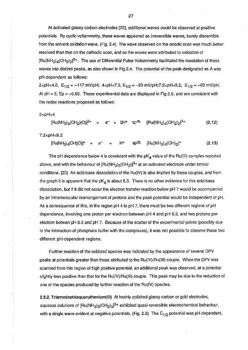
2<pH<3.8, El¡en -7 mV/pH; 4.2<pH<7.4, E1zn 46 mV/pH; 7.6<pH<9 .2,F.1¡z* -107 mV/pH.
The latter two pH-dependent regions are consistent with the ex¡stence of proton-coupled electron
transfer reactions involving one and two protons per electron respect¡vely. The voltammetric wave
can be assigned to the lollowing series of redox couples:
2.0<pH<3.8
tRu(NFld4(o+r2ls +e- + [Ru(NH3)a(oHz)zlz*
4.2<pH< 7.4
[Ru(NH3)a(OH2XOH)]2+ +€- + H+ + [Ru(NH3)a(OHz)zl2*
7.6<pH<9.2
[Ru(NH3)a(OH)2]+ + e- +2H+ + [Ru(NHj4(OHz)zl2*
(2.e)
(2.10)
(2.11)
The boundaries of the pH-dependent regions are in general agreement with the pK" values
quoted above. The formation of a Ru(ll) complex in the same state of protonation over the entire pH
range is consistent with the expectation that the pK" of that species would be similar to that of
[Ru(NH3)5OHzl2*, which has been found previously to be 12.3 under similar conditions of ionic
strength. [19b]
This work
I
19b
18
19b
-0.10
-0.194
-0.14
-0.204
-0.16
-0.48b
-0.19
-0.07
[Ru(NH3)3(OHС]3*/2*
[Ru (NH3 )3 (o Hz)z(O H ¡12+ t +
[Ru (l.IH3 ) a (OHr¡ r1z + r z+
[Ru (lrIH3 )a(O Ht (O H¡12+ t +
[Ru(NH3)5(OHr¡1s+rz+
[Ru(NH3)5(oH¡12+r+
[Ru(NH3)613+t7+
[Ru(oH2)673+t7+
Ref erenceEttZ(v vs SC,E)Couple
Table 2.2. Redox potentials of ammine aqua rutheniu cornplexes.(a) pH :5. (b)0.1 mol^ dm-3 NaOH.
27
At activated glassy carbon electrodes [22], additionalwaves could be observed at positive
potentials. By cyclic voltammetry, these waves appeared as irreversible waves, barely discernible
from the solvent oxidation wave. (Fi1.2.4) The wave observed on the anodic scan was much better
resolved than that on the cathodic scan, and so the waves were attributed to oxidation of
IRu(NH3)4(OHz)z]3*. The use of Differential Pulse Voltammetry facilitated the resolution of these
waves into distinct peaks, as also shown in Fi9.2.4. The potential of the peak designated as A was
pH-dependent as follows:
2<pH<4.0, E1p* -117 mV/pH; 4<pH<7.0,F-1t2n -93 mV/pH;7.2<pH<9.2, Eltz- -63 mV/pH.
At pH = 5, Ep = +0.60. These experimental data are displayed in Fi9.2.5, and are consistent with
the redox reactions proposed as follows:
2<pH<4
[Ru(NH3)a(OHzXO)]2* + e-+2H+ =
[Ru(NH3)a(OHz)z]3*
7.2<pH<9.2
tRu(NHja(OH)(O)f + e- + H+ + [Ru(NH3)a(OH)2]+
(2.12)
(2.13)
The pH dependence below 4 is consistent with the pK" value of the Ru(lll) complex reported
above, and with the behaviour of IRu(NHs)s(OHz)]3* at an activated electrode under similar
conditions. [23] An acid-base dissociation of the Ru(lV) is also implied by these couples, and from
the graph it is apparent that the pK" is about 6.3. There is no other evidence for this acid-base
dissociation, but if it did not occur lhe electron transfer reaction below pH 7 would be accompanied
by an intramolecular rearrangement of protons and the peak potential would be independent of pH.
As a consequence of this, in the region pH 4 to pH 7, there must be two different regions of pH
dependence, involving one proton per electron between pH 4 and pH 6.3, and two protons per
electron betwen pH 6.3 and pH 7. Because of the scatter of the experimental points (possibly due
to the interaction of phosphate buffer with the complexes), it was not possible to observe these two
different pH-dependent regions.
Further reaction of the oxidized species was indicated by the appearance of several DPV
peaks at potentials greaterthan those attributed to the Ru(lV)/Ru(lll) couple. When the DPV was
scanned from this region of high positive potential, an additional peak was observed, at a potential
slightly less positive than that for the Ru(lV)/Ru(lll) couple. This peak may be due to the reduction of
one of the species produced by further reaction of the Ru(lV) species.
2.S.2.Triamminetriaquaruthenium(lll) At freshly-polished glassy carbon or gold electrodes,
aqueous solutions of IRu(NHg)s(OHz)g]3* exhibited quasi-reversible electrochemical behaviour,
with a single wave evident at negative potentials. (Fig. 2.6) The E172 potentialwas pH dependent,
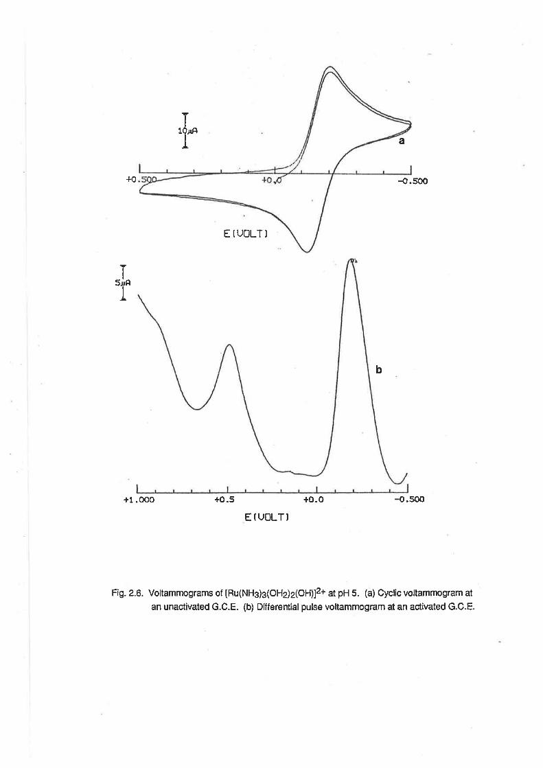
ï'r a
{.500
E IUOLTI
+1.OOO +o.5 +o.o -o.500
E f UOLT)
Fig. 2.6. Voltammograms of tHu(NHs)g(OHj2(OH)12+ at pH 5. (a) Cycfic vottammogram at
an unactivated G.C.E. (b) Differentialpulse voltammogram at an act¡vated G.C.E.
+0
Ttl'
b
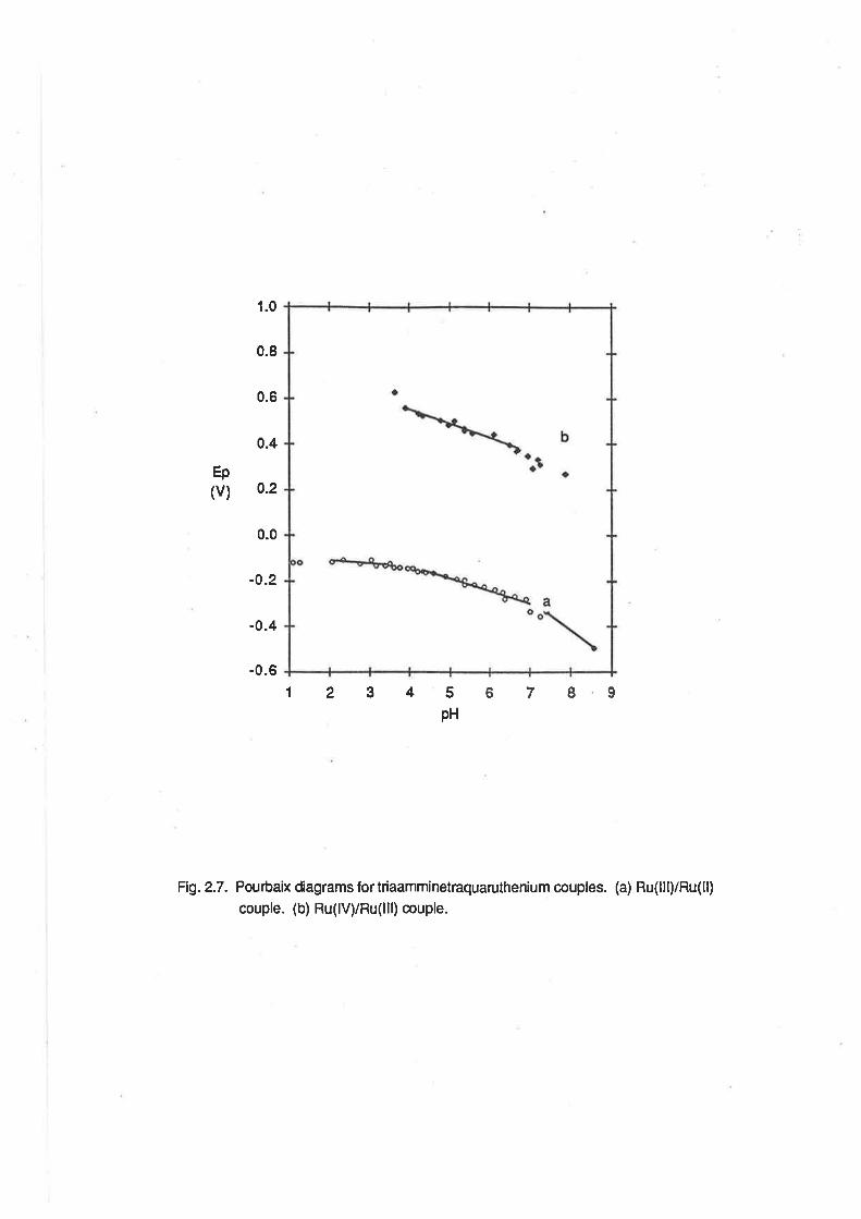
Ep(v)
1.0
0.8
0.6
0.4
0.2
0.0
-0.2
-0.4
-0.6
1234 67895pH
Fi1.2.7. Pourbaix diagrams fortriaamminetraquaruthenium couples. (a) Ru(l¡l)/Ru(ll)
couple. (b) Ru(lv)iRu(lll) couple.
28
as lollows:
2.0<pH<3.4,E,tnn-4mVlpH; 4.4<pH<7.0,81nn-S7mv/pH; 7.3<pH<8.6,Eu2n-118mV/pH
The Pourbaix diagram is shown in Fi9.2.7. At pH 1 .5,Eln= -{.10 V, and at pH 5, E1f2= -{.19 V.
Similar data for other ammine aqua ruthenium complexes are displayed in Table 2.2. The latter two
pH-dependecies are consístent with electron transfer coupled to the transfer of one and lwo
protons respectively. Over the pH ranges scanned, the wave was assigned to the following
couples:
2.0<pH<3.4
tRu(NFþ)3(ol-þ)3ls + e- + [Ru(NH3)3(oHz)g]2*
4.4<pH<7.0
[Ru(NHj3(OH2)2(OH)]2+ + e- + H+ =È [Ru(NH3)3(OHz)s]2*
7.3<pH<8.6
[Ru(NHj3(OH2XOH)2]+ + e- + 2¡1+ ç=l [Ru(NH3)s(OHz)s]2,
(2.14)
(2.1s)
(2.16)
These results are in agreement with the pK" values reported above. Again, although the
compound has not been isolated, the pK" of [Ru(NH3)s(OHz)s]2* is expected to be about 12,
similar to that of [Ru(NH3)sOHz]2*.
When the electrochemical experiment was conducted usíng an activated glassy carbon
eleclrode, an additional wave was observed at positive potentials. The CV peaks for this wave were
poorly resolved, indicating the presence of an irreversible electron transfer process, but on an
anodic DPV scan, the peak could be seen quite readily, as shown in Fig. 2.6. At pH 5, the potential
at which this peak occurred was +0.49 V, and over the range 3.9<pH<6.7, the potential had a pH
dependence of -63mV/pH. Outside of lhis pH range, the peak was not sufficiently well-resolved to
allow analysis of its pH dependence. At low pH values, the peak merged with that of the solvent
decomposition, and at high pH values chemical reactions occurred in the solution which gave rise to
extra peaks at potentials more negative than that due to the Ru(lll)/Ru(ll) couple. These reactions
were possibly related to the reactions which were observed at high pH in the spectrophotometric
studies. The DPV peak was assigned to the following redox react¡on:
3.9<pH<6.7
[Ru(NHj3(OH2XO)]2* + e +H+ + [Ru(NH3)3(oH2)2(oH)]2+ efil
A cathodic DPV scan over the potential range of interest revealed the presence of a small peak
at approximately +0.30V, which conesponded to a CV wave at this potentialwhich became more
pronounced with repeated scanning. This additional wave probably arose from the accumulation of
29
the products of reaction of the initial oxidation product
2.5.3. Discussion. Electrochemicalstudies of [Ru(NHs).1OH2)sl3+ and cis [Ru(NH$a(OHz)zlu
revealed Ru(lll)/Ru(ll) couples which are pH dependent, consistent with the measured pK" values
of the compounds, andsimilar to those in the penta- and frans tetraammine analogues. The general
trend in E1¡2 values with the decreasing number of ammine ligands (shown in Table 2.2.lis
believed to reflect the greater ligand lield strength of ammonia compared with water. ln the
absence of n-interactions, electron transfer becomes less favourable when there are more ammine
ligands present, because more CFSE is lost on reduction. [19b] Electrochemistry affords a better
appreciation of the differences in ligand field strength than uv/vis spectroscopy, where the
variations in absorption maxima are minimal, as noted in Section 2.4.
The ability ol IRu(NH3)s(OHe)s]3* and cis [Ru(NH3)a(OH2lzl3* to undergo electrochemical
oxidation via proton-coupled electron transfer is analogous to the pentaammineaqua case, which
was described recently. [23] ln these three cases, the proposalthat the Ru(lV) species contains
the Ru=O2+ group is supported by the pH-dependence of the peak potential. ln other systems,
such as aquapolypyridyl complexes [24] and aqua complexes containing macrocyclic tertiary
ammine ligands [25], there is lurther evidence for the existence of the Ru=O2+ group in
mononuclear complexes, because the complexes have been isolated and their spectra and crystal
structu res determined.
The evidence for the existence of mononuclear Ru=O2+ complexes is based on the
assumpt¡on that a pH dependence of -59 mV/pH is due to the transler of one electron coupled
with the transfer ol one proton. However, there is the possibility that the electron lransfers could
involve two protons and only half an electron per ruthenium ion. This was reported to be the case
when Ru(HedtaXOHZ) was oxidized to yield an oxo-bddged dimer. [26] lt was not possible to
perform coulomelric experiments to confirm that the electron transfers observed voltammetrically
involved one electron per ruthenium ion, but the observation that the voltammetric peak heights
were approximately the same forthe [Ru(NH3)5(OH2)]3+4Ru(NHs)s(OH2)12+ and the
IRu(NH3)5(O¡¡2+4nu(NHs)s(OHz)]3+ implies that this is so [23], and this proposalalso provides the
simplest explanation for the electrochemical results.
A notable difference between the resulls obtained for the triammine complex and those for
other complexes referred to above is the irreversible nature of the oxidation of the triammine
complex. This irreversibility may be indicative that the oxidation of the complex at the electrode is
kinetically slow. Such an effect was also reported for the electrochemicaloxidation of the
complexes [Ru(bpy)z(OHzlzls*,IRu(trpyXbpyXOHz)]3+ and IRu(bpy)z(pyXoHz)]3* at unactivated
electrodes. I24,271 ln the case of the first of these complexes, act¡vation of the electrode proved
30
to be kinetically favourable and the reversibility of the electrode reaction was improved. [22] The
mechanisms by which the activation process is believed to enhance the electrode process were
discussed in Chaþter 1. However, in lhe present case, the reversibility of the electrode process is
very poor, even when an activated electrode is used. Hence, it would appear that the oxidation of
[Ru(NH3)s(OHz)g]s and crs [Ru(NHs)a(OHz)zl3t at the glassy carbon electrode is extremely slow,
indeed.
Another possible cause of lhe irreversibility is the decomposition of the oxidized species.
There is evidence in the DPV experiments on both the tetraammine and triammine complexes that
there is some reaction of the Ru(lV) complex. Poon and co-workers have suggested that the redox
chemistry of amineruthenium complexes is usually complicated by the oxidative dehydrogenation
of coordinated primary and secondary amines. [28] Other workers have proposed that the
dehydrogenation is associated with imine formation involving ínternalelectron transfer between the
ligands and Ru(lV). lt was found that the oxidation of 2(aminomethyl)pyridine coordinated to
Ru(bpy)24+ (which is a step in the oxidation of 2(aminomethyl)pyridine in [Ru(bpy)2(ampy)]4+) has a
relatively large rate constant for the proton/electron transfer reaction, with k of the order of 102 at
200C. [29] lt would seem likely that this reaction could also occur when ammonia is coordinated
and, so, any IRu(NH3)3(OH2)2(O)]2+ foimed in solution by chemicalor electrochemical means
would decompose rapidly. lt is interesting to note, however, that other wod<ers have been able to
isolate the Ru(Vl) complex, frans [Ru(NHg)¿(O)z]2*, from a reaction involving RuO4, without the
complicating effects of the dehydrogenation reaction. [30] Furthermore, in an initial report, it has
been noted that IRu(NHg)s(OHz)]3* and [Ru(NHg)sCl]2* show two reversible oxidations at a coated
graphite electrode and can be oxidized by Cs4+ to give a Ru(V) complex which is a catalyst for the
oxidatíon of water to oxygen. [31]
Attempts were made to achieve the chemical oxidation of [Ru(NH3)s(OHz)s]3*. To this end, a
buffered solution of [Ru(NH3)g(OHz)s]3* was treated with a stoichiometric amount of hydrogen
peroxide at 50oC. After about fifteen minutes, the reaction mixture had a dark brown colour. A CV
of the reaction mixture did not contain any electrochemicalwaves which could be attributed to a
redox couple associated with [Ru(NHg)s(OHz)g]S. Treatment of the reaction mixture with
concentrated hydrochloric acid solution did not yield Ru(NHg)C|3, which would have been the
expected product if the triammine portion of the original complex was still intact. Similar dark
solutions were obtained when both cis and lrans [Ru(NH3)a(OHz)z]3* were treated with a solution
of hydrogen peroxide. [32]
The potentials at which oxidatíons involving the RuO2+/RuOH23+ couples occur for the
ammine complexes are slightly negative of those at which the polypyridine complexes are oxidized.
31
127,321 However, the d¡fference in the potent¡al of the RuO2+¡pu6H23+ couple and the
pu3+¡gu2+ couple is much greater ¡n the case of the ammine complexes than in the polypyridyl
complexes. The factors contr¡buting to these elfects will be discussed in Chapter 3.
2.6. Redox Chemistry
As is to be expected, from its moderate reduction potential, [Ru(NHs)a(OHz)g]3* is readily
reduced by chemical mearìs to [Ru(NH3)s(OHz)s]2*. Amalgamated zinc or hydrogen over platinum
black are suitable reducing reagents. During the reduction, the course of the reaction can be
followed by Sampled Current Polarography. The uv/vis spectrum of the golden-yellow solution ( in
1 moldm-3 H2SOd could be determined when the reaction was shown, by SCP, to be
substantially complete. The main features of the spectrum are listed in Table 2.1, as are those of
the pentaammine and tetraammine analogues. The spectrum is displayed in Fig. 2.8, and it can be
seen that the spectrum is very similar to the other ammineaqua complexes of ruthenium(ll) which
have been isolated. lt was not possible to isolate the Ru(ll) species, but studies could be performed
on samples of the complex generated in situ.
When [Ru(Nft)3(O4z)el2* was formed in acidic solution, in the presence of perchlorate ion, it
was found that the perchlorate could be reduced to chloride. The formation of chloride was
indicated by a positive Ag+lAgCltest. The redox reaction would be:
B[Ru(NH3)3(OHz)s]2* + CIO¡ + 8[Ru(NH3)3(OHz)g]s + Cl- + 4H2O (2.18)
Previous workers have shown that [Ru(NHs)sOHz]2* and [Ru(OH z)ol2* are also capable of
reducing perchlorate, with chlorate being the initial product. [33, 341 However, in the hexaaqua
system, small amounts of IRu(OH2)Cl]* were also found, consistent with the further reduction of
lower oxidation states of chlorine. lt has been proposed [35]that, in the pentaammine case, the
reaction is:
lnulNHs¡5oFl2l2+ + cto¡ + [Ru(NHs)s(o)]2+ + clq- + Hzo (2.1e)
but the Ru(lV) species was not observed due to rapid reaction with excess Ru(ll) ions. i.e
Ru(lV) + Ru(ll) -+ 2Ru(lll) (2.2o)
The ability of [Ru(NHjg(OHe)s]2* lo reduce perchlorate was first manifested during attempts at
bulk electrolytic reduction of [Ru(NHs)s(OHz)s]3+. lt was found that lhe current did not decay
exponentially, but reached a steady-state value much higher than expected of a solution in which all
the oxidized form had been reduced. This is consistent with the electrocatalytic reduction of the
perchlorate ion in the supporting electrolyte.
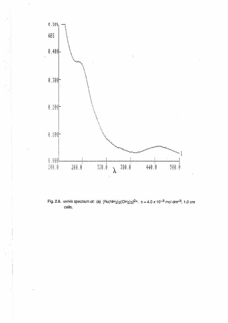
4[
tü
iir
É85
fl
ü
I
I
il I'll'l
. l'! li
fl :l!t
I
Fig. 2.8. uv/vís spectrum of : (a) [Ru(NH3)g(OHds]2*. c = 4.0 x 10⁴ mot dm4, 1.0 cmcells.
l.{?t
it lË[tlirjffiË[ tl I I¡llI44[ L,t
I
1
-.
I
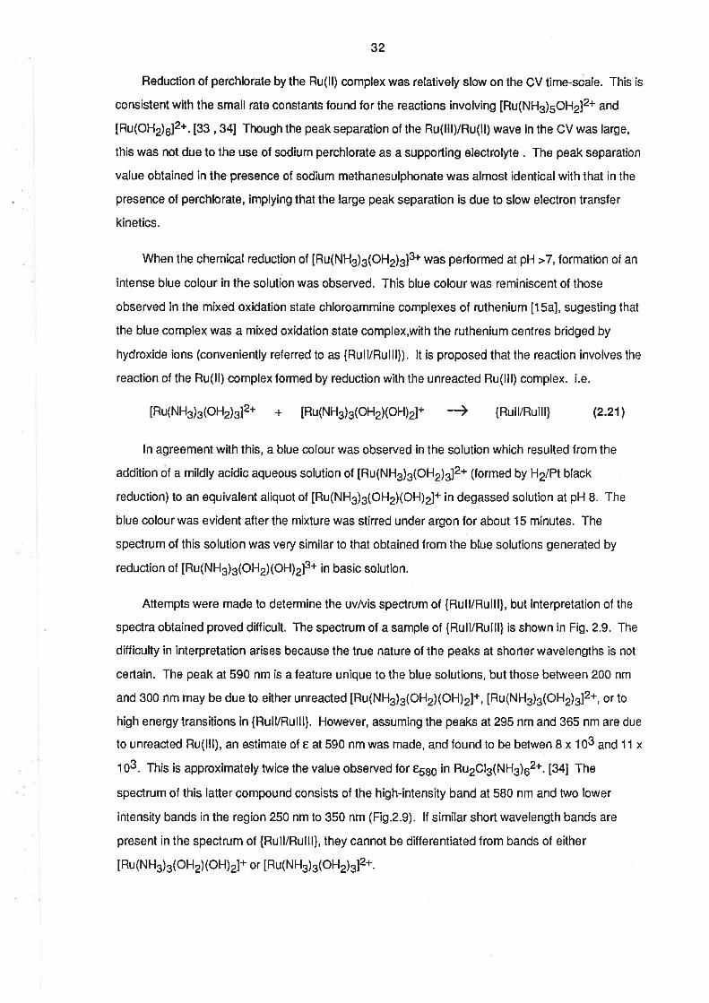
32
Reduction of perchlorate by the Ru(ll) complex was relatively slow on the CV time-scale. This is
consistent with the small rate constants found for the reactions involving [Ru(NH3)5OH2]2+ and
[Ru(OH2)5]2*. [33 , 34] Though the peak separation of the Ru(lll)/Ru(ll) wave in the CV was large,
this was not due to the use of sodium perchlorate as a supporting electrolyte . The peak separation
value obtained in the presence of sodium methanesulphonate was almost identical with that in the
presence of perchlorate, implying that the large peak separation is due to slow electron transfer
kinetics.
When the chemical reduction of [Ru(NH3)s(OHz)slS was performed at pH >7, formation of an
intense blue colour in the solution was observed. This blue colour was reminiscent of those
observed in the mixed oxidation state chloroammine complexes of ruthenium [15a], sugesting that
the blue complex was a mixed oxidation state complex,with the ruthenium centres bridged by
hydroxide ions (conveniently referred to as {Rull/Rulll}). lt is proposed that the reaction involves the
reaction of the Ru(ll) complex formed by reduction with the unreacted Ru(lll) complex. i.e.
[Ru(NH3)3(OHz)s12' + [Ru(NHj3(OHd(OH)2]+ -) {Ruil/Ruilt} (2.21)
ln agreement with this, a blue colour was observed in the solution which resulted from the
addition of a mildly acidic aqueous solution of [Ru(NH3)s(OHz)s]2* (formed by H2lPt black
reduction)to an equivalent aliquot of [Ru(NH3)3(OH2XOH)2]+ in degassed solution at pH B. The
blue colour was evident after the mixture was stirred under argon for about 15 minutes. The
spectrum of this solution was very similar to that obtained from the blue solutions generated by
reduction ol IRu(NH3)3(OH2XOH)z]3+ ¡n basic solution.
Attempts were made to determine the uv/vis spectrum of (Rulf/Rulll), but interpretation of the
spectra obtained proved difficult. The spectrum of a sample of {Rull/Rulll} is shown in Fig. 2.9. The
diff iculty in interpretation arises because the true nature of the peaks at shorter wavelengths is not
certain. The peak at 590 nm is a feature unique to the blue solutions, but those between 200 nm
and 300 nm may be due to either unreacted [Ru(NH3)3(OHz)(OH)2]+, [Ru(NH3)3(OH2)3]2+, or to
high energy transitions in {Rull/Rulll}. However, assuming the peaks at 295 nm and 365 nm are due
to unreacted Ru(lll), an estimate of e at 590 nm was made, and found to be betwen I x 103 and 11 x
103. This is approximately twice the value observed for e5ss in Ru2Cl3(NHs)02*. [3a] The
spectrum of this latter compound consists of the high-intensity band at 580 nm and two lower
intensity bands in the region 250 nm to 350 nm (Fi9.2.9). lf similar short wavelength bands are
present in the spectrum of {Rull/Rulll}, they cannot be differentiated from bands of either
IRu (NH3)3(OH2XOH)2]+ or IRu(NHs)s(OHz) g]2*.
I i[
ÈEi
itfi
f!¡
Fig. 2.9. uv&is spectrum of lhe blue mixed-oxidation state species.
It
4úü ü¡[ü'ü
I
:tt
tr rJ i¡
i[[ [
a
I
h|JlJÍt!tl I I ü¡üül
33
When the blue solutions were exposed to oxygen or acidifíed, the blue colour faded to a pale
yellow. Because no blue colourwas observed if the reaction was performed at pH <7, it is apparent
that at least two hydroxy! groups must be present on the Ru(lll) ion in order lor the bridged species
to form.
The blue solutions showed identical cyclic voltammogrars, regardless of the method by which
the solutions were made. Two waves are visible in the voltammogram (Fig 2.10), one reversible and
one irreversible. The reversible wave, A(\p= -0.50 V), is consistent with the presence of either
or both of lnullllruus)s(oH2xoH)21+ or 1null1ruu.¡.1oH2)sl2+ in the solution, as the E1¿2 potential is
lhe same as that of the lll/ll couple for these compounds at pH 8. By commencing the cathod¡c scan
at potentials at which the first of those two compounds is stable, {Rull/Rulll} is oxidized. The
irreversible wave, B, (Epa = 4.22 V) was assigned to the oxidation of {Rull/Rulll}. The EC shape of
this wave is indicative that the oxidized form decomposes quite rapidly to the mononuclear Ru(lll)
complex. As the cyclíc experiment was continued, more of this complex formed in solution, giving
rise to a higher peak current at -0.59 V. The cyclic voltammogram was determined at scan rates of
up to 2 V sec-1, but the cathodic peak associated with peak B could not be observed. lt was thus
concluded that the decomposition of di-Ru(lll) complex occurred very rapidly compared with the
electron transfer reaction, according to reaction 2.22,wilh k1 being large.
k1
{RutuRullt} + e- + {RulluRuilt} + 2Ru(ilr) (2.22l-
As a comparison, voltammograms of Ru2Cl3(NHg)e2* contained a reversible oxidation wave at
+0.32 V. [36] This is considerably more positive than the potential at which {Rull/Rulll} is oxidized,
confirming the observation that {Rull/Rulll} was less stable to oxidation than Ru2Cl3(NHg)e2*.
lf the reduction of [Ru(NHd3(OH2XOH)2]+ at high pH was allowed to continue, the blue colour
slowly faded, and [Ru(NHs)g(OHz)g]2+ was formed almost quantitatively. Thus, it is apparent that
the blue species, {Rull/Rulll}, is only a transisent in the reduction of [Ru(NH3)3(OH2XOH)2]+. For
this reason, (Rull/Rulll) could not be isolated and its spectrum could only be determined semi-
quantitatively.
2.7. Electrochemistry in Organic Solvents.
Solutions of [Ru(NH3)s(OHz)s]3* in organic solvents acquired intense colours upon standing
Such colour changes were indicative of replacement of the àqua ligands by solvent molecules.
Except for methanol, the solvents generally had high ultraviolet cut-off wavelengths, so that uv/vis
spectroscopy was not the appropriate method by whch to study the solvolysis reactions.
Consequently, the CV of [Ru(NH3)3(OH2)3]3* was studied in the solvents methanol, acetone,
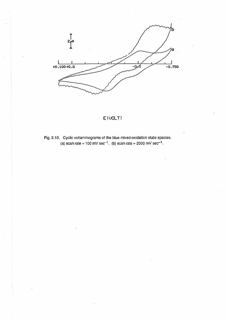
T
T
+o.100+o.o -0.750
EIUOLTI
Fig. 2.10. Cyclic vollammograms of the blue mixedoxidation state species.
(a) scan râtê = 100 mV sec-1. (b) scan rate = 2oo0 mV sec-1.
34
acetonitr¡le and dimethylsulphoxide. The voltammograms obtained are displayed in Fig. 2.11 and
the main features of the voltammograms are listed in Table 2.3.
2.8.1. Methanol. The CV of [Ru(NHg)3(OH2)g]3+ in methanolconsisted of one reversible wave and
one irreversible wave. (Fig 2.11a) The E1/2 potentials for these peaks were estimated, f rom DPV,
to be -0.18 V and -0.33 V. The reversible wave is consistent wfth fhe presence of
IRu(NH3)3(OHe)s]3* or [Ru(NH3)3(OH2)2(OH)12*. The irreversibte wave had the characteristic
shape of an EC mechanism of electron transfer followed by chemical reaction. This is consistent
with the presence of a complex, [Ru(NH3)g(OHz)(s-n)(MeOH)n¡3+, pdor to electron transfer,
This work
tl
I
4
This work
19a
This work
41
-0.334
+0.24+0.60+0.95
+0.19
+0.43
+0.76
+O.O4a
+0.40, +0.57b
+0.75b
[Ru (NHs )¡ (M e OH)o( O Hz)ynf3 * tz*
lRu (l.IH: ) ¡ ( M e CII) ( O H7) rl3 + t 2 +
[Ru (NH¡ )¡ ( M e CTrI)2 ( O H2)f3 + t 2 +
[Ru (l.IHs )¡ ( MeC\03 ] 3+ rz+
[Ru (NHs )s ( M e Clrl¡ 1
3 + /2+
lRu (l.IH¡ )s (D M S O X O Hz)273 + tz +
[Ru Q.IH3 )5(D MSO¡1 3+a+
[Ru(lrlH3)3(o acetone),r(OHС-o]3*2
[Ru (NH3)3 (æ acetone)o(OHz)s-n] 3*/2
ßu (NH¡ )s(r acetone ¡13 + tz+
Ref erenceElz(V vs SCE)Couple
Table 2.3. Redox potentials of solvoþed ammine aqua ruthenium complexes.(a) cathodic peak only. (b) anodic peak only.
followed by rapid hydrolysis of the analogous Ru(ll) species. This proposal is consistent with the
uv/vis spectrum after a short time, where it was observed that some degree of solvolysis occurred
quite rapidly. No attempt was made to measure the voltammogram of solutions after a long time.
That hydrolysis of [Ru(NHs)s(OH2)(s-n¡(MeOH)nl2* occurs in very low concentrations of water, as
indicated by the EC shape of the wave, suggests that the methanol ligand is much better suited to
the stabilization of the higher oxidation state than the lower.
2.8.2. Acelonitrile. The CV of [Ru(NH3)g(OHz)s]3* in acetonitrile consisted of several reversible
waves with E172 values of +0.24 V, +0.60 V and +0.95 V, in addition to a cathodic peak at -0.15 V
(Fig 2.1 1b) The shape of this cathodic peak is also characteristic of an EC mechanism, and the
potential at which it occurs is indicative of the reduction of [Ru(NHs)3(OHz)g]3*. Operation of the
EC mechanism is evidence of fairly rapid solvolysis of the Ru(ll) species. lt is proposed that the
{-.F
{o.e50 {o.o
+o.250
.o,?l0
I"f
+1.200+t
+t {o.5
gfu(Lft
{.300
-o.5æ
T'r
{.ãoEITTLT'
a
Fig. 2.11. voltammograms of [Ru(NH3)3(oHdg]3* in organic sotvents. (a) cycticvoltammogram and ditferentialpulse voltammogram in methanol. (b) Cyclicvo ltammogram and diff erential pu lse voltammogram in acetonitri le.
-o.s10.0
{o.o.o
b
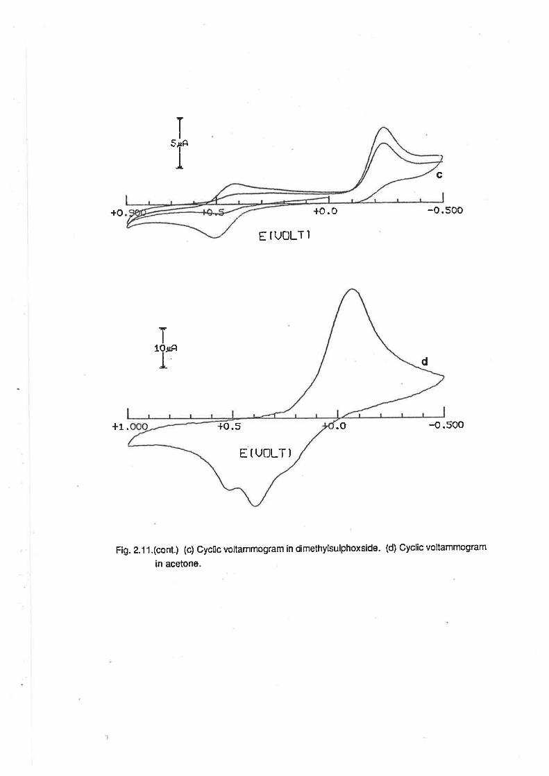
c
+o +o. o -o.500
EÍUOLT]
+1. .o -{.500
Fig. 2.11.(cont.) (c) Cyclic voltammogram in dimethylsulphoxside. (d) Gyclic voltammogram
¡n acetone
T
:r
ï1Or¡ÊI d
EIUOLT)
+o.5
35
three reversible waves are due to the presence of the mono, bis and tris acetonitrile complexes
respectively. The E¡¡2potential of the wave assigned to the mono substituted complex is similar to
the value of +0.19 V previously reported for reduction of [Ru(NH3)5NCMe¡3+. [4] Over a period ol
time, the peak currents of the more positive waves increased, and those of the less positive
decreased, indicative of the progressive formation of the higher substitution products by a slow
substitution process involving the Ru(lll) complex. lt is apparent, therefore, that the reduction ol the
Ru(lll) complex catalyzes solvoþsis by acetonitrile, but that the rate of substitution on the Ru(lll)
complex is still appreciable.
2.8.3. Dimethylsulphoxide. ln DMSO, and at low scan rates, the CV of [Ru(NHds(OH2)3]e
consisted of a cathodic EO{ype peak at -O.25 V and a reversible wave alEl¡2= +0'43 V' (Fig.
2.11c) At very high scan rates (v > 1 V serl) the peak at {.25 V was resolved into a quasi-
reversible wave with peak separation of at least 185 mV. ln aqueous DMSO mixtures, lhe E1¡2ot
this wave is pH-dependent and occurs in the appropriate region for the various aqua/hydroxo
complexes. ln DMSO/o.1 moldm-3 MeSO3H (60/40), the cathodic peak is at the same potential as
that due to reduction of [Ru(NH3)g(OHz)s]3*. Operation of an EC mechanism is evidence that
solvolysis of [Ru(NH3)31OH2)sl2+ by DMSO occurs relatively rapidly, in a manner similarto that
described above lor solvolysis by acetonitrile.
The positive E1¡2 potential for the DMSO complex is indicative of a r-bonded ligand, possibly
involving coordination through the sulphur atom. Such is the case with the complexes,
[Ru(NH3)5DMSO¡2+, [Ru(DMSO)3C13]- and [Ru(DMSO) 4ãl2l,lor which the crystal structures have
been determined. [37-39] lt has been concluded that up to three DMSO ligands can adopt the S-
bound form around Ru(ll). ln cases where there are more than three, there will be a mixture of S-
bound and O-bound ligands. The E172 potentíalfor [Ru(NH.)5DMSO]2+ is +0.76 V. [19a] On the
basis of this information, it may be concluded that S-bound DMSO is a very strong æ'acceptor
ligand. The more moderate E172 potential of the triammine complex, compared wíth the
pentaammine, is perhaps due to lhe abilig of the aqua ligands to provide electron density to the
electron-deficient ruthenium centre by r-donation from the second non-bonding pair of electrons
on the oxygen atom. The contribution to the electron density at the metal centre from the aqua
ligands thus compensates for the slrong electron-withdrawing ability of the DMSO.
2.8.4. Acetone. The cyclic voltammogram of [Ru(NHjr1OH2)el3+ in acetone (Fig. 2.11d)
consistedofasinglecathodicpeakatamorepositivepotential(Ep=+0.04V)thanthatexhibitedby
the complex in aqueous acid, and two anodic peaks at positive potentíals' (Ep = +0'40 v and +0'53
V respectively) The addition of aqueous base or acid (to give 60/40 acetone/water) resulted in the
36
cathodic peak occurring at still more positive potenlials, suggesting strongly that there is o-bonded
acetone with no water in the coordination sphere, when 100% acetone is used. The peak
potentials of the anodic waves are characteristic of a product involving a æ-bonded species and the
EC shape of the voltammogram is indicative of the occurrence of some chemical reaction after the
electron transfer steps. The existence of multiple anodic peaks in the CV of the triammine
ruthenium complex is attributable to the presence of species with different numbers of æ-bonded
acetone ligands.
Similar behaviour has been observed previously in the electrochemistry of
IRu(NH3)5acetone]3+12+ and [Os(NH3)5acetone73+t2+. [40,41] Lay has suggested that the
coordinated acetone llips from an O-bound, q1 form to a ¡-bond ed,rl2lorm atter reduction from the
+3 oxidation state to the +2 state, with subsequent llipping back to the original form after oxidation
to the +3 state. This proposalwas supported by a crystalstructure determination of
[Os(NHg)5acetone]2+, which showed the acetone ligand to be present in the r¡2 form. [41]
[Ru(NH3)5acetone]3+ has not been investigated, but the Ru(ll) species has been found to be a
useful synthetic intermediate. [42]
lf one accepts the hypothesis about the various coord¡nation modes of the acetone in the two
oxidation states, it is apparent that the Ru(ll) ion is stabilized by the ¡¡-bonded, t12 acetone ligand,
and the Ru(lll) ion is stabilized by the q1, O-bound ligand. This is in contrast to the situation
applying in DMSO, where the S-bound form is favoured by both oxidation states.
2.8.5. Conclusion. From the above results, it is apparent that acetonitrile and dimethylsulphoxide
are strong electron accepting ligands, and that their coordination to the ruthenium centre is more
rapid when Ru(ll) is involved. Conversely, methanolis an electron donating ligand, and its
coordination to the ruthenium centre is favoured by the presence of Ru(lll). Acetone is capable of
acting as either an electron acceptor or donor, and can coordinate through either the oxygen atom
or through side-on coordination of the carbonyl group.
37
REFERENCES
(1) (") K. Gleu, K. Rehm, Z. Aaotg. ALlgem. Chem.,227,23?(1936). (b) ibid,
235,352(1938). (c) K.Gleu, Iff. Breubl, Z. Anory. Allgem. Chem.,237,
33s(1e38)
Ø J. A. Broom-head, F. Basolo, R. G. Pearson, Iootg. Chem.,3,826(1964)
(3) G. M. Bryast, J. E. Fergusson, Ausú. J. Chem.,z4,44l(1971)
(4) T. Matsubara, P. Ford, hory. Chem., 15, 1t07(1976)
(5) C. K. Poon, S. S. Kwong, C. M. Che, Y. P. Kan, J. Chem. Soc., DaltonTrans,
r4s7(1982)
(6) D. M. Stanbury, D. Goswick, H. Taube, Inory. Chem.,22, 1975(1983)
(7) J. A. Broomiead, L. Kane-Maguire, Ioory. Chem., 10, 85(1971)
(8) J. A. Stritar, H. Taube,Inorg. Chem.,8,228l(1969)
(9) R. E. Clarke, P. C. Eoró,Inorg. Chem.,9,227(1970)
(10) P. Ford, De F. P. Rudd, R. Gaunder, H. Taube, J. Amer. Chem. Soc., 90,
1 187(1e68)
(11) P. C. Ford, C. Sutton, Inory. Chem.,8, t544(1969)
(12) M. Farraggi, A. Feder,Inory. Chem., 12,236(1973)
(13) D. E. Harrison, H. Taube, J. Amer. Chem. Soc.,89,5706(1967)
(14) (u) C. M. Elson, I. J. Itzkovitch, J. A. Page, Can. J. Chem.,48, 1639(1970). (b)
L. A. P. Kane-Magr:tre, J. hory. Nuc/. Chem., 33, 3964(197t)
(15) (a) F. Bottomley, S. B. Tong, Can. J. Chem.,49,3739(197t). (b) F' Boüomley,
Can. J. Chem., 55,2788(tlSlt¡
(16) B. Anderes, S. T. Colli¡s, D. K. Lavallee, Inorg. Chem.,23,2201(1984)
(17) N. Dixon, H. J. Lancaster, G. A. Lawrence,A. M. Sargeson, Inotg. Chem.,2O,
470(te8t)
(18) T. Fliades, P. Reinsalu, R. O. Harris, Can. J. Chem.,4'1,3823(1969)
(19) (u) C.G. Kuehn, H. Taube, J. Amer. Chem. Soc.,98, 689(1976)' (b) H.S. Lim,
38
B. J. Barcl^y,F.C. A¡son, hory. Chem., 11, 1460(1972)
(20) W. P. ffiffittr, The Chemistry of the Raræ Platinum Metals, lnterscience, London,
1967, pl66
(2t) Z. Hanion, G. Navon, Inorg. Chem., 19,2236(1980)
(22) G. E. Cabaniss, A. A. Diamanris, \il'. R. Murphy, L. W. Linton, T. J. Meyer, J.
Amer Chem. Soc., l0'1,1845(1985)
(23) A. A. Diamantis, rW. R. Muqphy, T. J. Meyer, hotg. Chem., 23, 3230(1984)
(24) K. J. Takeuchi, M. S. Thompson, D. W. Pipes, T. J. Meyer,Ioory.Chem.,23,
1845(1 984), and references tåerein.
(25) C. M. Che, K. Y. Wong, W. H, Leong, C. K. Poon, Inory. Chem., 25, 345(1986)
and references therein.
(26) R. B. Baar, F. C. A¡rson, J. Electroanal. Chem., t87,265(1985)
(27) K. J. Takeuchi, G. J. Samuels, S. W. Gersten, J. A. Gilbert, T. J. Meyer, hory,
Chem.,22, 1407 (1983)
(28) C. M. Che, K. Y. Wong, C. K. Poon, Ioory.Chem.,z4,l60t(1985)
(29) (a) M.J. Ridd, F. R. Keene,J. Amer. Chem. Soc., 103,5733(1981). (b) P. A.
Adcock, F. R. Keene, R. S. Smythe, M. R. Snow, Iuory.Chem.,23,236(l9U)
(30) W. P. Gdffith, D. Pawson, J. Chem. Soc., DaltonTrans,1315(1973)
(31) R. Ramaraj, A. Kira, M. Kaneko, . J. Chem. Soc., Chem. Comm.,227(1987)
(32) K, J, Yakeuchi, M. S. Thompson, D. !V. Pipes, T. J. Meyer,Ioory. Chem.,23,
184s(1e84)
(33) T. W. Kallen,J.EaÃey, hory.Chem., 10, 1152(1971)
(34) J. F. Endicott, H. Taube , J. Amer. Chem. Soc., 84, 4984(1962)
(35) H. Taube, private communication.
(36) E. E. Mercer, L. W. Gray, J. Amer. Chem. Soc., 94, 6y';26ç972)
(37) F. C. Marsh, G. Feqgusson, Can. J. Chem.,49,3590(1971)
39
(38) R. S. McMillan, A. Mercer, B. R. James, J. Trotter, J. Chem. Soc., DaltonTtzns,
1006(1e75)
(39) A. Mercer, J. Trotter, J. Chem. Soc., DaltonTtzas,2480(1975)
(40) P. A. Lay, privatc communication
(41) W. D. Hannatr, D. P. Fairlie, H. Taube, J. Aner. Chem. Soa, 108, 8223(1986)
(42) J.A. Baumann, T. J. Meyer, hotg. Chem., 19,345(1980), and references therein.
Ghapter 3.
SU BSTITUTED TRIAMMIN ERUTH ENI UMCOMPLEXES.
3.1. lntroduction.
The substitution reactions of ruthenium(ll) and ruthenium(lll) ammine complexes have been of
much interest for the past two decades, and the subject has been reviewed by Ford and, more
recently, by Seddon. [1, 2] Hitherto, such reactions have been confined to the pentaammine and
tetraammine groups of complexes, with spectral and electrochemicalstudies providing information
about bonding, electron transfer reactions and substitution mechanisms. Much work has been
done involving the use of analogous series of complexes where groups of ligands with various
organic substituents (for example, substituted pyridines [3], and a range of carboxylate anions [4])
have been introduced into the coordination sphere. These series of complexes have been
particularly useful in making comparisons between Ru(ll) and other dG ions such as Co(lll), and
between Ru(lll) and other d5 ions such as Fe(lll).
An important consideration when discu{ssing substitution reactions on ammineruthenium
complexes is the relative lability of the +2 and +3 oxidation states towards substitution. Most
substitution reactions on Ru(ll) and Ru(lll) proceed via a dissociative mechanism so that, generally,
Ru(ll) is more labile than Ru(lll). [5] This phenomenon is illustrated by reaction 3.1. For n = 2, the
rate constant, k1 = n æ"-1(at 29BK)[6], butwhen n =3, k1 =2.1x 10{sec-1 (al327Kand [Cl-]=
0.1 motom-3¡. 1z¡
[Ru(NH$5H2O]n+ + C¡ -+ [Ru(NHs)5C¡(n-1)+ * HzO (3.1)
The traditional precursors to substituted complexes have been the haloammine complexes of
ruthenium(lll) and one usual synthetic procedure has been to remove the halide ion from the
solution by precipitating it as its silver salt. This results in a solution of [Ru(NH3)sHzO]3*. Though
this complex is relatively inert to substitution, the substitution reaction can be calalyzed by the
presence of a small amount of the Ru(ll) analogue, formed in situ by the action of amalgated zinc or
some other reductant. Stritar and Taube first made use of this sequence in the synthesis of
pentaammine carboxylato complexes. [4] The preparative reaction scheme was:
[Ru(NHg5(O t-l]* + zn(Hs) + tRu(NH3)5O fi
k1
1nqNH3)5(o l-fl2+
+
Rco2H/Rcot +02+
IRu(NH3)5(RCo2)]*
tRu(NH3)5(rco2)12+[Ru(NH$5ficq)]+
+
(3.2)
41
lf the reaction solution is degassed prior to the addition of the reductant, a ruthenium(ll) complex
can be obtained, especially when the ligand is a good fi-acceptor.
A modification of the above procedure is to introduce the reductant directly to a solution of the
haloammine complex and ligand. The hydrolysis of the haloammineruthenium(ll) complex is quite
rapid and, on"å th" substitution reaction is complete, the product can be isolated, halide free, by
the addition of a precipitating agent, such as NaClOa or NH4PF6. Using these procedures, it has
been possible to prepare ammineruthenium complexes containing ligands such as carboxylates
[4], organonitriles [8], aromatic nitrogen heterocycles [3,9], carbon monoxide [10] and dinitrogen.
[1 1l
There is a trend away from the use of the perchlorate, hexafluorophosphate and
tetrafluoroborate as non-coordinating anions. Perchlorale is prone to violent decomposition, and is
also readily reduced. [12] This second feature of its chemistry means that perchlorate can be
troublesome when present in electrochemical experiments. The fluorophosphate and fluoroborate
ions are susceptible to hydrolysis, leading to the formation of fluoride ion in aqueous solutions.
The sulphonate ions, e.g. methanesulphonate and trifluoromethanesulphonate, are finding favour
because of their stability and poor coordinating ability. Trifluoromethanesulphonate is especially
favoured because of its solubility in a wide range of organic solvents. [12]
The above procedures work well when the ligand is soluble in water, but complications arise
when it is not. A convenient procedure in this case is to replace water with an organic solvent in
which the precursor complex and ligand are soluble. lt has been shown that acetone will replace
the aqua ligand in [Ru(NH3)s(OHz)]2+, and the coordinated acetone may then be replaced by some
other ligand. [1 1, 12] Similarly, it has been shown that the acid hydrolysis of [Ru(NH3)sCl]3+ ¡n
anhydrous HTFMS results in the formation of a labile product, [Ru(NH3)5TFMS](TFMS)2, which is
soluble in a range of organic solvents. [13, 14] TFMS is a very weak ligand and is readily replaced by
almost any neutral ligand. Examples of this include replacement by water-insoluble organonitriles,
pyridine, formamide and methanol.
The substitution reactions of ruthenium ammine complexes have been paralleled by the
preparation of complexes of Ru(ll) and (lll) containing pentadentate edta as the major ligand. [15 -
171 Ligand substitution on ions in both oxidation states appeared to be enhanced by the action of
the pendant arm of the edta ligand, through its ability to hydrogen bond with the aqua ligand. [15]
Variation of the ligand was shown to affect the properties of the complexes in the same trends as in
the substituted ammine complexes.
42
With the availability of [Ru(NH3)s(OHz)g]3*, it was proposed to study the substitution reactions
of bidentate ligands with this ammineruthenium complex. Substitution on the
tetraammineruthenium ions has lead to a range of complexes which also conta¡n bidentate ligands,
and to studies of their spectra and electrochemistry. This new complex otfers the scope to vary the
coordination sphere about the ruthenium centre while leaving the ammine and aqua ligands intact.
Thus, in addition to the conventional spectral and electrochemical studies, it was possíble to study
the effect of the non-ammine ligand on the acid/base properties of the coordinated water, as well as
the proton-coupled oxidation of the RuOH23+ group to the Ru=O2+ group. There was also the
possibility of studying complexes of the type [Ru(NH3)3L3]n+ where L may be varied from æ-
acceptors to rt- or o-donors, as in the cases of [Ru(NH3)5L]n+ and [Ru(NH3)4L2]n+ complexes.
3.2. Ruthenium(lll) Complexes.
3.2.1. Preparation of Complexes. The solvolysis of [Ru(NH3)g(OHz)g]3* by alcohols was discussed
in Chapter 2. ln alcoholic solutions, [Ru(NH3)3(OHz)s]3* reacted with uninegative, bidentate
ligands, in the presence of lithium acetate to give a series of complexes, [Ru(NH3)3(LLXOH2)]2+ lLL
= âcâc, dpm, hna, oxine, bzac, dbm, and sal). The progress of the reaction could be monitored
voltammetrically by adding samples of the reaction mixture to aliquots of 0.1 mol dm3 MeSOgH, in
which the [Ru(NH3)s(OHz)g]3*/2+ çsupls had a DPV peak at -0.09 V. During the course of the
reaction, the peak current at th¡s potential decreased, with the concomitant increase in the peak
current of a peak at more negative potentials. The reaction mixtures containing the diketonate
ligands were purple, and with the other ligands they were green.
The complexes containing the first three ligands were isolated as their dithionate salts after the
solvent had been removed from the reaction mixture, and the resulting residue treated with dilute
dithionic acid. IRu(NH3)3(oxine)(OH2)](TFMS)2 was isolated by an analogous method, using 3 mol
dm-3 HTFMS instead of dithionic acid. ln this case the reaction yield was low, because there was a
tendency for the acid to protonate the ligand, giving [Ru(NH3)s(OH2)3](TFMS)3 as a product.
Attempts were made to isolate the oxine complex as a dithionate and ptoluenesulphonate salt, but
without success.
The ligands,bzac,dbm, and salall reacted with IRu(NHj3(OHz)g]3* underthe same
conditions, but it was not possible to isolate the products by the addition of precipitating agents to
the reaction mixture. When reaclion mixtures containing bzac and dbm came in contact with
aqueous solutions, a white precipitate was observed whích was identified as the free ligand. lt is not
certain whether the ligand was precipitated because of the decomposition of the product, or
because there was still some unreacted ligand in solution. lf the white precipitate was filtered from
the aqueous solution, the purple colour of the reaction mixture was still evident, and that the filtrate
43
conta¡ned very little of the start¡ng material was apparent lrom DPV analysis of the solution. The
filtrate of the bzac reaction mixture contained a species with a DPV peak at -{.28 V in acid solution.
From a comparison with the DPV of [Ru(NH3)3(acac)(OH2)]2+, and knowing the difference between
the E1¡2 values of Ru(acac)s (-0.67 V vs Ag/AgCl), Ru(bzac)3 (-0.55 V)and Ru(dbm)3 (-0.42 V), it
was possible to identify the species in the solution as the desired product. Similarly, the
electrochemistry of Ru[(NHg)3(sal)(OH2)]2+ was stud¡ed by adding aliquots of the ethanolic reaction
mixture to the electrochemical medium.
[Ru(NHj3(C 2O4)(pH'2llz(Szoo) was prepared by an adaptation of the procedure of Stritar and
Taube for the preparation of ammine caröoxylate complexes. [a] [Ru(NHg)3(OH2)s]3+was reduced
for a short time by hydrogen over platinum black in the presence of an equimolar amount of an
oxalate buffer solution. After the reduction was stopped, the resulting red Ru(ll) solution was
heated at 50oC in the air for a period of time until the colour had changed to yellow. The solvent
was then evaporated, and the product precipitated by the addition of a dilute solution of dithionic
acid.
Examples of substituted ammine ruthenium complexes with these ligands are rare. The
preparation of ammine carboxylate complexes has been discussed briefly above, and
IRu(NH3)a(C2Od]2(S2O6) has been used as a synthetic intermediate for some time. [19]
[Ru(en)2(C2O4)]+ exists in salts with a variety of anions. [20] Recently, interest has been shown in
the chemistry of tetraammine diketonate complexes, such as [Ru(NH3)4(acac)](TFMS)2 [21], and
the complex [Ru(H2edta)(acac)] has also been reported. [17] A series of
tris(diketonate)ruthenium(lll) complexes has been studied, as referred to in Chapter 1, and the
tris(oxalate) complex, Ru(C2O4l;, is also known. 118,221Hydroxynaphthaldehyde is an analogue
of salicylaldehyde, a well-known ligand, which has been used as a ligand in a series of
bis(chelate)(diene)ruthenium(ll) complexes, as has 8-hydroxyquinoline. [23]
3.2.2. Electronic Spectra. The uv/vis spectra of the Ru(lll) complexes all contained LMCT bands,
and the spectra of the complexes with uninegative ligands also contained bands attributable to
intraJigand absorptions. The LMCT bands arise lrom transitions between the po ligand orbitals and
the vacancy in a t2n orbital of the d5 metal centre. [24] The spectra of the complexes prepared are
displayed in Fig. 3.1 , and the main fealures are listed in Table 3.1 , together with those of some
related Ru(lll) complexes.
Allthe spectra were pH dependent, reflecting the acidity of the coordinated water molecule.
The intra-ligand bands were relatively insensitive to changes in pH, but the charge transfer bands
were strongly inlluenced by changes in pH. ln the hydroxo complexes, the charge transfer bands
of the complexes with uninegative ligands were at higher energy than in the aqua complexes. The
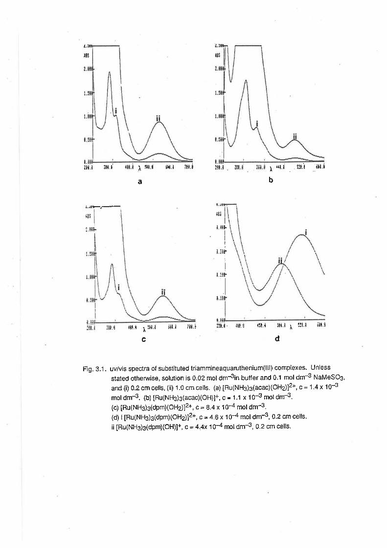
t8s|ls
200.N $8.t I f¡¡.t í¡N.N ?ll.l 2t0.9
a
3. J!F.-
i8s I
I
I il¡fL
¿8¡ l ¡¡l.i ¡?9.¡ tll'à
b
I
H!¡
¡ ¡¡¡.
I
l.¡¡F
I
9.3!r
I
¡
N.
:¡t.i :ll.N {00.1 I:¡N.N i¡i.i i00.1 t¡r.i
c
Fig. 3.1. uv/vis spectra of substituted triammineaquaruthenium(lll) complexes. Unless
stated otherwise, solution is 0.02 moldmJin buffer and 0.1 moldm{ NaMeSOg,
and(i)0.2cmcells,(i¡) 1.0cmcells. (a)[Ru(NH3)3(acac)(OH2)]2+,c=1'4x10⁻³
mol dm-3. (b) tRu(NHg)3(acac)(OH)l+, c = 1.1 x 10⁻³ mol dm-3.
(c) [Ru(NHs)s(dpmXOHz)]2*, c= 8.4 x 10⁴ moldm-3.
(d) i [Ru(NHg)s(dpmXOH 2)12+, c= 4.6 x 1É mol dm-3, 0.2 cm cells.
ii [Ru(NHs)3(dpmXOH)]+, c = 4.4x 10{ mol dm4, 0.2 cm cells.
::Þ.¡' lNN.¡ i5t.i 5li.Û ¡ :¡e'N
d
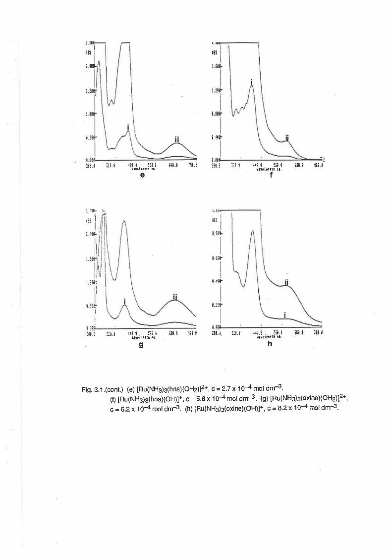
a.
|l5-'5l
I 7îE-
t.
I l.{{F
Iit¡.t ¡10., l¿N.¡. :¡¡.1
J¡ual aaatt â¡6{t.0 ;il.t !,tN.! ::!.t
e
t.É¡fr
l.gF
!!6-
I
{40.¿. :í¡.ir¡Yilf[tln il.
T
ú8¡.t $¡¡.¡
t
äl¡
!NF
I
¿.:![F
ils I
¿.¡tt+
iï
I 'Nft
ilNr
II
t.
hg
t...¡t J trl.'l. :g¡.i i¡l.l :119.¿ i|¡.i. i3!.¡ 't{î Û-,.--.:61.1
t¡Ylielllt[ ll¡. L Yellntln i¡'5¡N.t tNN.¿
Fig. 3.1 .(cont.) (e) [Ru(NH3)3(hna)(OH2)12*, c = 2.7 x 10⁴ mol dm4.(fl tRu(NHs)3(hna)(OH)l+, c = 5.6 x 1É mol dm-3. (g) tpu(NHg)3(oxine)(OH2)12+,
c = 6.2x 1O{ mol drn-3. (h) [Hu(NHg)s(oxineXOH)]*, c = 8.2 x 10+ mol dm4.
rit
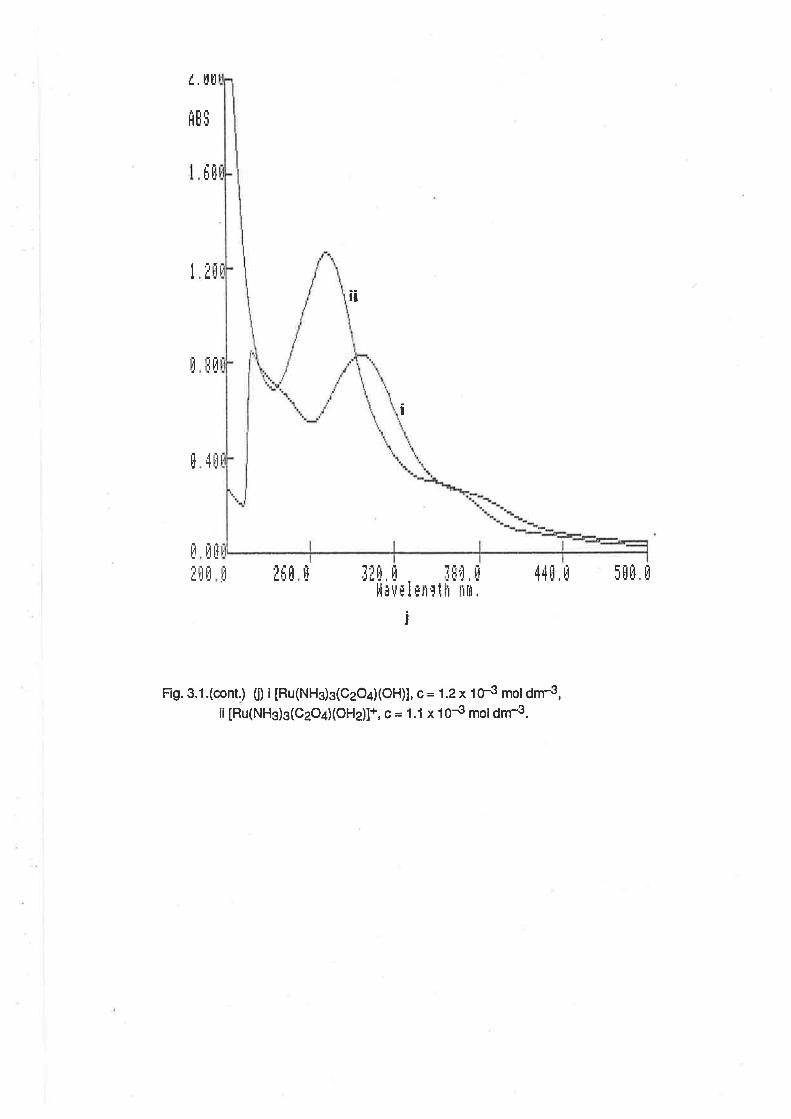
/ ¡rlt
flE
Fig. 3.1.(cont.) (il ¡ tRu(NHg)g(CzO¿XOH)1, c = 1.2 x 1f3 mol dnr3,ii [Ru(NH3)3(CzO¿XOHz)l+, c = 1.1 x 10{ mol dm-3.
I
rlÈ6üü
.1
Ëü
iI
lj tcü
4üI
Ë ilfi
I[[
i¡
i
IHÊ È44fl. ü¡[ü [Ël]rlh l'ltlì.
ilrj ü
ll¡trr I
¡
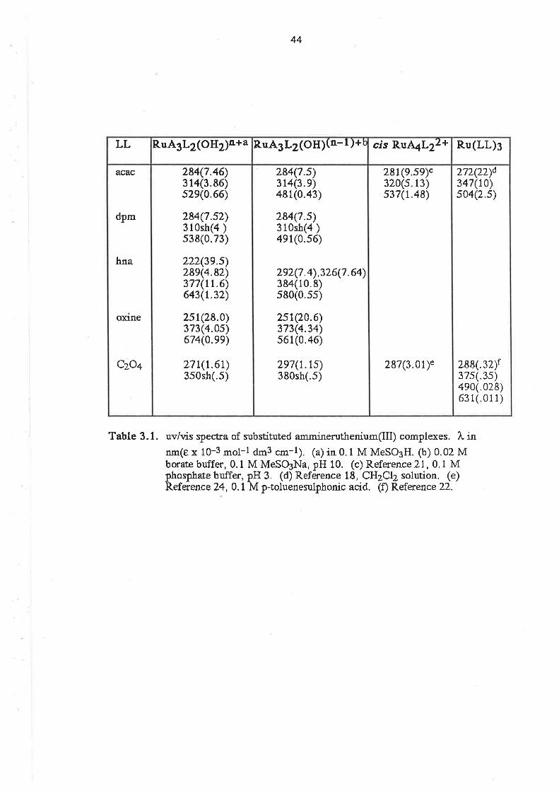
Table 3.1.
44
uv/vis qpectra of substituted ammineruthenium(Ill) complexes. l, innm(e x 10-3 mol-l dm3 cm-l). (a) in 0.1 M MeSO3H. (b) 0.02 Mborate buffer, 0. 1 M MeSO3Na, pH 10. (c) Reference?I,0. 1 Mphoqphate buffer, pH 3. (d) Reference 18, CH2CI2 solution. (e)Reference 24,0.1 M p-toluenezuþhonic acid. (f) Reference22.
2?2(7Ðd347(t0)s04(2.s)
288(.32)f37s(.3s)4e0(.028)63 1( 011)
28132'0537
(e(s(1
5l¡c13)48)
287(3.}r)e
284(7.s)314(3. e)481(0.43)
284(7.s)310sh(4 )4e1(0.s6)
292(7 .4),326(7 .64)384(10.8)s80(0.ss)
2st(70.6)3?3(4.34)s61(0.46)
2e7(r.ts)38Osh(.5)
284(7.46)314(3.86)s2e(0.66)
284(7.s2)310sh(a )s38(0.73)
222(3e.s)28e(4.82)3?7(rt.6)643(r.32)
zst(28.0)373(4.0s)674(0.ee)
z?r(r350sh
.61)(.s)
acac
dP-
hna
oxlne
Czo+
R.u(LL)3cis RIA4L2¿+RuA3L2(OHXn-r )+tR uA3L2(OH2)n+aLL
45
shift in the absorption occurs because the energy of the partially fitled t2n orbital is increased, due to
the strongly n-donating character of the OH- ligand compared with the H2O ligand. Assuming that
the energy of the ligand orbitals is unaffected by the deprotonation of the aqua ligand, the energy
of the LMCTtransition must be increased. The spectrum of [Ru(NHg)s(acacXOHz)12*,with the
LMCT band at 529 nm, is similar to that of [Ru(NH$a(acac)]2+, from whence the assignment of the
band to a LMCT comes. [21] This is further evidence of the similarity of the spectra of ruthenium
complexes with varying proportions of ammine and aqua ligands.
ln the spectrum of [Ru(NH3)s(C2Od(OH2)]+, the LMCT band was at 271 nm and in
[Ru(NHs)s(C2Od(OH)] it was at 297 nm. That the energy of the absorption is lower in the hydroxo
complex implies that the LMCT transition involves the coordinated H2O/OH- group and not the
bidentate ligand. Similar effects are observed in the spectra of the ammine aqua/hydroxo
complexes reported in Chapter 2, where the spectra have an absorption at about 295 nm when
there is ioordinated OH-. Because OH- is a better æ-donor ligand than H2O, the donor orbitals are
at higher energy, and so the energy of the transition to the Ru t2n oôital is lowered. The LMCT
band in [Ru(NH3)a(CzO¿)]+ occurs a|287 nm. [24] By analogy with the spectrum of
IRu(NHj5(CH3CO2)12+, this band can be assigned to a LMCT lrom the carboxylate ligand. [25]
Thus, in [Ru(NH3)3(C2Od(OH2)]+, theLMCT from the oxalate ligand probably occurs in the same
region, but is masked by the charge ransfer from OH-.
3.2.3. Vibrational Spectra. The vibrational spectra of the complexes were recorded as nujol mulls
on KBr discs. The presence of vibrations attributable to the ligands was confirmation that the
ligands were coordinated. The ligand bands were assigned by reference to the previously
recorded spectra of similar complexes. [26]
ln all cases there were absorptions attributable to the OH and NH stretch¡ng vibrations. ln the
spectrum of [Ru(NHs)s(C2Od(OHz)]z(SzOo).H2O, the (t))OH mode was evident from the presence
of two relatively sharp bands at 3480 and 3750 "r-1,
and in [Ru(NH3)3(acac)(OH2)J(S2O6).H2O
there were analagous bands at 3450 and 3670 cm-1, whích were broadened due to the interaction
between the coordinated water and lattice water. ln the spectra of the other complexes, this
interaction was more dominant, with the (D)OH bands being evident as a single broad absorption at
about 3500 cm-1. For all the complexes, there were two or three bands between 3190 and 3300
cm-1 , attributable to the o(NH) mode; a broad band between 1620 and 1640 cm-1, attributable to
tne õ¿1ttU¡ mode and several bands between 1250 and 1350 cm-1 , attributable to the õs(NH)
mode. Though ammíne complexes are also expected to show absorptions between 950 and 590
cm-1, due to the rocking vibrations [47a], these bands were obscured by bands due to ligand or
anion vibrations.
46
The spectra of the diketonate complexes contained bands attributable to the combinatíon of
the (ù)C=O and (l))C=C vibralions. These were at 1500, 1570 and 1588 cm-1 for the acac
complex, at 1530 and 1540 cm-1 forthe dpm complex, and at 1526 and 1565 cm-1 forthe hna
complex. ln the spectrum of [Ru(NHs)s(C2Od(OH2)]2(S2O6).HzO,there were (D)C=O absorptions
at 1635, 1670, 1700 and 1705 cm-1.
The spectra of those complexes with an aromatic moiety in the ligand had absorptions
between 1500 and 1600 cm-1 due to ring-expansion vibrations.
3.2.4. Acid-Base Properties. The pK" for the dissociation of a proton from the aqua ligand in each
complex, according to reaction 3.3, was measured spectrophotometrically, using the molar
absorptivities at the absorption maxima in acid and base solutions.
tRu(NH3)3(LL)(OHz)ln* + tRu(NH3)3(Ll-)(oH2)l(rÌ-1)+ + Ftr' (3.3)
The pK" values determined are listed in Table 3.2. lt can be seen that the coordination of a
negatively charged ligand to the triammineaquaruthenium(lll) group causes an increase of 2 to 3
units in the pK" compared with that of [Ru(NH3)s(OHr¡.¡s+ aI3.77.
This increase in pK" can be explained in terms of the increase in electron density on the Ru(lll)
centre brought about by the o- and æ-donation from the ligand to the metal centre. The pK" of
water is decreased on coordination because of the ability of the electron deficient metal centre to
stabilize the negative charge induced by proton dissociation. lf the electron density at the metal
centre is increased by the replacement of one of the other ligands by a stronger electron donor,
then the pK" of the coordinated water will increase because the metal centre has lost some of its
ability to stabilize the negative charge. Thus, the pK" of a complex in a series of aqua complexes
may be considered to be a measure of the electron donating ability of the substituting ligand. Thus,
it can be concluded that, for the complexes prepared here, the electron donating ability of the
ligands increases in the order: C2O4= hna < sal <bzac < oxine <acac. dpm.
3.2.5. The Etectrochemistry of IRu(NH3)3(LLXOH2)12+ Complexes. The electrochemistry of the
Ru(lll) cnmplexes was studied in aqueous buffer solutions at glassy carbon electrodes. At freshly
polished electrodes, allthe complexes showed a reversible reduction wave in the CV at negative
potentials. The potential at which this wave occurred was pH dependent, and the E172 potentials
forthe complexes at pH 3 are listed in Table 3.2, togetherwith those of some similar complexes. At
anodically activated electrodes, in addition to the reduction wave in the CV (which tended to have a
larger peak separation than at unactivated electrodes), there was also an irreversible wave visible at
positive potentials. On an anodic DPV scan, it was possible to resolve this wave into a distinct peak
with a peak current equal to, at best, 3/4 of that due to lhe +31+2 wave. The potential of this second
47
+I.O4L-0.38s
-0.33h
acac
czo+
cis RuA4L21* or cis RuA4L2+
6.1(r.1)c6.ld
6.5(r.1)c6.6d
5.9d
5.8d
5.7(+.1)c5.8d
6.0(r.1)c5.9d
5.7(+.1)c5.8d
3.77(+.04)
+0.57
+0.56
+0.73
+0.61
+0.42f
+0.49
-0.32
-0.41
-0.30e
-0.2?e
-0.26
-0.3 1
-0.32
-0.t2
acac
dP-
bzac
sal
hna
oxlne
czo+
(Hzo)z
PKaEp(rv/rrr)bE1¡2(llllll)aL
Table 3.2, Redox potenrials and pKa values for zubstituted triammine aquo andtetîaarrlnilnre complexes of Ru(II| @otentials vs SCE; 0.2 M buffer,0.1 M MeSO3Na). (a) pH : l. (b) pH = 5. (c) spectral measurement.(d) from Pourbaix diagram. (e) compound not isolaæd, in situmeasurement. (f) pH = 6.2. (g) Reference2t, 0. 1 M phosphatebuffer. (h) Reference 31, pH 3, triflate buffer. (i) Reference 18a, inCHzClz,0. 1 M (Bu)allBFa, potential corrected to SCE scale.
48
peak was also pH dependent. However, when the scan direction was reversed, to give a cathodic
scan, the peak was very poorly resolved, even at slow scan rates. This result is indicative that the
oxidation was irreversible.
For [Ru(NH3)3(acac)(OH2)]2+, the voltammograms of which are shown in Fig. 3.2,lhe E1¡2
potential of the +3/+2 wave was -O.32 V at pH 3, and was independent of pH in the range
2.6 <pH<5.0. ln the range 7.6<pH<9.0, from the Pourbaix diagram (Fig. 3.3) the pH dependence
for this wave was -55 mV/pH, which is very close to the predicted value of -59 mV/pH for a one
electron transfer reaction coupled with the transfer of one proton. The electrochemical wave can,
therefore, be assigned to the reactions 3.4 and 3.5.
2.6<pH<5.3
tRr(NHj 3(acac) (O,12)!2* + [Ru(NH3)3(acacXoH2)]+ (3.4)+e-
7.6<pH<9.0
[Ru(NHj3(acac)(OH)]' + e- + H+ + [Ru(NH$s(acac)(OH2)]+ (3.s)
The ¡ntersection of the two lines in the Pourbaix diagram corresponds to a pK" value of 6.1, which is
in close agreement with the value determined spectroscopically.
The E1¡2for the reduction in acid solution is about 60 mV less negative than E172 for the
[Ru(NH3)4(acacll2+l+ couple, recently reported as {.38 V in 0.1 moldm-3 phosphate buffer. [21]
ln Chapter 2, it was shown that the substitution of an aqua ligand on a metal complex by an ammine
ligand leads to a decrease in the E1¡2 of approximately 20 mV. Also, it has been shown recently
that the E1¡2of a redox couple involving a ruthenium complex can be affected by the concentralion
of phosphate buffer in the solution.l2Tl An increase in the phosphate buffer concentral¡on from
0.01 mol dm{ to 0.1 mol dm€ caused a decrease in the Ê1¡2ol the Ru(NH3¡u3+12+ couple by
approximately 40 mV, and was attributed to the formation of labile ion pairs. The combined effects
of the extra ammine ligand and the phosphate ion in solution are probably responsible for the
relatively large difference in the E172 values of the tñammineaqua and pentaammine
pentanedionate complexes. There was no evidence to suggest that the acac ligand in
[Ru(NHs)3(acac)(oH2)]2+ was protonated at low pH, as was observed in the electrochemicalstudies
on [Ru(NHs)4(acac)]2+. [21 ]
The Pourbaix diagram forthe peak at positive potentials is also displayed in Fig. 3.3. At low pH,
the positive peak became less well resolved, a phenomenon which was observed when activated
glassy carbon electrodes were used to study the oxidation of [Ru(NHs)s(OHz)]g+. [ZB]
Furthermore, at high pH, the peak was also less easily resolved because of its merging with the
solvent oxidation wave. However, over a limited range, it was possible to observe electrochemical
+o.5
Ttro
+t -o.600
+t.ooo +o.5 -o.5 -0.700
Fig. 3.2. Vottammograms of [Ru(NH3)3(acac)(OHj]2+ at pH 4. (a) Cyclic voltammogram at
an unactivated G.c.E. (b) cyclic vollammogram at an activated G.c.E.
(c) Differential pulse voltammogram at an activated G.C.E. (d) Differentialpulse
voltammogram at an activated G.C.E., corrected for background current'
T
"rf
+o.o
E(UOLTI
a.
oo
ba?oo
\ot
oa
0.8
0.6
0.4
o.2
0.0
-0.2
-0.4
-0.6
1 2 3 4 5 6 7 I I 10pH
Fig. 3.3. Pourbaix diagrams for [Ru(NHs)s(acacXOH2)] redox couples. (a) Ru(lll)/Ru(ll)
couple. (b) Ru(lV)/Ru(lll) couple.
E(v)
behaviour consistent with the presence of a RuO2+/RuOHr3+ couple. tn the range, 4.4<pH<5.3,
the pH dependence was -112 mV/pH, close to the ideal value for two proton/one electron transfer,
and between pH 6.0 and 6.7, the slope was -57 mV/pH. These results are consistent with the
following reactions.
4.4<pH<5.3
IRu(NHs)3(acacxO)]+ IRu(NHs)3(acac)(oH2)]2+ (3.6)
49
{€- + 2H+ ==
{€-+H++ IRu(NH3)3(acac)(OH)]+ (3.7)
The intersection of the two lines in the Pourbaix diagram is at slightly less than the spectroscopically
determined pK. value of the Ru(lll) complex.
For [Ru(NH3)s(dpm)(OH2)]2+, the voltammograms of which are shown in Fig.3.4., the E1¡2
potential of the +3/+2 wave was -0.41 V at pH 3, and was independent of pH in the range 3.4
<pH<5.4. From the Pourbaix diagram (Fig.3.5), above pH7.7, the pH dependence forthis wave
was -57 mV/pH, which is close to the idealvalue of -59 mV/pH for a one proton/one electron
transfer. The electrochemicalwave can, therefore, be assigned to the reactions 3.8 and 3.9.
3.4<pH<5.4
IRu(NH$3(dpm) (oH2)]2+ +e- == [Ru(NHs)3(dpm)(OH2)]+ (3.8)
7.7<pH<9.1
[Ru(NHj3(dpm)(OH)]+ +e- + H+ + [Ru(NHsþ(dpm)(oH2)]+ (3.s)
The intersection of the two lines in the Pourbaix diagram lies between pH 6.5 and 6.6, which is in
agreement with the pK" value determined spectroscopically.
The Pourbaix diagram for the peak at positive potentials ís also displayed in Fig. 3.5. The pH
range over which reliable measurements could be made was limited by the same factors as those
which were operating in the DPV experiments on [Ru(NH3)g(acac)(OH2)]2+, but to a greater extent
than in the fírst case. Again, it was possible to observe electrochemical behaviour consistent with
the presence of a RuO2+¡pugF{2+ couple. Between pH7.4 and 9.0, the pH dependence was -56
mV/pH. This is consistent with the following reaction.
7.4<pH<9.0
[Ru(NH3)3(dpmXo)]+ {€- + H+ + [Ru(NH3)3(dpmXoH)]+ (3.10)
6.0<pH<6.7
[Ru(NH$3(acac)(O)]+

T'f+1. -o.700
+1.OOO +o.5 +0.o
E(UOLT)
-o.s -o.700
Fig.3.4. Voltammograms of [Ru(NHg)s(dpmXOH2)12+ ed.pH 4.5. (a) Cyclic voltammogram at
an unactÍvated G.C.E. (b) Cyclíc voltammogram at an activated G.C.E.
(c) Differential pulse voltammogram at an act¡vated G.C.E. (d) Differential pulse
voltammogram at an activated G.C.E., corrected for background current.
T500n4I

aI
o
'ooaa
o O+.€ro oosoocà
"a. a aa
0.8
0.6
0.4
o.2
0.0
-0.2
-0.4
-0.6
-0.8
E
(v)
8 9 10 11 12
Fig. 3.5. Pourbaix diagrams for [Ru(NH3)s(dpmXOHz)] redox couples. (a) Ru(lll)/Ru(ll)
couple. (b) Ru(lV)/Ru(lll) couple.
7pH
23456
50
ln the Differential Pulse Voltammogram of [Ru(NH3)g(dpm)(OHz)]z* at the activated electrode,
a broad peak at approximately +0.05 V was distinguishable, in addition to the peaks assigned to the
pu3+/2+ and RuO2+/RuOH2+ couptes. This peak is believed to be due to some electrochemical
process involving the solvent and the activated electrode. When an instrumental background
correction procedure was applied, this broad peak was no longer evident in the voltammogram.
The voltammograms of [Ru(NH3)3(hna)(OH2)]2* are shown in Fig. 3.6. The CV appears poorly
resolved because of the operation of a normalization process in the instrumental display. The
display is normalized so that the point on the curve with the highest current is at the highest point
possible in the display. ln this case, the "tail" due to the solvent oxidation wave has the highest
current so that ¡¡" pu3+/2+ wave appears poorly resolved. The wave due to the RuO2+/RuOH23+
couple is evident in the CV as a shoulder to the solvent oxidation wave. ln the DPV, this peak is
better resolved. The Pourbaix diagram lor these voltamnxcgrams, shown in Fig. 3.7, is conéistent
with the pH dependence of the two electrochemical processes being similar to those discussed
above.
For the ¡u3+/2+ CV wave, lhe E1¡2value at pH 3 was -0.26 V, and independent of pH below
pH 4.6. Above pH 6.6, this wave varied with pH to the extent of -56 mV/pH. These results are
indicative of the following redox processes.
pH<4.6
IRu(NH3)3(hna)(OH2)]2+ + [Ru(Nft)3(hna)(oH2)]+ (3.11)+e-
pH>6.6
[Ru(NH3)3þna)(OH)]+ + e- + H+ => [Ru(NH3)3(hna)(OH2)]+ (3.12)
For the RuO2+/RuOH23+ peak, the peak potential was +0.75 V at pH 5, and +0.37 V at pH L
Between pH 4.8 and 5.6, the pH dependence was -114 mV/pH, and above 6.3 it was -59 mV/pH.
This corresponds to the reactions 3.13 and 3.14.
4.BcpH<5.6
[Ru(NH3)3(hna)(O)]+ + e- + 2H+ + [Ru(NH3)3(hna)(OH2)]2+ (3.13)
pH>6.3
[Ru(NH3)3(hnaXO)]+ + e- + H+ + [Ru(NH3)s(hna)(OH)]+ (3.14)
The intersection of the lines in the Pourbaix diagram was at pH 5.8 for the p¡¡3+/2+ wave, and at 6.0
for the RuO2+/RuOH23+ peak, both of which are close to the spectroscopically determined pK"
value of 5.7, though somewhat higher.
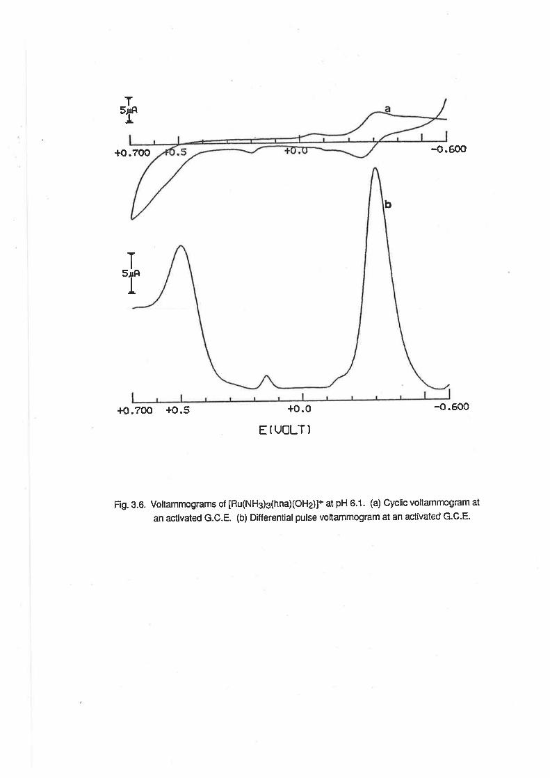
Ttr:
T
T
+o.700 -o.600
+o.700 +o,5 -o.600
Fig. 3.6. Voltammograms of [Hu(NHs)s(hna)(OHj]+ at pH 6.1. (a) Cyclic voltammogram at
an actlvated G.C.E. (b) Differentialpulse voltammogram at an activated G.C.E.
b
+o.0
EfUOLT)
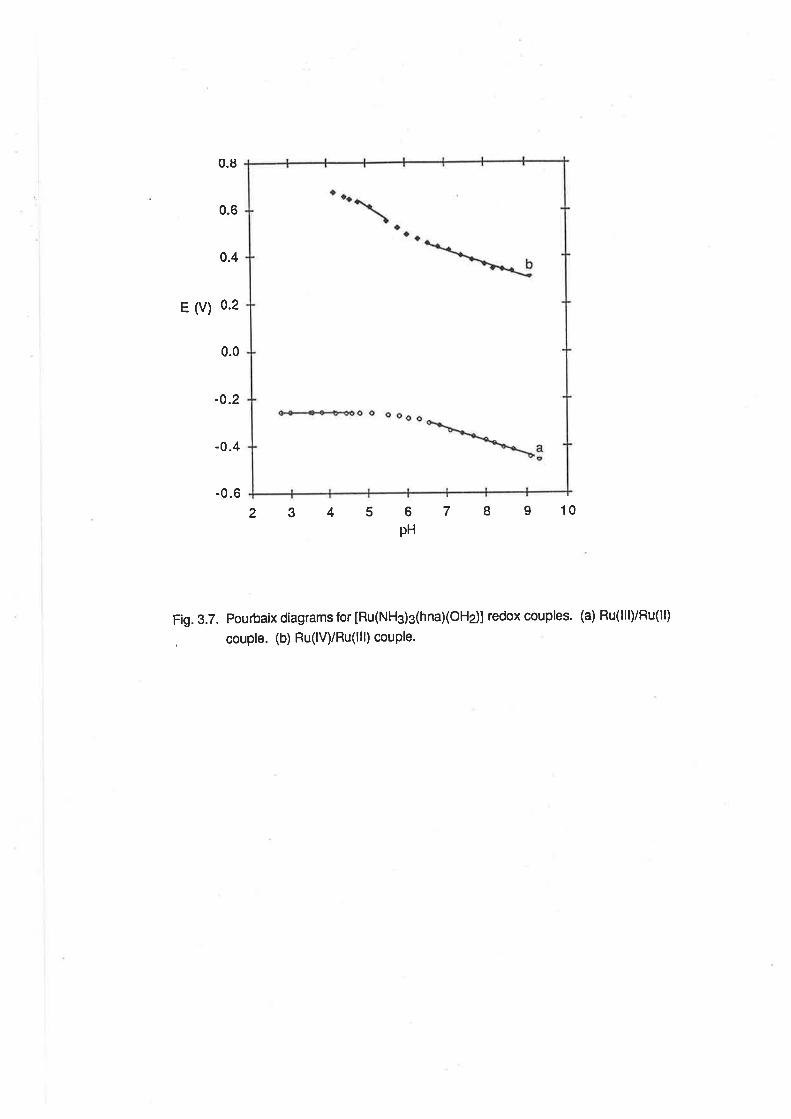
0.6
E U) 0.2
-0.2
-0.4
-0.6
0.8
0.4
0.0
2345 6pH
7 8 9 10
Fig. 3.7. Pourbaix diagrams for [Ru(NH3)s(hnaXOHe)] redox couples' (a) Ru(lll)/Ru(ll)
, couple. (b) Ru(lV)/Ru(lll) couple.
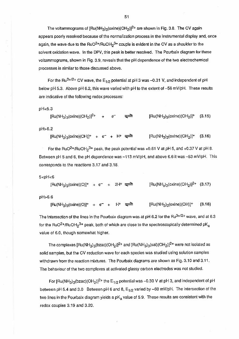
51
The voltammograms ol [Ru(NH3)3(oxine)(OH 2)12+ are shown in Fig. 3.8. The CV again
appears poorly resolved because of the normalization process in the instrumental display and, once
again, the wave due to the RuO2+/RuOHr3+ couple is evident in the CV as a shoulder to the
solvent oxidation wave. ln the DPV, this peak is better resolved. The Pouôaix diagram for these
voltammograms, shown in Fig. 3.9, reveals that the pH dependence of the two electrochemical
processes is similar to those discussed above.
For the qr3+/2+ CV wave, the E172 potential at pH 3 was -0.31 V, and independent of pH
below pH 5.3. Above pH 6.2, this wave varied with pH to the extent of -56 mV/pH. These results
are indicative of the lollowing redox processes:
pH<5.3
IRu(NH3)3(oxine) (OH2)]2+ + [Ru(NH3)s(oxine)(OH2)]+ (3.1s)+e-
pH>6.2
[Ru(NH3)3(oxine)(OH)]+ + e- + g+ ¡- [Ru(NH3)3(oxine)(OH2)]+ (3.16)
For the RuO2+/RuOH23+ peak, the peak potential was +0.61 V at pH 5, and +0.37 V at pH B.
Between pH 5 and 6, the pH dependence was -113 mV/pH, and above 6.6 it was -53 mV/pH. This
corresponds to the reactions 3.17 and 3.18.
5<pH<6
[Ru(NHj3(oxine)(O)]+ + e- + IRu(NH.)3(oxine)(oH2)]2+ (3.17)
pH>6.6
[Ru(NH3)s(oxineXO)]+ + e- +
2H+
H+
-\-
-IRu(NH3)3(oxine)(OH)]+ (3.18)
The intersection of the lines in the Pouóaix diagram was at pH 6.2 forthe Fru3+12+ wave, and at 6.3
for the RuO2+/RuOH23+ peak, both of which are close to the spectroscopically determined pK"
value of 6.0, though somewhat higher.
The complexes [Ru(NH3)3(bzac)(Otl2)12* and IRu(NH3)3(sal)(OH2)]2+ were not isolated as
solid samples, but the CV reduction wave for each species was studied using solution samples
withdrawn from the reaction mixtures. The Pourbaix diagrams are shown as Fig. 3.10 and 3.11.
The behaviour of the two complexes at activated glassy carbon electrodes was not studied.
For [Ru(NH3)3(bzac)(OH2)]2+ the E172 potentialwas -0.30 V at pH 3, and independent of pH
between pH 5.4 and 3.0 Between pH 6 and S,Eltzvaried by -60 mV/pH. The intersection of the
two lines in the Pourbaix diagram yields a pK" value of 5.9. These results are consistent with the
redox couples 3.19 and 3.20.
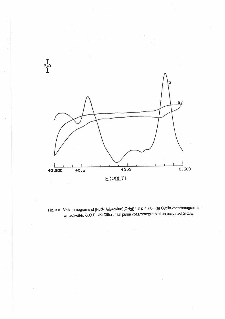
T
T
+0.800 +o.5 -o.Éo0
Fig.3.S.Voltammogramsof[Ru(NH3)3(oxineXoHz)]+atpH7.0.(a)Cyclicvoltammogramatan activated G.C.E. (b) Differentialpulse voltammogram at an act¡vated G'C-E.
+0.0
E IUOLTl
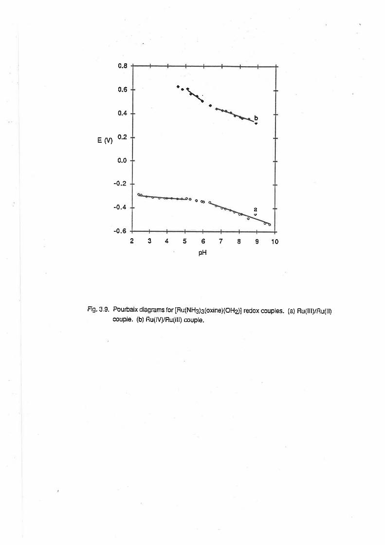
a
o6
b!
a
a
\
a
E (V)
0.8
0.6
0.4
0.2
0.0
-0.2
-0.4
-0.6
6
PH
7 I 9 10
Fí9. 3.9. Pourbaix diagrams for [Ru(NH3)3(oxine)(oHdl redox couptes. (a) Hu(Iil)/Ru(lt)couple. (b) Ru(lV)/Ru(Iil) coupte.
2345

.0.20
-0.25
-0.30
-0.40
-0.35ts
(v)
-0.45
-0.50
-0.55
-0.60
12345678pl'l
Fig. 3.10. Pourbaix diagram for the Ru(tll)/Ru(ll) redox couple of [Ru(Nft)3(bzac)(OHj]2+.
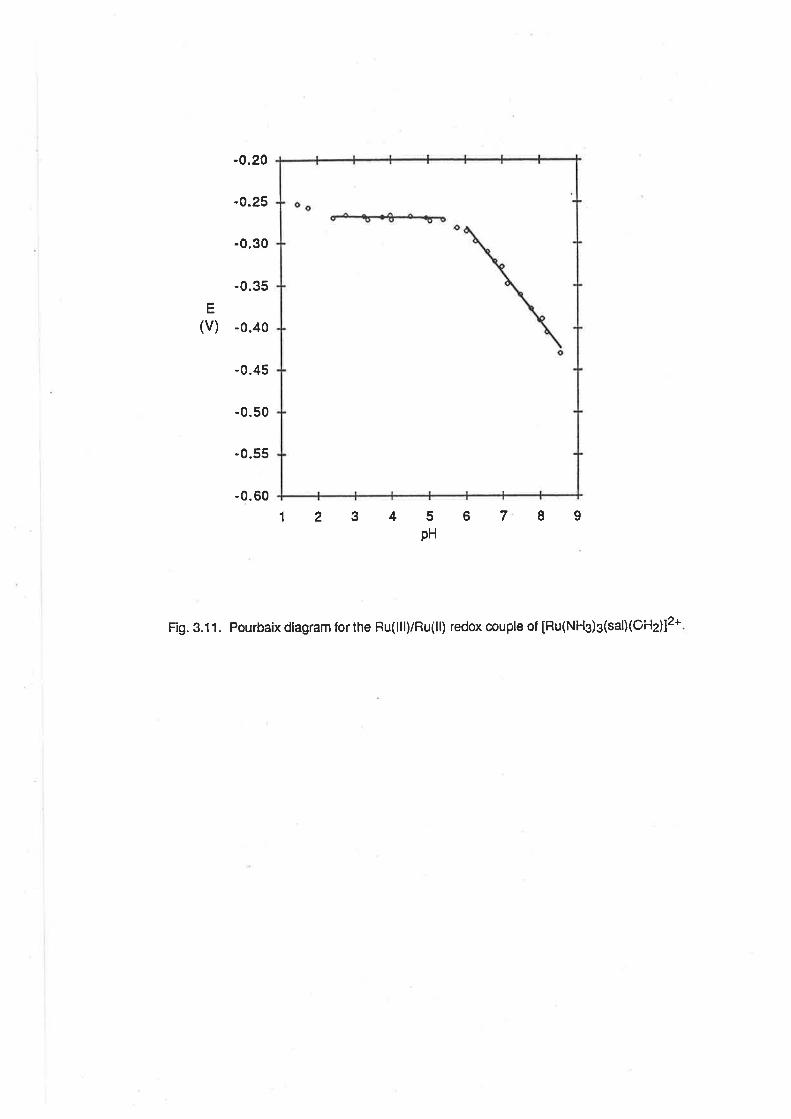
-0.20
-0.25
-0.30
-0.35
E
(v) -0.40
-0.45
-0.50
-0.55
-0.60
1234 5pH
6789
Fig. 3.11. Pourbaix diagram for the Ru(lll)/Ru(ll) redox couple of [Ru(NHg)g(salXOHz)]2+
52
3.0<pH<5.4
[Ru(NHs)3 (bzacXOH2)]2+ + e- + [Ru(NHs)s(bzac)(OH2)]+ (3.19)
6.0<pH<8.0
[Ru(NH3)3(bzacXOH)It + e- + l-f'- + [Ru(NH3)3(bzac)(OH2)]+ (3.20)
For [Ru(NH3)3(sal)(OH2)]2* tne E172 potentialwas 4.27 V at pH 3, and independent of pH
between pH 5.4 and2.4. Above pH 6, E172 varied by -56 mV/pH. The intersection of the two lines
in the Pourbaix diagram yields a pK" value of 5.8. These results are consistent with the redox
couples 3.21 and 3.22.
2.4<pH<5.4
[Ru(NH3)3(sal)(oH2)]2+ + e- =à [Ru(NH3)3(sal)(oH2)]+ (3.21)
pH>6.0
[Ru(NHj3(salXoH)]+ + e- + H+ + [Ru(NHjs(sal)(OH2)]+ (3.221
3.2.6. The Electrochemistry of [Ru(NH3)3(C2Oa)(OH2)]+. The electrochemistry of
IRu(NH3)3(C2Od(OHz)]+ was also studied in aqueous buffer solutions at glassy carbon electrodes.
It was possible to observe a reversib¡s pu3+/2+ wave in the CV at negative potentials. The
behaviour of this wave was identical at freshly polished and anodically activated electrodes (Fig,
3.12). At pH 3, the E172 potential of this wave was -0.32 V and was independent of pH in the range
pH 5.0 to pH 2.6. Between pH 7 and pH 9, the pH dependence for this wave was -59 mV/pH (Fig.
3.13). These results are consistent with the existence of two redox couples shown below. The
intersection of the two lines in the Pourbaix diagram, at pH =5.8, is in close agreement with the
spectroscopically determined pK" value of 5.7.
2.6<pH<5.0
IRu(NH3)3 (C2of (oH2)]+ + [Ru(NH3)3(C2od(oH2)] (3.23)+e-
7.0<pH<9.0
[Ru(NH3)3(C2Od(OH)I + e- + H+ => [Ru(NH3)3(C2Od(OH2)I @.24)
The E172 potentialof [Ru(NH3)3(C2Od(OHz)]+ at pH 3 is very close to that of
[Ru(NH3)a(CzO¿)]* [31], as can be seen from the data in Table 3.2. The difference in the two
values is much smaller than in the case of the pentanedionate complexes, probably because there
was no phosphate ion effect in the electrochemicalstudies on [Ru(NH3)¿(CzO¿)]+.
An electrochemicalwave due to the oxidation of [Ru(NHs)s(C2O4)(OHz)]+ could be observed
at both freshly polished and anodically activated electrodes (Fig 3.12 ). At very slow scan rates (10 -
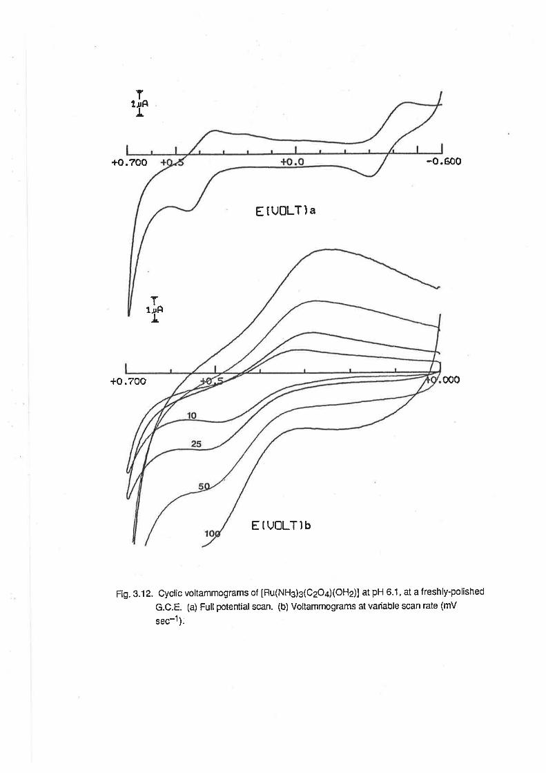
t
+o.700 -o.600
EtUOLTla
*O:70O 000
EtvoLTlb
Fig. 3.12. Cyclic voltammograms of [Ru(NH3)3(C2O¿XOHd] at pH 6.1, at a freshly-polished
G.C.E. (a) Full potential scan. (b) Voltammograms at variable scan rate (mV
sec-1 ).
ï¡AT
T1¡âI
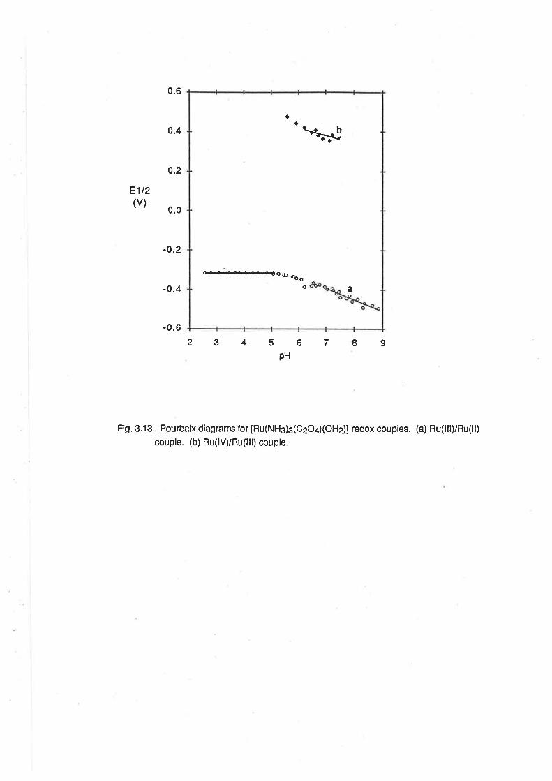
0.6
0.4
o.2
E1t2(v)
0.0
-0.2
-0.4
-0.6
23456789pH
Fig. 3.13. Pourbaix diagrams for [Ru(NH3)s(CeO¿XOHj] redox couples. (a) Ru(lll)/Ru(ll)
couple. (b) Ru(lV)/Ru(lll) couple.
o+a+a-aa€oo çoo
Cto
\*a
a
53
25 mV sec-1¡, it was possible to observe a quasi-reversible wave at neutralto high pH. At higher
scan rates, and at lower pH, lhis wave broadened and meqed with the solvent oxidation wave. ln
the Pourbaix diagram lor this wave (Fig. 3.131,81p and the cathodic peak potential varied by -71
mV/pH above pH 6. The anodic peak potential remained almost constant with increasing pH
because of the merging of this peak with the solvent oxidation wave. However, that the cathodic
wave is well defined in this range, and its pH dependence is reasonably close to the ideal for a one
proton/one electron transfer, is evidence that [Ru(NHg)3(C2Od(OH)] undergoes oxidation
according to 3.25.
pH>6.0
[Ru(NH3)3(CzO¿XO)] + e- + H+ + [Ru(NH3)3(C2Od(OH)] (3.2s)
When the electrochemicaloxidation was performed at an activated electrode, there was no
improvement in the resolution e1 pu4+7R¡3+ CV wave over that when a freshly polished electrode is
used at low scan rates. The behaviour of the DPV peak was quite unusual. As the pH ol the
solution was increased, the pH dependence of lhe peak increased. Thus, at pH 4 the pH
dependence was 41 mV/pH, while at pH B it was -150 mV/pH. This behaviour is the opposite to
what would be expected if the DPV peak was due to the redox couple 3.25. lt is apparent that the
activated electrode affected the oxidation of [Ru(NH3)s(CzOd(OH2)]+ in a manner ditferent from
the other examples where the activation procedure was used. One possible explanation is that, at
high pH, the oxidation process was also involving the ammine NH groups. lt has been shown
previously that at high pH, an activated electrode can be used to observe the proton-coupled
oxidation of [Ru(NHs)e]3*. [ZZ] lf this same process was operating in the present case, then the pH
dependence of the electrochemical oxidation would be much higher than anticipated.
The chemical oxidation of [Ru(NHs)g(C2O/(OH2)]+ was attempted. However, when a solution
of the complex, buffered at pH 7, reacted with hydrogen peroxide solution, a dark brown c¡lour was
observed in the solution, indicative of oxidative decomposition, as occurred with
IRu(NH3)3(OHz)sl3* and cr's [Ru(NH3)a(OH2)2]3+. [29]
3.2.7. Discussion of Electrochemical Results. The existence of well-behaved, pH dependent CV
waves for the [Ru(NH3)g(LL)(OH2¡¡2+/+ couples is in agreement with what was to be expected from
consideration of the ammine aqua complexes referred to in Chapter 2, andfrom the behaviour of
substituted edta complexes of ruthenium(lll). tl4 As noted above, there are lew examples of
bidentate, uninegative ammine ruthenium(lll) complexes with which these results may be
compared. The similarity in the E172 potentials of [Ru(NHs)s(acac)(OH2)]2+ and
[Ru(NH3)a(acac¡]2+has already been noted. Allthe substituted ammineaqua ruthenium(lll)
complexes ¡¿ys gu3+/2+ couples with E172 potentials more negative than that of
54
[Ru(NHs)3(OHz)gl3*. This ref]ects the electron donating ability of the ligands used, as was also
evident from the comparison of pK" values. The reduction potentials were more negative because
the donation of electron density into the metal orbitals by the ligands makes the addition of an
electron by electrochemical reduction less energetícally favourable.
The relative order of the electron donating ability, as illustrated by reduction polential, is
dpm>acac>oxine>bzac>sal>hna. The same ranking for the diketonate ligands, dpm, acac and
bzac, was found for the reduclion of Ru(diketonate)3 complexes. [18, 30] Similarly, in a series of
vanadium complexes with benzolyhydrazone ligands, {R=N-N-C(O)Pn¡z-, the reduction potentials
of the V(lV) complexes varied with R in the same order. [31]
The existence of a RuO2+/RuOH23+ couple in the electrochemistry of the substituted
ammineaqua ruthenium(lll) complexes is also to be expected on the basis of comparison with the
ammineaqua complexes reported in Chapter 2, and also by comparison with the substituted
aquaruthenium 2+ and 3+ complexes referred to in Chapter 1. lt is apparent from the data in Table
3.3 that the Ru(lV)/Ru(lll) couples (rccur over a smaller range of polentials than the Ru(lll)/Ru(ll)
couples and that, in the absence of ¡r-backbonding, the ditference in the potentials of the
Ru(lV)/Ru(lll) couple and the Ru(lll)/Ru(ll) couple is within the range 0.6 V - 0.8 V. The small range
over which the Ru(lV)/Ru(lll) couples occur has been noted by other authors, and this has been
attributed to the similar o-donor properties of the ammine and polypyridyl ligands, while the large
range of potentials for the Ru(lll)/Ru(ll) couples was attributed to the eff ect of the drr-rr. interaction in
the Ru(ll) state. [28] These present results lead to lhe conclusion that it is not only the odonor
properties of the co-ligands, but the overall electron donating ability which determines the potential
of the proton-coupled oxidation of Ru(lll). The potentials for the oxidation of the two polypyridyl
complexes listed in the table are indicative that, perhaps, the æ-acceptor ability of the ligands has
some impact on the oxidation. ln similar comparitive studies, it was found lhat, for a large range of
Ru(diketonate)g complexes, there was a relatively constant difference between the reduction and
oxidation potentials. [18, 30]
There is now, also, an apparent trend in the stability of the oxoruthenium(lV) complexes. On a
voltammetric scale, the substituted triammineoxoruthenium complexes with the uninegative ligands
are less stable than the triamminetriaqua, which, it has already been noted, ís less stable than the
pentaammine complex, the polypyridylcomplexes and the macrocyclic tertiary amine complexes.
a28,29,32, 33] A possible explanatíon for this trend is that lhe æ-donating ability of the 02- tigand,
when combined with lhat of the bidentate, uninegative ligands, is too great lo be accomodated on a
Ru(lV) centre. ln previous cases where the RuO2+/RuOHr3+ couple has been observed, the other
ligands have been either odonors, as in the case of [Ru(NHjr1oH2)313+,[Ru(NH3)5(OH2)]3+ t28al
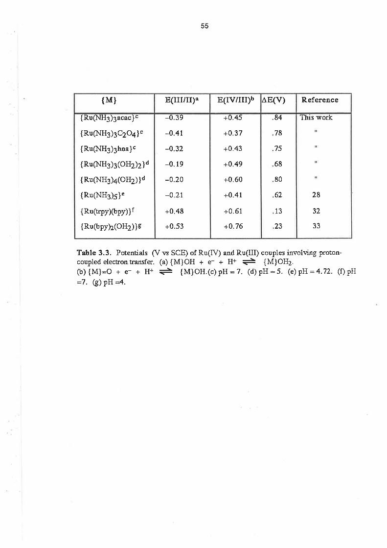
55
This work
28
32
33
84
78
75
68
80
62
13
23
+0.45
+0.37
+0.43
+0.49
+0.60
+0.41
+0.61
+0.76
-0.39
-0.4t
-0.32
-0.19
-0.20
-0.21
+0.48
+0.53
{Ru$lH3)3acaclc
{Ru(NH3)3Czo+l'
{Ru(NH3)3hna}c
{Ru(NH3)3 (o}Jùz]¡d
{Ru(l.IH3)a(oHz)}d
{RuQ.IH3)5}e
{Ru(tqpy)(bpy)}f
{Ru(bpy)2(oH2)}s
R.eferenceAE(v)E(IV/III)bE(III/II)a{M}
Table 3.3. Potentiuls (V vs SCE) of Ru(IV) and Ru(III) couples involving proton-coupled electrontransfer. (a) {M}OH + e- + H+ + {M}OH2.(b) {M}:O + e- + H+ + {M}OH.(c)pH:7. (d)pH:5. (e)pH :4.'77. (f)pH
-7. G) pH :4.

56
and trans [Ru(tmcXOHzXO)]2* [29], or o-donor/r¡-acceptor ligands as in the case of
[Ru(trpy)(bpyxO)]2* [32] andcis tRu(bpy)2(OHZXO)12*. [33] ln these latter cases, there was not any
great competition to the 02- ligand from other r-donors. lt would appear, therefore, that the
inclusion of a n-donor ligand in lhe coordination sphere destabilizes the RulV=O group and
possibly favours the formation of Ru(V) complexes. This destabilization process probably
competes with the oxidative dehydrogenation reaction for the decomposition of the Ru(lV)
complex.
Support lor lhis proposal can be found in a report of oxovanadium complexes containing
tridentate, dinegative ligands. [34] lt was found that, with dinegative ligands having O-N-O donor
atoms, and for which stable VIV(L)z complexes could be obtained, V(lV) complexes of the type,
V(LXO)]2, were very readily oxidized to the corresponding V(V) complexes. lt seems likely,
therefore, that this effect is also important in determining the stability of the oxoruthenium(lV)
complexes.
That it was possible to observe a quasi-reversible proton-coupled pu4+79u3+ wave in the
oxalate complex supports the arguments used concerning the æ-donating ligands destabilizing the
Rulv=O species. The pK" of [Ru(NH3)s(C2Od(OH2)]+ implies that the oxalate ion is not a good æ-
donor ligand because, despite being a dinegative ligand, the pK" value of the complex is the same
as that of the hna complex, which is only a uninegative ligand. Clearly then, the electron-donor
properties of the oxalate ion are not sufficiently strong to interfere with the stabilization of the Ru(lV)
complex by the oxo ligand.
3.2.8. Comparison olEl¡2with pK". ln discussing the pK" values andEl¡2potentials of the
substituted triammine complexes it was stated that both depended on the electron donating ability
of the particular ligand. lt might be expected that, if the same ligand effects are operating in
determining both quantities, there would be a good correlation between them. ln Fig. 3.14 the
E172 potentials for the complexes under consideration are plotted against pK". lt can be seen from
the diagram that there is a linear relationship between E1¡2al pH 3 and pK" for the six complexes
with uninegative, bidentate ligands.
It can also be noticed that the E172 potential of [Ru(NHs)3(C2Od(OHj]+ is more negative than
would be predicted from consideration of its pK". However this apparent anomaly should not be
regarded as serious, because there are several possible reasons why the comparison of the oxalate
complex with the others should not be valid. The oxalate complex differs from the others in at least
three respects. The oxalate ligand is dinegative whereas the ligands in the other complexes are
uninegative. The metal ligand ring is S-membered rather than G-membered as in the case of the
complexes with the uninegative ligands. Thus there may be additional steric factors to be
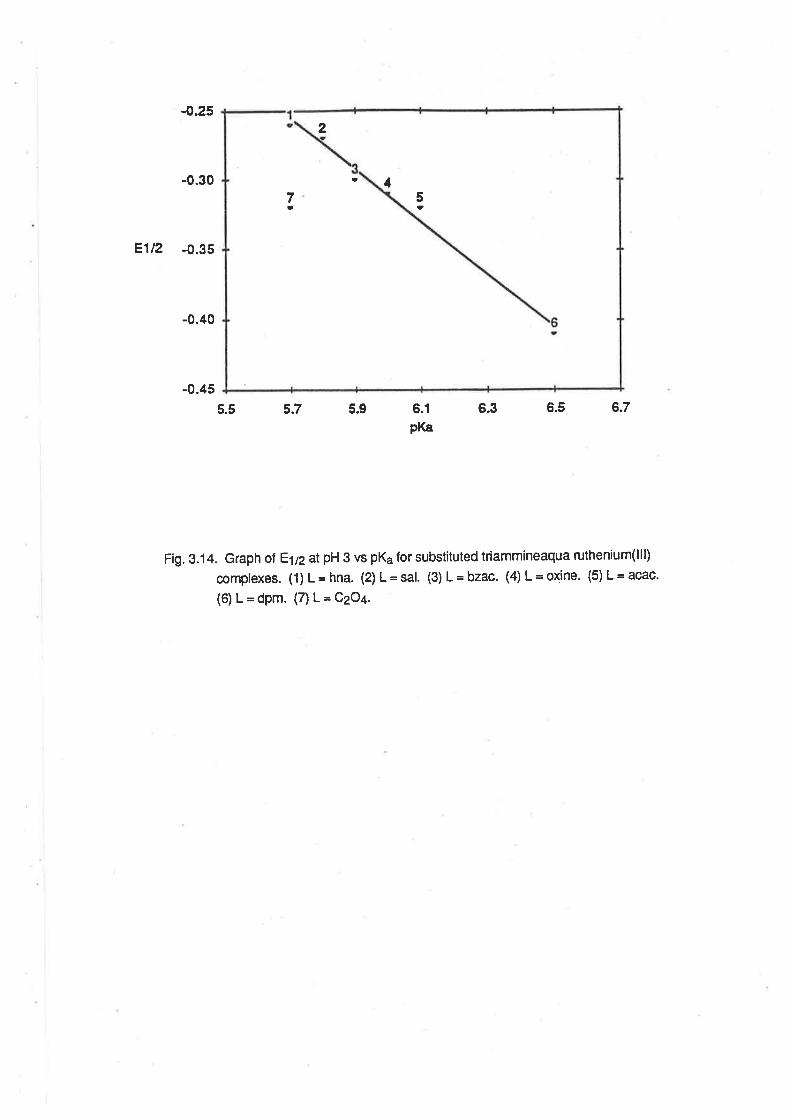
-0.2s
-0.30
Ê1t2 -0.35
-0.40
-0.45
2
a 17t
5a
5.5 5.7 5.9 6.1
pK86.3 6.5 6.7
Fig. 3.14. Graph ol El, at pH 3 vs pK¿ for substítuted triammineaqua ruthenium(lll)
complexes. (1) L= hna. (2) L=Sâ1. (3) L=bzac. (4) L=oxine. (5) L=acac.
(6) L=dpm. (7) L=QzOt.

57
cons¡dered. Finally, the dissociation of a proton from the Ru(lll) aqua complex or reduction to Ru(ll)
results in a neutral species in solution, whereas the other complexes retain a single positive charge
after both these reactions. lt is likely that there will be ditferences in the solvation of these
complexes because of this factor, and so comparisons between the oxalate complex and the other
Ru(lll) complexes must be made with care.
3.2.9. Electrolytic Reduction of Ru(lll) Complexes. The complexes [Ru(NHs)3(C2Od(OHz)]*,
[Ru(NH3)3(oxine)(OH2)]2+ and tRu(NH3)3(hna)(OH2)12+ were reduced at a mercury poolcathode in
a 0.1 moldm-3 methanesulphonic acid solution. The reduction potentialwas chosen to be 150 mV
cathodic of the E1¡2 determined by CV. The solutions were degassed with either argon or nitrogen
gas, and in the latter case, there was no evidence of the formation of a dinitrogen complex with any
of the reduced species. Reduction with amalgamated zinc produced lhe same results as
electrolytic reduction.
When [Ru(NH3)3(C2O4XOHj]+ was reduced, the initial yellow colour of the solution faded to
almost colourless. After about 45 minutes, at which time a charge equalto 0.8 electron per
ruthenium had been passed, and the electrolytic current had decayed to a steady state, a CV of the
solution contained a single wave with E1n= -0.10 V, consistent with the presence of
IRu(NHg)3(OHz)g)]3+. The identity of the product was confimed by the uv/vis spectrum, which was
the same as lor fresh samples of [Ru(NH3)g(OHz)g]s* under similar conditions.
The hydrolysis of the oxalate complex was to be expected given the lability of the carboxylate
ligand in other Ru(ll) ammine complexes. [4] Clearly, though the CV of [Ru(NH3)3(C2Ofl(OHz)]+ is
reversible at moderate scan rates (25 - 100 mV sec-1), on a longertime scale the hydrolysis reaction
is able to occur to an appreciable extent.
During the initialstage of the reduction of [Ru(NH3)3(hna)(OH2)]2+, the green colour of the
original complex was quickly replaced by a purple colour. The purple species was very unstable to
oxidation, decomposing to the original complex if the reduction was halted momentarily, or if a
sample was removed from the reduction cellto allow determination of its uv/vis spectrum. lf the
reduction was allowed to conlinue, over a period of about 45 minutes, the purple colour faded to
yellow, and the total charge which had passed when the current reached a steady state was
equívalent to more than two electrons per ruthenium. There was no current flow if , at this stage, an
oxidizing potentialwas applied to the cell. A CV of the yellow solution contained a single,
irreversible wave, with a cathodic peak potential of -0.28 V. lt is apparent that the Bu(ll) complex,
which is the initial reduction product, is capable of accepting further electrons, possibly into the æ
system of the ligand, and that the resulting species is not capable of undergoing electrochemical
oxidation.

58
When [Ru(NH3)3(oxine)(OH2)]2+ was reduced, the initialgreen colourwas gradually replaced
by a golden-brown colour. A CV of the reduced solution showed that the golden-brown species
was [Ru(NH3)3(oxine)(OH2)]+ and if the solution was exposed to lhe air, the green colour returned.
It was possible to determine a semi-quantitative uv/vis spectrum of the Ru(ll) complex. As the
reduction réaction proceeded, the bands at 250 nm, 370 nm and 674 nm in the uv/vis spectrum of
[Ru(NHs)s(oxine)(OH2)]2+ were replaced by bands at 235 nm and 465 nm, with isosbestic points at
242, 265,335 and 400 nm (Fig. 3.1 5.) The estimated molar absorptivities were E2gs = 3 x 1 04 and
t465 = 6 x 10³. lt was a little sufrprising that these intra-ligand bands were so greatly affected by the
reduction of Ru(lll) to Ru(ll), because it would be expected that the reduction would not have a great
effect on the energy of the ligand orbitals. However, similar spectral changes have been noted
during the electrolytic reduction of Ru(dbm)3 to Ru(dbm)3-.t181
3.2.10. Description of the Crystal Structure of [Ru(NH3)3(acacXOH2)](S2O6).2H2O. Cubic crystals
of [Ru(NH3)3(acac)(OH2)](5206).2H2O were obtained from the synthesis reaction mixture after
ethanol had been added to a solution of the reaction product in dilute dithionic acid. The crystal
structure was determined by Dr. E. R. T. Tiekink, and is shown in Fig. 3.16, which also shows the
alomic numbering scheme employed. The bond lengths and angles are listed in Tables 3.4 and
3.5, which follow Fig. 3.16.
The structure comprises an almost perfectly octahedral arrangement of donor atoms about the
ruthenium íon. All the bond angles are close to either 90 or 180 degrees. The ammine ligands
occupy one face of the octahedron, as is the case in the precursor complex, Ru(NH3)3Cls. [35]
The average length of Ru-N bonds for the ammine ligands trans to acac is 2.111 Ä, which is
slightly shorter than the average Ru-N distance in Ru(NHs)sClg (2.1214¡ 1SS¡, and slightly longer
than the average length in [Ru(NH3)o](BF+)s e.104 Ä). tgOl The comparison with the structure of
Ru(NHg)3C|3 must be treated caut¡ously, due to the presence of extensive hydrogen bonding in
that complex. The slight increase in the Ru-N distance in the acac complex, compared with the
hexaammine, is probably due to the lower charge on the ruthenium ion. The Ru-N bond which is
translo the the aqua ligand is slightly shorter than the others, being 2.046(1 1) Ä. This is possibly
due to the lesser competition, between the ammine and aqua ligand for the same metal orbital, than
between the ammine ligands and the anionic acac ligand.
The Ru-O bond distance to the aqua ligand is 2.058(10) A, which is longer than the average
Ru-O distance (2.029 Ä¡ in lnulOHz)ol(pTS)g [37], possibly due to the lower charge on the
ruthenium centre. The average Ru-O distance between the metal and the acac ligand is 2.004 Ä,
which is very close to average Ru-O distance in Ru(acac)s (2.00 Ä). IgAl The aqua ligand was
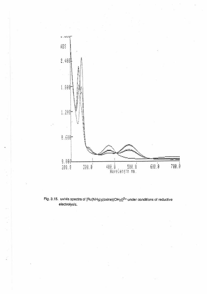
!.tE::
4E
I ,tH
',' !,t
lJ riJl! !!f t
ü rlil!üu
IrjIli
Itli'i li 5ilä,i' 'ili'rr
iin: iñ 'nrrr
,
ür[ü üIflEIfifl
Fig. 3.15. uv/vis spectra of [Ru(NH3)s(oxine)(OH2¡72+ under conditíons of reductive
electrolysis.
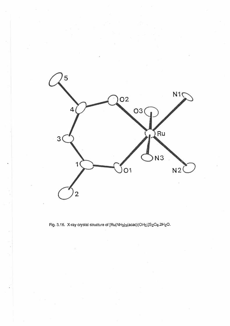
5
o2o3
Ru
N3
o1
Fig. 3. 1 6. X-ray crystal structure of [Ru(NHs) g(acacXOHe)lSzOe.2HzO.
4
N1
N2
?t\)
1
2
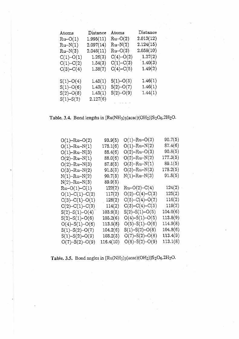
AtomsRu-o(1)Ru-N(1)Ru-N(3)c(1)-o(1)c(1)-c(2)c(3)-c(4)
s(1)-o(4)s(1)-o(6)s(2)-o(8)s(1)-s(2)
Dista.nce1.ee5(11)2.0e7(L+)2.046(11)
1.26(3)1.54(3)1.38(2)
1.45(1)1.43(1)1.45(1)
2.127(6)
AtomsRu-O(2)Ru-N(2)Ru-o(3)c(4)-o(2)c(1)-c(3)c(4)-c(5)
s(1)-o(5)s(2)-o(7)s(2)-o(e)
Dista,uce2.013(12)2.124(L5)2.058(10)
1.27(2)'1.40(3)
1.4s(2)
1
1
1
.46(
.46(
.44(
1)1)1)
Table. 3.4. B ond lengths in [Ru(NH3) 3 (acac) (OHz)] SzOe.2HzO.
o(r)-Ru-o(2)o(1)-Ru-N(1)o(r)-Ru-N(3)o(2)-Ru-N(1)o(2)-Ru-N(3)o(3)-Ru-N(2)N(1)-Ru-N(2)N(2)-Ru-N(3)Ru-o(r)-c(1)o(1)-c(1)-c(2)c(3)-c(1)-o(1)c(2)-c(1)-c(3)s(2)-s(1)-o(4)s(2)-s(1)-o(6)o(4)-s(1)-o(6)s(1)-s(2)-o(7)s(1)-s(2)-o(e)o(7)-s(2)-o(e)
e3.e(5)178.1(6)88.4(6)88.0(6)87.8(5)e1.5(5)e0.7(5)8e.e(5)L22(2)117(2)128(2)114(2)
103.e(5)105.2(6)113.5(8)104.2(6)103.2(5)
116.4(10)
o(t)-Ru-o(3)o(1)-Ru-N(2)o(2)-Ru-o(3)o(2)-Ru-N(2)o(3)-Ru-N(1)o(3)-Ru-N(3)N(1)-Ru-N(3)
eo.7(5)87.4(6)eo.8(5)
177.3(5)8e.1(5)
178.2(õ)e1.8(5)
Ru-o(2)-c(a)o(2)-c(4)-c(3)c(5)-c(4)-o(2)c(3)-c(4)-c(5)s(2)-s(1)-o(5)o(4)-s(1)-o(5)o(5)-s(1)-o(6)s(1)-s(2)-o(8)o(7)-s(2)-o(8)o(8)-s(2)-o(e)
124(2)t25(2)116(2)11e(2)
104.0(6)113.8(e)i14.e(8)104.8(6)113.4(e)113.1(8)
Table. 3.5. B ond an gles in [Ru(NH3)3 (acac)(OHz)]S zOe.2HzO.
59
involved in significant hydrogen bonding contacts with the dithionate anion, with the O-O'
distances being 2.64 and 2.67 A.
3.3. Ruthenium(ll) Complexes.
3.3.1. Preparation of Complexes. ln aqueous solution, [Ru(NH3)3(OHZ)g]2* reacted with æ-
acceptor ligands to give brightly coloured solutions of the substituted ammine complexes.
[Ru(NH3)3(OHz)s]2* was either generated prior to reaction with the tigand by reduction of
IRu(NH3)3(OHz)s]3* with hydrogen over platinum black, or by reduction of the Ru(lll) complex by
amalgamated zinc or hydrogen over platinum black in the presence of the ligand. The latter
procedure causes a lowering of the pH, which assists the reactions through the dissolution of some
protonated ligands in the aqueous solvent. Solutions of [Ru(NH3)s(OHz)g]2* had a distinctive
golden colour which changed when ligands were added to the solution. The progress of the
reactions could be monitored voltammetrically using 100 ¡rlsamples ol the reaction mixture in
aliquots of 0.1 moldm4 MeSO3H. ln this medium, the [Ru(NHs)s(OHz)s]3*+ couple had a DPV
peak at approximately -0.15 V (on an anodic scan). The peak currenl at this potential could be
seen to decrease during the course of the reaction, with the concomitant appearance of a peak at
positive potentials. lf the ligand was to be added to a solution of the pre-reduced complex, SCP
was used to show that reductíon was substantially complete before the ligand was added.
When the ligand was 1 ,1O-phenanthroline, lhe reaction involved stirring a solution of
[Ru(NHs)3(OHz)g12, with 1 molar equivalent of the ligand at room temperature for periods of up to 4
hours, at which time DPV showed the substitution reaction to be substantially complete. At this
stage, the reaction mixture was an intense red-coloured solution from which
IRu(NH3)3(phen)(OH2)](PF6)2 could be isolated after addilion of excess NH4PF6. However, when
the ligand was 2,2'-bipyridine, reaction under similar conditions resulted in the isolation of
IRu(NHj2(bpy)z](PFo)2. The identity of this product was verified by microanalysis and comparison
of its uv/vis spectrum with that reporled for this complex in the líterature. [39]
The possibility of obtaining [Ru(NH3)3(bpy)(OH2)]3*, ¡n order to study its reduclion, was
ínvestigated. The reaction betwen bpy and [Ru(NH3)3(OHz)s]3* in ethanolwas monitored
voltammetrically. Using DPV, it was possible lo observe the appearance of [Ru(NH3)2(bPV)zl3* {eo
= +0.62 V) while there were still appreciable amounts ol [Ru(NH3)s(OHz)g]s* in solution. The mono
bpy complex could be identified by the presence of a DPV peak at +0.24 V, which is close to the
E172 potential of the [Ru(NH3)4(bpy¡73+t2+ couple. [40] When phen was used, however, there was
no evidence of the formation of [Ru(NH3)2(phen)2]3+ until about 10 hours had elapsed, by which
time there was no evidence of any significant amounts of [Ru(NHs)s(OHz)g]3* in solution. lt is
apparent that the reaction of bpy with [Ru(NHs)s(bpyXOHz)]S proceeds more readily than the
60
reaction of bpy with [Ru(NHg)g(OHe)sl3+, whereas there is no such comptication in the reaction
between phen and [Ru(NHs)3(OHdg]3*.
[Ru(NHs)3(OH2)3]2+reacted with one molar equivalent of 2(aminomethyl)pyddine, to give
[Ru(NH3)3(ampy)(OH2)]2+, the PF5-salt of which precipitated from the reaction mixture as a canary
yellow solid after the addition of NH4PF6. Solutions and solid samples of this complex were
unstable, even when stored in the freezer compartment of the refrigerator. On standing overnight,
the yellow samples turned red. DPV analysis of the red samples revealed that, in addition to a peak
at +0.03 V, which was characteristíc of the fresh sample, there was an additional peak at +0.38 V. lf
NaClOa was used as the precipitating agent in lhe reaction, the red colour formed immediately in
the reaction mixture. This led to the conclusion that the change in colour was due to an oxidation
reaction, in this latter case by the CIO¡ ion. lt has been reported previously that
IRu(NH3)a(ampy)2]2+ undergoes a ligand-based oxidation, resulting in the formation of a red-
coloured 2-iminomethylpyridine complex. [40] A similar reaction has also been observed in the
case of [Ru(bpy)z(ampy)12+. [41] That the red compound was the result of oxidation and not due to
the substitution of a second ampy ligand on the Ru(ll) centre is supported by consideration of its
E1¡2 potential. The E172 potential of cis [Ru(NH3)4(pfizl2' is +0.26 V 1421, and it would be
expected that a bis ampy complex would have a DPV peak near this value. That this is less than the
oxidation potentialof the red compound implies that the imine portion of the iminomethylpyridine
ligand is a better æ-acceptor than a second pyridine molecule.
[Ru(NH3)3(MeCN)3]2+ was formed by reaction of [Ru(NHs)g(OHz)g]2* with acetonitrile.
IRu(NH3)3(OHz)g]3* was reduced by amalgamated zinc in degassed 0.01 mol dm-3 HTFMS
solution in the presence of excess ligand for 4.5 hours. After filtering the resultant canary yellow
solution, a pale yellow solid could be precipitated by the addition of NH4PF6. The chloride and
bromide salts were then obtained by dissolving the PF6- salt in butanone and adding an excess of
the appropriate tetra(n-butyl)ammonium halide salt to the sotution. When [Ru(NHs)s(OHz)sl2*
reacted with two eqivalents of benzonitrile (PhCN), the solid isolated after the addition of excess
NH4PF6 contained a mixture of three compounds in the ratio 1 :10:1, as shown by the relative
heights of the DPV peaks. The potentials of the three peaks (+0.25 V, +0.64 V and +0.95 V) were
indicative of a mixture of the mono, bis and tris cyanobenzene complexes. The E1¡2values lor
IRu(NH3)5(PhCtrt¡¡2+ and cis [Ru(NH3)a(PhCN)2]2+ are, +0.27 V and +0.63 V, respectively [42],
implying that the major component ¡n the mixture was the bis(cyanobenzene) complex. lt is
apparent from this result that the presence of an aqua group in the coordination sphere of the Ru(ll)
ion will readily allow the substitution of an organonitrile ligand, even when there are already two
such ligands attached to the metal centre.
61
When [Ru(NHis(OHz)s]2*, formed by reduction of [Ru(NH3).1OH2)sl3+ by amalgamated zinc
in a pyridinium methanesulphonate butfer, reacted with the excess pyridine it was possible to
isolate [Ru(NHs)3(py)g]2+ as its CIO¡ or PF6- salt by the addition of an appropriate precipitating
agent. The golden yellow perchlorate salt was recrystallized from hot methanol. Attempts to isolate
the bis pyridine complex were unsuccessful, resulting in the formation of at least a small amount of
the tris pyridine complex in all cases. This is similar to the results obtained for the attempted
synthesis of the bis benzonitrile complex.
3.3.2. Electronic Spectra. The uv/vis spectra of the Ru(ll) complexes all contained intense MLCT
bands, and bands attributable to intra-ligand absorptions. The MLCT bands arise from transitions
between the filled t2n orbitals of Ru(ll) and the empty æ. orbitals of the l¡gand. The main features of
the spectra of the complexes prepared, together with those of some related ruthenium complexes,
are given in Table 3.6.
The uv/vis spectrum of [Ru(NHj3(phen)(OHz)]2+ (shown in Fig. 3.17) was pH dependent, as
was to be expected given the presence of the aqua ligand. (The pK" of the complex was found, by
pH titration, to be 10.6(t 0.1)). ln 0.1 moldm-3 MeSOgH, the charge transfer band was observed at
470 nm (e = 4.01 x 10³), while the intra ligand bands were observed at222nm (e = 3.85 x 10⁴) and
265 nm (e = 3.22x t O4). ln NaOH solution, the charge transfer band was observed at 490 nm(e =
2.95 x 193¡ and the intraligand bands were at the same wavelength, but with increased intensities
(az2z= 4.84 x 10⁴, e265 =4.37 x 10⁴). The colour of the solution changed quite rapidly alter the
addition of the base to the complex, indicating that the complex was unstable in NaOH solution,
possibly due to aerial oxidation. lf the NaOH solution was degassed with argon prior to adding it to
the solid sample, the colour change was much slower and it was posssible to determine the
spec'trum within an hour. The spectrum in acid solution is very similar to that reported for
IRu(NH3)4(phen)]2+. [43]
It is apparent from the shift in the MLCT band that the replacement of the aqua ligand with a
hydroxo ligand raises the energy of the highest occupied metal-based orbital, so that the transition
to the lowest lying æ. orbital is of lower energy, consistent with HO- being a stronger æ-donor ligand
than H2O.
The spectrum of [Ru(NH s)zþpflzl2* was close to that previously reported for this complex.
[39] The spectrum of the prepared sample contained two charge transfer bands, at 349 nm (e =
7.35 x 10³¡ and 488 nm (e = 9.10 x 10³), compared with 350 nm (e = 8.02 x 10³) and 495 nm (e =
9.34 x 10³) in the previously determined spectrum. These absorptions are also comparable with
the two charge transfer bands in [Ru(NH3)a(bpy)]2*. [40] The molar absorptivities of the intra-ligand
absorptions are diagnostic of the of the number of bpy ligands present in the complex, being
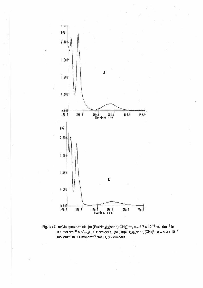
fl8$
?
t.
a
1.80
t.6
tt00.0 ll0 {tt.
ll¡fûú. ü tüt
lÈn9th nt7BÐ. I
t¡Ð
â8$
0 .5t
I
b
0
¿0r. r 300,0 ,t0t t 5t0.l¡velenrlh nr
6t0.0 7m.0
Fig. 3.17. uv&is spectrum of : (a) [Ru(NH3)g(phenXOH ù12*, c = 6.7 x t 04 mol dm{ ¡n
0.1 moldm-g MeSOsH, 0.2 cm cells. (b) [Ru(NHg)sþhen)(OH)]+ , c = 4.2x10⁴moldm{ in 0.1 moldm4 NaOH, 0.2 cm celts.
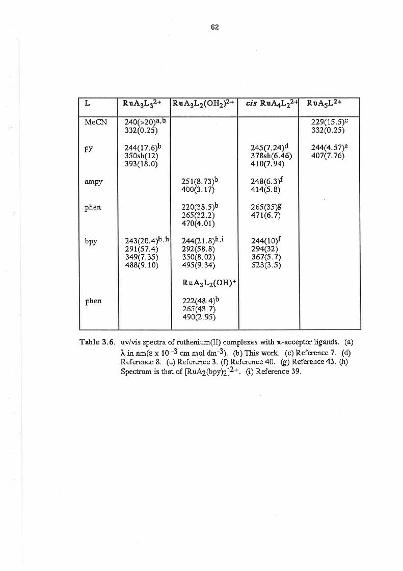
62
229(ts.s)c332(0.2s)
244(4.sT)e407(7.76>
24s(?.2Ðd378sh(6.46)410(7.e4)
248(63)1414(s.8)
26s(3s)s4?t(6.7)
244{ilof2e4(32)367(s.7)s23(3.s)
2s1(8.73)b400(3.17)
220(38.s)b26s(32.2)470(4.0r)
244Qr.8)h,i292(s8.8)3s0(8.02)4es(e.34)
R.uA3L2(OH)+
222(48.Ðb26s(43.?)4e0Q.es)
240(>20¡a,o332(0.2s)
244(L7.qb350sh3e3(1
(r2)8.0)
243(20.4)b 'h2et(s7.4)34e(7.3s)488(e.10)
MeCbtr
PY
amPy
phen
bpy
phen
RuA5Lz+cis R.uAaL2z{RuA3L2(OHr¡z+RuA3L32+L
Table 3.6. uv/vis qpect¡a of ruthenium(Il) complexes witå r"-acceÍ)tot ligands. (a)
1, in nm(e x 10 -3 cm mol ¿m-3). (b) This work. (c) Reference 7. (d)Reference 8. (e) Reference 3. (f) Reference 40. (g) Reference 43. (h)
Spectrum is tåat of [RuA2(bpy>Z]z+. (i) Reference 39.
63
approx¡mately proportional to the number of ligands. ln [Ru(NH3)4(bpy)]2* the molar absorptivities
of the intra-ligand bands ile e;244= 1.0 x 10⁴ and E2g4 = 3.2 x 10⁴ which are approximately half the
values observed for the intra-ligand bands in [Ru(NH3) z(bpy)zl2+, EzM = 2.1 x 10⁴ and
Ê2g2= 5'g x 104'
ln the bpy and phen complexes reported here, the intra-lígand bands are at lower energy than
the corresponding bands in the free ligand. This shifl is normally observed in the spectra of such
complexes and is ascribed to the effect of the positive charge on the energy levels of the ligand.
[39] A similar shift is also observed when the free ligands are protonated. [44]
ln the spectrum of [Ru(NHs)s(ampy)(H zO)12* a single intra-ligand absorption was observed at
251 nm (e = 8.73 x 10³), and the MLCT band was at 400 nm (e = 317 x 103¡. These bands are
similar to those observed in [Ru(NH3)¿(ampy)]2* and IRu(NHs)s(py)]2+. [40,3]
ln the case of IRu(NH3)s(py)g]2*, the absorption bands were at wavelengrths similar to those in
[Ru(NH3)5(py)]2* (Fig. 3.18) The intra-ligand band was at244 nm (e = 1.76x 10⁴) and the MLCT
band was at 393 nm (e = 1.2 x 1O\. ln addition, there is a shoulder to the charge transfer band, at
350 nm (e = 1.8 x 10⁴). This spectrum compares well with those of the cis and trans isomers of
[Ru(NH3)a(pÐzl2+. [B] The frans isomer has a single MLCT band at 423 nm (e = 1.66 x 10⁴¡, while
the cis isomer shows a MLCT band similarto the tris pyridine complex, with a peak at 410 nm (e =
7.94x 10³) and a shoulder at 375 nm (e = 6.45 x 10³). I has been shown that two independent
MLCT transitions may be expected lor the tetraammine complexes, but that the higher energy band
is not observed unless there is some distortion to the ligand arrangement. [45] A ligand field
treatment of the orbital energies in cr.s MX3Y3 complexes has shown that [Ru(NHs)s(py)s]2* would
be expected to have only one MLCT band. [46] However, if there is some distortion of the
coordination sphere, then the degeneracy of the t2 orbitals would be lifted, somewhat, so that a
second transition 1o the ligand ru* orbital may become apparent. Given the presence of 3 pyridine
ligands in a facial arrangement, it is quite possible that there would be some distortion.
The spectrum of [Ru(NHs)3(MeCN)s]2+ consisted of a single absorption band at 332 nm (e =
2.53 x 10²) assigned to a MLCT. There was also a band al24} nm with e>2x 10⁴. This is
comparable with the spectrum of [Ru(NH3)s(MeC$]2+. [fl The presence ol a single MLCT implies
that there is very little distortion of coordination sphere, which might be expected, given the small
bulk of the acetonitrile molecule compared with the pyridine molecule.
3.3.3. Vibrational Spectra. The vibrational spectra of the complexes were recorded as nujol mulls
on KBr plates. The presence of ligand vibration bands in the spectra served as confirmation that
the ligands were coordinated. ln the complexes containing aromatic heterocycle ligands, there

a. il(l
Fig. 3.18. uvtuis spectrumof [Ru(NHg)g(py)g]2*, ç = 4.2x 10{ mol dm4 in 0.1 mol dm4
MeSOgH
.i I't !i
üE
Í[
tI
5tI
[ ür¡
!üü ü
flEt
,)g
I
tl iüfl r1üËil[4u[ ü i[ü, r!
I,levelÊllrtlt ritl.
64
were multiple absorptions in the regions 1600 -1620 cm-1 , 1500 - 1600 cm-1 and 1400 -1500
cm-1 aüributable to ring vibrations. Additionally, there were absorptions between 600 and 750
cm-1 due to the C-H out of plane deformations of the aromatic rings.
ln all cases there were absorptions attributable to the NH groups of the ammine ligands. These
were found in tfrree regions of the spectrum: lwo or three bands belween 3200 and 3350 cm-1,
attributable to the u(NH) mode;a broad band between 1620 and 1640 cm-1, ailributable to the
ô¿1ruU¡ mode and several bands between 1250 and 1350 cm-1 , attributable to the õs(NH) mode.
Though ammine complexes are also expected to show absorptions belween 950 and 590 cm-1,
due to NH rocking víbrations [47a], these bands were obscured by bands due to ligand or anion
vibrations. When the anion was PF6-, the D(NH) bands were very sharp, because of specific
hydrogen binding between the anion and the ammine ligands. ln the spectrum of
IRu(NH3)3(MeCN)3]C12.2H20 the u(NH) bands are very broad because of hydrogen bonding
between the molecules of lattice water and the ammine ligands. lt was not possible to reliably
assign the u(RuN) absorption bands because these are usually very weak and occur close to the
lower limit of the accessible range in KBr plates. [47b].
The spectrum of [Ru(NH3)3(phen)(OH2)](PF6)2 showed strong bands at 3600 cm-1 and 3670
cm-1 due to the u(OH) mode of the coordinated water. A strong absorption 1630 cm-1, due to
ô¿1ruH¡ and an aromat¡c ring vibration, obscured any possible õ¿1OU¡ bands, nor was it possible to
locate any Ru-O absorptions, for the reasons outlined above for the u(RuN) bands.
ln the spectrum of [Ru(NH3)3(MeCN)3]C12.2H20 there were three bands attributable to the
D(CN) mode. Two were evident as sharp peaks al2262cm-1 and 2278 cm-l. The lower energy
peak was about twice as intense as that at higher energy. These bands were assigned to the E and
A1 mode vibralions, respeclively, by comparison with the spectra of fac M(CO)3 complexes. [a8a]
The third band was evident as a shoulder a12266 cm-1. Group theory predicts that a molecule of
C3y slmmetry, such as [Ru(NH3)3(MeCN)3]2+, would have two ir active absorptions due to ù(CN),
but anisotropy in the environment ol the complex will cause slight splitting of the E mode. [a8b] ln
the spectrum of the PF6- salt, there was a single peak at 2285 cm-1 and for the Br- salt, the peak
was at 2272 cm-1. The anion dependence of the stretching frequency is due to polarization
effects of the anion. Anions positively polarize the hydrogens of the ammine ligands, allowing
greater electron release to the ruthenium and thence to the ligand æ* orbitals. The smaller the
anion radius the greater is the electron release and the lower the vibralion frequency. [49] lt seems
possible that with the Br and PF¡ ions the intensity of the A1 mode vibration is lowered to the

65
extent that it is not visible in the spectrum. A similar effect has been noted ¡n the ir spectra of
dinitrogen complexes, where the intensity of the absorption decreased as its energy increased.
[50]
The t(CN) band in free acetonitrile is seen at 2254 cm-1, and, for [Ru(NHg)5(MeCN)]2+, the
energy of the u(CN) band is modified by coordination, but is also dependent on the anion. For the
CF, Br- and PF6- salts the bands are aI2297.2238 and 2277 cm-l, respectively. t49] lt has been
noted previously that, lor the pentaammine complexes, the energy of the u(CN) vibration is
lowered in Ru(ll) complexes, and increased in Ru(lll) complexes, reflecting the slrength of æ-
backbonding in the binding of the nitrile group to the Ru(ll) centre and the effect of o-donation to
the Ru(lll) centre. [1] That the D(CN) frequency is greater in the triammine tris(acetonitrile) complex
than in the free ligand is probably due to a combination of effects. The presence of only 3 o-
donating NH3 groups means that the æ-backbonding ability of the triammine Ru(ll) centre will be less
than that of the pentaammine. Secondly, what fi-donation does occur is spread over three r-accepting ligands instead of only one. The end result is that because of reduced æ-donation to
each nitdle ligand, the o- contribution is predominant, as in the case of Ru(lll) complexes, and there
is an increase in the frequency of the u(CN) vibration. This explanation is borne out by the
frequencies of the u(CN) vibrations in a series of tris(phosphine) tris(acetonitr¡le) Ru(ll) complexes,
where the presence of the weakly o-donating/æ-accepting phosphine ligands causes the D(CN)
frequencies in the complexes to be higher than in the free ligand. [51]
3.3.4. Electrochemical Studies. The electrochemistry of the Ru(ll) complexes was studied in
aqueous solution at a freshly polished glassy carbon electrode. Allthe complexes showed a
reversible or quasi-reversible oxidation wave at potentials greater than zero in 0.1 mol dm-3
methanesulphonic acid. The E172 potentials of these complexes are listed in Table 3.7, along with
those of similar ruthenium complexes.
The E1¡2potential for the [Ru(NHs)s(phen)(OH2)]3+t2+ ssupl¿ was pH dependent, due to the
presence of the aqua tigand. ln 0.1 moldm4 acid, E1/z= +0.26 V. At pH B,Ejn= -0.02 V, and at
pH 12, E1n= -0.23 V. The cyclic voltammograms at pH 1 and pH B are shown in Fig. 3.19. The
E172 potential in acid solution is similar to that for the [Ru(NH3)+(phen)]3+/2+ couple. [43] The
Pourbaix diagram showed three distinct regions of pH dependence (Fig.3.20). Below pH 3 and
above pH 11.5, the E172 potential was independent of pH, and betweeen pH 4 and pH 11 , the
slope of the graph of was -62 mV/pH. These results can be summarized in the redox couples
shown as reactions 3.26 to 3.28.
1 <pH<3
IRu(NHs)3(phen) (oH2)]3+ +e- + [Ru(NH3)3(phen)(OH2)]2+ (3.26)
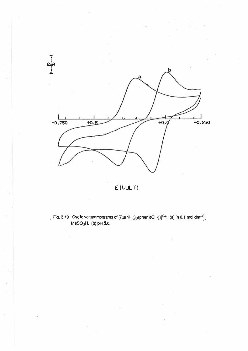
ï
T
+0.750 {o -o.250
E f UT]LT]
Fig. 3.19. Cyclic voltammograms of [Ru(NHs)3(phen)(OHj]2+. (a) in 0.1 mol dm4MeSO3H. (b)pH 1.0.
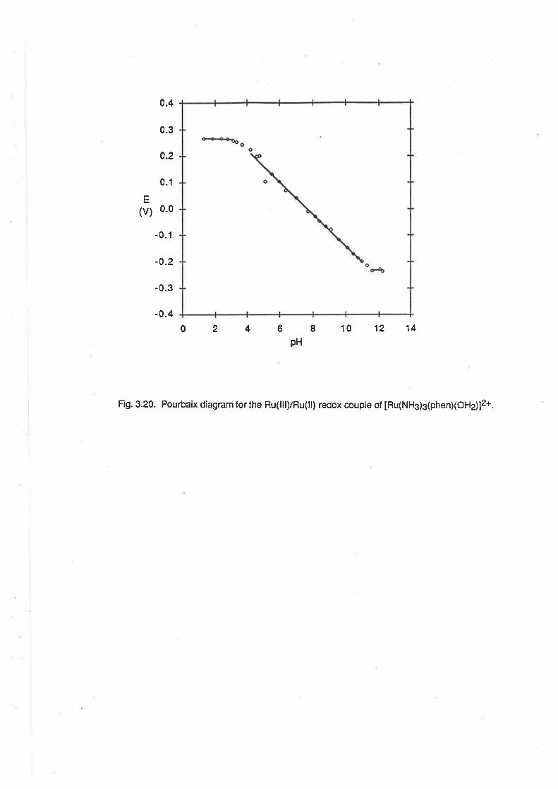
0.4
0.1
E
(v) o-o
0.3
0.2
-0.1
-0.2
-0.3
-0.4
02468101214pH
Fig. 3.20. Pourbaix diagram forthe Ru(lll)/Ru(ll) redox couple of [Ru(NH3)3(phen)(OHj]2+.
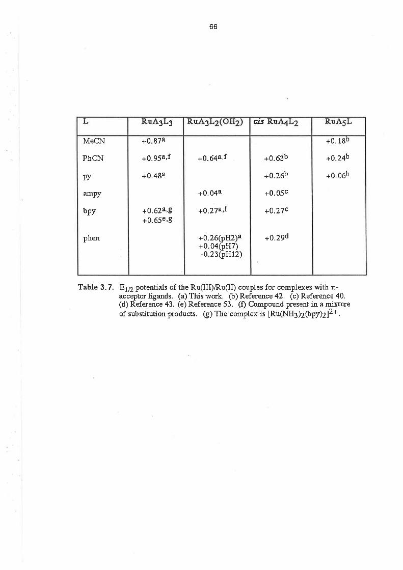
Table 3.7
66
E172 potentials of the Ru(III)/Ru(II) couples for complexes with n-acceptor ligands. (a) This work. (b) Reference 42. (c) Reference 40.(d) Reference 43. (e) Reference 53. (f) Compound present in a mixÈureof substitution products. (g) The complex is [Ru(NH3)ZþpÐZ]2*.
+0.18
+0.24b
+0.06b
+0.63b
+0.26b
+0.05c
+0.27c
+0.29d
+O.64a,1
26(ptfz)a0a(pH7)23(.p}ltz)
+0+0-0
+0.044
+0.27a,1
+0.874
+0.95a,f
+0.484
+0.624,8+0.65e'9
MeC\l
PhCN
PY
ar]nPy
bpy
phen
cfs3u33
67
4<pH<1 1
[Ru(NH3)3(phen)(OH)]2+ + e- + H+ IRu(NH3)3(phen)(OH2)]2+ (3.27)
IRu(NH3)3(phen)(oH)]+ (3.28)
pH>11.5
IRu (NH3)3(phen)(OH)]2+ +e-
From the intercepts of the lines on the Pourbaix díagram, it can be seen that, in a solution of ionic
strength 0.1, the pK" of the aqua ligand on the Ru(lll) complex is about 3.5, and the pK" of the aqua
ligand on the Ru(ll) complex is about 11.4. Because the pK" value is dependent upon the ionic
stength of the solution [54], this pK" is comparable with the pK" of the Ru(ll) complex in water
(10.6). The estimated pK" of the Ru(lll) complex is higher than that of [Ru(terpy)(bpyXHzO)¡s+ (1.7)
and [Ru(bpy)z(pyXHzO)]3+ (0.85). 132,521. lt is expected that the complexes with the equivalent of
five pyridine rings in their coordination spheres would have lower pK" values due to the cumulative
electron accepting properties of the pyridine rings in the ligands.
The E172 potential for the oxidation of freshly prepared [Ru(NHj3(ampy)(H2O)]2+ was also pH
dependent. ln 0.1 moldm-3 acid, E1p= +0.04 V, and at pH B, E172 = {.20 V. 'l'he E172 potential
in acid solution is very similar to that of [Ru(NHs)s(py)]2*. [42] The Pourbaix diagram is shown in Fig.
3.21, and between pH 4.5 and pH 8, the slope of the graph is -58 mV/pH, indicative of a one
electron/one proton electron transfer. This corresponds to the redox couples shown in 3.29 and
3.30.
3.pH
[Ru(NHj3(ampyXHzO)]s + e- + [Ru(NH3)3(ampy)(H2O)]2+ (3.2s)
4.5<pH<8
[Ru(NH3)3(ampy)(OH)]2+ + e- + H+ =å [Ru(NH3)3(ampyXHzo)]2+ (3.30)
From the intercepts of the lines in the Pou¡baix diagram, it is apparent that the pK" for the Ru(lll)
complex is approximately 4.
The cyclic voltammogram of [Ru(NHg)g(py)s]2* consisted of a single quasi reversible wave with
E1n= +0.48 V. At a scan rate of 25 mV sec-1, the peak separalion, ÂEp, = 72 mY, while at 900 mV
sec-1 , ^Ep
= 126 mV. The E1¡2potential is approximately mid way between the value lor cis
[Ru(NH3)a(pr¡r13+t2+ (+0.26 V) and [Ru(NHs)2(brr¡r73+t2+ (+0.65 v).142,53] Wth the E1¡2
potentialfor [Ru(NHg)s(py)]3*/2* being +0.06 V 1421, it is apparent that replacement of an ammine
ligand by a pyridine ligand causes an increase in the oxidation potential of the ruthenium complex of
approximately 0.2 V.
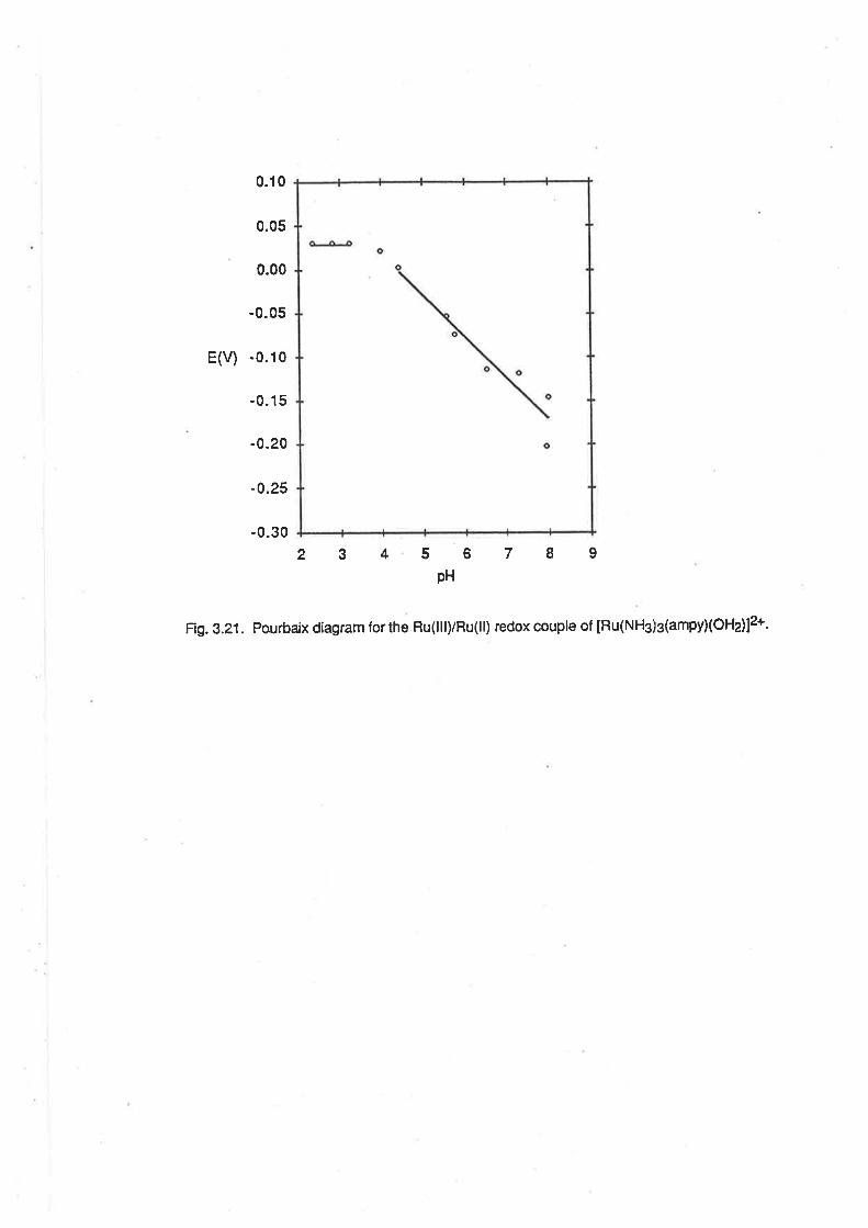
0.10
0.05
0.00
-0.05
E(v) -0.10
-0.15
-0.20
-0.25
-0.30
23456789pH
Fig. 3.21. Por¡rbaix diagram forthe Ru(lll)/Ru(ll) redox couple of [Ru(NH3)g(ampyXOHdl2+.
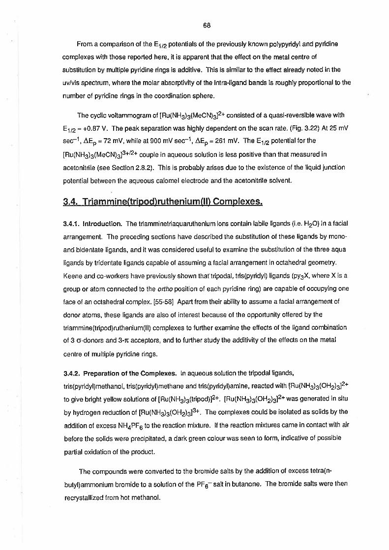
68
From a comparison of the E1¡2 potentials of the previously known polypyridyland pyridine
complexes with those reported here, it is apparent that the effect on the meta¡ centre of
substitution by multiple pyridine dngs is additive. This is similar to the effect already noted in the
uv/vis spectrum, where the molar absorptivity of the intra-ligand bands is roughly proportionalto the
number of pyridine rings in the coordination sphere.
The cyclic voltammogram of IRu(NHg)g(MeCN)3]2+ corsisted of a quasi-reversible wave with
E1n= +0.87 V. The peak separation was highly dependent on the scan rate, (Fig. 3.22) At 25 mV
sec-1 , ^Ep
= 72 mY, while at 900 mV sec-1, ^Ep
= 261 mV. The E1¡2potential for the
IRu(NH3)3(MeCN¡a¡3+/2+ couple in aqueous solution is less positive than that measured in
acetonítrile (see Section 2.8.2). This is probably arises due to the existence of the liquid junctíon
potential between the aqueous calomel electrode and the acetonitrile solvent.
3.4. Triam m ine(tripod) rutheniu m (l l) Complexes.
3.4.1. lntroduction. The triamminetriaquaruthenium iors contain labile ligands (i.e. H2O) in a facial
arrangement. The preceding sections have described the substitution of these ligands by mono-
and bidentate ligands, and it was considered usefulto examine the substitution of the three aqua
ligands by tridentate ligands capable of assuming a facial arrangement in octahedral geometry.
Keene and co-workers have previously shown that tripodal, tris(pyridyl) ligands (pygX, where X is a
group or atom connected lo the ortho position of each pyridine ring) are capable of occupying one
face of an octahedral complex. [55-58] Apart from their ability to assume a facial arrangement of
donor atoms, these ligands are also of interest because of the opportunity offered by the
triammine(tripod)ruthenium(ll) complexes to further examine the effects of the ligand combination
of 3 o-donors and 3-rc acceptors, and to further sludy the additivity of the effects on the metal
centre of multiple pyridine rings.
3.4.2. Preparation of the Complexes. ln aqueous solution the tripodal ligands,
tris(pyridyl)methanot, tris(pyridyl)methane and tris(pyridyl)amine, reacted with [Ru(NH.¡.1OH2)sl2+
to give bright yellow solutions of [Ru(NHs)s(tripod)]2+. [Ru(NH3)3(OHz)gl2* was generated in situ
by hydrogen reduction of [Ru(NH3)g(OHe)g]+. The complexes could be isolated as solids by the
addition of excess NHaPF6 to the reaction mixture. lf the reastion mixtures came in contact with air
before the solids were precipitated, a dark green colour was seen to form, indicative of possible
partial oxidation of lhe product.
The compounds were converted to the bromide salts by the addition of excess tetra(n-
butyl)ammonium bromíde to a solution of the PF¡ salt in bulanone. The bromide salts were then
recrystallized from hot methanol.
T'r
+1 +o.5 +o.ooo
EIUOLT)
Ftl.3.22. Cyclic vottammogramsof [Ru(NH3)3(MeCN)sJ2+ ¡n 0.1 moldm-3 MeSO3H.(a) 100 mV secl. (b) 9OO mV serl.
69
ln both [Ru(NHs)3{(py)gCH}]2+ and [Ru(NHg)g{(py)sN}]2* the ligands are believed to be
coordinated through the three pyridyl nitrogen atoms, in a facial arrangement (N, N', N). A crystal
structure delermination has revealed that the ligand, (py)gCOH is coordinated through two pyridyl
rings and the oxygen atom of the alcohol group (N, N', O). ln the complex, [Ru{(py)3COH)}2]2+,
both modes of coordination are observed. [56]
Attempts were made to prepare the complex with tris(pyddyl)phosphine as the tripod ligand,
but DPV analysis of the reaction mixture indicated the formation of at least two species in the
reaction. No attempt was made to refine the reaction conditions to give the required complex as the
sole product.
3.4.3. Electronic Spectra. As with the Ru(ll) complexes reported above, the uv/vis spectra of the
three complexes with tripod ligands all contained bands attributable to intra-ligand absorptions and
to MLCT. The spectra are displayed in Fig. 3.23 and the main features are listed in Table 3.8, where
they are enmpared with the spectra of some Ru(tripod)22+ complexes prepared previously. [56, 57]
The spectra of [Ru(NH3)g((py)gOH]12+ and [Ru(NHs)s{(py)sNll2+ are similar. The MLCT bands
are at 369 and 415 nm for tripod = (py)sOH and at 367 and 414 nm for tripod = (py)gN. The intra-
ligand bands are a|249 and 280 nm in both cases buÎ, of these, the lower energy band is only
evident as a shoulder in the spectrum of [Bu(NH3)g{(py)gCH}]2+. The MLCT bands are similar in
energy and intensity to those in the spectra of IRu(NHs)g(py)g]2*, [Ru(NH3)a(pÐzl2* [8] and the
Ru(tripod)22+ complexes, but no intra-ligand bands were quoted for the last examples. [57] lt was
noted, when discussing the spectrum of IRu(NH3)g(py)s]2*, that complexes of Cgy stmmetry would
be expected to show only one MLCT band unless there was some distortion of the coordination
sphere. The presence of two distinct MLCT peaks suggests that there is a larger degree of
distortion in these two complexes, than in the tris(pyridine) complex. lt is not surprising that there
should be some distortion of the coordination sphere in this case, because the three pyridyl rings
are subject to the rigidity constraints imposed by the bond to the bridgehead group.
The spectrum of [Ru(NHds((py)gCOH]12*, shown in Fig. 3.24, was expected to be pH
dependent, due to the presence of a weakly acidic -OH group on lhe ligand. ln 0.1 moldm-g
MeSOgH, the intra-ligand bands were present at 260 nm and a1240 nm (shoulder), and at pH 8, the
band at 240 was resolved into a peak, and the lower energy band was at 265 nm. The expected
MLCT bands were evident as weak shoulders at 350 (e =1.5 x 10³¡ and 415 nm (t = 4.5 x 10²) in
acid sofution, and in base there was a shoulder at 350 nm. The intra-ligand bands are comparable to
those in the spectra of [Ru{(py)gCOH}2]2+, which has one peak a1246 nm when the -OH group is
protonated, and one peak at 253 nm when lhe O-H group is deprotonated. [56] However, the
MLCT absorptions are surprising. The energy at which they occur is comparable with the energy of
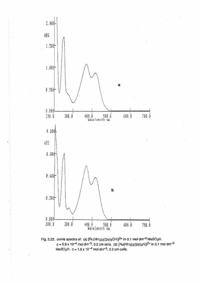
fE
lr lJi.i
I B[
ÈEË
lj,I!ilü
ü[
a
lit It1 fù
,lüfl. ü 5
I'l¡u¡ I¡n¡ thtÈü [i,t
ll
E 5ü
ÉEì
ü t[
4ûI
Ëü
¡È
0ff [ütìi
Fig.3.23. uvtuis spectra of: (a) tRu(NHg)g((py)sCHll2+ in 0.1 npl¿nrs ¡¡teSOgl'|.
G = 5.9 x 10{ moldm4, 0.2 cm cells. (b) [Ru(NHg)g{(py)sN}]2+ in 0.1 moldr¡r3MeSO3H. c = 1.9 x 10⁴ mol dm4, 0.2 cm cells.
üt¡il¡ trllt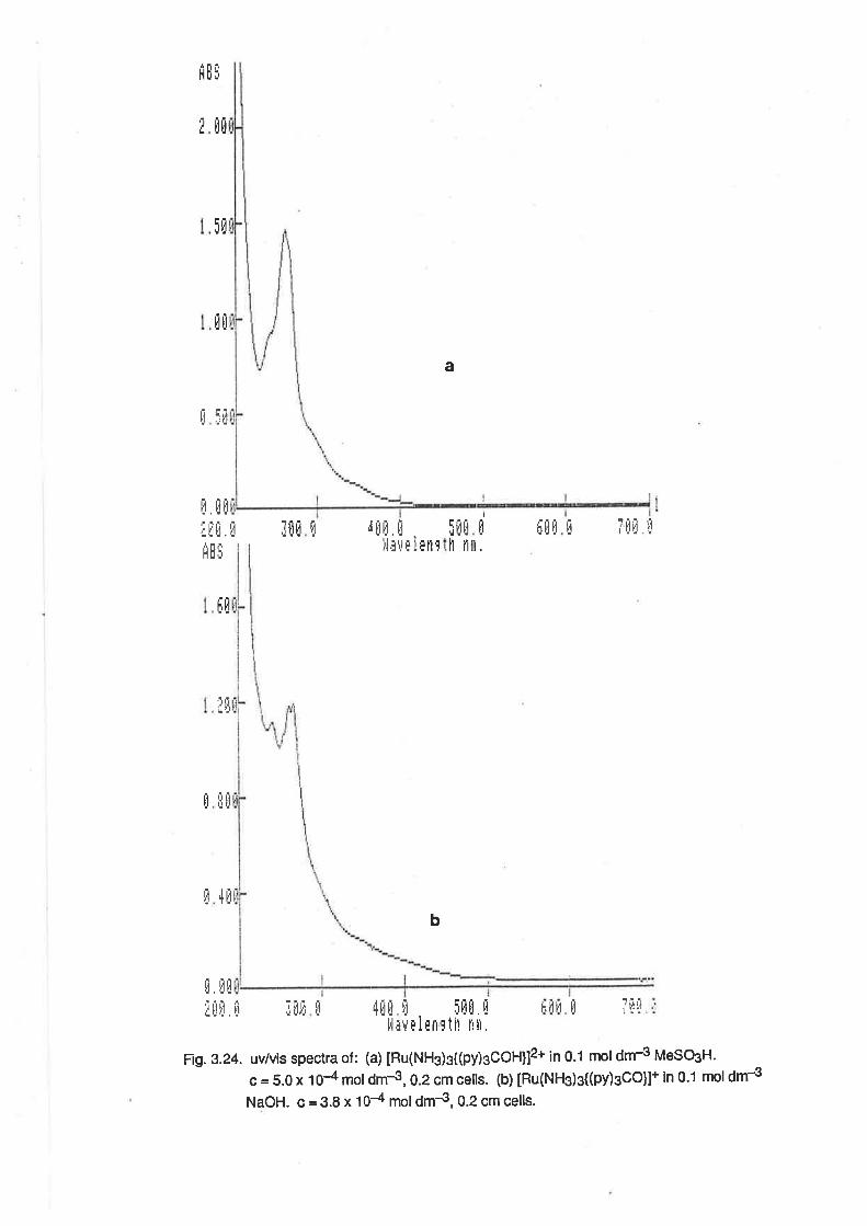
a
riEl
Itü
5[
fl[
H b.i.¡ll .Ìtl
t . üil
,¡ !l I'l
iit
tI
r' ¡'l
Ëüüür¡ n1ü
rlltfr fi-tliulfl ,j
ht
I
iì llüü iI
UIJilil
4[
E
l,l¡u
Frg. B.24. uv/vis spectra of : (a) tRu(NHs)s((py)gçOHll2+ in 0.1 mol dnr3 MeSOgH.
c = 5.0 x 10⁴ mol dm-3, 0.2 cm cells. (b) [Ru(NHg)g{(py)gCO}l+ in 0.1 mol dm-3
NaOH. c = 3.8 x 10⁻⁴ mol dm{, 0.2 cm cells.
tì fitti ll
Ë
lb
|l fltl
i[ü ltI !,1 ¡,i¡,flü l'litil i!
tlitttlh nin,'iIr]
i,t LiItrt¡ t¡
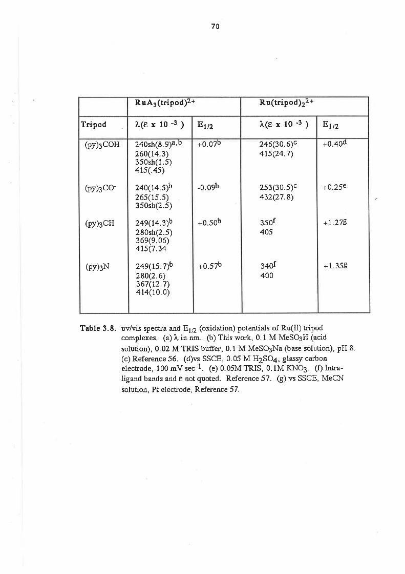
70
Table 3.8. uv/vis spectra and E1¡2 (oxidation) potentials of Ru(II) tripodcomplexes. (a) î, in nm. (b) This work, 0.1 M MeSO3H (acid
solution), 0.02 M TRIS buffer, 0.1 M MeSO3Na (base solution), pH 8.
(c) Reference 56. (d)vs SSCE, 0.05 M H2SO4, glasry carbonelecrrode, 100 mv r""-1. (e) 0.05M TRIS, 0.lM IÕIO3. (f) Ir:tra-
[gand bands and e not quoted. Reference 57. G) * SSCE, MeClrl
solution, Pt. electrode, Reference 57.
+0.40d
+0.25e
+1.278
+ 1.358
246(30.6)c4tsQ4.7)
2s3(30.5)c432Q7.8)
350f405
340f400
+0.07þ
-0.09b
+0.50b
+0.57b
240sh(8.9)a'b260(14.3)350sh(1.5)4ts(.4s)
240(14.Ðb26s(rs.s)350sh(2.5)
24s(t43)b280sh(2.s)36e(9.06)4ts(7.34
24s(rs.7)b280(2.6)367(12.7)4 14(10.0)
(py)3coH
(py)¡co-
(py)scH
(py)¡N
EtnÀ(exlo-3)EtnÀ(erlo-3)Tripod
R.u(tripod)22+R uA3(tripod)2+
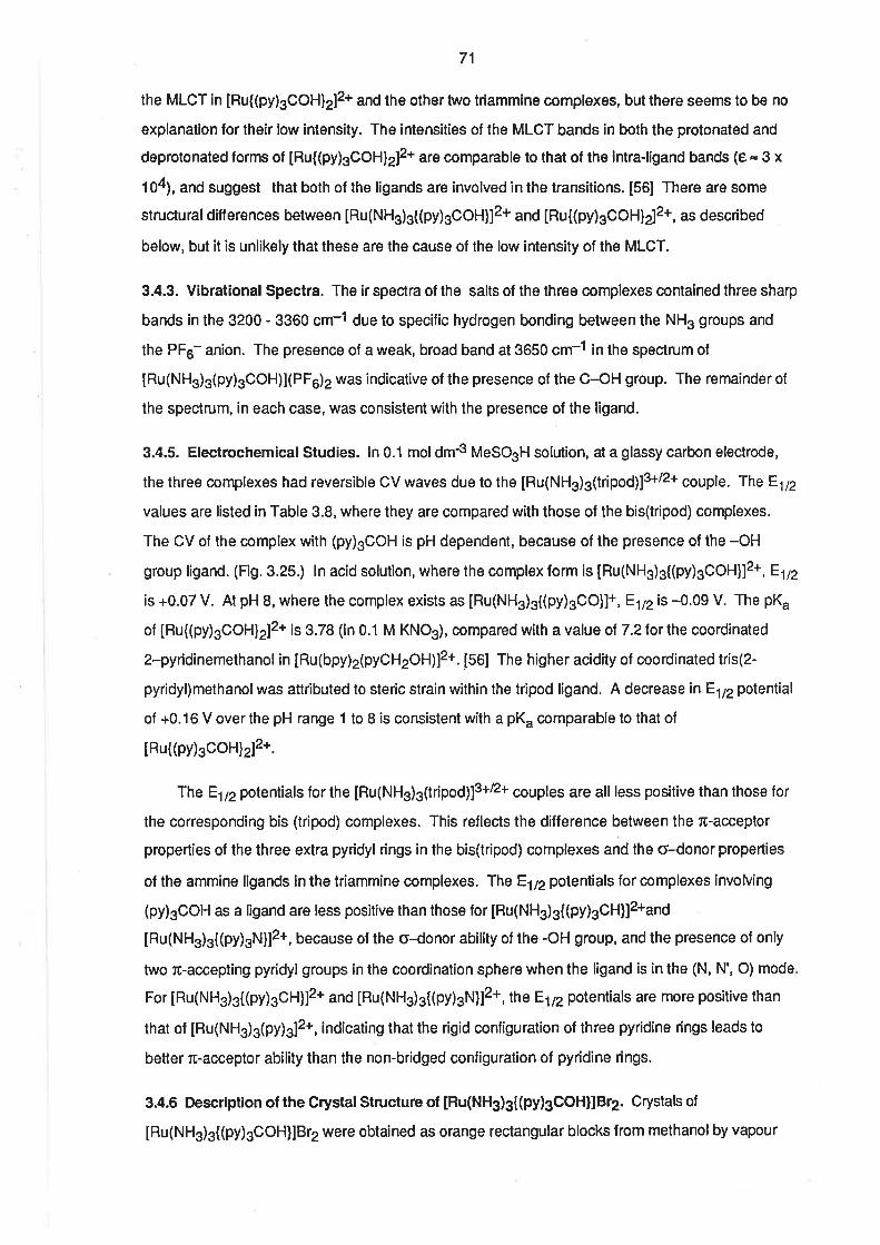
7',|
the MLCT in [Ru{(py)3COH}2]2+ and the other two triammine complexes, but lhere seems to be no
explanation for their low intensity. The intensities of the MLCT bands in both the protonated and
deprotonated forms of [Ru{(py)3COH}2]2+ are comparable to that of the intra-ligand bands (8 = 3 x
104), and suggest that both of the ligands are involved in the transitions. [56] There are some
structuraldifferences between [Ru(NHs)s((py)sCOH]]2+ and [Ru{(py)sCOH}2]2+, as described
below, but it is unlikely that these are the cause of the low intensity of the MLCT.
3.4.3. Vibrational Spectra. The ir spectra of the salts of the three complexes conta¡ned three sharp
bands in the 3200 - 3360 cm-1 due to specific hydrogen bonding belween the NH3 groups and
the PF5- anion. The presence of a weak, broad band at 3650 cm-1 ¡n the spectrum of
[Ru(NHs)s(py)gCOH)XPF6)2 was indicative of the presence of the C-OH group. The remainder of
the spectrum, in each case, was consistent with the presence of the ligand.
3.4.5. Electrochemicalstudies. ln 0.1 moldm-3 MeSOgH solution, at a glassy carbon electrode,
the three complexes had reversible CV waves due to the [Ru(NHg)3(tripoq]3+/2+ çeup¡s. The E172
values are listed in Table 3.8, where they are compared with those of the bis(tripod) complexes.
The CV of the complex with (py)3COH is pH dependent, because of the presence of the -OH
group ligand. (Fig. 3.25.) ln acid solution, where the complex form is IRu(NH3)31(py)sCOH]12*,81n
is +0.07 V. At pH 8, where the complex exists as [Ru(NH3)3{(Þy)sCO}]+, E172 is -0.09 V. The pK"
of [Ru{(py)3COH)2]2+ is 3.78 (in 0.1 M KNO3), compared with a value of 7.2lor the coordinated
2-pyridinemethanol in [Ru(bpy)2(pyOH2OH)]2*. 156l The higher acidity of coordinated tris(2-
pyridyl)methanol was attributed to steric strain within the tripod ligand. A decrease in E1¡2 potential
of +0.16 V over the pH range 1 to B is consistent with a pK" comparable to that of
IRu((py)3coHlzlz*.
The E1¡2potent¡als for the [Ru(NH3)s(tripod)]3+/2+ couples are all less positive than those for
the corresponding bis (tripod) complexes. This reflecls the difference between the æ-acceptor
properties of the three extra pyridyl rings in the bis(tripod) complexes and the o-donor properties
of the ammine ligands in the triammine complexes. The E172 potentials for complexes involving
(py)gCOH as a ligand are less positive than those for [Ru(NH3)g{(py)gOH}]2*and
tRu(NH3)3{(py)gN}12*, because of the o-donor ability of the -OH group, and the presence of only
two ß-accepting pyridyl groups ¡n the coordinatíon sphere when the ligand is in the (N, N', O) mode
For IRu(NH3)g{(py)sOH}]2* and tRu(NH3)3{(py)sN}]2+, the E1¡2 potentials are more positive than
that of [Ru(NH3)3(py)s]2+, indicating that the rigid configuration of three pyridine rings leads to
better fi-acceptor ability than the non-bridged configuration of pyridine rings.
3.4.6 Description of the Crystal Structure of [Bu(NH3)g{(py)gCOH}]8r2. Crystals of
[Ru(NH3)3((py)sOOH]IBt2wêtê obtained as orange rectangular blocks from methanolby vapour
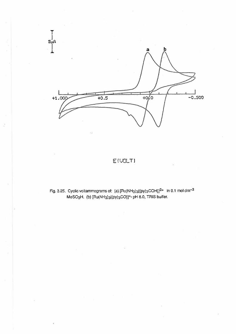
ï'r
a b
+1.
EIUOLT]
Fig. 3.25. Cyclic voltammograms of: (a) [Ru(NH3)g{(py)gCOH}lz+ in 0.1 moldm-3
MeSO3H. (b) [Ru(NHg)g((py)sCO]l+'pH 8.0, TRIS buffer.
-0.500+0.5
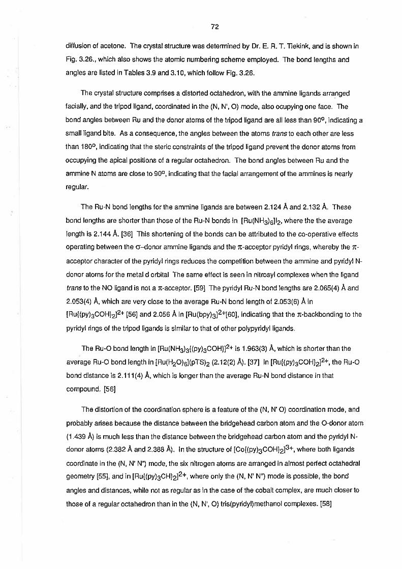
72
diffusion of acetone. The crystalstructure was determined by Dr. E. R. T. Tekink, and is shown in
Fig.3.26., which also shows the atomic numbering scheme employed. The bond lengths and
angles are listed in Tables 3.9 and 3.10, which follow Fig.3.26.
The crystalstructure comprises a distorted octahedron, with the ammine ligands arranged
facially, and the tripod ligand, coordinated in the (N, N', O) mode, also ocupying one face. The
bond angles between Ru and the donor atoms of the tripod ligand are all less lhan 90o, indicating a
small ligand bite. As a consequence, the angles between the atoms translo each other are less
than 180o, indicating that the steric constraints of the tripod ligand prevent the donor atoms from
occupying the apical positions of a regular octahedron. The bond angles between Ru and the
ammine N atoms are close to 90o, indicating that the facial arrangement of the ammines is nearly
regular.
The Ru-N bond lenglhs for the ammine ligands are between 2.124
^ and 2.132 À. These
bond lengths are shorter than those of the Ru-N bonds in [Ru(NH3)6]12, where the the average
length is 2.144 A. ¡S0¡ This shortening of the bonds can be attributed to the co-operative effects
operating between the o-donor ammine ligands and the n-acceptor pyridyl rings, whereby the æ-
acceptor character of the pyridyl rings reduces lhe competition between the ammine and pyddyl N-
donor atoms for the metal d orbital The same effect is seen in nitrosyl complexes when the ligand
translo the NO ligand is not a lc-acceptor. [59] The pyridyl Ru-N bond lengths are 2.065(4) Â and
2.053(4) A, which are very close to the average Ru-N bond length of 2.053(6) A in
IRu{(py)3COHlzlz* [56] and 2.056 A in [Ru(bpy)312*[eo], indicating that the æ-backbonding to the
pyridyl rings of the tripod ligands is similar to that of other polypyridyl ligands.
The Ru-O bond length in [Ru(NH3)g((py)sCOH]12* ¡s 1.963(3) Ä, which is shorter than the
average Ru-O bond length in [Ru(H2O)e]þTS)z e.12(2) A). tgZI ln [Ru((py)gCOH]212+, the Ru-O
bond distance is 2.11 1(4) A, which is longer than the average Ru-N bond distance in that
compound. [56]
The distortion of the coordination sphere is a feature of the (N, N'O) coordination mode, and
probably arises because the distance between the bridgehead carbon atom and the O-donor atom
(1.439 Ä¡ is much less than the distance between the bridgehead carbon atom and the pyridyl N-
donor atoms (2.9824 and 2.388 Â). ln the structure of [Co{(py)3COH}2]3+, where both ligands
coordinate in the (N, N' N") mode,lhe six nitrogen atoms are arranged in almost perfect octahedral
geometry [55], and in [Ru{(py)3Ct]lZl2+,where only the (N, N' NT mode is possible, the bond
angles and distances, while not as regular as in the case of the cobalt complex, are much closer to
those of a regular octahedron than in the (N, N', O) trís(pyridyl)methanol complexes. [581
r3
l+
C¡t
ú
ts
ñü¡2
7
9
x
Io
a
3
4Nz s
N3
NI
Fig. 3.26. X-ray crystal structure of [Ru(NHs)g{(py)gCOH}lBrz.
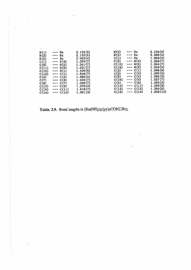
I
l
t24c4)06s (4)e63(3)333(7)354 (7)343(e)365 (8)3e5 (e)365 (8)527(7)356 (s)36e (S)380 (8)3se ( 1o)
221
It1
ItItIIII
RuRuRuN(4)N(5)N(6)c(1)c(2)c(4)c(6)c(8)c(11)c(12)c(14)
Q)(3)(s)(16)(e)(r2)(13)(15)
cccccccc
124(,5)132(s)053 (4)350 (7)341(7)32t(7)43e (6)546 (7)3æ (e)406 (7)3e8 (7)378(e)518 (7)361 (e)
2.2.2.1.1.1.l.1.1.1.1.1.1.1.
(5)(6)(1)(1)(3)(6)(7)(e)(1 1)(13)
RURURuN(4)NN0ccccccc
N(1)N(3)N(5)c(r)c(6)c(11)c(16)c(16)c(4)c(7)c(8)c(10)c(16)c(14)
Table. 3.9. Bond lengths in [Ru(NH¡)¡t(py)¡COH]lBrz.
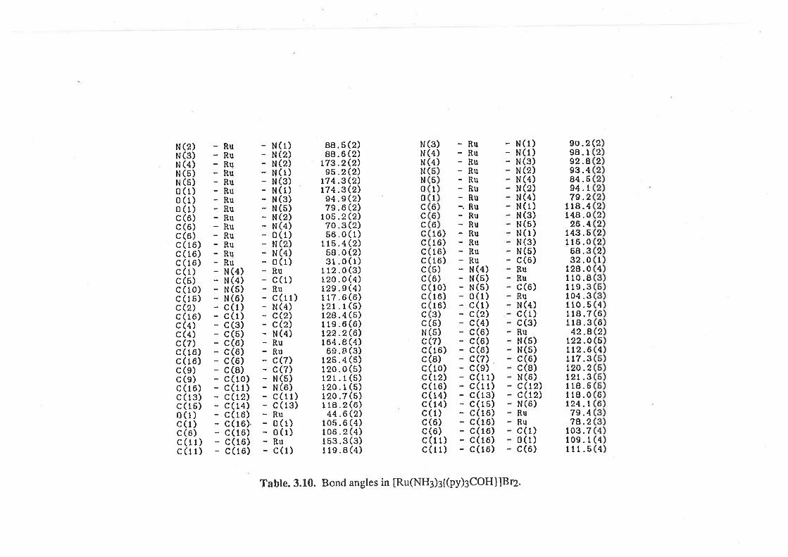
66
2222221
4
466254
222
(9U .298. I92 .893.484 .694. t79.2
f18.4148 .026.4
143.6116.068.332 .0
128.0110.8119 .3104 .3110 .5I r8.7118.342.8
t22.0112.6I 17.3t20.2121.3118.6118.0t24.L7s.474.2
t.03.7109. t111.s
N(1)N(1)N(3)N (2)N (4)N (2)N (4)N(1)N (3)N (5)N (1)N (3)N (5)c(6)RuRuc (ô)RuN (4)c(l)c (3)RuN(5)N(s)c(ô)c (8)N (6)c(12)c(12)N (6)RuRuc(1)0(1)c (6)
-Ru-Ru-Ru-Ru-Ru-Ru-Ru-. Ru-Ru-Ru-Ru-Ru-Ru-Ru- N(4)- N(5)- N(5)- 0(1)- c(t)- c(2)- c(4)- c(6)- c(6)- c(6)- c(7)- c(e)- c(r 1)- c(11)- c(13)- c(16)- c(r.6)- c(16)- c(16)- c(16)- c(16)
(3)(4)(4)
NNN
NN00ccccccccccccccNccccccccccccc
(2)e)Q)Q)Q)Q)(2)Q)Q\(2)(1)Q)e)(1)(3)(4)(4)(6)(s)(5)(ô)(6)(4)(3)(5)(5)(5)(5)(5)(6)o)(4)(4)(3)(4)
N(l)N(2)N (2)N(1)N (3)N(1)N (3)N (5)N (2)N (4)0(t)N (2)N (4)o (1)Ruc (1)Ru
88.588.6
L73.295 .2
174.3174.394 .979 .6
105 .270 .35ô .0
115 .458.031 .0
112 .0120.0129.9I 17.6121 .1t24.4119 .6L22.2164 .86C.e
125.4120.0I2L.L120.1120.71 18.244.ô
105 .6106.2r53.3119 .8
N (2)N(3)N (4)N (6)N (5)0 (1)0 (1)0 (1)c (6)c(6)c (6)c(16)c(16)c(16)c (1)c (6)c(10)c(16)c (2)c (16)c (4)c (4)c(7)c (16)c (r6)c (e)c (e)c(16)c(r3)c(16)0 (1)c(l)c (6)c(rl)c(11)
(
6)6)ô)ô)))0)ô)6))
51
Ib661
1
t1
561
II3
(r l)(4)Q)Q)(4)
(5)(5)Q)(r6)(8)( 10)(12)( 16)( 14)( 14)(1)(6)(6)(11)(11)
uuQ')(7)(5)(6)(1r)(13)
0(1)o (1)Ruc (1)
cNccNRRccNNcc
44
Ru
Table. 3.10. Bond angles in [Ru(NH:)¡{(py)gCOH}lBrz.
73
Similar distortions about the (N, N', O) ligand are also apparent ¡n the structure of
Ru{(py)gCOHlzl2*, but, in that case, the deviations from regular geometry are greater, with the N-
Ru-O angles being 76.20 and 77.2o compared with 79.60 and 79.2o lor the triammine complex'
[56] The bond angles belween Ru and the donor atoms of the (N, N', N") ligand are closer to 90o
than those of the (N, N', O) tigand, and are almost comparable with the bond angles betwen Ru and
the ammine ligands in the triammine complex.
A notable diflerence between the structures of [Ru(NHg)s{þy)sCOH}¡2+ .nO
Ru{(py)3COHlzl2* is the Ru-O distance. ln the triammine complex this bond is shorter than the Ru-
N bonds, and also shorter than the average bond length in [Ru(H2O)e](pTS)2. [37] However, in the
bis(tripod) complex, the Ru-O bond length is greaterthan any of the Ru-N bond lengths, and longer
lhan the same bond in [Ru(NH3)g{(py)gCOH¡¡z+. [67] lt would, perhaps, be expected lhat the etfect
of five pyridine rings on the Ru(ll) centre would be to make it more like a Ru(lll) ion, but this would
tend to shorten the Ru-O bond.
Another diference between the structures of the two tris(pyr¡dyl) methanol complexes is the
orientation of the uncoordinated pyridyl ring. ln Ru{(py)sCOHlzl2* the planes of the three pyridine
rings of the (N, N', O) ligand are aligned at 120o and the uncordinated ring is almost coplanar with
the ring trans to the coordinated OH group. [56] However, in [Ru(NHs)s{(py)sCOH}]2+, the
uncoordinated pyridine ring lies perpendicular to the line intersecting the planes of the other two
rings of the (N, N', O) ligand. This may, in some way, contribute to the low intensity of the MLCT
bands, but this seems unlikely, because this arrangement of the rings reduces the overall symmetry
of the complex, which would be expected to result in more ¡ntense uv/vis absorptions.
3.5 Conclusions.
It has been possible to synthesize a series of substituted triammine complexes of
ruthenium(lll) and ruthenium(ll) having ligands which range from strong æ-donors to strong æ-
acceptors. The stabilization of the two oxidation states by the various ligands fits in with the pattern
which had previously been established for the ruthenium penta- and tetraamine and edta
complexes.
The substituted triammineaqua ruthenium(lll) and ruthenium (ll) complexes show pH-
dependent reductions and oxidations which are c¡nsistent with the pK¿ of the coordinated water in
each case. There is a linear relationship between the reduction potential of the aqua complex and
the pK¿ of the Ru(lll) complexes containing uninegative, bidentate ligands. At high pH, the E1¡2lor
the Ru3+/Ru2+ couple is more negative than that at low pH, because of the greater n-donating
character of the OH- ligand cnmpared with the H2O ligand. At low pH, the E1¡2lor 1¡g ¡v3+7pu2+
74
couple is very similar to that for the same couple in the penta- and tetraammine complexes with the
same ligands, confirming the conclusion that the electrondonating propert¡es of the aqua and
ammine ligands are similar. lt was possible to achieve electrochemical reduction of some of the
Ru(lll) complexes, but reaction of the reduced species with dinitrogen did not yield substituted
triammine dinitrogen complexes. ' r
It is apparent that æ-donor ligands destabilize the RulV=O group, but that æ-donation from the
oxalate ligand is sufficiently weak to allow the observation of a quasi-reversible RulV=gTpulllgF{
couple.
The uv/vis spectra contain the LMCT bands expected for Ru(lll) complexes with o- and æ-
donor ligands, and the MLCT bands expected for Ru(ll) complexes with æ-acceptor ligands. The
spectra of the complexes containing the aqua ligand are pH dependent, reflecting the effect of the
differing æ-donating abilities of the hydroxo and aqua ligands. At low pH, the spectra are very similar
to those of the penta- and tetraammine complexes with the same ligands. ln most spectra there
were also intra-ligand bands, with their intensity roughly proportional to the number of such ligands
present in the complex.
Crystalstructure determinations show that the facialarrangement of ammine ligands which is
present in Ru(NH3)3Clg is retained in the substituted triammine complexes. The ruthenium(lll)
complex with acac as the ligand had a regular octahedral arrangement, but the ruthenium(ll)
complex with (py)3COH as the ligand was considerably distorted due to the coordination through
the OH group and two pyridyl nitrogen atoms.
75
REFERENCES.
(1) P. C. Ford, Coord. Chem. Rev.,5,75(1970)
(2) E. A. Seddon, K. R. Seddon, "The Chemistryof Rutl¡enium", Ftse\rief,, Amsterdam,
t984.
(3) P. Ford, DeF. P. Rudd,R. Gaunder, H. Taube,J. Amer. Chem. Soc.,90,
1 187(1968)
(4) J. A. Stritar, H. Taube, Ioory. Chem.,8,228I(1969)
(5) T. Matsubara, C. Cheutz, Inory. Chem., 18, 1856(1979)
(6) G. N. Coleman, J. W. Cresler, F. A. Shidey, J. R. Kuempel, hory. Chem.,12,
1036(1e73)
(7) J. A. Broom-head, L. Kane-Maguire, hory.Chem., 10, 85(1971)
(8) R. E. Cla¡ke, P. C. Ford,Inotg. Chem.,9,227(1970)
(9) P. C. Ford, C. Sutton, Inorg. Chem.,8, 1544(1969)
(10) A. D. Allen, T. Ft.iades, R. O. Ha:ris, P. Reinsalu , Can. J. Chem., 47, t605(1969)
(11) D. E. Haffison, H. Taube, J. Amer. Chem. Soc.,89,5706(1967)
(lZ) (a)N. E. Dixon, G. A. Lawrcnce, P. A,.Lay, A. M. Sargeson, hory.Chem.,23,2940(1984). (b) G. A. Lawrence, Chem. Rev.,86, 17(1986)
(13) R. W. Callahan, G. M. Brown, T. J. Meyer,Inorg. Chem., t9, L443(1975)
(14) J. A. Baumann, T. J. Meyer, hory. Chem., 19, 345(1980)
(15) B. Anderes, S. T. Collins, D. K. Lavallee, hory. Chem.,23,220t(1984)
(16) A. A. Diarnantis, J. V. Dubrawski, hory.Chem.,20,1142(1981)
(17) A. A. Diamantis, J. V. Dubrawski, Inotg. Chem.,22, 1934(1983)
(18) (a) G. A. Heath, K. R. Seddon. J. B. A. F. Smeulders, Unpublished resulrs. (b) A.
Endo, K. Shimizu, G. P. Sato, Chem. Lett., 581(1985), und references therein.
(19) S. Pell, J. N. Armot, Inorg. Chem.,12,873(1973)
(20) J. A. Broomiead,L. Kane-Magube, J. Chem. Soc., A, 546(1967)
76
(21) R. Sa¡ai, A. K. Kabisatpathy, J. D. Peterson, Inotg. Chin. Aæa, 115, L33(1986)
(22) R. W. Olliff, A. L. Odell, J. Chem. Soc.,24l7(19æ)
(23) J. Powell, J, Otganomet. Chem.,65,89(1974)
(24) F. Ige, F. Ojo, O. Olubuyide, Inorg. Chem.,2O, 1757(1981)
Q5) S. W. Lin, A. F. Schreiner,Inorg. Chim. Acta,5,290(1971)
(26) K. Nakamoto, "lnflured and Raman Spectra of lnorganic and Coordination
Compounds. " (3rd Ed.) J. Wiley & Sons, New York, 1978.
(27) R. T. Jones, HonoursReport, TheUniversityof Adelaide, 1986.
(28) (a) A. A. Diamantis, lff. R. Muqphy, T. J. Meyer, Ioory. Chem.,z3,3230(1984)
(b) A. A. Diamantis, priv. conun.
(29) C. M. Che, K-Y. Wong, C-K. Poon,Ioory.Chem.,24,1797(1985)
(30) Y. Takeuchi, A. Endo. K. Shimizu, G. P. Sato, J Electroanal. Chem., 185,
18s(1e8s)
(31) Md. A. Salam, Ph. D. Thesis, The University of Adelaide, 1986
(32) K. J. Takeuchi, M. S. Thompson, D. W. Pipes, T. J. Meyer,Iootg.Chem.,23,r84s(t984)
(33) K. J. Takeuchi, G. Samuels, S. W. Gersuen, J. A. Gilbert, T.J. Meyer, Ioory.
Chem.,22, t407(1983)
(34) A. A. Diama¡ris, J. M. Frederiksen, Md. A. Salam, M. R. Snow, E. R. T. Tieki¡k,
Aust. J. Chem., 39, 1081(1986)
(35) F. Bottomley, Can. J. Chem.,55,2788(1977)
(36) H. C. Stynes, J. A. Ibers,Ioory. Chem.,lO,2304(1971)
(37) P. Ber¡hard, H-8. Burgi,J. Hauser, H. Lehman,A. Ludi, Inorg. Chem.,2t,3e36(1e82)
(38) G. K-J. Chao, R. L. Sime, R. J. Sime, Acta. Crystallogr., Sect. 8.,29,2M5(1973)
(39) G. M. Bryant, J. E. Fergusson, H. K. J. Powell, Aust. J. Chem.,24,257(I97I)
77
(40) V. E. Alvarez, R. J. Allen, T. Matsubar¿, P. C. Ford, J. '4mæ. Chem. 5oc.,96,7686(re74)
(41) M. J. Ridd, F. R. Keene, J. Amer. Chem. Soc., 103,5733(1981)
(42) T. Matsubara, P. C. Ford, hory. Chem., 15, 1tO7(1976)
(43) G. N. Brown, N. Sutin, J. Amer. Chem. Soc., 101, 883(1979)
(44) W. R. McWhirurie, J. D. Miller, Adv. Inotg. Chem. Radiochem.,12,135(1969)
(45) A. B. P. Lever, "lnorganic Electronic Spectroscopy" , (znd Ed), Elsevier, New York,
1984, p337
(46) R. Krishna:nutthy,W. B. Schaap, J. Chem. 8d.,46,799(1969)
(47) K. Nakamoto, "lnfmred and Raman Spectra of lnorganic and Coordination
Compounds" (3rdEd.), J. Wiley & Sons, New York , 1978. (a) pl97. (b) p199
(48) P. S. Braterman "Metal Carbonyl Spectra", AcademicPress, London, 1975. (aþzla(b) pa6
(49) J. Chatt, G. J. Leigh, N. Thankanjan, J. Chem. Soc., A, 3168(1971)
(50) D. J. Darensbowg, Inotg. Chem., 11, 1436(1972), and references therei¡.
(51) R. H. Crabtree, A. J. Pearman, J. Organomet. Chem.,157,335(1978)
(52) B. A. Moyer, T. J. Meyer, J. Amæ. Chem. Soc., 100, 360(1978)
(53) G. M. Brown, T. R. Weaver, F. R. Keene, T. J. Meyer, Inory. Chem.,15,
1e0(1e76)
(54) H, S. Lim, B. J. Barclay,F.C. Anson, Inorg. Chem.,lL,1460(1972)
(55) D. J. Szalda, F. R. Keene, hory. Chem.,25,2795(1986)
(56) F. R. Keene, D. J. Szafda, T. A. Wilson, Inorg.Chem., inpress
(57) F. R. Keene, P. J. Stephenson, unpublished rezults.
(58) F. R. Keene, P. J. Stephenson, M. R. Snow, E. R. T. Tieki¡k, unpublished results
(59) F. Bottomley, Coord. Chem. Rev., 26,7(1978)
78
(60) D. P. Rillema, O. S. Jones, H. A. L"oy, J. Chem. Soc., Chem., Comm., 849(1979)
Chapter 4.
RUTHENIUM COMPLEXES WITH TRIDENTATELIGANDS.
4.1. lntroduction.
!n Chapter 1, the stabilization of the +4 oxidation state of ruthenium was discussed, and it was
shown that this oxidation state was favoured by complexes having ligands capable of strong rc-
donation. One of the ligands discussed in this context was the oxo 1O2-¡ [gand, and the work
described in Chapters 2 and 3 has shown that the Ru(lV) oxidation state can be obtained in solution
by proton-coupled electrochemical oxidation. The pH dependence of the proton-coupled
oxidations was evidence that the oxo group was being formed on the Ru(lV) centre. The
irreversibility of the voltammetric waves indicated that the Ru(lV) species were unstable, probably
due to the effects of the other ligands.
Other workers in this laboratory have shown that dinegative, tridentate ligands derived from
Schiff's bases are capable of eliminating the oxo ligand from the V=O2+ group, and stabilizing the
+4 oxidation state of vanadium. [1-3] The ligands formed by the reaction between aroyl hydrazines
and ß-diketones, have been known for some time [4], and are capable of forming complexes of the
type, M(tridentate)2, with a variety of metal ions. Crystallographic studies have shown that the
ligands retain their planar form upon coordination, resulting in geometries ranging lrom trigonal
prismatic to meridionaloctahedra. Examples of these complexes include V(acacBH)2' ,
Ti(bzacBH)2 and Ti(acacBH)2 [3] and Mn(salDHP)2. [5] Other tridentate, dinegative ligands with O-
N-O donor atoms can be lormed by using 2-hydroxy aromatic carbonylcompounds instead of
diketones, or by using 2-hydroxy amines instead of aroylhydrazines. There are potentially four
classes of ligand available, using various combinations of the two component parts, as shown in Fig
4.1.
The oxo (O2-) group is almost ubiquitous in vanadium(lV) complexes, and this is due to its
ability to stabilze the V4+ ion by p¡-d¡ donation. [6] The ability of the dínegative, tridentate ligands
to replace the oxo ligand in vanadium(lV) complexes is strong evidence that their æ-donor abilities
are comparable to that of the oxo group. Given the ease with which the tridentate ligands are able
to replace the oxo group, and the ability of the oxo group to stabilize Ru(lV), it was considered likely
that the dinegative, tridentate ligands would also be useful in stabilízing the Ru(lV) ion.
'Refer to the list of abbreviations.
R'CHs N fi
R'
o-
R - CeHs, R'- CHg, L - ÞacOAP
o-R-H,L-hnaOAP
N
o-
o-
bzacBH
N- trl
R
N
o-
R
G o-R=CHs,L-hapBH
Flg. 4.1. Some examples of tridentate ligands.
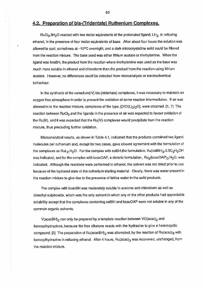
80
4.2. Preparation of bis-([ridentate) Ruthenium Complexes.
RuClg.3H2O reacted with two molar equivalents of the protonated ligand, LH2, in refluxing
ethanol, in the presence of lour molar equivalents ol base. After about four hours the solution was
allowed to cool, sometimes at -1OoC overnight, and a dark microcrystalline solid could be filtered
from the reaction mixture. The base used was either lithium acetate or triethylamine. When the
ligand was hnaBH, the product from the reaction where triethylamine was used as the base was
much more soluble in ethanol and chloroform than the product from the reaction using lithium
acetate. However, no differences could be detected lrom microanalysis or electrochemical
behavíour.
ln the synthesis of the vanadium(lV) bis-(tridentate) complexes, it was necessary to maintain an
oxygen free atmosphere in order to prevent the oxidation of some reaction intermediates. lf air was
allowed in to the reaction mixture, complexes of the type, [{VO(L)2}2O], were obtained. [1, 7] The
reaction between RuCl3 and the ligands in the presence of air was expected to favour oxidation of
the Ru(lll), and it was expected that the Ru(lV) complexes would precip¡tate from the reaction
mixture, thus precluding further oxidation.
Microanalytical results, as shown in Table 4.1, indicated that the products contained two ligand
molecules per ruthenium and, except for two cases, gave closest agreement with the formulation of
the complexes as RuL2.H2O. For the complex with salBH,the formulation, Ru(salBH)2.0.5C2H5OH
was indicated, and forthe complex with bzacOAP, a dimeric formulation , Ru2(bzacOAP)2.H2O, was
indicated. Although the reactions were performed in ethanol, the solvent was not dried prior lo use
because of the hydrated state of the ruthenium starting material. Clearly, there was water present in
the reaction mixture to give rise to the presence of lattice water in the solid products.
The complex with bzacBH was moderately soluble in acetone and chloroform as wellas
dimethyl sulphoxide, which was the only solvent in which any of the other products had appreciable
solubililty except that the complexes conta¡ning salBH and bzacOAP were not soluble in any of the
common organic solvents.
V(acacBH)2 can only be prepared by a template reaction between VO(acac)2 and
benzoylhydrazinze, because the lree diketone reacts with the hydrazine to give a heterocyclic
compound. [2] The preparation of Ru(acacBH)2 was attempted, by the reaction of Ru(acac)g with
benzoylhydrazíne in refluxing ethanol. After 4 hours, Ru(acac)3 was recovered, unchanged, from
the reaction mixture.
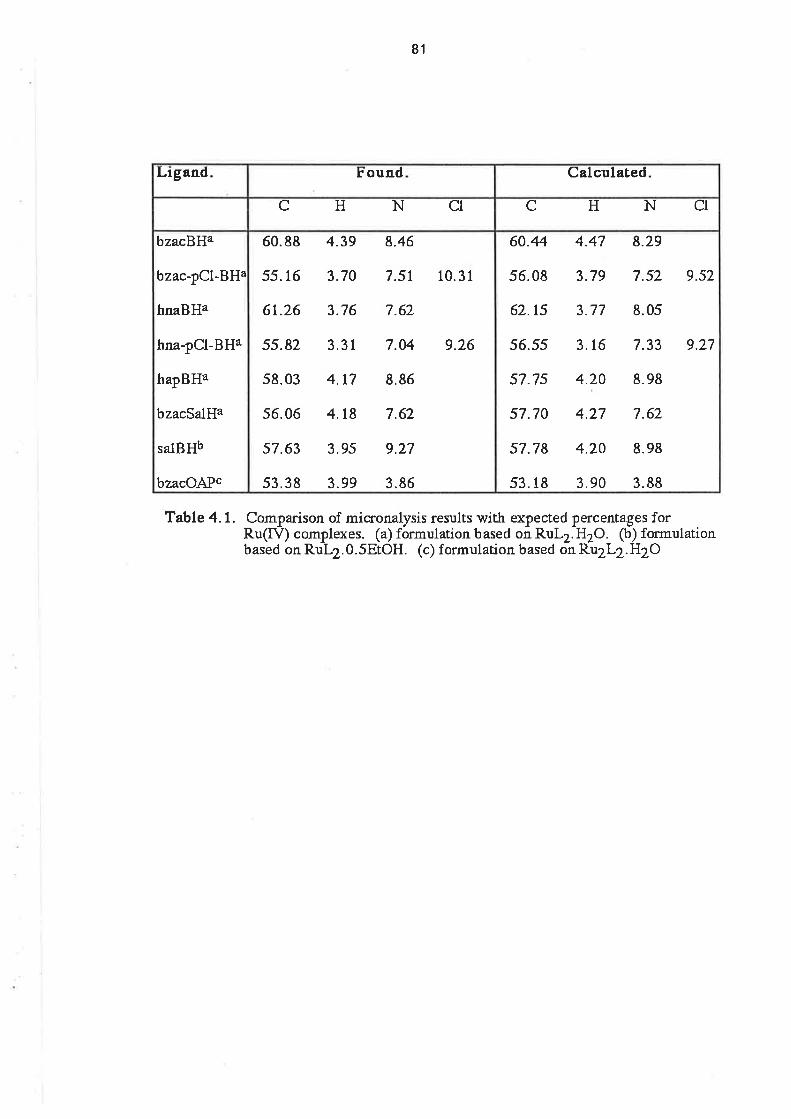
81
Table 4- 1. Comparison of micronalysis results with expected percentages forRu(tÐ complexes. (a) formulacion based on RuL2. H2O. (b) formulationbased on RuL2.0.5EIOH. (c) formulation based on Ru2L2.H2O
60.44 4.47 8.29
56.08 3.79 7.52 9.s2
62.15 3.77 8.05
56.55 3.16 7.33 9.77
57.75 4.20 8.98
57.70 4.27 7.62
57.78 4.20 8.98
53.18 3.90 3.88
60.88 4.39 8.46
55.16 3.70 7.5r 10.31
6t.26 3.76 7.62
55.82 3.31 7.04 9.26
58.03 4.t7 8.86
56.06 4.t8 7.62
57.63 3.95 9.27
53.38 3.99 3.86
bzacBBa
bzac-pCl-BHa
hn¿BHa
hna-pCl-BHa
hapBHa
bzacSalHa
salBHb
bzacOAPc
C HNCIC H NCl
Calculated.FoundLigand

82
4.3. Electrochemical Studies.
The electrochemistry of ruthenium bis(tridentate) complexes was studied at a platinum
electrode in DMSO solution, with 0.1 moldm4 NaClO4 supporting etectrolyte, and a SCE
reference electrode. (Under these conditions, ferrocene was oxidized al +0.447 V.) For all but one
of the complexes, it was possible lo observe two quasi-reversible waves which were assigned to
the Ru4+/Ru3+ and ¡y3+¡gu2+ couples. The potential of the gu3+¡gu2+ couple was 1.3 V to 1.6 V
more negative than that of the Ru4+/Ru3+ couple. The E1¡2 potentials of these couples are listed in
Table 4.2, along with those of some similar complexes.
Linear sweep voltammetry on stirred solutions, in the potential range of the Ru4/Ru3+
couples, was used to determine the ruthenium oxidation state in each complex. For the complexes
with bzacBH and bzac-pCI-BH the LSV showed that there was no current at potentials more positive
than E172, and there was a cathodic current at potentials less positive than E172 (Figs. 4.2 and 4.3).
This result was evidence that these two complexes were in the Ru(lV) state. For the complexes
with the ligands hnaOAP, hapBH, hnaBH and hna-pOl-BH, the LSV showed that an anodic current
flowed at potentials more positive than E1¡2, and there was no current at potentials less positive
than E172. This result was evidence that these complexes were in the Ru(lll) state. For lhe complex
with bzacSalH, there was a small anodic current in the stirred solution voltammogram at potent¡als
more positive than E1¡2and a small cathodic current at potentials less positive than E172. (F¡9. 4.4).
This was an indication that, perhaps, the producl of the reaction was a mixture of oxidation states.
The stirred solution voltammogram was identical for severalfreshly-prepared samples of the
product, so it seems unlikely that there was a mixture of compounds. lt may be that the product was
a Ru(lll) complex which reacted in the voltammetric solution to give a mixlure of oxidation states.
The CV of the complex with hnaOAP consisted of a single quasi-reverisble wave with E1¡2 =
+0.46 and AEo = 104 mV at a scan rate of 1OOmV sec-1. On a cathodic scan f rom +0.75 V, the
OSWV voltammogram showed a peak at +0.57 V, which is considerably different from the E172
determined from the CV. On continuing this cathodíc scan multiple peaks were observed at
negative potentials, with the most intense being at -1.09 V. The peak at -1.09 V was probably due
to the Ru3+/Ru2+ couple, but the limited solubility of this complex in DMSO made interpretation of
the results difficult.
ln addition to the two reversible waves, the cyclic voltammograms of the complexes containing
bzacSalH, hapBH and hnaBH also contained a cathodic peak at approximately -0.5 V (Fi$s.4.4,
4.5, 4.6). ln the case of the bzacsalH complex, after the scan direction had been reversed at
-1.75 V, the height of this peak was less on subsequent cathodic scans. ln the voltammograms of
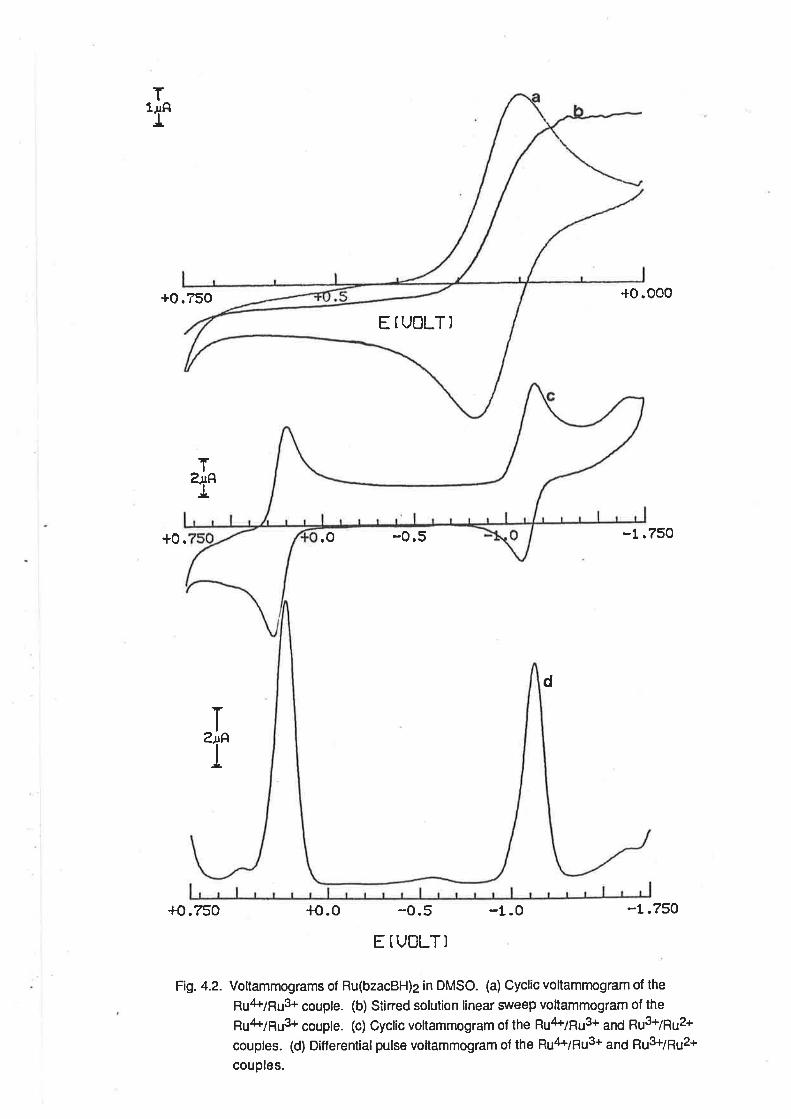
E IUOLT)
T'f
+o.750
+0.
+o.750
T
T
ïarf
.O
+o.o
-0.5
-0.5
E TUOLT]
-t.0
+o.oo0
-1.750
-L.7SO
d
Fig.4.2- Voltammograms of Ru(bzacBH)2 in DMSO. (a) Cyclic voltamrnogram of thepu¿k7puS+ couple. (b) Stined solution linear srveep voltammogram of thepu4+¡¡r3+ couple. (c) Cyclic vollammogram of the gu4+79u3+
"n¿ ¡r3+7pu2+
couples. (d) Differential pulse voltammogram of the R¡J4+79u3+ ¿n6 pu3+79u2+
couples.
EfUOLT)
b
a
+0
+o
+0.750
T,f
ï'r
-t.o
+0.000
-1.750
-r.750
T
T
+o.o -o,5
+o.o
d
-o.5
E IUOLT)
Fig. 4.3. Voltammograms of Ru(bzac-pOl-BH)2 in DMSO. (a) Cyclic voltammogram of thepu4+¡gu3+ couple. (b) Stined solution linear sweep voltammogram of thegu4+79u3+ couple. (c) Cyclic voltammogram of the ¡u4+79u3+ ¿¡6 gu3+¡pu2+
couples. (d) Differential pulse voltammogram of the Ru4+/RuS+ ¿¡6 pu3+7¡u2+
couples.
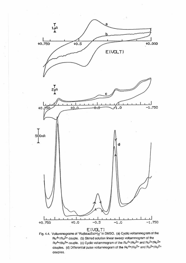
+o.5+o.-¿50 {o.000
E f UOLT)
+o -t.750
+0.?50 +o.o -0.5 -1.0 -1.750
E IUOLT) :
Fig. 4.4. Voltammograms of "Ru(bzacSalH)2" in DMSO. (a) Cyclic voltammogram of the
gu4+7¡u3+ couple. (b) Stined solution linear sweep voltammogram of thegu4Þ7pu3+ couple. (c) Cyclic voltammogram of the pu4+7pu3+ ¿¡6 gu3+7pu2+
couples. (d) Differential pulse voltammogram of the ¡u'l+¡gu3+ ¿¡6 gu3+¡¡u2+
couples.
T1J¡AI
T'f c
o
d
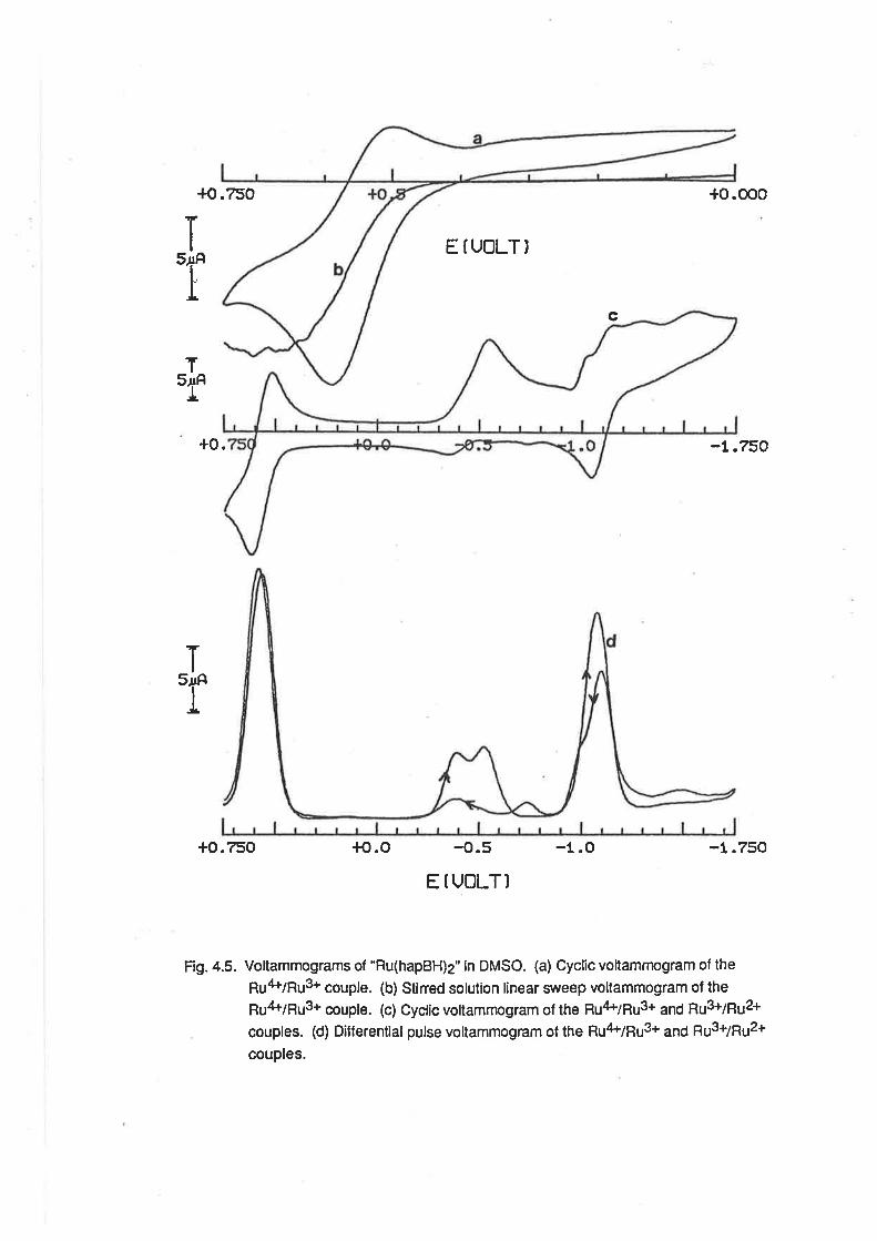
+0.750
+o.750 {o.o -1.O
+o.ooo
-r.750
-t.750
ï
'r
T'f
EÍUOLT)
c
+0.
T'f
-0.5
E IUOLT)
Fig. 4.5. Voltammograms of "Ru(hapBH)2" in DMSO. (a) Cyclic voltammogram of thepu¿l+¡pu3+ couple. (b) Stined solution linear srreep voltammogram of thegu4+¡¡u3+ couple. (c) Cyclic voltammogram of the pu4vgu3+ ¿¡6 pu3+7pu2+
couples. (d) Ditferential pulse voltammogram of the gu4+¡gu3+ ¿¡¿ gu3+7pu2+
couples.
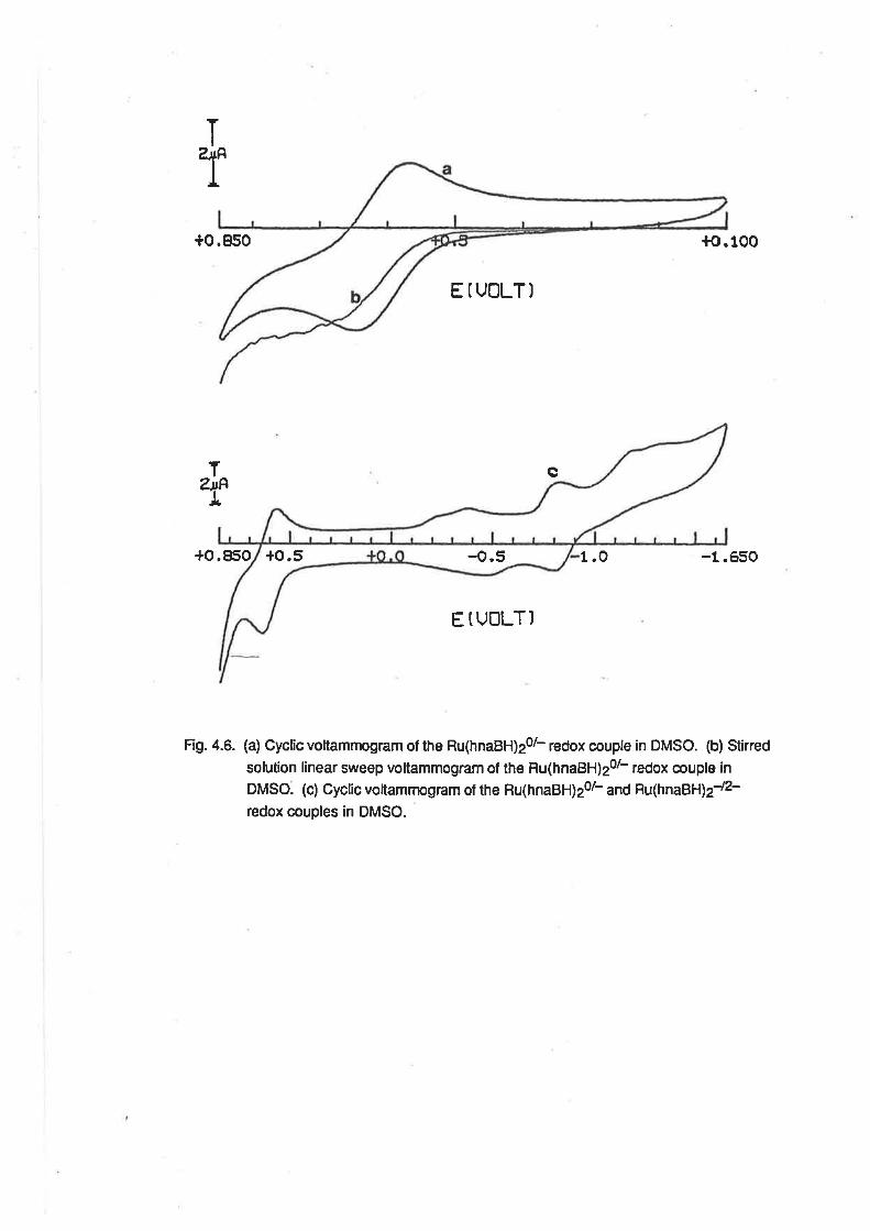
+0.950 +o.100
ETUOLT)
c
+0.850 1.0 -1.650
E ÍUOLTI
Fig. 4.6. (a) Cyclic voltammogram of the Ru(hnaBH)20l- redox couple in DMSO. (b) Stirred
solution linear sweep voltammogram of the Ru(hnaBH)2ol- redox couple in
DMSO. (c) Cyclic voltammogram of the Ru(hnaBH)2ol- and Hu(hnaBH¡2-/2-
redox couples in DMSO.
ï
'r
T
"f{,5+o.5
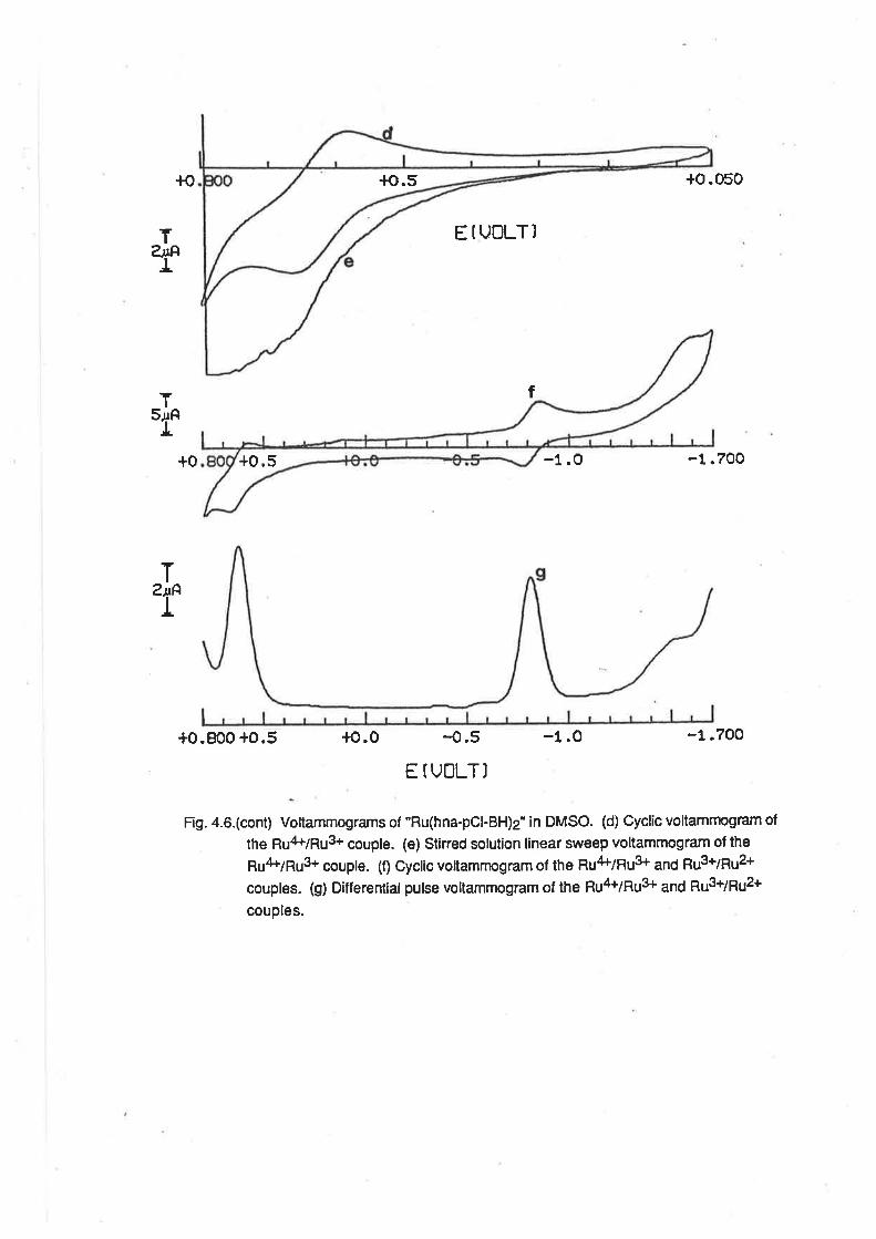
+0.5
{o.5+o
E IUOLT)
Í
+o.050
-1.700
-1 .?oo
TàtAI
T'f
T,f
+0
+0.800 +0.5
-1.0
-1.0+o.0 -0.5
E TUOLT]
Fig.4.6.(cont) Voltammograms of "Ru(hna-pOl-BH)z'in DMSO. (d) Cyclic voltammogram ol
the Ru't+/Ru3+ couple. (e) Stirred solution linear sweep voltammogram of the
¡u4Þ7¡u3+ couple. (f) Cyclic vollammogram of the pu4+7gL¡3+ 3¡6 gL¡3+7pu2+
couples. (g) Differential pulse voltammogram of the pu4+¡pu3+ ¿¡6 ¡u3+7¡u2+
couples.

83
+4
+4
+3
+3
+J
+J
+J
1.36
1.35
1.34
1.55
1.63
t.47
|.44
1.89
1.87
t.87
-t.12(74)
- 1 .08(83)
-0.et(74)
-1.09c
-1.ogc
-0.8s(6s)
-0.81(74)
-1.68(160)
-1.71(eo)
-1.76(80)
+0.24(80)
+0.27(7?)
+0.43(77)
+0.46(104)
+0.ss(91)
+0.62(77)
+0.6s(98)
-0.3s(e0)
-0.2e(r70)
-0.21(1e0)
-0.16(80)
-0. 1 1(80)
-0.89
-0.75
+0.14
Ru(bzacBHþ
Ru(bzac-pCl-BH)z
Ru(bzacSalH)2
Ru(hnaOAPþ
Ru(hapBHþ
Ru(hnaBHþ
Ru(hna-pCl-BH)z
V(bzacBHþ
V(bzac-pCl-BH)2
V(hnaOAPþ
V(hnaBH)2
V(bzacSalH)2
Ti(bzacBHþ
Ti(hnaBH)2
¡4¡(bzacBH)2
OxidtrStateb
LEtnE1¡2(ÂEp)ilI/il
E1¡2(ÂEp)aIV/III
Compler
Table 4.2. Redox potentials of M(triderÊaleþ complexes. Rgthenir:m cornplexes,this worl' potentiars vs SCE t
Hi"r'8tËiif,, J,ñä1':fr.ii,,I'Jtr"-omplex, Reference 9, rn SCE with EqNCI
(a) V(mÐ (b) Ruthenium oxidation sLate
determined from stirred solution LSV. (c) Etz determined from peakpotential in OSWV experimenr.
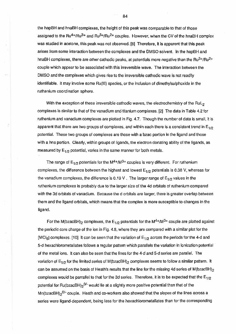
84
the hapBH and hnaBH complexes, the height of this peak was comparable to that of those
assigned to the Rurt+/Ru3+ and pu3+¡pu2+ couples. However, when the CV of the hnaBH complex
was studied in acetone, this peak was not observed. [8] Therefore, it is apparent that this peak
arises from some interaction between the complexes and the DMSO solvent. ln the hapBH and
hnaBH complexes, there are other cathodic peaks, at potentials more negatíve than ¡¡e gu3+¡pu2+
couple which appear to be associated with this irreversible wave. The interaction between the
DMSO and the complexes which gives rise to the írreversible cathodic wave is not readily
identifiable. lt may involve some Ru(lll) species, or the inclusion of dimethylsulphoxíde in the
ruthenium coordination sphere.
With the exception of these irreversible cathodic waves, the electrochemistry of the RuL2
complexes is similar to that of the vanadium and titanium complexes. [2] The data in Table 4.2 for
ruthenium and vanadium complexes are plotted in Fig. 4.7. Though the number of data is small, it is
apparent that there are two groups of complexes, and within each there is a consistent trend in E1¡2
potential. These two groups of complexes are those with a bzac portion in the ligand and those
with a hna portion. Clearly, within groups of ligands, the electron donating ability of the ligands, as
measured by Elnpotential, varies in the same mannerfor both metals.
The range ol E1¡2potentials for the y4+7y3+ couples is very different. For ruthenium
complexes, the difference between the h¡ghest and lowest E172 potentials is 0.38 V, whereas for
the vanadium complexes, the difference is 0.19 V . The larger range of E172 values in the
ruthenium complexes is probably due to the larger size of the 4d orbitals of ruthenium compared
with the 3d orbitals of vanadium. Because the d orbitals are larger, there is greater overlap between
them and the ligand orbitals, which means that the complex is more susceptible to changes in the
ligand.
For the M(bzacBH)2 complexes, lhe E1¡2potentials for the y4+7y3+ couple are plotted against
the periodic core charge of the ion in Fig. 4.8, where lhey are compared with a similar plot for the
[MCl6] complexes. [10] lt can be seen that the variation ol E1¡2 across the periods for the 4-d and
5-d hexachlorometallates follows a regular pattern which parallels the variation ín ionization potential
of the metal ions. lt can also be seen that the lines for the 4-d and 5-d series are parallel. The
variation ol E1¡2for the limited ser¡es of M(bzacBH)2 complexes seems to follow a similar pattern. lt
can be assumed on the basis of Heath's results that the line for the missing 4d series of M(bzacBH)2
complexes would be parrallel to that for the 3d series. Therefore, it is to be expected that the E172
potential for Ru(bzacBH)20r would lie at a slightly more positive potentialthan that of the
Mn(bzacBH)20l- couple. Heath and co-workers also showed that the slopes of the línes across a
series were ligand-dependent, being less lor the hexachlorometallates than for the corresponding
EU2(Ru)
o.7
0.6
0.5
0.4
0.3
o.2
-0.4 -0.3 0.1 0.0-0.2E112(Vanadium)
Flg- 4.7. Plot of E1¡21or Ru(tridentatel20t- redox couples vs E1p for V(tridentate)20l-
redox couples. (1) L = bzacBH. (2) L = bzac-pOt-BH. (3) L = bzacSalH.
(4) L - hnaBH.(S) L = hnaOAP.

0.8
0.6
0.4
0.2
0.0
-0.2
-0.4
-0.6
-0.8
-1.0
t?
p-A
E1t2(v)
ta
T¡ v Cr Mn (Ru) Co
tó2
2 t¡¡ctc¡0t- (q)
rdz
ô3/ô'
t¡J
b
d2td'
fMcl6¡3t6'(d)
- 3.0r ¡ ¡ ¡ ¡ r,
=rozm Ru Rh Pd
¡Hl To W Pt0sRe lr
Fig. 4.8 Trends in Êtnpotentials for: (a) M(bzacBH)20l- redox couples (po¡nts for Cr and
co est¡mated by extrapolation). (b) [Mcl5]zz-1 redox couples. [101
85
f luoro complexes. [10] This can be attributed to the greater nephelauxetic etfect of the chloro
ligand and it is consistent with the much smaller slope involved with the limited number of
M(bzacBH)2 complexes.
Another feature that is common to M(tridentate)2 complexes of ruthenium and vanadium is that
there is a relatively constant separation of the potentials of the two redox couples for each metal.
There is some uncertainty about the titanium complexes because it is not known whether the redox
couples at more negative potentials are ligand- or metal-centred. For the ruthenium complexes, the
potential separation between the two couples is between 1.3 V and 1.6 V, and for the vanadium
complexes, the separations are between 1.6 and 1.8 V. The separations between the ruthenium
couples may be compared with the potential separations of between 1.55 V and 1.9 V for the
Ru/Ru3r and Ru3+/Ru2+ couples in a large number of Ru(diketonate)3 complexes. [1 1-13] The
ligand-dependence of the separation between successive couples for any one element has also
been dealt with by Heath's group and was explained in terms of electron-electron correlations and
the nephelauxetic effect.
ln dichloromethane solution, the E172 potentials for oxidation s11¡s pLilllldiketonate)3
complexes were in the range +1.05 V to +1.84 V (converted to SCE scale), which is considerably
greater than the potentials at which tn" *ulll6ridentate)2 complexes are oxidized. This is a
reflection of the greater æ-donating ability of two dinegative ligands compared with three
uninegative ligands.
As noted above, the complexes with the lowest E1¡2 potentials for the Ru4+/Ru3+ couples
were isolated as Ru(lV) complexes, while those with the highest E172 potentials for the gu4+79u3+
couples were isolated as Ru(lll) complexes. This result is consistent with the capacities of the
various ligands to stabilize higher oxidation states by æ-donation. The better æ-donors would be
expected to favour the higher oxidation state, and to give complexes with less negative E172
potentials. Given that the microanalytical results for these compounds were all consistent with the
formulation of the compounds as RuL2.H2O, the question of the exact composítion of these
complexes becomes important. Wth two dinegative anions present in the compound, a
ruthenium(lll) complex requires the presence of a cation to maintain neutrality of charge. At first it
was considered that the cation in the compound must be lithium, coming from the lithium acetate
used as a base in the preparative reaction. However, this is not so. Analysis of the Ru(lll)
compounds showed that each contained less than 0.05% Li.
86
4.4. Mass Spectra.
The complexes were too involatile to allow lhe determination of the mass spectra by the
conventional electron impact process. The mass spectra of the complexes could be obtained
using Fast Atom Bombardment (FAB) mass spectrometry. ln this technique, the sample under
investigation is dissolved in an inert matrix, and then subjected to bombardment by a stream of high
energy heavy atoms (in this case argon atoms were used). The bombardment of the sample results
in the formation of posilüe and negative ions, which may be detected separately by changing the
acceleration potential in the spectrometer from +8 kV (for positive ion spectra) to -8 kV (for negative
ion spectra). There are 7 isotopes of ruthenium and their mass numbers and relative abundances
are: 96(5.51%), 98(1 .87V"1, 99(12.72%), 100(12.62/"), 101('17.07%), 102(91.63%) and
104(18.58%). Complexes of ruthenium have mass spectra which show a pattern of 7 peaks, with
their intensities roughly proportional to the relative abundances. The most intense peak generally
corresponds to the Ru102 isotope. ln some cases, high resolution mass spectrometry was used in
order to obtain the molecular weight, and so clarify the composition of the Bu(lll) complexes. ln this
process, the most intense peak was calibrated against a sample of caesium iodide, and its molecular
weight determined, instrumentally, by comparison with a well-defined peak in the calibrant.
4.4.1. RulVL2 Comptexes. The most intense peak in the molecular ion ruthenium isostope peak
for Ru(bzacBH)2 was almlz = 659 amu which agrees with the molecularweight expected for
Ru1o2(bzacBH)2. ln the spectrum of Ru(bzac-pOl-BH)2, the two most intense peaks in the
molecular ¡on pattem were at mlz=727 amu and mlz =729 amu which correspond to the molecular
weights of RuL2 for the pu1029¡356135 and pu1029¡37çFs isotopic combinations. The ruthenium
isotope patterns for the two complexes were identical in both the positive ion and negative ion
spectra. There were no fragmentations observed.
The complexes with the ligands salBH and bzacOAP could only be studied by mass
spectrometry, because they were not sutficiently soluble in any solvent for electrochemical or
spectral measurements. ln lhe absence of any evidence to the contrary, both were assumed to be
ruthenium(lV) complexes. The complex with salBH was formulated as Ru(salBH)2.O.5EIOH. The
mass spectrum of this compound contained a molecular ion signal with a regular ruthenium isotope
pattern. The most intense peak in the pattern was at nlz = 578 amu, which corresponds to the
molecular weight of Ru1o2(salBH)2.
On the basis of microanalysis, the complex with bzacOAP was formulated as Ru2(bzacOAP)2.
However, the highest molecular weight fragment in the mass spectrum was at r/z = 605 amu, which
is the molecular weight expected for Ru102(bzacQAP)2. Additionally, in this spectrum, a distinct
fragmentation pattern was observed. There were two other ions having the ruthenium isotope
87
paltern, with the most ¡ntense peaks being at m/z = 353 amu and rìy'z = 338 amu. This spec'trum
corresponds to the following lragmentation pattern.
[Ru1oz1nzacOAe¡r¡+ + lRu1o21bzacoAP)2-bzacOAP]+
rn/z = 605 ny'z = 353
J1Ru1
o2lbzacOAP)2 - bzacOAP - CH.¡+
nvz = 338
This is quite strong evidence that the complex is actually Ru(bzacOAP)2 and implies that the
microanalytical results were inaccurate, probably due to some contamination of the sample.
4.4.2. pullll2 Complexes. The spectra of the complexes with hnaBH, hna-pCl-BH and bzacsalH
were determined as both positive ion and negalive ion spectra. The negative ion spectra of the
three complexes all had molecular ion peaks with the expected ruthenium isotope pattern. The
positive ion spectra showed a molecular ion with a peak pattern which was consistent with the
overlap ol two ruthenium isotope patterns, an indication that, possibly, both RuL2+ and HRuL2+
molecular ions were formed.
For the complex with hnaBH, the negative ion spectrum contained an iso$tope pattern with
the maximum peak almlz = 678 amu, which is equalto the molecularweight of the species
*rt021hnaBH)2. Despite the unususat isotope pattern in the positive spectrum, the most intense
peak was al mlz = 679 amu. ln the high resolution speclrum, this peak was found to be
679.13(1.05) amu, which is close to the value predicted for [HRuL2], 679.092 amu. lt would appear
that the Ru(lll) species contains the two tridentate ligands, perhaps with the charge of the complex
being balanced by the presence of a hydrogen ion. However, the position of this hydrogen ion is
not certain, and it is also known that [M+H]+ ions (where M represents the rnolecular ion) may be
generated buring the bombardment of the sample. [14] Therefore it is not possible to conclusively
assign the complex as [HRu(hnaBH)2]
The spectra of the complexes with hna-pOl-BH and bzacSalh were similar to that of the hnaBH
complex, with the negative ion spectra being consistent with RuL2, and the most intense peak in
the positive ion spectrum being consistent with the formulation, [HRuL2]. The results of the
positive ion and negative ion spectra are consistent with each other. lf the formula of the
complexes is [HRuL2], then, in the negative ion spectrum, it would be expected that the molecular
ion which was observed would be readily generated by the loss of a proton from the complex.
88
4.5. Vibrational Spectra.
' lt was anticipated that the vibrational spectra of the complexes would conlirm some of the
assumptions made about their composition. ln particular, the presence of lattice water would be
readily indicated by the presence of the.characteristic (U)OH bands between 3300 and 3800 cm-1,
or the assfignment of some of the complexes as H[RuL2] would be confirmed by the presence of
bands characteristic of the (D)OH mode between 3200 and 3450 cm-1. The presence of strong
bands between 850 and 1000 cm-l would have been indicative of the possible presence of Ru=O
groups [15], which would have implied that the higher oxidation state complexes were not of the
type, RuL2. The ir spectra of the complexes were recorded as nujol mulls on KBr windows and the
Ru(lll) complexes were also recorded in KBr pellets in the range 2500 cm-1 to 3600 cm-1.
Generally, the spectra were similar to those ol the VIVL2 complexes. [2] The absence of any
intense bands between 850 cm-1 and 1000 cm-1 ¡n all spectra confirmed the absence of a Ru=O
bond. Allthe spectra contained weak broad bands centred in the range 3420 cm-1 to 3460 cm-1
providing evidence of the presence of lattice water. ln the spectrum of the salBH complex, there
was no evidence of the presence of ethanol in the lattice which was indicated by the microanalysis
result.
ln the KBr pellet spectra of some of the Ru(lll) complexes lhere were weak absorptions in the
region 3180 cm-1 to 3220 cm-1 which were assigned to a (ù)NH vibration. The aromatíc (1J)CH
vibrations were evident as sharp bands between 2980 cm-1 and 3050 cm-1, so the weak bands al
slightly higher energies were not due to this mode of vibration. ln the vanadium(lV) complexes,
V(OXCIXHL), there is one uninegative, tridentate ligand. The ir spectra of these complexes
showed weak, broad bands at approximately 3200 cm-1, which were attributed to the (Ð)NH
vibration. [2] lt would appear, therefore, that the 3+ charge in the Ru(lll) complexes is accomodated
by partial deprotonation of one of the tridentate ligands instead of double deprotonation of both of
them.
4.6. Magnetic Properties.
The magnetic properties of the RuL2 complexes were determine at a single temperature by
the Gouy method. The lack ol a suitable instrument prevented the determination of magnetic
suscetibilities at many temperatures. The interpretation of single temperature magnetic
measurements can not be conclusive because the magnetic suscept¡bility of a complex is
proportional to the square root of the temperature at which the measurement is made. [16]
However, it is possible to compare experimental results with the magnetic susceptibility of known
examples. The magnetic properties of the RuL2 complexes are presented in Table 4.3.
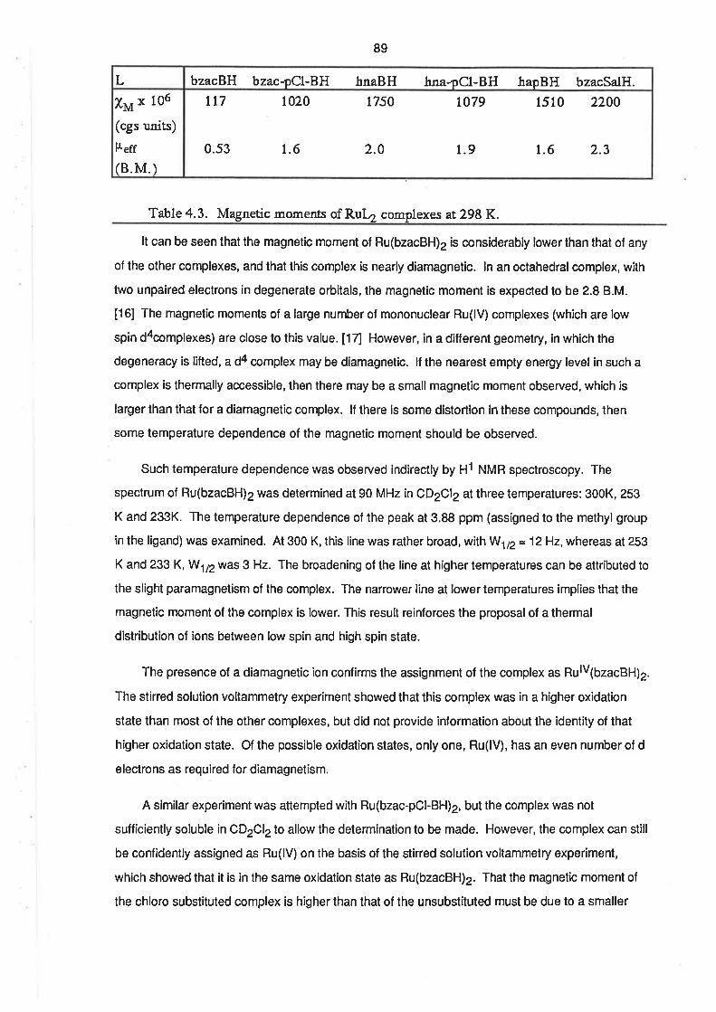
89
tt7 1020 1750 to79 1510 2200
0.53 1.6 2.0 1.9 1.6 2.3
X¡¿ x 106
(cgs units)
l¿eff
(8.M.)
bzacBH bzac-pCl-BH hnaBH hna-pCl-BH hapBH bzacSalH.L
Table 4.3. Magnetic moments of Ruþ complexes ar.298 K.
It can be seen that the magnetic moment of Ru(bzacBH)2 is considerably lower than that of any
of the other complexes, and that this complex is nearly diamagnetic. ln an octahedral complex, with
two unpaired electrons in degenerate orbitals, the magnetic moment is expected to be 2.8 B.M.
[16] The magnetic moments of a large number ol mononuclear Ru(lV) complexes (which are low
spin d4complexes) are close to this value. [17] However, in a different geomelry, in which the
degeneracy is lifted, a d4 complex may be diamagnetic. lf the nearest empty energy levet in such a
complex is thermally accessible, then there may be a smallmagnetic moment observed, which is
larger than that for a diamagnetic complex. lf there is some distortion in these compounds, then
some temperature dependence of the magnetic moment should be observed.
Such temperature dependence was observed indirectly by H1 NMR spectroscopy. The
spectrum of Ru(bzacBH)2 was determined at 90 MHz inCD2Cl2at three temperatures: 300K, 253
K and 233K. The temperature dependence of the peak at 3.88 ppm (assigned to the methyl group
in the ligand) was examined. At 300 K, this line was rather broad, with W172 = 12H2, whereas at 253
K and 233 K, W172 was 3 Hz. The broadening of the line at higher temperalures can be attributed to
the slight paramagnetism of the complex. The narrower line at lower temperatures implies that the
magnetic moment of the complex is lower. This result reinforces the proposal of a thermal
distribution of ions between low spin and high spin state.
The presence of a diamagnetic ion confirms the assignment of the complex as RulvlbzacBH)2.
The stirred solution voltammetry experiment showed that this complex was in a higher oxidation
state than most of the other complexes, but did not provide information about the identity of that
higher oxidation state. Of the possible oxidation states, only one, Ru(lV), has an even number of d
electrons as required for diamagnetism.
A similar experiment was atlempted with Ru(bzac-pOl-BH)2, but the complex was not
sufficiently soluble in CD2C|2 to allow the determination to be made. However, the complex can still
be confidently assigned as Ru(lV) on lhe basis of the stined solution voltammetry experíment,
which showed that it is ¡n the same oxidation state as Ru(bzacBH)2. That the magnetic moment of
the chloro substituled complex is higher than that of the unsubstituted must be due to a smaller

90
energy gap between the filled energy levels in the diamagnetic form and the next highest energy
levels.
The magnetic moments of the complexes with hnaBH, hna-pOl-BH, and bzacSalH are all
consistent with the presence of one unpaired electron [16], which is indicative of the complexes
being low spin ¿3 complexes. Though the magnetic moment of the complex with hapBH was
indicative of slightly less than one unpaired electron per ruthenium ion it, too, can be assigned as
Ru(lll) on the basis of its similar voltammetry in the stined solution.
4.7. Electronic Spectra.
The uv/vis spectra of Ru(bzacBH)2, Ru(bzac-pOl-BH)2 and the complexes with hnaBH and
hna-pCl-BH in DMSO solution were recorded , as were the spectra of the protonated ligands. The
concentration of the complex solutions was 2.5 x 10⁴ motdm4 and of the ligand solutions was
5.0 x 10+ moldm-omoldm-3. The spectra are displayed in Figs. 4.9 - 4.12, and the absorption
maxima are listed in Table 4.4.
It can be seen that the spectra of the complexes contain absorptions which can be readily
assigned to intra-ligand transitions. ln addition to these absorptions, there is a fairly strong
absorption in each spectrum betwen 410 nm and 445 nm, sometimes being evident as a shoulder
to an intra-ligand band. This absorption is also present in the spectra of the ligands, bzacBH and
bzac-pGl-BH ,in DMSO, but with a lower intensity than in the complex spectra. The spectra of these
ligands in CH2C|2 contained a single band, which was the same as the higher energy absorption in
DMSO solution. The lower energy bands were also observed in methanol in the presence ol
lithium acetate, and their intensity is roughly proportionalto the amount of base in solution. lt is
possible that there is some degree of enolization of the ligands in the presence of DMSO, so that
there is some contribution to the spectrum from the enolate forms. There are two possible enol
structures in each of the tridentate ligands, and the ligands present in the complexes are
deprotonated at both enol sites. lt has been shown lor polypyridyl complexes of ruthenium(ll) that
the intra-ligand bands in the complex spectrum are almost identicalwith those in the spectrum of
the protonated ligands. [18] lt is possible that there exists a similar relationship between the spectra
of the protonated and coordinated tridentate ligands. On this basis, the lower energy band in the
spectra of the ligand can be assigned to a second intra-ligand lransition of the double enolform of
the ligand.
Except in the case of the complex with hna-pOl-BH, the intensities of the higher energy
absorption bands in the complex spectra are approximately twice as intense as the same bands in
the spectra of the free ligands. This is consislent with the presence of two tridentate ligands per
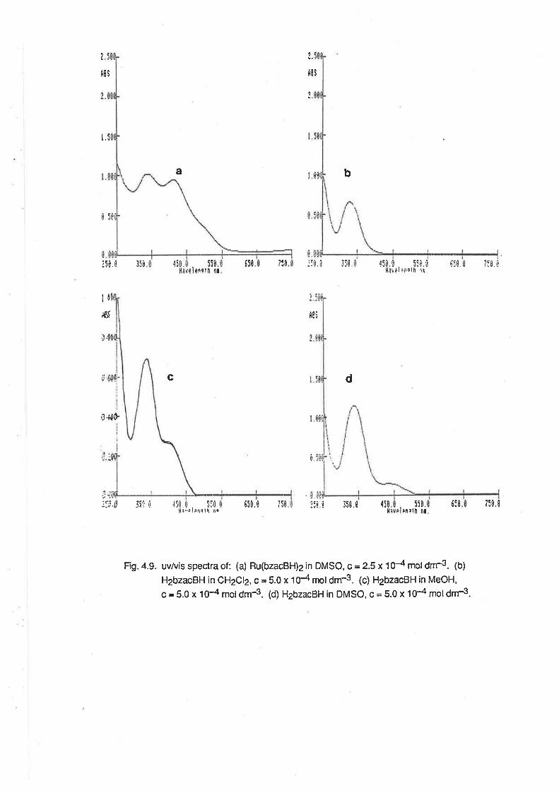
I
fi8S
I
f,89
ttl..J
t. 5t
¡5r. ri l5u {5ü.ll¡
a r,[0 b
65ú 750 i:ü. a ttl 45ü.0 55ri. Il{¡!pl¡ir¡lh.1t
{t0. rl 75ü , û
d
650 750,0lnrl|¡ ¡¡
.J.J0.
lrns t lt nrve
It-r(f i8!
0
c
flÈ
T
'::j .t Sirr ú ,l5rl . r! . 5:ù.11:¡,ú I Éf! I r, rç
650.0 15È r:i Í 35ü. 0 {50ll
550
Fig. 4.9. uv/vis specîra of : (a) Ru(bzacBH)2 in DMSO, c = 2.5 x 10⁻⁴ mol dnrs. (b)
H2bzacBH in CH2C|2, c = 5.0 x 10⁴ moldm-3. (c) H2bzacBH in MeOH,
c = 5.0 x 1O-4 mol dm-3. (d) H2bzacBH in DMSO, c = 5.0 x 1É moldm4
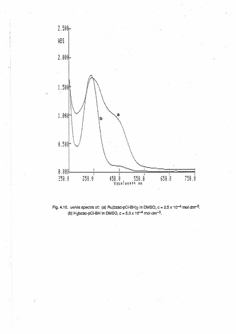
t
i
iI
I
I
l
I
I
I
l
,1 ET,..J
f,BË
Eü
IELt
a
Fig. 4.10. uvlvis spectra of: (a) Ru(bzac-pCl-BH)2 in DMSO, c - 2.5 x 10⁴ mol dnr3.(b) H2bzac-pOl-BH in DMSO, c - 5.0 x tÉ moldm4.
I ü[
lr l.l, r rlü
I]. EÈ
!5rJ. I rîü t¡
I
5itl, It lt t't tr
'tÍ[, ü
l,l,1tr¡ I ¡ti¡I7Ífiilriü
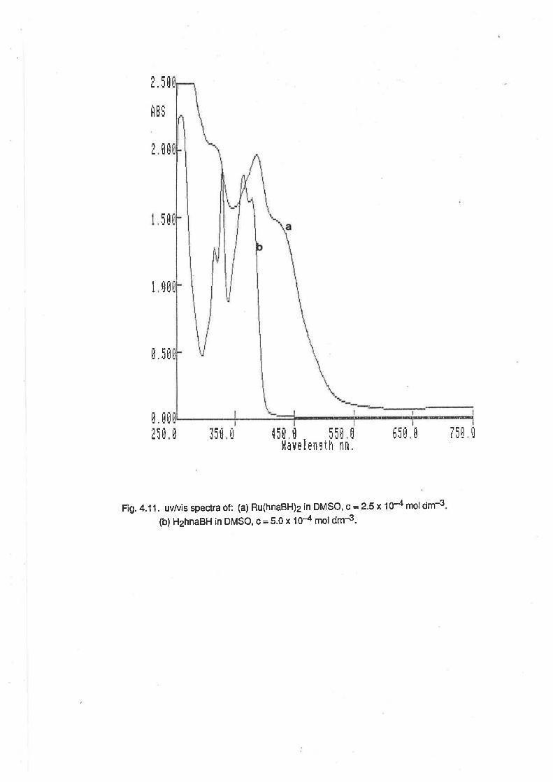
¿.5Ð
üü
5ü
rlüI
ü, iü
f,E$
¿
ff tÈIi[, È¡5[Efln.ttll5uü ,15ü.Ð . Ífü,ü
l,l¡v¡linrrh nm.
Fig.4.11. uv/vis spectra of: (a) Ru(hnaBH)2 in DMSO, c=2-S x 104 moldm€
(b) H2hnaBH in DMSO, c = 5.0 x 10⁴ mol dm4.
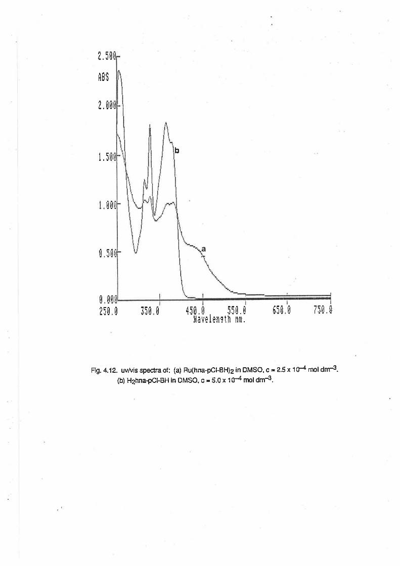
b
¿. i0
flBf
l.üü
t.5
ü35üI
llO
¡[ü
ü üü
¿5ü ,l5t,il¡
tifl. üüË5ü5i[. ü
I¿nlth nlrt,"/È
Frg.4.12. uv/vis spectra of: (a) Ru(hna-pCl-BH)2 in DMSO, c=2.5x 1d nrol drrs-(b) H2hna-pCþBH in DMSO, c = 5.0 x td moldm{.
91
ruthenium. The additive properties of intra-ligand absorptions were observed in the Ru(ll)
polypyridyl complexes, as described in Chapter 3. ln the spectrum of the complex with hna-pOl-BH,
the intra-ligand bands have less than half the intensity of the same bands in the lree ligand. There
is no apparent explanation for this.
33sQO4t2(18
2)
410sh(27).?)
342(33.0)450sh(18)
312sh(41)38s(3e.2)44osh(26)723(1.6)
3t4Qo.6)326(21.6)368(20.0)380(20.4)43Osh(11)
32s(6.46)033s(12.0)c433sh(2.7)c
336(17.0)450sh(.95)
3t2(r2.6), 32s(18.6)362(18.t), 377(16.3)
313(12.3)326(t7.8)3tr(18.1)
bzacBE
bzac-pCl-BH
hnaBH
hna-pCl-BH
RuL2LHzoL
Table 4.4. wlvis spectra of RuLr comolexes and lieands LHr.(a) ?i in?rmle x 10-3 cín mof dm-3). (b) i:r CH1CI;solution. (c) in DMSO solution.
4.8 Gonclusion.
It is clear that ruthenium(lV) complexes with the dinegative tridentate ligands derived from
Schiff's bases can be obtained lor ligands which have sufficiently strong ¡u-electron donating ability
Of the ligands used in this study, only bzacBH and bzac-pOl-BH are sutficiently strong rc-donors to
stabilize the Ru(lV) state. This is apparent from the electrochemistry of the RuL2 complexes. The
only two complexes which were identified as being Ru(lV) were Ru(bzacBH)2 and Ru(bzac-pOl-
BH)2, which were the complexes with the lowest E172 potential for the pu¿f+¡pu3+ couple. The
complexes with the ligands hapBH, hnaBH, hna-pOl-BH, bzacSalH and hnaOAP were all Ru(lll)
complexes, and the E172 potentials for pu4+¡puS+ couple in these complexes were all greater than
those forthe Ru(lV) complexes.
There is some spectral evidence to support the formulation of the Ru(lll) complexes as
[Ru(LXHL)], but the mass spectra may be subject to some interference from the positive ion
generated in the spectrometer. The microanalytical results are all consistent with the formulation,
[RuL2].H2O. The proposed formulation is, at best, tentative.
92
REFERENCES.
(1) A.A. Diamanlis, M. R. Snow, J. A. Va¡zo, J. Chem. Soc., Chem. Comm.,
264(1e76)
(2) Md. A. Salam, Ph. D. Thesis, The Unversity of Adelaide, 1986.
(3) A.A. Diamanfis, M. Manikas, Md. A. Salzun, M. R. Snow, E. R. T. Tieki¡k, hory.
Chem., zubmined for publication.
(4) H. Ohta, BulI. Chem. Soc. Japan,33, 1337(1960)
(5) D. P. Kessissoglou, W. M. Butler, V. L. Pecoraro, J. Chem. Soc., Chem. Comm.,
r2s3(re86)
(6) F. A. Cotton, G. Wilki¡son, "Advanced lnorganic Chemistry, 4th Ed. ", J. Wiley &Sons, New York, 1980, p 716.
(7) A.A. Diamartis, J. M. Frederiksen, Md. A. Salam, M. R. Snow, E. R. T. Tiekink,
Aust. J. Chem., 39, 1081(1986)
(8) A.A. Diamantts, private communication.
(9) A.A. Diama¡rtis, R. T. Jones, R. Keough, M. Marúkas, unpublished rezults
(10) G. A. Heath, K. A. Moock, D. W. A. Shaqp, L. J. Yellowlees, J Chem. Soc.,
Chem. Comm., 1503(1985)
(11) G. A. Heath, K. R. Seddon. J. B. A. F. Smeulders, Unpublishedrezults
(12) A. Endo, K. Shimizu, G. P. Sato, Chem. Leæ.,581(1985)
(13) Y. Takeucli, A. Endo, K. Shimizu, G. P, Sato, J Electroanal. Chem., 1&5,
18s(1e8s)
(14) M. I. Bruce, M. J. Liddell, Appl.Otganomet. Chem., inpress.
(15) K. Nakamoto, "Infrared and Raman Spectra of lnorganic and Coordination
Compounds"(3rd Ed.), J, Wiley & Sor:s, New York, 1978, p 115.
(16) W. G. Porterfield, "Inorganic Chemistry. A Unified Approach", Addison-rffesley,
Reading, Mass. (USA), 1984,p457.
(I7) E. A. Seddon, K. R. Seddon, "The Chemistry of Rutåenium", Else\rier, Amsterdam,
1984, Chapter 7.
93
(18) W. R. McWhin¡ie, J. D. Miller, Adv. Inorg. Chem. Radiochem., 12,135(1969)
CHAPTER 5.
DINITROGEN AND NITROSYL COMPLEXES.
5.1. lntroduction.
5.1.1. Dinitrogen Complexes. lnterest in the chemistry of dinitrogen complexes arose because of
their potential to be used as models for the active site of the nitrogen-fixing enzyme, nitrogenase.
The first dinitrogen complex to be isolated and identified as such was [Ru(NH3)5N zl2r.lllFollowing this otiginal report, there was considerable activity in the field, much of which is recorded
in several review articles on the subject. [2 - 4] Though the ammineruthenium and osmium
dinitrogen complexes represenl only a small fraction of the total number of dinitrogen complexes
synthesized, they are the only examples of dinitrogen complexes containing o-donor ligands, and
the only examples which have been studied in aqueous solution. [5]
The dinitrogen molecule, N2, is isoelectronic with the tc-acceptor ligands, carbon monoxide
(CO) and cyanide (CN-). The bonding schemes which have been used to describe the
coordination of CO to transition metals have also been used to describe the bonding of the N2
ligand. These schemes are dealt with in the review article by Sellman. [4] ln a large proportion of
cases, the N2 ligand is coordinated to the metal centre in an "end on" fashion, in either a terminal or
bridging mode. The existence of the dinitrogen ligand in the bridging mode is an example of the
nucleophilicity of the coordinated ligand, which is reported to be comparable to, or better than, that
of free dinitrogen. [6] ln the "end on" configuration the ligand molecule possesses a 3on orbital
capable of o-donation of electrons to the metal centre, and a lrcn anti-bonding orbital which is
unoccupied and capable of participating in æ-back-bonding from the metal t2n orbitals. The major
difference between the bonding of CO and N2 to transition metals lies in the differences which exist
between the orbital energy levels of the two ligands. Both the HOMO and LUMO orbitals of N2 are
at a much lower energy than those of CO (-15.6 and -7 eV respectively for N2, ând -14 eV and -6
eV respectively for CO). Most metal complexes possess orbítals (r.e. en and t2n orbitals) whose
energies correspond better to those of the o and æ* orbitals of CO than those of the N2 molecule.
ln general, carbon monoxide is both a better o-donor and æ-acceptor than N2, hence the greater
stability of coordinated CO compared.with N2. Additionally, lhe nature of the 3on orbital of the N2
molecule is such that most of the electron density is between the two nitrogen atoms, rather than at
one atom, as is the case with CO. Thus, the major contribution to the metal N2 bond is the n-
donation to the 1æn orbital of the ligand, whereas the metd-CO bond is a mixture of o- and n-
bonding.
95
The uv/vis spectra of the ruthenium ammine dinitrogen complexes contain absorptions which
are characteristic of the bonding mode of the N2 ligand. These absorptions occur at about 225 nm
lor terminally bound N2 and about 265 nm for bridging Nz. [2] These absorptions are related to
transitions between essentially metal-centred orbitals olT2nsymmetry and essentially ligand-
centred orbitals.
The operation ol the back-bonding to the æ* orbital of the ligand is manifest in changes to the
ir absorption frequency of the N-N stretching mode. ln N2, this vibration is only Raman-aclive,
absorbing at 2331 cm-1.[7] ln dinitrogen complexes, the (u)N-N absorption is also infrared active,
and the frequency of the absorption has been shown to be dependent on the anion present in the
sah. For the complexes, [Ru(NH3)sNe]Xz, (X = Cl, Br, l, BF4, PF6) the frequencies for (u)N-N
decrease with the decrease in the anion radius, as polarization of the metal centre by the counter
ion causes electron release into the dinitrogen ligand. [8]
The tetraammineaqua dinitrogen complex, cis IRu(NH3)4(OH2)N2]2+ has been reported [9],
and so has the bis(dinitrogen) complex, crb [Ru(NH3)¿(Nz)z]2*. [10] The bis diaminoethane
analogues are also known, including [Ru(en)2(N2)zl2* lt1] and IRu(en)2Ns(Nz)]*, the only
substítuted amine terminaldinitrogen complex of ruthenium known. [12] Wth the availability of the
triamminediaqua moiety it was considered possible to obtain complexes of the type,
[Ru(NH3)3L2N2], where L2 represents two monodentate ligands or a bidentate ligand. lt was
considered that it may be possible to observe the effect of the adjacent coordination environment
on the strength of the metal-N2 bond, and on the N-N bond itself .
5.1.2. Nitrosyl Complexes. The properties of ruthenium nitrosyl complexes are of interest to this
work because the nitrosyl complexes are precursors to the synthesis of analogous dinitrogen
complexes. The formatíon of a terminal dinitrogen ligand by nucleophilic attack on a coordinated
ligand offered a solution to the problem of reaction of N2 with more than one ruthenium ion, leading
to the formation of a bridged dinitrogen complex .
Ruthenium is believed to form more complexes with the nitric oxide ligand than any other
element. [13] The chemistry of these ruthenium nitrosylcomplexes has been reviewed by many
authors. [14 - 161 The interaction between the NO molecule and a transition metalcentre differs
from that involving most other ligands in that the NO is formally a three-electron donor. Feltham and
Enemark have descibed a scheme in which the ruthenium-nitrosyl group may be considered as a
{RuNO}n group, where n is equal to the number of electrons in the metal d orbitals, plus the
number of electrons in the æ. orbitals of the NO ligand. [16]
96
It is convenient to cons¡der that the bonding interaction arises from the transfer of a single
electron from NO to the metalcentre. The resulting NO+ is isoelectronic with N2 and CO,and
accepts electron density from the reduced metal ion into its n* orbital. This scheme provides a
useful basis for comparison between the nitrosyl complexes and those of other æ-acid ligands.
This conventional explanation is reinforced by the substantial strengthening ol the frans
ligand-ruthenium bond which is observed in a variety of complexes, as illustrated by the shorter
length of this bond compared with cis ligand-ruthenium bonds. ['14 and references therein] The
explanation is that the NO+ ligand is a very poor o-donor, but a very good fi-acceptor. lt does not
compete with the frans ligand for the empty orbital of the ruthenium centre, and further, it assists in
the o-donation from the frans ligand by delocalizing the electron density into its æ' orbital.
The extent of donation of electron density from the metal d orbitals to the ligand æ* orbital can
be seen in the variation of the infrared absorption lrequency for the NO stretching mode. Free nitric
oxide has an infrared absorption for this mode at 1840cm-1 and the nitrosyl cation, which is the
formal state of the ligand, shows (o)NO between 2150 cm-1 and 2400 cm-1. [1fl ln ruthenium
nitrosyl complexes, a range of frequencies is observed. Those above 1620 cm-1 are associated
with linear M-NO groups, and those less than 1610 cm-1 are associated with bent M-NO groups.
[18] Furthermore, it has been suggested that linear {MNO}6 nitrosyls with (u)NO greater than about
1850 cm-1 are sufficiently similar to the NO+ formulation to render the ligand susceptible to
nucleophilic attack on the nitrogen atom. [19] Bottomley has reviewed this aspect of the chemistry
of nitrosyl complexes [20], and has also carded out investigations of direct relevance to the current
work. When hydrazine or hydroxylamine were reacted with [Ru(NH3)5NO]3+, either
IRu(NH3)5N 212+, o, a mixture of [Ru(NH3)sNzO]2+ and [Ru(NHg)sNg]+, were produced, depending
on the reaction conditions used. [21, 22]
5.2. Preparation and Gharacterization of Nitrosyl Complexes.
5.2.1. [Ru(NH3)3NO(Cl)2lCl. Nitrosyl complexes of the triammine series could be prepared by the
direct reaction of nitric oxide gas. Aqueous suspensions of Ru(NH3)3C13 reacted with nitric oxide
gas to yield a nitrosylcomplex, [Ru(NH3)3NO(Cl)2]+, which precipitated from the reaction mixture as
its chloride salt. The addition of ethanol to the reaction mixture increased the yield of product.
Depending on the reaction condilions employed, two products were obtaíned. Reaction at high
temperatures (up to 90oC) gave two products. The first was a red crystalline solid and the second
was a pale pink powder. At lower temperatures (<40oC) formation of the crystalline solid was
favoured, but with a lower overall yield.
97
Both products had ir absorptions characteristic of the nitrosyl ligand in the region 1800 cm-l to
2000 cm-1, but it was in this feature that the major difference between the two forms of the complex
was apparent. ln a nujol mull, the spectrum of the crystalline product had three ir absorptions in the
nitrosyl region, at 1920 cm-1,1910 cm-1 and 1882 cnr1, whereas the spectrum of the pink solid
had a single broad band at 1850 cm-1. ln aqueous solution, however, the spectrum of the
crystalline form contained only a single, sharp peak at 1912 cm-1. Consideration of the molecular
symmetry of the complex requires that there would be only one peak due to the (D)NO mode.
However, different site symmetries in the crystalstructure could produce the split peaks, as also
observed in the complexes, cis [Ru(NH3)aNOC|]C|2, crs [Ru(NHs)aNOBr]Br2 and cis
[Ru(NHjaNO(OH2)2](ClOds. [23] On the basis of this comparíson, it would seem that the two
products obtained from the nitrosylation reaction are the same complex in two different solid state
environments. The energy of the ir absorptions are very similar to those of previously measured
nitrosyl complexes, as can be seen from the data listed in Table 5.1.
The red form of the compound was considerably more soluble in water than the pink.
However, both were sufficiently soluble to allow determination of the uv/vis spectrum, and both
gave the same spectrum. ln water, the uv/vis spectrum changed considerably with time.
Absorptions initially present a|207 nm (e =12000) and 360 nm (e = 1OO)gradually gave way to
absorptions at 196 nrn (e = 12000) and 350 nm (e =150) respectively. Additionally, there was a
weak, broad band at 500 nm (e = 30), which was unchanged over the same time span. The energy
and intensity of these absorption bands are comparable to those in the frans tetraamine nitrosyl
complexes [24], the main features of which are listed in Table 5.2. ln a previous report it was
proposed that the spectrum of [Ru(NH3)sNOCl2]Clconsisted of bands aI242 nm (e = 2950), 343
nm (e =80) and 490 nm (e=120). [25] The same report contains some speculation that, in dilute
aqueous solution, the form of the complex is [Ru(NH3)3NOC¡(OHz)12*. However, some ol the
evidence used to support this proposal is selfaontradictory, so the validity of any comparison is
questionable.
There is some uncertainty surrounding the nature of the transitions giving rise to these
absorptions. ln considedng the spectra of the frans tetraammineruthenium nitrosyls, Schreiner and
co-wolkers assigned the weak low energy bands to dn(Ru) -Ð lr*(NO)transitions with a spin-
forbidden d+d component also included. The medium intensity, medium energy bands were
assigned to mainly d-+d transitions, and lhe high energy bands to ligand (Cl-) to metal charge
transfers. [24] When Pell and Armor studied the spectra of the cis telraammines, they agreed with
the assignments of Schreiner, but expressed reservations about the assignment of the high
energy bands as due to LMCT transitions. [23] These high energy absorptions were not present in
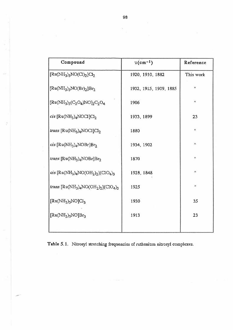
98
Table 5.1. Nitroryl stretching frequencies of ruthenium nitroryl complexes
This work
23
il
35
23
1920,1910, l8g2
t932, t915, 1909, 1885
1906
1933, 1899
1880
t934,1902
1870
t928,1848
1925
1930
1913
[Ru(NH3)3No(Cl)2]Cl2
[Ru (ltIH3 )3NO ( B r)2] B 12
[Ru (NH 3 ) 3
( Cb O4)No ]zczo +
cis [RuþtrH3)4NOCl]Clz
rrans [Ru $lH3 )4]locll Clz
crs [Ru (lrlH3 )aNO Br] B 12
trans [Ru (NH3 )al.IO B r] B 12
cis [Ru (lttrH3 )4NO (O H2)z] ( Cl O4)3
&zns [Ru (lrlH3 )¿NO ( OHtz] (Clo4)3
[Ru(NH3)5NO]Cl3
[Ru(NH3)5NO]B13
R eferenceu(cm-1¡Compound

99
Table 5.2. uv/vis spectra of ruthenium nitroryl complexes.
This work
23
24
23
25
l.2l x l}a1.01 x 102
3.0 x 101
1.23 x 10⁴
1.5 x 10²
3.0 x 101
7.5 x 10³
1.7 x 10⁷
3.6 x 101
t.26 x 103
1.9 x 101
4.2x 10³
2.64 x l}z1.7 x 101
2.0 x 10³
8.7 x 101
1.6 x 101
5.6 x 101
1.5 x 101
207
360
500
196
350
500
225
330
462
345
480
245
330
441
230(sh)
325
435
305
455
[Ru(NH3)3No(Cl)2]+
[Ru(NH3)3No(oHrz]z+
[Ru(NH3)3(Cbo4)No]+
cis [Ru QrlH3 )4NO(C[)]z+
rrans [Ru (NH3 )4l.lo(Cl ) ]2+
cis [Ru (l.IHs )4NO (O Hr]z+
[RuQ.lH3)5NO]r+
R.eferences(l mol-l cm-l)î,(nm¡Compound
100
the spectra above 200 nm, but it was thought that the LMCT might be expected at shorter
wavelengths and perhaps hidden by ligand-localized transitions. This conclusion was based on a
comparison of the LMCT bands in cr.s and frans [Co(en)2(Cl)Br]+where the band in the crs complex
is at higher energy than in the frans. [26] However, Nikol'skii and co-workers considered the lowest
energy bands to be d+d, with the occupied metal orbital having some Cl character when this ligand
was present. The medium intensity bands were assigned to d¡(Ru) + zc.(NO) transitions. [27j
This conclusion was reached after it was found that irradíation of aqueous solutions of
[RuCl5(NO)]2- in the medium energy absorption region resulted in replacement of NO by H2O,
implying lhat transitions involving NO lie there.The time dependence of the spectrum of
IRu(NHjsNO(Cl)2]Clwas indicative of hydrolysis involving replacement of the chloro ligands and, in
2 moldm-3 sodium chloride solution, the change in the spectrum was considerably slower. The
hydrolysis of the complex in dilute aqueous solution was confirmed by the results of conductance
measurements. A fresh solution of [Ru(NH3)gNO(Cl)2]Cl in water had a molar conductance at
infinite dilution, À0r = 170 Q-1 cm-1 mol-1 dm-3, indicative of a 1:1 electrolyte. The same
solution, 24 hours later, had Â0, = 371 O-1 cm-1 mol-1 dm-3, indicative of a 3:1 electrolyte.
5.2.2. [Ru(NH3)3No(Br)2JBr and [Ru(NH3)3No(c2od]2c2o4. Reacrion of [Ru(NHdsNo(ct)2]ct
with 4 mol dm-3 HBr solution resulted in the formation of [Ru(NH3)3NO(Br)2]Br. Similarty, reaction
with 3 molar equivalents of oxalic acid in the presence of lithium acetate yielded
IRu(NH3)3NO(C2Od]2C2O4. These compounds were characterized by elementalanalysis, uv/vis
spectroscopy and by comparison of their ir spectra with that of [Ru(NHjsNo(cl)2]c12.
The intense absorptions due to charge transfer bands from bromide to the sdvent obscured
any features at low wavelengths in the uv/vis spectrum of [Ru(NHs)3NO(Br)2]Br. At higher
wavelengths, the spectrum was identicalto that of [Ru(NH3)3NO(OH2)2]3+, indicative of hydrolysis
of the bromo ligands. The uv/vis spectrum of [Ru(NHs)sNO(C2Od]2+ contained absorptions at
225 nm (s =7500), 330 nm (e = 170) and 462 ñm (e = 36), all characteñstic of ammine nitrosyl
complexes.
The ir spectrum of IRu(NH3)3NO(Br)2]Br contained split peaks due to (u)NO, similar to the
chloro cnmpound, with the peaks ocurring at 1932 cm-1, 1915 cm-1, 1g0g cm-l and 1BB5 cm-1.
The spectrum of [Ru(NH3)3NO(C2O¿)lz}z}+contained a single peak due to (ù)NO at 1906 cm-l.
Because of the splitting of the (r)NO peaks in the solid state, it is not possible to compare the
vibration frequencies under different coordination environments and draw any certain conclusions.
101
5.2.3 other Triammineruthenium Nitrosyl complexes. ln víew of the lability of
towards hydrolysis, it was considered that substitution reactions involving the replacement
ligands would be relatively facile. However, only the two substítuted triammine nitrosyl complexes
mentioned above could be isolated and characterized. When substitution reactions were
attempted with thiocyanate ion, benzonitrile and bis-1,2(diphenylphosphino)ethane the unreacted
starting complex and the ligand were the only compounds obtained.
IRu(NHg)3NO(Cl)2]C¡ had limited solubility ín refluxing ethanol, where reaction with acacH, in
the presence of lithium acetate, resulted in the formation of a dark brown solution from which no
solid could be isolated. [Ru(NH3)3NO(C|)2]C!could not be isolated from the reaction mixture by
treatment with concentrated HCI solution. lt is possible that nucleophilic attack occurred at the NO
ligand , resulting in the formation of the complex, [Ru(NH3)s(c¡)2{(o)N-c(c(o)cH3)2}], in a manner
similar to that reported for the reaction of [RuOl(bpy)zNO]2* with acac-. [28]
When bpy reacted with [Ru(NHs)3NO(Cl)2]Cl in reluxing ethanol, frans [Ru(NH3)4NOCl]Ct2 was
isolated in approximately 4}l"yield. This was apparent from the analytical results, and a comparison
with the previously reported ir and uv/vis spectra. (Tables 5.1. and 5.2., respectively). [23,24] The
yellow solid which resulted from the reaction of [Ru(NH3)3NO(Cl)2]Cl2 with ethylenediamine had an
absorption at 1850 cm-1 ¡n its ir spectrum, but was shown by elemental analysis to contain less than
2/" ca¡bon. This was possibly an impure form of a tetraammine complex. lt is apparent from these
results that extraneous reactions were occurring. lt is possible that the N-donor ligands were initially
bound to the ruthenium, but then underwent decomposition reactions which lefl the ligand N atom
bound to the ruthenium centre. An alternative explanation is that, under the conditions employed
(refluxing ethanolsolution), the nitrosylcomplex underwent a seríes of dismutation reactions,
leading to lhe formation of the tetraammine complexes.
It is possible that the lack of success in forming more examples of substituted triammine
nitrosyl complexes is due to the problem of finding solvents in which both [Ru(NHd3NO(Cl)2]Cl2
and the organic ligands were soluble. The limited solubility of the chloride salt in organic solvents
meant that rather severe conditions were required to dissolve sufficient of the complex. Those
same conditions, however, apparently facilitated undesirable side reactions.
5.3. lsolation of Triammineruthenium Dinitrogen Complexes.
The isolation of terminal dinitrogen complexes of triammineruthenium was a major aim of the
project. Previous work in this field gave an indication of the reactions most likely to lead to the
required complexes.
102
Banham obtained a bridged dinitrogen complex as the product of the reaction between
dinitrogen gas and a triammineaquaruthenium(ll) complex, formed by heterogeneous reduction of
Ru(NH3)gCls in water. [29] This is to be expected, given that nitrogen gas has a poor solubility in
water and that low concentrations of N2 favour the formation of bridged dinitrogen complexes. The
formation of the bridged complex is further favoured by the ability of coordinated dinitrogen to act
as a nucleophile, and the presence of two aqua ligands on the Ru(ll) ion with which the bound N2
ligand may react. Clearly, attempts to synthesize terminal dinitrogen complexes by the reaction of
[Ru(NH3)3(OHz)sl2* with N2 gas would result in the formation of large amounts of a bridged
dinitrogen complex. Hence, a more reliable method for the synthesis of terminal dintrogen
complexes was required.
The conversion of coordinated nitrosylto term¡naldintrogen was first reported by Bottomley
and crawford Í21,221. Hydroxide ion reacted with [Ru(NHs)sNo]3* to yield a mixture of
IRu(NH$5N 272+ and crs [RuOH(NH3)4NO]2+. Similarly, the reaction between hydrazine and
[Ru(NHj5NO]3+ y¡elded [Ru(NH3)5N zl2* ina room temperature reaciion, and a mixture of
[Ru(NH$5N 2Ol2+ and [Ru(NHg5N3]+ in a reacrion ar -2goO. [20]
ln a similar way, Diamantis and Sparrow were able to show that coordinated N2O could be
conveded to coordinated N2 in the presence of a reducing agent. [30] The reducing agent could
be zinc metal, hydrogen over platinum, or even [Ru(NH3)5H 2Ol2+.
Both these synthetic methods offered scope for obtaining terminal dinitrogen complexes
without the complication of forming bridged complexes as by-products, because the dinitrogen
precursor was already in the metal coordination sphere prior to the reaction.
IRu(NHs)sNO(Cl)2]Cl reacted with hydrazine hydrate in either aqueous solution or methanol¡c
suspension to give a solution containing the complex IRu(NHs)3N z(OHz)zl2r. Depending on the
procedure used a variely of products could be isolated. (i) Addition of an excess of elhanolto the
methanolic reaction mixture resulted in the precipitation of a yellow complex, [Ru(NH3)3N2(C|)2], (ii)
addition of excess NaX (X = Br, l) to aqueous reaction mixtures yielded the complexes,
[Ru(NHs)3Nz(X)zl and (iii) addition of excess NH4A (A = PFs, BF4)to aqueous reaction mixtures
resulted in the isolation of the salts, [Ru(NHd3N2(OH2)2]42.
The presence of the terminal dinitrogen ligand was confírmed by comparison of the ir and
uv/vis spectra of the products with those of other terminal dinitrogen complexes. However, it was
not possible to isolate any of the terminal dinitrogen complexes as the sole reaction product.
Microanalysis of the PF¡ salt indicated that the complex contained more nitrogen than expected,
and the results were consistent with the presence of one hydrazine molecule per ruthenium ion.
103
Coordinated hydrazine has been reported previously in ruthenium pentaammine complexes. [31]
The uv/vis spectra were indicative of the presence of a small amount of the bridged dinitrogen
complex, and electrochemical experiments showed evidence of some oxidation of the terminal
dinitrogen complex.
An intense MLCT band was observeQ in the uv/vis spectrum a1222 nm. The position of this
band was the same, regardless of the anion or ligand present, indicating rapid hydrolysis of the
neutral complexes, similar to that which occurred with the nitrosyl complexes. The highest apparent
value obtained for e at this wavelength was 12000 dm3 moFl cm-1 for [Ru(NH3)3N2(OH2)2]2+ as its
PF6- salt. The spectrum, shown in Fig. 5.1, also had a small peak at 265 nm, indicative of the
presence of small amount of the bridged complex. From a comparison with the spectra of other
ammineruthenium dinitrogen complexes (Table 5.3), the molar absorptivity at the lower of these two
wavelengths would be expected to be in the range 14000 to 18000 dm3 mol-1 cm-1 . The spectra
of the bromo and iodo complexes were obtained after the solution of the complex had been
passed through an anion exchange column in the chloride form. This procedure was necessary
because lhe intense bands due to charge transfer lrom halide to solvent obscured the metal-N2
charge transfer bands. ln addition to the peaks attributable to the dinitrogen complexes, there was
also a shoulder at approximately 350 nm. This shoulder was probably due to absorption by a Ru(lll)
impurity which was identifed at a later stage. The presence of appreciable amounts of
IRu(NH3)3(OHz)zlzNza* implied that even though the terminal dinitrogen ligand was formed from a
precursor ligand, there was still the opportun¡ty for the terminal dinitrogen complex to react with
other complexes in the reaction mixture to form the bridged complex.
The CV of [Ru(NH3)3N2(OH2)2]2+ as its PF5- salt was determined in 0.1 mol dm-3
methanesulphonic acid. This voltammogram, shown in Fig. 5.2, contained three main features.
(i) An irreversible cathodic wave with Ep = +0.84 V, (ii) a reversible wave with Ê1n= {.15 V, which
was present on the initial cathodic scan from 0.0 V, and (iii) an increase in the peak current of the
reversible wave on scanning subsequent to the scan past the oxidation at the irreversible wave.
That the initial wave at -0.15 V increased after the dinitrogen complex had been oxidized implies
that the reversible wave was due to the product of the decomposition reaction which occurred after
the oxidation of the dinitrogen complex. lt is expected that upon oxidation of the dinitrogen
complex, the dinitrogen ligand would be lost because there is insufficient elecÍtron density on the
Ru(lll) centre to provide sufficent back-bonding to keep the dinitrogen bound. The use of scan
rates of up to 2 V serl díd not reveal any sign of a cathodic peak associated with the anodic peak at
+0.84 V.
f,85
60I
¿.00
t5
Fig. S.1. uv/vis spectrum of [Ru(NH3)'(OH¿ZNZ]2+ in 0.1 mol dm4 perchloric acid.
E2z2* 12 ooo.
tü
tÐ
4ùü
Düü
Hü.
I
ü4tü410. üil0, ü 3i[. ü
l'l¡velPttrth ttftt.
ü,ûü
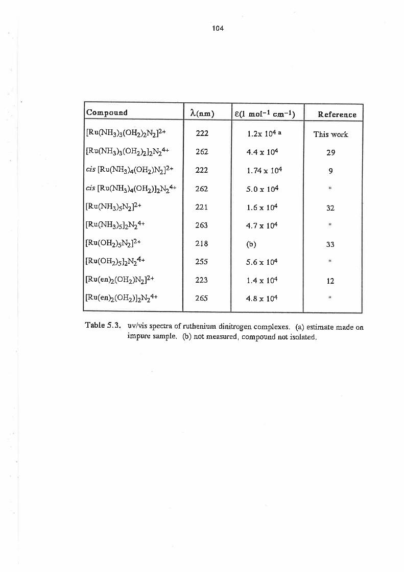
104
Table 5.3. uv/vis spectra of rutfienium dinitrogen complexes. (a) estimate made onimpure sample. (b) not measured, compound not isolated.
This work
29
9
32
il
33
I
t2
t.2x 104 a
4.4 x l}a
1.74 x l}a
5.0 x 10⁴
1.6 x 10⁴
4.7 x t}a
(b)
5.6 x 10⁴
t.4 x IOa
4.8x t}a
222
262
222
262
22r
263
2t8
25s
223
265
[Ru$lH3)3(oHrzNz]2+
[Ru(NH3)3(OHtz]2N24+
crs [Ru Q.IH3 )4(OHrNz]z+
cis [Ru (NH3 )4(oHr]zNz4+
[Ru(NH3)5N2]2+
[RuQ.IH3)5]zNza*
[Ru(OH)5N212+
[Ru(OH)5]zNz4*
[Ru(en)2(OHz)Nz]2*
[Ru(en)2(OHÐ]zNza*
References(l mol-l "m-t)
l"(nm)Compound
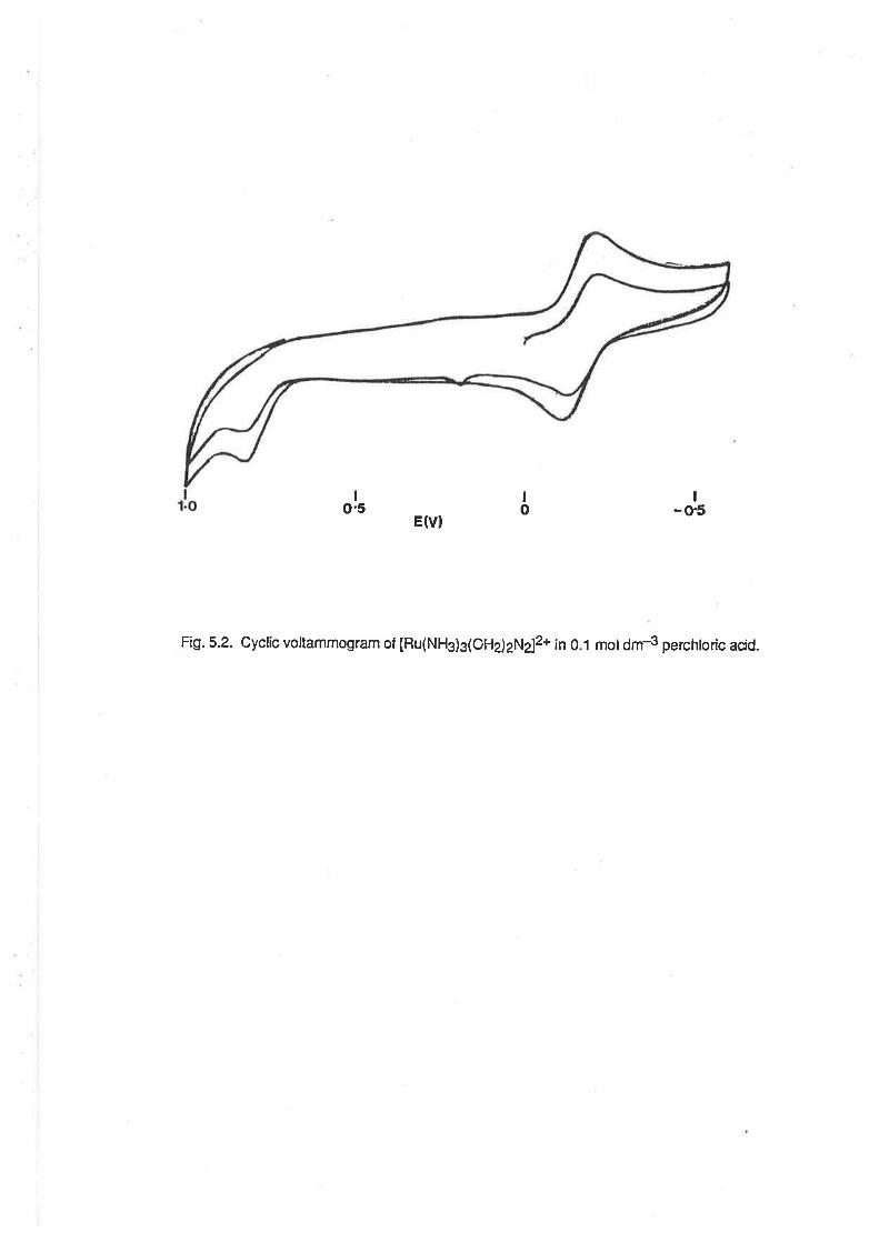
EÛ)
I-o'5
Fig.5.2. Cyclic voltammogram of [Ru(NH3)3(OHd2N¿2+ ¡n 0.1 motdm4 perchtoric acid.
Io.5
Io
105
There would appear to be many components in the reaction mixture capable of producing a
Ru(lll) species which was the same as the decomposition product of the terminal triammine
dinitrogen complex. Bottomley and Crawford have shown that hydrazine is capable of oxidizing a
Ru(ll) complex.[22] Fufihermore, [Ru(NH3)5NzO]2*, which has been identified as an intermediate
in the reaction belween [Ru(NH3)5NO]3* and hydrazine [20], has been shown to disproportionate
to [Ru(NHdsNz]2* and [Ru(NHg)sOH]2*. [30] Thus the presence of the decomposition producr of
the terminal dinitrogen complex in the isolated salts ís not surprísing. However, lhe formulation of
this decomposition product is stillopen to speculation.
The decomposition product is not IRu(NH3)3(oH2)s¡3+, because the E1¡2 value for its
reduction (-0.15 V) is 50 mV more negative than that of the triaqua complex. lf the dinitrogen
complex also contains coordinated hydrazine, as implied by the microanalysis results, then it might
be expected that the decomposition product after oxidation would have an E172 value close to
-0.14 V, which is the E172 value for the redox couple crs [Ru(NHs)¿(OHz)z]3*t2+. ¡ Ru(lll) ammine
hydrazine complex appears most likely to be the decomposition product.
The anodic peak potentialin the CV is very simitarto that of [Ru(NH3)5N2]2+. The
electrochemistry of this complex and of [Ru(NHs)aN z(oïz)I2* were studied by page and co-
workers. [34] The CV ol the terminalpentaammine dinitrogen complex had an anodic peak at +0.81
V, but there was no corresponding cathodic peak at scan rates up to 100 V sec-1, indicating that
the loss of the dinitrogen ligand from the oxidized lorm occurred very rapidly. ln the case of the
bridged pentaammine complex it was possible to observe an anodic and a cathodic peak with a
peak separation of 60 mV, and Eln= +0.465 V. From the voltammetric studies it was estimated
that the rate constant for the decomposition of [Ru(NH3)5N2Ru(NHs)s]s* to IRu(NHs)5N2]2+ and
nullllruUa¡rX was 0.1 ser1. When these oxidation potentials are compared with the E1¡2values
for the [Ru(NH3)3(OH2)t73+t2+ and IRu(NHg)s(OHe)]3*/2+ couples, it can be seen that the
dintrogen ligand greatly stabilizes the Ru(ll) centre towards oxidation, although the bridged
complexes are more readily oxidized than than are the terminalcomplexes. The electrochemistry of
[Os(NH3)5N2]2* , was also studÍed by Page , and it was found that the CV wave was reversible, and
had an oxidation potential less positive than that of the ruthenium analogue. The rate constant
measured for the decomposition of [Os(NHs)sNz]3* ro OrtlllNHs)5X and N2 wâs 0.02 sec-1
Different techniques were used in further attempts to obtain the terminaldinitrogen complex in
a pure form. The method of Diamantis and Sparrow [30]was used, whereby N2O gas was bubbled
through a solution of [Ru(NH3)g(OHz)g]3* in the presence of a reducing agent (amalgamated zinc).
It was hoped that in this reaction there would not be the opportunity lor the dinitrogen complex to
oxidize. However, while the oxidative decomposition of the terminaldinitrogen complex might not
106
have been occurring in this case, there were still appreciable amounts of the bridged complex
formed, so this approach was not pursued further.
Similarly, attempts were made to isolate the terminalcomplex from a high pressure reaclion in a
solution conta¡ning a precipitating agent (e.9. NH4PF5) lt was intended that the high pressure
conditions would increase the concentration of nitrogen gas in the reaction solution, thus favouring
formation of the terminal complex. The presence of the excess of precipitating anion was expected
to result in the precipitation of the dinitrogen complex upon formation, and so prevent further
reaction to give the bridged dinitrogen complex. However, to precipitate the complex immediately
upon formation requires a high concentration of the ruthenium precursor in solution, a condition
which favours the formation of the bridged species. Again, appreciable amounts of the bridged
complex were formed in this reaction.
ln the ir spectra, the N2 stretching frequency was dependent on the anion present ín the
dinitrogen complex or its salt. The values for (u)N2 are listed in Table 5.4, and it can be seen that
the variation in stretching frequency parallels that observed in terminal pentaammine
dinitrogenruthenium salts. [8]
Table 5.4. Frequencies of N-N stretching mode in dinitrogen complexes, in cm-1.(a) The anion may be pnesent as a couÍtef, ion to a cæionic aqua complexor as a coordinated ltsí d. (b) Reference 8.
This effect involves interaction between the anion and the ammine ligands, whereby the
release of electron density into the dinitrogen ligand from the metalcentre is increased when the
metal centre is polarized to a greater extent by an anion of smaller radius. [B]
5,4. Gonclusion.
It was possible to synthesize terminal dintrogen complexes of triammineruthenium(ll), but it
was found that other reactions occur in the reaction solution which lead to the formation of
impurities in the form of the bridged dinitrogen complex and the product of oxidative
decomposition of the terminal dinitrogen complex. The synthesis of a range of substituted
triamminenitrosylruthenium complexes was hindered by the insolubility of the parent complex,
RuQrlH3)3X2N2a
lRu(NHq)sNrlXrb
2tt2
2092
2tt8
2t09
2r22
2120
2t45
2t47
2t64
2167
x- cl Br I BF+ PFe
107
[Ru(NH3)3NO(CI)2]Cl, in the organic solvents in which the various ligands were soluble, and by the
react¡on of some ligands with this complex to yield tetraamminenitrosylruthenium complexes.
108
REFERENCES
(1) A. D. Allen, C. V. Senoff, Chem. Commun., 621(1965)
(2) J. Chatt, J. R. Dilworth, R. L. Richards, Chem. Rev., 78,589(1978)
(3) A. D. Allen, F. Bottomley, Acc. Chem. Res., 1, 360(1968)
(4) D. Sellman, Angew. Chem., Intl. Ed., 13, 639(1974)
(5) A. A. Diama¡tis, J. V. Dubrawski, Ioory. Chem.,20, 1142(1981)
(6) I. J. Itzkovich, J. A. Page, Can. J. Chem.,46,2743(1968)
(7) F. A. Conon, G. Wilkinson, "Advanced Inoqganic Chemistry", 3rd Ed., J. \ililey &Sons, New York, 1972, p7l0
(8) J. Chatt, G. J. Leigh, N. Thankarajan, J. Chem. Soc., A.,3169(1971)
(9) (a)C. M. Elson, I. J. Itzkovitch, J. A. Page, Can. J. Chem.,48, 1639(1970). (b)L. A.
P. Kane-Maguire, J. Inotg. Nuc/. Chem.,33,39&((19?t)
(10) J. E. Fergusson, J. L. Love, Chem. Commun., 399(1969)
(11) L. A. P. Kane-Maguire, P. S. Sheridan, F. Basolo, R. G. Pearsor, J. Amer Chem.
Soc. , 92,5865(1970)
(12) L. A. P. Kane-Maguire,P. S. Sheridan, F. Basolo, Inorg. Synth.,L2,23(1970)
(13) W. P. Griffith, "The Chemistty of the RarerPlatinun Metaß', Inferscience, London,
t967
(14) F. Bottomley, Coord. Chem. Rev., 26,7(1978)
(15) B. F. G. Johnson, J. A. Mcl.everry, Progr. Iootg. Chem.,7,277(1966)
(16) J. H. Enemark, R. D. Feltham, Coord. Chem. Rev., 13,339(L974)
(17) F. A. Cot¡on, G. Wilkinsofl, "Advanced lnorganic Chemistry", 3rd Ed., J. Wiley&Sons, New York, 1972, p356
(18) B. L. Haymore, J. A. Ibers, Inotg. Chem., 14, 3060(1975)
(19) R. W. Adams, J. Chatl. N. E. Hooper, G. J. Leigh , J. Chem. Soc., Dalton Trans.,
rLzs(1e77)
109
(20) F. Bottornley, Acc. Chem. Res., 11, 158(1978)
(21) F. Bottomley, J. R. Crawford, Chem. Commua.,2OO(1971)
(22) F. Bottomley, J. R. Crawford, J. ,üner. Chem. 1oc.,94,9092(1972)
(23) S. Pell, J. N. Armor, Inorg. Chem.,12,873(1973)
(24) A. F. Schreiner, S. \{'. Lim, P. J. Hauser, E. A. Hopcus, D. J. Hamm, J. D.Gunrer,
Inorg. Chem., 11, 880(1972)
(25) N. M. Sinitsin, A. A. Sverlov, N. B. Brikova, Koord. Khim., 3, 593(1977)
(26) K. Nakamoto, J. Fujita, M. Kobayaski, R. Tuschida, J. Chem. Phys. ,27 , 439(l9S?)
(27) A.B. Nikol'skä, A. M. Popov, I. V. Vasilevskü, SovietJ. Coord. Chem.,2,s08(1e76)
(28) M. Mukaida, M. Yanida,T. Nomura, 8u11. Chem. Soc. Japan,50,3053(1977)
(29) P. W. Baniram, Honours Thesis, The University of Adelaide, 1980
(30) A. A. Diamantis, G. J., Sparrow, Chem. Commun.,469(1969)
(31) J. Chatt,R. L. Richards,J. E. Fergusson,J. L. Love, Chem. Conmun.,
ts22(1e68)
(32) D. E. Harrison, H. Taube, Science, 159, 320(1968)
(33) C. Creutz, H. Taube, Inotg. Chem.,lO,26Ø(1971)
(34) C. M. Elson,J. Gulens, L J. Itzkovitch,J.A. Page, J. Chem. Soc.,D.,8?5(1970)
(35) M. B. Fairey, R. J. hwing , Spectochim. Acta,zz,359(1966)
Ghapter 6.
EXPER¡MENTAL.
6.1. Materials.
6.1.1. Preparative Materials. Ruthenium trichloride trihydrate was purchased lrom Johnson
Matthey Ltd.
Trilluoromethanesulphonic acid, ammonium hexaf luorophosphate, 2,2,6,6-lelramethylhepta-
3,5-dione (dipivaloylmethane), 2(aminomethyl)pyridine and 2-hydroxynaphthaldeyde (Tech) were
obtained from Aldrich Chemicals.
Methanesulphonic acid (techn. 70"/" aqueous solution), tetra(n-butyl)ammonium bromide
(purum) and 1-phenylbuta-l,3-dione (purum) were obtained from Fluka AG.
Silver carbonate, sodium perchlorate and penta-2,4,-dione were obtained from BDH chemicals
as Analar reagents.
The tridentate ligands were prepared by literature methods by Dr. Md. A. Salam. [1] The
tripodal ligands were supplied by Dr. F. R. Keene, having been prepared by literature methods. [2,
3l
All other materials used in preparative work were reagent grade chemicals.
Micranalyses were perlormed by the Canadian Microanalytical Service, Ltd.
6.1.2. Electrochemical Materials. Glassy carbon rod was obtained from Atomergic Chemicals. High
purity water was obtained by treating deionized water in a Millipore Mill¡-QrM water purification
system. The water was passed through activated charcoal, cation and anion exchange columns
and then through an Organexru cartridge.
Sodium methanesulphonate was prepared by the neutralization of methanesulphonic acid by
an equimolar amount of sodium carbonate (AR, Univar Chemicals). The sodium
methanesulphonate was tw¡ce recrystallízed from water/ethanolbefore use. Sodium perchlorate
was used as received.
The solvents dimethylsulphoxide, methanol and acetone were BDH Analar grade. Acetonitrile
was Waters Associates Liquid Chromatography grade.
High purity argon, nitrogen and hydrogen and medical grade nitrous oxide were obtained from
Commonwealth lndustrial Gases Ltd. Nitric oxide was obtained from Matheson Gas Producls. The
111
argon was passed through a purging column containing an acid solution of chromous ion, followed
by a water scrubber to dissolve traces of acid.
The materials used to prepare the buffer solutions were all analytical grade reagents. The
buffers used and their pH ranges were: chloroacetate, 2<pH<4; phthalate, 3<pH<5; acetate,
3.5<pH<5.5; MES, 5<pH<7; phosphate, 5<pH<7; TRIS, 7<pH<9; borate, 9<pH<11. The pH of the
electrochemicalsolutions was adjusted by the dropwise addition of 0.1 moldm{ sodium hydroxide
or methanesulphonic acid. The solution pH was measured using an Activon combined pH
electrode connected to an Orion Research 701A digital pH meter.
Alumina slurries for electrode polishing were obtained from Buehler. Diamond pastes for
electrode polishing were obtained from Hyprez, and were used with a Buehler extender oil.
Platinum electrodes were polished on a Microclothru polishing pad (Buehler), and glassy carbon
electrodes were polished on a TexmetîM polishing pad (Buehler).
6.2. lnstruments.
Electrochemical experimenls were performed with either of two combinations of instruments.
(1)A Princeton Applied Research 174 potentiostat and a Pine lnstruments RDE 3 potentiostat
connected to a Hewlet Packard 7015 B X-Y recorder. (2)A BioanalyticalSystems BAS 100
electrochemical analyser connected to an Amust P88-2 digital pr¡nter and a Houston lnstruments
HIPLOT DMP40 digital plotter. Sampled Current Polarography experiments were performed using
a locally made dropping mercury electrode attached to the BAS 100.
Electronic spectra were recorded on a Varian DMS 100 uv/visible spectrophotometer
connected to a Epson MX-80 digital printer. lnfrared spectra were recorded on a Perkin Elmer 683
lnfrared spectrophotometer. NMR spectra were recorded on a Bruker HX90E ¡nstrument, and mass
spectra were obtained on a VG ZAB 2HF spectromeler.
Magnetic susceptibilities were measured using a Johnson Matthey Magnetic Susceptibility
Balance. Conductivity measuremenls were made using a locally made resistance bridge connected
to a 1 cm cell having a cell constant of 0.77.
6.3. Electrochemical Procedures.
6.3.1. Electrochemical Cells. Electrochemical experiments were performed in a single
compartment three electrode cell. The cell compartment consisted of a small glass vessel, fitted
with a teflon cap. There were holes in the cap to accomodate the electrodes and the teflon tubing
which carried the purging gas.
112
The reference electrode was a Saturated Calomel Electrode (Titron, Pty. Ltd.) and the counter
electrode was a platinum wire. ln non-aqueous solutiors the working electrode was a commercially
available platinum disc electrode (Bioanalytical Systems). For aqueous solutions, the worfting
electrode was a glassy carbon disc. When a freshly polished electrode was used, this was a
commercially available model (BioanalyticalSystems), and, when an activated electrode was used,
the electrode was made locally by heat-shrinking a teflon shroud around a piece of glassy carbon
rod.
6.3.2. Electrode Preparation. All electrodes were initially ground on a high speed polishing wheel
with caóorundum paper (600 grit). Platinum electrodes, and glassy carbon electrodes which were
not to be activated were then polished with 1 pm alumina slurry, and then with 0.05 pm alumina
slurry. Between each polishing step the electrodes were rinsed with a stream of high purity water
and then washed in an ultrasonic bath for two minutes in high purity water. This rinsing and washing
procedure was repeated twice. For polishing after normal use, the electrodes were polished with
the 0.05 pm alumina slurry, and the same rinsing and washing procedure applied.
After lhe carborundum grinding step, glassy carbon electrodes which were to be activated
were polished with 1 pm alumina slurry and then with 1 pm diamond paste. After the diamond paste
polishing step, the electrodes were rinsed in a stream of high purity water and then cleaned in high
purity water in an ultrasonic bath for two minutes. Following this, the ultrasonic washing was
repeated, using ethanol, and then the ultrasonic washing with water was repeated.
Glassy carbon electrodes were activated using a modification of a method described in the
literature. [4] ln 0.1 mol dm-3 H2SO4, the potential of the glassy carbon electrode was held at
+1.80 V for 30 seconds, and the potentialwas then changed to -0.20 V for 15 seconds. This cycle
was performed six times, at which stage the state of activation was examined by determining a DPV
of [Ru(NH3)s(OHz)]3* at pH 4. Further activation cycles were applied, as required, untilthe peak
height of the Ru&/Ru3+ couple remained constant relative to that of the Rus lìu2+ couple. The
electrode was rinsed with water between the activating procedure and the recording of
voltammograms, and was cleaned in an ultrasonic bath between the experiments in ditferent
electrochemical media. Freshly polished glassy carbon electrodes had a mirror-like surface and the
surface of the activated electrodes was slightly dull, with a blue/bronze tinge.
6.3.3. Experimental Procedures. For studies in aqueous solution, the compound under
investigation was dissolved in an electrochemical medium (approximately 5 mg of solid in 4 - 5 cm3)
containing 0.1 moldm-S sodium methanesulphonate as a supporting electrolyte and 0.02 mol
dm-3 of the appropriate buffer. For experiments in acid solution, 0.1 moldm-3 methanesulphonic
acid fulfilled the requirements for bulfer and supporting electrolyte. When monitoring preparative
113
reactions, 50 - 100p1 of the reaction mixture was added by syringe to 4 - 5 cm3 of the
electrochemical medium. Points in the Pourbaix diagrams were fitted to straight lines by means of a
regression analysis programme on a hand-held calculator. The data points for the line were chosen
to give the largest value for the correlation coefficent, which was usually greater than 0.9.
For studies in non-aqueous solutions, about 5 mg of a solid sample was dissolved in 4 - 5 cm3
of the solution containing 0.1 mol dm4 sodium perchlorate as supporting electrolyte. The
saturated calomel electrode was placed directly into the electrochemical medium, and no allowance
was made for any possible liquid junction potential.
For all the voltammetric experiments, unless stated otherwise, there was a 2 second "quiet
time" between the application of the ¡n¡tial potential and the commencement of the scan. Unless
stated otherwise, cyclic voltammograms were recorded at 100 mV sec-1. The standard conditions
for recording sampled current polarography were: scan rate = 5 mV sec-1, dropping time = 0.5 sec
and sample width = 0.02 sec. The standard conditions for recording differential pulse
voltammograms were: scan rale = 5 mV sec-1, pulse amplitude = 5 mV, sample width = 0.02 sec,
pulse width = 0.06 sec and pulse period = 0.5 sec. The standard conditions for recording
Osteryoung square wave voltammograms were: square wave amplitude = 25 mV, square wave
frequency = 15 Hz, potential step = 4 mV, with 256 sample points per line cycle.
6.4. Synthesis.
6.4.1. Starting Materials. [Ru(NH3)sCl]Cl2 [5], [Ru(NH3)4C2O4|2S2O6 [6], and cls
IRu(NH3)a(H2O)2](TFMS)3 [7]were prepared according to literature methods. Ru(NH3)sOl3 was
prepared using the method of Banham. [8] [Ru(NH3)aC2Oal2S2O6 (0.a3 g, .60 mmol) was heated
in refluxing hydrochloric acid (5 mol dm-3, g5 cm3) for 4 hours. The reaction mixture was then
cooled in an ice bath for t hour. Red brown crystals were collected from a red mother liquor by
vacuum filtration through a sintered glass funnel. The solid was washed with three 5 cm3 portions
of cold HCI (5 mol dm-3), one 5 cm3 portion of ethanol and then dried on a vacuum line for three
hours. Yield = 0.2 g (0.77 mmol, 64%).
6.4.2. [Ru(NH3)3(OH2)3]GFMS)3. Sílver carbonate (0.957 9,3.47 mmol) was dissolved in HTFMS
(2 mol dm{, Z0 cm3) in a conical flask with heating on an oil bath at 45oC. Ru(NHs)3Cls (0.6 g, 2.3
mmol) was then added, and washed in with an extra 5 cm3 of the HTFMS solution. As the red
ruthenium complex was dissolved by stirring a white, cloudy precipitate formed. The reaction was
allowed to proceed for two hours, after which time, the reaction mixture was filered, hot, through a
filter paper to give a clear, greenish-coloured filtrate. The precipitated silver chloride was washed
with three 5 cm3 portions of water, and the washings combined with the filtrate. Using a rotary
114
evaporator, the volume of the combined filtrate and washings was reduced to less than 10 cm3.
After cooling overnight in the refrigerator, the solution was filtered through a sintered glass funnel
(porosity 3), yielding the product as pale pink, feathery crystals. The solid was washed with three
portions of dry diethyl ether, and then dried on the vacuum líne for four hours. Yield = 1.04 g (1.6
mmol,69%) Analysis: Calc. for RuC3H15N3F9S3O12 (Mol. wt. 653.4) C - 5.51%, H - 2.31%, N -
6.43%. Found: C -5.53%, H - 2.34o/",N - 6.66%.
6.4.2a. [Ru(NH3)3(OH2)3](pTS)3. Silver carbonate (0.425 9,1.54 mmol) was dissolved in pTSH (8
mol dm-3 5 cm3¡, and after efferuescence had ceased, Ru(NH3)3Clg (0.6 g, 1.03 mmol) was added,
and washed in with an extra 5 cm3 of the pTSH solution. The resulting mixture was heated at ref lux
for t hour. After cooling to room temperature, the mixture was then filtered to remove the silver
chloride which had precipitated. The volume of the filtrate was reduced by half using the rotary
evaporator, and the solution was then allowed to cool in an ice bath for one hour. Colourless
needles were then collected by filtration, and washed with two 4 cm3 portions of ethyl acetate and
air dried. Yield = 0.06 g (0.08 mmol, 8%)
6.4.2b. [Ru(NH3)3(oHz)s]2*. (1) tRu(NH3)3(oH2)31(TFMS)3 (0.02 g, .03 mmol)was dissolved in
0.1 moldm-3 Htfn¡S (25 cms), and the uv/vis spectrum recorded. A 5 cm3 aliquot was then
degassed with argon, before amalgamated zinc was added and the mixture stirred under argon for 1
hour. The uv/vis spectrum of the golden yellow solution was recorded, and further reaction with the
amalgamated zinc followed until there was no further change to the spectrum.
(2) [Ru(NH3)3(OH2)3](TFMS)3 (0.02 g, .03 mmol) was dissolved in water (25 cm3¡, and the uvivis
spectrum recorded. A 5 cm3 aliquot was then degassed wilh argon, and hydrogen gas was then
bubbled into the solution through a platinum black gauze for t hour. The uv/vis spectrum of the
golden yellow solution was recorded, and further reaction with hydrogen followed untilthere was
no further change to the spectrum.
6.4.2c. Mixed oxidation state species. (1) [Ru(NH3)3(OH2)3](TFMS)3 (0.02 g, .03 mmol) was
dissolved in pH 7.4 buffer containing 0.1 mol dm4 sodium methanesulphonate (25 cm3), and the
solution was degassed with argon. Amalgamated zinc was then added, and the mixture stirred
under argon. A blue colour developed after about 15 minutes, and became very intense over a
period of t hour. Several samples were withdrawn to allow the uv/vis spectrum to be recorded, and
voltammograms were recorded about t hour after the start of the reduction. After 4 hours, the blue
colour of the solution faded to give a golden yellow solution, and the uv/vis spectrum of this
solution was also recorded.
(2) [Ru(NH3)3(OH2)3](TFMS)3 (0.02 g, .03 mmol)was dissolved in pH 7.4butter containing 0.1 mol
dm-3 sodium methanesulphonate (25 cm3), and the solution was degassed with argon. A similar
115
solut¡on was made up in water, degassed with argon, and then hydrogen gas was bubbled into the
solution through a platinum black gauze for t hour. SCP was used at this stage to determine that
the ruthenium was in the +2 oxidation state. A 5 cm3 aliquot of the Ru(ll) solution was then added to
an equal volume of the high pH Ru(lll) solution, and the resulting solution stined under argon. A
blue colour became apparent atter 15 minutes, and the uv/vis spectrum was determined after 1
hour.
6.4.2d. Oxidation ot [Ru(NH3¡3(OH2)313+. [Ru(NH3)3(OH2)3](TFMS)3 (0.008 g, .'t.2x l0-s mol)
was dissolved in pH 5 buffer (acetate, s cm3¡ and an aliquot of hydrogen peroxide solution (0.1 mol
dm-3, 60 ¡rl) was added. The reaction mixture was then heated on a steam bath at 50oC for 15
minutes. The solution turned red almost inslantaneously, and then changed to a brown colour.
The CV of the solution was then determined, and found to contain no peaks attributable to the
[Ru(NH3)3(OHz)s]3* complex. The reaction was repeated at room temperature, but the same
reactions were observed, occurring over about an hour.
6.4.3. Subs-tituted Triammineruthen ium(lll) Complexes.
(a) tRu(NH3)g(acacXOH2lSz06.2H20. [Ru(NH3)s(OH2)3](TFMS)s (0.24 g, .37 mmol)was dissolved
in ethanol (5 cm3). The solution was stirred at 40oC for 2 hours in a round bottomed flask. 2,4-
pentadione (38 pl, .38mmol) was then added to the solution, followed by lithium acetate (0.038 g,
.37 mmol), which was washed in with ethanol (5 cm3). The reaction was monitored by DPV untilthe
peak at -0.09 V was no longer evident. The solvent was then removed using a rotary evaporator,
and the residue dissolved in the minimum volume of dithionic acid (1.1 mol Om-3). An equal volume
of ethanol was then added, and the solution stored at -1OoC for 80 hours. Purple crystals were
collected f rom the mother liquor by vacuum filtration through a sintered glass funnel. The crystals
were washed with two 3'cm3 portions of ethanol, and one 5 cm3 portion of dry diethyletherthen air
dried by suct¡on. Yield = 0.075 g (0.18 mmol, 49%). Analysis: Calc for RuC5H19N3S2O11 (Mol. wt =
433.4)C-12.90y",H-4.76"/",N-9.03%. Found: C-12.75y",H-4.63"/o,N-9'11%.
(b) tRu(NH3)g(dpmXOHzlSzOo. IRu(NH3)3(OH2)3](TFMS)s (0.25 g, .38 mmol)was dissolved in
methanol (5 cm3¡. The solution was stirred at 40oC for 2 hours in a round bottomed flask. 2,2,6,6-
tetramethylhepta-3,S-dione in solution in methanol Pa% solution, .205 g, .38 mmol) was added
followed by lithium acetate (0.039 g, .38 mmol), which was washed into the flask with methanol (5
cm3¡. The reaction was monitored by DPV until the peak at -0.09 V was no longer evident. The
reaction mixture was then liltered to remove a solid impurity, and the solvent removed from the
filtrate using a rotary evaporator. 5 cm3 of dithionic acid (1.1 mol dm-3) was added to the residue
and a purple solid precipitated, which was collected from the mother liquor by vacuum filtration
through a sintered glass funnel. The solid was washed with two 3 cm3 portions of ethanol, and one
116
5 cm3 port¡on of dry diethyl ether then air dried by suct¡on. Yield = 0.08 g (0.16 mmol, 40%).
Analysis: CalcforRuCllH3sN3S2Oe (Molwt= 513.6) C-25.73y", H -5.89%, N -8.18%. Found:
C' 25.29"/", H - 5.77"/", N - 8.06%.
(c) [Bu(Nfu)3(hna)(On2]S2O6.H2O. IRu(NH3)3(OH2)3](TFMS)3 (0.129, .18 mmol)was dissolved in
ethanol (5 cm3¡. The solution was stirred at 45oC for t hour in a round bottomed flask.
2-hydroxynaphthaldehyde (0.031 g pl, .18mmol) was then added to the solution, followed by
lithium acetate (0.0184 g, .18 mmol), which was washed in with ethanol(5 cms). A green colour
formed almost immediately after the addition of the solid materials. Stirring continued for 2 hours,
after which time DPV analysis showed the reaction to be complete. The solvent was then removed
using a rotary evaporator, and the residue dissolved in the minimum volume of dithionic acid (1.1
mol dm-3). This solution was stored at -1OoC for 60 hours. A dark green solid was collected from
the mother liquor by vacuum filtration through a sintered glass funnel. The solid was washed with
two 3 cm3 portions of ethanol, and one 5 cm3 portion of dry diethyl ether then air dried by suction.
Yield = 0.052 g (0.10 mmol, 5B%). Analysis: Calc for RuC11H1gN3S2O9 (Mol wt = 501.4)
C -26.33y",H-3.62"/", N -8.38%. Found: C-26.59Vo, H -3.667o, N -8.19%.
(d) tBu(NH3)3(oxine)(OH2IGFMS)2.H20. [Ru(NHs)3(OH2)3]OFMS)s (0.179, .27 mmol)was
dissolved in ethanol (5 cm3). The solution was stirred at 45oC for t hour in a round bottomed flask.
B-hydroxyquinoline (0.040 g pl, .28mmol) was then added to the solution, followed by lithium
acetate (0.0265 g, .26 mmol), which was washed in with ethanol (5 cmS¡. A green colour formed
within half an hour of the addition of the solid materials. Stirring was continued for 4 hours, after
which time DPV analysis showed the reaction 1o be complete. The solvent was then removed
using a rotary evaporator, and the residue dissolved in 5 cm3 HTFMS (3.5 moldm-3). The volume
of the solution was reduced to half using a rotary evaporator, and the resulting solution was stored
at -1OoC for 85 hours. Dark green platelets were collected f rom the mother liquor by vacuum
filtration through a sintered glass funnel. The solid was washed w¡th three 3 cm3 portions of dry
diethyletherthen airdried by suction. Yield = 0.021 g (0.03 mmol, 12%). Attempts at isolating
greater yields from the react¡on, by further concentration of the mother liquor resulted in the
formation of the starting material. Analysis: Calc for RuC11H19NaS2F6O9 (Mol wt = 628.5)
C - 21.02V",H - 3.05%, N - 8.92%, . Found: C - 20.60%, H - 2.86"/", N - 8.1 1%.
(e) [Bu(NH3)3(C2Od(H2O)]2S206.H20. IRu(NH3)3(OH2)3](TFMS)s (0.0e89, .1 5 mmol) was
dissolved in water (5 cm3¡ in a round-bottomed f lask fitted with an outflow bubbler. Oxalic acid
dihydrate (0.011 g, .075 mmol) and sodium oxalate (0.0095 g, .075 mmol) were then added, and
hydrogen gas was bubbled into the solution over a platinum black gauze for t hour without prior
degassing. The red solution was then heated on an oil bath at 50oC for t hour, at which time the
117
colour of the solution had changed to yellow and DPV analysis showed the reaction to be
complete. The solvent was then removed using a rotary evaporator, and the resídue treated with
dithionic acid (1 cm3, 0.25 moldm4). A pale yellow solid precípitated immediately. The mixture was
stored a 4oC for t hour, and the product was collected by vacuum filtration through a sintered glass
funnel. The solid was washed with two 3 cm3 portions of ethanol, and one 5 cm3 portion of dry
diethyl ether then air dried by suction. Yield = 0.038 g (0.1 1 mmol, 75%). Analysis: Calc for
Ru2C4H24NeSzOz(Molwt= 676.5) C-6.9P/", H -3.48%, N- 12.10%. Found: C-6.90%,
H-3.370/", N - 11.93%.
(fl tRu(NH3)3(sal)(H2O)12+. [Ru(NHs)g(OHz)g](TFMS)3 (0.179, .26 mmol)was dissolved in ethanol
(5 cms). The solution was stirred at 45oC for t hour in a round bottomed llask. Salicytaldehyde
(0.032 g, .26 mmol) was then added to the solution, followed by lithium acetate (0.027 g, .26
mmol), which was washed in with ethanol (5 cm3). A green cotourformed almost immediately after
the addition of the solid materials. Stining continued for 2 hours, after which time DPV analysis
showed the reaction to be complete. 100 pl samples of the solution were added to 5 cm3 aliquots
of the aqueous electrochemical media, and the CV determined at several pH values.
(g) tRu(NH3)3(bzac)(H2O¡2+. [Ru(NH3)3(OH2)3](TFMS)3 (0.51g, .79 mmol) was dissolved in
ethanol (5 cm3¡. The solution was stirred at 45oC for t hour in a round bottomed f lask.
1-phenylbuta-2,4-dione (0.127 g , .79 mmol) was then added to the solution, followed by lithium
acetate (0.080 g, .79 mmol), which was washed in with ethanol (5 cm3). A red colour formed almost
immediately after the addition of the solid materials, and slowly changed to purple. Stirring
continued for 2 hours, after which time DPV analysis showed the reaction to be complete. 100 pl
samples of the solution were added to 5 cm3 aliquots of the aqueous electrochemical media and,
after filtering off a white precipitate, the CV of the purple aqueous solutions were determined at
severalpH values.
6.4.4. Substituted Triammineruthenium(ll) Gomplexes.
(a) [Ru(NH3)3(phen)(H2O)l(PF6)2. Two procedures were used which gave the same products but
with varying yield: (1) [Ru(NH3)3(OH2)3]OFMS)3 (0.2359, .36 mmol) was dissolved in water (5
cm3¡, the solution was degassed by bubbling argon through it and hydrogen gas was then bubbled
into the solution through a platinum black gauze for t hour. The golden yellow solution of
IRu(NHi3(OHz)sl2* was then filtered through a Schlenk filter into a round-bottomed Schlenk flask.
1 ,10-phenanthroline hydrate (0.065 g, .36 mmol) was then added and the reaction mixture stirred
under argon for 4 hours, at which time DPV analysis showed the reaction to be complete. An
excess of NH4PF6 was added to the solution and the mixlure was then stirred under argon for 15
minutes, before being stored at -1OoC for 5 hours. The brick red solid was collected by vacuum
118
filtration through a sintered glass funnel. The product was washed with two 5 cm3 portions of ice-
cold water, and one 3 cm3 portion of dry diethyl etherthen air dried by suction. Yietd = 0.104 g
(0.16 mmol, 45%). Analysis: Calc for RuC12H19N sPzF12O (Molwt = 642.5) C - 22.51o/",H - 2.99Vo,
N - 10.94%. Found: C-22.08y", H -3.10%, N - 10.80%.
(2) [Ru(NH3)3(OH2)3](TFMS)3 (0.25g, .38 mmol)was d'ssotved in water (10 cm3), and the solution
was degassed by bubbling argon through it. 1,1O-phenanthroline hydrate (0.076 g, .38 mmol) was
added and hydrogen gas was then bubbled into the solution through a platinum black gauze. After
2 hours, at which time DPV analysis showed the reactíon to be complete, the deep red solution was
filtered through a Schlenk filter, and excess NH4PF6 was added to the solution. The mixture was
stined under argon for 15 minutes, and then stored at -1OoC for 5 hours. The brick red solid was
collected by vacuum filtration through a sintered glass funnel. The producl was washed with two 5
cm3 portions of ice-cold water, and one 3 cm3 portion of dry diethyl ether then air dried by suction.
Yield = 0.195 g (0.11 mmol,80%).
(b) [Ru(NH3)z(bpy)z](PF5)2. [Ru(NHg)s(OHz)s](TFMS)s (0.1919, .29 mmol) was dissolved in water
(10 cm3), the solution was degassed by bubbling argon through it and hydrogen gas was then
bubbled into the solution through a platinum black gauze for t hour. The golden yellow solution of
IRu(NH3)3(OHz)s]2* was then filtered through a Schlenk filter into a round-bottomed Schlenk f lask
containing a degassed solution 2,2'-bipyridine (0.063 g, .40 mmol) in ethanol (12 cm3) and the
reaction mixture was stirred under argon for 4 hours. An excess of NH4PF6 was then added to the
solution and the mixture was stirred under argon for 15 minutes, before being stored at -1OoC lor
16 hours. The brick red solid was collected by vacuum liltration through a sintered glass funnel.
The product was washed with two 3 cm3 portions of ice-cold ethanol, and one 5 cm3 portion of dry
diethyl ether, then air dried by suction. Yield = 0.043 g (0.058 mmol, 20%). Analysis: Calc lor
RuC2sH24NaPzFp(Molwt =741.6)C -32.39V.,H-3.26y", N - 11.33%. Found: C - 32.41"/o,
H-2.85y",N-11.09%.
(c) [Ru(NH3)3(py)3](ClOa)2. Following the method of Ford & Sutton [9], a 1 moldm-3
pyridine/pyridinium buffer was prepared by mixing pyridine (Z cm3) and MeSO3H (2.4 mol dm-3, 5
cm3¡ and making up to 25 cm3 with water. [Ru(NH3)3(OHd3]OFMS)3 (0.159g, .244 mmol) was
dissolved in this buffer (10 cm3), and the solution was degassed by bubbling argon through it.
Amalgamated zinc was lhen added, and the reaction mixture stirred under argon for 6.5 hours, at
which time DPV analysis showed the reaction to be complete. The bright gold-coloured solution
was then filtered through a Schlenk filter into a round-bottomed Schlenk flask. An excess of
NaClO4 was then added and a cloudy precipitate formed almost immediately. The mixture was
cooled on an ice bath for t hour, and the product was collected as a yellow powder by vacuum
filtration through a sintered glass funnel. The product was washed with two 5 cm3 portions of ice-
119
cold sodium perchlorate solut¡on, and one 5 cm3 portion of dry diethyl ether, then air dried by
suction. The crude product was recrystallized lrom hot methanol, yietding bright yellow cubic
crystals. Yield = 0.077 g (0.12 mmol, 48%). Analysis: Calc for RuC15H24N6OsCt2 (Molwt = 588.37)
C-30.62y",H-4.11y", N-14.29%, Cl-12.05%. Found: C-g0.OS%, H-4.5Vo,N-14,S%,Cl-
12.3%.
(d) [Ru(NH3)s(py)sl(Br)2. [Ru(NH3)3(py)s](ClO4IzQ.077 g, .12 mmol) was dissolved in butanone
(10 cm3), and then an excess of tetra(n-butyl)ammonium bromide was added to the solution. A pale
precipitate formed almost immediately, and the mixture was stirred for 0.5 hour. Atter cooling in ice
for t hour, the pale yellow solid was collecled by vacuum filtration through a sintered glass funnel.
The product was washed with two 5 cm3 portions of butanone, and one 5 cm3 portion of dry diethyl
ether, then air dried by suction. Yield = 0.063 g (0.11 mmol, 96%).
(e) [Ru(NH3)3(MeCN)3]CI2.2H2O. [Ru(NH3)s(OH2)3](TFMS)3 (0.2369, .36 mmol)was dissolved in
0.01 mol dm-3 HtfUS (10 cmS), and then acetonitrile (1.5 cms) was added. The solution was
degassed by bubbling argon through it, then amalgamated zinc was added and the mixture stirred
for 4.5 hours, at which time DPV analysis showed the reaction to be complete. The golden yellow
solution was then filtered through a Schlenk filter into a round-bottomed Schlenk flask and an
excess of NHaPF6 was added to the solution. The mixture was then stined under argon for 15
minutes, before being stored at -1OoC for 2 hours. The pale yellow solid was collected by vacuum
filtration through a sintered glass funnel. The product was washed with one 5 cm3 portion of ice-
cold water, and one 3 cm3 portion of dry diethyl ether. This solid was dissolved in butanone (10
cm3¡, and then a solution of tetra(n-butyl)ammonium chloride (0.5 g) in butanone (3 cm3) was
added. A pale precipitate formed almost immediately, and the mixture was stirred for 5 minutes.
After cooling at -1OoC for 2 hours, the pale yellow solid was collected by vacuum f iltration through a
sintered glass funnel and washed with two 5 cm3 portions of butanone, and one 5 cm3 portion of
dry diethyl ether, then air dried by suction. The crude product was recrystallized from
methanol/butanone, resulting in pale yellow, feathery crystals. Yield = 0.051 g (0.13 mmol, 37%).
Analysis:CalcforRuCsH22NsClzO2(Molwt=382.3)C-18.85%,H-5.96%,N-21.99%,Cl-
18.85%. Found: C - 18.80%, H-5.77"/o,N -21.72./",Cl-18.27%.
(f) [Ru(NH3)3(MeCN)3]812. [Ru(NH3)sffeON)3](PF6)2 was prepared as described in (e) above. This
solid was then dissolved in butanone (10 cm3), and an excess of tetra(n-butyl)ammonium bromide
was added to the solution. A pale precipitate formed almost immediately, and the mixture was
stirred for 5 minutes. After cooling at -1OoC for 2 hours, the pale yellow solid was collected by
vacuum filtration through a sintered glass funnel and washed with two 5 cm3 portions of butanone,
120
and one 5 cm3 portion of dry diethyl ether, then air dried by suction. Yield = 0.084 g (0.19 mmol,
42%).
(g) [Ru(NH3)3(ampy)(H2O)l(PF6)2. [Ru(NH3)3(OH2)3](TFMS)3(0.279,.41 mmol) was dissolved in
water (5 cm3¡, and the solution was degassed by bubbling argon through it. Hydrogen gas was
then bubbled'into the solution through a platinum black gauze for 0.75 hour before
2(aminomethylpyridine) was added (20% solution in ethanol ,2751tl,.41 mmol). The treatment with
hydrogen was continued for 0.5 hour, and then the solution was stirred at 40oC for 5 hours, at
which time DPV analysis showed the reaction to be complete. The solution was then filtered
through a Schlenk filter into a round-bottomed Schlenk llask and an excess of NHaPF6 was added
to the filtrate. The mixture was stirred under argon for 15 minutes, and then cooled in an ice bath for
t hour. The yellow solid which precipitated was collected by vacuum filtration through a sintered
glass funnel. The product was washed wilh two 3 cm3 portions of ice-cold ethanol, and one 5 cm3
portion of dry diethylether, then air dried by suct¡on. Yield = 0.106 g (0.18 mmol, 44%). The
product unden¡rent decomposition before the microanalysis could be performed. Freshly prepared
samples were used to determine the pH-dependent electrochemistry.
6.4.5. Triammi ne(tripod)ruthenium(ll) Complexes.
(a) [Ru(NH3)g{(py)gcoH}]8r2. [Ru(NHd3(OH2)3](TFMS)s (0.1729,.26 mmol)was dissolved in
water (7 cm3¡, and the solution was degassed by bubbling argon through it. Hydrogen gas was
then bubbled into the solution through a platinum black gauze for 1.5 hours. Tris(pyridyl)methanol
(0.068 g, .26 mmol) was then added and the treatment with hydrogen conlinued for 45 minutes.
The reaction mixture was lhen stirred under argon until DPV analysis showed the reaction to be
complete. The dark golden solution was filtered through a Schlenk filter, and excess NH4PF6 was
added to the filtrate. The mixture was stirred under argon for 15 minutes and then stored at -1OoC
lor 24 hours. The yellow solid was collected by vacuum filtration through a sintered glass funnel
and washed with two 5 cm3 portions of ice-cold elhanol, and one 3 cm3 portion ol dry diethyl ether
then air dried by suction. The crude product (0.108 g, 55%) was d¡ssolved in butanone (5 cm3) and
excess tetra(n-butyl)ammonium bromide was added to the solution. A flocculent precipitate formed
almost immediately, and the mixture was stirred for 5 minutes. After cooling at -1OoC for 16 hours,
the pale yellow solid was collecled by vacuum filtration through a síntered glass funnel and washed
with two 3 cm3 portions of butanone, and one 5 cm3 portion of dry diethyl elher, then air dried by
suction. Yield = 0.068 g (0.12 mmol, 46%). The crude bromide salt was recrystallized from
methanol/butanone. Analysis: Calc for RuC16H22N6OBr2 (Mol wt = 575.3) C - 33.41"/., H - 3.85%,
N - 14.61%. Found: C -32.57o/",H -3.71"/", N - 13.71%.
121
(b) [Ru(NH3)g{(py)sCH}]8r2. [Ru(NH3)3(OH2)3](TFMS)3 (0.189, .27 mmol) was dissolved in water (5
cms¡, and the solution was degassed by bubbling argon through it. Hydrogen gas was then
bubbled into the solution through a platinum black gauze for 45 minutes. Tris(pyridyl)methane
(0.067 g, .27 mmol) was then added and the treatment with hydrogen continued for t hour. The
reaction mixture was stirred under argon lor 2.5 hours¡ at which time DPV analysis showed the
reaction to be complete. The bright yellow solution was filtered lhrough a Schlenk fílter, and excess
NH4PF6 was added to the filtrate. The mixture was stirred under argon in an ice bath for 2 hours.
The yellow solid was collected by vacuum filtration through a sintered glass funnel and washed with
two 5 cm3 portions of ice-cold water, and one 3 cm3 portion of dry diethyl ether then air dried by
suction. The crude product was dissolved in butanone (5 cm3) and excess tetra(n-butyl)ammonium
bromide was added to the solution. A flocculent precipitate formed almost immediately, and the
mixture was slirred for 5 minutes. After cooling at -1OoC for 16 hours, the pale yellow solid was
collected by vacuum filtration through a sintered glass funnel and washed with two 3 cm3 portions
of butanone, and one 5 cm3 port¡on of dry diethyl ether, then air dried by suction. Yield = 0.052 g
(0.09 mmol, 33%). The crude bromide salt was recrystallized from methanoUbutanone. Analysis:
calcforRucl6H22N6Br2 (Molwt=559.3) C-34.36./",H -3.96%, N - 15.03%, Br-28.57"/". Found:
C - 34.52"/., H - 4.37V", N - 1 1.95%, Br - 28.04%.
(c) [Ru(NHg)g{(py)gN}](ClO4)2. Tris(pyridyl)amine (0.026 g, .105 mmol) was added to a 2-neck
reaclion vessel. [Ru(NH3)3(OH2)3](TFMS)3 (0.079, .107 mmol) was added and washed into the
flask with water (B tt3¡. The solution was degassed by bubbling argon through it and hydrogen
gas was then bubbled into the solution through a platinum black gauze for 3.5 hours, at which time
DPV analysis showed the reaction to be complete. The golden yellow solution was filtered through
a Schlenk filter, and excess NH4PF6 was added to the filtrate. The mixture was stirred under argon
for 8 hours. The yellow solid was collected by vacuum liltration through a sintered glass funnel and
washed with two 5 cm3 portions of ice-cold water, and one 3 cm3 portion of dry diethyl ether then air
dried by suction. Crude yield = 0.049 (0.063 mmol, 60%). A sample of the crude product (26 mg)
was dissolved in the minimum volume of hot water. This solution was filtered through a plug of
cotton wool and excess sodium perchlorate was added to the filtrate and dissolved with stirring.
The mixture was stored at -1OoC for 2 hours and a yellow powderwas collected by vacuum filtration
through a sintered glass funnel and washed with two 5 cm3 portions of ice-cold water, and one 3
cm3 portion of dry diethyl ether, then air dried by suction. Yield = 0.01 g (after accidental spillage).
Analysis:CalcforRuC15H21N6C|2OB(Molwt=599.4)C-30.06%,H-3.53%,N-16.36%,Cl-
11.83%. Found: C -30.47y", H - 3.58%, N - 15.96%, Cl- 11.65%.
6.4.6. Bis(tridentate)ruthenium Complexes.
(a) Ru(bzacBH)2.H20. N'-(1-methyl-3-oxo-3-phenylpropylidene)benzohydrazide (H2bzacBH,
122
0.385 g, 1.37 mmol) and lithium acetate (0.288 9,2.82 mmol) were added to RuCl3.3H2O (0.175 g,
.669 mmol), the mixture was dissolved in ethanol (30 cm3) and heated at reflux for 4 hours. The
solvent volume was lhen reduced to 10 cm3 on a rotary evaporator, and the concentrated solution
stored at -1OoC overnight. Black crystals were collected by vacuum filtration through a sintered
glass funnel, washed with two 10 cm3 portions of ice-cold ethanot, and three 5 cm3 portion of dry
diethyl ether, then air dried by suction. Yield = 0.162 g (0.24 mmol, 36%). Analysis: Calc for
RuC3aH3sNaO5 (Mol wt = 677) C - 60.Mo/",H - 4.47o/",N - 8.29%. Found: C - 60.88%, H - 4.39%,
N - 8.46%
(b) Ru(bzac-pCl-BH)2.H2O. N'-(1-methyl-3-oxo-3-phenylpropylidene)-4-chlorobenzohydrazide
(H2bzac-pOl-BH, 0.340 g, 1.08 mmol) and lithium acetate (0.222 9,2.18 mmol) were added to
RuCls.3H2O (0.340 g, .547 mmol), the mixture was dissolved in ethanol (11 cm3) and heated at
reflux lor 4 hours. The solvent volume was then reduced by half on a rotary evaporator, and the
concentrated solution stored at -1OoC overnight. Black crystals were collected by vacuum filtration
through a sintered glass funnel, washed with two 10 cm3 portions of ice-cold ethanol, and three 5
cmg portion of dry diethyl ether, then air dried by suc"tion. Yield = 0.126 g (0.17 mmol, g1%).
Analysis: Calc for RuCs4H2sNaO5Cl2 (Mol wt =745.9) C - 56.08%, H - 3J9/q N - 7.52/., Cl -
9.52%. Found: C - 55.16%, H-3.70V", N - 7.51%, Cl- 10.31%.
(cl) Ru(hnaBH)2.H2O. N'-(2-hydroxynaphthylmethylidene)benzohydrazide (H2hnaBH, 0.409 g,
1.41 mmol) and lithium acetate (0.294 g, 2.88 mmol) were added to RuCl3.3H2O (0.185 g, .70
mmol), the mixture was dissolved in ethanol (11 cm3) and heated at reflux for 4 hours. The solvent
volume was then reduced by half on a rotary evaporalor, and the concentrated solution stored at
-1OoC overnight. A brown powder precipitated which was collected by vacuum filtration through a
sintered glass funnel, washed with two 10 cm3 portions of ice-cold ethanol, and three 5 cm3 portion
of dry diethylether, then air dried by suction. Yield = 0.184 g (0.26 mmol, 37%). Analysis: Calc for
RuC36H26N4O5(Molwt=696) C-62.15y",H-3.77"/",N-8.05%. Found: C-61.26/",H-3.76yo,
N - 7.62%.
(c2) Ru(hnaBH)2.H20. N'-(2-hydroxynaphthylmethylidene)benzohydrazide (H2hnaBH, 0.80 g,
2.76 mmol) and triethylamine (0.77 cm3, 5.52 mmol) were added to RuCl3.3H2O (0.361 g, 1.38
mmol), the mixture was dissolved in ethanol (15 cm3) and heated at refiux lor 4 hours. The solvent
volume was then reduced by half on a rotary evaporator, and the concenlrated solution stored at
-1OoC overnight. A brown powder precipitated which was collected by vacuum f iltration through a
sintered glass f unnel, washed with two 10 cm3 portions of ice-cold elhanol, and three 5 cm3 porlion
of dry diethyl ether, then aír dried by suction. Yield = 0.29 (0.26 mmol, 37%). Analysis: Calc for
123
RuC42H4sN5Oa (based on Et3NH[Ru(hnaBH)2[ (Mol wt = 779.91 C - 64.68%, H - 5.177%, N -
8.98%. Found: C - 62.12/0,H - 3.98%, N -9.82%.
(d) Ru(hna-pCl-BH)2.H20. N'-(2-hydroxynaphthylmethylidene)-4-chlorobenzohydrazide (H2hna-
pCl-BH, 0.529 g, 1.63 mmol) and lithium acetate (0.333 g, 3.26 mmol) were added to RuCl3.3H2O
(0.204 g, .814 mmol), the mixture was dissolved in ethanol (24 cms) and heated at reflux for 4
hours. The solvent volume was then reduced to 10 cm3 on a rotary evaporator, and the
concentrated solution stored at -1OoC overnight. A brown powder precipitated which was
collected by vacuum f iltration through a sintered glass funnel, washed with two 10 cm3 portions of
ice-cold ethanol, and three 5 cm3 portion of dry diethyl ether, then air dried by suction. Yield =
0.153 g (0.2 mmol, 250/"1. Analysis: Calc for RuC36H24N aOsO.l2(Molwt = 764.9) C - 56.55%, H -
3.16/", N -7.33%, Cl-9.27%. Found: C -55.82%, H -3.31%, N -7.04%, Cl-9.26%.
(e) Ru(hapBH)2.H2O. N'-(1-(2-hydroxy)phenylethylidene)benzohydrazide (H2hapBH, 0.314 g,
1.23 mmol) and lithium acetate (0.255 g, 2.50 mmol) were added to RuCl3.3H2O (0.159 g, .62
mmol), the mixture was dissolved in ethanol (13 cm3) and heated at reflux for 4 hours. The solvent
volume was then reduced by half on a rotary evaporator, and the concentrated solution stored at
-1OoC overnight. A brown powder precipitated which was collected by vacuum filtration through a
sintered glass funnel, washed with two 10 cm3 portions of ice-cold ethanol, and three 5 cms portion
of dry diethyl ether, then air dried by suction. Yield = 0.160 g (0.25 mmol, 41%). Analysis: Calc for
RuC3sH25NaO5 (Mol rvt = 627.6) C - 57JíV",H - 4.20o/", N - 8.98%. Found: C - 58.03%, H -
4.17"/", N - 8.86%.
(f) Ru(bzacSalH)2.H20. ¡'-11-methyl-3-oxo-3-phenylpropylidene)-2-hydroxybenzohydrazide
(H2bzacSalH, 0.512 9,1.73 mmol) and lithium acetate (0.364 g, 3.57 mmol) were added to
RuCl3.3H2O (0.2929, .899 mmol), the mixture was dissolved in ethanol (13 cm3) and heated at
reflux for 4 hours. The solvent volume was then reduced by half on a rotary evaporator, and the
concentrated solution stored at -1OoC overnight. A brown powder precipitated which was
collected by vacuum filtration through a sintered glass funnel, washed with two 10 cm3 portions of
ice-cold ethanol, and three 5 cm3 portions of dry diethyl ether, then air dried by suc'tion. Yield =
0.044 g (after accidental spillage). Analysis: Calc for RuCs4H3sN4O7 (Molwt = 691.1) C - 57.70h,H
- 4.27o/",N-7.62%. Found: C - 56.06%, H - 4.18/o,N -7.62%.
(g) Ru(salBH)2.0.5EIOH. N'-(2-hydroxyphenylmethylidene)benzohydrazide (H2salBH, 0.242 g,
0.92 mmol) and lithium acetate (0.186 g, 1.82 mmol) were added to RuCls.3H2O (0.117 g, .453
mmol), the mixture was dissolved in ethanol (13 cm3) and heated at reflux for 4 hours. The reaction
mixture was filtered through a sintered glass funnel after standing overnight. The black crystals so
obtained were washed with two 10 cm3 portions of ethanol, and three 5 cm3 portion of dry diethyl
124
ether, then air dried by suction. Yield = 0.196 g (0.30 mmol, 66%). Analysis: calc for
RuC29H23NaOa.5 (Molwt= 650.1) C-57.78o/o,H-4.20/", N -8.98%. Found: C-57.63%, H-
3.95%, N -9.27%.
(h) Ru(bzacOAP)2. ¡'-1t -methyl-3-oxo-3-phenylpropylidene)-2-hydroxyphenylimine (H2bzacOAP,
0.269 g, 1.06 mmol) and lithium acetate (0.218 9,2.12 mmol) were added to RuCl3.3H2O (0.137 g,
.531 mmol), the mixture was dissolved in ethanol (10 cm3) and heated at reflux for 5 hours. Black
crystals precipitated which were collected by vacuum filtration through a sintered glass funnel,
washed with two 10 cm3 portions of ethanol, and three 5 cm3 portions of dry diethyl ether, then air
dried by suction. Yield = 0.04 g (0.06 mmol, 11%). Analysis: Calc for RuC32HzoNzO¿ (Molwt =
605) C -63.67"/",H-4.34y", N -4.64%. Found: C-53.38%, H-3.99%, N -3.86%. Fits
Ru2C32H26NZO¿. C -53.18%, H -3.90%, N -3.88%.
(i) Attempted preparation of Ru(acacBH)2. Following the procedure of Diamantis & Vanzo [11],
tris(pentanedionato)ruthenium(lll) (0.2 g, .5 mmol)was dissolved in methanol and benzoylhydrazine
(0.138 g 1.01 mmol) was added. The reaction mixture was heated at reflux, in air, for 3 hours. At
this stage the reaction mixture still had the red colour of Ru(acac)g, ând its uv/vis spectrum was
consistent with the presence of this compound, implying that reaction had not occurred.
6.4.7. Nitrosyl Complexes.
(a) [Ru(NH3)3(C!)2NO]C|. Ru(NHj3Cl3 (0.16 g, .61 mmol) was added lo water (9 cm3¡ in a round-
bottomed flask fitted with an outflow bubbler. The mixture was degassed by bubbling nitrogen gas
through it and nitric oxide was then bubbled through the mixture for 2 hours while it was stirred at
7IoC. After this period, any nitric oxide remaining in the solution was removed by passing nitrogen
through the solution for 30 minutes. The reaction mixture was filtered by gravity, yielding a bright
red filtrate. The volume of the filtrate was reduced to 1 cm3 by evaporation on a steam bath. As the
concentrated solution cooled, red crystals precipitated, and the precipitation was enhanced by the
addition of a few drops of ethanol. The product was collected by vacuum filtration through a
sintered glass funnel and washed wíth two 2 cm3 portions of ethanol, and one 2 cm3 portion of dry
diethyl ether, then air dried by suction. Yield = 0.123 g (0.43 mmol, 70t"1. Analysis: Calc for
RuHeNaCl3O (Molwt = 288.5) H - 3.14V", N - 19.42%, Cl - 36.87%. Found: H - 2.91y", N - 18.55%,
ct- 35.5%.
(b) [Ru(NH3)3(Br)2NO]Br. [Ru(NH3)3(C|)2NO]C| (0.05 g, .17 mmol) was heated on a steam bath in 5
mol dm-3 HBr (10 cm3) for 2 hours. The solution was then cooled in ice and filtered to yield a pale
orange filtrate. The volume of the filtrate was reduced using a rotary evaporator, and an excess of
ethanolwas added. An orange powder precipitated from the solution and was collected by vacuum
filtration through a sintered glass funnel and washed with two 2 cmS portions of ethanol, and one 2
125
cms ponion of dry diethyl ether, then air dried by suction. Yield = 0.027 g (0.06 mmol, 35%).
Analysis: Calcfor RuHgN4BrgO (Molt¡rl= 421.5) H - 2.15Vo, N - 13.28%, Br- 56.82%. Found: H -
1.17y", N - 7.13%, Br -37.0%. (Contaminated sample)
(c) [Ru(NHg)3(CzO¿)NOI2C2O4. Lithium acetate (0.0779,.7 mmol) and oxalic acid dihydrate (0.042
g, .3 mmol) were added to [Ru(NHs)s(Cl)2NO]Cl (0.06 g, .2 mmol) in water (20 cms), and the mixture
was heated at rellux for 30 minutes. Most of the solvent was then evaporated from the resultant
orange solution, and the concentrated solution was cooled on an ice bath. Ethanolwas added,
dropwise, until a faint cloudiness appeared, and orange crystals then precipitated lrom solution.
The product was collected by vacuum liltration through a sintered glass lunnel and washed with two
2 cm3 portions of ethanol, and one 2 cm3 portion of dry diethyl ether, then air dried by suction.
Yield = 0.031 g (0.05 mmol, 50%). Analysis: Calc for RuC5HgN¿Org (Mol wt = 628.4) C - 11.47"/o,H
-2.89o/", N - 17.83%. Found: C -11.37Vo, H - 3.09%, N - 16.66%.
(d) Attempted synthesis of [Ru(NH3þþpy)NO]C13. [Ru(NH3)3(Cl)2NOlCl (0.19 g, .66 mmol) was
dissolved in a75125 mixture of water and ethanol (20 cm3), 2.2'-bipyridine (0.1 'l g, .7 mmol) was
added and the mixture was heated at reflux for 30 minutes. Most of the solvent was then
evaporated from the resultant orange solulion, ethanol (1 cm3)was added, and the concentrated
solution was then cooled on an ice bath. Orange crystals precipitated from solution. The product
was collected by vacuum filtration through a sintered glass funnel and washed with two 2 cm3
portions of ethanol, and one 2 cm3 portion of dry diethyl ether, then air dried by suction. Yield =
0.0259. Analysis:CalcforRuClsHlTN6OCI3(Molwt=444.7)C-27.01y",H-3.85%,N-18.90%,
Cl - 23.92. Found: C - 0.57"/o, H - 4.08%, N - 22.45'/0, Cl - 34.14%. For [Ru(NH3)4(CDNO]C12, calc
lor RuH12N5OC|3: C - 0y", H - 4.00%, N - 22.92y", Cl - 34.80%.
6.4.8. Dinitrogen Complexes.
(a) [Ru(NH$3(OH2)2NO](PF6)2. Following the method of Bottomley [10], [Ru(NHs)3(Cl)2NO]Cl
(0.05 g, .18 mmol) was dissolved in water 1e0 cms) in a Schlenk tube, and the solution degassed by
bubbling nitrogen through it for 10 minutes. Against a flow of nitrogen, a50y" solution of hydrazine
hydrate (1 cm3, 10 mmol) was then added, causing effervescence to occur. After 30 minutes,
NH4PF6 (1 g) was added and stirred into the solution, and the mixture was then cooled on an ice
bath for 30 minutes. A pale precipitate formed, which was collected by vacuum liltration through a
sintered glass funnel and washed with two 2 cm3 portions of ethanol, and one 2 cm3 portion of dry
diethyl ether, then air dried by suction. Yield = 0.045 g (0.09 mmol, 50%). Analysis: Calc for
RuH13N4O2PzF'tz(Molwt=506.1) H-2.59o/o,N- 13.84%, P -12.24%. Found: H-3.16%, N-
18.46/",P - 12.1%. For [Ru(NH3)4(N2Hd(H2OXN2)](PF6)2, calc for RuH15N7OP2Fp: H - 2.90y",
N-18.82%,P-11.89%.
126
The same procedure was used to prepare samples containing the triammine dinitrogen
complex containing the anions Br, l- and BF¡, by using the appropriate sodium salt instead of
NH4PF6.
(b) [Ru(NH3)3(C|)2N2]. [Ru(NH3)3(Cl)2NO]Cl(0.081 g, .28 mmol) was suspended in methanol (20
cm3¡ in a Schlenk tube, and lhe solvent degassed by bubbling nitrogen through it for 10 minutes.
Against a flow of nitrogen, hydrazine hydrate (1 cnF, 10 mmol) was then added, causing
effervescence to occur. After 30 minutes, the red reaction mixture was filtered, and ethanol (10
cmS¡ was added to the filtrate. A pale precipitate formed on cooling at -1OoC for three hours, and
this was collected by vacuum filtration through a sintered glass funnel and washed with two 2 cm3
portions of ethanol, and one 2 cm3 portion of dry diethyl ether, then dried on a vacuum line.
Yield = O.047 g (0.19 mmol, 68%). Because the uv/vis spectrum was indicative of substantial
contamination with [Ru(NHg)3(Cl)2]N2, no analysis was attempted.
(c) Attempted preparations of terminaldinitrogen complexes frcm [Ru(NHg)S(OHZ)S]2*.
(1) [Ru(NH3)3(OH2)3](TFMS)3 (0.1729,.26 mmol) was dissolved in saturated NaBF4 solution (4
cm3¡ at OoC, and the solution was degassed by bubbling argon through it. Hydrogen gas was then
bubbled into the solution through a platinum black gauze for 1.5 hours. The golden yellow solution
was then filtered using a Schlenk filter and, while the filtrate was kept cool, nitrogen gas was
bubbled through it. After 2 hours of reaction with nitrogen, there was no sign of any precipitate,
though the solution was cloudy. After storing the mixture overnight at -1OoC, NaBFa was filtered
from the yellow solution. The uv/vis spectrum of the filtrate contained bands attributable to the
terminal and bridged dinitrogen complexes.
(2) [Ru(NH3)3(OH2)3](TFMS)s (0.179, .26 mmol)was dissolved in saturated NaBFa solution (7
cm3¡, and the solution was degassed by bubbling argon through it. Hydrogen gas was then
bubbled into the solution through a platinum black gauze for 1.5 hours. The golden yellow solution
was then filtered using a Schlenk filter and transferred, under nitrogen, to a high pressure reaction
vessel which had previously been flushed with nitrogen. The reaction vesselwas pressurized to 20
atmospheres pressure with nitrogen, and maintained at this pressure for two hours. At the end of
this period, the pressure was released and the vesselopened. Bubbles were observed in the
solution, indicative of the slow release of nitrogen. The uv/vis spectrum of the solution contained
bands attributable to the terminal and bridged dinitrogen complexes.
(3) [Ru(NH3)3(OH2)3](TFMS)3 (0.159, .23 mmol)was dissolved in saturated NaBFa solution (4
cm3¡, and the solution was degassed by bubbling argon through it. Amalgamated zinc was added
to the flask and the mixture was stirred while a stream of nitrous oxide was passed through ilfot 2
127
hours. There was no noticeable precipiate, and the uv/vis spectrum of the solution contained
bands attributable to the terminal and bridged dinitrogen complexes.
128
References.
(1) L. Sacconi , Z. Anorg. A11g. Chem.,275,249(1954)
(2) D. J. Szalda, F. R. Keene, Ioory. Chem.,25,2795(L986)
(3) J. P. Wibaut, A. P. de Jonge, H. G. P. Van der Voort, H. L. Otto, Rec. Tnv. Chim.
Pay-Bas., 70, lO54(195 1)
(4) G. E. Cabaniss, A. A. Dia-ma¡ris, W. R. Muqphy, L. W Iirúon, T. J. Meyer, J
Amer. Chem. Soc., 1O7,1845(985)
(5) F. M. Lever, A. R. Powell, J. Chem., Soc., A., t477(1969)
(6) S. Pell, J. N. Armor, Inotg. Chem., L2, 873(1973)
(7) D. M. D. Goswick, G. M. Brovm, H. Taube, Inorg. Chem.,22, 1975(1983)
(8) P. W. Banham, Honours Thesis, The Universiry of Adelaide, 1980.
(9) P. C. Ford, C. Sutton,Inorg. Chem.,8, 1544(1969)
(10) F. Bottomley, J. R. Cralvford, Chem. Commun.,200(1971)
(11) A.A. Diamanlis, M. R. Snow, J. A. Vanzo, J. Chem. Soc. , Chem. Comm.,
264(re76)