derived hydroxyapatite graft as bone substitution - Semantic ...
11
176 J Med Sci Volume 45, No. 4, Desember 2013: 176-186 * corresponding author: [email protected] Structural evaluation and animal implantation of porous eggshell waste- derived hydroxyapatite graft as bone substitution Yudha Mathan Sakti 1 *, Rahadyan Magetsari 1 1 Departement of Orthopaedi and Traumatologi Dr. Sardjito General Hospital, Faculty of Medicine Universitas Gadjah Mada, Yogyakarta, Indonesia ABSTRACT The development of hydroxyapatite graft with high economically value is needed for orthopedic practice in developing countries. Eggsell waste is well known as natural substance for calcium resource. It has been used as raw material in producing hydroxyapatite. This study was conducted to synthesize porous hydroxyapatite from eggshell waste and evaluate its activity as bone substitution. The porous hydroxyapatite graft was manufactured from eggshell and sugar as a raw material using hydrothermal process. The porous eggshell waste-derived hydroxyapatite (EW-HAP) graft was characterized using X ray difractometer (XRD) and analytical scanning electron microscope (SEM) and compared with commercial hydroxyapatite (HAP) JCPDS 09-432 graft (Bangros ® ) as standard. The porous EW-HAP graft obtained was then implanted on critically sized femoral defects surgically created in the right thigh of male Wistar rats ( Rattus norvegicus ) with Bangros ® as control. Radiological examination using XRD and histological examination using hematoxyline-and-eosin staining of the bone femour were performed at 28 days after implantation. The results showed that the XRD pattern for EW-HAP was likely similar with the HAP standard. However, the SEM examination showed that the pasticle size of EW-HAP graft (2.5-3 μm) was higher than those HAP standard graft (1.5-2 μm). Radiographs according to the International of Limb Salvage (ISOLS) radiological evaluation system between EW-HAP graft (6.1 ± 1.45) and HAP control graft (6.9 ± 2.10) was not significantly different (p>0.05). Moreover, histological examination according to Lane and Shandu scoring system between the both graft (4.0 ± 0.94 versus 4.4 ± 0.92) was also not significantly different (p>0.05). It can be concluded that the structure EW-HAP graft is similar with HAP graft standard. The both grafts have also equal outcome as bone substitution. ABSTRAK Pengembangan graft hidroksiapatit dengan nilai ekonomi tinggi diperlukan dalam bidang ortopedi di negara berkembang. Limbah cangkang terus dikenal luas sebagai bahan alam sumber kalsium. Limbah cangkang telur telah digunakan sebagai bahan untuk menghasilkan hidroksiapatit. Penelitian ini dilakukan untuk mensintesis graft hidroksiapatit berpori dari limbah cangkang telur dan mengkaji aktivitasnya sebagai pengganti tulang. Graft hidroksiapatit berpori dibuat dari bahan dasar cangkang telur (HAP-LCT) dan gula menggunakan proses hidrotermal. Graft yang dihasilkan dikarakterisasi difraktometer X-ray (DXR) dan mikroskop elektron skaning (MES) analitik dan dibandingkan dengan graft JCPDS 09-432 hidroksiapatit komersial (Bangros ® ) sebagai standar. Graft berpori HAP-LCT yang diperoleh kemudian diimplantasikan pada critically sized tulang femur yang dibuat melalui operasi pada paha kanan tikus Wistar jantan (Rattus norvegicus) dengan Bangros ® sebagai kontrol. Pemeriksaan radiologi menggunakan DXR dan histologi dengan pengecatan hematosilin-eosin tulang femur dilakukan pada hari ke 28 setelah implantasi. Hasil
-
Upload
khangminh22 -
Category
Documents
-
view
1 -
download
0
Transcript of derived hydroxyapatite graft as bone substitution - Semantic ...

176
J Med Sci, Volume 45, No. 4, Desember 2013: 176-186J Med SciVolume 45, No. 4, Desember 2013: 176-186
* corresponding author: [email protected]
Structural evaluation and animalimplantation of porous eggshell waste-derived hydroxyapatite graft as bonesubstitution
Yudha Mathan Sakti1*, Rahadyan Magetsari11 Departement of Orthopaedi and Traumatologi Dr. Sardjito General Hospital,Faculty of Medicine Universitas Gadjah Mada, Yogyakarta, Indonesia
ABSTRACTThe development of hydroxyapatite graft with high economically value is needed for orthopedicpractice in developing countries. Eggsell waste is well known as natural substance for calciumresource. It has been used as raw material in producing hydroxyapatite. This study was conductedto synthesize porous hydroxyapatite from eggshell waste and evaluate its activity as bonesubstitution. The porous hydroxyapatite graft was manufactured from eggshell and sugar as araw material using hydrothermal process. The porous eggshell waste-derived hydroxyapatite(EW-HAP) graft was characterized using X ray difractometer (XRD) and analytical scanningelectron microscope (SEM) and compared with commercial hydroxyapatite (HAP) JCPDS 09-432graft (Bangros®) as standard. The porous EW-HAP graft obtained was then implanted on criticallysized femoral defects surgically created in the right thigh of male Wistar rats (Rattus norvegicus)with Bangros® as control. Radiological examination using XRD and histological examinationusing hematoxyline-and-eosin staining of the bone femour were performed at 28 days afterimplantation. The results showed that the XRD pattern for EW-HAP was likely similar with theHAP standard. However, the SEM examination showed that the pasticle size of EW-HAP graft(2.5-3 µm) was higher than those HAP standard graft (1.5-2 µm). Radiographs according to theInternational of Limb Salvage (ISOLS) radiological evaluation system between EW-HAP graft(6.1 ± 1.45) and HAP control graft (6.9 ± 2.10) was not significantly different (p>0.05).Moreover, histological examination according to Lane and Shandu scoring system between theboth graft (4.0 ± 0.94 versus 4.4 ± 0.92) was also not significantly different (p>0.05). It canbe concluded that the structure EW-HAP graft is similar with HAP graft standard. The bothgrafts have also equal outcome as bone substitution.
ABSTRAKPengembangan graft hidroksiapatit dengan nilai ekonomi tinggi diperlukan dalam bidang ortopedidi negara berkembang. Limbah cangkang terus dikenal luas sebagai bahan alam sumber kalsium.Limbah cangkang telur telah digunakan sebagai bahan untuk menghasilkan hidroksiapatit.Penelitian ini dilakukan untuk mensintesis graft hidroksiapatit berpori dari limbah cangkang telurdan mengkaji aktivitasnya sebagai pengganti tulang. Graft hidroksiapatit berpori dibuat daribahan dasar cangkang telur (HAP-LCT) dan gula menggunakan proses hidrotermal. Graft yangdihasilkan dikarakterisasi difraktometer X-ray (DXR) dan mikroskop elektron skaning (MES) analitikdan dibandingkan dengan graft JCPDS 09-432 hidroksiapatit komersial (Bangros®) sebagai standar.Graft berpori HAP-LCT yang diperoleh kemudian diimplantasikan pada critically sized tulangfemur yang dibuat melalui operasi pada paha kanan tikus Wistar jantan (Rattus norvegicus)dengan Bangros® sebagai kontrol. Pemeriksaan radiologi menggunakan DXR dan histologi denganpengecatan hematosilin-eosin tulang femur dilakukan pada hari ke 28 setelah implantasi. Hasil

177
Sakti & Magetsari, Structural evaluation and animal implantation of porous eggshellwaste-derived hydroxyapatite graft as bone substitution
penelitian menunjukkan bahwa gambaran DXR graft HAP-LCT sama dengan graft HAP standar.Namun demikian, hasil pemeriksaan MES menunjukkan ukuran partikel HAP-LCT (2,5-3 µm) lebihtinggi dari pada graft HAP standar (1,5-2 µm). Gambaran radiografi menurut sistem evaluasiradiologi International of Limb Salvage (ISOLS) graft HAP-LCT (6,1 ± 1,45) dan and graft HAPkontrol (6,9 ± 2,10) berbeda bermakna (p>0.05). Lebih lanjut dari hasil pemeriksaan histologimenurut sistem penilaian Lane dan Shandu antara kedua graft (4,0 ± 0,94) tidak berbedabermakna (p>0,05). Dapat disimpulkan bahwa struktur graft HAP-LCT sama dengan graft HAPstandar. Kedua graft juga mempunyai luaran sama sebagi pengganti tulang.
Key words: eggshell - porous hydroxyapatite graft – bone substitution – X-ray
INTRODUCTION
In orthopedic and traumotology, bonedamage or bone defect is one of health problemsthat can cause mechanical instability, dead spacefor bacterial infection and hampered bonehealing process.1 Extensive bone damage cannot heal spontaneously, therefore a bone graftprocedure is needed.2 In USA, at least 2.2million bone grafting have been conductedannually with 300 million US $ are spent overone year period.3,4 The bone graft is expectedto increase mechanical stability of bone andbone healing process.5
Bone grafting having biomaterial propertiesthat obtained from patient’s own tissue(outologous bone graft) is considered to to bethe most ideal of bone graft. However, theoutologous bone graft has several limitationssuch as donor morbidity, tissue rejection anddisease transmission.5-6 Recently, syntheticmaterials have been developed as synthetic bonegraft to solve the problem. One of them iscarbonated hydroxyapatite which hascrystallographic similarity with human bone,excellent osteoconductivity and good resorptionrate.7,8
Eggsell waste has been used as naturalsubstance for calcium resource. The majorcosntituent present in the eggshell is CaCO3,which account around 91% of the total weight.Eggshell waste is available in huge quantityfrom food processing, egg breaking and hatchingindustries.9,10 It has been used as raw material
in producing hydroxyapatite and used asattractive biomedical materials. In this study aporous eggshell waste-derivet hydroxyapatite(EW-HAP) graft has been synthesized and itsapplication as bone substitution on criticallysized femoral defect in animal rats has beenperformed.
MATERIALS AND METHODS
Fabrication of porous EW-HAPgraftThe hydroxyapatite graft was made from
eggshell waste as a raw material. The eggshellwas washed, dried and then powdered usingmechanical bowl mill in order to obtain a softpowder. The powder was sifted using a sievewith diameter of 270 mesh to obtain powderwith a particle size of 53 µm. Calcinationprocess of the powder was conducted by heatingwith holding time at 900 oC for four hours inorder to change CaCOs to CaO. Hydroxyapatitewas synthesized from CaO as a raw materialusing hydrothermal process. The CaO powderwas mixed with 0.5 M diammonium hydrogenphosphate and heated at 400 oC until eggshellwaste-derivet hydroxyapatite (EW-HAP)powder obtained. Porous eggshell waste-derivethydroxyapatite graft was made by mixing of EW-HAP powder and sugar (1:1). The mixture wasthen mechanically compacted using isostaticpresurizing (CARVER Hydraulic model 3912)at a pressure of 10 Mpa. The porous EW-HAPgraft was obtained after sintering process at

178
J Med Sci, Volume 45, No. 4, Desember 2013: 176-186
1300 oC for two hours performed. The porousEW-HAP graft was characterized using X raydifractometer (XRD Rigaku) to examine itschrystallography phase and purity and comparedwith an hydroxyapatite crystal standard ofJCPDS (09-432) [Ca5(PO4)2(OH)]. Further-
more, the structure and morphology of EW-HAPgraft was analized using Analytical ScanningElectron Microscope/SEM (JEOL type JSM-636OLA). The fabrication process flow chartof the porous EW-HAP graft is presented inFIGURE 1.
FIGURE 1. The fabrication process flow chart of the porous EW-HAPgraft
Implantation of the porous EW-HAPgraftin critically sized femoral defect of animal
Twenty healthy male Wistar rats (Rattusnorvegicus) aged 12 to 16 weeks with 250 to300 g body weight were used in the study.Critically sized femoral defects were surgicallycreated in the right thigh of all rats. At the timeof surgery, rats were randomly assigned to atreatment group (n = 10), in which the femoraldefect was filled with EW-HAP graft andcontrol group (n = 10), in which the femoraldefect was filled with commercial HAP graftJCPDS 09-432 (Bongros ®). Clinical andphysical examination was performed during 28days experimental. Rats were sacrificed at 28days after implantation.
Anterior-posterior and lateral radiologicalexamination using X-ray diffractometer (XRD)of bone femour of the right thigh were performedat 28 days after implantation. Radiographs were
obtained and evaluated for bone union and boneresorption according to the International Societyof Limb Salvage (ISOLS) radiological implantsevaluation system after modification.
The operated bone femur sample wasdecalcified in formic acid working solution forseven days. Sample was then embedded inparaffin and cut longitudinally into 5 µm thicksections. Sections were stained with hemato-xyline-and-eosin (H&E). Histologicalexamination was performed according to Laneand Shandu scoring system as modified byHeiple et al.11
Radiological and histological data wereexpressed as mean ± standard deviation. Astatistical analysis of data was performed usingMann Whitney U Test. Probability values (p)for significance were calculated, with p < 0.05considered significant. The animal applicationflow chart of the porous EW-HAP graft and
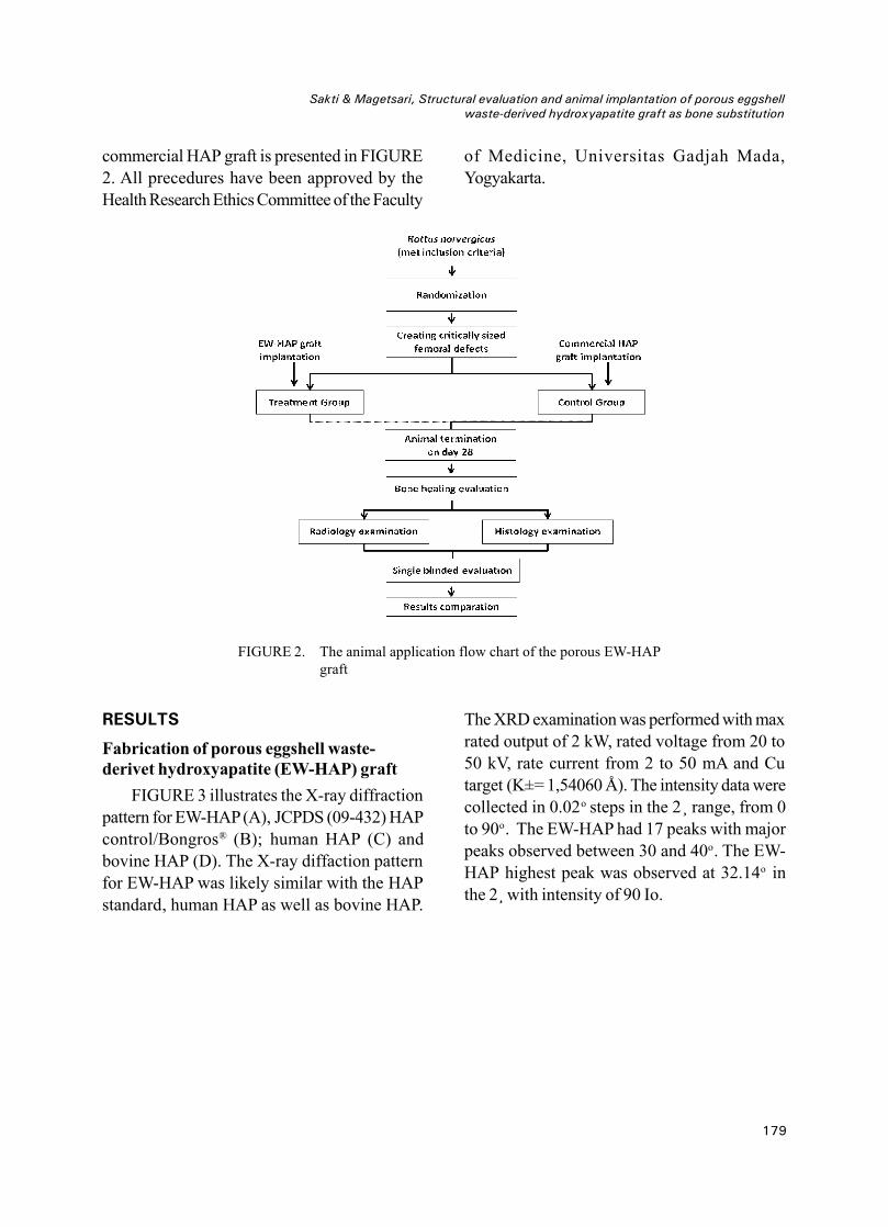
179
Sakti & Magetsari, Structural evaluation and animal implantation of porous eggshellwaste-derived hydroxyapatite graft as bone substitution
commercial HAP graft is presented in FIGURE2. All precedures have been approved by theHealth Research Ethics Committee of the Faculty
of Medicine, Universitas Gadjah Mada,Yogyakarta.
FIGURE 2. The animal application flow chart of the porous EW-HAPgraft
RESULTS
Fabrication of porous eggshell waste-derivet hydroxyapatite (EW-HAP) graft
FIGURE 3 illustrates the X-ray diffractionpattern for EW-HAP(A), JCPDS (09-432) HAPcontrol/Bongros® (B); human HAP (C) andbovine HAP (D). The X-ray diffaction patternfor EW-HAP was likely similar with the HAPstandard, human HAP as well as bovine HAP.
The XRD examination was performed with maxrated output of 2 kW, rated voltage from 20 to50 kV, rate current from 2 to 50 mA and Cutarget (K±= 1,54060 Å). The intensity data werecollected in 0.02o steps in the 2¸ range, from 0to 90o. The EW-HAP had 17 peaks with majorpeaks observed between 30 and 40o. The EW-HAP highest peak was observed at 32.14o inthe 2¸ with intensity of 90 Io.

180
J Med Sci, Volume 45, No. 4, Desember 2013: 176-186
FIGURE 3. X-R Diffraction pattern of EW-HAP (A); HAP control/Bongros® (B) human HAP (C)and bovine HAP (D)
The SEM examination result of EW-HAPcomparet with HAP control are presented inFIGURE 4. In magnitude of 50 times, themorphology of EW-HAP surface was likelymore rough, irregular, random with irregularpore size of 150 – 500 µm. Whereas the HAPcontrol (Bangros®) suface appeared more finewith regular pore size of 300-500 µm. Inmagnitude of 2500 times, both of HAP appeared
a regular crystal form with particles in were inmicron dimension. The microstructure consistedof microsized agglomerates HAP and they weretightly bonded to each other. However, thedifference in particles size of both HAP clearlyobserved in the magnitude of 2500 times. Thepasticle size of EW-HAP (2.5-3 µm) was higherthan those HAP control (1.5-2 µm).

181
Sakti & Magetsari, Structural evaluation and animal implantation of porous eggshellwaste-derived hydroxyapatite graft as bone substitution
Implantation of the porous EW-HAPgraftin critically sized femoral defect of animal
The physical and clinical conditions of ratsafter implantation of the porous EW-HAP graftor HAP control graft during 28 days observationare presented in TABLE 1. During 28 daysobservation periode, two rats on the HAPcontrol group were excluded due to woundinfection. In general the others rats exhibited
good physical and clinical conditions. On day14, hemispica removal and operation woundcare were conducted. Fixation of hemispica onfour rats i.e. two in each group was torn. Postoperative swelling was observed on five ratsi.e. two in EW-HAP graft group and three inHAP control group. Tissue necrosis on distalhemispica was observed only in one rat of HAPcontrol group.
FIGURE 4. The SEM examination results of EW-HAP in magnitude 50 x (A1),magnitude 2500 x (A2), HAP control magnitude 50 x (B1) andmagnitude 2500 x (B2)

182
J Med Sci, Volume 45, No. 4, Desember 2013: 176-186
TABLE 1. Physical and clinical conditions of rats on EW-HAP treatment graft group and HAPcontrol graft group during 28 days observation
Representative of X-Ray of bone femouron 28 days after implantation of EW-HAP graftand HAP graft control are presented in TABLE.Whereas, the representative of histological
section of bone femour on 28 days afterimplantation EW-HAP graft is presented inTABLE 6.

183
Sakti & Magetsari, Structural evaluation and animal implantation of porous eggshellwaste-derived hydroxyapatite graft as bone substitution
FIGURE 5. Representative of X-Ray of bone femouron 28 days after implantation. A1(anterio-posteriol) and A2 (lateral) viewsof EW-HAP graft. B1 (anterio-posteriol)and B2 (lateral) views of HAP graftcontrol
FIGURE 6. Representative histological section of bone femour on 28 days afterimplantation EW-HAP graft. A and B (EW-HAP graft), C (osteochondraltissue), D (osteoid and new bone cells).
The results of radiological and histologicalexamination of of bone femour of the right thighof rats of both groups are presented in TABLE2. No signifantly difference in radiological and
histological pictures were observed after EW-HAP graft compare to HAP graft controlimplantation on 28 days (p>0.05).

184
J Med Sci, Volume 45, No. 4, Desember 2013: 176-186
TABLE 2. Radiological and histological examination of of bone femour of the right thigh of rats onEW-HAP graft and HAP control graft groups
Note: Min= minimum; Max=maximum; *Mann Whitney U Test with p<0.05
DISCUSSION
The synthesis HAP graft from eggshell asraw material of calcium source has beenconducted by some authors.9,12,13 Eggshell wasteis available in huge quantity from foodprocessing, egg breaking and hatchingindustries.About 250,000 tons of eggshell wasteis produced annually worldwide by foodprocessing industry only. The process involveda simple hydrothermal reaction of calciumoxide obtained from the burning of eggshell. Thehydroxyapatite crystal was formed through achemical reactions as follows:
The XRD pattern for EW-HAP was likelysimilar with the HAP standard, human HAP aswell as bovine HAP. The EW-HAP has similarpeaks with HAP standard (JCPDS 09-432). Thehighest peak of HAP standard was observed at31.73o in the 2¸ with intensity of 100 Io, whilethe highest peak of EW-HAP was observed at32.14o in the 2¸ with intensity of 90 Io. Thesimilarity of the both graft was also indicatedwith major peaks observed between 30 and 40oC. In addition, EW-HAP was likely similarwith the human dan bovine HAP that producedby Herliansyah.14
The morphoplogy of EW-HAP surface waslikely more rough, irregular and random in theSEM examination. In this study sugar was used
to obtain a porous EW-HAP graft. Symsudinreported that the use of sugar as porogenprovided HAP graft with rough, irregular andrandom surface.15 Morphologically, it seems asugar crystale which have cube-shaped orirregular square in the form of three-dimensio-nal mixed.16 However, the SEM examinationshowed the EW-HAP graft had similarity withprevious studies.15,17-19 The EW-HAP had idealporous diamter to help for better osteogenic cellsattachment and bone healing process.
In general, all rats exhibited good physicaland clinical conditions after implantation of theporous EW-HAP or HAP control grafts during28 days observation. The results of radiologicaland histological examination of bone femour ofthe right thigh of rats after EW-HAP graftimplantation were not significantly compare tothose HAP graft control. It was indicated thatEW-HAP exhibited similar outcome as bonesubstituion compare to HAP control graft(Bangros®).
Studies the use of EW-HAP as bone graftsubstitution have been reported by some authors.Park et al.20 evaluated of bone healing with heneggshell-derived bone graft substitutes in ratcalvaria and showed the potential efficacy ofhen eggshell-derived bone graft as anosteoconductive bone substitute in a rat calvarialdefect model. Lee et al .21 compared hydro-xuapatite from eggshell and synthetichydroxyapatite for bone regeneration andreported that both eggshell and synthetic
CaCO3 CaO CO2
5CaO + 3(NH4)2HPO4 2H2O Ca 5(PO4)3OH + 6NH4OH

185
Sakti & Magetsari, Structural evaluation and animal implantation of porous eggshellwaste-derived hydroxyapatite graft as bone substitution
hydroxyapatite showed highe bone formationthan unfilled control. However, eggshellhydroxyapatite had significantly higher boneformation then the unfilled control at eight weelsafter operation. Durmus et al.22 evaluated thebiocompatibility and osteoproductive activityof ostrich eggshell powder in experimentallyinduced calvarial defect in rabits. The resultshowed that ostrich eggshell powder was aworth-while bone substitute because it is a safe,cheap, and easily available material.
AKNOWLEDGEMENTS
We would like to thank all technicians whogived valuable assistence druring the study.
CONCLUSION
The synthesis of hydroxyapatite fromeggshell waste and sugar as raw materials hasbeen successed performed providing porousEW-HAP graft. The structure and morphologyof EW-HAP graft iss likely similar with theHAP standard (Bangros®). Furthermore, theimplantation of EW-HAP graft of bone femourof the right thigh of rats provide similar outcomeoutcome as bone substituion compare to HAPcontrol graft (Bangros®).
REFFERENCES1. World Health Organization. Global burden of
disease: global programme on evidence for healthpolicy discussion paper 50. Geneva : WorldHealth Organization, 2000.
2. Phelps JB, Hubbard GB, Wang X, Agrawal CM.Microstructural heterogeneity and the fracturetoughness of bone. J Biomed Mat Res 2000;51:735-41.
3. Lewandrowski KU, Gresser JD, Wise DL, TrantolDJ. Bioresorbable bone graft substitutes ofdifferent osteoconductivities: a histologicevaluation of osteointegration of poly(propyleneglycol-co-fumaric acid)-based cement implantsin rats. Biomaterials 2000; 21(8):757-64.
4. Thomas C, Burg KJL. Tissue engineered bonereplacements systems. Eur Cells Mat 2007;13(2): 1-2.
5. Camillo FX. Arthrodesis of the spine. In: CanaleST & Beaty JH editors. Campbell’s operativeorthopaedics, 11 th eds. Philadelphia: MosbyElsevier, 2007; 1851-2.
6. De Long WG, Einhorn TA, Koval K, McKee M,Smith W, Sanders, R et al. Bone graft and bonegraft substitutes in orthopaedic trauma surgery: acritical analysis. J Bone Joint Surg Am2007;89:649-58. Doi:10.2106/JBJS.F.00465.
7. Jamali A, Hilpert A, Debes J, Afshar P, Rahban S,Holmes R. Hydroxyapatit/calcium carbonateversus plester of Paris: a histomorphometric andradiographic study in a rabbit tibial defect model.Calcif Tissue Int 2002;71:172-78.
8. Orsini G, Ricci J, Scarano A, Pecora G, PetroneG, Iezzi G. Bone-defect healing with calcium-sulfate particles and cement: an experimentalstudy in rabbit. J Biomed Mater Res Part B 2004;199–208.
9. Raihanan MF, Sopyan I, Hamdi M, Ramesh S.Novel chemical conversion of eggshell todydroxyapatite powder. In: Osman NAA, IbrahimF, Wan WABA, Rahman HSA, Ting HN editors.Proceedings of the 4th Kuala Lumpur InternationalConference of Biomedical Engineering; 2008June 25-28, Kuala Lumpur, Malaysia.
10. Sasikumar S and Vijayaraghavan. Low temperaturesynthesis of nanocrystalline hydroxyapatite fromeggshells by Combustion method. Trens BiomaterArtif Organs 2006; 19(2):70-3.
11. Heiple KG, Goldberg VM, Powell AE, Bos GD,Zika JM. Biology of cancellous bone grafts.Orthop Clin N Am 1987;18: 179–85.
12. Saeed AM, Hassan RA, Thajeel. Synthesis ofcalcium hydroxyapatite powder from hen’seggshell. Iraqi J Phys 2011; 9(16): 24-8.
13. Kumar S. Eggshell derived nanophase bio-ceramics. Archives of BioCeramics ResearchVolume 9. Proceeding of the 9 th Asian Bio-ceramics Symposium; 2009 December 8-11,Nagoya, Japan.
14. Herliansyah MK. Development and fabrication ofbovine hydroxyapatie bone graft for biomedicalapplication [Ph.D thesis]. Kualalumpur: Universityof Malaya, Malaysia, 2009.
15. Syamsudin. Analisis uji tekan dan porositasmaterial kompaksi sinter HA/ZnO sebagai material

186
J Med Sci, Volume 45, No. 4, Desember 2013: 176-186
substitusi tulang [Tesis S–2]. Yogyakarta: BidangRekayasa Biomedis, Universitas Gadjah Mada.
16. Hynes RC, Le Page Y. Sucrose, a convenient testcrystal for absolute structures. J ApplCrystallograp 1991; 24 (4): 352. Doi:10.1107/S0021889891002492.
17. Lee JH, Hwang CJ, Song BW, Koo KH, Chang BS,Lee CK. A prospective consecutive study ofinstrumented posterolateral lumbar fusion usingsynthetic hydroxyapatite (Bongros1-HA) as a bonegraft extender. J Biomed Mat Res 2009; 90A(3):804-10. DOI: 10.1002/jbm.a.32113
18. Abdurrahim T and Sofyan I. Recent progress onthe development of porous bioactive calciumphosphate for biomedical applications. RecentPatents on Biomedical Engineering 2008;1: 213-29.
19. Damien E and Revell PA. Coralline hydroxyapatitebone graft substitute: a review of experimental
studies and biomedical application. J Appl BiomatBiomech 2004; 2: 65-73.
20. Pa JW, Bae SR, Suh JY, Lee DH, Kim SH, Kim H,et al. Evaluation of bone healing with eggshell-derived bone graft substitutes in rat calvaria: apilot study. J Biomed Mater Res A 2008;87(1):203-14.
21. Lee SW, Kim SG, Balázsi C, Chae WS, Lee HO.Comparative study of hydroxyapatite fromeggshells and synthetic hydroxyapatite for boneregeneration. Oral Sur Oral Med Oral Phathol OralRadiol 2012;113(3):348-55.
22. Durmus E, Celik I, Aydin AF, Yildirim G, Sur E.Evaluation of the biocompatibility andosteoproductive activity of ostrich eggshellpowder in experimentally induced calvarialdefects in rabbits. J Biomed Mat Res Part B ApplBiomat 2008; 86(1):82-9.