ARE CALCIUM CHANNELS INVOLVED? - University of ...
98
CYTOTOXIC EFFECTS OF COBALT METAL IONS ON OSTEOBLASTS: ARE CALCIUM CHANNELS INVOLVED? Sarah Wiseman BSc This Thesis is submitted in part fulfilment for the degree of Masters of Science in Biomedical Engineering August 2012 Department of Bioengineering
-
Upload
khangminh22 -
Category
Documents
-
view
4 -
download
0
Transcript of ARE CALCIUM CHANNELS INVOLVED? - University of ...

CYTOTOXIC EFFECTS OF
COBALT METAL IONS ON
OSTEOBLASTS: ARE
CALCIUM CHANNELS
INVOLVED?
Sarah Wiseman BSc
This Thesis is submitted in part fulfilment for the degree of
Masters of Science in Biomedical Engineering
August 2012 Department of Bioengineering

i
Declaration of Authenticity and
Authors Rights
This thesis is the result of the author’s original research. It has been composed by
the author and has not been previously submitted for examination which has led to
the award of a degree.
The copyright of this thesis belongs to the author under the terms of the United
Kingdom Copyright Acts as qualified by the University of Strathclyde Regulation
3.50. Due acknowledgement must always be made of the use of any material
contained in, or derived from, this thesis.
Signed:
Date:
ii
Acknowledgements
First and foremost, I would like to sincerely thank my supervisor, Professor M.H.
Grant for all her support throughout the duration of this project. Without her
guidance, motivation and endless knowledge, there would certainly be no thesis.
I would also like to thank Katie Handerson for her endless help during the
experimentation phase; for answering all my questions and assisting me with
anything and everything lab-related. Thanks also to Christopher Hamilton and Olga
Posada for all their help with the confocal microscope, and any other assistance
they may have provided in the lab.
Above all, and as ever, I would like to express my deepest gratitude to my family. To
my parents, for the love, encouragement and every kind of support possible, that
they have always shown me, but especially over the course of this Masters Degree.
And to my brothers for keeping the bar impossibly high. Thanks to my friends, both
at home and overseas, for being very willing to take my mind off my thesis, if only
for a short time. And especially for Ruth and David; for the inspirational messages.
iii
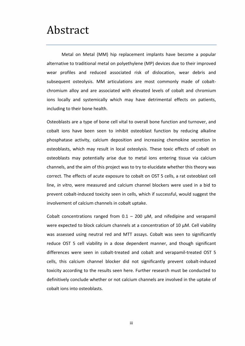
Abstract
Metal on Metal (MM) hip replacement implants have become a popular
alternative to traditional metal on polyethylene (MP) devices due to their improved
wear profiles and reduced associated risk of dislocation, wear debris and
subsequent osteolysis. MM articulations are most commonly made of cobalt-
chromium alloy and are associated with elevated levels of cobalt and chromium
ions locally and systemically which may have detrimental effects on patients,
including to their bone health.
Osteoblasts are a type of bone cell vital to overall bone function and turnover, and
cobalt ions have been seen to inhibit osteoblast function by reducing alkaline
phosphatase activity, calcium deposition and increasing chemokine secretion in
osteoblasts, which may result in local osteolysis. These toxic effects of cobalt on
osteoblasts may potentially arise due to metal ions entering tissue via calcium
channels, and the aim of this project was to try to elucidate whether this theory was
correct. The effects of acute exposure to cobalt on OST 5 cells, a rat osteoblast cell
line, in vitro, were measured and calcium channel blockers were used in a bid to
prevent cobalt-induced toxicity seen in cells, which if successful, would suggest the
involvement of calcium channels in cobalt uptake.
Cobalt concentrations ranged from 0.1 – 200 µM, and nifedipine and verapamil
were expected to block calcium channels at a concentration of 10 µM. Cell viability
was assessed using neutral red and MTT assays. Cobalt was seen to significantly
reduce OST 5 cell viability in a dose dependent manner, and though significant
differences were seen in cobalt-treated and cobalt and verapamil-treated OST 5
cells, this calcium channel blocker did not significantly prevent cobalt-induced
toxicity according to the results seen here. Further research must be conducted to
definitively conclude whether or not calcium channels are involved in the uptake of
cobalt ions into osteoblasts.

iv
Contents
1. INTRODUCTION…………………………………………………………………………………………....…1
1.1. Hip Joint………………………………………………………………………………………….1
1.1.1. Hip Replacement………………………………………………………………………..1
1.1.1.1. History of Hip Replacement…………………………………..…2
1.1.1.2. Modern Process………………………………………………………..2
1.1.2. Hip Resurfacing…………………………………………………………………………..3
1.1.3. Materials Used in Hip Replacement…………………………………………..4
1.1.4. Risks and Complications……………………………………………………………..5
1.2. Medical Device Alerts Regarding MM Hip Implants……………………….6
1.3. Cobalt………………………………………………………………………………………………8
1.3.1. Cobalt Levels in Body Fluids……………………………………………………….9
1.3.2. Metal Toxicity…………………………………………………………………………..10
1.3.3. Apoptosis………………………………………………………………………………….11
1.3.4. Immunological Effects of Cobalt……………………………………………...12
1.3.4.1. Macrophages and Chemokines……………………………….12
1.3.4.2. Lymphocytes…………………………………………………………..13
1.3.5. Pseudotumours………………………………………………………………………..13
1.3.6. Infection…………………………………………………………………………………..15
1.3.7. Systemic Effects……………………………………………………………………….16
1.3.7.1. Canada Beer Drinkers Cardiomyopathy…………………..17
1.3.7.2. Case Studies of Systemic Toxicity…………………………..18
1.3.8. Neurological Effects………………………………………………………………….20
1.4. Osteoblasts……………………………………………………………………………………21
1.4.1. Cobalt and Osteoblasts…………………………………………………………….22
1.5. Calcium Channels………………………………………………………………………….24
1.5.1. Calcium Channels and Osteoblasts…………………………………………..25
1.5.2. Calcium Channel Blockers…………………………………………………………26
1.5.2.1. Nifedipine and Verapamil……………………………………….26
1.6. Aim of Project………………………………………………………………………………..27
2. MATERIALS AND METHODS………………………………………………………………….……..29 2.1. OST 5 Cells………………………………………………………………………………………30
2.1.1. Medium Preparation…………………………………………………………………30
2.1.2. Passaging Cells………………………………………………………………………….30
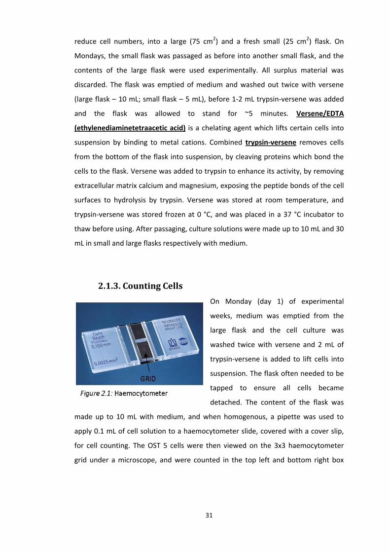
2.1.3. Counting Cells…………………………………………………………………………..31
v
2.2. Cobalt Concentrations……………………………………………………………………33
2.3. Nifedipine Concentrations……………………………………………………………..33
2.4. Verapamil Concentrations……………………………………………………………..33
2.5. Administration to OST 5 Cells…………………………………………………………34
2.6. Viability Assays……………………………………………………………………………….39
2.6.1. PBS……………………………………………………………………………………………39
2.6.2. Neutral Red………………………………………………………………………………39
2.6.3. MTT………………………………………………………………………………………….40
2.7. Confocal Microscopy………………………………………………………………………40
2.7.1. Staining for Actin Using Phalloidin-FITC……………………………………41
2.7.2. Cell Viability Staining Using Propidium Iodide and CFDA…………42
2.8. Statistics…………………………………………………………………………………………43
3. RESULTS……………………………………………………………………………………………………...45 3.1. Effect of Cobalt on OST 5 Cell Viability……………………………………………45
3.2. Effect of Nifedipine on OST 5 Cell Viability…………………………………….49
3.2.1. NIfedipine…………………………………………………………………………………49
3.2.2. Nifedipine and Cobalt (100 µM fixed)……………………………………….51
3.3. Effect of Verapamil on OST 5 Cell Viability…………………………………….53
3.3.1. Verapamil ………………………………………………………………………………..53
3.3.2. Verapamil and Cobalt (100 µM fixed)………………………………………54
3.3.3. Verapamil (10 µM fixed) and Cobalt………………………………………..56
3.4. Osteoblast Images………………………………………………………………………….60
3.4.1. Unstained OST 5 Cells……………………………………………………………….60
3.4.2. Phalloidin-FITC Stained OST 5 Cells………………………………….……….61
3.4.3. Propidium Iodide and CFDA Stained OST 5 Cells ……………………..62
4. DISCUSSION………………………………………………………………………………………………..64
4.1. Are Calcium Channels Involved?........................................................67
4.2. Alternative Materials in THA…………………………………………………………..69
4.2.1. Ceramics…………………………………………………………………………………..69
4.2.2. Titanium………………………………………………………………………………..…70
4.3. What’s Next for Cobalt-Chromium in THA?.....................................70
4.3.1. Infection Control………………………………………………………………………71
REFERENCES……………………………………………………………………………………………….……..73
1
1. Introduction
1.1. Hip Joint
The hip joint is a congruous, synovial ball-and-socket joint formed by the
articulation of the spherical femur head and the cup-like acetabulum of the pelvis. It
forms the primary connection between the bones of the lower limb and pelvis. Its
primary function is supporting the body, in both dynamic and static postures, and is
the most important joint in retaining balance. The femoral head is held in the
acetabulum by various ligaments, and the articulating joint surfaces are covered by
a strong, lubricated layer of hyaline cartilage, which cushions compressive forces
and lubricates the joint. The acetabulum forms at the union of three bones; the
ilium, pubis and ischium. The femur head is attached to the shaft by a thin neck
region often prone to fracture in the elderly, mainly due to the degenerative effects
of osteoporosis. Various muscles act on the joint, including the hamstring, adductor
muscles and abductor muscles, to facilitate movement, and as the joint is
congruous, rotation occurs in all directions.
1.1.1. Hip Replacement
Hip replacement refers to a medical procedure in which one or more sections of the
hip joint are replaced by synthetic implants, and is the most successful, inexpensive
and safest form of joint replacement surgery. Total hip arthroplasty (THA) involves
replacing the acetabulum and femoral head, and is generally conducted to treat
pain or joint failure caused by osteoarthritis, a joint disorder due to aging and wear-
and-tear on the hip. The operation aims to reduce pain and improve overall hip
function, and though it is the most common joint replacement surgery, patient
satisfaction, both short and long-term, varies greatly.
2
1.1.1.1. History of Hip Replacement
Hip replacement dates back to 1891, when German Professor T. Gluck attempted
replacement of the femoral head with ivory and nickel-plated screws (1). The first
metallic hip replacement was performed by American Dr. Austin T. Moore in 1940
at Johns Hopkins hospital using a cobalt-chrome alloy, ‘Vitallium’ (1, 2), to replace
the femur. During the 1940s steel and chrome artificial hips became more
widespread, but were prone to loosening and wear. A later version of Moore’s
prosthesis using a fenestrated stem to allow bone ingrowth and longer-term
attachment was introduced in 1952, and is still currently in use (1).
1.1.1.2. Modern Process
In 1962, Sir John Charnley designed a cemented, low friction arthroplasty joint
consisting of a metal femoral head and stem articulated with a polyethylene
acetabular component, both lubricated with synovial fluid. Due to its lack of friction,
this design yielded excellent clinical results and was instrumental in the
development of the modern artificial hip joint and had almost completely replaced
previous designs by the 1970s (3). The cemented Exeter device was also developed
during this era in the UK. However, early implants had the potential to loosen,
become painful and cause bone erosion, which prompted searches for alternative
methods. In recent times, there have been several evolutionary improvements
made in the procedure and prostheses of THA. Ceramic is being introduced as an
implant material in the place of polyethylene, due to reported reductions in joint
wear. Metal-on-metal (MM) implants are also gaining popularity for this same
reason, as they have been shown to reduce the need for revision of the acetabular
component.
3
1.1.2. Hip Resurfacing
Hip resurfacing (HR) is an alternative to total hip arthroplasty (THA) where only the
articulating surfaces of the joint are replaced (4); the femoral head is coated with a
cylindrical metal cap and the acetabulum is replaced just as in a THA (5). Developed
in the mid 1990’s, especially for younger, physically active patients (6), resurfacing
offers several advantages over THA; including the preservation of bone stock, if a
revision should ever be needed (4), the elimination of the problem of femoral shaft
loosening, restoration of normal joint mechanics (7, 8), reduced stress-shieldings (8,
9) and superior wear properties (10, 11). Revision of a modern resurfacing
procedure is claimed to be less problematic than that of THA, in terms of operation
time, blood loss and functional results (5), an advantage with younger patients who
may outlive the predicted lifespan of their hip implants. Ball et al, 2007, found the
results of metal-on-metal hip resurfacing (MM HR) to THA conversion comparable
to primary THA in terms of surgical effort, safety and clinical outcome (10).
The first generation HR with metal-on-polyethylene (MP) bearing couples were
implanted in the 1970s and early 1980s, but poor clinical results were yielded (12,
13). The introduction of metal-on-metal (MM) articulation renewed the interest in
HR due to the potential of MM prostheses to survive over 20 year with low wear
and no osteolysis incidence (14-16). MM THA and MMHR have become the most
commonly used UK procedures in treating osteoarthritis in patients less than 60
years of age (17, 18). HR has shown encouraging short-term reports with a low
incidence of failure, even after the resumption of high-level activities (17, 19, 20).
Ten year success rates of hip resurfacing from studies in England report success
equal to or greater than standard THA. In the US, the first modern resurfacing
device received FDA approval in May 2006, while some 90 000 resurfacings have
been performed world-wide. However, according to the National Joint Register for
England and Wales in 2008, the three year revision rate for HR was 4.4% compared
to 1.3% for cemented THA (www.njrcentre.org.uk), and the Australian Arthroplasty
Register also reported a higher three year revision rate for hip resurfacing than for
THA (3.1% v 2.1%) (www.dmac.adelaide.edu/au.aoanjrr) (21).
4
1.1.3. Materials used in Hip Replacement
THA has greatly improved the quality of life of patients suffering from
traumatic/degenerative joint diseases, but first generation polyethylene prostheses
released significant amounts of wear particles through friction at the bearing
interface. This was a major concern for the longevity of these THA (22), as these
polyethylene wear particles have been cited as a main source of osteolysis (23-25).
In an attempt to circumvent polyethylene-induced osteolysis and improve implant
longevity, the orthopaedic industry developed articulations producing minimal
amounts of wear particles (26-29). Ceramic-on-ceramic (CC) bearings offer high
wear-resistance, low wear and low noise friction and unanticipated bearing fracture
(30); highly cross linked polyethylene bearing reduce wear of polyethylene (31), but
long-term clinical results are not yet available. There has been a resurgence in MM
arthroplasty, due to their low volumetric wear in hip simulators (32, 33) and in
clinical practice (14), which results in significantly less wear debris and subsequent
peri-prosthetic osteolysis (34-36). As a result, many MM implants used over the last
two decades are still functioning in patients who received implants at a young age
(37). MM bearings are commonly used among young patients, whose life
expectancy is greater than that of traditional elderly hip replacement patients, and
where longevity is particularly important. Extended durability of the hip
replacement and a reduction in the need for revision are anticipated with MM
arthroplasty (38, 39). 45% of hip replacements are in men less than 54 years old in
the UK (40). Though the use of MM bearings eliminate, or substantially reduce the
need for polyethylene bearings, concern has been expressed about the increased
particulate and ionic metal generation (39), and the biological implications of these
metal species. Titanium alloys have been shown to have limitations as articulating
surfaces and should not be considered for wear couples in THR due to significant
wear being shown (38). Cobalt and chromium alloy has shown reasonable wear and
corrosion profiles and continues to be evaluated, in a bid to overcome wear debris-
related issues.
5
1.1.4. Risks and Complications
Risks and complications are similar to those associated with all joint replacements –
including femoral neck fracture (41, 42), dislocation, loosening (43), impingement
(44), infection, osteolysis, metal sensitivity, nerve palsy, pain and death. Andrews et
al, 2011, cites the most common adverse events necessitating revision after MM
THA as early prosthetic fracture, the failure of osseo-integration of the prosthesis
which results in aseptic loosening, unexplained pain and inflammatory masses (21,
45-49). According to Queally et al, 2009, the leading cause of arthroplasty implant
failure is wear debris-mediated
osteolysis with associated aseptic
loosening (50-52). ~10% of primary THA
require revision 10 years after the initial
procedure (51, 53).
Smith et al, 2012, carried out an analysis
of the National Joint Registry of England
and Wales for primary hip replacements
between 2003 and 2011; 31 171 of the
402 051 THA procedures carried out
were stemmed MM, and these were
found to fail at high rates. Larger heads
failed earlier, 3.2% incidence of revision
was seen for 28 mm and 5.1% for 52 mm
heads at 5 years in men ages 60 years. In
younger women, 5 year revision rates
were 6.1% for 46 mm MM devices compared with 1.6% for MP implants, and CC
articulations were associated with improved survival by comparison. Failure rate
statistics such as these have lead to the recall of MM THA and HR devices in recent
times (54)
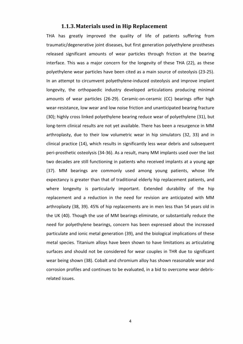
Figure 1.1: X-ray of implanted DePuy ASRTM
XL MM acetabular hip system prosthesis
from Mao et al, 2011.
6
1.2. Medical Device Alerts Regarding MM Hip
Implants
In recent times, the subject of MM THA and HR has attracted a lot of attention in
both the media and the medical world. Though previously believed to be at the
forefront of progress in the area of hip prosthetics, the health hazards of these
devices are beginning to be documented and reported, as the safety, well being and
an improved quality of life must ultimately be the aspiration of the procedure.
In September 2010, the Medicines and Healthcare products Regulatory Agency
(MHRA) released a medical device alert urging immediate action on the subject of
DePuy ASRTM (articular surface replacement) MM hip replacement implants
(MDA/2012/044), in which those involved in the implantation of these prostheses
were instructed to cease the use of this particular model, return all unused models
to the manufacturer, and inform all previous recipients of the DePuy ASRTM about
the recall, and organise follow-up appointments for them. This particular system
consisted of acetabular cups for resurfacing or replacement, surface replacement
heads for resurfacing, and XL femoral heads for replacement. DePuy ASRTM hip
implants have been used in the UK since 2003, and the main reason cited for the
recall was the prostheses being associated with much higher rates of revision at 5
years than initially expected. At the British Hip Society annual conference in March
2011, some alarming figures were presented on MM devices, which displayed
revision results similar to the withdrawn DePuy ASRTM implant. Research showed
that 49% of DePuy ASRTM devices needed to be replaced within 6 years, and 12-15%
of other MM hip devices required revision within 5 years. Pain was reported as the
first symptom experienced by patients who eventually underwent implant revision,
and X-rays depicting loosening along with elevated blood levels of chromium and
cobalt ions were sometimes, but not always seen. MRI scans of cystic and/or solid
masses and fluid collections were also reported.
In January 2012, an investigation was launched into MM hip implants by the media
and several medical regulators due to the occurrence of elevated metal ion levels
7
seen in some MM implant patients and the potential dangers of these events. In the
UK, media outlets such as the BBC and The Telegraph newspaper felt it was
necessary to bring this issue to the attention of the public, as concerns were
growing that risks from this device were beginning to appear much greater than
previously thought, and Channel 4 news reported that over 30 000 people in the UK
had received MM hip implants at this time. An MHRA statement released on the 30
January 2012 assured the threat posed by MM devices was low, with only an
extreme minority experiencing serious problems, but further investigation and
monitoring of evidence would be ongoing.
The most recent medical device alert at the time of this project was issued by the
MHRA on the 25 June 2012, where updated advice for clinicians managing patients
with several variations of MM hip implants was given (MDA/2012/036). Four
different MM hip replacements were listed as carrying a risk; DePuy ASRTM hip
replacements, MM hip resurfacing implants, MM total hip replacements with a
head diameter ≥36 mm and also those with a head diameter of >36 mm. The main
problem outlined in this warning was the development of soft tissue reactions seen
in the minority of patients who had received one of these implants, caused by a
reaction to metal wear particles leached from the implant, and the systemic risks
carried by the entry of these metal particles into the bloodstream. As this will
adversely affect revision surgery, extra vigilance is recommended in cases of poorly
performing hip implants, as early revision is important. Follow-up recommendations
are provided in this alert, in the areas of annual follow-up following implantation,
appropriate imaging to visualise the prosthesis and whether it appears to have
undergone corrosion, blood testing to assess blood metal ion levels, and situations
where revision should be considered, in symptomatic and asymptomatic patients.
Patients presenting with muscle or bone damage on MRI scans should be of utmost
concern.
8
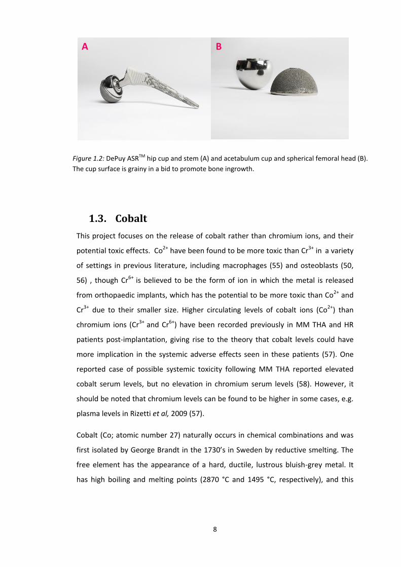
Figure 1.2: DePuy ASRTM hip cup and stem (A) and acetabulum cup and spherical femoral head (B).
The cup surface is grainy in a bid to promote bone ingrowth.
1.3. Cobalt
This project focuses on the release of cobalt rather than chromium ions, and their
potential toxic effects. Co2+ have been found to be more toxic than Cr3+ in a variety
of settings in previous literature, including macrophages (55) and osteoblasts (50,
56) , though Cr6+ is believed to be the form of ion in which the metal is released
from orthopaedic implants, which has the potential to be more toxic than Co2+ and
Cr3+ due to their smaller size. Higher circulating levels of cobalt ions (Co2+) than
chromium ions (Cr3+ and Cr6+) have been recorded previously in MM THA and HR
patients post-implantation, giving rise to the theory that cobalt levels could have
more implication in the systemic adverse effects seen in these patients (57). One
reported case of possible systemic toxicity following MM THA reported elevated
cobalt serum levels, but no elevation in chromium serum levels (58). However, it
should be noted that chromium levels can be found to be higher in some cases, e.g.
plasma levels in Rizetti et al, 2009 (57).
Cobalt (Co; atomic number 27) naturally occurs in chemical combinations and was
first isolated by George Brandt in the 1730’s in Sweden by reductive smelting. The
free element has the appearance of a hard, ductile, lustrous bluish-grey metal. It
has high boiling and melting points (2870 °C and 1495 °C, respectively), and this
A B

9
temperature stability makes its alloys suitable for use in parts for aircraft engines
that require magnetic, high-strength and wear/corrosion resistant alloys; it is widely
used in batteries and electroplating, due to its high resistance to oxidation; it is
important commercially in its cobalt60 isotope, which is used in the treatment of
cancer and as a radioactive tracer. It is a useful gamma ray source as it can be
produced in high activity and predictable quantities. Cobalt is also essential to many
animals as a trace dietary mineral as it is a component of vitamin B12. In inorganic
form, it is also an active nutrient for algae, bacteria and fungi. The resistance of
cobalt to wear and corrosion makes its alloys useful in orthopaedic implants.
Cobalt-chromium alloys are currently used in prosthetic knee and hip implants (59),
and cobalt alloys are also useful in dental alloys, for nickel allergy sufferers (60).
Cobalt-chrome alloy in solid form is very well tolerated by the body, and previous
literature states in vivo testing shows no pathological changes compatible with
systemic toxicity (61).
1.3.1. Cobalt Levels in Body Fluids
Cobalt ions can be generated in the peri-prosthetic space due to electrochemical
corrosion of metallic particles, or possibly as a result of phagocytosis of cobalt-
chromium (CoCr) particles by cells and the exposure of particles to a series of
oxidative mechanisms designed to destroy foreign bodies (62-64). Andrews et al,
2011, claims normal circulating physiological levels of cobalt and chromium to be
<0.25 µg/L (0.005 µM), but following MM THA, elevated circulating levels of the
metal ions can occur in hip synovial fluid and peripheral blood (21). Whole blood
concentrations of cobalt ions (Co2+) can reach 4.6 µM (65), while local hip synovial
fluid levels have been measured at 30 µM in vivo (66), which are higher than values
of circulating total chromium ion levels, which have been recorded at 2.3 µM and
25 µM for blood and hip synovial fluid concentrations respectively (65, 66).
Circulating levels of cobalt are usually highest in the first few months after
implantation, but persistent elevation can occur as late as ten years post-surgery
10
(67). Toxicity caused by these metal ions locally (in the peri-prosthetic space) and
systemically is a cause of concern for metal on metal implants (50, 68), potentially
causing chromosomal changes (64, 69), hypersensitivity and instability (45, 70)
There may also be complex issues present with the definitive collection and analysis
of metal ion levels, as well as with the statistical methodologies and reporting of
results and previous literature has shown a characteristic variability in these factors.
(71). As MM THA is performed more and more frequently, manufacturing
differences (clearance, roughness, metallurgy, head size, etc.) are apparently
affecting performance. Modern MM bearings have uniformly shown higher levels of
blood cobalt and chromium, the principal elements in CoCr alloy used in MM HR
implants (72), when compared to pre-operative values, and in comparison to the
levels in MP patients (19, 73-76). Engh et al, 2009, measured 2 year post-operative
ion levels for 28 mm metal on polyethylene bearings compared with 28 and 36 mm
metal on metal bearings. The MM cobalt serum, erythrocyte and urine ion levels
were higher two years post-implantation than MP levels (77). Elevated cobalt ion
levels are commonly seen in intermediate and long-term follow-up studies of MM
arthroplasties (67), which have shown to be higher than levels associated with
bearings of different materials (78). Despite satisfactory short-term implant survival,
there is an increasing concern that CoCr alloy MM HR implants release a large
amount of very small wear particles and metal ions (79).
1.3.2. Metal toxicity
Metal toxicity remains a concern (locally, in peri-prosthetic space, and systemically)
(80-82). As previously mentioned, substantial levels of metallic products can be
transferred into the host environment from a metallic device (83, 84). CoCr alloy is
used for MM bearings, due to its wear performance profile, but cobalt and
chromium ions are produced by mechanical wear and corrosion in the
periprosthetic tissue as a result of MM articulation (85, 86). Cobalt and chromium
constitute 90% of the CoCr alloy used in MM hip resurfacing implants (87), and

11
particles generated from these alloys have a size range in the nanometer scale (88).
Although wear rates are lower in MM bearing surfaces, previous research has found
that the amount of particles generated by these implants can be up to 500 times
higher than the amount of micrometer sized particles from conventional MP
bearings (20). Metal nanoparticles corrode to either produce a protective passive
layer or dissolve into charged free ions (85). Cobalt and chromium ions have been
detected in solution during metal alloy corrosion (89). However, other studies have
found that MP bearings can generate 100 mm3 500 nm particles per year, while MM
implants can generate 1 mm3 40 nm CoCr particles (17, 90). The vast range of metal
ion generation reported in the literature is one reason why this area needs further
attentions. These ions will eventually reach the blood circulation where metal ion
concentration has shown to be significantly elevated (>20 nM) in patients with MM
THA (75, 91, 92). Periprosthetic tissues cobalt concentration measured in 6 patients
20 months post-implantation was found to be 1000-times higher than control
patients (30mg/g vs. 0.03mg/g) (93). The toxicity of released cobalt ions from MM
THA is a cause for concern (55, 56, 94, 95), but the role of these ions in
physiopathology of MM THA infection and cytotoxicity has not yet been studied
fully (96).
1.3.3. Apoptosis
Petit et al, 2004, analyzed the effects of cobalt and chromium ions on bcl-2, bax,
caspase-3 and caspase-8 expression; these protein families play a central role in the
modulation of apoptosis. Cobalt and chromium ions inhibited bcl-2 expression,
stimulated bax and caspase-3 expression, and Cr ions stimulated caspase-8
expression also. Therefore, bcl-2 and caspase protein family expression is implicated
in the induction of macrophage apoptosis, specifically the modulation of expression
of these proteins (97). Cateles et al, 2001, analyzed the effects of Co2+ and Cr3+ ions
on macrophages in vitro, by analyzing the implications of caspase-3 in the apoptotic
pathway, with Co2+ and Cr3+ both inducing macrophage mortality in a dose-

12
dependent manner. Co2+ was found to be more toxic, inducing cell mortality up to
28% at 10 ppm, compared to 37% at 500 ppm Cr3+ (55). DNA fragmentation was
induced by both ion types, but at a lower concentration of cobalt. The appearance
of the caspase-3 active fragment is implicated in several apoptotic pathways.
Cateles et al, 2003, looked at the effects of cobalt and chromium ions on
macrophages in a more pro-longed incubation period (94). ELISA results confirmed
that Co2+ and Cr3+ induced concentration and time-dependent increase of TNF-α, a
cytokine involved in systemic inflammation which has the ability to induce
apoptotic cell death. Cell mortality increased as concentrations increased, especially
at 48 hours. This study found that both Co2+ and Cr3+ induced macrophage
apoptosis, measured by DNA analysis, with a stronger signal at 24 hours than 48
hours, suggesting the presence of more necrosis at 48 hours. A subsequent study
which backed up this result evaluated cell death in vitro by transmission electron
microscopy in addition to cell death detection ELISA, and also found non-
inflammatory apoptosis predominant at 24 hours, and necrosis predominant at 48
hours at higher ion concentrations (98). However, the apoptotic effect of Cr6+ was
not assessed in the above literature, which has also been found to cause apoptosis
in macrophages (99).
1.3.4. Immunological Effects of Cobalt
1.3.4.1. Macrophages and Chemokines
The cytotoxic effects of cobalt ions have been demonstrated in vitro in
macrophages, by induction of apoptosis and necrosis, TNF-α secretion and
oxidation of proteins (50, 55, 94, 95, 97, 98). Cobalt has also previously been shown
to enhance the in vitro secretion of IL-8 and MCP-1 in renal, gastric and colon
epithelium, as well as monocytes and neutrophils (68), which shows the potent
bioactivity of cobalt ions in a variety of cell types as well as their potential to induce
a proinflammatory response. Products of metallic degradation may be transported
from the site of implant and distributed systemically. Though the toxicological
13
importance of these trace metallic elevations remains to be elucidated, urine and
serum metal concentrations may serve as useful tribological markers in the
performance of MM in vivo (39). MM bearings are reported to release
approximately three times more cobalt and chromium ions than MP hips (39, 100).
Concern has been expressed that MP release ions that can cause DNA damage (64)
and immune dysfunction, in the form of T-lymphocyte reduction (101), and
threshold levels of blood cobalt and chromium were associated with reduced CD8+
T-cell counts (102) following implantation of a MM device. Hart et al, 2006, studied
the relationship between metal ion levels and lymphocyte counts in patients with
MM HR and found cobalt and chromium levels significantly elevated in patients
with reduced lymphocyte numbers.
1.3.4.2. Lymphocytes
Elevated cobalt and chromium levels have previously been linked to lymphocytic
DNA damage (103). In a pilot study, Hart et al, 2006, found lower peripheral
lymphocyte counts in patients with MM hip replacements, compared to MP
articulation patients (102). Hart et al, 2009, compared Co2+ and Cr3+ levels in whole
blood and lymphocytes in patients with MM, CC and MP hip replacements, with the
aim of elucidating whether higher metal ion levels were related to differences in
lymphocyte counts. CD8+ and CD3+ lymphopenia was present in 15% and 13% of
patients respectively with unilateral MM hips, and significant differences in
absolute CD8+ lymphocyte subsets were seen in MM group in comparison to the
other groups (104).
1.3.5. Pseudotumours
Concern has recently been expressed about soft-tissue reactions to metal wear
debris, collectively called pseudotumours (46). These masses (tumours) may be
cystic or solid (‘pseudo’), and have been referred to as bursae (105), cysts (106) and
inflammatory masses (107). These pseudotumours can appear with varying levels of
14
interference, from asymptomatic abnormality detected on a scan to a destructive
lesion leading to revision of MM HR, and are associated with elevated cobalt and
chromium serum levels (median Co2+ level - 3.8 µg/L or 0.6 µM/L; total chromium
level median – 11.5 µg/L or 0.2 µM) (46). Hip aspirate cobalt and chromium levels
have also shown to be higher (Co2+ – 701 µg/L or 12 µM; Cr3+ and Cr6+ – 329 µg/L or
6 µM)(46). Occurrence of such reactions is rare, and precise prevalence remains
unknown, but they are becoming increasingly reported (46, 65, 104, 108, 109).
These reactions have previously been shown following MP THA (110, 111), and
more recently following MM HR in Pandit et al, 2008, which included reports of
bilateral pseudotumours in bilateral arthroplasites. In this study, pseudotumours
were locally destructive, causing the need for revision in the majority of patients
(46). Another series reported incidence of revision for pseudotumours at 8 years as
being 0.5% in men, 5% in women over 40 years of age, and 25% in women less than
40 years of age (112); however, other long-term series have not reported high rates
(113). Their histology has some features of aseptic-lymphocytic-vasculitic-
associated lesions (ALVAL) (45), although they feature more marked necrosis. This
histological feature is thought to represent a metal-induced systemic T-lymphocyte
delayed (type IV) hypersensitivity reaction. This would partly explain the high
prevalence in females, and bilateral appearance of pseudotumour, which is possibly
related to previous exposure to jewellery. Based on these findings, delayed reaction
to cobalt and chromium ions has been suggested to play a role in the aetiology of
pseudotumours.
Grammatopoulos et al, 2009, aimed to determine the extent of the problem posed
by pseudotumours by assessing the outcome of MM HR revised to conventional
THA due to pseudotumour formation, which was previously claimed to yield good
results (10, 114). Grammatopoulos states that outcome after revision of MM HR
depends on indication for revision, and with indications other than inflammatory
pseudotumours, the outcome was generally good and similar to primary THA.
Outcome after revision for pseudotumour was poor with major complications
occurring in ~50% of patients, with ~30% requiring further revision, and this study
15
recommended that patients presenting with symptomatic pseudotumours following
MM HR should be given early revision to limit soft tissue damage (114). The effects
on macrophages are also relevant here, due to the histological appearance of
pseudotumours which are characterized by the presence of macrophages
surrounding the extensive necrosis of the connective tissue, where metal particle
clusters can also be found (46, 79). Additionally, macrophages are the dominant
infiltrating cells that respond rapidly to biomaterial implantation in soft and hard
tissue (115), and can act as antigen presenting cells in delayed hypersensitivity
reactions driven by T lymphocytes (116). Kwon et al, 2009, investigated the
potential cytotoxic effects of cobalt and chromium on Macrophage viability,
observing dose dependent cytotoxic effects on macrophages in vitro with high
concentrations of cobalt ions only. Based on the findings of this study, the
cytotoxicity of high concentrations of metal nanoparticles phagocytosed by
macrophages in the periprosthetic tissue may be an important factor in the
pathogenesis of pseudotumours (79).
1.3.6. Infection
Infection following MM THA is a devastating complication, occurring in 1.5-2.5% of
primary THA, and associated with a significant increase in hospitilization-related
morbidity and mortality (96, 117). Straphylococcus epidermis is the most common
etiological agent recovered from infected THA (118). Neutrophils, the first line of
defence against infection, are potentially exposed to cobalt ions in the peri-
prosthetic tissue by the wear of metal on metal THA. Neutrophils are most exposed
to the potential effects of cobalt, as these innate immune cells are recruited during
MM THA infections. While the toxicity of cobalt is still being debated, these ions
potentially inhibit the Hv1 proton channel, which sustains superoxide production by
neutrophils. This study shows how the antibacterial activity of human neutrophils
can be altered by Co2+ in vitro by the inhibition of Hv1 proton channels;
submillimolar concentrations of cobalt inhibit proton currents, impair cystolic acid

16
extrusion and decrease the production of superoxide. Thus, cobalt reduces the
ability of human neutrophils to kill two strains of S. Epidermis by up to 7-fold at
concentrations of 100 mM. By inhibiting proton channels, Co2+ released by metal
prostheses may therefore promote bacterial infections in patients with MM THA
(96).
1.3.7. Systemic Effects
The systemic effects of metal ions remain relatively unknown. Once generated in
the periprosthetic space, cobalt ions bind to serum proteins (mainly albumin) and
are transported systemically before being excreted by the renal and gastrointestinal
systems (45, 55, 67, 78, 86). Elevated serum levels of cobalt have been
demonstrated systemically, for which the clinical implications have not yet been
elucidated (94, 97). High concentrations of cobalt chloride have been shown to be
toxic to the heart and liver in mice (119). In the endocrine system, Co2+ can prevent
the uptake of iodine into the hormone thyroxine by inhibiting the tyrosine iodinase
enzyme, which can induce hypothyroidism (120). Cobalt ions can also influence
oestrogen signalling, by binding to cellular oestrogen receptors (121). A 2007 review
of toxic effects of orthopaedic metals listed chromium ions as having a greater
effect systemically, than cobalt ions (122). Cr6+ can induce changes in haemoglobin
and haematocrit values in blood (123); the ions can also cause hepatocellular
necrosis with acute ingestion (124) and impair renal function when accumulated in
epithelial kidney cells (125). Chronic Cr6+exposure can cause detrimental effects to
male fertility, including decreased sperm count and sperm abnormalities (126). The
effect of both chromium and cobalt ions on the respiratory system has been
documented due to occupational exposure, and include an increased incidence of
asthma and inflammation (127). Increased levels of cobalt and chromium ions have
been seen in cord blood of women who became pregnant following MM prosthesis
implantation, suggesting metallic ions may translocate into foetal circulation (128).
17
1.3.7.1. Canada Beer Drinkers Cardiomyopathy
The story of Quebecs beer-drinkers cardiomyopathy began in August 1965, when
the first patient was admitted to a Quebec hospital complaining of epigastric pain,
localized to the upper abdomen. The patient was also short of breath, anxious,
agitated and cyanotic, especially in the face, neck and abdomen. Cardiovascular
examination revealed increased heartrate and low blood pressure. The patient
admitted to regularly drinking ~25 pints of a certain beer a day, and was thus
diagnosed with what doctors believed to be a rare form of alcoholic
cardiomyopathy. He was given a large dose of thiamine, recovered slowly and went
back to work (129).
Between August 1965 and April 1966, a total of 48 similar cases were seen in
Quebec; 46 men and 2 women, ranging in ages from 25 to 66 (130). All patients
were unusually heavy beer drinkers, with an average daily been intake of 24 pints
per day, and over time it became apparent that the link between patients in this
series was a preference for Brand XXX beer. This locally-brewed beer is popular in
Quebec, and at the time of the incident commanded 80% of its market. Cobalt
sulphate had been added to this beer in order to stabilize foam since July 1965
(129).
Most patients were in good health up until shortly, between one and 90 days,
before admission (130). Gastrointestinal symptoms were the first to appear;
nausea, vomiting and inability to intake food. Signs of heart failure appeared
progressively; thoracic pain, dyspnea, ankle oedema, cyanosis, and a regular but
rapid heartrate. This contrasted with other cardiomyopathies, where arrhythmias
are common. Arterial emboli were seen in five cases, being the cause of death in
two of these (129). Patients were given various different treatments; digitalis,
diuretics, thiamine, anticoagulants, and corticosteroids, for the treatment of shock.
20 of the 48 patients died, mostly in shock, which was unresponsive to medication;
14 within 24 hours of admission, one died between 24 and 48 hours, and five more
after 48 hours (130). There were striking elevations in serum enzymes, which
correlated with central hepatic necrosis found in the end stages of the disease.
18
Immunological and virological studies were negative, as was the search for toxic
substances, such as lead and arsenic (129).
Following discharge, all symptoms, cardiovascular or otherwise, had subsided,
despite many patients failing to follow correct diet and medication regiments, and
many returning to their previous drinking habits (129). The syndrome which
appeared in Quebec between August 1965 and April 1966 was similar to one
present in Omaha, Nebraska and Minneapolis, Minnesota (131). This study
recommended experimental reproduction in animals, but believed the
epidemiological, clinical and pathological evidence seen in Quebec was strong
enough to establish cobalt as an essential causative factor in this syndrome, though
as a similar syndrome had not been seen before 1965, despite cobalt previously
being used in higher dosages than the dose present in Brand XXX beer, evidence
indicates that other unknown factors may have been present which increased the
susceptibility of the patients to the toxicity of cobalt (129).
1.3.7.2. Case Studies of Systemic Toxicology
Two case studies appear in an Australian journal regarding cobalt toxicity in patients
who received ASR XL Acetabular Hip Systems by DePuy Orthopaedics (58). The first
of which regards a 73 year old woman who, five years earlier, underwent a hip
replacement due to osteoarthritis. She presented with neurological symptoms,
including memory difficulties, depression and cognitive decline; a continuous metal
taste in her mouth; severe headaches and weight loss. She also complained of mild
groin pain, but no other hip-related symptoms. X-rays showed that the implant was
well-fixed and correctly aligned, but displayed mild osteopaenia around the
acetabular component. Her serum cobalt level was 410 nmol/L (reference range 0 –
20 nmol/L) and serum chromium level was 240 nmol/L (reference range 0 - 100
nmol/L). Four years post-implantation, the woman suffered a cerebrovascular
episode, during which stroke-like symptoms occurred: headaches, disorientation
and dizziness, nausea and vomiting, and a feeling of being off-balance. Difficulty in
19
remembering names and registering information (written and aural) were cited as
particularly difficult. The original hip implant was performed in 2006, consisting of a
large diameter CoCr metal cup and a large modular metal head on a titanium Corail
stem. In March 2011, a revision total hip replacement was performed, due
predominantly to the elevated metal concentration and the systemic symptoms, in
which the metal cup was replaced with a polyethylene cemented cup, and the
metal head was changed to a ceramic one. Cobalt concentrations of the turbid joint
fluid were measured at 4218 nmol/L at the time of revision surgery, and
cerebrospinal fluid showed a cobalt ion level of 9 nmol/L, confirming that the metal
ions had crossed the blood brain barrier. At an eight week post-surgery follow up,
the patient felt much improved. She reported greater energy and less fatigue, and
the disappearance of the metallic taste in her mouth. She also had regained a
normal appetite and had gained weight. The hip pain dissipated and her serum
cobalt level dropped from 410 to 60 nmol/L.
The second case study featured in Mao et al, 2011, discussed a 60 year old male
who presented with systemic symptoms at a 4 year follow up for a right total hip
replacement, performed to treat pain and stiffness caused by osteoarthritis, and
before which he enjoyed general good health. No hip symptoms were reported but
marked bone loss around the acetabular component was seen by x-ray. Three years
post-surgery, symptoms began to develop which increased steadily in severity,
including painful muscle fatigue in limbs and cramps in hands and feet, dyspnoea
and feeling faint during simple tasks, and the inability to climb a flight of stairs
without needing to rest. Problems with remembering names and poor
concentration was also noted and hypertension, which had been previously stable,
became uncontrolled and required further medication. At this time, the patients
serum cobalt level measured at 185 nmol/L, and subsequently remained elevated at
between 213 and 258 nmol/L. Chromium levels were never elevated. A revision THR
was performed in February 2011 due to systemic symptoms, and the metal head
and cup were replaced with ceramic and polyethylene versions. There was no
apparent localised tissue reaction of metal debris. A serum cobalt level of 258

20
Table 1.2: elevated cobalt and chromium levels in various body fluids, references figures in brackets, in a
patient following MM THA in Rizetti et al, 2009.
nmol/L at time of revision surgery fell to 42 nmol/L in the eight weeks post surgery.
At this follow up, the patient reported significant decrease in muscle pains,
improved energy levels and exercise tolerance.
1.3.8. Neurological Effects
Rizetti et al, 2009, reports a case study in which a 58 year old patient suffered
progressive visual and hearing loss, after a left hip arthroplasty, which was revised
five years later due to rupture of the ceramic head (57). On admission, neurological
examination showed impairment of cranial nerves II and VIII bilaterally and mild
distal sensory-motor disturbances. Haematological, infectious, neoplastic, metabolic
and immunological diseases were subsequently ruled out and the patient
underwent various investigative procedures, and within 2 months, the patient was
completely blind, severely deaf, wheelchair-bound (due to lower limb hyposthenia).
As tests for immune-mediated processes remained negative, the case was referred
to toxicology, where raised concentrations of cobalt and chromium were
unexpectedly found in different biological samples:
Total Cobalt Ions (µg/L) Total Chromium Ions (µg/L)
24 hour urine collection 1187 (0.1 – 1.5) 510 (0.05 – 2.2)
Blood 549 (0.05 – 2.7) 54 (0.1 – 0.5)
Plasma 90 (0.1 – 0.6) 210 (0.1 – 0.5)
Cerebrospinal Fluid (CSF) 11.4 (0.05 – 0.15) 4.4 (0.01 – 0.2)
Chromium levels were much lower than cobalt levels in all cases except plasma.
Cobalt-chromium poisoning due to metal wear debris was proposed, although
21
radiology (including CT) showed no sign of prosthetic loosening. Metal ion chelating
treatments were administered, which decreased metal ion concentrations, but
caused only negligible neurological improvement. Resection arthroplasty was then
carried out, during which infiltration of peri-prosthetic tissue by metallic debris was
evident, and analysis of peri-prosthetic fluid showed high cobalt and chromium
concentrations. Evident wear of the head and neck of the removed prosthesis
supported the endogenous CoCr poisoning hypothesis. In the eight months
following revision the patient showed progressive improvement, though vision was
only partially improved. Metal ion concentrations improved, but remained higher
than reference (57). This study recommends longer term follow up to evaluate
adverse chronic systemic effects due to prolonged exposure to high serum cobalt
concentrations (132), including neurological and toxicological examinations
whenever a patient with a metallic prosthesis complains of visual loss and hearing
disturbances, limb weakness, numbness or paraesthesia, even in the absence of
osteoarticular symptoms.
1.4. Osteoblasts
Wear particles from artificial hip prostheses can induce many adverse cellular
responses in the periprosthetic area, resulting in an inflammatory reaction and
subsequent bone loss (50). Multiple cell types (macrophages, osteoclasts,
osteoblasts, fibroblasts) are activated locally and secrete a range of pro-
inflammatory cytokines and matrix metalloproteins (MMPs) (34-36, 133), e.g. IL-6
(134), IL-8 (81, 82), TNF-α (135), PGE2 (80). These mediators contribute to an
osteolytic cascade, resulting in the direct bone resorption by MMPs, or indirectly via
osteoclast activation. Bone loss is a very serious risk with hip replacement, as it can
lead to loosening, infection and reduce the overall lifespan of the device.
Bone is composed of different cell types and extracellular matrix (ECM), which
becomes mineralized by calcium hydroxyapatite, which gives bone rigidity and
22
strength (136). Three distinct bone cells exist, two of which have interlinked
functions; bone-forming osteoblasts and bone-resorbing osteoclasts (137). The
balance between bone formation and resorption, known as bone remodelling, is
paramount to healthy bone, and is carried out by factors secreted by osteoblasts,
which regulate osteoclast differentiation, and factors secreted by osteocytes, the
third distinct cell, which regulate the activity of both osteoblasts and osteoclasts
(138). Therefore, the role of the osteoblast is very important in creation and
maintenance of the skeletal architecture; as well as being responsible for bone
formation, osteoblasts have the ability to regulate bone-resorption through the
expression of these ligands, which link to pre-osteoclast receptors, inducing
differentiation and causing bone resorption. Osteoblasts can also produce
osteoprotegerin (OPG), which blocks this ligand action, preventing differention and
resorption (139). Further coordination between osteoblasts and osteoclasts is
mediated by the release of growth factors from bone during resorption (140).
The word osteoblast comes from the Greek words for ‘bone’ and ‘germ’. They are
cells of mesenchymal origin, and when terminally differentiated, they produce most
of the ECM proteins, e.g. sialoprotein and osteocalcin, and control the
mineralization of this matrix to form bone (137). Osteoblasts arise from
osteoprogenitor cells located in deep periosteum layers and bone marrow, and are
mono-nuclear, specialized cells, with a large golgi apparatus and an abundant rough
endoplasmic reticulum when active. Osteoblasts form tight junctions with adjacent
osteoblasts, and have regions of plasma membrane specialized in vesicular
trafficking and secretion (141). The ability to secrete bone matrix arises as the
osteoblasts differentiate (139).
1.4.1. Cobalt and Osteoblasts
Previous studies have shown that short-term exposure to metal species may affect
human osteoclast and osteoblast function and survival (21); high concentrations of
Co2+ (above 10 µg/mL) have been cytotoxic to osteoblasts and can reduce their cell

23
activity in vitro (142), including osteoblast proliferation (56, 142, 143), viability (56,
143) and function (142-144). Co2+ has proved more toxic than Cr3+ (50, 56). In the
periprosthetic tissue, cobalt ions have induced local effects, such as apoptosis,
necrosis, chemokine secretion (IL-8 and MCP-1) and TNF-α secretion in both
macrophages and osteoblasts (50, 55, 56, 70, 84, 94, 95, 97, 98, 122). Cobalt and
chromium have both inhibited the release of osteocalcin and the synthesis of Type I
collagen from human osteogenic sarcoma cells (at concentrations of 10-100 ng/ml)
(145). It has also been shown that short term exposure to cobalt ions at sub-lethal
doses can decrease resorptive activity in rat osteoclasts (146). However, the longer-
term effect of chronic exposure of human osteoblasts to clinically relevant cobalt
ion concentrations is unknown. Queally et al, 2009, showed cobalt ions significantly
reduce alkaline phosphatase activity and calcium deposition in human osteoblasts
(50). This study was the first to demonstrate that cobalt ions stimulated the
expression and secretion of chemokines from primary human osteoblasts, which
contributes to osteolysis by the recruitment of inflammatory leukocytes and
osteoclastic cells to the peri-prosthetic area and may interfere with bone ingrowth
onto the implant (50). This study also showed that cobalt ions directly inhibited
osteoblastic function, thereby possibly contributing to osteolysis by suppressing
bone formation at the bone implant interface. The reduction in osteoblastic
function has been directly correlated to the degree of toxicity, supporting the
hypothesis that adverse local cellular responses (particularly necrotic responses)
associated with metal debris from implanted metallic devices may be due in part to
metal ions released from implants or from particulate debris (143). The importance
of osteoblasts in overall bone regulation and function, and how this may be
impeded by interaction with cobalt ions combined with the severity of osteolysis
caused by MM hip prostheses forms the basis of why an osteoblast cell line was
selected for cobalt toxicity testing in this project.
24
1.5. Calcium Channels
It is important to elucidate the mechanism by which cobalt ions enter cells and
exert their effects, both locally and systemically, in order to gain a proper
understanding of this toxicity and how it may be prevented in MM THA and HR
patients. Cobalt is commonly regarded as a calcium channel blocker, with the ability
to penetrate cells via calcium channels and reduce calcium influx (147-150). This
gives rise to the hypothesis that cobalt may elicit its toxic effects via these channels.
Also, previous literature has found that L-type calcium channels are present in
osteoblasts (151), and the entry of cobalt through these channels may be the
pathway by which cobalt wear particles induce osteolysis in patients with MM hip
devices.
Calcium channels are ion channels in the plasma membrane permeable selectively
to calcium ions and they can be ligand-gated or voltage-dependent (VDCC). VDCCs
are found in all excitable cells (152, 153), including muscle, neurons, glial cells and,
at lower levels in most non-excitable cells, such as osteoblasts (154). The
development of Ca2+ signals is a tightly regulated cellular process involving
concerted actions of plasma membrane and intracellular Ca2+ channels (154). VDCCs
are closed at resting membrane potential and are activated (opened) in response to
membrane depolarization, which allows Ca2+ enter the cell from extracellular space.
The transient increase in intracellular calcium levels characterizes calcium signals
and triggers a variety of physiological effects, depending on the cells involved, e.g.
muscular contraction. Ca+ channels, along with Na+ and K+ channels, form the
superfamily of voltage-gated ion channels.
L-type calcium channels are a subset of VDCCs, and referring to the length of
activation, the “L” stands for long-lasting. L-type calcium channels are widely
expressed in skeletal muscle, smooth muscle and cardiac nodal tissue, where they
are believed to play an important role in pacemaker currents and action potentials.
However, the importance of L-type calcium channels in human physiology is not
restricted to cardiovascular indications; they have been found in neurons, and bone,
25
endocrine and sensory cells, and play roles in neuronal plasticity (155), modulation
of neuronal firing (156) and gene-expression (157), memory (158), controlling mood
and drug-related behaviours (159, 160), vision and hearing (161), the release of a
variety of hormones and neurotransmitter, and skeletal muscle contraction (162).
1.5.1. Calcium Channels and Osteoblasts
Duncan et al, 1998, reported that calcium channels play fundamental roles in
cellular responses to external stimuli, including mechanical forces and hormonal
signals in osteoblasts and bone cells of osteoblast lineage (154). Previous literature
(163, 164) reports L-type VDCCs playing an important role in the influx of Ca2+ into
osteoblasts after a mechanical stimulus. Calcium channel blocker nifedipine has
shown the ability to reverse a mechanically-induced increase in extracellular matrix
proteins, osteopontin and osteocalcin (163), and another previous study has shown
that in vivo blockage of L-type VDCCs by both nifedipine and verapamil, another
calcium channel blocker, substantially suppressed a mechanically-induced bone
formation increase (165). Inhibition of L-type VDCCs, removal of extracellular Ca2+
and plasma membrane depolarization could inhibit gap junction-mediated
intracellular calcium signalling in osteoblast cells (166).
Another study found that parathyroid hormone (PTH) and mechanical loading might
act synergistically on bone formation in vivo (167). Mechanical loading and PTH
together enhanced bone formation significantly, and this enhancement was found
to be suppressed by verapamil, indicating that L-type VDCCs mediate load-induced
bone formation and enhancement of bone adaptation by PTH in vivo. Previous
literature also describes how verapamil and nifedipine interact with osteoblasts via
L-type VDCCs, promoting their differentiation by stimulating alkaline phosphatase
activity (168). Regulation of bone turnover is complex and regulated by many
factors, including hormones, growth factors, cytokines, etc. (169), but little is known
about the signals coupling bone formation to bone resorption. L-type calcium
channels are also proposed to modulate paracrine signals between bone-forming

26
osteoblasts and bone-resorbing osteoclasts at local sites of bone remodelling (154,
170), and signalling between these bone cells is important in bone homeostasis and
regulating bone remodelling (169, 171, 172).
1.5.2. Calcium Channel Blockers
A calcium channel blocker (CCB) is a chemical that disrupts the movement of
calcium (Ca2+) through calcium channels and they have widespread clinical uses as
anti-arrhythmics and anti-hypertensives. Currently approved CCBs bind to L-type
calcium channels and by blocking calcium entry into vascular and cardiac cells, they
exert vasodilative and cardiodepressive properties, e.g. decreased contractility,
heart rate and conduction velocity. Vasodilation causes a reduction in total
peripheral resistance, while decreased contractility reduces cardiac output, and the
end result is a drop in blood pressure. CCBs are also used in the treatment of angina
pectoris pain, which is caused by a deficient oxygen supply to the heart –
vasodilation increases cardiac blood and oxygen supply.
There are three classes of CCBs that bind specifically to L-type VDCCs (165), which
differ in chemical structure as well as their selectivity for cardiac or vascular L-type
VDCCs. Dihydropyridines, e.g. nifedipine, have high vascular selectivity;
phenylalkamines, e.g. verapamil, are relatively selective for the myocardium and
less effective as vasodilators; and benzothiazepines, e.g. diltiazem, have both
cardiodepressive and vasodilative actions.
1.5.2.1. Verapamil and Nifedipine
90-100% of verapamil is absorbed when administered orally, but due to a high first-
pass metabolism, bioavailability is in the much lower 20-35% range. It is 90% bound
to plasma proteins and has a volume of distribution of 3-5 L/kg. It is metabolised in
the liver to ~12 inactive metabolites, 70% of which are excreted as metabolites by
the kidneys. Onset of action is 1-2 hours after oral dosage and the half-life with

27
chronic doses is 4.5-12 hours (173). Verapamil is used in the treatment of angina,
arrhythmias and hypertension.
Nifedipine has a bioavailability of 84-89%, when administered in extended release
form, with 92-98% binding to protein. It has a half life of ~2 hours; 7 hours for
extended release nifedipine. It is metabolised by gastrointestinal and hepatic means
and 60-80% is excreted renally as metabolites. Patients are warned against
consuming grapefruit or grapefruit juice while taking nifedipine, as the drug
interaction raises the blood level of nifedipine, possibly by lowering CYP3A4 activity
(174). NIfedipine is used in the treatment of hypertension.
1.6. Aim of Project
Previous studies on primary human osteoblasts showed effects at nifedipine
concentrations of 10 µM, where it significantly altered TGF-β1 calcium signalling
increases in α5 integrin expression (172). Similarly, human osteoblast-like cells,
SaSO-2, treated with 10 µM verapamil for 30 minutes significantly inhibited a shear-
stress induced increase in TGF-β1 mRNA levels, with which L-type voltage-gated
calcium channels are believed to be involved (175). Verapamil was seen to elicit
therapeutic effects at lower doses also; one experiment utilising an osteoblast-like
clonal osteosarcoma cell line found that treating cells for 90 days with 1.5 µM
verapamil was sufficient to inhibit a PTH-induced increase in free cytosolic calcium
by 50% (176), by blockade of phase I calcium channels. Based on these
observations, a range of 0.1 – 100 µM was used, with the belief that the therapeutic
concentration desired would fall between these two concentrations.
Cobalt was used in the concentration range 0.1 – 200 µM in accordance with
previous literature; Using SaOS-2 cells, Zijlstra et al, 2012, carried out their
experiment with concentrations of 1, 10 and 100 µM, reporting significant
reductions in the number of osteoblast like cells, compared to control groups, at 10
and 100 µM, after 24, 48, 72 and 96 hours (177). This range of cobalt concentrations
28
correlates with elevated circulating cobalt levels recorded in previous literature in
whole blood, up to 4.6 µM (65), and local hip synovial fluid, up to 30 µM (66)
following MM THA. Also, in vivo cobalt levels in Australian journal Mao et al, 2011,
were recorded in serum at 185 – 410 nmol/L (0.185 – 0.410 µM) and joint fluid at
4218 nmol/L (4.218 µM), and these also fell within the concentration parameters of
this project (58).
Although L-type VDCC blockers have previously been claimed to elicit inhibitory
effects on osteoblastic function, and their long-term use can produce adverse
effects on normal bone metabolism, the aim of this experiment is to find the
maximum dose of verapamil and nifedipine tolerated by the OST 5 cells in vitro, in a
bid to elicit therapeutic effects without causing toxicity. Investigation will then be
carried out into whether, at this therapeutic dose, the CCBs will prevent the cobalt-
induced toxicity in the OST 5 cells. If it were possible to elucidate beyond doubt that
cobalt elicits its toxicity via L-type voltage-gated calcium channels, and if it were
possible to control these effects by blockade of these channels, it could help
eradicate metal toxicity in MMTHA patients, reducing pain, distress and other
symptoms, and ultimately the need for revision.
To summarize, the aim of this project is:
Measure the acute effects of exposure to increasing concentrations of cobalt
ions on OST 5 cell in vitro to discover the concentrations at which toxicity
occurs
Measure the acute effects of exposure to increasing concentrations of
calcium channel blockers, nifedipine and verapamil, on OST 5 cells to
discover the highest concentrations at which toxicity does not occur
Elucidate whether cobalt ions act via calcium channels by investigating
whether cells pre-treated with a toxic concentration of cobalt would show
reduced toxicity when a non-toxic concentration of calcium channel blocker
is also administered; i.e. discover whether calcium channel blockers can
prevent cobalt-induced toxicity in OST 5 cells in vitro.

29
2. Materials and Methods
Material Laboratory
Sabouraud Dextrose Liquid Medium Oxoid, UK
Brain Heart Infusion Broth Oxoid, UK
Versene Sigma Aldrich, UK
Trypsin Sigma Aldrich, UK
Dulbecco’s Modified Eagle Medium
(DMEM)
Lonza, UK
Penicillin/Streptomycin Sigma Aldrich, UK
Non-Essential Amino Acids (NEAA) Lonza, UK
Foetal Bovine Serum (FBS) Biosera, UK
Cobalt Chloride Alfa Aesar, UK
Nifedipine Sigma Aldrich, UK
Verapamil Sigma Aldrich, UK
DMSO Sigma Aldrich, UK
PBS Sigma Aldrich, UK
NR Sigma Aldrich, UK
MTT Sigma Aldrich, UK
Ethanol Sigma Aldrich, UK
Glacial Acetic Acid BDH, UK
Phalloidin-FITC Sigma Aldrich, UK
Methanol Fisher, UK
Bovine Serum Albumin (BSA) Sigma Aldrich, UK
Formalin Sigma Aldrich, UK
Table 2.1: A List of all chemicals used throughout the course of the experiment, and the laboratory
from which they originated. Chemicals were Analar grade where appropriate.

30
Immunomount Mounting Fluid Thermo Electron, USA
Propidium Iodide Invitrogen, UK
CFDA Invitrogen, UK
2.1. OST 5 Cells
Ost 5 cells are a rat osteoblast cell line cultured in vitro with a ~10% foetal bovine
serum medium, non-essential amino acids and penicillin/streptomycin in an
incubator at 37°C. Prior to beginning experimentation, it was ensured that the OST
5 cells tested negative for bacteria and fungi by incubation with microorganism
culture media, Sabouraud Dextrose Liquid Medium and Brain Heart Infusion Broth,
and were therefore considered sterile. All work on OST 5 cells, up to the point of
preparation for cell viability assays, was carried out in a class II Laminar flow hood,
which provided the aseptic environment necessary for cell culture experiments.
2.1.1. Medium Preparation
Dulbecco’s Modified Eagle Medium (DMEM) was used, which is a cell culture
medium containing a higher concentration of amino acids, vitamins and glucose
than many other media types. To 500 mL DMEM, 50 mL Bovine Foetal Serum (BFS)
was added to supply the cells with growth factors. 5 mL each of
streptomycin/penicillin and non-essential amino acids were also added as an
antibiotic to prevent bacterial infection occurring in the cell culture and to reduce
the metabolic burden on the cells and allow cell proliferation, respectively. Medium
is kept at 4 °C and must be pre-warmed to 37 °C before using.
2.1.2. Passaging Cells
Cells were passaged twice a week, to prevent the cultures becoming over confluent.
On Thursdays, a small (5 mL) culture flask was passaged, in a split ratio of 1:10 to
31
reduce cell numbers, into a large (75 cm2) and a fresh small (25 cm2) flask. On
Mondays, the small flask was passaged as before into another small flask, and the
contents of the large flask were used experimentally. All surplus material was
discarded. The flask was emptied of medium and washed out twice with versene
(large flask – 10 mL; small flask – 5 mL), before 1-2 mL trypsin-versene was added
and the flask was allowed to stand for ~5 minutes. Versene/EDTA
(ethylenediaminetetraacetic acid) is a chelating agent which lifts certain cells into
suspension by binding to metal cations. Combined trypsin-versene removes cells
from the bottom of the flask into suspension, by cleaving proteins which bond the
cells to the flask. Versene was added to trypsin to enhance its activity, by removing
extracellular matrix calcium and magnesium, exposing the peptide bonds of the cell
surfaces to hydrolysis by trypsin. Versene was stored at room temperature, and
trypsin-versene was stored frozen at 0 °C, and was placed in a 37 °C incubator to
thaw before using. After passaging, culture solutions were made up to 10 mL and 30
mL in small and large flasks respectively with medium.
2.1.3. Counting Cells
On Monday (day 1) of experimental
weeks, medium was emptied from the
large flask and the cell culture was
washed twice with versene and 2 mL of
trypsin-versene is added to lift cells into
suspension. The flask often needed to be
tapped to ensure all cells became
detached. The content of the flask was
made up to 10 mL with medium, and when homogenous, a pipette was used to
apply 0.1 mL of cell solution to a haemocytometer slide, covered with a cover slip,
for cell counting. The OST 5 cells were then viewed on the 3x3 haemocytometer
grid under a microscope, and were counted in the top left and bottom right box

32
using a cell counter (including cells resting on the top and left lines of each box) and
the average (A) of these two figures was obtained – A x104 = the number of cells/mL
of solution.
96 well plates were used, and approximately 5x10³ cells were placed in each well, a
number that was subsequently reduced to 2.5x10³ cells after cells were visibly over-
confluent in week 1. Depending on how many plates were needed per week, the
desired cell number was multiplied by the number of wells to be used. The total cell
number needed was then divided by the cells/mL reading calculated by the
haemocytometer cell counting to give the volume of cell solution needed.
The total medium volume to fill each well with 200 µL of media, whilst ensuring that
approximately the correct cell number was placed in each well, was calculated by
the following equation:
The total medium volume consisted of the calculated volume of cell solution
needed from the above equation, made up to the correct volume with medium.
When the solution had been correctly prepared, a multiple-tipped pipette and
troughs were used to place 200 µL of the solution into all wells of each plate being
used, with the exception of outer wells, which are prone to more evaporation in the
incubator, and therefore cells were not placed here. Pipette tips and troughs were
sterilised by microwaving for 5 minutes before use. Well plates were then incubated
for 24 hours to ensure the cells were firmly attached to the bottom of wells before
33
medium was removed and concentrations of cobalt or calcium channel blockers
were applied to the cells.
2.2. Cobalt Concentrations
Solid cobalt chloride (CoCl2.6H2O) (MW = 237.93) was diluted in distilled H2O to
make a 10 mM stock solution (0.0594825 g/25 mL), which was passed through a 0.2
µM filter to remove any contaminates and stored at 4°C. The stock solution was
then diluted with medium to obtain various concentrations in the range of 1 – 200
µM over the course of the 6 week experimentation period.
2.3. Nifedipine Concentrations
Solid nifedipine (MW = 346.3) is insoluble in H2O, and was therefore diluted in
DMSO to make a 100 mM stock solution (0.3463 g/10 mL). The stock solution must
be 100 mM, as opposed to 10 mM, as the vehicle DMSO is toxic to cells in high
concentrations and must be diluted 1000-fold (i.e. 0.1% DMSO). This stock solution
need not be filtered as DMSO will not support any bacterial growth, and can be
stored at 4 °C. The nifedipine stock solution was then diluted serially using medium
to make concentrations in the range of 0.1 – 100 µM and was used as a calcium
channel blocker in week 3.
2.4. Verapamil Concentrations
Solid verapamil (MW = 491.9) was diluted in distilled H2O to make a 10 mM stock
solution (0.123 g/25 mL) and passed through a 0.2 µm filter to remove impurities.
Again, this solution was stored at 4 °C. Dilutions were carried out on the stock
solution with medium to make concentrations in the same concentration range as

34
with nifedipine (0.1 – 100 µM) and was used as a calcium channel blocker in weeks
4 through to 6.
2.5. Administration of Cobalt, Nifedipine and Verapamil
to OST 5 Cells
Weeks 1 and 2
Varying concentrations of cobalt were added to the OST 5 cells 24 hours after
seeding the cells in well plates, with the aim of assessing the effect of the metal on
cell viability and growth. In week 1, concentrations of 1 – 100 µM of cobalt were
used and viability was quantified by NR and MTT assays at 24 and 48 hours. In week
2, concentrations of 10 – 200 µM were used, and viability was quantified at 72
hours, in addition to at 24 and 48 hours, post cobalt treatment. The control in these
weeks was medium.
Concentration Volume of
Cobalt Stock
(10 mM)
Volume of
Medium
Rows of
Wellplate
Final
Volume
Dilution Factor
(from stock)
100 µM (A) 200 µL 19.8 mL 10, 11 20 mL 1:100
50 µM (B) 100 µL 19.9 mL 8, 9 20 mL 1:200
10 µM (C) 20 µL 19.98 mL 6, 7 20 mL 1:1000
1 µM (D) 2 mL (Sol. C) 18 mL 4, 5 20 mL 1:10 (from 10
µM)
Control - 20 mL 2, 3 20 mL -
Table 2.1: Dilution procedure for final cobalt concentrations in week 1

35
Table 2.2: Dilution procedure for final nifedipine concentrations in week 3
Weeks 3 and 4
Nifedipine and verapamil were added in increasing concentrations to the OST 5 cells
24 hours after cell seeding in weeks 3 and 4, respectively, to assess the LD50 of these
calcium channel blockers, in a bid to identify the drug which causes the least toxic
effects to the OST 5 cells. The less toxic of these drugs would then be used as a
calcium channel blocker in an attempt to block cobalt entry into cells and
counteract the cobalt-induced toxicity. NR and MTT assays were again used to
assess toxicity of both drugs. The control in these assays was medium, but in the
case of nifedipine, 0.1% DMSO was necessary in the control medium, as a vehicle
control, to avoid inaccuracies in assessing toxicity.
Concentration Volume of
Nifedipine
(100 mM)
Volume of
Medium
Rows of
Wellplate
Final
Volume
Dilution
Factor
100 µM (A) 25 µL 24.975 mL 10, 11 25 mL 1:1000
10 µM (B) 2.5 mL (Sol
A)
22.5 mL 8, 9 25 mL 1:10 (from Sol
A)
1 µM (C) 2.5 mL (Sol
B)
22.5 mL 6, 7 25 mL 1:10 (from Sol
B)
0.1 µM (D) 2.5 mL (Sol
D)
22.5 mL 4, 5 25 mL 1:10 (from Sol
C)
Control - 24.975 mL
(25 µL DMSO
– 0.1%)
2, 3 25 mL -

36
Table 2.3: Dilution procedure for final verapamil concentrations in week 4
Concentration Volume of
Verapamil
(10 mM)
Volume of
Medium
Rows of
Wellplate
Final
Volume
Dilution
Factor
100 µM (A) 250 µL 24.75 mL 10, 11 25 mL 1:100
10 µM (B) 2.5 mL (Sol
A)
22.5 mL 8, 9 25 mL 1:10 (from Sol
A)
1 µM (C) 2.5 mL (Sol
B)
22.5 mL 6, 7 25 mL 1:10 (from Sol
B)
0.1 µM (D) 2.5 mL (Sol
C)
22.5 mL 4, 5 25 mL 1:10 (from Sol
C)
Control - 25 mL 2, 3 25 mL -
Also in weeks 3 and 4, the respective drug was added to separate plates of OST 5
cells at 24 hours after cell seeding for 2 hours in a bid to block calcium channels
before any administration of cobalt, after which the drug was removed and a
combination of calcium channel blocker (increasing concentrations) and cobalt
(fixed concentration) was added to the wells. 100 µM was the cobalt concentration
chosen, as this had shown to be toxic in weeks 1 and 2. The control in these assays
was 100 µM cobalt, and this served to assess if the addition of calcium channel
blocker will result in toxicity being lower in these wells, than in cobalt-only wells.
All plates at weeks 3 and 4 underwent MTT and NR assays at 24 and 48 hours.
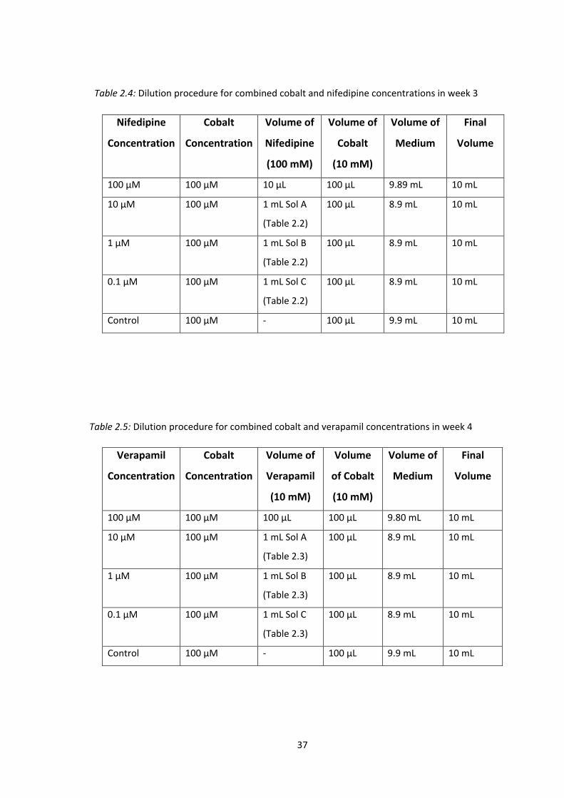
37
Table 2.4: Dilution procedure for combined cobalt and nifedipine concentrations in week 3
Table 2.5: Dilution procedure for combined cobalt and verapamil concentrations in week 4
Nifedipine
Concentration
Cobalt
Concentration
Volume of
Nifedipine
(100 mM)
Volume of
Cobalt
(10 mM)
Volume of
Medium
Final
Volume
100 µM 100 µM 10 µL 100 µL 9.89 mL 10 mL
10 µM 100 µM 1 mL Sol A
(Table 2.2)
100 µL 8.9 mL 10 mL
1 µM 100 µM 1 mL Sol B
(Table 2.2)
100 µL 8.9 mL 10 mL
0.1 µM 100 µM 1 mL Sol C
(Table 2.2)
100 µL 8.9 mL 10 mL
Control 100 µM - 100 µL 9.9 mL 10 mL
Verapamil
Concentration
Cobalt
Concentration
Volume of
Verapamil
(10 mM)
Volume
of Cobalt
(10 mM)
Volume of
Medium
Final
Volume
100 µM 100 µM 100 µL 100 µL 9.80 mL 10 mL
10 µM 100 µM 1 mL Sol A
(Table 2.3)
100 µL 8.9 mL 10 mL
1 µM 100 µM 1 mL Sol B
(Table 2.3)
100 µL 8.9 mL 10 mL
0.1 µM 100 µM 1 mL Sol C
(Table 2.3)
100 µL 8.9 mL 10 mL
Control 100 µM - 100 µL 9.9 mL 10 mL

38
Table 2.6: Dilution procedure for combined cobalt (alternations) and verapamil (fixed)
concentrations in weeks 5 and 6
Weeks 5 and 6
In weeks 5 and 6, four 24 hour cell-seeded plates were treated with increasing
concentrations of cobalt (MTT and NR at 24 and 48 hours), for which the control
was medium.
A further 4 plates were treated with 10 µM verapamil for 2 hours, which was then
removed and a combination of cobalt (increasing concentrations) and verapamil
(fixed concentration) was added. Verapamil was chosen as the calcium channel
blocker in the final weeks of experimentation, as it exhibited a much lower LD50
than nifedipine, and a 10 µM concentration was chosen as no toxic effects were
apparent at this level in week 3. The aim in weeks 3 and 4 was to compare the
viability of cobalt treated cells with cobalt and verapamil treated cells, to attempt to
quantify the ability of verapamil to reduce the toxic effects of cobalt on OST 5 cells,
and thus help to elucidate whether cobalt-induced toxicity takes place via calcium
channels.
Verapamil
Concentration
Cobalt
Concentration
Volume of
Verapamil
(10 mM)
Volume
of Cobalt
(10 mM)
Volume of
Medium
Final
Volume
10 µM 200 µM 15 µL 300 µL 14.685 mL 15 mL
10 µM 100 µM 15 µL 150 µL 14.835 mL 15 mL
10 µM 50 µM 15 µL 75 µL 14.910 mL 15 mL
10 µM 10 µM 15 µL 15 µL 14.970 mL 15 mL
10 µM Control 15 µL - 14.985 mL 15 mL
39
2.6. Viability Assays
The spectrophotometer used for all cell viability assays was a Shimadzu UV-Vis UV-
2401PC, and the plate reader used was a Labsystems Multiskan Ascent.
2.6.1. PBS
Two phosphate buffer solutions (PBS) were made up by dissolving PBS tablets in
500 ml distilled water. Naturally assuming a pH of 7.4, one PBS solution was left at
this pH, while HCL was added dropwise to the other solution to get a pH of 6.75.
2.6.2. Neutral Red
Neutral red (NR) is used as a vital stain. It readily penetrates the cell membranes of
viable cells and, being slightly cationic, binds to anionic sites on the lysosomal
matrix. As neutral red accumulates in the lysosomes, the colour intensity increases
(178).
5 mg neutral red is dissolved in 100 ml neutral pH PBS, incubated overnight at 37 °C
and passed through a 0.2 µm filter, to remove any undissolved crystals remaining.
This can be stored at 4 °C for several weeks.
Method
When cells are ready for NR staining, all surrounding medium was removed, 100 µL
NR solution was added to each well and the plate was incubated at 37 °C for 3
hours. NR was then removed, and each well washed with 200 µL neutral PBS. 100
µL destain was then added to each well, and the plate was shaken for ~30 minutes,
or until a homogenous colour was seen in all wells, allowing a better determination
of viability, expressed as colour intensity. Destain was created by adding 1 mL
glacial acetic acid to 50 mL ethanol and 49 mL distilled H2O in a fume hood.
40
Absorbance was measured at 540 nm, after the plate reader has been allowed
warm up for ~30 minutes.
2.6.3. MTT
MTT assays are colorimetric viability assays which measure enzyme activity in living
cells, by the ability of the enzymes to reduce MTT to formazan, which gives a purple
colour. Dimethyl sulfoxide (DMSO) is used to dissolve the purple formazan
produced into a soluble, coloured solution, the absorbance of which can be
quantified by a spectrometer.
A 10 mM MTT solution was made up by dissolving 0.4143 g MTT in 100 ml PBS pH
6.75, and filtering the resulting solution through a 0.2 µm filter to remove
undissolved crystals. At 4 °C, the MTT solution can be stored for 2 weeks.
Method
Once cells are ready, the surrounding medium was removed from all wells, before
the addition of 50 µL of MTT solution to each well and the plate was incubated for 4
hours at 37 °C. The MTT solution was then removed by pipette, removing as much
as possible so as not to affect the final colour, whilst taking care not to remove the
formazan product created. Plates were centrifuged (5 minutes at ~3500 rpm) to
ensure cells remain at the bottom of the wells. 200 µL DMSO was added to dissolve
formazan, and was mixed to an even colour by pipetting up and down twice in each
well before the absorbance is read at 540 nm.
2.7. Confocal Microscopy and Photography
In week 7, OST 5 cells were set up and stained with a number of chemicals to be
viewed under a confocal microscope and photographed. Cells were seeded in either
41
24 well plates, 1.56 x 10⁴ cells per 2 cm2 well, or 9.6 cm2 petri dishes, 7.5 x 10⁴ cells
per dish. After 24 hours, seeded cells were divided into four groups, with 2
dishes/wells per group: medium only control; cobalt only sample (100 µM);
verapamil only sample (10 µM); combined cobalt and verapamil sample (100 and 10
µM respectively). Staining, viewing and photography all took place at 48 hours post
treatment, where, based on assay results, effects were more likely to be seen than
at 24 hours. The confocal microscope used for the following was a Carl Zeiss Axio
Imager Microscope and the software used alongside the microscope was Axiovision
4.6.
2.7.1. Staining for Actin using Phalloidin-FITC
Phalloidin binds in a highly-selective manner to actin, a protein found in all
eukaryotic cells, which forms microfilaments, and is a cytoskeletal subclass. The
main function of actin is to mechanically support cells and provide routes for
cytoplasmic trafficking and signal transduction (179). Phalloidin can be labelled with
fluorescent analogues to investigate in high-contrast the distribution of the protein
using light microscopy, in this case, the fluorescent green dye fluorescein (FITC). In
this experiment, phalloidin-FITC was used to determine the structure of the
cytoskeleton in each case, and observe the effects that verapamil and cobalt had on
OST 5 cells in comparison with the control sample, and the combined cobalt and
verapamil treated sample.
Phalloidin is an extremely toxic substance, a peptide isolated from the Amanita
phalloides ‘death cap’ mushroom. It must be handled with care and all waste must
be disposed of through correct chemical means; collected in appropriate, labelled
containers taking care to ensure no phalloidin or washings enter drains, and
disposal is entrusted to an Industrial Waste Disposal Company.
0.1 mg Phalloidin-FITC was dissolved in 1 mL methanol, injected directly into the vial
through the rubber seal without opening it. This is carried out in a fume hood for
safety. 4 mL 1% w/v BSA (bovine serum albumin) in PBS was added, giving a dilution
42
of 1/50. The solution was aliquoted into Eppendorff tubes and stored at -20 °C in
the dark.
Method
OST 5 cells are seeded in 24 well plates on 13 mm diameter glass coverslips. After
the designated time, medium is removed and the wells are washed with warm PBS
(37 °C). The cells are then fixed with 4% formalin in PBS in the fume hood for 20
minutes, before being washed three times with PBS.
Staining
During the staining preparations, all materials must be kept in the dark (i.e. under
tin foil, or a dark container). The cells were incubated with Phalloidin-FITC (diluted
to 1/500 in PBS – a 1/10 dilution in PBS was performed on the 1/50 Phalloidin-FITC
above) in a moist chamber for 1 hour, before being washed three times in PBS (the
cells being allowed to steep in PBS for 5 minutes each time). Glass coverslips were
removed from wells using forceps, excess PBS drops were removed and they were
affixed to slides using Immunomount mounting fluid and viewed using fluorescence
microscopy with a dry lens at magnification of x20.
2.7.2. Cell Viability Staining using Propidium Iodide and
CFDA
Propidium Iodide (PI) is a fluorescent, intercalating agent used as a DNA stain in cell
viability evaluation. It can be used to differentiate between apoptotic, necrotic and
normal cells. It binds to DNA, intercalating between bases and once bound, it
fluoresces. Cell permeable dye, carboxyfluorescein diacetate (CFDA) gives a strong,
detectable green fluorescence, produced by hydrolysis inside the cell, with
negligible cell death. PI produces red fluorescence by interacting with the nuclei of
dead cells. Cell Viability can be detected by observing the ratio of green stain
43
(representing live cells) to red stain (representing dead cells) visible under the
microscope.
200 µg Propidium Iodide (PI) was diluted in 20 mL PBS pH 7.4. As CFDA may leak
from cells with time, cells must be prepared for staining and viewed one at a time,
to avoid specimens standing for prolonged periods of time, which may impair
viewing.
Method
The medium was removed from all petri dishes, which were then washed twice with
neutral PBS. 2 mL PI was added to petri dishes and they were incubated in the dark
for 1 minute. PI was removed and discarded to the appropriate waste bottle, and
dishes were washed a further three times with neutral PBS, the washes being
discarded to the same waste bottle. 2 mL CFDA (25 µM in pH 6.75 PBS – CFDA is pH
dependent, and must be used with PBS at pH 6.75) was added to dishes and they
were incubated in the dark at room temperature for 5 minutes. CFDA was removed
and discarded in a separate waste bottle, and cells were washed three times with
pH 6.75 PBS, which was discarded to the same waste bottle as the CFDA. 3ml pH
6.75 PBS was added to each petri dish, and specimens were viewed immediately
using fluorescence microscopy at 620 nm with a wet lens at a magnification of x20.
2.8. Statistics
Statistics were performed using Minitab software, version 16. One way Analysis of
Variance (ANOVA) testing was performed to compare absorbance means of
different concentration groups and controls in cobalt, nifedipine and verapamil-
treated cells. When significance was seen between means of test groups, Post Hoc
Fisher LSD (least significant difference) testing was carried out to explore all
possible pair-wise comparisons for significance.

44
Independent 2-sample Student’s t-tests compared cobalt-treated cells with cobalt
and verapamil-treated cells at each concentration of cobalt. This test is used as the
samples, from two different populations, are independent of each other, with a
similar distribution. Microsoft Excel was used to plot scatter graphs of viability
results and dose-response curves of cobalt and calcium channel blockers, from
which the LD50 values could be obtained.
45
3. Results
3.1. Effect of Cobalt on OST 5 Cell Viability
Data on graphs are expressed as the concentration of cobalt versus the value of NR
absorbance or the reduction of MTT, which are both directly proportional to cell
viability, as these actions are a measure of live cellular activity. A decrease in NR
absorption and MTT reduction is indicative of a decrease in cell viability.
Cobalt-treated OST 5 cells showed a dose-dependent decrease in NR absorbance
and MTT reduction. This was apparent at 24 hours post cobalt treatment, but a
greater effect was seen after 48 hours (see graph 3.2). At 24 hours, both NR (A) and
MTT (B) assays showed similar absorbance/reduction scores. At 48 hours, there was
more variation between the two assays, with NR (A) showing slightly higher scores
at lower concentrations than MTT (B), but both expressed similar
absorbance/reduction scores at the highest concentration used (200 µM). In week
2, viability was also assessed at 72 hours post cobalt-treatment, but as similar
results to those of 48 hour post treatment were yielded here, this time point was
not used again. Also, it was decided that a concentration of 100 µM cobalt would be
used in subsequent experimental stages, where a fixed control of cobalt was
needed, as toxicity was evident at this concentration.

46
Graph 3.1: The effect of increasing concentrations of cobalt on the viability of OST 5 cells at 24
hours, shown by NR (A) and MTT (B) assays. Cobalt was seen to decrease cell viability in a dose-
dependent manner. Data are expressed as mean±SD; n = 12. ANOVA testing showed significant
differences between control and cobalt-treated groups in NR and MTT assays. p > 0.05. Post-hoc
Fisher analysis found significant differences at 24 hours in comparing the control group with 100
and 200 μM; 10 μM with 100 (just NR) and 200 μM; 50 μM with 200 μM (just NR); CI = 95%.
0
0.02
0.04
0.06
0.08
0.1
0.12
0 50 100 150 200 250 N
R A
bso
rban
ce
Cobalt Concentration
0
0.02
0.04
0.06
0.08
0.1
0.12
0 50 100 150 200 250
MTT
Re
du
ctio
n
Cobalt Concentration
A
B
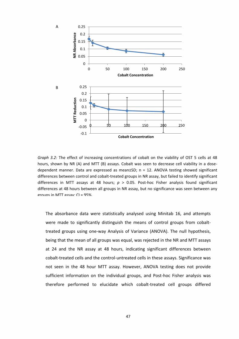
47
The absorbance data were statistically analysed using Minitab 16, and attempts
were made to significantly distinguish the means of control groups from cobalt-
treated groups using one-way Analysis of Variance (ANOVA). The null hypothesis,
being that the mean of all groups was equal, was rejected in the NR and MTT assays
at 24 and the NR assay at 48 hours, indicating significant differences between
cobalt-treated cells and the control-untreated cells in these assays. Significance was
not seen in the 48 hour MTT assay. However, ANOVA testing does not provide
sufficient information on the individual groups, and Post-hoc Fisher analysis was
therefore performed to elucidate which cobalt-treated cell groups differed
0
0.05
0.1
0.15
0.2
0.25
0 50 100 150 200 250
NR
Ab
sorb
ance
Cobalt Concentration
-0.1
-0.05
0
0.05
0.1
0.15
0.2
0.25
0 50 100 150 200 250
MTT
Re
du
ctio
n
Cobalt Concentration
Graph 3.2: The effect of increasing concentrations of cobalt on the viability of OST 5 cells at 48
hours, shown by NR (A) and MTT (B) assays. Cobalt was seen to decrease cell viability in a dose-
dependent manner. Data are expressed as mean±SD; n = 12. ANOVA testing showed significant
differences between control and cobalt-treated groups in NR assay, but failed to identify significant
differences in MTT assays at 48 hours; p > 0.05. Post-hoc Fisher analysis found significant
differences at 48 hours between all groups in NR assay, but no significance was seen between any
groups in MTT assay; CI = 95%.
A
B
48
significantly from the control and from each other. Significant differences between
various groups were seen in both MTT and NR assays, with the exception of the
MTT assay at 48 hours. In the 24 hour NR and MTT assays, significant differences
were seen when comparing the control group to the 100 and 200 μM groups,
comparing 10 μM with 200 μM group, and in the NR, but not the MTT assay,
significant difference was seen between the 10 and 100 μM groups and the 50 and
200 μM treated cells. At 48 hours, Post-hoc Fisher analysis showed significant
differences between all groups of the NR assay.
Dose response curves were constructed from the MTT and NR assay results,
expressing all results as a percentage of the control and plotting against the log10
concentration of cobalt to obtain LD50 values. At 48 hours post cobalt treatment,
the LD50 values were calculated as 98.08 µM from the NR assay and 169.69 µM from
the MTT assay. This offered further suggestion that a concentration of 100 µM
cobalt is a sufficiently toxic level of the metal to allow us to subsequently
investigate the effect of calcium channel blockers on the toxicity.

49
3.2. Effect of Nifedipine on OST 5 Cell Viability
3.2.1. Nifedipine
Nifedipine was the first of two calcium channel blockers to be used experimentally
in a bid to prevent cobalt-induced toxicity in these cells by blocking entry of the
y = -15.55ln(x) + 121.31
0
10
20
30
40
50
60
70
80
90
100
1 10 100 1000
Ce
ll V
iab
ility
Log10 Cobalt Concentration
y = -12.44ln(x) + 113.57
0
10
20
30
40
50
60
70
80
90
100
1 10 100 1000
Ce
ll V
iab
ility
Log10 Cobalt Concentration
Graph 3.3: Dose response curves were plotted, viability versus the log10 scale of cobalt concentration,
to obtain LD50 values shown by NR (A) – 98.08 µM; and MTT (B) – 169.69 µM at 48 hours; n = 12.
A
B
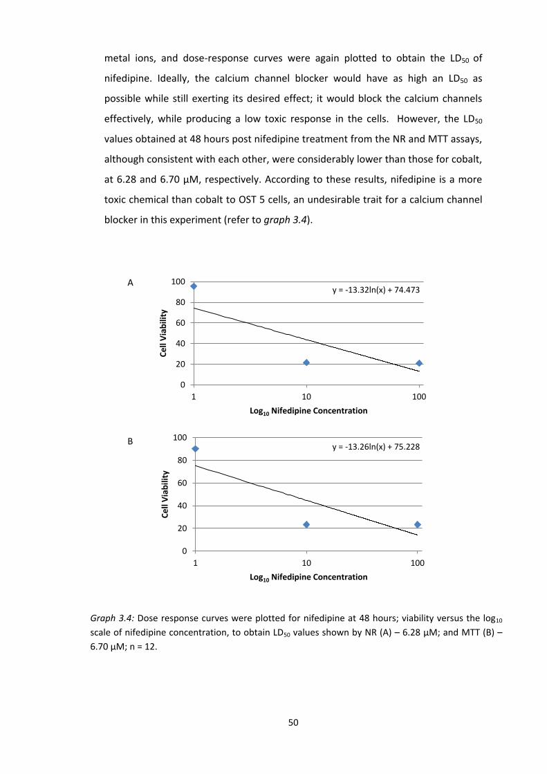
50
Graph 3.4: Dose response curves were plotted for nifedipine at 48 hours; viability versus the log10
scale of nifedipine concentration, to obtain LD50 values shown by NR (A) – 6.28 µM; and MTT (B) –
6.70 µM; n = 12.
metal ions, and dose-response curves were again plotted to obtain the LD50 of
nifedipine. Ideally, the calcium channel blocker would have as high an LD50 as
possible while still exerting its desired effect; it would block the calcium channels
effectively, while producing a low toxic response in the cells. However, the LD50
values obtained at 48 hours post nifedipine treatment from the NR and MTT assays,
although consistent with each other, were considerably lower than those for cobalt,
at 6.28 and 6.70 µM, respectively. According to these results, nifedipine is a more
toxic chemical than cobalt to OST 5 cells, an undesirable trait for a calcium channel
blocker in this experiment (refer to graph 3.4).
y = -13.32ln(x) + 74.473
0
20
40
60
80
100
1 10 100
Ce
ll V
iab
ility
Log10 Nifedipine Concentration
y = -13.26ln(x) + 75.228
0
20
40
60
80
100
1 10 100
Ce
ll V
iab
ility
Log10 Nifedipine Concentration
A
B
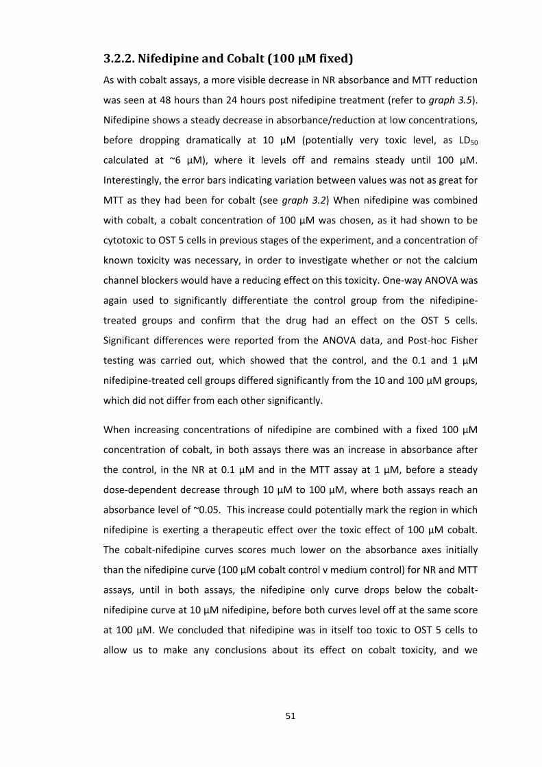
51
3.2.2. Nifedipine and Cobalt (100 µM fixed)
As with cobalt assays, a more visible decrease in NR absorbance and MTT reduction
was seen at 48 hours than 24 hours post nifedipine treatment (refer to graph 3.5).
Nifedipine shows a steady decrease in absorbance/reduction at low concentrations,
before dropping dramatically at 10 µM (potentially very toxic level, as LD50
calculated at ~6 µM), where it levels off and remains steady until 100 µM.
Interestingly, the error bars indicating variation between values was not as great for
MTT as they had been for cobalt (see graph 3.2) When nifedipine was combined
with cobalt, a cobalt concentration of 100 µM was chosen, as it had shown to be
cytotoxic to OST 5 cells in previous stages of the experiment, and a concentration of
known toxicity was necessary, in order to investigate whether or not the calcium
channel blockers would have a reducing effect on this toxicity. One-way ANOVA was
again used to significantly differentiate the control group from the nifedipine-
treated groups and confirm that the drug had an effect on the OST 5 cells.
Significant differences were reported from the ANOVA data, and Post-hoc Fisher
testing was carried out, which showed that the control, and the 0.1 and 1 μM
nifedipine-treated cell groups differed significantly from the 10 and 100 μM groups,
which did not differ from each other significantly.
When increasing concentrations of nifedipine are combined with a fixed 100 µM
concentration of cobalt, in both assays there was an increase in absorbance after
the control, in the NR at 0.1 µM and in the MTT assay at 1 µM, before a steady
dose-dependent decrease through 10 µM to 100 µM, where both assays reach an
absorbance level of ~0.05. This increase could potentially mark the region in which
nifedipine is exerting a therapeutic effect over the toxic effect of 100 µM cobalt.
The cobalt-nifedipine curves scores much lower on the absorbance axes initially
than the nifedipine curve (100 µM cobalt control v medium control) for NR and MTT
assays, until in both assays, the nifedipine only curve drops below the cobalt-
nifedipine curve at 10 µM nifedipine, before both curves level off at the same score
at 100 µM. We concluded that nifedipine was in itself too toxic to OST 5 cells to
allow us to make any conclusions about its effect on cobalt toxicity, and we

52
therefore changed to another calcium channel blocker, verapamil, to see was it less
toxic, enabling its use with cobalt.
0
0.05
0.1
0.15
0.2
0.25
0.3
0 20 40 60 80 100
Ab
sorb
ance
Nifedipine Concentration
Co+Nif
Nif
0
0.05
0.1
0.15
0.2
0.25
0 20 40 60 80 100
Ab
sorb
ance
Nifedipine Concentration
Co+Nif
Nif
Graph 3.5: The effect of increasing concentrations of the calcium channel blocker nifedipine, and the
effect of a fixed concentration of cobalt (100 µM) in combination with increasing concentrations of
calcium channel blocker nifedipine on OST 5 cell viability at 48 hours, as shown by NR absorbance (A)
and MTT reduction (B). Nifedipine was seen to decrease viability in a dose-dependent manner. Data
are expressed as mean±SD; n = 12. Cobalt and nifedipine combined were seen to show lower viability
at low concentrations, higher viability at a concentration of 10 µM nifedipine, and similar viability
results at 100 µM nifedipine, than nifedipine alone. ANOVA analysis showed significant differences in
the means of nifedipine-treated groups compared to controls in NR and MTT assays (p > 0.05). Post-
hoc Fisher analysis showed that each the control, 0.1 and 1 μM cobalt-treated groups differed
significantly from the 10 and 100 μM groups in the NR and MTT assays at 48 hours; CI = 95%.
A
B
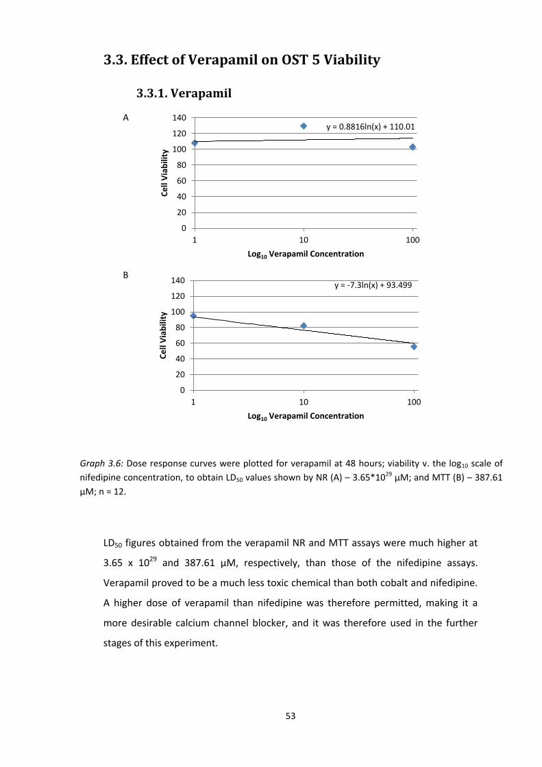
53
3.3. Effect of Verapamil on OST 5 Viability
3.3.1. Verapamil
LD50 figures obtained from the verapamil NR and MTT assays were much higher at
3.65 x 1029 and 387.61 µM, respectively, than those of the nifedipine assays.
Verapamil proved to be a much less toxic chemical than both cobalt and nifedipine.
A higher dose of verapamil than nifedipine was therefore permitted, making it a
more desirable calcium channel blocker, and it was therefore used in the further
stages of this experiment.
y = 0.8816ln(x) + 110.01
0
20
40
60
80
100
120
140
1 10 100
Ce
ll V
iab
ility
Log10 Verapamil Concentration
y = -7.3ln(x) + 93.499
0
20
40
60
80
100
120
140
1 10 100
Ce
ll V
iab
ility
Log10 Verapamil Concentration
Graph 3.6: Dose response curves were plotted for verapamil at 48 hours; viability v. the log10 scale of
nifedipine concentration, to obtain LD50 values shown by NR (A) – 3.65*1029 µM; and MTT (B) – 387.61
µM; n = 12.
A
B
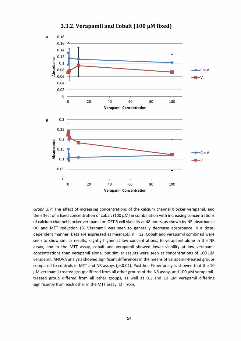
54
3.3.2. Verapamil and Cobalt (100 µM fixed)
0
0.02
0.04
0.06
0.08
0.1
0.12
0.14
0.16
0.18
0 20 40 60 80 100
Ab
sorb
ance
Verapamil Concentration
Co+V
V
0
0.05
0.1
0.15
0.2
0.25
0.3
0 20 40 60 80 100
Ab
sorb
ance
Verapamil Concentration
Co+V
V
Graph 3.7: The effect of increasing concentrations of the calcium channel blocker verapamil, and
the effect of a fixed concentration of cobalt (100 µM) in combination with increasing concentrations
of calcium channel blocker verapamil on OST 5 cell viability at 48 hours, as shown by NR absorbance
(A) and MTT reduction (B. Verapamil was seen to generally decrease absorbance in a dose-
dependent manner. Data are expressed as mean±SD; n = 12. Cobalt and verapamil combined were
seen to show similar results, slightly higher at low concentrations, to verapamil alone in the NR
assay, and in the MTT assay, cobalt and verapamil showed lower viability at low verapamil
concentrations than verapamil alone, but similar results were seen at concentrations of 100 µM
verapamil. ANOVA analysis showed significant differences in the means of verapamil-treated groups
compared to controls in MTT and NR assays (p<0.01). Post-hoc Fisher analysis showed that the 10
μM verapamil-treated group differed from all other groups of the NR assay, and 100 μM verapamil-
treated group differed from all other groups, as well as 0.1 and 10 μM verapamil differing
significantly from each other in the MTT assay. CI = 95%.
A
B

55
In verapamil-treated OST 5 cells at 48 hours, the NR assay showed a steady increase
in absorbance as concentration increased, with viability peaking at 10 µM verapamil
before dropping down, where the 100 µM verapamil point shows a similar score to
the 0.1 µM point. This would suggest that verapamil at these concentrations is non-
toxic, and even appears to increase cell numbers, with 10 µM appearing the most
therapeutic concentration, and this was used in further stages of this experiment in
a bid to prevent cobalt-induced toxicity, without exhibiting further toxic effects to
cells. However, the MTT assay at 48 hours showed slightly different results. The only
concentration which exhibits higher MTT reduction than the control is at 0.1 µM,
and the increase is minor. After this point, there is a steady dose-dependent
decrease in reduction as verapamil concentration increases. The MTT reduction for
the control and all verapamil concentrations is much higher than the NR
absorbance. One-way ANOVA testing was again used to prove there were significant
differences in the means of verapamil-treated cells in comparison to the control
mean, and the data then underwent Post-hoc Fisher analysis which showed a
significant difference between the 10 μM group in comparison with the control and
all other verapamil-treated groups in the NR assay. The MTT assay showed more
varied results, reporting significant differences between the 100 μM verapamil
group in comparison to all other groups, and also significant difference was seen
between 0.1 and 10 μM verapamil. The significance difference seen at 10 μM
verapamil in the NR assay is unsurprising due to the visible peak on graph 3.7 seen
at this concentration.
When viability of cobalt and verapamil combined was assessed, the NR and MTT
assays showed similar results and curves on their respective graphs. In the NR assay,
there was a dose-dependent decrease in absorbance, with the exception of the 0.1
µM point, which was lower on the graph than all other concentration points.
However, all points on the cobalt and verapamil curve fall above their respective
points on the verapamil only curve. The MTT assay cobalt and verapamil curve
displays more erratic points than the NR assay, with all verapamil concentrations
exhibiting higher MTT reduction points than the control, the highest of which being

56
at 0.1 µM, after which it drops dramatically to a point slightly higher than that of
the control for 1 and 10 µM, before rising again slightly at 100 µM. Again, all points
on the cobalt and verapamil curve fall above those of the verapamil only curve, with
the exception of the 100 µM point, where absorbance of both curves were very
similar.
3.3.3. Verapamil (10 µM fixed) and Cobalt
In the final stage of the experiment, a concentration of 10 µM verapamil was used
as it showed, in a previous MTT assay of this experiment, a non-toxic, and potential
calcium channel-blocking effect on the OST 5 cells. It was decided that this was the
verapamil concentration at which the drug was most likely to have a reducing effect
on the cytotoxicity caused to the OST 5 cells by cobalt, which was administered in
increasing doses, in an attempt to investigate the interaction with verapamil.
At 24 hours, theNR assay shows general dose-dependent decreases in absorbance
in both the combined cobalt-verapamil group and the cobalt-treated group.
Independent 2-sample Students’ t-tests were carried out to compare absorbance
levels at every concentration of cobalt between the two groups, and significant
differences were seen at 50 and 100 µM in this NR assay. All points on the cobalt
only curve fall above those on the cobalt and verapamil curve, besides the control.
In the MTT assay, the effect of cobalt on absorbance was more erratic than the NR
assay and it did not appear to represent a dose-dependent decrease in cell viability,
as at 10 and 200 µM, absorbance was higher than at their respective previous
points on the curve. Also, in this assay, two points of the cobalt only curve lay below
the corresponding points of the cobalt and verapamil curve, 10 and 100 µM. The
cobalt and verapamil curve of the MTT assay followed a dose-dependent MTT
reduction decrease similar to the one seen in absorbance in the NR assay, with the
exception of the 200 µM point, which has a higher reduction than at 100 µM.
Independent 2-sample Student’s t-tests showed significance between the two
57
groups at only 10 µM cobalt. All points on the MTT assay graph were much lower
than the points on the NR assay graph.
At 48 hours, the NR and MTT assays exhibited similar results to the assays at 24
hours. All points on the cobalt curve in the NR assay fall above those on the cobalt
and verapamil curve, with the exception of the 200 µM mark. Significance was
0
0.05
0.1
0.15
0.2
0.25
0.3
0 50 100 150 200
NR
Ab
sorb
ance
Cobalt Concentration (µM)
Co+V
Co
0
0.02
0.04
0.06
0.08
0.1
0.12
0 50 100 150 200
MTT
Re
du
ctio
n
Cobalt Concentration (µM)
Co+V
Co
Graph 3.8: The effect of cobalt on the viability of OST 5 cells when combined with a fixed
concentration of calcium channel blocker verapamil (10 µM) at 24 hours, shown by NR (A) and
MTT (B) assays. Data are expressed as mean±SD; n = 12. Independent 2-sample Students t-tests
showed significance at 50 and 100 μM in NR and at 10 μM in MTT; p > 0.05.
A
B
58
shown by t-test between the cobalt and cobalt-verapamil groups at cobalt
concentrations of 10, 50 and 100 µM. The cobalt generally follows a dose-
dependent decrease pattern, but at 1 µM, the absorbance is higher than the
control. The cobalt and verapamil curve follows an obvious dose dependent
pattern, which can be likened to the same curve at 24 hours. The MTT assay
displays a dose-dependent decrease in MTT reduction in the cobalt and verapamil
curve, and also in the cobalt only curve, with the exception of the last point, which
shows an increase from the previous point. Students t-test found significance
between the two treatment groups at 10 and 200 µM. Again, most points of the
cobalt curve fall above the corresponding points of the cobalt and verapamil curve.
According to the results from these viability assays, and the graphs constructed
from the results, the addition of a fixed concentration of verapamil (10 µM) to
increasing concentrations of cobalt does not significantly raise the NR absorbance
or MTT reduction of the OST 5 cells, in comparison to cells treated with only cobalt.
Therefore, the verapamil does not appear to prevent the cobalt-induced
cytotoxicity observed in this experiment.
59
0
0.05
0.1
0.15
0.2
0.25
0.3
0.35
0.4
0.45
0 50 100 150 200
NR
Ab
sorb
ance
Cobalt Concentration
Co+V
Co
0
0.05
0.1
0.15
0.2
0.25
0.3
0 50 100 150 200
MTT
Re
du
ctio
n
Cobalt Concentration
Co+V
Co
Graph 3.9: The effect of cobalt on the viability of OST 5 cells when combined with a fixed
concentration of calcium channel blocker verapamil (10 µM) at 48 hours, shown by NR (A) and
MTT (B) assays. Data are expressed as mean±SD; n = 12. Independent 2-sample Students t-tests
showed significance at 10, 50 and 100 μM cobalt (NR) and in the control and at 10 and 200 μM
cobalt; p > 0.05.
A
B

60
3.4. Osteoblast Images
3.4.1. Unstained OST 5 Cell Images
L
Images were taken of the unstained OST 5 cells under a microscope at a
magnification of x200 at 48 hours post-treatment with control, 10 µM verapamil,
combined 10 µM verapamil and 100 µM cobalt combined, and 100 µM cobalt. The
cells appeared much less confluent in the verapamil (B), and the combined
verapamil and cobalt (C) treated cells, in comparison with the control (A). There
appears to be more empty space between the cells, which are aggregated in
Figure 3.1: The visible effects of fixed concentrations of 10 µM verapamil (B), a combination of 100
µM cobalt and 10 µM verapamil (C) and 100 µM cobalt (D) on OST 5 cells, cultured for 24 hours,
compared to a control (A) at 48 hours post-treatment. There were notable deviations in
appearance of cells in all groups when compared to the control cells, but the cobalt and
cobalt+verapamil treated groups bore resemblances to each other.
A B
C D

61
clusters, especially in the verapamil group. There are less nuclei visible in the cobalt
(D) treated cells, and the cells themselves appear much more granular than the cells
of the other groups. However, the cobalt-treated image (D) appears to have less
empty space between cells than in verapamil (B) and cobalt-verapamil (C) treated
cells. Dose-response curves for verapamil and cobalt in this experimentation show a
decrease in OST 5 cell viability for both chemicals, with the latter exhibiting a more
toxic response. The reduction in living cells in the above images correlates with the
reduction in living cells exhibited by the dose-response curves. However, the
appearance of the cells in the verapamil (B) and cobalt-verapamil (C) treated cells
suggests that verapamil has an effect on the morphology of these cells.
3.4.2. Phalloidin-FITC Stained OST 5 Cells
Cells were seeded for 24 hours before treatment, and all images were taken 48
hours post-treatment. Phalloidin-FITC staining showed visible reductions in the
presence of actin in all treatment groups in comparison to the control (A). The
control group showed a multidimensional presence of actin layers with few gaps. In
verapamil (B) and verapamil and cobalt (C) treated cells, there is an obvious
decrease in the volume of actin filaments; there appears to be less 3-dimensional
layers of actin and more gaps between filaments. The actin also appears to be a
brighter colour in the control group (A) when compared to group B and C. The
cobalt treated (D) group displays actin much more sparsely than the control; there
is very little 3D layering and there are much more gaps between filaments than in
all other groups. Groups B and C seem to have a similar level of actin filaments
present in these images, and cobalt-treated cells seemed to have less actin
filaments present compared to cells treated with verapamil as well as cobalt. Actin
filaments appear to be distributed evenly throughout all cells observed in control
group and all treatment groups.
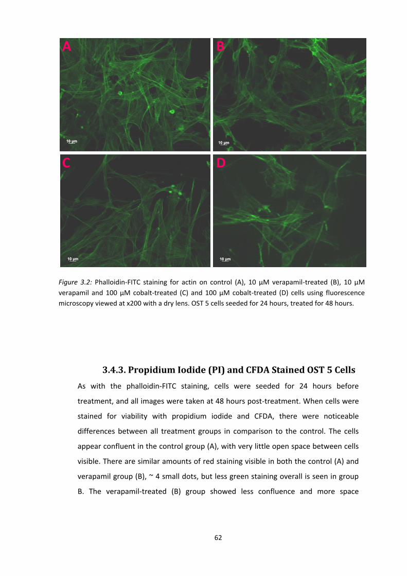
62
3.4.3. Propidium Iodide (PI) and CFDA Stained OST 5 Cells
As with the phalloidin-FITC staining, cells were seeded for 24 hours before
treatment, and all images were taken at 48 hours post-treatment. When cells were
stained for viability with propidium iodide and CFDA, there were noticeable
differences between all treatment groups in comparison to the control. The cells
appear confluent in the control group (A), with very little open space between cells
visible. There are similar amounts of red staining visible in both the control (A) and
verapamil group (B), ~ 4 small dots, but less green staining overall is seen in group
B. The verapamil-treated (B) group showed less confluence and more space
Figure 3.2: Phalloidin-FITC staining for actin on control (A), 10 µM verapamil-treated (B), 10 µM
verapamil and 100 µM cobalt-treated (C) and 100 µM cobalt-treated (D) cells using fluorescence
microscopy viewed at x200 with a dry lens. OST 5 cells seeded for 24 hours, treated for 48 hours.
A B
C D
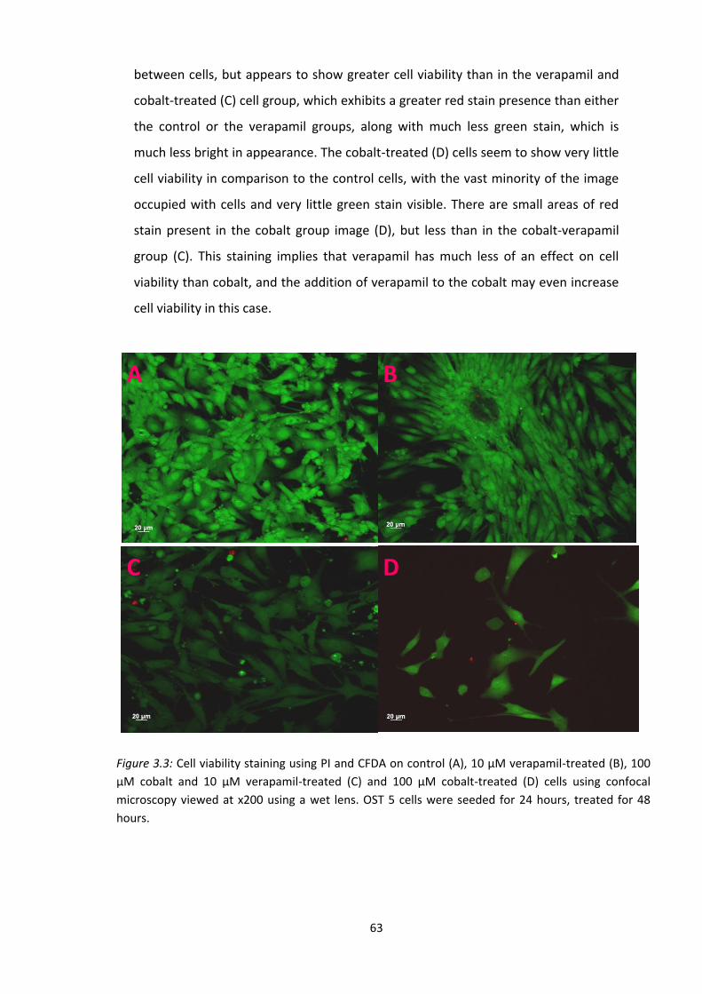
63
Figure 3.3: Cell viability staining using PI and CFDA on control (A), 10 µM verapamil-treated (B), 100
µM cobalt and 10 µM verapamil-treated (C) and 100 µM cobalt-treated (D) cells using confocal
microscopy viewed at x200 using a wet lens. OST 5 cells were seeded for 24 hours, treated for 48
hours.
between cells, but appears to show greater cell viability than in the verapamil and
cobalt-treated (C) cell group, which exhibits a greater red stain presence than either
the control or the verapamil groups, along with much less green stain, which is
much less bright in appearance. The cobalt-treated (D) cells seem to show very little
cell viability in comparison to the control cells, with the vast minority of the image
occupied with cells and very little green stain visible. There are small areas of red
stain present in the cobalt group image (D), but less than in the cobalt-verapamil
group (C). This staining implies that verapamil has much less of an effect on cell
viability than cobalt, and the addition of verapamil to the cobalt may even increase
cell viability in this case.
A B
C D

64
4. Discussion
This study confirmed that cobalt significantly reduced viability of OST 5 cells in a
dose dependent manner. The reduced viability in NR assay is represented by
decreased absorbance, indicative of damage to cell lysosomes which, when healthy,
red NR dye accumulates, increasing in intensity. NR assay at 24 hours confirmed
significant reduction in viability in higher cobalt concentrations (≥ 100 µM) and in
concentrations ≥ 10 µM at 48 hours, which suggested that the toxic effects of cobalt
were increased with incubation time. Though the MTT graphs exhibited similar
curves to the NR graphs, significant differences was not seen between control cells
and cobalt-treated cells in the MTT 48 hour assay, and this is most likely due to the
large standard deviation in the figures of cells of each group, especially at 200 μM
(Graphs 3.1, 3.2).
According to dose-response curves, nifedipine appeared more cytotoxic to OST 5
cells than cobalt, and was substituted by the less toxic verapamil, which displayed
an LD50 which was of a much higher concentration than both nifedipine and cobalt.
In this study, nifedipine a dose-dependent reducing effect on the OST 5 cells,
exhibited by the decreased absorbance of NR and the reduction of MTT and
increasing concentrations of nifedipine did not significantly alter the extent of
cobalt-induced toxicity, at a fixed concentration of 100 µM cobalt, in OST 5 cells at
the concentrations and time scales measured. Combined nifedipine and cobalt-
treated cells showed lower absorbance figures at 10 and 100 µM nifedipine, than
the 100 µM cobalt only control at 48 hours (Graph 3.5).
However, in graph 3.7, verapamil and cobalt combined have significantly higher
MTT reduction at 100 µM verapamil – 100 µM cobalt, than in the control of 100 µM
cobalt at 48 hours. This may suggest the involvement of verapamil and calcium
channels in the alteration of the toxic effects of cobalt. However, this is not strong
evidence, as it is not seen in the NR assay of the same time point. Interestingly,
65
there is a significant increase in absorbance at 10 µM verapamil compared with the
control and 1 µ verapamil, and this indicates an increase in cell numbers. Graph
curves depicting the viability scores of 10 µM verapamil in combination with
increasing cobalt concentrations closely followed the curves of increasing
concentrations of cobalt only, at 24 and 48 hours (Graphs 3.8, 3.9). Though
significance was seen between groups of the cobalt-treated and verapamil and
cobalt-treated graphs at various concentrations of cobalt, the vast majority of this
significance indicated that cobalt-treated cells exhibited higher absorbance levels
than verapamil and cobalt-treated cells, in both MTT and NR assays at 24 and 48
hours. Therefore, there was no significant evidence seen in these viability assays
that the calcium channel blocker verapamil had a limiting effect on the toxicity
exerted by cobalt on OST 5 cells.
Differences in NR and MTT assay viability absorbance may be due to the fact that,
though both a capable demonstrator of viability, they work by different
mechanisms; NR measures the ability of the intracellular lysosymes to bind and
retain the red dye, while MTT measures the respiratory capacity of the cell. The
generally-higher viability scores seen in NR assays across the experiment, in
comparison to MTT viability scores, may be due to the fact that MTT is a slightly
more sensitive assay in assessing toxicity regarding material as small as
nanoparticle-sized (180).
Unstained OST 5 photos magnified at x200 show that cells treated with verapamil
and a combination of verapamil and cobalt portray a much less confluent picture
than that of the untreated, control group. The cells appear to be fewer in number,
indicating that cell division has been inhibited. Cells appear less confluent in the
cobalt-treated group, compared to the control, and the cells appear noticeably
more granular than in all other groups. The phalloidin-FITC stained cells portray
multidimensional layers of actin filaments in the control group. There is an obvious
decline in 3-dimensional actin layering in verapamil, cobalt, and combined
verapamil and cobalt-treated OST 5 cells, with the latter being the most affected,
66
displaying extremely sparse filaments. This is again most likely to be due to the cells
growing less densely, with cell division having been inhibited. As the function of
actin protein in the cell cytoskeleton is to provide mechanical support and aid
cytoplasmic trafficking and signalling, a reduction in the presence of actin may
suggest less viable cells are present, and that proliferation may have been altered
by the cobalt, and the verapamil and cobalt-verapamil combination in the other
groups. There is no evidence to suggest apoptosis occurs in these OST 5 cells. In
apoptotic cells, actin moves to encircle the cell membrane, making it stronger so
that cell contents to do leak out, and this does not appear to occur in these images.
Viability testing with propidium iodide and CFDA exhibited similar results in control
and verapamil-treated cells, where there is much green stain visible, suggesting
confluent and viable cells. Verapamil and cobalt treated cell images appear less
confluent than control cell images, where more space can be seen between cells.
Cobalt-treated cells appear to have decreased substantially in number compared to
the control. Very little cells can be seen in this image. Distinctive loss of membrane
integrity cannot be seen in these pictures, which is a reason apoptosis would be
suspected as a method of cell death here over necrosis. PI has become a widely-
used method of evaluating apoptosis in cells, relying on the fact that apoptotic cells
are characterized partly by DNA fragmentation, and this chemical binds to and
labels DNA, making it possible to analyse cellular DNA content (181). A TUNEL assay
would be another method by which to further confirm or deny the incidence of
apoptosis in these cells. However, the cells do not appear to be withdrawing from
each other, which is a classic indication of apoptosis. Also, there is a lack of red
staining seen in these images here, and combined with the dwindling
concentrations of green staining this may suggest that, rather than being
responsible for cell death, the cobalt may be affecting and perhaps halting cell
proliferation, resulting in a reduced number of cells, without an increased
occurrence of cell death. MTT and NR assays support this theory also, with viability
assays measure a decrease in viability, not necessarily cell death, and a reduction in
viable cells could be responsible for the decreases seen in NR absorbance and MTT

67
reduction in this project. Also, cobalt-treated cells appeared less confluent than
control cells at 48 hours when examined under microscope in well plates prior to
viability assays being carried out.
4.1. Are Calcium Channels Involved?
It would appear from the results obtained in this experiment that cobalt does not
exert its toxic effects via L-type voltage-dependent calcium channels (VDCCs).
However, there is not enough conclusive evidence present here to completely
eradicate the theory, based on the viability assay results seen following the addition
of the calcium channel blockers. One would envisage that had calcium channels
been involved in the toxicity seen in these OST 5 cells, their blockage would be
sufficient to significantly reduce the effects of cobalt, thus increasing the viability
scores seen in NR and MTT assays, which failed to occur. Previous experimentation
has suggested that at concentrations of 10 µM, verapamil has inhibited cellular
processes in osteoblasts believed to be governed by L-type voltage-gated calcium
channels (175) and, at concentrations of as low as 1.5 µM, verapamil has prevented
a PTH-induced increase in free cytosolic calcium by 50% (176).
However, sufficient steps to ensure verapamil had completely blocked all calcium
channels at a dose of 10 µM were not taken in this experiment. Also, had the
calcium channels been adequately obstructed by verapamil, would it be possible for
cobalt to act via different channels? It would be beneficial to investigate the effects
of cobalt on alternative entry routes into osteoblasts, and ultimately, other cells
also.
Further investigation must be carried out to help clarify the results seen here, and
help further elucidate the relationship between calcium channels and cobalt
toxicity. Firstly, Ca2+ regulation could be monitored in cells, to ensure that verapamil
and nifedipine were adequately obstructing calcium channels at the concentrations
used. Calcium imaging is carried out by fluorescent protein indicators which can
68
chelate calcium ions. The fluorescent dye Fura-2, a pentacarboxylate calcium
indicator developed in the 1970s, is considered the most popular fluorescent
cystolic calcium probe (182), allowing the monitoring of the free cytosolic
concentration of calcium, [Ca2+]i, in living cells. Fura-2-acetoxymethyl ester (Fura-
2AM) is the membrane-permeable derivative of Fura-2, which regenerates into
Fura-2 once diffused into cells when cellular esterases remove the acetoxymethyl
ester group. Fura-2 becomes excited at 340 – 380 nm of light, and the emissions at
these wavelengths directly correspond to the amount of intracellular calcium
present. If the calcium channel blockers used in this experiment were sufficient in
their concentrations, [Ca2+]i would be lower in verapamil/nifedipine treated groups
in comparison to the control group.
Another method by which to assess the adequacy of the calcium channel blockers
would be to introduce a calcium channel agonist, and investigate whether the
actions of the agonist were revoked by the concentrations of the antagonists,
verapamil and nifedipine, used in this experiment. Bay K8644 is a known L-type
voltage-gated calcium channel agonist. This chemical is primarily used
experimentally in this manner, and has been known to increase [Ca2+]i in a
concentration-dependent manner in osteoblasts (163) and human oral cancer cells
(OC2) (183). Bay K8644 has also stimulated the secretion of bone matrix protein
osteocalcin in osteoblast-like osteosarcoma cells, and increases bone creation and
bone resorption (184); the resorption seen was impeded by desmethoxyverapamil,
a verapamil type radioligand developed for studying calcium channels (185). This
Bay K8644-induced Ca2+ influx has also previously been blocked by nifedipine (183).
If, at the concentrations used in this study, the increase of [Ca2+]i is blocked, the
nifedipine and verapamil would be considered to have impeded the action of Bay
K8644, and are therefore sufficient in these concentrations as calcium channel
antagonists.
69
4.3. Alternative Materials in Total Hip Arthroplasty
Due to the recent adverse attention commanded by the use of metal in hip
prosthetics, and the ongoing pursuit to develop the most inert and compliant
implant possible for human use, other materials are continually being assessed and
considered for clinical application. Presently, there exists no faultless alternative
material to cobalt chromium for use in THA; there are pros and cons associated with
every potential substance.
4.3.1. Ceramics
Alumina ceramic (Al2O3) is an example of one such material, investigated due to its
reported good performance as a bearing material in terms of annual wear (89, 186,
187). However, to avoid excessive wear, extreme care must be taken during the
positioning of the device (188), and impingement at the femoral neck can lead to
wear and osteolysis (189). Less volumetric wear is believed to occur over time in
ceramic on ceramic (CC) implants, rather than MM, as alumina is a harder material
(i.e. Young’s modulus is higher) (190, 191), but more, smaller particles are expected
to be released from the former (192). There is little known about the accumulation
of alumina ceramic wear debris in the periprosthetic tissue and its effect in vivo, in
comparison to what is known about MM and MP wear debris and its effects. The
potential effects of these particles on cells must be determined in order to establish
the probable long-term effects of the generation of these particles in vivo. In a
study using histiocytes and fibroblasts, Germain et al, 2002, found Al2O3 particles to
be toxic in nanoparticle size, possibly due to the lowering at this size in the
oxidation state of alumina, which is usually stable in large particles. However, these
particles were still seen to be less cytotoxic than CoCr particles in this study (89).
Zirconia ceramic was proposed to be tougher, with improved wear properties, than
alumina ceramic (193). But the case studies investigated in this paper did not put to
rest the concerns of ceramic being a brittle substance that can break, and the
improved zirconia ceramic could not eliminate the incidence of catastrophic

70
breakage. The dilemma with these devices involves choosing low wear ceramic,
which carry a risk of fracture, or higher wear cobalt-chromium, which will not
fracture. Hummer et al, 1996, recommended a return to cobalt-chromium as the
material of choice given all the information available at the time (193). Park et al,
2006, also recommended discontinued use of alumina ceramic, due to catastrophic
breakage, and an audible squeaking noise has been reported with CC implants,
which could irritate the patient (194, 195).
4.3.2. Titanium
Titanium alloys are strong and excellently tolerated by the body, and have long
since been used successfully in dental implants. However, despite desirable
mechanical characteristics, they exhibit a low resistance to wear. Titanium alloys
are used in THA, not as the articulating surface, but as the stem component,
coupled with a femoral head constructed from another material in order to avoid
the titanium stem being subjected to abrasion and rubbing. An attractive
characteristic of titanium is its elasticity; an elastic stem can evenly distribute and
transport stresses applied from the body’s weight through the whole stem.
Corrosion of the titanium stem is also rare, due to passivation by titanium oxide
(196). However, these implants are not without their problems, as intractable pain
and crevice corrosion leading to cortical hypertrophy have been reported in
association with titanium stems (196).
4.4. What’s Next for Cobalt-Chromium in THA?
Cobalt was the focal point of this experiment, rather than chromium, due to its
ability to enter cells quickly and be transported readily to all organs of the body,
indicating great potential for widespread systemic damage. Higher circulating levels
of cobalt ions than chromium also indicate the implications cobalt has for adverse

71
systemic effects in patients, as well as the local damage it has been seen to cause in
the periprosthetic area of MM THA and HR patients.
While it is insisted that patients with CoCr hip prostheses who experience such
adverse effects outlined in this study are in the extreme minority, with a process
that is being carried out at such an increasing rate worldwide and one which is vital
to the treatment of osteoporosis, any related adverse reaction encountered must
be regarded a cause of concern and investigated thoroughly. The results observed
in this experiment draw parallels with previous work, whether or not the effects are
mediated via calcium channels, using these concentrations of cobalt in lymphocytes
(197) and human osteoblast-like SaSO-2 cells (177).
4.4.1. Infection Control
Another method to potentially reduce need for revision in MM THA and HR is to
attempt to prevent the development of infection, which has previously been cited
as a main reason for implant failure. Emerging antibiotic-resistant strains of
common bacteria pose a serious problem for MM implants and implant-related
infections. Though the percentage of patients who develop implant-associated
infections is low, the procedure is gaining popularity, so there is a demand for new
methods of preventing these infections. A recent article in Nature magazine (April
2012) discussed mechanisms for the controlled release of antimicrobials at the site
of implant. These included the administration of a matrix, serving as a depot for
antimicrobial agents, with the implant, antimicrobial coatings on titanium
components to prevent intramedullary infection, the use of the antimicrobial silver
ion for incorporation into pre-coated implants, and fatty acid cis-2-deconoic acid
(C2DA) has shown the potential to prevent biofilm formation, and disrupt pre-
existing biofilms (198), which may be beneficial as targeting infections established
in biofilms is difficult (199).

72
Further research must be devoted to elucidating how cobalt operates at a cellular
level and every mechanism by which it elicits its toxic systemic and local tissue
effects. When a further understanding of cobalt is obtained, investigation can be
carried out into eradicating all adverse cobalt-induced reactions, particularly wear,
loosening, and osteolysis, the three dominant concerns recognised in THA, while
still utilising cobalt-chromium MM prostheses.
73
References
1. Gomez PF, Morcuende JA. Early attempts at hip arthroplasty--1700s to 1950s. Iowa Orthop J. 2005;25:25-9. PubMed PMID: 16089067. Pubmed Central PMCID: 1888777. Epub 2005/08/11. eng. 2. The classic. Metal hip joint. A case report. By Austin T. Moore and Harold R. Bohlman. 1943. Clin Orthop Relat Res. 1983 Jun(176):3-6. PubMed PMID: 6342894. Epub 1983/06/01. eng. 3. Wroblewski BM. Professor Sir John Charnley (1911–1982). Rheumatology. 2002;41(7):824-5. 4. Huo MH, Parvizi J, Bal BS, Mont MA. What's new in total hip arthroplasty. J Bone Joint Surg Am. 2009 Oct;91(10):2522-34. PubMed PMID: 19797590. Epub 2009/10/03. eng. 5. Sibanda N, Copley LP, Lewsey JD, Borroff M, Gregg P, MacGregor AJ, et al. Revision Rates after Primary Hip and Knee Replacement in England between 2003 and 2006. PLoS Med. 2008;5(9):e179. 6. Daniel J, Pynsent PB, McMinn DJ. Metal-on-metal resurfacing of the hip in patients under the age of 55 years with osteoarthritis. J Bone Joint Surg Br. 2004 Mar;86(2):177-84. PubMed PMID: 15046429. Epub 2004/03/30. eng. 7. Girard J, Lavigne M, Vendittoli PA, Roy AG. Biomechanical reconstruction of the hip: a randomised study comparing total hip resurfacing and total hip arthroplasty. J Bone Joint Surg Br. 88. England2006. p. 721-6. 8. Little JP, Taddei F, Viceconti M, Murray DW, Gill HS. Changes in femur stress after hip resurfacing arthroplasty: Response to physiological loads. Clinical biomechanics (Bristol, Avon). 2007;22(4):440-8. 9. Kishida Y, Sugano N, Nishii T, Miki H, Yamaguchi K, Yoshikawa H. Preservation of the bone mineral density of the femur after surface replacement of the hip. J Bone Joint Surg Br. 2004 Mar;86(2):185-9. PubMed PMID: 15046430. Epub 2004/03/30. eng. 10. Ball ST, Le Duff MJ, Amstutz HC. Early results of conversion of a failed femoral component in hip resurfacing arthroplasty. J Bone Joint Surg Am. 2007 Apr;89(4):735-41. PubMed PMID: 17403794. Epub 2007/04/04. eng. 11. Vassiliou K, Scholes SC, Unsworth A. Laboratory studies on the tribology of hard bearing hip prostheses: ceramic on ceramic and
74
metal on metal. Proc Inst Mech Eng H. 2007 Jan;221(1):11-20. PubMed PMID: 17315764. Epub 2007/02/24. eng. 12. Bell RS, Schatzker J, Fornasier VL, Goodman SB. A study of implant failure in the Wagner resurfacing arthroplasty. J Bone Joint Surg Am. 1985 Oct;67(8):1165-75. PubMed PMID: 3902844. Epub 1985/10/01. eng. 13. Head WC. Wagner surface replacement arthroplasty of the hip. Analysis of fourteen failures in forty-one hips. J Bone Joint Surg Am. 1981 Mar;63(3):420-7. PubMed PMID: 7204442. Epub 1981/03/01. eng. 14. McKellop H, Park S-H, Chiesa R, Doorn P, Lu B, Normand P, et al. In Vivo Wear of 3 Types of Metal on Metal Hip Prostheses During 2 Decades of Use. Clinical Orthopaedics and Related Research. 1996;329. 15. Howie DW, McCalden RW, Nawana NS, Costi K, Pearcy MJ, Subramanian C. The Long-Term Wear of Retrieved McKee-Farrar Metal-on-Metal Total Hip Prostheses. The Journal of arthroplasty. 2005;20(3):350-7. 16. Mabilleau G, Kwon Y-M, Pandit H, Murray DW, Sabokbar A. Metal-on-metal hip resurfacing arthroplasty: A review of periprosthetic biological reactions. Acta Orthopaedica. 2008 2008/01/01;79(6):734-47. 17. Treacy RBC, McBryde CW, Pynsent PB. Birmingham hip resurfacing arthroplasty. Journal of Bone & Joint Surgery, British Volume. 2005;87-B(2):167-70. 18. Heilpern GN, Shah NN, Fordyce MJ. Birmingham hip resurfacing arthroplasty: a series of 110 consecutive hips with a minimum five-year clinical and radiological follow-up. J Bone Joint Surg Br. 90. England2008. p. 1137-42. 19. MacDonald SJ, McCalden RW, Chess DG, Bourne RB, Rorabeck CH, Cleland D, et al. Metal-on-metal versus polyethylene in hip arthroplasty: a randomized clinical trial. Clin Orthop Relat Res. 2003 Jan(406):282-96. PubMed PMID: 12579029. Epub 2003/02/13. eng. 20. Sieber HP, Rieker CB, Kottig P. Analysis of 118 second-generation metal-on-metal retrieved hip implants. J Bone Joint Surg Br. 1999 Jan;81(1):46-50. PubMed PMID: 10068001. Epub 1999/03/06. eng. 21. Andrews RE, Shah KM, Wilkinson JM, Gartland A. Effects of cobalt and chromium ions at clinically equivalent concentrations after metal-on-metal hip replacement on human osteoblasts and

75
osteoclasts: implications for skeletal health. Bone. 2011 Oct;49(4):717-23. PubMed PMID: 21689801. Epub 2011/06/22. eng. 22. Campbell P, Doorn P, Dorey F, Amstutz HC. Wear and morphology of ultra-high molecular weight polyethylene wear particles from total hip replacements. Proc Inst Mech Eng H. 1996;210(3):167-74. PubMed PMID: 8885653. Epub 1996/01/01. eng. 23. Purdue PE, Koulouvaris P, Potter HG, Nestor BJ, Sculco TP. The Cellular and Molecular Biology of Periprosthetic Osteolysis. Clinical Orthopaedics and Related Research. 2007;454. 24. Harris WH. Wear and Periprosthetic Osteolysis: The Problem. Clinical Orthopaedics and Related Research. 2001;393. 25. Purdue PE, Koulouvaris P, Nestor BJ, Sculco TP. The central role of wear debris in periprosthetic osteolysis. HSS J. 2006 Sep;2(2):102-13. PubMed PMID: 18751821. Pubmed Central PMCID: 2488166. Epub 2008/08/30. eng. 26. Archibeck MJ, Jacobs JJ, Black J. Alternate Bearing Surfaces in Total Joint Arthroplasty: Biologic Considerations. Clinical Orthopaedics and Related Research. 2000;379. 27. Charnley J. The long-term results of low-friction arthroplasty of the hip performed as a primary intervention. J Bone Joint Surg Br. 1972 Feb;54(1):61-76. PubMed PMID: 5011747. Epub 1972/02/01. eng. 28. Fisher J, Jin Z, Tipper J, Stone M, Ingham E. PRESIDENTIAL GUEST LECTURE: Tribology of Alternative Bearings. Clinical Orthopaedics and Related Research. 2006;453. 29. Naudie D, Roeder CP, Parvizi J, Berry DJ, Eggli S, Busato A. Metal-on-metal versus metal-on-polyethylene bearings in total hip arthroplasty: A matched case-control study. The Journal of Arthroplasty. 2004;19(7, Supplement 2):35-41. 30. Bizot P, Nizard R, Hamadouche M, Hannouche D, Sedel L. Prevention of wear and osteolysis: alumina-on-alumina bearing. Clin Orthop Relat Res. 2001 Dec(393):85-93. PubMed PMID: 11764375. Epub 2002/01/05. eng. 31. Atienza C, Jr., Maloney WJ. Highly cross-linked polyethylene bearing surfaces in total hip arthroplasty. J Surg Orthop Adv. 2008 Spring;17(1):27-33. PubMed PMID: 18284901. Epub 2008/02/21. eng.
76
32. Chan FW, Bobyn JD, Medley JB, Krygier JJ, Tanzer M. Wear and Lubrication of Metal-on-Metal Hip Implants. Clinical Orthopaedics and Related Research. 1999;369. 33. Medley JB, Chan FW, Krygier JJ, Bobyn JD. Comparison of Alloys and Designs in a Hip Simulator Study of Metal on Metal Implants. Clinical Orthopaedics and Related Research. 1996;329. 34. Glant TT, Jacobs JJ, Molnár G, Shanbhag AS, Valyon M, Galante JO. Bone resorption activity of particulate-stimulated macrophages. Journal of Bone and Mineral Research. 1993;8(9):1071-9. 35. Shanbhag AS, Jacobs JJ, Black J, Galante JO, Glant TT. Human monocyte response to particulate biomaterials generated in vivo and in vitro. Journal of Orthopaedic Research. 1995;13(5):792-801. 36. Yao J, Glant TT, Lark MW, Mikecz K, Jacobs JJ, Hutchinson NI, et al. The potential role of fibroblasts in periprosthetic osteolysis: Fibroblast response to titanium particles. Journal of Bone and Mineral Research. 1995;10(9):1417-27. 37. Schmalzried TP, Peters PC, Maurer BT, Bragdon CR, Harris WH. Long-duration metal-on-metal total hip arthroplasties with low wear of the articulating surfaces. The Journal of Arthroplasty. 1996;11(3):322-31. 38. Merritt K, Brown SA. Distribution of Cobalt Chromium Wear and Corrosion Products and Biologic Reactions. Clinical Orthopaedics and Related Research. 1996;329. 39. Jacobs JJ, Skipor AK, Doorn PF, Campbell P, Schmalzried TP, Black J, et al. Cobalt and chromium concentrations in patients with metal on metal total hip replacements. Clin Orthop Relat Res. 1996 Aug(329 Suppl):S256-63. PubMed PMID: 8769339. Epub 1996/08/01. eng. 40. Anwar HA, Aldam CH, Visuvanathan S, Hart AJ. The effect of metal ions in solution on bacterial growth compared with wear particles from hip replacements. J Bone Joint Surg Br. 2007 Dec;89(12):1655-9. PubMed PMID: 18057369. Epub 2007/12/07. eng. 41. Shimmin AJ, Back D. Femoral neck fractures following Birmingham hip resurfacing: a national review of 50 cases. J Bone Joint Surg Br. 2005 Apr;87(4):463-4. PubMed PMID: 15795193. Epub 2005/03/30. eng. 42. Steffen RT, Foguet PR, Krikler SJ, Gundle R, Beard DJ, Murray DW. Femoral neck fractures after hip resurfacing. J Arthroplasty.
77
2009 Jun;24(4):614-9. PubMed PMID: 18555654. Epub 2008/06/17. eng. 43. Amstutz HC, Beaule PE, Dorey FJ, Le Duff MJ, Campbell PA, Gruen TA. Metal-on-metal hybrid surface arthroplasty: two to six-year follow-up study. J Bone Joint Surg Am. 2004 Jan;86-A(1):28-39. PubMed PMID: 14711942. Epub 2004/01/09. eng. 44. Lavigne M, Masse V, Girard J, Roy AG, Vendittoli PA. [Return to sport after hip resurfacing or total hip arthroplasty: a randomized study]. Rev Chir Orthop Reparatrice Appar Mot. 2008 Jun;94(4):361-7. PubMed PMID: 18555862. Epub 2008/06/17. Activites sportives apres resurfacage et prothese totale de hanche : une etude prospective randomisee. fre. 45. Willert HG, Buchhorn GH, Fayyazi A, Flury R, Windler M, Koster G, et al. Metal-on-metal bearings and hypersensitivity in patients with artificial hip joints. A clinical and histomorphological study. J Bone Joint Surg Am. 2005 Jan;87(1):28-36. PubMed PMID: 15637030. Epub 2005/01/08. eng. 46. Pandit H, Glyn-Jones S, McLardy-Smith P, Gundle R, Whitwell D, Gibbons CL, et al. Pseudotumours associated with metal-on-metal hip resurfacings. J Bone Joint Surg Br. 2008 Jul;90(7):847-51. PubMed PMID: 18591590. Epub 2008/07/02. eng. 47. Fried M, Hainer V, Basdevant A, Buchwald H, Deitel M, Finer N, et al. Inter-disciplinary European guidelines on surgery of severe obesity. Int J Obes. 2007;31(4):569-77. 48. Berton C, Girard J, Krantz N, Migaud H. The Durom large diameter head acetabular component: early results with a large-diameter metal-on-metal bearing. J Bone Joint Surg Br. 2010 Feb;92(2):202-8. PubMed PMID: 20130309. Epub 2010/02/05. eng. 49. Haddad FS, Thakrar RR, Hart AJ, Skinner JA, Nargol AV, Nolan JF, et al. Metal-on-metal bearings: the evidence so far. J Bone Joint Surg Br. 2011 May;93(5):572-9. PubMed PMID: 21511920. Epub 2011/04/23. eng. 50. Queally JM, Devitt BM, Butler JS, Malizia AP, Murray D, Doran PP, et al. Cobalt ions induce chemokine secretion in primary human osteoblasts. J Orthop Res. 2009 Jul;27(7):855-64. PubMed PMID: 19132727. Epub 2009/01/10. eng. 51. Alsema R, Deutman R, Mulder TJ. Stanmore total hip replacement. A 15- to 16-year clinical and radiographic follow-up. J Bone Joint Surg Br. 1994 Mar;76(2):240-4. PubMed PMID: 8113284. Epub 1994/03/01. eng.
78
52. Malchau H, Herberts P, Ahnfelt L. Prognosis of total hip replacement in Sweden. Follow-up of 92,675 operations performed 1978-1990. Acta Orthop Scand. 1993 Oct;64(5):497-506. PubMed PMID: 8237312. Epub 1993/10/01. eng. 53. Lie SA, Havelin LI, Furnes ON, Engesaeter LB, Vollset SE. Failure rates for 4762 revision total hip arthroplasties in the Norwegian Arthroplasty Register. J Bone Joint Surg Br. 2004 May;86(4):504-9. PubMed PMID: 15174543. Epub 2004/06/04. eng. 54. Smith AJ, Dieppe P, Vernon K, Porter M, Blom AW. Failure rates of stemmed metal-on-metal hip replacements: analysis of data from the National Joint Registry of England and Wales. Lancet. 379. England: 2012 Elsevier Ltd; 2012. p. 1199-204. 55. Catelas I, Petit A, Zukor DJ, Huk OL. Cytotoxic and apoptotic effects of cobalt and chromium ions on J774 macrophages – Implication of caspase-3 in the apoptotic pathway. Journal of Materials Science: Materials in Medicine. 2001;12(10):949-53. 56. Fleury C, Petit A, Mwale F, Antoniou J, Zukor DJ, Tabrizian M, et al. Effect of cobalt and chromium ions on human MG-63 osteoblasts in vitro: morphology, cytotoxicity, and oxidative stress. Biomaterials. 2006 Jun;27(18):3351-60. PubMed PMID: 16488005. Epub 2006/02/21. eng. 57. Rizzetti MC, Liberini P, Zarattini G, Catalani S, Pazzaglia U, Apostoli P, et al. Loss of sight and sound. Could it be the hip? Lancet. 2009 Mar 21;373(9668):1052. PubMed PMID: 19304018. Epub 2009/03/24. eng. 58. Mao X, Wong AA, Crawford RW. Cobalt toxicity--an emerging clinical problem in patients with metal-on-metal hip prostheses? Med J Aust. 2011 Jun 20;194(12):649-51. PubMed PMID: 21692725. Epub 2011/06/23. eng. 59. Shahgaldi BF, Heatley FW, Dewar A, Corrin B. In vivo corrosion of cobalt-chromium and titanium wear particles. J Bone Joint Surg Br. 1995 Nov;77(6):962-6. PubMed PMID: 7593115. Epub 1995/11/01. eng. 60. Disegi JA, Kennedy RL, Pilliar R. Cobalt-Base Alloys for Biomedical Application1999. 61. Howie DW, Rogers SD, McGee MA, Haynes DR. Biologic effects of cobalt chrome in cell and animal models. Clin Orthop Relat Res. 1996 Aug(329 Suppl):S217-32. PubMed PMID: 8769336. Epub 1996/08/01. eng.
79
62. Galle P, Berry JP, Galle C. Role of alveolar macrophages in precipitation of mineral elements inhaled as soluble aerosols. Environ Health Perspect. 1992 Jul;97:145-7. PubMed PMID: 1396450. Pubmed Central PMCID: 1519563. Epub 1992/07/01. eng. 63. Lundborg M, Falk R, Johansson A, Kreyling W, Camner P. Phagolysosomal pH and dissolution of cobalt oxide particles by alveolar macrophages. Environ Health Perspect. 1992 Jul;97:153-7. PubMed PMID: 1396451. Pubmed Central PMCID: 1519542. Epub 1992/07/01. eng. 64. Case CP. Chromosomal changes after surgery for joint replacement. J Bone Joint Surg Br. 2001 Nov;83(8):1093-5. PubMed PMID: 11764417. Epub 2002/01/05. eng. 65. Langton DJ, Jameson SS, Joyce TJ, Webb J, Nargol AVF. The effect of component size and orientation on the concentrations of metal ions after resurfacing arthroplasty of the hip. Journal of Bone & Joint Surgery, British Volume. 2008;90-B(9):1143-51. 66. Kwon Y-M, Ostlere SJ, McLardy-Smith P, Athanasou NA, Gill HS, Murray DW. “Asymptomatic” Pseudotumors After Metal-on-Metal Hip Resurfacing Arthroplasty: Prevalence and Metal Ion Study. The Journal of Arthroplasty. 2011;26(4):511-8. 67. Grubl A, Marker M, Brodner W, Giurea A, Heinze G, Meisinger V, et al. Long-term follow-up of metal-on-metal total hip replacement. J Orthop Res. 2007 Jul;25(7):841-8. PubMed PMID: 17405158. Epub 2007/04/04. eng. 68. Devitt BM, Queally JM, Vioreanu M, Butler JS, Murray D, Doran PP, et al. Cobalt ions induce chemokine secretion in a variety of systemic cell lines. 2010 Dec;81(6):756-64. PubMed PMID: 21110705. eng. 69. Dunstan E, Ladon D, Whittingham-Jones P, Carrington R, Briggs TW. Chromosomal aberrations in the peripheral blood of patients with metal-on-metal hip bearings. J Bone Joint Surg Am. 2008 Mar;90(3):517-22. PubMed PMID: 18310701. Epub 2008/03/04. eng. 70. MacDonald SJ. Metal-on-metal total hip arthroplasty: the concerns. Clin Orthop Relat Res. 2004 Dec(429):86-93. PubMed PMID: 15577471. Epub 2004/12/04. eng. 71. MacDonald SJ, Brodner W, Jacobs JJ. A consensus paper on metal ions in metal-on-metal hip arthroplasties. The Journal of Arthroplasty. 2004;19(8, Supplement):12-6.
80
72. Jacobs JJ, Skipor AK, Patterson LM, Hallab NJ, Paprosky WG, Black J, et al. Metal release in patients who have had a primary total hip arthroplasty. A prospective, controlled, longitudinal study. J Bone Joint Surg Am. 1998 Oct;80(10):1447-58. PubMed PMID: 9801213. Epub 1998/11/04. eng. 73. Back DL, Dalziel R, Young D, Shimmin A. Early results of primary Birmingham hip resurfacings. Journal of Bone & Joint Surgery, British Volume. 2005;87-B(3):324-9. 74. Brodner W, Bitzan P, Meisinger V, Kaider A, Gottsauner-Wolf F, Kotz R. Serum cobalt levels after metal-on-metal total hip arthroplasty. J Bone Joint Surg Am. 2003 Nov;85-A(11):2168-73. PubMed PMID: 14630848. Epub 2003/11/25. eng. 75. Clarke MT, Lee PTH, Arora A, Villar RN. Levels of metal ions after small- and large-diameter metal-on-metal hip arthroplasty. Journal of Bone & Joint Surgery, British Volume. 2003;85-B(6):913-7. 76. Savarino L, Greco M, Cenni E, Cavasinni L, Rotini R, Baldini N, et al. Differences in ion release after ceramic-on-ceramic and metal-on-metal total hip replacement. Journal of Bone & Joint Surgery, British Volume. 2006;88-B(4):472-6. 77. Engh C, MacDonald S, Sritulanondha S, Thompson A, Naudie D, Engh C. 2008 John Charnley Award: Metal Ion Levels After Metal-on-Metal Total Hip Arthroplasty: <i>A Randomized Trial</i>. Clinical Orthopaedics and Related Research®. 2009;467(1):101-11. 78. Rasquinha VJ, Ranawat CS, Weiskopf J, Rodriguez JA, Skipor AK, Jacobs JJ. Serum metal levels and bearing surfaces in total hip arthroplasty. J Arthroplasty. 2006 Sep;21(6 Suppl 2):47-52. PubMed PMID: 16950061. Epub 2006/09/05. eng. 79. Kwon YM, Xia Z, Glyn-Jones S, Beard D, Gill HS, Murray DW. Dose-dependent cytotoxicity of clinically relevant cobalt nanoparticles and ions on macrophages in vitro. Biomed Mater. 2009 Apr;4(2):025018. PubMed PMID: 19349653. Epub 2009/04/08. eng. 80. Horowitz SM, Gonzales JB. Inflammatory Response to Implant Particulates in a Macrophage/Osteoblast Coculture Model. Calcified Tissue International. 1996;59(5):392-6. 81. Fritz EA, Jacobs JJ, Glant TT, Roebuck KA. Chemokine IL-8 induction by particulate wear debris in osteoblasts is mediated by NF-κB. Journal of Orthopaedic Research. 2005;23(6):1249-57.
81
82. Fritz EA, Glant TT, Vermes C, Jacobs JJ, Roebuck KA. Chemokine gene activation in human bone marrow-derived osteoblasts following exposure to particulate wear debris. Journal of Biomedical Materials Research Part A. 2006;77A(1):192-201. 83. Coleman RF, Herrington J, Scales JT. Concentration of wear products in hair, blood, and urine after total hip replacement. Br Med J. 1973 Mar 3;1(5852):527-9. PubMed PMID: 4692678. Pubmed Central PMCID: 1588720. Epub 1973/03/03. eng. 84. Merritt K, Brown SA. Effects of Metal Particles and Ions on the Biological System. Techniques in Orthopaedics. 1993;8(4). 85. Jacobs JJ, Gilbert JL, Urban RM. Corrosion of metal orthopaedic implants. J Bone Joint Surg Am. 1998 Feb;80(2):268-82. PubMed PMID: 9486734. Epub 1998/03/05. eng. 86. Cobb AG, Schmalzreid TP. The clinical significance of metal ion release from cobalt-chromium metal-on-metal hip joint arthroplasty. Proceedings of the Institution of Mechanical Engineers, Part H: Journal of Engineering in Medicine. 2006;220(2):385-98. 87. Grigoris P, Roberts P, Panousis K, Bosch H. The evolution of hip resurfacing arthroplasty. Orthop Clin North Am. 2005 Apr;36(2):125-34, vii. PubMed PMID: 15833450. Epub 2005/04/19. eng. 88. Doorn PF, Campbell PA, Worrall J, Benya PD, McKellop HA, Amstutz HC. Metal wear particle characterization from metal on metal total hip replacements: Transmission electron microscopy study of periprosthetic tissues and isolated particles. Journal of Biomedical Materials Research. 1998;42(1):103-11. 89. Germain MA, Hatton A, Williams S, Matthews JB, Stone MH, Fisher J, et al. Comparison of the cytotoxicity of clinically relevant cobalt-chromium and alumina ceramic wear particles in vitro. Biomaterials. 2003 Feb;24(3):469-79. PubMed PMID: 12423602. Epub 2002/11/09. eng. 90. Dumbleton JH, Manley MT. Metal-on-Metal Total Hip Replacement: What Does the Literature Say? The Journal of Arthroplasty. 2005;20(2):174-88. 91. Lhotka C, Szekeres T, Steffan I, Zhuber K, Zweymüller K. Four-year study of cobalt and chromium blood levels in patients managed with two different metal-on-metal total hip replacements. Journal of Orthopaedic Research. 2003;21(2):189-95.
82
92. Milošev I, Pišot V, Campbell P. Serum levels of cobalt and chromium in patients with Sikomet metal–metal total hip replacements. Journal of Orthopaedic Research. 2005;23(3):526-35. 93. Bohler M, Kanz F, Schwarz B, Steffan I, Walter A, Plenk H, Jr., et al. Adverse tissue reactions to wear particles from Co-alloy articulations, increased by alumina-blasting particle contamination from cementless Ti-based total hip implants. A report of seven revisions with early failure. J Bone Joint Surg Br. 2002 Jan;84(1):128-36. PubMed PMID: 11837818. Epub 2002/02/12. eng. 94. Catelas I, Petit A, Zukor DJ, Antoniou J, Huk OL. TNF-α secretion and macrophage mortality induced by cobalt and chromium ions in vitro-Qualitative analysis of apoptosis. Biomaterials. 2003;24(3):383-91. 95. Catelas I, Petit A, Vali H, Fragiskatos C, Meilleur R, Zukor DJ, et al. Quantitative analysis of macrophage apoptosis vs. necrosis induced by cobalt and chromium ions in vitro. Biomaterials. 2005;26(15):2441-53. 96. Daou S, El Chemaly A, Christofilopoulos P, Bernard L, Hoffmeyer P, Demaurex N. The potential role of cobalt ions released from metal prosthesis on the inhibition of Hv1 proton channels and the decrease in Staphyloccocus epidermidis killing by human neutrophils. Biomaterials. 2011 Mar;32(7):1769-77. PubMed PMID: 21138781. Epub 2010/12/09. eng. 97. Petit A, Mwale F, Zukor DJ, Catelas I, Antoniou J, Huk OL. Effect of cobalt and chromium ions on bcl-2, bax, caspase-3, and caspase-8 expression in human U937 macrophages. Biomaterials. 2004;25(11):2013-8. 98. Huk OL, Catelas I, Mwale F, Antoniou J, Zukor DJ, Petit A. Induction of apoptosis and necrosis by metal ions in vitro. The Journal of Arthroplasty. 2004;19(8, Supplement):84-7. 99. Vandana S, Ram S, Ilavazhagan M, Kumar GD, Banerjee PK. Comparative cytoprotective activity of vitamin C, E and beta-carotene against chromium induced oxidative stress in murine macrophages. Biomed Pharmacother. 60. France2006. p. 71-6. 100. Maezawa K, Nozawa M, Hirose T, Matsuda K, Yasuma M, Shitoto K, et al. Cobalt and chromium concentrations in patients with metal-on-metal and other cementless total hip arthroplasty. Arch Orthop Trauma Surg. 2002 Jun;122(5):283-7. PubMed PMID: 12070648. Epub 2002/06/19. eng.

83
101. Granchi D, Savarino L, Ciapetti G, Cenni E, Rotini R, Mieti M, et al. Immunological changes in patients with primary osteoarthritis of the hip after total joint replacement. J Bone Joint Surg Br. 2003 Jul;85(5):758-64. PubMed PMID: 12892206. Epub 2003/08/02. eng. 102. Hart AJ, Hester T, Sinclair K, Powell JJ, Goodship AE, Pele L, et al. The association between metal ions from hip resurfacing and reduced T-cell counts. J Bone Joint Surg Br. 2006 Apr;88(4):449-54. PubMed PMID: 16567777. Epub 2006/03/29. eng. 103. Ladon D, Doherty A, Newson R, Turner J, Bhamra M, Case CP. Changes in metal levels and chromosome aberrations in the peripheral blood of patients after metal-on-metal hip arthroplasty. The Journal of Arthroplasty. 2004;19(8, Supplement):78-83. 104. Hart AJ, Skinner JA, Winship P, Faria N, Kulinskaya E, Webster D, et al. Circulating levels of cobalt and chromium from metal-on-metal hip replacement are associated with CD8+ T-cell lymphopenia. J Bone Joint Surg Br. 2009 Jun;91(6):835-42. PubMed PMID: 19483243. Epub 2009/06/02. eng. 105. Campbell P, Ebramzadeh E, Nelson S, Takamura K, De Smet K, Amstutz HC. Histological features of pseudotumor-like tissues from metal-on-metal hips. Clin Orthop Relat Res. 2010 Sep;468(9):2321-7. PubMed PMID: 20458645. Pubmed Central PMCID: 2914255. Epub 2010/05/12. eng. 106. Gruber FW, Bock A, Trattnig S, Lintner F, Ritschl P. Cystic lesion of the groin due to metallosis: a rare long-term complication of metal-on-metal total hip arthroplasty. J Arthroplasty. 2007 Sep;22(6):923-7. PubMed PMID: 17826287. Epub 2007/09/11. eng. 107. Boardman DR, Middleton FR, Kavanagh TG. A benign psoas mass following metal-on-metal resurfacing of the hip. J Bone Joint Surg Br. 2006 Mar;88(3):402-4. PubMed PMID: 16498023. Epub 2006/02/25. eng. 108. Bosker BH, Ettema HB, Boomsma MF, Kollen BJ, Maas M, Verheyen CC. High incidence of pseudotumour formation after large-diameter metal-on-metal total hip replacement: A prospective cohort study. J Bone Joint Surg Br. 2012 Jun;94(6):755-61. PubMed PMID: 22628588. Epub 2012/05/26. eng. 109. Ollivere B, Darrah C, Barker T, Nolan J, Porteous MJ. Early clinical failure of the Birmingham metal-on-metal hip resurfacing is associated with metallosis and soft-tissue necrosis. J Bone Joint Surg Br. 2009 Aug;91(8):1025-30. PubMed PMID: 19651828. Epub 2009/08/05. eng.

84
110. Leigh W, O'Grady P, Lawson EM, Hung NA, Theis JC, Matheson J. Pelvic pseudotumor: an unusual presentation of an extra-articular granuloma in a well-fixed total hip arthroplasty. J Arthroplasty. 2008 Sep;23(6):934-8. PubMed PMID: 18534517. Epub 2008/06/07. eng. 111. Svensson O, Mathiesen EB, Reinholt FP, Blomgren G. Formation of a fulminant soft-tissue pseudotumor after uncemented hip arthroplasty. A case report. J Bone Joint Surg Am. 1988 Sep;70(8):1238-42. PubMed PMID: 3417709. Epub 1988/09/01. eng. 112. Murray DW. Pseudotumour following hip resurfacing. Early intervention in the hipand knee. . The Great Debate. London2009. 113. Treacy RB, McBryde CW, Pynsent PB. Birmingham hip resurfacing arthroplasty. A minimum follow-up of five years. J Bone Joint Surg Br. 2005 Feb;87(2):167-70. PubMed PMID: 15736736. Epub 2005/03/02. eng. 114. Grammatopolous G, Pandit H, Kwon YM, Gundle R, McLardy-Smith P, Beard DJ, et al. Hip resurfacings revised for inflammatory pseudotumour have a poor outcome. J Bone Joint Surg Br. 91. England2009. p. 1019-24. 115. Xia Z, Triffitt JT. A review on macrophage responses to biomaterials. Biomed Mater. 2006 Mar;1(1):R1-9. PubMed PMID: 18458376. Epub 2008/05/07. eng. 116. Hallab N, J. Jacobs J, Black J. Hypersensitivity to metallic biomaterials: a review of leukocyte migration inhibition assays. Biomaterials. 2000;21(13):1301-14. 117. Hanssen AD, Rand JA. Evaluation and treatment of infection at the site of a total hip or knee arthroplasty. Instr Course Lect. 1999;48:111-22. PubMed PMID: 10098033. Epub 1999/03/31. eng. 118. Lentino JR. Prosthetic joint infections: bane of orthopedists, challenge for infectious disease specialists. Clin Infect Dis. 2003 May 1;36(9):1157-61. PubMed PMID: 12715311. Epub 2003/04/26. eng. 119. Liu YK, Xu H, Liu F, Tao R, Yin J. Effects of serum cobalt ion concentration on the liver, kidney and heart in mice. Orthop Surg. 2010 May;2(2):134-40. PubMed PMID: 22009928. Epub 2010/05/01. eng. 120. Brock T, Stopford W. Bioaccessibility of metals in human health risk assessment: evaluating risk from exposure to cobalt compounds. J Environ Monit. 2003 Aug;5(4):71N-6N. PubMed PMID: 12948221. Epub 2003/09/02. eng.

85
121. Darbre PD. Metalloestrogens: an emerging class of inorganic xenoestrogens with potential to add to the oestrogenic burden of the human breast. J Appl Toxicol. 2006 May-Jun;26(3):191-7. PubMed PMID: 16489580. Epub 2006/02/21. eng. 122. Keegan GM, Learmonth ID, Case CP. Orthopaedic metals and their potential toxicity in the arthroplasty patient: A review of current knowledge and future strategies. J Bone Joint Surg Br. 2007 May;89(5):567-73. PubMed PMID: 17540737. Epub 2007/06/02. eng. 123. Ani M, Moshtaghie AA. The effect of chromium on parameters related to iron metabolism. Biol Trace Elem Res. 1992 Jan-Mar;32:57-64. PubMed PMID: 1375087. Epub 1992/01/01. eng. 124. Kurosaki K, Nakamura T, Mukai T, Endo T. Unusual findings in a fatal case of poisoning with chromate compounds. Forensic science international. 1995;75(1):57-65. 125. Oliveira H, Santos TM, Ramalho-Santos J, de Lourdes Pereira M. Histopathological Effects of Hexavalent Chronium in Mouse Kidney. Bulletin of Environmental Contamination and Toxicology. 2006;76(6):977-83. 126. Aruldhas MM, Subramanian S, Sekar P, Vengatesh G, Chandrahasan G, Govindarajulu P, et al. Chronic chromium exposure-induced changes in testicular histoarchitecture are associated with oxidative stress: study in a non-human primate (Macaca radiata Geoffroy). Hum Reprod. 2005 Oct;20(10):2801-13. PubMed PMID: 15980013. Epub 2005/06/28. eng. 127. Nemery B. Metal toxicity and the respiratory tract. Eur Respir J. 1990 Feb;3(2):202-19. PubMed PMID: 2178966. Epub 1990/02/01. eng. 128. Kanojia RK, Junaid M, Murthy RC. Embryo and fetotoxicity of hexavalent chromium: a long-term study. Toxicology Letters. 1998;95(3):165-72. 129. Morin Y, Tetu A, Mercier G. Cobalt cardiomyopathy: clinical aspects. Br Heart J. 1971;33:Suppl:175-8. PubMed PMID: 5280148. Pubmed Central PMCID: 503291. Epub 1971/01/01. eng. 130. Mercier G, Patry G. Quebec beer-drinkers' cardiomyopathy: clinical signs and symptoms. Can Med Assoc J. 1967 Oct 7;97(15):884-8. PubMed PMID: 6051257. Pubmed Central PMCID: 1923407. Epub 1967/10/07. eng. 131. Alexander CS. Cobalt-beer cardiomyopathy. A clinical and pathologic study of twenty-eight cases. Am J Med. 1972

86
Oct;53(4):395-417. PubMed PMID: 4263183. Epub 1972/10/01. eng. 132. Busse B, Hahn M, Niecke M, Jobke B, Puschel K, Delling G, et al. Allocation of nonbirefringent wear debris: darkfield illumination associated with PIXE microanalysis reveals cobalt deposition in mineralized bone matrix adjacent to CoCr implants. J Biomed Mater Res A. 2008 Nov;87(2):536-45. PubMed PMID: 18186044. Epub 2008/01/11. eng. 133. Nakashima Y, Sun DH, Maloney WJ, Goodman SB, Schurman DJ, Smith RL. Induction of matrix metalloproteinase expression in human macrophages by orthopaedic particulate debris in vitro. J Bone Joint Surg Br. 1998 Jul;80(4):694-700. PubMed PMID: 9699840. Epub 1998/08/12. eng. 134. Haynes DR, Rogers SD, Hay S, Pearcy MJ, Howie DW. The differences in toxicity and release of bone-resorbing mediators induced by titanium and cobalt-chromium-alloy wear particles. J Bone Joint Surg Am. 1993 Jun;75(6):825-34. PubMed PMID: 8314823. Epub 1993/06/01. eng. 135. Haynes DR, Hay SJ, Rogers SD, Ohta S, Howie DW, Graves SE. Regulation of bone cells by particle-activated mononuclear phagocytes. J Bone Joint Surg Br. 1997 Nov;79(6):988-94. PubMed PMID: 9393919. Epub 1997/12/11. eng. 136. Caetano-Lopes J, Canhao H, Fonseca JE. Osteoblasts and bone formation. Acta Reumatol Port. 2007 Apr-Jun;32(2):103-10. PubMed PMID: 17572649. Epub 2007/06/19. eng. 137. Ducy P, Schinke T, Karsenty G. The Osteoblast: A Sophisticated Fibroblast under Central Surveillance. Science. 2000;289(5484):1501-4. 138. Seeman E. Osteocytes—martyrs for integrity of bone strength. Osteoporosis International. 2006;17(10):1443-8. 139. Gori F, Hofbauer LC, Dunstan CR, Spelsberg TC, Khosla S, Riggs BL. The expression of osteoprotegerin and RANK ligand and the support of osteoclast formation by stromal-osteoblast lineage cells is developmentally regulated. Endocrinology. 2000 Dec;141(12):4768-76. PubMed PMID: 11108292. Epub 2000/12/07. eng. 140. Krane SM. Identifying genes that regulate bone remodeling as potential therapeutic targets. The Journal of Experimental Medicine. 2005;201(6):841-3.
87
141. Mackie EJ. Osteoblasts: novel roles in orchestration of skeletal architecture. The International Journal of Biochemistry & Cell Biology. 2003;35(9):1301-5. 142. Anissian L, Stark A, Dahlstrand H, Granberg B, Good V, Bucht E. Cobalt ions influence proliferation and function of human osteoblast-like cells. Acta Orthop Scand. 2002 Jun;73(3):369-74. PubMed PMID: 12143988. Epub 2002/07/30. eng. 143. Hallab NJ, Vermes C, Messina C, Roebuck KA, Glant TT, Jacobs JJ. Concentration- and composition-dependent effects of metal ions on human MG-63 osteoblasts. J Biomed Mater Res. 2002 Jun 5;60(3):420-33. PubMed PMID: 11920666. Epub 2002/03/29. eng. 144. Sun ZL, Wataha JC, Hanks CT. Effects of metal ions on osteoblast-like cell metabolism and differentiation. J Biomed Mater Res. 1997 Jan;34(1):29-37. PubMed PMID: 8978650. Epub 1997/01/01. eng. 145. Wang JY, Wicklund BH, Gustilo RB, Tsukayama DT. Prosthetic Metals Interfere With the Functions of Human Osteoblast Cells In Vitro. Clinical Orthopaedics and Related Research. 1997;339. 146. Nichols KG, Puleo DA. Effect of metal ions on the formation and function of osteoclastic cells in vitro. J Biomed Mater Res. 1997 May;35(2):265-71. PubMed PMID: 9135175. Epub 1997/05/01. eng. 147. Study RE, Breakefield XO, Bartfai T, Greengard P. Voltage-sensitive calcium channels regulate guanosine 3',5'-cyclic monophosphate levels in neuroblastoma cells. Proc Natl Acad Sci U S A. 1978 Dec;75(12):6295-9. PubMed PMID: 216020. Pubmed Central PMCID: 393168. Epub 1978/12/01. eng. 148. Moger WH. Effects of the calcium-channel blockers cobalt, verapamil, and D600 on Leydig cell steroidogenesis. Biol Reprod. 1983 Apr;28(3):528-35. PubMed PMID: 6303461. Epub 1983/04/01. eng. 149. Shibuya I, Douglas WW. Calcium channels in rat melanotrophs are permeable to manganese, cobalt, cadmium, and lanthanum, but not to nickel: evidence provided by fluorescence changes in fura-2-loaded cells. Endocrinology. 1992 Oct;131(4):1936-41. PubMed PMID: 1327724. Epub 1992/10/01. eng. 150. Puopolo M, Raviola E, Bean BP. Roles of subthreshold calcium current and sodium current in spontaneous firing of mouse midbrain dopamine neurons. J Neurosci. 2007 Jan 17;27(3):645-56. PubMed PMID: 17234596. Epub 2007/01/20. eng.
88
151. Grygorczyk C, Grygorczyk R, Ferrier J. Osteoblastic cells have L-type calcium channels. Bone Miner. 1989 Sep;7(2):137-48. PubMed PMID: 2478237. Epub 1989/09/01. eng. 152. Dolphin AC. Calcium Channel Diversity. eLS: John Wiley & Sons, Ltd; 2001. 153. Randall AD, Tsien RW. Contrasting biophysical and pharmacological properties of T-type and R-type calcium channels. Neuropharmacology. 1997;36(7):879-93. 154. Duncan RL, Akanbi KA, Farach-Carson MC. Calcium signals and calcium channels in osteoblastic cells. Semin Nephrol. 1998 Mar;18(2):178-90. PubMed PMID: 9541272. Epub 1998/04/16. eng. 155. Thibault O, Hadley R, Landfield PW. Elevated postsynaptic [Ca2+]i and L-type calcium channel activity in aged hippocampal neurons: relationship to impaired synaptic plasticity. J Neurosci. 2001 Dec 15;21(24):9744-56. PubMed PMID: 11739583. Epub 2001/12/12. eng. 156. Durante P, Cardenas CG, Whittaker JA, Kitai ST, Scroggs RS. Low-threshold L-type calcium channels in rat dopamine neurons. J Neurophysiol. 2004 Mar;91(3):1450-4. PubMed PMID: 14645383. Epub 2003/12/03. eng. 157. Davidoff AJ, Maki TM, Ellingsen O, Marsh JD. Expression of calcium channels in adult cardiac myocytes is regulated by calcium. J Mol Cell Cardiol. 1997 Jul;29(7):1791-803. PubMed PMID: 9236134. Epub 1997/07/01. eng. 158. White JA, McKinney BC, John MC, Powers PA, Kamp TJ, Murphy GG. Conditional forebrain deletion of the L-type calcium channel Ca V 1.2 disrupts remote spatial memories in mice. Learn Mem. 2008 Jan;15(1):1-5. PubMed PMID: 18174367. Epub 2008/01/05. eng. 159. Pazzaglia PJ, Post RM, Ketter TA, Callahan AM, Marangell LB, Frye MA, et al. Nimodipine monotherapy and carbamazepine augmentation in patients with refractory recurrent affective illness. J Clin Psychopharmacol. 1998 Oct;18(5):404-13. PubMed PMID: 9790159. Epub 1998/10/28. eng. 160. Premkumar LS. Selective potentiation of L-type calcium channel currents by cocaine in cardiac myocytes. Mol Pharmacol. 1999 Dec;56(6):1138-42. PubMed PMID: 10570040. Epub 1999/11/26. eng. 161. Calin-Jageman I, Lee A. Cav1 L-type Ca2+ channel signaling complexes in neurons. Journal of Neurochemistry. 2008;105(3):573-83.
89
162. Monnier N, Procaccio V, Stieglitz P, Lunardi J. Malignant-hyperthermia susceptibility is associated with a mutation of the alpha 1-subunit of the human dihydropyridine-sensitive L-type voltage-dependent calcium-channel receptor in skeletal muscle. Am J Hum Genet. 1997 Jun;60(6):1316-25. PubMed PMID: 9199552. Pubmed Central PMCID: 1716149. Epub 1997/06/01. eng. 163. Walker LM, Publicover SJ, Preston MR, Said Ahmed MA, El Haj AJ. Calcium-channel activation and matrix protein upregulation in bone cells in response to mechanical strain. J Cell Biochem. 2000 Sep 14;79(4):648-61. PubMed PMID: 10996855. Epub 2000/09/21. eng. 164. Ryder KD, Duncan RL. Parathyroid hormone enhances fluid shear-induced [Ca2+]i signaling in osteoblastic cells through activation of mechanosensitive and voltage-sensitive Ca2+ channels. J Bone Miner Res. 2001 Feb;16(2):240-8. PubMed PMID: 11204424. Epub 2001/02/24. eng. 165. Li J, Duncan RL, Burr DB, Turner CH. L-Type Calcium Channels Mediate Mechanically Induced Bone Formation In Vivo. Journal of Bone and Mineral Research. 2002;17(10):1795-800. 166. Jorgensen NR, Henriksen Z, Sorensen OH, Eriksen EF, Civitelli R, Steinberg TH. Intercellular calcium signaling occurs between human osteoblasts and osteoclasts and requires activation of osteoclast P2X7 receptors. J Biol Chem. 2002 Mar 1;277(9):7574-80. PubMed PMID: 11756404. Epub 2002/01/05. eng. 167. Li J, Duncan RL, Burr DB, Gattone VH, Turner CH. Parathyroid hormone enhances mechanically induced bone formation, possibly involving L-type voltage-sensitive calcium channels. Endocrinology. 2003 Apr;144(4):1226-33. PubMed PMID: 12639904. Epub 2003/03/18. eng. 168. Nishiya Y, Kosaka N, Uchii M, Sugimoto S. A potent 1,4-dihydropyridine L-type calcium channel blocker, benidipine, promotes osteoblast differentiation. Calcif Tissue Int. 2002 Jan;70(1):30-9. PubMed PMID: 11907705. Epub 2002/03/22. eng. 169. Jorgensen NR. Short-range intercellular calcium signaling in bone. APMIS Suppl. 2005 (118):5-36. PubMed PMID: 16279161. Epub 2005/11/11. eng. 170. Bergh JJ, Shao Y, Puente E, Duncan RL, Farach-Carson MC. Osteoblast Ca2+ permeability and voltage-sensitive Ca2+ channel expression is temporally regulated by 1,25-dihydroxyvitamin D3. American Journal of Physiology - Cell Physiology. 2006;290(3):C822-C31.
90
171. Jorgensen L, Engstad T, Jacobsen BK. Bone mineral density in acute stroke patients: low bone mineral density may predict first stroke in women. Stroke. 2001 Jan;32(1):47-51. PubMed PMID: 11136913. Epub 2001/01/04. eng. 172. Nesti LJ, Caterson EJ, Wang M, Chang R, Chapovsky F, Hoek JB, et al. TGF-beta1 calcium signaling increases alpha5 integrin expression in osteoblasts. J Orthop Res. 2002 Sep;20(5):1042-9. PubMed PMID: 12382972. Epub 2002/10/18. eng. 173. www.drugs.com/pro/verapamil.html 2012 [12 July 2012]. 174. http://www.drugs.com/pro/nifedipine-extended-release-tablets.html 2012 [12 July 2012]. 175. Sakai K, Mohtai M, Iwamoto Y. Fluid shear stress increases transforming growth factor beta 1 expression in human osteoblast-like cells: modulation by cation channel blockades. Calcif Tissue Int. 1998 Dec;63(6):515-20. PubMed PMID: 9817947. Epub 1998/11/18. eng. 176. Yamaguchi DT, Hahn TJ, Iida-Klein A, Kleeman CR, Muallem S. Parathyroid hormone-activated calcium channels in an osteoblast-like clonal osteosarcoma cell line. cAMP-dependent and cAMP-independent calcium channels. J Biol Chem. 1987 Jun 5;262(16):7711-8. PubMed PMID: 2438281. Epub 1987/06/05. eng. 177. Zijlstra WP, Bulstra SK, van Raay JJ, van Leeuwen BM, Kuijer R. Cobalt and chromium ions reduce human osteoblast-like cell activity in vitro, reduce the OPG to RANKL ratio, and induce oxidative stress. J Orthop Res. 2012 May;30(5):740-7. PubMed PMID: 22025323. Epub 2011/10/26. eng. 178. Borenfreund E, Puerner JA. Toxicity determined in vitro by morphological alterations and neutral red absorption. Toxicol Lett. 1985 Feb-Mar;24(2-3):119-24. PubMed PMID: 3983963. Epub 1985/02/01. eng. 179. GM. C. The Cell: A Molecular Approach. Available from: http://www.ncbi.nlm.nih.gov/books/NBK9961/ July 2012: Sinauer Associates; 2000. 2nd edition. :[ 180. Lanone S, Rogerieux F, Geys J, Dupont A, Maillot-Marechal E, Boczkowski J, et al. Comparative toxicity of 24 manufactured nanoparticles in human alveolar epithelial and macrophage cell lines. Part Fibre Toxicol. 6. England2009. p. 14. 181. Riccardi C, Nicoletti I. Analysis of apoptosis by propidium iodide staining and flow cytometry. Nat Protoc. 1. England2006. p. 1458-61.
91
182. Roe MW, Lemasters JJ, Herman B. Assessment of Fura-2 for measurements of cytosolic free calcium. Cell Calcium. 1990;11(2–3):63-73. 183. Chi C-C, Huang C-C, Chien J-M, Chu S-T, Chen W-C, Chang H-T, et al. L-type Ca2+ channel opener BayK 8644-induced Ca2+ influx and Ca2+ release in human oral cancer cells (OC2). Drug Development Research. 2008;69(8):508-13. 184. Guggino SE, Lajeunesse D, Wagner JA, Snyder SH. Bone remodeling signaled by a dihydropyridine- and phenylalkylamine-sensitive calcium channel. Proc Natl Acad Sci U S A. 1989 Apr;86(8):2957-60. PubMed PMID: 2468165. Pubmed Central PMCID: 287039. Epub 1989/04/01. eng. 185. Nawrath H, Raschack M. Effects of (-)-desmethoxyverapamil on heart and vascular smooth muscle. J Pharmacol Exp Ther. 1987 Sep;242(3):1090-7. PubMed PMID: 3656109. Epub 1987/09/01. eng. 186. Boutin P, Christel P, Dorlot JM, Meunier A, de Roquancourt A, Blanquaert D, et al. The use of dense alumina-alumina ceramic combination in total hip replacement. J Biomed Mater Res. 1988 Dec;22(12):1203-32. PubMed PMID: 3069846. Epub 1988/12/01. eng. 187. Mittelmeier H, Heisel J. Sixteen-years' experience with ceramic hip prostheses. Clin Orthop Relat Res. 1992 Sep(282):64-72. PubMed PMID: 1516330. Epub 1992/09/01. eng. 188. Mahoney OM, Dimon JH, 3rd. Unsatisfactory results with a ceramic total hip prosthesis. J Bone Joint Surg Am. 1990 Jun;72(5):663-71. PubMed PMID: 2355027. Epub 1990/06/01. eng. 189. Wirganowicz PZ, Thomas BJ. Massive osteolysis after ceramic on ceramic total hip arthroplasty. A case report. Clin Orthop Relat Res. 1997 May(338):100-4. PubMed PMID: 9170369. Epub 1997/05/01. eng. 190. Tsaousi A, Jones E, Case CP. The in vitro genotoxicity of orthopaedic ceramic (Al2O3) and metal (CoCr alloy) particles. Mutation Research/Genetic Toxicology and Environmental Mutagenesis. 2010;697(1–2):1-9. 191. Schmalzried TP, Callaghan JJ. Wear in total hip and knee replacements. J Bone Joint Surg Am. 1999 Jan;81(1):115-36. PubMed PMID: 9973062. Epub 1999/02/11. eng. 192. Case CP, Langkamer VG, Howell RT, Webb J, Standen G, Palmer M, et al. Preliminary observations on possible premalignant changes
92
in bone marrow adjacent to worn total hip arthroplasty implants. Clin Orthop Relat Res. 1996 Aug(329 Suppl):S269-79. PubMed PMID: 8769341. Epub 1996/08/01. eng. 193. Hummer CD, 3rd, Rothman RH, Hozack WJ. Catastrophic failure of modular zirconia-ceramic femoral head components after total hip arthroplasty. J Arthroplasty. 10. United States1995. p. 848-50. 194. Jarrett CA, Ranawat AS, Bruzzone M, Blum YC, Rodriguez JA, Ranawat CS. The squeaking hip: a phenomenon of ceramic-on-ceramic total hip arthroplasty. J Bone Joint Surg Am. 91. United States2009. p. 1344-9. 195. Park YS, Hwang SK, Choy WS, Kim YS, Moon YW, Lim SJ. Ceramic failure after total hip arthroplasty with an alumina-on-alumina bearing. J Bone Joint Surg Am. 88. United States2006. p. 780-7. 196. Hallam P, Haddad F, Cobb J. Pain in the well-fixed, aseptic titanium hip replacement. The role of corrosion. J Bone Joint Surg Br. 2004 Jan;86(1):27-30. PubMed PMID: 14765860. Epub 2004/02/10. eng. 197. Akbar M, Brewer JM, Grant MH. Effect of chromium and cobalt ions on primary human lymphocytes in vitro. J Immunotoxicol. 2011 Jun;8(2):140-9. PubMed PMID: 21446789. Epub 2011/03/31. eng. 198. Davies DG, Marques CN. A fatty acid messenger is responsible for inducing dispersion in microbial biofilms. J Bacteriol. 191. United States2009. p. 1393-403. 199. Harro JM, Peters BM, O'May GA, Archer N, Kerns P, Prabhakara R, et al. Vaccine development in Staphylococcus aureus: taking the biofilm phenotype into consideration. FEMS Immunol Med Microbiol. 59. England2010. p. 306-23.