L-type calcium channels in growth plate chondrocytes participate in endochondral ossification
10
Journal of Cellular Biochemistry 101:389–398 (2007) L-Type Calcium Channels in Growth Plate Chondrocytes Participate in Endochondral Ossification Edna E. Mancilla, 1,5 * Mario Galindo, 2 Barbara Fertilio, 3 Mario Herrera, 3 Karime Salas, 3 Hector Gatica, 4 and Annelise Goecke 3,4 1 Program of Pathophysiology, Faculty of Medicine, Institute of Biomedical Sciences, University of Chile, Santiago, Chile 2 Program of Cellular and Molecular Biology, Faculty of Medicine, Institute of Biomedical Sciences, University of Chile, Santiago, Chile 3 Program of Physiology and Biophysics, Faculty of Medicine, Institute of Biomedical Sciences, University of Chile, Santiago, Chile 4 Division of Rheumatology, Clinical Hospital of the University of Chile, Santiago 5 Department of Endocrinology, Roberto del Rı ´o Children’s Hospital, Santiago, Chile Abstract Longitudinal bone growth occurs by a process called endochondral ossification that includes chondrocyte proliferation, differentiation, and apoptosis. Recent studies have suggested a regulatory role for intracellular Ca 2þ (Ca i 2þ ) in this process. Indirect studies, using Ca 2þ channel blockers and measurement of Ca i 2þ , have provided evidence for the existence of Ca 2þ channels in growth plate chondrocytes. Furthermore, voltage-gated Ca 2þ channels (VGCC), and specifically L- and T-type VGCCs, have been recently described in murine embryonic growth plates. Our aim was to assess the effect of L-type Ca 2þ channel blockers on endochondral ossification in an organ culture. We used cultures of fetal rat metatarsal rudiments at 20 days post gestational age, with the addition of the L-type Ca 2þ channel blockers verapamil (10–100 mM) or diltiazem (10–200 mM) to the culture medium. Longitudinal bone growth, chondrocyte differentiation (number of hypertrophic chondrocytes), and cell proliferation (incorporation of tritiated thymidine) were measured. Verapamil dose-dependently decreased growth, the number of hypertrophic chondrocytes, and cell proliferation, at concentrations of 10–100 mM. Growth and the number of hypertrophic chondrocytes decreased significantly with diltiazem at 50–100 mM, and proliferation decreased significantly at concentrations of 10–200 mM. Additionally, there was no increase in apoptosis over physiological levels with either drug. We confirmed the presence of L-type VGCCs in rat rudiments using immunohistochemistry, and showed that the antagonists did not alter the pattern of VGCC expression. In conclusion, our data suggest that L-type Ca 2þ channel activity in growth plate chondrocytes is necessary for normal longitudinal growth, participating in chondrocyte proliferation and differentiation. J. Cell. Biochem. 101: 389–398, 2007. ß 2007 Wiley-Liss, Inc. Key words: growth plate; calcium channels; bone growth; chondrocytes; cartilage Longitudinal bone growth occurs by a complex and finely coordinated process called endochondral ossification, which takes place at the growth plate. Chondrogenesis is the result of proliferation of growth plate chondro- cytes, followed by hypertrophy and apoptosis [Caplan and Boyan, 1994]. Hypertrophic chon- drocytes secrete matrix vesicles with a high calcium content, together with a number of substances that form the cartilaginous matrix. There is a concomitant invasion of bone cell precursors and vessels at the metaphyseal end of the growth plate. This process is regulated by multiple local and systemic factors, including parathyroid hormone related protein (PTHrP), fibroblast growth factors (FGFs), Indian hedge- hog (Ihh), bone morphogenetic proteins (BMPs), ß 2007 Wiley-Liss, Inc. Grant sponsor: Direccio ´n de Investigacio ´n Universidad de Chile (to A.G. and to M.G.); Grant numbers: ENL 06/17, REIN 05/4; Grant sponsor: FONDECYT (to M.G.); Grant number: 1060772; Grant sponsor: Fundacio ´n Andes (to M.G.); Grant number: C13960/10. *Correspondence to: Dr. Edna E. Mancilla, Programa de Fisiopatologı ´a, ICBM, Facultad de Medicina, Universidad de Chile, Independencia 1027, Santiago, Chile. E-mail: [email protected] Received 13 June 2006; Accepted 3 October 2006 DOI 10.1002/jcb.21183
-
Upload
harwajdatvjhdvckh -
Category
Documents
-
view
2 -
download
0
Transcript of L-type calcium channels in growth plate chondrocytes participate in endochondral ossification

Journal of Cellular Biochemistry 101:389–398 (2007)
L-Type Calcium Channels in Growth PlateChondrocytes Participate in Endochondral Ossification
Edna E. Mancilla,1,5* Mario Galindo,2 Barbara Fertilio,3 Mario Herrera,3 Karime Salas,3
Hector Gatica,4 and Annelise Goecke3,4
1Program of Pathophysiology, Faculty of Medicine, Institute of Biomedical Sciences,University of Chile, Santiago, Chile2Program of Cellular and Molecular Biology, Faculty of Medicine, Institute of Biomedical Sciences,University of Chile, Santiago, Chile3Program of Physiology and Biophysics, Faculty of Medicine, Institute of Biomedical Sciences,University of Chile, Santiago, Chile4Division of Rheumatology, Clinical Hospital of the University of Chile, Santiago5Department of Endocrinology, Roberto del Rıo Children’s Hospital, Santiago, Chile
Abstract Longitudinal bone growth occurs by a process called endochondral ossification that includeschondrocyte proliferation, differentiation, and apoptosis. Recent studies have suggested a regulatory role for intracellularCa2þ (Cai
2þ) in this process. Indirect studies, using Ca2þ channel blockers and measurement of Cai2þ, have provided
evidence for the existence of Ca2þ channels in growth plate chondrocytes. Furthermore, voltage-gated Ca2þ channels(VGCC), and specifically L- andT-typeVGCCs, havebeen recently described inmurine embryonic growthplates.Our aimwas to assess the effect of L-typeCa2þ channel blockers onendochondral ossification in anorganculture.Weused culturesof fetal rat metatarsal rudiments at 20 days post gestational age, with the addition of the L-type Ca2þ channel blockersverapamil (10–100 mM) or diltiazem (10–200 mM) to the culture medium. Longitudinal bone growth, chondrocytedifferentiation (number of hypertrophic chondrocytes), and cell proliferation (incorporation of tritiated thymidine) weremeasured. Verapamil dose-dependently decreased growth, the number of hypertrophic chondrocytes, and cellproliferation, at concentrations of 10–100 mM. Growth and the number of hypertrophic chondrocytes decreasedsignificantly with diltiazem at 50–100 mM, and proliferation decreased significantly at concentrations of 10–200 mM.Additionally, there was no increase in apoptosis over physiological levels with either drug.We confirmed the presence ofL-type VGCCs in rat rudiments using immunohistochemistry, and showed that the antagonists did not alter the pattern ofVGCC expression. In conclusion, our data suggest that L-type Ca2þ channel activity in growth plate chondrocytes isnecessary for normal longitudinal growth, participating in chondrocyte proliferation and differentiation. J. Cell. Biochem.101: 389–398, 2007. � 2007 Wiley-Liss, Inc.
Key words: growth plate; calcium channels; bone growth; chondrocytes; cartilage
Longitudinal bone growth occurs by acomplex and finely coordinated process called
endochondral ossification, which takes place atthe growth plate. Chondrogenesis is theresult of proliferation of growth plate chondro-cytes, followed by hypertrophy and apoptosis[Caplan and Boyan, 1994]. Hypertrophic chon-drocytes secrete matrix vesicles with a highcalcium content, together with a number ofsubstances that form the cartilaginous matrix.There is a concomitant invasion of bone cellprecursors and vessels at the metaphyseal endof the growth plate. This process is regulated bymultiple local and systemic factors, includingparathyroid hormone related protein (PTHrP),fibroblast growth factors (FGFs), Indian hedge-hog (Ihh), bonemorphogenetic proteins (BMPs),
� 2007 Wiley-Liss, Inc.
Grant sponsor: Direccion de Investigacion Universidad deChile (to A.G. and to M.G.); Grant numbers: ENL 06/17,REIN 05/4; Grant sponsor: FONDECYT (to M.G.); Grantnumber: 1060772; Grant sponsor: Fundacion Andes (toM.G.); Grant number: C13960/10.
*Correspondence to: Dr. Edna E. Mancilla, Programa deFisiopatologıa, ICBM, Facultad de Medicina, Universidadde Chile, Independencia 1027, Santiago, Chile.E-mail: [email protected]
Received 13 June 2006; Accepted 3 October 2006
DOI 10.1002/jcb.21183
growth hormone (GH), insulin-like growthfactor I (IGF-I), estrogens, androgens, glucocor-ticoids, thyroid hormone, 1,25-dihydroxyvita-min D3 (1,25(OH)2D3) and its metabolites [vander Eerden et al., 2003].
The level of intracellular calcium (Cai2þ)
increases throughout chondrogenesis. Anincrease in both cytosolic free Ca2þ and totalCai
2þ content has been observed as growthplatechondrocytes undergo hypertrophy [Iannottiet al., 1989; Gunter et al., 1990]. Cai
2þmay playa regulatory role in chondrocyte maturation, asanarrest in chondrocytematurationwas observ-ed in chicken growth plate chondrocyte cultureswhen Cai
2þ was decreased by the application ofextracellular EGTA [Zuscik et al., 2002].
Intracellular Ca2þ can increase owing torelease from intracellular stores or by mechan-isms of regulated calcium entry through theplasma membrane. The latter mechanismsinclude voltage-gated Ca2þ channels (VGCC),receptor-operated, store-operated, and secondmessenger-operated calcium channels. VGCCsubtypes are identified by their electrophysio-logical and pharmacological characteristics.The more widely studied VGCCs to date arethe L-, T-, N-, and P-type [Spedding andPaoletti, 1992].
Several in vitro and in vivo studies provideindirect evidence for a role for different types ofVGCCs in growth plate physiology. Two in vivostudies using growing rabbits showed a reduc-tion in the hypertrophic zone with the admin-istration of nifedipine, a dihydropyridine thatspecifically blocksL-typeVGCCs [Messler et al.,1990; Duriez et al., 1993]. A decrease in femorallength was also observed in one of these studies[Duriez et al., 1993]. Verapamil, an L-type Ca2þ
channel blocker that acts at a different site onthe channel, produced similarmorphologic chan-ges in rabbit growth plates [Messler et al., 1990].
In vitro studies, on the other hand, haveproduced contradictory results. In experimentsin which mouse embryonic mesenchymal budchondrocytes were cultured with several VGCCantagonists, differentiation was blocked bynifedipine and verapamil, while mineralizationwas sensitive to lanthanum [Zimmermannet al., 1994]. Lanthanum is a calcium channelblocker with less specificity for L-type channels.The Cai
2þ response to depolarization with Kþ
has been investigated in cultures of growthplate chondrocytes from juvenile chickens. Anincrease in Cai
2þ that was sensitive to Cd2þ but
insensitive to the dihydropyridines was found,suggesting the existence ofN-type, and not L- orT- type VGCCs [Zuscik et al., 1997].
Recently, the expression of VGCC proteins inmouse limb-bud chondrocytes was confirmedby immunocytochemistry and Western blottechniques, using a non-specific pan a-subunitantibody that recognizes the a subunit ofthe different types of VGCCs [Shakibaei andMobasheri, 2003].More specifically, bothL- andT-type VGCC a subunit proteins have beendetected in mouse growth plate chondrocytesthroughout embryonic skeletal development[Shao et al., 2005].
Wehypothesized that L-typeVGCC in the cellmembrane of growth plate chondrocytes play arole in the regulation of endochondral ossifica-tion. To investigate this hypothesis we used afetal rat metatarsal rudiment culture method,which has been well established as a methodthat maintains the intercellular interactionsamongst chondrocytes [Mancilla et al., 1998].We cultured the bone rudimentswith verapamilor diltiazem, two L-type VGCC blockers whichact at distinct sites on the a1 subunit ofthese channels [Spedding and Paoletti, 1992].We show here the effects of L-type VGCCactivity blockade on bone growth, chondrocyteproliferation and differentiation.
MATERIALS AND METHODS
Organ Culture
The second, third, and fourthmetatarsal bonerudiments were dissected from Sprague–Daw-ley rat fetuses at 20 days post-conception andcultured individually in 24-well plates. Eachwell contained 0.5 ml MEM (Gibco, InvitrogenCorporation) supplemented with 0.05 mg/mlascorbic acid (Sigma Aldrich, St Louis, MO),0.3 mg/ml L-glutamine (Sigma), 1 mM sodiumglycerophosphate (Sigma), 0.2% BSA (Sigma),50 U/ml penicillin, and 50 mg/ml streptomycin(Gibco). Either verapamil (Sigma) at concentra-tions of 10–100 mM or diltiazem (Sigma) atconcentrations of 10–200 mM were added.Verapamil was dissolved in DMSO and a 0.1%DMSO control was included. Diltiazem wasdissolved in distilled water. Plates were incu-bated in humidified air containing 5% CO2 at378C. Culture medium was changed daily.Animal procedures were approved by the Bio-ethics Committee for Animal Research of theFaculty of Medicine of the University of Chile.
390 Mancilla et al.

Measurement of Longitudinal Bone Growth
The length of each bone rudiment wasmeasured daily using a dissecting microscopeequipped with an eyepiece micrometer.
Assessment of Cell Proliferation
Cell proliferation was assessed by measuring[3H]-thymidine incorporation into newlysynthesized DNA as previously described [Bagiand Miller, 1992]. After 2 days of culture, [3H]-thymidine (Amersham, Arlington Heights, IL;SA, 25 Ci/mmol) was added to the culturemedium at a concentration of 5 mCi/ml, and therudiments were incubated for an additional 3 h.The metatarsals were then washed three timesfor 10 min each time and solubilized overnightusing Soluene-350. Total [3H]-thymidine in-corporation was then measured by liquidscintillation counting. Each metatarsal rudi-ment was treated as an individual sample andassayed once.
Assessment of Cellular Hypertrophy
At the end of the second day of culture,metatarsalswerefixed in10%buffered formalin(Merck) for 24 h. After routine processing themetatarsals were embedded in paraffin, andlongitudinal 5 mm sections were stainedwith hematoxilin Harris and toluidine blue.Hypertrophic cells were defined as having aheight along the longitudinal axis greater then10 mm under light microscopy. The number ofhypertrophic chondrocytes was quantitated bya blinded observer. In one experiment usingverapamil, at concentrations of 0, 40, and400 mM, hypertrophy was assessed using thelocalization of activity of alkaline phosphataseby histochemistry. Ten-micrometer cryostatrudiment sections were treated with 4%formaldehyde in PBS for 10 min at roomtemperature, rinsed in PBS and then placed in0.1 M Triethanolamine-HCl, pH 8 for 10 min.They were rinsed in DEPC-treated water andstained for alkaline phosphatase for 3 min atroom temperature using the simultaneous-coupling azo dye method [Burstone, 1960]. Inthe working solution (0.5% N,N-dimethylfor-mamide and Tris buffer, pH 9.1), 0.03%naphthol AS phosphate (Sigma) was used assubstrate, with 0.1% Fast blueBB salt (Sigma)as the azo dye. Slides were rinsed and thencounterstained with 0.25% safranin O (Sigma)for 3 s.
Assessment of Apoptosis
Apoptosis was detected by the TUNELmethod. Slides were processed similarly to thesamples used for immunohistochemistry.Standard methods for TUNEL were followedusing the Apoptag Intergen Kit (Cat. NoS71000, Intergen, NY). A positive control wascarried out with tonsillar tissue. A negativecontrol was done following the same procedure,omitting the addition of the enzyme TdT.
Immunohistochemical Analysis ofL-Type VGCC Expression
Metatarsals were fixed in 10% bufferedformalin for 3 h. After routine processing themetatarsals were embedded in paraffin andlongitudinal 5 mm sections were placed onslides treated with 3-aminopropyltriethoxysi-lane. Paraffin was then removed with xylol,followed by ethanol hydration. Some slidesunderwent antigen retrieval with trypsin orheat in citrated buffer, obtaining the sameresults as in those processed without antigenretrieval. Endogenous peroxidase was blockedwith 3% H2O2 for 10 min at room temperature.Three percent BSA at room temperature wasused to block non-specific binding. The slideswere incubated at 48C overnight with anti Cav1.2 (a1c) antibody (ACC-003 Alomone Labs,Israel) diluted 1:50 in 0.05 M TBS, pH 7.6 with200 ml/L Tween 20. The LSAB-HRP system(DAKO Corp., CA) followed by the Liquid DABsubstrate chromogen system were used todetect and visualize binding. Nuclei werestained with Mayer’s hematoxilin. Slides werethen dehydrated and mounted on permanenthydrophobic media. Controls consisted ofrudiments processedwithout primary antibody.
Statistics
All data were expressed as the mean�SEM.Statistical significance was determined byANOVA and post hoc Student’s t-test forlongitudinal growth and hypertrophy. In thecase of proliferation, which was expressed aspercentage of control, Kruskal–Wallis ANOVAand Mann–Whitney U-test were used.
RESULTS
Longitudinal Bone Growth
Fetal rat metatarsal bone rudiments werecultured for 3 days in serum-free medium
Calcium Channels in Growth Plate 391
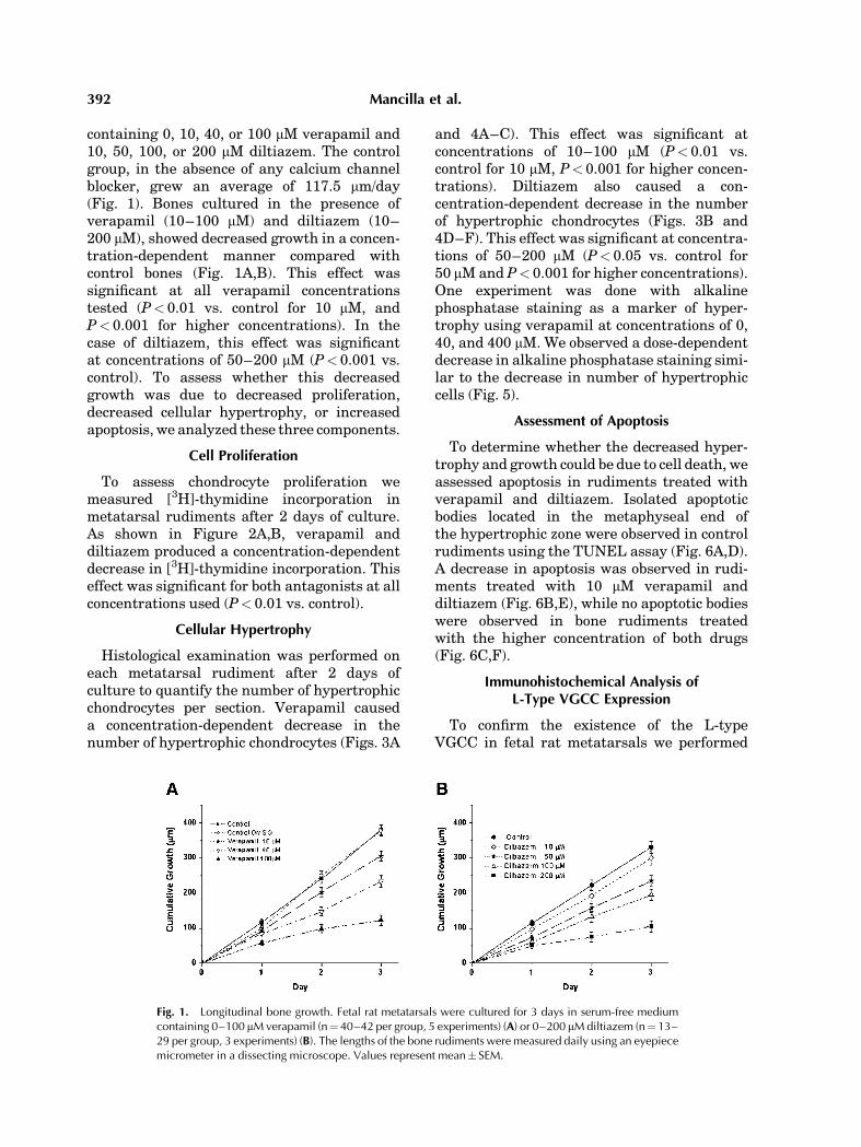
containing 0, 10, 40, or 100 mM verapamil and10, 50, 100, or 200 mM diltiazem. The controlgroup, in the absence of any calcium channelblocker, grew an average of 117.5 mm/day(Fig. 1). Bones cultured in the presence ofverapamil (10–100 mM) and diltiazem (10–200 mM), showed decreased growth in a concen-tration-dependent manner compared withcontrol bones (Fig. 1A,B). This effect wassignificant at all verapamil concentrationstested (P< 0.01 vs. control for 10 mM, andP< 0.001 for higher concentrations). In thecase of diltiazem, this effect was significantat concentrations of 50–200 mM (P< 0.001 vs.control). To assess whether this decreasedgrowth was due to decreased proliferation,decreased cellular hypertrophy, or increasedapoptosis, we analyzed these three components.
Cell Proliferation
To assess chondrocyte proliferation wemeasured [3H]-thymidine incorporation inmetatarsal rudiments after 2 days of culture.As shown in Figure 2A,B, verapamil anddiltiazem produced a concentration-dependentdecrease in [3H]-thymidine incorporation. Thiseffect was significant for both antagonists at allconcentrations used (P< 0.01 vs. control).
Cellular Hypertrophy
Histological examination was performed oneach metatarsal rudiment after 2 days ofculture to quantify the number of hypertrophicchondrocytes per section. Verapamil causeda concentration-dependent decrease in thenumber of hypertrophic chondrocytes (Figs. 3A
and 4A–C). This effect was significant atconcentrations of 10–100 mM (P< 0.01 vs.control for 10 mM, P< 0.001 for higher concen-trations). Diltiazem also caused a con-centration-dependent decrease in the numberof hypertrophic chondrocytes (Figs. 3B and4D–F). This effect was significant at concentra-tions of 50–200 mM (P< 0.05 vs. control for50 mMand P< 0.001 for higher concentrations).One experiment was done with alkalinephosphatase staining as a marker of hyper-trophy using verapamil at concentrations of 0,40, and 400 mM. We observed a dose-dependentdecrease in alkaline phosphatase staining simi-lar to the decrease in number of hypertrophiccells (Fig. 5).
Assessment of Apoptosis
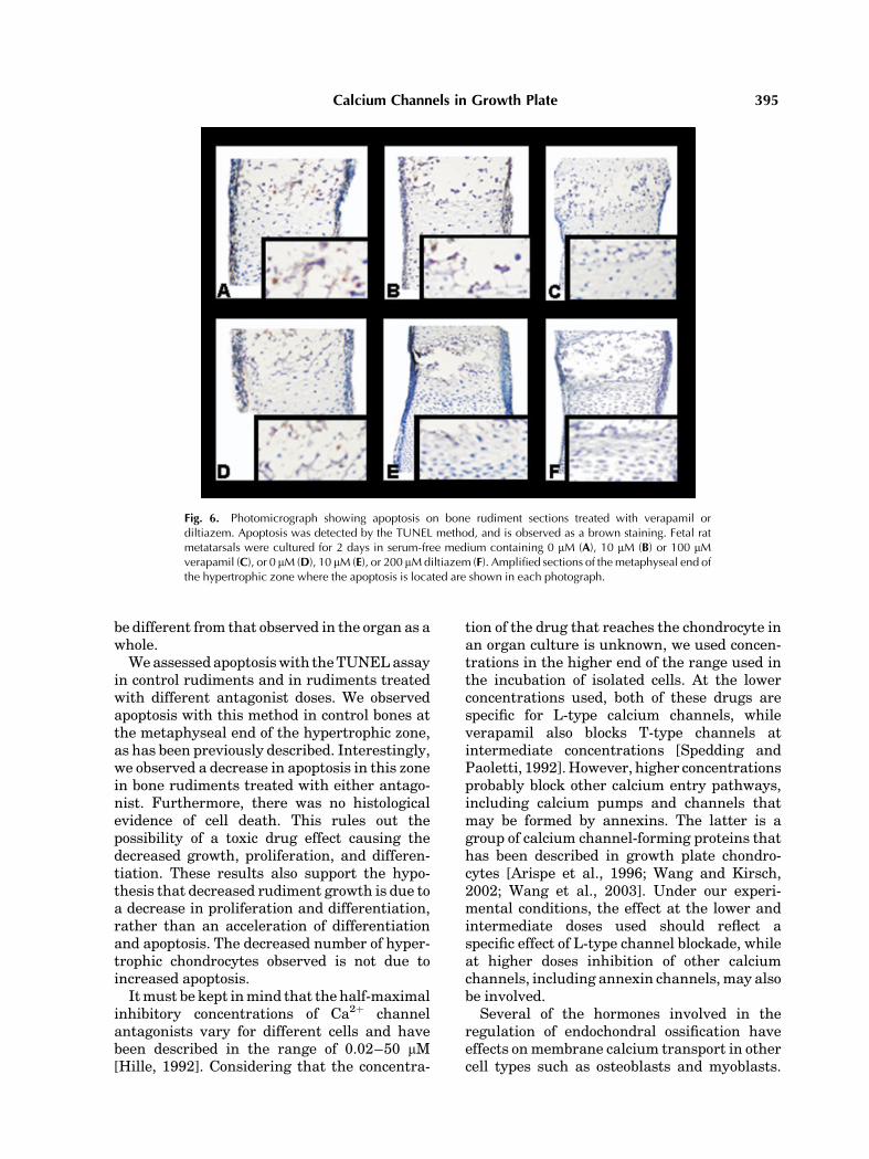
To determine whether the decreased hyper-trophy and growth could be due to cell death, weassessed apoptosis in rudiments treated withverapamil and diltiazem. Isolated apoptoticbodies located in the metaphyseal end ofthe hypertrophic zone were observed in controlrudiments using the TUNEL assay (Fig. 6A,D).A decrease in apoptosis was observed in rudi-ments treated with 10 mM verapamil anddiltiazem (Fig. 6B,E), while no apoptotic bodieswere observed in bone rudiments treatedwith the higher concentration of both drugs(Fig. 6C,F).
Immunohistochemical Analysis ofL-Type VGCC Expression
To confirm the existence of the L-typeVGCC in fetal rat metatarsals we performed
Fig. 1. Longitudinal bone growth. Fetal rat metatarsals were cultured for 3 days in serum-free mediumcontaining 0–100 mMverapamil (n¼40–42 per group, 5 experiments) (A) or 0–200 mMdiltiazem (n¼13–29 per group, 3 experiments) (B). The lengths of the bone rudiments were measured daily using an eyepiecemicrometer in a dissecting microscope. Values represent mean� SEM.
392 Mancilla et al.

immunostaining using a specific antibody forthe L-type VGCC a-subunit in bone rudimentsused as controls. Immunostainingwas observedin chondrocytes throughout the rudiment, inthe resting, proliferative, and hypertrophiczones (Fig. 7A,C–G,J). No immunostainig wasobserved in control rudiments treated withoutthe primary antibody (Fig. 7B).Additionally,weanalyzed channel protein expression in bonerudiments treated with verapamil and diltia-zem.Weobserved thatmetatarsals treatedwithboth antagonists presented a distribution ofimmunostaining for this ion channel that wassimilar to controls (Fig. 7H,I,K,L).
DISCUSSION
The existing literature confirms the existenceof the a subunit of the L- and T-type VGCC inembryonic growth plate chondrocytes, andsuggests a regulatory role for calcium inendochondral ossification [Shakibaei andMobasheri, 2003; Shao et al., 2005]. However,indirect studies are contradictory with regardsto the mechanisms responsible for calciumentry in growth plate chondrocytes, and towhich type of VGCC may play a role inendochondral ossification [Messler et al., 1990;Duriez et al., 1993; Zimmermann et al., 1994;
Fig. 2. Total [3H]-thymidine incorporation. Fetal rat metatarsals were cultured for 2 days in serum-freemedium containing 0–100 mMverapamil (n¼17–19 per group) (A) or 0–200 mMdiltiazem (n¼ 15–18per group) (B). Rudimentswere incubatedwith 5mCi/ml [3H]-thymidine. Total [3H]-thymidine incorporationwasmeasured by liquid scintillation counting after solubilization. In the case of verapamil the control group(0 mM verapamil) is control vehicle. Values represent mean� SEM. * P<0.01 versus control.
Fig. 3. Quantitation of hypertrophic chondrocytes. Fetal rat metatarsals were cultured for 2 days in serum-freemedium containing 0–100 mMverapamil (n¼ 6–10 per group) (A) or 0–200 mMdiltiazem (n¼6–8 pergroup) (B). Rudiments were embedded in paraffin and 5 mm sections were obtained. Hypertrophicchondrocytes were operationally defined by a height along the longitudinal axis 10 mm or greater. Valuesrepresent mean� SEM. (CDMSO is control vehicle). P< 0.01 versus control, ** P< 0.05 versus control, ***P<0.001 versus control.
Calcium Channels in Growth Plate 393

Zuscik et al., 1997, 2002]. In our system offetal rat metatarsal rudiments in culture,verapamil and diltiazem, two L-type VGCCblockers, produced a dose-dependent decreasein longitudinal bone growth. With thisculture method we maintained the interactionsamongst chondrocytes at different stages ofdifferentiation, therefore obtaining results thatare a better reflection of physiology thanstudies on isolated chondrocytes. We used two
different types of L-type VGCC antagonists, aphenylalkylamine (verapamil) and a benzothia-zepine (diltiazem), because these act on distinctsites of the a1 subunit of the channel. Weobserved a similar, dose-dependent, effect ongrowth with both drugs, suggesting that theeffect is due to channel blockade rather than anon-specific effect of the drug. This is inaccordance with a study performed in youngrabbits in vivo using nifedipine, another L-typeVGCC blocker, where a decrease in humeralgrowth was noted [Duriez et al., 1993].
To clarify the mechanism by which long-itudinal growth may be affected, we assessedtwo components of bone growth: proliferationand hypertrophy. Both blockers produced adose-dependent decrease in these two para-meters. This is in agreement with an in vitrostudy using mesenchymal limb bud chondro-cyte cultures, in which a decrease in chondro-cyte differentiationwas observedwith nifedipine[Zimmermann et al., 1994]. A decrease inchondrocyte hypertrophy was also observed invivo in young rabbits administered nifedipineand verapamil [Messler et al., 1990; Duriezet al., 1993]. However, our results contradictthose of another study using juvenile chickengrowth plate chondrocyte cultures, in which noeffect of nifedipinewas observed on the depolar-ization-induced increase in Cai
2þ [Zuscik et al.,1997]. The effect on isolated chondrocytes may
Fig. 4. Representative photomicrographs of fetal metatarsals cultured with verapamil or diltiazem. Bonerudiments were cultured for 2 days in serum-freemediumwith 0 mM, 10 mM, or 100 mMverapamil (A–C) or0 mM, 10 mM, or 100 mM diltiazem (D–F). Representative proliferative and hypertrophic chondrocytes arelabeled p and h, respectively. [Color figure can be viewed in the online issue, which is available atwww.interscience.wiley.com.]
Fig. 5. Photomicrographsdemonstrating the effect of verapamilon alkaline phosphatase activity: 0 mM verapamil (A), 40 mMverapamil (B), 400mMverapamil (C). Sampleswere embedded inOCT compound, and 10 mm cryostat sections were cut andmounted onto poly-L-lysine-coated slides. Enzyme histochem-istry was performed using the simultaneous azo dye method.Alkaline phosphatase is observed as a purple-blue stain. [Colorfigure can be viewed in the online issue, which is available atwww.interscience.wiley.com.]
394 Mancilla et al.
be different from that observed in the organ as awhole.We assessed apoptosiswith theTUNELassay
in control rudiments and in rudiments treatedwith different antagonist doses. We observedapoptosis with this method in control bones atthe metaphyseal end of the hypertrophic zone,as has been previously described. Interestingly,we observed a decrease in apoptosis in this zonein bone rudiments treated with either antago-nist. Furthermore, there was no histologicalevidence of cell death. This rules out thepossibility of a toxic drug effect causing thedecreased growth, proliferation, and differen-tiation. These results also support the hypo-thesis that decreased rudiment growth is due toa decrease in proliferation and differentiation,rather than an acceleration of differentiationand apoptosis. The decreased number of hyper-trophic chondrocytes observed is not due toincreased apoptosis.Itmust be kept inmind that the half-maximal
inhibitory concentrations of Ca2þ channelantagonists vary for different cells and havebeen described in the range of 0.02–50 mM[Hille, 1992]. Considering that the concentra-
tion of the drug that reaches the chondrocyte inan organ culture is unknown, we used concen-trations in the higher end of the range used inthe incubation of isolated cells. At the lowerconcentrations used, both of these drugs arespecific for L-type calcium channels, whileverapamil also blocks T-type channels atintermediate concentrations [Spedding andPaoletti, 1992].However, higher concentrationsprobably block other calcium entry pathways,including calcium pumps and channels thatmay be formed by annexins. The latter is agroup of calcium channel-forming proteins thathas been described in growth plate chondro-cytes [Arispe et al., 1996; Wang and Kirsch,2002; Wang et al., 2003]. Under our experi-mental conditions, the effect at the lower andintermediate doses used should reflect aspecific effect of L-type channel blockade, whileat higher doses inhibition of other calciumchannels, including annexin channels,may alsobe involved.
Several of the hormones involved in theregulation of endochondral ossification haveeffects onmembrane calcium transport in othercell types such as osteoblasts and myoblasts.
Fig. 6. Photomicrograph showing apoptosis on bone rudiment sections treated with verapamil ordiltiazem. Apoptosis was detected by the TUNEL method, and is observed as a brown staining. Fetal ratmetatarsals were cultured for 2 days in serum-free medium containing 0 mM (A), 10 mM (B) or 100 mMverapamil (C), or 0 mM (D), 10 mM (E), or 200 mMdiltiazem (F). Amplified sections of the metaphyseal end ofthe hypertrophic zone where the apoptosis is located are shown in each photograph.
Calcium Channels in Growth Plate 395

IGF-I, PTHrP, 1,25 (OH)2 Vitamin D3 and itsmetabolites have been shown to affect calciumchannels [Caffrey and Farach-Carson, 1989;Poiradeau et al., 1997; Solem and Thomas,1998; Brines and Broadus, 1999; Wang et al.,1999; Liu et al., 2000; Lalonde et al., 2001;Zanello and Norman, 2003; Zheng et al., 2004].PTHrP has been described as the gatekeeper ofchondrocyte maturation, limiting progressionto hypertrophy. This effect has been demon-strated in mice lacking or overexpressing thegene for this hormone, and in patients present-ing an activating mutation of its receptor[Schipani et al., 1995; Vortkamp et al., 1996;Lanske et al., 1999; Povot and Schiapani, 2005].In growth plate chondrocytes, it was shown that
the PTHrP gene may be regulated by Cai2þ
[Zuscik et al., 2002]. At lower Cai2þ concentra-
tions there was an increased expression ofPTHrP, and a domain in the PTHrP gene wasidentified that conferred calcium sensitivity tothis gene. Therefore, L-type Ca2þ channels maybe related to local and systemic factors involvedin endochondral ossification in two ways; byresponding directly or indirectly to these factorsand altering calcium entry, or by producingchanges in Cai
2þ that affect the synthesis andaction of these factors. Further studies arenecessary to solve these questions.
L-type Ca2þ channels are present in growthplate chondrocytes at all stages of differentia-tion as shown by our immunohistochemical
Fig. 7. Photomicrograph showing immunostaining for the asubunit of L-type voltage gated calcium channel (VGCC) insections of control bone rudiments (A, G, J), and rudimentstreated with 10 mM verapamil (H), 100 mM verapamil (I), 10 mMdiltiazem (K), or 200 mM diltiazem (L). Metatarsals were fixed,embedded in paraffin and longitudinal 5 mm sections were
placed on slides that were incubated with anti Cav 1.2 (a1c)antibody. Amplified sections of the different maturation zones ofthe immunostained control bone (A) are shown separately.(C: hypertrophic, D: proliferative, and E, F: resting zone).A control rudiment treated without primary antibody showingno immunostaining is depicted in B.
396 Mancilla et al.
analysis. Calcium entry may be differentiallyregulated at the different maturation stages,and our data suggest that L-type channels areone of the entry pathways for Ca2þ. Theregulation of chondrogenesis by changes inCai
2þ may depend, at least in part, on L-typeCa2þ channel activity. We conclude that L-typeCa2þ channel activity in growth plate chondro-cytes is necessary for normal longitudinalgrowth by participating in chondrocyte prolife-ration and differentiation.
ACKNOWLEDGMENTS
We thank Dr. Jeffrey Baron and Mr. KevinBarnes from the Unit of Growth, NICHD, NIHfor their suggestions and their assistance withthe alkaline phosphatase staining of bonerudiments.
REFERENCES
Arispe N, Rojas E, Genge BR, Wu LN, Wuthier RE. 1996.Similarity in calcium channel activity of annexinV and matrix vesicles in planar lipid bilayers. BiophysJ 71:1764–1775.
Bagi CM, Miller SC. 1992. Dose-related effects of 1, 25-dihydroxyvitamin D3 on growth, modeling, and morphol-ogy of fetal rat metatarsals cultured in serum-freemedium. J Bone Miner Res 7:29–40.
Brines MA, Broadus AE. 1999. Parathyroid hormonerelated protein potentiates depolarization inducedrelease in PC-12 cells via L-type voltage-sensitive Ca2þ
channels. Endocrinology 140:2646–2651.Burstone MS. 1960. Histochemical observations on enzy-matic processes in bone and teeth. Ann NY Acad Sci 85:431–444.
Caffrey JM, Farach-Carson MC. 1989. Vitamin D3 meta-bolites modulate dihydropyridine-sensitive calcium cur-rents in rat osteosarcoma cells. J Biol Chem 264(34):20265–20274.
Caplan AI, Boyan BD. 1994. Endochondral bone formation:The lineage cascade. In: Hall BK, editor. Bone Volume 8.Mechanisms of bone development and growth. BocaRaton: CRC Press. pp 41–45.
Duriez J, Flautre B, Blary MC, Hardouin P. 1993. Effects ofthe calcium channel blocker nifedipine on epiphysealgrowth plate and bone turnover: A study in rabbit. CalcifTissue Int 52(2):120–124.
Gunter TE, Zuscik MJ, Puzas JE, Gunter KK, Rosier RN.1990. Cytosolic free calcium concentrations in aviangrowth plate chondrocytes. Cell Calcium 11:445–457.
Hille B. 1992. Calcium channels. In: Hille B, editor. Ionicchannels of excitable membranes. 2nd edition. Sunder-land, Massachusetts: Sinauer Associates, Inc. 108p.
Iannotti JP, Brighton CT, Stambough JE. 1989. Cytosolicionized calcium concentration in isolated chondrocytesfrom each zone of the growth plate. J Orthop Res 7:511–518.
Lalonde M, Li B, Kline L, Pang P, Karpinski E. 2001.Modulation of L-type Ca2þ channels in UMR 106 cells by
parathyroid hormone-related protein. Life Sci 70(5):503–515.
Lanske B, Amling M, Neff L, Guiducci J, Baron R,Kronenberg H. 1999. Ablation of PTHrP gene or PTH/PTHrP receptor gene leads to distinct abnormalities inbone development. J Clin Invest 104:399–407.
Liu R, Li W, Karin NJ, Bergh JJ, Adler-Storthz K, Farach-Carson MC. 2000. Ribozyme ablation demonstrates thatthe cardiac subtype of voltage sensitive calcium channelis the molecular transducer of 1, 25 dihydroxyvitamin D3stimulated Ca influx in osteoblast cells. J Biol Chem275(12):8711–8718.
Mancilla EE, De Luca F, Uyeda JA, Czerwiec FS, Baron J.1998. Effects of fibroblast growth factor-2 on longitudinalbone growth. Endocrinology 139:2900–2904.
Messler HH, Koch W, Muzenberg KJ. 1990. Analogouseffects of organic calcium antagonists and magnesiumon the epiphyseal growth plate. Clin Orthop 258:135–141.
Poiradeau S, Lieberherr M, Kergoise N, Corvol MT. 1997.Different mechanisms are involved in intracellularcalcium increase by insulin-like growth factors 1 and 2in articular chondrocytes: Voltage-gated calcium chan-nels, and/or phospholipase C coupled to pertussis-sensitive G protein. J Cell Biochem 64:414–422.
Povot S, Schiapani E. 2005. Molecular mechanisms ofendochondral bone development. Biochem Biophysic ResCommun 328:658–665.
Schipani E, Kruse K, Juppner H. 1995. A constitutivelyactive mutant PTH-PTHrP receptor in Jansen metaphy-seal chondrodysplasia. Science 268:98–100.
Shakibaei M, Mobasheri A. 2003. b1-integrins co-localizewith Na, K-ATPase, epithelial sodium channels (EnaC)and voltage activated calcium channels (VACC) inmechanoreceptor complexes of mouse limb-bud chondro-cytes. Histol Histopathol 18:343–351.
Shao Y, Alocknavitch M, Farach-Carson C. 2005. Expres-sion of voltage sensitive calcium channel (VSCC) L-typeCav1.2 (a1c) and T-type (a1H) subunits during mousedevelopment. Dev Dyn 234:54–62.
Solem ML, Thomas AP. 1998. Modulation of cardiac Ca2þ
channels by IGF-I. Biochem Biophys Res Commun252:151–155.
Spedding M, Paoletti R. 1992. Classification of calciumchannels and the sites of action of drugs modifyingchannel function. Pharmacological Reviews 44(3):353–375.
Van der Eerden BCJ, KarperienM,Wit JM. 2003. Systemicand local regulation of the growth plate. EndocrineReviews 24:782–801.
Vortkamp A, Lee K, Lanske B, Segre GV, Kronenberg HM,Tabin CJ. 1996. Regulation of rate of cartilage differ-entiation by Indian hedgehog and PTH-related protein.Science 273:613–622.
Wang W, Kirsch T. 2002. Retinoic acid stimulates annexin-mediated growth plate chondrocyte mineralization.J Cell Biol 157:1061–1070.
Wang ZM, Messi ML, Renganathan M, Delbono O. 1999.Insulin-like growth factor-1 enhances rat skeletal musclecharge movement and L-type Ca2þ channel gene expres-sion. J Physiol 516:331–341.
Wang W, Xu J, Kirsch T. 2003. Annexin-mediated Ca2þ
influx regulates growth plate chondrocyte maturationand apoptosis. J Biol Chem 278:3762–3769.
Calcium Channels in Growth Plate 397

Zanello LP, Norman AW. 2003. Multiple molecularmechanisms of 1a, 25 (OH)2- vitamin D3 rapid modula-tion of three ion channel activities in osteoblasts. Bone33:71–79.
Zheng Z, Wang ZM, Delbono O. 2004. Ca(2þ) calmodulinkinase and calcineurin modulate IGF-1 induced skeletalmuscle dihydropyridine receptor alpha (1s) transcrip-tion. J Membr Biol 197(2):101–112.
Zimmermann B, Lange K, Mertens P, Bernimoulin JP.1994. Inhibition of chondrogenesis and endochondral
mineralization in vitro by different calcium channelblockers. Eur J Cell Biol 63(1):114–121.
Zuscik MJ, Gunter TE, Puzas JE, Rosier RN. 1997.Characterization of voltage-sensitive calcium channelsin growth plate chondrocytes. Biochem Biophys ResComm 234:432–438.
ZuscikMJ, D’SouzaM, Gunter KK, Gunter TE, O’Keefe RJ,Schwarz EM, Puzas JE, Rosier RN. 2002. Growth platechondrocyte maturation is regulated by basal intracel-lular calcium. Exp Cell Res 276:310–319.
398 Mancilla et al.