Reparation of chondral defects in rabbits by autologous and allogenous chondrocytes seeded on...
12
61 Acta Veterinaria-Beograd 2014, 64 (1), 61-72 UDK: 616.71-018.3-08 DOI: 10.2478/acve-2014-0007 REPARATION OF CHONDRAL DEFECTS IN RABBITS BY AUTOLOGOUS AND ALLOGENOUS CHONDROCYTES SEEDED ON COLLAGEN/HYALURONAN SCAFFOLD OR SUSPENDED IN FIBRIN GLUE HORŇÁK Slavomír 1 , HARVANOVÁ Denisa 2* , LEDECKÝ Valent 1 , HLUCHÝ Marian 1 , VALENČÁKOVÁ-AGYAGOSOVÁ Alexandra 1 , AMRICHOVÁ Judita 2 , ROSOCHA Ján 2 , VAŠKO Gabriel 3 , ŠVIHLA Róbert 3 , PETROVIČ Vladimír 1 1 University of Veterinary Medicine and Pharmacy, Clinic of Small Animals, Komenského 73, 041 81 Košice, Slovak Republic; 2 Tissue Bank, Pavol Jozef Šafárik University, Faculty of Medicine and University Hospital of L. Pasteur, Trieda SNP 1, 040 66 Košice, Slovak Republic; 3 Department of Orthopaedics and Traumatology of Locomotory Apparatus, Pavol Jozef Šafárik University Faculty of Medicine and University Hospital of L. Pasteur, Trieda SNP 1, 041 90 Košice, Slovak Republic (Received 08 January 2014; Accepted 28 January 2014) INTRODUCTION Articular hyaline cartilage is a specialized connective tissue important for the distribution of pressure and shear forces within joints with a specific role in animal locomotion *Corresponding author: e-mail: [email protected] Research article The topic of the study was to verify in vivo survival of in vitro cultured autologous and allogenous chondrocytes suspended in a fibrin glue Beriplast ® or seeded on Collagen type I-Hyaluronan (Col type I-HYA) scaffolds for the regeneration of articular cartilage defects in rabbits. The study was carried out on 15 domestic rabbits randomly assigned to five groups (n = 3 in each) with different treatments of artificially created chondral defects (ChD´s). These defects were made in a non-load-bearing area of medial condyle of the distal femur, and were treated as follows: 1 st and 3 rd group: the ChD´s were filled with autologous or allogenous chondrocytes seeded on Col type I-HYA scaffolds, respectively. The scaffolds were fixed to the ChD´s by fibrin glue Beriplast ® ; 2 nd and 4 th group: the ChD´s were filled with a suspension of autologous or allogenous chondrocytes in fibrin glue Beriplast ® , respectively, and they were immediately covered by unseeded Col type I-HYA scaffolds; Control group: the ChD´s were left to heal spontaneously without any treatment. Macroscopical, histological and immunohistochemical analyses of the ChD´s were performed 12 months after the treatment. In all treated groups, the chondrocytes were capable to proliferate and produce the cartilage extracellular matrix, including proteoglycans and type II collagen, as compared to the control “untreated” group. On the other hand, the production of hyaline-like cartilage tissue confirmed that both therapeutic methods using autologous chondrocytes can be applied successfully for the treatment of chondral defects in rabbits. Key words: rabbits, articular cartilage defect, chondrocyte implants, collagen, hyaluronan
-
Upload
independent -
Category
Documents
-
view
0 -
download
0
Transcript of Reparation of chondral defects in rabbits by autologous and allogenous chondrocytes seeded on...

61
Acta Veterinaria-Beograd 2014, 64 (1), 61-72UDK: 616.71-018.3-08
DOI: 10.2478/acve-2014-0007
REPARATION OF CHONDRAL DEFECTS IN RABBITS BY AUTOLOGOUS AND ALLOGENOUS CHONDROCYTES SEEDED ON COLLAGEN/HYALURONAN SCAFFOLD OR SUSPENDED IN FIBRIN GLUE
HORŇÁK Slavomír1, HARVANOVÁ Denisa2*, LEDECKÝ Valent1, HLUCHÝ Marian1, VALENČÁKOVÁ-AGYAGOSOVÁ Alexandra1, AMRICHOVÁ Judita2, ROSOCHA Ján2, VAŠKO Gabriel3, ŠVIHLA Róbert3, PETROVIČ Vladimír1
1University of Veterinary Medicine and Pharmacy, Clinic of Small Animals, Komenského 73, 041 81 Košice, Slovak Republic; 2Tissue Bank, Pavol Jozef Šafárik University, Faculty of Medicine and University Hospital of L. Pasteur, Trieda SNP 1, 040 66 Košice, Slovak Republic; 3Department of Orthopaedics and Traumatology of Locomotory Apparatus, Pavol Jozef Šafárik University Faculty of Medicine and University Hospital of L. Pasteur, Trieda SNP 1, 041 90 Košice, Slovak Republic
(Received 08 January 2014; Accepted 28 January 2014)
INTRODUCTION
Articular hyaline cartilage is a specialized connective tissue important for the distribution of pressure and shear forces within joints with a specifi c role in animal locomotion
*Corresponding author: e-mail: [email protected]
Research article
The topic of the study was to verify in vivo survival of in vitro cultured autologous and allogenous chondrocytes suspended in a fi brin glue Beriplast® or seeded on Collagen type I-Hyaluronan (Col type I-HYA) scaffolds for the regeneration of articular cartilage defects in rabbits. The study was carried out on 15 domestic rabbits randomly assigned to fi ve groups (n = 3 in each) with different treatments of artifi cially created chondral defects (ChD´s). These defects were made in a non-load-bearing area of medial condyle of the distal femur, and were treated as follows: 1st and 3rd group: the ChD´s were fi lled with autologous or allogenous chondrocytes seeded on Col type I-HYA scaffolds, respectively. The scaffolds were fi xed to the ChD´s by fi brin glue Beriplast®; 2nd and 4th group: the ChD´s were fi lled with a suspension of autologous or allogenous chondrocytes in fi brin glue Beriplast®, respectively, and they were immediately covered by unseeded Col type I-HYA scaffolds; Control group: the ChD´s were left to heal spontaneously without any treatment. Macroscopical, histological and immunohistochemical analyses of the ChD´s were performed 12 months after the treatment. In all treated groups, the chondrocytes were capable to proliferate and produce the cartilage extracellular matrix, including proteoglycans and type II collagen, as compared to the control “untreated” group. On the other hand, the production of hyaline-like cartilage tissue confi rmed that both therapeutic methods using autologous chondrocytes can be applied successfully for the treatment of chondral defects in rabbits.
Key words: rabbits, articular cartilage defect, chondrocyte implants, collagen, hyaluronan

Acta Veterinaria-Beograd 2014, 64 (1), 61-72
62
[1]. Mature articular cartilage has a limited potential for the repair of critical-sized defects because of its avascularity and the absence of stem cells [2]. The response of the normal articular cartilage to damage or degeneration is frequently partial and repair of the damaged tissue is incomplete. Surface defects of articular cartilage do not heal spontaneously and usually turn to extensive degeneration [3]. It was observed that despite the limited ability of chondrocytes to migrate from marginal zones into the defective site, they cannot multiply in these locations and produce macromolecules needed for the repair of the organized intercellular matrix typical of normal articular cartilage [4].Cartilage properties result from its microscopic structure. Detailed studies of morphology and biology of mature cartilage showed that it is a highly organized structure. Complex connections between the chondrocytes and the matrix are maintained actively and are responsible for the preservation of the shape and function of the tissue owing to the structural character of the solid component (e.g.: collagen, proteoglycans and glycoproteins). Therefore, the tissue that would reconstruct correctly the damaged surface of the articular cartilage has to replicate these structures very precisely thus cartilaginous tissue defects could be repaired.Current scientifi c trends present new possibilities for the repair of cartilage defects. Transplantation of autologous or allogenous chondrocytes, synovial mesenchymal stem cells, and biodegradable scaffolds are the methods that have been tested experimentally and clinically [5-11]. Implants seeded with chondrocytes cultured in vitro support the production of the extracellular matrix by chondrocytes, mainly glycosaminoglycans and type II collagen [12,13]. The implantation of chondrocyte–collagen composites into cartilage defects has proved a promising method of cartilage repair [14,15] and numerous in vivo studies in animals have shown that hyaluronan-based scaffolds seeded with autologous chondrocytes are useful for inducing the formation of hyaline-like cartilage tissue and are reabsorbed in the absence of an infl ammatory response [16,17].The aims of our study were to investigate a repair process of artifi cially created cartilage defects in rabbits treated by autologous and allogenous chondrocytes in a suspension in fi brin glue Beriplast® or seeded on Collagen type I-Hyaluronan (Col type I-HYA) scaffolds. Chondral defects (ChD´s) were created in such a manner as not to affect the zone of calcifi ed cartilage in a non-load-bearing area of the medial condyle of the distal femur. The healing process of ChD´s was evaluated macroscopically, as well as by histological and immunohistochemical analyses 12 months after the treatment.
MATERIALS AND METHODS
Animal care
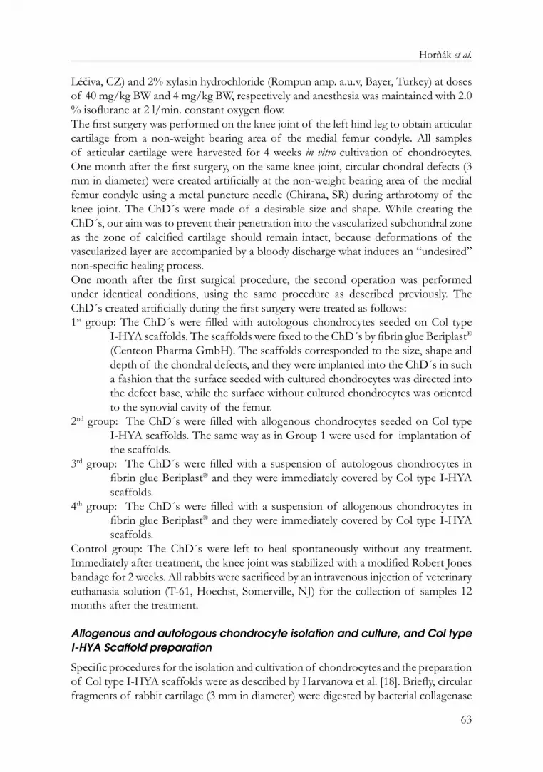
This study was performed at the University of Veterinary Medicine Košice, Slovakia respecting the guidelines for animal experiments with the approval of the Ethical Committee. This experiment was carried out on 15 domestic rabbits assigned to four treated groups and the untreated “control” group (n = 3 in each). Within four weeks, two surgical procedures were performed. In both cases, the rabbits were premedicated with an intramuscular injection of 1% ketamin hydrochloride (Narkamon inj. a.u.v.,
Horňák et al.
63
Léčiva, CZ) and 2% xylasin hydrochloride (Rompun amp. a.u.v, Bayer, Turkey) at doses of 40 mg/kg BW and 4 mg/kg BW, respectively and anesthesia was maintained with 2.0 % isofl urane at 2 l/min. constant oxygen fl ow. The fi rst surgery was performed on the knee joint of the left hind leg to obtain articular cartilage from a non-weight bearing area of the medial femur condyle. All samples of articular cartilage were harvested for 4 weeks in vitro cultivation of chondrocytes. One month after the fi rst surgery, on the same knee joint, circular chondral defects (3 mm in diameter) were created artifi cially at the non-weight bearing area of the medial femur condyle using a metal puncture needle (Chirana, SR) during arthrotomy of the knee joint. The ChD´s were made of a desirable size and shape. While creating the ChD´s, our aim was to prevent their penetration into the vascularized subchondral zone as the zone of calcifi ed cartilage should remain intact, because deformations of the vascularized layer are accompanied by a bloody discharge what induces an “undesired” non-specifi c healing process. One month after the fi rst surgical procedure, the second operation was performed under identical conditions, using the same procedure as described previously. The ChD´s created artifi cially during the fi rst surgery were treated as follows:1st group: The ChD´s were fi lled with autologous chondrocytes seeded on Col type
I-HYA scaffolds. The scaffolds were fi xed to the ChD´s by fi brin glue Beriplast® (Centeon Pharma GmbH). The scaffolds corresponded to the size, shape and depth of the chondral defects, and they were implanted into the ChD´s in such a fashion that the surface seeded with cultured chondrocytes was directed into the defect base, while the surface without cultured chondrocytes was oriented to the synovial cavity of the femur.
2nd group: The ChD´s were fi lled with allogenous chondrocytes seeded on Col type I-HYA scaffolds. The same way as in Group 1 were used for implantation of the scaffolds.
3rd group: The ChD´s were fi lled with a suspension of autologous chondrocytes in fi brin glue Beriplast® and they were immediately covered by Col type I-HYA scaffolds.
4th group: The ChD´s were fi lled with a suspension of allogenous chondrocytes in fi brin glue Beriplast® and they were immediately covered by Col type I-HYA scaffolds.
Control group: The ChD´s were left to heal spontaneously without any treatment. Immediately after treatment, the knee joint was stabilized with a modifi ed Robert Jones bandage for 2 weeks. All rabbits were sacrifi ced by an intravenous injection of veterinary euthanasia solution (T-61, Hoechst, Somerville, NJ) for the collection of samples 12 months after the treatment.
Allogenous and autologous chondrocyte isolation and culture, and Col type I-HYA Scaffold preparation
Specifi c procedures for the isolation and cultivation of chondrocytes and the preparation of Col type I-HYA scaffolds were as described by Harvanova et al. [18]. Briefl y, circular fragments of rabbit cartilage (3 mm in diameter) were digested by bacterial collagenase

Acta Veterinaria-Beograd 2014, 64 (1), 61-72
64
type II (1mg/mL) (Gibco BRL®) in Nutrient mixture F-12 medium (Gibco BRL®) supplemented with 1% (v/v) antibiotic/antimycotic solution for 16-20 hours. The obtained chondrocytes were collected and cultivated in Nutrient mixture F-12 medium supplemented with 20% (v/v) fetal bovine serum (Gibco BRL®), 1% (v/v) insulin-selenium-transferin A (Gibco BRL®), 1% (v/v) antibiotic/antimycotic solution at 37°C in humidifi ed air with 5% CO2 for one month. Isolated chondrocyte cultures were passaged before confl uence was obtained with trypsin-EDTA (Gibco BRL®) and the medium was exchanged every three days. In the 3rd and 4th group, autologous and allogenous chondrocytes were used for the in vivo application with fi brin glue Beriplast® at density of 0.65-0.8 x 10⁶ per ml and seeded on the Col type I-HYA scaffold, respectively. Enzymatically treated fi brous collagen type I (bovine Collagen type I from Achilles tendon, Hypro, Otrokovice, Czech Republic) was mixed with 0,5 M solution of acetic acid and aqueous solution of sodium hyaluronate (10mg/ml, 1500 kDa, CONTIPRO Ltd., Dolní Dobrouč, Czech Republic). Starch dialdehyde 2% (v/v) aqueous solution was added to the collagen/hyaluronan complex and homogenized in a blender at 1400 RPM for 1 minute, pre-frozen for 12 hours and freeze dried (JOUAN LP3) at -45 °C, 2x10⁻² mBa for 6-8 hours. After lyophilisation, scaffolds were sealed in polyethylene bags and sterilized by 60Co radiation with a dose of 30 kGy. Lyophilized scaffolds were cut to a desired shape (3 mm in diameter) and were left in the culture medium for 1 hour before in vitro cultivation. In the 1st and 2nd group, autologous and allogenous chondrocytes were seeded on the Col type I-HYA scaffold in at density of 0.47-0.6 x 10⁶ per cm2 and were cultured at 37°C in humidifi ed air with 5% CO2 for 4 weeks, respectively. The culture medium was exchanged every three days.
Locomotory activities
Evaluation was based on the time needed to full load limb recovery after treatment and on monitoring of patella position, septic arthritis, production of exudate compactness of suture, and infection of wounds which can also refl ect the status of the defect repair.
Macroscopic evaluation
The gross appearance of the ChD´s was evaluated for color, integrity, contour and smoothness of the surface.
Histological and immunohistochemical analyses
The specimens of treated medial condyles of the distal femur in each rabbit were fi xed in 10 % phosphate-buffered formalin (Sigma-Aldrich®, Slovak Republic) for 48 h. Subsequently, they were washed in water and decalcifi ed in 25% Chelaton III (v/v) pH 7.4 (Bioptika, Italy) at 37°C for 7 days. After decalcifi cation, the samples were washed in water for 24 h, embedded in paraffi n wax, and 15 μm thick slices were cut sagittally, and were stained in situ with Haemotoxylin-Eosin (H&E) and Safranine O (specifi c for proteoglycans). Immunohistochemical staining was carried out using antibodies against collagen type II fi bres (Rabbit Anti-Human Collagen type II, BIODESIGN

Horňák et al.
65
International, Saco, ME USA) and porcine immunoglobulin against human IgG tagged with fl orescein-isothiocyanate (SwAHu/IgG-FITC, SeVac, Prague, Czech Republic). Both antibodies were diluted 1:100 in 1% phosphate buffered saline (10 mM sodium phosphate, 0.15 M sodium chloride, pH 7.4). The evaluation was performed by light microscopy and fl uorescence microscopy using Olympus advanced inverted microscope IX-71 with an improved fl uorescence illuminator. The histological sections were examined for the quality of the repair process and were graded with use of histological scale described by [6]. All stained slides were evaluated in a blind test by three experienced histologists.
RESULTS
Locomotory activities
Despite repeated arthrotomy, none of the animals showed complications in relation to patella luxation, septic arthritis and excessive production of exudate, dehiscence of sutures or secondary infection of wounds. After the second operation, we observed that the rabbits spontaneously put less weight on the operated limb. The full load and extension of the hind leg recovered completely at the end of the 3rd week after treatment, it means, one week after the bandage removal. Moreover, pain as a factor which signifi cantly limits movements, is not expected in this type of defect (partial cartilage loss) due to the avascular and aneural character of the articular cartilage.
Macroscopic evaluation
The patellar joints were fi lled with a clear viscous synovial fl uid in all rabbits. In all treated groups, neither detachment of the Col type I-HYA scaffolds (except for a severally irregular surface of the treated ChD in one rabbit in the 4th group) nor signs of articular surface damage (such as chondromalacia, osteoarthritis or synovitis) or hypertrophy were found. In the 1st and 3rd group treated by the autologous or allogenous chondrocytes, respectively, the ChD´s have been fi lled with smooth cartilaginous tissue of a color similar to healthy cartilage (Fig. 1a). The ChD´s were completely fi lled in 3 rabbits (one in the 1st group and two in the 3rd group), but one edge of the scaffolds was not completely integrated with the ChD´s in the other rabbits. On the other hand, in the 2nd and 4th group treated with the suspension of autologous or allogenous chondrocytes, respectively, we have found just partial healing of the ChD´s in all rabbits (Fig. 1b). In the control group, no signs of cartilage reparation processes were observed and the ChD´s retained sharp edges even 12 months after surgery (Fig. 1c). Surprisingly, in the rabbit no. 15, the ChD was covered with a whitish tissue that did not resemble the cartilage tissue.
Histological and immunohistochemical analyses
Results of the histological examination of all stained slides are presented in Table 1. In the 1st group, the ChD´s were fi lled with a hyaline-like cartilage tissue (HyLCa) (Fig. 2a), but the intensity of Safranin O staining was high in all layers (deep, middle and

Acta Veterinaria-Beograd 2014, 64 (1), 61-72
66
surface layer) of the ChD (Fig. 2b) only in rabbit no. 3. Except for HyLCa, we have also observed sporadic segments of fi brocartilage (FiCa), particularly in the surface layer of the ChD´s in rabbit no. 1 and no. 2 from the same group. Safranin O staining revealed a relatively high production of proteoglycans (PG´s) and collagen type II fi bres, in the deep layer, but lower intensity of staining has been exhibited in the surface layer of the ChD´s. Chondrocytes in the deep layer have a round shape and their density was higher as compared to the surface layer of the ChD´s. Immunohistochemical staining confi rmed the presence of cells with the chondrocyte phenotype in all ChD´s (Fig. 2c). In rabbit no. 1, a slight hypertrophy of HyLCa was detected probably due to the shape of the scaffold which could be not completely suitable for the insertion into the defect. All in all, a local infl ammatory reaction was not observed in all cases.
In the 2nd group, apparently lower numbers of cells with the chondrocyte phenotype occupied the ChD´s (Fig. 3a), as well as decreased production of PG´s (Fig. 3b) and collagen type II fi bers (Fig. 3c) were found than in the 1st group. In addition, the ChD´s healed with both types of cartilage tissues, but predominantly by FiCa. A local
Figure 2. Histological and immunohistochemical analyses of the chondral defects repaired by autologous chondrocyte seeded on Col type I-HYA scaffold (1st group) a) H&E staining, 150x magnifi cation b) Safranin O staining, 100x magnifi cation, arrow shows a weakened adhesion of the autologous chondrocyte seeded on Col type I-HYA scaffold to the native chondral cartilage c) Immunohistochemical staining, 200x magnifi cation. (HyLCa = hyaline-like cartilage tissue, 1–native chondral cartilage, 2–autologous chondrocytes seeded on Col type I-HYA scaffold)
Figure 1. Macroscopic view of the chondral defects repaired a) by autologous chondrocyte seeded on Col type I-HYA scaffold (1st group), b) by suspension of allogenous chondrocytes in a fi brin glue (4th group) c) without any treatment (Control group). Arrows show the position of the chondral defects.

Horňák et al.
67
infl ammatory reaction was not confi rmed by histological analysis, what indicates that the allogenous chondrocytes did not cause the scaffold rejection accompanied by its lacked mechanical stability in the defect leading to infl ammation. In the 3rd group, the ChD´s were fi lled completely with HyLCa in two rabbits (no. 8 and no. 9). In the deep layer of the ChD´s, the cells resembling chondrocytes of the native cartilage tissue have a round shape (Fig. 4a, 4b). The surface layer of the ChD´s showed the presence of collagen type II fi bers produced by chondrocytes seeded on Col type I-HYA scaffold as in the 1st group. The intensity of PG´s was low in the surface layer, particularly at the edges of the ChD´s, but in the middle and deep layer were higher (Fig. 4c, 4d). Immunohistochemical staining proved the presence of a cellular population of the chondrocyte phenotype in the ChD´s (Fig. 4e). In the 4th group, we have observed only partial regeneration of the ChD´s in two rabbits (no. 10 and no. 12). A severely irregular surface of the treated ChD was found in the rabbit no. 11. Reparation of the ChD´s was incomplete and damage was located mostly at the edges of the defects. HyLCa has been observed in the middle and deep layer of the defects, but the periphery of the ChD´s was fi lled with FiCa. The production of PG´s and the density of cells with the chondrocyte phenotype were low. Fibrin glue components without the cell population were seen in these ChD´s, too.
CategoryPoints 1st group 2nd group 3rd group 4th group Control
groupNumber of rabbit 1 2 3 4 5 6 7 8 9 10 11 12 13 14 15Cell morphologyHyaline cartilage 0 0 0 0Mostly hyaline cartilage 1 1 1 1
Mostly fi brocartilage 2 2 2 2 2 2 2 2Mostly non-cartilage 3Non-cartilage 4 4 4Matrix staining (metachromatic)Normal 0 0 0 0Slighty reduced 1 1 1 1Markedly reduced 2 2 2 2 2 2 2No metachromatic staining 3 3 3 3
Surface regularitySmooth ( 3/4) 0 0 0 0 0 0Moderate ( 1/2-3/4) 1 1 1Irregular (1/4-1/2) 2 2 2 2 2Severely irregular 3 3 3 3 3Thickness of cartilage 2/3 0 0 0 01/3-2/3 1 1 1 1 1 1 1 1 1 1 1 1/3 2 2 2Integration of donor with host adjacent cartilageBoth edges integrated 0 0 0 0 0One edge integrated 1 1 1 1 1 1 1 1 1 1Neither edge integrated 2 2 2
Total maximum 14 4 4 0 8 8 7 4 0 0 8 9 8 14 14 10
Table 1. Histological grading scale for the repair of the chondral defects in individual rabbits.

Acta Veterinaria-Beograd 2014, 64 (1), 61-72
68
In the control group, the ChD´s were left to heal spontaneously. The defects were empty in 2 rabbits (no. 13 and no. 14). In the rabbit no. 15, non-specifi c repair mechanisms were observed in the deep layer of the ChD by the formation of FiCa. Neither the production of PG´s nor collagen type II fi bers were found in this group.
Figure 3. Histological and immunohistochemical analyses of the chondral defects repaired by allogenous chondrocyte seeded on Col type I-HYA scaffold (2nd group) a) H&E staining, 150x magnifi cation, b) Safranin O staining, 100x magnifi cation, very low production of PG´s. c) Im-munohistochemical staining, 100x magnifi cation, production of collagen type II localised mainly in deep layer of the chondral defect. (PG´s = proteoglycans, 1–native chondral cartilage, 2–al-logenous chondrocytes seeded on Col type I-HYA scaffold, 3 – subchondral bone)
Figure 4. Histological and immunohistochemical analyses of the chondral defects repaired by a suspension of autologous chondrocytes in fi brin glue (3rd group) a) H&E staining, 100x magnifi cation b) H&E staining, 150x magnifi cation, c) Safranin O stain, magnifi cation 100x, surface layer created by collagen type I fi bres from the scaffold, and marginal zone of the chon-dral defect with a low production of PG´s, and deep layer of the defect is repaired by HyLCa d) Safranin O stain, magnifi cation 150x, deep and middle layer of the chondral defect with high production of PG´s d) Immunohistochemical staining, 150x magnifi cation. (HyLCa = hyaline-like cartilage tissue, PG´s = proteoglycans, 1 – surface layer of the chondral defect, 2 – deep layer of the condral defect, 3 – deep layer of the native chondral cartilage, 4 – subchondral bone, arrows indicate the marginal zones of the chondal defect)

Horňák et al.
69
DISCUSSION
In the last few years, a great effort was made to defi ne a suitable method for the implantation of chondrocytes into the defective cartilage in order to support the regeneration of cartilage tissue. Studies comparing methods for cartilage tissue reconstruction using autologous chondrocytes, as well as allogenous chondrocytes are lacking. Therefore, we have decided to use two different methods of implantation of autologous and allogenous chondrocytes into the ChD´s: (a) a suspension of chondrocytes in a fi brin glue covered by the Col type I-HYA scaffolds, and (b) chondrocytes seeded on the Col type I-HYA scaffolds fi xed with a fi brin glue. In addition, Lee et al. [19] found that chondrocytes cultured for 4 weeks are able to produce higher amounts of hyaline cartilage after their implantation into chondral defects than these cultured for a signifi cantly shorter time (only 12h). Therefore, the culturing period of chondrocytes for one month was chosen in our study. Different scaffolds were used as cell or tissue carriers for cartilage tissue engineering so far [20-23]. The major advantage of collagen scaffolds, which were used in our experiment, is their tissue abundance and a highly organized structure. It is known, that porous three-dimensional structures consisting of collagen fi bers support the viability of chondrocytes and their production of extracellular matrix in vitro. Collagen contains cell-adhesion domain sequences that induce specifi c cellular interactions [24]. Substrate-bonded hyaluronan exhibits size-dependent stimulation of chondrogenic differentiation and also improves the proliferative activity of cultured chondrocytes [25]. As previously published [26-28], small amounts of hyaluronan (2% w/w) in three-dimensional collagen scaffolds can enhance chondrogenesis. Based on our previous study [18], we can corroborate that the Col type I-HYA scaffolds had not any adverse effects on chondrocyte viability, morphology and phenotype during long term (2-3 months) in vitro cultivation. Moreover, Lin et al. [23] found that chondrocytes seeded on Hyaluronan/Collagen II microspheres could exhibit a high activity in repairing a cartilage defect, what is in accordance with our fi ndings. On the other hand, suspension with chondrocytes can be injected with ease to the defect or target site. Defects orientated downwards are infl uenced by the gravitational forces which can affect the initial distribution of transplantated chondrocytes and may have an important role in the fi nal clinical outcome [29]. This phenomenon was not observed in our study, but the partial detachment of the Col type I-HYA scaffold was found in one rabbit treated with the suspension of allogenous chondrocytes in fi brin glue. By our opinion, adhesive properties of the fi brin glue could be altered due to its dilution with a medium of cultured chondrocytes. All in all, comparing autologous vs. allogenous chondrocyte application, better proliferation of cells with the chondrocyte fenotype and production of PG´s, as well as the creation of HyLCa were observed in both groups treated with autologous chondrocytes. Hong et al. [30] hypothesized that autologous structures have better biocompatibility with native tissues, which was corroborated with our fi ndings too.As a limitation of this study can be considered that, a small number of experimental animals (only 3 rabbits per group) was used. This discouraged the use of statistical

Acta Veterinaria-Beograd 2014, 64 (1), 61-72
70
analysis within the groups thus individual examination was chosen to present our fi ndings instead. Moreover, rabbits are not an optimal animal model for the evaluation of the ChD´s due to the relatively small dimensions of the knee joint and a thin layer of chondrium, which is not thicker than 1 mm. In all treated groups, the chondrocytes were capable to proliferate and produce the cartilage extracellular matrix, including proteoglycans and type II collagen fi bers, as compared to the control “untreated” group. The fi brin glue ensured a suffi cient fi xation of the scaffolds when it was applied undiluted, but its fi xative properties could be altered by a medium of chondrocytes added thus further investigations with a higher number of animals involved are required. For this reason, it can be concluded that the autologous chondrocytes seeded on a Collagen type I-Hyaluronan scaffold represents a very successful way of treatment of articular cartilage defects in rabbits.
ACKNOWLEDGEMENT
This research was supported by VEGA grant no. 1/0406/12 from the Ministry of Education, Science, Research and Sport of the Slovak Republic
REFERENCES
1. Adam A, Pohunková H, Klézl Z, Pešáková V, Čech O: Use of bioimplants to replace cartilage. Part II: application of implants in animal experiments. Acta Chir Orthop Traumtol Cech 1997, 64:207–210.
2. Brittberg M, Nilsson A, Lindahl A, Ohlsson C, Peterson L: Rabbit articular cartilage defects treated with autologous cultured chondrocytes. Clin Orthop1996, 326:270-83.
3. Buckwalter JA: Articular cartilage: injuries and potential for healing. J Orthop Sports Phys Ther 1998, 28:192–202.
4. Shapiro F, Koide S, Glimcher MJ: Cell origin and differentiation in the repair of full thickness defects of articular cartilage. J Bone Joint Surg Am 1993, 75:532–553.
5. Brittberg M, Lindahl A, Nilsson A, Ohlsson C, Isaksson O, Peterson L: Treatment of deep cartilage defects in the knee with autologous chondrocyte transplantation. N Engl J Med 1994, 331:889-895.
6. Wakitani S, Goto T, Pineda SJ, Young RG, Mansour JM, Caplan AI, Goldberg VM: Mesenchymal cell-based repair of large, full-thickness defects of articular cartilage. J Bone Joint Surg Am 1994, 76:579-592.
7. Trippel SB: Autologous chondrocyte transplantation. N Engl J Med 1995, 332:539-540.8. Hardingham T, Tew S, Murdoch A: Tissue engineering: chondrocytes and cartilage. Arthritis
Res 2002, 4:S63–S68.9. Plonczak M, Czubak J, Kawiak J: Repair of articular cartilage defects with cultured
chondrocytes on polysulphonic scaffold: experimental studies in rabbits. Transplant Proc 2006, 38:320–322.
10. Furth ME, Atala A, Van Dyke ME: Smart biomaterials design for tissue engineering and regenerative medicine. Biomaterials 2007, 28:5068-5073.
11. Harvanová D, Tóthová T, Šarišský M, Amrichová J, Rosocha J: Isolation and characterization of synovial mesenchymal stem cells. Folia Biol (Praha) 2011, 57:119-124.

Horňák et al.
71
12. Pavesio A, Abatangelo G, Borrione A, Brocchetta D, Hollander AP, Kon E, Torasso F, Zanasi S, Marcacci M: Hyaluronan-based scaffolds (Hyalograft C) in the treatment of knee cartilage defects: preliminary clinical fi ndings. Novartis Found Symp 2003, 249:203–217.
13. Trattnig S, Ba-Ssalamah A, Pinker K, Plank C, Vecsei V, Marlovits S: Matrix-based autologous chondrocyte implantation for cartilage repair: noninvasive monitoring by high-resolution magnetic resonance imaging, Magn Reson Imaging 2005, 23:779–787.
14. Masuoka K, Asazuma T, Ishihara M, Sato M, Hattori H, Ishihara M, Yoshihara Y, Matsui T, Takase B, Kikuchi M, Nemoto K: Tissue engineering of articular cartilage using an allograft of cultured chondrocytes in a membrane-sealed atelocollagen honeycomb-shaped scaffold (ACHMS scaffold). J Biomed Mater Res B Appl Biomater 2005, 75:177-184.
15. Willers C, Chen J, Wood D, Xu J, Zheng MH: Autologous chondrocyte implantation with collagen bioscaffold for the treatment of osteochondral defects in rabbits. Tissue Eng 2005, 11:1065-1076.
16. Grigolo B, Roseti L, Fiorini M, Fini M: Transplantation of chondrocytes seeded on a hyaluronan derivative (hyaff-11) into cartilage defects in rabbits. Biomaterials 2001, 22:2417–24.
17. Patti AM, Gabriele A, Vulcano A, Ramieri MT, Della Rocca C: Effect of hyaluronic acid on human chondrocyte cell lines from articular cartilage, Tissue Cell 2001, 33:294–300.
18. Harvanová D, Rosocha J, Bakoš D, Švihla R, Vaško G, Horňák S, Ledecký V, Gromošová S, Cibur P, Raši R: Collagen/hyaluronan membrane as a scaffold for chondrocytes cultivation. Biologia 2009, 64:1032-1038.
19. Lee CR, Grodzinsky AJ, Hsu HP, Spector M: Effects of a cultured autologous chondrocyte-seeded type II collagen scaffold on the healing of a chondral defect in a canine model. J Orthop Res 2003, 21:272–281.
20. Lin LC, Chang SJ, Kuo SM, Niu GCC, Keng HK, Tsai PH: Preparation and evaluation of b-TCP/polylactide microspheres as osteogenesis materials, J Appl Polym Sci 2008, 108:3210–3217.
21. Tan H, Wu J, Lao L, Gao C: Gelatin/chitosan/hyaluronan scaffold integrated with PLGA microspheres for cartilage tissue engineering. Acta Biomater 2009, 5:328–337.
22. Singh M, Sandhu B, Scurto A, Berkland C, Detamore MS: Microsphere-based scaffolds for cartilage tissue engineering: using subcritical CO2 as a sintering agent. Acta Biomater 2010, 6:137–143.
23. Lin LC, Chang SJ, Lin CY, Lin YT, Chuang CW, Yao CH, Kuo SM: Repair of Chondral Defects With Allogenous Chondrocytes-Seeded Hyaluronan/Collagen II Microsphers in Rabit Model. Artif Organs 2012, 36:E102-E109.
24. Silver FH, Pins G: Cell growth on collagen: a review of tissue engineering using scaffolds containing extracellular matrix, J Long Term Eff Med Implants 1992, 2:67-80.
25. Kujawa MJ, Carrino DA, Caplan AI: Substrate-bonded hyaluronic acid exhibits a size-dependent stimulation of chondrogenic differentiation of stage 24 limb mesenchymal cells in culture. Dev Biol 1986, 114:519–528.
26. Allemann F, Mizuno S, Eid K, Yates KE, Zaleske D, Glowacki J: Effects of hyaluronan on engineered articular cartilage extracellular matrix gene expression in 3-dimensional collagen scaffolds. J Biomed Mater Res 2001, 55:13-19.
27. Chang CH, Kuo TF, Lin CC, Chou CH, Chen KH, Lin FH, Liu HC: Tissue engineering-

Acta Veterinaria-Beograd 2014, 64 (1), 61-72
72
based cartilage repair with allogenous chondrocytes and gelatin-chondroitin-hyaluronan tri-copolymer scaffold: a porcine model assessed at 18, 24, and 36 weeks. Biomaterials 2006, 27:1876-1888.
28. Ko CS, Huang JP, Huang CW, Chu IM: Type II collagen-chondroitin sulfate-hyaluronan scaffold cross-linked by genipin for cartilage tissue engineering. J Biosci Bioeng 2009, 107:177-182.
29. Sohn DH, Lottman LM, Kim SG, Pedowitz RA, Coutts RD, Sah RL: Effect of gravity on localization of chondrocytes implanted in cartilage defects. Clin Orthop Relat Res 2002, 394:254–262.
30. Hong HJ, Lee JS, Choi JW, Min BH, Lee HB, Kin CH: Transplantation of Autologous Chondrocytes Seede on a Fibre/Hyaluronan Composite Gel Into Trachela Cartilage Defects In Rabbits. Artif Organs 2012, 36:998-1006.
REPARACIJA DEFEKATA HRSKAVICE KUNIĆA AUTOLOGNIM I ALOGENIM HONDROCITIMA ZASEJANIM NA KOLAGEN/HIJALURONAN OSNOVU ILI SUSPENDOVANIM U FIBRINSKOM LEPKU
HORŇÁK Slavomír, HARVANOVÁ Denisa, LEDECKÝ Valent, HLUCHÝ Marian,
VALENČÁKOVÁ-AGYAGOSOVÁ Alexandra, AMRICHOVÁ Judita, ROSOCHA Ján2, VAŠKO Gabriel, ŠVIHLA Róbert, PETROVIČ Vladimír
Cilj studije je bio da se verifi kuje in vivo preživljavanje in vitro kultivisanih autolognih i alogenih hondrocita suspendovanih u fi brinski Beriplast® lepak ili na kolagen Hyaluronan tip I (Col type I-HYA) osnovu radi regeneracije oštećenja zglobne hrskavice kod kunića. Ispitivanje je obavljeno na 15 domaćih kunića, odabranih slučajnim izborom koji su podeljeni u pet grupa (3 po grupi) kod kojih su veštački izazvana oštećenja hrskavice (ChD), tretirana na različite načine. Oštećenja su napravljena na površinama koje nisu noseće na medijalnom kondilusu distalnog femura, a tretman je podrazumevao: prva i treća grupa: ChD su punjene sa autolognim ili alogenim hondrocitima zasejanim na Col tip I-HYA osnovu. Osnove su fi ksirane na ChD pomoću fi brinskog lepka Beriplast®; druga i četvrta grupa: oštećenja su punjena suspenzijom autolognih ili alogenih hondrocita u fi brinskom Beriplast® lepku, a neposredno posle toga, prekriveni su sa nezasejanom Col tip I-HYA osnovom; kontrolna grupa: oštećenja su ostavljena da spontano zarastu, bez tretmana. Dvanaest meseci posle tretmana obavljene su makroskopske, histološke i imunohistološke analize promena. Kod svih životinja tretiranih grupa, hondrociti su bili sposobni da proliferišu i proizvode ekstracelularni matriks hrskavice, uključujući proteoglikane i II tip kolagena u poređenju sa kontrolnom “netretriranom” grupom. Sa druge strane, proizvodnja hrskavičavog tkiva nalik na hijalin potvrđuje da se oba terapijska metoda koji koriste autologne hondrocite mogu da uspešno primene u tretiranju defekata hrskavice kod kunića.