Site1 protease is essential for endochondral bone formation in mice
14
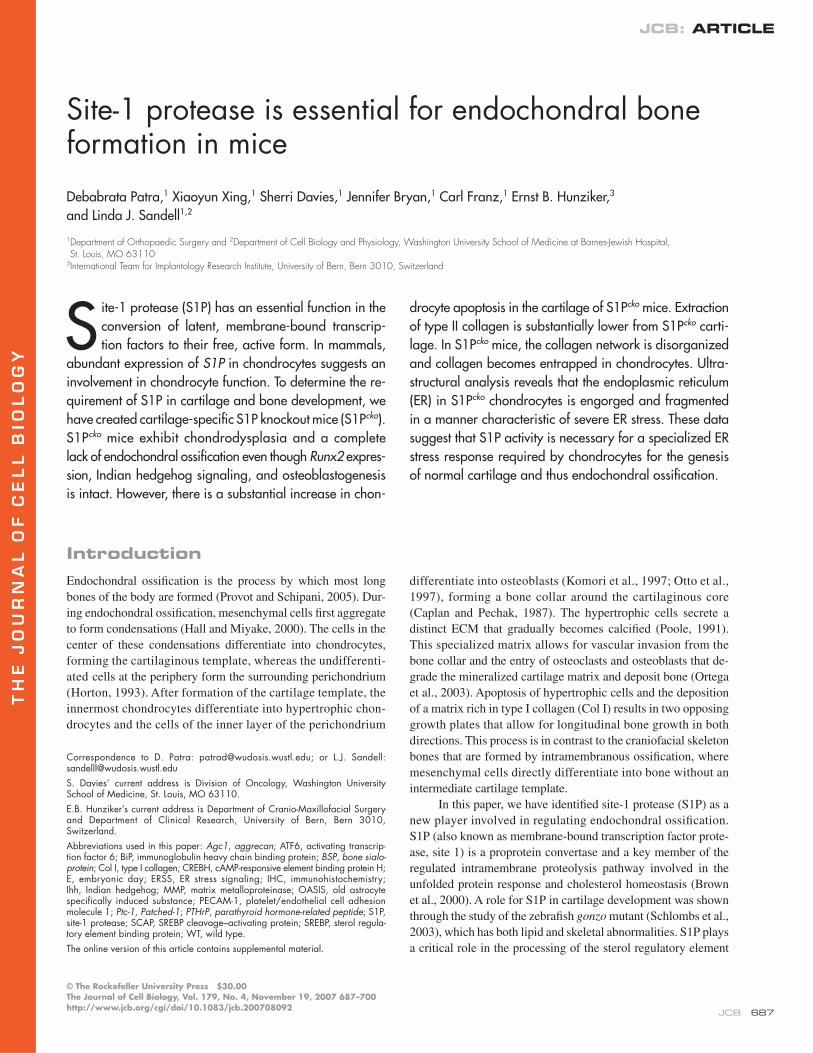
THE JOURNAL OF CELL BIOLOGY JCB: ARTICLE © The Rockefeller University Press $30.00 The Journal of Cell Biology, Vol. 179, No. 4, November 19, 2007 687–700 http://www.jcb.org/cgi/doi/10.1083/jcb.200708092 JCB 687 Introduction Endochondral ossification is the process by which most long bones of the body are formed (Provot and Schipani, 2005). Dur- ing endochondral ossification, mesenchymal cells first aggregate to form condensations (Hall and Miyake, 2000). The cells in the center of these condensations differentiate into chondrocytes, forming the cartilaginous template, whereas the undifferenti- ated cells at the periphery form the surrounding perichondrium (Horton, 1993). After formation of the cartilage template, the innermost chondrocytes differentiate into hypertrophic chon- drocytes and the cells of the inner layer of the perichondrium differentiate into osteoblasts (Komori et al., 1997; Otto et al., 1997), forming a bone collar around the cartilaginous core (Caplan and Pechak, 1987). The hypertrophic cells secrete a distinct ECM that gradually becomes calcified (Poole, 1991). This specialized matrix allows for vascular invasion from the bone collar and the entry of osteoclasts and osteoblasts that de- grade the mineralized cartilage matrix and deposit bone (Ortega et al., 2003). Apoptosis of hypertrophic cells and the deposition of a matrix rich in type I collagen (Col I) results in two opposing growth plates that allow for longitudinal bone growth in both directions. This process is in contrast to the craniofacial skeleton bones that are formed by intramembranous ossification, where mesenchymal cells directly differentiate into bone without an intermediate cartilage template. In this paper, we have identified site-1 protease (S1P) as a new player involved in regulating endochondral ossification. S1P (also known as membrane-bound transcription factor prote- ase, site 1) is a proprotein convertase and a key member of the regulated intramembrane proteolysis pathway involved in the unfolded protein response and cholesterol homeostasis (Brown et al., 2000). A role for S1P in cartilage development was shown through the study of the zebrafish gonzo mutant (Schlombs et al., 2003), which has both lipid and skeletal abnormalities. S1P plays a critical role in the processing of the sterol regulatory element Site-1 protease is essential for endochondral bone formation in mice Debabrata Patra, 1 Xiaoyun Xing, 1 Sherri Davies, 1 Jennifer Bryan, 1 Carl Franz, 1 Ernst B. Hunziker, 3 and Linda J. Sandell 1,2 1 Department of Orthopaedic Surgery and 2 Department of Cell Biology and Physiology, Washington University School of Medicine at Barnes-Jewish Hospital, St. Louis, MO 63110 3 International Team for Implantology Research Institute, University of Bern, Bern 3010, Switzerland S ite-1 protease (S1P) has an essential function in the conversion of latent, membrane-bound transcrip- tion factors to their free, active form. In mammals, abundant expression of S1P in chondrocytes suggests an involvement in chondrocyte function. To determine the re- quirement of S1P in cartilage and bone development, we have created cartilage-specific S1P knockout mice (S1P cko ). S1P cko mice exhibit chondrodysplasia and a complete lack of endochondral ossification even though Runx2 expres- sion, Indian hedgehog signaling, and osteoblastogenesis is intact. However, there is a substantial increase in chon- drocyte apoptosis in the cartilage of S1P cko mice. Extraction of type II collagen is substantially lower from S1P cko carti- lage. In S1P cko mice, the collagen network is disorganized and collagen becomes entrapped in chondrocytes. Ultra- structural analysis reveals that the endoplasmic reticulum (ER) in S1P cko chondrocytes is engorged and fragmented in a manner characteristic of severe ER stress. These data suggest that S1P activity is necessary for a specialized ER stress response required by chondrocytes for the genesis of normal cartilage and thus endochondral ossification. Correspondence to D. Patra: [email protected]; or L.J. Sandell: [email protected] S. Davies’ current address is Division of Oncology, Washington University School of Medicine, St. Louis, MO 63110. E.B. Hunziker’s current address is Department of Cranio-Maxillofacial Surgery and Department of Clinical Research, University of Bern, Bern 3010, Switzerland. Abbreviations used in this paper: Agc1, aggrecan; ATF6, activating transcrip- tion factor 6; BiP, immunoglobulin heavy chain binding protein; BSP, bone sialo- protein; Col I, type I collagen; CREBH, cAMP-responsive element binding protein H; E, embryonic day; ERSS, ER stress signaling; IHC, immunohistochemistry; Ihh, Indian hedgehog; MMP, matrix metalloproteinase; OASIS, old astrocyte specifically induced substance; PECAM-1, platelet/endothelial cell adhesion molecule 1; Ptc-1, Patched-1; PTHrP, parathyroid hormone-related peptide; S1P, site-1 protease; SCAP, SREBP cleavage–activating protein; SREBP, sterol regula- tory element binding protein; WT, wild type. The online version of this article contains supplemental material.
-
Upload
independent -
Category
Documents
-
view
5 -
download
0
Transcript of Site1 protease is essential for endochondral bone formation in mice
TH
EJ
OU
RN
AL
OF
CE
LL
BIO
LO
GY
JCB: ARTICLE
© The Rockefeller University Press $30.00The Journal of Cell Biology, Vol. 179, No. 4, November 19, 2007 687–700http://www.jcb.org/cgi/doi/10.1083/jcb.200708092
JCB 687
IntroductionEndochondral ossifi cation is the process by which most long
bones of the body are formed (Provot and Schipani, 2005). Dur-
ing endochondral ossifi cation, mesenchymal cells fi rst aggregate
to form condensations (Hall and Miyake, 2000). The cells in the
center of these condensations differentiate into chondrocytes,
forming the cartilaginous template, whereas the undifferenti-
ated cells at the periphery form the surrounding perichondrium
(Horton, 1993). After formation of the cartilage template, the
innermost chondrocytes differentiate into hypertrophic chon-
drocytes and the cells of the inner layer of the perichondrium
differentiate into osteoblasts (Komori et al., 1997; Otto et al.,
1997), forming a bone collar around the cartilaginous core
(Caplan and Pechak, 1987). The hypertrophic cells secrete a
distinct ECM that gradually becomes calcifi ed (Poole, 1991).
This specialized matrix allows for vascular invasion from the
bone collar and the entry of osteoclasts and osteoblasts that de-
grade the mineralized cartilage matrix and deposit bone (Ortega
et al., 2003). Apoptosis of hypertrophic cells and the deposition
of a matrix rich in type I collagen (Col I) results in two opposing
growth plates that allow for longitudinal bone growth in both
directions. This process is in contrast to the craniofacial skeleton
bones that are formed by intramembranous ossifi cation, where
mesenchymal cells directly differentiate into bone without an
intermediate cartilage template.
In this paper, we have identifi ed site-1 protease (S1P) as a
new player involved in regulating endochondral ossifi cation.
S1P (also known as membrane-bound transcription factor prote-
ase, site 1) is a proprotein convertase and a key member of the
regulated intramembrane proteolysis pathway involved in the
unfolded protein response and cholesterol homeostasis (Brown
et al., 2000). A role for S1P in cartilage development was shown
through the study of the zebrafi sh gonzo mutant (Schlombs et al.,
2003), which has both lipid and skeletal abnormalities. S1P plays
a critical role in the processing of the sterol regulatory element
Site-1 protease is essential for endochondral bone formation in mice
Debabrata Patra,1 Xiaoyun Xing,1 Sherri Davies,1 Jennifer Bryan,1 Carl Franz,1 Ernst B. Hunziker,3
and Linda J. Sandell1,2
1Department of Orthopaedic Surgery and 2Department of Cell Biology and Physiology, Washington University School of Medicine at Barnes-Jewish Hospital, St. Louis, MO 63110
3International Team for Implantology Research Institute, University of Bern, Bern 3010, Switzerland
Site-1 protease (S1P) has an essential function in the
conversion of latent, membrane-bound transcrip-
tion factors to their free, active form. In mammals,
abundant expression of S1P in chondrocytes suggests an
involvement in chondrocyte function. To determine the re-
quirement of S1P in cartilage and bone development, we
have created cartilage-specifi c S1P knockout mice (S1Pcko).
S1Pcko mice exhibit chondrodysplasia and a complete
lack of endochondral ossifi cation even though Runx2 expres-
sion, Indian hedgehog signaling, and osteoblastogenesis
is intact. However, there is a substantial increase in chon-
drocyte apoptosis in the cartilage of S1Pcko mice. Extraction
of type II collagen is substantially lower from S1Pcko carti-
lage. In S1Pcko mice, the collagen network is disorganized
and collagen becomes entrapped in chondrocytes. Ultra-
structural analysis reveals that the endoplasmic reticulum
(ER) in S1Pcko chondrocytes is engorged and fragmented
in a manner characteristic of severe ER stress. These data
suggest that S1P activity is necessary for a specialized ER
stress response required by chondrocytes for the genesis
of normal cartilage and thus endochondral ossifi cation.
Correspondence to D. Patra: [email protected]; or L.J. Sandell: [email protected]
S. Davies’ current address is Division of Oncology, Washington University School of Medicine, St. Louis, MO 63110.
E.B. Hunziker’s current address is Department of Cranio-Maxillofacial Surgery and Department of Clinical Research, University of Bern, Bern 3010, Switzerland.
Abbreviations used in this paper: Agc1, aggrecan; ATF6, activating transcrip-tion factor 6; BiP, immunoglobulin heavy chain binding protein; BSP, bone sialo-protein; Col I, type I collagen; CREBH, cAMP-responsive element binding protein H; E, embryonic day; ERSS, ER stress signaling; IHC, immunohistochemistry; Ihh, Indian hedgehog; MMP, matrix metalloproteinase; OASIS, old astrocyte specifi cally induced substance; PECAM-1, platelet/endothelial cell adhesion molecule 1; Ptc-1, Patched-1; PTHrP, parathyroid hormone-related peptide; S1P, site-1 protease; SCAP, SREBP cleavage–activating protein; SREBP, sterol regula-tory element binding protein; WT, wild type.
The online version of this article contains supplemental material.
JCB • VOLUME 179 • NUMBER 4 • 2007 688
binding proteins (SREBP-1a, -1c, and -2; Eberle et al., 2004).
SREBPs are membrane-bound transcription factors in the ER
and regulate cholesterol and fatty acid biosynthesis and uptake.
When cholesterol levels are high, SREBP is retained in the ER
membrane as a complex with the sterol-sensing protein SREBP
cleavage–activating protein (SCAP) and the retention protein
INSIG (insulin induced gene). When cholesterol levels drop, the
SREBP–SCAP complex dissociates from INSIG and trans-
locates to the Golgi bodies, where SREBP is sequentially cleaved
by S1P and S2P to release the amino-terminal domain of SREBP
containing the basic helix-loop-helix leucine zipper region. The
basic helix-loop-helix leucine zipper region translocates to the
nucleus to bind to cis-acting sterol responsive elements. In a
similar fashion, S1P is also involved in the activation of other ER
membrane-bound proteins such as activating transcription factor 6
(ATF6; Haze et al., 1999; Ye et al., 2000), old astrocyte specifi -
cally induced substance (OASIS; Murakami et al., 2006), and
cAMP-responsive element binding protein H (CREBH; Zhang
et al., 2006), which are transcription factors for the unfolded pro-
tein response target genes.
To elucidate the role of S1P in all aspects of skeletal
development, we created cartilage-specifi c S1P knockout mice
(S1Pcko) using a Col2a1 promoter. S1Pcko mice die shortly after
birth and exhibit severe chondrodysplasia. The cartilage matrix
is abnormal in S1Pcko mice with defects in Col II protein secre-
tion and assimilation into the matrix, and endochondral bone
formation is completely absent. This is the fi rst example of a
defect in a regulated intramembrane proteolysis enzyme that
affects cartilage development and endochondral ossifi cation in
mice. Deletions of various matrix metalloproteinases (MMPs),
such as MMP13 or MMP9 (Inada et al., 2004; Stickens et al.,
2004), thought to be important in bone morphogenesis did not
abolish endochondral ossifi cation. Thus, S1P is a unique en-
zyme that plays an integral role in skeletal development.
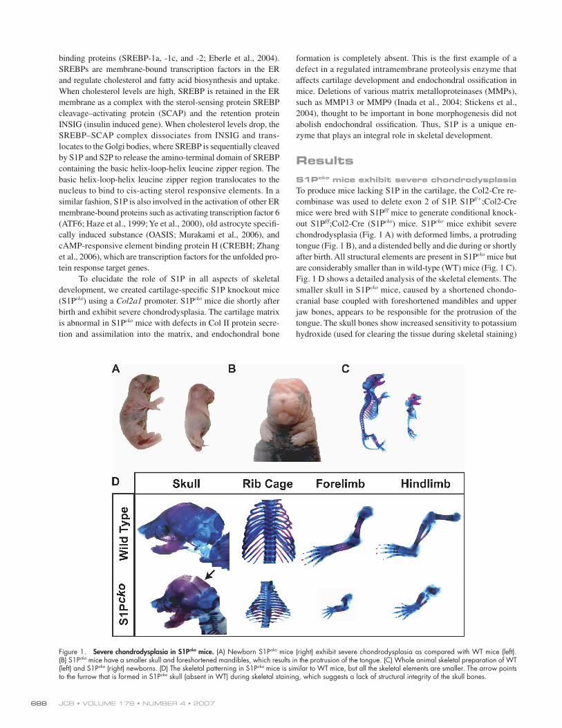
ResultsS1Pcko mice exhibit severe chondrodysplasiaTo produce mice lacking S1P in the cartilage, the Col2-Cre re-
combinase was used to delete exon 2 of S1P. S1Pf/+;Col2-Cre
mice were bred with S1Pf/f mice to generate conditional knock-
out S1Pf/f;Col2-Cre (S1Pcko) mice. S1Pcko mice exhibit severe
chondrodysplasia (Fig. 1 A) with deformed limbs, a protruding
tongue (Fig. 1 B), and a distended belly and die during or shortly
after birth. All structural elements are present in S1Pcko mice but
are considerably smaller than in wild-type (WT) mice (Fig. 1 C).
Fig. 1 D shows a detailed analysis of the skeletal elements. The
smaller skull in S1Pcko mice, caused by a shortened chondo-
cranial base coupled with foreshortened mandibles and upper
jaw bones, appears to be responsible for the protrusion of the
tongue. The skull bones show increased sensitivity to potassium
hydroxide (used for clearing the tissue during skeletal staining)
Figure 1. Severe chondrodysplasia in S1Pcko mice. (A) Newborn S1Pcko mice (right) exhibit severe chondrodysplasia as compared with WT mice (left). (B) S1Pcko mice have a smaller skull and foreshortened mandibles, which results in the protrusion of the tongue. (C) Whole animal skeletal preparation of WT (left) and S1Pcko (right) newborns. (D) The skeletal patterning in S1Pcko mice is similar to WT mice, but all the skeletal elements are smaller. The arrow points to the furrow that is formed in S1Pcko skull (absent in WT) during skeletal staining, which suggests a lack of structural integrity of the skull bones.
SITE-1 PROTEASE AND CARTILAGE AND BONE • PATRA ET AL. 689
that resulted in the dissolution of the bones in the back of the
skull (Fig. 1 D, arrow), which remain intact in the WT. The chest
cavity is small and presses the internal organs into the abdominal
cavity, giving the belly a distended appearance. The smaller
chest cavity probably causes acute neonatal respiratory distress
and subsequent lethality. The skewed orientation of the limbs,
particularly the hind limbs, suggests anomalous articular joint
development. Heterozygotes (S1Pf/+;Col2-Cre) appear normal and
do not show any phenotype (unpublished data).
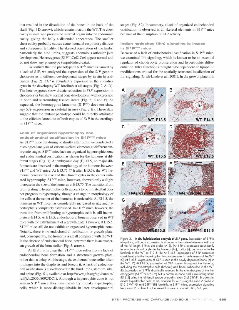
To confi rm that the phenotype in S1Pcko mice is caused by
a lack of S1P, we analyzed the expression of the S1P gene in
chondrocytes in different developmental stages by in situ hybrid-
ization (Fig. 2). S1P is abundantly expressed in the chondro-
cytes in the developing WT forelimb at all stages (Fig. 2, A–D).
The heterozygotes show drastic reduction in S1P expression in
chondrocytes but show normal bone development, with expression
in bone and surrounding tissues intact (Fig. 2, E and F). As
expected, the homozygous knockout (S1Pcko) does not show
any S1P expression in skeletal tissues (Fig. 2 H). These data
suggest that the mutant phenotype could be directly attributed
to the effi cient knockout of both copies of S1P in the cartilage
in S1Pcko mice.
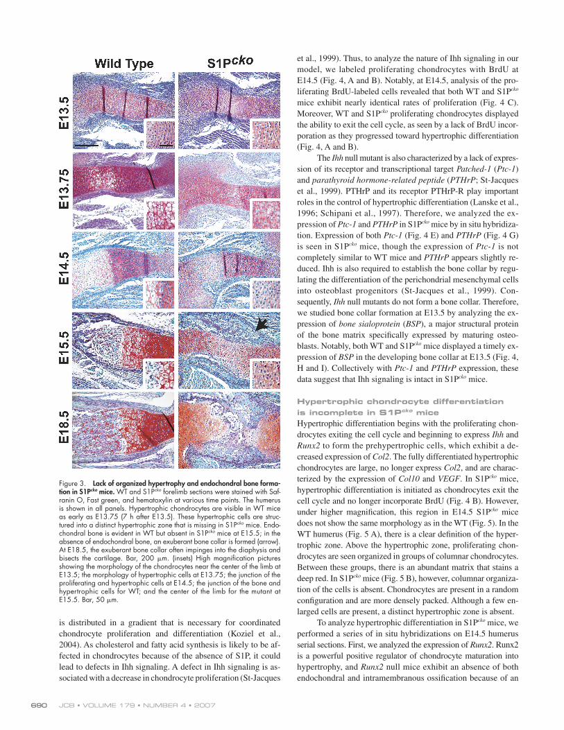
Lack of organized hypertrophy and endochondral ossifi cation in S1Pcko miceAs S1Pcko mice die during or shortly after birth, we conducted a
histological analysis of various skeletal elements at different em-
bryonic stages. S1Pcko mice lack an organized hypertrophic zone
and endochondral ossifi cation, as shown for the humerus at dif-
ferent stages (Fig. 3). At embryonic day (E) 13.5, no major dif-
ferences are observed in the morphology of the humerus between
S1Pcko and WT mice. At E13.75 (7 h after E13.5), the WT hu-
merus increased in size and the chondrocytes in the center initi-
ated hypertrophy. S1Pcko mice, however, showed only a slight
increase in the size of the humerus at E13.75. The transition from
proliferating to hypertrophic cells appears to be initiated but does
not progress to hypertrophy, though a change in morphology of
the cells at the center of the humerus is noticeable. At E14.5, the
humerus in WT mice has considerably increased in size and hy-
pertrophy is completely established. In S1Pcko mice, however, the
transition from proliferating to hypertrophic cells is still incom-
plete at E14.5. At E15.5, endochondral bone is observed in WT
mice with the establishment of a growth plate. However, at E15.5,
S1Pcko mice still do not exhibit an organized hypertrophic zone.
Notably, there is no endochondral ossifi cation or growth plate,
and, consequently, the humerus is small compared with the WT.
In the absence of endochondral bone, however, there is an exuber-
ant growth of the bone collar (Fig. 3, arrow).
At E18.5, it is clear that S1Pcko mice suffer from a lack of
endochondral bone formation and a structured growth plate,
rather than a delay. At this stage, the exuberant bone collar often
impinges into the diaphysis and bisects it. A lack of endochon-
dral ossifi cation is also observed in the hind limbs, sternum, ribs,
and spine (Fig. S1, available at http://www.jcb.org/cgi/content/
full/jcb.200708092/DC1). Although a hypertrophic zone is not
seen in S1Pcko mice, they have the ability to make hypertrophic
cells, which is more distinguishable in later developmental
stages (Fig. S2). In summary, a lack of organized endochondral
ossifi cation is observed in all skeletal elements in S1Pcko mice
because of the disruption of S1P activity.
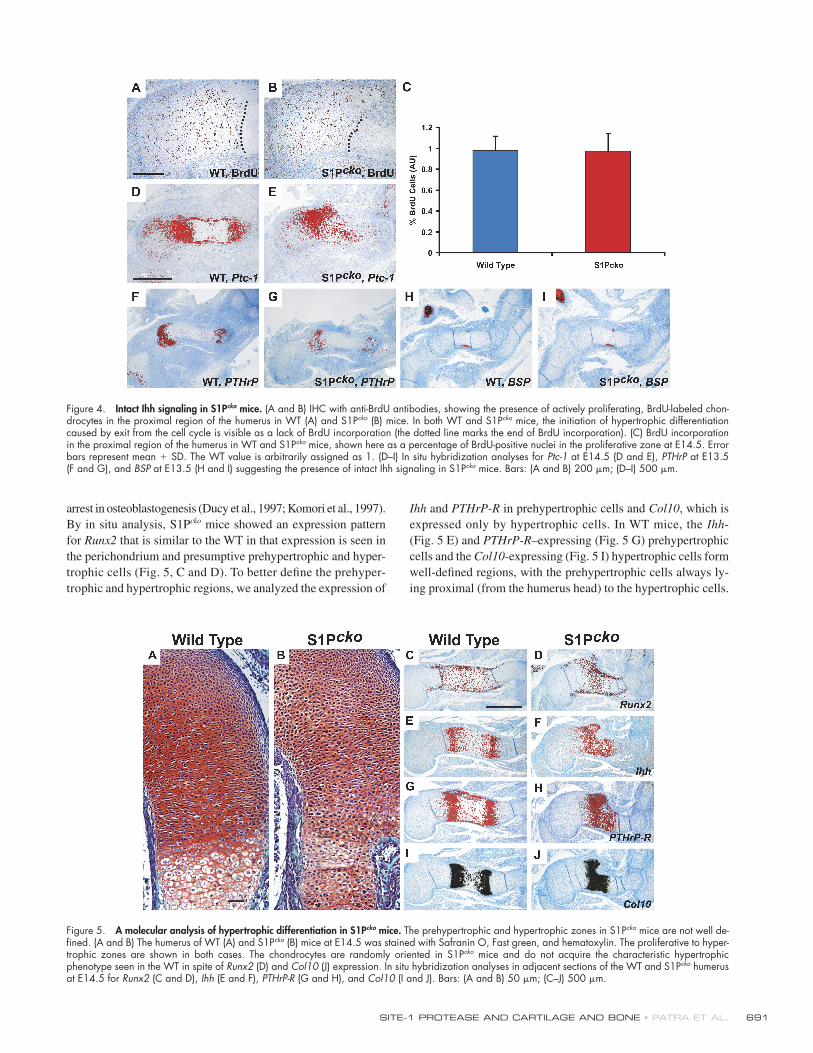
Indian hedgehog (Ihh) signaling is intact in S1Pcko miceBecause of a lack of endochondral ossifi cation in S1Pcko mice,
we examined Ihh signaling, which is known to be an essential
regulator of chondrocyte proliferation and hypertrophic differ-
entiation. Ihh’s function is thought to be dependent on lipophilic
modifi cations critical for the spatially restricted localization of
Ihh signaling (Gritli-Linde et al., 2001). In the growth plate, Ihh
Figure 2. In situ hybridization analysis of S1P gene. Expression of S1P is ubiquitous, although expression is stronger in the skeletal elements with use of the full-length S1P in situ probe (A–E). (A) S1P is expressed abundantly in immature chondrocytes in the humerus (hu), radius (r), and ulna (u) in the forelimb of the WT at E13.5. (B) At E14.5, expression of S1P decreases considerably in the hypertrophic (h) chondrocytes in the humerus of the WT. (C) At E15.5, expression of S1P is seen in the newly deposited bone (b) in the WT. (D) At E18.5, expression of S1P is seen throughout the humerus, including the hypertrophic cells (bracket) and bone trabeculae in the WT. (E) Expression of S1P is drastically reduced in the chondrocytes of the het-erozygotes (S1Pf/+;Col2-Cre) but is normal in bone and surrounding tissue (E18.5) using the full-length probe or against exon 2 of S1P (F). Brackets in-dicate hypertrophic cells. In situ analysis for S1P using the exon 2 probe in E13.5 WT (G) and S1Pcko (H) forelimb. In S1Pcko mice, expression signaling from exon 2 is absent in the skeletal tissues. s, scapula. Bar, 500 μm.
JCB • VOLUME 179 • NUMBER 4 • 2007 690
is distributed in a gradient that is necessary for coordinated
chondrocyte proliferation and differentiation (Koziel et al.,
2004). As cholesterol and fatty acid synthesis is likely to be af-
fected in chondrocytes because of the absence of S1P, it could
lead to defects in Ihh signaling. A defect in Ihh signaling is as-
sociated with a decrease in chondrocyte proliferation (St-Jacques
et al., 1999). Thus, to analyze the nature of Ihh signaling in our
model, we labeled proliferating chondrocytes with BrdU at
E14.5 (Fig. 4, A and B). Notably, at E14.5, analysis of the pro-
liferating BrdU-labeled cells revealed that both WT and S1Pcko
mice exhibit nearly identical rates of proliferation (Fig. 4 C).
Moreover, WT and S1Pcko proliferating chondrocytes displayed
the ability to exit the cell cycle, as seen by a lack of BrdU incor-
poration as they progressed toward hypertrophic differentiation
(Fig. 4, A and B).
The Ihh null mutant is also characterized by a lack of expres-
sion of its receptor and transcriptional target Patched-1 (Ptc-1)
and parathyroid hormone-related peptide (PTHrP; St-Jacques
et al., 1999). PTHrP and its receptor PTHrP-R play important
roles in the control of hypertrophic differentiation (Lanske et al.,
1996; Schipani et al., 1997). Therefore, we analyzed the ex-
pression of Ptc-1 and PTHrP in S1Pcko mice by in situ hybridiza-
tion. Expression of both Ptc-1 (Fig. 4 E) and PTHrP (Fig. 4 G)
is seen in S1Pcko mice, though the expression of Ptc-1 is not
completely similar to WT mice and PTHrP appears slightly re-
duced. Ihh is also required to establish the bone collar by regu-
lating the differentiation of the perichondrial mesenchymal cells
into osteoblast progenitors (St-Jacques et al., 1999). Con-
sequently, Ihh null mutants do not form a bone collar. Therefore,
we studied bone collar formation at E13.5 by analyzing the ex-
pression of bone sialoprotein (BSP), a major structural protein
of the bone matrix specifi cally expressed by maturing osteo-
blasts. Notably, both WT and S1Pcko mice displayed a timely ex-
pression of BSP in the developing bone collar at E13.5 (Fig. 4,
H and I). Collectively with Ptc-1 and PTHrP expression, these
data suggest that Ihh signaling is intact in S1Pcko mice.
Hypertrophic chondrocyte differentiation is incomplete in S1Pcko miceHypertrophic differentiation begins with the proliferating chon-
drocytes exiting the cell cycle and beginning to express Ihh and
Runx2 to form the prehypertrophic cells, which exhibit a de-
creased expression of Col2. The fully differentiated hypertrophic
chondrocytes are large, no longer express Col2, and are charac-
terized by the expression of Col10 and VEGF. In S1Pcko mice,
hypertrophic differentiation is initiated as chondrocytes exit the
cell cycle and no longer incorporate BrdU (Fig. 4 B). However,
under higher magnifi cation, this region in E14.5 S1Pcko mice
does not show the same morphology as in the WT (Fig. 5). In the
WT humerus (Fig. 5 A), there is a clear defi nition of the hyper-
trophic zone. Above the hypertrophic zone, proliferating chon-
drocytes are seen organized in groups of columnar chondrocytes.
Between these groups, there is an abundant matrix that stains a
deep red. In S1Pcko mice (Fig. 5 B), however, columnar organiza-
tion of the cells is absent. Chondrocytes are present in a random
confi guration and are more densely packed. Although a few en-
larged cells are present, a distinct hypertrophic zone is absent.
To analyze hypertrophic differentiation in S1Pcko mice, we
performed a series of in situ hybridizations on E14.5 humerus
serial sections. First, we analyzed the expression of Runx2. Runx2
is a powerful positive regulator of chondrocyte maturation into
hypertrophy, and Runx2 null mice exhibit an absence of both
endochondral and intramembranous ossifi cation because of an
Figure 3. Lack of organized hypertrophy and endochondral bone forma-tion in S1Pcko mice. WT and S1Pcko forelimb sections were stained with Saf-ranin O, Fast green, and hematoxylin at various time points. The humerus is shown in all panels. Hypertrophic chondrocytes are visible in WT mice as early as E13.75 (7 h after E13.5). These hypertrophic cells are struc-tured into a distinct hypertrophic zone that is missing in S1Pcko mice. Endo-chondral bone is evident in WT but absent in S1Pcko mice at E15.5; in the absence of endochondral bone, an exuberant bone collar is formed (arrow). At E18.5, the exuberant bone collar often impinges into the diaphysis and bisects the cartilage. Bar, 200 μm. (insets) High magnifi cation pictures showing the morphology of the chondrocytes near the center of the limb at E13.5; the morphology of hypertrophic cells at E13.75; the junction of the proliferating and hypertrophic cells at E14.5; the junction of the bone and hypertrophic cells for WT; and the center of the limb for the mutant at E15.5. Bar, 50 μm.
SITE-1 PROTEASE AND CARTILAGE AND BONE • PATRA ET AL. 691
arrest in osteoblastogenesis (Ducy et al., 1997; Komori et al., 1997).
By in situ analysis, S1Pcko mice showed an expression pattern
for Runx2 that is similar to the WT in that expression is seen in
the perichondrium and presumptive prehypertrophic and hyper-
trophic cells (Fig. 5, C and D). To better defi ne the prehyper-
trophic and hypertrophic regions, we analyzed the expression of
Ihh and PTHrP-R in prehypertrophic cells and Col10, which is
expressed only by hypertrophic cells. In WT mice, the Ihh-
(Fig. 5 E) and PTHrP-R–expressing (Fig. 5 G) prehypertrophic
cells and the Col10-expressing (Fig. 5 I) hypertrophic cells form
well-defi ned regions, with the prehypertrophic cells always ly-
ing proximal (from the humerus head) to the hypertrophic cells.
Figure 4. Intact Ihh signaling in S1Pcko mice. (A and B) IHC with anti-BrdU antibodies, showing the presence of actively proliferating, BrdU-labeled chon-drocytes in the proximal region of the humerus in WT (A) and S1Pcko (B) mice. In both WT and S1Pcko mice, the initiation of hypertrophic differentiation caused by exit from the cell cycle is visible as a lack of BrdU incorporation (the dotted line marks the end of BrdU incorporation). (C) BrdU incorporation in the proximal region of the humerus in WT and S1Pcko mice, shown here as a percentage of BrdU-positive nuclei in the proliferative zone at E14.5. Error bars represent mean + SD. The WT value is arbitrarily assigned as 1. (D–I) In situ hybridization analyses for Ptc-1 at E14.5 (D and E), PTHrP at E13.5 (F and G), and BSP at E13.5 (H and I) suggesting the presence of intact Ihh signaling in S1Pcko mice. Bars: (A and B) 200 μm; (D–I) 500 μm.
Figure 5. A molecular analysis of hypertrophic differentiation in S1Pcko mice. The prehypertrophic and hypertrophic zones in S1Pcko mice are not well de-fi ned. (A and B) The humerus of WT (A) and S1Pcko (B) mice at E14.5 was stained with Safranin O, Fast green, and hematoxylin. The proliferative to hyper-trophic zones are shown in both cases. The chondrocytes are randomly oriented in S1Pcko mice and do not acquire the characteristic hypertrophic phenotype seen in the WT in spite of Runx2 (D) and Col10 (J) expression. In situ hybridization analyses in adjacent sections of the WT and S1Pcko humerus at E14.5 for Runx2 (C and D), Ihh (E and F), PTHrP-R (G and H), and Col10 (I and J). Bars: (A and B) 50 μm; (C–J) 500 μm.
JCB • VOLUME 179 • NUMBER 4 • 2007 692
In S1Pcko mice, however, such well-defi ned regions are absent.
Zones expressing Ihh (Fig. 5 F), PTHrP-R (Fig. 5 H), and Col10
(Fig. 5 J) largely overlap each other. Thus, even though Ihh,
Runx2, and Col10 are expressed, which indicates hypertrophic
differentiation, the characteristic hypertrophic cell morphology
and organization is missing in S1Pcko mice.
These results indicate that the loss of S1P does not prevent
the molecular program necessary for hypertrophic differentiation.
Rather, the loss of S1P appears to affect cell morphology and
may affect matrix production. Therefore, we analyzed the ex-
pression of the matrix genes Col2 and aggrecan (Agc1) and
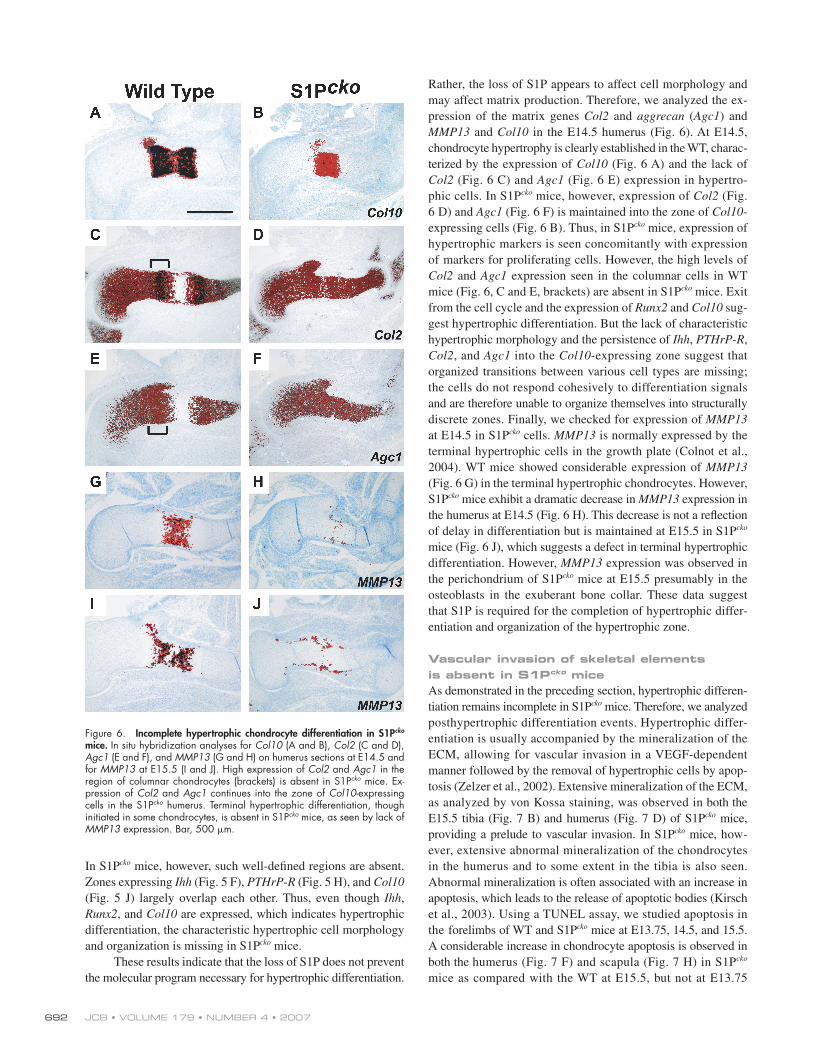
MMP13 and Col10 in the E14.5 humerus (Fig. 6). At E14.5,
chondrocyte hypertrophy is clearly established in the WT, charac-
terized by the expression of Col10 (Fig. 6 A) and the lack of
Col2 (Fig. 6 C) and Agc1 (Fig. 6 E) expression in hypertro-
phic cells. In S1Pcko mice, however, expression of Col2 (Fig.
6 D) and Agc1 (Fig. 6 F) is maintained into the zone of Col10-
expressing cells (Fig. 6 B). Thus, in S1Pcko mice, expression of
hypertrophic markers is seen concomitantly with expression
of markers for proliferating cells. However, the high levels of
Col2 and Agc1 expression seen in the columnar cells in WT
mice (Fig. 6, C and E, brackets) are absent in S1Pcko mice. Exit
from the cell cycle and the expression of Runx2 and Col10 sug-
gest hypertrophic differentiation. But the lack of characteristic
hypertrophic morphology and the persistence of Ihh, PTHrP-R,
Col2, and Agc1 into the Col10-expressing zone suggest that
organized transitions between various cell types are missing;
the cells do not respond cohesively to differentiation signals
and are therefore unable to organize themselves into structurally
discrete zones. Finally, we checked for expression of MMP13
at E14.5 in S1Pcko cells. MMP13 is normally expressed by the
terminal hypertrophic cells in the growth plate (Colnot et al.,
2004). WT mice showed considerable expression of MMP13
(Fig. 6 G) in the terminal hypertrophic chondrocytes. However,
S1Pcko mice exhibit a dramatic decrease in MMP13 expression in
the humerus at E14.5 (Fig. 6 H). This decrease is not a refl ection
of delay in differentiation but is maintained at E15.5 in S1Pcko
mice (Fig. 6 J), which suggests a defect in terminal hypertrophic
differentiation. However, MMP13 expression was observed in
the perichondrium of S1Pcko mice at E15.5 presumably in the
osteoblasts in the exuberant bone collar. These data suggest
that S1P is required for the completion of hypertrophic differ-
entiation and organization of the hypertrophic zone.
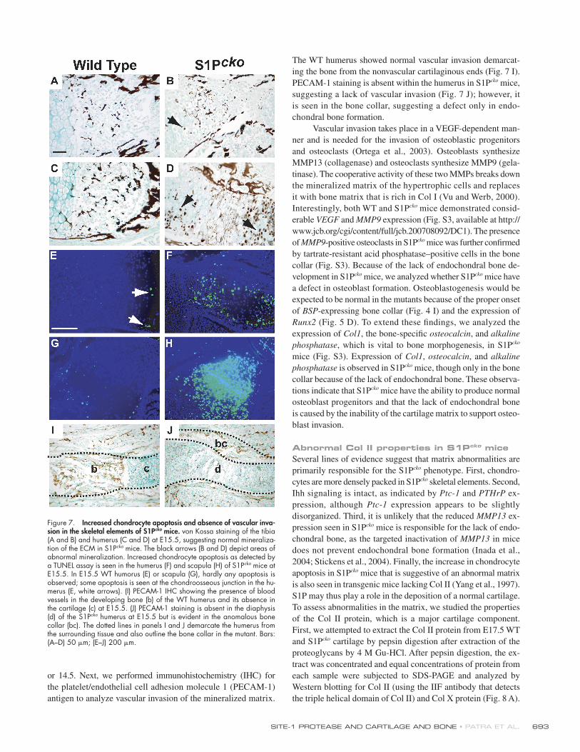
Vascular invasion of skeletal elements is absent in S1Pcko miceAs demonstrated in the preceding section, hypertrophic differen-
tiation remains incomplete in S1Pcko mice. Therefore, we analyzed
posthypertrophic differentiation events. Hypertrophic differ-
entiation is usually accompanied by the mineralization of the
ECM, allowing for vascular invasion in a VEGF-dependent
manner followed by the removal of hypertrophic cells by apop-
tosis (Zelzer et al., 2002). Extensive mineralization of the ECM,
as analyzed by von Kossa staining, was observed in both the
E15.5 tibia (Fig. 7 B) and humerus (Fig. 7 D) of S1Pcko mice,
providing a prelude to vascular invasion. In S1Pcko mice, how-
ever, extensive abnormal mineralization of the chondrocytes
in the humerus and to some extent in the tibia is also seen.
Abnormal mineralization is often associated with an increase in
apoptosis, which leads to the release of apoptotic bodies (Kirsch
et al., 2003). Using a TUNEL assay, we studied apoptosis in
the forelimbs of WT and S1Pcko mice at E13.75, 14.5, and 15.5.
A considerable increase in chondrocyte apoptosis is observed in
both the humerus (Fig. 7 F) and scapula (Fig. 7 H) in S1Pcko
mice as compared with the WT at E15.5, but not at E13.75
Figure 6. Incomplete hypertrophic chondrocyte differentiation in S1Pcko mice. In situ hybridization analyses for Col10 (A and B), Col2 (C and D), Agc1 (E and F), and MMP13 (G and H) on humerus sections at E14.5 and for MMP13 at E15.5 (I and J). High expression of Col2 and Agc1 in the region of columnar chondrocytes (brackets) is absent in S1Pcko mice. Ex-pression of Col2 and Agc1 continues into the zone of Col10-expressing cells in the S1Pcko humerus. Terminal hypertrophic differentiation, though initiated in some chondrocytes, is absent in S1Pcko mice, as seen by lack of MMP13 expression. Bar, 500 μm.
SITE-1 PROTEASE AND CARTILAGE AND BONE • PATRA ET AL. 693
or 14.5. Next, we performed immunohistochemistry (IHC) for
the platelet/endothelial cell adhesion molecule 1 (PECAM-1)
antigen to analyze vascular invasion of the mineralized matrix.
The WT humerus showed normal vascular invasion demarcat-
ing the bone from the nonvascular cartilaginous ends (Fig. 7 I).
PECAM-1 staining is absent within the humerus in S1Pcko mice,
suggesting a lack of vascular invasion (Fig. 7 J); however, it
is seen in the bone collar, suggesting a defect only in endo-
chondral bone formation.
Vascular invasion takes place in a VEGF-dependent man-
ner and is needed for the invasion of osteoblastic progenitors
and osteoclasts (Ortega et al., 2003). Osteoblasts synthesize
MMP13 (collagenase) and osteoclasts synthesize MMP9 (gela-
tinase). The cooperative activity of these two MMPs breaks down
the mineralized matrix of the hypertrophic cells and replaces
it with bone matrix that is rich in Col I (Vu and Werb, 2000).
Interestingly, both WT and S1Pcko mice demonstrated consid-
erable VEGF and MMP9 expression (Fig. S3, available at http://
www.jcb.org/cgi/content/full/jcb.200708092/DC1). The presence
of MMP9-positive osteoclasts in S1Pcko mice was further confi rmed
by tartrate-resistant acid phosphatase–positive cells in the bone
collar (Fig. S3). Because of the lack of endochondral bone de-
velopment in S1Pcko mice, we analyzed whether S1Pcko mice have
a defect in osteoblast formation. Osteoblastogenesis would be
expected to be normal in the mutants because of the proper onset
of BSP-expressing bone collar (Fig. 4 I) and the expression of
Runx2 (Fig. 5 D). To extend these fi ndings, we analyzed the
expression of Col1, the bone-specifi c osteocalcin, and alkaline phosphatase, which is vital to bone morphogenesis, in S1Pcko
mice (Fig. S3). Expression of Col1, osteocalcin, and alkaline phosphatase is observed in S1Pcko mice, though only in the bone
collar because of the lack of endochondral bone. These observa-
tions indicate that S1Pcko mice have the ability to produce normal
osteoblast progenitors and that the lack of endochondral bone
is caused by the inability of the cartilage matrix to support osteo-
blast invasion.
Abnormal Col II properties in S1Pcko miceSeveral lines of evidence suggest that matrix abnormalities are
primarily responsible for the S1Pcko phenotype. First, chondro-
cytes are more densely packed in S1Pcko skeletal elements. Second,
Ihh signaling is intact, as indicated by Ptc-1 and PTHrP ex-
pression, although Ptc-1 expression appears to be slightly
disorganized. Third, it is unlikely that the reduced MMP13 ex-
pression seen in S1Pcko mice is responsible for the lack of endo-
chondral bone, as the targeted inactivation of MMP13 in mice
does not prevent endochondral bone formation (Inada et al.,
2004; Stickens et al., 2004). Finally, the increase in chondrocyte
apoptosis in S1Pcko mice that is suggestive of an abnormal matrix
is also seen in transgenic mice lacking Col II (Yang et al., 1997).
S1P may thus play a role in the deposition of a normal cartilage.
To assess abnormalities in the matrix, we studied the properties
of the Col II protein, which is a major cartilage component.
First, we attempted to extract the Col II protein from E17.5 WT
and S1Pcko cartilage by pepsin digestion after extraction of the
proteoglycans by 4 M Gu-HCl. After pepsin digestion, the ex-
tract was concentrated and equal concentrations of protein from
each sample were subjected to SDS-PAGE and analyzed by
Western blotting for Col II (using the IIF antibody that detects
the triple helical domain of Col II) and Col X protein (Fig. 8 A).
Figure 7. Increased chondrocyte apoptosis and absence of vascular inva-sion in the skeletal elements of S1Pcko mice. von Kossa staining of the tibia (A and B) and humerus (C and D) at E15.5, suggesting normal mineraliza-tion of the ECM in S1Pcko mice. The black arrows (B and D) depict areas of abnormal mineralization. Increased chondrocyte apoptosis as detected by a TUNEL assay is seen in the humerus (F) and scapula (H) of S1Pcko mice at E15.5. In E15.5 WT humorus (E) or scapula (G), hardly any apoptosis is observed; some apoptosis is seen at the chondroosseous junction in the hu-merus (E, white arrows). (I) PECAM-1 IHC showing the presence of blood vessels in the developing bone (b) of the WT humerus and its absence in the cartilage (c) at E15.5. (J) PECAM-1 staining is absent in the diaphysis (d) of the S1Pcko humerus at E15.5 but is evident in the anomalous bone collar (bc). The dotted lines in panels I and J demarcate the humerus from the surrounding tissue and also outline the bone collar in the mutant. Bars: (A–D) 50 μm; (E–J) 200 μm.

JCB • VOLUME 179 • NUMBER 4 • 2007 694
We were able to extract the full-length Col II from the WT head
(Fig. 8 A, lane 3) and the rest of its skeleton (Fig. 8 A, lane 1).
Notably, we were not able to extract the full-length Col II from
any of the S1Pcko skeletal elements (Fig. 8 A, lanes 2 and 4).
In some preparations, some lower molecular weight proteins
with immunoreactivity to the IIF antibody are seen (unpublished
data). But we were unable to extract comparable amounts of the
full-length Col II protein in any of our attempts. By Western
blotting analysis of twofold dilutions of the pepsin-digested
material, we determined that we were extracting �64-fold less
of the full-length Col II protein from the S1Pcko mice as compared
with WT (unpublished data). In contrast, we were able to ex-
tract comparable amounts of the Col X protein from both WT
and S1Pcko mice.
These data suggested that the Col II protein is less abun-
dant or absent in S1Pcko mice. However, we were able to detect
the full-length Col II procollagen in the medium of cultured
chondrocytes from both E18.5 WT and S1Pcko mice by Western
blotting (Fig. 8 B). Furthermore, by IHC analysis, Col II protein
was detected in the humerus of E18.5 WT (Fig. 8 C) and S1Pcko
(Fig. 8 D) mice. We also tested for the presence of the Col IIA
protein (using the IIA antibody that detects exon 2), which is
normally detected in the matrix surrounding chondroprogenitor
cells and early immature chondrocytes. The Col IIA isoform was
detected in both WT (Fig. 8 E) and S1Pcko mice (Fig. 8 F). In the
WT, detection of Col IIA protein was strongest at the location of
the early, immature chondrocytes close to the articular surface.
Surprisingly, in S1Pcko mice, Col IIA was detected throughout
the length of the humerus (Fig. 8 F). Therefore, we analyzed
whether alternative splicing of the procollagen mRNA from
Col2A to Col2B was normal in S1Pcko mice. For this analysis, we
performed in situ hybridization using highly specifi c 24-mer
oligonucleotide probes spanning the exon 1 and 2 junction for
Col2A analysis and the exon 1 and 3 junction for the Col2B anal-
ysis. Splicing of both Col2A and 2B mRNA was, however, found
to be normal in S1Pcko mice (Fig. S4, available at http://www.jcb
.org/cgi/content/full/jcb.200708092/DC1). These data suggested
that Col II synthesis was taking place in S1Pcko mice, although
the turnover of Col II is abnormal, as total Col II and Col IIA was
detected throughout the length of the humerus. These data also
suggested that the organization of the cartilage ECM may be ab-
normal. Therefore, to assess the structural organization of the
collagens in the ECM, we fi rst determined the birefringent prop-
erties of the Col II fi brils in both WT and S1Pcko mice. A marked
Figure 8. Abnormal Col II properties in S1Pcko mice. (A) The cartilage from the spine and limbs (1 and 2) or head (3 and 4) of WT (1 and 3) and S1Pcko (2 and 4) mice were pepsin digested after extraction of the proteoglycans with Gu-HCl. The pepsin-released collagens were concentrated and analyzed by SDS-PAGE and Western blotted for Col II (top) and Col X (bottom) proteins. (B) Western blot analysis for the Col II procollagen (Pro-Col II) from the me-dium of WT (2) or S1Pcko (3) chondrocyte cultures (1 is control medium before the addition of chondrocytes). IHC for the Col II (C and D; IIF antibody) and Col IIA (E and F; IIA antibody) proteins demonstrating their expression in the mutant mice. Detection of protein was performed with DAB. Polarized light microscopy to analyze the birefringence properties of the Col II fi brillar network in the E16.5 humerus of WT (G) and S1Pcko (H) mice. White line in B indi-cates that intervening lanes have been spliced out. Bar, 500 μm.
SITE-1 PROTEASE AND CARTILAGE AND BONE • PATRA ET AL. 695
decrease in the birefringence of the collagen network was ob-
served in the E16.5 humerus in S1Pcko mice (Fig. 8 H) as com-
pared with the WT (Fig. 8 G), which suggests a poorly organized
collagen fi brillar network in the cartilage of S1Pcko mice. This re-
duced birefringence in S1Pcko mice is similar to that seen in mice
with inactivation of the Col2a1 gene (Li et al., 1995). These data
together suggest that one of the molecular defects caused by a
lack of S1P is the abnormal organization of the Col II protein,
which results in an abnormal cartilage that prevents vascular in-
vasion and endochondral ossifi cation in S1Pcko mice.
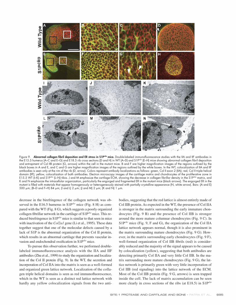
To pursue this observation further, we performed double-
labeled immunofl uorescence analysis with the IIA and IIF
antibodies (Zhu et al., 1999) to study the organization and localiza-
tion of the Col II protein (Fig. 9). In the WT, the secretion and
incorporation of Col IIA into the matrix is seen as a well-formed
and organized green lattice network. Localization of the colla-
gen triple helical domains is seen as red immunofl uorescence,
which in the WT is seen as a distinct red lattice network with
hardly any yellow colocalization signals from the two anti-
bodies, suggesting that the red lattice is almost entirely made of
Col IIB protein. As expected in the WT, the presence of Col IIA
is stronger in the matrix surrounding the early immature chon-
drocytes (Fig. 9 B) and the presence of Col IIB is stronger
around the more mature columnar chondrocytes (Fig. 9 C). In
S1Pcko mice (Fig. 9, F and G), the organization of the Col IIA
lattice network appears normal, though it is also prominent in
the matrix surrounding mature chondrocytes (Fig. 9 G). How-
ever, in the matrix surrounding early chondrocytes (Fig. 9 F),
well-formed organization of Col IIB fi brils (red) is consider-
ably reduced and the majority of the signal appears to be caused
by colocalization (yellow), suggesting that both antibodies are
detecting primarily Col IIA and very little Col IIB. In the ma-
trix surrounding more mature chondrocytes (Fig. 9 G), the lat-
tice network is primarily green with very little incorporation of
Col IIB (red signaling) into the lattice network of the ECM.
Most of the Col IIB protein (Fig. 9 G, arrows) is seen trapped
inside the cell. The lack of matrix accumulation can be seen
more clearly in cross sections of the ribs (at E18.5) in S1Pcko
Figure 9. Abnormal collagen fi bril deposition and ER stress in S1Pcko mice. Double-labeled immunofl uorescence studies with the IIA and IIF antibodies in the E15.5 humerus (A–C and E–G) and E18.5 rib cross sections (D and H) in WT (A–D) and S1Pcko (E–H) mice showing abnormal collagen fi bril deposition and entrapment of Col IIB protein (G, arrows) within the cell in the mutant mice. B and F are higher magnifi cation images of the regions outlined by the black boxes in A and E, and C and G are higher magnifi cation images of the regions outlined by the white boxes. In the WT, colocalization of IIA and IIF antibodies is seen only at the rim of the rib (D, arrow). Colors represent antibody localizations as follows: green, Col II exon 2 (IIA); red, Col II triple helical domain (IIF); yellow, colocalization of both antibodies. Electron microscopy images of the cartilage matrix and chondrocytes of the proliferative zone in E15.5 WT (I–K) and S1Pcko (L–N) tibia. J and M emphasize the cartilage ECM, showing the decrease in collagen fi brillar density in the S1Pcko matrix, and K and N emphasize the intracellular organization, particularly the engorged and fragmented ER in the mutant mice (black arrows). The engorged ER in the mutant is fi lled with materials that appear homogenously or heterogeneously stained with partially crystalline appearance (N, white arrow). Bars: (A and E) 500 μm; (B–D and F–H) 84 μm; (I and L) 2 μm; (J and M) 2 μm; (K and N) 1 μm.
JCB • VOLUME 179 • NUMBER 4 • 2007 696
mice (Fig. 9 H). Again, primarily colocalization signals (yellow)
and the substantial absence of matrix when compared with the
WT (Fig. 9 D) is observed, which showed abundant matrix (red)
with yellow colocalization signals restricted to the rim of the rib.
These data suggested that in S1Pcko mice, although there is
no problem in the expression and secretion of Col IIA and its
incorporation into the matrix, the expression and/or secretion of
Col IIB is abnormal and results in a matrix with considerably re-
duced levels of Col IIB and therefore an inability to extract Col II
from the matrix (Fig. 8 A). To understand the mechanism for this
defect, we performed electron microscopic analysis of tibia
(Fig. 9, I–N) and humerus (not depicted) from E15.5 WT and
S1Pcko mice. In the WT (Fig. 9, I–K), the cartilage from the pro-
liferative zone showed the presence of well-formed and abun-
dant, homogenous Col II fi brils surrounding normal chondrocytes.
In S1Pcko mice, however (Fig. 9, L–N), the distribution of colla-
gen fi bril density is irregular and considerably reduced. The ER
is drastically enlarged, irregular in shape, and fragmented and
also often found fi lled with granular, homogenous, or hetero-
genously stained material that also sometimes appears to be crys-
talline. Similar observations were seen in the chondrocytes/ECM
of the articular, hypertrophic, and joint cartilage regions in S1Pcko
mice. These data suggest the induction of ER stress in chondro-
cytes in S1Pcko mice, presumably caused by the excessive de-
mand for complex matrix protein synthesis required for cartilage
deposition, and a requirement for S1P activity to alleviate ER
stress during matrix protein synthesis and deposition.
DiscussionIn this paper, we have established the importance of S1P to car-
tilage deposition and endochondral ossifi cation. Null mice with
a loss of S1P in all tissues were found to be embryonic lethal at
a very early stage and therefore did not provide a suitable ave-
nue to study its contribution to cartilage and bone development
(Yang et al., 2001). Our studies on S1Pcko mice have allowed us to
focus on the requirement of S1P in chondrocyte differentiation
and the genesis of a normal endochondral skeleton. Cartilage-
specifi c disruption of S1P in S1Pcko mice results in total lack of
endochondral bone in all skeletal elements that exhibit longitu-
dinal bone growth through endochondral ossifi cation.
The lack of endochondral bone seen in S1Pcko mice is also
seen in Ihh (St-Jacques et al., 1999) and Runx2 null mice (Komori
et al., 1997; Otto et al., 1997). In both these mutants, endochon-
dral bone development is hampered because of a defect in
osteoblastogenesis. Both Ihh and Runx2 null mice also suffer
from varying degrees of an abnormal chondrocyte differentia-
tion program (Inada et al., 1999; Kim et al., 1999). In S1Pcko
mice, we initially expected that the phenotype could have arisen
because of a defect in the Ihh signaling pathway. However, our
data suggest that S1Pcko mice have an intact Ihh signaling path-
way, as indicated by the expression of Ptc-1 and PTHrP in
S1Pcko skeletal elements. However, the mislocalization of Ptc-1–
expressing cells leaves open the possibility that S1P has an ef-
fect on the activity level and/or diffusion of Ihh. The morphological
phenotype that sets S1Pcko mice apart from Ihh and Runx2 null
mice is that S1Pcko mice exhibit an exuberant bone collar show-
ing normal osteoblastogenesis. Elimination of S1P results in an
uncoupling of normal endochondral bone formation from corti-
cal bone formation. Thus, like Ihh and Runx2, S1P is a major
positive regulator of endochondral ossifi cation. Expression of
S1P is observed in the perichondrium in the WT. However, in
S1Pcko mice, S1P activity would be expected to be absent in the
perichondrium because of active Col2-Cre activity in these
cells. These data suggest that S1P is not necessary for cortical
bone formation. Mice with double knockouts of the transcrip-
tion factors L-Sox5 and Sox6 also exhibit a lack of endochon-
dral ossifi cation with development of a thick cortical bone (Smits
et al., 2001). However, it is unlikely that this pathway is affected
in S1Pcko mice, as they exhibit abundant expression of L-Sox5
and Sox6 (unpublished data).
The lack of endochondral bone in S1Pcko mice appears to
be caused by an inability of the blood vessels to invade the
abnormal, mineralized cartilage in spite of apparently adequate
VEGF expression. According to the tenets of organogenesis,
where development is guided by tissue interactions, the devel-
opmental history of a tissue plays an important role in its ability
to respond to instructive cues (De Robertis, 2006). Thus, it
would be expected that an abnormal cartilage would constrain
its ability to respond to instructions from the perichondrium
and/or vascular tissue. The genesis of a normal matrix is neces-
sary for proper hypertrophic differentiation and endochondral
ossifi cation. Our data suggest that the defects in hypertrophic
differentiation and endochondral ossifi cation can be traced to an
abnormal matrix in S1Pcko mutants. In S1Pcko mice, the chondro-
cytes are densely packed and randomly oriented with very little
matrix between them. The increased chondrocyte apoptosis
seen in S1Pcko mice is also highly indicative of an abnormal car-
tilage that is unable to support chondrocyte survival. This is
similar to the increased apoptosis seen in transgenic mice lack-
ing Col II (Yang et al., 1997). The absence of a proper cartilage
could hinder not only the supply of nutrients to the avascular
ECM (Hunziker and Herrmann, 1990) but also have profound
effects on integrin-mediated pathways and distribution of
growth factors (Yoon and Fisher, 2006). The inability to extract
normal amounts of full-length Col II from cartilage, the de-
creased birefringence of Col II network, the lack of the Col IIB
lattice network, and the considerable reduction in collagen
fi brillar density as seen by electron microscopy all attest to the
fact that the cartilage is abnormal in S1Pcko mice. S1Pcko mice
also exhibit abnormal spine development in that the interverte-
bral discs are not well formed and lack the gelatinous nucleus
pulposus in the center. This is also seen in transgenic mice with
a null mutation in the Col2a1 gene (Aszodi et al., 1998).
An abnormal cartilage ECM could mask angiogenic sig-
naling by VEGF in spite of the adequate VEGF expression that
is seen in S1Pcko mice. Given the abnormal characteristics of the
cartilage matrix, even if VEGF is not masked, the MMPs may not
be able to process and break down the alien matrix in S1Pcko
mice. An inability to degrade the matrix is suggested by the per-
sistence of Col IIA in the S1Pcko cartilage, as Col IIA is seen
throughout the length of the humerus. These observations are
highly suggestive of a lack of structurally intact cartilage matrix
in S1Pcko mice that is normally a forerunner for endochondral
SITE-1 PROTEASE AND CARTILAGE AND BONE • PATRA ET AL. 697
bone development. Some of the phenotypes seen in S1Pcko mice
also bear resemblance to the phenotypes seen in mutants for other
matrix proteins. For example, a lack of columnar hypertrophic
chondrocytes, disorganized growth plates, chondrodysplasia, and
incomplete nucleus pulposus are seen in mice lacking perlecan
(Arikawa-Hirasawa et al., 1999; Gustafsson et al., 2003),
Agc1 (Watanabe et al., 1994; Wai et al., 1998), and/or link protein
(Watanabe and Yamada, 1999). These observations clearly attest
to the importance of a properly organized matrix for chondrocyte
differentiation. However, these mutations exhibit only reduced
or delayed endochondral ossifi cation. Thus, it is noteworthy
that only the disruption of the Col2a1 gene (Li et al., 1995) results
in a complete lack of endochondral ossifi cation as seen in S1Pcko
mice. These data suggest a convergence of S1P activity with a
property of Col II in relation to the cartilage matrix.
Thus, our paper shows that S1P is essential to endochon-
dral ossifi cation. The much accepted role of S1P is that of a
proprotein convertase required to activate the transcription fac-
tors SREBPs, ATF6, OASIS, and CREBH, the latter three play-
ing vital roles during ER stress response. Given that the knockout
of the SREBP molecules has not exhibited any known defect in
bone morphogenesis in mice that escape embryonic lethality
(Shimano et al., 1997; Liang et al., 2002), it seems unlikely that
the phenotype in S1Pcko mice is manifested through SREBPs.
Furthermore, in the zebrafi sh gonzo phenotype, knockdown of
SCAP (the lipid sensor) results only in strong lipid phenotypes
with normal cartilage (Schlombs et al., 2003). These data sug-
gest that the abnormal cartilage phenotype in S1Pcko mice is me-
diated by lipid-independent pathways. Given the engorgement
and fragmentation of the ER in S1Pcko mice, it seems likely that
S1P is required to activate ER stress signaling (ERSS) in re-
sponse to the hyperproduction of complex cartilage matrix pro-
teins seen in chondrocytes. Unlike other studies (Tsang et al., 2007),
it is noteworthy that the ER stress observed in S1Pcko mice
is not caused by the introduction of any mutant matrix pro-
tein but by the inability of the chondrocyte to respond to ER
stress because of a lack of S1P activity. Based on these data we
propose that the ER stress response is part of the vital protein
processing machinery in normal chondrocytes and is essential
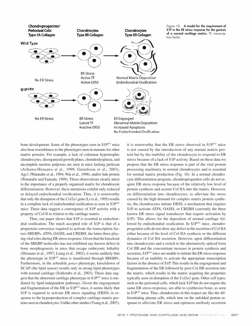
for normal matrix production (Fig. 10). In a normal chondro-
cyte differentiation program, chondroprogenitor cells do not re-
quire ER stress response because of the relatively low level of
protein synthesis and secrete Col IIA into the matrix. However,
on differentiation into chondrocytes, to alleviate the stress
caused by the high demand for complex matrix protein synthe-
sis, the chondrocytes initiate ERSS, a mechanism that requires
S1P to activate ATF6, OASIS, or CREBH (currently the three
known ER stress signal transducers that require activation by
S1P). This allows for the deposition of normal cartilage fol-
lowed by endochondral ossifi cation. In S1Pcko mice, chondro-
progenitor cells do not show any defect in the secretion of Col IIA
either because of the level of Col IIA synthesis or the different
dynamics of Col IIA secretion. However, upon differentiation
into chondrocytes and a switch to the alternatively spliced form
Col IIB and the concomitant increase in protein synthesis and
secretion, S1Pcko mice are unable to initiate the ER stress response
because of an inability to activate the appropriate transcription
factors in the absence of S1P. This results in the engorgement and
fragmentation of the ER followed by poor Col IIB secretion into
the matrix, which results in the matrix acquiring the properties
typically seen on disruption of the Col2a1 gene. Other cell types,
such as the periosteal cells, which lack S1P but do not require the
same ER stress response, are able to synthesize bone, as seen
in S1Pcko mice. Thus, chondrocytes in this respect are like the dif-
ferentiating plasma cells, which turn on the unfolded protein re-
sponse to alleviate ER stress and optimize antibody secretion
Figure 10. A model for the requirement of S1P in the ER stress response for the genesis of a normal cartilage matrix. TF, transcrip-tion factor.
JCB • VOLUME 179 • NUMBER 4 • 2007 698
(Gunn et al., 2004), or exocrine glands, where the ER stress
response is necessary for the full development of the secretory
machinery (Lee et al., 2005).
In the zebrafi sh S1P gonzo mutants, ER stress and un-
folded protein response were reported to be normal based on in
situ hybridization to two mRNA sequences known to be down-
stream of ATF6. In light of recent discoveries, this assessment
may have been misleading. First, the analysis in gonzo mutants
involved the in situ analysis of the full-length XBP1 mRNA and
not the specifi c splice form necessary for ER stress response
(Yoshida et al., 2001; Calfon et al., 2002). Second, the presence
of immunoglobulin heavy chain binding pretien (BiP) chaper-
one does not ensure an adequate ERSS in chondrocytes, and BiP
can be regulated by other transcription factors such as NF-Y
(Yoshida et al., 2000). In preliminary experiments, we also de-
tect BiP expression in both WT and S1Pcko mice (unpublished
data). Lastly, besides ATF6, at least two other ERSS transcrip-
tion factors, OASIS and CREBH, are regulated by S1P. Collec-
tively, these observations suggest the requirement of a new
specialized pathway for regulation of matrix protein synthesis
in chondrocytes. We are currently investigating these possibil-
ities using our mouse model to provide a more complete picture
of the utility of S1P in ER stress response, matrix synthesis, and
endochondral bone formation.
Materials and methodsGeneration of S1Pcko miceTo generate S1Pcko mice, S1Pf/f mice (mice homozygous for the S1Pfl ox al-lele in which the exon 2 of S1P is fl oxed, provided by J.D. Horton, Univer-sity of Texas Southwestern Medical Center, Dallas, TX; Yang et al., 2001) were bred with Col2-Cre (where the Cre recombinase is under the infl u-ence of the cartilage-specifi c Col2a1 promoter, limiting its expression pri-marily to cartilage; a gift of D. Ornitz, Washington University School of Medicine, St. Louis, MO; Jacob et al., 2006) transgenic mice of the C57BL/6J strain to produce S1Pf/+;Col2-Cre mice (mice heterozygous for the S1Pfl ox allele and positive for the Col2-Cre transgene). The S1Pf/+;Col2-Cre mice were bred with S1Pf/f mice to generate S1Pcko mice. Genotypes were verifi ed by PCR analysis of tail-derived DNA.
Isolation of collagens from WT and S1Pcko miceThe isolation of collagens was done from E17.5 WT and S1Pcko mice using a modifi cation of previously published protocols (Mayne et al., 1995). In brief, E17.5 embryos were skinned and eviscerated, and the head was processed separately from the rest of the body. The spine/limbs and head were frozen and crushed, and the proteoglycans were extracted with 4 M Gu-HCl in 0.05 M Tris-HCl, pH 7.5, containing 0.01 M EDTA and a 1× complete protease inhibitor cocktail (Roche), for 36 h at 4°C. The cartilage residues left after proteoglycan extraction were washed with cold water and digested with 1 mg/ml pepsin in 0.5 M acetic acid for 24 h at 4°C. The digest was then concentrated with centricon30 (Millipore) to �250 μl, dialyzed against 0.05 M Tris-HCl, pH 7.5, buffer containing 0.4 M NaCl and 5 mM EDTA, and stored at −20°C. Equal concentrations of protein preparations (determined by Bio-Rad Laboratories protein assay) from WT and S1Pcko mice were separated by SDS-PAGE followed by Western blot-ting for Col II (using the previously described rat antibody IIF against bovine Col II triple helical domain; Zhu et al., 1999) and Col X proteins (EMD). The molecular weight marker used in SDS-PAGE was BenchMark prestained protein ladder (Invitrogen).
Primary chondrocyte cultures from WT and S1Pcko miceTo study the biosynthesis and secretion of the Col II protein in vitro, primary cultures from WT and S1Pcko mice were established. Chondrocytes were isolated from the sternum and ribs of WT and S1Pcko mice as described previously (Lefebvre et al., 1994). In brief, rib cages were taken from WT or S1Pcko mice and treated with 2 mg/ml pronase (Roche) at 37°C for 30 min followed by 3 mg/ml collagenase (Roche) treatment in the pres-
ence of 8% CO2 for 1.5 h at 37°C or until all soft tissues are detached from the cartilage. The cartilage was then separated from the soft tissues and treated again with 3 mg/ml collagenase for 4 h at 37°C in the presence of 5% CO2. The digest was fi ltered through a cell strainer to remove un-digested bony parts and the cells were then pelleted by centrifugation, washed with 1× PBS, and plated at a density of 2.5 × 10⁵ per cm2 in DME with 10% heat-inactivated serum, 2% penicillin/streptomycin (Sigma-Aldrich), and 0.25 mM sodium ascorbate. 50 μl of media from the WT and S1Pcko chondrocyte cultures was harvested at regular intervals and separated by 7.5% SDS-PAGE (Bio-Rad Laboratories), and the type II pro-collagen was detected by Western blotting using the IIF antibody.
Morphological and histological analysisWhole-mount skeletal staining of embryos by alcian blue and alizarin red was performed as described previously (McLeod, 1980). For histological and in situ hybridization analyses on sections, embryonic tissues were col-lected at various embryonic time points and processed and sectioned as described previously (Hilton et al., 2005). Histological analyses were done primarily by Safranin O, Fast green, and hematoxylin staining. Detection of mineralization was performed by staining with 1% silver nitrate as per the von Kossa method, followed by counterstaining with Methyl green. In situ hybridization analyses were performed using 35S-labeled riboprobes as described previously (Long et al., 2001). All in situ probes with the ex-ception of Agc1 were provided by F. Long (Washington University School of Medicine) and have been described by Hilton et al. (2005) and refer-ences therein. The Agc1 in situ probe (a gift of E. Vuorio, University of Turku, Turku, Finland) covering the IGD sequence of the mouse Agc1 has been described previously (Glumoff et al., 1994). The full-length S1P and the S1P exon 2 in situ probes were derived from a full-length cDNA clone (American Type Culture Collection). For BrdU analyses, pregnant females were injected with BrdU as described previously (Hilton et al., 2005), and proliferation of chondrocytes was analyzed using a kit (Invitrogen). For PECAM-1 IHC, embryos were fi xed using 4% formaldehyde followed by 30% sucrose infi ltration. 10-μm cryostat sections were derived from tissues embedded in OCT (Tissue-Tek; Thermo Fisher Scientifi c) and IHC was per-formed using a monoclonal rat anti–mouse PECAM-1 antibody (BD Bio-sciences). Detection of PECAM-1 was done using HRP-conjugated anti–rat IgG and DAB. Methyl green was used for counterstaining. Detection of apoptosis was performed by a TUNEL assay using the in situ cell death de-tection kit (Roche) according to the manufacturer’s instructions. Nuclei were counterstained with DAPI and sections were examined by fl uorescence microscopy. Tartrate-resistant acid phosphatase–positive cells were stained using standard procedures with methyl green as counterstain. Study of the birefringence of collagen fi bers by polarized light microscopy was done on 5-μm-thick paraffi n-embedded sections as described previously (Li et al., 1995). Detection of total Col II protein by IHC was done on 5-μm-thick paraffi n-embedded sections at E15.5 and 18.5 stages using the IIF anti-body and procedure described previously (Zhu et al., 1999), with the ex-ception that detection was done using an HRP-conjugated goat anti–rat secondary antibody and a DAB substrate (Invitrogen). Detection of Col IIA protein was done similarly with the previously described rabbit antisera against recombinant human type IIA-GST (IIA; Oganesian et al., 1997; Zhu et al., 1999) and an HRP-conjugated goat anti–rabbit secondary anti-body. Double-labeled immunofl uorescence with IIF and IIA antibodies was performed as described previously (Zhu et al., 1999) on paraffi n embed-ded sections, with the exception that the IIA and IIF antibodies (and their corresponding secondary antibodies) were used sequentially with the IIA antibody and its secondary antibody applied fi rst. The secondary antibod-ies used were goat anti–rabbit Alexa Flour 488 (Invitrogen) and goat anti–rat Alexa Fluor 546 (Invitrogen). Images for double immunofl uorescence were collected with a 60×, 1.4 NA oil immersion objective using either a scanning laser confocal microscope (for E18.5 ribs cross sections; MRC600; Bio-Rad Laboratories) mounted on a microscope (Eclipse E800; Nikon) and LaserSharp 2000 software (Bio-Rad Laboratories) or a camera (2000R Fast1394; Retiga)and Q Capture Pro (for limbs; QImaging). All other sections (in situ/histology) were viewed with a microscope (BX51; Olympus) using a 10× objective (all in situ analysis) or 20 and 40× objec-tives (for stained sections or IHC), and images were captured with the digi-tal camera (DP70; Olympus) using DP controller software (Olympus). For in situ analysis, pictures of hybridization signals were hued red and super-imposed on toluidine blue–counterstained images using Photoshop (Adobe).
Online supplemental materialFig. S1 shows the lack of endochondral ossifi cation in the femur, ribs, ster-num, and spine of S1Pcko mice. Fig. S2 shows the abnormal hypertrophic
SITE-1 PROTEASE AND CARTILAGE AND BONE • PATRA ET AL. 699
differentiation in S1Pcko mice. Fig. S3 shows normal VEGF and MMP9 expres-sion and normal osteoblastogenesis in S1Pcko mice. Fig. S4 shows normal Col2 mRNA splicing in S1Pcko mice. Online supplemental material is avail-able at http://www.jcb.org/cgi/content/full/jcb.200708092/DC1.
We thank Jay D. Horton, Joseph L. Goldstein, Matthew J. Hilton, Fanxin Long, Eero Vuorio, Veronique Lefebvre, David Ornitz, Audrey McAlinden, Karen Lyons, and Stavros Thomopoulus for their generous gifts of mice, cDNA in situ probes, reagents, technical advice, and discussions and comments on the manuscript. We thank Simon Nuessli, Jeannine Wagner, and Tatjana Arp-agaus for technical assistance with electron microscopy. We also thank Crystal Idleburg for her assistance with histological analysis.
This work was supported by National Institutes of Health grants RO1 AR50847 and R01 AR36994 to L.J. Sandell and Swiss National Science Foundation grant 320000-118205 to E.B. Hunziker.
Submitted: 13 August 2007Accepted: 15 October 2007
ReferencesArikawa-Hirasawa, E., H. Watanabe, H. Takami, J.R. Hassell, and Y. Yamada.
1999. Perlecan is essential for cartilage and cephalic development. Nat. Genet. 23:354–358.
Aszodi, A., D. Chan, E. Hunziker, J.F. Bateman, and R. Fassler. 1998. Collagen II is essential for the removal of the notochord and the formation of inter-vertebral discs. J. Cell Biol. 143:1399–1412.
Brown, M.S., J. Ye, R.B. Rawson, and J.L. Goldstein. 2000. Regulated intra-membrane proteolysis: a control mechanism conserved from bacteria to humans. Cell. 100:391–398.
Calfon, M., H. Zeng, F. Urano, J.H. Till, S.R. Hubbard, H.P. Harding, S.G. Clark, and D. Ron. 2002. IRE1 couples endoplasmic reticulum load to secretory capacity by processing the XBP-1 mRNA. Nature. 415:92–96.
Caplan, A.I., and D.G. Pechak. 1987. The cellular and molecular embryology of bone formation. In Bone and Mineral Research, vol. 5. W.A. Peck, editor. Elsevier, New York. 117–183.
Colnot, C., C. Lu, D. Hu, and J.A. Helms. 2004. Distinguishing the contributions of the perichondrium, cartilage, and vascular endothelium to skeletal development. Dev. Biol. 269:55–69.
De Robertis, E.M. 2006. Spemann’s organizer and self-regulation in amphibian embryos. Nat. Rev. Mol. Cell Biol. 7:296–302.
Ducy, P., R. Zhang, V. Geoffroy, A.L. Ridall, and G. Karsenty. 1997. Osf2/Cbfa1: a transcriptional activator of osteoblast differentiation. Cell. 89:747–754.
Eberle, D., B. Hegarty, P. Bossard, P. Ferre, and F. Foufelle. 2004. SREBP transcription factors: master regulators of lipid homeostasis. Biochimie. 86:839–848.
Glumoff, V., M. Savontaus, J. Vehanen, and E. Vuorio. 1994. Analysis of aggre-can and tenascin gene expression in mouse skeletal tissues by northern and in situ hybridization using species specifi c cDNA probes. Biochim. Biophys. Acta. 1219:613–622.
Gritli-Linde, A., P. Lewis, A. McMahon, and A. Linde. 2001. The whereabouts of a morphogen: direct evidence for short- and graded long-range activity of hedgehog signaling peptides. Dev. Biol. 236:364–386.
Gunn, K.E., N.M. Gifford, K. Mori, and J.W. Brewer. 2004. A role for the un-folded protein response in optimizing antibody secretion. Mol. Immunol. 41:919–927.
Gustafsson, E., A. Aszodi, N. Ortega, E.B. Hunziker, H.W. Denker, Z. Werb, and R. Fassler. 2003. Role of collagen type II and perlecan in skeletal development. Ann. NY Acad. Sci. 995:140–150.
Hall, B.K., and T. Miyake. 2000. All for one and one for all: condensations and the initiation of skeletal development. Bioessays. 22:138–147.
Haze, K., H. Yoshida, H. Yanagi, T. Yura, and K. Mori. 1999. Mammalian tran-scription factor ATF6 is synthesized as a transmembrane protein and activated by proteolysis in response to endoplasmic reticulum stress. Mol. Biol. Cell. 10:3787–3799.
Hilton, M.J., X. Tu, J. Cook, H. Hu, and F. Long. 2005. Ihh controls carti-lage development by antagonizing Gli3, but requires additional effec-tors to regulate osteoblast and vascular development. Development. 132:4339–4351.
Horton, W.A. 1993. Cartilage morphology. In Extracellular Matrix and Heritable Disorder of Connective Tissue. P.M. Royce and B. Steinman, editors. Alan R. Liss, New York. 73–84.
Hunziker, E.B., and W. Herrmann. 1990. Ultrastructure of cartilage. In Ultrastructure of Skeletal Tissues. E. Bonucci and P.M. Motta, editors. Kluwer, New York. 79–109.
Inada, M., T. Yasui, S. Nomura, S. Miyake, K. Deguchi, M. Himeno, M. Sato, H. Yamagiwa, T. Kimura, N. Yasui, et al. 1999. Maturational disturbance of chondrocytes in Cbfa1-defi cient mice. Dev. Dyn. 214:279–290.
Inada, M., Y. Wang, M.H. Byrne, M.U. Rahman, C. Miyaura, C. Lopez-Otin, and S.M. Krane. 2004. Critical roles for collagenase-3 (Mmp13) in develop-ment of growth plate cartilage and in endochondral ossifi cation. Proc. Natl. Acad. Sci. USA. 101:17192–17197.
Jacob, A.L., C. Smith, J. Partanen, and D.M. Ornitz. 2006. Fibroblast growth fac-tor receptor 1 signaling in the osteo-chondrogenic cell lineage regulates sequential steps of osteoblast maturation. Dev. Biol. 296:315–328.
Kim, I.S., F. Otto, B. Zabel, and S. Mundlos. 1999. Regulation of chondrocyte differentiation by Cbfa1. Mech. Dev. 80:159–170.
Kirsch, T., W. Wang, and D. Pfander. 2003. Functional differences between growth plate apoptotic bodies and matrix vesicles. J. Bone Miner. Res. 18:1872–1881.
Komori, T., H. Yagi, S. Nomura, A. Yamaguchi, K. Sasaki, K. Deguchi, Y. Shimizu, R.T. Bronson, Y.H. Gao, M. Inada, et al. 1997. Targeted dis-ruption of Cbfa1 results in a complete lack of bone formation owing to maturational arrest of osteoblasts. Cell. 89:755–764.
Koziel, L., M. Kunath, O.G. Kelly, and A. Vortkamp. 2004. Ext1-dependent heparan sulfate regulates the range of Ihh signaling during endochondral ossifi cation. Dev. Cell. 6:801–813.
Lanske, B., A.C. Karaplis, K. Lee, A. Luz, A. Vortkamp, A. Pirro, M. Karperien, L.H.K. Defi ze, C. Ho, R.C. Mulligan, et al. 1996. PTH/PTHrP receptor in early development and Indian hedgehog–regulated bone growth. Science. 273:663–666.
Lee, A.H., G.C. Chu, N.N. Iwakoshi, and L.H. Glimcher. 2005. XBP-1 is re-quired for biogenesis of cellular secretory machinery of exocrine glands. EMBO J. 24:4368–4380.
Lefebvre, V., S. Garofalo, G. Zhou, M. Metsaranta, E. Vuorio, and B. De Crombrugghe. 1994. Characterization of primary cultures of chondro-cytes from type II collagen/beta-galactosidase transgenic mice. Matrix Biol. 14:329–335.
Li, S.W., D.J. Prockop, H. Helminen, R. Fassler, T. Lapvetelainen, K. Kiraly, A. Peltarri, J. Arokoski, H. Lui, M. Arita, et al. 1995. Transgenic mice with targeted inactivation of the Col2 alpha 1 gene for collagen II develop a skeleton with membranous and periosteal bone but no endochondral bone. Genes Dev. 9:2821–2830.
Liang, G., J. Yang, J.D. Horton, R.E. Hammer, J.L. Goldstein, and M.S. Brown. 2002. Diminished hepatic response to fasting/refeeding and liver X recep-tor agonists in mice with selective defi ciency of sterol regulatory element-binding protein-1c. J. Biol. Chem. 277:9520–9528.
Long, F., X.M. Zhang, S. Karp, Y. Yang, and A.P. McMahon. 2001. Genetic ma-nipulation of hedgehog signaling in the endochondral skeleton reveals a direct role in the regulation of chondrocyte proliferation. Development. 128:5099–5108.
Mayne, R., M. Van Der Rest, P. Bruckner, and T.M. Schmid. 1995. The collagens of cartilage (types II, IX, X, and XI) and the type IX-related collagens of other tissues (types XII and XIV). In Extracellular Matrix, A Practical Approach. M.A. Haralson and J.R. Hassell, editors. Oxford University Press, New York. 73–97.
McLeod, M.J. 1980. Differential staining of cartilage and bone in whole mouse fetuses by alcian blue and alizarin red S. Teratology. 22:299–301.
Murakami, T., S. Kondo, M. Ogata, S. Kanemoto, A. Saito, A. Wanaka, and K. Imaizumi. 2006. Cleavage of the membrane-bound transcription fac-tor OASIS in response to endoplasmic reticulum stress. J. Neurochem. 96:1090–1100.
Oganesian, A., Y. Zhu, and L.J. Sandell. 1997. Type IIA procollagen amino-pro-peptide is localized in human embryonic tissues. J. Histochem. Cytochem. 45:1469–1480.
Ortega, N., D. Behonick, D. Stickens, and Z. Werb. 2003. How proteases regulate bone morphogenesis. Ann. N. Y. Acad. Sci. 995:109–116.
Otto, F., A.P. Thornell, T. Crompton, A. Denzel, K.C. Gilmour, I.R. Rosewell, G.W. Stamp, R.S. Beddington, S. Mundlos, B.R. Olsen, et al. 1997. Cbfa1, a candidate gene for cleidocranial dysplasia syndrome, is essential for osteoblast differentiation and bone development. Cell. 89:765–771.
Poole, A.R. 1991. The growth plate: cellular physiology, cartilage assembly and mineralization. In Cartilage: Molecular Aspects. B.K. Hall and S.A. Newman, editors. CRC Press, Boca Raton, FL. 179–211.
Provot, S., and E. Schipani. 2005. Molecular mechanisms of endochondral bone development. Biochem. Biophys. Res. Commun. 328:658–665.
Schipani, E., B. Lanske, J. Hunzelman, A. Luz, C.S. Kovacs, K. Lee, A. Pirro, H.M. Kronenberg, and H. Juppner. 1997. Targeted expression of consti-tutively active receptors for parathyroid hormone and parathyroid hor-mone-related peptide delays endochondral bone formation and rescues mice that lack parathyroid hormone-related peptide. Proc. Natl. Acad. Sci. USA. 94:13689–13694.
JCB • VOLUME 179 • NUMBER 4 • 2007 700
Schlombs, K., T. Wagner, and J. Scheel. 2003. Site-1 protease is required for cartilage development in zebrafi sh. Proc. Natl. Acad. Sci. USA. 100:14024–14029.
Shimano, H., I. Shimomura, R.E. Hammer, J. Herz, J.L. Goldstein, M.S. Brown, and J.D. Horton. 1997. Elevated levels of SREBP-2 and cholesterol synthesis in livers of mice homozygous for a targeted disruption of the SREBP-1 gene. J. Clin. Invest. 100:2115–2124.
Smits, P., P. Li, J. Mandel, Z. Zhang, J.M. Deng, R.R. Behringer, B. de Crombrugghe, and V. Lefebvre. 2001. The transcription factors L-Sox5 and Sox6 are essential for cartilage formation. Dev. Cell. 1:277–290.
St-Jacques, B., M. Hammerschmidt, and A.P. McMahon. 1999. Indian hedgehog signaling regulates proliferation and differentiation of chondrocytes and is essential for bone formation. Genes. Dev. 13:2072–2086.
Stickens, D., D.J. Behonick, N. Ortega, B. Heyer, B. Hartenstein, Y. Yu, A.J. Fosang, M. Schorpp-Kistner, P. Angel, and Z. Werb. 2004. Altered en-dochondral bone development in matrix metalloproteinase 13-defi cient mice. Development. 131:5883–5895.
Tsang, K.Y., D. Chan, D. Cheslett, W.C. Chan, C.L. So, I.G. Melhado, T.W. Chan, K.M. Kwan, E.B. Hunziker, Y. Yamada, et al. 2007. Surviving en-doplasmic reticulum stress is coupled to altered chondrocyte differentia-tion and function. PLoS Biol. 5:e44.
Vu, T.H., and Z. Werb. 2000. Matrix metalloproteinases: effectors of develop-ment and normal physiology. Genes. Dev. 14:2123–2133.
Wai, A.W., L.J. Ng, H. Watanabe, Y. Yamada, P.P. Tam, and K.S. Cheah. 1998. Disrupted expression of matrix genes in the growth plate of the mouse cartilage matrix defi ciency (cmd) mutant. Dev. Genet. 22:349–358.
Watanabe, H., and Y. Yamada. 1999. Mice lacking link protein develop dwarfi sm and craniofacial abnormalities. Nat. Genet. 21:225–229.
Watanabe, H., K. Kimata, S. Line, D. Strong, L.Y. Gao, C.A. Kozak, and Y. Yamada. 1994. Mouse cartilage matrix defi ciency (cmd) caused by a 7 bp deletion in the aggrecan gene. Nat. Genet. 7:154–157.
Yang, C., S.W. Li, H.J. Helminen, J.S. Khillan, Y. Bao, and D.J. Prockop. 1997. Apoptosis of chondrocytes in transgenic mice lacking collagen II. Exp. Cell Res. 235:370–373.
Yang, J., J.L. Goldstein, R.E. Hammer, Y.-A. Moon, M.S. Brown, and J.D. Horton. 2001. Decreased lipid synthesis in livers of mice with disrupted Site-1 protease gene. Proc. Natl. Acad. Sci. USA. 98:13607–13612.
Ye, J., R. Rawson, R. Komuro, X. Chen, U. Dave, R. Prywes, M. Brown, and J. Goldstein. 2000. ER stress induces cleavage of membrane-bound ATF6 by the same proteases that process SREBPs. Mol. Cell. 6:1355–1364.
Yoon, D.M., and J.P. Fisher. 2006. Chondrocyte signaling and artifi cial matrices for articular cartilage engineering. Adv. Exp. Med. Biol. 585:67–86.
Yoshida, H., T. Okada, K. Haze, H. Yanagi, T. Yura, M. Negishi, and K. Mori. 2000. ATF6 activated by proteolysis binds in the presence of NF-Y (CBF) directly to the cis-acting element responsible for the mammalian unfolded protein response. Mol. Cell. Biol. 20:6755–6767.
Yoshida, H., T. Matsui, A. Yamamoto, T. Okada, and K. Mori. 2001. XBP1 mRNA is induced by ATF6 and spliced by IRE1 in response to ER stress to produce a highly active transcription factor. Cell. 107:881–891.
Zelzer, E., W. McLean, Y.S. Ng, N. Fukai, A.M. Reginato, S. Lovejoy, P.A. D’Amore, and B.R. Olsen. 2002. Skeletal defects in VEGF(120/120) mice reveal multiple roles for VEGF in skeletogenesis. Development. 129:1893–1904.
Zhang, K., X. Shen, J. Wu, K. Sakaki, T. Saunders, D.T. Rutkowski, S.H. Back, and R.J. Kaufman. 2006. Endoplasmic reticulum stress activates cleavage of CREBH to induce a systemic infl ammatory response. Cell. 124:587–599.
Zhu, Y., A. Oganesian, D.R. Keene, and L.J. Sandell. 1999. Type IIA procol-lagen containing the cysteine-rich amino propeptide is deposited in the extracellular matrix of prechondrogenic tissue and binds to TGF-β1 and BMP-2. J. Cell Biol. 144:1069–1080.