Essential Role of Ubiquitin-Specific Protease 8 for Receptor Tyrosine Kinase Stability and Endocytic...
11
MOLECULAR AND CELLULAR BIOLOGY, July 2007, p. 5029–5039 Vol. 27, No. 13 0270-7306/07/$08.000 doi:10.1128/MCB.01566-06 Copyright © 2007, American Society for Microbiology. All Rights Reserved. Essential Role of Ubiquitin-Specific Protease 8 for Receptor Tyrosine Kinase Stability and Endocytic Trafficking In Vivo † Sandra Niendorf, 1 Alexander Oksche, 2 Agnes Kisser, 1 Ju ¨rgen Lo ¨hler, 3 Marco Prinz, 4 Hubert Schorle, 5 Stephan Feller, 6 Marc Lewitzky, 6 Ivan Horak, 1 and Klaus-Peter Knobeloch 1 * Leibniz Institut fu ¨r Molekulare Pharmakologie, Abteilung Molekulare Genetik, Krahmerstr. 6, D-12207 Berlin, Germany 1 ; Institut fu ¨r Pharmakologie, Charite ´, Universita ¨tsmedizin Berlin, Campus Benjamin Franklin, D-14195 Berlin, Germany 2 ; Heinrich-Pette-Institut fu ¨r Experimentelle Virologie und Immunologie, D-20251 Hamburg, Germany 3 ; Institut fu ¨r Neuropathologie, Georg-August-Universita ¨t Go ¨ttingen, D-37075 Go ¨ttingen, Germany 4 ; Institut fu ¨r Pathologie, Universita ¨t Bonn, D-53127 Bonn, Germany 5 ; and Weatherall Institute of Molecular Medicine, Cancer Research UK Signalling Group, University of Oxford, Oxford OX3 9DS, United Kingdom 6 Received 22 August 2006/Returned for modification 12 October 2006/Accepted 3 April 2007 Posttranslational modification by ubiquitin controls multiple cellular functions and is counteracted by the activities of deubiquitinating enzymes. UBPy (USP8) is a growth-regulated ubiquitin isopeptidase that inter- acts with the HRS-STAM complex. Using Cre-loxP-mediated gene targeting in mice, we show that lack of UBPy results in embryonic lethality, whereas its conditional inactivation in adults causes fatal liver failure. The defect is accompanied by a strong reduction or absence of several growth factor receptor tyrosine kinases (RTKs), like epidermal growth factor receptor, hepatocyte growth factor receptor (c-met), and ERBB3. UBPy- deficient cells exhibit aberrantly enlarged early endosomes colocalizing with enhanced ubiquitination and have reduced levels of HRS and STAM2. Congruently immortalized cells gradually stop proliferation upon induced deletion of UBPy. These results unveil a central and nonredundant role of UBPy in growth regulation, endosomal sorting, and the control of RTKs in vivo. Posttranslational modification of proteins by mono- or polyubiquitination represents a central mechanism for modu- lating a wide range of cellular functions, like protein stability, intracellular transport, protein interactions, and transcrip- tional activity. Ubiquitin is covalently bound to substrates by the activity of E1, E2, and E3 conjugating enzymes (17, 44, 53). Analogous to other posttranslational modifications, ubiquiti- nation is a reversible process counteracted by deubiquitinating enzymes (DUBs), which cleave the isopeptide linkage between the protein substrate and the ubiquitin residue (14). While the processes and biological consequences of ubiquitin conjuga- tion have been intensively studied, the role of DUBs is just beginning to emerge. Based on structural predictions, the hu- man genome contains more than 90 putative DUBs. These fall into the subclasses of ubiquitin C-terminal hydrolases, ubiq- uitin-specific proteases (USPs), Machado Joseph disease pro- tein domain proteases, ovarian tumor proteases, and JAMM motif proteases (1, 37, 42). The USPs, with more than 50 members, comprise the largest class of DUBs. Members of the USP family have been associated with the regulation of differ- ent cellular pathways. USP7 (HAUSP) regulates p53 stability by deubiquitination of p53 and Mdm2 (32, 33), USP2a was reported to regulate the stability of fatty acid synthase (12), and USP1 has been shown to deubiquitinate the monoubiq- uitinated proteins Fanconia anemia protein FANCD2 (41) and DNA replication processivity factor PCNA (19). USP8 (UBPy/HUMORF8) was first described as a growth- regulated ubiquitin isopeptidase that accumulates upon growth stimulation. Protein levels of UBPy decrease, when cells un- dergo growth arrest by contact inhibition, suggesting a possible role in the control of mammalian-cell proliferation (40). An oncogenic fusion product of the 5 end of phosphatidylinositol 3-kinase p85 fused to the 3 end of UBPy, which contains the catalytic domain, was isolated from a patient with chronic myelogenous leukemia (21). Besides the catalytic domain, UBPy contains a nonclassical PX(V/I)(D/N)RXXKP Src ho- mology 3 domain binding motif. It has been reported that via this motif, UBPy binds to Src homology 3 domains of STAM2 (23, 24) and the Grb2-like adaptor protein Mona/Gads (16). STAM2 (Hbp), together with Hrs, plays a regulatory role in endocytic trafficking of growth factor receptor complexes through early endosomes (4). Other interaction partners of UBPy that have been reported are the ubiquitin E3 ligase NRDP1 (55), the brain-specific Ras guanine nucleotide ex- change factor CDC25(Mm)/Ras-GRF1 (11), and the E3 ligase GRAIL, which is crucial in the induction of CD4 T-cell anergy (50). In the course of the work presented here, two laboratories employed RNA interference knockdown of UBPy. Controver- sially, they reported either accelerated degradation of epider- mal growth factor receptor (EGFR) (38) or inhibition of EGFR degradation (7) upon EGF stimulation. Here, we used conditional Cre-loxP-mediated gene targeting in mice to inactivate UBPy, and we show that lack of UBPy is lethal. It leads to growth arrest, a strong reduction of EGFR * Corresponding author. Mailing address: Leibniz Institut fu ¨r Moleku- lare Pharmakologie, Krahmerstr. 6, D-12207 Berlin, Germany. Phone: 493084371915. Fax: 493084371922. E-mail: [email protected]. † Supplemental material for this article may be found at http://mcb .asm.org/. Published ahead of print on 23 April 2007. 5029
-
Upload
independent -
Category
Documents
-
view
1 -
download
0
Transcript of Essential Role of Ubiquitin-Specific Protease 8 for Receptor Tyrosine Kinase Stability and Endocytic...
MOLECULAR AND CELLULAR BIOLOGY, July 2007, p. 5029–5039 Vol. 27, No. 130270-7306/07/$08.00�0 doi:10.1128/MCB.01566-06Copyright © 2007, American Society for Microbiology. All Rights Reserved.
Essential Role of Ubiquitin-Specific Protease 8 for Receptor TyrosineKinase Stability and Endocytic Trafficking In Vivo�†
Sandra Niendorf,1 Alexander Oksche,2 Agnes Kisser,1 Jurgen Lohler,3 Marco Prinz,4 Hubert Schorle,5Stephan Feller,6 Marc Lewitzky,6 Ivan Horak,1 and Klaus-Peter Knobeloch1*
Leibniz Institut fur Molekulare Pharmakologie, Abteilung Molekulare Genetik, Krahmerstr. 6, D-12207 Berlin, Germany1;Institut fur Pharmakologie, Charite, Universitatsmedizin Berlin, Campus Benjamin Franklin, D-14195 Berlin, Germany2;
Heinrich-Pette-Institut fur Experimentelle Virologie und Immunologie, D-20251 Hamburg, Germany3; Institut furNeuropathologie, Georg-August-Universitat Gottingen, D-37075 Gottingen, Germany4; Institut fur Pathologie,
Universitat Bonn, D-53127 Bonn, Germany5; and Weatherall Institute of Molecular Medicine,Cancer Research UK Signalling Group, University of Oxford,
Oxford OX3 9DS, United Kingdom6
Received 22 August 2006/Returned for modification 12 October 2006/Accepted 3 April 2007
Posttranslational modification by ubiquitin controls multiple cellular functions and is counteracted by theactivities of deubiquitinating enzymes. UBPy (USP8) is a growth-regulated ubiquitin isopeptidase that inter-acts with the HRS-STAM complex. Using Cre-loxP-mediated gene targeting in mice, we show that lack of UBPyresults in embryonic lethality, whereas its conditional inactivation in adults causes fatal liver failure. Thedefect is accompanied by a strong reduction or absence of several growth factor receptor tyrosine kinases(RTKs), like epidermal growth factor receptor, hepatocyte growth factor receptor (c-met), and ERBB3. UBPy-deficient cells exhibit aberrantly enlarged early endosomes colocalizing with enhanced ubiquitination and havereduced levels of HRS and STAM2. Congruently immortalized cells gradually stop proliferation upon induceddeletion of UBPy. These results unveil a central and nonredundant role of UBPy in growth regulation,endosomal sorting, and the control of RTKs in vivo.
Posttranslational modification of proteins by mono- orpolyubiquitination represents a central mechanism for modu-lating a wide range of cellular functions, like protein stability,intracellular transport, protein interactions, and transcrip-tional activity. Ubiquitin is covalently bound to substrates bythe activity of E1, E2, and E3 conjugating enzymes (17, 44, 53).Analogous to other posttranslational modifications, ubiquiti-nation is a reversible process counteracted by deubiquitinatingenzymes (DUBs), which cleave the isopeptide linkage betweenthe protein substrate and the ubiquitin residue (14). While theprocesses and biological consequences of ubiquitin conjuga-tion have been intensively studied, the role of DUBs is justbeginning to emerge. Based on structural predictions, the hu-man genome contains more than 90 putative DUBs. These fallinto the subclasses of ubiquitin C-terminal hydrolases, ubiq-uitin-specific proteases (USPs), Machado Joseph disease pro-tein domain proteases, ovarian tumor proteases, and JAMMmotif proteases (1, 37, 42). The USPs, with more than 50members, comprise the largest class of DUBs. Members of theUSP family have been associated with the regulation of differ-ent cellular pathways. USP7 (HAUSP) regulates p53 stabilityby deubiquitination of p53 and Mdm2 (32, 33), USP2a wasreported to regulate the stability of fatty acid synthase (12),and USP1 has been shown to deubiquitinate the monoubiq-
uitinated proteins Fanconia anemia protein FANCD2 (41) andDNA replication processivity factor PCNA (19).
USP8 (UBPy/HUMORF8) was first described as a growth-regulated ubiquitin isopeptidase that accumulates upon growthstimulation. Protein levels of UBPy decrease, when cells un-dergo growth arrest by contact inhibition, suggesting a possiblerole in the control of mammalian-cell proliferation (40). Anoncogenic fusion product of the 5� end of phosphatidylinositol3-kinase p85� fused to the 3� end of UBPy, which contains thecatalytic domain, was isolated from a patient with chronicmyelogenous leukemia (21). Besides the catalytic domain,UBPy contains a nonclassical PX(V/I)(D/N)RXXKP Src ho-mology 3 domain binding motif. It has been reported that viathis motif, UBPy binds to Src homology 3 domains of STAM2(23, 24) and the Grb2-like adaptor protein Mona/Gads (16).STAM2 (Hbp), together with Hrs, plays a regulatory role inendocytic trafficking of growth factor receptor complexesthrough early endosomes (4). Other interaction partners ofUBPy that have been reported are the ubiquitin E3 ligaseNRDP1 (55), the brain-specific Ras guanine nucleotide ex-change factor CDC25(Mm)/Ras-GRF1 (11), and the E3 ligaseGRAIL, which is crucial in the induction of CD4 T-cell anergy(50).
In the course of the work presented here, two laboratoriesemployed RNA interference knockdown of UBPy. Controver-sially, they reported either accelerated degradation of epider-mal growth factor receptor (EGFR) (38) or inhibition ofEGFR degradation (7) upon EGF stimulation.
Here, we used conditional Cre-loxP-mediated gene targetingin mice to inactivate UBPy, and we show that lack of UBPy islethal. It leads to growth arrest, a strong reduction of EGFR
* Corresponding author. Mailing address: Leibniz Institut fur Moleku-lare Pharmakologie, Krahmerstr. 6, D-12207 Berlin, Germany. Phone:493084371915. Fax: 493084371922. E-mail: [email protected].
† Supplemental material for this article may be found at http://mcb.asm.org/.
� Published ahead of print on 23 April 2007.
5029
and other receptor tyrosine kinases (RTKs), loss of hepatocytegrowth factor-regulated tyrosine kinase substrate (HRS)-STAM complex integrity, and endosomal enlargement.
MATERIALS AND METHODS
Generation of conditional UBPy mutant mice. To generate conditional UBPy-deficient mice, a genomic DNA fragment containing exons III, IV, and V wasisolated from a 129/Sv mouse genomic P1 artificial chromosome library (Re-source Center of the German Human Genome Project, MPI for MolecularGenetics, Berlin, Germany). The targeting vector was constructed from a 3.5-kbPCR-amplified fragment containing exon III as 5� homology and the adjacentSacI fragment as 3� homology. The 5� homology was introduced into the BamHIrestriction site of the pPNTloxPneo vector. The 3� homology encompassingexons IV and V with an additional loxP site, which was inserted into the EcoRVrestriction site was cloned in the XhoI site of pPPNT. Embryonic stem (ES) cells(embryonic day 14.1 [E14.1]) were electroporated with NotI-linearized targetvector. Selection and isolation of ES cell clones was performed as describedpreviously (25). Two ES cell clones carrying the desired mutation were identifiedby Southern blotting due to the presence of an additional 5-kb BamHI/EcoRVfragment detected by probe A, which encompasses exon II. One clone was usedfor injection into C57BL/6 blastocysts to generate germ line chimeras. Thechimeras were mated with EIIa Cre mice to excise the inserted Neo cassette asdescribed previously (18). Mice were genotyped with the following primers:5�-CCATGACTGCCTTTCCAGTT-3� (primer1) and 5�-GCGATGATGAAATTGAAATAGAT-3� (primer2). For the wild-type (wt) allele, primers 1 and 2amplified a 200-bp fragment, while the insertion of the loxP site increased thesize of the PCR fragment to 260 bp. The UBPy flox/flox (UBPyfl/fl) mice werecrossed with MX-cre mice, where Cre expression can be transiently induced bythe application of the interferon inducer polyinosinic-poly(C) [poly(I � C)]. Toinduce expression of Cre in mice carrying the myxovirus resistance gene-Cre(Mx-cre) allele, a single dose of 250 �g poly(I � C) (Sigma) was injected intra-peritoneally. Mice used for analysis were on a 129OLA-C57BL/6 mixed back-ground and were kept under standard pathogen-free conditions.
Antibodies and reagents. Recombinant murine EGF was obtained from Har-bor Bio-Products. Recombinant murine beta interferon was obtained fromMerck (Darmstadt, Germany); ERBB3 (C-17), c-met (SP260), and anti-�-actin(I-19) antibodies were purchased from Santa Cruz (Heidelberg; Germany); andthe �-tubulin monoclonal (2-28-33) and rabbit ubiquitin antisera were fromSigma. Antibodies against polyubiquitinated and monoubiquitinated proteins(P4D1) were purchased from Santa Cruz. The antibodies against ALIX (49) andStat1 (42) were obtained from BD Biosciences (Heidelberg, Germany). Mono-clonal antibodies against TSG101 (4A10) were purchased from abcam (Cam-bridge, United Kingdom). Antibodies against Erk1/2, p-Erk1/2 (Thr202/Tyr204),and p-Stat1 (Tyr701) were obtained from Cell Signaling. The antibody againstGAPDH (glyceraldehyde-3-phosphate dehydrogenase) (6C5) was purchasedfrom Chemicon (Temecula, CA).
The EGFR antibody was a gift from I. Dikic (Frankfurt; Germany). The HRSand STAM2 antibodies were previously described (4, 46) and were kindly pro-vided by H. Stenmark (Oslo, Norway). The rabbit anti-UBPy antiserum wasraised against 12- and 30-amino-acid peptides from the N terminus of UBPy.Antibodies against �1 integrin and �-catenin were from P. M. Kloetzel (Berlin,Germany). Secondary horseradish peroxidase-conjugated antibodies were pur-chased from Santa Cruz. Antibodies for immunofluorescence were as follows:EEA1 (C15) was purchased from Santa Cruz, polyclonal cy2-or cy3-conjugatedsecondary antibody was purchased from Dianova (Hamburg, Germany), andHoechst 33258 was obtained from Invitrogen (Karlsruhe, Germany).
Histological analysis. Mice were euthanized, and their organs were removedand fixed in either 4% buffered formaldehyde or Bouin’s fixative. Sections werestained according to standard laboratory protocols.
EGFR immunohistochemistry. Liver tissue was fixed in 4% buffered formalinand embedded in paraffin. SC-03 (Santa Cruz Biotechnology, Santa Cruz, CA)was used for immunohistochemical analysis of EGFR expression.
Retroviral infection. The pMIEG3 plasmid (54) was obtained from D. A.Williams. Modified human estrogen receptor (ERT2) fused to the C terminus ofCre recombinase (10) was a gift from Pierre Chambon. The Cre-ERT2 sequencewas inserted in the EcoRI site of the pMieg3 plasmid, resulting in a retroviralconstruct driving bicistronic expression of Cre-ERT2 and enhanced green fluo-rescent protein (GFP).
For virus production, Phoenix-gp cells were plated at a density of 3 � 106 to5 � 106 cells per 10-cm dish. The next day, the cells were transfected by calciumprecipitation with plasmids (M57) encoding Gag-Pol, the ecotropic envelope
(K73), and the Cre-Ert2-MIEG3 in the presence of 25 mM chloroquine. Six to10 h later, the medium was replaced. The supernatant, containing retroviralparticles, was harvested 48 h after transfection.
For retroviral infection of murine embryonic fibroblasts (MEFs), cells wereplated at a density of 1 � 106 cells per 10-cm dish 1 day prior to infection. Thenext day, the cells were infected with virus in the presence of Polybrene at a finalconcentration of 8 ng/ml; 16 h later, the medium was replaced with fresh virussupernatant for another 8 h.
Cell culture and cell lines. MEFs were isolated and cultured as describedpreviously (43). Cells were immortalized by frequent passaging. To generate acell line in which a UBPy deletion could be induced with 4-hydroxytamoxifen,immortalized UBPyfl/fl MEFs were infected with a retrovirus carrying an expres-sion construct for Cre fused to the mutated ligand-binding domain of the humanestrogen receptor (Cre-ERT2), followed by an internal ribosome entry site GFPcassette. Six days after infection, the cells were sorted for GFP expression usinga FACSVantage cell sorter at the Flow Cytometry and Cell Sorting Core Facilityof DRFZ (Campus Charite Mitte, Berlin, Germany). After one more week inculture, a second round of sorting was performed. To induce deletion, cells werecultured in media containing 1 �M 4-hydroxytamoxifen.
EGF stimulation. One day prior to stimulation, 3.5 � 105 MEFs were platedin a 6-cm dish and starved for 16 h in medium containing 0.1% serum. The nextday, the cells were stimulated with 100 ng/ml EGF for the indicated time. Thecells were washed with ice-cold phosphate-buffered saline (PBS) and subse-quently lysed in 120 �l sodium dodecyl sulfate (SDS) buffer as described below.
Determination of MEF growth. To determine cell growth, 0.8 � 106 to 1 � 106
MEFs were plated in triplicate on 10-cm dishes; 2 to 3 days later, the totalnumber of cells was determined, and 0.8 � 106 to 1 � 106 cells were replated toprevent contact inhibition. The accumulated total cell numbers were calculated.Bromodeoxyuridine (BrdU) incorporation in MEFs was performed according tothe manufacturer’s instructions (Roche). The cells were counterstained withHoechst 33258 and analyzed with a fluorescence microscope.
Immunofluorescence. For immunofluorescence studies, cells were washedtwice with ice-cold KRH buffer (130 mM NaCl, 4.7 mM KCl, 2.5 mM CaCl2, 1.2mM KH2PO4, 1.2 mM MgSO4, 5.5 mM glucose, 10 mM HEPES) and fixed withparaformaldehyde (2.5% [wt/vol] in PBS, pH 7.4). The cells were permeabilizedwith 0.1% Triton X-100 in PBS for 3 min. Nonspecific binding of antibodies wasblocked with 0.5% bovine serum albumin in PBS prior to incubation with pri-mary antibody. Polyclonal cy2- or cy3-conjugated secondary antibody (Dianova,Hamburg, Germany) was used for detection. The cells were mounted withImmumount (Thermo Electron, Dreieich, Germany).
The samples were analyzed at a �exc of 488 nm and �em of �515 nm for cy2staining and a �exc of 525 and �em of �568 nm for cy3 staining with the LSM 410inverted laser scanning microscope (Carl Zeiss, Jena, Germany).
Protein extracts and immunoblotting. Cells in 6-cm dishes were lysed withSDS buffer (7% SDS, 0.125 M Tris-HCl, pH 6.8) or RIPA buffer (50 mMTris-HCl, pH 7.4, 1% NP-40, 0.25% sodium deoxycholate, 50 mM NaCl, 10 mMEDTA) containing protease inhibitors (Roche), and the lysates were sonified andcentrifuged. Protein was separated on SDS-polyacrylamide gels, and Westernblots were incubated with primary antibodies according to the manufacturer’sprotocol. Tissues were directly homogenized in SDS buffer.
Northern blot analysis. Total RNA was extracted with Tri-reagent (Sigma); 15�g RNA per lane was loaded on a 1.2% agarose gel containing 1% formaldehydeand blotted. Probes were radioactively labeled with Rediprime (Amersham) andhybridized using Express-Hyb-Solution (Clontech) according to the manufactur-er’s protocol.
cDNA probes were amplified using the following primers: EGFR, 5�-TGCCCA TGC GGA ACT TAC AGG A-3� (upper strand) and 5�-GTT TCG GGGGCA CTT CTT CAC AC-3� (lower strand); GAPDH, 5�-GGG GTG AGG CCGGTG CTG AGT AT-3� (upper strand) and 5�-CAT TGG GGG TAG GAA CACGGA AGG-3� (lower strand).
TUNEL stains. The terminal deoxynucleotidyltransferase-mediated dUTP-biotin nick end labeling (TUNEL) assay of liver sections and fixed cells wasperformed according to the manufacturer’s instructions (Roche). Apoptotic cellswere analyzed with a fluorescence microscope.
Blood plasma analysis. Animals were sacrificed, and blood was collected bycardiac puncture. Heparin plasma was separated, and samples were analyzed asdescribed previously (49).
Analysis of proteasomal activity. The chymotryptic activity of the proteasomewas assessed in liver lysates or cell lysates of MEFs using the synthetic peptidesubstrate Suc-Leu-Leu-Val-Tyr linked to the fluorometric reporter AMC. Cellswere lysed in TSDG buffer (10 mM Tris-HCl, pH 7.0, 25 mM KCl, 10 mM NaCl,1.1 mM MgCl2, 0.1 mM EDTA, 1 mM dithiothreitol, 1 mM NaN3, 10% glycerol,2 mM ATP) containing protease inhibitors (Roche). After centrifugation, the
5030 NIENDORF ET AL. MOL. CELL. BIOL.
supernatants were used for determination of the protein concentration andenzymatic activity. The lysates were incubated for 30 min at 37°C in TSDG buffercontaining 0.2 mM Suc-Leu-Leu-Val-Tyr AMC. AMC hydrolysis was quantitatedin a BioTek FLx800 plate reader using 360-nm excitation and 460-nm emissionwavelengths. Enzymatic activity was normalized to the protein concentration.
Electron microscopy. For electron microscopy, ultrathin sections of glutaral-dehyde-fixed wt and UBPy cells were treated with uranyl acetate and leadcitrate. Electron micrographs were analyzed using the analySIS Docu System(Soft Imaging System GmbH, Germany).
Statistical analysis. Values are given as means standard errors of the mean.Statistical differences were determined using Student’s t test.
RESULTS
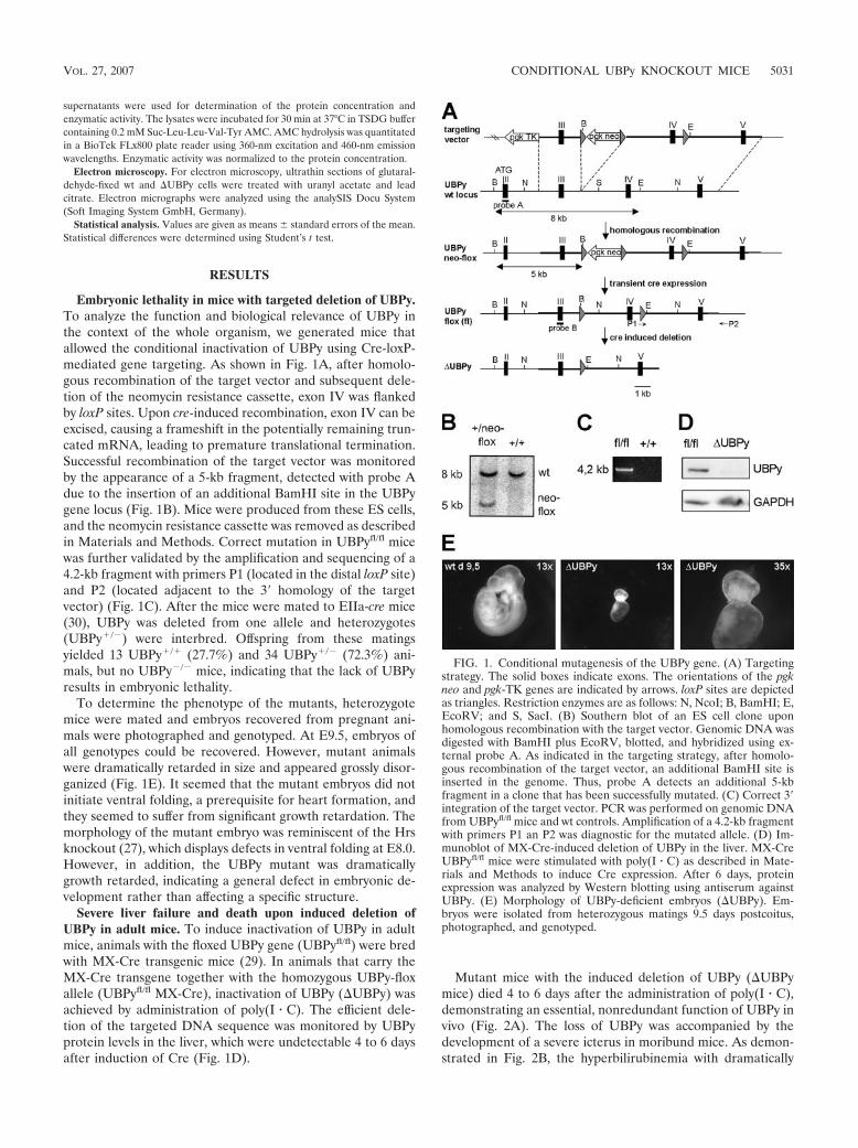
Embryonic lethality in mice with targeted deletion of UBPy.To analyze the function and biological relevance of UBPy inthe context of the whole organism, we generated mice thatallowed the conditional inactivation of UBPy using Cre-loxP-mediated gene targeting. As shown in Fig. 1A, after homolo-gous recombination of the target vector and subsequent dele-tion of the neomycin resistance cassette, exon IV was flankedby loxP sites. Upon cre-induced recombination, exon IV can beexcised, causing a frameshift in the potentially remaining trun-cated mRNA, leading to premature translational termination.Successful recombination of the target vector was monitoredby the appearance of a 5-kb fragment, detected with probe Adue to the insertion of an additional BamHI site in the UBPygene locus (Fig. 1B). Mice were produced from these ES cells,and the neomycin resistance cassette was removed as describedin Materials and Methods. Correct mutation in UBPyfl/fl micewas further validated by the amplification and sequencing of a4.2-kb fragment with primers P1 (located in the distal loxP site)and P2 (located adjacent to the 3� homology of the targetvector) (Fig. 1C). After the mice were mated to EIIa-cre mice(30), UBPy was deleted from one allele and heterozygotes(UBPy�/�) were interbred. Offspring from these matingsyielded 13 UBPy�/� (27.7%) and 34 UBPy�/� (72.3%) ani-mals, but no UBPy�/� mice, indicating that the lack of UBPyresults in embryonic lethality.
To determine the phenotype of the mutants, heterozygotemice were mated and embryos recovered from pregnant ani-mals were photographed and genotyped. At E9.5, embryos ofall genotypes could be recovered. However, mutant animalswere dramatically retarded in size and appeared grossly disor-ganized (Fig. 1E). It seemed that the mutant embryos did notinitiate ventral folding, a prerequisite for heart formation, andthey seemed to suffer from significant growth retardation. Themorphology of the mutant embryo was reminiscent of the Hrsknockout (27), which displays defects in ventral folding at E8.0.However, in addition, the UBPy mutant was dramaticallygrowth retarded, indicating a general defect in embryonic de-velopment rather than affecting a specific structure.
Severe liver failure and death upon induced deletion ofUBPy in adult mice. To induce inactivation of UBPy in adultmice, animals with the floxed UBPy gene (UBPyfl/fl) were bredwith MX-Cre transgenic mice (29). In animals that carry theMX-Cre transgene together with the homozygous UBPy-floxallele (UBPyfl/fl MX-Cre), inactivation of UBPy (UBPy) wasachieved by administration of poly(I � C). The efficient dele-tion of the targeted DNA sequence was monitored by UBPyprotein levels in the liver, which were undetectable 4 to 6 daysafter induction of Cre (Fig. 1D).
Mutant mice with the induced deletion of UBPy (UBPymice) died 4 to 6 days after the administration of poly(I � C),demonstrating an essential, nonredundant function of UBPy invivo (Fig. 2A). The loss of UBPy was accompanied by thedevelopment of a severe icterus in moribund mice. As demon-strated in Fig. 2B, the hyperbilirubinemia with dramatically
FIG. 1. Conditional mutagenesis of the UBPy gene. (A) Targetingstrategy. The solid boxes indicate exons. The orientations of the pgkneo and pgk-TK genes are indicated by arrows. loxP sites are depictedas triangles. Restriction enzymes are as follows: N, NcoI; B, BamHI; E,EcoRV; and S, SacI. (B) Southern blot of an ES cell clone uponhomologous recombination with the target vector. Genomic DNA wasdigested with BamHI plus EcoRV, blotted, and hybridized using ex-ternal probe A. As indicated in the targeting strategy, after homolo-gous recombination of the target vector, an additional BamHI site isinserted in the genome. Thus, probe A detects an additional 5-kbfragment in a clone that has been successfully mutated. (C) Correct 3�integration of the target vector. PCR was performed on genomic DNAfrom UBPyfl/fl mice and wt controls. Amplification of a 4.2-kb fragmentwith primers P1 an P2 was diagnostic for the mutated allele. (D) Im-munoblot of MX-Cre-induced deletion of UBPy in the liver. MX-CreUBPyfl/fl mice were stimulated with poly(I � C) as described in Mate-rials and Methods to induce Cre expression. After 6 days, proteinexpression was analyzed by Western blotting using antiserum againstUBPy. (E) Morphology of UBPy-deficient embryos (UBPy). Em-bryos were isolated from heterozygous matings 9.5 days postcoitus,photographed, and genotyped.
VOL. 27, 2007 CONDITIONAL UBPy KNOCKOUT MICE 5031

increased concentrations of both conjugated and total bilirubinindicates severely disturbed liver function. Liver injury in micelacking UBPy was further confirmed by strongly increased se-rum concentrations of aspartate transaminase and alaninetransaminase and was accompanied by higher alkaline phos-phatase and gamma-glutamyl-transferase, as well as glutaral-dehyde dehydrogenase levels (Fig. 2B). Histological analysis ofliver sections revealed that pathological changes started 4 daysafter the administration of poly(I � C), as manifested by apop-totic hepatocytes (Councilman bodies) around the central vein(Fig. 3A). On day 4, extended sinoids and apoptotic figureswere observed. Cells around the portal field, which represent amore immature form of hepatocytes, showed swelling and werevacuolized. Starting from day 6, apoptotic figures and ubiqui-tous degeneration of hepatocytes were spread over all zones ofhepatic lobules, with lytic necrosis visible on day 7 afterpoly(I � C) injection. Apoptotic hepatocytes in UBPy micewere visualized by TUNEL staining (Fig. 3B). Together, thedata provide strong evidence that the death of UBPy mice iscaused by a severe liver failure.
Induced deletion of UBPy in an immortalized MEF cell lineleads to growth arrest. To further dissect the consequences ofUBPy deletion on a cellular level and to evaluate the molecularmechanisms, we employed a model system that allowed induc-ible inactivation of UBPy in cell culture. Therefore, a tamox-
FIG. 2. Deletion of UBPy in adult mice leads to death caused bydisturbed liver function. (A) Survival rates of mice upon induction ofUBPy deletion. Cre-mediated deletion of UBPy in UBPyfl/fl MX-Cre andMX-Cre mice (10 per group) was induced with poly(I � C) as described inMaterials and Methods. (B) Analysis of liver parameters in UBPy mice.Seven days after poly(I � C) induction, plasmas of 10 UBPY�/� MX-Cre�
controls (open bars) and 12 UBPYfl/fl MX-Cre� (UBPy; solid bars) micewere analyzed. The liver-specific enzymes were as follows: AST, aspartatetransaminase; ALT, alanine transaminase; GLDH, glutaraldehyde-dehy-drogenase; �GT, gamma-glutamyl-transferase; AP, alkaline-phosphatase.The error bars indicate standard errors of the mean.
FIG. 3. Lack of UBPy causes progressive liver injury. (A) Morpho-logical analysis of liver sections. Paraffin sections were stained withhematoxylin and eosin. Abbreviations are as follows: CV, central vene;CB, Councilman bodies; S, liver sinosoid. (B) Enhanced apoptosis inhepatocytes lacking UBPy. TUNEL staining was performed 4 daysafter poly(I � C) induced UBPy deletion.
5032 NIENDORF ET AL. MOL. CELL. BIOL.
ifen-inducible recombination system was used (9). An immor-talized embryonic fibroblast cell line derived from UBPyfl/fl
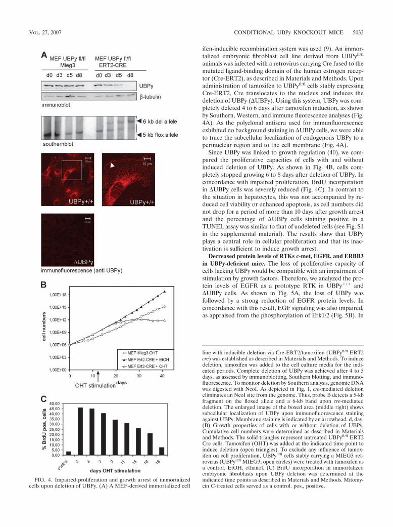
animals was infected with a retrovirus carrying Cre fused to themutated ligand-binding domain of the human estrogen recep-tor (Cre-ERT2), as described in Materials and Methods. Uponadministration of tamoxifen to UBPyfl/fl cells stably expressingCre-ERT2, Cre translocates to the nucleus and induces thedeletion of UBPy (UBPy). Using this system, UBPy was com-pletely deleted 4 to 6 days after tamoxifen induction, as shownby Southern, Western, and immune fluorescence analyses (Fig.4A). As the polyclonal antisera used for immunfluorescenceexhibited no background staining in UBPy cells, we were ableto trace the subcellular localization of endogenous UBPy to aperinuclear region and to the cell membrane (Fig. 4A).
Since UBPy was linked to growth regulation (40), we com-pared the proliferative capacities of cells with and withoutinduced deletion of UBPy. As shown in Fig. 4B, cells com-pletely stopped growing 6 to 8 days after deletion of UBPy. Inconcordance with impaired proliferation, BrdU incorporationin UBPy cells was severely reduced (Fig. 4C). In contrast tothe situation in hepatocytes, this was not accompanied by re-duced cell viability or enhanced apoptosis, as cell numbers didnot drop for a period of more than 10 days after growth arrestand the percentage of UBPy cells staining positive in aTUNEL assay was similar to that of undeleted cells (see Fig. S1in the supplemental material). The results show that UBPyplays a central role in cellular proliferation and that its inac-tivation is sufficient to induce growth arrest.
Decreased protein levels of RTKs c-met, EGFR, and ERBB3in UBPy-deficient mice. The loss of proliferative capacity ofcells lacking UBPy would be compatible with an impairment ofstimulation by growth factors. Therefore, we analyzed the pro-tein levels of EGFR as a prototype RTK in UBPy�/� andUBPy cells. As shown in Fig. 5A, the loss of UBPy wasfollowed by a strong reduction of EGFR protein levels. Inconcordance with this result, EGF signaling was also impaired,as appraised from the phosphorylation of Erk1/2 (Fig. 5B). In
FIG. 4. Impaired proliferation and growth arrest of immortalizedcells upon deletion of UBPy. (A) A MEF-derived immortalized cell
line with inducible deletion via Cre-ERT2/tamoxifen (UBPyfl/fl ERT2cre) was established as described in Materials and Methods. To inducedeletion, tamoxifen was added to the cell culture media for the indi-cated periods. Complete deletion of UBPy was achieved after 4 to 5days, as assessed by immunoblotting, Southern blotting, and immuno-fluorescence. To monitor deletion by Southern analysis, genomic DNAwas digested with NcoI. As depicted in Fig. 1, cre-mediated deletioneliminates an NcoI site from the genome. Thus, probe B detects a 5-kbfragment on the floxed allele and a 6-kb band upon cre-mediateddeletion. The enlarged image of the boxed area (middle right) showssubcellular localization of UBPy upon immunofluorescence stainingagainst UBPy. Membrane staining is indicated by an arrowhead. d, day.(B) Growth properties of cells with or without deletion of UBPy.Cumulative cell numbers were determined as described in Materialsand Methods. The solid triangles represent untreated UBPyfl/fl ERT2Cre cells. Tamoxifen (OHT) was added at the indicated time point toinduce deletion (open triangles). To exclude any influence of tamox-ifen on cell proliferation, UBPyfl/fl cells stably carrying a MIEG3 ret-rovirus (UBPyfl/fl MIEG3; open circles) were treated with tamoxifen asa control. EtOH, ethanol. (C) BrdU incorporation in immortalizedembryonic fibroblasts upon UBPy deletion was determined at theindicated time points as described in Materials and Methods. Mitomy-cin C-treated cells served as a control. pos., positive.
VOL. 27, 2007 CONDITIONAL UBPy KNOCKOUT MICE 5033
contrast, the lack of UBPy did not affect STAT-1 phosphory-lation upon alpha/beta interferon induction (see Fig. S2 in thesupplemental material), showing that the capability to trans-duce extracellular signals was not globally disturbed.
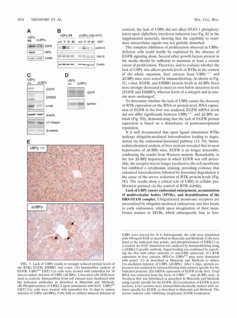
The complete inhibition of proliferation observed in UBPy-deficient cells could hardly be explained by the absence ofEGFR signaling alone. Several other growth factors present inthe media should be sufficient to maintain at least a certainextent of proliferation. Therefore, and to evaluate whether thelack of UBPy also affects protein levels of RTKs in the contextof the whole organism, liver extracts from UBPy�/� andUBPy mice were tested by immunoblotting. As shown in Fig.5C, c-met, EGFR, and ERBB3 protein levels in UBPy liverswere strongly decreased (c-met) or even below detection levels(EGFR and ERBB3), whereas levels of �-integrin and �-cate-nin were unchanged.
To determine whether the lack of UBPy causes the decreaseof RTK expression on the RNA or protein level, RNA expres-sion of EGFR in the liver was analyzed. EGFR mRNA levelsdid not differ significantly between UBPy�/� and UBPy an-imals (Fig. 5D), demonstrating that the lack of EGFR proteinexpression is based on a disturbance of posttranscriptionalregulation.
It is well documented that upon ligand stimulation RTKsundergo ubiquitin-mediated internalization leading to degra-dation via the endosomal-lysosomal pathway (13, 39). Immu-nohistochemical analysis of liver sections revealed that in mosthepatocytes of UBPy mice, EGFR is no longer detectable,confirming the results from Western analysis. Remarkably, inthe few UBPy hepatocytes in which EGFR was still detect-able, the receptor was no longer localized to the cell membranebut exhibited a cytoplasmic staining, providing evidence thatenhanced internalization followed by lysosomal degradation isthe cause of the severe reduction of RTK protein levels (Fig.5E). The results show a critical role of UBPy in cellular pro-liferation pursued via the control of RTK stability.
Lack of UBPy causes endosomal enlargement, accumulationof multivesicular bodies (MVBs), and destabilization of theHRS-STAM complex. Ubiquitinated membrane receptors areinternalized by ubiquitin-mediated endocytosis and first locateto early endosomes, which upon invagination of their mem-branes mature to MVBs, which subsequently fuse to lyso-
FIG. 5. Lack of UBPy results in strongly reduced protein levels ofthe RTKs EGFR, ERBB3, and c-met. (A) Immunoblot analysis ofEGFR. UBPyfl/fl ERT2 Cre cells were treated with tamoxifen for 10days to induce deletion of UBPy (UBPy). Untreated cells (fl/fl) wereused as controls. Immunoblots from cell extracts were incubated withthe indicated antibodies as described in Materials and Methods.(B) Phosphorylation of ERK1/2 upon stimulation with EGF. UBPyfl/fl
ERT2 Cre cells were treated with tamoxifen for 10 days to inducedeletion of UBPy (UBPy). Cells with or without induced deletion of
UBPy were starved for 16 h. Subsequently, the cells were stimulatedwith 100 ng/ml EGF as described in Materials and Methods. Cells werelysed at the indicated time points, and phosphorylation of ERK1/2 asa readout for EGF stimulation was analyzed by immunoblotting usinga pERK1/2-specific antibody. Equal loading was confirmed by reprob-ing the blot with either antiactin or anti-ERK antiserum. (C) RTKexpression in liver extracts. MX-Cre UBPyfl/fl mice were stimulatedwith poly(I � C) as described in Materials and Methods to induceCre-mediated deletion of UBPy (UBPy). After 6 days, protein ex-pression was analyzed by immunoblotting with antisera specific for theindicated proteins. (D) mRNA expression of EGFR in the liver. TotalRNA was extracted from the livers of UBPy�/� and UBPy mice. ANorthern blot was hybridized as described in Materials and Methodsusing a probe specific for the EGFR. (E) Localization of EGFR in liversections. Liver sections were immunohistochemically stained with an-tisera specific for EGFR, as described in Materials and Methods. Thearrows indicate cells exhibiting cytoplasmic EGFR localization.
5034 NIENDORF ET AL. MOL. CELL. BIOL.
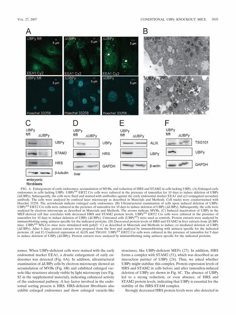
somes. When UBPy-deficient cells were stained with the earlyendosomal marker EEA1, a drastic enlargement of early en-dosomes was detected (Fig. 6A). In addition, ultrastructuralexamination of UBPy cells by electron microscopy showed anaccumulation of MVBs (Fig. 6B) and exhibited enlarged vac-uole-like structures already visible by light microscopy (see Fig.S3 in the supplemental material), indicating enhanced activityof the endosomal pathway. A key factor involved in the endo-somal sorting process is HRS. HRS-deficient fibroblasts alsoexhibit enlarged endosomes and show enlarged vacuole-like
structures, like UBPy-deficient MEFs (27). In addition, HRSforms a complex with STAM2 (51), which was described as aninteraction partner of UBPy (24). Thus, we asked whetherUBPy might stabilize this complex. Protein expression levels ofHRS and STAM2 in cells before and after tamoxifen-induceddeletion of UBPy are shown in Fig. 6C. The absence of UBPyled to a strong reduction, or even absence, of HRS andSTAM2 protein levels, indicating that UBPy is essential for thestability of the HRS-STAM complex.
Strongly decreased HRS protein levels were also detected in
FIG. 6. Enlargement of early endosomes, accumulation of MVBs, and reduction of HRS and STAM2 in cells lacking UBPy. (A) Enlarged earlyendosomes in cells lacking UBPy. UBPyfl/fl ERT2 Cre cells were cultured in the presence of tamoxifen for 10 days to induce deletion of UBPy(UBPy). Subsequently, the cells were fixed and stained with antibodies against the early endosomal marker EEA1 and cy2-conjugated secondaryantibody. The cells were analyzed by confocal laser microscopy as described in Materials and Methods. Cell nuclei were counterstained withHoechst 33258. The arrowheads indicate enlarged early endosomes. (B) Ultrastructural examination of cells upon induced deletion of UBPy.UBPyfl/fl ERT2 Cre cells were cultured in the presence of tamoxifen for 10 days to induce deletion of UBPy (UBPy). Subsequently, the cells wereanalyzed by electron microscopy as described in Materials and Methods. The arrows indicate MVBs. (C) Induced inactivation of UBPy in theMEF-derived cell line correlates with decreased HRS and STAM2 protein levels. UBPyfl/fl ERT2 Cre cells were cultured in the presence oftamoxifen for 10 days to induce deletion of UBPy (UBPy). Untreated cells (UBPyfl/fl) were used as controls. Protein extracts were analyzed byimmunoblotting using antisera specific for the indicated proteins. (D) Decreased protein levels of HRS and STAM2 in liver extracts from UBPymice. UBPyfl/fl MX-Cre mice were stimulated with poly(I � C) as described in Materials and Methods to induce cre-mediated deletion of UBPy(UBPy). After 6 days, protein extracts were prepared from the liver and analyzed by immunoblotting with antisera specific for the indicatedproteins. (E and F) Unaltered expression of ALIX and TSG101. UBPyfl/fl ERT2 Cre cells were cultured in the presence of tamoxifen for 9 daysto induce deletion of UBPy (UBPy). Protein extracts were analyzed by immunoblotting using antisera specific for the indicated proteins.
VOL. 27, 2007 CONDITIONAL UBPy KNOCKOUT MICE 5035
vivo, as seen from the analysis of liver extracts of UBPy mice(Fig. 6D). In contrast, levels of the UIM-containing proteinsALIX (36) and TSG101 (5, 31), which are also associated withendosomal sorting processes, were not affected by the loss ofUBPy (Fig. 6E and F). These data provide strong evidence fora functional role of UBPy in endosomal sorting pursued bystabilizing the HRS-STAM complex.
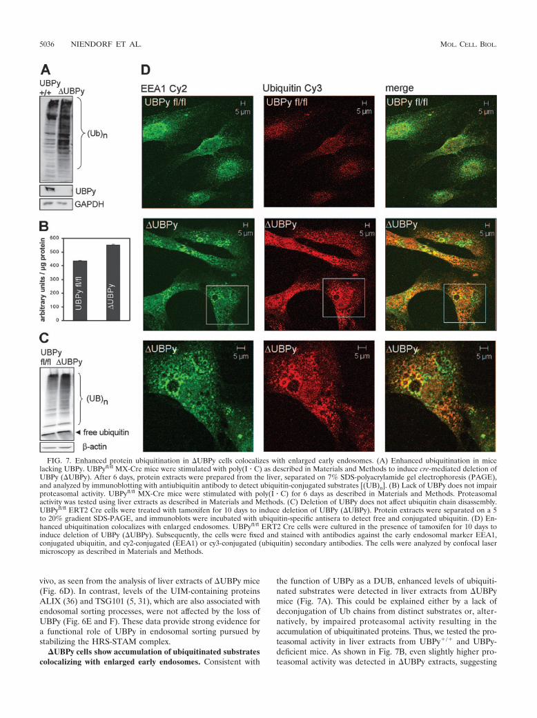
�UBPy cells show accumulation of ubiquitinated substratescolocalizing with enlarged early endosomes. Consistent with
the function of UBPy as a DUB, enhanced levels of ubiquiti-nated substrates were detected in liver extracts from UBPymice (Fig. 7A). This could be explained either by a lack ofdeconjugation of Ub chains from distinct substrates or, alter-natively, by impaired proteasomal activity resulting in theaccumulation of ubiquitinated proteins. Thus, we tested the pro-teasomal activity in liver extracts from UBPy�/� and UBPy-deficient mice. As shown in Fig. 7B, even slightly higher pro-teasomal activity was detected in UBPy extracts, suggesting
FIG. 7. Enhanced protein ubiquitination in UBPy cells colocalizes with enlarged early endosomes. (A) Enhanced ubiquitination in micelacking UBPy. UBPyfl/fl MX-Cre mice were stimulated with poly(I � C) as described in Materials and Methods to induce cre-mediated deletion ofUBPy (UBPy). After 6 days, protein extracts were prepared from the liver, separated on 7% SDS-polyacrylamide gel electrophoresis (PAGE),and analyzed by immunoblotting with antiubiquitin antibody to detect ubiquitin-conjugated substrates [(UB)n]. (B) Lack of UBPy does not impairproteasomal activity. UBPyfl/fl MX-Cre mice were stimulated with poly(I � C) for 6 days as described in Materials and Methods. Proteasomalactivity was tested using liver extracts as described in Materials and Methods. (C) Deletion of UBPy does not affect ubiquitin chain disassembly.UBPyfl/fl ERT2 Cre cells were treated with tamoxifen for 10 days to induce deletion of UBPy (UBPy). Protein extracts were separated on a 5to 20% gradient SDS-PAGE, and immunoblots were incubated with ubiquitin-specific antisera to detect free and conjugated ubiquitin. (D) En-hanced ubiquitination colocalizes with enlarged endosomes. UBPyfl/fl ERT2 Cre cells were cultured in the presence of tamoxifen for 10 days toinduce deletion of UBPy (UBPy). Subsequently, the cells were fixed and stained with antibodies against the early endosomal marker EEA1,conjugated ubiquitin, and cy2-conjugated (EEA1) or cy3-conjugated (ubiquitin) secondary antibodies. The cells were analyzed by confocal lasermicroscopy as described in Materials and Methods.
5036 NIENDORF ET AL. MOL. CELL. BIOL.
that proteasomal inhibition is not the cause of the enhancedubiquitination levels. It has been speculated that UBPy mightbe the functional homologue of yeast DOA4, which has beenshown to be essential for the regeneration of ubiquitin frompolyubiquitin chains (3). To examine the impact of the lack ofUBPy on polyubiquitin chain disassembly, we compared thelevels of free ubiquitin in wt and UBPy-deficient cells. Asshown in Fig. 7C, both types of cells showed the same amountof free ubiquitin, demonstrating that UBPy is not necessary torestore the pool of free ubiquitin and does not exert a functionsimilar to that of DOA4 in this respect. The results suggest thatUBPy directly counteracts ubiquitination on distinct sub-strates.
In order to determine whether the enhanced ubiquitinatedsubstrates were uniformly distributed over the cell or wereattributed to defined subcellular structures, cells were stainedwith an antibody recognizing conjugated ubiquitin. As shownin Fig. 7D, enhanced ubiquitination colocalized with the en-larged early endosomes, providing evidence that the lack ofUBPy results in aberrant ubiquitination of substrates that aretargeted to the endosomal lysosomal degradation pathway.Alternatively, UBPy might exert its deubiquitinating activityexclusively on early endosomal structures.
DISCUSSION
While considerable progress has been made in defining thefunctional properties of different USPs using in vitro and cellculture systems, their physiological relevance in the context ofthe whole organism is only poorly understood. Recently, micedeficient in CYLD, a USP that is altered in patients withfamilial cylindromatosis, were generated. Contrary to resultsfrom transfection experiments, which suggested a negative reg-ulatory function of CYLD on NF- B activation (8, 28, 52),bone marrow-derived macrophages from wt and CYLD�/�
animals did not differ in innate immune receptor signaling.Analysis of these mice instead demonstrated an essential rolefor CYLD in T-cell development (47), underlining the neces-sity of mouse models to evaluate functional properties ofDUBs in vivo.
It was a matter of debate whether and to what extent dif-ferent DUBs might have redundant functions (2). UBPy-defi-cient animals are embryonic lethal, and induced inactivation inadulthood causes fatal liver failure. This clearly shows that lackof UBPy cannot be compensated for by other DUB familymembers in vivo. Embryonic lethality might at least partially beattributed to the lack of HRS, as HRS- deficient animals alsoexhibited embryonic lethality caused by ventral-folding defects.However, while HRS-deficient embryos were often as large aswt controls and even had beating hearts by day 9.5 (27),UBPy�/� animals were extremely developmentally retardedand exhibited completely disorganized morphology at thattime point. This shows that besides the stabilization of HRS,UBPy is essential for embryonic growth. The observation thatUBPy-deficient fibroblasts underwent growth arrest furthersupports an essential role of UBPy in the control of cellularproliferation. MX-Cre-mediated deletion of floxed genes ingeneral leads to the fastest and most efficient deletion in theliver (29), which most likely explains why the phenotype man-ifests in that organ, although UBPy is also expressed in other
cell types. The strong liver phenotype demonstrates that, be-sides a role in proliferation, UBPy is also essential in quiescentcells, like hepatocytes. As loss of UBPy in hepatocytes causedapoptosis and cell death while embryonic fibroblasts did notexhibit enhanced apoptosis, functional differences of UBPy indifferent cell types or in proliferating versus nonproliferatingcells apparently exist.
Pathology in the liver can most likely be attributed to thecombined effects of disturbed endosomal function, loss of sev-eral RTKs, and disturbed liver regeneration caused by theabsence of c-met, which has been shown to be essential for thisprocess (6, 20). Due to the severity of the phenotype in theliver and early death, cell-type-specific inactivation will be nec-essary to define the physiological relevance in other organs orcell types.
We have shown that in vivo UBPy is essential to maintainproper protein levels of RTKs, like EGFR, c-met, and ERBB3,and thus reduced growth factor stimulation might at least par-tially contribute to the growth arrest in UBPy�/� cells. As acommon feature of several RTKs, ligand binding induces ubiq-uitination as a signal to internalize the receptor via endocytosis(15). Although the detailed mechanisms leading to the reduc-tion or even absence of several RTKs in UBPy-deficient miceneed to be defined, the observed cytoplasmic localization ofEGFR in UBPy-deficient hepatocytes strongly suggests that,due to the loss of UBPy, RTKs undergo enhanced internaliza-tion followed by lysosomal degradation. The results are com-patible with a model in which UBPy counteracts ubiquitinationof RTKs or components of the endocytic machinery and thusregulates thresholds for the internalization of growth factorreceptors.
The observed accumulation of ubiquitinated proteins inUBPy-deficient cells that colocalize with the enlarged endo-somes might not only represent ubiquitinated conjugated sub-strates associated with the endosome, but could also reflectenhanced amounts of ubiquitinated cargo derived from the cellmembrane. Such a functional role of UBPy exerted on cellmembrane components is compatible with the cellular local-ization of endogenous UBPy.
As described here, the absence of UBPy caused a severereduction of HRS. HRS binds directly to ubiquitinated pro-teins, which are then sorted to clathrin-coated microdomainswhile nonubiquitinated receptors, like transferrin or LDL re-ceptor, are rapidly recycled to the cell surface (45). Endosomalenlargement in UBPy-deficient cells most likely can be attrib-uted to the lack of HRS, as HRS-deficient murine cells anddrosophila HRS�/� mutant larvae (27, 34) were also reportedto exhibit this phenotype. While this work was in progress, tworeports also described endosomal enlargement when UBPywas knocked down by RNA interference (7, 48), supporting anessential role of UBPy in endocytic trafficking.
As HRS itself was identified as a protein that is tyrosinephosphorylated upon stimulation with hepatocyte growth fac-tor, platelet-derived growth factor, and epidermal growth fac-tor (26) and HRS�/� drosophila pupae have reduced levels ofEGFR (34), it is appealing to see a functional relationshipbetween the lack of RTKs and the lack of HRS in UBPy-deficient cells and animals. However, the knockdown of HRSin cultured cells, as well as the combined inactivation ofSTAM1 and STAM2, was reported to even inhibit the degra-
VOL. 27, 2007 CONDITIONAL UBPy KNOCKOUT MICE 5037
dation of ligand-activated RTK degradation (4, 22, 35). Thus,the observed reduction of RTKs in UBPy�/� cells is unlikely tobe caused by the lack of HRS but rather reflects a function ofUBPy independent of the stabilization of the HRS-STAMcomplex. This is supported by results from Mizuno et al., whodescribed a direct interaction of UBPy and the EGFR resultingin EGFR deubiquitination (38). The observed accumulation ofMVBs shows that in UBPy�/� cells, endosomes can efficientlymature to MVBs even in the absence of the HRS-STAMcomplex, a prerequisite of RTK degradation via the lysosomalpathway.
The results suggest that UBPy exerts two independent func-tions in the RTK degradation pathway, first by directly coun-teracting RTK internalization and second by stabilizing theHRS-STAM complex. Both activities might act in concert toregulate thresholds for RTK degradation.
ACKNOWLEDGMENTS
We are particularly grateful to Ivan Dikic for suggestions and forcritically reading the manuscript. We thank Marcus Wietstruk, ElviraRhode, Melanie Benedict, Claudia Pallasch, Bianca Verrett, and UlrikeKuckelkorn for technical assistance and Harald Stenmark, Simono Polo,Daniel Metzger, and Ivan Dikic for providing essential reagents andantibodies.
We declare that we do not have competing financial interests.This work was supported by grant KN590/2-1 to K.-P. Knobeloch
from the Deutsche Forschungsgemeinschaft.
REFERENCES
1. Amerik, A. Y., and M. Hochstrasser. 2004. Mechanism and function ofdeubiquitinating enzymes. Biochim. Biophys. Acta 1695:189–207.
2. Amerik, A. Y., S. J. Li, and M. Hochstrasser. 2000. Analysis of the deubiq-uitinating enzymes of the yeast Saccharomyces cerevisiae. Biol. Chem. 381:981–992.
3. Amerik, A. Y., J. Nowak, S. Swaminathan, and M. Hochstrasser. 2000. TheDoa4 deubiquitinating enzyme is functionally linked to the vacuolar protein-sorting and endocytic pathways. Mol. Biol. Cell. 11:3365–3380.
4. Bache, K. G., C. Raiborg, A. Mehlum, and H. Stenmark. 2003. STAM andHrs are subunits of a multivalent ubiquitin-binding complex on early endo-somes. J. Biol. Chem. 278:12513–12521.
5. Bishop, N., A. Horman, and P. Woodman. 2002. Mammalian class E vpsproteins recognize ubiquitin and act in the removal of endosomal protein-ubiquitin conjugates. J. Cell Biol. 157:91–101.
6. Borowiak, M., A. N. Garratt, T. Wustefeld, M. Strehle, C. Trautwein, and C.Birchmeier. 2004. Met provides essential signals for liver regeneration. Proc.Natl. Acad. Sci. USA 101:10608–10613.
7. Bowers, K., S. C. Piper, M. A. Edeling, S. R. Gray, D. J. Owen, P. J. Lehner,and J. P. Luzio. 2006. Degradation of endocytosed epidermal growth factorand virally ubiquitinated major histocompatibility complex class I is inde-pendent of mammalian ESCRTII. J. Biol. Chem. 281:5094–5105.
8. Brummelkamp, T. R., S. M. B. Nijman, A. M. G. Dirac, and R. Bernards.2003. Loss of the cylindromatosis tumour suppressor inhibits apoptosis byactivating NF-kappa B. Nature 424:797–801.
9. Feil, R., J. Brocard, B. Mascrez, M. LeMeur, D. Metzger, and P. Chambon.1997. Ligand-inducible gene targeting in mice. Naunyn-Schmiedebergs Arch.Pharmacol. 355:41.
10. Feil, R., J. Wagner, D. Metzger, and P. Chambon. 1997. Regulation of Crerecombinase activity by mutated estrogen receptor ligand-binding domains.Biochem. Biophys. Res. Commun. 237:752–757.
11. Gnesutta, N., M. Ceriani, M. Innocenti, I. Mauri, R. Zippel, E. Sturani, B.Borgonovo, G. Berruti, and E. Martegani. 2001. Cloning and characteriza-tion of mouse UBPy, a deubiquitinating enzyme that interacts with the Rasguanine nucleotide exchange factor CDC25(Mm)/Ras-GRF1. J. Biol. Chem.276:39448–39454.
12. Graner, E., D. Tang, S. Rossi, A. Baron, T. Migita, L. J. Weinstein, M.Lechpammer, D. Huesken, J. Zimmermann, S. Signoretti, and M. Loda.2004. The isopeptidase USP2a regulates the stability of fatty acid synthase inprostate cancer. Cancer Cell 5:253–261.
13. Haglund, K., P. P. Di Fiore, and I. Dikic. 2003. Distinct monoubiquitinsignals in receptor endocytosis. Trends Biochem. Sci. 28:598–603.
14. Haglund, K., and I. Dikic. 2005. Ubiquitylation and cell signaling. EMBO J.24:3353–3359.
15. Haglund, K., S. Sigismund, S. Polo, I. Szymkiewicz, P. P. Di Fiore, and I.
Dikic. 2003. Multiple monoubiquitination of RTKs is sufficient for theirendocytosis and degradation. Nat. Cell Biol. 5:461–466.
16. Harkiolaki, M., M. Lewitzky, R. J. C. Gilbert, E. Y. Jones, R. P. Bourette, G.Mouchiroud, H. Sondermann, I. Moarefi, and S. M. Feller. 2003. Structuralbasis for SH3 domain-mediated high-affinity binding between Mona/Gadsand SLP-76. EMBO J. 22:2571–2582.
17. Hicke, L., and R. Dunn. 2003. Regulation of membrane protein transport byubiquitin and ubiquitin-binding proteins. Annu. Rev. Cell Dev. Biol. 19:141–172.
18. Holzenberger, M., C. Lenzner, P. Leneuve, R. Zaoui, G. Hamard, S. Vaulont,and Y. L. Bouc. 2000. Cre-mediated germline mosaicism: a method allowingrapid generation of several alleles of a target gene. Nucleic Acids Res.28:e92.
19. Huang, T. T., S. M. Nijman, K. D. Mirchandani, P. J. Galardy, M. A. Cohn,W. Haas, S. P. Gygi, H. L. Ploegh, R. Bernards, and A. D. D’Andrea. 2006.Regulation of monoubiquitinated PCNA by DUB autocleavage. Nat. CellBiol. 8:3309–3347.
20. Huh, C. G., V. M. Factor, A. Sanchez, K. Uchida, E. A. Conner, and S. S.Thorgeirsson. 2004. Hepatocyte growth factor/c-met signaling pathway isrequired for efficient liver regeneration and repair. Proc. Natl. Acad. Sci.USA 101:4477–4482.
21. Janssen, J. W. G., L. Schleithoff, C. R. Bartram, and A. S. Schulz. 1998. Anoncogenic fusion product of the phosphatidylinositol 3-kinase p85 beta sub-unit and HUMORF8, a putative deubiquitinating enzyme. Oncogene 16:1767–1772.
22. Kanazawa, C., E. Morita, M. Yamada, N. Ishii, S. Miura, H. Asao, T.Yoshimori, and K. Sugamura. 2003. Effects of deficiencies of STAMs andHrs, mammalian class E Vps proteins, on receptor downregulation. Bio-chem. Biophys. Res. Commun. 309:848–856.
23. Kaneko, T., T. Kumasaka, T. Ganbe, T. Sato, K. Miyazawa, N. Kitamura,and N. Tanaka. 2003. Structural insight into modest binding of a non-PXXPligand to the signal transducing adaptor molecule-2 Src homology 3 domain.J. Biol. Chem. 278:48162–48168.
24. Kato, M., K. Miyazawa, and N. Kitamura. 2000. A deubiquitinating enzymeUBPY interacts with the Src homology 3 domain of Hrs-binding protein viaa novel binding motif PX(V/I)(D/N)RXXKP. J. Biol. Chem. 275:37481–37487.
25. Knobeloch, K. P., O. Utermohlen, A. Kisser, M. Prinz, and I. Horak. 2005.Reexamination of the role of ubiquitin-like modifier ISG15 in the phenotypeof UBP43-deficient mice. Mol. Cell. Biol. 25:11030–11034.
26. Komada, M., and N. Kitamura. 1995. Growth factor-induced tyrosine phos-phorylation of Hrs, a novel 115-kilodalton protein with a structurally con-served putative zinc finger domain. Mol.d Cell. Biol. 15:6213–6221.
27. Komada, M., and P. Soriano. 1999. Hrs, a FYVE finger protein localized toearly endosomes, is implicated in vesicular traffic and required for ventralfolding morphogenesis. Genes Dev. 13:1475–1485.
28. Kovalenko, A., C. Chable-Bessia, G. Cantarella, A. Israel, D. Wallach, andG. Courtois. 2003. The tumour suppressor CYLD negatively regulates NF-kappa B signalling by deubiquitination. Nature 424:801–805.
29. Kuhn, R., F. Schwenk, M. Aguet, and K. Rajewsky. 1995. Inducible genetargeting in mice. Science 269:1427–1429.
30. Lakso, M., J. G. Pichel, J. R. Gorman, B. Sauer, Y. Okamoto, E. Lee, F. W.Alt, and H. Westphal. 1996. Efficient in vivo manipulation of mouse genomicsequences at the zygote stage. Proc. Natl. Acad. Sci. USA 93:5860–5865.
31. Li, L. M., and S. N. Cohen. 1996. tsg101: a novel tumor susceptibility geneisolated by controlled homozygous functional knockout of allelic loci inmammalian cells. Cell 85:319–329.
32. Li, M. Y., C. L. Brooks, N. Kon, and W. Gu. 2004. A dynamic role of HAUSPin the p53-Mdm2 pathway. Mol. Cell 13:879–886.
33. Li, M. Y., D. L. Chen, A. Shiloh, J. Y. Luo, A. Y. Nikolaev, J. Qin, and W. Gu.2002. Deubiquitination of p53 by HAUSP is an important pathway for p53stabilization. Nature 416:648–653.
34. Lloyd, T. E., R. Atkinson, M. N. Wu, Y. Zhou, G. Pennetta, and H. J. Bellen.2002. Hrs regulates endosome membrane invagination and tyrosine kinasereceptor signaling in Drosophila. Cell 108:261–269.
35. Lu, Q., L. W. Q. Hope, M. Brasch, C. Reinhard, and S. N. Cohen. 2003.TSG101 interaction with HRS mediates endosomal trafficking and receptordown-regulation. Proc. Natl. Acad. Sci. USA 100:7626–7631.
36. Matsuo, H., J. Chevallier, N. Mayran, I. Le Blanc, C. Ferguson, J. Faure,N. S. Blanc, S. Matile, J. Dubochet, M. Sadoul, R. G. Parton, F. Vilbois, andJ. Gruenberg. 2004. Role of LBPA and Alix in multivesicular liposomeformation and endosome organization. Science 303:531–534.
37. Millard, S. M., and S. A. Wood. 2006. Riding the DUBway: regulation ofprotein trafficking by deubiquitylating enzymes. J. Cell Biol. 173:463–468.
38. Mizuno, E., T. Iura, A. Mukai, T. Yoshimori, N. Kitamura, and M. Komada.2005. Regulation of epidermal growth factor receptor down-regulation byUBPY-mediated deubiquitination at endosomes. Mol. Biol. Cell 16:5163–5174.
39. Mosesson, Y., K. Shtiegman, M. Katz, Y. Zwang, G. Vereb, J. Szollosi, andY. Yarden. 2003. Endocytosis of receptor tyrosine kinases is driven by mo-noubiquitylation, not polyubiquitylation. J. Biol. Chem. 278:21323–21326.
40. Naviglio, S., C. Matteucci, B. Matoskova, T. Nagase, N. Nomura, P. P. Di
5038 NIENDORF ET AL. MOL. CELL. BIOL.
Fiore, and G. F. Draetta. 1998. UBPY: a growth-regulated human ubiquitinisopeptidase. EMBO J. 17:3241–3250.
41. Nijman, S. M. B., T. T. Huang, A. M. G. Dirac, T. R. Brummelkamp, R. M.Kerkhoven, A. D. D’Andrea, and R. Bernards. 2005. The deubiquitinatingenzyme USP1 regulates the Fanconi anemia pathway. Mol. Cell 17:331–339.
42. Nijman, S. M. B., M. P. A. Luna-Vargas, A. Velds, T. R. Brummelkamp,A. M. G. Dirac, T. K. Sixma, and R. Bernards. 2005. A genomic andfunctional inventory of deubiquitinating enzymes. Cell 123:773–786.
43. Osiak, A., O. Utermohlen, S. Niendorf, I. Horak, and K. P. Knobeloch. 2005.ISG15, an interferon-stimulated ubiquitin-like protein, is not essential forSTAT1 signaling and responses against vesicular stomatitis and lymphocyticchoriomeningitis virus. Mol. Cell. Biol. 25:6338–6345.
44. Pickart, C. M. 2001. Mechanisms underlying ubiquitination. Annu. Rev.Biochem. 70:503–533.
45. Raiborg, C., K. G. Bache, D. J. Gillooly, I. H. Madshush, E. Stang, and H.Stenmark. 2002. Hrs sorts ubiquitinated proteins into clathrin-coated mi-crodomains of early endosomes. Nat. Cell Biol. 4:394–398.
46. Raiborg, C., K. G. Bache, A. Mehlum, E. Stang, and H. Stenmark. 2001. Hrsrecruits clathrin to early endosomes. EMBO J. 20:5008–5021.
47. Reiley, W. W., M. Y. Zhang, W. Jin, M. Losiewicz, K. B. Donohue, C. C.Norbury, and S. C. Sun. 2006. Regulation of T cell development by thedeubiquitinating enzyme CYLD. Nat. Immunol. 7:411–417.
48. Row, P. E., I. A. Prior, J. McCullough, M. J. Clague, and S. Urbe. 2006. Theubiquitin isopeptidase UBPY regulates endosomal ubiquitin dynamics and isessential for receptor down-regulation. J. Biol. Chem. 281:12618–12624.
49. Schumann, G., R. Bonora, F. Ceriotti, G. Ferard, C. A. Ferrero, P. F. H.Franck, F. J. Gella, W. Hoelzel, P. J. Jorgensen, T. Kanno, A. Kessner, R.Klauke, N. Kristiansen, J. M. Lessinger, T. P. J. Linsinger, H. Misaki, M.
Panteghini, J. Pauwels, F. Schiele, H. G. Schimmel, G. Weidemann, and L.Siekmann. 2002. IFCC primary reference procedures for the measurementof catalytic activity concentrations of enzymes at 37°C. Part 5. Referenceprocedure for the measurement of catalytic concentration of aspartate amino-transferase. Clin. Chem. Lab. Med. 40:725–733.
50. Soares, L., C. Seroogy, H. Skrenta, N. Anandasabapathy, P. Lovelace, C. D.Chung, E. Engleman, and C. G. Fathman. 2004. Two isoforms of otubain 1regulate T cell anergy via GRAIL. Nat. Immunol. 5:45–54.
51. Takata, H., M. Kato, K. Denda, and N. Kitamura. 2000. A Hrs bindingprotein having a Src homology 3 domain is involved in intracellular degra-dation of growth factors and their receptors. Genes Cells 5:57–69.
52. Trompouki, E., E. Hatzivassiliou, T. Tsichritzis, H. Farmer, A. Ashworth,and G. Mosialos. 2003. CYLD is a deubiquitinating enzyme that negativelyregulates NF-kappa B activation by TNFR family members. Nature 424:793–796.
53. Welchman, R. L., C. Gordon, and R. J. Mayer. 2005. Ubiquitin and ubiq-uitin-like proteins as multifunctional signals. Nat. Rev. Mol. Cell Biol. 6:599–609.
54. Williams, D. A., W. Tao, F. C. Yang, C. Kim, Y. Gu, P. Mansfield, J. E.Levine, B. Petryniak, C. W. Derrow, C. Harris, B. Q. Jia, Y. Zheng, D. R.Ambruso, J. B. Lowe, S. J. Atkinson, M. C. Dinauer, and L. Boxer. 2000.Dominant negative mutation of the hematopoietic-specific Rho GTPase,Rac2, is associated with a human phagocyte immunodeficiency. Blood 96:1646–1654.
55. Wu, X. L., L. Yen, L. Irwin, C. Sweeney, and K. L. Carraway. 2004. Stabili-zation of the E3 ubiquitin ligase Nrdp1 by the deubiquitinating enzymeUSP8. Mol. Cell. Biol. 24:7748–7757.
VOL. 27, 2007 CONDITIONAL UBPy KNOCKOUT MICE 5039