Actin in the endocytic pathway: From yeast to mammals
8
Minireview Actin in the endocytic pathway: From yeast to mammals Henrique Girao 1 , Maria-Isabel Geli * , Fatima-Zahra Idrissi Instituto de Biologı ´a Molecular de Barcelona (IBMB-CSIC), PCB, Edifici He `lix, Baldiri Reixac 15, 08028 Barcelona, Spain Received 20 February 2008; revised 2 April 2008; accepted 9 April 2008 Available online 15 April 2008 Edited by Christos Stournaras Abstract Genetic analysis of endocytosis in yeast early pointed to the essential role of actin in the uptake step. Efforts to identify the machinery involved demonstrated the important contribution of Arp2/3 and the myosins-I. Analysis of the process using live- cell fluorescence microscopy and electron microscopy have re- cently contributed to refine molecular models explaining clathrin and actin-dependent endocytic uptake. Increasing evidence now also indicates that actin plays important roles in post-internaliza- tion events along the endocytic pathway in yeast, including trans- port of vesicles, motility of endosomes and vacuole fusion. This review describes the present knowledge state on the roles of actin in endocytosis in yeast and points to similarities and differences with analogous processes in mammals. Ó 2008 Federation of European Biochemical Societies. Pub- lished by Elsevier B.V. All rights reserved. Keywords: Actin; Myosin; Arp2/3; Formin; Endocytosis; Yeast 1. Actin in endocytic uptake Genetic analysis of endocytosis in Saccharomyces cerevisiae early pointed to the essential role of actin in the uptake step. Not only mutations on actin cause internalization defects stronger than those observed in clathrin mutants [1,2] but also, a number of proteins directly involved in the control of actin dynamics are essential in the process (for review see [3,4]). A key player initiating actin polymerization at sites of endocyto- sis is the Arp2/3 complex [5,6]. Upon activation by nucleation promoting factors (NPFs), Arp2/3 induces actin polymeriza- tion and participates in assembly of branched actin networks that usually grow by adding monomers close to the surface where the NPF is located, typically a cellular membrane [7]. Addition of actin monomers pushes the NPF covered surface forward, either causing membrane deformation or rocketing of the membrane enclosed organelle [7]. Multiple NPFs partic- ipate in endocytic uptake in yeast, indicating that actin poly- merization might be ignited at different points during membrane budding. The endocytic NPFs include the yeast WASP (Wiskott–Aldrich Syndrome Protein) homologue Las17 [8,9], the eps15 family member Pan1 [10,11], the actin binding protein Abp1 [12,13] and the unconventional type I myosins Myo3 and Myo5 [14,15]. Besides the force generated by local polymerization of actin, the mechanochemical activity of the type I myosin [15] might also actively contribute to de- form the lipid bilayer. In response to ATP hydrolysis, the Myo5 N-terminal motor head is capable of translocating actin filaments with respect to its tail [15], which bears a phospho- lipid binding domain followed by the C-terminal extension that interacts with the actin polymerization machinery [16–19]. Development of live-cell imaging to study endocytic uptake very much contributed to order the sequence of molecular events occurring during endocytic budding and the role of the different NPFs in the process [15,20–26]. Upon clathrin ar- rival to the PM, other components of the endocytic coat including clathrin adaptors and Pan1 are assembled concomi- tant with recruitment of Las17 [20,21,23]. After a roughly 30 s period of restrained motility, the coat undergoes a 200 nm slow inward movement, which takes 10–15 s. Recruitment of the yeast amphiphysins Rvs161 and Rvs167 then precedes the fast movement of the coat into the cytosol [20]. Myo5 arrives to the endocytic patch at the onset of the slow inward movement, slightly after arrival of Arp2/3 and coincident with the start of massive actin polymerization [15,26]. Similar to Las17 and the amphiphysins, Myo5 is left at the PM as the vesicle moves into the cytosol [15,20,21,26]. The recruitment of Abp1 and a number of actin binding proteins such as fimbrin, cofilin and capping protein follows in an actin-dependent manner. To- gether with Arp2/3, these components remain associated with the vesicles as they move into the cytosol [20,21,25]. Based on the amphiphysin recruitment time point, it was proposed that the slow 200 nm coat inward movement corre- sponds to the initial PM invagination [27]. However, the reso- lution of the fluorescence microscopy techniques was insufficient to define when fission actually occurs. Idrissi et al. have recently demonstrated, using electron microscopy (EM), that endocytic internalization in yeast associates with the formation of tubular invaginations of about 50 nm in diameter and up to 180 nm in length, which accumulate the endocytic coat components at the tip [28]. Amphiphysin accu- mulates at the neck of the longest profiles confirming that fis- sion occurs once the tubular profile has elongated (Fig. 1). Statistical analysis of the distribution of immuno-gold particles labeling different proteins along PM-associated tubular pro- files of increasing length [28] has allowed refinement of models explaining the molecular forces driving membrane invagina- tion [27] (Fig. 1, stage 3). Treatment of cells with the actin monomer sequestering drug Latrunculin A prevents the slow 200 nm coat inward movement [21], indicating that actin poly- merization is essential to drive elongation of the incipient * Corresponding author. E-mail address: [email protected] (M.-I. Geli). 1 Present address: IBILI, Centro de Oftalmologia, Azinhaga de Sta Comba, Coimbra, Portugal. 0014-5793/$34.00 Ó 2008 Federation of European Biochemical Societies. Published by Elsevier B.V. All rights reserved. doi:10.1016/j.febslet.2008.04.011 FEBS Letters 582 (2008) 2112–2119
Transcript of Actin in the endocytic pathway: From yeast to mammals
FEBS Letters 582 (2008) 2112–2119
Minireview
Actin in the endocytic pathway: From yeast to mammals
Henrique Girao1, Maria-Isabel Geli*, Fatima-Zahra Idrissi
Instituto de Biologıa Molecular de Barcelona (IBMB-CSIC), PCB, Edifici Helix, Baldiri Reixac 15, 08028 Barcelona, Spain
Received 20 February 2008; revised 2 April 2008; accepted 9 April 2008
Available online 15 April 2008
Edited by Christos Stournaras
Abstract Genetic analysis of endocytosis in yeast early pointedto the essential role of actin in the uptake step. Efforts to identifythe machinery involved demonstrated the important contributionof Arp2/3 and the myosins-I. Analysis of the process using live-cell fluorescence microscopy and electron microscopy have re-cently contributed to refine molecular models explaining clathrinand actin-dependent endocytic uptake. Increasing evidence nowalso indicates that actin plays important roles in post-internaliza-tion events along the endocytic pathway in yeast, including trans-port of vesicles, motility of endosomes and vacuole fusion. Thisreview describes the present knowledge state on the roles of actinin endocytosis in yeast and points to similarities and differenceswith analogous processes in mammals.� 2008 Federation of European Biochemical Societies. Pub-lished by Elsevier B.V. All rights reserved.
Keywords: Actin; Myosin; Arp2/3; Formin; Endocytosis; Yeast
1. Actin in endocytic uptake
Genetic analysis of endocytosis in Saccharomyces cerevisiae
early pointed to the essential role of actin in the uptake step.
Not only mutations on actin cause internalization defects
stronger than those observed in clathrin mutants [1,2] but also,
a number of proteins directly involved in the control of actin
dynamics are essential in the process (for review see [3,4]). A
key player initiating actin polymerization at sites of endocyto-
sis is the Arp2/3 complex [5,6]. Upon activation by nucleation
promoting factors (NPFs), Arp2/3 induces actin polymeriza-
tion and participates in assembly of branched actin networks
that usually grow by adding monomers close to the surface
where the NPF is located, typically a cellular membrane [7].
Addition of actin monomers pushes the NPF covered surface
forward, either causing membrane deformation or rocketing
of the membrane enclosed organelle [7]. Multiple NPFs partic-
ipate in endocytic uptake in yeast, indicating that actin poly-
merization might be ignited at different points during
membrane budding. The endocytic NPFs include the yeast
WASP (Wiskott–Aldrich Syndrome Protein) homologue
Las17 [8,9], the eps15 family member Pan1 [10,11], the actin
binding protein Abp1 [12,13] and the unconventional type I
*Corresponding author.E-mail address: [email protected] (M.-I. Geli).
1Present address: IBILI, Centro de Oftalmologia, Azinhaga de StaComba, Coimbra, Portugal.
0014-5793/$34.00 � 2008 Federation of European Biochemical Societies. Pu
doi:10.1016/j.febslet.2008.04.011
myosins Myo3 and Myo5 [14,15]. Besides the force generated
by local polymerization of actin, the mechanochemical activity
of the type I myosin [15] might also actively contribute to de-
form the lipid bilayer. In response to ATP hydrolysis, the
Myo5 N-terminal motor head is capable of translocating actin
filaments with respect to its tail [15], which bears a phospho-
lipid binding domain followed by the C-terminal extension
that interacts with the actin polymerization machinery [16–19].
Development of live-cell imaging to study endocytic uptake
very much contributed to order the sequence of molecular
events occurring during endocytic budding and the role of
the different NPFs in the process [15,20–26]. Upon clathrin ar-
rival to the PM, other components of the endocytic coat
including clathrin adaptors and Pan1 are assembled concomi-
tant with recruitment of Las17 [20,21,23]. After a roughly 30 s
period of restrained motility, the coat undergoes a 200 nm slow
inward movement, which takes 10–15 s. Recruitment of the
yeast amphiphysins Rvs161 and Rvs167 then precedes the fast
movement of the coat into the cytosol [20]. Myo5 arrives to the
endocytic patch at the onset of the slow inward movement,
slightly after arrival of Arp2/3 and coincident with the start
of massive actin polymerization [15,26]. Similar to Las17 and
the amphiphysins, Myo5 is left at the PM as the vesicle moves
into the cytosol [15,20,21,26]. The recruitment of Abp1 and a
number of actin binding proteins such as fimbrin, cofilin and
capping protein follows in an actin-dependent manner. To-
gether with Arp2/3, these components remain associated with
the vesicles as they move into the cytosol [20,21,25].
Based on the amphiphysin recruitment time point, it was
proposed that the slow 200 nm coat inward movement corre-
sponds to the initial PM invagination [27]. However, the reso-
lution of the fluorescence microscopy techniques was
insufficient to define when fission actually occurs. Idrissi
et al. have recently demonstrated, using electron microscopy
(EM), that endocytic internalization in yeast associates with
the formation of tubular invaginations of about 50 nm in
diameter and up to 180 nm in length, which accumulate the
endocytic coat components at the tip [28]. Amphiphysin accu-
mulates at the neck of the longest profiles confirming that fis-
sion occurs once the tubular profile has elongated (Fig. 1).
Statistical analysis of the distribution of immuno-gold particles
labeling different proteins along PM-associated tubular pro-
files of increasing length [28] has allowed refinement of models
explaining the molecular forces driving membrane invagina-
tion [27] (Fig. 1, stage 3). Treatment of cells with the actin
monomer sequestering drug Latrunculin A prevents the slow
200 nm coat inward movement [21], indicating that actin poly-
merization is essential to drive elongation of the incipient
blished by Elsevier B.V. All rights reserved.
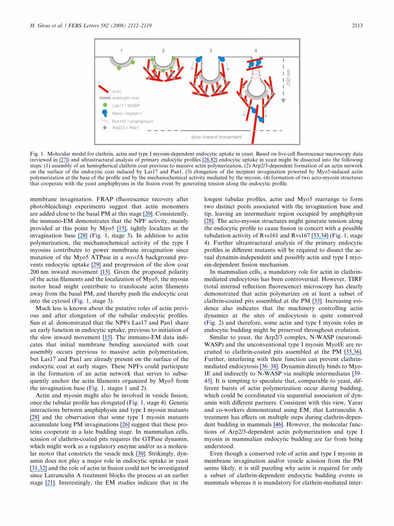
Fig. 1. Molecular model for clathrin, actin and type I myosin-dependent endocytic uptake in yeast. Based on live-cell fluorescence microscopy data(reviewed in [27]) and ultrastructural analysis of primary endocytic profiles [28,82] endocytic uptake in yeast might be dissected into the followingsteps: (1) assembly of an hemispherical clathrin coat previous to massive actin polymerization, (2) Arp2/3-dependent formation of an actin networkon the surface of the endocytic coat induced by Las17 and Pan1, (3) elongation of the incipient invagination powered by Myo5-induced actinpolymerization at the base of the profile and by the mechanochemical activity mediated by the myosin, (4) formation of two acto-myosin structuresthat cooperate with the yeast amphiphysins in the fission event by generating tension along the endocytic profile.
H. Girao et al. / FEBS Letters 582 (2008) 2112–2119 2113
membrane invagination. FRAP (fluorescence recovery after
photobleaching) experiments suggest that actin monomers
are added close to the basal PM at this stage [20]. Consistently,
the immuno-EM demonstrates that the NPF activity, mainly
provided at this point by Myo5 [15], tightly localizes at the
invagination base [28] (Fig. 1, stage 3). In addition to actin
polymerization, the mechanochemical activity of the type I
myosins contributes to power membrane invagination since
mutation of the Myo5 ATPase in a myo3D background pre-
vents endocytic uptake [29] and progression of the slow coat
200 nm inward movement [15]. Given the proposed polarity
of the actin filaments and the localization of Myo5, the myosin
motor head might contribute to translocate actin filaments
away from the basal PM, and thereby push the endocytic coat
into the cytosol (Fig. 1, stage 3).
Much less is known about the putative roles of actin previ-
ous and after elongation of the tubular endocytic profiles.
Sun et al. demonstrated that the NPFs Las17 and Pan1 share
an early function in endocytic uptake, previous to initiation of
the slow inward movement [15]. The immuno-EM data indi-
cates that initial membrane bending associated with coat
assembly occurs previous to massive actin polymerization,
but Las17 and Pan1 are already present on the surface of the
endocytic coat at early stages. These NPFs could participate
in the formation of an actin network that serves to subse-
quently anchor the actin filaments organized by Myo5 from
the invagination base (Fig. 1, stages 1 and 2).
Actin and myosin might also be involved in vesicle fission,
once the tubular profile has elongated (Fig. 1, stage 4). Genetic
interactions between amphiphysin and type I myosin mutants
[28] and the observation that some type I myosin mutants
accumulate long PM invaginations [26] suggest that these pro-
teins cooperate in a late budding stage. In mammalian cells,
scission of clathrin-coated pits requires the GTPase dynamin,
which might work as a regulatory enzyme and/or as a molecu-
lar motor that constricts the vesicle neck [30]. Strikingly, dyn-
amin does not play a major role in endocytic uptake in yeast
[31,32] and the role of actin in fission could not be investigated
since Latrunculin A treatment blocks the process at an earlier
stage [21]. Interestingly, the EM studies indicate that in the
longest tubular profiles, actin and Myo5 rearrange to form
two distinct pools associated with the invagination base and
tip, leaving an intermediate region occupied by amphiphysin
[28]. The acto-myosin structures might generate tension along
the endocytic profile to cause fission in concert with a possible
tubulation activity of Rvs161 and Rvs167 [33,34] (Fig. 1, stage
4). Further ultrastructural analysis of the primary endocytic
profiles in different mutants will be required to dissect the ac-
tual dynamin-independent and possibly actin and type I myo-
sin-dependent fission mechanism.
In mammalian cells, a mandatory role for actin in clathrin-
mediated endocytosis has been controversial. However, TIRF
(total internal reflection fluorescence) microscopy has clearly
demonstrated that actin polymerizes on at least a subset of
clathrin-coated pits assembled at the PM [35]. Increasing evi-
dence also indicates that the machinery controlling actin
dynamics at the sites of endocytosis is quite conserved
(Fig. 2) and therefore, some actin and type I myosin roles in
endocytic budding might be preserved throughout evolution.
Similar to yeast, the Arp2/3 complex, N-WASP (neuronal-
WASP) and the unconventional type I myosin MyoIE are re-
cruited to clathrin-coated pits assembled at the PM [35,36].
Further, interfering with their function can prevent clathrin-
mediated endocytosis [36–38]. Dynamin directly binds to Myo-
IE and indirectly to N-WASP via multiple intermediates [39–
45]. It is tempting to speculate that, comparable to yeast, dif-
ferent bursts of actin polymerization occur during budding,
which could be coordinated via sequential association of dyn-
amin with different partners. Consistent with this view, Yarar
and co-workers demonstrated using EM, that Latrunculin A
treatment has effects on multiple steps during clathrin-depen-
dent budding in mammals [46]. However, the molecular func-
tions of Arp2/3-dependent actin polymerization and type I
myosin in mammalian endocytic budding are far from being
understood.
Even though a conserved role of actin and type I myosin in
membrane invagination and/or vesicle scission from the PM
seems likely, it is still puzzling why actin is required for only
a subset of clathrin-dependent endocytic budding events in
mammals whereas it is mandatory for clathrin-mediated inter-
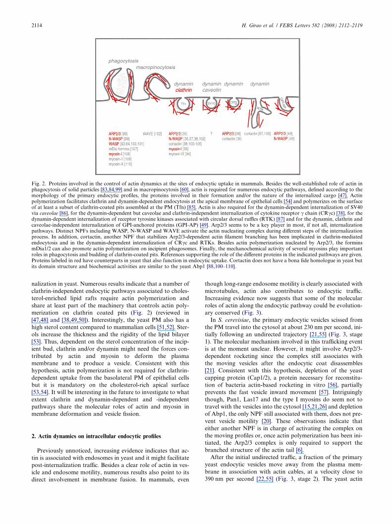
Fig. 2. Proteins involved in the control of actin dynamics at the sites of endocytic uptake in mammals. Besides the well-established role of actin inphagocytosis of solid particles [83,84,99] and in macropinocytosis [60], actin is required for numerous endocytic pathways, defined according to themorphology of the primary endocytic profiles, the proteins involved in their formation and/or the nature of the internalized cargo [47]. Actinpolymerization facilitates clathrin and dynamin-dependent endocytosis at the apical membrane of epithelial cells [54] and polymerizes on the surfaceof at least a subset of clathrin-coated pits assembled at the PM (Tfn) [85]. Actin is also required for the dynamin-dependent internalization of SV40via caveolae [86], for the dynamin-dependent but caveolae and clathrin-independent internalization of cytokine receptor c chain (CRcc) [38], for thedynamin-dependent internalization of receptor tyrosine kinases associated with circular dorsal ruffles (RTK) [87] and for the dynamin, clathrin andcaveolae-independent internalization of GPI-anchored proteins (GPI-AP) [49]. Arp2/3 seems to be a key player in most, if not all, internalizationpathways. Distinct NPFs including WASP, N-WASP and WAVE activate the actin nucleating complex during different steps of the internalizationprocess. In addition, cortactin, another NPF that stabilizes Arp2/3-dependent actin filament branching has been implicated in clathrin-mediatedendocytosis and in the dynamin-dependent internalization of CRcc and RTKs. Besides actin polymerization nucleated by Arp2/3, the forminsmDia1/2 can also promote actin polymerization on incipient phagosomes. Finally, the mechanochemical activity of several myosins play importantroles in phagocytosis and budding of clathrin-coated pits. References supporting the role of the different proteins in the indicated pathways are given.Proteins labeled in red have counterparts in yeast that also function in endocytic uptake. Cortactin does not have a bona fide homologue in yeast butits domain structure and biochemical activities are similar to the yeast Abp1 [88,100–110].
2114 H. Girao et al. / FEBS Letters 582 (2008) 2112–2119
nalization in yeast. Numerous results indicate that a number of
clathrin-independent endocytic pathways associated to choles-
terol-enriched lipid rafts require actin polymerization and
share at least part of the machinery that controls actin poly-
merization on clathrin coated pits (Fig. 2) (reviewed in
[47,48] and [38,49,50]). Interestingly, the yeast PM also has a
high sterol content compared to mammalian cells [51,52]. Ster-
ols increase the thickness and the rigidity of the lipid bilayer
[53]. Thus, dependent on the sterol concentration of the incip-
ient bud, clathrin and/or dynamin might need the forces con-
tributed by actin and myosin to deform the plasma
membrane and to produce a vesicle. Consistent with this
hypothesis, actin polymerization is not required for clathrin-
dependent uptake from the basolateral PM of epithelial cells
but it is mandatory on the cholesterol-rich apical surface
[53,54]. It will be interesting in the future to investigate to what
extent clathrin and dynamin-dependent and -independent
pathways share the molecular roles of actin and myosin in
membrane deformation and vesicle fission.
2. Actin dynamics on intracellular endocytic profiles
Previously unnoticed, increasing evidence indicates that ac-
tin is associated with endosomes in yeast and it might facilitate
post-internalization traffic. Besides a clear role of actin in ves-
icle and endosome motility, numerous results also point to its
direct involvement in membrane fusion. In mammals, even
though long-range endosome motility is clearly associated with
microtubules, actin also contributes to endocytic traffic.
Increasing evidence now suggests that some of the molecular
roles of actin along the endocytic pathway could be evolution-
ary conserved (Fig. 3).
In S. cerevisiae, the primary endocytic vesicles scissed from
the PM travel into the cytosol at about 230 nm per second, ini-
tially following an undirected trajectory [21,55] (Fig. 3, stage
1). The molecular mechanism involved in this trafficking event
is at the moment unclear. However, it might involve Arp2/3-
dependent rocketing since the complex still associates with
the moving vesicles after the endocytic coat disassembles
[21]. Consistent with this hypothesis, depletion of the yeast
capping protein (Cap1/2), a protein necessary for reconstitu-
tion of bacteria actin-based rocketing in vitro [56], partially
prevents the fast vesicle inward movement [57]. Intriguingly
though, Pan1, Las17 and the type I myosins do seem not to
travel with the vesicles into the cytosol [15,21,26] and depletion
of Abp1, the only NPF still associated with them, does not pre-
vent vesicle motility [20]. These observations indicate that
either another NPF is in charge of activating the complex on
the moving profiles or, once actin polymerization has been ini-
tiated, the Arp2/3 complex is only required to support the
branched structure of the actin tail [6].
After the initial undirected traffic, a fraction of the primary
yeast endocytic vesicles move away from the plasma mem-
brane in association with actin cables, at a velocity close to
390 nm per second [22,55] (Fig. 3, stage 2). The yeast actin
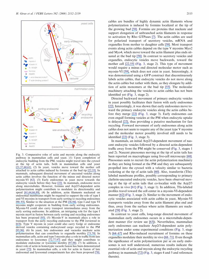
Fig. 3. Comparative roles of actin and myosin along the endocyticpathway in mammalian cells and yeast. (1) Upon completion ofendocytic budding from the PM, vesicles might travel into the cytosolat the tip of actin tails, both in mammalian cells and yeast[21,57,60,61]. (2) In yeast, vesicles move toward the mother cellattached to actin cables induced by formins at the bud tip [22,55]. Inmammals, subsequent directed movement of uncoated vesicles alongactin cables involves the function of the minus end directed motormyosin-VI [62]. (3) Early endosomes in yeast move towards theendocytic vesicle before they fuse [22]. In mammals, endosome movealong microtubules. However, formins and Arp2/3-dependent actinpolymerization might contribute to modulate its directionality andspeed [61,64,66,89]. (4) In addition, actin filaments nucleated onendosomal membranes might be required to support the role of type Iand VI myosins in transport from early sorting to recycling endosomes[90–93]. Similar to the situation at the PM [36,94], type I and type VImyosins might cooperate in budding from early sorting endosomes.Myosin-VI could also carry transport intermediates away from theearly sorting endosome. In addition, an intriguing role for the type Imyosin myr4 in fusion between early sorting and recycling endosomeshas been proposed [93]. (5) Myosin-V in mammals plays a role intransport from the early recycling endosome to the PM (reviewed in[63]). Type V myosin in yeast could also carry Golgi or endosomederived vesicles containing endocytosed cargo recycled to the PM[95,96]. (6) In yeast, late endosomes and vacuoles nucleate actinpolymerization that can contribute to organelle motility [73,79]. Inmammalian cells, actin and type I myosin play a role in traffic from lateendosomes to lysosomes [72,77,97] and they might contribute tomodulate endosome or lysosome motility [97,98]. (7) In addition, adirect role of actin in homotypic vacuole fusion has been demonstratedin yeast [75]. In mammalian cells, a role for actin in fusion of lateendosomal and lysosomal compartments has also been proposed [78].
H. Girao et al. / FEBS Letters 582 (2008) 2112–2119 2115
cables are bundles of highly dynamic actin filaments whose
polymerization is induced by formins localized at the tip of
the growing bud [58]. Formins are proteins that nucleate and
support elongation of unbranched actin filaments in response
to activation by Rho GTPases [7]. The actin cables are used
for polarized transport of secretory vesicles, mRNA and
organelles from mother to daughter cells [58]. Most transport
events along actin cables depend on the type V myosins Myo2
and Myo4, which move toward the actin filament plus ends sit-
uated at the bud tip [58]. In contrast to secretory vesicles and
organelles, endocytic vesicles move backwards, toward the
mother cell [22,55] (Fig. 3, stage 2). This type of movement
would require a minus end directed molecular motor such as
myosin-VI [59], which does not exist in yeast. Interestingly, it
was demonstrated using a GFP construct that discontinuously
labels actin cables, that endocytic vesicles do not move along
the actin cables but rather with them, as they elongate by addi-
tion of actin monomers at the bud tip [55]. The molecular
machinery attaching the vesicles to actin cables has not been
identified yet (Fig. 3, stage 2).
Directed backward movement of primary endocytic vesicles
in yeast possibly facilitates their fusion with early endosomes
[22]. Interestingly, it was shown that early endosomes move to-
ward the primary endocytic vesicles along the actin cables be-
fore they merge [22] (Fig. 3, stage 3). Early endosomes can
even engulf forming vesicles at the PM when endocytic uptake
is delayed [22], thus providing a putative mechanism for fast
recycling. Forward movement of early endosomes along actin
cables does not seem to require any of the yeast type V myosins
and the molecular motor possibly involved still needs to be
identified [22] (Fig. 3, stage 3).
In mammals, an initial Arp2/3-dependent movement of nas-
cent endocytic vesicles followed by a directed actin-dependent
traffic away from the PM might be conserved (Fig. 3, stages 1
and 2). Nascent pinosomes moving at the tip of actin tails has
been reported on macrophages using live-cell microscopy [60].
Pinosomes seem to recruit the actin polymerization machinery
as they are being formed at the PM and they are subsequently
propelled into the cytosol with a velocity similar to bacteria
rocketing at the tip of actin tails [60]. Also, transferrin (Tfn)-
labeled membrane profiles, possibly corresponding to primary
clathrin-uncoated endocytic vesicles, have been observed mov-
ing at the tip of actin tails that co-localize with the Arp2/3
complex in vivo [61] (Fig. 3, stage 1). In addition, Tfn-labeled
profiles travel toward the cell center in a myosin-VI-dependent
manner [62] (Fig. 3, stage 2). Similar to the movement of endo-
cytic vesicles associated with actin cables in yeast, Myosin-VI
transports vesicles away from the actin filament plus end and
thus, away from the surface where actin filaments are nucle-
ated [59] (Fig. 3, stage 2).
In contrast to yeast cells, long-range directed movement of
mammalian early endosomes occurs in a microtubule-depen-
dent manner (for review see [63]). Nevertheless, mammalian
early endosomes can nucleate Arp2/3-dependent actin poly-
merization under some experimental conditions (Fig. 3, stage
3) [64–67] and Rho-induced recruitment of formins on these
organelles modulate their motility in vivo [68–71]. Even though
the significance of actin polymerization per se on early endo-
somes is not well understood, numerous results indicate the
essential role of actin and myosin along the endocytic recycling
pathway in mammals [72] (Fig. 3, stages 4 and 5 and references
therein).
2116 H. Girao et al. / FEBS Letters 582 (2008) 2112–2119
Similar to nascent endocytic vesicles and endosomes, yeast
late endocytic compartments also move in an actin-dependent
manner (Fig. 3, stage 6). Chang et al. used a GFP-tagged ver-
sion of the G-protein coupled receptor Ste2 to label intracellu-
lar compartments [73], which were later shown to most likely
correspond to late endosomes [22]. The Ste2-GFP compart-
ments move in an undirected manner sometimes making tight
turns at a speed of 150–190 nm per second [73]. In contrast to
the directed movement of early endosomes, disruption of the
yeast cables does not affect the nature of the movement or
the speed of the Ste2-GFP-labeled organelles, whereas Latrun-
culin A treatment or mutation of WASP/Las17, significantly
slow them down. Strikingly also, fusion of the Las17 domains
required for Arp2/3 activation to the Ste2 cytoplasmic tail
recovers organelle motility in a las17 mutant [74]. The authors
proposed an actin rocketing-based mechanism to explain the
movement of Ste2-GFP labeled endosomes. However, actin
tails associated to these compartments have not been demon-
strated and depletion of the yeast capping protein does not
seem to affect the process [57].
The capacity of late endosomes to nucleate Arp2/3-depen-
dent actin polymerization might also be conserved in higher
eukaryotes. Late endosomes and lysosomes from Hela cells
are capable of nucleating actin tails upon activation of protein
kinase C on Xenopus extracts [67]. However, the physiological
significance of the Arp2/3-driven late endosomal motility
in vivo has not been investigated in detail yet (Fig. 3, stage 6).
Besides the well-established role of actin in membrane bud-
ding from the PM and vesicle and organelle motility, increas-
ing evidence demonstrates that actin also has a direct role in
fusion of yeast vacuoles (Fig. 3, stage 7). Eitzen et al. pointed
out that yeast bearing mutations in Arp2/3, Las17, Vrp1 (The
yeast WIP, wasp interacting protein) and the type I myosins
have fragmented vacuoles, a feature of strains defective in
homotypic fusion of the yeast lysosomal compartment
[75,76]. Arp2/3, Las17 and Vrp1 are enriched in vacuoles and
interfering with their function prevents vacuolar fusion
in vitro [75]. Working under experimental conditions that dis-
criminate between vacuole priming, docking and fusion reac-
tions, the authors demonstrated that actin depolymerization
is first required to allow vacuole docking, whereas actin poly-
merization is needed for the terminal steps leading to mem-
brane fusion [75]. Actin accumulates at the vertex of docked
vacuoles where fusion is thought to occur, suggesting a direct
contribution of actin to distortion of the lipid bilayer [75].
Recent results indicate that actin could also be involved in
fusion of late endocytic compartments in mammalian cells
(Fig. 3, stage 7). Actin is required to transfer internalized cargo
from late endosomes to lysosomes in vivo [72,77] and for
in vitro fusion of late endosomes and lysosomes [78]. In addi-
tion, similar to yeast vacuoles, the late endosomal/lysosomal
fraction is capable of inducing actin polymerization in this as-
say conditions [78,79]. The machinery involved in fusion of
late endosomes with lysosomes in mammalian cells seems to
very much resemble the biochemical requirement for homo-
typic vacuolar fusion in yeast [80]. Interestingly, it was recently
shown that overexpression of one component of the HOPS
complex, which also participates in docking of yeast vacuoles,
induces clustering of late endosomes and lysosomes and pro-
motes recruitment of actin, and type I and V myosins in
NRK cells [81]. Investigation of the possible molecular roles
of actin and myosin in membrane fusion in mammalian cells
will require detailed analysis of the in vitro fusion reaction.
3. Perspectives
Analysis of endocytic uptake in yeast has very much contrib-
uted to our understanding of the molecular roles of actin and
type I myosins in endocytic budding. The level of knowledge
that allows building molecular models to explain endocytic
membrane budding at the PM has required the effort of yeast
genetics to identify the machinery involved, the development
of live-cell imaging techniques and the ultrastructural analysis
of the primary endocytic profiles. The reconstitution of the
process using purified components will now be required to
definitively nail down the forces contributed by the actin
machinery in membrane bending or vesicle fission.
The role of actin in post-internalization endocytic traffic is
only now starting to emerge. In most cases, we still need to de-
fine the functional significance of actin dynamics on endo-
somes and its exact contribution to cargo transport along the
pathway. Molecular markers to unequivocally define the endo-
somal compartments in yeast and development of assays to
quantitatively measure transit of cargo through those com-
partments in vivo would help detailing the exact roles of actin
and myosin in post-internalization endocytic traffic. Even
though the endocytic pathway is clearly more complex and
subject to more numerous signaling inputs in mammalian cells,
genetic analysis of the yeast post-internalization endocytic traf-
fic might help unveiling some of the molecular roles of actin
and myosin in endosome and vesicle motility and/or mem-
brane budding and fusion.
Acknowledgements: We thank I.-M. Fernandez-Golbano for criticalreading of the manuscript. This work was granted by the MEC(BUF2005-04089). H.G. was a recipient of a postdoctoral fellowshipfrom FCT of the Portuguese Government. F.-Z.I. is a recipient ofRamon y Cajal postdoctoral contract from the MEC.
References
[1] Kubler, E. and Riezman, H. (1993) Actin and fimbrin arerequired for the internalization step of endocytosis in yeast.Embo J. 12, 2855–2862.
[2] Payne, G.S., Hasson, T.B., Hasson, M.S. and Schekman, R.(1987) Genetic and biochemical characterization of clathrin-deficient Saccharomyces cerevisiae. Mol. Cell Biol. 7, 3888–3898.
[3] Ayscough, K.R. and Drubin, D.G. (1996) ACTIN: generalprinciples from studies in yeast. Annu. Rev. Cell Dev. Biol. 12,129–160.
[4] Engqvist-Goldstein, A.E. and Drubin, D.G. (2003) Actinassembly and endocytosis: from yeast to mammals. Annu.Rev. Cell Dev. Biol. 19, 287–332.
[5] Moreau, V., Galan, J.M., Devilliers, G., Haguenauer-Tsapis, R.and Winsor, B. (1997) The yeast actin-related protein Arp2p isrequired for the internalization step of endocytosis. Mol. Biol.Cell 8, 1361–1375.
[6] Martin, A.C. et al. (2005) Effects of Arp2 and Arp3 nucleotide-binding pocket mutations on Arp2/3 complex function. J. CellBiol. 168, 315–328.
[7] Pollard, T.D. (2007) Regulation of actin filament assembly byArp2/3 complex and formins. Annu. Rev. Biophys. Biomol.Struct. 36, 451–477.
[8] Naqvi, S.N., Zahn, R., Mitchell, D.A., Stevenson, B.J. andMunn, A.L. (1998) The WASp homologue Las17p functions
H. Girao et al. / FEBS Letters 582 (2008) 2112–2119 2117
with the WIP homologue End5p/verprolin and is essential forendocytosis in yeast. Curr. Biol. 8, 959–962.
[9] Winter, D., Lechler, T. and Li, R. (1999) Activation of the yeastArp2/3 complex by Bee1p, a WASP-family protein. Curr. Biol. 9,501–504.
[10] Wendland, B., McCaffery, J.M., Xiao, Q. and Emr, S.D. (1996)A novel fluorescence-activated cell sorter-based screen for yeastendocytosis mutants identifies a yeast homologue of mammalianeps15. J. Cell Biol. 135, 1485–1500.
[11] Duncan, M.C., Cope, M.J., Goode, B.L., Wendland, B. andDrubin, D.G. (2001) Yeast Eps15-like endocytic protein, Pan1p,activates the Arp2/3 complex. Nat. Cell Biol. 3, 687–690.
[12] Wesp, A., Hicke, L., Palecek, J., Lombardi, R., Aust, T., Munn,A.L. and Riezman, H. (1997) End4p/Sla2p interacts with actin-associated proteins for endocytosis in Saccharomyces cerevisiae.Mol. Biol. Cell 8, 2291–2306.
[13] Goode, B.L., Rodal, A.A., Barnes, G. and Drubin, D.G. (2001)Activation of the Arp2/3 complex by the actin filament bindingprotein Abp1p. J. Cell Biol. 153, 627–634.
[14] Geli, M.I. and Riezman, H. (1996) Role of type I myosins inreceptor-mediated endocytosis in yeast. Science 272, 533–535.
[15] Sun, Y., Martin, A.C. and Drubin, D.G. (2006) Endocyticinternalization in budding yeast requires coordinated actinnucleation and myosin motor activity. Dev. Cell 11, 33–46.
[16] Pollard, T.D., Doberstein, S.K. and Zot, H.G. (1991) Myosin-I.Annu. Rev. Physiol. 53, 653–681.
[17] Lechler, T., Shevchenko, A. and Li, R. (2000) Direct involve-ment of yeast type I myosins in Cdc42-dependent actinpolymerization. J. Cell Biol. 148, 363–373.
[18] Geli, M.I., Lombardi, R., Schmelzl, B. and Riezman, H. (2000)An intact SH3 domain is required for myosin I-induced actinpolymerization. Embo J. 19, 4281–4291.
[19] Evangelista, M. et al. (2000) A role for myosin-I in actinassembly through interactions with Vrp1p, Bee1p, and the Arp2/3 complex. J. Cell Biol. 148, 353–362.
[20] Kaksonen, M., Toret, C.P. and Drubin, D.G. (2005) A modulardesign for the clathrin- and actin-mediated endocytosis machin-ery. Cell 123, 305–320.
[21] Kaksonen, M., Sun, Y. and Drubin, D.G. (2003) A pathway forassociation of receptors, adaptors, and actin during endocyticinternalization. Cell 115, 475–487.
[22] Toshima, J.Y., Toshima, J., Kaksonen, M., Martin, A.C., King,D.S. and Drubin, D.G. (2006) Spatial dynamics of receptor-mediated endocytic trafficking in budding yeast revealed byusing fluorescent alpha-factor derivatives. Proc. Natl. Acad. Sci.USA 103, 5793–5798.
[23] Newpher, T.M., Smith, R.P., Lemmon, V. and Lemmon, S.K.(2005) In vivo dynamics of clathrin and its adaptor-dependentrecruitment to the actin-based endocytic machinery in yeast.Dev. Cell 9, 87–98.
[24] Sun, Y., Carroll, S., Kaksonen, M., Toshima, J.Y. and Drubin,D.G. (2007) PtdIns(4,5)P2 turnover is required for multiplestages during clathrin- and actin-dependent endocytic internal-ization. J. Cell Biol. 177, 355–367.
[25] Okreglak, V. and Drubin, D.G. (2007) Cofilin recruitment andfunction during actin-mediated endocytosis dictated by actinnucleotide state. J. Cell Biol. 178, 1251–1264.
[26] Jonsdottir, G.A. and Li, R. (2004) Dynamics of yeast Myosin I:evidence for a possible role in scission of endocytic vesicles. Curr.Biol. 14, 1604–1609.
[27] Kaksonen, M., Toret, C.P. and Drubin, D.G. (2006) Harnessingactin dynamics for clathrin-mediated endocytosis. Nat. Rev.Mol. Cell Biol. 7, 404–414.
[28] Idrissi, F.Z., Grotsch, H., Fernandez-Golbano, I.M., Presciatto-Baschong, C., Riezman, H. and Geli, M.I. (2008) Distinct acto/myosin-I structures associate with endocytic profiles at theplasma membrane. J. Cell Biol. 180, 1219–1232.
[29] Grosshans, B.L. et al. (2006) TEDS site phosphorylation of theyeast myosins I is required for ligand-induced but not forconstitutive endocytosis of the G protein-coupled receptor Ste2p.J. Biol. Chem. 281, 11104–11114.
[30] Song, B.D. and Schmid, S.L. (2003) A molecular motor or aregulator? Dynamin�s in a class of its own. Biochemistry 42,1369–1376.
[31] Gammie, A.E., Kurihara, L.J., Vallee, R.B. and Rose, M.D.(1995) DNM1, a dynamin-related gene, participates in endo-somal trafficking in yeast. J. Cell Biol. 130, 553–566.
[32] Yu, X. and Cai, M. (2004) The yeast dynamin-related GTPaseVps1p functions in the organization of the actin cytoskeleton viainteraction with Sla1p. J. Cell Sci. 117, 3839–3853.
[33] Takei, K., Slepnev, V.I., Haucke, V. and De Camilli, P. (1999)Functional partnership between amphiphysin and dynamin inclathrin-mediated endocytosis. Nat. Cell Biol. 1, 33–39.
[34] Roux, A., Uyhazi, K., Frost, A. and De Camilli, P. (2006) GTP-dependent twisting of dynamin implicates constriction andtension in membrane fission. Nature 441, 528–531.
[35] Merrifield, C.J., Qualmann, B., Kessels, M.M. and Almers, W.(2004) Neural Wiskott Aldrich Syndrome Protein (N-WASP)and the Arp2/3 complex are recruited to sites of clathrin-mediated endocytosis in cultured fibroblasts. Eur. J. Cell Biol.83, 13–18.
[36] Krendel, M., Osterweil, E.K. and Mooseker, M.S. (2007)Myosin 1E interacts with synaptojanin-1 and dynamin and isinvolved in endocytosis. FEBS Lett. 581, 644–650.
[37] Benesch, S., Polo, S., Lai, F.P., Anderson, K.I., Stradal, T.E.,Wehland, J. and Rottner, K. (2005) N-WASP deficiency impairsEGF internalization and actin assembly at clathrin-coated pits.J. Cell Sci. 118, 3103–3115.
[38] Sauvonnet, N., Dujeancourt, A. and Dautry-Varsat, A. (2005)Cortactin and dynamin are required for the clathrin-independentendocytosis of gammac cytokine receptor. J. Cell Biol. 168, 155–163.
[39] Mise-Omata, S., Montagne, B., Deckert, M., Wienands, J. andAcuto, O. (2003) Mammalian actin binding protein 1 is essentialfor endocytosis but not lamellipodia formation: functionalanalysis by RNA interference. Biochem. Biophys. Res. Com-mun. 301, 704–710.
[40] Otsuki, M., Itoh, T. and Takenawa, T. (2003) Neural Wiskott–Aldrich syndrome protein is recruited to rafts and associateswith endophilin A in response to epidermal growth factor. J. BiolChem. 278, 6461–6469.
[41] Qualmann, B. and Kelly, R.B. (2000) Syndapin isoformsparticipate in receptor-mediated endocytosis and actin organi-zation. J. Cell Biol. 148, 1047–1062.
[42] Yamabhai, M., Hoffman, N.G., Hardison, N.L., McPherson,P.S., Castagnoli, L., Cesareni, G. and Kay, B.K. (1998)Intersectin, a novel adaptor protein with two Eps15 homologyand five Src homology 3 domains. J. Biol. Chem. 273, 31401–31407.
[43] Sengar, A.S., Wang, W., Bishay, J., Cohen, S. and Egan, S.E.(1999) The EH and SH3 domain Ese proteins regulate endocy-tosis by linking to dynamin and Eps15. Embo J. 18, 1159–1171.
[44] Lundmark, R. and Carlsson, S.R. (2004) Regulated membranerecruitment of dynamin-2 mediated by sorting nexin 9. J. Biol.Chem. 279, 42694–42702.
[45] Lundmark, R. and Carlsson, S.R. (2003) Sorting nexin 9participates in clathrin-mediated endocytosis through interac-tions with the core components. J. Biol. Chem. 278, 46772–46781.
[46] Yarar, D., Waterman-Storer, C.M. and Schmid, S.L. (2005) Adynamic actin cytoskeleton functions at multiple stages ofclathrin-mediated endocytosis. Mol. Biol. Cell 16, 964–975.
[47] Mayor, S. and Pagano, R.E. (2007) Pathways of clathrin-independent endocytosis. Nat. Rev. Mol. Cell Biol. 8, 603–612.
[48] Kirkham, M. and Parton, R.G. (2005) Clathrin-independentendocytosis: new insights into caveolae and non-caveolar lipidraft carriers. Biochim. Biophys. Acta 1746, 349–363.
[49] Chadda, R., Howes, M.T., Plowman, S.J., Hancock, J.F.,Parton, R.G. and Mayor, S. (2007) Cholesterol-sensitive Cdc42activation regulates actin polymerization for endocytosis via theGEEC pathway. Traffic 8, 702–717.
[50] Yarar, D., Waterman-Storer, C.M. and Schmid, S.L. (2007)SNX9 couples actin assembly to phosphoinositide signals and isrequired for membrane remodeling during endocytosis. Dev.Cell 13, 43–56.
[51] Zinser, E., Sperka-Gottlieb, C.D., Fasch, E.V., Kohlwein, S.D.,Paltauf, F. and Daum, G. (1991) Phospholipid synthesis andlipid composition of subcellular membranes in the unicellular
2118 H. Girao et al. / FEBS Letters 582 (2008) 2112–2119
eukaryote Saccharomyces cerevisiae. J. Bacteriol. 173, 2026–2034.
[52] Lange, Y., Swaisgood, M.H., Ramos, B.V. and Steck, T.L.(1989) Plasma membranes contain half the phospholipid and90% of the cholesterol and sphingomyelin in cultured humanfibroblasts. J. Biol. Chem. 264, 3786–3793.
[53] van Meer, G. and Simons, K. (1988) Lipid polarity and sortingin epithelial cells. J. Cell Biochem. 36, 51–58.
[54] Gottlieb, T.A., Ivanov, I.E., Adesnik, M. and Sabatini, D.D.(1993) Actin microfilaments play a critical role in endocytosis atthe apical but not the basolateral surface of polarized epithelialcells. J. Cell Biol. 120, 695–710.
[55] Huckaba, T.M., Gay, A.C., Pantalena, L.F., Yang, H.C. andPon, L.A. (2004) Live cell imaging of the assembly, disassembly,and actin cable-dependent movement of endosomes and actinpatches in the budding yeast, Saccharomyces cerevisiae. J. CellBiol. 167, 519–530.
[56] Loisel, T.P., Boujemaa, R., Pantaloni, D. and Carlier, M.F.(1999) Reconstitution of actin-based motility of Listeria andShigella using pure proteins. Nature 401, 613–616.
[57] Kim, K., Galletta, B.J., Schmidt, K.O., Chang, F.S., Blumer,K.J. and Cooper, J.A. (2006) Actin-based motility duringendocytosis in budding yeast. Mol. Biol. Cell 17, 1354–1363.
[58] Pruyne, D., Legesse-Miller, A., Gao, L., Dong, Y. andBretscher, A. (2004) Mechanisms of polarized growth andorganelle segregation in yeast. Annu. Rev. Cell Dev. Biol. 20,559–591.
[59] Wells, A.L. et al. (1999) Myosin VI is an actin-based motor thatmoves backwards. Nature 401, 505–508.
[60] Merrifield, C.J., Moss, S.E., Ballestrem, C., Imhof, B.A., Giese,G., Wunderlich, I. and Almers, W. (1999) Endocytic vesiclesmove at the tips of actin tails in cultured mast cells. Nat. CellBiol. 1, 72–74.
[61] Kaksonen, M., Peng, H.B. and Rauvala, H. (2000) Associationof cortactin with dynamic actin in lamellipodia and onendosomal vesicles. J. Cell Sci. 113 (Pt. 24), 4421–4426.
[62] Aschenbrenner, L., Lee, T. and Hasson, T. (2003) Myo6facilitates the translocation of endocytic vesicles from cellperipheries. Mol. Biol. Cell 14, 2728–2743.
[63] Soldati, T. and Schliwa, M. (2006) Powering membrane traffic inendocytosis and recycling. Nat. Rev. Mol. Cell Biol. 7, 897–908.
[64] Llado, A. et al. (2008) Protein kinase C{delta} and calmodulinregulate epidermal growth factor receptor recycling from earlyendosomes through Arp2/3 complex and cortactin. Mol. Biol.Cell 19, 17–29.
[65] Schafer, D.A. (2004) Regulating actin dynamics at membranes: afocus on dynamin. Traffic 5, 463–469.
[66] Southwick, F.S., Li, W., Zhang, F., Zeile, W.L. and Purich, D.L.(2003) Actin-based endosome and phagosome rocketing inmacrophages: activation by the secretagogue antagonists lan-thanum and zinc. Cell Motil. Cytoskeleton 54, 41–55.
[67] Taunton, J., Rowning, B.A., Coughlin, M.L., Wu, M., Moon,R.T., Mitchison, T.J. and Larabell, C.A. (2000) Actin-dependentpropulsion of endosomes and lysosomes by recruitment of N-WASP. J. Cell Biol. 148, 519–530.
[68] Wallar, B.J., Deward, A.D., Resau, J.H. and Alberts, A.S.(2007) RhoB and the mammalian Diaphanous-related forminmDia2 in endosome trafficking. Exp. Cell Res. 313, 560–571.
[69] Fernandez-Borja, M., Janssen, L., Verwoerd, D., Hordijk, P.and Neefjes, J. (2005) RhoB regulates endosome transport bypromoting actin assembly on endosomal membranes throughDia1. J. Cell Sci. 118, 2661–2670.
[70] Gasman, S., Kalaidzidis, Y. and Zerial, M. (2003) RhoDregulates endosome dynamics through Diaphanous-related For-min and Src tyrosine kinase. Nat. Cell Biol. 5, 195–204.
[71] Tominaga, T., Sahai, E., Chardin, P., McCormick, F., Court-neidge, S.A. and Alberts, A.S. (2000) Diaphanous-relatedformins bridge Rho GTPase and Src tyrosine kinase signaling.Mol. Cell 5, 13–25.
[72] Durrbach, A., Louvard, D. and Coudrier, E. (1996) Actinfilaments facilitate two steps of endocytosis. J. Cell Sci. 109 (Pt.2), 457–465.
[73] Chang, F.S., Stefan, C.J. and Blumer, K.J. (2003) A WASPhomolog powers actin polymerization-dependent motility ofendosomes in vivo. Curr. Biol. 13, 455–463.
[74] Chang, F.S., Han, G.S., Carman, G.M. and Blumer, K.J. (2005)A WASp-binding type II phosphatidylinositol 4-kinase requiredfor actin polymerization-driven endosome motility. J. Cell Biol.171, 133–142.
[75] Eitzen, G., Wang, L., Thorngren, N. and Wickner, W. (2002)Remodeling of organelle-bound actin is required for yeastvacuole fusion. J. Cell Biol. 158, 669–679.
[76] Seeley, E.S., Kato, M., Margolis, N., Wickner, W. and Eitzen,G. (2002) Genomic analysis of homotypic vacuole fusion. Mol.Biol. Cell 13, 782–794.
[77] van Deurs, B., Holm, P.K., Kayser, L. and Sandvig, K. (1995)Delivery to lysosomes in the human carcinoma cell line HEp-2involves an actin filament-facilitated fusion between matureendosomes and preexisting lysosomes. Eur. J. Cell Biol. 66, 309–323.
[78] Kjeken, R. et al. (2004) Fusion between phagosomes, early andlate endosomes: a role for actin in fusion between late, but notearly endocytic organelles. Mol. Biol. Cell 15, 345–358.
[79] Isgandarova, S., Jones, L., Forsberg, D., Loncar, A., Dawson,J., Tedrick, K. and Eitzen, G. (2007) Stimulation of actinpolymerization by vacuoles via Cdc42p-dependent signaling. J.Biol. Chem. 282, 30466–30475.
[80] Wickner, W. and Haas, A. (2000) Yeast homotypic vacuolefusion: a window on organelle trafficking mechanisms. Annu.Rev. Biochem. 69, 247–275.
[81] Poupon, V., Stewart, A., Gray, S.R., Piper, R.C. and Luzio, J.P.(2003) The role of mVps18p in clustering, fusion, and intracel-lular localization of late endocytic organelles. Mol. Biol. Cell 14,4015–4027.
[82] Rodal, A.A., Kozubowski, L., Goode, B.L., Drubin, D.G. andHartwig, J.H. (2005) Actin and septin ultrastructures at thebudding yeast cell cortex. Mol. Biol. Cell 16, 372–384.
[83] Coppolino, M.G., Krause, M., Hagendorff, P., Monner, D.A.,Trimble, W., Grinstein, S., Wehland, J. and Sechi, A.S. (2001)Evidence for a molecular complex consisting of Fyb/SLAP, SLP-76, Nck, VASP and WASP that links the actin cytoskeleton toFcgamma receptor signalling during phagocytosis. J. Cell Sci.114, 4307–4318.
[84] Castellano, F., Le Clainche, C., Patin, D., Carlier, M.F. andChavrier, P. (2001) A WASp-VASP complex regulates actinpolymerization at the plasma membrane. Embo J. 20, 5603–5614.
[85] Merrifield, C.J., Feldman, M.E., Wan, L. and Almers, W. (2002)Imaging actin and dynamin recruitment during invagination ofsingle clathrin-coated pits. Nat. Cell Biol. 4, 691–698.
[86] Pelkmans, L., Puntener, D. and Helenius, A. (2002) Local actinpolymerization and dynamin recruitment in SV40-inducedinternalization of caveolae. Science 296, 535–539.
[87] Orth, J.D., Krueger, E.W., Weller, S.G. and McNiven, M.A.(2006) A novel endocytic mechanism of epidermal growth factorreceptor sequestration and internalization. Cancer Res. 66,3603–3610.
[88] Olazabal, I.M. and Machesky, L.M. (2001) Abp1p and cortac-tin, new hand-holds for actin. J. Cell Biol. 154, 679–682.
[89] Schafer, D.A., D�Souza-Schorey, C. and Cooper, J.A. (2000)Actin assembly at membranes controlled by ARF6. Traffic 1,892–903.
[90] Chibalina, M.V., Seaman, M.N., Miller, C.C., Kendrick-Jones,J. and Buss, F. (2007) Myosin VI and its interacting proteinLMTK2 regulate tubule formation and transport to the endo-cytic recycling compartment. J. Cell Sci. 120, 4278–4288.
[91] Durrbach, A., Collins, K., Matsudaira, P., Louvard, D. andCoudrier, E. (1996) Brush border myosin-I truncated in themotor domain impairs the distribution and the function ofendocytic compartments in an hepatoma cell line. Proc. Natl.Acad. Sci. USA 93, 7053–7058.
[92] Raposo, G., Cordonnier, M.N., Tenza, D., Menichi, B., Durr-bach, A., Louvard, D. and Coudrier, E. (1999) Association ofmyosin I alpha with endosomes and lysosomes in mammaliancells. Mol. Biol. Cell 10, 1477–1494.
[93] Huber, L.A. et al. (2000) Both calmodulin and the unconven-tional myosin Myr4 regulate membrane trafficking along therecycling pathway of MDCK cells. Traffic 1, 494–503.
[94] Buss, F., Arden, S.D., Lindsay, M., Luzio, J.P. and Kendrick-Jones, J. (2001) Myosin VI isoform localized to clathrin-coated
H. Girao et al. / FEBS Letters 582 (2008) 2112–2119 2119
vesicles with a role in clathrin-mediated endocytosis. Embo J. 20,3676–3684.
[95] Pruyne, D.W., Schott, D.H. and Bretscher, A. (1998) Tropomy-osin-containing actin cables direct the Myo2p-dependent polar-ized delivery of secretory vesicles in budding yeast. J. Cell Biol.143, 1931–1945.
[96] Pelham, H.R. (2002) Insights from yeast endosomes. Curr. Opin.Cell Biol. 14, 454–462.
[97] Cordonnier, M.N., Dauzonne, D., Louvard, D. and Coudrier, E.(2001) Actin filaments and myosin I alpha cooperate withmicrotubules for the movement of lysosomes. Mol. Biol. Cell 12,4013–4029.
[98] Loubery, S., Wilhelm, C., Hurbain, I., Neveu, S., Louvard, D.and Coudrier, E. (2008) Different microtubule motors moveearly and late endocytic compartments. Traffic 9, 492–509.
[99] May, R.C., Caron, E., Hall, A. and Machesky, L.M. (2000)Involvement of the Arp2/3 complex in phagocytosis mediated byFcgammaR or CR3. Nat. Cell Biol. 2, 246–248.
[100] Tsuboi, S. and Meerloo, J. (2007) Wiskott–Aldrich syndromeprotein is a key regulator of the phagocytic cup formation inmacrophages. J. Biol. Chem. 282, 34194–34203.
[101] Lorenzi, R., Brickell, P.M., Katz, D.R., Kinnon, C. and Thrasher,A.J. (2000) Wiskott–Aldrich syndrome protein is necessary forefficient IgG-mediated phagocytosis. Blood 95, 2943–2946.
[102] Innocenti, M. et al. (2005) Abi1 regulates the activity of N-WASP and WAVE in distinct actin-based processes. Nat. CellBiol. 7, 969–976.
[103] Cao, H., Orth, J.D., Chen, J., Weller, S.G., Heuser, J.E. andMcNiven, M.A. (2003) Cortactin is a component of clathrin-
coated pits and participates in receptor-mediated endocytosis.Mol. Cell Biol. 23, 2162–2170.
[104] Merrifield, C.J., Perrais, D. and Zenisek, D. (2005) Couplingbetween clathrin-coated-pit invagination, cortactin recruitment,and membrane scission observed in live cells. Cell 121, 593–606.
[105] Zhu, J., Zhou, K., Hao, J.J., Liu, J., Smith, N. and Zhan, X.(2005) Regulation of cortactin/dynamin interaction by actinpolymerization during the fission of clathrin-coated pits. J. CellSci. 118, 807–817.
[106] Krueger, E.W., Orth, J.D., Cao, H. and McNiven, M.A. (2003)A dynamin-cortactin-Arp2/3 complex mediates actin reorgani-zation in growth factor-stimulated cells. Mol. Biol. Cell 14,1085–1096.
[107] Colucci-Guyon, E., Niedergang, F., Wallar, B.J., Peng, J.,Alberts, A.S. and Chavrier, P. (2005) A role for mammaliandiaphanous-related formins in complement receptor (CR3)-mediated phagocytosis in macrophages. Curr. Biol. 15, 2007–2012.
[108] Swanson, J.A., Johnson, M.T., Beningo, K., Post, P., Mooseker,M. and Araki, N. (1999) A contractile activity that closesphagosomes in macrophages. J. Cell Sci. 112 (Pt. 3), 307–316.
[109] Olazabal, I.M., Caron, E., May, R.C., Schilling, K., Knecht,D.A. and Machesky, L.M. (2002) Rho-kinase and myosin-IIcontrol phagocytic cup formation during CR, but not Fcgam-maR, phagocytosis. Curr. Biol. 12, 1413–1418.
[110] Cox, D., Berg, J.S., Cammer, M., Chinegwundoh, J.O., Dale,B.M., Cheney, R.E. and Greenberg, S. (2002) Myosin X is adownstream effector of PI(3)K during phagocytosis. Nat. CellBiol. 4, 469–477.