Tyrosine kinase inhibitors for cancer treatment
230
Tyrosine kinase inhibitors for cancer treatment Citation for published version (APA): Tullemans, B. M. E. (2021). Tyrosine kinase inhibitors for cancer treatment: effects on platelets. [Doctoral Thesis, Maastricht University]. ProefschriftMaken. https://doi.org/10.26481/dis.20211006bt Document status and date: Published: 01/01/2021 DOI: 10.26481/dis.20211006bt Document Version: Publisher's PDF, also known as Version of record Please check the document version of this publication: • A submitted manuscript is the version of the article upon submission and before peer-review. There can be important differences between the submitted version and the official published version of record. People interested in the research are advised to contact the author for the final version of the publication, or visit the DOI to the publisher's website. • The final author version and the galley proof are versions of the publication after peer review. • The final published version features the final layout of the paper including the volume, issue and page numbers. Link to publication General rights Copyright and moral rights for the publications made accessible in the public portal are retained by the authors and/or other copyright owners and it is a condition of accessing publications that users recognise and abide by the legal requirements associated with these rights. • Users may download and print one copy of any publication from the public portal for the purpose of private study or research. • You may not further distribute the material or use it for any profit-making activity or commercial gain • You may freely distribute the URL identifying the publication in the public portal. If the publication is distributed under the terms of Article 25fa of the Dutch Copyright Act, indicated by the “Taverne” license above, please follow below link for the End User Agreement: www.umlib.nl/taverne-license Take down policy If you believe that this document breaches copyright please contact us at: [email protected] providing details and we will investigate your claim. Download date: 15 Jul. 2022
-
Upload
khangminh22 -
Category
Documents
-
view
1 -
download
0
Transcript of Tyrosine kinase inhibitors for cancer treatment
Tyrosine kinase inhibitors for cancer treatment
Citation for published version (APA):
Tullemans, B. M. E. (2021). Tyrosine kinase inhibitors for cancer treatment: effects on platelets. [DoctoralThesis, Maastricht University]. ProefschriftMaken. https://doi.org/10.26481/dis.20211006bt
Document status and date:Published: 01/01/2021
DOI:10.26481/dis.20211006bt
Document Version:Publisher's PDF, also known as Version of record
Please check the document version of this publication:
• A submitted manuscript is the version of the article upon submission and before peer-review. There canbe important differences between the submitted version and the official published version of record.People interested in the research are advised to contact the author for the final version of the publication,or visit the DOI to the publisher's website.• The final author version and the galley proof are versions of the publication after peer review.• The final published version features the final layout of the paper including the volume, issue and pagenumbers.Link to publication
General rightsCopyright and moral rights for the publications made accessible in the public portal are retained by the authors and/or other copyrightowners and it is a condition of accessing publications that users recognise and abide by the legal requirements associated with theserights.
• Users may download and print one copy of any publication from the public portal for the purpose of private study or research.• You may not further distribute the material or use it for any profit-making activity or commercial gain• You may freely distribute the URL identifying the publication in the public portal.
If the publication is distributed under the terms of Article 25fa of the Dutch Copyright Act, indicated by the “Taverne” license above,please follow below link for the End User Agreement:
www.umlib.nl/taverne-license
Take down policyIf you believe that this document breaches copyright please contact us at:
providing details and we will investigate your claim.
Download date: 15 Jul. 2022

Tyrosine kinase inhibitors for cancer treatment: Effects on platelets
Bibian M.E. Tullemans

Tyrosine kinase inhibitors for cancer treatment: Effects on platelets
Thesis: Maastricht University
ISBN: 978-94-6423-418-3
Production: ProefschrifMaken || www.proefschriftmaken.nl
© Bibian Tullemans, Maastricht 2021
Cover design by Bregje Jaspers of STUDIO 0404 || www.proefschriftontwerp.nl

Tyrosine kinase inhibitors for cancer treatment: Effects on platelets
Proefschrift
Ter verkrijging van de graad van doctor aan de Universiteit Maastricht,
op gezag van de Rector Magnificus, Prof. Dr. Rianne M. Letschert,
volgens het besluit van het College van Decanen,
in het openbaar te verdedigen op
Woensdag 06 oktober 2021 om 16.00 uur
door
Beatrice Maria Emanuelle TullemansGeboren op 27 oktober 1992 te Weert
PromotorProf. Dr. J.W.M. Heemskerk
CopromotorDr. M.J.E. Kuijpers
Dr. M.J.B. Aarts
BeoordelingscommissieProf. Dr. H. ten Cate (Voorzitter)
Prof. Dr. G.M.J. Bos
Dr. S. Séverin (Université Paul Sabatier, France)
Prof. Dr. J.C. Sluimer
Dr. A.A.M. van der Veldt (Erasmus Medisch Centrum)
The research described in this thesis was partially supported by a research grant from Pfizer
(WI209458)
Financial support for the publication of this thesis by Pfizer is gratefully acknowledged.
Financial support for the publication of this thesis by Hart Onderzoek Nederland is gratefully
acknowledged.

ContentsChapter 1 General Introduction 7
Chapter 2 Acquired platelet antagonism: off-target antiplatelet effects of
malignancy treatment 19
Chapter 3 Tyrosine kinase inhibitor pazopanib inhibits platelet procoagulant
activity in renal cell carcinoma patients 41
Chapter 4 Tyrosine kinase inhbitor sunitinib delays platelet-induced coagulation:
additive effects of aspirin 61
Chapter 5 Quantitative and qualitative changes in platelet traits of sunitinib-
treated patients with renal cell carcinoma in relation to circulating
sunitinib levels: a proof-of-concept study 89
Chapter 6 Comparison of inhibitory effects of irreversible and reversible Btk
inhibitors on platelet function 111
Chapter 7 Multiparameter screening for platelet-inhibitory effects of tyrosine
kinase inhibitors used for cancer treatment 151
Chapter 8 General Discussion 175
Chapter 9 Summary 197
Samenvatting 203
Impact 209
Curriculum Vitae 215
Publications 219
Acknowledgements 225


Chapter 1
General Introduction
Chapter 1
8
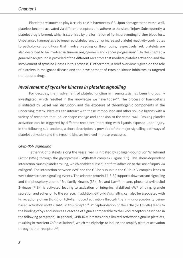
Platelets are known to play a crucial role in haemostasis1-3. Upon damage to the vessel wall,
platelets become activated via different receptors and adhere to the site of injury. Subsequently, a
platelet plug is formed, which is stabilised by the formation of fibrin, preventing further blood loss.
Unbalanced haemostasis by impaired platelet function or increased platelet reactivity contributes
to pathological conditions that involve bleeding or thrombosis, respectively. Yet, platelets are
also described to be involved in tumour angiogenesis and cancer progression4, 5. In this chapter, a
general background is provided of the different receptors that mediate platelet activation and the
involvement of tyrosine kinases in this process. Furthermore, a brief overview is given on the role
of platelets in malignant disease and the development of tyrosine kinase inhibitors as targeted
therapeutic drugs.
Involvement of tyrosine kinases in platelet signallingFor decades, the involvement of platelet function in haemostasis has been thoroughly
investigated, which resulted in the knowledge we have today1-3. The process of haemostasis
is initiated by vessel wall disruption and the exposure of thrombogenic components in the
underlying matrix. Platelets can interact with these immobilised and other soluble ligands with a
variety of receptors that induce shape change and adhesion to the vessel wall. Ensuing platelet
activation can be triggered by different receptors interacting with ligands exposed upon injury.
In the following sub-sections, a short description is provided of the major signalling pathways of
platelet activation and the tyrosine kinases involved in these processes.
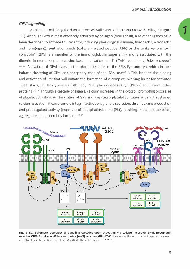
GPIb-IX-V signallingTethering of platelets along the vessel wall is initiated by collagen-bound von Willebrand
Factor (vWF) through the glycoprotein (GP)Ib-IX-V complex (Figure 1.1). This shear-dependent
interaction causes platelet rolling, which enables subsequent firm adhesion to the site of injury via
collagen6. The interaction between vWF and the GPIbα subunit in the GPIb-IX-V complex leads to
weak downstream signalling events. The adapter protein 14-3-3ζ supports downstream signalling
and the phosphorylation of Src family kinases (SFK) Src and Lyn7, 8. In turn, phosphatidylinositol
3-kinase (PI3K) is activated leading to activation of integrins, stabilised vWF binding, granule
secretion and adhesion to the surface. In addition, GPIb-IX-V signalling can also be associated with
Fc receptor γ-chain (FcRγ) or FcRγIIa induced activation through the immunoreceptor tyrosine-
based activation motif (ITAM) in this receptor8. Phosphorylation of the FcRγ (or FcRγIIa) leads to
the binding of Syk and induces a cascade of signals comparable to the GPVI receptor (described in
the following paragraph). In general, GPIb-IX-V initiates only a limited activation signal in platelets,
resulting in transient Ca2+ oscillations6, which mainly helps to induce and amplify platelet activation
through other receptors7, 9.
General introduction
9
1GPVI signalling
As platelets roll along the damaged vessel wall, GPVI is able to interact with collagen (Figure
1.1). Although GPVI is most efficiently activated by collagen (type I or III), also other ligands have
been described to activate this receptor, including physiological (laminin, fibronectin, vitronectin
and fibrin(ogen)), synthetic ligands (collagen-related peptide, CRP) or the snake venom toxin
convulxin10. GPVI is a member of the immunoglobulin superfamily and is associated with the
dimeric immunoreceptor tyrosine-based activation motif (ITAM)-containing FcRγ receptor8,
11, 12. Activation of GPVI leads to the phosphorylation of the SFKs Fyn and Lyn, which in turn
induces clustering of GPVI and phosphorylation of the ITAM motif1, 8. This leads to the binding
and activation of Syk that will initiate the formation of a complex involving linker for activated
T-cells (LAT), Tec family kinases (Btk, Tec), PI3K, phospholipase C-γ2 (PLCγ2) and several other
proteins1, 2, 13. Through a cascade of signals, calcium increases in the cytosol, promoting processes
of platelet activation. As stimulation of GPVI induces strong platelet activation with high sustained
calcium elevation, it can promote integrin activation, granule secretion, thromboxane production
and procoagulant activity (exposure of phosphatidylserine (PS)), resulting in platelet adhesion,
aggregation, and thrombus formation2, 14.
Figure 1.1. Schematic overview of signalling cascades upon activation via collagen receptor GPVI, podoplanin receptor CLEC-2 and von Willebrand factor (vWF) receptor GPIb-IX-V. Shown are the most potent agonists for each receptor. For abbreviations: see text. Modified after references 1, 2, 7, 8, 12, 13.

Chapter 1
10
CLEC-2 signallingA less well studied receptor that mediates platelet activation is C-type lectin-like type
II membrane glycoprotein (CLEC-2) (Figure 1.1). CLEC-2 is activated through its natural ligand
podoplanin or the snake venom toxin rhodocytin15, 16. The CLEC-2 receptor contains a single
hemi-immunoreceptor tyrosine-based activation motif (hemITAM), and its signalling involves
many molecules that are also present in the GPVI pathway. Upon activation the hemITAM motif
becomes phosphorylated through Fyn and Lyn (and possibly Btk or Tec)1, 13, 17. This enables
dimerization of CLEC-2 and the binding of Syk to the hemITAM motif, leading to the downstream
signalling through the LAT complex, inducing an increase of intracellular calcium and resulting
in platelet activation1, 2. Although GPVI and CLEC-2 signalling are in many ways alike, CLEC-2
signalling appears to be more reliant on Syk, whereas GPVI signalling is more SFK dependent18.
Integrin activationUpon platelet activation, one of the induced responses is the activation of integrins through
inside-out signalling (Figure 1.2A)1, 19. Integrins are transmembrane proteins consisting of an α
and β chain that exist as a noncovalent heterodimeric complex. Integrins are important for stable
adhesion to the vessel wall and platelet aggregate formation1. For integrins to become active, a
conformational change in this complex is necessary to obtain a high-affinity ligand binding state2,
20. In the low-affinity state of integrins, Csk forms a complex with Src and maintains the β-chain
in an inactive state. Activation induces dissociation of Csk from the complex which leads to the
activation of Src8, 21. Subsequently, the actin cytoskeletal proteins talin-1 and kindlin-3 bind to
Src and provoke the conformational change that leads to the increase in affinity (Figure 1.2A).
Figure 1.2. Schematic overview of (A) inside-out and (B) outside-in signalling of platelet integrins including αIIbβ3, α2β1 and α6β1. Shown are the most potent agonists for each receptor. For abbreviations: see text. Modified after references 1, 2, 8, 23, 31.
General introduction
11
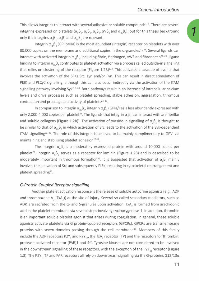
1This allows integrins to interact with several adhesive or soluble compounds1, 2. There are several
integrins expressed on platelets (α2β1, α5β1, α6β1, αƲβ3 and αIIbβ3), but for this thesis background
only the integrins α2β1, α6β1 and αIIbβ3 are relevant.
Integrin αIIbβ3 (GPIIb/IIIa) is the most abundant (integrin) receptor on platelets with over
80,000 copies on the membrane and additional copies in the α-granules22, 23. Several ligands can
interact with activated integrin αIIbβ3, including fibrin, fibrinogen, vWF and fibronectin21, 23. Ligand
binding to integrin αIIbβ3 contributes to platelet activation via a process called outside-in signalling
that relies on clustering of the receptor (Figure 1.2B)1, 2. This activates a cascade of events that
involves the activation of the SFKs Src, Lyn and/or Fyn. This can result in direct stimulation of
PI3K and PLCγ2 signalling, although this can also occur indirectly via the activation of the ITAM
signalling pathway involving Syk2, 8, 24. Both pathways result in an increase of intracellular calcium
levels and drive processes such as platelet spreading, stable adhesion, aggregation, thrombus
contraction and procoagulant activity of platelets23, 25.
In comparison to integrin αIIbβ3, integrin α2β1 (GPIa/IIa) is less abundantly expressed with
only 2,000-4,000 copies per platelet23. The ligands that integrin α2β1 can interact with are fibrillar
and soluble collagens (Figure 1.2B)1. The activation of outside-in signalling of α2β1 is thought to
be similar to that of αIIbβ3 in which activation of Src leads to the activation of the Syk-dependent
ITAM signalling23, 26. The role of this integrin is believed to be mainly complimentary to GPVI via
maintaining and stabilising platelet adhesion27-29.
The integrin α6β1 is a moderately expressed protein with around 10,000 copies per
platelet22. Integrin α6β1 serves as a receptor for laminin (Figure 1.2B) and is described to be
moderately important in thrombus formation30. It is suggested that activation of α6β1 mainly
involves the activation of Src and subsequently PI3K, resulting in cytoskeletal rearrangement and
platelet spreading31.
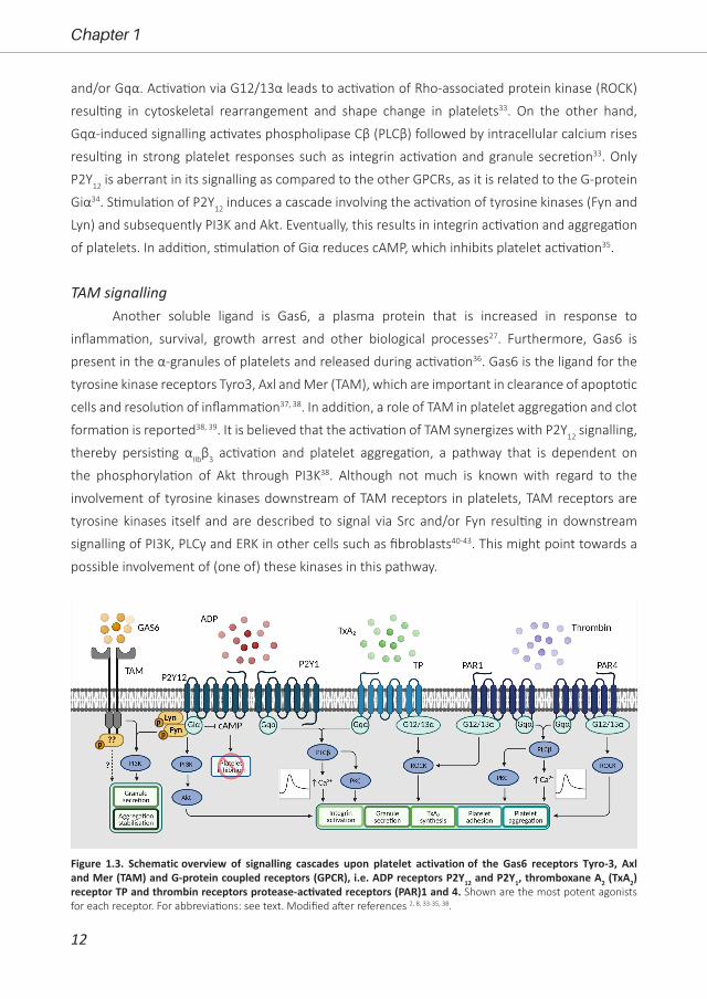
G-Protein Coupled Receptor signallingAnother platelet activation response is the release of soluble autocrine agonists (e.g., ADP
and thromboxane A2 (TxA2)) at the site of injury. Several so-called secondary mediators, such as
ADP, are secreted from the α- and δ-granules upon activation. TxA2 is formed from arachidonic
acid in the platelet membrane via several steps involving cyclooxygenase-1. In addition, thrombin
is an important soluble platelet agonist that arises during coagulation. In general, these soluble
agonists activate platelets via G protein-coupled receptors (GPCRs). GPCRs are transmembrane
proteins with seven domains passing through the cell membrane32. Members of this family
include the ADP receptors P2Y1 and P2Y12, the TxA2 receptor (TP) and the receptors for thrombin,
protease-activated receptor (PAR)1 and 427. Tyrosine kinases are not considered to be involved
in the downstream signalling of these receptors, with the exception of the P2Y12 receptor (Figure
1.3). The P2Y1, TP and PAR receptors all rely on downstream signalling via the G-proteins G12/13α
Chapter 1
12
and/or Gqα. Activation via G12/13α leads to activation of Rho-associated protein kinase (ROCK)
resulting in cytoskeletal rearrangement and shape change in platelets33. On the other hand,
Gqα-induced signalling activates phospholipase Cβ (PLCβ) followed by intracellular calcium rises
resulting in strong platelet responses such as integrin activation and granule secretion33. Only
P2Y12 is aberrant in its signalling as compared to the other GPCRs, as it is related to the G-protein
Giα34. Stimulation of P2Y12 induces a cascade involving the activation of tyrosine kinases (Fyn and
Lyn) and subsequently PI3K and Akt. Eventually, this results in integrin activation and aggregation
of platelets. In addition, stimulation of Giα reduces cAMP, which inhibits platelet activation35.
TAM signallingAnother soluble ligand is Gas6, a plasma protein that is increased in response to
inflammation, survival, growth arrest and other biological processes27. Furthermore, Gas6 is
present in the α-granules of platelets and released during activation36. Gas6 is the ligand for the
tyrosine kinase receptors Tyro3, Axl and Mer (TAM), which are important in clearance of apoptotic
cells and resolution of inflammation37, 38. In addition, a role of TAM in platelet aggregation and clot
formation is reported38, 39. It is believed that the activation of TAM synergizes with P2Y12 signalling,
thereby persisting αIIbβ3 activation and platelet aggregation, a pathway that is dependent on
the phosphorylation of Akt through PI3K38. Although not much is known with regard to the
involvement of tyrosine kinases downstream of TAM receptors in platelets, TAM receptors are
tyrosine kinases itself and are described to signal via Src and/or Fyn resulting in downstream
signalling of PI3K, PLCγ and ERK in other cells such as fibroblasts40-43. This might point towards a
possible involvement of (one of) these kinases in this pathway.
Figure 1.3. Schematic overview of signalling cascades upon platelet activation of the Gas6 receptors Tyro-3, Axl and Mer (TAM) and G-protein coupled receptors (GPCR), i.e. ADP receptors P2Y12 and P2Y1, thromboxane A2 (TxA2) receptor TP and thrombin receptors protease-activated receptors (PAR)1 and 4. Shown are the most potent agonists for each receptor. For abbreviations: see text. Modified after references 2, 8, 33-35, 38.
General introduction
13
1Roles of platelets in malignant disease
Platelets are not only important in haemostasis to prevent excessive bleeding or thrombosis,
they are also involved in malignant diseases and inflammation44, 45. It is known that cancer patients
are at risk for thrombosis46, 47, as aberrations in coagulation and haemostasis were observed in
these patients more than 150 years ago48. Nowadays, the interactions between platelets and
cancer have been more clearly described49. Solid tumours are known to induce thrombocytosis,
an elevated platelet count >400x109/L, but tumour cells are also able to induce platelet activation4,
45. Platelets (and their function) are suggested to be important in the growth of tumours and
formation of metastasis via angiogenesis. Platelets contain many anti- and proangiogenic
molecules in their granules which are secreted upon their activation. Platelets are also described
as main transporters of vascular endothelial growth factor (VEGF), which is an important stimulus
of angiogenesis50. Furthermore, platelets are able to sequester all kinds of plasma proteins into
their granules through active endocytosis51. In order for platelets to release their granule content
close to the tumour site, platelet activation is necessary. Tumour cells are known to express or
secrete several proteins that are platelet-activating, such as thrombin, ADP and podoplanin52.
Platelet activation through PAR1 and P2Y12 is proposed to mainly induce the secretion of VEGF45.
Other than VEGF, platelets are also able to release other pro-angiogenic growth factors, such as
platelet-derived growth factor (PDGF) and transforming growth factor beta (TGF-β) from specific
α-granules. Release of these factors can promote angiogenesis, tumour neovascularization and
growth53, 54. Besides stimulating angiogenesis and tumour growth, platelets can also be involved
in tumour cell survival and metastasis52. Platelets express various adhesion molecules on their
surfaces (integrins, selectins, etc.), allowing them to bind to tumour cells after entering into
the blood vessels. This results in shielding and protection of the tumour cells from the immune
system55, 56, as well as the high shear forces in the blood stream57, 58. In addition, platelets in these
so-called platelet-tumour aggregates can also help in arrest and stabilisation of tumour cells to
the endothelial wall of a distant blood vessel, hence further facilitating metastasis59. Altogether,
this shows the involvement of platelets in tumour progression and metastasis.
Off-target effects of tyrosine kinase inhibitorsAs more knowledge is gained on cancer progression and the underlying mechanisms, new
types of therapeutic drugs have been developed to interfere with these processes, including
angiogenesis. In the last decades, several types of tyrosine kinase inhibitors (TKIs) have been
developed to target (multiple) receptor and/or downstream tyrosine kinases which are important
in signalling pathways that mediate tumour progression. As these TKIs can be orally administered
and are overall well tolerated in patients, increasing numbers are being developed and several
have been approved for clinical use in several types of cancer27. However, given that TKIs inhibit
important tyrosine kinases in cancer cells, due to a high tyrosine kinase expression in other cells

Chapter 1
14
such as platelets, “off-target” effects can be expected. Hence, for several TKIs (severe) side effects
have been reported, while also cancers can become resistant toward these drugs.
Aims and outlines of this thesisThis thesis has as overall aim to provide better insight into the off-target effects of tyrosine
kinase inhibitors (TKIs) used for cancer treatment on platelet functions. The general introduction
in this Chapter provides background information on key pathways of platelet activation
and the involvement of most relevant tyrosine kinases in these pathways. Chapter 2 reviews
nearly 40 multi-target TKIs that are currently used for treatment of different types of cancer, to
prolong progression-free survival. This chapter describes how several TKIs can inhibit activation
mechanisms in platelets, and which are the clinical consequences of these antiplatelet effects.
In Chapter 3, we investigated effects of the antiangiogenic compound pazopanib on platelet
function in vitro and in blood of advanced renal cell carcinoma (RCC) patients during treatment.
Patients treated with pazopanib are reported to have an increased mild bleeding risk. Given the
well-established role of protein tyrosine kinases in platelet activation, we hypothesized that the
increased bleeding risk could be due to an off-target effect by pazopanib on platelet tyrosine
kinases. The focus of Chapter 4 is sunitinib, another multi-target TKI used for the treatment of
advanced RCC. Sunitinib was already described to affect collagen-induced activation under non-
coagulating conditions. We investigated the effects of sunitinib on thrombus formation induced
by other tyrosine kinase-dependent receptors, as well as the effects under coagulating conditions
in a whole blood microfluidics system. Since cancer patients often experience cardiovascular
diseases as a co-morbidity, they are often on a combined therapy of sunitinib with antiplatelet
or anticoagulant drugs. As both are associated with an increased bleeding risk, we examined the
synergistic effects of sunitinib with aspirin, as a common antiplatelet drug, on thrombus and fibrin
formation. In Chapter 5 we describe a patient study with sunitinib-treated RCC patients. Here
we explored the effects of sunitinib-treatment on quantitative and qualitative platelet traits in
relation to the sunitinib levels in plasma or serum, and also assessed the occurrence of bleeding
to evaluate the haemostatic consequences and clinical relevance.
Several types of TKIs have also been reported to increase the bleeding risk, often lacking a
clear explanation. It is of importance to investigate possible underlying antiplatelet mechanisms,
to come to better-informed treatment decisions, especially when prescribed in combination with
antiplatelet or anticoagulant drugs. Therefore, in Chapter 6, we investigated the effect of three
Btk inhibitors (acalabrutinib, ibrutinib and MK-1026) on platelet function pathways in vitro and in
patients. Furthermore, we assessed the off-target effects in Btk knock-out mice.
In the literature, the effects of other TKIs on platelets have only partially been assessed. The
reported effects of several TKIs on the GPVI pathway in platelets make those TKIs, at a lower dose,
interesting as possible antiplatelet drugs, since GPVI inhibition is reported not to be associated
General introduction
15
1with an increased bleeding tendency. On the other hand, effects on other platelet signalling
pathways remain largely unknown, which may possibly explain the reported increased risk of
bleeding. To explore whether TKIs are of potential interest as antiplatelet drugs, we systematically
investigated the effects of eight clinically used TKIs on (physiological) platelet function in whole
blood under flow and isolated platelets using different agonists in Chapter 7. In the last chapter
of this thesis, Chapter 8, the most important findings of this thesis are highlighted and brought in
context with the current literature.
References1. Versteeg HH, Heemskerk JW, Levi M, Reitsma PH. New fundamentals in hemostasis. Physiol Rev.
2013; 93: 327-58.2. Estevez B, Du X. New concepts and mechanisms of platelet activation signaling. Physiology
(Bethesda). 2017; 32: 162-77.3. van der Meijden PEJ, Heemskerk JWM. Platelet biology and functions: new concepts and clinical
perspectives. Nat Rev Cardiol. 2019; 16: 166-79.4. Jain S, Harris J, Ware J. Platelets: linking hemostasis and cancer. Arterioscler Thromb Vasc Biol. 2010;
30: 2362-7.5. Yeung J, Li W, Holinstat M. Platelet signaling and disease: targeted therapy for thrombosis and other
related diseases. Pharmacol Rev. 2018; 70: 526-48.6. Ruggeri ZM, Mendolicchio GL. Interaction of von Willebrand factor with platelets and the vessel wall.
Hamostaseologie. 2015; 35: 211-24.7. Du X. Signaling and regulation of the platelet glycoprotein Ib-IX-V complex. Curr Opin Hematol. 2007;
14: 262-9.8. Senis YA, Mazharian A, Mori J. Src family kinases: at the forefront of platelet activation. Blood. 2014;
124: 2013-24.9. Kasirer-Friede A, Cozzi MR, Mazzucato M, De Marco L, Ruggeri ZM, Shattil SJ. Signaling through GP
Ib-IX-V activates alpha IIb beta 3 independently of other receptors. Blood. 2004; 103: 3403-11.10. Harbi MH, Smith CW, Nicolson PLR, Watson SP, Thomas MR. Novel antiplatelet strategies targeting
GPVI, CLEC-2 and tyrosine kinases. Platelets. 2021; 32: 29-41.11. Nieswandt B, Watson SP. Platelet-collagen interaction: is GPVI the central receptor? Blood. 2003;
102: 449-61.12. Damaskinaki FN, Moran LA, Garcia A, Kellam B, Watson SP. Overcoming challenges in developing
small molecule inhibitors for GPVI and CLEC-2. Platelets. 2021: 1-9.13. Rayes J, Watson SP, Nieswandt B. Functional significance of the platelet immune receptors GPVI and
CLEC-2. J Clin Invest. 2019; 129: 12-23.14. Munnix IC, Kuijpers MJE, Auger J, et al. Segregation of platelet aggregatory and procoagulant
microdomains in thrombus formation: regulation by transient integrin activation. Arterioscler Thromb Vasc Biol. 2007; 27: 2484-90.
15. Navarro-Núñez L, Langan SA, Nash GB, Watson SP. The physiological and pathophysiological roles of platelet CLEC-2. Thromb Haemost. 2013; 109: 991-8.
16. Suzuki-Inoue K, Inoue O, Ozaki Y. Novel platelet activation receptor CLEC-2: from discovery to prospects. J Thromb Haemost. 2011; 9 Suppl 1: 44-55.
17. Manne BK, Badolia R, Dangelmaier C, et al. Distinct pathways regulate Syk protein activation downstream of immune tyrosine activation motif (ITAM) and hemITAM receptors in platelets. J Biol Chem. 2015; 290: 11557-68.
18. Watson SP, Herbert JM, Pollitt AY. GPVI and CLEC-2 in hemostasis and vascular integrity. J Thromb Haemost. 2010; 8: 1456-67.
19. Nieswandt B, Varga-Szabo D, Elvers M. Integrins in platelet activation. J Thromb Haemost. 2009; 7 Suppl 1: 206-9.
Chapter 1
16
20. Shattil SJ, Kim C, Ginsberg MH. The final steps of integrin activation: the end game. Nat Rev Mol Cell Biol. 2010; 11: 288-300.
21. Huang J, Li X, Shi X, et al. Platelet integrin αIIbβ3: signal transduction, regulation, and its therapeutic targeting. J Hematol Oncol. 2019; 12: 26.
22. Burkhart JM, Vaudel M, Gambaryan S, et al. The first comprehensive and quantitative analysis of human platelet protein composition allows the comparative analysis of structural and functional pathways. Blood. 2012; 120: e73-82.
23. Cosemans JMEM, Iserbyt BF, Deckmyn H, Heemskerk JWM. Multiple ways to switch platelet integrins on and off. J Thromb Haemost. 2008; 6: 1253-61.
24. Watson SP, Auger JM, McCarty OJ, Pearce AC. GPVI and integrin alphaIIb beta3 signaling in platelets. J Thromb Haemost. 2005; 3: 1752-62.
25. Swieringa F, Kuijpers MJ, Heemskerk JW, van der Meijden PE. Targeting platelet receptor function in thrombus formation: the risk of bleeding. Blood Rev. 2014; 28: 9-21.
26. Samaha FF, Kahn ML. Novel platelet and vascular roles for immunoreceptor signaling. Arterioscler Thromb Vasc Biol. 2006; 26: 2588-93.
27. Abdul Sater H. Receptor tyrosine kinases in human platelets: A review of expression, function and inhibition in relation to the risk of bleeding or thrombocytopenia from phase I through phase III trials. J Cancer Prev Curr Res. 2017; 8.
28. Kuijpers MJE, Schulte V, Bergmeier W, et al. Complementary roles of glycoprotein VI and alpha2beta1 integrin in collagen-induced thrombus formation in flowing whole blood ex vivo. Faseb j. 2003; 17: 685-7.
29. Kuijpers MJE, Pozgajova M, Cosemans JMEM, et al. Role of murine integrin alpha2beta1 in thrombus stabilization and embolization: contribution of thromboxane A2. Thromb Haemost. 2007; 98: 1072-80.
30. Grüner S, Prostredna M, Schulte V, et al. Multiple integrin-ligand interactions synergize in shear-resistant platelet adhesion at sites of arterial injury in vivo. Blood. 2003; 102: 4021-7.
31. Chang JC, Chang HH, Lin CT, Lo SJ. The integrin alpha6beta1 modulation of PI3K and Cdc42 activities induces dynamic filopodium formation in human platelets. J Biomed Sci. 2005; 12: 881-98.
32. Trzaskowski B, Latek D, Yuan S, Ghoshdastider U, Debinski A, Filipek S. Action of molecular switches in GPCRs-theoretical and experimental studies. Curr Med Chem. 2012; 19: 1090-109.
33. Offermanns S. Activation of platelet function through G protein-coupled receptors. Circ Res. 2006; 99: 1293-304.
34. Gachet C. P2 receptors, platelet function and pharmacological implications. Thromb Haemost. 2008; 99: 466-72.
35. Noé L, Peeters K, Izzi B, Van Geet C, Freson K. Regulators of platelet cAMP levels: clinical and therapeutic implications. Curr Med Chem. 2010; 17: 2897-905.
36. Law LA, Graham DK, Di Paola J, Branchford BR. GAS6/TAM pathway signaling in hemostasis and thrombosis. Front Med (Lausanne). 2018; 5: 137.
37. Lemke G. Biology of the TAM receptors. Cold Spring Harb Perspect Biol. 2013; 5: a009076.38. Cosemans JMEM, Van Kruchten R, Olieslagers S, et al. Potentiating role of Gas6 and Tyro3, Axl and
Mer (TAM) receptors in human and murine platelet activation and thrombus stabilization. J Thromb Haemost. 2010; 8: 1797-808.
39. Zhou J, Yang A, Wang Y, et al. Tyro3, Axl, and Mertk receptors differentially participate in platelet activation and thrombus formation. Cell Commun Signal. 2018; 16: 98.
40. Abrantes JL, Tornatore TF, Pelizzaro-Rocha KJ, et al. Crosstalk between kinases, phosphatases and miRNAs in cancer. Biochimie. 2014; 107 Pt B: 167-87.
41. Feneyrolles C, Spenlinhauer A, Guiet L, et al. Axl kinase as a key target for oncology: focus on small molecule inhibitors. Mol Cancer Ther. 2014; 13: 2141-8.
42. Torii T, Yamauchi J. Gas6-Tyro3 signaling is required for Schwann cell myelination and possible remyelination. Neural Regen Res. 2016; 11: 215-6.
43. Verma A, Warner SL, Vankayalapati H, Bearss DJ, Sharma S. Targeting Axl and Mer kinases in cancer. Mol Cancer Ther. 2011; 10: 1763-73.
44. Franco AT, Corken A, Ware J. Platelets at the interface of thrombosis, inflammation, and cancer. Blood. 2015; 126: 582-8.
General introduction
17
145. Sabrkhany S, Griffioen AW, Oude Egbrink MG. The role of blood platelets in tumor angiogenesis.
Biochim Biophys Acta. 2011; 1815: 189-96.46. De Stefano V. Arterial thrombosis and cancer: the neglected side of the coin of Trousseau syndrome.
Haematologica. 2018; 103: 1419-21.47. Navi BB, Reiner AS, Kamel H, et al. Risk of arterial thromboembolism in patients with cancer. J Am
Coll Cardiol. 2017; 70: 926-38.48. Trousseau A. Plemasia alba dolens. In: The Sydenham Society (ed) Clinique Medicale de l’Hotel-Dieu
de Paris. 1865; 3: 654-712.49. van Es N, Sturk A, Middeldorp S, Nieuwland R. Effects of cancer on platelets. Seminars in Oncology.
2014; 41: 311-8.50. Verheul HM, Hoekman K, Luykx-de Bakker S, et al. Platelet: transporter of vascular endothelial
growth factor. Clin Cancer Res. 1997; 3: 2187-90.51. Klement GL, Yip TT, Cassiola F, et al. Platelets actively sequester angiogenesis regulators. Blood.
2009; 113: 2835-42.52. Palacios-Acedo AL, Mège D, Crescence L, Dignat-George F, Dubois C, Panicot-Dubois L. Platelets,
thrombo-inflammation, and cancer: Collaborating with the enemy. Front Immunol. 2019; 10: 1805.53. Nierodzik ML, Karpatkin S. Thrombin induces tumor growth, metastasis, and angiogenesis: Evidence
for a thrombin-regulated dormant tumor phenotype. Cancer Cell. 2006; 10: 355-62.54. Wojtukiewicz MZ, Sierko E, Hempel D, Tucker SC, Honn KV. Platelets and cancer angiogenesis nexus.
Cancer Metastasis Rev. 2017; 36: 249-62.55. Wang S, Li Z, Xu R. Human cancer and platelet interaction, a potential therapeutic target. Int J Mol
Sci. 2018; 19.56. Nieswandt B, Hafner M, Echtenacher B, Männel DN. Lysis of tumor cells by natural killer cells in mice
is impeded by platelets. Cancer Res. 1999; 59: 1295-300.57. Li N. Platelets in cancer metastasis: To help the "villain" to do evil. Int J Cancer. 2016; 138: 2078-87.58. Egan K, Cooke N, Kenny D. Living in shear: platelets protect cancer cells from shear induced damage.
Clin Exp Metastasis. 2014; 31: 697-704.59. Schlesinger M. Role of platelets and platelet receptors in cancer metastasis. J Hematol Oncol. 2018;
11: 125.


Chapter 2
Acquired platelet antagonism: off-target
antiplatelet effects of malignancy treatment
with tyrosine kinase inhibitors
Tullemans BME, Heemskerk JWM and Kuijpers MJE
J Thromb Haemost 2018; 16:1686-1699Reprinted with permission

20
Chapter 2
AbstractPlatelets can contribute to tumour progression and metastasis. Cancer patients are at
increased risk of thrombosis, while advanced stages of cancer associate with thrombocytosis or
increased platelet reactivity. Tyrosine kinase inhibitors (TKIs) are widely used as a targeted strategy
for cancer treatment, aiming to prolong progression-free survival of the patients. Because of
their broad kinase target spectrum, most TKIs inevitably have off-target effects. Platelets rely on
on tyrosine kinase activity for their activation. Frequently observed side effects are lowering of
platelet count and inhibition of platelet functions, whether or not accompanied by an increased
bleeding risk. In this review we aim to give insight into: (i) 38 TKIs that are currently used for
treatment of different types of cancer, either on the market or in clinical trials, (ii) how distinct
TKIs can inhibit activation mechanisms in platelets, and (iii) the clinical consequences of the
antiplatelet effects due to TKI treatment. For several TKIs, the knowledge on affinity for their
targets does not align with published effects on platelets and reported bleeding events. Together,
this review should raise awareness of the potential antiplatelet effects of several TKIs, which will
be enhanced in the presence of antithrombotic drugs.

21
Off-target antiplatelet effects of tyrosine kinase inhibitors
2
IntroductionHuman blood platelets are required for haemostasis, and also contribute to pathological
thrombosis1. Platelets normally circulate in the blood at a concentration of 150-450x109/L, with
a life span of around 10 days. Thrombocytopenia, a low platelet concentration which increases
the bleeding risks, is defined as a count <50x109/L. By implication, only a fraction of the normally
circulating platelets seems to be required for proper haemostasis, as could be demonstrated in
mice2. Hence, the majority of circulating platelets are likely to contribute to other physiological
processes, which can include maintenance of vascular integrity, tissue repair, immune responses,
and infection prevention. The existence of different populations of platelets, primed by their
environment3, may even suggest a certain degree of specialisation in these functions. Considering
the ‘overload’ of circulating platelets, it is not a surprise that these a-nucleated cells also contribute
to pathological processes, including cancer.
It is well known that cancer patients are at increased risk for thrombosis4, and that
advanced stages of cancer are associated with increased platelet reactivity5. Recent papers
furthermore suggest that the growth factor composition and even the RNA profile of platelets can
change due to the presence of cancer6, 7. On top of this, malignancy can lead to thrombocytosis
(elevated platelet count), which is regarded as a negative predictor of survival8. The suggested
mechanism is enhanced platelet formation by megakaryocytes, due to tumour-derived
interleukin-6 production and high thrombopoietin production in the liver9. This points to a cycle of
tumour-induced platelet formation and activation, followed by growth factor release and tumour
promotion and metastasis. This reciprocal interaction between platelet activation and tumour
growth has recently been reviewed by others10. The model proposed in 2000 is that tumours
secrete chemokines which recruit blood cells, including platelets, and thus promote the process
of angiogenesis, i.e., the formation of new blood vessels from pre-existing vessels, in order to
secure tumour development and metastasis11. Platelets contain numerous pro-angiogenic (next
to antiangiogenic) molecules in their α-granules, which are either synthesised by megakaryocytes
in the bone marrow or are taken up via endocytosis12.
In the majority of cancers, oncogene-encoded proteins or growth factor receptors
are amplified or mutated, which carry tyrosine protein kinase activity or steer both tyrosine
and serine/threonine kinases in the downstream signalling pathways. Accordingly, abnormal
protein kinase activity is considered as a hallmark of tumour biology13, resulting in altered cell
proliferation, survival, motility, metabolism, angiogenesis, and evasion of immune responses14.
This insight has prompted the search for tyrosine kinase inhibitors (TKIs) as targeted therapeutic
drugs in oncology.
Protein tyrosine kinases are classified into the receptor-linked tyrosine kinases, e.g., the
vascular endothelial growth factor (VEGF) and platelet-derived growth factor (PDGF) receptors;
and the cytosolic non-receptor kinases, such as Src family kinases (SFK), Syk and Btk. In tumours,
22
Chapter 2
often gain-of-function mutations are seen in the genes encoding for tyrosine kinases, resulting
in constitutively active proteins. Examples of such aberrant oncoproteins are BCR-ABL and NPM-
ALK. The latter are fusion genes that originate from reciprocal translocation of genetic material
between two chromosomes, resulting in proteins that are ‘always on’ and leading to uncontrolled
cell division. In the past fifteen years almost 40 TKIs have been developed and approved for cancer
treatment (Table 2.1). In most cases, these are small molecules with relatively high affinity to the
so-called targeted kinases, but often with similar affinities to other tyrosine kinases. As many of
these tyrosine kinases are also expressed in non-proliferative cells, including platelets, TKIs clearly
can have relevant off-target effects.
Blood platelets are unable to proliferate or differentiate but display high tyrosine kinase
activities in comparison to other cell types. In platelet activation, tyrosine phosphorylation is one
of the key signal transduction mechanisms. As described below, non-receptor tyrosine kinases are
activated by the collagen receptor, glycoprotein (GP)VI; the C-type lectin-like receptor 2 (CLEC-
2); the von Willebrand factor (vWF) receptor, GPIb-IX-V; and by adhesion-regulating integrins15,
16. In this review we aim to give insight into: (i) the TKIs that are currently used for treatment of
different types of cancer, either on the market or in clinical trials, (ii) how distinct TKIs can inhibit
activation mechanisms in platelets, and (iii) the clinical consequences of the antiplatelet effects
due to TKI treatment.
Cancer treatment with tyrosine kinase inhibitorsTKIs are defined as pharmaceutical drugs that inhibit tyrosine kinases. The TKIs that are
currently in use are in majority small molecules that can pass the cell membrane. Their common
mode of action is by competition with ATP in the conserved catalytic binding site in the superfamily
of (non)receptor tyrosine kinases. Because of the comparable structure of the catalytic pocket of
tyrosine kinases, TKIs often target multiple kinases that play a role in several signalling pathways.
At present, around 40 drugs with TKI activity have been approved by the USA Food and Drug
Administration or have entered clinical trials for anticancer treatment (Table 2.1).
Based on their precise action, TKIs can be classified into four categories17. Type I inhibitors
target the active conformation of the kinase and compete with the ATP-binding site (example:
sunitinib). Type II inhibitors instead recognise the inactive conformation, and thereby indirectly
compete with ATP by occupying a hydrophobic pocket near the ATP-binding site. Accordingly, type
II inhibitors are considered to modify the kinase activity in an allosteric manner (examples are
imatinib and sorafenib). Type III compounds are the so-called allosteric inhibitors. These bind more
distantly from the ATP-binding site and inhibit kinase activity via classical allosteric interference
(examples are MEK1 inhibitors such as selumetinib). Type IV inhibitors are also known as covalent
inhibitors, forming an irreversible covalent bond near the active site of the kinase, usually by
reacting with a cysteine residue. This blocks the binding of ATP and prevents activation of the
23
Off-target antiplatelet effects of tyrosine kinase inhibitors
2
kinase (examples are ibrutinib and vantedanib).
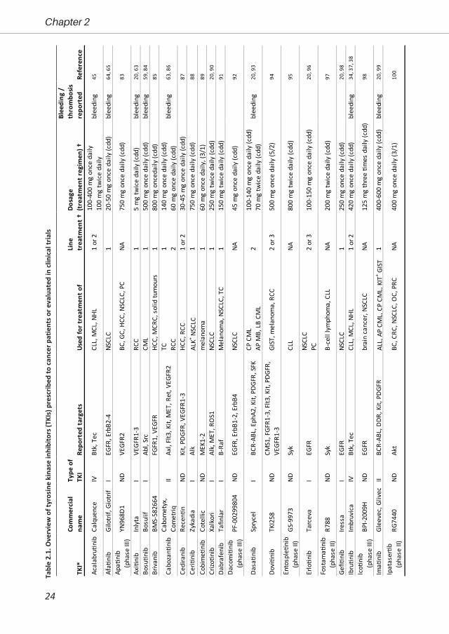
Although directed against specific targets, almost all TKIs affect multiple kinases. Table 2.1
provides an overview of the currently used TKIs that are prescribed for which cancer type, together
with their target kinases in tumour cells. The majority of the drugs are targeted at receptor tyrosine
kinases for growth factors (EGF, FGF, PDGF, VEGF) or for differentiation/proliferation factors (Flt,
Fms, Kit, Ret). Several of the drugs (in addition) target intracellular tyrosine kinases (ABL, B-Raf,
Btk, Itk, MEK, SFK, Syk). Although affinities towards individual kinases will differ, essentially all TKIs
have a more or less broad efficacy, which in some cases is intentional. For instance, both VEGF
and PDGF are known to be important in tumour angiogenesis. Sunitinib, blocking signalling via
both types of receptors, thus will have a broader action spectrum than vatalanib, targeting only
VEGF receptors18. In general, the use of TKIs for treatment of specific tumours depends on the
affinity for the targeted kinases19 and the pharmacokinetics at indicated dosages and treatment
regimens20 (Table 2.1). As far as understood, the ultimate specificity is not strongly correlated with
chemical structure or the subfamily of the targeted kinase18.
Receptor-linked tyrosine kinases are also abundantly expressed in non-tumour cells,
and treatment with TKIs will inevitably have off-target effects, thus interfering with the normal
function of non-diseased cells and tissues. This can explain side effects of treatment, varying from
general complications as fatigue, diarrhoea, and nausea, to specific complications like hand-foot
syndrome21. In spite of drug-to-drug variations, many TKIs can cause skin toxicity, even in >50%
of patients22. For antiangiogenic TKIs, ‘blood-related’ side effects have been reported, such as
hypertension, myelosuppression and bleeding23. Incidentally, also on-treatment cardiovascular
events have been reported, for instance linked to affected vascular integrity of the endothelium24.
TKI effects on the vessel wall comprise endothelial dysfunction and increased capillary leakage25,
which may contribute to an increased bleeding tendency. Paradoxically, interfering with the
integrity of endothelial cells can shift the haemostatic balance in favour of thrombosis, which
might explain why treatment with TKIs can associate with arterial thromboembolic events in
cancer patients25, 26.
Despite clinical benefit of the prescribed TKIs, some of the patients who initially respond
to the therapy experience a relapse, e.g., due to acquired drug resistance of the tumour14. In such
cases, the treatment schedule is adjusted, or a switch is made to an alternative TKI as a second-
or third-line treatment (Table 2.1). In specific cases, several TKIs can be combined for effective
blockade of one or two signalling pathways14, 21. Furthermore, chemotherapy or radiation therapy
can be complemented with TKI treatment13.
Protein tyrosine kinases implicated in platelet activationA global overview of platelet tyrosine kinases signalling underneath relevant receptors, as
well as the downstream platelet responses is given in Table 2.2.
24
Chapter 2
Tabl
e 2.
1. O
verv
iew
of t
yros
ine
kina
se in
hibi
tors
(TKI
s) p
resc
ribed
to c
ance
r pat
ient
s or e
valu
ated
in c
linic
al tr
ials
TKI*
Com
mer
cial
na
me
Type
of
TKI
Repo
rted
targ
ets
Used
for t
reat
men
t of
Line
tr
eatm
ent †
Dosa
ge
(tre
atm
ent r
egim
en) †
Blee
ding
/
thro
mbo
sis
repo
rted
Refe
renc
e10
0-40
0 m
g on
ce d
aily
100
mg
twic
e da
ilyAf
a�ni
bGi
lotri
f, Gi
otrif
IEG
FR, E
rbB2
-4NS
CLC
120
-50
mg
once
dai
ly (c
dd)
blee
ding
64, 6
5
Axi�
nib
Inly
taI
VEGF
R1-3
RCC
15
mg
twic
e da
ily (c
dd)
blee
ding
20, 6
3
Bosu�n
ibBo
sulif
IAb
l, Sr
cCM
L1
500
mg
once
dai
ly (c
dd)
blee
ding
59, 8
4
Briv
anib
BMS-
5826
64FG
FR1,
VEG
FR
HCC,
MCR
C, s
olid
tum
ours
180
0 m
g on
ceda
ily (c
dd)
85
TC1
140
mg
once
dai
ly (c
dd)
RCC
260
mg
once
dai
ly (c
dd)
Cedi
rani
bRe
cen�
nND
Kit,
PDGF
R, V
EGFR
1-3
HCC,
RCC
1 or
230
-45
mg
once
dai
ly (c
dd)
87
Ceri�
nib
Zyka
dia
IAl
kAL
K+ NSC
LC1
750
mg
once
dai
ly (c
dd)
88
Cobi
me�
nib
Cote
llic
NDM
EK1-
2m
elan
oma
160
mg
once
dai
ly, (
3/1)
89
Crizo
�nib
Xalk
ori
IAl
k, M
ET, R
OS1
NSCL
C1
250
mg
twic
e da
ily (c
dd)
20, 9
0
Dabr
afen
ibTa
finla
rI
B-Ra
fM
elan
oma,
NSC
LC, T
C1
150
mg
twic
e da
ily (c
dd)
91
CP C
ML
100-
140
mg
once
dai
ly (c
dd)
AP M
B, L
B CM
L70
mg
twic
e da
ily (c
dd)
Dovi�n
ibTK
I258
NDCM
S1, F
GFR1
-3, F
lt3, K
it, P
DGFR
, VE
GFR1
-3GI
ST, m
elan
oma,
RCC
2 or
350
0 m
g on
ce d
aily
(5/2
)94
Ento
sple�n
ib
(pha
se II
)GS
-997
3ND
Syk
CLL
NA80
0 m
g tw
ice
daily
(cdd
)95
NSCL
CPC
Fost
ama�
nib
(pha
se II
)R7
88ND
Syk
B-ce
ll ly
mph
oma,
CLL
NA20
0 m
g tw
ice
daily
(cdd
)97
Gefi�
nib
Iress
aI
EGFR
NSCL
C1
250
mg
once
dai
ly (c
dd)
20, 9
8
Ibru�n
ib
Imbr
uvic
aIV
Btk,
Tec
CLL,
MCL
, NHL
1 or
242
0 m
g on
ce d
aily
(cdd
)bl
eedi
ng34
, 37,
38
Ima�
nib
Glee
vec,
Gliv
ecII
BCR-
ABL,
DDR
, Kit,
PDG
FRAL
L, A
P CM
L, C
P CM
L, K
IT+ G
IST
140
0-60
0 m
g on
ce d
aily
(cdd
)bl
eedi
ng20
, 99
Acal
abru�n
ibCa
lque
nce
IVBt
k, T
ecCL
L, M
CL, N
HLbl
eedi
ng45
YN96
8D1
NDVE
GFR2
BC, G
C, H
CC, N
SCLC
, PC
NA75
0 m
g on
ce d
aily
(cdd
)83
1 or
2
Cabo
met
yx,
Com
etriq
IIAx
l, Fl
t3, K
it, M
ET, R
et, V
EGFR
2bl
eedi
ng63
, 86
92
Dasa�n
ibSp
ryce
lI
BCR-
ABL,
Eph
A2, K
it, P
DGFR
, SFK
2bl
eedi
ng20
, 93
PF-0
0299
804
NDEG
FR, E
rbB1
-2, E
rbB4
NSCL
CNA
45 m
g on
ce d
aily
(cdd
)
20, 9
6
BPI-2
009H
NDEG
FRbr
ain
canc
er, N
SCLC
NA12
5 m
g th
ree �m
es d
aily
(cdd
)98
Tarc
eva
IEG
FR2
or 3
100-
150
mg
once
dai
ly (c
dd)
100
RG74
40ND
Akt
BC, C
RC, N
SCLC
, OC,
PRC
NA40
0 m
g on
ce d
aily
(3/1
)
Apa�
nib
(pha
se II
I)
Ipat
aser�b
(pha
se II
)
Ico�
nib
(pha
se II
I)
Daco
mi�
nib
(pha
se II
I)
Erlo�n
ib
Cabo
zan�
nib

25
Off-target antiplatelet effects of tyrosine kinase inhibitors
2
ALK,
ana
plas
tic ly
mph
oma
kina
se; A
LL, a
cute
lym
phoc
ytic
leuk
aem
ia, A
P, a
cute
-pha
se; B
C, b
reas
t ca
ncer
; CLL
, chr
onic
lym
phoc
ytic
leuk
aem
ia; C
ML,
chr
onic
mye
loid
le
ukae
mia
; CP,
chr
onic
pha
se; C
RC, c
olor
ecta
l can
cer;
CSF-
R, c
olon
y-sti
mul
ating
fact
or r
ecep
tor;
CSF-
1R, c
olon
y-sti
mul
ating
fact
or 1
rec
epto
r; EG
FR, e
pide
rmal
gro
wth
fa
ctor
rec
epto
r; Er
bB2,
hum
an e
pith
elia
l gr
owth
fac
tor
2; F
GFR
, fib
robl
ast
grow
th f
acto
r re
cept
or;
GC,
gas
tric
can
cer;
GIS
T, g
astr
oint
estin
al s
trom
al t
umou
r; H
CC,
hepa
toce
llula
r car
cino
ma;
HR,
hor
mon
e re
cept
or; I
PF, i
diop
athi
c pu
lmon
ary
fibro
sis; L
B, ly
mph
oid
blas
t; M
B, m
yelo
id b
last
; MCL
, man
tle c
ell l
ymph
oma;
MCR
C, m
etas
tatic
co
lore
ctal
can
cer;
MTC
, med
ulla
ry th
yroi
d ca
ncer
; NA,
not
app
licab
le; N
D, n
ot d
eter
min
ed; N
HL,
Non
-Hod
gkin
lym
phom
a; N
SCLC
, non
-sm
all-c
ell l
ung
canc
er; O
C, o
varia
n ca
ncer
; PC,
pan
crea
tic ca
ncer
; PDG
FR, p
late
let-d
eriv
ed g
row
th fa
ctor
rece
ptor
; Ph+
, Phi
lade
lphi
a-po
sitive
; PN
ET, p
rimiti
ve n
euro
ecto
derm
al tu
mou
r; PR
C, p
rost
ate
canc
er;
RA, r
heum
atoi
d ar
thriti
s; R
CC, r
enal
cel
l car
cino
ma;
SFK
, Src
fam
ily k
inas
e; S
TS, s
oft ti
ssue
sarc
oma;
TC,
thyr
oid
canc
er; V
EGFR
, vas
cula
r end
othe
lial g
row
th fa
ctor
. *Sm
all-
mol
ecul
e in
hibi
tors
of k
inas
e en
zym
es a
re in
dica
ted
with
the
suffi
x ‘n
ib’. T
KIs a
re in
dica
ted
with
the
suffi
x ‘ti
nib’
. Ang
ioge
nesis
inhi
bito
rs a
re in
dica
ted
with
the
suffi
x ‘a
nib’
. Ra
pidl
y ac
cele
rate
d fib
rosa
rcom
a (R
AF) k
inas
e in
hibi
tors
are
indi
cate
d w
ith th
e su
ffix
‘rafe
nib’
. †3/
1, 3
wee
ks o
f dos
ing,
follo
wed
by
1 w
eek
of re
st; 4
/2, 4
wee
ks o
f dos
ing,
fo
llow
ed b
y 2
wee
ks o
f res
t; 5/
2, 5
day
s of
dos
ing,
follo
wed
by
2 da
ys o
f res
t; cd
d, c
ontin
uous
dai
ly d
osin
g.
TKI*
Com
mer
cial
na
me
Type
of
TKI
Repo
rted
targ
ets
Used
for t
reat
men
t of
Line
tr
eatm
ent †
Dosa
ge
(tre
atm
ent r
egim
en) †
Blee
ding
/
thro
mbo
sis
repo
rted
Refe
renc
e
ErbB
2+ BC
ErbB
2+ and
HR- B
CEr
bB2+ a
nd H
R+ BC
Lenv
a�ni
bLe
nvim
aII
FGFR
, Kit,
PDG
FRα,
Ret
, VEG
FRRC
C, T
C24
mg
once
dai
ly (c
dd)
63, 1
02
Nilo�n
ibTa
sign
aII
BCR-
ABL,
CSF
-1R,
DDR
, Kit,
PDG
FRCP
CM
L, G
IST
130
0-40
0 m
g tw
ice
daily
(cdd
)th
rom
bosi
s20
, 99
1 2O
sim
er�n
ibTa
gris
so, T
agrix
NDEG
FRNS
CLC
280
mg
once
dai
ly (c
dd)
104
Pazo
pani
bVo
trien
tI
FGFR
, FM
S, It
k, K
it, L
ek, P
DGFR
, VE
GFR
NSCL
C, O
C, R
CC, S
TS, T
C1
800
mg
once
dai
ly (c
dd)
blee
ding
20, 6
3
Pona
�nib
Iclu
sig
IIBC
R-AB
L, F
GFR,
KIT
, PDG
FR, R
et,
Src,
VEG
FRPh
+ ALL
, CM
L1
or 2
30 m
g on
ce d
aily
(cdd
)bl
eedi
ng10
5
Rego
rafin
ibS�
varg
aII
B-Ra
f, FG
FR, K
it, P
DGFR
, Ret
, Tie
2,
VEGF
R1-3
adva
nced
GIS
T, M
CRC
316
0 m
g on
ce d
aily
(3/1
)10
6
Selu
me�
nib
AZD6
244
IIIM
EK1-
2KR
ASm
utat
ed N
SCLC
2 or
375
mg
twic
e da
ily (c
dd)
107
Sora
feni
bNe
xava
rI
VEGF
RHC
C, R
CC
1-3
400
mg
twic
e da
ily (c
dd)
blee
ding
20, 6
0, 6
3, 8
5
Suni�n
ibSu
tent
ICS
F-R,
Flt3
, Kit,
PDG
FR, V
EGFR
RCC,
GIS
T, P
NET
1 or
237
.5-5
0 m
g on
ce d
aily
(4/2
)bl
eedi
ng
20, 6
0, 6
1, 6
3
Tram
e�ni
bM
ekin
ist
IIIM
EK1-
2BR
AF V
600+ m
elan
oma
12
mg
once
dai
ly (c
dd)
blee
ding
10
8
Vand
etan
ibCa
prel
saIV
EGFR
, Ret
, VEG
FRM
TC, N
SCLC
1 or
230
0 m
g on
ce d
aily
(cdd
)20
, 109
Vem
uraf
enib
Zelb
oraf
IB-
Raf
BRAF
V60
0+ mel
anom
a2
or 3
960
mg
twic
e da
ily (c
dd)
20, 1
11
Lapa
�nib
Tyke
rbII
EGFR
, Erb
B1-2
1 or
210
00-1
500
mg
once
dai
ly (c
dd)
20, 1
01
blee
ding
63, 1
03
PTK7
87/
Z
K-22
2584
IIIKi
t, PD
GFRβ
, VEG
FRM
CRC
112
50 m
g on
ce d
aily
(cdd
)11
0
Ofe
v, V
arga
tef
IIFG
FR, F
MS,
PDG
FR, V
EGFR
IPF,
adv
ance
d NS
CLC
150
mg
twic
e da
ily (c
dd)
Vata
lani
b
(pha
se II
I)
Nint
edan
ib

26
Chapter 2
CLEC-2, C-type lectin-like receptor 2; GP, glycoprotein; SFK, Src family kinase; SLP76, Src homology 2 domain-containing leukocyte phosphoprotein
SFK Other GPVI Src, Fyn, Lyn, Fgr Btk, Syk, Tec Ca2+ mobilisa�on, integrin ac�va�on,
degranula�onCLEC-2 Src, Fyn, Lyn Btk, Syk, Tec Ca2+ mobilisa�on, integrin ac�va�on,
degranula�onGPIb-IX-V Src, Fyn, Lyn Btk Integrin ac�va�onIntegrin αIIbβ3 Src, Fyn, Lyn FAK, Syk, SLP76, Pyk2 Spreading, outside-in signaling,
clot retrac�on
ReceptorSignalling tyrosine kinases
Platelet response
GPVI signalling. Collagen-induced platelet activation is established via the tyrosine
kinase-linked receptor GPVI, a member of the immunoglobulin superfamily. GPVI is linked to
the Fc receptor γ (FcRγ) chain, which contains two immunoreceptor tyrosine-based activation
motives (ITAM) requiring phosphorylation to mediate platelet activation27. Ligand binding and
dimerization of the GPVI-FcRγ complex leads to activation of the SFK isoforms Src, Fyn and Lyn,
which in turn phosphorylate the FcRγ ITAM tyrosine residues to recruit and phosphorylate Syk1,
28. The tyrosine kinases SFK and Syk furthermore phosphorylate downstream targets, including
the transmembrane adapter linker for activated T cells (LAT) and the Src homology 2 domain-
containing leukocyte phosphoprotein (SLP76). The consequence is the formation of a large
signalling complex, including LAT, SLP76, Btk, isoforms of phosphoinositide-3 kinase and Tec
family kinases29. A key downstream event is the phosphorylation and activation of the second
messenger-generating phospholipase (PLC)γ2, resulting in Ca2+ mobilisation and protein kinase C (PKC) activity. Further responses are integrin activation, thromboxane A2 release, granule
secretion and phosphatidylserine exposure.
CLEC-2 signalling. A similar powerful activation pathway of platelets is induced via CLEC-2, a
C-type lectin receptor also acting through tyrosine phosphorylation. Known ligands of this receptor
are podoplanin (expressed in tumour tissue among others) and the snake venom rhodocytin.
Signalling occurs through a so-called hemITAM motif30. The clustering of CLEC-2 induces more or
less similar events as described for GPVI. Starting with the tyrosine phosphorylation of SFK and
Syk, a signalling complex is formed including Tec family tyrosine kinases, with as result PLCγ2 and
PI3K activation. Consequences are, again, integrin activation, granule release, and thromboxane A2 production30.
GPIb-IX-V signalling. Interaction of vWF with the GPIb-IX-V receptor is one of the first
steps in platelet tethering and adhesion under shear flow1. This interaction causes only weak
signalling, e.g., leading to restructuring of the actin cytoskeleton, with under certain conditions phosphorylation of SFK (Src, Fyn, Lyn) and activation of PI3K isoforms1. Integrin αIIbβ3 activation
and platelet spreading are a result of this.
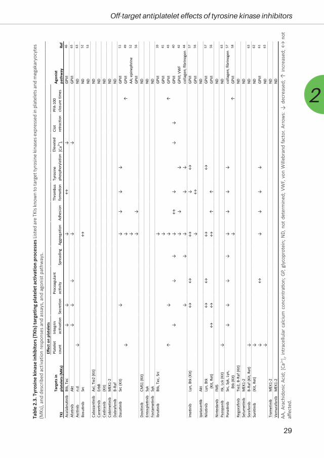
Table 2.2. Protein tyrosine kinases implicated in platelet activation responses: global overview of tyrosine kinases implicated in signaling via key platelet receptors, as well as downstream platelet responses. Summarized from 1, 16, 18, 30
27
Off-target antiplatelet effects of tyrosine kinase inhibitors
2
Integrin-dependent signalling. Platelet integrins, in particular αIIbβ3, α2β1 and αƲβ3, regulate
adhesion, aggregation and thrombus formation28. Especially regarding integrin αIIbβ3 (ligands:
fibrinogen, vWF and other matrix proteins) much research has been performed to the outside-in
signalling events triggered by the occupied, activated conformation. Several tyrosine kinases are
implicated in this signalling pathway, including FAK, Pyk2, Src, SLP76 and Syk29, 31.
Effects of TKIs on platelet functionTyrosine kinases are targeted by TKIs as treatment for cancer, where their most important
effect is the prolongation of progression free survival. Given the presence of on-target or related
off-target tyrosine kinases in megakaryocytes and platelets, it is to be expected that several TKIs
interfere with platelet formation and/or platelet activation processes.
ABL1
ABL2
AXL
BRAF
Btk
CSF
EphA
4Ep
hB1
FGR
FYN
Itk Lck
LYN
MEK
1M
EK2
SRC
Syk
Tec
TIE1
TIE2
YES
AfatinibAxitinibBosutinibBrivanibCediranibCrizotinibDasatinibDovitinibErlotinibFostamatinibGefitinibIbrutinibImatinibLapatinibNilotinibNintedanibPazopanibSelumetinibSorafenibSunitinibVandetanib
Asso
ciat
ed
with
ble
edin
g
Vatalanib
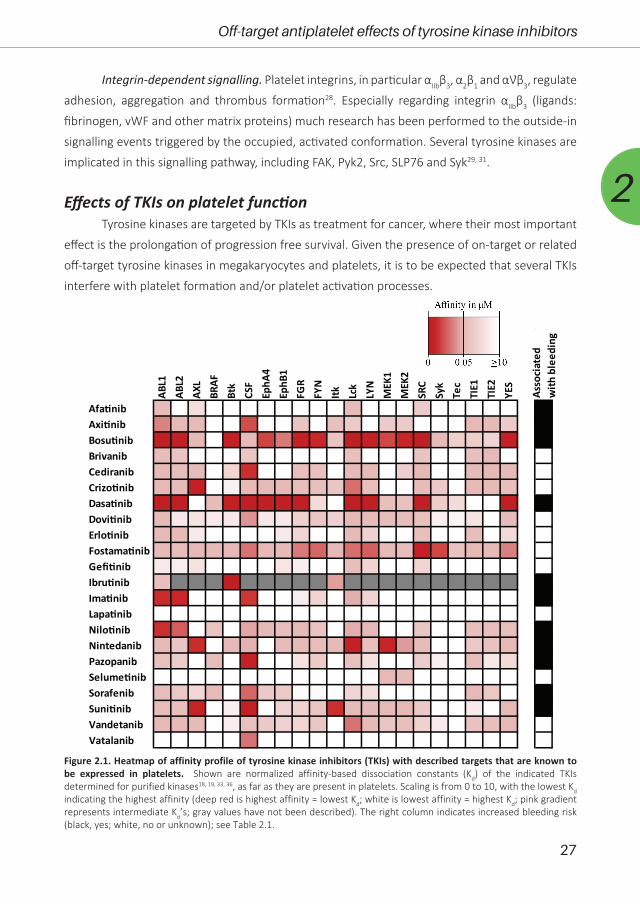
Figure 2.1. Heatmap of affinity profile of tyrosine kinase inhibitors (TKIs) with described targets that are known to be expressed in platelets. Shown are normalized affinity-based dissociation constants (Kd) of the indicated TKIs determined for purified kinases18, 19, 33, 36, as far as they are present in platelets. Scaling is from 0 to 10, with the lowest Kd indicating the highest affinity (deep red is highest affinity = lowest Kd; white is lowest affinity = highest Kd; pink gradient represents intermediate Kd’s; gray values have not been described). The right column indicates increased bleeding risk (black, yes; white, no or unknown); see Table 2.1.

28
Chapter 2
To provide an overview, we evaluated the inhibition profiles of TKIs (22 in total) against
target protein tyrosine kinases that are known to be expressed in platelets32. Markedly, the
majority of these TKIs demonstrated relatively low affinities for kinases with crucial roles in
platelet activation processes (SFK, Syk, Btk, MEK and Eph isoforms), with as exceptions bosutinib,
dasatinib, fostamatinib, nintedanib and sunitinib (Figure 2.1). For such an in vitro affinity-based
analysis18, 19, 33, it should be realised that the presence of blood plasma and blood cells can
profoundly change the bioavailability of a TKI, apart from its metabolic and pharmacokinetic
profile. Markedly, bleeding symptoms have been reported for 11 of these TKIs. For individual TKIs,
based on their known targets, we summarised in Table 2.3 which of these targets are expressed
in platelets. Furthermore, we made an inventory of the published effects of these TKIs on platelet
responses.
Ibrutinib is a covalently acting TKI that is targeted at the family members Btk and Tec,
which probably explains the strong effects observed on platelets34. It is frequently used for the
treatment of mantle cell lymphoma (MCL) and chronic lymphocytic leukaemia (CLL). Emerging
data suggest that ibrutinib could also be used to treat solid tumours35 and has been shown
to inhibit several other kinases such as Itk, JAK3, Hck, Blk, EGFR, ErbB2 and ErbB436. Ibrutinib
treatment is associated with a risk of bleeding34, 37, 38. Being the most investigated TKI with regard
to platelets, ibrutinib has been demonstrated to potently inhibit collagen-induced responses of
platelets from patients on-treatment39-44. Efficient inhibition by ibrutinib of the GPVI pathway
(in response to collagen or collagen-related peptide) has been demonstrated to proceed via
reduced PLCγ2 phosphorylation42. In treated patients, the suppression of collagen-induced
platelet aggregation correlated with the occurrence of bleeding events42. Subsequent studies
showed that ibrutinib suppressed multiple (mostly) GPVI-dependent platelet responses, including
adhesion, spreading, calcium fluxes, secretion, phosphatidylserine exposure and clot retraction40,
43. In addition, evidence was obtained for reduced αIIbβ3-dependent outside-in signalling, linked
to thrombus instability in vitro40. Several studies confirmed that Btk can act as a central target of
ibrutinib in GPVI-stimulated platelets, although downstream tyrosine kinases may be affected as
well40, 42, 44. In vitro, ibrutinib was found to fully inhibit the tyrosine phosphorylation of Src and
PLCγ242. Other affected events were the phosphorylation of Fyn, Lyn, Btk, Tec and Syk40, 44. Taken
together, this pointed to a potent suppression of ibrutinib of the GPVI signalosome with as major
target the Tec family kinases.
The second-generation drug, acalabrutinib, is a more selective, irreversible Btk inhibitor.
In a clinical trial with patients with relapsed CLL, acalabrutinib showed promising safety and
efficacy profiles, although some minor bleedings were still reported45. A recent study comparing
treatment with ibrutinib or acalabrutinib indicated that both compounds impaired the platelet
aggregation responses after collagen receptor stimulation46. Both drugs inhibited platelet Btk
and Tec at physiological concentrations, while only ibrutinib inhibited SFK isoforms. This provided
29
Off-target antiplatelet effects of tyrosine kinase inhibitors
2
Tabl
e 2.
3. T
yros
ine
kina
se in
hibi
tors
(TKI
s) ta
rget
ing
plat
elet
act
ivat
ion
proc
esse
s List
ed a
re T
KIs k
now
n to
targ
et ty
rosin
e ki
nase
s exp
ress
ed in
pla
tele
ts a
nd m
egak
aryo
cyte
s (M
Ks),
and
desc
ribed
act
ivat
ion
resp
onse
s an
d as
says
, and
ago
nist
pat
hway
s.
AA, A
rach
idon
ic A
cid;
[Ca2+
] i, int
race
llula
r ca
lciu
m c
once
ntra
tion;
GP,
gly
copr
otei
n; N
D, n
ot d
eter
min
ed; V
WF,
von
Will
ebra
nd fa
ctor
. Arr
ows:
↓ d
ecre
ased
; ↑ in
crea
sed;
↔ n
ot
affe
cted
.
Plat
elet
co
unt
Inte
grin
ac�v
a�on
Secr
e�on
Proc
oagu
lant
ac�v
itySp
read
ing
Aggr
ega�
onAd
hesi
onTh
rom
bus
form
a�on
Tyro
sine
ph
osph
oryl
a�on
Elev
ated
[C
a2+] i
Clot
re
trac�
onPF
A-10
0 cl
osur
e �m
es
Acal
abru�n
ibBt
k, T
ec↓
↓↓
↔↓
GPVI
46
Afa�
nib
Akt
↓↓
↓↓
↓GP
VI65
Axi�
nib
n.d.
↓ND
63
↔ND
52
ND53
Cabo
zan�
nib
Axl,
Tie2
(Kit)
NDCa
ner�
nib
ErbB
NDCe
dira
nib
(Kit)
NDCo
bim
e�ni
bM
EK1-
2ND
Dabr
afen
ibB-
Raf
ND↓
↓↓
↓↓
GPVI
51
↓↓
↑GP
VI
49
↓AA
; epi
neph
rine
52
↓↓
GPVI
56
Dovi�n
ibCM
S1 (K
it)ND
Ento
sple�n
ibSy
kND
Fost
ama�
nib
Syk
ND↓
GPVI
39
↓GP
VI
41
↑↓
↓↑
GPVI
43
↓↓
↓↓
↔↓
↓↓
GPVI
40
↓↓
↓GP
VI; V
WF
42
↓↓
↓↓
↓co
llage
n; fi
brin
ogen
44
↔↔
↔↓
↔GP
VI
57
↓↔
GPVI
56
Ipat
aser�b
Akt
ND↔
↔↔
↔GP
VI
57
↔↔
↔↑
↑GP
VI
56
Nint
edan
ibFM
SND
Pazo
pani
bItk
, Lck
(Kit)
↓ND
63
↓↓
↓↓
↓↓
↓↓
colla
gen;
fibr
inog
en57
↓↑
GPVI
58
Rego
rafin
ibTi
e2, B
-Raf
(Kit)
NDSe
lum
e�ni
bM
EK1-
2ND
Sora
feni
bB-
Raf (
Kit,
Ret)
↓ND
63
↓ND
62
↓↔
↓↓
↓↓
GPVI
61
↓ND
63
Tram
e�ni
bM
EK1-
2ND
Vem
uraf
enib
MEK
1-2
NDAgon
ist
path
way
Ref
Dasa�n
ibSr
c (K
it)
Ibru�n
ibBt
k, T
ec, S
rc
Bosu�n
ibSr
c
TKI
Targ
ets i
n pl
atel
ets (
MKs
)
Effec
t on
plat
elet
s
Suni�n
ib(K
it, R
et)
Ima�
nib
Lyn,
Btk
(Kit)
Nilo�n
ibLy
n, B
tk
(Ki
t, Re
t)
Pona
�nib
Src,
Syk
, Lyn
, B
tk (K
it)

30
Chapter 2
an explanation why only ibrutinib caused dysfunctional thrombus formation, in contrast to
acalabrutinib46.
Imatinib, targeting the oncogenic kinase BCR-ABL, was the first tyrosine kinase inhibitor
developed for chronic myeloid leukaemia (CML). Imatinib is known to block Kit and PDGF receptors,
as well as Erk and Akt isoforms47. This TKI has been reported to evoke thrombocytopenia in 18%
of the treated patients, with haemorrhages being reported as well48. Despite a high efficacy of
imatinib, 15% of patients appear to develop clinical resistance and relapse after initial response
to therapy.
Dasatinib and nilotinib have been developed as second generation BCR-ABL inhibitors for
the treatment of CML. Two case reports showed that dasatinib could induce a bleeding diathesis
correlated to platelet dysfunction, which was reversed upon cessation of the dasatinib intake49,
50. Studies with blood from patients or healthy subjects showed an impairment of platelet
aggregation by dasatinib given in vivo or in vitro51, 52. In particular, platelet aggregation in response
to collagen, arachidonic acid and epinephrine was affected52, along with collagen-induced
thrombus formation51, 53, thus suggesting interference with GPVI signalling and other pathways.
The mechanism of dasatinib-induced inhibition in platelets was elucidated from changes in
the protein phosphorylation profile after collagen stimulation. Herein, dasatinib appeared to
interfere with SFK, PLC and PI3K activities51. Thrombocytopenia is a relatively common side
effect of dasatinib treatment54. This is proposed to be due to reduced megakaryocytopoiesis,
thus resulting in lower platelet production55. The combined quantitative (via megakaryocytes)
and qualitative (via signalling) platelet dysfunction most likely is the reason of the relatively high
bleeding incidence in dasatinib-treated patients. Compared to dasatinib, imatinib induces less
antiplatelet activity and causes less bleeding at clinically achievable doses, likely due to the fact
that it is less inhibitory for SFK isoforms. On the other hand, nilotinib is reported to potentiate
thrombus formation and increase the risk of thrombosis56.
Ponatinib and bosutinib are third generation BRC-ABL inhibitors for the treatment of CML,
which are applicable when patients develop resistance to imatinib. The use of ponatinib has been
associated with bleeding diatheses, along with changes in platelet count and function, which thus
may explain the impaired haemostasis57, 58. One study describes inhibiting effects of ponatinib,
imatinib and nilotinib on platelet activation, secretion, and aggregation in response to collagen57.
Herein, ponatinib appeared to be the most potent platelet inhibitor. Ponatinib increased PFA-100
closure times, likely by suppressing ITAM signalling via several tyrosine kinases, including Src,
Lyn, Syk, and Btk57, 58. In contrast, imatinib and nilotinib did not affect platelet granule secretion,
procoagulant activity or aggregation, and only moderately reduced the tyrosine phosphorylation
of Lyn and Btk, as compared to ponatinib57. Yet, whole blood thrombus formation was reduced
by imatinib and ponatinib to a similar extent. For bosutinib (also targeting Src), bleeding has
been reported as well59, although in patients it did not affect platelet aggregation in response to
31
Off-target antiplatelet effects of tyrosine kinase inhibitors
2
several agonists52. In microfluidic whole blood testing of platelet thrombus formation, bosutinib
was found to be less potent than dasatinib53. Overall, it appears that BCR-ABL inhibitors used for
leukaemia treatment have an inhibiting effect on platelet functions, with the possible exception
of nilotinib.
Sunitinib, sorafenib and pazopanib are multi-target TKIs used for the treatment of renal
cell carcinoma, aiming to inhibit tumour angiogenesis via interference with the receptors for VEGF
and PDGF. Such multi-target TKIs may potentially also inhibit tyrosine kinases present in platelets.
For sunitinib and sorafenib, a meta-analysis of 23 trials (>6,500 patients) showed an increased
incidence of bleeding events with 16.7% on all grades of bleeding, and a 2.4% probability of
high-grade bleeding events60. For sunitinib, both quantitative and qualitative effects on platelets
are reported. The compound can be taken up by platelets from healthy donors and renal cell
carcinoma patients, and hence reduce the platelet functionality in a way depending on protein
tyrosine phosphorylation61. In patients on-treatment, this is accompanied by a decrease in platelet
count61, 62. For sorafenib, similarly to other TKIs used for treatment of renal cell carcinoma such as
pazopanib, an increased risk of bleeding has also been observed63, but effects on platelet function
have not been investigated.
Afatinib is used for the first-line treatment of particularly patients with non-small cell lung
cancers64. It acts as an irreversible inhibitor of EGF and HER receptors. An increased bleeding risk
of patients receiving afatinib has been linked to impaired platelet function and apoptosis65. In
particular, afatinib impaired GPVI-induced platelet responses, including calcium fluxes, integrin
activation, secretion, procoagulant activity and platelet aggregation (Table 2.3). On the other
hand, thrombin-induced platelet responses were less strongly inhibited. Further investigation
showed that the collagen-induced phosphorylation of Akt was decreased by afatinib65.
Cabozantinib, cediranib, dovitinib, lenvatinib, regorafinib and other TKIs co-target the
receptor kinase Kit, which acts as a receptor for stem cell factor in megakaryocytopoiesis,
regulating platelet production. The Kit receptor is known to signal via SFK, PI3K, Ras/MEK and
JAK/STAT pathways66. In an analogous way, the thrombopoietin receptor Mpl signals via JAK2
and Tyk2 as immediate effector kinases, resulting in the activation of STAT isoforms67. For these
signalling pathways, key tyrosine kinases are also present in platelets and/or megakaryocytes
(Table 2.2 and 2.3), which raises the possibility that Kit-targeted TKIs can also modulate platelet
function as well as platelet count. So far, the signalling effects of these TKIs on platelets have not
been investigated in detail. Cabozantinib intake has been associated with a higher bleeding risk63.
Related drugs are ruxolitinib, pacritinib and lesaurtinib, which also target JAK1/2 and are usually
prescribed for myeloproliferative disorders, such as essential thrombocythemia, characterised by
an elevated platelet count (thrombocytosis). The latter compounds appear to lower (normalise)
the concentration of circulating platelets68, most likely via inhibition of the thrombopoietin
pathway in megakaryocytes. However, effects on platelet function are still unclear. It is known that
32
Chapter 2
thrombopoietin potentiates agonist-induced platelet activation via JAK2 and Tyk2 signalling69.
This indicates that this pathway is not strong enough to induce platelet activation on its own.
Inhibition of JAK2 in platelets has been reported to only attenuate collagen-induced responses70.
For multiple prescribed TKIs with a broad target range, bleeding symptoms have not been
reported (Table 2.1). Examples of these are fostamatinib, dovitinib and vantedanib, which based
on their affinity profiles should affect SFK and/or MEK isoforms in platelets (Figure 2.1). In these
cases, additional research is needed to verify effects on platelet count and activation, and to
determine if there is a bleeding risk. Summarising the current knowledge of TKI effects on platelet
activation responses, it appears that the majority of multi-target TKIs in particular suppresses
GPVI-induced signalling pathways, but that they can also target kinase events downstream of
other platelet receptors. As the inhibition of GPVI alone has been shown to prevent occlusive
thrombus formation without causing bleeding71, it is most likely that the targeting of pathways
downstream of additional receptors is responsible for an increased bleeding risk with these
compounds.
Clinical implications of platelet inhibition with TKIsPlatelets are known to interact with cancer cells in the blood. Tumours exposed to the
blood stream can activate platelets, while platelets can stimulate tumour angiogenesis, growth,
and metastasis. The question has been raised whether and how interfering in the platelet-cancer
loop via antiplatelet treatment may also influence cancer progression. Several studies have
reported that aspirin or P2Y12 receptor inhibitors suppress tumour angiogenesis in vitro72 and
tumour growth in vivo73. A low daily aspirin intake has been associated with a reduced risk for the
development of cancer as well as prevention of metastases74. The mechanism is not completely
resolved, but it has been shown that inhibition of platelets by aspirin decreased their ability
to stimulate cancer cell proliferation through modulation of the Myc oncoprotein75. For other
antiplatelet drugs, clinical evidence of a beneficial effect on cancer is still lacking.
Of growing interest is the recent finding that platelets are capable to sequester bioactive
compounds from the plasma, including growth factors12, 76, 77 and RNA species7. In addition,
platelets accumulate the anticancer drugs bevacizumab78 and sunitinib (in granules)61. Other
TKIs can also be taken up by platelets, which usually is a requirement for interaction with the
intracellular kinases. Whether these TKIs can also secondarily be released by platelets and,
perhaps, then contribute to tumour inhibition is matter of speculation. Another consideration is
that uptake of TKIs by platelets (e.g., in patients with thrombocytosis) can reduce the availability of
the compounds for tumour inhibition. On the other hand, it is likely that a decreased aggregation
tendency of platelets (as induced by TKIs) also lowers tumour-induced platelet activation, and
thereby reduces metastasis of tumour cells. However, the evidence for such a mechanism is at
best indirect, and further investigation here is required. Taken together, it appears that platelets
33
Off-target antiplatelet effects of tyrosine kinase inhibitors
2
can interfere with drug effects in different ways and by different mechanisms, which should be
taken into account upon therapeutic drug monitoring.
Application of the TKIs axitinib, dasatinib, pazopanib, sorafenib and sunitinib ‒ all of
which have indications may cause bleeding ‒ can lead to a reduction of the platelet concentration
in blood (Table 2.3). In cancer patients, thrombocytosis has been associated with a worse
overall prognosis, when compared to patients with a normal platelet count8, 9. In addition, in
glioblastoma patients, who underwent concomitant radiotherapy and chemotherapy, a decrease
in platelet count correlated with longer overall survival79. In renal cell carcinoma patients with
metastasis, a reduction in platelet count after start of sunitinib treatment was associated with
a better treatment outcome62. Although more research is needed, these studies suggest that
platelet count can be used as one of the parameters to monitor anticancer treatment efficacy.
Besides platelet count, also platelet function can be affected by many TKIs, as indicated
in Table 2.3. This suggests that combined platelet count and function tests could be used in clinical
practice to assess the risk of bleeding upon TKI treatment. For the availability of such (point-of-
care) tests, we like to refer to the expert literature80. In the future it may be possible to use flow-
based assays for this purpose. Our laboratory recently found that this method detects additive
effects of low platelet count and impaired platelet functionality in patients with a bleeding
phenotype81.
It is known that cancer patients are at increased risk of thrombosis, also indicated as
Trousseau’s syndrome82. As a consequence, many cancer patients have a history of cardiovascular
disease and are treated with antiplatelet or anticoagulant drugs. When additional treatment with
a TKI is started, the clinical problem of an enhanced bleeding risk may arise. Another treatment
issue is that some oral anticoagulants and TKIs (ibrutinib for instance) have a common metabolic
pathway (CYP3A4), which can affect the pharmacokinetics of either drug. These interactions
between TKIs and antithrombotic treatments need to be further investigated.
ConclusionsSeveral of the TKIs used for the treatment of cancer can increase the risk of bleeding.
For some but not all of these, the bleeding tendency is linked to a lowering of platelet count
and/or an impairment of platelet function. Based on their binding profiles, some other TKIs are
predicated to have an antiplatelet effect, although no bleeding side effects have been reported
so far. For a third group of TKIs, no published data on platelets are available. Given the suspected
role of platelets in tumour progression, platelet inhibition may be a clinically relevant side-effect
of several TKIs. Together, the available knowledge asks for awareness of the potential antiplatelet
effects of TKIs, which are likely to be further enhanced in combination with antithrombotic drugs.

34
Chapter 2
AcknowledgementsThis study was supported by the Maastricht Thrombosis Expertise Centre as part of the
Heart and Vascular Centre (HVC) of the MUMC+.
Author Contributions StatementBMET reviewed the literature, compiled tables and Fig. 2.1, and wrote the manuscript.
JWMH was responsible for the outline, and wrote and revised the manuscript. MJEK was
responsible for the outline, reviewed the literature, and wrote and revised the manuscript.
Conflict of Interest StatementMJEK and JWMH report receiving grants from Pfizer during the conduct of the study. BMET
states that she has no conflict of interest.
References1. Versteeg HH, Heemskerk JW, Levi M, Reitsma PH. New fundamentals in hemostasis. Physiol Rev.
2013; 93: 327-58.2. Morowski M, Vogtle T, Kraft P, Kleinschnitz C, Stoll G, Nieswandt B. Only severe thrombocytopenia
results in bleeding and defective thrombus formation in mice. Blood. 2013; 121: 4938-47.3. Baaten CCFMJ, Ten Cate H, van der Meijden PEJ, Heemskerk JWM. Platelet populations and priming
in hematological diseases. Blood Rev. 2017; 31: 389-99.4. Franco AT, Corken A, Ware J. Platelets at the interface of thrombosis, inflammation, and cancer.
Blood. 2015; 126: 582-8.5. Cooke NM, Egan K, McFadden S, et al. Increased platelet reactivity in patients with late-stage
metastatic cancer. Cancer Med. 2013; 2: 564-70.6. Sabrkhany S, Kuijpers MJE, van Kuijk SMJ, et al. A combination of platelet features allows detection of
early-stage cancer. Eur J Cancer. 2017; 80: 5-13.7. Best MG, Sol N, Kooi I, et al. RNA-seq of tumor-educated platelets enables blood-based pan-cancer,
multiclass, and molecular pathway cancer diagnostics. Cancer Cell. 2015; 28: 666-76.8. Menter DG, Tucker SC, Kopetz S, Sood AK, Crissman JD, Honn KV. Platelets and cancer: a casual or
causal relationship: revisited. Cancer Metastasis Rev. 2014; 33: 231-69.9. Lin RJ, Afshar-Kharghan V, Schafer AI. Paraneoplastic thrombocytosis: the secrets of tumor self-
promotion. Blood. 2014; 124: 184-7.10. Goubran HA, Burnouf T, Radosevic M, El-Ekiaby M. The platelet-cancer loop. Eur J Intern Med. 2013;
24: 393-400.11. Carmeliet P. Mechanisms of angiogenesis and arteriogenesis. Nat Med. 2000; 6: 389-95.12. Klement GL, Yip TT, Cassiola F, et al. Platelets actively sequester angiogenesis regulators. Blood. 2009;
113: 2835-42.13. Hanahan D, Weinberg RA. Hallmarks of cancer: the next generation. Cell. 2011; 144: 646-74.14. Gross S, Rahal R, Stransky N, Lengauer C, Hoeflich KP. Targeting cancer with kinase inhibitors. J Clin
Invest. 2015; 125: 1780-9.15. Swieringa F, Kuijpers MJ, Heemskerk JW, van der Meijden PE. Targeting platelet receptor function in
thrombus formation: the risk of bleeding. Blood Rev. 2014; 28: 9-21.16. Levade M, Severin S, Gratacap MP, Ysebaert L, Payrastre B. Targeting kinases in cancer therapies:
adverse effects on blood platelets. Curr Pharm Des. 2016; 22: 2315-22.17. Wu P, Nielsen TE, Clausen MH. FDA-approved small-molecule kinase inhibitors. Trends Pharmacol
Sci. 2015; 36: 422-39.18. Fabian MA, Biggs WH, 3rd, Treiber DK, et al. A small molecule-kinase interaction map for clinical
35
Off-target antiplatelet effects of tyrosine kinase inhibitors
2
kinase inhibitors. Nat Biotechnol. 2005; 23: 329-36.19. Karaman MW, Herrgard S, Treiber DK, et al. A quantitative analysis of kinase inhibitor selectivity. Nat
Biotechnol. 2008; 26: 127-32.20. Yu H, Steeghs N, Nijenhuis CM, Schellens JH, Beijnen JH, Huitema AD. Practical guidelines for
therapeutic drug monitoring of anticancer tyrosine kinase inhibitors: focus on the pharmacokinetic targets. Clin Pharmacokinet. 2014; 53: 305-25.
21. Gotink KJ, Verheul HM. Antiangiogenic tyrosine kinase inhibitors: what is their mechanism of action? Angiogenesis. 2010; 13: 1-14.
22. Hartmann JT, Haap M, Kopp HG, Lipp HP. Tyrosine kinase inhibitors - a review on pharmacology, metabolism and side effects. Curr Drug Metab. 2009; 10: 470-81.
23. Eskens FA, Verweij J. The clinical toxicity profile of vascular endothelial growth factor (VEGF) and vascular endothelial growth factor receptor (VEGFR) targeting angiogenesis inhibitors; a review. Eur J Cancer. 2006; 42: 3127-39.
24. Moslehi JJ, Deininger M. Tyrosine kinase inhibitor–associated cardiovascular toxicity in chronic myeloid leukemia. J Clin Oncol. 2015; 33: 4210-8.
25. Bair SM, Choueiri TK, Moslehi J. Cardiovascular complications associated with novel angiogenesis inhibitors: emerging evidence and evolving perspectives. Trends in cardiovascular medicine. 2013; 23: 104-13.
26. Choueiri TK, Schutz FA, Je Y, Rosenberg JE, Bellmunt J. Risk of arterial thromboembolic events with sunitinib and sorafenib: a systematic review and meta-analysis of clinical trials. J Clin Oncol. 2010; 28: 2280-5.
27. Nieswandt B, Watson SP. Platelet-collagen interaction: is GPVI the central receptor? Blood. 2003; 102: 449-61.
28. Senis YA, Mazharian A, Mori J. Src family kinases: at the forefront of platelet activation. Blood. 2014; 124: 2013-24.
29. Watson SP, Auger JM, McCarty OJ, Pearce AC. GPVI and integrin alphaIIb beta3 signaling in platelets. J Thromb Haemost. 2005; 3: 1752-62.
30. Navarro-Núñez L, Langan SA, Nash GB, Watson SP. The physiological and pathophysiological roles of platelet CLEC-2. Thromb Haemost. 2013; 109: 991-8.
31. Shattil SJ, Newman PJ. Integrins: dynamic scaffolds for adhesion and signaling in platelets. Blood. 2004; 104: 1606-15.
32. Burkhart JM, Vaudel M, Gambaryan S, et al. The first comprehensive and quantitative analysis of human platelet protein composition allows the comparative analysis of structural and functional pathways. Blood. 2012; 120: e73-82.
33. Davis MI, Hunt JP, Herrgard S, et al. Comprehensive analysis of kinase inhibitor selectivity. Nat Biotechnol. 2011; 29: 1046-51.
34. Shatzel JJ, Olson SR, Tao DL, McCarty OJT, Danilov AV, DeLoughery TG. Ibrutinib-associated bleeding: pathogenesis, management and risk reduction strategies. J Thromb Haemost. 2017; 15: 835-47.
35. Molina-Cerrillo J, Alonso-Gordoa T, Gajate P, Grande E. Bruton’s tyrosine kinase (BTK) as a promising target in solid tumors. Cancer Treat Rev. 2017; 58: 41-50.
36. Rauf F, Festa F, Park JG, et al. Ibrutinib inhibition of ERBB4 reduces cell growth in a WNT5A-dependent manner. Oncogene. 2018; 37: 2237-50.
37. Byrd JC, Furman RR, Coutre SE, et al. Targeting BTK with ibrutinib in relapsed chronic lymphocytic leukemia. N Engl J Med. 2013; 369: 32-42.
38. Caron F, Leong DP, Hillis C, Fraser G, Siegal D. Current understanding of bleeding with ibrutinib use: a systematic review and meta-analysis. Blood Adv. 2017; 1: 772-8.
39. Alberelli MA, Innocenti I, Sica S, Laurenti L, De Candia E. PO-54 - Clinical and laboratory characterization of platelet dysfunction caused by ibrutinib treatment in patients with chronic lymphocytic leukemia. Thromb Res. 2016; 140 Suppl 1: S196.
40. Bye AP, Unsworth AJ, Vaiyapuri S, Stainer AR, Fry MJ, Gibbins JM. Ibrutinib inhibits platelet integrin alphaIIbbeta3 outside-in signaling and thrombus stability but not adhesion to collagen. Arterioscler Thromb Vasc Biol. 2015; 35: 2326-35.
41. Kamel S, Horton L, Ysebaert L, et al. Ibrutinib inhibits collagen-mediated but not ADP-mediated platelet aggregation. Leukemia. 2015; 29: 783-7.
36
Chapter 2
42. Levade M, David E, Garcia C, et al. Ibrutinib treatment affects collagen and von Willebrand factor-dependent platelet functions. Blood. 2014; 124: 3991-5.
43. Lipsky AH, Farooqui MZ, Tian X, et al. Incidence and risk factors of bleeding-related adverse events in patients with chronic lymphocytic leukemia treated with ibrutinib. Haematologica. 2015; 100: 1571-8.
44. Rigg RA, Aslan JE, Healy LD, et al. Oral administration of Bruton’s tyrosine kinase inhibitors impairs GPVI-mediated platelet function. Am J Physiol Cell Physiol. 2016; 310: C373-80.
45. Byrd JC, Harrington B, O’Brien S, et al. Acalabrutinib (ACP-196) in relapsed chronic lymphocytic leukemia. N Engl J Med. 2016; 374: 323-32.
46. Bye AP, Unsworth AJ, Desborough MJ, et al. Severe platelet dysfunction in NHL patients receiving ibrutinib is absent in patients receiving acalabrutinib. Blood Adv. 2017; 1: 2610-23.
47. Yang K, Fu LW. Mechanisms of resistance to BCR-ABL TKIs and the therapeutic strategies: A review. Crit Rev Oncol Hematol. 2015; 93: 277-92.
48. Breccia M, Stefanizzi C, Cannella L, et al. Differences in hematological and non-hematological toxicity during treatment with imatinib in patients with early and late chronic phase chronic myeloid leukemia. Leuk Lymphoma. 2008; 49: 2328-32.
49. Kostos L, Burbury K, Srivastava G, Prince HM. Gastrointestinal bleeding in a chronic myeloid leukaemia patient precipitated by dasatinib-induced platelet dysfunction: Case report. Platelets. 2015; 26: 809-11.
50. Mustafa Ali MK, Sabha MM, Al-Rabi KH. Spontaneous subdural hematoma in a patient with Philadelphia chromosome-positive acute lymphoblastic leukemia with normal platelet count after dasatinib treatment. Platelets. 2015; 26: 491-4.
51. Gratacap MP, Martin V, Valéra MC, et al. The new tyrosine-kinase inhibitor and anticancer drug dasatinib reversibly affects platelet activation in vitro and in vivo. Blood. 2009; 114: 1884-92.
52. Quintás-Cardama A, Han X, Kantarjian H, Cortes J. Tyrosine kinase inhibitor-induced platelet dysfunction in patients with chronic myeloid leukemia. Blood. 2009; 114: 261-3.
53. Li R, Grosser T, Diamond SL. Microfluidic whole blood testing of platelet response to pharmacological agents. Platelets. 2017; 28: 457-62.
54. Shah NP, Kantarjian HM, Kim DW, et al. Intermittent target inhibition with dasatinib 100 mg once daily preserves efficacy and improves tolerability in imatinib-resistant and -intolerant chronic-phase chronic myeloid leukemia. J Clin Oncol. 2008; 26: 3204-12.
55. Mazharian A, Ghevaert C, Zhang L, Massberg S, Watson SP. Dasatinib enhances megakaryocyte differentiation but inhibits platelet formation. Blood. 2011.
56. Alhawiti N, Burbury KL, Kwa FA, et al. The tyrosine kinase inhibitor, nilotinib potentiates a prothrombotic state. Thromb Res. 2016; 145: 54-64.
57. Loren CP, Aslan JE, Rigg RA, et al. The BCR-ABL inhibitor ponatinib inhibits platelet immunoreceptor tyrosine-based activation motif (ITAM) signaling, platelet activation and aggregate formation under shear. Thromb Res. 2015; 135: 155-60.
58. Neelakantan P, Marin D, Laffan M, Goldman J, Apperley J, Milojkovic D. Platelet dysfunction associated with ponatinib, a new pan BCR-ABL inhibitor with efficacy for chronic myeloid leukemia resistant to multiple tyrosine kinase inhibitor therapy. Haematologica. 2012; 97: 1444-.
59. Pasvolsky O, Leader A, Iakobishvili Z, Wasserstrum Y, Kornowski R, Raanani P. Tyrosine kinase inhibitor associated vascular toxicity in chronic myeloid leukemia. Cardiooncology. 2015; 1: 5.
60. Je Y, Schutz FAB, Choueiri TK. Risk of bleeding with vascular endothelial growth factor receptor tyrosine-kinase inhibitors sunitinib and sorafenib: a systematic review and meta-analysis of clinical trials. Lancet Oncol. 2009; 10: 967-74.
61. Sabrkhany S, Griffioen AW, Pineda S, et al. Sunitinib uptake inhibits platelet function in cancer patients. Eur J Cancer. 2016; 66: 47-54.
62. Hamilton Z, Lee HJ, Jimenez J, et al. Change in platelet count as a prognostic indicator for response to primary tyrosine kinase inhibitor therapy in metastatic renal cell carcinoma. BJU Int. 2016; 118: 927-34.
63. Crist M, Hansen E, Chablani L, Guancial E. Examining the bleeding incidences associated with targeted therapies used in metastatic renal cell carcinoma. Crit Rev Oncol Hematol. 2017; 120: 151-62.
64. Keating GM. Afatinib: a review of its use in the treatment of advanced non-small cell lung cancer.
37
Off-target antiplatelet effects of tyrosine kinase inhibitors
2
Drugs. 2014; 74: 207-21.65. Cao H, Bhuyan AAM, Umbach AT, Bissinger R, Gawaz M, Lang F. Inhibitory effect of afatinib on platelet
activation and apoptosis. Cell Physiol Biochem. 2017; 43: 2264-76.66. Cardoso HJ, Figueira MI, Socorro S. The stem cell factor (SCF)/c-KIT signalling in testis and prostate
cancer. J Cell Commun Signal. 2017; 11: 297-307.67. Hitchcock IS, Kaushansky K. Thrombopoietin from beginning to end. Br J Haematol. 2014; 165: 259-
68.68. Quintas-Cardama A, Verstovsek S. Spleen deflation and beyond: the pros and cons of Janus kinase 2
inhibitor therapy for patients with myeloproliferative neoplasms. Cancer. 2012; 118: 870-7.69. Moore SF, Hunter RW, Harper MT, et al. Dysfunction of the PI3 kinase/Rap1/integrin alpha(IIb)beta(3)
pathway underlies ex vivo platelet hypoactivity in essential thrombocythemia. Blood. 2013; 121: 1209-19.
70. Lu WJ, Lin KC, Huang SY, et al. Role of a Janus kinase 2-dependent signaling pathway in platelet activation. Thromb Res. 2014; 133: 1088-96.
71. Andrews RK, Arthur JF, Gardiner EE. Targeting GPVI as a novel antithrombotic strategy. J Blood Med. 2014; 5: 59-68.
72. Etulain J, Fondevila C, Negrotto S, Schattner M. Platelet-mediated angiogenesis is independent of VEGF and fully inhibited by aspirin. Br J Pharmacol. 2013; 170: 255-65.
73. Mezouar S, Darbousset R, Dignat-George F, Panicot-Dubois L, Dubois C. Inhibition of platelet activation prevents the P-selectin and integrin-dependent accumulation of cancer cell microparticles and reduces tumor growth and metastasis in vivo. Int J Cancer. 2015; 136: 462-75.
74. Rothwell PM, Wilson M, Price JF, Belch JF, Meade TW, Mehta Z. Effect of daily aspirin on risk of cancer metastasis: a study of incident cancers during randomised controlled trials. Lancet. 2012; 379: 1591-601.
75. Mitrugno A, Sylman JL, Ngo AT, et al. Aspirin therapy reduces the ability of platelets to promote colon and pancreatic cancer cell proliferation: Implications for the oncoprotein c-MYC. Am J Physiol Cell Physiol. 2017; 312: C176-C89.
76. Verheul HM, Hoekman K, Luykx-de Bakker S, et al. Platelet: transporter of vascular endothelial growth factor. Clin Cancer Res. 1997; 3: 2187-90.
77. Peterson JE, Zurakowski D, Italiano JE, Jr., et al. VEGF, PF4 and PDGF are elevated in platelets of colorectal cancer patients. Angiogenesis. 2012; 15: 265-73.
78. Verheul HM, Lolkema MP, Qian DZ, et al. Platelets take up the monoclonal antibody bevacizumab. Clin Cancer Res. 2007; 13: 5341-7.
79. Williams M, Liu ZW, Woolf D, et al. Change in platelet levels during radiotherapy with concurrent and adjuvant temozolomide for the treatment of glioblastoma: a novel prognostic factor for survival. J Cancer Res Clin Oncol. 2012; 138: 1683-8.
80. Chen F, Maridakis V, O’Neill E A, et al. A randomized clinical trial comparing point-of-care platelet function assays and bleeding time in healthy subjects treated with aspirin or clopidogrel. Platelets. 2012; 23: 249-58.
81. Nagy M, Mastenbroek TG, Mattheij NJA, et al. Variable impairment of platelet functions in patients with severe, genetically linked immune deficiencies. Haematologica. 2018; 103: 540-9.
82. Carrier M, Le Gal G, Wells PS, Fergusson D, Ramsay T, Rodger MA. Systematic review: the Trousseau syndrome revisited: should we screen extensively for cancer in patients with venous thromboembolism? Ann Intern Med. 2008; 149: 323-33.
83. Li J, Zhao X, Chen L, et al. Safety and pharmacokinetics of novel selective vascular endothelial growth factor receptor-2 inhibitor YN968D1 in patients with advanced malignancies. BMC Cancer. 2010; 10: 529.
84. Keller G, Schafhausen P, Brummendorf TH. Bosutinib: a dual SRC/ABL kinase inhibitor for the treatment of chronic myeloid leukemia. Expert Rev Hematol. 2009; 2: 489-97.
85. Johnson PJ, Qin S, Park JW, et al. Brivanib versus sorafenib as first-line therapy in patients with unresectable, advanced hepatocellular carcinoma: results from the randomized phase III BRISK-FL study. J Clin Oncol. 2013; 31: 3517-24.
86. Choueiri TK, Escudier B, Powles T, et al. Cabozantinib versus everolimus in advanced renal-cell carcinoma. N Engl J Med. 2015; 373: 1814-23.

38
Chapter 2
87. Langenberg MH, van Herpen CM, De Bono J, et al. Effective strategies for management of hypertension after vascular endothelial growth factor signaling inhibition therapy: results from a phase II randomized, factorial, double-blind study of Cediranib in patients with advanced solid tumors. J Clin Oncol. 2009; 27: 6152-9.
88. Soria J-C, Tan DSW, Chiari R, et al. First-line ceritinib versus platinum-based chemotherapy in advanced ALK-rearranged non-small-cell lung cancer (ASCEND-4): a randomised, open-label, phase 3 study. Lancet. 2017; 389: 917-29.
89. Boespflug A, Thomas L. Cobimetinib and vemurafenib for the treatment of melanoma. Expert Opin Pharmacother. 2016; 17: 1005-11.
90. Shaw AT, Yasothan U, Kirkpatrick P. Crizotinib. Nat Rev Drug Discov. 2011; 10: 897-8.91. Gibney GT, Zager JS. Clinical development of dabrafenib in BRAF mutant melanoma and other
malignancies. Expert Opin Drug Metab Toxicol. 2013; 9: 893-9.92. Reckamp KL, Giaccone G, Camidge DR, et al. A phase 2 trial of dacomitinib (PF-00299804), an oral,
irreversible pan-HER (human epidermal growth factor receptor) inhibitor, in patients with advanced non-small cell lung cancer after failure of prior chemotherapy and erlotinib. Cancer. 2014; 120: 1145-54.
93. Aguilera DG, Tsimberidou AM. Dasatinib in chronic myeloid leukemia: a review. Ther Clin Risk Manag. 2009; 5: 281-9.
94. Joensuu H, Blay JY, Comandone A, et al. Dovitinib in patients with gastrointestinal stromal tumour refractory and/or intolerant to imatinib. Br J Cancer. 2017.
95. Sharman J, Hawkins M, Kolibaba K, et al. An open-label phase 2 trial of entospletinib (GS-9973), a selective spleen tyrosine kinase inhibitor, in chronic lymphocytic leukemia. Blood. 2015; 125: 2336-43.
96. Rocha-Lima CM, Raez LE. Erlotinib (Tarceva) for the treatment of non–small-cell lung cancer and pancreatic cancer. P T. 2009; 34: 554-64.
97. McAdoo SP, Tam FWK. Fostamatinib Disodium. Drugs Future. 2011; 36: 273.98. Shi Y, Zhang L, Liu X, et al. Icotinib versus gefitinib in previously treated advanced non-small-cell lung
cancer (ICOGEN): a randomised, double-blind phase 3 non-inferiority trial. Lancet Oncol. 2013; 14: 953-61.
99. Blay J-Y, von Mehren M. Nilotinib: a novel, selective tyrosine kinase inhibitor. Sem Oncol. 2011; 38: S3-S9.
100. Kim S-B, Dent R, Im S-A, et al. Ipatasertib plus paclitaxel versus placebo plus paclitaxel as first-line therapy for metastatic triple-negative breast cancer (LOTUS): a multicentre, randomised, double-blind, placebo-controlled, phase 2 trial. Lancet Oncol. 2017; 18: 1360-72.
101. Blackwell KL, Burstein HJ, Storniolo AM, et al. Randomized study of lapatinib alone or in combination with trastuzumab in women with ErbB2-positive, trastuzumab-refractory metastatic breast cancer. J Clin Oncol. 2010; 28: 1124-30.
102. Schlumberger M, Tahara M, Wirth LJ, et al. Lenvatinib versus placebo in radioiodine-refractory thyroid cancer. N Engl J Med. 2015; 372: 621-30.
103. Richeldi L, du Bois RM, Raghu G, et al. Efficacy and safety of nintedanib in idiopathic pulmonary fibrosis. N Engl J Med. 2014; 370: 2071-82.
104. Goss G, Tsai C-M, Shepherd FA, et al. Osimertinib for pretreated EGFR Thr790Met-positive advanced non-small-cell lung cancer (AURA2): a multicentre, open-label, single-arm, phase 2 study. Lancet Oncol. 2016; 17: 1643-52.
105. Jain P, Kantarjian H, Jabbour E, et al. Ponatinib as first-line treatment for patients with chronic myeloid leukaemia in chronic phase: a phase 2 study. Lancet Haematol. 2015; 2: e376-e83.
106. Ferraro D, Zalcberg J. Regorafenib in gastrointestinal stromal tumors: clinical evidence and place in therapy. Ther Adv Med Oncol. 2014; 6: 222-8.
107. Casaluce F, Sgambato A, Maione P, Sacco PC, Santabarbara G, Gridelli C. Selumetinib for the treatment of non-small cell lung cancer. Exp Opin Invest Drugs. 2017; 26: 973-84.
108. Lugowska I, Koseła-Paterczyk H, Kozak K, Rutkowski P. Trametinib: a MEK inhibitor for management of metastatic melanoma. Onco Targets Ther. 2015; 8: 2251-9.
109. Wells SA, Robinson BG, Gagel RF, et al. Vandetanib in patients with locally advanced or metastatic medullary thyroid cancer: a randomized, double-blind phase III trial. J Clin Oncol. 2012; 30: 134-41.

39
Off-target antiplatelet effects of tyrosine kinase inhibitors
2
110. Hecht JR, Trarbach T, Hainsworth JD, et al. Randomized, placebo-controlled, phase III study of first-line oxaliplatin-based chemotherapy plus PTK787/ZK 222584, an oral vascular endothelial growth factor receptor inhibitor, in patients with metastatic colorectal adenocarcinoma. J Clin Oncol. 2011; 29: 1997-2003.
111. Bollag G, Tsai J, Zhang J, et al. Vemurafenib: the first drug approved for BRAF-mutant cancer. Nat Rev Drug Discov. 2012; 11: 873.

Chapter 3
Tyrosine kinase inhibitor pazopanib inhibits
platelet procoagulant activity in renal cell
carcinoma patients
Tullemans BME, Nagy M, Sabrkhany S, Griffioen AW, oude Egbrink MGA, Aarts MJB, Heemskerk JWM and Kuijpers MJE
Front Cardiovasc Med 2018; 5:142Reprinted with permission
42
Chapter 3
AbstractPazopanib is an angiostatic tyrosine kinase inhibitor (TKI) presently used for cancer
treatment, particularly in patients with renal cell carcinoma (RCC). This treatment can be
accompanied by mild bleeding as an adverse effect. Given the role of protein tyrosine kinases
in platelet activation processes, we investigated whether and how pazopanib can affect platelet
functions in purified systems and during treatment of advanced RCC patients. In isolated
platelets from healthy volunteers, pazopanib dose-dependently reduced collagen-induced
integrin activation and secretion, as well as platelet aggregation. Pazopanib addition diminished
glycoprotein (GP)VI-dependent tyrosine phosphorylation of multiple platelet proteins, including
the tyrosine kinase Syk. Furthermore, pazopanib inhibited GPVI-induced Ca2+ elevation, resulting
in reduced exposure of the procoagulant phospholipid phosphatidylserine (PS). Upon perfusion of
control blood over a collagen surface, pazopanib inhibited thrombus size as well as PS exposure.
Blood samples from ten RCC patients were also analysed before and after 14 days of pazopanib
treatment as monotherapy. This treatment caused an overall lowering in platelet count, with three
out of ten patients experiencing mild bleeding. Platelets isolated from pazopanib-treated patients
showed a significant lowering of PS exposure upon activation. In addition, platelet procoagulant
activity was inhibited in thrombi formed under flow conditions. Control experiments indicated
that higher pazopanib concentrations were required to inhibit GPVI-mediated PS exposure in the
presence of plasma. Together, these results indicated that pazopanib suppresses GPVI-induced
platelet activation responses in a way partly antagonized by the presence of plasma. In treated
cancer patients, pazopanib effects were confined to a reduction in GPVI-dependent PS exposure.
Together with the reduced platelet count, this may explain the mild bleeding tendency observed
in pazopanib-treated patients.

43
Pazopanib inhibits platelet procoagulant activity
3
IntroductionTyrosine kinase inhibitors (TKIs) are widely approved drugs, aiming to target tyrosine kinase
signalling pathways that regulate uncontrolled cellular growth and proliferation. Currently, several
TKIs are in clinical use for the treatment of malignancies, such as lung, breast, kidney, and neuro-
endocrine pancreatic cancers as well as gastro-intestinal stromal tumours and chronic myeloid
leukaemia1-3. Their common way of action is by competition with adenosine triphosphate (ATP)
in the conserved catalytic binding site in the protein tyrosine kinase superfamily. In spite of this
action mechanism, individual TKIs can target partially different spectra of intracellular tyrosine
kinases, can have different pharmacokinetics, and vary in their adverse effects1. Commonly, TKIs
are clinically applied as a multi-target therapy to intervene in tumour proliferation4, 5. Specific
targets are the receptors for vascular endothelial growth factor (VEGF), platelet-derived growth
factor (PDGF) and epidermal growth factor (EGF), which are all involved in (tumour) angiogenesis3,
6. The expected effects are to reduce tumour lesions, delay disease development, and thus
prolong the progression-free survival of patients7.
Platelets contain several protein tyrosine kinases as key signal transducers, which regulate
the function of platelets in haemostasis8. Downstream of glycoprotein (GP)VI (collagen receptor),
GPIb-IX-V (von Willebrand receptor) and CLEC-2 (podoplanin receptor), Src family tyrosine
kinases control the signalling routes to most platelet responses8-10. Activation of GPVI also implies
tyrosine phosphorylation of the immunoreceptor tyrosine-based activation motif (ITAM) present
on the Fc-receptor γ-chain, which is co-expressed with GPVI11. This results in activation of the
protein tyrosine kinase Syk10, 12, and further downstream Bruton tyrosine kinase (Btk), culminating
in the phosphorylation and activation of phospholipase Cγ2 (PLCγ2), an event required for
integrin activation and granule secretion. A similar set of protein tyrosine kinases (Src-family
kinases, Syk and JAK isoforms) is known to play a critical role in megakaryocyte development and
thrombocytopoiesis13.
Considering the role of protein tyrosine kinases in both platelet activation and platelet
formation, it can be expected that treatment of patients with a broad-spectrum TKI affects
haemostasis. Indeed, for several TKIs, anti-haemostatic effects have been reported, like anaemia,
neutropenia, thrombocytopenia, and bleeding incidences14-19. In particular, the TKIs sunitinib,
ibrutinib, and ponatinib may cause a bleeding risk that relates to impaired GPVI-induced signalling,
platelet aggregation, and thrombus formation14, 15, 18. Also for other TKIs a decrease in platelet
count has been described upon treatment18, 20-22.
Pazopanib is currently used as a first-line therapy for advanced clear-cell renal cell
carcinoma (RCC), the most common type of kidney cancer in adults. Pazopanib is aimed to target
the VEGF and PDGF receptors, stem-cell factor receptor cKit and Flt-323. The inhibition of these
receptors decreases tumour angiogenesis and growth, and hence prolongs patient survival24, 25.
In vitro studies have indicated that pazopanib also inhibits several other tyrosine kinase-linked
44
Chapter 3
receptors, including fibroblast growth factor receptor, IL-2 receptor inducible T-cell kinase
(Itk), leukocyte-specific protein tyrosine kinase (Lck), and the glycoprotein receptor c-Fms24. In
addition, other in vitro kinase targets of pazopanib have been described, of which ABL1, ABL2,
FGR, Src, Fyn and Lck are present in platelets26, 27. Patients are commonly treated with a high daily
dose of pazopanib (800 mg), resulting in a steady-state plasma concentration of up to 45 µg/mL
after several weeks24. This treatment regimen can reduce the platelet count and lead to bleeding
events24. Since effects of pazopanib on platelet function have not been reported, we aimed to
investigate this in vitro and ex vivo, using blood from RCC patients and control subjects.
Materials and MethodsMaterials
Pazopanib (Votrient) was obtained from LC Laboratories (Woburn MA, USA). Arachidonic
acid was obtained from Bio/DATA Corporation (Horsham PA, USA), fibrillar Horm type I collagen
from Takeda (Hoofddorp, The Netherlands), thrombin from Enzyme Research Laboratories
(South Bend IN, USA), thrombin receptor activating peptide 6 (TRAP-6) from Bachem (Bubendorf,
Switzerland) and U46619 (thromboxane A2 receptor agonist) from Cayman Chemicals (Ann Arbor
MI, USA). Fluorescein isothiocyanate (FITC)-labelled PAC-1 monoclonal antibody (mAb) against
activated human integrin αIIbβ3 was from BD Bioscience (Franklin Lakes NJ, USA; nr. 340507),
while FITC-labelled anti-human CD62 mAb was from Beckman Coulter (Sydney, Australia; nr.
65050). Collagen-related peptide (CRP-XL) was obtained from the University of Cambridge
(Cambridge, UK). 2Methylthio-adenosine-diphosphate (2Me-S-ADP), D-phenylalanyl-prolyl-
arginyl chloromethyl ketone (PPACK) and mouse anti-Syk antibody were purchased from Santa
Cruz Biotechnology (Dallas TX, USA). Fibrinogen and unfractionated heparin were obtained
from Sigma-Aldrich (Saint Louis MO, USA). FITC-labelled annexin A5 was from Pharmatarget
(Maastricht, The Netherlands). Fura-2-AM was from Invitrogen (Carlsbad CA, USA); Pluronic F-127
from Molecular Probes (Eugene OR, USA). For Western blotting, the following antibodies were
used: mouse anti-phosphotyrosine mAb (clone 4G10) obtained from Millipore (Billerica MA, USA),
rabbit anti-tubulin Ab from Abcam (Cambridge, UK), rabbit anti-phospho Syk Tyr525/526, HRP-
conjugated anti-rabbit-IgG from Cell Signalling, (Leiden, The Netherlands) and HRP-conjugated
anti-mouse-IgG from VWR international (Amsterdam, The Netherlands).
Blood collection from patients and healthy volunteersThe study was approved by the medical ethics committee of the Maastricht University
Medical Centre+ (MUMC+, The Netherlands). All participants provided written informed consent
in accordance with the Declaration of Helsinki. Blood was obtained from healthy volunteers
and ten patients diagnosed with metastatic renal cell carcinoma at the Department of Medical
Oncology of MUMC+. Patients were included, if eligible for treatment with pazopanib as a single
45
Pazopanib inhibits platelet procoagulant activity
3
agent (800 mg/day). Excluded were subjects who used anticoagulants or platelet inhibitory drugs.
From healthy volunteers, one blood sample was collected, while the patients donated two blood
samples: one day before and at 14 days after starting pazopanib treatment, i.e., when a steady-
state plasma concentration was reached24. Blood samples were collected from the antecubital
vein into 3.2% trisodium citrate; the first 5 mL of blood was discarded after which 10 mL was
collected.
Blood composition and platelet isolationHaematological parameters, including platelet count, were determined with a Sysmex
XP300 (Chuo-ku Kobe, Japan). Washed platelets were obtained as described28. In brief, platelet-
rich plasma (PRP) was collected after 15 minutes centrifugation at 240 g, followed by a washing
step. Washed platelets were resuspended into Hepes buffer pH 7.45 [10 mM Hepes, 136 mM
NaCl, 2.7 mM KCl, 2 mM MgCl2, 0.1% glucose and 0.1% bovine serum albumin (BSA)]. Platelet
count was adjusted, as required for the particular assay.
Blood from healthy volunteers was used for in vitro experiments of pazopanib effects.
Samples of whole blood, PRP or washed platelets were pre-incubated with pazopanib or vehicle
(dimethylsulfoxide) for 10 minutes at 37°C.
Light transmission aggregometryAggregation of platelets, washed or in PRP (250x109 platelets/L) was measured using a
Chronolog aggregometer (Havertown PA, USA) under constant stirring (37°C); pazopanib (5, 10 or
30 μM) or vehicle was present as indicated. Aggregation responses were quantified as maximal
amplitude in light transmission29. Aggregation of washed platelets was induced with collagen (1
μg/mL), 2Me-S-ADP (1 μM) in the presence of fibrinogen (25 μg/mL), thrombin (1 nM), TRAP-
6 (10 μM), U46619 (1 μM), or arachidonic acid (10 μM). Aggregation of platelets in PRP was
induced with collagen (1 μg/mL).
Flow cytometryWashed platelets (100x109/L) were incubated for 10 minutes at 37°C with vehicle or
pazopanib (10 μM). The cells in Hepes buffer pH 7.45 containing 2 mM CaCl2 were then stimulated
with CRP-XL (1 μg/mL), 2Me-S-ADP (1 μM), or thrombin (1 nM). Using described flow cytometry
procedures, integrin αIIbβ3 activation and P-selectin expression were determined with FITC-
conjugated PAC1 mAb (1:10) and FITC-conjugated anti-CD62P mAb (1:10), respectively30. For the
measurement of PS exposure, platelets were stimulated with CRP-XL (5 μg/mL) and thrombin (4
nM) for 60 minutes at 37°C31. Exposure of PS was determined with FITC-conjugated annexin A5
(1 μg/mL).
46
Chapter 3
To assess PS exposure in the presence of plasma, platelet count of washed platelets or
PRP were adjusted to 100x109 platelets/L. Washed platelet were diluted in Hepes buffer pH 7.45
with 2 mM CaCl2; PRP was diluted with autologous plasma supplemented and 6.3 mM CaCl2 plus
3.2 mM MgCl2. Washed platelets and PRP were mixed in various ratios to obtain 0, 10, 30, 50 and
100% plasma. The mixed samples were preincubated with pazopanib (1, 5, 10, 30, 50, 75 or 100
µM) or vehicle for 10 minutes at 37°C, and then activated with CRP-XL (5 µg/mL) and TRAP-6 (15
µM) in the presence of PPACK (40 µM). Exposure of PS was determined after 60 minutes at 37°C
with FITC-conjugated annexin A5 (1 μg/mL). Flow cytometric measurements were performed in
duplicate using a BD Accuri C6TM flow cytometer and corresponding software (Erembodegem,
Belgium).
Whole blood perfusion experimentsWhole blood perfusion experiments were performed as described before32. In short, citrate-
anticoagulated blood samples were incubated with pazopanib (30 μM) or vehicle for 10 minutes
at room temperature. After recalcification in the presence of thrombin inhibitor (40 μM PPACK,
6.3 mM CaCl2, 3.2 mM MgCl2, f.c.), the samples were perfused through a transparent parallel-
plate flow chamber, containing a coverslip coated with type I collagen (50 μg/mL) at a wall-shear
rate of 1000 s-1. After 4 minutes, thrombi formed on coverslip were stained with FITC-conjugated
annexin A5 (1 μg/mL in Hepes buffer pH 7.45, containing 2 mM CaCl2 and 1 U/mL heparin). At
least 10 random brightfield and fluorescence images were captured with an EVOS microscope
(Bothell WA, USA). Microscopic digital images were analysed for platelet deposition (% of surface
area coverage, SAC), multilayer % SAC, integrated feature size and staining for PS (% SAC), using
ImageJ 1.45s software (ImageJ ecosystem, from imagej.nih.gov/ij/). The integrated feature size
is a parameter of platelet aggregation, taking into account the proportional contribution of large
and small thrombi on microspots33. Further details of image analysis are described elsewhere34.
Cytosolic Ca2+ measurementsWashed platelets were incubated with Fura-2-AM (3 μM) in the presence of pluronic (600
μM) for 45 minutes at 37°C. After another wash step, the Fura-2 loaded platelets (100x109/L) were
used for cytosolic Ca2+ ([Ca2+]i) measurements, as described previously35. In brief, using polystyrene
cuvettes, platelets in suspension (0.5 mL) were pre-incubated with pazopanib (10 μM) or vehicle
for 8 minutes at room temperature and 2 minutes at 37°C. After baseline measurement, CaCl2 (1
mM) was added, followed by an agonist. Changes in Fura-2 fluorescence were measured by ratio
fluorometry at dual excitation wavelengths of 340 and 380 nm and an emission wavelength of
510 nm. After correction for background fluorescence, ratio values were converted into levels of
[Ca2+]i. Maximal rises in [Ca2+]i and [Ca2+]i-time integrals (4 minutes) were determined36.
47
Pazopanib inhibits platelet procoagulant activity
3
Western blottingWashed platelets (500x109/L) were pre-incubated with pazopanib (10 or 30 µM) or vehicle
and stimulated under stirring conditions with CRP-XL (5 μg/mL) or left unstimulated. Samples
were lysed with NP40 lysis buffer, supplemented with a cocktail of phosphatase inhibitors, and
protein content was determined using a BioRad protein assay (Hercules CA, USA). Platelet lysates
were separated by polyacrylamide gel electrophoresis and subjected to standard western blotting.
Blots were stained for tyrosine phosphorylation profile as described18, using anti-phosphotyrosine
mAb 4G10 (1:2000) and secondary HRP-conjugated Ab (1:500). Specific phosphorylation of Syk
was visualized, as described37, using anti-Syk Tyr525/526 mAb (1:1000) and secondary HRP-
conjugated secondary Ab (1:500). Total Syk was determined by reprobing with anti-Syk mAb
(1:1000) and HRP-conjugated secondary Ab (1:1000). As a control for total platelet proteins, blots
were also probed for α-tubulin (1:1000). The intensity of stained bands was analysed with ImageJ
1.45s software.
Statistical analysisData were checked for Gaussian distribution using the Kolmogorov-Smirnov Normality test.
Normally distributed data are presented as means ± SEM; data that are not normally distributed
are presented as median ± interquartile ranges. Statistical significance between in vitro datasets
(vehicle vs. pazopanib-treated samples) was determined using paired t-test. Paired datasets of
the patients (before and after pazopanib therapy) were compared using the Wilcoxon matched-
pairs signed-rank test. When more than 2 conditions were compared, a one-way ANOVA was
used. GraphPad Prism 5.0 software (La Jolla CA, USA) was used for statistical analyses. A P-value
less than 0.05 was considered to be statistically significant.
ResultsPazopanib in vitro suppresses collagen- and ADP-induced platelet aggregation, secretion and PS exposure
Platelet activation in haemostasis involves multiple agonists and their receptors38.
Adhesion of platelets to extracellular matrix proteins, like collagen, is followed by platelet
activation (characterized by integrin αIIbβ3 activation and secretion), aggregation (via released
ADP and thromboxane A2) and thrombus formation. Highly activated platelets also expose the
procoagulant phospholipid PS, which promotes thrombin generation and fibrin formation39, 40.
We first investigated the effect of pazopanib on the aggregation response of washed platelets
to collagen (acting via GPVI) or ADP (acting via P2Y receptors). Dose-response curves indicated
near-complete inhibition with either agonist already at a relatively low dose of 10 µM pazopanib
(Figure 3.1A). In following experiments, the concentration of 10 µM appeared to suppress platelet
aggregation with ADP, arachidonic acid and U46619 (thromboxane A2 analogue). On the other

48
Chapter 3
hand, platelet aggregation induced by thrombin or the PAR1 receptor agonist, TRAP-6, was not
affected by pazopanib at this dose (Figure 3.1B).
Subsequently, flow cytometry was used to determine the effects of pazopanib on specific
platelet responses, i.e. integrin αIIbβ3 activation and α-granule secretion (P-selectin expression).
Markedly, pazopanib at 10 µM strongly inhibited integrin activation and granule secretion induced
by the GPVI receptor agonist (CRP-XL), with no or limited effect in response to ADP or thrombin
stimulation (Figure 3.1C). Pazopanib also reduced CRP-XL plus thrombin-induced PS exposure by
>50% (Figure 3.1D). Taken together, these results indicate that, in washed platelets, pazopanib is
Vehicle
ADP 1 min
20%
1 µM
10 µM30 µM
Vehicle1 µM
30 µM
Collagen 1 min
20%
10 µM
ADP
0
20
40
60
80
100
**
Thrombin
0
20
40
60
80
100
0
20
40
60
80
100
*0
20
40
60
80
100 VehiclePazopanib
A B
C D
i)
ii) Arachidonic Acid
0
20
40
60
80
100
*
Thrombin
0
20
40
60
80
100TRAP-6
0
20
40
60
80
100
U46619
0
20
40
60
80
100
*
Collagen
0 10 20 300
20
40
60
80
100
Pazopanib (μM)
Inhi
bitio
nof
aggr
egat
ion
(%) *
ADP
0 10 20 300
20
40
60
80
100
Pazopanib (μM)
Inhi
bitio
nof
aggr
egat
ion
(%)
CRP
0
20
40
60
80
100
**
αIIbβ 3
posi
tive
plat
elet
s(%
)
0
20
40
60
80
100
***P-se
lect
inpo
sitiv
epl
atel
ets
(%)
ADP
0
20
40
60
80
100
**
Tran
smis
sion
(%)
Collagen
0
20
40
60
80
100
****Tran
smis
sion
(%)
CRP + Thrombin
PSex
posi
ngpl
atel
ets
(%)
0
10
20
30
40
50
***
Figure 3.1. Pazopanib inhibits collagen- and ADP-induced platelet aggregation, integrin activation, secretion and PS exposure. Washed platelets from healthy donors were incubated with 1, 10, or 30 μM pazopanib or vehicle (0.1% DMSO) for 10 minutes. (A) Representative aggregation curves and dose-response graphs of inhibition of aggregation (n = 4). Arrow indicates addition of (i) 1 μg/mL collagen or (ii) 25 μg/mL fibrinogen and 1 μM Me-S-ADP. (B) Aggregation of washed platelets in presence (black bars) or absence (white bars) of 10 μM pazopanib was induced by 1 μg/mL collagen, 1 nM thrombin, 10 μM TRAP-6, 1 μM Me-S-ADP in presence of 25 μg/mL fibrinogen, 30 μM arachidonic acid or 1 μM U46619. Histograms indicate maximal amplitude of aggregation (n = 6). (C) Platelets, pre-treated with vehicle or pazopanib (10 μM), were stimulated with 5μg/mL CRP-XL, 1μM Me-S-ADP or 1 nM thrombin for 10–20min, and analysed by flow cytometry (n = 6). Shown are percentages of platelets binding FITC-labelled PAC-1 monoclonal antibody against integrin aIIbb3 or FITC-labelled anti-human CD62 mAb. (D) Platelets pre-treated with vehicle or pazopanib (10μM) were stimulated with 5 μg/mL CRP-XL and 4 nM thrombin for 60 minutes, and analysed by flow cytometry (n = 6). Exposure of PS was determined by FITC-labelled annexin A5. Histogram shows percentages of platelets binding FITC-annexin A5. Data are means ± SEM, *p<0.05, **p<0.01, ***p<0.001, ****p<0.0001.

49
Pazopanib inhibits platelet procoagulant activity
3
an efficient antagonist of platelet responses induced by GPVI agonists (collagen or CRP-XL), but is
less effective in antagonizing responses induced by the G-protein coupled receptor agonists ADP,
thromboxane A2 or thrombin.
Pazopanib inhibits phosphorylation of Syk, and reduces platelet Ca2+ responsesThe decreased platelet responses to GPVI agonists suggested that pazopanib
influences platelet signalling via the tyrosine kinase-operating ITAM pathway. To
investigate this in more detail, we assessed the effect of pazopanib on protein
tyrosine phosphorylation patterns of GPVI-stimulated platelets using western blotting.
Pre-incubation of 500x109 platelets/L with 30 µM pazopanib (i.e., equivalent to 15 µM for 250x109
platelets/L, used for aggregation experiments) strongly reduced the CRP-XL induced tyrosine
phosphorylation of multiple proteins (Figure 3.2A). This suggested interference of pazopanib
early in the GPVI signalling cascade. This was confirmed by the finding that pazopanib strongly
inhibited the phosphorylation of Syk (Figure 3.2B).
250150
10075
37
50
Mwt (kDa)
CRP - -+ + +
- - 5 15 15
α-tubulin
CRPPazopanib per
250x109 plt (µM)
- -+ + +
- - 5 15 15
60 120 180 240 300 3600
50
100
150
200
CRP Time (s)
[Ca2+
] i(n
M)
Vehicle
Pazopanib
CRP
0
50
100
150
200
**
[Ca2+
] ipe
akle
vel(
nM)
CRP
0
2500
5000
7500
10000
12500
15000
**
Tota
l[Ca
2+] i
(AU
Cin
nM⋅s
)
A B
C D
VehiclePazopanib
4G10
phos
phor
ylatio
n pro
file
α-tubulin
Syk
p-Syk Tyr525/5260.0
0.5
1.0
1.5
2.0
Rat
iop
-Syk
:Syk
corre
cted
with
tubu
lin
*****
Pazopanib per250x109 plt (µM)
Figure 3.2. Pazopanib inhibits GPVI-induced protein tyrosine phosphorylation via Syk and cytosolic Ca2+ rises. (A,B) Representative western blots (n=4) with washed platelets (500×109/L) incubated with vehicle or pazopanib (10 or 30 μM) for 10 minutes, and stimulated with 5 μg/mL CRP-XL for 3 minutes. For comparison to Figure 1, pazopanib concentrations were re-calculated to match a platelet concentration of 250×109/L. (A) Protein tyrosine phosphorylation was visualized with 4G10 mAb. Total amount of protein was visualized by staining for a-tubulin. (B) Protein phosphorylation of Syk (Tyr525/526) and Syk, and parallel staining for a-tubulin. Shown is the ratio of phospho-Syk/total Syk assessed by gray intensity analysis (n=4). (C) Representative traces of changes in cytosolic Ca2+ of Fura-2-loaded platelets (100×109/L), pre-incubated with vehicle or 10 μM pazopanib for 10 minutes, and stimulated with 1 μg/mL CRP-XL or 1 nM thrombin in the presence of 1 mM CaCl2. (D) Histograms show maximal rise in [Ca2+]i and total [Ca2+]i (area-under-curve, AUC) (n=5). Data are means ± SEM, **p<0.01, ***p<0.001.
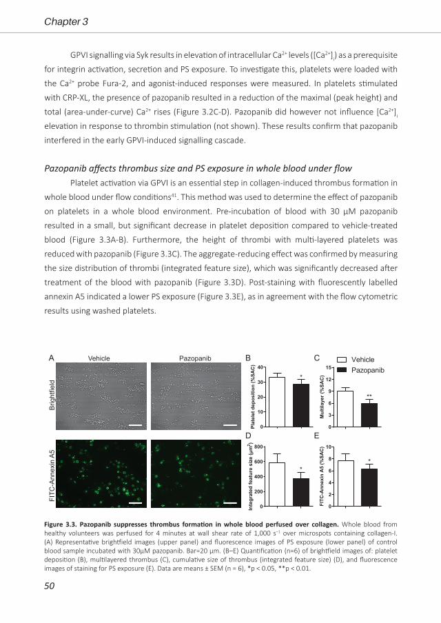
50
Chapter 3
GPVI signalling via Syk results in elevation of intracellular Ca2+ levels ([Ca2+]i) as a prerequisite
for integrin activation, secretion and PS exposure. To investigate this, platelets were loaded with
the Ca2+ probe Fura-2, and agonist-induced responses were measured. In platelets stimulated
with CRP-XL, the presence of pazopanib resulted in a reduction of the maximal (peak height) and
total (area-under-curve) Ca2+ rises (Figure 3.2C-D). Pazopanib did however not influence [Ca2+]i
elevation in response to thrombin stimulation (not shown). These results confirm that pazopanib
interfered in the early GPVI-induced signalling cascade.
Pazopanib affects thrombus size and PS exposure in whole blood under flowPlatelet activation via GPVI is an essential step in collagen-induced thrombus formation in
whole blood under flow conditions41. This method was used to determine the effect of pazopanib
on platelets in a whole blood environment. Pre-incubation of blood with 30 µM pazopanib
resulted in a small, but significant decrease in platelet deposition compared to vehicle-treated
blood (Figure 3.3A-B). Furthermore, the height of thrombi with multi-layered platelets was
reduced with pazopanib (Figure 3.3C). The aggregate-reducing effect was confirmed by measuring
the size distribution of thrombi (integrated feature size), which was significantly decreased after
treatment of the blood with pazopanib (Figure 3.3D). Post-staining with fluorescently labelled
annexin A5 indicated a lower PS exposure (Figure 3.3E), as in agreement with the flow cytometric
results using washed platelets.
0
10
20
30
40
*
Plat
elet
depo
sitio
n(%
SAC
)
0
2
4
6
8
10
*
FITC
-Ann
exin
A5
(%SA
C)
0
200
400
600
800
*
Inte
grat
edfe
atur
esi
ze( µ
m2 )
0
3
6
9
12
15
**
Mul
tilay
er(%
SAC
)
VehiclePazopanib
Vehicle Pazopanib
Brig
htfie
ldFI
TC-A
nnex
in A
5
A B C
D E
Figure 3.3. Pazopanib suppresses thrombus formation in whole blood perfused over collagen. Whole blood from healthy volunteers was perfused for 4 minutes at wall shear rate of 1,000 s−1 over microspots containing collagen-I. (A) Representative brightfield images (upper panel) and fluorescence images of PS exposure (lower panel) of control blood sample incubated with 30μM pazopanib. Bar=20 μm. (B–E) Quantification (n=6) of brightfield images of: platelet deposition (B), multilayered thrombus (C), cumulative size of thrombus (integrated feature size) (D), and fluorescence images of staining for PS exposure (E). Data are means ± SEM (n = 6), *p < 0.05, **p < 0.01.
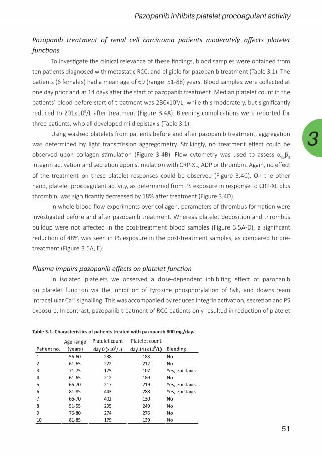
51
Pazopanib inhibits platelet procoagulant activity
3
Pazopanib treatment of renal cell carcinoma patients moderately affects platelet functions
To investigate the clinical relevance of these findings, blood samples were obtained from
ten patients diagnosed with metastatic RCC, and eligible for pazopanib treatment (Table 3.1). The
patients (6 females) had a mean age of 69 (range: 51-88) years. Blood samples were collected at
one day prior and at 14 days after the start of pazopanib treatment. Median platelet count in the
patients’ blood before start of treatment was 230x109/L, while this moderately, but significantly
reduced to 201x109/L after treatment (Figure 3.4A). Bleeding complications were reported for
three patients, who all developed mild epistaxis (Table 3.1).
Using washed platelets from patients before and after pazopanib treatment, aggregation
was determined by light transmission aggregometry. Strikingly, no treatment effect could be
observed upon collagen stimulation (Figure 3.4B). Flow cytometry was used to assess αIIbβ3
integrin activation and secretion upon stimulation with CRP-XL, ADP or thrombin. Again, no effect
of the treatment on these platelet responses could be observed (Figure 3.4C). On the other
hand, platelet procoagulant activity, as determined from PS exposure in response to CRP-XL plus
thrombin, was significantly decreased by 18% after treatment (Figure 3.4D).
In whole blood flow experiments over collagen, parameters of thrombus formation were
investigated before and after pazopanib treatment. Whereas platelet deposition and thrombus
buildup were not affected in the post-treatment blood samples (Figure 3.5A-D), a significant
reduction of 48% was seen in PS exposure in the post-treatment samples, as compared to pre-
treatment (Figure 3.5A, E).
Plasma impairs pazopanib effects on platelet functionIn isolated platelets we observed a dose-dependent inhibiting effect of pazopanib
on platelet function via the inhibition of tyrosine phosphorylation of Syk, and downstream
intracellular Ca2+ signalling. This was accompanied by reduced integrin activation, secretion and PS
exposure. In contrast, pazopanib treatment of RCC patients only resulted in reduction of platelet
Pa�ent no.Age range
(years)Platelet count day 0 (x109/L)
Platelet count day 14 (x109/L) Bleeding
1 56-60 238 183 No2 61-65 222 212 No3 71-75 175 107 Yes, epistaxis4 61-65 212 189 No5 66-70 217 219 Yes, epistaxis6 81-85 443 288 Yes, epistaxis7 66-70 402 130 No8 51-55 295 249 No9 76-80 274 276 No10 81-85 179 139 No
Table 3.1. Characteristics of patients treated with pazopanib 800 mg/day.

52
Chapter 3
PS exposure. We hypothesized that the presence of plasma can interfere with the antiplatelet
activity of pazopanib. To investigate this, experiments were performed with both PRP and washed
platelets, which were incubated with different concentrations of pazopanib. In undiluted PRP,
collagen-induced aggregation was not affected by pazopanib, whereas this response was strongly
inhibited at 10 µM in washed platelets (Figure 3.6A). Pre-incubation of PRP with increasing doses
of pazopanib until 20 µM did not inhibit PS exposure, in contrast to pre-incubation of washed
platelets (Figure 3.6B).
It has been described that pazopanib is highly bound by plasma proteins at concentration
ranges of 10-100 µg/mL24. This corresponds with a concentration of 23-230 µM. Hence, also
higher concentrations of pazopanib (50-100 µM) were tested in platelets incubated at different
A
C D
Pre-treatmentOn-treatment
B
Pre-treatment On-treatment0
100
200
300
400
500
PLT
(x10
9 /L)
**Collagen
0
20
40
60
80
100
%Tr
ansm
issi
on
ADP
0
20
40
60
80
100
Thrombin
0
20
40
60
80
100
CRP
0
20
40
60
80
100
αIIbβ
3po
sitiv
epl
atel
ets
(%)
0
20
40
60
80
100
P-se
lect
inpo
sitiv
epl
atel
ets
(%)
ADP
0
20
40
60
80
100Thrombin
0
20
40
60
80
100
0
20
40
60
80
100
0
20
40
60
80
100
CRP + Thrombin
0
20
40
60
80
100
PSex
posi
ngpl
atel
ets
(%)
*
Figure 3.4. Treatment of carcinoma patients with pazopanib decreased platelet count and suppressed phosphatidylserine exposure in washed platelets. Blood from patients was collected the day before and at 2 weeks after pazopanib treatment. (A) Platelet count was measured in blood from RCC patients before and on treatment with pazopanib. Each dot represents a single patient. (B) Platelets were isolated and aggregation was induced by 1μg/mL collagen, 1μM Me-S-ADP (in the presence of 25μg/mL fibrinogen), or 1 nM thrombin. Histograms indicate maximal amplitude of aggregation (n = 6). (C) Platelets before and during treatment of patients were stimulated with 5μg/mL CRP-XL, 1μM 2Me-S-ADP or 1 nM thrombin for 10–20min, and analyzed by flow cytometry (n = 10). Shown are percentages of platelets binding FITC-PAC1 mAb (aIIbb3 expression) or FITC-anti-CD62P mAb (P-selectin expression). (D) Patient platelets were stimulated with 5 μg/mL CRP-XL and 4 nM thrombin for 60min, and analyzed by flow cytometry (n = 6). Histogram shows percentages of platelets binding FITC-annexin A5 (PS exposure). Data are medians ± interquartile ranges, *p<0.05, **p<0.01.

53
Pazopanib inhibits platelet procoagulant activity
3Figure 3.5. Treatment of pazopanib patients suppressed phosphatidylserine exposure upon thrombus formation on collagen. (A) Whole blood from patients was perfused for 4min at wall shear rate of 1,000 s−1 over a collagen surface. Shown are representative brightfield images (upper panel) and fluorescence images of PS exposure (lower panel) for blood samples taken before and after 2 weeks of pazopanib treatment. Bar=20 μm. (B–E) Quantification of brightfield images of platelet deposition (B), multilayered thrombus (C), cumulative size of thrombus (integrated feature size, IFS) (D), and fluorescence images of staining for PS exposure (E). Data are medians ± interquartile ranges (n=10), **p<0.01.
Pre-treatment On-treatmentBr
ight
field
FITC
-Ann
exin
A5
A B C
D E
Pre-treatmentOn-treatment
05
101520253035
Plat
elet
depo
sitio
n(%
SAC)
0
5
10
15
Mul
tilay
er(%
SAC
)
0
200
400
600
800
1000
Inte
grat
edfe
atur
esi
ze(µ
m2 )
0
2
4
6
FITC
-Ann
exin
A5
(%SA
C)
**
Washed plateletsPRP
A B
C
CRP + thrombin
0 5 10 15 200
25
50
75
100
125
Pazopanib (µM)
PSex
posi
ngpl
atel
ets
(%of
cont
rol)
* *
CRP + TRAP-6
Pazopanib (µM)
PSex
posi
ngpl
atel
ets
(%of
cont
rol)
0 20 40 60 80 1000
25
50
75
100
1250% plasma10% plasma30% plasma50% plasma100% plasma*
****** ****
****
*
****
**
Collagen
0 5 10 15 20 25 300
25
50
75
100
125
Pazopanib (µM)
Agg
rega
tion
(%of
cont
rol)
*
Figure 3.6. Plasma suppresses effects of pazopanib on platelet activation in vitro. Washed platelets or PRP from healthy donors were incubated with 1, 5, 10, or 30 μM pazopanib or vehicle (0.1% DMSO) for 10 minutes. (A) Aggregation of platelets in buffer or PRP in response to 1 μg/mL collagen. (B) Exposure of PS measured in washed platelets or in PRP, stimulated with 5 μg/mL CRP-XL and 4 nM thrombin for 60 minutes, and analyzed by flow cytometry using FITC-labeled annexin A5. (C) Platelets reconstituted at different plasma concentrations were pre-incubated with pazopanib (1–100 μM), and stimulated with 5 μg/mL CRP-XL plus 15 μM TRAP-6; PS exposure was quantified by flow cytometry using FITC-labeled annexin A5 (PS exposure). Data are means ± SEM (n=3), *p<0.05, **p<0.01, ****p<0.0001.

54
Chapter 3
plasma concentrations (0, 10, 30, 50 or 100% plasma). It appeared that pazopanib treatment
substantially suppressed PS exposure in response to CRP-XL plus TRAP-6 in the presence of low
amounts of plasma (Figure 3.6C). However, at 100 µM pazopanib, PS exposure was still significantly
reduced by 25% with 30-100% plasma (Figure 3.6C, p<0.01). This effect is comparable to the
moderate inhibition of PS exposure in patients treated with pazopanib (see Figure 3.4D). These
results suggest that the presence of plasma interferes with the incorporation of pazopanib into
platelets.
DiscussionIn the present study, we demonstrate that the multi-target TKI pazopanib markedly
reduces the collagen-induced activation responses of isolated platelets, including aggregation,
PS exposure and Ca2+ signalling through inhibition of tyrosine kinases, including Syk. In blood
samples from RCC patients treated with pazopanib, these effects were confined to a suppression
of GPVI-mediated PS exposure, observed in isolated platelets as well as in whole blood thrombus
formation under flow.
Several protein tyrosine kinases are known to contribute to platelet activation processes
and haemostasis8, 10, 11. An activity-based kinase profiling already showed that pazopanib can target
various tyrosine kinases that are highly expressed in platelets42. Here we confirm that, in washed
platelets, the GPVI-dependent phosphorylation of multiple proteins, including Syk, is suppressed
by pazopanib, in a way accompanied by reduced platelet activation processes. Syk is known to
have multiple sites of phosphorylation which both regulate activity and serve as docking motifs
for other proteins43. These sites include Tyr-348 and Tyr-352 within the SH2-linker region, Tyr-525
and Tyr-526 within the activation loop of the kinase domain, Tyr-630 in the C terminus of Syk,
and other sites such as Thr-384 and Ser-297. It has been shown that Tyr-525/526 is essential for
Syk function44. Therefore, we selected this phosphorylation site to study the effect of pazopanib
on platelet function. However, from the present data it cannot be concluded whether pazopanib
inhibits Syk directly, or if its phosphorylation is reduced through inhibition of upstream tyrosine
kinases. Most likely, it can be both as pazopanib has been shown to have affinity for multiple
TKs in platelets. We have observed that the Syk inhibitor II completely inhibits PS-exposure in
washed platelets37, which was much stronger than the inhibitory effect of pazopanib (Figure
3.1D). Furthermore deficiency and selective inhibition of Syk has been shown to prevent platelet
aggregation and activation in response to collagen-receptor stimulation in mice45.
Interestingly, pazopanib also suppressed platelet responses to ADP, arachidonic acid and
TxA2 analogue, while responses to thrombin were unaffected. In agreement with this is the recent
finding that three tyrosine kinases are phosphorylated upon platelet activation with ADP using
phosphoproteomics46. These are JAK3, Btk and TNK2, indicating TKs are involved in signalling
underneath ADP. Whether TKs are also phosphorylated under AA and thromboxane A2 receptor

55
Pazopanib inhibits platelet procoagulant activity
3
stimulation has not yet been investigated with phosphoproteomics, but this is not unlikely.
Although it appears that the majority of the targets of pazopanib are underneath GPVI, these data
indicate that pazopanib may also target kinase events downstream of other platelet receptors.
Moreover, as it has been shown that inhibition of GPVI alone prevents occlusive thrombus
formation without causing bleeding47, these results support the hypothesis that the targeting of
pathways downstream of additional receptors may be responsible for the increased bleeding risk
with pazopanib treatment.
Pazopanib is known to have an extremely high plasma protein binding compared to other
TKIs24, 48. Therefore, we explored if the presence of blood plasma affected its inhibitory effect on
platelet responses. Upon increasing plasma concentrations, it appeared that the inhibition of
collagen-induced aggregation became lost at pazopanib concentrations up to 100 μM, whereas
the inhibition of PS exposure was still present albeit diminished. Albumin, as a major plasma
protein (about 55% of plasma proteins) present at 35-50 mg/mL49, is considered to be the main
pazopanib-binding plasma component50. This is in agreement with the residual, but consistent
inhibition of PS exposure observed in the platelets from patients treated with pazopanib, as well
as in whole blood thrombus formation under flow.
Patients with advanced RCC are commonly treated with 800 mg pazopanib per day.
The reported steady state, maximal concentration here is 45 µg/mL51, which corresponds to a
concentration of 100 µM. In phase III trials, pazopanib effectively delays disease progression and
reduces tumour lesions25, 52. In spite of the only partial response rate25, the affinity of pazopanib
for VEGF and PDGF receptors is relatively high42, and likely is higher than that for the intracellular
tyrosine kinases implicated in platelet activation. This may explain why the pazopanib dose used
for effective cancer treatment does not completely abolish platelet activation processes, but only
platelet procoagulant activity (PS exposure), i.e. a response that is most sensitive to inhibition of
the cytosolic Ca2+ rises.
We and others24 observed a moderate decrease in platelet count upon pazopanib
treatment. This by itself is not expected to result in bleeding, with values still within the
normal range of 150–400x109 platelets/L53. This effect may suggest interference in platelet
formation (megakaryocytopoiesis). This has not been reported so far, but there is evidence that
megakaryocytic signalling via Src family and Syk kinases is required for megakaryocyte migration,
and platelet formation13.
The combination of a reduced platelet count and impaired PS exposure may explain the
mostly minor bleeding events observed during pazopanib treatment. In the present study, this held
for three out of ten patients, all experiencing epistaxis. This number is relatively high compared
to a published clinical trial, reporting mild bleeding in only 13% of the patients24. However, we
like to note that the power of our study is low. Mild bleeding has also been reported with the
use of other TKIs in cancer therapy18, 19. Ibrutinib – affecting collagen- and von Willebrand factor-
56
Chapter 3
dependent platelet functions54 – can cause a risk of mild bleeding in about half of the treated
patients, whilst 4-8% of these experiencing major haemorrhages14. Treatment with the drug
ponatinib resulted in a prolonged PFA-100 closure times in patients’ blood samples, indicating
a loss of platelet function15. Bleeding occurred here in about 10% of the patients, who however
occasionally used other anticoagulants or antiplatelet drugs55. Treatment with the TKI dasatinib
was associated with mild thrombocytopenia and an increased risk of bleeding, likely due to
combined effects on megakaryocytes and platelets56. Also in the latter case, the patients’ platelets
were less responsive to collagen stimulation, resulting in decreased thrombus formation57. To
take this further, we recently reviewed how distinct TKIs inhibit platelet activation mechanisms,
as well as the clinical consequences of antiplatelet effects due to TKI treatment58. Comparison of
affinity profiles of TKIs for platelet targets, as well as literature regarding effects on platelet count,
platelet function and bleeding, enabled us to distinguish three categories of TKIs. (i) For several
TKIs the bleeding tendency is linked to a lowering of platelet count and/or an impairment of
platelet function, (ii) other TKIs are predicated to have an antiplatelet effect, although no bleeding
side effects have been reported so far, and (iii) for some TKIs no published data on platelets are
available.
In comparison to pazopanib, treatment of RCC patients with sunitinib was found to result
in a more profound inhibition of platelet activation18. Sunitinib is taken up by platelets, and can
thus effectively reduce collagen-receptor induced aggregation and thrombus formation18. A
comparative study of sunitinib versus pazopanib treatment indicated that both drugs provided
a progression-free survival benefit when compared to placebo; however, pazopanib had a better
safety and quality-of-life profile52. Sunitinib causes bleeding in up to 20% of patients, and life-
threatening bleeding in 3% of the patients16. As indicated above, for pazopanib these numbers are
lower, but still non-negligible24. Accordingly treatment of metastatic RCC with either pazopanib
or sunitinib should be accompanied by special attention of the haemostatic condition, especially
when the patients are also treated with antiplatelet drugs.
In summary, the present work demonstrates that platelet treatment with pazopanib
in vitro results in strong inhibition of collagen-induced platelet activation, aggregation and PS
exposure, whereas pazopanib treatment of RCC patients is restricted to inhibition of the platelet
procoagulant activity. In combination with the reduction in platelet count, these effects are likely
to contribute to the higher bleeding tendency in pazopanib treated RCC patients. Therefore, anti-
platelet effects of TKIs should be taken into account in therapy decisions for patients, especially
when prescribed in combination with antiplatelet drugs.
AcknowledgementsThis study was supported by the Maastricht Thrombosis Expertise Centre as part of the
Heart and Vascular Centre (HVC) of the MUMC+.
57
Pazopanib inhibits platelet procoagulant activity
3
Author Contributions StatementBMET performed experiments, analysed data and wrote manuscript. MN performed
experiments and critically reviewed manuscript. SS performed experiments, wrote medical ethical
approval and critically reviewed manuscript. AWG provided essential tools, discussed results and
critically reviewed manuscript, MGAoE discussed results and critically reviewed manuscript,
MJBA included and took care of RCC patients and critically reviewed manuscript, JWMH designed
and discussed experiments, critically reviewed and revised manuscript, MJEK designed and
performed experiments, discussed results, wrote and revised manuscript.
Conflict of Interest StatementThe authors declare no personal, professional or financial relationships that could
potentially be construed as a conflict of interest.
References1. Jorg Thomas, H., et al., Tyrosine Kinase Inhibitors – A Review on Pharmacology, Metabolism and Side
Effects. Cur Drug Metab, 2009. 10(5): p. 470-481.2. Natoli, C., et al., Tyrosine Kinase Inhibitors. Curr Canc Drug Targets, 2010. 10(5): p. 462-483.3. Broekman, F., E. Giovannetti, and G.J. Peters, Tyrosine kinase inhibitors: Multi-targeted or single-
targeted? World J Clin Oncol, 2011. 2(2): p. 80-93.4. Gotink, K.J. and H.M. Verheul, Anti-angiogenic tyrosine kinase inhibitors: what is their mechanism of
action? Angiogenesis, 2010. 13(1): p. 1-14.5. Rovithi, M., et al., Alternative scheduling of pulsatile, high dose sunitinib efficiently suppresses
tumor growth. J Exp Clin Cancer Res, 2016. 35(1): p. 138.6. Griffioen, A.W. and G. Molema, Angiogenesis: potentials for pharmacologic intervention in the
treatment of cancer, cardiovascular diseases, and chronic inflammation. Pharmacol Rev, 2000. 52(2): p. 237-68.
7. Gross, S., et al., Targeting cancer with kinase inhibitors. J Clin Invest, 2015. 125(5): p. 1780-1789.8. Watson, S.P., et al., GPVI and integrin alphaIIb beta3 signaling in platelets. J Thromb Haemost, 2005.
3(8): p. 1752-62.9. Furman, M.I., et al., Platelet tyrosine kinases and fibrinogen receptor activation. Circ Res, 1994.
75(1): p. 172-80.10. Senis, Y.A., A. Mazharian, and J. Mori, Src family kinases: at the forefront of platelet activation. Blood,
2014. 124(13): p. 2013-2024.11. Nieswandt, B. and S.P. Watson, Platelet-collagen interaction: is GPVI the central receptor? Blood,
2003. 102(2): p. 449-61.12. Gross, B.S., S.K. Melford, and S.P. Watson, Evidence that phospholipase C-gamma2 interacts with
SLP-76, Syk, Lyn, LAT and the Fc receptor gamma-chain after stimulation of the collagen receptor glycoprotein VI in human platelets. Eur J Biochem, 1999. 263(3): p. 612-23.
13. Mazharian, A., et al., Critical role of Src-Syk-PLC{gamma}2 signaling in megakaryocyte migration and thrombopoiesis. Blood, 2010. 116(5): p. 793-800.
14. Shatzel, J.J., et al., Ibrutinib-associated bleeding: pathogenesis, management and risk reduction strategies. J Thromb Haemost, 2017. 15(5): p. 835-847.
15. Neelakantan, P., et al., Platelet dysfunction associated with ponatinib, a new pan BCR-ABL inhibitor with efficacy for chronic myeloid leukemia resistant to multiple tyrosine kinase inhibitor therapy. Haematologica, 2012. 97(9): p. 1444.
16. Je, Y., F.A. Schutz, and T.K. Choueiri, Risk of bleeding with vascular endothelial growth factor receptor tyrosine-kinase inhibitors sunitinib and sorafenib: a systematic review and meta-analysis of clinical

58
Chapter 3
trials. Lancet Oncol, 2009. 10(10): p. 967-74.17. Rigg, R.A., et al., Oral administration of Bruton’s tyrosine kinase inhibitors impairs GPVI-mediated
platelet function. Am J Physiol Cell Physiol, 2016. 310(5): p. C373-80.18. Sabrkhany, S., et al., Sunitinib uptake inhibits platelet function in cancer patients. Eur J Cancer, 2016.
66: p. 47-54.19. Levade, M., et al., Targeting Kinases in Cancer Therapies: Adverse Effects on Blood Platelets. Curr
Pharm Des, 2016. 22(16): p. 2315-22.20. Hamilton, Z., et al., Change in platelet count as a prognostic indicator for response to primary tyrosine
kinase inhibitor therapy in metastatic renal cell carcinoma. BJU Int, 2016. 118(6): p. 927-934.21. Crist, M., et al., Examining the bleeding incidences associated with targeted therapies used in
metastatic renal cell carcinoma. Crit Rev Oncol Hematol, 2017. 120: p. 151-162.22. Kostos, L., et al., Gastrointestinal bleeding in a chronic myeloid leukaemia patient precipitated by
dasatinib-induced platelet dysfunction: Case report. Platelets, 2015. 26(8): p. 809-11.23. Bukowski, R.M., U. Yasothan, and P. Kirkpatrick, Pazopanib. Nat Rev Drug Discov, 2010. 9(1): p. 17-18.24. McCormack, P.L., Pazopanib: a review of its use in the management of advanced renal cell carcinoma.
Drugs, 2014. 74(10): p. 1111-25.25. Sternberg, C.N., et al., Pazopanib in Locally Advanced or Metastatic Renal Cell Carcinoma: Results of
a Randomized Phase III Trial. J Clin Oncol, 2010. 28(6): p. 1061-1068.26. Fabian, M.A., et al., A small molecule-kinase interaction map for clinical kinase inhibitors. Nat
Biotechnol, 2005. 23(3): p. 329-36.27. Karaman, M.W., et al., A quantitative analysis of kinase inhibitor selectivity. Nat Biotechnol, 2008.
26(1): p. 127-32.28. Mattheij, N.J., et al., Dual mechanism of integrin alphaIIbbeta3 closure in procoagulant platelets. J
Biol Chem, 2013. 288(19): p. 13325-36.29. Strehl, A., et al., Dual role of platelet protein kinase C in thrombus formation: stimulation of pro-
aggregatory and suppression of procoagulant activity in platelets. J Biol Chem, 2007. 282(10): p. 7046-55.
30. Baaten, C.C., et al., Gradual increase in thrombogenicity of juvenile platelets formed upon offset of prasugrel medication. Haematologica, 2015. 100(9): p. 1131-8.
31. van Kruchten, R., et al., Both TMEM16F-dependent and TMEM16F-independent pathways contribute to phosphatidylserine exposure in platelet apoptosis and platelet activation. Blood, 2013. 121(10): p. 1850-7.
32. De Witt, S.M., et al., Identification of platelet function defects by multi-parameter assessment of thrombus formation. Nat Commun, 2014. 5: p. 4257.
33. Van Kruchten, R., J.M. Cosemans, and J.W. Heemskerk, Measurement of whole blood thrombus formation using parallel-plate flow chambers - a practical guide. Platelets, 2012. 23(3): p. 229-42.
34. De Witt, S., et al., Multi-parameter assessment of thrombus formation on microspotted arrays of thrombogenic surfaces. Nat Prot Exchange, 2014.
35. Feijge, M.A., et al., Inter-individual variability in Ca2+ signalling in platelets from healthy volunteers: effects of aspirin and relationship with expression of endomembrane Ca2+-ATPases. Br J Haematol, 1998. 102(3): p. 850-9.
36. Heemskerk, J.W., et al., The Ca2+-mobilizing potency of alpha-thrombin and thrombin-receptor-activating peptide on human platelets -- concentration and time effects of thrombin-induced Ca2+ signaling. Eur J Biochem, 1997. 249(2): p. 547-55.
37. Van der Meijden, P.E., et al., Key role of integrin alpha(IIb)beta (3) signaling to Syk kinase in tissue factor-induced thrombin generation. Cell Mol Life Sci, 2012. 69(20): p. 3481-92.
38. Versteeg, H.H., et al., New fundamentals in hemostasis. Physiol Rev, 2013. 93(1): p. 327-58.39. Munnix, I.C., et al., Segregation of platelet aggregatory and procoagulant microdomains in thrombus
formation: regulation by transient integrin activation. Arterioscler Thromb Vasc Biol, 2007. 27(11): p. 2484-90.
40. Kuijpers, M.J., et al., Key role of platelet procoagulant activity in tissue factor-and collagen-dependent thrombus formation in arterioles and venules in vivo differential sensitivity to thrombin inhibition. Microcirculation, 2008. 15(4): p. 269-82.
41. Nieswandt, B., et al., Glycoprotein VI but not alpha2beta1 integrin is essential for platelet interaction

59
Pazopanib inhibits platelet procoagulant activity
3
with collagen. Embo j, 2001. 20(9): p. 2120-30.42. Kitagawa, D., et al., Activity-based kinase profiling of approved tyrosine kinase inhibitors. Genes
Cells, 2013. 18(2): p. 110-22.43. Sada, K., et al., Structure and function of Syk protein-tyrosine kinase. J Biochem, 2001. 130(2): p.
177-86.44. Zhang, J., et al., Phosphorylation of Syk activation loop tyrosines is essential for Syk function. An in
vivo study using a specific anti-Syk activation loop phosphotyrosine antibody. J Biol Chem, 2000. 275(45): p. 35442-7.
45. van Eeuwijk, J.M., et al., The Novel Oral Syk Inhibitor, Bl1002494, Protects Mice From Arterial Thrombosis and Thromboinflammatory Brain Infarction. Arterioscler Thromb Vasc Biol, 2016. 36(6): p. 1247-53.
46. Beck, F., et al., Temporal quantitative phosphoproteomics of ADP stimulation reveals novel central nodes in platelet activation and inhibition. Blood, 2017. 129(2): p. e1-e12.
47. Andrews, R.K., J.F. Arthur, and E.E. Gardiner, Targeting GPVI as a novel antithrombotic strategy. J Blood Med, 2014. 5: p. 59-68.
48. O’Brien, Z. and M.F. Moghaddam, A Systematic Analysis of Physicochemical and ADME Properties of All Small Molecule Kinase Inhibitors Approved by US FDA from January 2001 to October 2015. Curr Med Chem, 2017. 24(29): p. 3159-3184.
49. Anderson, N.L. and N.G. Anderson, The human plasma proteome: history, character, and diagnostic prospects. Mol Cell Proteomics, 2002. 1(11): p. 845-67.
50. Imbs, D.C., et al., Determination of unbound fraction of pazopanib in vitro and in cancer patients reveals albumin as the main binding site. Invest New Drugs, 2016. 34(1): p. 41-8.
51. Hurwitz, H.I., et al., Phase I trial of pazopanib in patients with advanced cancer. Clin Cancer Res, 2009. 15(12): p. 4220-7.
52. Motzer, R.J., et al., Pazopanib versus sunitinib in metastatic renal-cell carcinoma. N Engl J Med, 2013. 369(8): p. 722-31.
53. Giles, C., The platelet count and mean platelet volume. Br J Haematol, 1981. 48(1): p. 31-7.54. Levade, M., et al., Ibrutinib treatment affects collagen and von Willebrand factor-dependent platelet
functions. Blood, 2014. 124(26): p. 3991.55. Nazha, A., et al., The clinical impact of ponatinib on the risk of bleeding in patients with chronic
myeloid leukemia. Haematologica, 2013. 98(10): p. e131-e131.56. Mazharian, A., et al., Dasatinib enhances megakaryocyte differentiation but inhibits platelet
formation. Blood, 2011. 117(19): p. 5198-5206.57. Gratacap, M.-P., et al., The new tyrosine-kinase inhibitor and anticancer drug dasatinib reversibly
affects platelet activation in vitro and in vivo. Blood, 2009. 114(9): p. 1884.58. Tullemans, B.M.E., J.W.M. Heemskerk, and M.J.E. Kuijpers, Acquired platelet antagonism: off-target
antiplatelet effects of malignancy treatment with tyrosine kinase inhibitors. J Thromb Haemost, 2018. 16:1686-1699.

Chapter 4
Tyrosine kinase inhibitor sunitinib delays
platelet-induced coagulation: additive
effects of aspirin
Tullemans BME, Fernández DI*, Veninga A*, Baaten CCFMJ, Peters LJF, Aarts MJB, Eble JA, Campello E, Spiezia L, Simioni P, van der Vorst EPC, van der Meijden PEJ, Heemskerk JWM
and Kuijpers MJE (* equal contribution)
Thromb Haemost 2021; Online ahead of printReprinted with permission
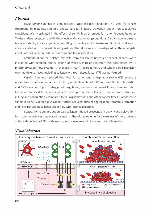
62
Chapter 4
AbstractBackground: Sunitinib is a multi-target tyrosine kinase inhibitor (TKI) used for cancer
treatment. In platelets, sunitinib affects collagen-induced activation under non-coagulating
conditions. We investigated (i) the effects of sunitinib on thrombus formation induced by other
TK-dependent receptors, and (ii) the effects under coagulating conditions. Cardiovascular disease
is a co-morbidity in cancer patients, resulting in possible aspirin treatment. Sunitinib and aspirin
are associated with increased bleeding risk, and therefore we also investigated (iii) the synergistic
effects of these compounds on thrombus and fibrin formation.
Methods: Blood or isolated platelets from healthy volunteers or cancer patients were
incubated with sunitinib and/or aspirin or vehicle. Platelet activation was determined by TK
phosphorylation, flow cytometry, changes in [Ca2+]i, aggregometry and whole blood perfusion
over multiple surfaces, including collagen with(out) tissue factor (TF) was performed.
Results: Sunitinib reduced thrombus formation and phosphatidylserine (PS) exposure
under flow on collagen type I and III. Also, sunitinib inhibited GPVI-induced TK phosphorylation
and Ca2+ elevation. Upon TF-triggered coagulation, sunitinib decreased PS exposure and fibrin
formation. In blood from cancer patients more pronounced effects of sunitinib were observed
in lung and pancreatic as compared to neuroglioblastoma and other cancer types. Compared to
sunitinib alone, sunitinib plus aspirin further reduced platelet aggregation, thrombus formation
and PS exposure on collagen under flow with(out) coagulation.
Conclusions: Sunitinib suppresses collagen-induced procoagulant activity and delays fibrin
formation, which was aggravated by aspirin. Therefore, we urge for awareness of the combined
antiplatelet effects of TKIs with aspirin, as this may result in increased risk of bleeding.
Visual abstract
63
Sunitinib and aspirin in platelet-induced coagulation
4
IntroductionTreatment of cancer patients with sunitinib results in anticancer effects and improves
progression free survival1, 2. Sunitinib is a broad-spectrum tyrosine kinase inhibitor (TKI), affecting
not only tumour cells, but also the blood vessel wall3. A side effect of sunitinib treatment is mild
bleeding (mainly epistaxis, and mucocutaneous and gastrointestinal bleeding, which occurs
in approximately 19% of sunitinib-treated patients)3. Fortunately, in the majority of cases, the
effects on haemostasis remain sub-clinical.
Platelets contribute to haemostasis by signalling pathways involving multiple protein
tyrosine kinases (TKs) such as Syk, Btk and Src family kinases (SFKs)4. We and other authors
have previously shown that sunitinib is sequestered by platelets, which affects collagen-induced
platelet function under non-coagulating conditions5, 6. TKs do not only signal under the collagen
receptor glycoprotein (GP)VI, but also under the receptors GPIb, CLEC-2 and integrins α6β1 and
αIIbβ3 that bind von Willebrand factor (vWF), podoplanin, laminin and fibrinogen, respectively4.
The effects of sunitinib on thrombus formation stimulated via these receptors has not been
studied. Furthermore, the question arises whether the suppression of collagen-induced thrombus
formation is also present under physiological conditions of coagulation.
The link between cancer and arterial thrombosis has been well-established7, 8. Cancer
patients with a cardiovascular history receive treatment with anti-platelet drugs like aspirin to
prevent recurrent events. Aspirin irreversibly inhibits platelet activation and is associated with an
increased bleeding risk9 by preventing the formation of thromboxane A2 (TxA2)10. TxA2 acts as an
important soluble agonist released by platelets upon GPVI stimulation. In this respect, aspirin and
other platelet-targeted pharmacologic treatments are under investigation as adjuvant anti-cancer
therapy11, 12. The combined effects of these drugs on platelet function have not been investigated
thus far.
The aim of the present study was to further investigate the effect of sunitinib on thrombus
formation by stimulation with multiple agonists, as well as under coagulating conditions stimulated
by collagen plus TF. Furthermore, we assessed whether treatment with sunitinib in combination
with aspirin increasingly inhibited collagen-induced thrombus formation as compared to either
compound alone. The results indeed show an enhancing effect of this dual treatment of platelets
with sunitinib and aspirin on collagen-induced platelet phosphatidylserine (PS) exposure and
ensuing fibrin formation. Finally, platelet-dependent fibrin formation in whole blood from cancer
patients was significantly inhibited by sunitinib.
Materials and MethodsMaterials
Sunitinib malate (Sutent) was provided by Pfizer (New York NY, USA). Aspirin, bovine
serum albumin (BSA), D(+)-glucose, unfractionated heparin, and apyrase were purchased from

64
Chapter 4
Sigma-Aldrich (Saint Louis MO, USA). The agonists collagen-related peptide crosslinked (CRP-XL)
and Von Willebrand factor III (vWF-III) were obtained from CambCol Laboratories (Cambridge,
UK), whereas thrombin was obtained from Enzyme Research Laboratories (South Bend IN, USA)
and Horm collagen type I from Takeda (Hoofddorp, The Netherlands). 2-Methylthio-adenosine-
diphosphate (2MeS-ADP) and D-phenylalanyl-prolyl-arginyl chloromethyl ketone (PPACK) were
obtained from Santa Cruz Biotechnology (Dallas TX, USA). Recombinant human TF (Innovin)
was purchased from Dade Behring (Deerfield IL, USA). Laminin came from Octapharma (Berlin,
Germany). Fura-2-AM was obtained from Invitrogen (Carlsbad CA, USA). Plastic syringes and
fluorescein isothiocyanate (FITC)-labelled PAC1 monoclonal antibody (mAb) against activated
human integrin αIIbβ3 were purchased from BD Bioscience (nr. 340507; Franklin Lakes NJ, USA),
while FITC-conjugated α-fibrinogen mAb was purchased from DAKO (F0111; Santa Clara CA, USA).
Alexa Fluor (AF) 647-labelled CD62-P mAb was obtained from Biolegend (London, UK), whereas
the FITC-labelled anti-human CD62-P mAb was obtained from Beckman Coulter (nr. 65050;
Sydney, Australia). 3,3-dihexyloxacarbocyanine iodide (DiOC6) was purchased from Anaspec
(Reeuwijk, The Netherlands). FITC-conjugated annexin A5 was from Pharmatarget (Maastricht,
The Netherlands). AF568-conjugated annexin A5 and AF647-labelled fibrinogen were purchased
from Molecular Probes, Life Technologies (New York NY, USA).
Blood collection and platelet isolationWith approval from the medical ethics committee from the Maastricht University Medical
Centre+ (MUMC+) and informed consent in accordance with the Declaration of Helsinki, blood was
collected from healthy volunteers. Furthermore, blood was collected from 11 patients diagnosed
with several cancer types (lung, pancreas, neuroglioblastoma, ovarian or paraganglioma) at the
University Hospital of Padua (Italy), also with approval of the local medical ethical committee and
after informed consent. Blood was collected in 3.2% trisodium citrate tubes by venipuncture,
after discarding the first 3 mL of blood. Platelets, plasma or serum were isolated from whole
blood as described previously13. Whole blood or isolated platelets were incubated with either
vehicle or sunitinib (10 or 30 µM as indicated) for 10 minutes at room temperature (unless stated
otherwise) before measurements.
Flow cytometryWashed platelets (100x109 platelets/L) were supplemented with 2 mM CaCl2 and
stimulated by a combination of CRP-XL (5 μg/mL) and thrombin (4 nM) for 1 hour at 37°C.
Phosphatidyl-serine (PS) exposing platelets were labelled with FITC-conjugated annexin A5 and
measured by flow cytometry using a BD Accuri C6TM flow cytometer and accompanying software
(Erembodegem, Belgium).
65
Sunitinib and aspirin in platelet-induced coagulation
4
Cytosolic Ca2+ measurementsCytosolic Ca2+ ([Ca2+]i) measurements in Fura-2 loaded platelets (200x109 platelet/L) were
performed as described using a Shimadzo RF-5001PC spectrofluorophotometer (Kyoto, Japan)14.
Levels of [Ca2+]i were determined by the conversion of ratio values15 with correction for background
fluorescence and maximal values of [Ca2+]i (peak level) were used as output.
Light transmission aggregometryWashed platelets or PRP (250x109 platelets/L) were incubated with vehicle or sunitinib (10
or 30 µM as indicated) for 10 minutes at 37°C. Where indicated, aspirin (100 µM) was incubated
in PRP for 30 minutes at 37°C before further platelet isolation. Platelet aggregation was induced
by collagen type I (5 µg/mL), 2MeS-ADP (1 µM) or thrombin (1 nM) and aggregation responses
were measured using a Chronolog optical aggregometer (Havertown PA, USA) and maximum
amplitude was quantified.
Pam-Gene kinase assayWashed platelets (500x109 platelets/L) were pretreated with vehicle (control) or 30 μM
sunitinib for 10 minutes at 37°C and were subsequently stimulated with 5 μg/mL CRP-XL in the
presence of 2 mM CaCl2. Unstimulated, resting platelets served as control. After 90 seconds of
stimulation, samples were lysed by adding 1:1 M-PER Mammalian Extraction Buffer containing
Halt Phosphatase Inhibitor and EDTA-free Halt Protease Inhibitor Cocktail (1:100 each; Thermo
Fischer Scientific). Samples were lysed for 15 minutes on ice and afterwards centrifuged for 15
minutes at 10,000 g at 4°C. Supernatants were collected and protein content was quantified with
a BioRad DC protein kit (Hercules CA, USA).
Tyrosine kinase profiles were determined using the PamChip® peptide tyrosine kinase
microarray system on PamStation®12 (PTK; PamGene International, ´s-Hertogenbosch, The
Netherlands). Each PTK-PamChip® array contains 144 individual phospho-site(s) that are peptide
sequences derived from substrates for TKs. Each peptide on the chip builds a 15-amino acid
sequence representing a putative endogenous phosphorylation site which functions as a TK
substrate. The phosphorylation of the peptides is visualized by detection of the fluorescent signal
which is emitted as a result of the binding of the FITC-conjugated PY20 anti-phosphotyrosine
antibody.
For the PTK assay, 7.5 µg of protein was applied per array (N=3 per condition) and carried
out using the standard protocol supplied by Pamgene. All reagents used for PTK activity profiling
were supplied by Pamgene International B.V. Initially, to prepare the PTK Basic Mix, the freshly
frozen lysate was added to 4 µL of 10x protein PTK reaction buffer (PK), 0.4 µL of 100x BSA, 0.4 µL
of 1 M dithiothreitol (DTT) solution, 4 µL of 10x PTK additive, 4 µL of 4 mM ATP and 0.6 µL of
monoclonal anti-phosphotyrosine FITC-conjugate detection antibody (clone PY20). Total volume
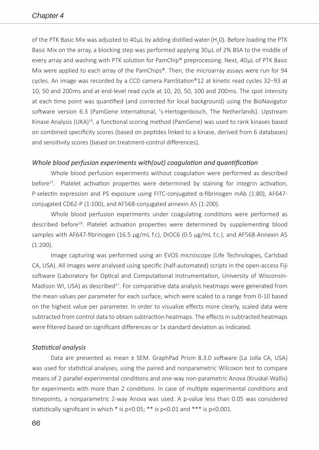
66
Chapter 4
of the PTK Basic Mix was adjusted to 40 µL by adding distilled water (H20). Before loading the PTK
Basic Mix on the array, a blocking step was performed applying 30 µL of 2% BSA to the middle of
every array and washing with PTK solution for PamChip® preprocessing. Next, 40 µL of PTK Basic
Mix were applied to each array of the PamChips®. Then, the microarray assays were run for 94
cycles. An image was recorded by a CCD camera PamStation®12 at kinetic read cycles 32–93 at
10, 50 and 200 ms and at end-level read cycle at 10, 20, 50, 100 and 200 ms. The spot intensity
at each time point was quantified (and corrected for local background) using the BioNavigator
software version 6.3 (PamGene International, 's-Hertogenbosch, The Netherlands). Upstream
Kinase Analysis (UKA)16, a functional scoring method (PamGene) was used to rank kinases based
on combined specificity scores (based on peptides linked to a kinase, derived from 6 databases)
and sensitivity scores (based on treatment-control differences).
Whole blood perfusion experiments with(out) coagulation and quantificationWhole blood perfusion experiments without coagulation were performed as described
before17. Platelet activation properties were determined by staining for integrin activation,
P-selectin expression and PS exposure using FITC-conjugated α-fibrinogen mAb (1:80), AF647-
conjugated CD62-P (1:100), and AF568-conjugated annexin A5 (1:200).
Whole blood perfusion experiments under coagulating conditions were performed as
described before18. Platelet activation properties were determined by supplementing blood
samples with AF647-fibrinogen (16.5 µg/mL f.c), DiOC6 (0.5 µg/mL f.c.), and AF568-Annexin A5
(1:200).
Image capturing was performed using an EVOS microscope (Life Technologies, Carlsbad
CA, USA). All images were analysed using specific (half-automated) scripts in the open-access Fiji
software (Laboratory for Optical and Computational Instrumentation, University of Wisconsin-
Madison WI, USA) as described17. For comparative data analysis heatmaps were generated from
the mean values per parameter for each surface, which were scaled to a range from 0-10 based
on the highest value per parameter. In order to visualize effects more clearly, scaled data were
subtracted from control data to obtain subtraction heatmaps. The effects in subtracted heatmaps
were filtered based on significant differences or 1x standard deviation as indicated.
Statistical analysisData are presented as mean ± SEM. GraphPad Prism 8.3.0 software (La Jolla CA, USA)
was used for statistical analyses, using the paired and nonparametric Wilcoxon test to compare
means of 2 parallel experimental conditions and one-way non-parametric Anova (Kruskal-Wallis)
for experiments with more than 2 conditions. In case of multiple experimental conditions and
timepoints, a nonparametric 2-way Anova was used. A p-value less than 0.05 was considered
statistically significant in which * is p<0.05; ** is p<0.01 and *** is p<0.001.

67
Sunitinib and aspirin in platelet-induced coagulation
4
ResultsSunitinib reduces thrombus formation and PS exposure in whole blood under flow on collagen type I and III
To widely explore the multi-target inhibition of sunitinib on thrombus formation, we
investigated thrombus formation induced by several TK-dependent receptors using multiple
surfaces for coating in whole blood flow chamber experiments. First, we performed dose-
response studies with sunitinib and showed that approximately 3x higher concentration of this
compound was required to inhibit aggregation induced by collagen (1 μg/ml) in the presence of
plasma when compared to washed platelets (Suppl. Figure 4.1). This agrees with the knowledge
that sunitinib is highly protein-bound1.We therefore assessed the effects on thrombus formation
during whole blood perfusion under flow using 30 μM sunitinib. This dose is in range with the
plasma concentrations observed in patients with higher peak levels of sunitinib19,but exceeded
the therapeutic target levels1.
The flow studies were performed in a microspot setting, allowing the quasi-simultaneous
measurement of sunitinib on adhesive surfaces consisting of collagen type I, collagen type III,
vWF co-coated with rhodocytin, vWF co-coated with laminin, vWF co-coated with fibrinogen and
vWF co-coated with ristocetin. Hence, roles of different platelet receptors, such as GPVI, CLEC-2,
GPIb and integrin α6β1 and αIIbβ3, were investigated as described before20-22. Captured microscopic
images were analysed (Suppl. Figure 4.2), and a heatmap was generated for a more systematic
analysis of the seven thrombus parameters, which were averaged and scaled per parameter
(Figure 4.1). Parameters of thrombus formation were only consistently affected on GPVI-
dependent surfaces, i.e. collagen type I and III (Figure 4.1A, B). In the presence of sunitinib, these
thrombi were smaller and less compact in structure as compared to the control condition (Suppl.
Figure 4.2A and B, brightfield images). Quantification showed a reduction in platelet deposition,
thrombus height and contraction (Figure 4.1). P-selectin exposure was significantly reduced in
thrombi formed on collagen III and vWF co-coated with laminin, fibrinogen and ristocetin (Suppl.
Figure 4.2B and 4.2C). Notably, thrombus parameters for CLEC-2, GPIb- and integrin αIIbβ3/α6β1-
dependent surfaces were not affected by sunitinib (Figure 4.1C-F, Suppl. Figure 4.2C-F), suggesting
a mostly GPVI-specific effect.
Sunitinib inhibits multiple tyrosine kinases, intracellular Ca2+ responses and PS exposure upon GPVI stimulation
We have previously shown that overall tyrosine phosphorylation upon GPVI stimulation
is inhibited by sunitinib5. To study the effect of sunitinib on GPVI-induced phosphorylation of
multiple specific TKs simultaneously, we performed a PamGene kinase assay. We observed that
in isolated platelets, in total 34 TKs were significantly phosphorylated by stimulation with CRP-XL,
which were subsequently significantly inhibited by sunitinib preincubation (Figure 4.2A). Most
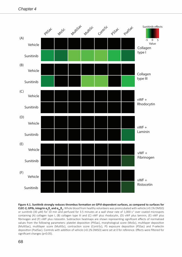
68
Chapter 4
Vehicle
Sunitinib
PltSac
MoScMultiS
ac
MultiSc
ContrSc
PSSacPse
lSac
Vehicle
Sunitinib
Vehicle
Sunitinib
Vehicle
Sunitinib
Vehicle
Sunitinib
Vehicle
Sunitinib
Collagentype I
Collagen type III
vWF + Laminin
vWF + Rhodocytin
vWF + Ristocetin
vWF + Fibrinogen
(A)
(B)
(C)
(D)
(E)
(F)
-5 50
Sunitinib e�ects
Value
Figure 4.1. Sunitinib strongly reduces thrombus formation on GPVI-dependent surfaces, as compared to surfaces for CLEC-2, GPIb, integrin α6ß1 and αIIbß3. Whole blood from healthy volunteers was preincubated with vehicle (<0.1% DMSO) or sunitinib (30 µM) for 10 min and perfused for 3.5 minutes at a wall shear rate of 1,000 s-1 over coated microspots containing (A) collagen type I, (B) collagen type III and (C) vWF plus rhodocytin, (D) vWF plus laminin, (E) vWF plus fibrinogen and (F) vWF plus ristocetin. Subtraction heatmaps are shown representing significant effects of normalized values from the following parameters: platelet deposition (PltSac), morphological score (MoSc), multilayer deposition (MultiSac), multilayer score (MultiSc), contraction score (ContrSc), PS exposure deposition (PSSac) and P-selectin deposition (PselSac). Controls with addition of vehicle (<0.1% DMSO) were set at 0 for reference. Effects were filtered for significant changes (p<0.05).

69
Sunitinib and aspirin in platelet-induced coagulation
4
relevant for GPVI signalling are Syk and the SFKs Src, Fyn, and Yes, which all show a high level
of inhibition in the presence of sunitinib. Interestingly, the Focal Adhesion Kinases (FAK1 and 2)
which signal mostly downstream of integrins23, as well as the Gas6 receptor Tyro3/Sky showed a
similar pattern of regulation. Furthermore, also other non-GPVI-linked TKs were activated upon
GPVI stimulation, most likely via agonists secreted from the α-granules. Overall, this assay clearly
shows the multi-target nature of sunitinib with regard to TK inhibition in platelets.
(A)
(B)
VehicleSunitinib
Thrombin
0
100
200
300
400
[Ca2+
] ipe
ak le
vel (
nM)
120 240 360 480 6000
50
100
150
200
CRP Time (s)
[Ca2+
] i (n
M)
ControlSunitinib 10 µM
(C)
CRP
0
50
100
150
200
[Ca2+
] ipe
ak le
vel (
nM)
*
CRP + Thrombin
PS e
xpos
ure
(%)
0
10
20
30
40
50
*
Decrease IncreaseMedian kinase statistics
AblALK
BLKBrk CTK
EphA1
EphA3
EphA8
FAK1FAK2
Fes FGFR1
FgrFRK
Fyn HCKHER3
InSRJA
K3Lck LTK
Met PDGFR[alpha]
PDGFR[beta]
RonSrc Srm Syk TRKA
TRKBTRKC
Tyro3/S
ky
Yes ZAP70
CRP vs. Unstimulated
CRP + sunitinib vs. CRP
-8 -7 -6 -5 -4 -3 -2 -1 0 1 2 3 4 5 6 7 8
Figure 4.2. Sunitinib inhibits GPVI-induced tyrosine kinase activity, Ca2+ responses and PS exposure in washed platelets. (A) Heatmaps visualize all tyrosine kinases that are significantly up- or downregulated in both comparisons (sorted alphabetically). Cut-off has been set at a median final score of at least 1.2. Colour indicator reflects the calculated median kinase statistic reflecting effect size and directionality (red: upregulated; blue: downregulated). (B) Representative curve of a calcium response of Fura-2 loaded platelets (200x109/L); arrow indicates addition of CRP-XL (5 µg/mL). Bar graphs show quantification of maximum (peak) level of the calcium response induced by CRP-XL (5 µg/mL) or thrombin (1 nM) of Fura-2 loaded platelets in presence (black bars) or absence of sunitinib (white bars). All data are represented as means + SEM (n=5-7), *p<0.05. (C) Isolated platelets from healthy volunteers were incubated with vehicle (<0.1% DMSO) or sunitinib (10 µM) for 10 minutes at RT. Platelets (100x10⁹/L) were activated in the presence (black bars) or absence of sunitinib (white bars) by CRP-XL (5 µg/mL) in combination with thrombin (4 nM) and incubated for 60 minutes at 37°C. PS exposure was detected by FITC-conjugated annexin A5 and measured by flow cytometry.
70
Chapter 4
GPVI signalling results in sustained elevation of intracellular calcium levels
([Ca2+]i) upon stimulation, resulting in PS-exposure24. In accordance with the effects on TK
phosphorylation, also the Ca2+ increase in response to CRP-XL was nearly abolished in the presence
of sunitinib (Figure 4.2B). Elevation in [Ca2+]i upon thrombin stimulation was not significantly
affected. Also, platelet procoagulant activity (PS exposure), was inhibited by sunitinib in isolated
platelets stimulated with CRP plus thrombin (Figure 4.2C). This was in agreement the reduction of
PS exposure in whole blood under flow, only on collagen type I (Figure 4.1A).
Considering that GPVI and CLEC-2 display many similarities in the downstream signalling
pathways25,we additionally determined the effects of sunitinib on CLEC-2 dependent platelet
activation. We observed that in isolated platelets, both rhodocytin-induced aggregation, as well
as activation of integrin αIIbβ3 and expression of P-selectin were significantly reduced by sunitinib
(Suppl. Fig 4.3A, B). Also, PS exposure as induced by rhodocytin plus thrombin was significantly
reduced by sunitinib (Suppl. Fig. 4.3C). However, we did not observe an effect of sunitinib in
whole blood thrombus formation on rhodocytin plus vWF (Fig. 4.1C, Suppl. Fig 4.2C), suggesting
that CLEC-2-induced thrombus formation was not affected by sunitinib in whole blood.
So far, all data showed that sunitinib most prominently reduced phosphorylation of TKs
and rises of intracellular Ca2+ levels, resulting in inhibition of thrombus formation and PS exposure
induced by the collagen receptor GPVI. Therefore, we used collagen type I for further experiments.
Aspirin enhances sunitinib-induced inhibition of platelet aggregation and thrombus formation
To investigate the combined effect of sunitinib and aspirin on platelet function, we started
by measuring platelet aggregation. Sunitinib alone caused a minor inhibition (±14%) of the
aggregation response induced by a high dose (5 μg/ml) of collagen, whereas aspirin inhibited this
collagen-induced aggregation by ±70% (Figure 4.3A). The combination of sunitinib and aspirin
reduced the aggregation response even further as compared to aspirin alone (>80%; Figure 4.3A).
Similar results could be observed when platelets were stimulated with 2MeS-ADP (Figure 4.3B).
However, both sunitinib and/or aspirin treatment did not affect the aggregation response elicited
by thrombin (Figure 4.3C).
Next, we determined the effects of sunitinib and/or aspirin in whole blood thrombus
formation over collagen type I. When aspirin was added in vitro, we only observed a significant
effect on thrombus contraction, while other parameters were not affected (Figure 4.3D). No
additional effects on thrombus formation in whole blood could be observed when both aspirin
and sunitinib were added in vitro (Figure 4.3D). Subsequently, we obtained blood from healthy
volunteers who had taken a single dose of aspirin (100 mg) one day before. Treatment with aspirin
inhibited platelet deposition as compared to controls without aspirin intake (Figure 4.3E, Suppl.
Figure 4.4). Addition of sunitinib to blood from aspirin-treated donors further enhanced the

71
Sunitinib and aspirin in platelet-induced coagulation
4
effects that were observed with aspirin alone (Figure 4.3E). Especially thrombus height (multilayer)
and PS exposure were reduced when both drugs were present, as compared to aspirin alone
(Figure 4.3E). Altogether, these results suggest that dual treatment with anti-platelet therapy and
sunitinib increases the inhibition of platelet function.
(A) (B) (C)
***
*Vehicle
Sunitinib
Aspirin
Aspirin + Sunitinib0
20
40
60
80
100
Collagen
Agg
rega
tion
(%)
0
20
40
60
80
100
ADP
0
20
40
60
80
100
Thrombin
* *
(D)
Aspirin intake
Aspirin intake+ sunitinib
PltSac
MoScMultiS
ac
MultiSc
ContrSc
PSSacPse
lSac
Controls
-5 50
Treatment e�ects
ValueVehicle
Sunitinib
Aspirin
Aspirin + Sunitinib
PltSac
MoScMultiS
ac
MultiSc
ContrSc
PSSacPse
lSac
(E)
Figure 4.3. Aspirin enhances the effect of sunitinib in collagen-induced platelet aggregation and in ex vivo thrombus formation on collagen in whole blood after aspirin intake. Washed platelets (250x10⁹/L) were incubated with vehicle (<0.1% DMSO), aspirin (100 µM) and/or sunitinib (10 µM). Aggregation of washed platelets was induced by (A) 5 µg/mL collagen, (B) 1 µM 2MeS-ADP or (C) 1 nM thrombin. Data are represented as means + SEM (n=3), *p<0.05, ***p<0.001. (D, E) Whole blood from healthy volunteers was preincubated with vehicle (<0.1% DMSO), sunitinib (30 µM) and/or aspirin (100 µM) for 10 min. Recalcified whole blood was perfused over a collagen surface at 1000 s-¹ for 3.5 minutes, followed by perfusion with buffer supplemented with AF568-annexin A5 and AF647-α-CD62P to detect PS exposure and P-selectin expression, respectively. (D) Subtraction heatmaps representing the significant effects of normalized values of the parameters. Controls with addition of vehicle (<0.1% DMSO) were set at 0 for reference. Effects were filtered for significant alterations by addition of sunitinib and/or aspirin. (E) Subtraction heatmaps representing the significant effects of normalized values of parameters as for supplemental Figure 4.4B. Healthy controls were set at 0 for reference to aspirin intake or aspirin intake with in vitro addition of sunitinib. Effects were filtered for significant changes of the different platelet parameters (p≤0.05).
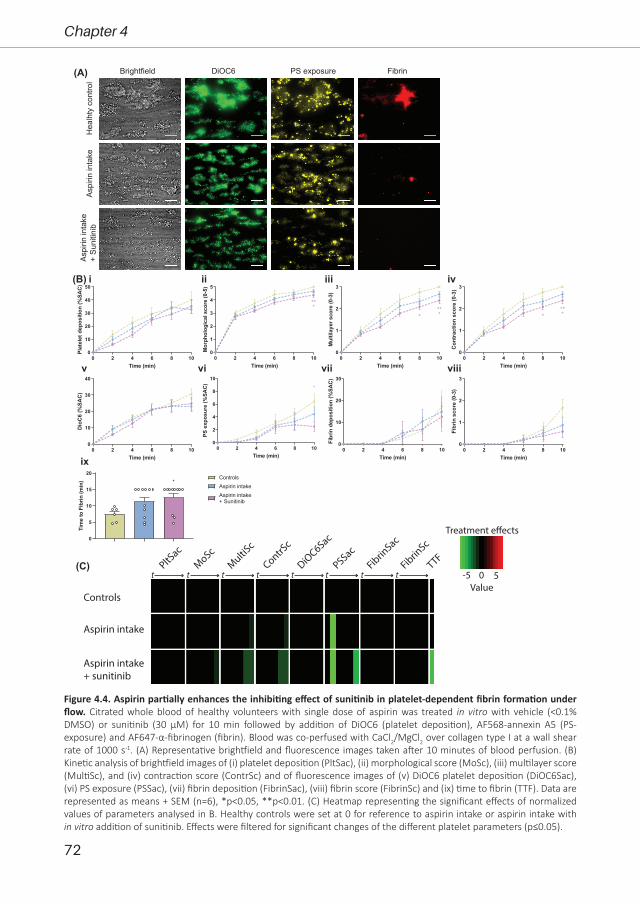
72
Chapter 4
0 2 4 6 8 100
10
20
30
40
50
Time (min)
Plat
elet
depo
sitio
n(%
SAC)
0 2 4 6 8 100
1
2
3
4
5
Time (min)
Mor
phol
ogic
alsc
ore
(0-5
)
***
0 2 4 6 8 100
1
2
3
Time (min)
Mul
tilay
ersc
ore
(0-3
)
***
*
0 2 4 6 8 100
1
2
3
Time (min)
Con
trac
tion
scor
e(0
-3)
***
*
0 2 4 6 8 100
10
20
30
40
Time (min)
Dio
C6
(%SA
C)
0 2 4 6 8 100
2
4
6
8
10
Time (min)
PSex
posu
re(%
SAC
)
*
0 2 4 6 8 100
10
20
30
Time (min)
Fibr
inde
posi
tion
(%SA
C)
0 2 4 6 8 100
1
2
3
Time (min)Fi
brin
scor
e(0
-3)
0
5
10
15
20
Tim
eto
Fibr
in(m
in)
Aspirin intakeAspirin intake+ Sunitinib
Controls*
i ii iii iv
v vi vii viii
ix
(A)H
ealh
ty c
ontro
lAs
pirin
inta
ke+
Suni
tinib
Brightfield DiOC6 PS exposure
Aspi
rin in
take
Fibrin
PltSac
MoScMultiS
c
ContrSc
DiOC6Sac
PSSacFibrin
Sac
FibrinSc
TTF
Aspirin intake
Aspirin intake+ sunitinib
t t t t t t t t
Controls
(C)-5 50
Treatment e�ects
Value
(B)
Figure 4.4. Aspirin partially enhances the inhibiting effect of sunitinib in platelet-dependent fibrin formation under flow. Citrated whole blood of healthy volunteers with single dose of aspirin was treated in vitro with vehicle (<0.1% DMSO) or sunitinib (30 µM) for 10 min followed by addition of DiOC6 (platelet deposition), AF568-annexin A5 (PS-exposure) and AF647-α-fibrinogen (fibrin). Blood was co-perfused with CaCl2/MgCl2 over collagen type I at a wall shear rate of 1000 s-1. (A) Representative brightfield and fluorescence images taken after 10 minutes of blood perfusion. (B) Kinetic analysis of brightfield images of (i) platelet deposition (PltSac), (ii) morphological score (MoSc), (iii) multilayer score (MultiSc), and (iv) contraction score (ContrSc) and of fluorescence images of (v) DiOC6 platelet deposition (DiOC6Sac), (vi) PS exposure (PSSac), (vii) fibrin deposition (FibrinSac), (viii) fibrin score (FibrinSc) and (ix) time to fibrin (TTF). Data are represented as means + SEM (n=6), *p<0.05, **p<0.01. (C) Heatmap representing the significant effects of normalized values of parameters analysed in B. Healthy controls were set at 0 for reference to aspirin intake or aspirin intake with in vitro addition of sunitinib. Effects were filtered for significant changes of the different platelet parameters (p≤0.05).

73
Sunitinib and aspirin in platelet-induced coagulation
4
Aspirin enhances sunitinib effects on thrombus fibrin formationAs we observed that the exposure of procoagulant PS was reduced by sunitinib (plus
aspirin), we further investigated whether these effects extended to conditions of thrombin
generation and fibrin formation. Hence, we assessed the kinetics of platelet-fibrin thrombus
formation under flow on a collagen surface co-coated with TF18. Recalcified whole blood was
labelled with DiOC6, AF568-Annexin A5 and AF647-fibrinogen, and was perfused over a
microspot consisting of collagen type I plus TF. Using control blood samples, we observed time-
dependent increases in platelet deposition and thrombus formation in time, with the start of
fibrin formation after 7-8 minutes (Figure 4.4A). Preincubation of blood with sunitinib did not
affect the parameters of platelet deposition and thrombus size in time, but sunitinib significantly
reduced PS exposure with a concomitant delay in fibrin formation (Suppl. Figure 4.5). Of note,
sunitinib did not affect thrombin generation under static conditions, as measured with the
calibrated automatic thrombogram26 in PRP stimulated with collagen (data not shown). Markedly,
using blood from aspirin-treated healthy volunteers, we observed a reduction in thrombus size,
height and density as compared to healthy volunteers without aspirin intake (Figure 4.4A, B).
Furthermore, a slight decrease in PS exposure as well as a delay in fibrin formation was observed
upon aspirin intake (Figure 4.4). These effects were further reduced by in vitro addition of sunitinib
(Figure 4.4C). Especially, effects on thrombus height, contraction, PS exposure and time to fibrin
generation were increased upon dual treatment with aspirin and sunitinib as compared to aspirin
alone. Together, these results suggest that sunitinib reduces GPVI-induced activation of platelets
resulting in decreased PS exposure and thereby delaying the time for fibrin to be formed, which
is enhanced by aspirin intake.
Sunitinib reduces platelet-dependent fibrin formation under flow in whole blood from cancer patients
Previously, we demonstrated that sunitinib reduced thrombus formation and PS exposure
in whole blood from renal cell carcinoma (RCC) patients under non-coagulating conditions5. To
explore whether sunitinib could also reduce platelet-dependent fibrin formation under flow in
blood from cancer patients, we obtained blood from 11 patients diagnosed with lung, pancreatic
and (neuro)glioblastoma cancer (Suppl. Table 4.1). Sunitinib has been reported to be used as
a (combination) therapeutic strategy for these types of cancer27-29. As a proof-of-principle, we
preincubated recalcified whole blood from these patients with vehicle or sunitinib followed by
labelling with DiOC6, AF568-Annexin A5 and AF647-fibrinogen. Upon perfusion of the vehicle-
treated blood over collagen co-coated with TF, we observed increases in platelet deposition and
thrombus formation in time, with a median start of fibrin formation after 7-8 minutes, comparable
to healthy controls (Suppl. Figure 4.6A, B). Three patients were treated with anticoagulant
or antiplatelet drugs (Suppl. Table 4.1), but this did not affect thrombus/fibrin formation as
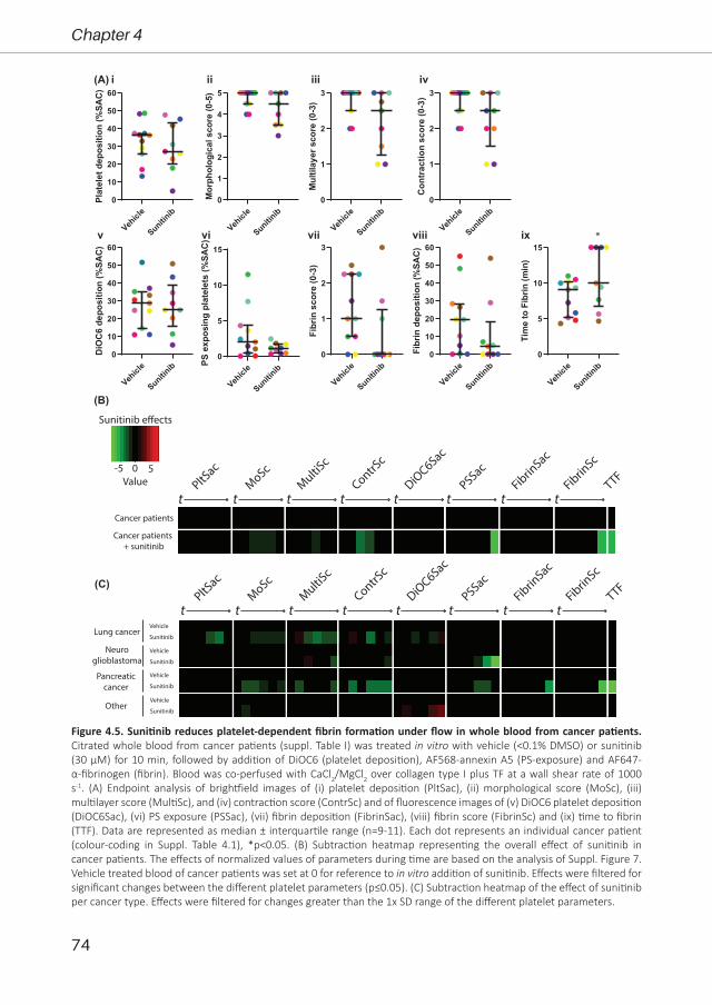
74
Chapter 4
-5 50
Sunitinib e�ects
Value
(B)
Vehicl
e
Sunitinib
0
10
20
30
40
50
60Pl
atel
etde
posi
tion
(%SA
C)
Vehicl
e
Sunitinib
0
1
2
3
4
5
Mor
phol
ogic
alsc
ore
(0-5
)
Vehicl
e
Sunitinib
0
1
2
3
Mul
tilay
ersc
ore
(0-3
)
Vehicl
e
Sunitinib
0
1
2
3
Con
trac
tion
scor
e(0
-3)
Vehicl
e
Sunitinib
0
10
20
30
40
50
60
DiO
C6
depo
sitio
n(%
SAC)
Vehicl
e
Sunitinib
0
5
10
15
PSex
posi
ngpl
atel
ets
(%SA
C)
Vehicl
e
Sunitinib
0
1
2
3
Fibr
insc
ore
(0-3
)
Vehicl
e
Sunitinib
0
10
20
30
40
50
60
Fibr
inde
posi
tion
(%SA
C)
Vehicl
e
Sunitinib
0
5
10
15
Tim
eto
Fibr
in(m
in)
(A) i ii iii iv
v vi vii viii ix *
PltSac
MoScMultiS
c
ContrSc
DiOC6Sac
PSSacFibrin
Sac
FibrinSc
TTF
t t t t t t t tLung cancer
Neuroglioblastoma
Pancreaticcancer
Other
Vehicle
Sunitinib
Vehicle
Sunitinib
Vehicle
Sunitinib
Vehicle
Sunitinib
PltSac
MoScMultiS
c
ContrSc
DiOC6Sac
PSSacFibrin
Sac
FibrinSc
TTF
t t t t t t t tCancer patients
Cancer patients + sunitinib
(C)
Figure 4.5. Sunitinib reduces platelet-dependent fibrin formation under flow in whole blood from cancer patients. Citrated whole blood from cancer patients (suppl. Table I) was treated in vitro with vehicle (<0.1% DMSO) or sunitinib (30 µM) for 10 min, followed by addition of DiOC6 (platelet deposition), AF568-annexin A5 (PS-exposure) and AF647-α-fibrinogen (fibrin). Blood was co-perfused with CaCl2/MgCl2 over collagen type I plus TF at a wall shear rate of 1000 s-1. (A) Endpoint analysis of brightfield images of (i) platelet deposition (PltSac), (ii) morphological score (MoSc), (iii) multilayer score (MultiSc), and (iv) contraction score (ContrSc) and of fluorescence images of (v) DiOC6 platelet deposition (DiOC6Sac), (vi) PS exposure (PSSac), (vii) fibrin deposition (FibrinSac), (viii) fibrin score (FibrinSc) and (ix) time to fibrin (TTF). Data are represented as median ± interquartile range (n=9-11). Each dot represents an individual cancer patient (colour-coding in Suppl. Table 4.1), *p<0.05. (B) Subtraction heatmap representing the overall effect of sunitinib in cancer patients. The effects of normalized values of parameters during time are based on the analysis of Suppl. Figure 7. Vehicle treated blood of cancer patients was set at 0 for reference to in vitro addition of sunitinib. Effects were filtered for significant changes between the different platelet parameters (p≤0.05). (C) Subtraction heatmap of the effect of sunitinib per cancer type. Effects were filtered for changes greater than the 1x SD range of the different platelet parameters.
75
Sunitinib and aspirin in platelet-induced coagulation
4
compared to the patients without this treatment. Preincubation of blood with sunitinib showed
a trend in overall inhibition of thrombus size and height in time, as well as a reduction in PS
exposure with a significant delay in fibrin formation (Figure 4.5A and Suppl. Figure 4.6A, B). To
visualize the overall effects of sunitinib in blood from cancer patients, we generated a heatmap
(Figure 4.5B). This heatmap clearly shows that sunitinib inhibits platelet PS-exposure and fibrin
formation, but also some effects on thrombus formation parameters can be observed. We
included patients with different types of cancer, which this may result in different cancer-specific
responses. Therefore, we also generated a heatmap with the patients averaged per cancer type
(Figure 4.5C). This heatmap shows that in lung and pancreatic cancer, more pronounced effects
of sunitinib were observed on thrombus formation parameters (i.e. height, size and contraction)
as compared to neuroglioblastoma and other cancer types. This variable response to sunitinib
treatment was even more pronounced when a heatmap was generated per patient (suppl. Figure
4.6C). This heatmap shows that the effects of sunitinib on thrombus and fibrin formation under
flow varied more between individual patients than between the types of cancer. Overall, in 6 of
the 9 patients were sunitinib was added we observed clear inhibition of thrombus formation
and/or PS exposure and/or fibrin generation. This suggested that the in vitro inhibitory effects of
sunitinib were more pronounced in cancer patients (Figure 4.5C and suppl. Figure 4.6C) than in
healthy controls (Suppl. Figure 4.5).
DiscussionIn the present study we further explored the multi-target inhibition of sunitinib on
thrombus formation, by investigating thrombus formation by stimulation with multiple agonists,
as well as under coagulating conditions stimulated by collagen plus TF. We observed that sunitinib
predominantly inhibited collagen-induced exposure of phosphatidylserine, resulting in delayed
fibrin formation. Furthermore, we found an enhancing effect of dual treatment of platelets with
sunitinib and aspirin in these processes. Finally, platelet-dependent fibrin formation in whole
blood from cancer patients was significantly inhibited by sunitinib.
We have previously reported that platelets sequester sunitinib in vitro as well as in cancer
patients and that this uptake inhibits platelet function in response to collagen5. As sunitinib is a
multi-target TKI and TKs do not only signal under the collagen receptor GPVI, but also under the
receptors GPIb, CLEC-2 and integrins α6β1 and αIIbβ321, we investigated the effects of sunitinib on
thrombus formation in whole blood perfused over multiple agonists. However, sunitinib did not
affect thrombus formation on CLEC-2, GPIb- and integrin αIIbβ3/α6β1-dependent surfaces, which
pointed to GPVI as the main signalling pathway inhibited by this TKI. We did observe that sunitinib
inhibited CLEC-2-induced aggregation and activation responses in isolated platelets. This is in line
with the many similarities in the downstream signalling pathways underneath GPVI and CLEC-225.
The inhibiting effects of sunitinib on CLEC-2-induced thrombus formation were however absent in
76
Chapter 4
whole blood. This may be explained by a stronger dependence on Syk of CLEC-2 signalling, while
GPVI signalling is more dependent on Src25. Inhibition of Src by sunitinib upon GPVI stimulation
was more pronounced as compared to inhibition of Syk (Figure 4.2A), suggesting that the effects
of sunitinib on CLEC-2 signalling may be less prominent, especially in whole blood conditions.
Furthermore, we noted that higher collagen doses overrule the inhibiting effects of sunitinib at
10 μM. Hence, a higher concentration of sunitinib may be required to inhibit platelet activation
at higher agonist concentrations. A possible role for integrin α2β1, the other collagen receptor on
platelets, should be mentioned here as well, as this receptor also signals via Src and Syk kinases30.
It has been shown that integrin α2β1 has a supporting role with regard to platelet activation and
contributes to stable thrombus formation on collagen31. As we have shown that sunitinib strongly
reduced phosphorylation of Syk and Src, the inhibiting effects observed in thrombus formation
on collagen may be partly due to inhibition of integrin α2β1 signalling. In addition, thrombus
formation under flow on collagen type III was reduced by sunitinib treatment, although not as
strong as compared to thrombus formation on collagen type I. Together, this is consistent with
the multi-target nature of sunitinib as a broad-spectrum protein TKI, having lower affinity for
multiple TKs rather than high affinity for one specific signalling protein32, 33. This also explains why
the effects of sunitinib on platelet activation are not as strong as compared to specific inhibitors
of Syk or Src34, 35.
We showed that in total 34 TKs were activated upon GPVI stimulation, which were
subsequently inhibited in the presence of sunitinib. Several TKs could be directly linked to GPVI
signalling, such as Syk and the SFKs, but most TKs were most likely activated by agonists secreted
from the α-granules upon GPVI stimulation, such as fibrinogen, activating FAK1/2 via integrin
αIIbβ323, as well as Gas6, activating Tyro3/Sky. As platelets contain many bioactive molecules in their
granules, most likely also the other non-ITAM-linked TKs that showed phosphorylation upon GPVI
stimulation, have been activated via similar secondary events. As we have shown that platelet
secretion is inhibited by sunitinib5, the inhibiting effects of sunitinib on non-GPVI-linked TKs may
also be due to reduced secretion of secondary mediators. Prolonged elevation of intracellular
calcium levels is a prerequisite for platelet PS exposure, a hallmark of procoagulant activity24. The
observed effects of sunitinib on intracellular calcium rises in response to GPVI stimulation fits with
the currently observed reduction in TK phosphorylation and agrees with previously shown effects
on collagen-induced tyrosine phosphorylation of multiple proteins5. This implies interference
of sunitinib early in the signalling cascade used by the collagen receptor GPVI, e.g. with TKs in
the GPVI signalosome or with Syk as a central regulator of collagen-induced platelet activation.
In agreement, another study reported that collagen-induced tyrosine phosphorylation of c-Src
is reduced in platelets treated with sunitinib6. Others have shown that also in cardiomyocytes
sunitinib treatment results in reduced calcium responses accompanied with decreased sarcomere
shortening36. We showed that sunitinib reduced platelet PS exposure in isolated platelets in vitro,

77
Sunitinib and aspirin in platelet-induced coagulation
4
as well as during whole blood thrombus formation under flow in the presence and absence of
coagulation. This reduction in platelet procoagulant activity upon sunitinib treatment resulted in
delayed and reduced fibrin clot formation. An earlier study with other TKIs has also demonstrated
that platelet tyrosine phosphorylation is required for procoagulant activity and fibrin formation37.
As both aspirin and sunitinib are associated with increased bleeding risk3, 9, we investigated
if the combination of these drugs could reinforce the inhibiting effects on platelets. We observed
that indeed this aspirin-sunitinib combination resulted in stronger inhibition of collagen-induced
platelet aggregation as compared to either compound alone. However, in whole blood perfusion
experiments (with and without coagulation) we did not observe these additive effects when
aspirin and sunitinib were added in vitro. In contrast, platelet deposition and thrombus height
were reduced in blood from aspirin-treated donors as compared to controls without aspirin
intake. Moreover, addition of sunitinib further enhanced the effects that were observed with
aspirin alone, accompanied by further reductions in PS exposure and fibrin formation. In an
earlier study from our group with patients suffering from peripheral artery disease (PAD), no
effects of aspirin intake were observed on thrombus formation under flow as compared to
healthy controls without aspirin intake38. This could be due to the increased platelet reactivity
observed in these PAD patients, that was counterbalanced by the aspirin intake. In contrast,
in this study in vitro addition of aspirin to blood from healthy volunteers did reduce thrombus
formation and PS exposure under flow38, which was also observed by others39, 40. The present
findings are compatible to the variable effects of aspirin intake alone on thrombus formation,
while the combination of aspirin with other anti-platelet drugs causes a marked reduction41-45.
Furthermore, a recent study has shown concomitant effects of aspirin and ibrutinib on collagen-
induced aggregation46. Altogether, these results suggest that dual treatment with anti-platelet
therapy and TKIs increase the inhibition of platelet function.
In the present study we included a limited number of cancer patients, who suffered from
different cancer types. Also, we added the sunitinib in vitro, which may be a limitation to the
current study. The effects of sunitinib on platelet-dependent coagulation in blood from patients
treated with sunitinib requires further study. Also, the combined effects of sunitinib and aspirin or
other anti-platelet drugs on platelets from patients on treatment in relation to bleeding should be
further investigated. Despite these limitations, we clearly observed in this proof-of-principle study
that the in vitro inhibitory effects of sunitinib on thrombus size and height under coagulating
conditions were more pronounced in several individual cancer patients as compared to healthy
controls. It is known that cancer patients have a procoagulant phenotype47, 48, and therefore
platelets from cancer patients may be more sensitive to inhibitory compounds as compared to
platelets from healthy individuals. We also observed more pronounced effects of sunitinib on
thrombus formation parameters in lung and pancreatic cancer as compared to neuroglioblastoma
and other cancer types. It has been reported that tumour-induced changes in platelet proteins

78
Chapter 4
and mRNA are tumour-type specific49, 50, hence we hypothesize that these specific tumour-
induced changes may influence the platelet response to inhibiting drugs.
Altogether, we conclude that sunitinib inhibited GPVI-induced phosphorylation of TKs (a.o.
Syk and Src), with subsequent reduction of Ca2+ elevation, resulting in reduced platelet activation
and PS-exposure. This was accompanied by delayed and reduced formation of fibrin, all of which
were aggravated in the presence of aspirin. This asks for awareness among clinicians for the
combined antiplatelet effects of TKIs together with anti-platelet drugs.
AcknowledgementsThis study was supported by Pfizer as an Investigator-Initiated Research grant to MJEK
(Tracking Number WI209458) and funds from the Department of Medicine, University of Padua
Medical School (to PS). DIF received funding from the European Union’s Horizon 2020 research
and innovation program under Marie Sklodowska-Curie grant agreement No. 766118 and is
enrolled in a joint PhD program of the universities of Maastricht and Santiago de Compostela. JAE
was funded by the Deutsche Forschungsgemeinschaft (DFG grant: SFP1009 project A09). EPCvdV
was funded by a grant from the Interdisciplinary Center for Clinical Research within the faculty of
Medicine at the RWTH Aachen University and NWO-ZonMw Veni (91619053).
Conflict of Interest StatementJWMH is a cofounder and shareholder of FlowChamber B.V. The other authors declare no
relevant conflicts of interest.
References1. Goodman VL, Rock EP, Dagher R, et al. Approval summary: sunitinib for the treatment of imatinib
refractory or intolerant gastrointestinal stromal tumors and advanced renal cell carcinoma. Clin Cancer Res 2007; 13(5): 1367-73.
2. Adams VR, Leggas M. Sunitinib malate for the treatment of metastatic renal cell carcinoma and gastrointestinal stromal tumors. Clin Ther 2007; 29(7): 1338-53.
3. Je Y, Schutz FA, Choueiri TK. Risk of bleeding with vascular endothelial growth factor receptor tyrosine-kinase inhibitors sunitinib and sorafenib: a systematic review and meta-analysis of clinical trials. Lancet Oncol 2009; 10(10): 967-74.
4. Versteeg HH, Heemskerk JW, Levi M, et al. New fundamentals in hemostasis. Physiol Rev 2013; 93(1): 327-58.
5. Sabrkhany S, Griffioen AW, Pineda S, et al. Sunitinib uptake inhibits platelet function in cancer patients. Eur J Cancer 2016; 66: 47-54.
6. Walraven M, Homs MYV, van der Veldt AAM, et al. Platelet function is disturbed by the angiogenesis inhibitors sunitinib and sorafenib, but unaffected by bevacizumab. Angiogenesis 2018; 21(2): 325-34.
7. De Stefano V. Arterial thrombosis and cancer: the neglected side of the coin of Trousseau syndrome. Haematologica 2018; 103(9): 1419-21.
8. Navi BB, Reiner AS, Kamel H, et al. Risk of Arterial Thromboembolism in Patients With Cancer. J Am Coll Cardiol 2017; 70(8): 926-38.
9. Silvain J, Cayla G, O'Connor SA, et al. Antiplatelet options for secondary prevention in acute coronary syndromes. Expert Rev Cardiovasc Ther 2011; 9(11): 1403-15.
79
Sunitinib and aspirin in platelet-induced coagulation
4
10. Arthur JF, Jandeleit-Dahm K, Andrews RK. Platelet Hyperreactivity in Diabetes: Focus on GPVI Signaling-Are Useful Drugs Already Available? Diabetes 2017; 66(1): 7-13.
11. Gresele P, Momi S, Malvestiti M, et al. Platelet-targeted pharmacologic treatments as anti-cancer therapy. Cancer Metastasis Rev 2017; 36(2): 331-55.
12. Haemmerle M, Stone RL, Menter DG, et al. The Platelet Lifeline to Cancer: Challenges and Opportunities. Cancer Cell 2018; 33(6): 965-83.
13. van der Meijden PE, Feijge MA, Giesen PL, et al. Platelet P2Y12 receptors enhance signalling towards procoagulant activity and thrombin generation. A study with healthy subjects and patients at thrombotic risk. Thromb Haemost 2005; 93(6): 1128-36.
14. Feijge MA, van Pampus EC, Lacabaratz-Porret C, et al. Inter-individual variability in Ca2+ signalling in platelets from healthy volunteers: effects of aspirin and relationship with expression of endomembrane Ca2+-ATPases. Br J Haematol 1998; 102(3): 850-9.
15. Grynkiewicz G, Poenie M, Tsien RY. A new generation of Ca2+ indicators with greatly improved fluorescence properties. J Biol Chem 1985; 260(6): 3440-50.
16. Chirumamilla CS, Fazil M, Perez-Novo C, et al. Profiling Activity of Cellular Kinases in Migrating T-Cells. Methods Mol Biol 2019; 1930: 99-113.
17. de Witt SM, F. S, J. C, et al. Multi-parameter assessment of thrombus formation on microspotted arrays of thrombogenic surfaces. Nat Prot Exchange 2014.
18. Swieringa F, Baaten CC, Verdoold R, et al. Platelet Control of Fibrin Distribution and Microelasticity in Thrombus Formation Under Flow. Arterioscler Thromb Vasc Biol 2016; 36(4): 692-9.
19. Numakura K, Fujiyama N, Takahashi M, et al. Clinical implications of pharmacokinetics of sunitinib malate and N-desethyl-sunitinib plasma concentrations for treatment outcome in metastatic renal cell carcinoma patients. Oncotarget 2018; 9(38): 25277-84.
20. Swieringa F, Kuijpers MJ, Heemskerk JW, et al. Targeting platelet receptor function in thrombus formation: the risk of bleeding. Blood Rev 2014; 28(1): 9-21.
21. Tullemans BME, Heemskerk JWM, Kuijpers MJE. Acquired platelet antagonism: off-target antiplatelet effects of malignancy treatment with tyrosine kinase inhibitors. J Thromb Haemost 2018; 16(9): 1686-99.
22. de Witt SM, Swieringa F, Cavill R, et al. Identification of platelet function defects by multi-parameter assessment of thrombus formation. Nat Commun 2014; 5: 4257.
23. Guidetti GF, Torti M, Canobbio I. Focal Adhesion Kinases in Platelet Function and Thrombosis. Arterioscler Thromb Vasc Biol 2019; 39(5): 857-68.
24. Fernandez DI, Kuijpers MJE, Heemskerk JWM. Platelet calcium signaling by G-protein coupled and ITAM-linked receptors regulating anoctamin-6 and procoagulant activity. Platelets 2020: 1-9.
25. Rayes J, Watson SP, Nieswandt B. Functional significance of the platelet immune receptors GPVI and CLEC-2. J Clin Invest 2019; 129(1): 12-23.
26. Hemker HC, Giesen P, AlDieri R, et al. The calibrated automated thrombogram (CAT): a universal routine test for hyper- and hypocoagulability. Pathophysiol Haemost Thromb 2002; 32(5-6): 249-53.
27. Qiang H, Chang Q, Xu J, et al. New advances in antiangiogenic combination therapeutic strategies for advanced non-small cell lung cancer. J Cancer Res Clin Oncol 2020; 146(3): 631-45.
28. Faivre S, Niccoli P, Castellano D, et al. Sunitinib in pancreatic neuroendocrine tumors: updated progression-free survival and final overall survival from a phase III randomized study. Ann Oncol 2017; 28(2): 339-43.
29. Grisanti S, Ferrari VD, Buglione M, et al. Second line treatment of recurrent glioblastoma with sunitinib: results of a phase II study and systematic review of literature. J Neurosurg Sci 2019; 63(4): 458-67.
30. Inoue O, Suzuki-Inoue K, Dean WL, et al. Integrin alpha2beta1 mediates outside-in regulation of platelet spreading on collagen through activation of Src kinases and PLCgamma2. J Cell Biol 2003; 160(5): 769-80.
31. Kuijpers MJ, Pozgajova M, Cosemans JM, et al. Role of murine integrin alpha2beta1 in thrombus stabilization and embolization: contribution of thromboxane A2. Thromb Haemost 2007; 98(5): 1072-80.
32. Faivre S, Demetri G, Sargent W, et al. Molecular basis for sunitinib efficacy and future clinical development. Nat Rev Drug Discov 2007; 6(9): 734-45.
80
Chapter 4
33. Karaman MW, Herrgard S, Treiber DK, et al. A quantitative analysis of kinase inhibitor selectivity. Nat Biotechnol 2008; 26(1): 127-32.
34. Jooss NJ, De Simone I, Provenzale I, et al. Role of Platelet Glycoprotein VI and Tyrosine Kinase Syk in Thrombus Formation on Collagen-Like Surfaces. Int J Mol Sci 2019; 20(11).
35. Auger JM, Kuijpers MJ, Senis YA, et al. Adhesion of human and mouse platelets to collagen under shear: a unifying model. FASEB J 2005; 19(7): 825-7.
36. Rainer PP, Doleschal B, Kirk JA, et al. Sunitinib causes dose-dependent negative functional effects on myocardium and cardiomyocytes. BJU Int 2012; 110(10): 1455-62.
37. Diaz-Ricart M, Palomo M, Fuste B, et al. Inhibition of tyrosine kinase activity prevents the adhesive and cohesive properties of platelets and the expression of procoagulant activity in response to collagen. Thromb Res 2008; 121(6): 873-83.
38. van Geffen JP, Kleinegris MC, Verdoold R, et al. Normal platelet activation profile in patients with peripheral arterial disease on aspirin. Thromb Res 2015; 135(3): 513-20.
39. Li R, Diamond SL. Detection of platelet sensitivity to inhibitors of COX-1, P2Y(1), and P2Y(1)(2) using a whole blood microfluidic flow assay. Thromb Res 2014; 133(2): 203-10.
40. Hosokawa K, Ohnishi T, Sameshima H, et al. Analysing responses to aspirin and clopidogrel by measuring platelet thrombus formation under arterial flow conditions. Thromb Haemost 2013; 109(1): 102-11.
41. Bossavy JP, Thalamas C, Sagnard L, et al. A double-blind randomized comparison of combined aspirin and ticlopidine therapy versus aspirin or ticlopidine alone on experimental arterial thrombogenesis in humans. Blood 1998; 92(5): 1518-25.
42. Cadroy Y, Bossavy JP, Thalamas C, et al. Early potent antithrombotic effect with combined aspirin and a loading dose of clopidogrel on experimental arterial thrombogenesis in humans. Circulation 2000; 101(24): 2823-8.
43. Mendolicchio GL, Zavalloni D, Bacci M, et al. Variable effect of P2Y12 inhibition on platelet thrombus volume in flowing blood. J Thromb Haemost 2011; 9(2): 373-82.
44. Andre P, LaRocca T, Delaney SM, et al. Anticoagulants (thrombin inhibitors) and aspirin synergize with P2Y12 receptor antagonism in thrombosis. Circulation 2003; 108(21): 2697-703.
45. Sakakibara M, Goto S, Eto K, et al. Application of ex vivo flow chamber system for assessment of stent thrombosis. Arterioscler Thromb Vasc Biol 2002; 22(8): 1360-4.
46. Ninomoto J, Mokatrin A, Kinoshita T, et al. Effects of ibrutinib on in vitro platelet aggregation in blood samples from healthy donors and donors with platelet dysfunction. Hematology 2020; 25(1): 112-7.
47. Sheth RA, Niekamp A, Quencer KB, et al. Thrombosis in cancer patients: etiology, incidence, and management. Cardiovasc Diagn Ther 2017; 7(Suppl 3): S178-s85.
48. Hisada Y, Mackman N. Cancer-associated pathways and biomarkers of venous thrombosis. Blood 2017; 130(13): 1499-506.
49. Sabrkhany S, Kuijpers MJE, van Kuijk SMJ, et al. A combination of platelet features allows detection of early-stage cancer. Eur J Cancer 2017; 80: 5-13.
50. Best MG, Sol N, Kooi I, et al. RNA-Seq of tumor-educated platelets enables blood-based pan-cancer, multiclass, and molecular pathway cancer diagnostics. Cancer Cell 2015; 28(5): 666-76.
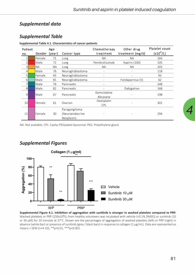
81
Sunitinib and aspirin in platelet-induced coagulation
4
Supplemental data
Supplemental Table
0
20
40
60
80
100
Collagen (1 µg/ml)
Agg
rega
tion
(%)
WP PRP
Vehicle
Sunitinib 10 µM
Sunitinib 30 µM
**
**
***
**
Supplemental Figure 4.1. Inhibition of aggregation with sunitinib is stronger in washed platelets compared to PRP. Washed platelets or PRP (250x10⁹/L) from healthy volunteers was incubated with vehicle (<0.1% DMSO) or sunitinib (10 or 30 µM) for 10 minutes at 37°C. Shown are the percentages of aggregation of washed platelets (left) or PRP (right) in absence (white bar) or presence of sunitinib (grey / black bars) in response to collagen (1 µg/mL). Data are represented as means + SEM (n=4-10), **p<0.01, ***p<0.001.
GenderAge (year) Cancer type
Chemotherapy treatment
Other drug treatment (mg/d)
Platelet count (x109/L)
1 Female 71 Lung NA NA 1642 Male 72 Lung Pembrolizumab Aspirin (100) 1053 NA NA Lung NA NA 2034 Male 76 Neuroglioblastoma - - 1185 Female 45 Neuroglioblastoma - - 936 Male 81 Neuroglioblastoma - Fondaparinux (5) 627 Male 76 Pancrea�c - - 2488 Male 82 Pancrea�c - Dabigatran 168
11 Female 30Paraganglioma (Neuroendocrine Neoplasm)
- - 294
Pa�ent no.
9
- 301
GemcitabineAbraxane
Oxalipla�nCPL
10 Female 61 Ovarian
Male 67 Pancrea�c - 198
Supplemental Table 4.1. Characteristics of cancer patients
NA: Not available; CPL: Caelyx PEGylated liposomal; PEG: Polyethylene glycol
Supplemental Figures
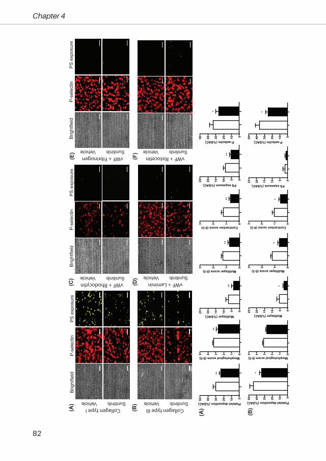
82
Chapter 4
Brig
htfie
ldP-
sele
ctin
PS e
xpos
ure
Vehicle Sunitinib
(A) Vehicle Sunitinib
Vehicle SunitinibVehicle Sunitinib
Vehicle Sunitinib Vehicle Sunitinib
Collagen type I Collagen type III
vWF + Ristocetin
vWF + Rhodocytin vWF + Laminin
vWF + Fibrinogen
(B)
(C)
(D)
(E)
(F)
Brig
htfie
ldP-
sele
ctin
PS e
xpos
ure
01020304050 Platelet deposition (%SAC)
*
012345 Morphological score (0-5)
0510152025 Multilayer (%SAC)
*
0123 Multilayer score (0-3)
*
0123 Contraction score (0-3)
*
0510152025 PS exposure (%SAC)
01020304050 P-selectin (%SAC)
*
01020304050 Platelet deposition (%SAC)
**
012345 Morphological score (0-5)
**
0510152025 Multilayer (%SAC)
**
0123 Multilayer score (0-3)
**
0123 Contraction score (0-3)**
0510152025 PS exposure (%SAC)
**
01020304050 P-selectin (%SAC)
*
(A)
(B)
Brig
htfie
ldP-
sele
ctin
PS e
xpos
ure
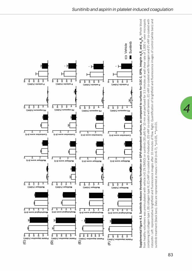
83
Sunitinib and aspirin in platelet-induced coagulation
4
Supp
lem
enta
l Fig
ure
4.2.
Sun
itini
b re
duce
s th
rom
bus
form
ation
on
GPV
I-dep
ende
nt s
urfa
ces,
as
com
pare
d to
sur
face
s fo
r CLE
C-2,
GPI
b, in
tegr
in α
6ß 1 and
αIIb
ß 3. Who
le b
lood
fr
om h
ealth
y vo
lunt
eers
was
pre
incu
bate
d w
ith v
ehic
le (<
0.1%
DM
SO) o
r sun
itini
b (3
0 µM
) for
10
min
and
per
fuse
d fo
r 3.5
min
utes
at w
all s
hear
rate
of 1
,000
s-1
ove
r mic
rosp
ots
cont
aini
ng (A
) col
lage
n ty
pe I,
(B) c
olla
gen
type
III,
(C) v
WF
co-c
oate
d w
ith rh
odoc
ytin,
(D) v
WF
co-c
oate
d w
ith la
min
in, (
E) v
WF
co-c
oate
d w
ith fi
brin
ogen
and
(F) v
WF
co-c
oate
d w
ith
risto
cetin
. Left
: rep
rese
ntati
ve b
rightf
ield
and
fluo
resc
ence
imag
es a
fter 1
0 m
inut
es o
f flow
. Rig
ht: Q
uanti
ficati
on o
f brig
htfie
ld a
nd fl
uore
scen
ce im
ages
of c
ontr
ol (w
hite
bar
s) a
nd
suni
tinib
trea
tmen
t (bl
ack
bars
). Da
ta a
re re
pres
ente
d as
mea
ns +
SEM
(n=6
-7),
*p<0
.05,
**p
<0.0
1.
Vehi
cle
Suni
tinib
01020304050 Platelet deposition (%SAC)
012345 Morphological score (0-5)
0510152025 Multilayer (%SAC)
0123 Multilayer score (0-3)
0123 Contraction score (0-3)
0510152025 PS exposure (%SAC)
01020304050 P-selectin (%SAC)
*
01020304050 Platelet deposition (%SAC)
012345 Morphological score (0-5)
0510152025 Multilayer (%SAC)
0123 Multilayer score (0-3)
0123 Contraction score (0-3)
0510152025 PS exposure (%SAC)
01020304050 P-selectin (%SAC)
01020304050 Platelet deposition (%SAC)
012345 Morphological score (0-5)
0510152025 Multilayer (%SAC)
0123 Multilayer score (0-3)
0123 Contraction score (0-3)
0510152025 PS exposure (%SAC)
01020304050 P-selectin (%SAC)
*
0102030405060 Platelet deposition (%SAC)
*
012345 Morphological score (0-5)
0510152025 Multilayer (%SAC)
0123 Multilayer score (0-3)
0123 Contraction score (0-3)
0510152025 PS exposure (%SAC)
01020304050 P-selectin (%SAC)
*
(C)
(D)
(E)
(F)
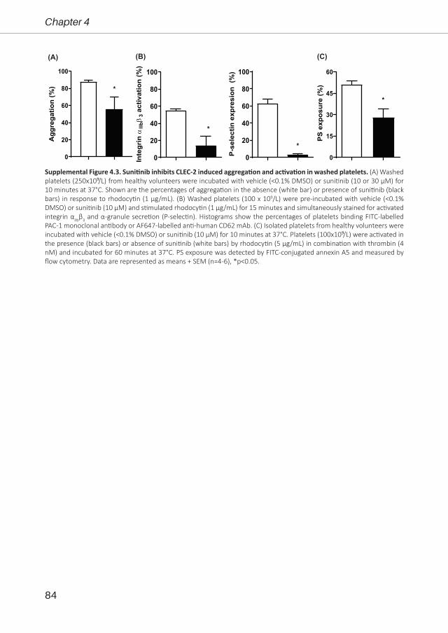
84
Chapter 4
0
20
40
60
80
100
Agg
rega
tion
(%) *
0
20
40
60
80
100
P-se
lect
in e
xpre
sion
(%
)
*0
20
40
60
80
100
αIIbβ
3ac
tivat
ion
(%)
*In
tegr
in
0
15
30
45
60
PS e
xpos
ure
(%)
*
(C)(B)(A)
Supplemental Figure 4.3. Sunitinib inhibits CLEC-2 induced aggregation and activation in washed platelets. (A) Washed platelets (250x10⁹/L) from healthy volunteers were incubated with vehicle (<0.1% DMSO) or sunitinib (10 or 30 µM) for 10 minutes at 37°C. Shown are the percentages of aggregation in the absence (white bar) or presence of sunitinib (black bars) in response to rhodocytin (1 µg/mL). (B) Washed platelets (100 x 109/L) were pre-incubated with vehicle (<0.1% DMSO) or sunitinib (10 µM) and stimulated rhodocytin (1 µg/mL) for 15 minutes and simultaneously stained for activated integrin αIIbβ3 and α-granule secretion (P-selectin). Histograms show the percentages of platelets binding FITC-labelled PAC-1 monoclonal antibody or AF647-labelled anti-human CD62 mAb. (C) Isolated platelets from healthy volunteers were incubated with vehicle (<0.1% DMSO) or sunitinib (10 µM) for 10 minutes at 37°C. Platelets (100x10⁹/L) were activated in the presence (black bars) or absence of sunitinib (white bars) by rhodocytin (5 µg/mL) in combination with thrombin (4 nM) and incubated for 60 minutes at 37°C. PS exposure was detected by FITC-conjugated annexin A5 and measured by flow cytometry. Data are represented as means + SEM (n=4-6), *p<0.05.

85
Sunitinib and aspirin in platelet-induced coagulation
4
ControlsAspirin intakeAspirin intake+ Sunitinib
0
1
2
3
4
5
Mor
phol
ogic
al s
core
(0-5
)
*
0
10
20
30
40
Plat
elet
dep
ositi
on (%
SAC)
***
0
1
2
3
Con
trac
tion
scor
e (0
-3)
*
0
1
2
3
Mul
tilay
er s
core
(0-3
)
*
0
2
4
6
8
10
Mul
tilay
er (%
SAC
)
**
*
0
5
10
15
PS e
xpos
ing
plat
elet
s (%
SAC
)
**
*
0
10
20
30
40
50
P-se
lect
in (%
SAC
)
**
i ii iii iv
v vi vii
(A)C
ontro
lAs
pirin
inta
ke+
Suni
tinib
Brightfield P-selectin PS exposureAs
pirin
inta
ke
(B)
Supplemental Figure 4.4. Aspirin intake enhances the effect of sunitinib in ex vivo thrombus formation on collagen in whole blood. Recalcified whole blood of healthy volunteers or healthy volunteers with single dose of aspirin was treated in vitro with vehicle or sunitinib (30 µM) for 10 min. Subsequently, blood was perfused over a collagen surface at 1000 s-¹ for 3.5 minutes, followed by perfusion with buffer supplemented with AF568-annexin A5 and AF647-α-CD62P to visualize PS exposure and P-selectin expression, respectively. (A) Representative brightfield and fluorescent images. (B) Quantification of brightfield images of (i) platelet deposition, (ii) morphological score, (iii) multilayer (%SAC), (iv) multilayer score, (v) contraction score, and fluorescence images of (vi) PS exposure and (vii) P-selectin expression of vehicle (white bar), aspirin intake (dark grey bars), or aspirin intake with in vitro sunitinib (black bar). Data are represented as means + SEM (n=3-6), *p<0.05, **p<0.01.

86
Chapter 4
PltSac
MoScMultiS
ac
ContrSc
DiOC6Sac
PSSacFibrin
Sac
FibrinSc
TTFt t t t t t t t
Vehicle
Sunitinib
-5 50
Sunitinib e�ects
Value
Supplemental Figure 4.5. The effects of sunitinib on platelet-dependent fibrin formation under flow. Whole blood from healthy volunteers was preincubated with vehicle (<0.1% DMSO) or sunitinib (30 µM) for 10 min. Subsequently, the blood was supplemented with DiOC6, AF568-annexin A5 and AF647-α-fibrinogen to visualize platelet deposition, PS-exposure and fibrin formation, respectively. Blood samples were co-perfused with CaCl2/MgCl2 over collagen type I co-coated with TF microspot at a wall shear rate of 1000 s-1. Subtraction heatmaps representing the significant effects of normalized values of the parameters. Controls with addition of vehicle (<0.1% DMSO) were set at 0 for reference. Effects were filtered for significant alterations by addition of sunitinib (p≤0.05).
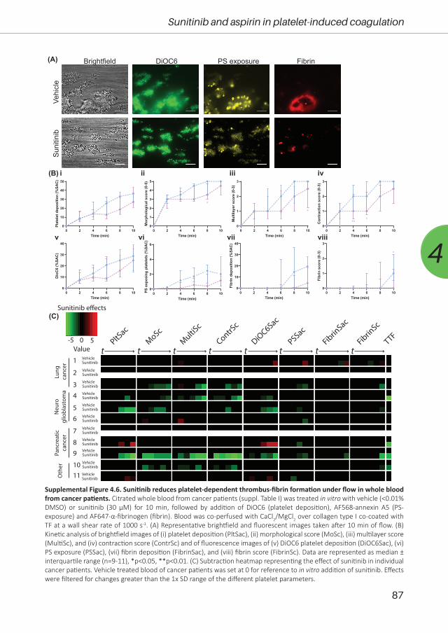
87
Sunitinib and aspirin in platelet-induced coagulation
4
(A)
i ii iii iv
v vi vii viii0 2 4 6 8 10
0
10
20
30
40
50
Time (min)
Plat
elet
depo
sitio
n(%
SAC)
0 2 4 6 8 100
1
2
3
4
5
Time (min)
Mor
phol
ogic
alsc
ore
(0-5
)
*
0 2 4 6 8 100
1
2
3
Time (min)M
ultil
ayer
scor
e(0
-3)
* *
0 2 4 6 8 100
1
2
3
Time (min)
Con
trac
tion
scor
e(0
-3)
***
0 2 4 6 8 100
10
20
30
40
Time (min)
Dio
C6
(%SA
C)
0 2 4 6 8 100
2
4
6
Time (min)
PSex
posi
ngpl
atel
ets
(%SA
C)
0 2 4 6 8 100
10
20
30
40
Time (min)
Fibr
inde
posi
tion
(%SA
C)
0 2 4 6 8 100
1
2
3
Time (min)
Fibr
insc
ore
(0-3
) *
(B)
PltSac
MoScMultiS
c
ContrSc
DiOC6Sac
PSSacFibrin
Sac
FibrinSc
TTF
t t t t t t t t1
2
3
4
5
6
7
89
1011
VehicleSunitinib
VehicleSunitinib
VehicleSunitinib
VehicleSunitinib
VehicleSunitinib
VehicleSunitinib
VehicleSunitinib
VehicleSunitinib
VehicleSunitinib
VehicleSunitinib
VehicleSunitinib
Lung
ca
ncer
Neu
rogl
iobl
asto
ma
Panc
reat
icca
ncer
Oth
er
-5 50
Sunitinib e�ects
Value
(C)
Vehi
cle
Brightfield DiOC6 PS exposureSu
nitin
ibFibrin
Supplemental Figure 4.6. Sunitinib reduces platelet-dependent thrombus-fibrin formation under flow in whole blood from cancer patients. Citrated whole blood from cancer patients (suppl. Table I) was treated in vitro with vehicle (<0.01% DMSO) or sunitinib (30 µM) for 10 min, followed by addition of DiOC6 (platelet deposition), AF568-annexin A5 (PS-exposure) and AF647-α-fibrinogen (fibrin). Blood was co-perfused with CaCl2/MgCl2 over collagen type I co-coated with TF at a wall shear rate of 1000 s-1. (A) Representative brightfield and fluorescent images taken after 10 min of flow. (B) Kinetic analysis of brightfield images of (i) platelet deposition (PltSac), (ii) morphological score (MoSc), (iii) multilayer score (MultiSc), and (iv) contraction score (ContrSc) and of fluorescence images of (v) DiOC6 platelet deposition (DiOC6Sac), (vi) PS exposure (PSSac), (vii) fibrin deposition (FibrinSac), and (viii) fibrin score (FibrinSc). Data are represented as median ± interquartile range (n=9-11), *p<0.05, **p<0.01. (C) Subtraction heatmap representing the effect of sunitinib in individual cancer patients. Vehicle treated blood of cancer patients was set at 0 for reference to in vitro addition of sunitinib. Effects were filtered for changes greater than the 1x SD range of the different platelet parameters.

Chapter 5
Quantitative and qualitative changes in
platelet traits of sunitinib-treated patients with
renal cell carcinoma in relation to circulating
sunitinib levels: a proof-of-concept study
Tullemans BME, Brouns SLN, Swieringa F, Sabrkhany S, van den Berkmortel FWPJ, Peters NJB, de Bruijn P, Koolen SLW,
Heemskerk JWM, Aarts MJB and Kuijpers MJE
Submitted

90
Chapter 5
AbstractBackground: Tyrosine kinase inhibitors (TKIs), such as sunitinib, are used for cancer
treatment, but may also affect platelet count and function with possible hemostatic consequences.
Here, we investigated how patient treatment with the TKI sunitinib affected quantitative and
qualitative platelet traits as a function of the sunitinib level and the occurrence of bleeding.
Methods: Blood was collected from 20 metastatic renal cell carcinoma (mRCC) patients
before treatment, and at 2 weeks, 4 weeks and 3 months after sunitinib administration. We
measured blood cell counts, platelet aggregation, and the concentrations of sunitinib as well as
its N-desethyl metabolite in plasma, serum and isolated platelets. Progression of disease (PD) and
bleeding were monitored after 3 months.
Results: In sunitinib-treated mRCC patients, concentrations of (N-desethyl)sunitinib
in plasma and serum were highly correlated. In the patients' platelets the active metabolite
levels were relatively increased as compared to sunitinib. On average, a sustained reduction in
platelet count was observed on-treatment, which was significantly related to the inhibitor levels
in plasma/serum. Principal component and correlational analysis showed that the (N-desethyl-)
sunitinib levels in plasma/serum were linked to a reduction in both platelet count and collagen-
induced platelet aggregation. The reduced aggregation associated in part with reported bleeding,
but did not correlate to PD.
Conclusions: The sunitinib-induced reduction in quantitative and qualitative platelet traits
may reflect the effective sunitinib levels in the patient. These results may serve as a proof-of-
principle for other TKI-related drugs, where both platelet count and functions are affected, which
could be used therapeutic drug monitoring.

91
Changes in platelet traits and circulating levels of sunitinib-treated patients
5
IntroductionIn the past two decades, over 40 tyrosine kinase inhibitors (TKIs) have been developed
and approved for the treatment of many cancer types1. The majority of these drugs target the
receptor-linked tyrosine kinases for growth factors, e.g., receptors for vascular endothelial growth
factor, and/or differentiation/proliferation factors (e.g., Flt, Fms, Kit, and Ret). Several other TKIs
target intracellular tyrosine kinases (Abl, B-Raf, Btk, Itk, Src-family kinases and Syk). However, in
spite of the intended specific targets, most TKIs used for treatment also have broad off-target
effects, invariably affecting a range of protein tyrosine kinases. It is debated whether these off-
target effects help to improve progression-free survival (PFS) and overall survival (OS) of the
treated patients2.
Until shortly, the survival of metastatic clear cell renal cell carcinoma (ccRCC) patients
was poor with a 5-year survival rate of 12%3. However, in recent years treatment options have
increased by the availability of immune- and targeted therapies, using anti-PD1 and anti-CTLA4
antibodies, as well as new TKIs. Randomized controlled trials testing the targeted therapies showed
an overall improvement in response rates, PFS and OS, especially for the intermediate and poor
risk groups4-7. For the favorable risk patients, it appeared that sunitinib, a broad-spectrum TKI, was
superior to the immuno-agents nivolumab/ipilimumab, and was equivalent to pembrolizumab
plus axitinib4, 5. Therefore, sunitinib has become a mainstay not only for first-line treatment in this
particular risk group, but also in second line treatment after nivolumab/ipilimumab.
Upon activation of platelets via non-G-protein coupled receptors, tyrosine phosphorylation,
via Src-family kinases and Syk, is one of the key signal transduction mechanisms8. Especially,
platelet activation via the collagen receptor glycoprotein (GP) VI fully relies on this tyrosine kinase
pathway. Clinical studies have shown that several TKIs, next to affecting platelet function, also
affect platelet count. Both effects on platelets can contribute to the increased bleeding risk,
regularly observed in patients treated with TKIs1, 9. Also for sunitinib treatment, mild bleeding
diathesis has been described as a side effect. This was observed as epistaxis, or mucocutaneous
and gastrointestinal bleeding, which occurred in approximately 19% of the sunitinib-treated
patients10.
Recently, we have demonstrated a rapid uptake of sunitinib by platelets, which was
accompanied by a reduced collagen receptor-induced aggregation, secretion of α-granules and
thrombus formation under flow conditions, both in vitro and ex vivo in RCC patients on treatment11.
Furthermore, we and others have shown that the platelet concentration decreases upon sunitinib
treatment11-13, an effect that has been postulated as a prognostic factor for the sunitinib treatment
response in RCC12. However, to which extent these sunitinib effects are related to each other and
to the circulating sunitinib concentration and/or bleeding is unclear.
In the present study with 20 patients, we investigated as a proof-of-concept with regards to
other TKIs, whether the effects of sunitinib on quantitative (count) and qualitative (aggregation)

92
Chapter 5
platelet traits is associated with the circulating inhibitor level. Furthermore, we set out to deduce
how this relates to reported bleeding symptoms.
Materials and MethodsMaterials
Sunitinib malate (Sutent) was provided by Pfizer (New York NY, USA). Bovine serum
albumin (BSA), D(+)-glucose, and apyrase were purchased from Sigma-Aldrich (Saint Louis MO,
USA). Horm collagen type I was from Takeda (Hoofddorp, the Netherlands). Ilomedin (iloprost)
injection fluid was obtained from Bayer (Mijdrecht, the Netherlands).
Study population and blood collectionThe study was performed in accordance with the declaration of Helsinki and approved
by the local medical ethical committee of Maastricht University Medical Center+ (MUMC+). Full
informed consent was obtained from all participants. Blood samples were collected from 20
patients diagnosed with metastatic renal cell carcinoma (mRCC) at the Department of Medical
Oncology of MUMC+ (Maastricht, the Netherlands), Zuyderland Medical Centre (Sittard-Geleen,
the Netherlands) and SJG Hospital (Weert, the Netherlands). Blood was also collected from 10
healthy donors of similar age and gender. Patients were included, when eligible for treatment
with sunitinib as a single agent (50 mg/day) with a treatment schedule of 4 weeks on and 2 weeks
off medication. Patient blood samples were collected at four different timepoints: (i) before start
with sunitinib, and after (ii) 2 weeks, (iii) 4 weeks and (iv) 3 months of sunitinib administration.
Blood samples were obtained from the antecubital vein. At each timepoint; 10 ml was
collected using a vacuum container containing 3.2% trisodium citrate, and a second blood sample
of 10 ml was collected in a Clot Activator Tube (CAT) Serum Separator (Greiner Bio-One, Alphen
a/d Rijn, the Netherlands). Blood cell counts and haematological parameters were assessed in
patient and healthy control samples, using a Sysmex XP300 (Kobe, Japan).
Reporting of bleedingPatients were asked to fill out a self-assessment bleeding form after 2 weeks, 4 weeks and
3 months of sunitinib administration, as was used earlier14. The physicians provided additional
information with regards to bleeding. Any bleeding was scored as 1, no bleeding was scored as 0.
Response evaluationA response to sunitinib treatment was defined as partial response (PR), stable disease (SD)
or progressive disease (PD) according to Response Evaluation Criteria in Solid Tumors (RECIST)
1.1 guidelines, based on a CT scan performed 3 months after initiating sunitinib, as compared
to the CT scan prior to treatment. For some of the 20 patients, values were only collected at
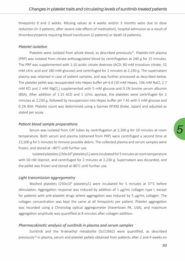
93
Changes in platelet traits and circulating levels of sunitinib-treated patients
5
timepoints 0 and 2 weeks. Missing values at 4 weeks and/or 3 months were due to dose
reduction (in 3 patients, after severe side effects of medication), hospital admission as a result of
thrombocytopenia requiring blood transfusion (2 patients) or death (4 patients).
Platelet isolationPlatelets were isolated from whole blood, as described previously15. Platelet-rich plasma
(PRP) was isolated from citrate-anticoagulated blood by centrifugation at 240 g for 15 minutes.
The PRP was supplemented with 1:10 acidic citrate dextrose (ACD; 80 mM trisodium citrate, 52
mM citric acid and 180 mM glucose) and centrifuged for 2 minutes at 2,230 g. The supernatant
plasma was retained in case of patient samples, and was further processed as described below.
The platelet pellet was resuspended into Hepes buffer pH 6.6 (10 mM Hepes, 136 mM NaCl, 2.7
mM KCl and 2 mM MgCl2) supplemented with 5 mM glucose and 0.1% bovine serum albumin
(BSA). After addition of 1:15 ACD and 1 U/mL apyrase, the platelets were centrifuged for 2
minutes at 2,230 g, followed by resuspension into Hepes buffer pH 7.45 with 5 mM glucose and
0.1% BSA. Platelet count was determined using a Sysmex XP300 (Kobe, Japan) and adjusted as
stated per assay.
Patient blood sample preparationsSerum was isolated from CAT tubes by centrifugation at 2,200 g for 10 minutes at room
temperature. Both serum and plasma (obtained from PRP) were centrifuged a second time at
22,500 g for 5 minutes to remove possible debris. The collected plasma and serum samples were
frozen, and stored at –80°C until further use.
Isolated platelets (250x109 platelets/L) were incubated for 5 minutes at room temperature
with 50 nM iloprost, and centrifuged for 2 minutes at 2,230 g. Supernatant was discarded, and
the pellet was frozen and stored at -80°C until further use.
Light transmission aggregometryWashed platelets (250x109 platelets/L) were incubated for 5 minutes at 37°C before
stimulation. Aggregation response was induced by addition of 1 µg/mL collagen type I, except
for patients with anti-platelet drugs where aggregation was induced by 5 µg/mL collagen. The
collagen concentration was kept the same at all timepoints per patient. Platelet aggregation
was recorded using a Chronolog optical aggregometer (Havertown PA, USA), and maximum
aggregation amplitude was quantified at 8 minutes after collagen addition.
Pharmacokinetic analysis of sunitinib in plasma and serum samplesSunitinib and the N-desethyl metabolite (SU12662) were quantified, as described
previously16 in plasma, serum and platelet pellets obtained from patients after 2 and 4 weeks on
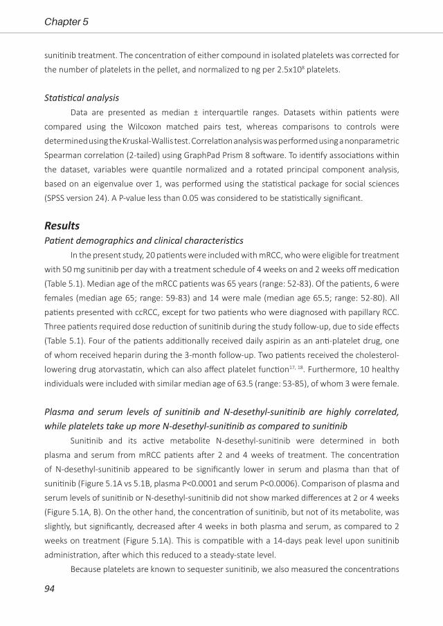
94
Chapter 5
sunitinib treatment. The concentration of either compound in isolated platelets was corrected for
the number of platelets in the pellet, and normalized to ng per 2.5x10⁸ platelets.
Statistical analysisData are presented as median ± interquartile ranges. Datasets within patients were
compared using the Wilcoxon matched pairs test, whereas comparisons to controls were
determined using the Kruskal-Wallis test. Correlation analysis was performed using a nonparametric
Spearman correlation (2-tailed) using GraphPad Prism 8 software. To identify associations within
the dataset, variables were quantile normalized and a rotated principal component analysis,
based on an eigenvalue over 1, was performed using the statistical package for social sciences
(SPSS version 24). A P-value less than 0.05 was considered to be statistically significant.
ResultsPatient demographics and clinical characteristics
In the present study, 20 patients were included with mRCC, who were eligible for treatment
with 50 mg sunitinib per day with a treatment schedule of 4 weeks on and 2 weeks off medication
(Table 5.1). Median age of the mRCC patients was 65 years (range: 52-83). Of the patients, 6 were
females (median age 65; range: 59-83) and 14 were male (median age 65.5; range: 52-80). All
patients presented with ccRCC, except for two patients who were diagnosed with papillary RCC.
Three patients required dose reduction of sunitinib during the study follow-up, due to side effects
(Table 5.1). Four of the patients additionally received daily aspirin as an anti-platelet drug, one
of whom received heparin during the 3-month follow-up. Two patients received the cholesterol-
lowering drug atorvastatin, which can also affect platelet function17, 18. Furthermore, 10 healthy
individuals were included with similar median age of 63.5 (range: 53-85), of whom 3 were female.
Plasma and serum levels of sunitinib and N-desethyl-sunitinib are highly correlated, while platelets take up more N-desethyl-sunitinib as compared to sunitinib
Sunitinib and its active metabolite N-desethyl-sunitinib were determined in both
plasma and serum from mRCC patients after 2 and 4 weeks of treatment. The concentration
of N-desethyl-sunitinib appeared to be significantly lower in serum and plasma than that of
sunitinib (Figure 5.1A vs 5.1B, plasma P<0.0001 and serum P<0.0006). Comparison of plasma and
serum levels of sunitinib or N-desethyl-sunitinib did not show marked differences at 2 or 4 weeks
(Figure 5.1A, B). On the other hand, the concentration of sunitinib, but not of its metabolite, was
slightly, but significantly, decreased after 4 weeks in both plasma and serum, as compared to 2
weeks on treatment (Figure 5.1A). This is compatible with a 14-days peak level upon sunitinib
administration, after which this reduced to a steady-state level.
Because platelets are known to sequester sunitinib, we also measured the concentrations

95
Changes in platelet traits and circulating levels of sunitinib-treated patients
5Day 0
Week 2
Week 4
Month 3
2M
ale
66Cl
ear c
ell
50-
164
102
7912
1N
o-
Yes
20%
incr
ease
3Fe
mal
e66
Clea
r cel
l50
-45
515
5-
-N
o-
NA
†4
Fem
ale
59Cl
ear c
ell
50-
146
24-
-Ye
sHe
mat
uria
, GI b
leed
ing
NA
†5
Mal
e56
Clea
r cel
l50
-26
051
202
204
No
-N
o-
6Fe
mal
e83
NA
50-
301
199
137
162
Yes
Epist
axis,
gin
givi�s
No
20%
dec
reas
e7
Fem
ale
64Cl
ear c
ell
50-
218
182
9520
1Ye
sHe
mat
uria
No
32%
dec
reas
e
9Fe
mal
e60
Clea
r cel
l50
-22
529
--
No
-N
AHo
spita
l upt
ake
10M
ale
52Cl
ear c
ell
50-
169
128
128
203
Yes
GI b
leed
ing,
gin
givi�s
No
-11
Mal
e72
Clea
r cel
l50
-15
720
049
-N
o-
NA
↓ D
osag
e12
Mal
e63
Papi
llary
50-
446
114
--
No
-N
A↓
Dos
age
13M
ale
75N
A50
ASA
(80)
221
142
148
273
No
-Ye
s35
% in
crea
se14
Mal
e68
Clea
r cel
l50
ATO
R (4
0)42
619
416
532
6N
o-
No
30%
dec
reas
e
15M
ale
55Cl
ear c
ell
50-
148
151
139
142
Yes
Epist
axis,
hem
opty
sis, G
I bl
eedi
ngN
o-
1 6M
ale
54Cl
ear c
ell
50-
170
116
161
-Ye
sHe
mat
uria
, gin
givi�s
Yes
16%
incr
ease
, ↓
Dosa
ge17
Mal
e68
Clea
r cel
l50
-19
517
718
7-
Yes
Hem
opty
sisYe
s8%
incr
ease
18Fe
mal
e82
Clea
r cel
l50
-28
621
298
134
Yes
Epist
axis,
gin
givi�s
, he
mop
tysis
, vag
inal
ble
edin
g N
o23
% d
ecre
ase
19M
ale
70Cl
ear c
ell
50AS
A (8
0)23
399
3699
No
-N
o-
20M
ale
65Cl
ear c
ell
50-
201
133
56-
No
-N
A†35
% in
crea
se-
100
-Ye
sGI
ble
edin
g a�
er h
epar
intr
eatm
ent
Yes
NA
†
8M
ale
80Pa
pilla
ry50
ASA
(80)
309
360
385
244
-N
o-
Plat
elet
cou
nt (x
109 /L
)
Blee
ding
Prog
ress
ion
1M
ale
58Cl
ear c
ell
50AS
A (1
00);
ATO
R (4
0)
Pa�e
nt
no.
Gen
der
Age
(yea
r)Ty
pe R
CCSu
ni�n
ib
trea
tmen
t (m
g/d)
Oth
er d
rug
trea
tmen
t (m
g/d)
Tabl
e 5.
1. D
emog
raph
ics a
nd c
linic
al c
hara
cter
istic
s of R
CC p
atie
nts t
reat
ed w
ith su
nitin
ib w
ith a
3-m
onth
follo
w-u
p
ASA:
ace
tylsa
licyl
ic a
cid
(asp
irin)
; ATO
R: a
torv
asta
tin; G
I: ga
stro
inte
stina
l; N
A: n
ot a
vaila
ble;
RCC
: ren
al c
ell c
arci
nom
a; †
: pati
ent
dece
ased
dur
ing
follo
w-u
p pe
riod;
↓ D
osag
e:
decr
ease
d do
sage
of s
uniti
nib
due
to s
ide
effec
ts.

96
Chapter 5
of sunitinib and the active metabolite in isolated platelets from mRCC patients. Markedly,
the concentration of the metabolite was significantly higher in platelets after 2 and 4 weeks
on treatment, as compared to that of sunitinib (Figure 5.1C). This contrasted to the lower
concentration of the metabolite in the plasma and serum samples, suggesting a preferential uptake
of the metabolite by platelets. Furthermore, the concentrations of sunitinib and metabolite after
2 and 4 weeks of treatment showed strong correlations between the plasma, serum and platelet
samples (r>0.67033, P<0.01).
Prolonged changes in platelet count by sunitinib treatment of mRCC patients are related to inhibitor levels in plasma and serum
Whole blood cell counts, haemoglobin and haematocrit were determined in blood
samples from mRCC patients at four time points (Figure 5.2), i.e., the day before start of sunitinib
treatment, after 2 and 4 weeks (1 treatment cycle) and after 3 months (2 complete cycles).
Before start of sunitinib treatment, no significant differences were observed in these parameters
Plasma Serum Plasma Serum0
50
100
150
200
Suni
tinib
(ng/
mL)
**
2 weekson-treatment
4 weekson-treatment
Plasma Serum Plasma Serum0
20
40
60
80
N-de
seth
yl-S
uniti
nib
(ng/
mL)
2 weekson-treatment
4 weekson-treatment
(A) (B)
(C)
Sunitinib N-desSunitinib
Sunitinib N-desSunitinib
0.0
0.5
1.0
1.5
2.0
2.5
Con
cent
ratio
n(n
g/2.
5x10
8pl
atel
ets)
2 weekson-treatment
4 weekson-treatment
*** **
Figure 5.1. Comparison of sunitinib and N-desethyl-sunitinib levels in plasma, serum and isolated platelets from mRCC patients on 2 and 4 weeks on treatment. The concentration of sunitinib (A) and N-desethyl-sunitinib (B) were measured in plasma and serum samples obtained from mRCC patients at 2 and 4 weeks of sunitinib treatment. (C) The concentrations of both compounds were determined in isolated platelets, normalized to ng per 2.5x10⁸ platelets. Individual dots in the scatterplots represent one patient, for color coding see Table 5.1. Lines and error bars represent median ± interquartile range (n=14-20). *P<0.05. **P<0.01, ***P<0.001

97
Changes in platelet traits and circulating levels of sunitinib-treated patients
5
between patients and healthy controls (Figure 5.2). However, after 2 weeks of treatment platelet
counts were decreased (>10%) in 15 patients (range: 17-87% reduction), while 5 patients showed
no decrease or even an increase (5-10%, Figure 5.2A). Seven of the patients who presented a
reduced platelet count showed an even further reduction after 4 weeks, which effect for six of
them persisted for 3 months. The median platelet concentration of all patients increased after 3
months as compared to 4 weeks, yet was still lower than before the start of treatment (Figure
5.2A). In total, fifteen patients had a platelet count between 24-148x109/L (Table 5.1), which was
below the normal range (150-400x109/L, according to the Dutch Society of Hematology, NVH). Of
note, 5 patients presented with formal thrombocytopenia (<50x109 platelets/L) after 2-4 weeks
of treatment. White blood cell (WBC) counts decreased (>10%) in 13 patients (range: 19-60%
reduction) after 2 weeks of sunitinib treatment (Figure 5.2B), which decrease persisted in most
patients for 3 months. According to the Common Terminology Criteria 7 patients presented with
grade 2 neutropenia during the study period with no more than 3 patients per time point. No
patients had grade 3 or 4 neutropenia. After two weeks of sunitinib administration, overall red
(A) (B) (C)
(E)(D)
Hea
lthy
Con
trols
Pre-
treat
men
t
2w
eeks
4w
eeks
3m
onth
s
0
100
200
300
400
500
PLT
(x10
9 /L)
****
*** ***
On-treatment
Hea
lthy
Con
trols
Pre-
treat
men
t
2w
eeks
4w
eeks
3m
onth
s
0
5
10
15
20
25
WB
C(x
109 /L
) ******* *
On-treatment
Hea
lthy
Con
trols
Pre-
treat
men
t
2w
eeks
4w
eeks
3m
onth
s
0
1
2
3
4
5
6
7
RB
C(x
1012
/L)
* **** **
On-treatment
Hea
lthy
Con
trols
Pre-
treat
men
t
2w
eeks
4w
eeks
3m
onth
s
0
2
4
6
8
10
12
HG
B(m
mol
/L)
*****
***
On-treatment
Hea
lthy
Con
trols
Pre-
treat
men
t
2w
eeks
4w
eeks
3m
onth
s
0.0
0.1
0.2
0.3
0.4
0.5
HC
T(L
/L)
** ** **
On-treatment
Figure 5.2. Treatment of mRCC patients with sunitinib decreases platelet count and other hematological parameters over time. Blood was collected from 10 healthy controls and 20 mRCC patients the day before, after 2 weeks, 4 weeks and 3 months on sunitinib treatment. Hematological parameters were measured in whole blood. Scatterplots show (A) platelet count, (B) white blood cell count (WBC), (C) red blood cell count (RBC), (D) hemoglobin concentration (HGB) and (E) hematocrit levels (HCT). Individual dots in the scatterplots represent one patient, for color coding see Table 1. Lines and error bars represent median ± interquartile range (n=10-20). *P<0.05. **P<0.01, ***P<0.001.
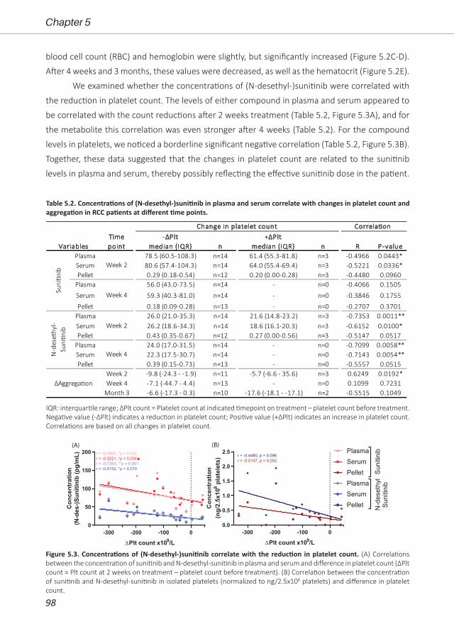
98
Chapter 5
blood cell count (RBC) and hemoglobin were slightly, but significantly increased (Figure 5.2C-D).
After 4 weeks and 3 months, these values were decreased, as well as the hematocrit (Figure 5.2E).
We examined whether the concentrations of (N-desethyl-)sunitinib were correlated with
the reduction in platelet count. The levels of either compound in plasma and serum appeared to
be correlated with the count reductions after 2 weeks treatment (Table 5.2, Figure 5.3A), and for
the metabolite this correlation was even stronger after 4 weeks (Table 5.2). For the compound
levels in platelets, we noticed a borderline significant negative correlation (Table 5.2, Figure 5.3B).
Together, these data suggested that the changes in platelet count are related to the sunitinib
levels in plasma and serum, thereby possibly reflecting the effective sunitinib dose in the patient.
Plasma 78.5 (60.5-108.3) n=14 61.4 (55.3-81.8) n=3 -0.4966 0.0443*Serum 80.6 (57.4-104.3) n=14 64.0 (55.4-69.4) n=3 -0.5221 0.0336*Pellet 0.29 (0.18-0.54) n=12 0.20 (0.00-0.28) n=3 -0.4480 0.0960
Plasma 56.0 (43.0-73.5) n=14 - n=0 -0.4066 0.1505
Serum 59.3 (40.3-81.0) n=14 - n=0 -0.3846 0.1755
Pellet 0.18 (0.09-0.28) n=13 - n=0 -0.2707 0.3701Plasma 26.0 (21.0-35.3) n=14 21.6 (14.8-23.2) n=3 -0.7353 0.0011**Serum 26.2 (18.6-34.3) n=14 18.6 (16.1-20.3) n=3 -0.6152 0.0100*Pellet 0.43 (0.35-0.67) n=12 0.27 (0.00-0.56) n=3 -0.5147 0.0517
Plasma 24.0 (17.0-31.5) n=14 - n=0 -0.7099 0.0058**Serum 22.3 (17.5-30.7) n=14 - n=0 -0.7143 0.0054**Pellet 0.39 (0.15-0.73) n=13 - n=0 -0.5557 0.0515
Week 2 -9.8 (-24.3 - -1.9) n=11 -5.7 (-6.6 - 35.6) n=3 0.6249 0.0192*Week 4 -7.1 (-44.7 - 4.4) n=13 - n=0 0.1099 0.7231
Month 3 -6.6 (-17.3 - 0.3) n=10 -17.6 (-18.1 - -17.1) n=2 -0.5515 0.1049
Correla�on
n n R P-value
∆Aggrega�on
Time point
-∆Plt median (IQR)
+∆Plt median (IQR)
Suni�n
ib
Week 2
Week 4
N-d
eset
hyl-
Suni�n
ib Week 2
Week 4
Variables
Change in platelet count
IQR: interquartile range; ΔPlt count = Platelet count at indicated timepoint on treatment – platelet count before treatment. Negative value (-ΔPlt) indicates a reduction in platelet count; Positive value (+ΔPlt) indicates an increase in platelet count. Correlations are based on all changes in platelet count.
Table 5.2. Concentrations of (N-desethyl-)sunitinib in plasma and serum correlate with changes in platelet count and aggregation in RCC patients at different time points.
Suni
tinib
N-d
eset
hyl
Suni
tinib
PlasmaSerumPelletPlasmaSerumPellet
-300 -200 -100 00.0
0.5
1.0
1.5
2.0
2.5
∆Plt count x109/L
Con
cent
ratio
n(n
g/2.
5x10
8pl
atel
ets)
r = -0.4480, p = 0.096r = -0.5147, p = 0.052
-300 -200 -100 00
50
100
150
200
∆Plt count x109/L
Con
cent
ratio
n(N
-des
-)Sun
itini
b(p
g/m
L) r = -0.4966, *p = 0.044r = -0.5221, *p = 0.034r = -0.7353, **p = 0.001r = -0.6152, *p = 0.010
(A) (B)
Figure 5.3. Concentrations of (N-desethyl-)sunitinib correlate with the reduction in platelet count. (A) Correlations between the concentration of sunitinib and N-desethyl-sunitinib in plasma and serum and difference in platelet count (ΔPlt count = Plt count at 2 weeks on treatment – platelet count before treatment). (B) Correlation between the concentration of sunitinib and N-desethyl-sunitinib in isolated platelets (normalized to ng/2.5x108 platelets) and difference in platelet count.
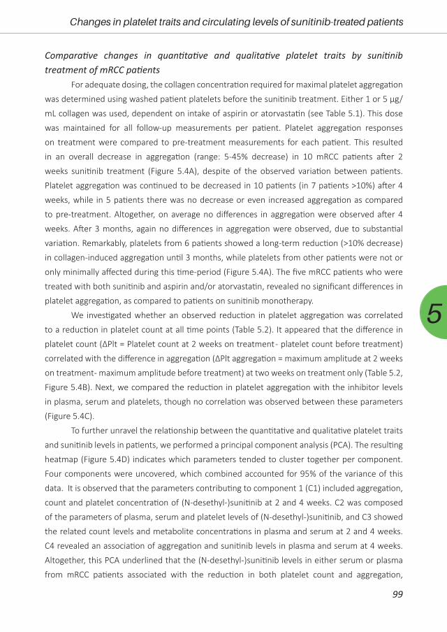
99
Changes in platelet traits and circulating levels of sunitinib-treated patients
5
Comparative changes in quantitative and qualitative platelet traits by sunitinib treatment of mRCC patients
For adequate dosing, the collagen concentration required for maximal platelet aggregation
was determined using washed patient platelets before the sunitinib treatment. Either 1 or 5 µg/
mL collagen was used, dependent on intake of aspirin or atorvastatin (see Table 5.1). This dose
was maintained for all follow-up measurements per patient. Platelet aggregation responses
on treatment were compared to pre-treatment measurements for each patient. This resulted
in an overall decrease in aggregation (range: 5-45% decrease) in 10 mRCC patients after 2
weeks sunitinib treatment (Figure 5.4A), despite of the observed variation between patients.
Platelet aggregation was continued to be decreased in 10 patients (in 7 patients >10%) after 4
weeks, while in 5 patients there was no decrease or even increased aggregation as compared
to pre-treatment. Altogether, on average no differences in aggregation were observed after 4
weeks. After 3 months, again no differences in aggregation were observed, due to substantial
variation. Remarkably, platelets from 6 patients showed a long-term reduction (>10% decrease)
in collagen-induced aggregation until 3 months, while platelets from other patients were not or
only minimally affected during this time-period (Figure 5.4A). The five mRCC patients who were
treated with both sunitinib and aspirin and/or atorvastatin, revealed no significant differences in
platelet aggregation, as compared to patients on sunitinib monotherapy.
We investigated whether an observed reduction in platelet aggregation was correlated
to a reduction in platelet count at all time points (Table 5.2). It appeared that the difference in
platelet count (ΔPlt = Platelet count at 2 weeks on treatment - platelet count before treatment)
correlated with the difference in aggregation (ΔPlt aggregation = maximum amplitude at 2 weeks
on treatment - maximum amplitude before treatment) at two weeks on treatment only (Table 5.2,
Figure 5.4B). Next, we compared the reduction in platelet aggregation with the inhibitor levels
in plasma, serum and platelets, though no correlation was observed between these parameters
(Figure 5.4C).
To further unravel the relationship between the quantitative and qualitative platelet traits
and sunitinib levels in patients, we performed a principal component analysis (PCA). The resulting
heatmap (Figure 5.4D) indicates which parameters tended to cluster together per component.
Four components were uncovered, which combined accounted for 95% of the variance of this
data. It is observed that the parameters contributing to component 1 (C1) included aggregation,
count and platelet concentration of (N-desethyl-)sunitinib at 2 and 4 weeks. C2 was composed
of the parameters of plasma, serum and platelet levels of (N-desethyl-)sunitinib, and C3 showed
the related count levels and metabolite concentrations in plasma and serum at 2 and 4 weeks.
C4 revealed an association of aggregation and sunitinib levels in plasma and serum at 4 weeks.
Altogether, this PCA underlined that the (N-desethyl-)sunitinib levels in either serum or plasma
from mRCC patients associated with the reduction in both platelet count and aggregation,
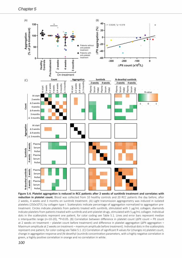
100
Chapter 5
(A) (B)
Patients without anti-platelet treatmentPatients with anti-platelet treatment
Plas
ma
Seru
mPe
llet
Plas
ma
Seru
m
Pelle
t
Plas
ma
Seru
mPe
llet
Plas
ma
Seru
m
Pelle
t
PlasmaSerumPellet
PlasmaSerum Pellet
PlasmaSerumPellet
PlasmaSerum Pellet
Δ 2
wee
ks2
wee
ksAt
star
t
At st
art
Δ 3
mon
ths
3 m
onth
sΔ
4 w
eeks
4 w
eeks
Coun
t
Δ 3 months3 monthsΔ 4 weeks
4 weeksΔ 2 weeks
2 weeksAt start
Count Aggrega�on
Δ 3
mon
ths
Δ 4
wee
ksΔ
2 w
eeks
Aggr
ega�
on
Δ 3 monthsΔ 4 weeksΔ 2 weeks
At start
N-d
eset
hyl-s
uni�
nib
2 w
eeks
4 w
eeks
Suni�n
ib 2 w
eeks
4 w
eeks
Suni�nib N-desethyl-suni�nib2 weeks 4 weeks 2 weeks 4 weeks
4
(C)
-1 0 1
R-value
Hea
lthy
Con
trols
Pre-
treat
men
t
2w
eeks
4w
eeks
3m
onth
s
0
50
100
150A
ggre
gatio
n(%
ofpr
e-tr
eatm
ent)
On-treatment
*
-300 -200 -100 0-60
-40
-20
0
20
40
∆Plt count (x109/L)
∆Pl
tagg
rega
tion
(%)
r = -0.6249, *p = 0.019
Figure 5.4. Platelet aggregation is reduced in RCC patients after 2 weeks of sunitinib treatment and correlates with reduction in platelet count. Blood was collected from 10 healthy controls and 20 RCC patients the day before, after 2 weeks, 4 weeks and 3 months on sunitinib treatment. (A) Light transmission aggregometry was induced in isolated platelets (250x109/L) by collagen type I. Scatterplots indicate percentage of aggregation normalized to aggregation pre-treatment. Circles indicate platelets from patients treated with sunitinib, stimulated with 1 µg/mL collagen; diamonds indicate platelets from patients treated with sunitinib and anti-platelet drugs, stimulated with 5 µg/mL collagen. Individual dots in the scatterplots represent one patient, for color coding see Table 5.1. Lines and error bars represent median ± interquartile range (n=10-20), *P<0.05. (B) Correlation between difference in platelet count (ΔPlt count = Plt count at 2 weeks on treatment – platelet count before treatment) and difference in platelet aggregation (ΔPlt aggregation = Maximum amplitude at 2 weeks on treatment – maximum amplitude before treatment). Individual dots in the scatterplots represent one patient, for color coding see Table 5.1. (C) Correlation of significant R values for (changes in) platelet count, change in aggregation response and (N-desethyl-)sunitnib concentration parameters, with a highly negative correlation in green, a highly positive correlation in orange and no correlation in white.
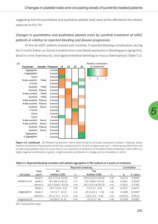
101
Changes in platelet traits and circulating levels of sunitinib-treated patients
5
suggesting that the quantitative and qualitative platelet traits were jointly affected by the relative
exposure to this TKI.
Changes in quantitative and qualitative platelet traits by sunitinib treatment of mRCC patients in relation to reported bleeding and disease progression
Of the 20 mRCC patients treated with sunitinib, 9 reported bleeding complications during
the 3-month follow-up. Events included minor nose bleeds (epistaxis) or bleeding gums (gingivitis),
blood in urine (haematuria), stool (gastrointestinal bleeding) or mucus (haemoptysis) (Table 5.1).
Figure 5.4. Continued. (D) Rotated component matrix determined by principal component analysis. Heatmap shows relative contribution of parameters to the four components (C1-4) with an eigenvalue over 1. Heatmap was filtered to only include values greater than 0.4 or less than -0.4 as important contributors to the determined component. Colors refer to a highly negative contribution in green, a highly positive contribution in orange and no correlation in white.
(D)
-1 0 1
Rela�ve contribu�on to componentParameter Sample Timepoint C1 C2 C3 C4
Aggregation 2 weeks∆ Aggregation 2 weeks
Count 4 weeksN-des-sunitinib Pellet 4 weeks
∆ Count 2 weeksSunitinib Pellet 4 weeksSunitinib Plasma 2 weeks
N-des-sunitinib Pellet 2 weeksSunitinib Pellet 2 weeks
N-des-sunitinib Plasma 2 weeksN-des-sunitinib Plasma 4 weeks
Sunitinib Serum 2 weeks∆ Count 4 weeks
Count 2 weeksCount at start
N-des-sunitinib Serum 2 weeksN-des-sunitinib Serum 4 weeks
Sunitinib Plasma 4 weeksSunitinib Serum 4 weeks
Aggregation 4 weeks∆ Aggregation 4 weeks
Week 2 132.5 (75.8-229.8) n=9 164.0 (119.0-194.8) n=8 -0.0515 0.8498Week 4 78.5 (49.0-202.0) n=7 132.3 (98.4-155.0) n=8 0.0201 0.9508
Month 3 162.5 (104.5-255.8) n=4 161.5 (137.8-202.0) n=5 -0.3651 0.3786Week 2 -10.7 (-29.6 - 4.0) n=6 -3.8 (-6.7 - 0.8) n=6 0.2612 0.4072
Week 4 -3.8 (-5.7 - 12.2) n=4 -24.0 (-62.3 - -7.8) n=8 -0.6680 0.0206*
Month 3 -10.2 (-22.1 - 12.7) n=4 -6.8 (-12.0 - -4.8) n=5 0.02623 0.9516Progression % - 10.0 (0.0 - 31.3) n=4 0 (-22.3 - 14) n=8 -0.2500 0.4285
Correla�on
n n R P-valueTime point
Platelet count
Nomedian (IQR)
Yesmedian (IQR)
∆Aggrega�on
Variables
Reported bleeding
IQR: interquartile range.
Table 5.3. Reported bleeding correlates with platelet aggregation in RCC patients at 4 weeks on treatment.
102
Chapter 5
Interestingly, of the 5 patients who were on dual treatment of sunitinib and aspirin, only 1 patient
(also treated with heparin) reported bleeding events during the first cycle of sunitinib treatment.
We analysed whether the altered platelet count correlated with the reported bleeding. As
shown in Table 5.3, this was not the case. On the other hand, we observed that platelet function,
as measured by collagen-induced aggregation, was negatively correlated with bleeding at 4 weeks
on-treatment (Table 5.3), although the number of patients included in this analysis was relatively
small (n=4-8). At the other time points this correlation was not observed.
After three months on treatment, CT scans were evaluated for cancer response in 15
patients (Table 5.1). In 5 of these patients progression of the disease was established, with an
average of 23% increase in tumor size. In the other 10 patients no progression was observed (Table
5.1). In this limited group of patients, no correlation was observed between disease progression
and reduction in blood cell counts or platelet aggregation (Suppl. Table 5.1).
DiscussionIn the present study with patients treated with sunitinib for mRCC, we observed that
changes in platelet count were related to the inhibitor levels in plasma and serum. The reduction
in platelet count may thus reflect the effective sunitinib levels in the patient, which may be used for
therapeutic drug monitoring19. Furthermore, we observed a strong correlation between effects on
platelet count and collagen-induced platelet aggregation, regardless of the treatment time point.
This pointed to a common effect of this TKI on quantitative and qualitative platelet traits, which
was confirmed by PCA. Next to RCC, sunitinib is approved for the treatment of gastrointestinal
stromal tumor (GIST) and pancreatic neuroendocrine tumors. Furthermore, phase 3 clinical trials
with sunitinib have been completed with positive outcome for non-small cell lung cancer20, 21.
Hence, the results of the present study may also be applicable to other cancer types. In addition,
platelet traits have been reported to be affected by other TKIs used for treatment of a large
variety of malignancies1, 9. Also, other anti-cancer drugs can be taken up by platelets22. Therefore,
the results of the present study with sunitinib may serve as a proof-of-concept with regards to
platelet trait effects of other drugs.
We observed that the patients on-treatment who showed a stronger inhibition in platelet
aggregation (at normalized platelet count), also displayed a lower platelet count in whole blood.
This can be explained by two separate mechanisms elicited by sunitinib, being (i) an effect on
megakaryocyte level, culminating in a lower platelet count, and (ii) an effect on platelet function
(aggregation), likely via an impaired tyrosine kinase-mediated signaling. Regarding the first
mechanism, sunitinib may affect megakaryocytes or precursor cells directly in the bone marrow
via interference in the proliferation of megakaryocytes and the production of proplatelets,
which processes are dependent on tyrosine kinase activity23. Alternatively, the effects on
megakaryocytes may be indirect via altered thrombopoietin production. Tumors can stimulate
103
Changes in platelet traits and circulating levels of sunitinib-treated patients
5
platelet production via de secretion of various cytokines, especially interleukin-6, which can
in turn enhance thrombopoietin expression in the liver24. A direct effect of sunitinib on tumor
proliferation may result in reduced levels of thrombopoietin and thereby lowering of the platelet
count. Besides platelet count, we confirmed that sunitinib also reduced the WBC count (up to
grade 2) in the mRCC patients. Furthermore, also the RBC count, and hemoglobin and hematocrit
levels were affected upon sunitinib treatment. These results point to an effect of sunitinib on
different hematopoietic cell populations in the bone marrow. Other studies with larger patient
numbers indeed concluded that a reduced neutrophil count could be a predictor of PFS and OS
probability13, 25. With regard to RBCs, sunitinib has been reported to affect erythropoiesis in both
directions, resulting in either anemia26 or in erythrocytosis27. Markedly, in our small patient group,
we observed a transient increase in RBC count, hemoglobin and hematocrit during the first cycle
of sunitinib treatment, as reported previously and explained by cycling kinetics during the dosing
schedule28.
Regarding the second mechanism of sunitinib affecting tyrosine kinase-mediated platelet
signaling, we observed that collagen-induced platelet aggregation was reduced in mRCC patients
after 14 days of sunitinib treatment. Others have shown that this inhibition was already present
after 24 hours of sunitinib treatment29. Besides aggregation, sunitinib also inhibits collagen-
induced thrombus formation, exposure of phosphatidylserine under flow, as well as α-granule
secretion11, 29. As discussed before, the protein tyrosine kinases that are affected by sunitinib are
c-Src29, as well as other kinases expressed in platelets (e.g., Axl, CSF and Itk)1.
As platelets are able to take up sunitinib11, we hypothesized that this affects the outcome
of sunitinib plasma measurements. Therefore, we compared the concentrations of sunitinib and
N-desethyl-sunitinib (active metabolite of sunitinib) in plasma, serum and isolated platelets from
mRCC patients at 2 and 4 weeks of treatment. The concentration of N-desethyl-sunitinib was
much lower than sunitinib in both serum and plasma, which is in line with previous results30.
Furthermore, there was no difference between serum and plasma levels of either sunitinib or
N-desethyl-sunitinib at 2 or 4 weeks of sunitinib administration. Honeywell et al. also reported
no differences for sunitinib in plasma as compared to serum for 5 patients on sunitinib treatment
after 24 hours and 3 weeks31. In contrast, another study demonstrated lower concentrations
of sunitinib in plasma as compared to serum in patients treated with sunitinib29. These studies
demonstrated a high variation in sunitinib plasma concentrations, which could only partly be
explained by patient- or medication-related factors32. This highlights the importance for further
research of therapeutic drug monitoring for individual dosage adjustments, especially in cases
of toxicity19. Interestingly, the concentrations of the active metabolite were significantly higher
in isolated platelets from mRCC patients at 2 and 4 weeks as compared to that of sunitinib itself.
The only difference between the two molecules is an ethyl (CH2-CH3) group that is removed from
sunitinib by cytochrome P45033. As this enzyme not present in platelets, these results suggest

104
Chapter 5
preferential uptake of the metabolite by these cells. Overall, we concluded that although the
concentration of the active metabolite in platelets was higher, the uptake of sunitinib in platelets
did not affect the sunitinib concentration in plasma versus serum. The serum concentration of
N-desethyl-sunitinib negatively correlated with platelet count at 4 weeks of treatment, suggesting
that higher platelet counts result in a lower concentration of metabolite present in serum.
Together with the observation that metabolite concentrations in isolated platelets were higher,
this may suggest that platelets do not secrete sunitinib after uptake.
Mild bleeding was reported in 9 of the 20 investigated cancer patients on sunitinib. No
correlation existed between the platelet count at any time point on treatment and the occurrence
of bleeding during the 3-month study period. This agrees with earlier reports that showed that
the platelet count is a poor predictor for bleeding34, 35. Hence, the reported bleeding events
may be explained by an accompanying inhibition of platelet activation by sunitinib. We observed
a correlation between platelet aggregation responses and bleeding during sunitinib treatment,
meaning that stronger reduction in collagen-induced platelet aggregation was observed in patients
that reported bleeding episodes. Of note, only a small number of patients was included in this
analysis, and at no other time points a correlation was observed between platelet aggregation
and reported bleeding. Also, Walraven et al. did not observe this correlation between bleeding
and platelet aggregation after 3 weeks of treatment in 5 patients29. As normal platelet functions
can be considered to be required for effective control of hemostasis, correlations between
bleeding and TKI-induced effects on platelet function should be investigated in larger clinical
studies. Of note, in patients treated with sunitinib, also endothelial dysfunction and increased
capillary leakage can be induced by treatment with sunitinib36, which may also play a role in the
increased bleeding tendency. Paradoxically, interfering with endothelial cell integrity can also shift
the hemostatic balance in favor of thrombosis, and treatment with sunitinib and other TKIs have
also been associated with an increase in arterial thromboembolic events in RCC patients36, 37.
A correlation between platelet count and disease progression in this study could not be
observed due to a relatively small number of patients. However, it has already been demonstrated
in large, retrospective patient studies that a decrease in platelet count following sunitinib
treatment more likely resulted in a response to therapy and longer OS in mRCC12 and GIST13.
In the present study we clearly showed that a reduction in platelet count is accompanied by an
inhibition of platelet aggregation, which were correlated to the (N-desethyl-)sunitinib levels in
either serum or plasma of mRCC patients (component 1 of the PCA). Altogether this suggested
that both quantitative and qualitative platelet traits were linked to the exposure to this TKI.
Renal cancer is a disease of the elderly, with most patients being diagnosed between 65
and 7438, 39. Older cancer patients generally have more comorbidities, especially cardio-vascular
disease, and therefore often receive anti-coagulant or anti-platelet drugs. In the present study,
five patients were treated with sunitinib in combination with aspirin and/or atorvastatin, both

105
Changes in platelet traits and circulating levels of sunitinib-treated patients
5
affecting platelet functions17, 18, 40. In our tests, this did not result in significant differences in
platelet aggregation as compared to patients on sunitinib without comedication. One explanation
may be an increased platelet reactivity observed in cancer patients41, which compensates for the
intake of the anti-platelet drugs. On the other hand, a recent study has shown combined effects
of ibrutinib and aspirin on collagen-induced aggregation42. Therefore, the effects of dual anti-
platelet and TKIs treatments on the inhibition of platelet function should be further investigated.
Our study encounters some limitations. Due to the limited number of patients and some
missing values, no (clear) correlations were observed between platelet count and function on
the one hand and bleeding and disease progression on the other hand. Also, only one platelet
function test could be performed due to the limited amount of blood obtained from the patients.
Out of multiple function tests, we opted for light transmission aggregometry as this is still the gold
standard for platelet function testing. Due to ethical restrictions, the relation between platelet
dysfunction after sunitinib treatment and abnormal (pro)platelet formation from megakaryocytes
in the bone marrow could not be assessed. For the TKI dasatinib it has been reported that it
promoted megakaryocyte differentiation, while it impaired migration and proplatelet formation43.
The effects of TKI treatment on megakaryocyte proliferation and function should therefore be
investigated in future studies.
In summary, in this study with mRCC patients treated with sunitinib a strong correlation was
found between the effects on platelet count and inhibition of aggregation as well as (N-desethyl)
sunitinib levels in plasma and serum. The effects on count and function may thus reflect the
relative exposure of the patient to sunitinib. This pointed to an association between the effect
of this TKI on quantitative and qualitative platelet traits. These results may serve as a proof-of-
principle with regards to other drugs, indicating that TKI treatment can affect both platelet count
and function, which could be used as a measure for relative drug exposure and therapeutic drug
monitoring.
AcknowledgementsThis study was supported by Pfizer as an Investigator-Initiated Research grant to MJEK
(Tracking Number WI209458).
References1. Tullemans BME, Heemskerk JWM, Kuijpers MJE. Acquired platelet antagonism: off-target antiplatelet
effects of malignancy treatment with tyrosine kinase inhibitors. J Thromb Haemost. 2018;16:1686-99.
2. Gross S, Rahal R, Stransky N, Lengauer C, Hoeflich KP. Targeting cancer with kinase inhibitors. J Clin Invest. 2015;125:1780-9.
3. Siegel RL, Miller KD, Jemal A. Cancer statistics, 2020. CA Cancer J Clin. 2020;70:7-30.4. Motzer RJ, Tannir NM, McDermott DF, Aren Frontera O, Melichar B, Choueiri TK, et al. Nivolumab plus
Ipilimumab versus Sunitinib in Advanced Renal-Cell Carcinoma. New Engl J Med. 2018;378:1277-90.5. Rini BI, Plimack ER, Stus V, Gafanov R, Hawkins R, Nosov D, et al. Pembrolizumab plus Axitinib versus
106
Chapter 5
Sunitinib for Advanced Renal-Cell Carcinoma. New Engl J Med. 2019;380:1116-27.6. Motzer RJ, Escudier B, McDermott DF, George S, Hammers HJ, Srinivas S, et al. Nivolumab versus
Everolimus in Advanced Renal-Cell Carcinoma. New Engl J Med. 2015;373:1803-13.7. Choueiri TK, Escudier B, Powles T, Tannir NM, Mainwaring PN, Rini BI, et al. Cabozantinib versus
everolimus in advanced renal cell carcinoma (METEOR): final results from a randomised, open-label, phase 3 trial. Lancet Oncol. 2016;17:917-27.
8. Versteeg HH, Heemskerk JW, Levi M, Reitsma PH. New fundamentals in hemostasis. Physiol Rev. 2013;93:327-58.
9. Levade M, Severin S, Gratacap MP, Ysebaert L, Payrastre B. Targeting Kinases in Cancer Therapies: Adverse Effects on Blood Platelets. Curr Pharm Des. 2016;22:2315-22.
10. Je Y, Schutz FA, Choueiri TK. Risk of bleeding with vascular endothelial growth factor receptor tyrosine-kinase inhibitors sunitinib and sorafenib: a systematic review and meta-analysis of clinical trials. Lancet Oncol. 2009;10:967-74.
11. Sabrkhany S, Griffioen AW, Pineda S, Sanders L, Mattheij N, van Geffen JP, et al. Sunitinib uptake inhibits platelet function in cancer patients. Eur J Cancer. 2016;66:47-54.
12. Hamilton Z, Lee HJ, Jimenez J, Lane BR, Wang S, Beksac AT, et al. Change in platelet count as a prognostic indicator for response to primary tyrosine kinase inhibitor therapy in metastatic renal cell carcinoma. BJU Int. 2016;118:927-34.
13. Den Hollander D, Van der Graaf WTA, Desar IME, Le Cesne A. Predictive factors for toxicity and survival of second-line sunitinib in advanced gastrointestinal stromal tumours (GIST). Acta Oncol. 2019;58:1648-54.
14. Stanworth SJ, Dyer C, Casbard A, Murphy MF. Feasibility and usefulness of self-assessment of bleeding in patients with haematological malignancies, and the association between platelet count and bleeding. Vox Sang. 2006;91:63-9.
15. van der Meijden PE, Feijge MA, Giesen PL, Huijberts M, van Raak LP, Heemskerk JW. Platelet P2Y12 receptors enhance signalling towards procoagulant activity and thrombin generation. A study with healthy subjects and patients at thrombotic risk. Thromb Haemost. 2005;93:1128-36.
16. de Bruijn P, Sleijfer S, Lam MH, Mathijssen RH, Wiemer EA, Loos WJ. Bioanalytical method for the quantification of sunitinib and its n-desethyl metabolite SU12662 in human plasma by ultra performance liquid chromatography/tandem triple-quadrupole mass spectrometry. J Pharm Biomed Anal. 2010;51:934-41.
17. Zhao L, Liu D, Liu B, Hu H, Cui W. Effects of atorvastatin on ADP-, arachidonic acid-, collagen-, and epinephrine-induced platelet aggregation. J Int Med Res. 2017;45:82-8.
18. Ni R, Peleg T, Gross PL. Atorvastatin delays murine platelet activation in vivo even in the absence of endothelial NO synthase. Arterioscl Thromb Vasc Biol. 2012;32:2609-15.
19. Verheijen RB, Yu H, Schellens JHM, Beijnen JH, Steeghs N, Huitema ADR. Practical Recommendations for Therapeutic Drug Monitoring of Kinase Inhibitors in Oncology. Clin Pharmacol Ther. 2017;102:765-76.
20. Scagliotti GV, Krzakowski M, Szczesna A, Strausz J, Makhson A, Reck M, et al. Sunitinib plus erlotinib versus placebo plus erlotinib in patients with previously treated advanced non-small-cell lung cancer: a phase III trial. J Clin Oncol. 2012;30:2070-8.
21. Baggstrom MQ, Socinski MA, Wang XF, Gu L, Stinchcombe TE, Edelman MJ, et al. Maintenance Sunitinib following Initial Platinum-Based Combination Chemotherapy in Advanced-Stage IIIB/IV Non-Small Cell Lung Cancer: A Randomized, Double-Blind, Placebo-Controlled Phase III Study-CALGB 30607 (Alliance). J Thorac Oncol. 2017;12:843-9.
22. Verheul HM, Lolkema MP, Qian DZ, Hilkes YH, Liapi E, Akkerman JW, et al. Platelets take up the monoclonal antibody bevacizumab. Clin Cancer Res. 2007;13:5341-7.
23. Noetzli LJ, French SL, Machlus KR. New Insights Into the Differentiation of Megakaryocytes From Hematopoietic Progenitors. Arterioscl Thromb Vasc Biol. 2019;39:1288-300.
24. Lin RJ, Afshar-Kharghan V, Schafer AI. Paraneoplastic thrombocytosis: the secrets of tumor self-promotion. Blood. 2014;124:184-7.
25. Rautiola J, Donskov F, Peltola K, Joensuu H, Bono P. Sunitinib-induced hypertension, neutropaenia and thrombocytopaenia as predictors of good prognosis in patients with metastatic renal cell carcinoma. BJU Int. 2016;117:110-7.

107
Changes in platelet traits and circulating levels of sunitinib-treated patients
5
26. Motzer RJ, Rini BI, Bukowski RM, Curti BD, George DJ, Hudes GR, et al. Sunitinib in patients with metastatic renal cell carcinoma. JAMA. 2006;295:2516-24.
27. Alexandrescu DT, McClure R, Farzanmehr H, Dasanu CA. Secondary erythrocytosis produced by the tyrosine kinase inhibitors sunitinib and sorafenib. J Clin Oncol. 2008;26:4047-8.
28. van der Veldt AA, Boven E, Vroling L, Broxterman HJ, van den Eertwegh AJ, Haanen JG. Sunitinib-induced hemoglobin changes are related to the dosing schedule. J Clin Oncol. 2009;27:1339-40; author reply 40-2.
29. Walraven M, Homs MYV, van der Veldt AAM, Dekker H, Koldenhof J, Honeywell R, et al. Platelet function is disturbed by the angiogenesis inhibitors sunitinib and sorafenib, but unaffected by bevacizumab. Angiogenesis. 2018;21:325-34.
30. Houk BE, Bello CL, Poland B, Rosen LS, Demetri GD, Motzer RJ. Relationship between exposure to sunitinib and efficacy and tolerability endpoints in patients with cancer: results of a pharmacokinetic/pharmacodynamic meta-analysis. Cancer Chemother Pharmacol. 2010;66:357-71.
31. Honeywell R, Yarzadah K, Giovannetti E, Losekoot N, Smit EF, Walraven M, et al. Simple and selective method for the determination of various tyrosine kinase inhibitors used in the clinical setting by liquid chromatography tandem mass spectrometry. J Chromatogr B Analyt Technol Biomed Life Sci. 2010;878:1059-68.
32. Lankheet NA, Knapen LM, Schellens JH, Beijnen JH, Steeghs N, Huitema AD. Plasma concentrations of tyrosine kinase inhibitors imatinib, erlotinib, and sunitinib in routine clinical outpatient cancer care. Ther Drug Monit. 2014;36:326-34.
33. Adams VR, Leggas M. Sunitinib malate for the treatment of metastatic renal cell carcinoma and gastrointestinal stromal tumors. Clin Ther. 2007;29:1338-53.
34. Slichter SJ. Relationship between platelet count and bleeding risk in thrombocytopenic patients. Transfus Med Rev. 2004;18:153-67.
35. Baaten C, Moenen F, Henskens YMC, Swieringa F, Wetzels RJH, van Oerle R, et al. Impaired mitochondrial activity explains platelet dysfunction in thrombocytopenic cancer patients undergoing chemotherapy. Haematologica. 2018;103:1557-67.
36. Bair SM, Choueiri TK, Moslehi J. Cardiovascular complications associated with novel angiogenesis inhibitors: emerging evidence and evolving perspectives. Trends Cardiovasc Med. 2013;23:104-13.
37. Choueiri TK, Schutz FA, Je Y, Rosenberg JE, Bellmunt J. Risk of arterial thromboembolic events with sunitinib and sorafenib: a systematic review and meta-analysis of clinical trials. J Clin Oncol. 2010;28:2280-5.
38. Howlader N, Noone AM, Krapcho M, Miller D, Brest A, Yu M, et al. SEER Cancer Statistics Review, 1975-2017. National Cancer Institute 2019.
39. Padala SA, Barsouk A, Thandra KC, Saginala K, Mohammed A, Vakiti A, et al. Epidemiology of Renal Cell Carcinoma. World J Oncol. 2020;11:79-87.
40. Warner TD, Nylander S, Whatling C. Anti-platelet therapy: cyclo-oxygenase inhibition and the use of aspirin with particular regard to dual anti-platelet therapy. Br J Clin Pharmacol. 2011;72:619-33.
41. Goubran HA, Stakiw J, Radosevic M, Burnouf T. Platelet-cancer interactions. Sem Thromb Hemost. 2014;40:296-305.
42. Ninomoto J, Mokatrin A, Kinoshita T, Marimpietri C, Barrett TD, Chang BY, et al. Effects of ibrutinib on in vitro platelet aggregation in blood samples from healthy donors and donors with platelet dysfunction. Hematology. 2020;25:112-7.
43. Mazharian A, Ghevaert C, Zhang L, Massberg S, Watson SP. Dasatinib enhances megakaryocyte differentiation but inhibits platelet formation. Blood. 2011;117:5198-206.
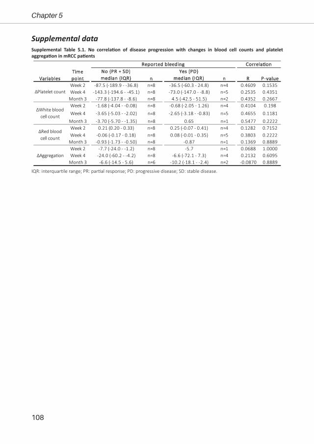
108
Chapter 5
Supplemental data
Week 2 -87.5 (-189.9 - -36.8) n=8 -36.5 (-60.3 - 24.8) n=4 0.4609 0.1535Week 4 -143.3 (-194.6 - -45.1) n=8 -73.0 (-147.0 - -8.8) n=5 0.2535 0.4351
Month 3 -77.8 (-137.8 - -8.6) n=8 4.5 (-42.5 - 51.5) n=2 0.4352 0.2667Week 2 -1.68 (-4.04 - -0.08) n=8 -0.68 (-2.05 - 1.26) n=4 0.4104 0.198
Week 4 -3.65 (-5.03 - -2.02) n=8 -2.65 (-3.18 - -0.83) n=5 0.4655 0.1181
Month 3 -3.70 (-5.70 - -1.35) n=8 0.65 n=1 0.5477 0.2222Week 2 0.21 (0.20 - 0.33) n=8 0.25 (-0.07 - 0.41) n=4 0.1282 0.7152Week 4 -0.06 (-0.17 - 0.18) n=8 0.08 (-0.01 - 0.35) n=5 0.3803 0.2222
Month 3 -0.93 (-1.73 - -0.50) n=8 -0.87 n=1 0.1369 0.8889Week 2 -7.7 (-24.0 - -1.2) n=8 -5.7 n=1 0.0688 1.0000Week 4 -24.0 (-60.2 - -4.2) n=8 -6.6 (-72.1 - 7.3) n=4 0.2132 0.6095
Month 3 -6.6 (-14.5 - 5.6) n=6 -10.2 (-18.1 - -2.4) n=2 -0.0870 0.8889
∆Red blood cell count
∆Aggrega�on
∆Platelet count
∆White blood cell count
VariablesTime point
Reported bleeding Correla�onNo (PR + SD)median (IQR) n
Yes (PD)median (IQR) n R P-value
Supplemental Table 5.1. No correlation of disease progression with changes in blood cell counts and platelet aggregation in mRCC patients
IQR: interquartile range; PR: partial response; PD: progressive disease; SD: stable disease.

109
5


Chapter 6
Comparison of inhibitory effects of
irreversible and reversible Btk inhibitors on
platelet function
Tullemans BME, Karel MFA, Léopold V, ten Brink MS, Baaten CCFMJ, Maas SL, de Vos AF, Eble JA, Nijziel MR, van der Vorst EPC, Cosemans JMEM, Heemskerk JWM, Claushuis TAM*
and Kuijpers MJE*
(* equal contribution)
Brit J Haematol 2021; 1-15Reprinted with permission

112
Chapter 6
AbstractAll irreversible Bruton tyrosine kinase (Btk) inhibitors, including ibrutinib and acalabrutinib
induce platelet dysfunction and increased bleeding risk. New reversible Btk inhibitors were
developed, like MK-1026. The mechanism underlying increased bleeding tendency with Btk
inhibitors remains unclear. We investigated the effects of ibrutinib, acalabrutinib and MK-1026
on platelet function in healthy volunteers, patients and Btk-deficient mice, together with off-
target effects on tyrosine kinase phosphorylation. All inhibitors suppressed GPVI- and CLEC-2-
mediated platelet aggregation, activation and secretion in a dose-dependent manner. Only
ibrutinib inhibited thrombus formation on vWF-co-coated surfaces, while on collagen this was
not affected. In blood from Btk-deficient mice, collagen-induced thrombus formation under flow
was reduced, but preincubation with either inhibitor was without additional effects. MK-1026
showed less off-target effects upon GPVI-induced TK phosphorylation as compared to ibrutinib
and acalabrutinib. In ibrutinib-treated patients, GPVI-stimulated platelet activation and adhesion
on vWF-co-coated surfaces were inhibited, while CLEC-2 stimulation induced variable responses.
The dual inhibition of GPVI and CLEC-2 signalling by Btk inhibitors might account for the increased
bleeding tendency, with ibrutinib causing more high-grade bleedings due to additional inhibition
of platelet-vWF interaction. As MK-1026 showed less off-target effects and only affected activation
of isolated platelets, it might be promising for future treatment.
113
Inhibitory effects of Btk inhibitors on platelet function
6
IntroductionIbrutinib, an irreversible Bruton’s tyrosine kinase (Btk) inhibitor, is widely used to treat
multiple haematological malignancies1-5. Ibrutinib treatment shows high response rates and
improved progression-free survival, with decreased toxicity when compared to conventional
treatment options1-6. However, ibrutinib is prescribed life-long (or until progression) and requires
side effect management. These toxicities are responsible for discontinuation in up to 20% of
patients6-9. A prominent side effect of ibrutinib is bleeding, with over 50% of patients experiencing
bleeding events within 3 years, ranging from mild mucocutaneous bleeding to life-threatening
haemorrhage3, 6, 8, 10. Studies have suggested that ibrutinib-associated bleeding tendency is caused
by interference with platelet GPVI signalling pathway through off-target effects on Src kinases11, 12.
Recently, multiple other Btk inhibitors have been developed, including the irreversible
Btk inhibitor acalabrutinib and the reversible Btk inhibitor MK-1026 (formerly known as ARQ-
531)13, 14. Opposed to data suggesting that acalabrutinib shows less off-target effects, it still
causes an increased bleeding tendency in patients, albeit showing mainly mild bleeding15.
Studies investigating GPVI inhibitors as potential antithrombotic drugs have shown that
inhibition on receptor level does not impair haemostasis16. Also, GPVI-deficient patients exhibit
only a mild bleeding diathesis17 and many potential patients are undiagnosed as they remain
asymptomatic18. In agreement, studies in mice have shown that GPVI deficiency alone does not
impair haemostasis19-21, while combined GPVI and CLEC-2 deficiency does21. Furthermore, Btk was
shown to be involved in GPIb-vWF mediated platelet adhesion, as well as integrin αIIbβ3 outside-in
activation which were inhibited by ibrutinib6, 22, 23. Therefore, the current mechanism of increased
bleeding tendency in patients using Btk inhibitors is still unclear and underlines the importance
for treatment decisions and the development of novel Btk inhibitors. We therefore investigated
the effect of multiple Btk inhibitors on platelet function pathways in healthy volunteers, patients
and Btk knock-out (KO) mice.
Materials and MethodsMaterials and methods are available in Supplemental Data.
ResultsIbrutinib and acalabrutinib inhibit platelet activation and aggregation by GPVI and CLEC-2 in a dose-dependent manner
Treatment with ibrutinib, but also acalabrutinib, can increase bleeding tendency6, 15. To
assess the underlying pathways, we investigated platelet activation mediated by multiple agonists
in presence of ibrutinib or acalabrutinib.
Both ibrutinib and acalabrutinib inhibited aggregation of washed platelets and platelet-
rich plasma induced by 1 µg/ml collagen in a dose-dependent manner (Fig 6.1A). In isolated

114
Chapter 6
platelets, a significant inhibition was observed at 1 µM and higher for both compounds (Fig 6.1Ai).
In the presence of plasma, a higher concentration of 5-10 µM was required to significantly inhibit
collagen-induced platelet aggregation (Fig 6.1Aii). The IC50 values for ibrutinib and acalabrutinib
were 7.7 and 6.0 times lower in washed platelets as compared to PRP (Suppl Table 6.1). At a higher
collagen concentration, aggregation was not affected by either inhibitor. (Suppl Fig 6.1Aii). CLEC-
2-induced platelet aggregation was also inhibited by both acalabrutinib and ibrutinib in a similar
dose-dependent manner (Fig 6.1B). GPVI-induced integrin αIIbβ3 activation, P-selectin expression,
and PS-exposure were also dose-dependently reduced by both inhibitors, although lower
AcalabrutinibIbrutinib
(A)i ii (B)
i ii (D)(C)
0.1 1 100
20
40
60
80
100
120
Collagen type I
TKI concentration (µM)
Agg
rega
tion
(%of
cont
rol)
****
p=0.09
PRP
0.1 1 100
20
40
60
80
100
120
Collagen type I
Agg
rega
tion
(%of
cont
rol)
******
***
*
Washed platelets
TKI concentration (µM)
0.1 1 100
20
40
60
80
100
120
CRP-XL
IIb3
activ
atio
n(%
ofco
ntro
l)
*****
****
**
0.1 1 100
20
40
60
80
100
120
P-se
lect
inex
pres
ion
(%of
cont
rol)
***
***
0.1 1 100
20
40
60
80
100
120
CRP-XL + Thrombin
PSex
posu
re(%
ofco
ntro
l)
********
****
TKI concentration (µM) TKI concentration (µM) TKI concentration (µM)
0
50
100
150
200
250
300
CRP-XL
[Ca2+
] ipe
akle
vel(
nM)
0.0
0.5
1.0
1.5
Slop
e(n
M/s
)
*
(E) i ii
Vehicle
Acalabrutinib
Ibrutinib
0.1 1 100
20
40
60
80
100
120
Rhodocytin
TKI concentration (µM)
Agg
rega
tion
(%of
cont
rol)
***
***
*
Figure 6.1. Ibrutinib and acalabrutinib inhibit GPVI- and CLEC-2-induced platelet activation and aggregation in a dose-dependent manner. Washed platelets (Ai, B) or PRP (Aii) (250x109/L) from healthy volunteers were incubated with different concentrations of acalabrutinib or ibrutinib for 10 minutes at 37°C. Shown are dose-response curves of aggregation induced by collagen type I (A, 1 µg/ml) or rhodocytin (B, 1 µg/ml) in presence of acalabrutinib (lime circles) or ibrutinib (green circles) normalised to control. (C) Washed platelets (100x109/L) were pre-incubated with different concentrations of acalabrutinib or ibrutinib and stimulated with collagen-related peptide (CRP-XL, 5 µg/mL) for 15 minutes and simultaneously stained for activated integrin αIIbβ3 (i) and α-granule secretion (P-selectin, ii). Dose response curves show the percentages of platelets binding FITC-labelled PAC-1 monoclonal antibody or AF647-labelled anti-human CD62 mAb normalized to control expression. (D) Platelets pre-treated with different concentrations of acalabrutinib or ibrutinib were stimulated using both CRP-XL (5 µg/mL) and thrombin (4 nM) for 60 minutes at 37°C and PS-exposure was determined by flow cytometry. Histogram shows percentage of platelets binding FITC-labelled annexin A5 normalized to control expression. (E) Fura-2-loaded platelets (200x109/L) pre-incubated with vehicle, acalabrutinib (1 µM) or ibrutinib (1 µM) were stimulated with CRP-XL (10 µg/mL) in presence of 2 mM CaCl2. Histograms show (i) maximal rise in [Ca2+]i and (ii) slope. Data are shown as means ± SEM (n=6-7). * p<0.05, ** p<0.01 vs control (vehicle).

115
Inhibitory effects of Btk inhibitors on platelet function
6
concentrations of ibrutinib (<1 µM) resulted in significant inhibition as compared to acalabrutinib
(Fig 6.1C-D). The IC50 values of ibrutinib for the inhibition of integrin αIIbβ3 activation, P-selectin
expression and PS exposure were 5.0, 8.2 and 2.2 times lower, respectively, than for acalabrutinib
(Suppl. Table 6.1). Furthermore, both compounds did not alter the peak level of intracellular Ca2+
elevations, but the slope of this response was reduced in ibrutinib-treated platelets (Fig 6.1E).
Aggregation induced by the P2Y12 agonist 2MeS-ADP, the thromboxane A2 analogue
U46619, and thrombin (Suppl Fig 6.1A) as well as ristocetin remained unaffected in presence
of Btk inhibitors (Suppl Fig 6.1B). ADP-stimulated αIIbβ3 activation and α-granule secretion
were mildly inhibited by both Btk inhibitors (Suppl Fig 6.1C). No effects on αIIbβ3 activation or
α-granule secretion were observed upon thrombin activation (Suppl Fig 6.1C). Altogether, these
results suggest that ibrutinib and acalabrutinib inhibit both GPVI- and CLEC-2-mediated platelet
aggregation, albeit lower concentrations of ibrutinib were required. This might explain the
increased bleeding tendency seen with both inhibitors.
Ibrutinib, but not acalabrutinib, impairs thrombus formation in whole blood on surfaces co-coated with vWF
We for the first time investigated the effects of ibrutinib and acalabrutinib on whole
blood thrombus formation under flow, simultaneously over multiple surfaces24. This allowed
systematic analysis of platelet activation and thrombus formation on collagen type I and III and
vWF co-coated with laminin, rhodocytin, ristocetin or fibrinogen. In the presence of plasma,
concentrations higher than 3.3 µM were required to significantly inhibit collagen-induced
aggregation (Fig 6.1A). Considering previous in vitro studies, as well as plasma concentrations of
ibrutinib and acalabrutinib in patients for inhibition of B-cell carcinomas15, 25, we selected 1 µM to
inhibit washed platelets and 5 µM to inhibit platelets in the presence of plasma.
Microscopic visualization of platelet adhesion, activation and thrombus formation on
collagen showed that neither of the two inhibitors affected these processes, except for reduced
PS-exposure by ibrutinib (Fig 6.2A). This was confirmed by image analysis resulting in five
thrombus parameters (P1-P5) and three activation markers (P6-8, Suppl Fig 6.2). To systematically
summarize the effects on all parameters, cumulative histograms were generated showing that
ibrutinib, but not acalabrutinib, reduced PS-exposure on collagen type I and III (Fig 2B, P8). Upon
blood perfusion over surfaces that trigger GPIb alone (vWF+ristocetin) or in combination with
CLEC-2 (vWF+rhodocytin), α6β1 (vWF+laminin) or αIIbβ3 (vWF+fibrinogen), only ibrutinib decreased
almost all parameters of thrombus formation (Fig 6.2B, Suppl Fig 6.2). In comparison to control,
thrombi were smaller and less compact in structure with ibrutinib and showed reduced expression
of activated integrin αIIbβ3 and P-selectin (α-granule release) (Suppl Fig 6.2). These results show
that, in contrast to acalabrutinib, platelet adhesion to surfaces co-coated with vWF was strongly
impaired by ibrutinib.

116
Chapter 6
Collagen type I vWF + rhodocytinVe
hicl
eAc
alab
rutin
ibIb
rutin
ib(A)
(B)
Bright�eld P-selectin PS exposure Bright�eld P-selectin PS exposureIntegrin αIIbβ3 Integrin αIIbβ3
Vehicl
e
Acalab
rutinib
Ibrutinib
0
20
40
60
80
Collagen type I
ΣP1-
P8
**
Vehicl
e
Acalab
rutinib
Ibrutinib
0
20
40
60
80
Collagen type III
***
ΣP1-
P8
Vehicl
e
Acalab
rutinib
Ibrutinib
0
20
40
60
80
vWF + Laminin
*******
ΣP1-
P8
Vehicl
e
Acalab
rutinib
Ibrutinib
0
20
40
60
80
vWF + Rhodocytin
*
***********
ΣP1-
P8
Vehicl
e
Acalab
rutinib
Ibrutinib
0
20
40
60
80
vWF + Ristocetin
ΣP1-
P8
*
*******
Vehicl
e
Acalab
rutinib
Ibrutinib
0
20
40
60
80
vWF + Fibrinogen
ΣP1-
P8
*
****
P8: PS exposureP7: P-selectin expressionP6: Integrin activationP5: Multilayer depositionP4: Multilayer scoreP3: Contraction scoreP2: Platelet depositionP1: Morphological score
i ii iii iv
v vi
Figure 6.2. Ibrutinib, but not acalabrutinib, affected thrombus formation in whole blood perfused over six different surfaces. Pre-incubated blood samples from healthy donors with vehicle (<0.1% DMSO), acalabrutinib (5 µM) or ibrutinib (5 µM) were recalcified in presence of PPACK. Blood was perfused for 3.5 minutes at a wall shear rate of 1,000 s-1 over six different surfaces. After perfusion, thrombi were stained with AF568-annexin A5, FITC-α-fibrinogen and AF647-α-CD62P to detect PS-exposure, integrin αIIbβ3 activation and P-selectin expression. (A) Representative images of thrombi formed on collagen type I and vWF co-coated with rhodocytin. (B) Histograms show the cumulative representation of scaled values from 0-10 for each parameter over different surfaces consisting of (i) collagen type I, (ii) collagen type III, (iii) vWF co-coated with laminin, (iv) vWF co-coated with rhodocytin, (v) vWF co-coated with ristocetin and (vi) vWF co-coated with fibrinogen in presence of acalabrutinib or ibrutinib. Colours reflect the adhesion parameters (P1-2; black, grey), aggregation parameters (P3-5; shades of green), and activation parameters (P6-8; yellow, orange and red). * p<0.05, ** p<0.01, *** p<0.001 vs vehicle.
117
Inhibitory effects of Btk inhibitors on platelet function
6
Effects of ibrutinib and acalabrutinib on wild-type and Btk-deficient mouse plateletsTo further examine the effects of ibrutinib and acalabrutinib on platelets, thrombus
formation and platelet activation were determined in blood from WT and Btk-KO mice. Blood
samples were incubated with ibrutinib or acalabrutinib before perfusion over collagen type I. In
control conditions (vehicle), thrombus formation was reduced in blood from Btk-KO mice with
regard to platelet deposition, multilayer formation (P1-2, P5) and PS-exposure (P8, Fig 6.3A-
B, Suppl Fig 6.3A). In WT blood, presence of acalabrutinib gave a similar inhibitory pattern of
thrombus formation (Fig 6.3A, C). Comparable to human blood, incubation of WT mouse blood
with ibrutinib resulted in limited effects on thrombus formation, only a decrease in PS-exposure
was observed (Fig 6.3A, C, Suppl Fig 6.3A). When Btk inhibitors were pre-incubated in blood from
Btk-KO mice, no additional inhibition on thrombus formation was observed (Figure 6.3A, C, suppl
Fig 6.3A). The only observed effect, was a reduction in P-selectin expression in the presence of
ibrutinib, as compared to vehicle, in Btk-KO mice (Figure 6.3A, C, Suppl Fig 6.3).
GPVI-induced platelet activation by CRP-XL was strongly reduced in Btk-KO platelets
as compared to WT with regard to αIIbβ3 activation, α-granule release and PS-exposure, as
determined by flow cytometry (Fig 6.3D-E). This was in agreement with the results obtained from
whole blood perfusion experiments. Interestingly, αIIbβ3 activation and P-selectin expression were
also moderately reduced in Btk-KO mice upon PAR4 stimulation, while activation with ADP was
not affected (Suppl Fig 6.3B-C). When blood was preincubated with Btk inhibitors, we observed
significant reduction of all platelet activation markers in WT platelets upon GPVI stimulation (Fig
6.3D-E). In platelets from Btk-KO mice, both inhibitors did not further reduce αIIbβ3 activation and
P-selectin expression regardless of the agonist. We only observed significant reduction of PS-
exposure in Btk-KO platelets in the presence of ibrutinib, as compared to vehicle (Fig 6.3E).
Overall, preincubation of whole blood from Btk-KO mice with acalabrutinib and ibrutinib
did not result in major additional effects on thrombus formation on collagen under flow, as well
as on platelet activation.
Btk inhibitors reduce GPVI-induced phosphorylation of multiple platelet tyrosine kinasesTo evaluate off-target effects of the Btk inhibitors, we performed a PamGene kinase assay
to visualize which tyrosine kinases were regulated upon platelet stimulation via GPVI, as well as
those tyrosine kinases that were subsequently influenced upon pre-incubation of the platelets
with 1 µM ibrutinib or acalabrutinib. We observed that in isolated platelets, in total 73 tyrosine
kinases were significantly phosphorylated by stimulation with CRP65, of which were significantly
inhibited by ibrutinib and acalabrutinib preincubation (Fig 6.4). Median kinase statistics showed
that ibrutinib inhibited tyrosine phosphorylation of these proteins on average 4.4-fold, while
acalabrutinib was less strong and reduced this response 1.8-fold, i.e. 2.5-fold weaker as compared
to ibrutinib.
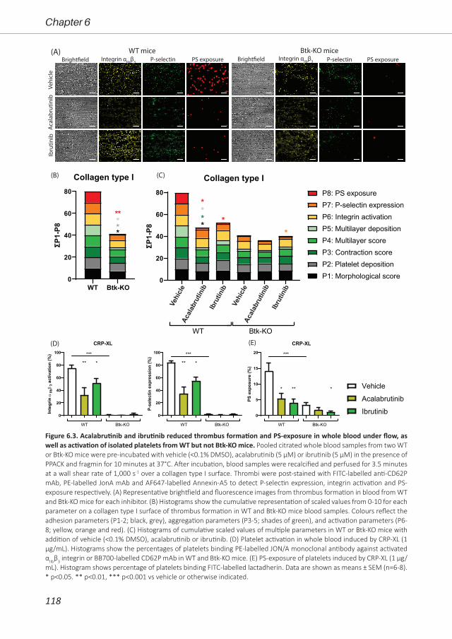
118
Chapter 6
(B) (C)
Bright�eld P-selectin PS exposure Bright�eld P-selectin PS exposureWT mice Btk-KO mice(A)
Vehi
cle
Acal
abru
tinib
Ibru
tinib
(D) (E)
Integrin αIIbβ3 Integrin αIIbβ3
WT Btk-KO0
20
40
60
80
Collagen type I
ΣP1-
P8
**
***
P8: PS exposureP7: P-selectin expressionP6: Integrin activationP5: Multilayer depositionP4: Multilayer scoreP3: Contraction scoreP2: Platelet depositionP1: Morphological score0
20
40
60
80
Collagen type I
ΣP1-
P8
*
***
*
WT Btk-KO
*
0
20
40
60
80
100
CRP-XL
Inte
grin
αIIbβ
3ac
tivat
ion
(%)
WT Btk-KO
**
***
*
0
20
40
60
80
100
P-se
lect
in e
xpre
ssio
n (%
)
WT Btk-KO
**
***
*
0
5
10
15
20
PS e
xpos
ure
(%)
WT Btk-KO
*
***
** *
CRP-XL
Vehicle
Acalabrutinib
Ibrutinib
Vehi
cle
Acal
abru
tinib
Ibru
tinib
Vehi
cle
Acal
abru
tinib
Ibru
tinib
Figure 6.3. Acalabrutinib and ibrutinib reduced thrombus formation and PS-exposure in whole blood under flow, as well as activation of isolated platelets from WT but not Btk-KO mice. Pooled citrated whole blood samples from two WT or Btk-KO mice were pre-incubated with vehicle (<0.1% DMSO), acalabrutinib (5 µM) or ibrutinib (5 µM) in the presence of PPACK and fragmin for 10 minutes at 37°C. After incubation, blood samples were recalcified and perfused for 3.5 minutes at a wall shear rate of 1,000 s-1 over a collagen type I surface. Thrombi were post-stained with FITC-labelled anti-CD62P mAb, PE-labelled JonA mAb and AF647-labelled Annexin-A5 to detect P-selectin expression, integrin activation and PS-exposure respectively. (A) Representative brightfield and fluorescence images from thrombus formation in blood from WT and Btk-KO mice for each inhibitor. (B) Histograms show the cumulative representation of scaled values from 0-10 for each parameter on a collagen type I surface of thrombus formation in WT and Btk-KO mice blood samples. Colours reflect the adhesion parameters (P1-2; black, grey), aggregation parameters (P3-5; shades of green), and activation parameters (P6-8; yellow, orange and red). (C) Histograms of cumulative scaled values of multiple parameters in WT or Btk-KO mice with addition of vehicle (<0.1% DMSO), acalabrutinib or ibrutinib. (D) Platelet activation in whole blood induced by CRP-XL (1 µg/mL). Histograms show the percentages of platelets binding PE-labelled JON/A monoclonal antibody against activated αIIbβ3 integrin or BB700-labelled CD62P mAb in WT and Btk-KO mice. (E) PS-exposure of platelets induced by CRP-XL (1 µg/mL). Histogram shows percentage of platelets binding FITC-labelled lactadherin. Data are shown as means ± SEM (n=6-8). * p<0.05. ** p<0.01, *** p<0.001 vs vehicle or otherwise indicated.
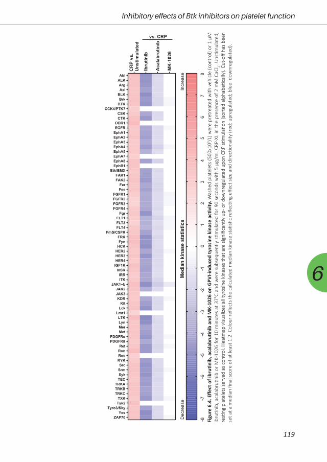
119
Inhibitory effects of Btk inhibitors on platelet function
6
Figu
re 6
.4. E
ffect
of i
bruti
nib,
aca
labr
utini
b an
d M
K-10
26 o
n G
PVI-i
nduc
ed ty
rosi
ne k
inas
e ac
tivity
. Was
hed
plat
elet
s (5
00x1
09 /L) w
ere
pret
reat
ed w
ith v
ehic
le (c
ontr
ol) o
r 1 μ
M
ibru
tinib
, aca
labr
utini
b or
MK-
1026
for
10 m
inut
es a
t 37°
C an
d w
ere
subs
eque
ntly
stim
ulat
ed fo
r 90
sec
onds
with
5 μ
g/m
L CR
P-XL
in th
e pr
esen
ce o
f 2 m
M C
aCl 2. U
nstim
ulat
ed,
resti
ng p
late
lets
ser
ved
as c
ontr
ol. H
eatm
ap v
isual
izes
all t
yros
ine
kina
ses
that
are
sig
nific
antly
up-
or d
ownr
egul
ated
upo
n CR
P sti
mul
ation
(sor
ted
alph
abeti
cally
). Cu
t-off
has
been
se
t at a
med
ian
final
sco
re o
f at l
east
1.2
. Col
our r
eflec
ts th
e ca
lcul
ated
med
ian
kina
se st
atisti
c re
flecti
ng e
ffect
size
and
dire
ction
ality
(red
: upr
egul
ated
; blu
e: d
ownr
egul
ated
).
CR
P vs
.U
nstim
ulat
ed
Ibru
tinib
Aca
labr
utin
ib
MK
-102
6
vs. CRP
01
23
45
67
8-8
-7-6
-5-4
-3-2
-1
Dec
reas
eIn
crea
seM
edia
n ki
nase
sta
tistic
s
AblALKArgAxl
BLKBrk
BTKCCK4/PTK7
CSKCTK
DDR1EGFR
EphA1EphA2EphA3EphA4EphA5EphA7EphA8EphB1
Etk/BMXFAK1FAK2
FerFes
FGFR1FGFR2FGFR3FGFR4
FgrFLT1FLT3FLT4
FmS/CSFRFRKFyn
HCKHER2HER3HER4
IGF1RInSRIRRITK
JAK1~bJAK2JAK3KDR
KitLck
Lmr1LTKLynMerMet
PDGFRαPDGFRß
RetRonRosRYKSrc
SrmSykTEC
TRKATRKBTRKC
TXKTyk2
Tyro3/SkyYes
ZAP70

120
Chapter 6
In comparison to these irreversible inhibitors, we investigated if the reversible Btk
inhibitor MK-102614 showed a more favourable anti-platelet effect. First, we evaluated which
platelet tyrosine kinases were affected by this compound upon GPVI stimulation. Only 18 tyrosine
kinases were significantly inhibited by MK-1026 (Fig 6.4). Median kinase statistics showed that
MK-1026 inhibited tyrosine phosphorylation of these proteins on average 0.52-fold, which was
8.3-fold lower as compared to ibrutinib. As expected, all three inhibitors significantly inhibited
the phosphorylation of Btk, and ibrutinib and acalabrutinib also inhibited Tec. Furthermore,
ibrutinib and acalabrutinib also inhibited the phosphorylation of Src family kinase (SFKs) and
other downstream proteins as previously described11, 12.
These results show that ibrutinib and acalabrutinib showed the same off-target effects on
GPVI-induced tyrosine kinase phosphorylation in platelets, although the effects of acalabrutinib
were less strong. The reversible inhibitor MK-1026 appeared to have less off-target effects in
platelets upon GPVI stimulation, in combination with a less strong inhibition profile as compared
to the irreversible inhibitors.
The reversible Btk inhibitor MK-1026 shows only limited anti-platelet effects in human and mouse platelets in vitro
As we demonstrated that irreversible inhibition of Btk can impair platelet function, we
also investigated the effects of the reversible Btk inhibitor MK-1026 on platelet function. First, we
performed collagen- and rhodocytin-induced dose-response aggregation experiments in PRP and
washed platelets. Similar to the irreversible Btk inhibitors, MK-1026 inhibited platelet aggregation
(with more prominent effects in washed platelets compared to PRP) at multiple concentrations
induced by both agonists (Fig 6.5A). GPVI-induced integrin αIIbβ3 activation, P-selectin expression,
and PS-exposure were also dose-dependently reduced by MK-1026 (Fig 6.5B). The calculated
IC50 values for MK-1026 in the presence of plasma were similar as for acalabrutinib (Suppl Table
6.1). Based on these results, in combination with reported in vivo plasma concentrations26, we
selected 5 µM for further experiments. Aggregation induced by ADP and U46619 were slightly
but significantly reduced at this concentration of MK-1026, while responses to thrombin and
ristocetin were unaffected (Suppl Fig 6.4A-B). Integrin αIIbβ3 activation and P-selectin expression
were not inhibited by MK-1026 in platelets stimulated with ADP or thrombin (Suppl Fig 6.4C).
Also, MK-1026 did not alter platelet adhesion and thrombus formation on six different surfaces,
as seen with acalabrutinib (Fig 6.5D, Suppl Fig 6.5).
We also investigated the effects of MK-1026 using blood from Btk-KO mice. Similarly as
for acalabrutinib and ibrutinib, we did not observe additional inhibition of whole blood thrombus
formation on collagen with MK-1026 in blood samples from WT or Btk-KO mice (Fig 6.6A-B, Suppl
Fig 6.6A). Also, no inhibitory effect of MK-1026 was found upon platelet activation or PS-exposure
from WT or Btk-KO mice with CRP-XL, ADP or PAR4 agonist (Fig 6.6C-D, Suppl Fig 6.6B-C).

121
Inhibitory effects of Btk inhibitors on platelet function
6
Figure 6.5. Effects of MK-1026 on aggregation and activation of isolated platelets in response to several agonists as well as whole blood thrombus formation under flow. (A) PRP or washed platelets (250x109/L) from healthy volunteers were incubated with different concentrations of MK-1026 for 10 minutes at 37°C. Shown are dose-response curves of aggregation induced by (i) collagen type I (1 µg/mL), or (ii) rhodocytin (1 µg/mL) in the presence of MK-1026 normalised to control expression. (B) Washed platelets (100x109/L) were pre-incubated with different concentrations of MK-1026 and stimulated with collagen-related peptide (CRP-XL, 5 µg/mL) for 15 minutes and simultaneously stained for activated integrin αIIbβ3 and α-granule secretion (P-selectin). Histograms show the percentages of platelets binding FITC-labelled PAC-1 monoclonal antibody or AF647-labelled anti-human CD62 mAb normalized to control expression. (C) Platelets pre-treated with different concentrations of MK-1026 were stimulated using both CRP-XL (5 µg/mL) and thrombin (4 nM) for 60 minutes at 37°C and PS-exposure was determined by flow cytometry. Histogram shows percentage of platelets binding FITC-labelled annexin A5 normalized to control expression. Data are shown as means ± SEM (n=6-7). (D) Pre-incubated blood samples from healthy donors with vehicle (<0.1% DMSO) or MK-1026 (5 µM) were recalcified in the presence of PPACK. Blood was perfused for 3.5 minutes at wall shear rate of 1,000 s-1 over six different coated surfaces. Thrombi were post-stained with AF568-annexin A5, FITC-α-fibrinogen and AF647-α-CD62P to detect PS-exposure, integrin activation and P-selectin expression. Cumulative histograms represent the scaled values (0-10) for each of the parameters. Colours reflect the adhesion parameters (P1-2; black, grey), aggregation parameters (P3-5; shades of green), and activation parameters (P6-8; yellow, orange and red). * p<0.05, ** p<0.01, *** p<0.001 vs vehicle.
(D)
(A)i ii
ii(B) (C)
P8: PS exposureP7: P-selectin expression
P6: Integrin activationP5: Multilayer deposition
P4: Multilayer scoreP3: Contraction score
P2: Platelet depositionP1: Morphological score
0.1 1 100
20
40
60
80
100
120
Collagen type I
MK-1026 concentration (µM)
Agg
rega
tion
(%of
cont
rol)
***
*
PRPWP
*
*** ***
0.1 1 100
20
40
60
80
100
120
MK-1026 concentration (µM)
Agg
rega
tion
(%of
cont
rol)
****
*
Rhodocytin
0.1 1 100
20
40
60
80
100
120
CRP-XL
Inte
grin
IIb3
activ
atio
n(%
)
******
*
0.1 1 100
20
40
60
80
100
120
P-se
lect
inex
pres
ion
(%)
*****
*
0.1 1 100
20
40
60
80
100
120
CRP-XL + Thrombin
PSex
posi
ngpl
atel
ets
(%)
***
MK-1026 concentration (µM) MK-1026 concentration (µM) MK-1026 concentration (µM)
i
Vehicl
e
MK-1026
0
20
40
60
80
Collagen type I
ΣP1-
P8
Vehicl
e
MK-1026
0
20
40
60
80
Collagen type III
ΣP1-
P8
Vehicl
e
MK-1026
0
20
40
60
80
vWF + Laminin
ΣP1-
P8
Vehicl
e
MK-1026
0
20
40
60
80
vWF + Rhodocytin
ΣP1-
P8
Vehicl
e
MK-1026
0
20
40
60
80
vWF + Ristocetin
ΣP1-
P8
Vehicl
e
MK-1026
0
20
40
60
80
vWF + Fibrinogen
ΣP1-
P8
*
iii iii iv v vi

122
Chapter 6
These data show that the reversible Btk inhibitor MK-1026 inhibited GPVI- and CLEC-2-
mediated aggregation of washed platelets, but did not impair platelet adhesion or activation in
whole blood perfused over collagen.
(B)
(C)
Bright�eld P-selectin PS exposure Bright�eld P-selectin PS exposureWT mice Btk-KO mice(A)
Vehi
cle
MK-
1026
(D)
Integrin αIIbβ3 Integrin αIIbβ3
Vehi
cle
MK
-102
6
Vehi
cle
MK
-102
6
0
20
40
60
80
Collagen type I
ΣP1-
P8
WT Btk-KO
0
20
40
60
80
100CRP-XL
Inte
grin
IIb3
activ
atio
n(%
)
***
WT Btk-KO
0
20
40
60
80
100
P-se
lect
inex
pres
sion
(%) **
WT Btk-KO
0
5
10
15
20
PSex
posu
re(%
)
*
WT Btk-KO
CRP-XL
P8: PS exposureP7: P-selectin expressionP6: Integrin activationP5: Multilayer depositionP4: Multilayer scoreP3: Contraction scoreP2: Platelet depositionP1: Morphological score
VehicleMK-1026
i ii
Figure 6.6. Thrombus formation, platelet activation and PS-exposure in blood from WT and Btk-KO mice was unaffected by MK-1026. Pooled citrated whole blood samples from two WT or Btk-KO mice were pre-incubated with vehicle (<0.1% DMSO) or MK-1026 (5 µM) in the presence of PPACK and fragmin for 10 minutes at 37°C. After incubation, blood samples were recalcified and perfused for 3.5 minutes at a wall shear rate of 1,000 s-1 over a collagen type I surface. Thrombi were post-stained with FITC-labelled anti-CD62P mAb, PE-labelled JON/A mAb and AF647-labelled Annexin-A5. (A) Representative brightfield and fluorescence images from thrombus formation in blood from WT and Btk-KO mice. (B) Histograms of cumulative scaled values of multiple parameters in WT or Btk-KO mice with addition of vehicle (<0.1% DMSO) or MK-1026. Colours reflect the adhesion parameters (P1-2; black, grey), aggregation parameters (P3-5; shades of green), and activation parameters (P6-8; yellow, orange and red). (C) Platelet activation in whole blood induced by CRP-XL (1 µg/mL). Histograms show the percentages of platelets binding PE-labelled JON/A monoclonal antibody or BB700-labelled CD62P mAb in WT and Btk-KO mice. (D) PS-exposure of platelets induced by CRP-XL (1 µg/mL). Histogram shows percentage of platelets binding FITC-labelled lactadherin. Data are shown as means ± SEM (n=6-8). * p<0.05, ** p<0.01, *** p<0.001.
123
Inhibitory effects of Btk inhibitors on platelet function
6
Ibrutinib inhibits thrombus formation under flow on multiple surfaces and GPVI-induced platelet activation responses in patients
Next, we assessed which platelet activation pathways were altered in patients using
ibrutinib. Sixteen patients were included, of which 7 did not receive ibrutinib and 9 were treated
with 420-560 mg ibrutinib once a day (Table 6.1). Baseline characteristics were largely similar
between groups. Importantly, none of the patients were thrombocytopenic, except one patient
with a platelet count of 50x109/L (Table 6.1). Of the ibrutinib-treated patients, 66% (6/9) reported
bleeding symptoms with a median ISTH-BAT score of 2 (range 1-7, Table 6.1). Of patients without
ibrutinib treatment, none reported bleeding symptoms.
Whole blood from patients was perfused over collagen type I or vWF plus rhodocytin
or laminin. No differences in thrombus formation were observed between patients without
ibrutinib and healthy controls (Suppl Fig 6.7). On collagen, treatment with ibrutinib reduced
thrombus contraction and height (multilayer) compared to non-treated patients. Furthermore,
αIIbβ3 activation, P-selectin expression and PS-exposure were significantly reduced (Fig 6.7A, Suppl
Fig 6.7A). For thrombi formed on vWF plus rhodocytin or laminin, αIIbβ3 activation and α-granule
secretion were reduced upon ibrutinib treatment (Fig 6.7A, Suppl Fig 6.7B-C).
Aggregation of isolated platelets from ibrutinib-treated patients showed on average
no significant inhibition upon stimulation with any agonist (Fig 6.7B). However, platelets from
5 ibrutinib-treated patients were highly nonresponsive to collagen stimulation, while platelets
from 2 patients responded normally to collagen. Also, with rhodocytin, platelets from 4 patients
receiving ibrutinib did not aggregate, while platelets from 4 patients showed normal aggregation.
When platelet activation was examined by flow cytometry, αIIbβ3 activation and P-selectin
expression were inhibited in CRP-XL-stimulated platelets from all ibrutinib-treated patients (Fig
6.7C). Upon ADP stimulation, platelets from patients on treatment showed a slight increase in
αIIbβ3 activation, while no effects on secretion were observed (Fig 6.7C). PS-exposure by thrombin
plus CRP-XL, was unchanged for all patients’ platelets (Fig 6.7D).
As 66% of our patients reported bleeding symptoms, we compared patients with significant
bleeding (>2 ISTH-BAT score) with patients with no/mild bleeding (0-1 ISTH-BAT score). Generally,
no significant changes were observed in platelet aggregation and thrombus formation in patients
with or without bleeding (Suppl Fig 6.8).
In summary, platelets from patients on ibrutinib treatment displayed inhibition of
activation by GPVI as well as reduced adhesion and thrombus formation under flow over collagen
and surfaces co-coated with vWF. Furthermore, variable responses were seen with CLEC-2
stimulation.
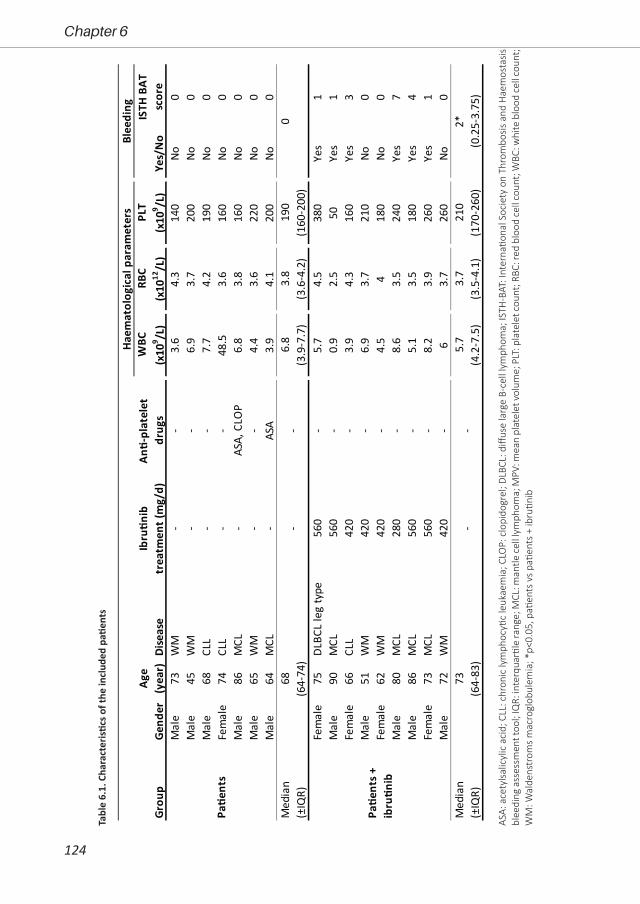
124
Chapter 6
Tabl
e 6.
1. C
hara
cter
istic
s of t
he in
clud
ed p
atien
ts
ASA:
ace
tylsa
licyl
ic a
cid;
CLL
: chr
onic
lym
phoc
ytic
leuk
aem
ia; C
LOP:
clo
pido
grel
; DLB
CL: d
iffus
e la
rge
B-ce
ll ly
mph
oma;
ISTH
-BAT
: Int
erna
tiona
l Soc
iety
on
Thro
mbo
sis a
nd H
aem
osta
sis
blee
ding
ass
essm
ent t
ool;
IQR:
inte
rqua
rtile
rang
e; M
CL: m
antle
cel
l lym
phom
a; M
PV: m
ean
plat
elet
vol
ume;
PLT
: pla
tele
t cou
nt; R
BC: r
ed b
lood
cel
l cou
nt; W
BC: w
hite
blo
od c
ell c
ount
; W
M: W
alde
nstr
oms
mac
rogl
obul
emia
; *p<
0.05
, pati
ents
vs
patie
nts
+ ib
rutin
ib
Mal
e73
WM
--
3.6
4.3
140
No0
Mal
e45
WM
--
6.9
3.7
200
No0
Mal
e68
CLL
--
7.7
4.2
190
No0
Fem
ale
74CL
L-
-48
.53.
616
0No
0M
ale
86M
CL-
ASA,
CLO
P6.
83.
816
0No
0M
ale
65W
M-
-4.
43.
622
0No
0M
ale
64M
CL-
ASA
3.9
4.1
200
No0
Fem
ale
75DL
BCL
leg
type
560
-5.
74.
538
0Ye
s1
Mal
e90
MCL
560
-0.
92.
550
Yes
1Fe
mal
e66
CLL
420
-3.
94.
316
0Ye
s3
Mal
e51
WM
420
-6.
93.
721
0No
0Fe
mal
e62
WM
420
-4.
54
180
No0
Mal
e80
MCL
280
-8.
63.
524
0Ye
s7
Mal
e86
MCL
560
-5.
13.
518
0Ye
s4
Fem
ale
73M
CL56
0-
8.2
3.9
260
Yes
1M
ale
72W
M42
0-
63.
726
0No
0
Haem
atol
ogic
al p
aram
eter
sBl
eedi
ngW
BC
(x10
9 /L)
RBC
(x10
12/L
)PL
T (x
109 /L
)
6.8
(3.9
-7.7
)3.
8(3
.6-4
.2)
190
(160
-200
)-
-
2*(0
.25-
3.75
)M
edia
n (±
IQR)
73(6
4-83
)5.
7(4
.2-7
.5)
Pa�e
nts +
ib
ru�n
ib
--
3.7
(3.5
-4.1
)21
0(1
70-2
60)
0M
edia
n (±
IQR)
68(6
4-74
)
Grou
pGe
nder
Dise
ase
An�-
plat
elet
dr
ugs
Yes/
No
ISTH
BAT
sc
ore
Age
(yea
r)Ib
ru�n
ib
trea
tmen
t (m
g/d)
Pa�e
nts
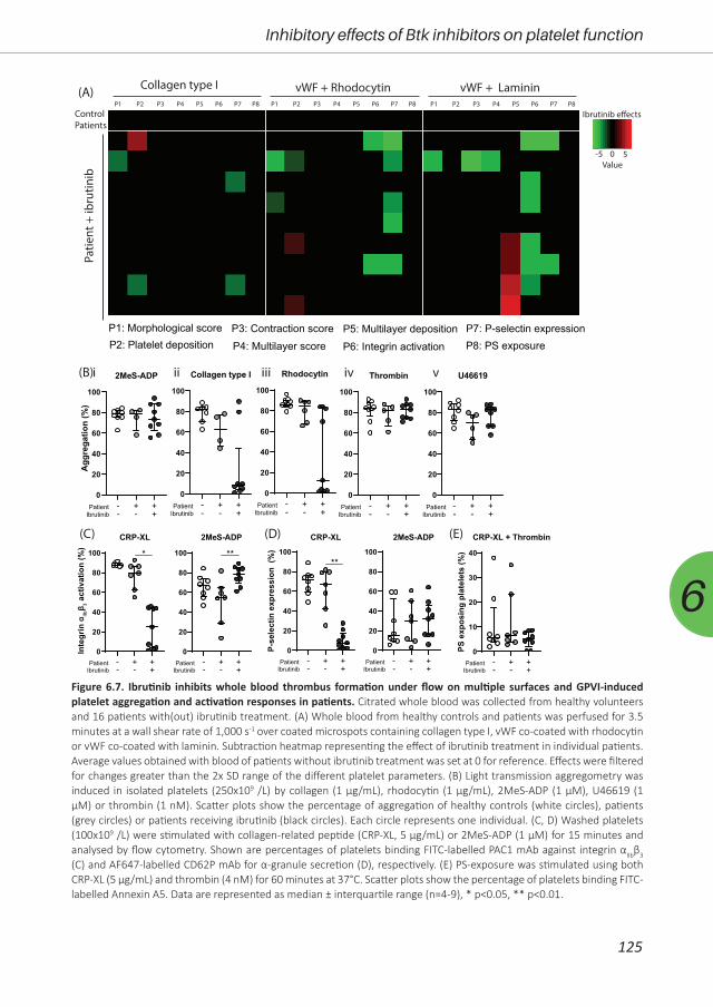
125
Inhibitory effects of Btk inhibitors on platelet function
6
(A)P1 P8
Collagen type I vWF + Rhodocytin vWF + Laminin
ControlPatients
Patie
nt +
ibru
tinib
(B)
(C) (D)
-5 50
Ibrutinib e�ects
Value
0
20
40
60
80
100
CRP-XL
*
PatientIbrutinib
- + +- - +
Inte
grin
αIIbβ 3
act
ivat
ion
(%)
0
20
40
60
80
100
2MeS-ADP
PatientIbrutinib
- + +- - +
**
0
10
20
30
40
CRP-XL + Thrombin
PS e
xpos
ing
plat
elet
s (%
)
PatientIbrutinib
- + +- - +
0
20
40
60
80
100
P-se
lect
in e
xpre
ssio
n (%
)
PatientIbrutinib
- + +- - +
**
0
20
40
60
80
100
PatientIbrutinib
- + +- - +
CRP-XL
0
20
40
60
80
100
Collagen type I
PatientIbrutinib
- + +- - +
0
20
40
60
80
100
Rhodocytin
PatientIbrutinib
- + +- - +
0
20
40
60
80
100
2MeS-ADP
PatientIbrutinib
- + +- - +
0
20
40
60
80
100
U46619
PatientIbrutinib
- + +- - +
0
20
40
60
80
100
Thrombin
PatientIbrutinib
- + +- - +
Agg
rega
tion
(%)
2MeS-ADP (E)
iiii ii iv v
P8: PS exposureP7: P-selectin expression
P6: Integrin activationP5: Multilayer deposition
P4: Multilayer scoreP3: Contraction score
P2: Platelet depositionP1: Morphological score
P2 P3 P4 P5 P7P6 P1 P8P2 P3 P4 P5 P7P6 P1 P8P2 P3 P4 P5 P7P6
Figure 6.7. Ibrutinib inhibits whole blood thrombus formation under flow on multiple surfaces and GPVI-induced platelet aggregation and activation responses in patients. Citrated whole blood was collected from healthy volunteers and 16 patients with(out) ibrutinib treatment. (A) Whole blood from healthy controls and patients was perfused for 3.5 minutes at a wall shear rate of 1,000 s-1 over coated microspots containing collagen type I, vWF co-coated with rhodocytin or vWF co-coated with laminin. Subtraction heatmap representing the effect of ibrutinib treatment in individual patients. Average values obtained with blood of patients without ibrutinib treatment was set at 0 for reference. Effects were filtered for changes greater than the 2x SD range of the different platelet parameters. (B) Light transmission aggregometry was induced in isolated platelets (250x109 /L) by collagen (1 µg/mL), rhodocytin (1 µg/mL), 2MeS-ADP (1 µM), U46619 (1 µM) or thrombin (1 nM). Scatter plots show the percentage of aggregation of healthy controls (white circles), patients (grey circles) or patients receiving ibrutinib (black circles). Each circle represents one individual. (C, D) Washed platelets (100x109 /L) were stimulated with collagen-related peptide (CRP-XL, 5 µg/mL) or 2MeS-ADP (1 µM) for 15 minutes and analysed by flow cytometry. Shown are percentages of platelets binding FITC-labelled PAC1 mAb against integrin αIIbβ3 (C) and AF647-labelled CD62P mAb for α-granule secretion (D), respectively. (E) PS-exposure was stimulated using both CRP-XL (5 µg/mL) and thrombin (4 nM) for 60 minutes at 37°C. Scatter plots show the percentage of platelets binding FITC-labelled Annexin A5. Data are represented as median ± interquartile range (n=4-9), * p<0.05, ** p<0.01.

126
Chapter 6
DiscussionDue to their promising effects on progression-free survival, Btk inhibitors are increasingly
prescribed in haematological malignancies4, 5. Btk inhibitors require lifelong treatment, and the
high percentage of discontinuation due to side effects (including bleeding tendency) stresses the
importance of understanding and managing these6-8. We found that ibrutinib and acalabrutinib
inhibit both GPVI- and CLEC-2-mediated platelet aggregation in a dose-dependent manner. Also,
all inhibitors dose-dependently impaired GPVI-induced αIIbβ3 activation and α-granule secretion.
Ibrutinib reduced thrombus formation on surfaces co-coated with vWF, but not on collagen.
Moreover, we are the first to study the effects on platelet function of MK-1026, a reversible Btk
inhibitor.
Other studies investigating effects of Btk inhibitors on platelet function have described
different findings. Several papers have shown inhibition of the GPVI pathway by ibrutinib or
acalabrutinib, either directly via Btk or due to off-target effects on Src kinases or other downstream
proteins11, 12, 23, 27-31. Others have implicated that GPIb-29, 32 or αIIbβ3-mediated signalling28, 31 were
affected by ibrutinib. In agreement, in the present study ibrutinib also inhibited thrombus
formation on ristocetin and fibrinogen co-coated with vWF. Levade et al. have also shown that
ibrutinib affected platelet adhesion to vWF only under flow, but did not assess acalabrutinib22.
Furthermore, we found that both ibrutinib and acalabrutinib impaired GPVI-mediated aggregation,
but not platelet adhesion on collagen under flow. Previous studies found conflicting results with
regards to the effects of ibrutinib on collagen adhesion, with some who did find inhibition12,33 and
others that could not34. A possible explanation for this is that platelet adhesion to collagen is also
regulated by integrin α2β1, which does not signal via Btk34. Our data now directly compared the
differences between ibrutinib and acalabrutinib in whole blood thrombus formation under flow
on six different surfaces, and shows for the first time that ibrutinib, but not acalabrutinib impairs
platelet adhesion surfaces co-coated with vWF. This might play a role in the increased incidence
of major bleeding seen in ibrutinib treatment compared to acalabrutinib.
Although GPVI is important for platelet adhesion and activation on collagen, recent studies
have shown that GPVI inhibitors do not impair haemostasis16. GPVI-deficient patients exhibit only
a mild bleeding diathesis17 and GPVI depletion in mice did not increase bleeding tendency19,20.
Therefore, the bleeding tendency seen with Btk inhibitors cannot solely be mediated by GPVI.
Depletion of both GPVI and CLEC-2 from mouse platelets did impair haemostasis21. In line with
this, we found that Btk inhibitors inhibited both GPVI- and CLEC-2-mediated aggregation.
Only few studies have investigated Btk-KO mice or X-linked agammaglobulinemia (XLA)
patients, a Btk deficiency in humans. Btk-KO mice showed impaired GPVI, CLEC-2 and GPIb
signalling35-38. In line with this, we found decreased GPVI-induced platelet granule secretion and
thrombus formation in Btk-KO mice. XLA patients demonstrated impaired GPVI- or CLEC-2-induced
PLCγ2 phosphorylation and platelet aggregation39, 40. As compared to XLA patients, ibrutinib has

127
Inhibitory effects of Btk inhibitors on platelet function
6
similar effects on GPVI, CLEC-2 and GPIb signalling. However, XLA patients are not associated with
an increased bleeding risk12. This suggests that Btk inhibitors affect other targets, most notably
Tec, which can substitute for Btk35 and is also inhibited by ibrutinib and acalabrutinib12. Also, off-
target effects on Src kinases have been implicated in the bleeding tendency11, 12. We assessed off-
target effects on GPVI-induced tyrosine kinase phosphorylation in platelets by using the PamGene
assay. These results showed that ibrutinib and acalabrutinib showed the same off-target effects,
although the effects of acalabrutinib were less strong. Importantly both inhibitors had an off-target
effect on Tec and Src kinases, questioning the previous conclusions that these were responsible
for the increased severe bleeding seen with ibrutinib compared to acalabrutinib.
When comparing results found in vitro in human and mice, we found that in human
samples, Btk inhibitors did not alter collagen-mediated adhesion under flow in agreement with
Zheng et al.31, whereas in mice, Btk inhibition (either by KO mice or acalabrutinib) reduced this
response. Btk-dependent signalling is completely absent in KO mice, and therefore, this may
result in stronger effects as compared to pharmacological inhibition. Btk proteins display 99.4%
similarity between human and mice41, so a species-dependent interaction with an inhibitor cannot
explain the observed differences. A possibility could be different bioavailability between mouse
and man. Ibrutinib and acalabrutinib are highly plasma protein-bound in human blood, 97.3%
and 97.5%, respectively42, 43, while acalabrutinib was reported to have lower protein binding in
mouse blood (75.4%)43. This may contribute to the more pronounced effects of acalabrutinib in
mouse blood as compared to human blood. We also observed different results between human
and mouse platelets with MK-1026 (same concentration) in flow cytometric experiments. This
may be explained by the use of murine whole blood in these experiments. Mouse blood contains
~three times more platelets as compared to human blood, and plasma proteins are also likely to
bind MK-1026. Altogether, this may explain the absent effect of MK-1026 on platelet functions of
wild-type mice.
As our data propose a direct role for Btk in impaired platelet function, we tested the
reversible Btk inhibitor MK-1026. MK-1026 showed promising effects on ibrutinib resistant CLL
cells in vitro and is currently being tested in clinical trials14. In the PamGene kinase assay MK-
1026 appeared to have less off-target effects in platelets upon GPVI stimulation, in combination
with a less strong inhibition profile as compared to the irreversible inhibitors. MK-1026 reduced
GPVI- and CLEC-2-mediated aggregation, as well as GPVI-induced platelet activation in washed
platelets, largely similar to acalabrutinib. In human and mouse blood, MK-1026 did not impair
collagen-induced thrombus formation under flow, in contrast to acalabrutinib which did affect this
response in mouse blood. Hence, this reversible inhibitor had less inhibitory effects on platelets
as compared to ibrutinib, in agreement with a recent study31. Hence, MK-1026 could be expected
to show a slightly reduced or comparable bleeding tendency compared to acalabrutinib.
The present dose-response experiments, in line with previous studies11, 33, show that at

128
Chapter 6
similar dose, ibrutinib is a more effective platelet inhibitor compared to other Btk inhibitors. This
has been attributed to the inhibition of drug efflux pumps by ibrutinib33. We observed that higher
concentrations of inhibitors are required to inhibit platelet aggregation in the presence of plasma,
as compared to washed platelets. This in line with previous observations with other TKIs44, 45. This
is most likely caused by binding of ibrutinib and acalabrutinib to albumin46-48. It has been reported
that prolongation of the incubation time lowered the IC50 values of ibrutinib and acalabrutinib
for GPVI-mediated aggregation49. However, in that study a much lower collagen concentration
was used, which may be more sensitive to longer incubation with low doses of inhibitors.
Furthermore, that study did not include CLEC-2-dependent platelet responses49. Although
inhibitor concentration could play a role in the extent of inhibition, we showed that ibrutinib and
acalabrutinib influence additional platelet pathways as compared to MK-1026, suggesting that
the extent of Btk inhibition might not be the reason for the increased bleeding tendency.
Previous studies assessing which platelet pathways are involved in bleeding tendency
in patients using ibrutinib found conflicting results. Some showed a correlation with collagen-
induced aggregation12, 22, 50, and some with platelet adhesion to collagen12, whereas others could
not find this22. Bleeding tendency was also associated with adhesion to vWF under flow22 and
one study found a correlation with ristocetin-induced platelet aggregation32, which another study
could not replicate28. Ibrutinib can also induce shedding of GPIbα, GPIX and integrin αIIbβ3 in
patients, but the correlation with bleeding remains unknown51.
A recent study assessed platelet parameters in patients with CLL and mantle cell lymphoma
(MCL)52, showing correlations between bleeding tendency, thrombocytopenia and decreased
ADP-induced platelet aggregation. However, a drawback of this study was that a large patient
proportion was thrombocytopenic, which can directly influence platelet aggregation, as platelet
concentration in PRP was not reported to be adjusted. This impaired the establishment if ibrutinib
or platelet count affected aggregation response. In our patients with (in general) normal platelet
counts, we also observed significant differences in platelet responses to several stimuli of patients
using ibrutinib. Furthermore, 66% of the included patients with ibrutinib treatment reported
bleeding symptoms. Although we could show that in patients, similar to healthy volunteers,
ibrutinib inhibited GPVI signalling, with variable effects on CLEC-2, as well as reduced thrombus
formation to surfaces co-coated with vWF, this could not differentiate for the bleeding tendency.
Generally, the patients showed a large variation in measurement outcomes, which might be
influenced by clinical factors. However, with patients on ibrutinib treatment, we have directly
compared whole blood thrombus formation under flow on multiple surfaces, including vWF plus
rhodocytin and laminin, which has not been reported with ibrutinib-treated patients thus far.
In conclusion, the present work demonstrated that ibrutinib, acalabrutinib and MK-1026
inhibited GPVI- and CLEC-2-mediated platelet aggregation, but only ibrutinib also inhibited GPVI-
induced platelet activation and thrombus formation on surfaces co-coated with vWF. The novel
129
Inhibitory effects of Btk inhibitors on platelet function
6
reversible BTK inhibitor MK-1026 might therefore be promising for future treatment in patients
at risk for bleeding.
AcknowledgementsTAMC was supported by the Heimburger award from CSL Behringer. JAE is financially
supported by the Interdisziplinary Center of Clinical Research (IZKF) of the University of Münster
(grant no. Ebl1/009/21). Additional support was received by ZonMW VIDI 91716421 to MFAK
and JMEMC and by the Dutch Heart Foundation (2015T79 to JMEMC and 2020T020 to CCFMJB).
CCFMJB was also supported by the START-Program of the Faculty of Medicine at the RWTH
Aachen University (105/20). EPCvdV was supported by a grant from the Interdisciplinary Center
for Clinical Research within the faculty of Medicine at the RWTH Aachen University, the Fritz
Thyssen Stiftung (10.20.2.043MN), and NWO-ZonMw Veni (91619053). All authors declare no
conflict of interest.
Author contributionsBMET, MFAK, VL, MStB, CCFMJB, SLM and EPCvdV acquired, analysed and interpreted
data. AFdV provided Btk-deficient mice. JAE contributed essential tools. MRN and TAMC provided
ethical approval and included patients. JMEMC and JWMH provided lab space with equipment
and supervision. TAMC and MJEK designed and supervised the research, and interpreted data.
BMET, TAMC and MJEK wrote and revised the paper. All the authors provided critical feedback,
edited, and approved the final manuscript.
References1. Byrd, JC, Furman, RR, Coutre, SE, Burger, JA, Blum, KA, Coleman, M et al. Three-year follow-up of
treatment-naive and previously treated patients with CLL and SLL receiving single-agent ibrutinib. Blood. 2015; 125, 2497-2506.
2. Byrd, JC, Brown, JR, O'Brien, S, Barrientos, JC, Kay, NE, Reddy, NM et al. Ibrutinib versus ofatumumab in previously treated chronic lymphoid leukemia. N Engl J Med. 2014; 371, 213-223.
3. Wang, ML, Blum, KA, Martin, P, Goy, A, Auer, R, Kahl, BS et al. Long-term follow-up of MCL patients treated with single-agent ibrutinib: updated safety and efficacy results. Blood. 2015; 126, 739-745.
4. Castillo, JJ, Advani, RH, Branagan, AR, Buske, C, Dimopoulos, MA, D'Sa, S et al. Consensus treatment recommendations from the tenth International Workshop for Waldenström Macroglobulinaemia. Lancet Haematol. 2020; 7, e827-e837.
5. Hallek, M. Chronic lymphocytic leukemia: 2020 update on diagnosis, risk stratification and treatment. Am J Hematol. 2019; 94, 1266-1287.
6. Shatzel, JJ, Olson, SR, Tao, DL, McCarty, OJT, Danilov, AV & DeLoughery, TG. Ibrutinib-associated bleeding: pathogenesis, management and risk reduction strategies. J Thromb Haemost. 2017; 15, 835-847.
7. Mato, AR, Roeker, LE, Allan, JN, Pagel, JM, Brander, DM, Hill, BT et al. Outcomes of front-line ibrutinib treated CLL patients excluded from landmark clinical trial. Am J Hematol. 2018; 93, 1394-1401.
8. Stephens, DM & Byrd, JC. How I manage ibrutinib intolerance and complications in patients with chronic lymphocytic leukemia. Blood. 2019; 133, 1298-1307.
9. de Weerdt, I, Koopmans, SM, Kater, AP & van Gelder, M. Incidence and management of toxicity
130
Chapter 6
associated with ibrutinib and idelalisib: a practical approach. Haematologica. 2017; 102, 1629-1639.10. Brown, JR, Moslehi, J, Ewer, MS, O'Brien, SM, Ghia, P, Cymbalista, F et al. Incidence of and risk factors
for major haemorrhage in patients treated with ibrutinib: An integrated analysis. Br J Haematol. 2019; 184, 558-569.
11. Nicolson, PLR, Hughes, CE, Watson, S, Nock, SH, Hardy, AT, Watson, CN et al. Inhibition of Btk by Btk-specific concentrations of ibrutinib and acalabrutinib delays but does not block platelet aggregation mediated by glycoprotein VI. Haematologica. 2018; 103, 2097-2108.
12. Bye, AP, Unsworth, AJ, Desborough, MJ, Hildyard, CAT, Appleby, N, Bruce, D et al. Severe platelet dysfunction in NHL patients receiving ibrutinib is absent in patients receiving acalabrutinib. Blood Adv. 2017; 1, 2610-2623.
13. Wen, T, Wang, J, Shi, Y, Qian, H & Liu, P. Inhibitors targeting Bruton's tyrosine kinase in cancers: drug development advances. Leukemia. 2021; 35, 312-332.
14. Reiff, SD, Mantel, R, Smith, LL, Greene, JT, Muhowski, EM, Fabian, CA et al. The BTK Inhibitor ARQ 531 Targets Ibrutinib-Resistant CLL and Richter Transformation. Cancer Discov. 2018; 8, 1300-1315.
15. Byrd, JC, Harrington, B, O'Brien, S, Jones, JA, Schuh, A, Devereux, S et al. Acalabrutinib (ACP-196) in Relapsed Chronic Lymphocytic Leukemia. N Engl J Med. 2016; 374, 323-332.
16. Denorme, F & Rondina, MT. Targeting Glycoprotein VI for Thromboembolic Disorders. Arterioscler Thromb Vasc Biol. 2019; 39, 839-840.
17. Matus, V, Valenzuela, G, Sáez, CG, Hidalgo, P, Lagos, M, Aranda, E et al. An adenine insertion in exon 6 of human GP6 generates a truncated protein associated with a bleeding disorder in four Chilean families. J Thromb Haemost. 2013; 11, 1751-1759.
18. Nagy, M, Perrella, G, Dalby, A, Becerra, MF, Garcia Quintanilla, L, Pike, JA et al. Flow studies on human GPVI-deficient blood under coagulating and noncoagulating conditions. Blood Adv. 2020; 4, 2953-2961.
19. Lockyer, S, Okuyama, K, Begum, S, Le, S, Sun, B, Watanabe, T et al. GPVI-deficient mice lack collagen responses and are protected against experimentally induced pulmonary thromboembolism. Thromb Res. 2006; 118, 371-380.
20. Nieswandt, B, Schulte, V, Bergmeier, W, Mokhtari-Nejad, R, Rackebrandt, K, Cazenave, JP et al. Long-term antithrombotic protection by in vivo depletion of platelet glycoprotein VI in mice. J Exp Med. 2001; 193, 459-469.
21. Bender, M, May, F, Lorenz, V, Thielmann, I, Hagedorn, I, Finney, BA et al. Combined in vivo depletion of glycoprotein VI and C-type lectin-like receptor 2 severely compromises hemostasis and abrogates arterial thrombosis in mice. Arterioscler Thromb Vasc Biol. 2013; 33, 926-934.
22. Levade, M, David, E, Garcia, C, Laurent, PA, Cadot, S, Michallet, AS et al. Ibrutinib treatment affects collagen and von Willebrand factor-dependent platelet functions. Blood. 2014; 124, 3991-3995.
23. Bye, AP, Unsworth, AJ, Vaiyapuri, S, Stainer, AR, Fry, MJ & Gibbins, JM. Ibrutinib Inhibits Platelet Integrin alphaIIbbeta3 Outside-In Signaling and Thrombus Stability But Not Adhesion to Collagen. Arterioscler Thromb Vasc Biol. 2015; 35, 2326-2335.
24. de Witt, SM, Swieringa, F, Cavill, R, Lamers, MM, van Kruchten, R, Mastenbroek, T et al. Identification of platelet function defects by multi-parameter assessment of thrombus formation. Nat Commun. 2014; 5, 4257.
25. Advani, RH, Buggy, JJ, Sharman, JP, Smith, SM, Boyd, TE, Grant, B et al. Bruton tyrosine kinase inhibitor ibrutinib (PCI-32765) has significant activity in patients with relapsed/refractory B-cell malignancies. J Clin Oncol. 2013; 31, 88-94.
26. Elgamal, OA, Mehmood, A, Jeon, JY, Carmichael, B, Lehman, A, Orwick, SJ et al. Preclinical efficacy for a novel tyrosine kinase inhibitor, ArQule 531 against acute myeloid leukemia. J Hematol Oncol. 2020; 13, 8.
27. Ninomoto, J, Mokatrin, A, Kinoshita, T, Marimpietri, C, Barrett, TD, Chang, BY et al. Effects of ibrutinib on in vitro platelet aggregation in blood samples from healthy donors and donors with platelet dysfunction. Hematology. 2020; 25, 112-117.
28. Rigg, RA, Aslan, JE, Healy, LD, Wallisch, M, Thierheimer, ML, Loren, CP et al. Oral administration of Bruton's tyrosine kinase inhibitors impairs GPVI-mediated platelet function. Am J Physiol Cell Physiol. 2016; 310, C373-380.

131
Inhibitory effects of Btk inhibitors on platelet function
6
29. Kamel, S, Horton, L, Ysebaert, L, Levade, M, Burbury, K, Tan, S et al. Ibrutinib inhibits collagen-mediated but not ADP-mediated platelet aggregation. Leukemia. 2015; 29, 783-787.
30. Alberelli, MA, Innocenti, I, Sica, S, Laurenti, L & De Candia, E. PO-54 - Clinical and laboratory characterization of platelet dysfunction caused by ibrutinib treatment in patients with chronic lymphocytic leukemia. Thromb Res. 2016; 140.
31. Zheng, TJ, Lofurno, ER, Melrose, AR, Lakshmanan, HHS, Pang, J, Phillips, KG et al. Assessment of the effects of Syk and BTK inhibitors on GPVI-mediated platelet signaling and function. Am J Physiol Cell Physiol. 2021.
32. Kazianka, L, Drucker, C, Skrabs, C, Thomas, W, Melchardt, T, Struve, S et al. Ristocetin-induced platelet aggregation for monitoring of bleeding tendency in CLL treated with ibrutinib. Leukemia. 2017; 31, 1117-1122.
33. Series, J, Garcia, C, Levade, M, Viaud, J, Sie, P, Ysebaert, L et al. Differences and similarities in the effects of ibrutinib and acalabrutinib on platelet functions. Haematologica. 2019; 104, 2292-2299.
34. Busygina, K, Jamasbi, J, Seiler, T, Deckmyn, H, Weber, C, Brandl, R et al. Oral Bruton tyrosine kinase inhibitors selectively block atherosclerotic plaque-triggered thrombus formation in humans. Blood. 2018; 131, 2605-2616.
35. Atkinson, BT, Ellmeier, W & Watson, SP. Tec regulates platelet activation by GPVI in the absence of Btk. Blood. 2003; 102, 3592-3599.
36. Pasquet, JM, Quek, L, Stevens, C, Bobe, R, Huber, M, Duronio, V et al. Phosphatidylinositol 3,4,5-trisphosphate regulates Ca(2+) entry via btk in platelets and megakaryocytes without increasing phospholipase C activity. EMBO J. 2000; 19, 2793-2802.
37. Liu, J, Fitzgerald, ME, Berndt, MC, Jackson, CW & Gartner, TK. Bruton tyrosine kinase is essential for botrocetin/VWF-induced signaling and GPIb-dependent thrombus formation in vivo. Blood. 2006; 108, 2596-2603.
38. de Porto, A, Claushuis, TAM, van der Donk, LEH, de Beer, R, de Boer, OJ, Florquin, S et al. Platelet Btk is Required for Maintaining Lung Vascular Integrity during Murine Pneumococcal Pneumosepsis. Thromb Haemost. 2019; 119, 930-940.
39. Quek, LS, Bolen, J & Watson, SP. A role for Bruton's tyrosine kinase (Btk) in platelet activation by collagen. Curr Biol. 1998; 8, 1137-1140.
40. Nicolson, PLR, Nock, SH, Hinds, J, Garcia-Quintanilla, L, Smith, CW, Campos, J et al. Low-dose Btk inhibitors selectively block platelet activation by CLEC-2. Haematologica. 2021; 106, 208-219.
41. Oeltjen, JC, Malley, TM, Muzny, DM, Miller, W, Gibbs, RA & Belmont, JW. Large-scale comparative sequence analysis of the human and murine Bruton's tyrosine kinase loci reveals conserved regulatory domains. Genome Res. 1997; 7, 315-329.
42. Bose, P, Gandhi, VV & Keating, MJ. Pharmacokinetic and pharmacodynamic evaluation of ibrutinib for the treatment of chronic lymphocytic leukemia: rationale for lower doses. Expert Opin Drug Metab Toxicol. 2016; 12, 1381-1392.
43. Podoll, T, Pearson, PG, Evarts, J, Ingallinera, T, Bibikova, E, Sun, H et al. Bioavailability, Biotransformation, and Excretion of the Covalent Bruton Tyrosine Kinase Inhibitor Acalabrutinib in Rats, Dogs, and Humans. Drug Metab Dispos. 2019; 47, 145-154.
44. Tullemans, BME, Nagy, M, Sabrkhany, S, Griffioen, AW, Oude Egbrink, MGA, Aarts, M et al. Tyrosine Kinase Inhibitor Pazopanib Inhibits Platelet Procoagulant Activity in Renal Cell Carcinoma Patients. Front Cardiovasc Med. 2018; 5, 142.
45. Tullemans, BME, Fernandez, DI, Veninga, A, Baaten, C, Peters, LJF, Aarts, MJB et al. Tyrosine Kinase Inhibitor Sunitinib Delays Platelet-Induced Coagulation: Additive Effects of Aspirin. Thromb Haemost. 2021.
46. Liu, X, Feng, D, Zheng, M, Cui, Y & Zhong, D. Characterization of covalent binding of tyrosine kinase inhibitors to plasma proteins. Drug Metab Pharmacokinet. 2020; 35, 456-465.
47. Tang, B, Tang, P, He, J, Yang, H & Li, H. Characterization of the binding of a novel antitumor drug ibrutinib with human serum albumin: Insights from spectroscopic, calorimetric and docking studies. J Photochem Photobiol B. 2018; 184, 18-26.
48. Abdelhameed, AS, Alanazi, AM, Bakheit, AH, Hassan, ES, Herqash, RN & Almutairi, FM. Novel BTK inhibitor acalabrutinib (ACP-196) tightly binds to site I of the human serum albumin as observed by

132
Chapter 6
spectroscopic and computational studies. Int J Biol Macromol. 2019; 127, 536-543.49. Denzinger, V, Busygina, K, Jamasbi, J, Pekrul, I, Spannagl, M, Weber, C et al. Optimizing Platelet GPVI
Inhibition versus Haemostatic Impairment by the Btk Inhibitors Ibrutinib, Acalabrutinib, ONO/GS-4059, BGB-3111 and Evobrutinib. Thromb Haemost. 2019; 119, 397-406.
50. Lipsky, AH, Farooqui, MZ, Tian, X, Martyr, S, Cullinane, AM, Nghiem, K et al. Incidence and risk factors of bleeding-related adverse events in patients with chronic lymphocytic leukemia treated with ibrutinib. Haematologica. 2015; 100, 1571-1578.
51. Dobie, G, Kuriri, FA, Omar, MMA, Alanazi, F, Gazwani, AM, Tang, CPS et al. Ibrutinib, but not zanubrutinib, induces platelet receptor shedding of GPIb-IX-V complex and integrin alphaIIbbeta3 in mice and humans. Blood Adv. 2019; 3, 4298-4311.
52. Dmitrieva, EA, Nikitin, EA, Ignatova, AA, Vorobyev, VI, Poletaev, AV, Seregina, EA et al. Platelet function and bleeding in chronic lymphocytic leukemia and mantle cell lymphoma patients on ibrutinib. J Thromb Haemost. 2020; 18, 2672-2684.

133
Inhibitory effects of Btk inhibitors on platelet function
6
Supplemental Data
Materials & methodsMaterials
Acalabrutinib was obtained from Selleckchem (Houston, TX, USA), MK-1026 (formerly
ARQ-531) was a provided by ArQule, Inc., a wholly-owned subsidiary of Merck & Co. (Kenilworth
NJ, USA) and ibrutinib was purchased from Toronto Research Chemicals (North York, Canada).
Bovine serum albumin (BSA), D(+)-glucose, unfractionated heparin and apyrase were purchased
from Sigma-Aldrich (Saint Louis MO, USA). Low molecular weight heparin (Fragmin®) was from
Prizer (Capelle a/d IJssel, The Netherlands). Horm® collagen type I came from Takeda (Hoofddorp,
The Netherlands), whereas the agonists collagen-related peptide crosslinked (CRP-XL) and von
Willebrand factor III (vWF-III) were obtained from CambCol Laboratories (Cambridge, UK).
Thrombin was from Enzyme Research Laboratories Inc (South Bend, IN, USA) and protease-
activated receptor 4 activating peptide (PAR4-AP) was obtained from Bachem (Bubendorf,
Switzerland). U46619 (thromboxane A2 receptor agonist) came from Cayman Chemicals (Ann
Arbor MI, USA). 2-Methylthio-adenosine-diphosphate (2MeS-ADP) and D-phenylalanyl-prolyl-
arginyl chloromethyl ketone (PPACK) were obtained from Santa Cruz Biotechnology (Dallas TX,
USA). Laminin was purchased from Octapharma (Lachen, Switzerland), whereas rhodocytin
was purified from Calloselasma rhodostoma venom as described previously1. Fura-2-AM was
obtained from Invitrogen (Carlsbad CA, USA). Fluorescein isothiocyanate (FITC)-labelled PAC1
monoclonal antibody (mAb) against activated human integrin αIIbβ3 were purchased from BD
Bioscience (nr. 340507; Franklin Lakes NJ, USA). FITC-conjugated α-fibrinogen mAb was purchased
from DAKO (F0111; Santa Clara CA, USA). Alexa Fluor (AF)568 and AF647-conjugated annexin A5
were purchased from Molecular Probes, Life Technologies (New York NY, USA). AF647-labelled
CD62-P mAb was obtained from Biolegend (London, UK). FITC-conjugated annexin A5 was from
Pharmatarget (Maastricht, The Netherlands), whereas FITC-conjugated lactadherin came from
Haematologic Technologies (Essex Junction VT, USA). FITC-labelled rat anti-mouse CD62P mAb
and phycoerythrin (PE) labelled rat anti-mouse JON/A mAb were obtained from Emfret Analytics
(Würzburg, Germany).
Blood collection and platelet isolation from healthy volunteers and patientsWith approval from the medical ethics committee from the Maastricht University Medical
Centre+ (MUMC+), blood was collected from healthy volunteers. Furthermore, blood was collected
from patients diagnosed with chronic lymphocytic leukaemia (CLL), mantle cell lymphoma and
Waldenstroms macroglobulinemia with or without ibrutinib treatment at the Catharina Hospital
in Eindhoven (the Netherlands) with approval of the local medical ethical committee. This study
was performed in accordance with the declaration of Helsinki and informed consent was obtained

134
Chapter 6
from all participants. Blood was collected in 3.2% trisodium citrate tubes by venipuncture, after
discarding the first 3 mL of blood. Blood cell counts and haematological parameters were assessed
of all blood samples using a Sysmex XP300 (Kobe, Japan). Patients characteristic and ISTH-BAT
score2 were acquired for each patient.
Platelet-rich plasma (PRP) or platelets were isolated from whole blood as described
previously3, 4. Washed platelets were resuspended in Hepes buffer, pH 7.45 (10 mM Hepes, 136
mM NaCl, 2.7 mM KCl, 2 mM MgCl2, 1 mg/mL glucose, and 1 mg/ml BSA). Platelet concentrations
were adjusted as stated per assay.
Blood collection from animalsSpecific-pathogen-free Btk-KO mice5 and littermate controls (wild-types, WT) were housed
in the Animal Research Institute Amsterdam facility under standard care. Studies were reviewed
and approved by the Central Authority for Scientific Procedures on Animals (CCD) and the Animal
Welfare Body (IvD) of the Academic Medical Centre, University of Amsterdam (ID number: 17-
4125-1-80). The animal care and use protocol adhered to the Dutch Experiments on Animals Act
(WOD) and European Directive of 22 September 2010 (Directive 2010/63/EU) in addition to the
Directive of 6 May 2009 (Directive 2009/41/EC).
Only male mice between 8 and 12 weeks of age were used for experiments. Citrated blood
was drawn from WT and Btk-KO mice from the vena cava inferior under full anaesthesia induced
by intraperitoneal injection of ketamine (125 mg/kg bodyweight) and dexmedetomidine (300 µg/
kg bodyweight). After blood collection mice were sacrificed by severing the diaphragm.
Whole blood thrombus formation under flowThrombus formation under flow with human blood was performed as described 6.
Briefly, glass coverslips were coated with 3 microspots of collagen type I (100 µg/mL), collagen
type III (100 µg/mL), vWF (50 µg/mL) co-coated with laminin (100 µg/mL), vWF co-coated with
rhodocytin (250 µg/mL), vWF co-coated with ristocetin (250 µg/mL), or vWF co-coated with
fibrinogen (250 µg/mL), and mounted in a parallel plate flow chamber. Citrated blood samples
from healthy donors were incubated with vehicle or 5 µM of Btk inhibitor (acalabrutinib, ibrutinib
or MK-1026) for 10 minutes at room temperature. Blood samples from CLL patients were used
directly. Whole blood from healthy volunteers or CLL patients was recalcified in the presence of
PPACK (40 µM), and perfused at a wall shear rate of 1000 s-1 for 3.5 minutes. Platelet activation
properties were determined by post-perfusion with Hepes buffer (with additional 2 mM CaCl2 and
1 U/mL heparin) containing FITC-conjugated α-fibrinogen mAb (1:80), AF647-conjugated CD62-P
(1:100), and AF568-conjugated annexin A5 (1:200).
Thrombus formation under flow with mouse blood was performed as described7. Glass
coverslips were coated with collagen type I (100 µg/mL) and blood samples, in the presence of
135
Inhibitory effects of Btk inhibitors on platelet function
6
PPACK (57.1 µM) and Fragmin (35.7 µM), were incubated with 5 µM Btk inhibitor (acalabrutinib,
ibrutinib or MK-1026) for 10 minutes at 37°C. Next, the blood samples were recalcified and
perfused at a shear rate of 1000 s−1 for 3.5 minutes. To measure platelet activation status, thrombi
were post-stained with Tyrode Hepes buffer, pH 7.45 (5 mM Hepes, 136 mM NaCl, 2.7 mM KCl,
0.42 mM NaH2PO4, 1 mg/mL glucose, 1 mg/ml BSA, 2 mM CaCl2, 2 mM MgCl2 and 1 U/mL heparin)
containing PE-labelled Jon-A (1:20) and FITC-conjugated anti-P-selectin mAb (1:40) and AF647-
labelled Annexin-A5 (1:200).
For both human and mouse experiments, brightfield and fluorescent images were captured
using an EVOS microscope (Bothel, WA, USA). Images were analysed using specific scripts in the
open-access Fiji software (Laboratory for Optical and Computational Instrumentation, University
of Wisconsin-Madison, WI, USA) as described6, 8. Values for the following parameters of thrombus
formation and platelet activation were obtained: morphological score of platelet adhesion and
thrombus formation (P1; scale 0-5), surface area coverage of adhered platelets (P2; %SAC),
platelet aggregate contraction score (P3; scale 0-3), platelet aggregate multilayer score (P4;
scale 0-3), coverage of multi-layered platelet aggregation (P5; %SAC), integrin αIIbβ3 activation
(P6; %SAC), P-selectin expression (P7; %SAC) and PS exposure (P8; %SAC). For comparative data
analysis, cumulative histograms were generated of scaled values from 0-10 for each parameter.
For patient data subtraction heatmap representing the effect of ibrutinib treatment in individual
patients. Average values obtained with blood of patients without ibrutinib treatment was set at
0 for reference. Effects were filtered for changes greater than the 2x SD range of the different
platelet parameters.
Light transmission aggregometryIsolated platelets (250x109 platelets/L) from healthy volunteers were incubated with
vehicle or 3.3 µM of Btk inhibitor (acalabrutinib, ibrutinib or MK-1026) for 10 minutes at 37°C,
whereas platelets from CLL patients were used directly after preheating to 37°C for 10 minutes.
Aggregation response was induced by collagen type I (1 µg/mL), 2MeS-ADP (1 µM), U46619
(1 µM), rhodocytin (1 µg/mL) or thrombin (1 nM). Platelet aggregation was recorded using a
Chronolog optical aggregometer (Havertown PA, USA) and maximum amplitude was quantified at
8 minutes after agonist addition.
Flow cytometryWashed platelets (100x109 platelets/L) from healthy volunteers or CLL patients were
supplemented with 2 mM CaCl2. Samples from healthy volunteers were incubated with vehicle or
1 µM of Btk inhibitor (acalabrutinib, ibrutinib or MK-1026) for 10 minutes at room temperature,
whereas patient samples were used directly. Platelets were stimulated by CRP-XL (5 μg/mL), 2MeS-
ADP (1 μM), or thrombin (1 nM) for 15 minutes at RT. Activation of integrin αIIbβ3 and P-selectin

136
Chapter 6
expression were determined using FITC-conjugated PAC1 mAb (1:20) and AF647-conjugated
CD62-P mAb (1:40) respectively. For measuring phosphatidyl-serine (PS) exposure platelets were
activated with a combination of CRP-XL (5 μg/mL) and thrombin (4 nM) for 1 hour at 37°C and
labelled with FITC-conjugated annexin A5. Flow cytometry was performed in duplicates using a
BD Accuri C6TM flow cytometer and accompanying software (Erembodegem, Belgium).
Platelet activation in whole blood from WT and Btk-KO mice was performed using pooled
blood samples (5 µl), in the presence of PPACK (57.1 µM) and Fragmin (35.7 µM), incubated with
5 µM Btk inhibitor (acalabrutinib, ibrutinib or MK-1026) for 10 minutes at 37°C. Platelets were
then stimulated for 30 minutes at room temperature with control buffer (PBS), CRP-XL (1 μg/ml),
2MeS-ADP (1 μM) or PAR4-AP (0.1 mg/ml). Platelet activation was assessed by flow cytometry
using APC-conjugated anti-CD61, BB700-labelled anti-CD62P, PE-conjugated JON/A and FITC-
labelled lactadherin. The labelling was terminated by addition of 40 volumes of buffer. Analysis
was carried out immediately by flow cytometry using a CytoFLEX (Beckman Coulter, Brea, CA)
until 10,000 CD61+ events were recorded. All stimulations and staining were done in duplicate.
Compensation settings were validated using single-stained controls. Percentage of positivity
was determined using unstimulated controls. Data analysis was done using FlowJoTM v10 (BD
Biosciences, San Jose, CA).
Cytosolic Ca2+ measurements Washed platelets (200x109 platelets/L) were loaded with Fura-2 acetoxymethyl ester and
changes in cytosolic Ca2+ ([Ca2+]i) were measured in 96-well plates using a FlexStation 3 (Molecular
Devices, San Jose, CA, USA) as described9,10. In brief, platelet suspension was preincubated with
vehicle or 1 µM Btk inhibitor (acalabrutinib, ibrutinib or MK-1026) for 10 minutes at RT. Fura-2
loaded platelets in presence of CaCl2 were stimulated by CRP-XL (10 µg/mL). Changes in Fura-2
fluorescence were measured in duplicate and ratio values were calculated and are presented as
[Ca+]i in nM.
PamGene kinase assay Washed platelets (500x109 platelets/L) were pretreated with vehicle (control) or 1 μM
ibrutinib, acalabrutinib or MK-1026 for 10 minutes at 37°C and were subsequently stimulated
with 5 μg/mL CRP-XL in the presence of 2 mM CaCl2. Unstimulated, resting platelets served as
control. After 90 seconds of stimulation, samples were lysed by adding 1:1 M-PER Mammalian
Extraction Buffer containing Halt Phosphatase Inhibitor and EDTA-free Halt Protease Inhibitor
Cocktail (1:100 each; Thermo Fischer Scientific). Samples were lysed for 15 minutes on ice and
afterwards centrifuged for 15 minutes at 10,000g at 4°C. Supernatants were collected and protein
content was quantified with a BioRad DC protein kit (Hercules CA, USA).
Tyrosine kinase profiles were determined using the PamChip® peptide tyrosine kinase

137
Inhibitory effects of Btk inhibitors on platelet function
6
microarray system on PamStation®12 (PTK; PamGene International, ´s-Hertogenbosch, The
Netherlands). Each PTK-PamChip® array contains 144 individual phospho-site(s) that are peptide
sequences derived from substrates for TKs. Each peptide on the chip builds a 15-amino acid
sequence representing a putative endogenous phosphorylation site which functions as a TK
substrate. The phosphorylation of the peptides is visualized by detection of the fluorescent signal
which is emitted as a result of the binding of the FITC-conjugated PY20 anti-phosphotyrosine
antibody.
For the PTK assay, 7.5 µg of protein was applied per array (N=4 per condition) and carried
out using the standard protocol supplied by Pamgene. All reagents used for PTK activity profiling
were supplied by Pamgene International B.V. Initially, to prepare the PTK Basic Mix, the freshly
frozen lysate was added to 4 µL of 10 x protein PTK reaction buffer (PK), 0.4 µL of 100 x bovine
serum albumin (BSA), 0.4 µL of 1 M dithiothreitol (DTT) solution, 4 µL of 10 x PTK additive, 4 µL
of 4 mM ATP and 0.6 µL of monoclonal anti-phosphotyrosine FITC-conjugate detection antibody
(clone PY20). Total volume of the PTK Basic Mix was adjusted to 40 µL by adding distilled water
(H20). Before loading the PTK Basic Mix on the array, a blocking step was performed applying 30 µL
of 2% BSA to the middle of every array and washing with PTK solution for PamChip® preprocessing.
Next, 40 µL of PTK Basic Mix were applied to each array of the PamChips®. Then, the microarray
assays were run for 94 cycles. An image was recorded by a CCD camera PamStation®12 at kinetic
read cycles 32–93 at 10, 50 and 200 ms and at end-level read cycle at 10, 20, 50, 100 and 200 ms.
The spot intensity at each time point was quantified (and corrected for local background) using the
BioNavigator software version 6.3 (PamGene International, 's-Hertogenbosch, The Netherlands).
Upstream Kinase Analysis (UKA)11, a functional scoring method (PamGene) was used to rank
kinases based on combined specificity scores (based on peptides linked to a kinase, derived from
6 databases) and sensitivity scores (based on treatment-control differences).
Statistical analysisData for healthy volunteers are shown as mean ± standard error of mean (SEM), whereas
CLL patients are presented as median ± interquartile ranges. GraphPad Prism 8.3.0 software (La
Jolla, CA, USA) was used for statistical analysis. For healthy donor and CLL patient samples a
non-parametric and unpaired t-test (Mann-Whitney) was used to compare vehicle and inhibitor
treatment in vitro, whereas for mouse samples statistics were calculated using a one-way
non-parametric Anova (Kruskal-Wallis) to compare WT versus Btk-KO samples, as well as the
comparison between vehicle and inhibitor treatment in both WT and Btk-KO mice. A p-value less
than 0.05 was considered to be statistically significant in which * is p<0.05, ** is p<0.01 and ***
is p<0.001.

138
Chapter 6
References1. Bergmeier, W, Bouvard, D, Eble, JA, Mokhtari-Nejad, R, Schulte, V, Zirngibl, H et al. Rhodocytin
(aggretin) activates platelets lacking alpha(2)beta(1) integrin, glycoprotein VI, and the ligand-binding domain of glycoprotein Ibalpha. J Biol Chem. 2001; 276, 25121-25126.
2. Rodeghiero, F, Tosetto, A, Abshire, T, Arnold, DM, Coller, B, James, P et al. ISTH/SSC bleeding assessment tool: a standardized questionnaire and a proposal for a new bleeding score for inherited bleeding disorders. J Thromb Haemost. 2010; 8, 2063-2065.
3. van der Meijden, PE, Feijge, MA, Giesen, PL, Huijberts, M, van Raak, LP & Heemskerk, JW. Platelet P2Y12 receptors enhance signalling towards procoagulant activity and thrombin generation. A study with healthy subjects and patients at thrombotic risk. Thromb Haemost. 2005; 93, 1128-1136.
4. Mattheij, NJ, Gilio, K, van Kruchten, R, Jobe, SM, Wieschhaus, AJ, Chishti, AH et al. Dual mechanism of integrin alphaIIbbeta3 closure in procoagulant platelets. J Biol Chem. 2013; 288, 13325-13336.
5. Hendriks, RW, de Bruijn, MF, Maas, A, Dingjan, GM, Karis, A & Grosveld, F. Inactivation of Btk by insertion of lacZ reveals defects in B cell development only past the pre-B cell stage. EMBO J. 1996; 15, 4862-4872.
6. de Witt, SM, Swieringa, F, Cavill, R, Lamers, MM, van Kruchten, R, Mastenbroek, T et al. Identification of platelet function defects by multi-parameter assessment of thrombus formation. Nat Commun. 2014; 5, 4257.
7. van Geffen, JP, Swieringa, F, van Kuijk, K, Tullemans, BME, Solari, FA, Peng, B et al. Mild hyperlipidemia in mice aggravates platelet responsiveness in thrombus formation and exploration of platelet proteome and lipidome. Sci Rep. 2020; 10, 21407.
8. van Geffen, JP, Brouns, SLN, Batista, J, McKinney, H, Kempster, C, Nagy, M et al. High-throughput elucidation of thrombus formation reveals sources of platelet function variability. Haematologica. 2019; 104, 1256-1267.
9. Feijge, MA, van Pampus, EC, Lacabaratz-Porret, C, Hamulyàk, K, Levy-Toledano, S, Enouf, J et al. Inter-individual variability in Ca2+ signalling in platelets from healthy volunteers: effects of aspirin and relationship with expression of endomembrane Ca2+-ATPases. Br J Haematol. 1998; 102, 850-859.
10. Jooss, NJ, De Simone, I, Provenzale, I, Fernandez, DI, Brouns, SLN, Farndale, RW et al. Role of Platelet Glycoprotein VI and Tyrosine Kinase Syk in Thrombus Formation on Collagen-Like Surfaces. Int J Mol Sci. 2019; 20. 2788.
11. Chirumamilla, CS, Fazil, M, Perez-Novo, C, Rangarajan, S, de Wijn, R, Ramireddy, P et al. Profiling Activity of Cellular Kinases in Migrating T-Cells. Methods Mol Biol. 2019; 1930, 99-113.

139
Inhibitory effects of Btk inhibitors on platelet function
6
Supplemental table
Supplemental figures
Supplemental Table 6.1. IC50 values (µM) of Btk inhibitors for inhibition of GPVI- and CLEC-2 induced platelet aggregation and activation in presence (PRP) or absence of plasma (WP).
CLEC-2 GPVI + PAR
PRP WP WPIntegrin
ac�va�on Secre�onPS
exposureIbru�nib 3.31 0.425 0.170 0.196 0.176 0.332Acalabru�nib 9.85 1.635 1.275 0.982 1.439 0.723MK-1026 8.84 0.820 0.487 0.388 0.807 0.410
Ac�va�onGPVI
Aggrega�onGPVI
Btk inhibitor
(A)i ii iii (B)
(C)
0
20
40
60
80
100
120
2MeS-ADP
Agg
rega
tion
(%of
cont
rol)
0
20
40
60
80
100
120
Collagen type I
0
20
40
60
80
100
120
Thrombin
0
20
40
60
80
100
120
U46619
0
20
40
60
80
100
120
Ristocetin
Agg
rega
tion
(%of
cont
rol)
iv
0
20
40
60
80
100
120
2MeS-ADP
IIb3
activ
atio
n(%
ofco
ntro
l)
***
0
20
40
60
80
100
120
Thrombin
0
20
40
60
80
100
120
2MeS-ADP
P-se
lect
inex
pres
sion
(%of
cont
rol)
**
**
0
20
40
60
80
100
120
Thrombini ii
Vehicle
Acalabrutinib
Ibrutinib
Supplemental Figure 6.1. Platelet aggregation and activation of blood samples from healthy volunteers with ibrutinib and acalabrutinib, as well as dose-response of collagen-induced aggregation. (A) Washed platelets (250x109/L) from healthy volunteers were incubated with vehicle, ibrutinib (3.3 µM) or acalabrutinib (3.3 µM). Aggregation was induced by (i) 2MeS-ADP (1 µM), (ii) collagen type I (5 µg/mL), (iii) thrombin (1 nM) or (iv) U46619 (1 µM). Aggregation responses were measured during 8 minutes. (B) PRP (250x109/L) was incubated with vehicle, ibrutinib (5 µM) or acalabrutinib (5 µM) for 10 minutes at 37°C and stimulated with ristocetin (2 mg/mL). Aggregation responses were measured during 8 minutes. (C) Washed platelets (100x109/L) were stimulated with 2MeS-ADP (1 µM) or thrombin (1 nM) for 15 minutes and analysed by flow cytometry. Shown are percentages of platelets binding FITC-labelled PAC1 mAb against integrin αIIbβ3 and AF647-labelled CD62P mAb for α-granule secretion, respectively. Data are shown as means ± SEM (n=5-6). * p<0.05, ** p<0.01, *** p<0.001 vs vehicle.
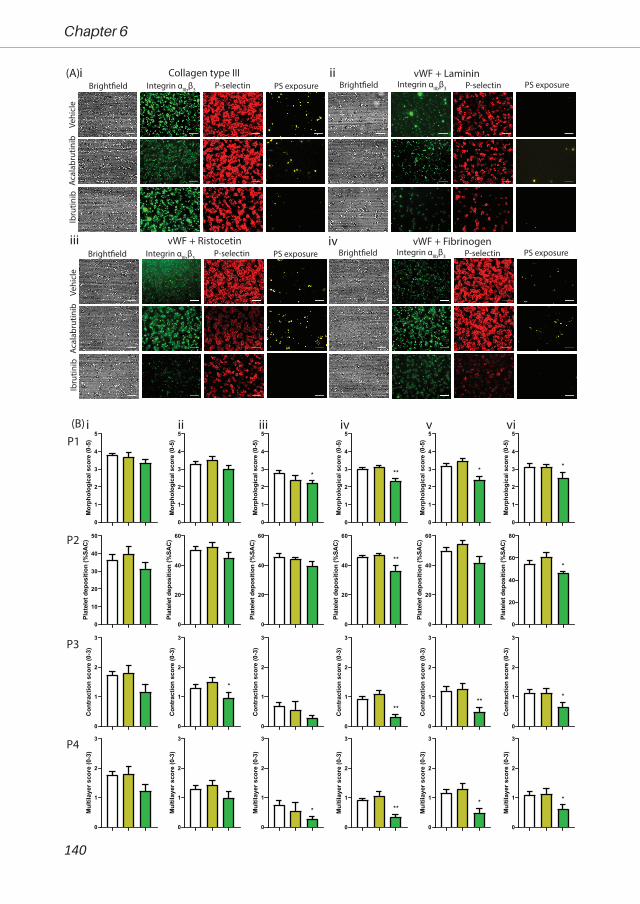
140
Chapter 6
(A) Collagen type III vWF + LamininVe
hicl
eAc
alab
rutin
ibIb
rutin
ib
Bright�eld P-selectin PS exposure Bright�eld P-selectin PS exposureIntegrin αIIbβ3 Integrin αIIbβ3
vWF + Ristocetin vWF + Fibrinogen
Vehi
cle
Acal
abru
tinib
Ibru
tinib
Bright�eld P-selectin PS exposure Bright�eld P-selectin PS exposureIntegrin αIIbβ3 Integrin αIIbβ3
i ii
iii iv
(B) ii iiiiP1
P2
P3
P4
0
1
2
3
4
5
Mor
phol
ogic
al s
core
(0-5
)
0
1
2
3
4
5
Mor
phol
ogic
al s
core
(0-5
)
0
1
2
3
4
5
Mor
phol
ogic
al s
core
(0-5
)
*
0
1
2
3
4
5
Mor
phol
ogic
al s
core
(0-5
)
**
0
1
2
3
4
5
Mor
phol
ogic
al s
core
(0-5
)
*
0
1
2
3
4
5
Mor
phol
ogic
al s
core
(0-5
)
*
0
10
20
30
40
50
Plat
elet
dep
ositi
on (%
SAC)
0
20
40
60
Plat
elet
dep
ositi
on (%
SAC)
0
20
40
60
Plat
elet
dep
ositi
on (%
SAC)
0
20
40
60
Plat
elet
dep
ositi
on (%
SAC)
**
0
20
40
60
Plat
elet
dep
ositi
on (%
SAC)
0
20
40
60
80
Plat
elet
dep
ositi
on (%
SAC)
*
0
1
2
3
Con
trac
tion
scor
e (0
-3)
0
1
2
3
Con
trac
tion
scor
e (0
-3)
*
0
1
2
3
Con
trac
tion
scor
e (0
-3)
0
1
2
3
Con
trac
tion
scor
e (0
-3)
**
0
1
2
3
Con
trac
tion
scor
e (0
-3)
**
0
1
2
3
Con
trac
tion
scor
e (0
-3)
*
0
1
2
3
Mul
tilay
er s
core
(0-3
)
0
1
2
3
Mul
tilay
er s
core
(0-3
)
0
1
2
3
Mul
tilay
er s
core
(0-3
)
*
0
1
2
3
Mul
tilay
er s
core
(0-3
)
**
0
1
2
3
Mul
tilay
er s
core
(0-3
)
*
0
1
2
3
Mul
tilay
er s
core
(0-3
)
*
iv v vi
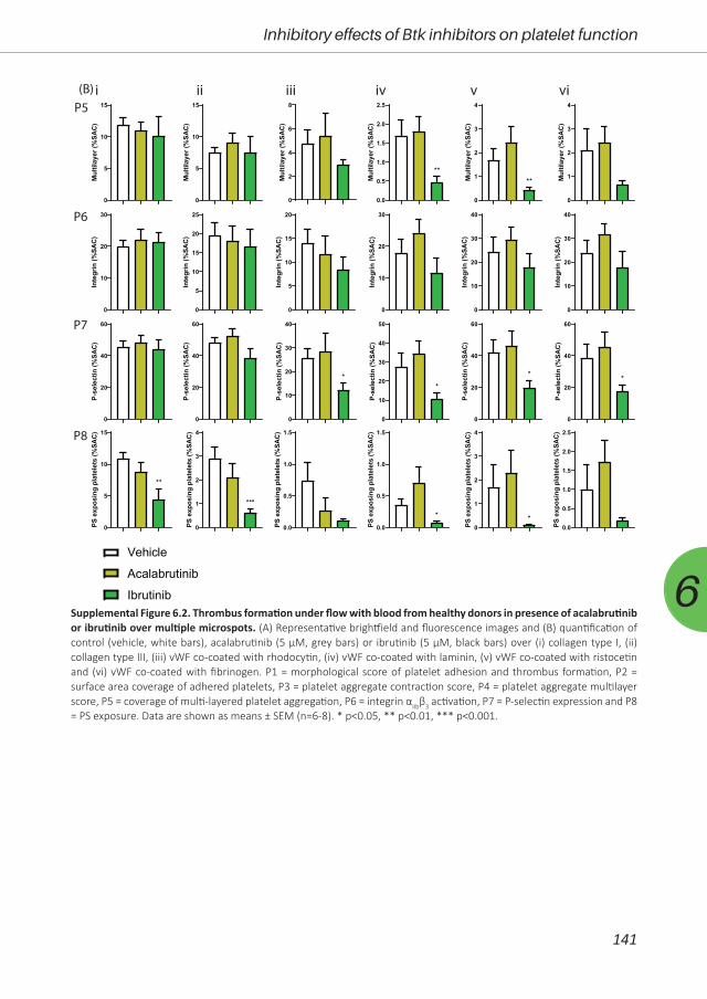
141
Inhibitory effects of Btk inhibitors on platelet function
6Supplemental Figure 6.2. Thrombus formation under flow with blood from healthy donors in presence of acalabrutinib or ibrutinib over multiple microspots. (A) Representative brightfield and fluorescence images and (B) quantification of control (vehicle, white bars), acalabrutinib (5 µM, grey bars) or ibrutinib (5 µM, black bars) over (i) collagen type I, (ii) collagen type III, (iii) vWF co-coated with rhodocytin, (iv) vWF co-coated with laminin, (v) vWF co-coated with ristocetin and (vi) vWF co-coated with fibrinogen. P1 = morphological score of platelet adhesion and thrombus formation, P2 = surface area coverage of adhered platelets, P3 = platelet aggregate contraction score, P4 = platelet aggregate multilayer score, P5 = coverage of multi-layered platelet aggregation, P6 = integrin αIIbβ3 activation, P7 = P-selectin expression and P8 = PS exposure. Data are shown as means ± SEM (n=6-8). * p<0.05, ** p<0.01, *** p<0.001.
Vehicle
Acalabrutinib
Ibrutinib
(B) ii iiiiP5
P6
P7
P8
iv v vi
0
5
10
15
Mul
tilay
er (%
SAC
)
0
5
10
15
Mul
tilay
er (%
SAC
)
0
2
4
6
8
Mul
tilay
er (%
SAC
)
0.0
0.5
1.0
1.5
2.0
2.5
Mul
tilay
er (%
SAC
)
**
0
1
2
3
4
Mul
tilay
er (%
SAC
)
**
0
1
2
3
4
Mul
tilay
er (%
SAC
)
0
10
20
30
Inte
grin
(%SA
C)
0
5
10
15
20
25
Inte
grin
(%SA
C)
0
5
10
15
20
Inte
grin
(%SA
C)
0
10
20
30
Inte
grin
(%SA
C)
0
10
20
30
40
Inte
grin
(%SA
C)
0
10
20
30
40
Inte
grin
(%SA
C)
0
20
40
60
P-se
lect
in (%
SAC
)
0
20
40
60
P-se
lect
in (%
SAC
)
0
10
20
30
40
P-se
lect
in (%
SAC
)
*
0
10
20
30
40
50
P-se
lect
in (%
SAC
)
*
0
20
40
60
P-se
lect
in (%
SAC
)
*
0
20
40
60
P-se
lect
in (%
SAC
)
*
0
5
10
15
PS e
xpos
ing
plat
elet
s (%
SAC
)
**
0
1
2
3
4
PS e
xpos
ing
plat
elet
s (%
SAC
)
***
0.0
0.5
1.0
1.5
PS e
xpos
ing
plat
elet
s (%
SAC
)
0.0
0.5
1.0
1.5
PS e
xpos
ing
plat
elet
s (%
SAC
)
*
0
1
2
3
4
PS e
xpos
ing
plat
elet
s (%
SAC
)
*0.0
0.5
1.0
1.5
2.0
2.5
PS e
xpos
ing
plat
elet
s (%
SAC
)
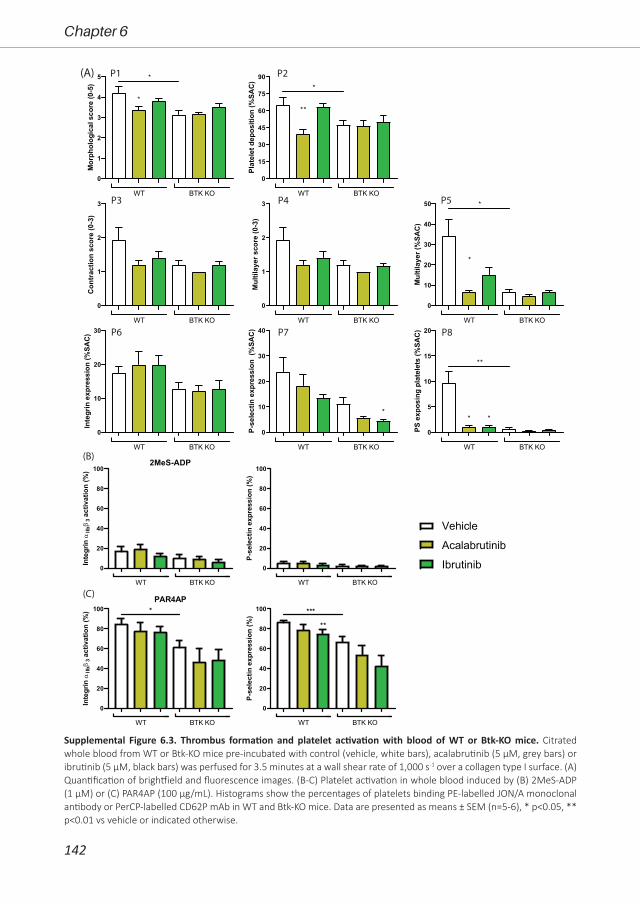
142
Chapter 6
P1 P2
P3 P4 P5
P6 P7 P8
(B)
(C)
(A)
Vehicle
Acalabrutinib
Ibrutinib
0
1
2
3
4
5
Mor
phol
ogic
al s
core
(0-5
)
WT BTK KO
*
*
0
15
30
45
60
75
90
Plat
elet
dep
ositi
on (%
SAC)
WT BTK KO
*
**
0
1
2
3
Con
trac
tion
scor
e (0
-3)
WT BTK KO
0
1
2
3
Mul
tilay
er s
core
(0-3
)
WT BTK KO
0
10
20
30
40
50
Mul
tilay
er (%
SAC
)
WT BTK KO
*
*
0
10
20
30
Inte
grin
exp
ress
ion
(%SA
C)
WT BTK KO
0
10
20
30
40
P-se
lect
in e
xpre
ssio
n (%
SAC
)
WT BTK KO
*
0
5
10
15
20
PS e
xpos
ing
plat
elet
s (%
SAC
)
WT BTK KO
**
* *
0
20
40
60
80
1002MeS-ADP
Inte
grin
αIIbβ
3ac
tivat
ion
(%)
WT BTK KO
0
20
40
60
80
100
P-se
lect
in e
xpre
ssio
n (%
)
WT BTK KO
0
20
40
60
80
100PAR4AP
Inte
grin
αIIbβ
3ac
tivat
ion
(%)
WT BTK KO
*
0
20
40
60
80
100
P-se
lect
in e
xpre
ssio
n (%
)
WT BTK KO
***
**
Supplemental Figure 6.3. Thrombus formation and platelet activation with blood of WT or Btk-KO mice. Citrated whole blood from WT or Btk-KO mice pre-incubated with control (vehicle, white bars), acalabrutinib (5 µM, grey bars) or ibrutinib (5 µM, black bars) was perfused for 3.5 minutes at a wall shear rate of 1,000 s-1 over a collagen type I surface. (A) Quantification of brightfield and fluorescence images. (B-C) Platelet activation in whole blood induced by (B) 2MeS-ADP (1 µM) or (C) PAR4AP (100 µg/mL). Histograms show the percentages of platelets binding PE-labelled JON/A monoclonal antibody or PerCP-labelled CD62P mAb in WT and Btk-KO mice. Data are presented as means ± SEM (n=5-6), * p<0.05, ** p<0.01 vs vehicle or indicated otherwise.
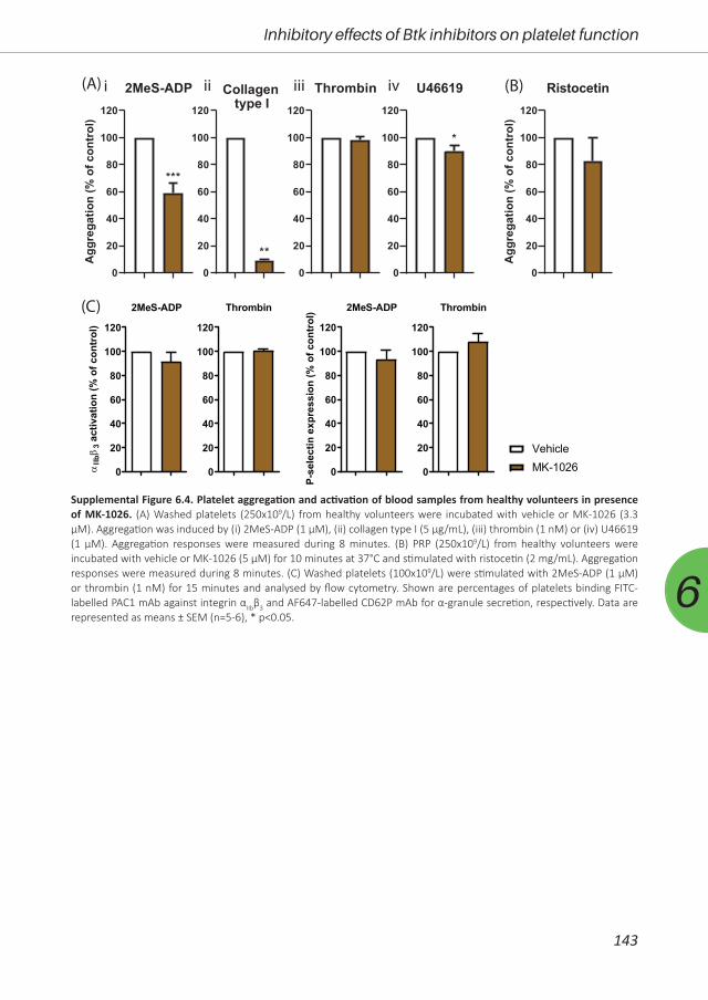
143
Inhibitory effects of Btk inhibitors on platelet function
6
(A) (B)
(C)
i ii iii
0
20
40
60
80
100
120
2MeS-ADPA
ggre
gatio
n(%
ofco
ntro
l)
***
0
20
40
60
80
100
120
Collagen type I
**
0
20
40
60
80
100
120
Thrombin
0
20
40
60
80
100
120
U46619
*
0
20
40
60
80
100
120
Ristocetin
Agg
rega
tion
(%of
cont
rol)
iv
0
20
40
60
80
100
120
2MeS-ADP
αIIbβ
3ac
tivat
ion
(% o
f con
trol
)
0
20
40
60
80
100
120
Thrombin
0
20
40
60
80
100
120
2MeS-ADPP-
sele
ctin
exp
ress
ion
(% o
f con
trol
)
0
20
40
60
80
100
120
Thrombin
VehicleMK-1026
Supplemental Figure 6.4. Platelet aggregation and activation of blood samples from healthy volunteers in presence of MK-1026. (A) Washed platelets (250x109/L) from healthy volunteers were incubated with vehicle or MK-1026 (3.3 µM). Aggregation was induced by (i) 2MeS-ADP (1 µM), (ii) collagen type I (5 µg/mL), (iii) thrombin (1 nM) or (iv) U46619 (1 µM). Aggregation responses were measured during 8 minutes. (B) PRP (250x109/L) from healthy volunteers were incubated with vehicle or MK-1026 (5 µM) for 10 minutes at 37°C and stimulated with ristocetin (2 mg/mL). Aggregation responses were measured during 8 minutes. (C) Washed platelets (100x109/L) were stimulated with 2MeS-ADP (1 µM) or thrombin (1 nM) for 15 minutes and analysed by flow cytometry. Shown are percentages of platelets binding FITC-labelled PAC1 mAb against integrin αIIbβ3 and AF647-labelled CD62P mAb for α-granule secretion, respectively. Data are represented as means ± SEM (n=5-6), * p<0.05.

144
Chapter 6
Bright�eld P-selectin PS exposure
Vehi
cle
MK-
1026
Vehi
cle
MK-
1026
Vehi
cle
MK-
1026
(A)Integrin αIIbβ3 Bright�eld P-selectin PS exposureIntegrin αIIbβ3
Collagen type IIICollagen type I
vWF + Laminin vWF + Rhodocytin
vWF + Ristocetin vWF + Fibrinogen
iii
iii iv
v vi
0
1
2
3
4
5
Mor
phol
ogic
al s
core
(0-5
)
0
1
2
3
4
5
Mor
phol
ogic
al s
core
(0-5
)
0
1
2
3
4
5
Mor
phol
ogic
al s
core
(0-5
)
0
1
2
3
4
5
Mor
phol
ogic
al s
core
(0-5
)
0
1
2
3
4
5
Mor
phol
ogic
al s
core
(0-5
)
0
1
2
3
4
5
Mor
phol
ogic
al s
core
(0-5
)
0
10
20
30
40
50
Plat
elet
dep
ositi
on (%
SAC)
0
20
40
60
Plat
elet
dep
ositi
on (%
SAC)
0
20
40
60
Plat
elet
dep
ositi
on (%
SAC)
0
20
40
60
Plat
elet
dep
ositi
on (%
SAC)
0
20
40
60
Plat
elet
dep
ositi
on (%
SAC)
0
20
40
60
80
Plat
elet
dep
ositi
on (%
SAC)
0
1
2
3
Con
trac
tion
scor
e (0
-3)
0
1
2
3
Con
trac
tion
scor
e (0
-3)
0
1
2
3
Con
trac
tion
scor
e (0
-3)
0
1
2
3
Con
trac
tion
scor
e (0
-3)
0
1
2
3
Con
trac
tion
scor
e (0
-3)
0
1
2
3
Con
trac
tion
scor
e (0
-3)
0
1
2
3
Mul
tilay
er s
core
(0-3
)
0
1
2
3
Mul
tilay
er s
core
(0-3
)
0
1
2
3
Mul
tilay
er s
core
(0-3
)
0
1
2
3
Mul
tilay
er s
core
(0-3
)
0
1
2
3
Mul
tilay
er s
core
(0-3
)
0
1
2
3
Mul
tilay
er s
core
(0-3
)
(B) ii iiiiP1
P2
P3
P4
iv v vi
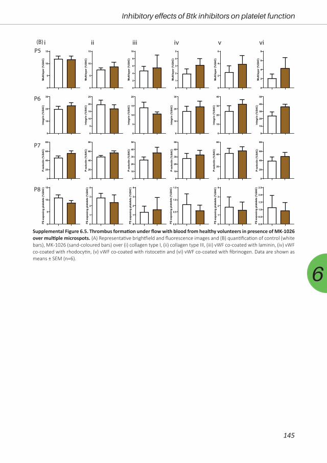
145
Inhibitory effects of Btk inhibitors on platelet function
6
Supplemental Figure 6.5. Thrombus formation under flow with blood from healthy volunteers in presence of MK-1026 over multiple microspots. (A) Representative brightfield and fluorescence images and (B) quantification of control (white bars), MK-1026 (sand-coloured bars) over (i) collagen type I, (ii) collagen type III, (iii) vWF co-coated with laminin, (iv) vWF co-coated with rhodocytin, (v) vWF co-coated with ristocetin and (vi) vWF co-coated with fibrinogen. Data are shown as means ± SEM (n=6).
0
5
10
15
Mul
tilay
er (%
SAC
)
0
5
10
15
Mul
tilay
er (%
SAC
)0
2
4
6
8
10
Mul
tilay
er (%
SAC
)
0
1
2
3
4
5
Mul
tilay
er (%
SAC
)
0
2
4
6
Mul
tilay
er (%
SAC
)
0
2
4
6
8
Mul
tilay
er (%
SAC
)
0
10
20
30
Inte
grin
(%SA
C)
0
5
10
15
20
25
Inte
grin
(%SA
C)
0
5
10
15
20
Inte
grin
(%SA
C)
0
10
20
30
Inte
grin
(%SA
C)
0
10
20
30
40
Inte
grin
(%SA
C)
0
10
20
30
40
50
Inte
grin
(%SA
C)
0
20
40
60
80
P-se
lect
in (%
SAC
)
0
20
40
60
80
P-se
lect
in (%
SAC
)
0
10
20
30
40
50
P-se
lect
in (%
SAC
)
0
10
20
30
40
50
P-se
lect
in (%
SAC
)
0
20
40
60
P-se
lect
in (%
SAC
)
0
20
40
60
80
P-se
lect
in (%
SAC
)
0
5
10
15
PS e
xpos
ing
plat
elet
s (%
SAC
)
0
1
2
3
4
PS e
xpos
ing
plat
elet
s (%
SAC
)
0
1
2
3
4
PS e
xpos
ing
plat
elet
s (%
SAC
)
0.0
0.5
1.0
1.5
PS e
xpos
ing
plat
elet
s (%
SAC
)
0
1
2
3
4
PS e
xpos
ing
plat
elet
s (%
SAC
)
0.0
0.5
1.0
1.5
2.0
2.5
PS e
xpos
ing
plat
elet
s (%
SAC
)
(B) ii iiiiP5
P6
P7
P8
iv v vi

146
Chapter 6
P1 P2 P3 P4 P5
P6 P7 P8
(B) (C)
(A)
0
20
40
60
80
100
2MeS-ADP
Inte
grin
IIb3
activ
atio
n(%
)
WT Btk-KO
0
20
40
60
80
100
P-se
lect
inex
pres
sion
(%)
WT Btk-KO
0
20
40
60
80
100
PAR4AP
Inte
grin
IIb3
activ
atio
n(%
) *
WT Btk-KO
0
20
40
60
80
100
P-se
lect
inex
pres
sion
(%) *
WT Btk-KO
0
1
2
3
4
5M
orph
olog
ical
scor
e(0
-5)
WT Btk-KO
*
0
15
30
45
60
75
90
Plat
elet
depo
sitio
n(%
SAC)
*
WT Btk-KO
0
1
2
3
Con
trac
tion
scor
e(0
-3)
WT Btk-KO
0
1
2
3
Mul
tilay
ersc
ore
(0-3
)
WT Btk-KO
0
10
20
30
40
50
Mul
tilay
er(%
SAC)
*
WT Btk-KO
0
10
20
30
Inte
grin
expr
essi
on(%
SAC)
WT Btk-KO
0
10
20
30
40
P-se
lect
inex
pres
sion
(%SA
C)
WT Btk-KO
0
5
10
15
20
PSex
posi
ngpl
atel
ets
(%SA
C)
**
WT Btk-KO
VehicleMK-1026
Supplemental Figure 6.6. Thrombus formation under flow with blood of WT or Btk-KO mice over collagen type I surface. (A) Quantification of brightfield and fluorescence images of control (white bars) or MK-1026 (sand-coloured bars) over collagen type I. Parameter 1-8 are as described for Supplement Figure 6.2. (B-C) Platelet activation in whole blood induced by (B) 2MeS-ADP (1 µM) or (C) PAR4AP (100 µg/mL). Histograms show the percentages of platelets binding PE-labelled JON/A monoclonal antibody or PerCP-labelled CD62P mAb in WT and Btk-KO mice. Data are presented as means ± SEM (n=5-6), * p<0.05, ** p<0.01 vs vehicle or indicated otherwise.
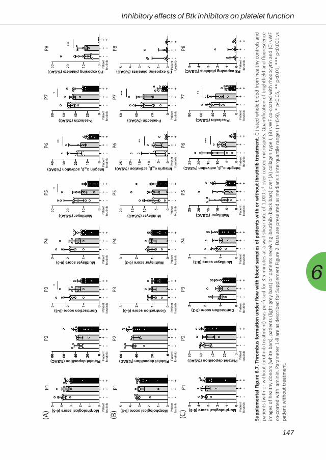
147
Inhibitory effects of Btk inhibitors on platelet function
6
012345 Morphological score (0-5)
Patie
ntIb
rutin
ib- -
-++ +
020406080 Platelet deposition (%SAC) Patie
ntIb
rutin
ib- -
-++ +
0123 Contraction score (0-3)
*
Patie
ntIb
rutin
ib- -
-++ +
0123 Multilayer score (0-3)
Patie
ntIb
rutin
ib- -
-++ +
0102030 Multilayer (%SAC)
*
Patie
ntIb
rutin
ib- -
-++ +
010203040
**
Patie
ntIb
rutin
ib- -
-++ +
Integrin αIIbβ3 activation (%SAC)
020406080 P-selectin (%SAC)
*
Patie
ntIb
rutin
ib- -
-++ +
0102030 PS exposing platelets (%SAC)
***
Patie
ntIb
rutin
ib- -
-++ +
012345 Morphological score (0-5)
Patie
ntIb
rutin
ib- -
-++ +
0204060 Platelet deposition (%SAC) Patie
ntIb
rutin
ib- -
-++ +
0123 Contraction score (0-3)
Patie
ntIb
rutin
ib- -
-++ +
0123 Multilayer score (0-3)
Patie
ntIb
rutin
ib- -
-++ +
05101520 Multilayer (%SAC) Patie
ntIb
rutin
ib- -
-++ +
0510152025
**
Patie
ntIb
rutin
ib- -
-++ +
Integrin αIIbβ3 activation (%SAC)
0204060 P-selectin (%SAC)
***
Patie
ntIb
rutin
ib- -
-++ +
012345 PS exposing platelets (%SAC) Patie
ntIb
rutin
ib- -
-++ +
012345 Morphological score (0-5)
Patie
ntIb
rutin
ib- -
-++ +
020406080 Platelet deposition (%SAC) Patie
ntIb
rutin
ib- -
-++ +
0123 Contraction score (0-3)
Patie
ntIb
rutin
ib- -
-++ +
0123 Multilayer score (0-3)
Patie
ntIb
rutin
ib- -
-++ +
0510152025 Multilayer (%SAC) Patie
ntIb
rutin
ib- -
-++ +
0510152025
***
Patie
ntIb
rutin
ib- -
-++ +
Integrin αIIbβ3 activation (%SAC)
020406080 P-selectin (%SAC)
**
Patie
ntIb
rutin
ib- -
-++ +
012345 PS exposing platelets (%SAC) Patie
ntIb
rutin
ib- -
-++ +
(A)
(B)
(C)
P1P2
P3P4
P5P6
P7P8
P1P2
P3P4
P5P6
P7P8
P1P2
P3P4
P5P6
P7P8
Supp
lem
enta
l Fig
ure
6.7.
Thr
ombu
s fo
rmati
on u
nder
flow
with
blo
od s
ampl
es o
f pati
ents
with
or w
ithou
t ibr
utini
b tr
eatm
ent.
Citr
ated
who
le b
lood
from
hea
lthy
cont
rols
and
patie
nts
(with
or w
ithou
t ibr
utini
b tr
eatm
ent)
was
per
fuse
d fo
r 3.5
min
utes
at a
wal
l she
ar ra
te o
f 1,0
00 s
-1 o
ver c
oate
d m
icro
spot
s. Q
uanti
ficati
on o
f brig
htfie
ld a
nd fl
uore
scen
ce
imag
es o
f hea
lthy
dono
rs (w
hite
bar
s), p
atien
ts (l
ight
gre
y ba
rs) o
r pati
ents
rece
ivin
g ib
rutin
ib (b
lack
bar
s) o
ver (
A) c
olla
gen
type
I, (B
) vW
F co
-coa
ted
with
rhod
ocyti
n an
d (C
) vW
F co
-coa
ted
with
lam
inin
. Par
amet
er 1
-8 a
re a
s de
scrib
ed fo
r Sup
plem
ent F
igur
e 2.
Dat
a ar
e pr
esen
ted
as m
edia
ns ±
inte
rqua
rtile
rang
es (n
=6-9
), *
p<0.
05, *
* p<
0.01
, ***
p<0
.001
vs
patie
nt w
ithou
t tre
atm
ent.
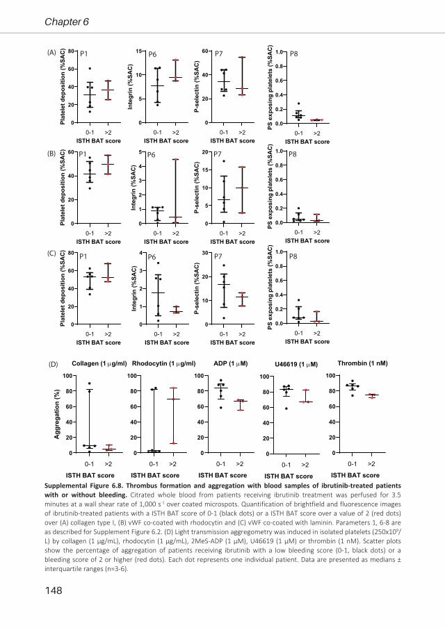
148
Chapter 6
0
20
40
60
80
100
Collagen (1 µg/ml)
Agg
rega
tion
(%)
0-1 >2
0
20
40
60
80
100
Rhodocytin (1 µg/ml)
0-1 >2
0
20
40
60
80
100
ADP (1 µM)
0-1 >2
0
20
40
60
80
100
Thrombin (1 nM)
0-1 >20
20
40
60
80
100
U46619 (1 µM)
0-1 >2
0
20
40
60
80
Plat
elet
dep
ositi
on (%
SAC)
0-1 >2ISTH BAT score
0
5
10
15
Inte
grin
(%SA
C)
0-1 >2ISTH BAT score
0
20
40
60
P-se
lect
in (%
SAC
)
0-1 >2ISTH BAT score
0.0
0.2
0.4
0.6
0.8
1.0
PS e
xpos
ing
plat
elet
s (%
SAC
)
0-1 >2ISTH BAT score
0
20
40
60
Plat
elet
dep
ositi
on (%
SAC)
0-1 >2ISTH BAT score
0
1
2
3
4
5
Inte
grin
(%SA
C)
0-1 >2ISTH BAT score
0
5
10
15
20
P-se
lect
in (%
SAC
)
0-1 >2ISTH BAT score
0.0
0.2
0.4
0.6
0.8
1.0
PS e
xpos
ing
plat
elet
s (%
SAC
)
0-1 >2ISTH BAT score
0
20
40
60
80
Plat
elet
dep
ositi
on (%
SAC)
0-1 >2ISTH BAT score
0
1
2
3
4
Inte
grin
(%SA
C)
0-1 >2ISTH BAT score
0
10
20
30
P-se
lect
in (%
SAC
)
0-1 >2ISTH BAT score
0.0
0.2
0.4
0.6
0.8
1.0
PS e
xpos
ing
plat
elet
s (%
SAC
)
0-1 >2ISTH BAT score
(A)
(B)
(C)
(D)
P1 P6 P7 P8
P1 P6 P7 P8
P1 P6 P7 P8
ISTH BAT score ISTH BAT score ISTH BAT score ISTH BAT score ISTH BAT scoreSupplemental Figure 6.8. Thrombus formation and aggregation with blood samples of ibrutinib-treated patients with or without bleeding. Citrated whole blood from patients receiving ibrutinib treatment was perfused for 3.5 minutes at a wall shear rate of 1,000 s-1 over coated microspots. Quantification of brightfield and fluorescence images of ibrutinib-treated patients with a ISTH BAT score of 0-1 (black dots) or a ISTH BAT score over a value of 2 (red dots) over (A) collagen type I, (B) vWF co-coated with rhodocytin and (C) vWF co-coated with laminin. Parameters 1, 6-8 are as described for Supplement Figure 6.2. (D) Light transmission aggregometry was induced in isolated platelets (250x109/L) by collagen (1 µg/mL), rhodocytin (1 µg/mL), 2MeS-ADP (1 µM), U46619 (1 µM) or thrombin (1 nM). Scatter plots show the percentage of aggregation of patients receiving ibrutinib with a low bleeding score (0-1, black dots) or a bleeding score of 2 or higher (red dots). Each dot represents one individual patient. Data are presented as medians ± interquartile ranges (n=3-6).

149
6

Chapter 7
Multiparameter screening for platelet-
inhibitory effects of tyrosine kinase inhibitors
used for cancer treatment
Tullemans BME, Veninga A, Fernandez DI, Aarts MJB, Eble JA, van der Meijden PEJ, Heemskerk JWM and Kuijpers MJE
In revision

152
Chapter 7
AbstractBackground: Currently antiplatelet drugs, used for treatment of arterial thrombosis, often
coincide with an increased risk of bleeding. Hence, platelet inhibitors that preserve haemostasis
are warranted. Multiple tyrosine kinase inhibitors (TKIs), used for cancer treatment, have been
reported to inhibit platelet function, with minor bleeding symptoms. In this study, we investigated
the antiplatelet properties of several clinically used TKIs as exploration of possible repurposing
these compounds as antiplatelet drugs.
Methods: Eight TKIs were selected based on the affinity for tyrosine kinases expressed
in platelets and limited bleeding symptoms. Whole blood samples, platelet-rich plasma (PRP)
or isolated platelets from healthy donors were pre-incubated with the TKI or vehicle. Platelet
aggregation, activation, intracellular calcium mobilisation and whole blood thrombus formation
under flow were determined.
Results: Overall, the TKIs with the highest affinities for platelet-expressed targets strongly
inhibited platelet functions. Dasatinib and sunitinib dose-dependently reduced collagen-induced
aggregation in PRP and washed platelets, while pazopanib, cabozantinib and vatalanib inhibited
this response in washed platelets only, and fostamatinib, axitinib and lapatinib showed no or
limited effects. Fostamatinib effectively reduced whole blood thrombus formation under flow on
collagen, as well as other substrates. Pazopanib, sunitinib, dasatinib, axitinib and vatalanib mildly
reduced thrombus formation on collagen. Intracellular calcium measurements and activation
responses in isolated platelets were inhibited by dasatinib, fostamatinib, sunitinib and pazopanib.
After glycoprotein-VI receptor-stimulation fostamatinib, cabozantinib and vatalanib decreased
the highly activated platelet population, whereas negative, resting populations were increased.
Conclusions: Strong inhibitory effects of dasatinib, fostamatinib, sunitinib and pazopanib
pointed to interference in early collagen receptor-induced signalling events, when compared to
cabozantinib and vatalanib. Since Fostamatinib, sunitinib, pazopanib and vatalanib are reported to
cause no more than mild bleeding symptoms, these are promising candidates to explore further
as antiplatelet drugs.

153
Multiparameter screening for platelet-inhibitory effects
7
IntroductionIschaemic heart disease and stroke accounted >15 million deaths worldwide in 2019,
making arterial thrombosis a leading global cause of death1. The major underlying cause are
the thrombotic complications of atherosclerosis, either via plaque rupture or erosion2, 3. Current
treatment is mostly (dual) antiplatelet therapy, such as aspirin and clopidogrel that prevent
thromboxane A2 release and ADP receptor activation, respectively4. However, these inhibitors
mostly have an irreversible mode of action on platelet G-protein coupled receptor (GPCRs)
signalling pathways and coincide with an increased risk of bleeding5. Therefore, novel antiplatelet
drugs are being developed that inhibit thrombosis, but do not interfere in haemostasis5. In
this respect glycoprotein (GP)VI and C-type lectin-like receptor 2 (CLEC-2) receptors, as well as
downstream tyrosine kinases, have been postulated as interesting antithrombotic targets6.
GPVI is an immunoreceptor tyrosine-based activation motif (ITAM)-linked receptor,
activated by collagen and fibrin(ogen), resulting in initial platelet activation and subsequent
thrombus formation7-9. Deficiency of GPVI in mice suppressed arterial thrombosis without
substantial prolongation of bleeding time10, 11. Inhibition of GPVI has also been reported to protect
from thrombosis upon plaque rupture in mice12, 13. Furthermore, patients with defects in GPVI
have only a mild bleeding diathesis14 or remain asymptomatic15. Two GPVI inhibitors showing
promising antithrombotic effects, are currently being tested in phase II clinical trials: the Fab
fragment ACT017 and Revacept, a collagen binding dimeric GPVI-Fc fusion protein6. However, a
disadvantage of these treatments is the administration via intravenous injections.
CLEC-2 is a hemITAM-linked receptor, activated by the endogenous ligand podoplanin16.
CLEC-2 deficient mice are protected from arterial thrombus formation and displayed only a
minor effect on haemostasis17, 18. In contrast, deficiency of both GPVI and CLEC-2 resulted in lack
of cessation of tail bleeding, as well as profound reduction of arterial thrombosis19. In human
blood, a monoclonal antibody (mAb) to CLEC-2 has been shown to block CLEC-2 induced platelet
activation in vitro20. However, no clinical trials targeting CLEC-2 have started until now.
Both GPVI and CLEC-2 heavily depend on tyrosine kinase signalling including Syk, Btk
and Src family kinases (SFK)21. In addition, other receptor signalling pathways, likely to a lesser
extent, use include tyrosine kinases, such as those evoked by GPIb, integrin αIIbβ3, Axl and Tie,
which are receptors for vWF, fibrinogen, Gas6 and angiopoietin, respectively22, 23. With such a
wide mode of action on platelet activation, while leaving GPCR signalling mostly intact, tyrosine
kinase inhibitors (TKIs) may provide an alternative way to inhibit platelet activation6. During the
past decades, many orally available TKIs have been developed for cancer treatment24, 25, with
different inhibitory profiles against tyrosine kinases (Table 7.1). These compounds have been
shown to be well tolerated, and in general cause only a mild increase in risk of bleeding. The
Syk inhibitor fostamatinib, used for treatment of chronic immune thrombocytopenic purpura
(ITP), has been reported to reduce GPVI- and CLEC-2 stimulated platelet aggregation, with only
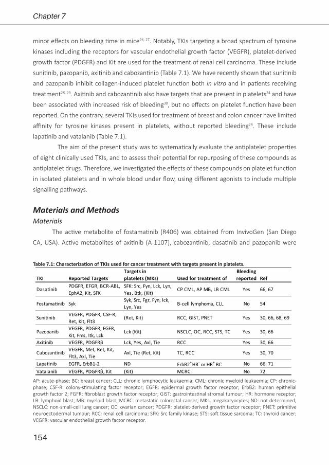
154
Chapter 7
minor effects on bleeding time in mice26, 27. Notably, TKIs targeting a broad spectrum of tyrosine
kinases including the receptors for vascular endothelial growth factor (VEGFR), platelet-derived
growth factor (PDGFR) and Kit are used for the treatment of renal cell carcinoma. These include
sunitinib, pazopanib, axitinib and cabozantinib (Table 7.1). We have recently shown that sunitinib
and pazopanib inhibit collagen-induced platelet function both in vitro and in patients receiving
treatment28, 29. Axitinib and cabozantinib also have targets that are present in platelets24 and have
been associated with increased risk of bleeding30, but no effects on platelet function have been
reported. On the contrary, several TKIs used for treatment of breast and colon cancer have limited
affinity for tyrosine kinases present in platelets, without reported bleeding24. These include
lapatinib and vatalanib (Table 7.1).
The aim of the present study was to systematically evaluate the antiplatelet properties
of eight clinically used TKIs, and to assess their potential for repurposing of these compounds as
antiplatelet drugs. Therefore, we investigated the effects of these compounds on platelet function
in isolated platelets and in whole blood under flow, using different agonists to include multiple
signalling pathways.
Materials and MethodsMaterials
The active metabolite of fostamatinib (R406) was obtained from InvivoGen (San Diego
CA, USA). Active metabolites of axitinib (A-1107), cabozantinib, dasatinib and pazopanib were
AP: acute-phase; BC: breast cancer; CLL: chronic lymphocytic leukaemia; CML: chronic myeloid leukaemia; CP: chronic-phase; CSF-R: colony-stimulating factor receptor; EGFR: epidermal growth factor receptor; ErbB2: human epithelial growth factor 2; FGFR: fibroblast growth factor receptor; GIST: gastrointestinal stromal tumour; HR: hormone receptor; LB: lymphoid blast; MB: myeloid blast; MCRC: metastatic colorectal cancer; MKs, megakaryocytes; ND: not determined; NSCLC: non-small-cell lung cancer; OC: ovarian cancer; PDGFR: platelet-derived growth factor receptor; PNET: primitive neuroectodermal tumour; RCC: renal cell carcinoma; SFK: Src family kinase; STS: soft tissue sarcoma; TC: thyroid cancer; VEGFR: vascular endothelial growth factor receptor.
Table 7.1: Characterization of TKIs used for cancer treatment with targets present in platelets.
TKI Reported TargetsTargets in platelets (MKs) Used for treatment of
Bleeding reported Ref
Fostama�nib SykSyk, Src, Fgr, Fyn, lck, Lyn, Yes B-cell lymphoma, CLL No 54
Suni�nib VEGFR, PDGFR, CSF-R, Ret, Kit, Flt3
(Ret, Kit) RCC, GIST, PNET Yes 30, 66, 68, 69
Pazopanib VEGFR, PDGFR, FGFR, Kit, Fms, Itk, Lck
Lck (Kit) NSCLC, OC, RCC, STS, TC Yes 30, 66
Axi�nib VEGFR, PDGFRβ Lck, Yes, Axl, Tie RCC Yes 30, 66
Lapa�nib EGFR, ErbB1-2 ND ErbB2+ HR- or HR+ BC No 66, 71Vatalanib VEGFR, PDGFRβ, Kit (Kit) MCRC No 72
Dasa�nib PDGFR, EFGR, BCR-ABL, EphA2, Kit, SFK
SFK: Src, Fyn, Lck, Lyn, Yes, Btk, (Kit)
Yes 66, 67CP CML, AP MB, LB CML
Cabozan�nib VEGFR, Met, Ret, Kit,Flt3, Axl, Tie
Axl, Tie (Ret, Kit) Yes 30, 70TC, RCC

155
Multiparameter screening for platelet-inhibitory effects
7
purchased from LC Laboratories (Woburn MA, USA). The active metabolite of lapatinib (GW-
572016) was purchased from Selleckchem (Houston TX, USA), whereas the active metabolite
of sunitinib came from Pfizer (New York NY, USA). The active metabolite of vatalanib (PTK787)
was obtained from Adooq Bioscience (Irvine CA, USA). Bovine serum albumin (BSA), D(+)-
glucose, unfractionated heparin and apyrase were purchased from Sigma-Aldrich (Saint Louis
MO, USA). Horm collagen type I came from Takeda (Hoofddorp, the Netherlands), whereas
the agonists collagen-related peptide crosslinked (CRP-XL) and von Willebrand factor III (vWF-
III) were obtained from CambCol Laboratories (Cambridge, UK). Rhodocytin was purified from
Calloselasma rhodostoma venom as described previously31. Thrombin was from Enzyme Research
Laboratories Inc (South Bend, IN, USA). U46619 (thromboxane A2 receptor agonist) came from
Cayman Chemicals (Ann Arbor MI, USA). 2-Methylthio-adenosine-diphosphate (2MeS-ADP)
and D-phenylalanyl-prolyl-arginyl chloromethyl ketone (PPACK) were obtained from Santa Cruz
Biotechnology (Dallas TX, USA). Laminin was purchased from Octapharma (Lachen, Switzerland),
whereas Fura-2-AM and allophycocyanin (APC)-labelled mouse anti-human CD63 monoclonal
antibody (mAb, clone MEM-259) were obtained from Invitrogen, Fisher Scientific (Carlsbad CA,
USA). Fluorescein isothiocyanate (FITC)-labelled PAC1 mAb against activated human integrin αIIbβ3
(340507), Phycoerythrin (PE)-labelled annexin A5, Brilliant Violet (BV)510-conjugated mouse anti-
human CD42b mAb (clone HIP1) and BV421-labelled rat anti-human TLT-1 mAb (clone 268420),
were purchased from BD Bioscience (Franklin Lakes NJ, USA). FITC-conjugated α-fibrinogen mAb
was purchased from DAKO (F0111; Santa Clara CA, USA). Alexa Fluor (AF)568-conjugated annexin
A5 was purchased from Molecular Probes, Life Technologies (New York NY, USA). Peridinin-
chlorophyll-protein cyanine5.5 (PerCP Cy5.5)-conjugated mouse anti-human CD62-P mAb (clone
AK4) and AF647-labelled mouse anti-human CD62-P mAb (clone AK4) were obtained from
Biolegend (London, UK). FITC-conjugated annexin A5 was from Pharmatarget (Maastricht, the
Netherlands).
Blood collection and platelet isolationIn accordance with the declaration of Helsinki and approval of the local medical ethical
committee (Maastricht University Medical Centre+, MUMC+), we obtained full informed consent
from all participants. Blood from healthy volunteers was collected in 3.2% trisodium citrate tubes
by venipuncture, after discarding the first 3 mL of blood. Blood cell counts and haematological
parameters were assessed, using a Sysmex XP300 (Kobe, Japan).
Platelet-rich plasma (PRP) or platelets were isolated from whole blood as described
previously32, 33. Washed platelets were resuspended in Hepes buffer, pH 7.45 (10 mM Hepes, 136
mM NaCl, 2.7 mM KCl, 2 mM MgCl2, 1 mg/mL glucose, and 1 mg/mL bovine serum albumin).
Platelet count was adjusted as stated per assay.

156
Chapter 7
Thrombus formationThrombus formation under flow was determined as described34. Briefly, glass coverslips
were coated with 3 microspots of collagen type I (100 µg/mL), collagen type III (100 µg/mL), vWF
(50 µg/mL) co-coated with laminin (100 µg/mL) or vWF co-coated with rhodocytin (250 µg/mL),
vWF co-coated with ristocetin (250 µg/mL), or vWF co-coated with fibrinogen (250 µg/mL) and
mounted in a parallel plate flow chamber. Citrated blood samples were incubated with the TKIs
axitinib, cabozantinib, dasatinib, fostamatinib, lapatinib, pazopanib, sunitinib, vatalanib or vehicle
for 10 minutes at room temperature. Pre-incubated whole blood samples were recalcified in
presence of PPACK (40 µM) and perfused at a wall shear rate of 1000 s-1 for 3.5 minutes. Platelet
activation properties were determined by post-perfusion with Hepes buffer (supplemented with
2 mM CaCl2 and 1 U/mL heparin) containing FITC-conjugated α-fibrinogen mAb (1:80), AF647-
conjugated αCD62P mAb (1:100), and AF568-conjugated annexin A5 (1:200). Brightfield and
fluorescence images were captured using an EVOS microscope (Bothel WA, USA).
Analysis of microscopic dataMicroscopic images were analysed using specific scripts in the open-access Fiji software
(Laboratory for Optical and Computational Instrumentation, University of Wisconsin-Madison, WI,
USA) as described34, 35. Multiple thrombus formation parameters were obtained from brightfield
images including P1: morphological score of platelet adhesion and thrombus formation (scale
0-5), P2: surface area coverage of adhered platelet (% SAC), P3: platelet aggregate contraction
score (scale 0-3), P4: platelet aggregate multilayer score (scale 0-3) and P5: coverage of multi-
layered platelet aggregation (% SAC). From fluorescence images, platelet activation parameters
were obtained by determining P6: integrin αIIbβ3 activation (% SAC), P7: P-selectin expression
(% SAC) and P8: phosphatidylserine (PS) exposure (% SAC). Heatmaps were generated from the
mean values per parameter for each surface for comparative data analysis. Values were scaled
from 0-10 based on the highest value of each parameter for each surface. For visualization of the
effects, subtraction heatmaps were generated by subtraction of scaled control data from each TKI
condition and heatmaps were filtered based on significant differences (p<0.05). Heatmaps were
generated using R Core Team (i386 3.2.5; Vienna, Austria).
Platelet aggregation by light transmission aggregometryIsolated platelets or PRP (250x109 platelets/L) were incubated with TKIs or vehicle for
10 minutes at 37°C. Maximal aggregation was induced by collagen type I (1 µg/mL), 2MeS-ADP
(1 µM), U46619 (1 µM), rhodocytin (1 µg/mL) or thrombin (1 nM). Platelet aggregation was
recorded using a Chronolog optical aggregometer (Havertown PA, USA). The maximum amplitude
was quantified at 8 minutes after agonist addition.

157
Multiparameter screening for platelet-inhibitory effects
7
Cytosolic Ca2+ measurementsWashed platelets (200x109 platelets/L) were loaded with Fura-2-AM and changes in
cytosolic Ca2+ ([Ca2+]i) were measured in 96-well plates using a FlexStation 3 (Molecular Devices,
San Jose, CA, USA), as described36, 37. Briefly, platelets in suspension were preincubated with
TKIs or vehicle for 10 minutes at room temperature. Pre-loaded and -incubated samples were
stimulated by CRP-XL (10 µg/mL). Changes in Fura-2 fluorescence were measured in duplicate,
and ratio values were calculated. Data are presented as [Ca2+]i in nM or area under the curve
(AUC) in nMs.
Platelet activation by flow cytometryWashed platelets (25x109 platelets/L) were incubated with TKIs or vehicle for 10 minutes
at room temperature. Platelets were stimulated, in the presence of 2 mM CaCl2, by CRP-XL (5
μg/mL), 2MeS-ADP (2 μM), or thrombin (4 nM) for 15 minutes, and simultaneously stained for
multiple activation markers using an antibody mix consisting of FITC-conjugated PAC1 (1:20),
BV510-conjugated αCD42b (1:50), BV421-conjugated αTLT1 (1:50), APC-conjugated αCD63
(1:20), PerCP-Cy5.5-conjugated αCD62P (1:500) mAbs. For measuring phosphatidyl-serine (PS)
exposure, platelets were activated with a combination of CRP-XL (5 μg/mL) and thrombin (4
nM) for 1 hour at 37°C, and post-labelled with PE-conjugated annexin A5. Samples for activation
markers as well as PS exposure were fixed after stimulation for at least 30 minutes at 4°C in the
dark using a 0.3% fixation buffer, before measuring 10,000 events per sample on a BD FACSCanto
II (BD bioscience, Franklin Lakes, NJ, USA).
Analysis of flow cytometric dataSeparate FCS files were opened in FlowJo V10 software (Treestar, Ashland OR, USA) and
checked for data anomalies by FlowAI38. Before concatenation, event count per sample was set at
5,000 events by DownSample for equal weighing of every sample. Resulting file was subjected to
t-distributed stochastic neighbour embedding (tSNE) analysis to reduce the high dimensionality
to a 2D plot. FlowSOM analysis was used to identify five clusters39. Fractions of platelets present
in the resulting FlowSOM populations were determined per original sample. Each platelet
population was characterised by the expression level of the different markers.
Statistical analysisData are shown as mean ± standard error of mean (SEM). GraphPad Prism 8.3.0 software
(La Jolla, CA, USA) was used for statistical analysis with an unpaired non-parametric t-test (Mann-
Whitney) or one-way ANOVA. A p-value less than 0.05 was considered to be statistically significant,
in which * is p<0.05, ** is p<0.01 and *** is p<0.001.

158
Chapter 7
ResultsCharacteristics of selected TKIs
For this study, eight reversible multi-target TKIs were selected based on published affinities
for tyrosine kinases (TKs) expressed in platelets, and reported minor bleeding risk (Table 7.1,
Figure 7.1)24. Dasatinib and fostamatinib display high affinity for platelet-expressed Syk and Src-
family kinases (SFKs), while sunitinib, pazopanib, axitinib and cabozantinib show moderate to mild
affinity for only some of these platelet-expressed targets (Figure 7.1). Dasatinib and fostamatinib
are known as inhibitors of the GPVI, CLEC-2 and αIIbβ3 activation pathways in platelets26, 40, but
have not been systematically evaluated. Lapatinib and vatalanib have unknown affinities for
platelet-expressed kinases (Figure 7.1, Table 7.1).
Distinct effects of selected TKIs on collagen-induced platelet aggregation in the presence or absence of plasma
Impaired platelet function by TKIs is described to be due to inhibition of in particular the
ITAM-linked receptor GPVI24, 25. Therefore, we first investigated the effects of the active metabolites
of different TKIs on collagen-induced aggregation responses. Dose-response curves with isolated
platelets showed distinct effects of the different TKIs (Figure 7.2, dotted lines). The most potent
aggregation inhibition was observed with dasatinib (>90% inhibition with 1 µM, Figure 7.2A).
Surprisingly, the combined SFK and Syk-inhibitor fostamatinib, with several targets in platelets,
did not show a relevant inhibitory effect (Figure 7.2B). For the weaker SFK inhibitors sunitinib,
pazopanib, and cabozantinib, aggregation in washed platelets became nearly abolished with 33
µM (Figure 7.2C-D, F) Unexpectedly, vatalanib without assumed targets in platelets also inhibited
this response at 33 µM (Figure 7.2H). IC50 values of these four TKIs were between 6-13 µM and a
Kd (nM) IC50 (nM)+++ Very strong <0.01 <0.1++ Strong <0.1 <1+ Moderate <1 <10± Mild <10 <100- No / ND >10 ND
Interac�on strengthSRC
Fgr
Fyn
Lck
Lyn
Yes
Btk
Tec
Axl
Tie1
MEK
1
MEK
2
Dasa�nib +++ +++ ± +++ +++ +++ ± +++ ± - - ± ±
Fostama�nib +++ ++ +++ ++ ++ ++ +++ + ± + + + +
Suni�nib ± ± ± + + + - ± - +++ ± + +
Pazopanib ± ± ± ± ± ± ± - - - ± - -
Axi�nib - ± - ± - ± - - - + + - -
Cabozan�nib - - - - - - - - - + ± - -
Lapa�nib - - - - - - - - - - - - -
Vatalanib - - - - - - - - - - - - -
Others
TKI
SFK
Syk
TFK
Figure 7.1. Affinity profile of common tyrosine kinase inhibitors (TKIs) for relevant tyrosine kinases implicated in platelet activation. Shown is a heatmap of the affinity-based dissociation constants (Kd)62-64 or IC50 values65 of the indicated TKIs. Values are from binding experiments with indicated purified kinases, including Src family kinases (SFK), Syk, Tec family kinases (TFK), focal adhesion kinases (FAK) and MEK. Colour coding with highest affinity (lowest Kd or IC50) shown in deep red and lowest affinity (highest Kd or IC50) shown in white. Data are based on ref 24.
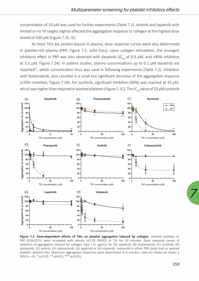
159
Multiparameter screening for platelet-inhibitory effects
7
concentration of 10 µM was used for further experiments (Table 7.2). Axitinib and lapatinib with
limited or no TK targets slightly affected the aggregation response to collagen at the highest dose
tested of 100 µM (Figure 7.2E, G).
As most TKIs are protein-bound in plasma, dose response curves were also determined
in platelet-rich plasma (PRP; Figure 7.2, solid lines). Upon collagen stimulation, the strongest
inhibitory effect in PRP was also observed with dasatinib (IC50 of 0.4 µM, and >90% inhibition
at 3.3 µM, Figure 7.2A). In patient studies, plasma concentrations up to 0.1 µM dasatinib are
reported41, which concentration thus was used in following experiments (Table 7.2). Inhibition
with fostamatinib, also resulted in a small but significant decrease of the aggregation response
(±20% inhibition, Figure 7.2B). For sunitinib, significant inhibition (60%) was reached at 33 µM,
which was higher than required in washed platelets (Figure 7.2C). The IC50 value of 33 µM sunitinib
PRPWP
(A) (B)
(D)
(C)
(E) (F)
(G) (H)
0 5 100
20
40
60
80
100
120
Dasatinib
TKI concentration (µM)
Agg
rega
tion
(%of
cont
rol)
** ***
*
* **
0 50 1000
20
40
60
80
100
120
Fostamatinib
TKI concentration (µM)
* ***
**
0 50 1000
20
40
60
80
100
120
Sunitinib
TKI concentration (µM)
*
*****
0 50 1000
20
40
60
80
100
120
Pazopanib
TKI concentration (µM)
Agg
rega
tion
(%of
cont
rol)
***
***
0 50 1000
20
40
60
80
100
120
Axitinib
TKI concentration (µM)0 50 100
0
20
40
60
80
100
120
Cabozantinib
TKI concentration (µM)
* **
0 50 1000
20
40
60
80
100
120
Lapatinib
TKI concentration (µM)
Agg
rega
tion
(%of
cont
rol)
*
0 50 1000
20
40
60
80
100
120
Vatalanib
TKI concentration (µM)
*
*
****
Figure 7.2. Dose-dependent effects of TKIs on platelet aggregation induced by collagen. Washed platelets or PRP (250x109/L) were incubated with vehicle (<0.1% DMSO) or TKI for 10 minutes. Dose response curves of inhibition of aggregation induced by collagen type I (1 µg/mL) for (A) dasatinib, (B) fostamatinib, (C) sunitinib, (D) pazopanib, (E) axitinib, (F) cabozantinib, (G) lapatinib or (H) vatalanib, measured in either PRP (solid line) or washed platelets (dotted line). Maximum aggregation responses were determined at 8 minutes; data are shown as means ± SEM (n = 6). * p<0.05. ** p<0.01, *** p<0.001.
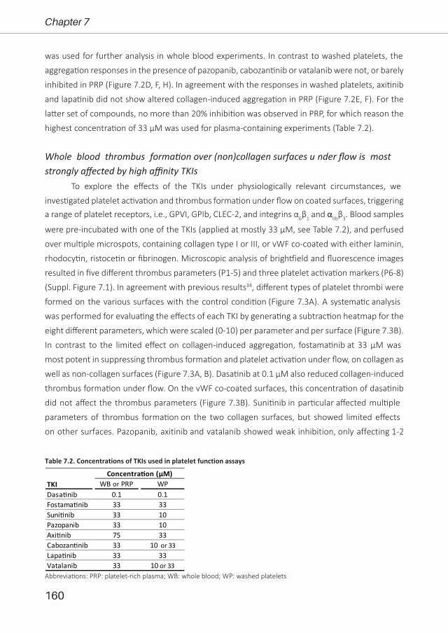
160
Chapter 7
WB or PRP WPDasa�nib 0.1 0.1Fostama�nib 33 33Suni�nib 33 10Pazopanib 33 10Axi�nib 75 33Cabozan�nib 33 10 or 33Lapa�nib 33 33Vatalanib 33 10 or 33
TKIConcentra�on (µM)
Abbreviations: PRP: platelet-rich plasma; WB: whole blood; WP: washed platelets
was used for further analysis in whole blood experiments. In contrast to washed platelets, the
aggregation responses in the presence of pazopanib, cabozantinib or vatalanib were not, or barely
inhibited in PRP (Figure 7.2D, F, H). In agreement with the responses in washed platelets, axitinib
and lapatinib did not show altered collagen-induced aggregation in PRP (Figure 7.2E, F). For the
latter set of compounds, no more than 20% inhibition was observed in PRP, for which reason the
highest concentration of 33 µM was used for plasma-containing experiments (Table 7.2).
Whole blood thrombus formation over (non)collagen surfaces u nder flow is most strongly affected by high affinity TKIs
To explore the effects of the TKIs under physiologically relevant circumstances, we
investigated platelet activation and thrombus formation under flow on coated surfaces, triggering a range of platelet receptors, i.e., GPVI, GPIb, CLEC-2, and integrins α6β1 and αIIbβ3. Blood samples
were pre-incubated with one of the TKIs (applied at mostly 33 µM, see Table 7.2), and perfused
over multiple microspots, containing collagen type I or III, or vWF co-coated with either laminin,
rhodocytin, ristocetin or fibrinogen. Microscopic analysis of brightfield and fluorescence images
resulted in five different thrombus parameters (P1-5) and three platelet activation markers (P6-8)
(Suppl. Figure 7.1). In agreement with previous results34, different types of platelet thrombi were
formed on the various surfaces with the control condition (Figure 7.3A). A systematic analysis
was performed for evaluating the effects of each TKI by generating a subtraction heatmap for the
eight different parameters, which were scaled (0-10) per parameter and per surface (Figure 7.3B).
In contrast to the limited effect on collagen-induced aggregation, fostamatinib at 33 µM was
most potent in suppressing thrombus formation and platelet activation under flow, on collagen as
well as non-collagen surfaces (Figure 7.3A, B). Dasatinib at 0.1 µM also reduced collagen-induced
thrombus formation under flow. On the vWF co-coated surfaces, this concentration of dasatinib
did not affect the thrombus parameters (Figure 7.3B). Sunitinib in particular affected multiple
parameters of thrombus formation on the two collagen surfaces, but showed limited effects
on other surfaces. Pazopanib, axitinib and vatalanib showed weak inhibition, only affecting 1-2
Table 7.2. Concentrations of TKIs used in platelet function assays
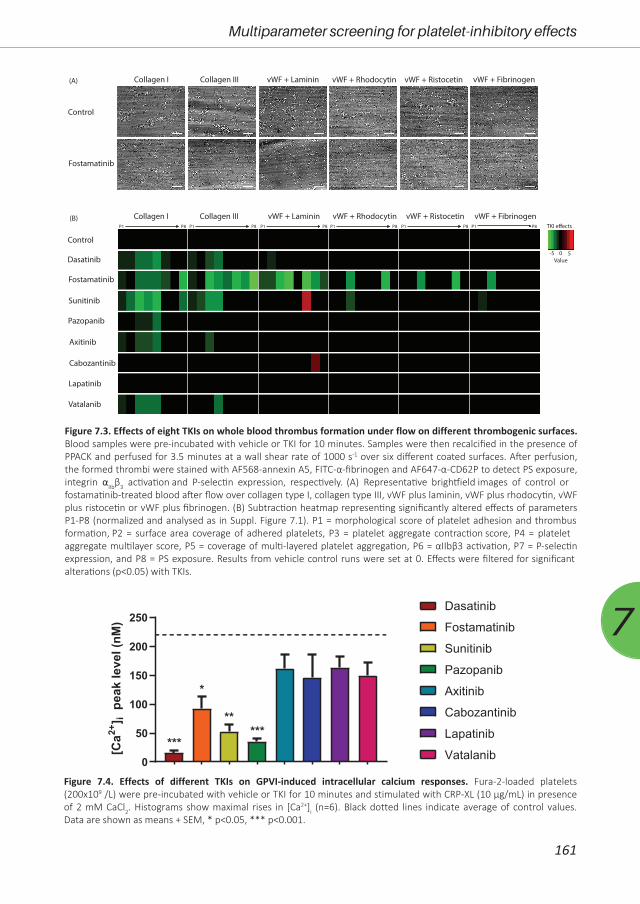
161
Multiparameter screening for platelet-inhibitory effects
7
0
50
100
150
200
250
[Ca2+
] ipe
akle
vel(
nM)
******
*
**
Dasatinib
Fostamatinib
Sunitinib
Pazopanib
Axitinib
Cabozantinib
Lapatinib
Vatalanib
Figure 7.4. Effects of different TKIs on GPVI-induced intracellular calcium responses. Fura-2-loaded platelets (200x109 /L) were pre-incubated with vehicle or TKI for 10 minutes and stimulated with CRP-XL (10 µg/mL) in presence of 2 mM CaCl2. Histograms show maximal rises in [Ca2+]i (n=6). Black dotted lines indicate average of control values. Data are shown as means + SEM, * p<0.05, *** p<0.001.
Control
Dasatinib
Lapatinib
Pazopanib
Sunitinib
Cabozantinib
Vatalanib
Axitinib
Fostamatinib
Collagen I Collagen III vWF + Laminin vWF + Rhodocytin vWF + Ristocetin vWF + FibrinogenP1 P8 P1 P8 P1 P8 P1 P8 P1 P8 P1 P8
Collagen I Collagen III vWF + Laminin vWF + Rhodocytin vWF + Ristocetin vWF + Fibrinogen
Control
Fostamatinib
-5 50
TKI e�ects
Value
(A)
(B)
Figure 7.3. Effects of eight TKIs on whole blood thrombus formation under flow on different thrombogenic surfaces. Blood samples were pre-incubated with vehicle or TKI for 10 minutes. Samples were then recalcified in the presence of PPACK and perfused for 3.5 minutes at a wall shear rate of 1000 s-1 over six different coated surfaces. After perfusion, the formed thrombi were stained with AF568-annexin A5, FITC-α-fibrinogen and AF647-α-CD62P to detect PS exposure, integrin αIIbβ3 activation and P-selectin expression, respectively. (A) Representative brightfield images of control or fostamatinib-treated blood after flow over collagen type I, collagen type III, vWF plus laminin, vWF plus rhodocytin, vWF plus ristocetin or vWF plus fibrinogen. (B) Subtraction heatmap representing significantly altered effects of parameters P1-P8 (normalized and analysed as in Suppl. Figure 7.1). P1 = morphological score of platelet adhesion and thrombus formation, P2 = surface area coverage of adhered platelets, P3 = platelet aggregate contraction score, P4 = platelet aggregate multilayer score, P5 = coverage of multi-layered platelet aggregation, P6 = αIIbβ3 activation, P7 = P-selectin expression, and P8 = PS exposure. Results from vehicle control runs were set at 0. Effects were filtered for significant alterations (p<0.05) with TKIs.
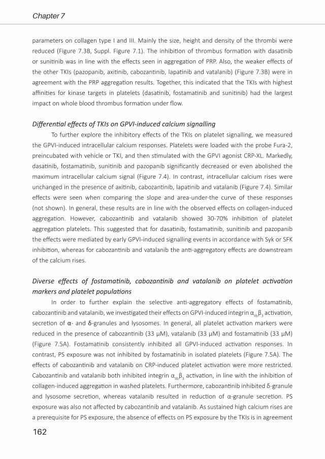
162
Chapter 7
parameters on collagen type I and III. Mainly the size, height and density of the thrombi were
reduced (Figure 7.3B, Suppl. Figure 7.1). The inhibition of thrombus formation with dasatinib
or sunitinib was in line with the effects seen in aggregation of PRP. Also, the weaker effects of
the other TKIs (pazopanib, axitinib, cabozantinib, lapatinib and vatalanib) (Figure 7.3B) were in
agreement with the PRP aggregation results. Together, this indicated that the TKIs with highest
affinities for kinase targets in platelets (dasatinib, fostamatinib and sunitinib) had the largest
impact on whole blood thrombus formation under flow.
Differential effects of TKIs on GPVI-induced calcium signallingTo further explore the inhibitory effects of the TKIs on platelet signalling, we measured
the GPVI-induced intracellular calcium responses. Platelets were loaded with the probe Fura-2,
preincubated with vehicle or TKI, and then stimulated with the GPVI agonist CRP-XL. Markedly,
dasatinib, fostamatinib, sunitinib and pazopanib significantly decreased or even abolished the
maximum intracellular calcium signal (Figure 7.4). In contrast, intracellular calcium rises were
unchanged in the presence of axitinib, cabozantinib, lapatinib and vatalanib (Figure 7.4). Similar
effects were seen when comparing the slope and area-under-the curve of these responses
(not shown). In general, these results are in line with the observed effects on collagen-induced
aggregation. However, cabozantinib and vatalanib showed 30-70% inhibition of platelet
aggregation platelets. This suggested that for dasatinib, fostamatinib, sunitinib and pazopanib
the effects were mediated by early GPVI-induced signalling events in accordance with Syk or SFK
inhibition, whereas for cabozantinib and vatalanib the anti-aggregatory effects are downstream
of the calcium rises.
Diverse effects of fostamatinib, cabozantinib and vatalanib on platelet activation markers and platelet populations
In order to further explain the selective anti-aggregatory effects of fostamatinib,
cabozantinib and vatalanib, we investigated their effects on GPVI-induced integrin αIIbβ3 activation,
secretion of α- and δ-granules and lysosomes. In general, all platelet activation markers were
reduced in the presence of cabozantinib (33 µM), vatalanib (33 µM) and fostamatinib (33 µM)
(Figure 7.5A). Fostamatinib consistently inhibited all GPVI-induced activation responses. In
contrast, PS exposure was not inhibited by fostamatinib in isolated platelets (Figure 7.5A). The
effects of cabozantinib and vatalanib on CRP-induced platelet activation were more restricted.
Cabozantinib and vatalanib both inhibited integrin αIIbβ3 activation, in line with the inhibition of
collagen-induced aggregation in washed platelets. Furthermore, cabozantinib inhibited δ-granule
and lysosome secretion, whereas vatalanib resulted in reduction of α-granule secretion. PS
exposure was also not affected by cabozantinib and vatalanib. As sustained high calcium rises are
a prerequisite for PS exposure, the absence of effects on PS exposure by the TKIs is in agreement
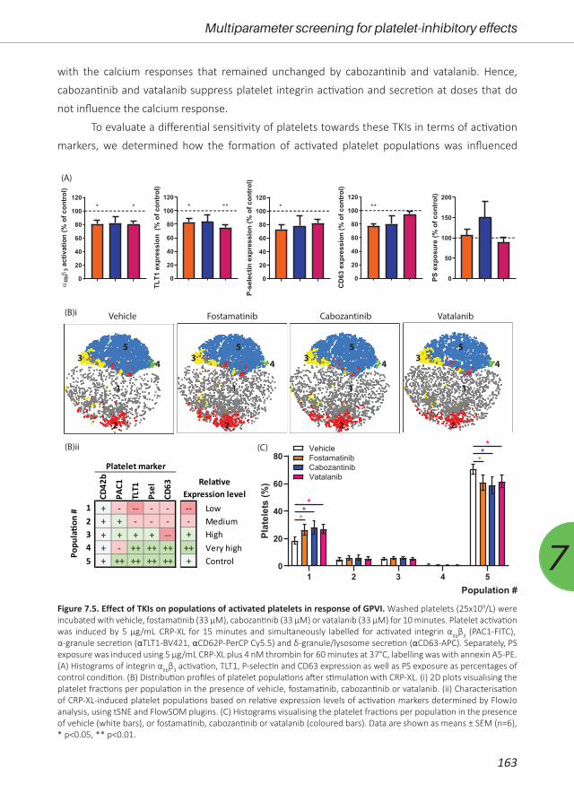
163
Multiparameter screening for platelet-inhibitory effects
7
with the calcium responses that remained unchanged by cabozantinib and vatalanib. Hence,
cabozantinib and vatalanib suppress platelet integrin activation and secretion at doses that do
not influence the calcium response.
To evaluate a differential sensitivity of platelets towards these TKIs in terms of activation
markers, we determined how the formation of activated platelet populations was influenced
CD42
b
PAC1
TLT1
Psel
CD63
1 + - -- - -2 + + - - -3 + + + + --4 + - ++ ++ ++5 + ++ ++ ++ ++
Platelet marker
Popu
la�o
n #
(A)
0
20
40
60
80
100
120
IIb3
activ
atio
n(%
ofco
ntro
l)
* *
0
20
40
60
80
100
120
TLT1
expr
essi
on(%
ofco
ntro
l)
* **
0
20
40
60
80
100
120
P-se
lect
inex
pres
sion
(%of
cont
rol)
*
0
20
40
60
80
100
120
CD
63ex
pres
sion
(%of
cont
rol)
**
0
50
100
150
200
PSex
posu
re(%
ofco
ntro
l)
Vehicle Fostamatinib Cabozantinib Vatalanib
-- Low- Medium+ High
++ Very high+ Control
Rela�ve Expression level
(B)ii
(B)i
1 2 3 4 50
20
40
60
80
Plat
elet
s(%
)
***
***
FostamatinibCabozantinibVatalanib
Vehicle
1 1 1 1
2 2 2 2
3 3 3 34 4 4 4
5 5 5 5
(C)
Population #
Figure 7.5. Effect of TKIs on populations of activated platelets in response of GPVI. Washed platelets (25x109/L) were incubated with vehicle, fostamatinib (33 µM), cabozantinib (33 µM) or vatalanib (33 µM) for 10 minutes. Platelet activation was induced by 5 µg/mL CRP-XL for 15 minutes and simultaneously labelled for activated integrin αIIbβ3 (PAC1-FITC), α-granule secretion (αTLT1-BV421, αCD62P-PerCP Cy5.5) and δ-granule/lysosome secretion (αCD63-APC). Separately, PS exposure was induced using 5 µg/mL CRP-XL plus 4 nM thrombin for 60 minutes at 37°C, labelling was with annexin A5-PE. (A) Histograms of integrin αIIbβ3 activation, TLT1, P-selectin and CD63 expression as well as PS exposure as percentages of control condition. (B) Distribution profiles of platelet populations after stimulation with CRP-XL. (i) 2D plots visualising the platelet fractions per population in the presence of vehicle, fostamatinib, cabozantinib or vatalanib. (ii) Characterisation of CRP-XL-induced platelet populations based on relative expression levels of activation markers determined by FlowJo analysis, using tSNE and FlowSOM plugins. (C) Histograms visualising the platelet fractions per population in the presence of vehicle (white bars), or fostamatinib, cabozantinib or vatalanib (coloured bars). Data are shown as means ± SEM (n=6), * p<0.05, ** p<0.01.
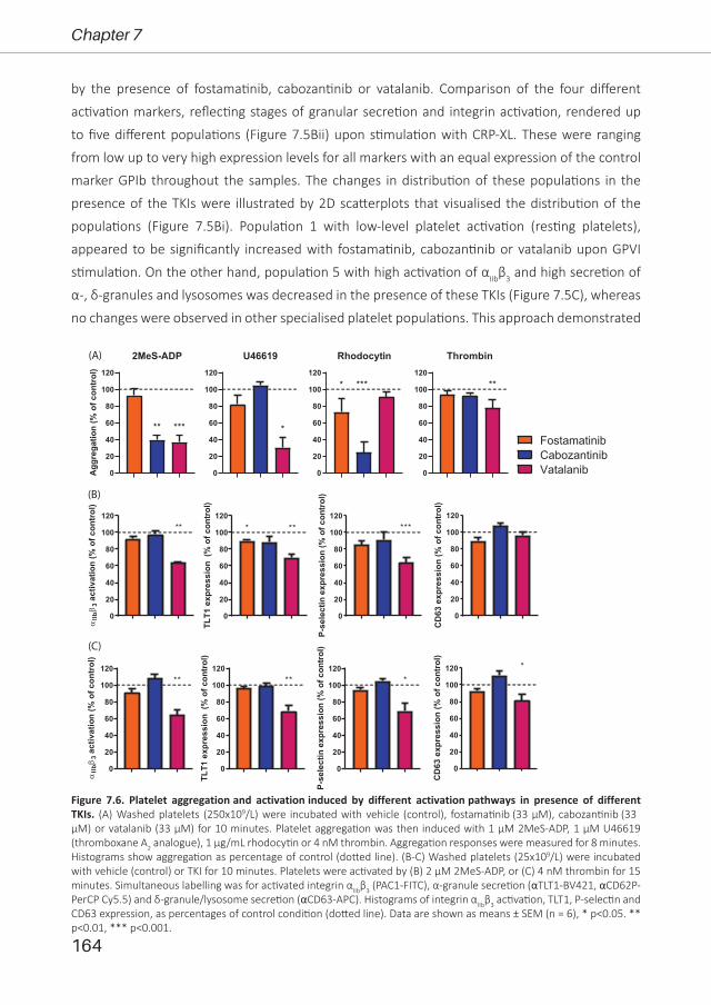
164
Chapter 7
by the presence of fostamatinib, cabozantinib or vatalanib. Comparison of the four different
activation markers, reflecting stages of granular secretion and integrin activation, rendered up
to five different populations (Figure 7.5Bii) upon stimulation with CRP-XL. These were ranging
from low up to very high expression levels for all markers with an equal expression of the control
marker GPIb throughout the samples. The changes in distribution of these populations in the
presence of the TKIs were illustrated by 2D scatterplots that visualised the distribution of the
populations (Figure 7.5Bi). Population 1 with low-level platelet activation (resting platelets),
appeared to be significantly increased with fostamatinib, cabozantinib or vatalanib upon GPVI
stimulation. On the other hand, population 5 with high activation of αIIbβ3 and high secretion of
α-, δ-granules and lysosomes was decreased in the presence of these TKIs (Figure 7.5C), whereas
no changes were observed in other specialised platelet populations. This approach demonstrated
0
20
40
60
80
100
120
2MeS-ADP
Agg
rega
tion
(%of
cont
rol)
** ***
0
20
40
60
80
100
120
U46619
*
0
20
40
60
80
100
120
Rhodocytin
***
0
20
40
60
80
100
120
Thrombin
***
0
20
40
60
80
100
120
IIb3
activ
atio
n(%
ofco
ntro
l)
**
0
20
40
60
80
100
120
TLT1
expr
essi
on(%
ofco
ntro
l)
* **
0
20
40
60
80
100
120
P-se
lect
inex
pres
sion
(%of
cont
rol)
***
0
20
40
60
80
100
120
CD
63ex
pres
sion
(%of
cont
rol)
(A)
(B)
(C)
0
20
40
60
80
100
120
IIb3
activ
atio
n(%
ofco
ntro
l)
**
0
20
40
60
80
100
120
TLT1
expr
essi
on(%
ofco
ntro
l)
**
0
20
40
60
80
100
120
P-se
lect
inex
pres
sion
(%of
cont
rol)
*
0
20
40
60
80
100
120
CD
63ex
pres
sion
(%of
cont
rol)
*
FostamatinibCabozantinibVatalanib
Figure 7.6. Platelet aggregation and activation induced by different activation pathways in presence of different TKIs. (A) Washed platelets (250x109/L) were incubated with vehicle (control), fostamatinib (33 µM), cabozantinib (33 µM) or vatalanib (33 µM) for 10 minutes. Platelet aggregation was then induced with 1 µM 2MeS-ADP, 1 µM U46619 (thromboxane A2 analogue), 1 µg/mL rhodocytin or 4 nM thrombin. Aggregation responses were measured for 8 minutes. Histograms show aggregation as percentage of control (dotted line). (B-C) Washed platelets (25x109/L) were incubated with vehicle (control) or TKI for 10 minutes. Platelets were activated by (B) 2 µM 2MeS-ADP, or (C) 4 nM thrombin for 15 minutes. Simultaneous labelling was for activated integrin αIIbβ3 (PAC1-FITC), α-granule secretion (αTLT1-BV421, αCD62P-PerCP Cy5.5) and δ-granule/lysosome secretion (αCD63-APC). Histograms of integrin αIIbβ3 activation, TLT1, P-selectin and CD63 expression, as percentages of control condition (dotted line). Data are shown as means ± SEM (n = 6), * p<0.05. ** p<0.01, *** p<0.001.
165
Multiparameter screening for platelet-inhibitory effects
7
the simultaneous inhibition of secretory and integrin-activating platelet responses by these TKIs.
In whole blood perfusion experiments, we observed inhibitory effects of fostamatinib
on thrombus formation on vWF co-coated surfaces. To determine effects of fostamatinib,
cabozantinib and vatalanib on platelet activation induced by other agonists, platelet aggregation
was investigated in response to ADP, the thromboxane A2 analogue U46619, the CLEC-2 agonist
rhodocytin or thrombin. Fostamatinib only reduced rhodocytin-induced aggregation (Figure
7.6A), in agreement with Syk signalling underneath CLEC-226. In contrast, cabozantinib significantly
inhibited the aggregation responses to ADP and rhodocytin, while vatalanib prominently reduced
the responses to ADP and U46619, and slightly to thrombin (Figure 7.6A). In line with these
aggregation results, vatalanib suppressed ADP- and thrombin-induced integrin αIIbβ3 activation,
TLT1 expression and P-selectin expression (Figure 7.6B, C). Upon thrombin stimulation also
δ-granule and lysosome secretion were partially inhibited (Figure 7.6C). In contrast to the observed
reduction in ADP-induced aggregation with cabozantinib, no inhibition of platelet activation was
observed in response to ADP (Figure 7.6B, C). Taken together, this suggested that the latter two
compounds inhibited signalling entities in pathways other than induced by GPVI.
DiscussionIn this work we explored the antiplatelet properties of the TKIs dasatinib, fostamatinib,
sunitinib, pazopanib, cabozantinib, axitinib, lapatinib, and vatalanib, to investigate the possible
repurposing of these compounds as antiplatelet drugs. Overall, we observed that the TKIs with
the highest reported affinities for targets in platelets (dasatinib, fostamatinib and sunitinib) at
doses used showed the strongest inhibition on collagen-dependent whole blood thrombus
formation under flow. Furthermore, fostamatinib was the most effective inhibitor of thrombus
formation also on vWF plus rhodocytin, or vWF plus laminin. In the presence of other TKIs
thrombus formation was no more than mildly reduced only on collagen. Intracellular calcium
measurements and activation responses in isolated platelets showed that the inhibitory effects of
dasatinib, fostamatinib, sunitinib and pazopanib were in line with proposed inhibitory effects on
Syk and/or SFK, i.e., downstream of GPVI-induced platelet activation. At the doses tested, calcium
measurements indicated that effects of the low-affinity inhibitors cabozantinib and vatalanib are
downstream of phospholipase Cγ phosphorylation.
The present strong effects of the active metabolite of fostamatinib (R406) with high
affinity for Syk on thrombus formation and PS exposure under flow are in line with a previous
study using the selective Syk inhibitor PRT-06031837. However, our results obtained in isolated
platelets are in contrast to other studies26, 42. These studies demonstrated significant inhibition of
collagen- or rhodocytin-induced platelet aggregation with low concentrations of R406, whereas
we observed only minor inhibition at higher concentrations. The difference may be due to longer
incubation time of the platelets with the inhibitor in our study. As fostamatinib is a reversible

166
Chapter 7
inhibitor, the incubation time could be of importance, however, we did observe strong inhibition
in whole blood perfusion experiments using the same pre-incubation time. Furthermore, others
have incubated with fostamatinib for even longer time-periods in platelets as well as other
cell types, making transient inhibition unlikely27, 43. In addition, fostamatinib strongly inhibited
GPVI-induced PS exposure in whole blood thrombus formation under flow, however, in isolated
platelets this response was not affected. PS exposure is dependent on sustained high intracellular
calcium levels44, which we showed to be inhibited by fostamatinib. As PS exposure is triggered by
dual stimulation of CRP and thrombin in our setting, this might overcome the partially decreased
calcium signal induced by GPVI-stimulation in the presence of fostamatinib and thereby resulting
in normal PS exposure.
Several of the investigated TKIs are used for treatment of metastatic renal cell carcinoma
(mRCC) patients, i.e., sunitinib, pazopanib, cabozantinib and axitinib. Our results with sunitinib
and pazopanib are consistent with a moderate inhibition of SFK, Syk and other relevant tyrosine
kinases in platelets (Figure 7.1). For both compounds only mild bleeding effects have been
reported30. Cabozantinib is used mainly as second- or third-line treatment for mRCC patients,
and axitinib (combined with pembrolizumab) in first line mRCC, but their effects on platelets
have not been investigated thus far. For cabozantinib, we report a reduced collagen-, CLEC-2- and
ADP-induced aggregation in washed platelets, but no effect on thrombus formation. This is in line
with no more than limited reported bleeding30. Axitinib at the dose of 33 µM used was essentially
ineffective in most assays. We did observe limited effects on thrombus height and contraction
in whole blood perfused over collagen. Notably, axitinib is described to induce mild (and in rare
cases severe) thrombocytopenia in 15-20% of the patients45-47, which points to a megakaryocytic
rather than platelet function defect contributing to the reported bleeding48, 49.
Lapatinib and vatalanib have been reported to have limited affinity for tyrosine kinases in
platelets and remain without reported bleeding24. In agreement with Li et al.50, lapatinib did not
alter thrombus formation in whole blood perfusion, as well as intracellular calcium responses
and aggregation in isolated platelets. Surprisingly, in the presence of vatalanib, we did observe
inhibition of thrombus size, height, and contraction on collagen under flow at 33 µM, which is
in contrast to the observations of Li et al., but the used concentration was not reported50. In
addition, in isolated platelets vatalanib inhibited collagen-, ADP-, TxA2- and thrombin-induced
aggregation, as well as platelet activation. As GPVI-induced intracellular calcium rises were not
affected by vatalanib, this suggested that vatalanib affects platelet signalling downstream of
calcium. Vatalanib was developed to inhibit all VEGF receptors, and is mostly used for treatment
of patients with colorectal cancer51. As no platelet tyrosine kinases are reported to be affected by
this compound, further research is required to investigate the inhibitory mechanism in platelets.
In washed platelets, dasatinib, sunitinib, pazopanib, cabozantinib, and vatalanib
prominently inhibited collagen-induced aggregation, while in PRP this response was unaffected

167
Multiparameter screening for platelet-inhibitory effects
7
(cabozantinib and pazopanib) or required a higher concentration of the TKI to achieve inhibition
(dasatinib, sunitinib and vatalanib). For pazopanib, this is in line with previous findings28, and
explained by high plasma binding52-55. Indeed, in patients with TKI treatment, the bioavailability
of the drug is considered to be influenced by plasma binding with implications for drug toxicity
and response56. In line with this, for the TKIs that did not or limitedly inhibited collagen-induced
aggregation in PRP, we also observed no or only limited effects on thrombus formation under
flow.
For the doses used of fostamatinib, cabozantinib and vatalanib we investigated effects
on secretion and integrin activation in more detail. Most consistent effects were observed with
fostamatinib, which moderately inhibited all GPVI-induced activation responses. Cabozantinib
and vatalanib inhibited integrin αIIbβ3 activation along with aggregation. Interestingly, cabozantinib
mildly suppressed δ-granule and lysosome secretion, whereas vatalanib reduced the α-granule
secretion. The reason for this difference is unclear. Furthermore, we used two markers to measure
of α-granule secretion, namely P-selectin (CD62P) and TLT-1. Although, in general we observed
similar results using both markers with several agonists as well as inhibitors, it has been reported
that TLT1 is more sensitive marker for platelet activation than P-selectin57. When a soluble TLT-1
is released in the plasma, it enhances platelet aggregation58, 59 and has been demonstrated to
regulate early clot formation though the stabilization of αIIbβ3 outside-in signalling60. Hence, this
may provide an additional mechanism of the inhibitory effect of vatalanib.
We determined the distribution of the single platelet activation markers among different
populations to further zoom in on the effects of fostamatinib, cabozantinib or vatalanib on
GPVI-induced platelet activation. In general, in the presence of TKIs the population with highly
expressed activation markers was decreased, whereas the population with negative expression of
these markers was increased. This integrated approach suggested that pre-incubation with TKIs
may affect the general responsiveness of the heterogeneous platelet populations61.
In conclusion, we observed that fostamatinib was the most effective inhibitor of
thrombus formation on collagen under flow, and that pazopanib, sunitinib, dasatinib, axitinib,
and vatalanib also mildly reduced this process. Furthermore, the TKIs showed variable inhibiting
effects in isolated platelets stimulated with several agonists. As especially fostamatinib, sunitinib,
pazopanib, and vatalanib have been associated with mostly manageable or no bleeding events,
these may be promising candidates to explore further as antiplatelet drugs. Clinicians should
be aware of the antiplatelet properties of TKIs in general, especially when patients are already
treated with antiplatelet or anticoagulant drugs.
AcknowledgmentsJAE was financially supported by the Interdisciplinary Centre for Clinical Research (IZKF) of
Münster University (grant: Ebl/009/21).
168
Chapter 7
Author contributionsConceptualization, MJEK, JWMH and PEJM; formal analysis, BMET, AV and DIF; investigation,
BMET, AV and DIF; resources, JAE and MJBA; writing—original draft preparation, BMET and MJEK;
writing—review and editing, BMET and MJEK; visualization BMET; supervision, MJEK, JWMH and
PEJM; project administration, MJEK; funding acquisition, MJEK, JWMH and PEJM. All authors have
read and agreed to the published version of the manuscript.
References1. WHO. The top 10 causes of death. WHO (World Health Organization), 2020.2. Bentzon JF, Otsuka F, Virmani R, Falk E. Mechanisms of plaque formation and rupture. Circ Res. 2014;
114: 1852-66.3. Libby P, Pasterkamp G, Crea F, Jang IK. Reassessing the mechanisms of acute coronary syndromes.
Circ Res. 2019; 124: 150-60.4. Roffi M, Patrono C, Collet JP, et al. 2015 ESC Guidelines for the management of acute coronary
syndromes in patients presenting without persistent ST-segment elevation: Task force for the management of acute coronary syndromes in patients presenting without persistent ST-segment elevation of the European society of cardiology (ESC). Eur Heart J. 2016; 37: 267-315.
5. McFadyen JD, Schaff M, Peter K. Current and future antiplatelet therapies: emphasis on preserving haemostasis. Nat Rev Cardiol. 2018; 15: 181-91.
6. Harbi MH, Smith CW, Nicolson PLR, Watson SP, Thomas MR. Novel antiplatelet strategies targeting GPVI, CLEC-2 and tyrosine kinases. Platelets. 2021; 32: 29-41.
7. Nieswandt B, Brakebusch C, Bergmeier W, et al. Glycoprotein VI but not alpha2beta1 integrin is essential for platelet interaction with collagen. Embo j. 2001; 20: 2120-30.
8. Alshehri OM, Hughes CE, Montague S, et al. Fibrin activates GPVI in human and mouse platelets. Blood. 2015; 126: 1601-8.
9. Mangin PH, Onselaer MB, Receveur N, et al. Immobilized fibrinogen activates human platelets through glycoprotein VI. Haematologica. 2018; 103: 898-907.
10. Nieswandt B, Schulte V, Bergmeier W, et al. Long-term antithrombotic protection by in vivo depletion of platelet glycoprotein VI in mice. J Exp Med. 2001; 193: 459-69.
11. Lockyer S, Okuyama K, Begum S, et al. GPVI-deficient mice lack collagen responses and are protected against experimentally induced pulmonary thromboembolism. Thromb Res. 2006; 118: 371-80.
12. Schönberger T, Siegel-Axel D, Bussl R, et al. The immunoadhesin glycoprotein VI-Fc regulates arterial remodelling after mechanical injury in ApoE-/- mice. Cardiovasc Res. 2008; 80: 131-7.
13. Kuijpers MJE, Gilio K, Reitsma S, et al. Complementary roles of platelets and coagulation in thrombus formation on plaques acutely ruptured by targeted ultrasound treatment: a novel intravital model. J Thromb Haemost. 2009; 7: 152-61.
14. Moroi M, Jung SM, Okuma M, Shinmyozu K. A patient with platelets deficient in glycoprotein VI that lack both collagen-induced aggregation and adhesion. J Clin Invest. 1989; 84: 1440-5.
15. Nagy M, Perrella G, Dalby A, et al. Flow studies on human GPVI-deficient blood under coagulating and noncoagulating conditions. Blood Adv. 2020; 4: 2953-61.
16. Suzuki-Inoue K, Kato Y, Inoue O, et al. Involvement of the snake toxin receptor CLEC-2, in podoplanin-mediated platelet activation, by cancer cells. J Biol Chem. 2007; 282: 25993-6001.
17. Suzuki-Inoue K, Inoue O, Ding G, et al. Essential in vivo roles of the C-type lectin receptor CLEC-2: embryonic/neonatal lethality of CLEC-2-deficient mice by blood/lymphatic misconnections and impaired thrombus formation of CLEC-2-deficient platelets. J Biol Chem. 2010; 285: 24494-507.
18. May F, Hagedorn I, Pleines I, et al. CLEC-2 is an essential platelet-activating receptor in hemostasis and thrombosis. Blood. 2009; 114: 3464-72.
19. Bender M, May F, Lorenz V, et al. Combined in vivo depletion of glycoprotein VI and C-type lectin-like receptor 2 severely compromises hemostasis and abrogates arterial thrombosis in mice. Arterioscler Thromb Vasc Biol. 2013; 33: 926-34.

169
Multiparameter screening for platelet-inhibitory effects
7
20. Gitz E, Pollitt AY, Gitz-Francois JJ, et al. CLEC-2 expression is maintained on activated platelets and on platelet microparticles. Blood. 2014; 124: 2262-70.
21. Versteeg HH, Heemskerk JWM, Levi M, Reitsma PH. New fundamentals in hemostasis. Physiol Rev. 2013; 93: 327-58.
22. Rivera J, Lozano ML, Navarro-Núñez L, Vicente V. Platelet receptors and signaling in the dynamics of thrombus formation. Haematologica. 2009; 94: 700-11.
23. Tsiamis AC, Hayes P, Box H, Goodall AH, Bell PR, Brindle NP. Characterization and regulation of the receptor tyrosine kinase Tie-1 in platelets. J Vasc Res. 2000; 37: 437-42.
24. Tullemans BME, Heemskerk JWM, Kuijpers MJE. Acquired platelet antagonism: off-target antiplatelet effects of malignancy treatment with tyrosine kinase inhibitors. J Thromb Haemost. 2018; 16: 1686-99.
25. Levade M, Severin S, Gratacap MP, Ysebaert L, Payrastre B. Targeting kinases in cancer therapies: adverse effects on blood platelets. Curr Pharm Des. 2016; 22: 2315-22.
26. Spalton JC, Mori J, Pollitt AY, Hughes CE, Eble JA, Watson SP. The novel Syk inhibitor R406 reveals mechanistic differences in the initiation of GPVI and CLEC-2 signaling in platelets. J Thromb Haemost. 2009; 7: 1192-9.
27. Braselmann S, Taylor V, Zhao H, et al. R406, an orally available spleen tyrosine kinase inhibitor blocks fc receptor signaling and reduces immune complex-mediated inflammation. J Pharmacol Exp Ther. 2006; 319: 998-1008.
28. Tullemans BME, Nagy M, Sabrkhany S, et al. Tyrosine kinase inhibitor pazopanib inhibits platelet procoagulant activity in renal cell carcinoma patients. Front Cardiovasc Med. 2018; 5: 142.
29. Sabrkhany S, Griffioen AW, Pineda S, et al. Sunitinib uptake inhibits platelet function in cancer patients. Eur J Cancer. 2016; 66: 47-54.
30. Crist M, Hansen E, Chablani L, Guancial E. Examining the bleeding incidences associated with targeted therapies used in metastatic renal cell carcinoma. Crit Rev Oncol Hematol. 2017; 120: 151-62.
31. Bergmeier W, Bouvard D, Eble JA, et al. Rhodocytin (aggretin) activates platelets lacking alpha 2 beta 1 integrin, glycoprotein VI, and the ligand-binding domain of glycoprotein Ibalpha. J Biol Chem. 2001; 276: 25121-6.
32. van der Meijden PEJ, Feijge MA, Giesen PL, Huijberts M, van Raak LP, Heemskerk JWM. Platelet P2Y12 receptors enhance signalling towards procoagulant activity and thrombin generation. A study with healthy subjects and patients at thrombotic risk. Thromb Haemost. 2005; 93: 1128-36.
33. Mattheij NJ, Gilio K, van Kruchten R, et al. Dual mechanism of integrin alphaIIbbeta3 closure in procoagulant platelets. J Biol Chem. 2013; 288: 13325-36.
34. de Witt SM, Swieringa F, Cavill R, et al. Identification of platelet function defects by multi-parameter assessment of thrombus formation. Nat Commun. 2014; 5: 4257.
35. van Geffen JP, Brouns SLN, Batista J, et al. High-throughput elucidation of thrombus formation reveals sources of platelet function variability. Haematologica. 2019; 104: 1256-67.
36. Feijge MA, van Pampus EC, Lacabaratz-Porret C, et al. Inter-individual variability in Ca2+ signalling in platelets from healthy volunteers: effects of aspirin and relationship with expression of endomembrane Ca2+-ATPases. Br J Haematol. 1998; 102: 850-9.
37. Jooss NJ, De Simone I, Provenzale I, et al. Role of platelet glycoprotein VI and tyrosine kinase Syk in thrombus formation on collagen-like surfaces. Int J Mol Sci. 2019; 20.
38. Monaco G, Chen H, Poidinger M, Chen J, de Magalhães JP, Larbi A. FlowAI: automatic and interactive anomaly discerning tools for flow cytometry data. Bioinformatics. 2016; 32: 2473-80.
39. Van Gassen S, Callebaut B, Van Helden MJ, et al. FlowSOM: Using self-organizing maps for visualization and interpretation of cytometry data. Cytometry A. 2015; 87: 636-45.
40. Gratacap MP, Martin V, Valéra MC, et al. The new tyrosine-kinase inhibitor and anticancer drug dasatinib reversibly affects platelet activation in vitro and in vivo. Blood. 2009; 114: 1884-92.
41. EMA. Sprycel, INN-dasatinib Scientific Discussion. 2006.42. Lhermusier T, van Rottem J, Garcia C, et al. The Syk-kinase inhibitor R406 impairs platelet activation
and monocyte tissue factor expression triggered by heparin-PF4 complex directed antibodies. J Thromb Haemost. 2011; 9: 2067-76.
43. Simpson SR, Singh MV, Dewhurst S, Schifitto G, Maggirwar SB. Platelets function as an acute viral reservoir during HIV-1 infection by harboring virus and T-cell complex formation. Blood Adv. 2020;
170
Chapter 7
4: 4512-21.44. Heemskerk JWM, Mattheij NJA, Cosemans JMEM. Platelet-based coagulation: different populations,
different functions. J Thromb Haemost. 2013; 11: 2-16.45. Rini BI. Targeted therapy for patients with renal-cell carcinoma. Lancet Oncol. 2011; 12: 1085-7.46. Rini BI. Metastatic renal cell carcinoma: many treatment options, one patient. J Clin Oncol. 2009;
27: 3225-34.47. Koksal UI, Goffin J, Lewis B, et al. A case report with severe thrombocytopenia induced by axitinib.
Case Rep Hematol. 2020; 2020: 7520783.48. Gunnarsson O, Pfanzelter NR, Cohen RB, Keefe SM. Evaluating the safety and efficacy of axitinib in
the treatment of advanced renal cell carcinoma. Cancer Manag Res. 2015; 7: 65-73.49. Abdul Sater H, Ghandi AS, Dainer P, Pantin J. Receptor tyrosine kinases in human platelets: a review
of expression, function and inhibition in relation to the risk of bleeding or thrombocytopenia from phase I through phase III trials. Journal of Cancer Prevention & Current Research. 2017; 8.
50. Li R, Grosser T, Diamond SL. Microfluidic whole blood testing of platelet response to pharmacological agents. Platelets. 2017; 28: 457-62.
51. Scott EN, Meinhardt G, Jacques C, Laurent D, Thomas AL. Vatalanib: the clinical development of a tyrosine kinase inhibitor of angiogenesis in solid tumours. Expert Opin Investig Drugs. 2007; 16: 367-79.
52. O'Brien Z, Moghaddam MF. A systematic analysis of physicochemical and ADME properties of all small molecule kinase inhibitors approved by US FDA from january 2001 to october 2015. Curr Med Chem. 2017; 24: 3159-84.
53. Podoll T, Pearson PG, Evarts J, et al. Bioavailability, biotransformation, and excretion of the covalent Bruton tyrosine kinase inhibitor acalabrutinib in rats, dogs, and humans. Drug Metab Dispos. 2019; 47: 145-54.
54. McAdoo SP, Tam FW. Fostamatinib Disodium. Drugs Future. 2011; 36: 273.55. Wang X, Owzar K, Gupta P, et al. Vatalanib population pharmacokinetics in patients with
myelodysplastic syndrome: CALGB 10105 (Alliance). Br J Clin Pharmacol. 2014; 78: 1005-13.56. Li J, Brahmer J, Messersmith W, Hidalgo M, Baker SD. Binding of gefitinib, an inhibitor of epidermal
growth factor receptor-tyrosine kinase, to plasma proteins and blood cells: in vitro and in cancer patients. Invest New Drugs. 2006; 24: 291-7.
57. Smith CW, Raslan Z, Parfitt L, et al. TREM-like transcript 1: a more sensitive marker of platelet activation than P-selectin in humans and mice. Blood Adv. 2018; 2: 2072-8.
58. Washington AV, Schubert RL, Quigley L, et al. A TREM family member, TLT-1, is found exclusively in the alpha-granules of megakaryocytes and platelets. Blood. 2004; 104: 1042-7.
59. Washington AV, Gibot S, Acevedo I, et al. TREM-like transcript-1 protects against inflammation-associated hemorrhage by facilitating platelet aggregation in mice and humans. J Clin Invest. 2009; 119: 1489-501.
60. Morales-Ortiz J, Reyes FS, Santiago O, et al. TLT-1- controls early thrombus formation and stability by facilitating αIIbβ3 outside-in signaling in mice. Int J Adv Res (Indore). 2018; 6: 1143-9.
61. Baaten CCFMJ, Ten Cate H, van der Meijden PEJ, Heemskerk JWM. Platelet populations and priming in hematological diseases. Blood Rev. 2017; 31: 389-99.
62. Fabian MA, Biggs WH, 3rd, Treiber DK, et al. A small molecule-kinase interaction map for clinical kinase inhibitors. Nat Biotechnol. 2005; 23: 329-36.
63. Karaman MW, Herrgard S, Treiber DK, et al. A quantitative analysis of kinase inhibitor selectivity. Nat Biotechnol. 2008; 26: 127-32.
64. Davis MI, Hunt JP, Herrgard S, et al. Comprehensive analysis of kinase inhibitor selectivity. Nat Biotechnol. 2011; 29: 1046-51.
65. Yakes FM, Chen J, Tan J, et al. Cabozantinib (XL184), a novel MET and VEGFR2 inhibitor, simultaneously suppresses metastasis, angiogenesis, and tumor growth. Mol Cancer Ther. 2011; 10: 2298-308.
66. Yu H, Steeghs N, Nijenhuis CM, Schellens JH, Beijnen JH, Huitema AD. Practical guidelines for therapeutic drug monitoring of anticancer tyrosine kinase inhibitors: focus on the pharmacokinetic targets. Clin Pharmacokinet. 2014; 53: 305-25.
67. Aguilera DG, Tsimberidou AM. Dasatinib in chronic myeloid leukemia: a review. Ther Clin Risk Manag. 2009; 5: 281-9.

171
Multiparameter screening for platelet-inhibitory effects
7
68. Je Y, Schutz FA, Choueiri TK. Risk of bleeding with vascular endothelial growth factor receptor tyrosine-kinase inhibitors sunitinib and sorafenib: a systematic review and meta-analysis of clinical trials. Lancet Oncol. 2009; 10: 967-74.
69. Sabrkhany S, Griffioen AW, Pineda S, et al. Sunitinib uptake inhibits platelet function in cancer patients. Eur J Cancer. 2016; 66: 47-54.
70. Choueiri TK, Escudier B, Powles T, et al. Cabozantinib versus everolimus in advanced renal-cell carcinoma. N Engl J Med. 2015; 373: 1814-23.
71. Blackwell KL, Burstein HJ, Storniolo AM, et al. Randomized study of lapatinib alone or in combination with trastuzumab in women with ErbB2-positive, trastuzumab-refractory metastatic breast cancer. J Clin Oncol. 2010; 28: 1124-30.
72. Hecht JR, Trarbach T, Hainsworth JD, et al. Randomized, placebo-controlled, phase III study of first-line oxaliplatin-based chemotherapy plus PTK787/ZK 222584, an oral vascular endothelial growth factor receptor inhibitor, in patients with metastatic colorectal adenocarcinoma. J Clin Oncol. 2011; 29: 1997-2003.
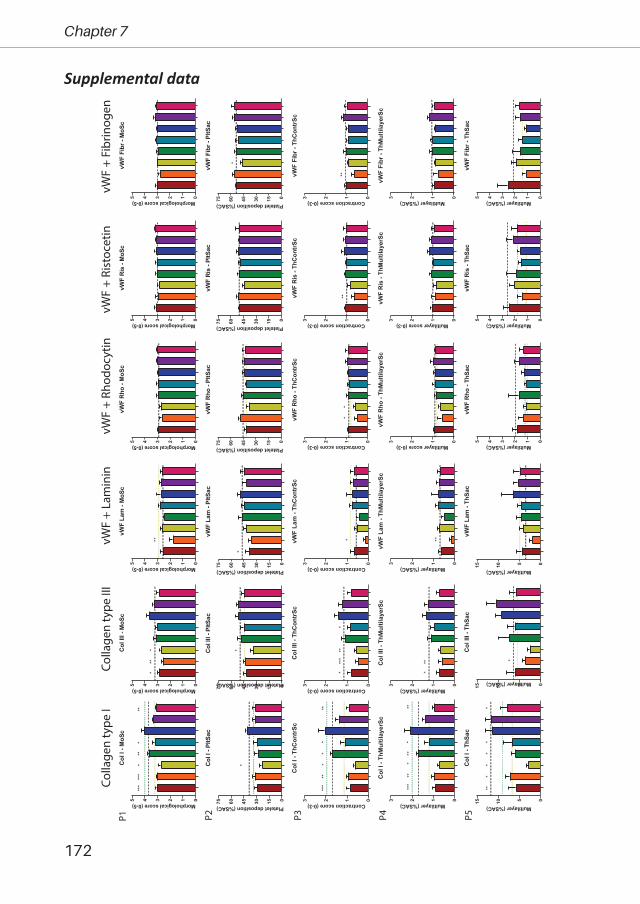
172
Chapter 7
Supplemental dataCo
llage
n ty
pe I
Colla
gen
type
III
vWF
+ Rh
odoc
ytin
vWF
+ La
min
invW
F +
Rist
ocet
invW
F +
Fibr
inog
enP1 P2 P3 P4 P5
012345
Col
I - M
oSc
Morphologicalscore(0-5)
***
**
012345
Col
III -
MoS
c
Morphologicalscore(0-5)
*
012345
vWF
Lam
- M
oSc
Morphologicalscore(0-5)
**
012345
vWF
Rho
- M
oSc
Morphologicalscore(0-5)
012345
vWF
Ris
- M
oSc
Morphologicalscore(0-5)
012345
vWF
Fibr
- M
oSc
Morphologicalscore(0-5)
01530456075
Col I
- Pl
tSac
Plateletdeposition(%SAC)
*
01530456075
Col I
II - P
ltSac
Plateletdeposition(%SAC)
*
01530456075
vWF
Lam
- Pl
tSac
Plateletdeposition(%SAC)
*
01530456075
vWF
Rho
- PltS
ac
Plateletdeposition(%SAC)
01530456075
vWF
Ris
- PltS
ac
Plateletdeposition(%SAC)
01530456075
vWF
Fibr
- Pl
tSac
Plateletdeposition(%SAC)
*
0123
Col
I - T
hCon
trSc
Contractionscore(0-3)
***
***
0123
Col
III -
ThC
ontr
Sc
Contractionscore(0-3)
*
0123
vWF
Lam
- Th
Con
trSc
Contractionscore(0-3)
*
0123
vWF
Rho
- Th
Con
trSc
Contractionscore(0-3)
**
0123
vWF
Ris
- Th
Con
trSc
Contractionscore(0-3)
**
0123
vWF
Fibr
- Th
Con
trSc
Contractionscore(0-3)
**
0123
Col
I - T
hMul
tilay
erSc
Multilayer(%SAC)
***
**
0123
Col
III -
ThM
ultil
ayer
Sc
Multilayer(%SAC)
0123
vWF
Lam
- Th
Mul
tilay
erSc
Multilayer(%SAC)
**
0123
vWF
Rho
- Th
Mul
tilay
erSc
Multilayerscore(0-3)0123
vWF
Ris
- Th
Mul
tilay
erSc
Multilayerscore(0-3)
0123
vWF
Fibr
- Th
Mul
tilay
erSc
Multilayer(%SAC)
051015
Col
I - T
hSac
Multilayer(%SAC)
**
051015
Col
III -
ThS
ac
Multilayer(%SAC)
*
051015
vWF
Lam
- Th
Sac
Multilayer(%SAC)
012345
vWF
Rho
- Th
Sac
Multilayer(%SAC)
012345
vWF
Ris
- ThS
ac
Multilayer(%SAC)
012345
vWF
Fibr
- Th
Sac
Multilayer(%SAC)
***
***
*
***
* ***
***
**
***
*** **
**
*
**
**
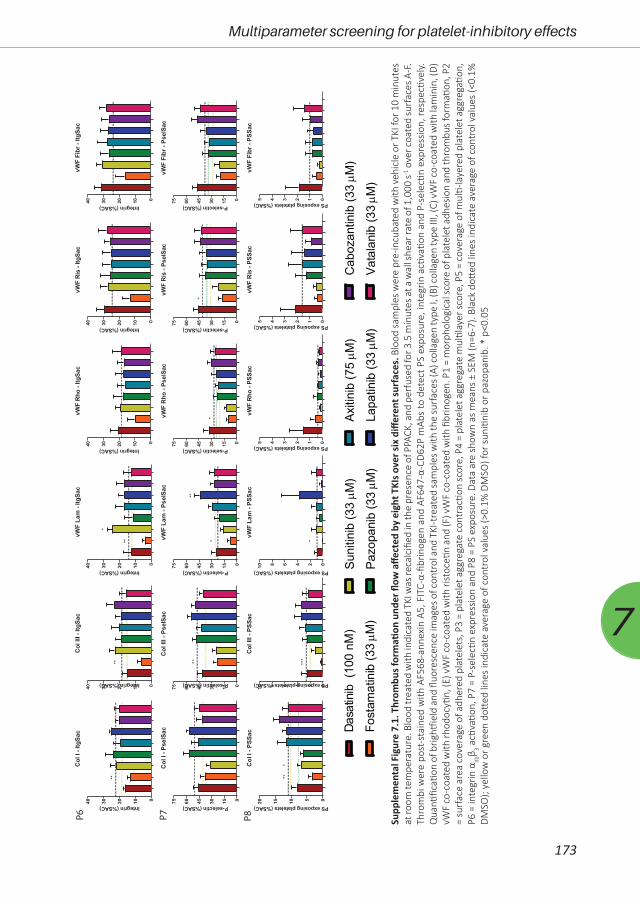
173
Multiparameter screening for platelet-inhibitory effects
7
Supp
lem
enta
l Fig
ure
7.1.
Thr
ombu
s for
mati
on u
nder
flow
affe
cted
by
eigh
t TKI
s ove
r six
diff
eren
t sur
face
s. B
lood
sam
ples
wer
e pr
e-in
cuba
ted
with
veh
icle
or T
KI fo
r 10
min
utes
at
room
tem
pera
ture
. Blo
od tr
eate
d w
ith in
dica
ted
TKI w
as re
calc
ified
in th
e pr
esen
ce o
f PPA
CK, a
nd p
erfu
sed
for 3
.5 m
inut
es a
t a w
all s
hear
rate
of 1
,000
s-1 o
ver c
oate
d su
rfac
es A
-F.
Thro
mbi
wer
e po
st-s
tain
ed w
ith A
F568
-ann
exin
A5,
FIT
C-α-
fibrin
ogen
and
AF6
47-α
-CD6
2P m
Abs
to d
etec
t PS
expo
sure
, int
egrin
acti
vatio
n an
d P-
sele
ctin
expr
essio
n, re
spec
tivel
y.
Qua
ntific
ation
of b
rightf
ield
and
fluo
resc
ence
imag
es o
f con
trol
and
TKI
-tre
ated
sam
ples
with
the
surf
aces
(A) c
olla
gen
type
I, (B
) col
lage
n ty
pe II
I, (C
) vW
F co
-coa
ted
with
lam
inin
, (D)
vW
F co
-coa
ted
with
rhod
ocyti
n, (E
) vW
F co
-coa
ted
with
rist
oceti
n an
d (F
) vW
F co
-coa
ted
with
fibr
inog
en. P
1 =
mor
phol
ogic
al sc
ore
of p
late
let a
dhes
ion
and
thro
mbu
s for
mati
on, P
2 =
surf
ace
area
cov
erag
e of
adh
ered
pla
tele
ts, P
3 =
plat
elet
agg
rega
te c
ontr
actio
n sc
ore,
P4
= pl
atel
et a
ggre
gate
mul
tilay
er sc
ore,
P5
= co
vera
ge o
f mul
ti-la
yere
d pl
atel
et a
ggre
gatio
n,
P6 =
inte
grin
αIIb
β 3 acti
vatio
n, P
7 =
P-se
lecti
n ex
pres
sion
and
P8 =
PS
expo
sure
. Dat
a ar
e sh
own
as m
eans
± S
EM (n
=6-7
). Bl
ack
dott
ed li
nes i
ndic
ate
aver
age
of c
ontr
ol v
alue
s (<0
.1%
DM
SO);
yello
w o
r gre
en d
otted
line
s in
dica
te a
vera
ge o
f con
trol
val
ues
(>0.
1% D
MSO
) for
sun
itini
b or
paz
opan
ib. *
p<0
.05
Dasa
tinib
(10
0 nM
)
Fost
amat
inib
(33µM
)Su
nitin
ib (3
3µM
)
Pazo
pani
b (3
3µM
)
Axiti
nib
(75µM
)
Lapa
tinib
(33µM
)
Cab
ozan
tinib
(33µM
)
Vata
lani
b (3
3µM
)
P6 P7 P8
010203040
Col I
- Itg
Sac
Integrin(%SAC)
**
010203040
Col I
II - I
tgSa
c
Integrin(%SAC)
**
010203040
vWF
Lam
- Itg
Sac
Integrin(%SAC)
**
*
010203040
vWF
Rho
- Itg
Sac
Integrin(%SAC)
010203040
vWF
Ris
- Itg
Sac
Integrin(%SAC)
010203040
vWF
Fibr
- Itg
Sac
Integrin(%SAC)
01530456075
Col
I - P
selS
ac
P-selectin(%SAC)
01530456075
Col
III -
Pse
lSac
P-selectin(%SAC)
**
01530456075
vWF
Lam
- Ps
elSa
c
P-selectin(%SAC)
*
**
01530456075
vWF
Rho
- Pse
lSac
P-selectin(%SAC)
*
01530456075
vWF
Ris
- Pse
lSac
P-selectin(%SAC)
*
01530456075
vWF
Fibr
- Ps
elSa
c
P-selectin(%SAC)
05101520
Col
I - P
SSac
PSexposingplatelets(%SAC)
***
0246810
Col
III -
PSS
ac
PSexposingplatelets(%SAC)
***
0246810
vWF
Lam
- PS
Sac
PSexposingplatelets(%SAC)
*
012345
vWF
Rho
- PSS
ac
PSexposingplatelets(%SAC)
012345
vWF
Ris
- PS
Sac
PSexposingplatelets(%SAC)
012345
vWF
Fibr
- PS
Sac
PSexposingplatelets(%SAC)


Chapter 8
General Discussion

176
Chapter 8
Since the beginning of this century, a new class of anti-cancer agents, tyrosine kinase
inhibitors (TKIs), has been developed and approved for the treatment of several malignancies1, 2.
These TKIs inhibit growth factor receptors (tyrosine kinases, Table 8.1) involving angiogenesis and
tumour development and thereby improving progression free survival (PFS) and overall survival
(OS)3. As most TKIs inhibit multiple protein tyrosine kinases (Table 8.1), they inevitably also affect
other cell types. One of the side effects described for several TKIs is (mild) bleeding or thrombosis4.
Platelets are well known for their involvement in haemostasis, and they also contain multiple
protein tyrosine kinases (Syk, Src family kinases (SFK), Btk), which are essential for their activation.
Hence, these TKIs may also affect platelet function. Therefore, the overall aim of this thesis was to
provide more insights in the off-target effects of TKIs on platelet function (Chapter 1). In the next
paragraphs of this chapter, the observations described throughout this thesis are discussed in
relation to the current literature, as well as clinical implications and future perspectives are given.
Altogether, the findings reported in this thesis may provide relevant insights for monitoring the
bleeding risk in patients receiving TKI treatment by assessing quantitative and qualitive platelet
traits, especially in the presence of anti-coagulant or anti-platelet drugs.
Platelet signalling and TKIsIt has been described that several TKIs, for different cancer types, inhibit platelet function
upon treatment, which can contribute to the risk of bleeding5-7. Chapter 2 shows an overview
Figure 8.1. Overview of the tyrosine kinases expressed in platelet signalling (in yellow) downstream of multiple platelet receptors, resulting in different platelet responses. The intervention points of the TKIs (based on the Kd values described in Chapter 2) investigated in this thesis are indicated in red.
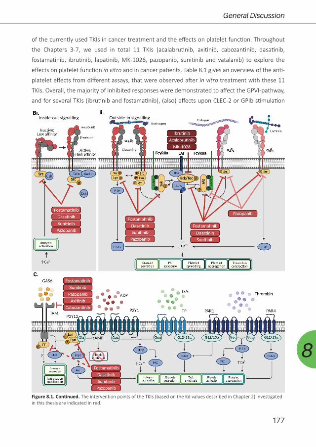
177
General Discussion
8
of the currently used TKIs in cancer treatment and the effects on platelet function. Throughout
the Chapters 3-7, we used in total 11 TKIs (acalabrutinib, axitinib, cabozantinib, dasatinib,
fostamatinib, ibrutinib, lapatinib, MK-1026, pazopanib, sunitinib and vatalanib) to explore the
effects on platelet function in vitro and in cancer patients. Table 8.1 gives an overview of the anti-
platelet effects from different assays, that were observed after in vitro treatment with these 11
TKIs. Overall, the majority of inhibited responses were demonstrated to affect the GPVI-pathway,
and for several TKIs (ibrutinib and fostamatinib), (also) effects upon CLEC-2 or GPIb stimulation
Figure 8.1. Continued. The intervention points of the TKIs (based on the Kd values described in Chapter 2) investigated in this thesis are indicated in red.
178
Chapter 8
could be observed. Given the defined affinities in our review in combination with the results
obtained for the 11 TKIs tested here, we visualised the proposed interference of these TKIs in the
platelet signalling pathways (Figure 8.1).
In whole blood perfusion, effects on thrombus formation were mainly restricted to the
glycoprotein (GP)VI-pathway as observed for dasatinib, sunitinib, pazopanib and vatalanib. Only
ibrutinib and fostamatinib showed to be effective towards GPIb (combined with CLEC-2 or α6β1)-
induced thrombus formation (Table 8.1, Chapter 3, 4, 6 and 7). Ibrutinib and fostamatinib specifically
inhibit Btk/Tec or Syk, respectively. Both proteins are important mediators in coupling cell surface
receptors to downstream signalling events and are in close proximity in most signalling pathways8,
9. This suggests that direct targeting of Btk or Syk in platelet signalling by TKIs is more effective to
inhibit platelet function as compared to TKIs primary targeting growth factor receptors. Also, a
difference can be observed between inhibition of Btk and Syk. Btk inhibition by ibrutinib mainly
inhibits GPIb-associated thrombus formation, whereas Syk inhibition with fostamatinib shows to
affect thrombus formation with GPIb, as well as GPVI. As binding and activation of Syk initiates
downstream signalling involving Btk in GPVI, CLEC-2 and GPIb-signalling, this hints towards the
concept that inhibition higher upstream in platelet signalling results in more efficient inhibition.
In isolated platelets almost all inhibitors more prominently inhibited GPVI-induced
aggregation, except fostamatinib, axitinib and lapatinib. For fostamatinib this contrasts to the
papers of Spalton et al. and Lhermusier et al.10, 11, who observed complete inhibition of collagen-
and/or rhodocytin-induced platelet aggregation with already low concentrations of the active
metabolite of fostamatinib. The only difference between these studies and the study in chapter 7,
is the incubation time of the platelets with the inhibitor. We pre-incubated for 10 minutes, while
in the earlier studies platelets were incubated for 1-5 minutes with fostamatinib. As fostamatinib
is a reversible inhibitor, the incubation time could be of importance, however, we did observe
strong inhibition in whole blood perfusion experiments using 10 minutes pre-incubation of whole
blood.
The lack of GPVI-induced aggregation for axitinib and lapatinib is in line with literature
findings. Although axitinib is associated with bleeding7, 12, no inhibition of platelet function has
been reported. In about 15-20% of the patients, axitinib is described to induce mild (grade 1-2)
thrombocytopenia13, 14, and in rare cases (<1%) severe (≥ grade 3) thrombocytopenia is reported15.
For axitinib it is proposed that increased bleeding in these patients is caused by a decrease in
platelet count rather than platelet inhibition. For lapatinib increased bleeding risk has not been
reported and, furthermore, thrombus formation under flow (in vitro) was also unaffected16. Hence,
these observations show that axitinib and lapatinib do not have off-target effects on platelets.
All other TKIs investigated in this thesis (acalabrutinib, cabozantinib, dasatinib, MK-1026,
pazopanib, sunitinib and vatalanib) show most pronounced effects in platelets upon collagen
stimulation. Inhibition in thrombus formation under flow was restricted to GPVI-induced

179
General Discussion
8
activation for pazopanib, sunitinib, dasatinib and vatalanib. This suggests that the GPVI pathway
is most sensitive for inhibition with TKIs. GPVI, GPIb-IX-V and CLEC-2 are most potent receptors
for thrombus formation at high-shear stress conditions17. They share several common tyrosine
kinases (Fyn, Lyn, Syk, Btk, etc) in signal transduction in platelets17, 18. As TKIs are able to affect
multiple of these tyrosine kinases, it is expected that more pathways should be inhibited.
However, a possible explanation for the restriction of effects to the GPVI pathway could be the
amount of copy numbers present in platelets. Of these receptors, GPIIbIIa (integrin αIIbβ3) and
GPIb are the most abundantly expressed receptor on platelets with around 80,000 and 25,000
copies, respectively19-21. GPVI and CLEC-2 receptors are far less expressed with only 6,000-10,000
and 2,000 copies reported, respectively19, 22, 23. Therefore, it could be expected that GPVI and
CLEC-2 are relatively more affected as compared to GPIb as less receptors (lower copy number)
remain active. Signalling pathways of receptors with a higher expression, such as GPIIbIIa and
GPIb, might still be partially functional as more receptors are available, making it more difficult
to observe inhibition. Moreover, it is indicated by Spalton et al.10 that GPVI is more dependent on
Src family kinase (SFK) phosphorylation and FcγR signalling, whereas the CLEC-2 pathway is more
reliant on Syk phosphorylation. Together this encourages that TKIs have probably a higher affinity
for SFKs rather that Syk, resulting in more specific inhibition of the GPVI pathway.
Plasma protein binding of TKIs influencing drug availability and platelet inhibition
In (almost) all chapters of this thesis inhibition of platelet traits, qualitative and/or
quantitative, by different TKIs is the central point of view. Although in vitro effects were present
in almost all conditions tested, inhibition by TKIs was more pronounced in isolated platelets as
compared to the presence of plasma (platelet-rich plasma or whole blood) (Table 8.1). This meant
that a higher dose of a TKI was often needed to elicit similar inhibition in PRP or whole blood
as compared to washed platelets. Strikingly, pazopanib and cabozantinib prominently inhibited
aggregation of washed platelets to several agonists, while in PRP this response was unaffected
(Table 8.1). This phenomenon is caused by high plasma binding of the TKIs (>95%)24-27. In patients
with TKI treatment, the bioavailability of the drug is likely to be influenced by this plasma binding
with implications for drug toxicity and response28. This shows the importance of the comparison
between in vitro testing of these agents in presence or absence of plasma, as well as measuring
effects present in patients receiving treatment. For clinical implications it is might point to adapting
dosing regimens based on the pharmacokinetic/dynamic characteristics of TKIs29 or more ideally
by monitoring the unbound plasma concentration of a TKI during treatment30. For example, we
measured levels of sunitinib and its metabolite in plasma, serum and isolated platelets at 2 and
4 weeks on treatment in patients with a 4/2 scheme (4 weeks dosing, 2 weeks rest) in Chapter
5. We showed that in plasma and serum, the unbound concentrations were comparable for
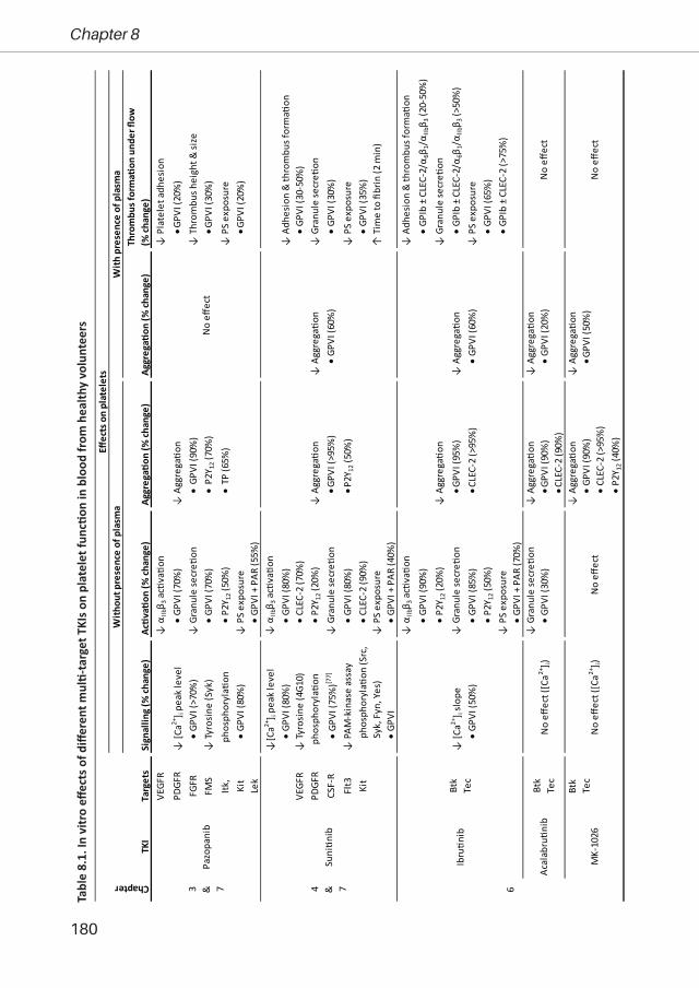
180
Chapter 8
VEGF
R↓
αIIbβ 3
ac�
va�o
n ↓
Pla
tele
t adh
esio
n
PDGF
R↓
[Ca2+
] i pe
ak le
vel
• GP
VI (7
0%)
↓ A
ggre
ga�o
n • G
PVI (
20%
)FG
FR•
GPVI
(>70
%)
↓ G
ranu
le se
cre�
on•
GPV
I (90
%)
↓ T
hrom
bus h
eigh
t & si
zeFM
S↓
Tyro
sine
(Syk
) •
GPVI
(70%
)•
P2Y
12 (7
0%)
• GPV
I (30
%)
Itk,
pho
spho
ryla�o
n•
P2Y 1
2 (50
%)
• TP
(65%
)↓
PS
expo
sure
Kit
• GP
VI (8
0%)
↓ P
S ex
posu
re
• GPV
I (20
%)
Lek
• GP
VI +
PAR
(55%
)↓
[Ca2+
] i pe
ak le
vel
↓ α
IIbβ 3
ac�
va�o
n •
GPVI
(80%
)•
GPVI
(80%
)↓
Adh
esio
n &
thro
mbu
s for
ma�
onVE
GFR
↓ Ty
rosi
ne (4
G10)
•
CLEC
-2 (7
0%)
• GP
VI (3
0-50
%)
PDGF
Rph
osph
oryl
a�on
• P2
Y 12 (
20%
)↓
Agg
rega�o
n↓
Agg
rega�o
n↓
Gra
nule
secr
e�on
CSF-
R•
GPVI
(75%
)[77]
↓ G
ranu
le se
cre�
on• G
PVI (
>95%
)•
GPVI
(60%
)•
GPVI
(30%
)
Flt3
↓ P
AM-k
inas
e as
say
• GP
VI (8
0%)
• P2Y
12 (5
0%)
↓ P
S ex
posu
reKi
t p
hosp
hory
la�o
n (S
rc,
• CL
EC-2
(90%
)•
GPVI
(35%
)Sy
k, F
yn, Y
es)
↓ P
S ex
posu
re↑
Tim
e to
fibr
in (2
min
)•
GPVI
•
GPVI
+ P
AR (4
0%)
↓ α
IIbβ 3
ac�
va�o
n ↓
Adh
esio
n &
thro
mbu
s for
ma�
on
• GP
VI (9
0%)
• GP
Ib ±
CLE
C-2/α 6β 1
/αIIbβ 3
(20-
50%
)
• P2
Y 12 (
20%
)↓
Agg
rega�o
n↓
Gra
nule
secr
e�on
↓ [C
a2+] i
slop
e↓
Gra
nule
secr
e�on
• GPV
I (95
%)
↓ A
ggre
ga�o
n•
GPIb
± C
LEC-
2/α 6β 1
/αIIbβ 3
(>50
%)
• GP
VI (5
0%)
• GP
VI (8
5%)
• CLE
C-2
(>95
%)
• GP
VI (6
0%)
↓ P
S ex
posu
re•
P2Y 1
2 (50
%)
• GP
VI (6
5%)
↓ P
S ex
posu
re•
GPIb
± C
LEC-
2 (>
75%
)•
GPVI
+ P
AR (7
0%)
↓ G
ranu
le se
cre�
on
↓ A
ggre
ga�o
n↓
Agg
rega�o
n•
GPVI
(30%
)• G
PVI (
90%
)•
GPVI
(20%
)• C
LEC-
2 (9
0%)
Btk
↓ A
ggre
ga�o
n↓
Agg
rega�o
nTe
c•
GPVI
(90%
)• G
PVI (
50%
)•
CLEC
-2 (>
95%
)•
P2Y 1
2 (40
%)
63 & 7
MK-
1026
No
effec
t ([C
a2+] i)
No
effec
t
Pazo
pani
b
Suni�n
ib
Ibru�n
ib
Acal
abru�n
ib
Btk
Tec
Btk
Tec
4 & 7
No
effec
t
No
effec
t
Sign
allin
g (%
chan
ge)
Ac�v
a�on
(% ch
ange
)Ag
greg
a�on
(% ch
ange
)Ag
greg
a�on
(% ch
ange
)Th
rom
bus f
orm
a�on
und
er fl
ow(%
chan
ge)
No
effec
t
No
effec
t ([C
a2+] i)
Chapter
TKI
Targ
ets
Effec
ts o
n pl
atel
ets
With
out p
rese
nce
of p
lasm
aW
ith p
rese
nce
of p
lasm
a
Tabl
e 8.
1. In
vitr
o eff
ects
of d
iffer
ent m
ulti-
targ
et T
KIs o
n pl
atel
et fu
nctio
n in
blo
od fr
om h
ealth
y vo
lunt
eers

181
General Discussion
8
PDGF
R↓
Thr
ombu
s hei
ght &
size
BCR-
ABL
↓[C
a2+] i
peak
leve
l↓
Agg
rega�o
n↓
Agg
rega�o
n•
GPVI
(35-
55%
)
EphA
2•
GPVI
(>90
%)
• GPV
I (>9
5%)
• GPV
I (50
%)
↓ α
IIbβ 3
ac�
va�o
nKi
t•
GPVI
(15%
)SF
K↓
All
para
met
ers (
exce
pt a
dhes
ion)
↓ α
IIbβ 3
ac�
va�o
n•
GPVI
(15-
70%
)
↓[C
a2+] i
peak
leve
l•
GPVI
(20%
)↓
Agg
rega�o
n↓
Agg
rega�o
n•
GPIb
+ α
6β1 (
30-9
0%)
• GP
VI (5
5%)
↓ G
ranu
le se
cre�
on• G
PVI (
20%
)• G
PVI (
20%
)↓
Thr
ombu
s con
trac�o
n•
GPVI
(55%
)•
GPVI
(20-
30%
)•
GPIb
± C
LEC-
2/α I
Ibβ 3
(30-
45%
)↓
Gra
nule
secr
e�on
• GP
Ib ±
CLE
C-2/α I
Ibβ 3
(50-
65%
)↓
Agg
rega�o
n• G
PVI (
15%
)VE
GFR2
Axl
↓ α
IIbβ 3
ac�
va�o
n↓
Agg
rega�o
nFl
t3•
GPVI
(20%
)• G
PVI (
60%
)Ki
t↓
Lyso
som
e se
cre�
on• C
LEC-
2 (8
0%)
MET
• GP
VI (2
0%)
• P2Y
12 (6
0%)
Ret
EGFR
↓ A
ggre
ga�o
nEr
bB1-
2• G
PVI (
15%
)VE
GFR
↓ α
IIbβ 3
ac�
va�o
n↓
Agg
rega�o
nPD
GFRb
• GP
VI (2
0%)
• GPV
I (80
%)
↓ A
ggre
ga�o
n↓
Adh
esio
n &
agg
rega�o
nKi
t↓
Gra
nule
secr
e�on
• P2Y
12 (6
0%)
• GPV
I (20
%)
• GP
VI
• GP
VI (2
0%)
• TP
(70%
)• P
AR1/
4 (3
0%)
No
effec
tN
o eff
ect
Vata
lani
bN
o eff
ect (
[Ca2+
] i)
Chapter
TKI
Targ
ets
Effec
ts o
n pl
atel
ets
With
out p
rese
nce
of p
lasm
aW
ith p
rese
nce
of p
lasm
a
Sign
allin
g (%
chan
ge)
Ac�v
a�on
(% ch
ange
)Ag
greg
a�on
(% ch
ange
)Ag
greg
a�on
(% ch
ange
)Th
rom
bus f
orm
a�on
und
er fl
ow(%
chan
ge)
No
effec
tN
o eff
ect
Cabo
zan�
nib
No
effec
t ([C
a2+] i)
No
effec
tN
o eff
ect
7
Dasa�n
ib-
Fost
ama�
nib
Syk
Axi�
nib
VEGF
RN
o eff
ect (
[Ca2+
] i)-
Lapa
�nib
No
effec
t ([C
a2+] i)
-
SFK:
Src
fam
ily k
inas
es; T
KI: T
yros
ine
kina
se in
hibi
tor

182
Chapter 8
sunitinib and its metabolite at both timepoints, however, a remarkable increase of metabolite
concentration was observed in isolated platelets as compared to sunitinib concentration. This
suggested a higher uptake of the active form of sunitinib by platelets in their granules, though
both compounds were equally present in plasma. Therefore, it might also be of interest to not
only monitor plasma concentrations at 2 of 4 weeks on treatment (dependent on treatment
schedule)31, but also measure the uptake of (active) compound by platelets as this might predict
the platelet inhibitory effects.
Involvement of quantitative and quantitative platelet traits in bleeding tendency in patients
In the review in Chapter 2, 11 out of almost 40 TKIs are reported with an increased risk of
bleeding or bleeding events. Given the key role of platelets in haemostasis, both platelet count
and platelet function are relevant to prevent bleeding32, 33. In Chapters 3 and 5, we have shown
that not only platelet function, but also platelet count is already decreased after 2 weeks after
pazopanib or sunitinib treatment, as compared to pre-treatment (Table 8.2). Although TKIs are
known to be associated with platelet dysfunction6, 34, impaired platelet function is not always
found to be the cause of increased bleeding tendency35, 36. For multiple TKIs, including dasatinib
(Chapter 7), an association is found with drug-induced immune thrombocytopenia, defined as
a platelet count below 50x109/L37-39, resulting in an increased risk of bleeding. While most of
the patients included in Chapters 3 and 5 did not show thrombocytopenia, we did observe an
overall decrease in platelet count upon pazopanib or sunitinib treatment (Table 8.2). This change
in platelet count correlated with the inhibition of platelet function after sunitinib treatment, as
well as with the plasma concentration of the metabolite of sunitinib (Chapter 5). This point to
the idea that platelets are exposed to significant levels of sunitinib resulting in (mild) anti-platelet
effects. Alternatively, sunitinib may act on the megakaryocytes, thereby decreasing platelet count
and possibly also affect platelet function. As plasma concentration and plasma protein binding of
sunitinib (or its metabolite) could be of importance in toxicity and response to a TKI treatment30,
this pointed toward a monitoring tool for off-target effects and/or progression-free survival.
Whilst several studies propose that TKI-associated platelet dysfunction does not cause
bleeding35, 36, almost all the patients included in Chapter 3, 5 and 6 did report (mild) bleeding
episodes upon TKI treatment in the absence of thrombocytopenia (Table 8.2). This may imply
that pharmacological platelet inhibition does play a role in the increase in bleeding risk. In
agreement, we see impairment of GPVI-induced platelet activation, aggregation or thrombus
formation upon treatment in vitro and/or in patients, some with additional effects on CLEC-2- or
GPIb-induced platelet responses. It has been reported that targeting GPVI via GPVI inhibitors
or GPVI depletion in mice or patients does not (significantly) impair haemostasis40-45. However,
a combined deficiency of GPVI and CLEC-2 in mice does show impaired haemostasis43. These

183
General Discussion
8
Qua
n�ta�v
e
RCC
↓ P
late
let c
ount
↓
PS
expo
sure
↓ P
S ex
posu
reYe
s
(n=1
0)(1
5%)
•
GPV
I (20
%)
•
GPV
I (50
%)
(3/1
0)
↓ G
PVI-i
nduc
ed α
IIbβ 3
↓
GPV
I-ind
uced
pla
tele
t
↓ P
late
let c
ount
Ac
�va�
on (1
5%)77
↓ G
PVI-i
nduc
ed a
dhes
ion
(20%
)77
(40%
), (3
0%)77
↓ G
PVI-i
nduc
ed
agg
rega�o
n (1
0%)
↓ G
PVI-i
nduc
ed P
S g
ranu
le se
cre�
on
(15-
50%
)77 a
nd (4
0%)77
exp
osur
e (7
5%)77
↓ P
late
let c
ount
↓
GPV
I-ind
uced
(50%
) a
ggre
ga�o
n (1
5%)
↓ P
late
let c
ount
(25%
)
↓ G
PVI-i
nduc
ed
↓ G
PVI-i
nduc
ed α
IIbβ 3
t
hrom
bus h
eigh
t
ac�v
a�on
(65%
) &
den
sity
(30%
)
↑ P
late
let v
olum
e ↓
GPV
I-ind
uced
↓
αIIbβ 3
ac�
va�o
n (>
50%
(20%
)gr
anul
e se
cre�
on
for
GPV
I, vW
F-CL
EC-2
,
(85%
) a
nd v
WF-α 6β 1
surf
ace)
↑ P
2Y12
-indu
ced
↓ P
S ex
posu
re (2
0% fo
r
α IIbβ 3
ac�
va�o
n G
PVI s
urfa
ce)
(35
%)
Chapter
Pa�e
nts
TKI t
reat
men
tTi
mep
oint
(w
eeks
)
Effec
ts o
n pl
atel
ets
Qua
lita�
veTh
rom
bus f
orm
a�on
und
er
flow
(% ch
ange
)
5Su
ni�n
ib50
2 4
3Pa
zopa
nib
800
2N
o eff
ect
RCC
(n=2
0)
and
(n=8
)77-
-
12-
No
effec
t-
No
effec
t
Dosa
ge
(mg/
day)
Blee
ding
(Yes
/No)
(%
chan
ge)
Ac�v
a�on
(% ch
ange
)Ag
greg
a�on
(% ch
ange
)
Yes
(9/2
0)an
d (4
/8)77
Yes
(6/9
)
CLL,
M
CL,
WM
(n=1
6)
Ibru�n
ib(n
=9)
642
0-56
0-
Tabl
e 8.
2. O
verv
iew
of t
he e
ffect
s of T
KI tr
eatm
ent i
n pa
tient
s on
quan
titati
ve a
nd q
ualit
ative
pla
tele
t tra
its a
nd re
port
ed b
leed
ing
durin
g tr
eatm
ent p
erio
d as
com
pare
d to
a c
ontr
ol g
roup
CLL:
Chr
onic
lym
phoc
ytic
leuk
aem
ia; M
CL: M
antle
cel
l lym
phom
a; R
CC: R
enal
cel
l car
cino
ma;
TKI
: Tyr
osin
e ki
nase
inhi
bito
r; W
M: W
alde
nstr
om m
acro
glob
ulin
emia

184
Chapter 8
studies mainly determined the contribution of platelet GPVI- and CLEC-2 receptors themselves in
haemostasis40, 43. However, in case of several TKIs, the inhibition in platelet signalling underneath
these receptors is described to be via SFKs (Fyn, Lyn) or Syk11, 46-49. As the GPVI, CLEC-2 and GPIb
pathways all require SFKs and/or Syk in its signalling, it overall suggests that multi-target TKIs can
elicit combined inhibition of GPVI plus CLEC-2 or possibly GPIb signalling pathways. This could be
more potent than targeting solely one receptor and, hence, relevant for bleeding tendency. In
combination with a decrease in platelet count, this might even further increase the bleeding risk.
As cancer patients often have a cardiovascular history50, 51, anti-platelet or anti-coagulant
drugs are prescribed to prevent recurrent cardiovascular events. Anti-platelet therapies, such as
clopidogrel and aspirin, are reported to be associated with an increased risk of bleeding52-54. As
TKIs are described to increase this risk as well, in Chapter 5, we investigated the synergistic effects
of sunitinib and aspirin as anti-platelet medication on thrombus formation and fibrin formation to
elucidate the impact on haemostasis. We demonstrated that sunitinib suppressed procoagulant
activity and prolonged the time to fibrin formation, which was aggravated by aspirin treatment.
Therefore, a combination of TKIs and anti-platelet therapies may be expected to increase the
frequency or grade of bleeding. Though such dual treatment of TKI with anti-platelet or anti-
coagulant drugs could be increasing the bleeding risk, this might also be beneficial for the
patients. It has been shown that anti-platelet therapies have anti-tumour effects in hepatocellular
and colorectal cancer55, 56. Hence a combination of TKI with anti-platelet therapy could enhance
the anti-tumour effects. For example, erlotinib together with aspirin is described to enhance
sensitivity of the cancer as compared to aspirin alone, thereby increasing the anti-cancer
activity57. The application of anti-platelet drugs is even more favourable, as it was shown that
low doses of aspirin could be used as treatment for off-target effects, such as skin rash caused
by erlotinib-treatment58. Additionally, for some TKIs, anti-platelet treatments can be useful as
nilotinib is associated with an increased risk of thrombosis59. Yet, not all anti-platelet drugs show
a positive correlation in combination with TKI treatment. The study of Haoula et al.60 highlights
the importance of drug-drug interactions between clopidogrel and other anticoagulants in
combination with TKIs. This urges for better understanding of dual treatment with TKIs and anti-
platelet or anti-coagulant therapies to monitor patients.
Overall, we showed that multiple TKIs inhibit platelet function via different pathways and
that this combination may contribute to the reported bleeding risk in patients. In addition, for
other TKIs inhibiting the GPVI-induced platelet function together with a decrease in platelet
count, as seen with pazopanib and sunitinib, might also explain the mild bleeding tendency
observed during treatment. Hence, the combination of the inhibitory effects on platelet traits
together with anti-platelet or anti-coagulant drugs urges for better understanding and possibly
more strict monitoring of patients as this may further increase the risk of bleeding.

185
General Discussion
8
Clinical implications Platelets not only have an important role in haemostasis, they have also been described
to be implicated in tumour development and growth61-64. Platelets are known to take up various
pro- and antiangiogenic proteins into their α-granules61, 62, 65. These granules can be secreted
upon platelet activation by activating factors expressed or secreted by tumour vasculature
such as thrombin and ADP. In turn, the secreted proangiogenic factors can stimulate tumour
growth and metastasis by stimulating angiogenesis61, 66, improving vasculature integrity67, 68 and
inducing resistance to therapy69, 70. Due to this promoting role of platelets in tumour angiogenesis
and progression, as well as the correlation with reduced survival, platelets are an interesting
target for cancer therapy. Although numerous drugs have been developed to target platelet
receptors71, limitations for implications occur due the role of platelets in haemostasis associated
with increased bleeding tendency52-54. Yet anti-platelet treatment have been described to reduce
tumour angiogenesis, growth and progression72, 73 by direct inhibition of the tumour or via indirect
via inhibition of platelet secretion, thereby decreasing proangiogenic protein concentrations in
the tumour environment74. Hence, the TKIs investigated in this thesis could also indirectly affect
tumour growth via the inhibiting effects on platelet function, as we did observe inhibition of
platelet activation and/or secretion upon GPVI stimulation with acalabrutinib, cabozantinib,
fostamatinib, ibrutinib, pazopanib, sunitinib and vatalanib in isolated platelets. However, the
principle of platelet-mediated anti-cancer effect of TKIs needs to be further investigated. Platelet
inhibition remains contradictory and needs to be balanced, as it can improve cancer prognosis,
as well as increase the risk of bleeding. In line with the inhibition of platelet function with TKIs
observed throughout this thesis, one might argue that monitoring patients is needed to profit
from the anti-tumour effects and keep the risk of undesirable bleeding to a minimum.
In addition, platelet production can be (greatly) enhanced by malignant (solid) tumours62,
75. An increased platelet count or thrombocytosis (platelet count of >400x109/L) is recognized as
a predictor of poor prognosis in patients with different cancer types75, 76. As platelets are able to
take up all sorts of compounds into their granules, it is not unlikely for anti-cancer agents to be
sequestered as well. The study of Sabrkhany et al.77 showed that platelets take up sunitinib in
their granules in vitro and in patients. In turn, we showed in Chapter 5 that both sunitinib and
its metabolite were present in isolated platelet of sunitinib-treated patients and that platelet
function is impaired. Besides high plasma binding of anti-cancer or anti-platelet agents, the
ability of platelets to sequester these compounds might influence the bioavailability for the
tumour, especially in thrombocytosis, i.e. increased amount of platelets. This might result in
reduced inhibition of tumour growth as a lower concentration would be circulating in the blood.
Also, thrombocytosis might not only be a consequence of cancer, it might be a paraneoplastic
abnormality that is associated with a vicious cycle of promoting tumour cells, platelet activation
and stimulating thrombocytosis75. Therefore, reducing the thrombocytosis before starting

186
Chapter 8
treatment by interfering with this feedback-loop could be relevant. This could be accomplished
via inhibition of platelet function with anti-platelet agents or heparins, however this could result
in bleeding implications78, 79. Another approach to intervene with tumour growth and metastasis
is inhibition of interleukin-6 (IL-6) by the IL-6 antibody siltuximab. This will deplete the increased
growth factor levels present and additionally decrease platelet count80. Clinical trials with ovarian
cancer, renal cell carcinoma and castration resistant prostate cancer already show beneficial
effects80-82.
Clinical significance for renal cell carcinoma patientsThe TKIs used for treatment of patients with metastatic renal cell carcinoma (mRCC), i.e.
sunitinib and pazopanib, have been the focus of Chapters 3, 4 and 5 of this thesis. Furthermore,
we also studied the effects of cabozantinib, used mainly as 2nd or 3rd line treatment for mRCC
patients, and axitinib, combined with pembrolizumab in first line mRCC83, 84, on platelet functions
in Chapter 7. In the Netherlands, good risk mRCC patients are treated with a TKI when metastases
have occurred. Patients often have a large tumour thrombus in the renal vein, which means that
local surgery is no longer possible. However, this is usually no consideration to start anticoagulation
therapy. When started with 50 mg/day sunitinib, systemic concentrations of sunitinib should be
50-100 ng/mL85. However, for a substantial group of patients, this dosing schedule may result in
much higher peak levels, resulting in toxicity86. In daily practice, sunitinib levels (steady state) in
serum could be determined if patients experience serious side effects and if the levels are too
high (>100 ng/mL31), the treatment dose could be adjusted accordingly. In addition, sunitinib
levels could also be determined if patients have no side effects at all, as the sunitinib level is
probably too low (<50 ng/mL31).
Hamilton et al.87 have shown that the decrease in platelet count upon sunitinib treatment
may serve as a prognostic value with regard to tumour development. In Chapter 5, we have
demonstrated that this reduction in platelet count also coincides with a reduction in platelet
function as well as sunitinib levels in plasma and serum. Together, this implies that both qualitative
and quantitative platelet traits may serve as a monitoring tool with regards to sunitinib treatment
in mRCC patients. Furthermore, similar observations have been reported for neutrophil-to-
lymphocyte ratio (NLR) and platelet-to-lymphocyte (PLR) ratio. A NLR greater than 1.9 but below
3 and PLR over 160 are described to be favourable prognostic factors88, 89. Also, an increase in
eosinophils have been reported to be a beneficial factor upon therapy in patients with renal cell
carcinoma upon treatment90, 91.
Also relevant for mRCC, although no longer used, is the classification of metastatic disease
into groups with regards to overall survival, according to the Memorial Sloan-Kettering Cancer
Centre (MSKCC)/Motzer citeria92; good, intermediate and poor risk. Currently, a switch has been
made to the International Metastatic RCC Database Consortium (IMDC) criteria to make group
187
General Discussion
8
classification. For this purpose, 6 parts of pre-treatment are scored, with one point per item,
looking back one year before starting systemic treatment: Time from diagnosis to treatment
initiation <1 year, Karnofsky performance status (KPS) <80%, haemoglobin (Hb) concentration
<12 g/L, serum calcium concentration >upper limit of normal (ULN), neutrophil count >ULN and
platelets count> ULN93. With no points, the patient has a good risk for survival, at 1-2 points
intermediate risk and from 3 on the risk is poor. For the good risk patients, it appeared that
sunitinib was superior to the immuno-agents nivolumab/ipilimumab, and was equivalent to
pembrolizumab plus axitinib94, 95. Therefore, sunitinib has become a mainstay not only for first-
line treatment in this particular risk group, but also in second line treatment after nivolumab/
ipilimumab. The results of the present thesis with regard to the effects of sunitinib on platelet
count and function, are thus in agreement with the incorporation of platelets into this IMDC
classification.
Future perspectives To summarise, this thesis increased the knowledge of how TKIs can influence platelet
function and contribute to reported bleeding events. In brief, in vitro experiments throughout
this thesis demonstrate that impaired platelet function is more pronounced in isolated platelets
as compared to whole blood perfusion, mainly affecting GPVI-induced platelet activation in
presence of plasma. Only ibrutinib and fostamatinib showed (additional) inhibition of CLEC-2
and/or GPIb-induced platelet activation and aggregation. In addition, the observed suppression
of aggregation and thrombus formation with sunitinib was aggravated by anti-platelet (aspirin)
therapy. Our studies involving patients demonstrated that pazopanib or sunitinib treatment
slightly decreased platelet count together with (limited) effects on the qualitative platelet traits,
whereas for patients treated with ibrutinib impaired collagen-, rhodocytin- and vWF-induced
aggregation and thrombus formation was observed. Moreover, we showed that for sunitinib the
inhibition in quantitative and qualitative platelet traits could be correlated with the (effective)
sunitinib levels in patients. Altogether, the inhibition of multiple platelet activation pathways, the
reduction in platelet count, or TKI treatment combined with antiplatelet agents might contribute
or even increase the reported bleeding tendencies of these TKIs. Therefore, patients should
be monitored by measuring platelet function or (active) compound levels in serum, plasma or
isolated platelets 2-4 weeks after start treatment or additionally in case of changes like dose
reduction, reoccurrence of side effects or altering treatment schedule. Though, more research is
needed to determine effects on sunitinib concentration in long term and whether possible effects
extinguish over time, as our study in Chapter 6 is limited to follow-up period of maximal 12 weeks
and limited in patient numbers due to dose reduction or discontinuation of medication (upon
decease).
To further develop the findings of this thesis towards better and improved patient care,
188
Chapter 8
more research is needed. Our patient study with sunitinib showed the correlation between
quantitative and qualitative platelet traits with (active) sunitinib levels. Due a limited number
of patients, no correlation could be observed between these parameters and bleeding and/or
progression. Therefore, it would be interesting to investigate whether changes in platelet traits or
concentration of unbound sunitinib (or other TKIs) in plasma or isolated platelets could predict
response to therapy or bleeding in a larger cohort. This knowledge might lead to a way to follow
patients more closely to prevent bleeding or monitor progression. As we observed aggravated
effects on platelet function of sunitinib together with aspirin, more research into the combined
anti-platelet effects of different anti-platelet or anti-coagulant drugs with TKIs is necessary.
Especially patients receiving both anti-cancer and anti-platelet therapies could be at increased
risk for bleeding.
The application of TKIs in treatment of other diseases, e.g. thrombosis, would also be
fascinating to study. TKIs already have inhibiting effects on platelets mainly via the GPVI pathway,
therefore, these drugs might be possible to use as anti-platelet drugs in (deep vein) thrombosis,
which is also suggested recently by other studies96, 97. These TKIs are already approved in the
clinic, ergo easier to test in other diseases involving thrombosis or thrombo-inflammation, in a
short period of time as side effects are already reported. Additionally, due to the relative high
affinity described for tyrosine kinases, relatively low therapeutic concentrations of TKI could be
able to selectively inhibit platelet function without possibly increasing bleeding risk.
References1. Jeong W, Doroshow JH, Kummar S. United States Food and Drug Administration approved oral kinase
inhibitors for the treatment of malignancies. Curr Probl Cancer. 2013; 37: 110-44.2. Roskoski R, Jr. Properties of FDA-approved small molecule protein kinase inhibitors: A 2020 update.
Pharmacol Res. 2020; 152: 104609.3. Gotink KJ, Verheul HM. Anti-angiogenic tyrosine kinase inhibitors: what is their mechanism of action?
Angiogenesis. 2010; 13: 1-14.4. Tullemans BME, Heemskerk JWM, Kuijpers MJE. Acquired platelet antagonism: off-target antiplatelet
effects of malignancy treatment with tyrosine kinase inhibitors. J Thromb Haemost. 2018; 16: 1686-99.
5. Deb S, Boknäs N, Sjöström C, Tharmakulanathan A, Lotfi K, Ramström S. Varying effects of tyrosine kinase inhibitors on platelet function-A need for individualized CML treatment to minimize the risk for hemostatic and thrombotic complications? Cancer Med. 2020; 9: 313-23.
6. Quintás-Cardama A, Han X, Kantarjian H, Cortes J. Tyrosine kinase inhibitor-induced platelet dysfunction in patients with chronic myeloid leukemia. Blood. 2009; 114: 261-3.
7. Abdul Sater H. Receptor Tyrosine Kinases in Human Platelets: A Review of Expression, Function and Inhibition in Relation to the Risk of Bleeding or Thrombocytopenia from Phase I through Phase III Trials. J Cancer Prev Curr Res. 2017; 8.
8. Hsu J, Zhang J, Kitson C, et al. Development of a pharmacodynamic assay based on PLCγ2 phosphorylation for quantifying spleen tyrosine kinase (SYK)-Bruton's tyrosine kinase (BTK) signaling. J Biomol Screen. 2013; 18: 890-8.
9. Tan SL, Liao C, Lucas MC, Stevenson C, DeMartino JA. Targeting the SYK-BTK axis for the treatment of immunological and hematological disorders: recent progress and therapeutic perspectives. Pharmacol Ther. 2013; 138: 294-309.

189
General Discussion
8
10. Spalton JC, Mori J, Pollitt AY, Hughes CE, Eble JA, Watson SP. The novel Syk inhibitor R406 reveals mechanistic differences in the initiation of GPVI and CLEC-2 signaling in platelets. J Thromb Haemost. 2009; 7: 1192-9.
11. Lhermusier T, van Rottem J, Garcia C, et al. The Syk-kinase inhibitor R406 impairs platelet activation and monocyte tissue factor expression triggered by heparin-PF4 complex directed antibodies. J Thromb Haemost. 2011; 9: 2067-76.
12. Gunnarsson O, Pfanzelter NR, Cohen RB, Keefe SM. Evaluating the safety and efficacy of axitinib in the treatment of advanced renal cell carcinoma. Cancer Manag Res. 2015; 7: 65-73.
13. Rini BI. Targeted therapy for patients with renal-cell carcinoma. Lancet Oncol. 2011; 12: 1085-7.14. Rini BI. Metastatic renal cell carcinoma: many treatment options, one patient. J Clin Oncol. 2009;
27: 3225-34.15. Koksal UI, Goffin J, Lewis B, et al. A Case Report with Severe Thrombocytopenia Induced by Axitinib.
Case Rep Hematol. 2020; 2020: 7520783.16. Li R, Grosser T, Diamond SL. Microfluidic whole blood testing of platelet response to pharmacological
agents. Platelets. 2017; 28: 457-62.17. van der Meijden PEJ, Heemskerk JWM. Platelet biology and functions: new concepts and clinical
perspectives. Nat Rev Cardiol. 2019; 16: 166-79.18. Estevez B, Du X. New Concepts and Mechanisms of Platelet Activation Signaling. Physiology
(Bethesda). 2017; 32: 162-77.19. Burkhart JM, Vaudel M, Gambaryan S, et al. The first comprehensive and quantitative analysis of
human platelet protein composition allows the comparative analysis of structural and functional pathways. Blood. 2012; 120: e73-82.
20. Cosemans JM, Iserbyt BF, Deckmyn H, Heemskerk JW. Multiple ways to switch platelet integrins on and off. J Thromb Haemost. 2008; 6: 1253-61.
21. Varga-Szabo D, Pleines I, Nieswandt B. Cell adhesion mechanisms in platelets. Arterioscler Thromb Vasc Biol. 2008; 28: 403-12.
22. Saboor M, Ayub Q, Ilyas S, Moinuddin. Platelet receptors; an instrumental of platelet physiology. Pak J Med Sci. 2013; 29: 891-6.
23. Gitz E, Pollitt AY, Gitz-Francois JJ, et al. CLEC-2 expression is maintained on activated platelets and on platelet microparticles. Blood. 2014; 124: 2262-70.
24. O'Brien Z, Moghaddam MF. A Systematic Analysis of Physicochemical and ADME Properties of All Small Molecule Kinase Inhibitors Approved by US FDA from January 2001 to October 2015. Curr Med Chem. 2017; 24: 3159-84.
25. Podoll T, Pearson PG, Evarts J, et al. Bioavailability, Biotransformation, and Excretion of the Covalent Bruton Tyrosine Kinase Inhibitor Acalabrutinib in Rats, Dogs, and Humans. Drug Metab Dispos. 2019; 47: 145-54.
26. McAdoo SP, Tam FW. Fostamatinib Disodium. Drugs Future. 2011; 36: 273.27. Wang X, Owzar K, Gupta P, et al. Vatalanib population pharmacokinetics in patients with
myelodysplastic syndrome: CALGB 10105 (Alliance). Br J Clin Pharmacol. 2014; 78: 1005-13.28. Li J, Brahmer J, Messersmith W, Hidalgo M, Baker SD. Binding of gefitinib, an inhibitor of epidermal
growth factor receptor-tyrosine kinase, to plasma proteins and blood cells: in vitro and in cancer patients. Invest New Drugs. 2006; 24: 291-7.
29. Roberts JA, Pea F, Lipman J. The clinical relevance of plasma protein binding changes. Clin Pharmacokinet. 2013; 52: 1-8.
30. Wu J, Lorusso PM, Matherly LH, Li J. Implications of plasma protein binding for pharmacokinetics and pharmacodynamics of the γ-secretase inhibitor RO4929097. Clin Cancer Res. 2012; 18: 2066-79.
31. Yu H, Steeghs N, Nijenhuis CM, Schellens JH, Beijnen JH, Huitema AD. Practical guidelines for therapeutic drug monitoring of anticancer tyrosine kinase inhibitors: focus on the pharmacokinetic targets. Clin Pharmacokinet. 2014; 53: 305-25.
32. Gieger C, Radhakrishnan A, Cvejic A, et al. New gene functions in megakaryopoiesis and platelet formation. Nature. 2011; 480: 201-8.
33. Nugent D, Kunicki T. Platelet genomics: the role of platelet size and number in health and disease. Platelets. 2017; 28: 27-33.
34. Neelakantan P, Marin D, Laffan M, Goldman J, Apperley J, Milojkovic D. Platelet dysfunction associated
190
Chapter 8
with ponatinib, a new pan BCR-ABL inhibitor with efficacy for chronic myeloid leukemia resistant to multiple tyrosine kinase inhibitor therapy. Haematologica. 2012; 97: 1444.
35. Sener Y, Okay M, Aydin S, Buyukasik Y, Akbiyik F, Dikmen ZG. TKI-Related Platelet Dysfunction Does Not Correlate With Bleeding in Patients With Chronic Phase-Chronic Myeloid Leukemia With Complete Hematological Response. Clin Appl Thromb Hemost. 2019; 25: 1076029619858409.
36. Yurttas NO, Eskazan AE. Tyrosine Kinase Inhibitor-Associated Platelet Dysfunction: Does This Need to Have a Significant Clinical Impact? Clin Appl Thromb Hemost. 2019; 25: 1076029619866925.
37. Shoukier M, Borthakur G, Jabbour E, et al. The effect of eltrombopag in managing thrombocytopenia associated with tyrosine kinase therapy in patients with chronic myeloid leukemia and myelofibrosis. Haematologica. 2020; Online ahead of print.
38. Radwi M, Cserti-Gazdewich C. Drug-induced immune thrombocytopenia associated with use of tyrosine kinase inhibitor imatinib. J Taibah Univ Med Sci. 2015; 10: 365-8.
39. Kimbara S, Imamura Y, Yakushijin K, et al. Regorafenib-induced exacerbation of chronic immune thrombocytopenic purpura in remission: A case report. Mol Clin Oncol. 2021; 14: 30.
40. Denorme F, Rondina MT. Targeting Glycoprotein VI for Thromboembolic Disorders. Arterioscler Thromb Vasc Biol. 2019; 39: 839-40.
41. Lockyer S, Okuyama K, Begum S, et al. GPVI-deficient mice lack collagen responses and are protected against experimentally induced pulmonary thromboembolism. Thromb Res. 2006; 118: 371-80.
42. Nieswandt B, Schulte V, Bergmeier W, et al. Long-term antithrombotic protection by in vivo depletion of platelet glycoprotein VI in mice. J Exp Med. 2001; 193: 459-69.
43. Bender M, May F, Lorenz V, et al. Combined in vivo depletion of glycoprotein VI and C-type lectin-like receptor 2 severely compromises hemostasis and abrogates arterial thrombosis in mice. Arterioscler Thromb Vasc Biol. 2013; 33: 926-34.
44. Baaten C, Meacham S, de Witt SM, et al. A synthesis approach of mouse studies to identify genes and proteins in arterial thrombosis and bleeding. Blood. 2018; 132: e35-e46.
45. Nagy M, Perrella G, Dalby A, et al. Flow studies on human GPVI-deficient blood under coagulating and noncoagulating conditions. Blood Adv. 2020; 4: 2953-61.
46. Loren CP, Aslan JE, Rigg RA, et al. The BCR-ABL inhibitor ponatinib inhibits platelet immunoreceptor tyrosine-based activation motif (ITAM) signaling, platelet activation and aggregate formation under shear. Thromb Res. 2015; 135: 155-60.
47. Gratacap MP, Martin V, Valéra MC, et al. The new tyrosine-kinase inhibitor and anticancer drug dasatinib reversibly affects platelet activation in vitro and in vivo. Blood. 2009; 114: 1884-92.
48. Levade M, David E, Garcia C, et al. Ibrutinib treatment affects collagen and von Willebrand factor-dependent platelet functions. Blood. 2014; 124: 3991-5.
49. Rigg RA, Aslan JE, Healy LD, et al. Oral administration of Bruton's tyrosine kinase inhibitors impairs GPVI-mediated platelet function. Am J Physiol Cell Physiol. 2016; 310: C373-80.
50. De Stefano V. Arterial thrombosis and cancer: the neglected side of the coin of Trousseau syndrome. Haematologica. 2018; 103: 1419-21.
51. Navi BB, Reiner AS, Kamel H, et al. Risk of Arterial Thromboembolism in Patients With Cancer. J Am Coll Cardiol. 2017; 70: 926-38.
52. Tsai TT, Ho PM, Xu S, et al. Increased risk of bleeding in patients on clopidogrel therapy after drug-eluting stents implantation: insights from the HMO Research Network-Stent Registry (HMORN-stent). Circ Cardiovasc Interv. 2010; 3: 230-5.
53. Pancholia AK. Association of aspirin use with major bleeding in patients with and without diabetes. Indian heart journal. 2012; 64: 615-.
54. Swan D, Loughran N, Makris M, Thachil J. Management of bleeding and procedures in patients on antiplatelet therapy. Blood Rev. 2020; 39: 100619.
55. Hayashi T, Shibata M, Oe S, Miyagawa K, Honma Y, Harada M. Antiplatelet Therapy Improves the Prognosis of Patients with Hepatocellular Carcinoma. Cancers (Basel). 2020; 12.
56. Grancher A, Michel P, Di Fiore F, Sefrioui D. [Aspirin and colorectal cancer]. Bull Cancer. 2018; 105: 171-80.
57. Hu X, Wu LW, Weng X, Lin NM, Zhang C. Synergistic antitumor activity of aspirin and erlotinib: Inhibition of p38 enhanced aspirin plus erlotinib-induced suppression of metastasis and promoted cancer cell apoptosis. Oncol Lett. 2018; 16: 2715-24.
191
General Discussion
8
58. Kanazawa S, Yamaguchi K, Kinoshita Y, et al. Effect of low-dose aspirin for skin rash associated with erlotinib therapy in patients with lung cancer. Platelets. 2009; 20: 70-1.
59. Muktiadmodjo S, Suharti C, Santosa S. Effect of clopidogrel on adenosine diphosphate level during nilotinib therapy in chronic myeloid leukemia. Ann Oncol. 2017; 28: x96.
60. Haouala A, Widmer N, Duchosal MA, Montemurro M, Buclin T, Decosterd LA. Drug interactions with the tyrosine kinase inhibitors imatinib, dasatinib, and nilotinib. Blood. 2011; 117: e75-87.
61. Sabrkhany S, Griffioen AW, Oude Egbrink MG. The role of blood platelets in tumor angiogenesis. Biochim Biophys Acta. 2011; 1815: 189-96.
62. Sabrkhany S, Kuijpers MJE, Oude Egbrink MGA, Griffioen AW. Platelets as messengers of early-stage cancer. Cancer Metastasis Rev. 2021.
63. Cho MS, Bottsford-Miller J, Vasquez HG, et al. Platelets increase the proliferation of ovarian cancer cells. Blood. 2012; 120: 4869-72.
64. Labelle M, Begum S, Hynes RO. Direct signaling between platelets and cancer cells induces an epithelial-mesenchymal-like transition and promotes metastasis. Cancer Cell. 2011; 20: 576-90.
65. Klement GL, Yip TT, Cassiola F, et al. Platelets actively sequester angiogenesis regulators. Blood. 2009; 113: 2835-42.
66. Wojtukiewicz MZ, Sierko E, Hempel D, Tucker SC, Honn KV. Platelets and cancer angiogenesis nexus. Cancer Metastasis Rev. 2017; 36: 249-62.
67. Battinelli EM, Markens BA, Italiano JE, Jr. Release of angiogenesis regulatory proteins from platelet alpha granules: modulation of physiologic and pathologic angiogenesis. Blood. 2011; 118: 1359-69.
68. Yan M, Jurasz P. The role of platelets in the tumor microenvironment: From solid tumors to leukemia. Biochim Biophys Acta. 2016; 1863: 392-400.
69. Huijbers EJ, van Beijnum JR, Thijssen VL, Sabrkhany S, Nowak-Sliwinska P, Griffioen AW. Role of the tumor stroma in resistance to anti-angiogenic therapy. Drug Resist Updat. 2016; 25: 26-37.
70. van Beijnum JR, Nowak-Sliwinska P, Huijbers EJ, Thijssen VL, Griffioen AW. The great escape; the hallmarks of resistance to antiangiogenic therapy. Pharmacol Rev. 2015; 67: 441-61.
71. Iqbal AM, Lopez RA, Hai O. Antiplatelet Medications. StatPearls. Treasure Island (FL): StatPearls Publishing Copyright © 2021, StatPearls Publishing LLC., 2021.
72. Rothwell PM, Wilson M, Price JF, Belch JF, Meade TW, Mehta Z. Effect of daily aspirin on risk of cancer metastasis: a study of incident cancers during randomised controlled trials. Lancet. 2012; 379: 1591-601.
73. Etulain J, Fondevila C, Negrotto S, Schattner M. Platelet-mediated angiogenesis is independent of VEGF and fully inhibited by aspirin. Br J Pharmacol. 2013; 170: 255-65.
74. Dovizio M, Tacconelli S, Sostres C, Ricciotti E, Patrignani P. Mechanistic and pharmacological issues of aspirin as an anticancer agent. Pharmaceuticals (Basel). 2012; 5: 1346-71.
75. Lin RJ, Afshar-Kharghan V, Schafer AI. Paraneoplastic thrombocytosis: the secrets of tumor self-promotion. Blood. 2014; 124: 184-7.
76. Sabrkhany S, Kuijpers MJE, Griffioen AW, Oude Egbrink MGA. Platelets: the holy grail in cancer blood biomarker research? Angiogenesis. 2019; 22: 1-2.
77. Sabrkhany S, Griffioen AW, Pineda S, et al. Sunitinib uptake inhibits platelet function in cancer patients. Eur J Cancer. 2016; 66: 47-54.
78. Stevenson JL, Varki A, Borsig L. Heparin attenuates metastasis mainly due to inhibition of P- and L-selectin, but non-anticoagulant heparins can have additional effects. Thromb Res. 2007; 120 Suppl 2: S107-11.
79. Erpenbeck L, Schön MP. Deadly allies: the fatal interplay between platelets and metastasizing cancer cells. Blood. 2010; 115: 3427-36.
80. Coward J, Kulbe H, Chakravarty P, et al. Interleukin-6 as a therapeutic target in human ovarian cancer. Clin Cancer Res. 2011; 17: 6083-96.
81. Rossi JF, Négrier S, James ND, et al. A phase I/II study of siltuximab (CNTO 328), an anti-interleukin-6 monoclonal antibody, in metastatic renal cell cancer. Br J Cancer. 2010; 103: 1154-62.
82. Hudes G, Tagawa ST, Whang YE, et al. A phase 1 study of a chimeric monoclonal antibody against interleukin-6, siltuximab, combined with docetaxel in patients with metastatic castration-resistant prostate cancer. Invest New Drugs. 2013; 31: 669-76.
83. BOM N-c. Cabozantinib als tweedelijns of laterelijns behandeling van het gevorderd of gemetastaseerd

192
Chapter 8
heldercellig niercelcarcinoom. Med Oncol. 2016: 41-4.84. BOM N-c. Eerstelijns combinatiebehandeling met pembrolizumab en axitinib bij het gemetastaseerd
heldercellig niercelcarcinoom. Med Oncol. 2020: 7-11.85. Goodman VL, Rock EP, Dagher R, et al. Approval summary: sunitinib for the treatment of imatinib
refractory or intolerant gastrointestinal stromal tumors and advanced renal cell carcinoma. Clin Cancer Res. 2007; 13: 1367-73.
86. Numakura K, Fujiyama N, Takahashi M, et al. Clinical implications of pharmacokinetics of sunitinib malate and N-desethyl-sunitinib plasma concentrations for treatment outcome in metastatic renal cell carcinoma patients. Oncotarget. 2018; 9: 25277-84.
87. Hamilton Z, Lee HJ, Jimenez J, et al. Change in platelet count as a prognostic indicator for response to primary tyrosine kinase inhibitor therapy in metastatic renal cell carcinoma. BJU Int. 2016; 118: 927-34.
88. Kim TW, Lee JH, Shim KH, et al. Prognostic significance of preoperative and follow-up neutrophil-to-lymphocyte ratio and platelet-to-lymphocyte ratio in patients with non-metastatic clear cell renal cell carcinoma. Investig Clin Urol. 2019; 60: 14-20.
89. Huszno J, Kolosza Z. Prognostic value of the neutrophil-lymphocyte, platelet-lymphocyte and monocyte-lymphocyte ratio in breast cancer patients. Oncol Lett. 2019; 18: 6275-83.
90. Wang HK, Wan FN, Gu WJ, et al. Eosinophil percentage elevation as a prognostic factor for overall survival in patients with metastatic renal cell carcinoma treated with tyrosine kinase inhibitor. Oncotarget. 2016; 7: 68943-53.
91. Verhaart SL, Abu-Ghanem Y, Mulder SF, et al. Real-world Data of Nivolumab for Patients With Advanced Renal Cell Carcinoma in the Netherlands: An Analysis of Toxicity, Efficacy, and Predictive Markers. Clin Genitourin Cancer. 2020.
92. Motzer Criteria. In: Schwab M, ed. Encyclopedia of Cancer. Berlin, Heidelberg: Springer Berlin Heidelberg, 2011, 2377.
93. Ko JJ, Xie W, Kroeger N, et al. The International Metastatic Renal Cell Carcinoma Database Consortium model as a prognostic tool in patients with metastatic renal cell carcinoma previously treated with first-line targeted therapy: a population-based study. Lancet Oncol. 2015; 16: 293-300.
94. Rini BI, Plimack ER, Stus V, et al. Pembrolizumab plus Axitinib versus Sunitinib for Advanced Renal-Cell Carcinoma. N Engl J Med. 2019; 380: 1116-27.
95. Motzer RJ, Escudier B, McDermott DF, et al. Nivolumab versus Everolimus in Advanced Renal-Cell Carcinoma. N Engl J Med. 2015; 373: 1803-13.
96. Harbi MH, Smith CW, Nicolson PLR, Watson SP, Thomas MR. Novel antiplatelet strategies targeting GPVI, CLEC-2 and tyrosine kinases. Platelets. 2021; 32: 29-41.
97. Payrastre B, Ribes A. Low-dose Btk inhibitors: an 'aspirin' of tomorrow? Haematologica. 2021; 106: 2-4.

193
8


Chapter 9
Summary
Samenvatting
Impact
Curriculum Vitae
Publications
Acknowledgements


Summary
Chapter 9
198
In the last decades, tyrosine kinase inhibitors (TKIs) have been developed as a new type
of anti-cancer agents. These TKIs inhibit growth factor receptors (tyrosine kinases) involved in
angiogenesis and tumour development, thereby improving progression free survival. As tyrosine
kinases are expressed in many cell-types, and most TKIs inhibit multiple protein tyrosine kinases,
they inevitably have off-target effects. One of the side effects described for several TKIs is (mild)
bleeding. Blood platelets, well-known for their involvement in haemostasis, also contain multiple
protein tyrosine kinases, which are essential for their activation. Therefore, the overall aim of
this thesis was to provide more insights in the unintended effects of TKIs on platelet function.
Chapter 1 provides a brief background on platelet activation processes and thrombus formation,
with additional focus on the involvement of tyrosine kinases in the different activation pathways.
To look more closely into the different TKIs and their anti-platelet effects, in Chapter 2 a
detailed literature-based review is given, which provides an extended overview and mechanistic
insight into the action of TKIs in current clinical use. The targets of these TKIs were compared
with the tyrosine kinases present in platelets and the possibility to be linked to anti-platelet
effects. This revealed that (for some TKIs) the knowledge on affinity for their targets does not
completely align with published effects on platelets and reported bleeding events. In addition,
clinical consequences of platelet inhibition by treatment with TKIs were discussed. Conversely,
given the role of platelets in tumour progression, we discussed whether platelet inhibition can be
an interesting strategy to inhibit tumour growth and monitor response to therapy.
Pazopanib is an oral TKI used for treatment of advanced renal cell carcinoma (RCC). In
Chapter 3, we investigated the effects of pazopanib on platelet function in vitro and in RCC patients
on-treatment. We found that pazopanib (dose-dependently) reduced GPVI-induced platelet
responses and thrombus formation under flow in blood from healthy volunteers. Pazopanib
inhibited tyrosine phosphorylation and intracellular calcium signalling in platelets stimulated
via the collagen receptor glycoprotein (GP)VI, subsequently leading to reduced exposure of
phosphatidylserine (PS) as a marker for procoagulant activity. In addition, we analysed blood
samples from 10 RCC patients before and 14 days after receiving pazopanib as monotherapy.
This treatment caused an overall lowering in platelet count, with 3 patients experiencing mild
bleeding. The effects of pazopanib on these patients’ platelets were mainly confined to a
reduction in GPVI-dependent procoagulant activity. Control experiments indicated that higher
pazopanib concentrations were required to inhibit GPVI-mediated PS exposure in the presence
of plasma. Together, these results indicated that pazopanib suppresses GPVI-induced platelet
activation responses in a way partly antagonized by the presence of plasma.
Another multi-target TKI used for the treatment of advanced RCC is sunitinib. This
compound was reported to affect collagen-induced activation under non-coagulating conditions.
In Chapter 4, we investigated the effects of sunitinib on thrombus formation induced by other
tyrosine kinase-dependent receptors, as well as the effects under coagulating conditions. We

Summary
199
9
observed that both thrombus formation and PS exposure under flow were affected by sunitinib
on collagen type I and III. Upon tissue factor-triggered coagulation, sunitinib decreased PS
exposure and fibrin formation. In blood from cancer patients more pronounced effects of
sunitinib were observed in lung and pancreatic as compared to neuroglioblastoma and other
cancer types. Cancer patients commonly suffer from cardiovascular disease as a co-morbidity,
and may be at risk for (secondary) thrombotic events. Therefore, these patients are treated with
anti-platelet drugs such as aspirin, which irreversibly inhibits platelet activation and is associated
with an increased bleeding risk. As both sunitinib and aspirin are associated with bleeding, we
further investigated the synergistic effects of both compounds in this setting. Compared to
sunitinib alone, the combination of sunitinib with aspirin resulted in further reduction of platelet
aggregation, thrombus formation and PS exposure on collagen under flow with(out) coagulation.
In conclusion, sunitinib suppressed collagen-induced procoagulant activity and delayed fibrin
formation, which was aggravated by aspirin treatment.
To investigate the effects of sunitinib not only in vitro, but also in cancer patients,
in Chapter 5 we investigated the quantitative and qualitative changes in platelet traits as a
function of the sunitinib levels and the occurrence of bleeding in RCC patients on treatment
with sunitinib. Blood was collected from 20 metastatic RCC (mRCC) patients before treatment,
and at 2 weeks, 4 weeks and 3 months after sunitinib administration. In sunitinib-treated mRCC
patients, concentrations of sunitinib and its active metabolite in plasma and serum were highly
correlated. The active metabolite was relatively increased in the patients’ platelets compared
to sunitinib. On average, a continued reduction in platelet count was observed on-treatment,
which was significantly related to the inhibitor levels in plasma/serum. Principal component and
correlational analysis showed that the concentration of (active) sunitinib in plasma/serum was
linked with a reduction in both platelet count and collagen-induced platelet aggregation. The
reduced aggregation in part associated with reported bleeding, but did not correlate to disease
progression. In conclusion, sunitinib induced reduction in quantitative and qualitative platelet
traits may reflect the effective sunitinib levels in the patient.
The mechanism underlying the increased bleeding tendency with TKIs often remains
unclear. Therefore, in Chapter 6 we investigated the effects of three Btk inhibitors, two irreversible
(ibrutinib and acalabrutinib) and one novel reversible inhibitor (MK-1026), on platelet function in
healthy volunteers, patients and Btk deficient mice, together with off-target effects on tyrosine
kinase phosphorylation. All three inhibitors impaired GPVI- and CLEC-2-mediated aggregation,
activation and secretion in isolated platelets in a dose-dependent manner. Only ibrutinib inhibited
thrombus formation on von Willebrand Factor (vWF) co-coated surfaces, while on collagen this
process was not affected. In blood from Btk-deficient mice, collagen-induced thrombus formation
under flow was reduced, but preincubation with either inhibitor was without additional effects.
MK-1026 showed less off-target effects upon GPVI-induced tyrosine kinase phosphorylation as
Chapter 9
200
compared to ibrutinib and acalabrutinib. In ibrutinib-treated patients, GPVI-stimulated platelet
activation and adhesion on vWF-co-coated surfaces were inhibited, while CLEC-2 stimulation
induced variable responses. The dual inhibition of GPVI and CLEC-2 signalling by Btk inhibitors
might account for the increased bleeding tendency, with ibrutinib causing more high-grade
bleedings due to additional inhibition of platelet-vWF interaction. As MK-1026 showed less off-
target effects and only affected activation of isolated platelets, it might be promising for future
treatment.
Although multiple TKIs are reported to increase the risk of bleeding, mainly minor
bleeding symptoms are described. The currently used anti-platelet drugs for treatment of arterial
thrombosis coincide with a greater risk of bleeding. Hence, platelet inhibitors that preserve
haemostasis are warranted. In Chapter 7, we investigated the anti-platelet properties of several
clinically used TKIs as exploration of possible repurposing of these compounds as anti-platelet
drugs. Eight TKIs were selected based on the affinity for tyrosine kinases expressed in platelets and
published bleeding symptoms. Overall, the TKIs with the highest affinities for platelet targets most
strongly inhibited platelet function. Dasatinib and sunitinib dose-dependently reduced collagen-
induced aggregation in platelet-rich plasma and washed platelets, while pazopanib, cabozantinib
and vatalanib inhibited this response in washed platelets only, and fostamatinib, axitinib and
lapatinib showed no or limited effects. Fostamatinib effectively reduced whole blood thrombus
formation on collagen under flow, as well as on vWF plus rhodocytin or laminin. Pazopanib,
sunitinib, dasatinib, axitinib and vatalanib (mildly) reduced thrombus formation only on collagen.
Intracellular calcium measurements and activation responses in isolated platelets were inhibited by
dasatinib, fostamatinib, sunitinib and pazopanib. Simultaneous measurement of different platelet
populations raised by collagen receptor-stimulation showed that fostamatinib, cabozantinib and
vatalanib decreased highly activated platelet populations, whereas negative, resting populations
were increased. Altogether, the inhibitory effects of dasatinib, fostamatinib, sunitinib and
pazopanib suggested interference in early collagen receptor-induced signalling events, whereas
for cabozantinib and vatalanib the effects may be more downstream. Fostamatinib, sunitinib,
pazopanib and vatalanib have been associated with mild or no bleeding events and, hence, may
be promising candidates to explore further as potential anti-platelet drugs.
In Chapter 8 are the most important findings of this thesis are discussed in perspective
of the current literature. With this thesis we would like to raise awareness for the potential anti-
platelet effects of several TKIs during cancer treatment, which could be enhanced in the presence
of antithrombotic drugs, thereby increasing the bleeding risk. Changes in platelet features and
unbound plasma concentrations of TKIs upon treatment could provide tools for monitoring and
managing (these) side effects.

201
9


Samenvatting
Chapter 9
204
In de afgelopen decennia zijn tyrosine-kinaseremmers (TKI’s) ontwikkeld als een nieuw
type antikankerbehandeling. Deze TKI’s remmen in de eerste plaats groeifactor-receptoren
(tyrosine kinasen) op tumor- en vaatwandcellen. Op deze manier worden angiogenese en
tumorontwikkeling geremd, waardoor de progressie-vrije overleving bij patiënten wordt
verbeterd. Omdat tyrosine kinasen in veel celtypen in het lichaam tot expressie komen, hebben
ze onvermijdelijk bijwerkingen. Een van de beschreven bijwerkingen bij meerdere TKI’s is (milde)
bloedingen. Bloedplaatjes, die bekend staan om hun betrokkenheid bij hemostase, bevatten ook
meerdere tyrosine kinasen die essentieel zijn voor hun activering. Het algemene doel van dit
proefschrift was om meer inzicht te geven in de onbedoelde effecten van TKI’s op de functie
van bloedplaatjes. Hoofdstuk 1 geeft een korte achtergrond over de processen van activering
van bloedplaatjes en daaropvolgende trombusvorming, waarin we extra focus leggen op de
betrokkenheid van tyrosine kinasen in de verschillende activeringsroutes.
Om meer inzicht te krijgen in de verschillende TKI’s en hun effecten op bloedplaatjes
effecten, wordt in Hoofdstuk 2 een gedetailleerd literatuuronderzoek beschreven. Hierin
tonen we een uitgebreid overzicht van TKI’s die in de huidige kliniek gebruikt worden voor de
behandeling van verschillende soorten kanker, almede de cellulaire aangrijpingspunten voor hun
werking. De doelwitten van deze TKI’s zijn vergeleken met de tyrosine kinasen die aanwezig zijn
in bloedplaatjes, en indien mogelijk gekoppeld aan anti-bloedplaatjes effecten. Hieruit bleek dat
(voor sommige TKI’s) de kennis over affiniteit voor hun doelwitten niet volledig overeenkomt
met gepubliceerde effecten op bloedplaatjes en de gerapporteerde bloedingen. Daarnaast zijn de
klinische gevolgen van bloedplaatjesremming door behandeling met TKI’s besproken. Omgekeerd,
gezien de rol van bloedplaatjes in tumorprogressie, vroegen we ons af of bloedplaatjesremming
een interessante strategie zou kunnen zijn om tumorgroei te remmen, danwel de respons op
therapie te volgen.
Pazopanib is een orale TKI die gebruikt wordt voor de behandeling van gevorderd
niercelcarcinoom (RCC). In Hoofdstuk 3 onderzochten we de effecten van pazopanib op
bloedplaatjesfunctie in vitro en bij RCC-patiënten tijdens hun behandeling. De resultaten
lieten zien dat pazopanib (dosisafhankelijk) de bloedplaatjesreacties en trombusvorming
onder stromingscondities na stimulatie van de collageen receptor glycoproteïne VI (GPVI)
verminderde in het bloed van gezonde vrijwilligers. Pazopanib remde de fosforylering van
tyrosine kinasen en de intracellulaire calciumsignalering in bloedplaatjes gestimuleerd via GPVI.
Dit leidde vervolgens tot verminderde expressie van fosfatidylserine (PS) welke belangrijk is
voor het bevorderen van de bloedstolling, de zogenaamde procoagulante activiteit. Daarnaast
analyseerden we bloedmonsters van 10 RCC-patiënten vóór en 14 dagen na inname van
pazopanib als monotherapie. Deze behandeling veroorzaakte een algehele verlaging van het
aantal bloedplaatjes, waarbij 3 patiënten lichte bloedingen ervaarden. De effecten van pazopanib
op de bloedplaatjes van deze patiënten waren voornamelijk beperkt tot een vermindering van

Samenvatting
205
9
de GPVI-afhankelijke bevordering van de procoagulante activiteit. Controle experimenten lieten
zien dat hogere pazopanib concentraties nodig waren om de GPVI-gemedieerde PS-expressie in
aanwezigheid van plasma te kunnen remmen. Samen gaven deze resultaten aan dat pazopanib
de GPVI-geïnduceerde activeringsreacties van bloedplaatjes onderdrukt op een manier die
gedeeltelijk wordt tegengewerkt door de aanwezigheid van plasma.
Een andere TKI die wordt gebruikt voor de behandeling van gevorderd RCC is sunitinib. Van
dit geneesmiddel is beschreven dat deze de collageen-geïnduceerde activering van bloedplaatjes
onder niet-stollende omstandigheden beïnvloedt. In Hoofdstuk 4 onderzochten we de effecten
van sunitinib op trombusvorming geïnduceerd door andere tyrosine kinase-afhankelijke
receptoren, evenals de effecten onder stollende condities. We hebben waargenomen dat zowel
trombusvorming als PS-expressie onder stromingscondities werden geremd door sunitinib
op collageen type I en III. Bij aanwezigheid van weefselfactor om bloedstolling te induceren,
verminderde sunitinib de PS-expressie op bloedplaatjes en de vorming van fibrine. In bloed
van kankerpatiënten werden meer uitgesproken effecten van sunitinib waargenomen in long-
en alvleesklierkanker in vergelijking tot hersenkanker (neuroglioblastoma) en andere typen van
kanker. Kankerpatiënten lijden vaak mede aan hart- en vaatziekten en hebben mogelijk verhoogd
risico op (secundaire) trombotische gebeurtenissen. Daarom worden deze patiënten behandeld
met medicatie tegen bloedplaatjesaggregatie zoals aspirine, wat de activering van bloedplaatjes
onomkeerbaar remt en gepaard gaat met een verhoogd bloedingsrisico. Omdat zowel sunitinib als
aspirine geassocieerd zijn met een verhoogd risico op bloedingen, hebben we de synergistische
effecten van beide geneesmiddelen in deze setting verder onderzocht. Vergeleken met sunitinib
alleen, resulteerde de combinatie van sunitinib en aspirine in een verdere vermindering van de
aggregatie, trombusvorming en PS-expressie van bloedplaatjes op het collageen oppervlak onder
stromingscondities, zowel in aan- als afwezigheid van bloedstolling. Samenvattend, sunitinib
onderdrukte de collageen-geïnduceerde bevordering van de bloedstolling en vertraagde de
vorming van fibrine; deze onderdrukking werd versterkt door behandeling met aspirine.
In Hoofdstuk 5 hebben we de kwantitatieve en kwalitatieve veranderingen in
eigenschappen van bloedplaatjes onderzocht in relatie tot de sunitinib concentratie in het bloed,
en het optreden van bloedingen bij RCC-patiënten na behandeling met sunitinib. Bloed werd
afgenomen van 20 patiënten met gevorderd RCC vóór start met behandeling en 2 weken, 4
weken en 3 maanden na behandeling met sunitinib. De concentraties van sunitinib en de actieve
metaboliet in het plasma en serum van RCC-patiënten behandeld met sunitinib waren sterk
gecorreleerd. De concentratie van de actieve metaboliet was relatief verhoogd in de bloedplaatjes
van de patiënten in vergelijking met sunitinib. Gemiddeld werd er tijdens de behandeling een
aanhoudende afname van het aantal bloedplaatjes waargenomen, wat significant verband
hield met de concentratie van de remmers in het plasma en/of serum. Principal component- en
correlatieanalyses toonden aan dat de concentratie van (actief) sunitinib in het plasma en/of

Chapter 9
206
serum verband hield met een vermindering van zowel het aantal bloedplaatjes als de collageen-
geïnduceerde aggregatie. De verminderde aggregatie was gedeeltelijk geassocieerd met de
gerapporteerde bloedingen, maar correleerde niet met de progressie van de ziekte. Concluderend
kan de door sunitinib geïnduceerde vermindering van kwantitatieve en kwalitatieve kenmerken
van bloedplaatjes een weerspiegeling zijn van de effectieve sunitinib concentratie in de patiënt.
Het mechanisme dat ten grondslag ligt aan de verhoogde bloedingsneiging bij TKI’s blijft
vaak onduidelijk. Daarom hebben we in Hoofdstuk 6 de (onbedoelde) effecten onderzocht van
drie Btk-remmers op de functie van bloedplaatjes in het bloed van gezonde vrijwilligers, patiënten
en Btk-deficiënte muizen. Het betreft twee irreversibele remmers (ibrutinib en acalabrutinib),
en een nieuwe, reversibele remmer (MK-1026). Alle drie de remmers verminderden de GPVI-
en CLEC-2-gemedieerde aggregatie, activatie en secretie van geïsoleerde bloedplaatjes op een
dosisafhankelijke manier. Alleen ibrutinib verminderde trombusvorming op von Willebrand
Factor (vWF) in combinatie met rhodocytine, laminine of fibrinogeen, terwijl op collageen geen
effecten werden gezien. In het bloed van Btk-deficiënte muizen was de collageen-geïnduceerde
trombusvorming onder stromingscondities verminderd, maar er werden geen additionele
effecten waargenomen na toevoeging van de Btk-remmers. MK-1026 liet minder onbedoelde
bijwerkingen zien bij GPVI-gestimuleerde tyrosine kinase fosforylatie in vergelijking tot ibrutinib
en acalabrutinib. Bij patiënten onder behandeling met ibrutinib werden de GPVI-geïnduceerde
activering en adhesie van bloedplaatjes geremd op de oppervlakken gecombineerd met vWF,
terwijl variabele responsen werden waargenomen met CLEC-2 stimulatie. De duale remming
van GPVI- en CLEC-2-signalering door Btk-remmers kan verantwoordelijk zijn voor de verhoogde
bloedingsneiging, met meer hoogwaardige bloedingen bij behandeling met ibrutinib als gevolg
van extra remming van bloedplaatjes adhesie aan vWF. Omdat MK-1026 minder off-target
effecten liet zien en alleen de activatie van geïsoleerde bloedplaatjes beïnvloedde, zou dit een
veelbelovende toekomstige behandeling kunnen zijn.
Hoewel het is beschreven dat meerdere TKI’s het risico op bloedingen verhogen, betreft
dit voornamelijk milde bloedingssymptomen. De momenteel voorgeschreven medicatie tegen
aggregatie van bloedplaatjes ter behandeling van arteriële trombose geven een groter risico
op bloedingen. Daarom zijn bloedplaatjes remmers noodzakelijk die de hemostase behouden.
In Hoofdstuk 7 hebben we van verschillende klinisch gebruikte TKI’s de remmende effecten op
bloedplaatjes onderzocht ter verkenning van het gebruik van deze geneesmiddelen als anti-
bloedplaatjes medicatie. Acht TKI’s werden geselecteerd op basis van de affiniteit voor tyrosine
kinasen die tot expressie komen in bloedplaatjes en de gepubliceerde bloedingssymptomen. Over
het algemeen remden de TKI’s met de hoogste affiniteit voor tyrosine kinasen in bloedplaatjes,
de functie van bloedplaatjes het sterkst. Dasatinib en sunitinib verminderden dosisafhankelijk
de collageen-geïnduceerde aggregatie in zowel de aanwezigheid van plasma, als in geïsoleerde
bloedplaatjes. Terwijl pazopanib, cabozantinib en vatalanib deze respons alleen in geïsoleerde
Samenvatting
207
9
bloedplaatjes remden. Fostamatinib, axitinib en lapatinib vertoonden geen of beperkte effecten.
Fostamatinib verminderde effectief de vorming van een trombus in bloed dat werd gestroomd
over een collageen oppervlak, evenals over een oppervlak van vWF samen met rhodocytine
of laminine. Pazopanib, sunitinib, dasatinib, axitinib en valatalanib verminderden (licht) de
vorming van de trombus alleen op het collageen oppervlak. Intracellulaire calciummetingen en
activeringsreacties in geïsoleerde bloedplaatjes werden geremd door dasatinib, fostamatinib,
sunitinib en pazopanib. Gelijktijdige meting van verschillende populaties van bloedplaatjes
opgewekt door stimulatie van de collageen receptor toonde aan dat fostamatinib, cabozantinib
en vatalanib de sterk geactiveerde populatie van bloedplaatjes verlaagden, terwijl de negatieve,
rustende populaties toenamen. Al met al suggereerden de remmende effecten van dasatinib,
fostamatinib, sunitinib en pazopanib interferentie in de vroege signalering van de collageen
receptor-geïnduceerde activering, terwijl voor cabozantinib en vatalanib de effecten verder
stroomafwaarts kunnen zijn. Fostamatinib, sunitinib, pazopanib en vatalanib zijn in verband
gebracht met milde of geen bloedingen en kunnen daarom veelbelovende kandidaten zijn om
verder te onderzoeken als potentiële geneesmiddelen om bloedplaatjes te remmen.
In Hoofdstuk 8 worden de belangrijkste bevindingen van dit proefschrift besproken in
het perspectief van de huidige literatuur. Met dit proefschrift willen we het bewustzijn vergroten
voor de mogelijke remmende effecten van verschillende TKI’s op bloedplaatjes tijdens de
behandeling van kanker. Deze zouden bovendien versterkt kunnen worden in aanwezigheid van
antitrombotische middelen, waardoor het risico op bloedingen verder toeneemt. Veranderingen
in de kenmerken van bloedplaatjes en ongebonden plasmaconcentraties na behandeling met
TKI’s kunnen handvaten bieden voor het monitoren en beheersen van (deze) bijwerkingen.


Impact
Chapter 9
210
Cancer is a leading cause of death worldwide, with almost 10 million deaths in 20201.
One of the hallmarks of cancer is to chronically sustain cell proliferation caused by mutations in
or amplification of oncogene-encoded proteins or growth factor receptors2. As these proteins
include (tyrosine) kinases, cancer cells are described to have abnormal kinase activity which
is known to drive processes such as cell proliferation, differentiation, migration, survival and
angiogenesis3, 4. In the past two decades, increased understanding of this process has resulted
in the development of many small molecule tyrosine kinase inhibitors (TKIs) as targeted therapy
for cancer. In current settings of cancer treatment, TKIs significantly increase the progression-free
survival of patients upon (lifelong) treatment. As tyrosine kinases are present in many cell-types,
long-term treatment is often associated with (serious) adverse events including bleeding.
Platelets are important players in haemostasis and platelet production and function rely
on several tyrosine kinases, suggesting that platelet count and function may be affected by TKIs.
Hence, this may play a role in the increased risk of bleeding observed upon treatment with
several TKIs. Yet, knowledge on whether and/or how platelets are affected by TKIs is limited.
Therefore, we aimed to provide more insights in the effects of different TKIs on platelet function.
To define what was known on the relation between TKIs and platelets at the start of this research,
in Chapter 2 an overview is given of the currently used TKIs in cancer treatment and the effects
on platelet function. In order to extent the existing knowledge, we investigated platelet responses
in the presence of different TKIs in vitro and in cancer patients (Chapter 3-7). Throughout this
thesis, we showed that the majority of TKIs inhibit platelet responses mediated by the collagen
receptor glycoprotein (GP)VI, e.g. activation, aggregation and/or thrombus formation. In some
cases (additionally) CLEC-2- or GPIb-induced thrombus formation was inhibited by TKI treatment.
These results unravelled parts of the underlying mechanism of platelet inhibition by these TKIs.
As these mechanisms still remain incomplete or unknown for some TKIs, this could provide
new fields of interest for fellow researchers. Furthermore, with our studies involving (specific)
cancer patients upon TKI treatment (Chapter 3, 5 and 6), we were able to gain knowledge on the
interactions between platelet traits and TKI levels in the blood of these patients. We found that
in renal cell carcinoma patients not only platelet function, but also platelet count, was affected
by the TKIs sunitinib and pazopanib. For sunitinib, we discovered that the (active) compound
levels in plasma or serum correlated with platelet count and function in these patients. This
effect was measured already two weeks after the start of treatment, while response to therapy
is normally not monitored until three months after start of treatment by a CT-scan. These plasma
concentrations of sunitinib (or its metabolite) could be of importance for toxicity and the response
to treatment, pointing towards platelets as a monitoring tool for compound effects, both on- and
off-target. These results could be of interest for clinicians and cancer patients (hopefully) resulting
in (early) therapeutic drug monitoring and increased quality of life during treatment. Therefore,
more studies on the unintended side effects/off-target effects of drugs (not only on platelets, but
Impact
211
9
also other cells) could be of great importance to provide ways to monitor and improve treatment.
These studies would enhance our understanding of the exact mechanism underlying the side
effects, which could lead to the development of new drugs with reduced side effects.
Cancer patients often have a cardiovascular history5, 6 and are prescribed with anti-platelet
or anti-coagulant drugs to prevent recurrent cardiovascular events. As anti-platelet medications,
such as clopidogrel and aspirin, are also associated with an increased bleeding risk bleeding7-9,
in Chapter 4 we examined whether there are synergistic effects on platelet function of dual
treatment that could further increase the bleeding risk in TKI-treated cancer patients. We showed
that aspirin aggravated the effects of sunitinib in platelet aggregation as well as delaying platelet-
dependent coagulation in whole blood perfusion over a collagen surface. With these results we
would like to raise awareness among clinicians of combining anti-platelet therapies and TKIs as
this may result in an increased bleeding risk in patients.
As current anti-platelet therapies are associated with bleeding and thrombotic events may
still occur, there is a need of better anti-thrombotic drugs. The platelet GPVI and CLEC-2 receptors
are important players in arterial thrombosis and thrombo-inflammation with supporting roles
in haemostasis10. A deficiency of GPVI or CLEC-2 does not significantly influence haemostasis11,
12, making these receptors interesting targets for anti-thrombotic drugs10, 13. As these receptors
rely on tyrosine kinases for their underlying signalling pathways, we studied the anti-platelet
properties of several clinically used TKIs with the aim to explore possible repurposing of these
compounds as anti-platelet drugs. As several of the tested TKIs are described with only minor
bleeding events, the GPVI inhibition observed with these TKIs might suggest that these could
be promising candidates as anti-platelet drugs. However, more research with regard to dosage
to estimate efficacy and toxicity in thrombotic patients is needed. With additional research,
repurposing TKIs could be of great interest for clinicians, in particular cardiologists, as these TKIs
are well-tolerated and already orally available in the clinic.

Chapter 9
212
References1. Ferlay J, Ervik M, Lam F, et al. Global cancer observatory: cancer today. Lyon: international agency for research on cancer. 2020.2. Hanahan D, Weinberg RA. Hallmarks of cancer: the next generation. Cell. 2011; 144: 646-74.3. Gross S, Rahal R, Stransky N, Lengauer C, Hoeflich KP. Targeting cancer with kinase inhibitors. J Clin Invest. 2015; 125: 1780-9.4. Paul MK, Mukhopadhyay AK. Tyrosine kinase - Role and significance in cancer. Int J Med Sci. 2004; 1: 101-15.5. De Stefano V. Arterial thrombosis and cancer: the neglected side of the coin of Trousseau syndrome. Haematologica. 2018; 103: 1419-21.6. Navi BB, Reiner AS, Kamel H, et al. Risk of arterial thromboembolism in patients with cancer. J Am Coll Cardiol. 2017; 70: 926-38.7. Tsai TT, Ho PM, Xu S, et al. Increased risk of bleeding in patients on clopidogrel therapy after drug-eluting stents implantation: insights from the HMO Research Network-Stent Registry (HMORN-stent). Circ Cardiovasc Interv. 2010; 3: 230-5.8. Pancholia AK. Association of aspirin use with major bleeding in patients with and without diabetes. Indian heart journal. 2012; 64: 615-.9. Swan D, Loughran N, Makris M, Thachil J. Management of bleeding and procedures in patients on antiplatelet therapy. Blood Rev. 2020; 39: 100619.10. Harbi MH, Smith CW, Nicolson PLR, Watson SP, Thomas MR. Novel antiplatelet strategies targeting GPVI, CLEC-2 and tyrosine kinases. Platelets. 2021; 32: 29-41.11. Nagy M, Perrella G, Dalby A, et al. Flow studies on human GPVI-deficient blood under coagulating and noncoagulating conditions. Blood Adv. 2020; 4: 2953-61.12. May F, Hagedorn I, Pleines I, et al. CLEC-2 is an essential platelet-activating receptor in hemostasis and thrombosis. Blood. 2009; 114: 3464-72.13. Payrastre B, Ribes A. Low-dose Btk inhibitors: an ‘aspirin’ of tomorrow? Haematologica. 2021; 106: 2-4.

213
9


Curriculum Vitae

Chapter 9
216
Beatrice Tullemans, roepnaam Bibian, werd geboren op 27 oktober 1992 te Weert,
Nederland. In 2005 begon ze haar middelbare schoolopleiding aan de Philips van Horne SG te
Weert, waar ze het HAVO diploma behaalde in 2010 (profiel Natuur & Gezondheid en Natuur
& Techniek). Vervolgens startte ze een bacheloropleiding Applied Sciences, in de richting Life
Sciences, aan de Fontys Hogeschool te Eindhoven. Na het behalen van het bachelorsdiploma
in 2014, volgde ze de masteropleiding Biomedical Sciences aan de Universiteit van Maastricht,
welke ze met succes afrondde in 2016. Tijdens deze opleidingen liep zij onderzoekstages met
als onderwerp immuniteit bij koeien (Vakgroep Infectieziekten en Immunolgie, Universiteit
Utrecht), ontwikkeling van immuniteit bij de mens (Vakgroep Interne Geneeskunde, Universiteit
Maastricht), invloed van macrofagen op de atherosclerose (Vakgroep Pathologie, Universiteit
Maastricht) en de invloed van kankerbehandelingen op de hemostase (Vakgroep Biochemie,
Universiteit Maastricht). In november 2016 begon zij als PhD-student in het Onderzoeksschool
Hart- en Vaatziekten Maastricht (CARIM) aan de Universiteit Maastricht binnen de Vakgroep
Biochemie. Hier heeft ze wetenschappelijk onderzoek uitgevoerd op het gebied van bloedplaatjes,
hemostase en kankerbehandeling, zoals in dit proefschrift beschreven. Zij werd hierin begeleid
door Dr. Marijke J.E. Kuijpers, Dr. Maureen J.B. Aarts en Prof. Dr. Johan W.M. Heemskerk. Tijdens
haar promotie-onderzoek voerde zij projecten uit in de laboratoria van Prof. Dr. C. Oury (Liège,
België), Prof. Dr. A. Greinacher (Greifswald, Duitsland), Prof. Dr. W. Ouwehand (Cambridge, Verenigd
Koninkrijk) en Prof. Dr. P. Simioni (Padua, Italië). Haar promotieonderzoek werd gepresenteerd op
verschillende (inter)nationale congressen.
Curriculum Vitae
217
9
Beatrice Tullemans, called Bibian, was born on 27th of October 1992 in Weert, the
Netherlands. In 2005 she started her secondary school at the Philips van Horne SG in Weert,
where she obtained her HAVO diploma in 2010 (specialization Nature & Health and Nature &
Technics). In the same year she started a bachelor study in Applied Sciences, with Life Sciences as
specialization, at the Fontys University in Eindhoven. After graduating for her bachelor’s degree
in 2014, she continued education with a master in Biomedical Sciences at Maastricht University,
where she obtained her degree in 2016. During this education, she performed internships on
immunity in cows (Department of Infectious Diseases and Immunology, Utrecht University),
development of immune cells in humans (Department of Internal Medicine, Maastricht University),
effects of macrophages in atherosclerosis (Department of Pathology, Maastricht University) and
effects of anti-cancer drugs on platelets in haemostasis (Department of Biochemistry, Maastricht
University). In November 2016, she started as a PhD-student in the Cardiovascular Research
Institute Maastricht (CARIM) at the Department of Biochemistry of Maastricht University. As a
PhD-student, she performed research on platelets, hemostasis and anti-cancer drugs, such as
described in this thesis. She performed this research under the supervision of Dr. Marijke J.E.
Kuijpers, Dr. Maureen J.B. Aarts and Prof. Dr. Johan W.M. Heemskerk. As a PhD student, she
worked in the laboratories of Prof. Dr. C. Oury (Liège, Belgium), Prof. Dr. A. Greinacher (Greifswald,
Germany), Prof. Dr. W. Ouwehand (Cambridge, United Kingdom) and Prof. Dr. P. Simioni (Padova,
Italy). She presented her research at several (inter)national scientific conferences.


Publications

Chapter 9
220
Full papers1. Marsch E, Demandt JA, Theelen TL, Tullemans BME, Wouters K, Boon MR, van Dijk TH,
Gijbels MJ, Dubois LJ, Meex SJ, Mazzone M, Hung G, Fisher EA, Biessen EA, Daemen MJ,
Rensen PC, Carmeliet P, Groen AK, Sluimer JC. Deficiency of the oxygen sensor prolyl
hydroxylase 1 attenuates hypercholesterolaemia, atherosclerosis, and hyperglycaemia. Eur Heart J 2016;37(39):2993-2997.
2. Mori J, Nagy Z, Di Nunzio G, Smith CW, Geer MJ, Al Ghaithi R, van Geffen JP, Heising S,
Boothman L, Tullemans BME, Correia JN, Tee L, Kuijpers MJE, Harrison P, Heemskerk JWM,
Jarvis GE, Tarakhovsky A, Weiss A, Mazharian A, Senis YA. Maintenance of murine platelet
homeostasis by the kinase Csk and phosphatase CD148. Blood 2018; 131(10):1122-1144.
3. Geer MJ, van Geffen JP, Gopalasingam P, Vögtle T, Smith CW, Heising S, Kuijpers MJE,
Tullemans BME, Jarvis GE, Eble JA, Jeeves M, Overduin M, Heemskerk JWM, Mazharian
A, Senis YA. Uncoupling ITIM receptor G6b-B from tyrosine phosphatases Shp1 and Shp2
disrupts murine platelet homeostasis. Blood 2018; 132(13):1413-1425.
4. Tullemans BME, Heemskerk JWM, Kuijpers MJE. Acquired platelet antagonism: off-target
antiplatelet effects of malignancy treatment with tyrosine kinase inhibitors. J Thromb Haemost 2018; 16(9):1686-1699.
5. Tullemans BME, Nagy M, Sabrkhany S, Griffioen AW, oude Egbrink MGA, Aarts M, Heemskerk
JWM, Kuijpers MJE. Tyrosine Kinase Inhibitor Pazopanib Inhibits Platelet Procoagulant
Activity in Renal Cell Carcinoma Patients. Front Cardiovasc Med 2018; 5:142.
6. Sternkopf M, Nagy M, Baaten CCFMJ, Kuijpers MJE, Tullemans BME, Wirth J, Theelen W,
Mastenbroek TG, Lehrke M, Winnerling B, Baerts L, Marx N, de Meester I, Döring Y, Cosemans
JMEM, Daiber A, Steven S, Jankowski J, Heemskerk JWM, Noels H. Native glucagon-like
peptide-1 but not its DPP4-cleaved form is a natural suppressor of thrombus growth under
physiological flow conditions. Atheroscler Thromb Vasc Biol 2020; 40(3):e65-e77
7. Van Geffen JP, Swieringa F, van Kuijk K, Tullemans BME, Solari FA, Peng B, Clemetson KJ,
Farndale RW, Dubois LJ, Sickmann A, Zahedi RP, Ahrends R, Biessen EAL, Sluimer JC, Heemskerk
JWM, Kuijpers MJE. Mild hyperlipidemia in mice aggravates platelet responsiveness in
thrombus formation and exploration of platelet proteome and lipidome. Sci Rep 2020;
10(1):21407
8. Dickhout A*, Tullemans BME*, Heemskerk JWM, Thijssen VLJL, Kuijpers MJE, Koenen
RR. Galectin-1 and platelet factor 4 (CXCL4) induce complementary responses in washed
platelets. PLoS One 2021; 16(1):e0244736; *equal contribution
9. Tullemans BME, Fernandez DI*, Veninga A*, Baaten CCFMJ, Peters LJF, Aarts MJB, Eble JA,
Campello E, Spiezia L, Simioni P, van der Vorst EPC, van der Meijden PEJ, Heemskerk JWM,
Kuijpers MJE. Tyrosine kinase inhibitor sunitinib delays platelet-induced coagulation: additive
effects of aspirin. Thromb Haemost 2021; Online ahead of print.
Publications
221
9
10. Tullemans BME, Karel MFA, Léopold V, ten Brink SM, Baaten CCFMJ, Maas SL, de Vos AF,
Eble JA, Nijziel MR, van der Vorst EPC, Cosemans JMEM, Heemskerk JWM, Claushuis TAM,
Kuijpers MJE. Comparison of inhibitory effects of irreversible and reversible Btk inhibitors on
platelet function. Br J Haematol 2021; 1-15.
11. Veninga A, Baaten CCFMJ, Tullemans BME, de Simone I, Kuijpers MJE, Heemskerk JWM, van
der Meijden PEJ. Effects of platelet agonists and priming on formation of platelet populations.
Thromb Haemost 2021; Online ahead of print.
Abstracts1. Tullemans BME, Nagy M, Sabrkhany S, Aarts M, Heemskerk JWM and Kuijpers MJE. Tyrosine
kinase inhibitor pazopanib inhibits platelets procoagulant activity in vitro and in cancer
patients. Congress of the International Society on Thrombosis and Haemostasis (ISTH),
Berlin, Germany, 2017.
2. Tullemans BME, Nagy M, Sabrkhany S, Griffioen AW, oude Egbrink MGA, Aarts MJB,
Heemskerk JWM and Kuijpers MJE. Platelet procoagulant activity is inhibited by the tyrosine
kinase inhibitor pazopanib in vitro and in renal cell carcinoma patients. Centre for Thrombosis
and Haemostasis (CTH) retreat, Mainz, Germany, 2017.
3. Tullemans BME, Nagy M, Sabrkhany S, Griffioen AW, oude Egbrink MGA, Aarts MJ, Heemskerk
JWM and Kuijpers MJE. Platelet procoagulant activity is inhibited by tyrosine kinase inhibitor
pazopanib in vitro and in renal cell carcinoma patients. 1st Italian-UK platelet meeting, Bath,
United Kingdom, 2017.
4. Tullemans BME, Nagy M, Sabrkhany S, Griffioen AW, oude Egbrink MGA, Aarts MJ, Heemskerk
JWM and Kuijpers MJE. Anti-platelet effects of tyrosine kinase inhibitors used for cancer
treatment: pazopanib as a proof of principle. 4th European Platelet Network Conference
(EUPLAN), Bruges, Belgium, 2018.
5. Van Geffen JP, van Kuijk K, Tullemans BME, Swieringa F, Solari FA, Peng B, Thomassen SCM,
Clemetson KJ, Farndale RW, Dubois LJ, Sickmann A, Zahedi RP, Ahrends R, Biessen EAL,
Heemskerk JWM, Sluimer JC and Kuijpers MJE. Plasma lipid environment in hyperlipidemic
and hypercholesterolemic mice enhances platelet and coagulation activation independent
of age. 4th European Platelet Network Conference (EUPLAN), Bruges, Belgium, 2018.
6. Tullemans BME, Veninga A, Fernández DI, Aarts MJB, van der Meijden PEJ, Heemskerk JWM
and Kuijpers MJE. Multiparameter screening for platelet-inhibitory effects of tyrosine kinase
inhibitors used for cancer treatment. The Platelet Society Meeting, Cambridge, United
Kingdom, 2019.
7. Veninga A, Baaten CCFMJ, Tullemans BME, Kuijpers MJE, Heemskerk and van der Meijden
PEJ. The effects of platelet agonists and priming on platelet populations. The Platelet Society
Meeting, Cambridge, United Kingdom, 2019.
Chapter 9
222
8. Tullemans BME, Nagy M, Sabrkhany S, Griffioen AW, oude Egbrink MGA, Aarts MJ,
Heemskerk JWM and Kuijpers MJE. Anti-platelet effects of tyrosine kinase inhibitors used for
cancer treatment: pazopanib as a proof of principle. Dutch Association for Thrombosis and
Haemostasis (NVTH) Symposium, Koudekerke, The Netherlands, 2019.
9. Tullemans BME, Fernández DI, Veninga A, Aarts MJB, van der Meijden PEJ, Heemskerk
JWEM and Kuijpers MJE. Tyrosine kinase inhibitor sunitinib delays platelet-induced
coagulation: additive effects of aspirin. Congress of the International Society on Thrombosis
and Haemostasis (ISTH), Milan, Italy, 2020.
10. Brouns SLN, Tullemans BME, Perrella G, Campello E, Spiezia L, van Geffen JP, Kuijpers MJE,
van Oerle R, Spronk HMH, Castoldi E, van der Meijden PEJ, Simioni P and Heemskerk JWM.
Paradoxical platelet inhibitory effect of protein C deficiency in shear-dependent thrombus
and fibrin formation. Congress of the International Society on Thrombosis and Haemostasis
(ISTH), Milan, Italy, 2020.
11. Fernández DI, Provenzale I, Tullemans BME, van Groningen J, Heemskerk JWM, van den
Hurk H and Kuijpers MJE. Disclosing inhibitory profiles of small molecule compounds through
ITAM-linked or G-protein couple receptors using high-throughput calcium flux measurement.
Congress of the International Society on Thrombosis and Haemostasis (ISTH), Milan, Italy,
2020.
12. Veninga A, Baaten CCFMJ, Tullemans BME, Kuijpers MJE, Heemskerk JWM and van der
Meijden PEJ. The effects of platelet agonists and priming on platelet populations. Congress
of the International Society on Thrombosis and Haemostasis (ISTH), Milan, Italy, 2020.
13. Handtke S, Tullemans BME, Brouns SLN, Veninga A, Aurich K, Greinacher A, Heemskerk
JWM and Thiele T. Large and small platelets differently contribute to whole-blood thrombus
formation. Congress of International Society on Thrombosis and Haemostasis (ISTH), Milan,
Italy, 2020.
14. Veninga A, Baaten CCFMJ, Tullemans BME, De Simone I, Kuijpers MJE, Heemskerk JWM
and van der Meijden PEJ. Analysis of platelet populations following platelet activation and
priming using dimensionality reduction and automated clustering tools. The 2nd Platelet
Society (Virtual) Meeting, Keele, United Kingdom, 2021.
15. Tullemans BME, Brouns SLN, Swieringa F, Sabrkhany S, van den Berkmortel FWPJ, Peters
NAJB, de Bruijn P, Koolen SLW, Heemskerk JWM, Aarts MJB and Kuijpers MJE. Quantitative
and qualitative changes in platelet traits of sunitinib-treated patients with renal cell
carcinoma in relation to circulating sunitinib levels: a proof-of-concept study. Congress of
the international Society on Thrombosis and Haemostasis (ISTH), Philadelphia, USA, 2021.
16. Hrdinova J, Fernández DI, Ercig B, Tullemans BME, Suylen DPL, Agten SM, Jurk K, Hackeng
TM, Vanhoorelbeke K, Voorberg J, Reutelingsperger CPM, Wichapong K, Heemskerk, JWM
and Nicolaes GAF. Structure-based design of cyclic glycoprotein Ibα-derived peptides
Publications
223
9
affecting platelet interaction with von Willebrand factor under shear conditions. Congress of
the international Society on Thrombosis and Haemostasis (ISTH), Philadelphia, USA, 2021.
17. Veninga A, Handtke S, Tullemans BME, Brouns SLN, Greinacher A, Heemskerk JWM, van der
Meijden PEJ and Thiele T. Highly reactive juvenile platelets express higher levels of GPVI in a
size related manner. Congress of the international Society on Thrombosis and Haemostasis
(ISTH), Philadelphia, USA, 2021.
Oral and poster presentations1. Tullemans BME. Platelet procoagulant activity is inhibited by the tyrosine kinase inhibitor
pazopanib in vitro and in renal cell carcinoma patients. Centre for Thrombosis and
Haemostasis (CTH) retreat, Mainz, Germany, 2017 (oral).
2. Tullemans BME. Platelet procoagulant activity is inhibited by tyrosine kinase inhibitor
pazopanib in vitro and in renal cell carcinoma patients. 1st Italian-UK platelet meeting, Bath,
United Kingdom, 2017 (poster).
3. Tullemans BME. Tyrosine kinase inhibitor pazopanib inhibits platelets procoagulant activity
in vitro and in cancer patients. Congress of the International Society on Thrombosis and
Haemostasis (ISTH), Berlin, Germany, 2017 (poster).
4. Tullemans BME. Anti-platelet effects of tyrosine kinase inhibitors used for cancer treatment:
pazopanib as a proof of principle. 4th European Platelet Network Conference (EUPLAN),
Bruges, Belgium, 2018 (poster).
5. Tullemans BME. Multiparameter screening for platelet-inhibitory effects of tyrosine kinase
inhibitors used for cancer treatment. The Platelet Society Meeting, Cambridge, United
Kingdom, 2019 (poster).
6. Tullemans BME. Tyrosine kinase inhibitor sunitinib delays platelet-induced coagulation:
additive effects of aspirin. Congress of the International Society on Thrombosis and
Haemostasis (ISTH), Milan, Italy, 2020 (poster).
7. Tullemans BME. Quantitative and qualitative changes in platelet traits of sunitinib-treated
patients with renal cell carcinoma in relation to circulating sunitinib levels: a proof-of-
concept study. Congress of the international Society on Thrombosis and Haemostasis (ISTH),
Philadelphia, USA, 2021 (poster).


Acknowledgements

Chapter 9
226
“It always seems impossible until it’s done” van Nelson Mandela, voor mij een toepasselijke
uitspraak. Nooit had ik gedacht een PhD te gaan doen, laat staan ook te voltooien. Gelukkig heb ik
deze kans gekregen en ben ik dit avontuur aangegaan, maar dit proefschrift zou nooit tot stand zijn
gekomen zonder de hulp en steun van veel mensen die ik daar graag voor zou willen bedanken.
Om te beginnen wil ik mijn promotieteam graag bedanken voor de mooie kans die zij mij
gegeven hebben en voor al hun hulp in deze periode.
Mijn promotor Prof. Johan Heemskerk, beste Johan. Bij jou stond altijd de deur open en
kon iedereen altijd binnenlopen voor vanalles en nog wat. Soms maakte dit het wel lastig, want
daardoor was je vaak druk in gesprek en soms moeilijk te vangen voor een brainstorm. Toch was je
altijd bereid om mee te denken over alle en dus ook mijn projecten om weer nieuwe experimenten
of insteken aan te dragen om er een goed verhaal van te maken. Het blijft indrukwekkend dat je
zo makkelijk tussen alle projecten wist te wisselen en iedereen bleef helpen. Dankjewel voor al je
tijd, ideeën en hulp tijdens mijn PhD.
Mijn co-promotor en dagelijks begeleider Dr. Marijke Kuijpers, beste Marijke. Zonder
jouw aanbod was ik (mogelijk) nooit aan mijn PhD begonnen en had ik het zeker niet af kunnen
krijgen. Ik ben je nog steeds heel dankbaar dat je mij bijna 6 jaar geleden hebt aangenomen als
stagiair en mij daarna gelukkig nog niet kwijt wilde waardoor ik deze mooie uitdaging aan ben
kunnen gaan! Je hebt me geholpen en gesteund, we hebben veel kunnen lachen, samen proeven
kunnen doen en een hoop werk verzet. Ook bij jou stond de deur altijd open, waar ik ook vaak
gebruik van heb gemaakt voor zowel de ups en downs van experimenten maar ook om gezellig
te kletsen. Nu tijdens het afronden kon ik ook niet zonder je, hier heb je een grote rol voor mij in
gespeeld en je hebt me door deze laatste tijd heen geholpen. Dankjewel voor alles wat je voor mij
hebt gedaan en mij geleerd hebt!
Mijn co-promotor Dr. Maureen Aarts, beste Maureen. Als medisch oncoloog heb je ons
geholpen met het includeren van patiënten in verschillende studies, maar tevens ook gezorgd
voor een klinisch oogpunt en het enthousiast meedenken tijdens verschillende vergaderingen.
Hartelijk dank voor alle hulp en patiënten inclusies die hebben gezorgd voor het tot stand komen
van mijn proefschrift.
Dear members of the assessment committee, Prof. Hugo ten Cate, Prof. Gerard Bos, Prof. Judith Sluimer, Dr. Sonia Severin and Dr. Astrid van der Veldt, thank you very much for reviewing
my thesis.
Een groot dankjewel voor de artsen vanuit Weert (Dr. Natascha Peters en Ted Goossens),
Sittard (Dr. Franchette van de Berkmortel en Paul Schormans) en Eindhoven (Dr. Dorith Claushuis en Dr. Martin Nijziel) voor het meehelpen includeren van patiënten. But also a big

Acknowledgements
227
9
appreciation to all the collaborators of this thesis I was able to work with in Maastricht (Prof. Miriam oude Egbrink), Amsterdam (Prof. Arjan Griffioen, Dr. Alex de Vos, Valentine Léopold
and Marieke ten Brink), Rotterdam (Dr. Stijn Koolen and Peter de Bruijn), Aachen (Dr. Emiel van der Vorst, Dr. Sanne Maas and Linsey Peters), Münster (Prof. Johannes Eble) and Padua
(Prof. Paolo Simioni, Dr. Luca Spiezia and Dr. Elena Campello). But also appreciation to the other
collaborators in Maastricht, Greifswald, Mainz, Liège, Cambridge, Birmingham and many more
for the interesting projects and publications. Thank you all very much for the opportunity, help
and results. En natuurlijk mag ik alle vrijwilligers niet vergeten die gedurende de jaren bloed
hebben gedoneerd of bloed hebben geprikt voor het mogelijk maken van zo goed als al mijn
experimenten. Ook zonder hen was dit proefschrift nooit tot stand gekomen.
Mijn paranimfen, Louet en Mieke, ook jullie zijn belangrijk geweest in deze periode en ik
zou jullie hier graag voor willen bedanken. Lieve Louet, ik ken jou al vanaf de middelbare school,
maar pas tijdens onze opleiding bij de Fontys en bij de universiteit zijn we echt bevriend geraakt.
We hebben samen de liefde voor de EFF gevonden en deze uitjes, maar ook de workshops en
gezellige momentjes samen hebben me zeker wel helpen ontspannen. Ik kijk uit naar nog meer
leuke momenten samen, maar bovenal naar de bruiloft van jou en Nick! Lieve Mieke, ook jij hebt
op dezelfde middelbare school gezeten, maar bent een hele andere richting op gegaan. Dat ik jou
als collega en kamergenootje mocht verwelkomen was een hele leuke verassing! Je hebt me veel
geholpen zowel persoonlijk, als in het lab. Je bent een hele grote steun voor mij geweest. Ik ben
heel blij dat we bevriend zijn geraakt na al die jaren. Ook voor jou is het eind van je PhD in zicht,
dus nog heel veel succes met de laaste loodjes en ik hoop ook daarna nog gezellig af te kunnen
spreken!
Natalie, not only a colleague and a friend, but also my “bonus paranymph”! I am really
happy you turned out to be one of my roomies. From the day you arrived we had a lot of (fun)
talks, great laughs, nice dinners and so much more. Thank you for your amazing personality, for
being there and for judging things whenever needed. You really were a big support and helped
me a lot. Good luck with your PhD, you know where to find me if you need help! Let’s make more
memories together in the future!
Mijn andere (oud-)kamergenootjes, Sanne & Ankie, wil ik ook graag bedanken. De tijd
dat ik met jullie samen heb mogen doorbrengen, de gesprekken, de lekkere dinertjes bij Novo,
maar ook al jullie hulp is zeer gewaardeerd. Ik heb me er altijd thuis gevoeld! Natuurlijk zijn er
meer mensen binnen het “superplatelet team” die ik niet mag vergeten. Alicia, dankjewel voor
de leuke uitjes buiten werk of de appjes wanneer je een serie had gevonden die ik perse moest
kijken. Maar ook je hulp en de samenwerking tijdens mijn ambitieuze experimenten. Het waren
altijd gezellige dagen en hebben heel wat gelachen en gepraat! Delia, also you helped me a lot in

Chapter 9
228
the lab when I was planning to do massive experiments or when I was facing Flexstation-related
problems. A big thank you for helping and keeping up with me, but also for the fun times and
talks! Daniëlle, ook jij bedankt voor de gezellige tijden en gesprekken, de koffie momentjes, je
onvergetelijke acties en je hulp tijdens het afronden. Frauke, we konden elkaar soms achter het
behang plakken, maar op het eind van de dag wisten we wat we aan elkaar hadden. Dankjewel
voor de leuke en gezellige dagen op het werk en we weten elkaar te vinden! Titus, bedankt voor
alle groepsuitjes die je georganiseerd hebt om de boel weer een beetje op te leuken! Maar ook
voor de leuke gesprekken en jouw kennis van technologie (eindelijk iemand die het snapt!). Magdi and Constance, although you both are already finished for a while, luckily, you are still part of the
biochemistry department. You both were a great inspiration when I started at the department.
I’m really grateful for all the things you taught me, but also that I could still come to your offices
and ask for help when needed until the end. En Constance, natuurlijk ook dankjewel voor de hulp
tijdens de revisies! Judith en Paola, dankjewel voor jullie kritische kijk op de papers tijdens onze
samenwerkingen en jullie vertrouwen in mij voor die korte tijd als manusje van alles. Simone,
dankjewel voor al de gezelligheid en je hulp in het lab. Ondanks dat jouw ongelukje ervoor zorgde
dat ik langer mocht blijven, ben ik blij dat je nu weer naar de universiteit kan komen en we nog
wat kleine dingen samen hebben kunnen verbeteren in het lab. Also thanks for everything to the
other (ex-)colleagues of the superplatelet team: Tom, Siamack, Ilaria, Isabella, Jingnan, Claudia, Gina, Giorgia, Jinmi, Stefano, Chukiat and Hilaire.
Alexandra. Annemiek en Danique, hoewel jullie geen onderdeel waren van het
superplatelet team, waren jullie wel veel in de weer met bloedplaatjes. Alexandra, jij stond altijd
klaar om te helpen, te brainstormen, of om te sparren over aspirine-donoren. We hebben over
veel dingen dezelfde gedachten en hebben veel goede gesprekken gehad. Dankjewel hiervoor,
maar natuurlijk ook voor de koffiemomentjes, de gezelligheid en het geruststellen. Annemiek,
met jou heb ik het geluk gehad een complete studie te mogen doen. Het waren leuke en gezellige
dagen vol experimenten, rare uitkomsten en breinbrekers voordat we klaar waren. Wat bewonder
ik jou enorm hoe jij overal mee om gaat en doorzet! Heel erg bedankt voor alle gezelligheid en alle
koffietjes. Heel veel succes met jouw boekje, maar dat gaat zeker lukken! Danique, wat blijkt een
wereld toch klein te zijn, ook jij komt uit Weert en ik kende je van gezicht toen je bij de afdeling
biochemie begon! In deze tijd hebben we het gezellig gehad op werk, in de trein en tijdens de
koffiemomentjes. Daarnaast hebben we veel ergenissen kunnen delen over de aggregometer.
Dankjewel!
Another thanks to all the people from the biochemistry department I worked with during
the years for all the fun times, the borrels and annual “Day-Out” events!
Niet alleen collega’s zijn belangrijk voor support en steun tijdens deze jaren en het maken
van mijn proefschrift. Ook de afleiding of de hulp van vrienden en familie is van groot belang. Lieve
Acknowledgements
229
9
Lisa, wat ben ik blij met onze vriendschap sinds de basisschool. We spreken niet zo vaak af als we
zouden willen, maar ik geniet iedere keer weer van de etentjes of de momentjes samen. Wat ben
ik trouwens blij dat je mij hebt geholpen met het uitvogelen van InDesign (ook al kon het alleen
even digitaal) en me op weg te helpen met mijn proefschrift. Heel erg bedankt hiervoor (het heeft
veel ergenissen gescheeld)! Hopelijk kunnen we snel weer wat leuks doen en naar jullie huisje
komen kijken als het af is! Britt, Mike, Nick, Xander en Louet, tijdens onze jaren bij de Fontys zijn
we altijd een hecht clubje geweest. Ook nu proberen we vaak wat te plannen, maar wat blijft een
datum vinden toch soms moeilijk. Toch wil ik ook jullie bedanken voor de keren dat we gezellig
wat hebben kunnen zitten drinken of Lego Movie hebben zitten kijken. Tijd voor een nieuwe
datum en meer van deze momenten. Guus, Janou, Dennis, Maurice, Niels en Kelly, al jaore
brînge vae beejeîn doeër, vere vae uutgebreijdj vastelaovendj en zeen de stapaovundje haole
klot. Saame gaon vae op weekendjes ewèg en bezeuke vae brouweri-jje of (luxe) restaurants. Det
hieët vör mich altiêd gezörgdj vör de noeëdige knoûwel en det ich met hieël waat ângers gaondje
kos zeen dan wêrk. Dânke vör ‘t gelach, gezwets en d’n hieële tispel! Op nao ‘t volgendje fieëstje!
Je kan ze misschien niet kiezen, maar voor mij gaat er niets boven familie. Pap, mam en Annette, mer ounog Joost en Lenn, mien alike femiêlie en de ”kaoje kânt” (ouch waal bekindj as
schoeënfemiêlie). Dânke vör al uch belangstelling in waat ich in al di-j jaore heb gedaon, ouch al
waas en es det dék neet gemaekelik te begriêpe. Dânke vör de loêsterendje oeëre en ’t gewoeën
beejeîn zeen met femiêlie, wi-j koffie beej opa, ‘t bbq’en of lekker online pubquizzen. Pap, mam
en Annette dânke vör al uch vertrowwe en stuuën, det ich aldaag en vör eeder wissewasje kos
belle of advies kos vraoge, vör as ich input noeëdig haaj of gewoeën effekes ewég woej. Op eîne
zîn, dânke vör de hieële santekraom.
En dan as léste mer zieëker neet de minste, Jasper. Ich weît hieël zieëker det ich dit neet
zónger uch haaj gekosj. Waat dejjae ammaol gedaon hetj is neet eins ammaol op te neume. Boete
dejjae al ‘t hoêshaoje hetj gedaon en mich oeëveral beej hetj geholpe waat kos, hejje oug vör Hop
en Lola gezörgdj en alles drejjendj weite te haoje. Ich haaj geîn baeter emes naeve mich kinne
hebbe staon as uch en ich bin uch dan ouch dânkbaar vör alles. Noow weer same geneete vuuer
vae ‘t kaomendj aventuër weer aangaon.
Thank you!