Polymorphisms, Mutations, and Amplification of the EGFR Gene in Non-Small Cell Lung Cancers

Inhibiting the Function of ABCB1 and ABCG2 by the EGFRTyrosine Kinase Inhibitor AG1478
Zhi Shia,b, Amit K Tiwaria, Suneet Shuklac, Robert W. Robeyd, In-Wha Kime, SmitabenParmara, Susan E. Batesd, Qiu-Sheng Sie, Curtis S. Goldblatt, Ioana Abrahama, Li-Wu Fub,Suresh V. Ambudkarc, and Zhe-Sheng Chen*aDepartment of Pharmaceutical Sciences, College of Pharmacy and Allied Health Professions, St.John's University, Jamaica, NY 11439, USAbState Key Laboratory for Oncology in South China, Cancer Center, Sun Yat-Sen University,Guangzhou 510060, ChinacLaboratory of Cell Biology, Center for Cancer Research, NCI, NIH, Bethesda, MD 2089dMedical Oncology Branch, Center for Cancer Research, NCI, NIH, Bethesda, MD 20892eDepartment of Pathology, Conemaugh Memorial Medical Center, Johnstown, PA 15905
AbstractThe tyrphostin 4-(3-chloroanilino)-6,7-dimethoxyquinazoline (AG1478) is a potent and specificEGFR tyrosine kinase inhibitor (TKI); its promising pre-clinical results have led to clinical trials.Overexpression of ATP-binding cassette (ABC) transporters such as ABCB1, ABCC1 and ABCG2is one of the main causes of multidrug resistance (MDR) and usually results in the failure of cancerchemotherapy. However, the interaction of AG1478 with these ABC transporters is still unclear. Inthe present study, we have investigated this interaction and found that AG1478 has differential effectson these transporters. In ABCB1-overexpressing cells, non-toxic doses of AG1478 were found topartially inhibit resistance to ABCB1 substrate anticancer drugs as well as increase intracellularaccumulation of [3H]-paclitaxel. Similarly, in ABCG2-overexpressing cells, AG1478 significantlyreversed resistance to ABCG2 substrate anticancer drugs and increased intracellular accumulationof [3H]-mitoxantrone as well as fluorescent compound BODIPY-prazosin. AG1478 also profoundlyinhibited the transport of [3H]-E217βG and [3H]-methotrexate by ABCG2. We also found thatAG1478 slightly stimulated ABCB1 ATPase activity and significantly stimulated ABCG2 ATPaseactivity. Interestingly, AG1478 did not inhibit the photolabeling of ABCB1 or ABCG2 with [125I]-iodoarylazidoprazosin. Additionally, AG1478 did not alter the sensitivity of parental, ABCB1- orABCG2-overexpressing cells to non-ABCB1 and non-ABCG2 substrate drug and had no effect onthe function of ABCC1. Overall, we conclude that AG1478 is able to inhibit the function of ABCB1and ABCG2, with a more pronounced effect on ABCG2. Our findings provide valuable contributionsto the development of safer and more effective EGFR TKIs for use as anticancer agents in the clinic.
KeywordsEGFR tyrosine kinase inhibitor; multidrug resistance; ABCB1; ABCG2
* Corresponding author: Phone: 1-718-990-1432, Fax: 1-718-990-1877. [email protected] (ZS. Chen).
NIH Public AccessAuthor ManuscriptBiochem Pharmacol. Author manuscript; available in PMC 2010 March 23.
Published in final edited form as:Biochem Pharmacol. 2009 March 1; 77(5): 781–793. doi:10.1016/j.bcp.2008.11.007.
NIH
-PA Author Manuscript
NIH
-PA Author Manuscript
NIH
-PA Author Manuscript

1. IntroductionThe tyrphostin (tyrosine phosphorylation inhibitor) 4-(3-chloroanilino)-6,7-dimethoxyquinazoline (AG1478) is a low molecular weight, highly potent, reversible, selectiveinhibitor of the epidermal growth factor receptor (EGFR, HER1/ErbB1) [1]. It not onlycompetes with ATP at the ATP binding site of the kinase domain of EGFR, but also inducesthe formation of inactive, unphosphorylated EGFR dimers in the presence and absence ofligand [2]. AG1478 inhibits the tyrosine kinase activity of EGFR with IC50 values in thenanomolar range [3], and its important chemical features for activity against EGFR include:the presence of electron-donating groups at positions 6 and 7 on the quinazoline; the presenceof small lipophilic groups at position 3 of the aniline; and the orientation of the quinazolinering nitrogens [4]. AG1478 is active in various types of cancer cell lines both in vitro and invivo. In cell culture experiments, AG1478 demonstrates potent antiproliferative effects [5-7],and also enhances cellular sensitivity to cytotoxic drugs such as cisplatin, doxorubicin andetoposide [8,9]. In nude mouse tumor xenograft models, AG1478 directly inhibits the growthof human glioma xenografts that overexpress mutant EGFR (Δ2-7 EGFR), and sensitizes thesexenografts to the cytotoxicity of cisplatin and temozolomide; it also enhances the activity ofthe monoclonal antibody mAb 806 [10,11]. In addition, AG1478 enhances the efficacy ofradioimmunotherapy with 90Y-CHX-A”-DTPA-hu3S193 in A431 squamous carcinoma cellsnude mouse xenografts [12]. In a preclinical analysis, the initial pharmacokinetics andpharmacodynamics of AG1478 were evaluated in mice with novel AG1478 formulations inβ-cyclodextrin (Captisol®) [13].
In this study, we investigated the effects of AG1478 on multidrug resistance (MDR)-linkedATP-binding cassette (ABC) transporters like ABCB1 (P-glycoprotein/P-gp, MDR1), ABCC1(multidrug resistance protein 1, MRP1) and ABCG2 (breast cancer resistance protein, BCRP/MXR) in cancer cells. Our results show that AG1478 slightly reverses ABCB1-mediated MDRwhereas it significantly inhibits ABCG2-mediated MDR by direct effect on drug efflux, buthad no effect on ABCC1-mediated MDR.
2. Materials and Methods2.1 Reagents
[3H]-paclitaxel (37.9 Ci/mmol), [3H]-mitoxantrone (4 Ci/mmol) and [3H]-methotrexate (23Ci/mmol) were purchased from Moravek Biochemicals Inc (Brea, CA). [3H]-E217βG (40.5Ci/mmol) and [125I]-Iodoarylazidoprazosin (IAAP) (2,200 Ci/mmol) were obtained fromPerkinElmer Life Sciences (Boston, MA). The fluorescent compound BODIPY-prazosin waspurchased from Molecular Probes (Eugene, OR). Monoclonal antibodies C-219 (againstABCB1) and BXP-34 (against ABCG2) were acquired from Signet Laboratories Inc. (Dedham,MA). Anti-actin monoclonal antibody (sc-8432) was obtained from Santa Cruz BiotechnologyInc (Santa Cruz, CA). Alexa flour 488 goat anti-mouse secondary antibody forimmunocytochemistry was purchased from Molecular Probes (Eugene, OR). AG1478 waspurchased from A.G. Scientific, Inc. (San Diego, CA). Fumitremorgin C (FTC) wassynthesized by Thomas McCloud Developmental Therapeutics Program, Natural ProductsExtraction Laboratory, NCI, NIH (Bethesda, MD). Valspodar (PSC833) was obtained fromNovartis Pharmaceuticals (East Hanover, NJ). A monoclonal anti-ABCB1 (MDR1) antibody(catalog no. P7965) and other chemicals were purchased from Sigma Chemical Co (St. Louis,MO).
2.2 Cell Lines and Cell CultureThe ABCB1-overexpressing drug-resistant cell line KB-C2 was established by step-wiseselection of the parental human epidermoid carcinoma cell line KB-3-1 in increasing
Shi et al. Page 2
Biochem Pharmacol. Author manuscript; available in PMC 2010 March 23.
NIH
-PA Author Manuscript
NIH
-PA Author Manuscript
NIH
-PA Author Manuscript

concentrations of colchicine and was cultured in medium containing 2 μg/ml of colchicines[14]. An ABCC1-overexpressing MDR cell line, KB-CV60, was also cloned from KB-3-1cells and was maintained in medium with 1 μg/ml of cepharanthine and 60 ng/ml of vincristine[15]. Both KB-C2 and KB-CV60 cell lines were kindly provided by Dr. Shin-ichi Akiyama(Kagoshima University, Japan). HEK293/pcDNA3.1, ABCG2-482-R5, ABCG2-482-G2, andABCG2-482-T7 cells were established by selection with G418 after transfecting HEK293 witheither empty pcDNA3.1 vector or pcDNA3.1 vector containing full length ABCG2 codingeither arginine (R), glycine (G), or threonine (T) at amino acid 482, respectively, and werecultured in medium with 2 mg/ml of G418 [16]. All the cell lines were grown as adherentmonolayers in flasks with DMEM culture medium (Hyclone Co., Omaha, NE) containing 10%bovine serum at 37°C in a humidified atmosphere of 5% CO2.
2.3 Preparation of Membrane Vesicles and Total Cell LysatesMembrane vesicles were prepared by the nitrogen cavitation method as previously described.[17] Vesicles were stored at −80°C until ready for use. To prepare the total cell lysates, cellswere harvested and rinsed twice with PBS. Cell extracts were prepared with RIPA buffer (1 ×PBS, 1% Nonidet P-40, 0.5% sodium deoxycholate, 0.1% SDS, 100 μg/mlphenylmethylsulfonyl fluoride, 10 μg/ml aprotinin, 10 μg/ml leupeptin) for 30 minutes withoccasional rocking followed by centrifugation at 12, 000g at 4°C for 15 minutes. Thesupernatant containing total cell lysate was stored at −80°C until ready for use. The proteinconcentration was determined by the Bradford method. High Five insect cells (Invitrogen,Carlsbad, CA) were infected with the recombinant baculovirus carrying the human MDR1 orABCG2 cDNAs with a poly-His tag at the C-terminal end [BV-MDR1(His6)] or [BV-ABCG2(His10)] as described previously [18]. The membrane vesicles of High Five insect cells wereprepared as previously described [19] and stored at −70°C.
2.4 Western Blot and Immunocytochemistry AnalysesTotal cell lysate (50 μg protein) or membrane vesicles (15 μg protein) were resolved by SDS-PAGE and electrophoretically transferred onto polyvinylidene fluoride (PVDF) membranes.After incubating in blocking solution in TBST buffer (10 mM Tris-HCl, pH 8.0, 150 mM NaCl,and 0.1% Tween 20) for 1 hour at room temperature, the membranes were first incubatedovernight with primary monoclonal antibodies against ABCB1(C219) or actin at 1:200 dilutionor ABCG2 at 1:500 dilution at 4°C, and were then overnight at 4°C with HRP-conjugatedsecondary antibody (1:1000 dilution). The protein-antibody complex was detected bychemoluminescence. The protein expression was quantified by Scion Image software (ScionCo, Frederick, MD). For immunocytochemistry analysis, cells (2×10³) are seeded in 24 wellplates, AG1478 at 10 μM was added into the wells after overnight culture. After 72 hours ofincubation, cells were washed with PBS and fixed with 4% paraformaldehyde for 15 min atroom temperature and then rinsed with PBS three times. A monoclonal antibody agaisntABCB1 (1:500) (Sigma Chemical Co., St. Louis, MO.) and a monoclonal antibody againstABCG2 (BXP-34, 1:200) were applied and incubated overnight, Alexa flour 488 goat anti-mouse IgG (1:1000, Molecular Probe, Carlsbad, CA) was added and cultured for 1 h. Propidiumiodide was used for nuclear counterstaining. Immunofluorescence images were taken with aninverted microscope (model IX70; Olympus, Center Valley, PA) with IX-FLA fluorescenceand CCD camera.
2.5 MTT Cytotoxicity Assay and Colony Formation AssayFor MTT assay, cells were harvested with trypsin and resuspended at a final concentration of4×104 cells/ml for KB-3-1 and 7.5×10⁴ cells/ml for all the other cell lines. Aliquots (160 μl)of each cell suspension were distributed evenly into 96-well multiplates. For reversalexperiments, different concentrations of chemotherapeutic drugs (20 μl/well) were added into
Shi et al. Page 3
Biochem Pharmacol. Author manuscript; available in PMC 2010 March 23.
NIH
-PA Author Manuscript
NIH
-PA Author Manuscript
NIH
-PA Author Manuscript

designated wells after AG1478, verapamil, valspodar or FTC (20 μl/well) was added. After 68hours of incubation, 20 μl of 1-(4,5-dimethylthiazol-2yl) -2,5-diphenyl tetrazolium bromide(MTT) solution (4 mg/ml ) was added to each well, and the plate was further incubated for 4hours, allowing viable cells to change the yellow-colored MTT into dark-blue formazancrystals. Subsequently the medium was discarded, and 100 μl of dimethylsulfoxide (DMSO)were added into each well to dissolve the formazan crystals. The absorbance was determinedat 570 nm by an OPSYS microplate reader from DYNEX Technologies, Inc (Chantilly, VA).For colony formation assay, cells (1 × 10³) were plated in 6-well plates in triplicate and cultureovernight in DMEM medium. Different concentrations of AG1478 were added into designatedwells next day. Cells were incubated for 10 days with one medium change and stained withGiemsa dye at room temperature for 30 min. Colonies were counted manually. For both MTTand colony formation assays, the concentrations required to inhibit growth by 50% (IC50) werecalculated from survival curves using the Bliss method [20].
2.6 Paclitaxel and Mitoxantrone AccumulationThe accumulation of paclitaxel in KB-3-1 and KB-C2 cells was measured using [3H]-paclitaxelas previously described [21], and the accumulation of mitoxantrone in ABCG2 related cellswere measured using [3H]-mitoxantrone. Confluent cells in 24-well plates were preincubatedwith or without the reversing agents for 1 hour at 37°C. To measure drug accumulation, cellswere then incubated with 0.1 μM [3H]-paclitaxel or 0.2 μM [3H]-mitoxantrone for 2 hours inthe presence or absence of the reversing agents at 37°C. After washing three times with ice-cold PBS, the cells were typsinized and lysed in 10 mM lysis buffer (pH 7.4, containing 1%Triton X-100 and 0.2% SDS). Each sample was placed in scintillation fluid and radioactivitywas measured in a Packard TRI-CARB® 1900CA liquid scintillation analyzer from PackardInstrument Company, Inc (Downers Grove, IL).
2.7 Flow CytometryFlow cytometry assays were performed as previously described [16]. Briefly, cells weretrypsinized, resuspended in complete media (phenol red-free IMEM with 10% fetal calf serum)containing 250 nM BODIPY-prazosin alone or with various concentrations of the inhibitorsfor 30 minutes at 37°C. Cells were then washed once in cold complete medium and thenincubated for another 1 hour at 37°C in substrate-free media continuing with or without thedescribed concentrations of the inhibitors to generate the efflux histograms. Subsequently, cellswere washed twice with cold PBS and placed on ice at dark environment until ready foranalysis. Cells were analyzed on a FACSort flow cytometer (BD, Franklin Lakes, NJ ) equippedwith a 488 nm argon laser. For all samples, at least 10,000 events were collected. Cell debriswas eliminated by gating on forward vs side scatter, and dead cells were excluded based onpropidium iodide staining.
2.8 In Vitro Transport AssaysTransport assays were performed using the rapid filtration method as previously described[22]. Membrane vesicles were incubated with various concentrations of inhibitors for 1 houron ice, and then transport reactions were carried out at 37°C for 10 minutes in a total volumeof 50 μl medium (membrane vesicles 10 μg, 0.25 M sucrose, 10 mM Tris-HCl, pH 7.4, 10 mMMgCl2, 4 mM ATP or 4 mM AMP, 10 mM phosphocreatine, 100 μg/ml creatinephosphokinase, and 0.25 μM [3H]-E217βG or 0.5 μM [3H]-methotrexate. Reactions werestopped by the addition of 3 ml of ice-cold stop solution (0.25 M sucrose, 100 mM NaCl, and10 mM Tris-HCl, pH 7.4). During the rapid filtration step, samples were passed through 0.22μm GVWP filters (Millipore Corporation, Billerica, MA) presoaked in the stop solution. Thefilters were washed three times with 3 ml of ice-cold stop solution. Radioactivity was measuredby the use of a liquid scintillation counter.
Shi et al. Page 4
Biochem Pharmacol. Author manuscript; available in PMC 2010 March 23.
NIH
-PA Author Manuscript
NIH
-PA Author Manuscript
NIH
-PA Author Manuscript
2.10 ATPase Assay of ABCB1 and ABCG2The Vi-sensitive ATPase activity of ABCB1 or ABCG2 in the membrane vesicles of HighFive insect cells was measured as previously described [18]. The membrane vesicles (10 μgof protein) were incubated in ATPase assay buffer (50 mM MES, pH 6.8, 50 mM KCl, 5 mMsodium azide, 2 mM EGTA, 2 mM dithiothreitol, 1 mM ouabain, and 10 mM MgCl2) with orwithout 0.3 mM vanadate at 37°C for 5 minutes, then incubated with different concentrationsof drugs at 37°C for 3 minutes. The ATPase reaction was induced by the addition of 5 mMMgATP, and the total volume was 0.1 ml. After incubating at 37°C for 20 minutes, the reactionswere stopped by adding 0.1 ml of 5% SDS solution. The liberated Pi was measured aspreviously described [23]. In the inhibition assays, the Vi-sensitive ABCB1 or ABCG2 activitywas measured in the presence of AG1478 alone or in combination with 50 μM verapamil or10 μM FTC, respectively.
2.11 Photoaffinity Labeling of ABCB1 and ABCG2 with [125I]-IAAPThe photoaffinity labeling of ABCB1 and ABCG2 with [125I]-IAAP was performed aspreviously described [24]. We used the crude membranes from MCF7/Flv1000 cells expressingR482 ABCG2 and membrane vesicles from High Five insect cells expressing ABCB1 forphotolabeling experiments. Membranes (50 μg of protein) were incubated at room temperaturewith different concentrations of drugs in ATPase assay buffer with [125I]-IAAP (7 nM) for 5minutes under subdued light. The samples were photo-cross-linked by exposure to a 365 nmUV light for 10 minutes at room temperature. ABCG2 was immunoprecipitated using BXP21antibody [23] while ABCB1 was immunoprecipitated as described previously except that C219antibody was used [18]. The samples were subjected to SDS-PAGE in a 7% Tris-acetateNuPAGE gel, the gel was dried and exposed to Bio-Max MR film (Eastman Kodak Co.,Rochester, NY) at −70°C for 8-12 hours. The radioactivity incorporated into the ABCB1 orABCG2 band was quantified using the STORM 860 PhosphorImager system and ImageQuaNT(Molecular Dynamics, CA).
2.12 Statistical AnalysisAll experiments were repeated at least 3 times and the differences were determined by usingthe Student's t-test. The statistical significance was determined at p < 0.05.
3. Results3.1 AG1478 sensitizes ABCB1- and ABCG2-overexpressing cells to chemotherapeutic drugs
AG1478 potently inhibits the growth of a wide range of cancer cell lines, and sensitizes cellsto various chemotherapeutic drugs in vitro and in vivo [5-9]. But the effect of AG1478 on drugresistant cancer cells, especially cells where MDR is mediated by ABC transporters, remainsto be elucidated. To resolve this question, we first examined the sensitivity of ABCB1-,ABCC1-, and ABCG2-mediated MDR cells to AG1478. Interestingly, the results of the MTTassay showed that AG1478 did not inhibit the growth of any of the cell lines used in this studyat concentrations up to 50 μM (Fig. 1A and 1C). As the MTT assay as an indirect measure ofcytotoxicity, we conducted a colony formation assay to confirm the results of MTT assay. Ourresults again demonstrated that AG1478 itself exhibited low toxicity to the cell lines used andwere consistent with those of MTT assay (Fig. 1B and 1D). The low cytotoxic potential ofAG1478 may relate to the low or no expression of EGFR in the cell lines we used in this study.Higher AG1478 may be needed to kill these cell lines by other mechanisms than EGFRblockade, but these mechanisms are not yet fully understood. Next, we examined whetherAG1478 could sensitize the MDR cells to chemotherapeutic drugs. As shown in Table 1, at aconcentration of 10 μM, AG1478 slightly decreased the IC50 values for the ABCB1 substratescolchicine, vinblastine, and paclitaxel in the ABCB1-overexpressing KB-C2 cells, but did not
Shi et al. Page 5
Biochem Pharmacol. Author manuscript; available in PMC 2010 March 23.
NIH
-PA Author Manuscript
NIH
-PA Author Manuscript
NIH
-PA Author Manuscript

alter the cytotoxicity of these chemotherapeutic drugs in parental KB-3-1 cells. Meanwhile,the known ABCB1 inhibitor verapamil at 10 μM completely reversed the resistance of KB-C2cells to vinblastine, but only partially reversed resistance to colchicine, and paclitaxel (Table1). We also used valspodar at 5 μM to reverse ABCB1-mediated drug resistance, and the resultsshowed that valspodar almost completely reversed the resistance of KB-C2 cells to vinblastine,colchicine, and paclitaxel (Table1). However, the IC50 values of another anticancer drug,cisplatin, which is not a substrate of ABCB1, were not affected by AG1478, verapamil orvalspodar in KB-3-1 and KB-C2 cells. Subsequently, the reversing effects of AG1478 onABCC1- and ABCG2-mediated MDR were also determined. In the ABCC1-overexpressingKB-CV60 cells, AG1478 at 10 μM did not reduce the IC50 value for vincristine, a knownsubstrate of ABCC1 (data not shown). It has been reported that mutations at amino-acid 482in ABCG2 alter the substrate and antagonist specificity of ABCG2 [16,25], therefore weexamined the reversing effect of AG1478 on both wild-type (R482) and mutant (R482G andR482T) ABCG2. As shown in Table 2, in cells expressing either the wild-type or mutantABCG2, AG1478 even at 2.5 μM was able to moderately reverse resistance to flavopiridol,mitoxantrone, and SN-38, all of which are substrates of ABCG2. At a concentration of 10μM, AG1478 further sensitized the cells to these three drugs, and its efficacy was close to thatof the known specific ABCG2 inhibitor FTC used at 2.5 μM. However, in the parental cellsHEK293/pcDNA3.1, the IC50 values for flavopiridol, mitoxantrone or SN-38 in the presenceor absence of AG1478 showed no significant difference. Similarly, the IC50 values for cisplatin,not a substrate of ABCG2, were not affected by AG1478 or FTC in the four cell lines. Therefore,our results suggest that AG1478 selectively sensitizes ABCB1- or ABCG2-overexpressingcells to chemotherapeutics drugs which are the substrates of ABCB1 or ABCG2. AG1478appeared to more specifically inhibit ABCG2-mediated resistance, as lower concentrations ofAG1478 were required to inhibit ABCG2 than ABCB1.
3.2 AG1478 increases the accumulation of [3H]-paclitaxel in ABCB1 overexpressing cells,[3H]-mitoxantrone in the ABCG2 overexpressing cells
To investigate the potential mechanism by which AG1478 sensitizes ABCB1- and ABCG2-overexpressing cells to chemotherapeutic drugs, we examined the effect of AG1478 on theaccumulation of chemotherapeutic drugs in ABCB1- or ABCG2-overexpressing cells. Theintracellular [3H]-paclitaxel was measured in ABCB1-overexpressing cells in the presence orabsence of AG1478, and the results are shown in Fig. 2A. The intracellular level of [3H]-paclitaxel in ABCB1-overexpressing KB-C2 cells were significantly lower than that of theparental KB-3-1 cells that do not express ABCB1. AG1478 at 10 μM slightly increased theintracellular level of [3H]-paclitaxel in KB-C2 cells, but its effect was less pronounced thanthat of verapamil at 10 μM. Neither AG1478 nor verapamil altered the intracellular levels of[3H]-paclitaxel in parental KB-3-1 cells. Similarly, the intracellular levels of [3H]-mitoxantrone were measured in the ABCG2-overexpressing cells in the presence or absenceof AG1478 (Fig. 2B). The intracellular level of [3H]-mitoxantrone in cells expressing eitherwild type or mutant ABCG2 were significantly less than that in empty-vector transfectedHEK293/pcDNA3.1 cells. In the presence of AG1478, either at 2.5 μM or 10 μM, all ABCG2-overexpressing cell lines displayed elevated intracellular [3H]-mitoxantrone levels, andintracellular levels of [3H]-mitoxantrone increased with increasing concentrations of AG1478.Furthermore, the effects of AG1478 at 10 μM were slightly less than those observed for FTCat 2.5 μM. Neither AG1478 nor FTC affected the intracellular levels of [3H]-mitoxantrone inHEK293/pcDNA3.1 cells.
3.3 AG1478 inhibits the ABCG2-mediated efflux of BODIPY-prazosin and transport ofE217βG and methotrexate
To assess the potency of AG1478 as an in vitro inhibitor of ABCG2, the ability of AG1478 toinhibit the transport activity of ABCG2 was analyzed using the chemotherapeutic drug
Shi et al. Page 6
Biochem Pharmacol. Author manuscript; available in PMC 2010 March 23.
NIH
-PA Author Manuscript
NIH
-PA Author Manuscript
NIH
-PA Author Manuscript

substrate [3H]-methotrexate and the physiological substrate [3H]-E217βG. In our previousstudy, only wild-type ABCG2 was shown being able to transport methotrexate and E217βG inthe in vitro transport system. Thus, we used membrane vesicles prepared from HEK293/pcDNA3.1 and ABCG2-482-R5 cells (Fig. 3A). AG1478 significantly inhibited the rates ofboth methotrexate and E217βG uptake in the membrane vesicles of ABCG2-482-R5 in aconcentration-dependent manner, but its inhibitory effect was weaker than that of FTC at thesame concentration. FTC also moderately decreased the rates of both methotrexate andE217βG uptake in the membrane vesicles of HEK293/pcDNA3.1, while AG1478 did not.These in vitro transport results suggest that AG1478 is able to directly inhibit the transport ofE217βG and methotrexate by wild-type ABCG2.
In addition, we evaluated the effect of AG1478 on the ABCG2-mediated efflux of BODIPY-prazosin which is a known fluorescent substrate of ABCG2. AG1478 significantly inhibitedthe the ABCG2-mediated efflux of BODIPY-prazosin in a concentration-dependent mannerin the cells expressing either wild-type or mutant ABCG2. Representative histograms forBODIPY-prazosin are shown in Fig. 3B. The effect of AG1478 at 10 μM was greater than thatof AG1478 at 2.5 μM, but weaker than that of FTC at 2.5 μM. Taken together, these datasuggest that AG1478 is able to inhibit the efflux function of ABCB1 and ABCG2 leading tothe increase of intracellular accumulation of [3H]-paclitaxel in the ABCB1-overexpressingcells and [3H]-mitoxantrone as well as BODIPY-prazosin in the ABCG2-overexpressing cells.
3.4 AG1478 does not alter the expression or localization of ABCB1 and ABCG2Reversal of ABCB1- and ABCG2-mediated MDR can be accomplished by either abrogatingtheir activity, or decreasing ABCB1 and ABCG2 expression. To examine whether AG1478affects the protein expression of ABCB1 and ABCG2, we treated KB-C2 and HEK293/ABCG2-482-R5 cells with AG1478 at 10 μM for 36 and 72 h. As shown in Figure 4C, theprotein levels of ABCB1 in KB-C2 cells and ABCG2 in HEK293/ABCG2-482-R5 cells werenot altered in the presence of 10 μM of AG1478 when incubated with the drug for up to 72 h.Additionally, inhibition of AKT phosphorylation might cause the translocation of ABCG2from the plasma membrane to the cytoplasm, leading to a reduction of functional ABCG2molecules [26]. Since AG1478 may indirectly modulate phosphorylation status of AKTthrough EGFR or HER2, we also examined the location of ABCB1 and ABCG2 after AG1478treatment. The results of immunocytochemistry experiments are shown in Figure 4D, and therewas no alteration of ABCB1 or ABCG2 protein expression in plasma membranes after cellswere treated with AG1478 for 72 h. These results suggest that the reversal of ABCB1- andABCG2-mediated MDR by AG1478 is not through altered expression or translocation ofABCB1 and ABCG2.
3.5 AG1478 activates the ATPase activity but does not affect the [125I]-IAAP photo-labelingof ABCB1 and ABCG2
In general, most drugs known to be transported by ABCB1 stimulate ATPase activity, andamong the reversal agents, some (e.g. verapamil) stimulate the activity whereas others (e.g.cyclosporin A) inhibit ATP hydrolysis [27]. To assess the effect of AG1478 on the ATPaseactivity of ABCB1 and ABCG2, the membrane vesicles of High Five insect cells whichoverexpress ABCB1 or ABCG2 were used to measure the ABCB1- and ABCG2-mediatedATP hydrolysis in the presence of various concentrations of AG1478 under conditions thatsuppressed the activity of other major membrane ATPases. As shown in Fig. 5A, AG1478 atthe indicated concentrations mildly stimulated the ATPase activity of ABCB1, but potentlystimulated the ATPase activity of ABCG2. The maximum stimulation of ATPase activities ofABCB1 and ABCG2 by AG1478 were up to 40-50% and 210-230%, respectively, and theconcentration of AG1478 required for stimulating 50% of maximum ATPase activity ofABCB1 and ABCG2 were 3-5 μM and 0.2-0.3 μM, respectively. These data indicate that
Shi et al. Page 7
Biochem Pharmacol. Author manuscript; available in PMC 2010 March 23.
NIH
-PA Author Manuscript
NIH
-PA Author Manuscript
NIH
-PA Author Manuscript
AG1478 may potentially be a weak substrate for ABCB1 compared to ABCG2. To determinethe ability of AG1478 to interact with ABCB1 and ABCG2, we investigated the effect ofverapamil at 50 μM or FTC 10 μM on ATPase activity stimulated by AG1478. As shown inFig. 5B, FTC at 10μM displayed direct inhibition of AG1478-activated ABCG2 ATPaseactivity (decreased from 61.6 ± 3.84 to 17.2 ± 2.22 nmoles pi/mg protein/min). On the otherhand, AG1478 had no significant effect on verapamil -stimulated ABCB1 ATPase activity,(increased from 72.95 ± 6.50 to 86.76 ± 4.06 nmoles Pi/mg protein/min in the presence of 5μM AG1478 compared to 50 μM verapamil alone).
The analogue of prazosin [125I]-IAAP, which is able to bind to both ABCB1 and ABCG2[23], has been widely used to determine the regions of ABCB1 and ABCG2 that interact withsubstrates and inhibitors. To elucidate whether AG1478 interacts at the prazosin binding siteof ABCB1 or ABCG2, the ability of AG1478 to prevent photolabeling of ABCB1 and ABCG2with [125I]-IAAP was examined by using the membrane vesicles from High Five insect cellstransfected with ABCB1 or ABCG2. As indicated in Figure 6A and B, AG1478 at 1~100 μMdid not alter the photoaffinity labeling of ABCB1 or ABCG2 with [125I]-IAAP. However, theABCB1 substrates cyclosporin A at 10 μM and verapamil at 20 μM inhibited the [125I]-IAAPphotolabeling of ABCB1 up to ~95% and ~50% respectively, and the ABCG2 inhibitor FTCat 20 μM suppressed the [125I]-IAAP photo-labeling of ABCG2 to ~50% compared tomembranes incubated with no inhibitor. Thus, AG1478 at concentrations up to 100 μM doesnot affect the [125I]-IAAP photolabeling of ABCB1 and ABCG2, suggesting that it binds to aseparate site not shared by IAAP.
4. DISCUSSIONIn the present study, we are the first to report the effect of EGFR TKI AG1478 on ABCB1-,ABCC1-and ABCG2-mediated MDR in cancer cells. Our data shows AG1478 had differenteffects on these three ABC transporters. Cytotoxicity assays showed that AG1478 mildlysensitized ABCB1 expressing cells to the substrates colchicine, vinblastine and paclitaxel;potently sensitized wild-type or mutant ABCG2-overexpressing cells to the substratesflavopiridol, mitoxantrone and SN-38; but did not sensitize ABCC1-overexpressing cells tovincristine. Furthermore, AG1478 did not sensitize any of the cells examined to cisplatin andhad no effect on the sensitivity of any of the parental cell lines (Table 1 and 2). These datasuggest that the reversing ability of AG1478 could be specific to ABCB1 and ABCG2, althoughAG1478 appeared to have greater affinity for ABCG2 than for ABCB1. Although AG1478 isknown to inhibit phosphorylation of EGF receptor [2], this drug is not toxic to both sensitive(KB3-1, HEK293) and drug resistant (KB-C2, HEK293/ABCG2-482-R5) cell lines (see Fig.1 A-D). These results clearly demonstrate that inhibition of EGF receptor function by AG1478is not playing any role in reversal of drug resistance by AG1478 in drug resistant cell lines.Consistent with cytotoxicity data, the results of drug accumulation studies showed that AG1478slightly enhances the intracellular accumulation of paclitaxel in the ABCB1 overexpressingcells, and significantly enhances the intracellular accumulation of mitoxantrone and BODIPY-prazosin in cells overexpressing either wild-type or mutant ABCG2. These results confirm thatAG1478 has a greater affinity for ABCG2 than for ABCB1. In addition, the results ofmembrane vesicles transport experiments demonstrated that AG1478 directly inhibits ABCG2-mediated the transport of E217βG and methotrexate. The results of drug accumulation studieswith whole cells and vesicles transport data indicate that AG1478 reverses ABCB1- andABCG2-mediated MDR by directly inhibiting the drug-transport function of ABCB1 andABCG2. It has been reported that AG1478 can abolish the EGF-induced increase of ABCG2at both the mRNA and protein levels [28]. Therefore, we examined the effect of AG1478 onABCB1 and ABCG2 expression by treating ABCB1 or ABCG2-overexpressing cells withAG1478 at 10 μM for 36 and 72 hours respectively, and the results showed that AG1478 didnot alter the protein expression of ABCB1 and ABCG2 (Fig. 1C). In addition, there was no
Shi et al. Page 8
Biochem Pharmacol. Author manuscript; available in PMC 2010 March 23.
NIH
-PA Author Manuscript
NIH
-PA Author Manuscript
NIH
-PA Author Manuscript

alteration of ABCB1 or ABCG2 protein expression in plasma membranes after treatment withAG1478 (Fig. 1D). Consequently, we infer that the mechanism by which AG1478 reversesABCB1- and ABCG2-mediated MDR is through direct inhibition of drug transport activity.However, we cannot rule out the possibility that part of the reversing effect of AG1478 resultsfrom its effect on some proteins which may regulate the pump function of ABCB1 and ABCG2.Further experiments are needed to exclude this possibility. In addition, human P-glycoproteinis known to be phosphorylated by protein kinase A and protein kinase C and thephosphorylation cites are S661, S667, S671 and S683. By replacing S661, S667, S671 andS683 with either A or D, it has been shown that lack of phosphorylation of P-glycoprotein hasno effect on the drug transport function of this transporter [29]. It is possible thatphosphorylation may have some effect on the trafficking or stabilization of P-glycoprotein inmammalian cells but this needs to be studied further.
Additionally, in this study we also investigated the interaction between AG1478 and ABCB1as well as ABCG2 by using the Vi (Vanadate)-sensitive ATPase assay and photoaffinitylabeling with [125I]-IAAP. Generally, the ATPase activity of ABC transporters is stimulatedin the presence of transport substrates, and the fact that AG1478 mildly stimulated the ATPaseactivity of ABCB1 and powerfully stimulated the ATPase activity of ABCG2 suggests that itmight potentially be a substrate of ABCB1 and ABCG2. Most reversing agents block drugtransport by acting as competitive or noncompetitive inhibitors which bind either to druginteraction sites or to other modulator binding sites, leading to allosteric changes that preventoutward transport of drugs [27]. Hence, AG1478 may act as a competitive inhibitor of ABCB1and ABCG2 by competing with drug substrates since AG1478 itself may be a substrate.Interestingly, when [125I]-IAAP was used to photolabel ABCB1 and ABCG2, we found thatAG1478 did not inhibit the photoaffinity labeling of ABCB1 and ABCG2 with [125I]-IAAP(Fig. 4 C and D). This indicates that AG1478 does not compete with IAAP for binding eitherto ABCB1 or ABCG2, suggesting that the binding sites of AG1478 on ABCB1 and ABCG2may be distinct from that of IAAP. This finding is interesting and requires additional work.
Mechanistically, AG1478 shares the same structural quinazoline backbone with two clinicallyused EGFR TKIs gefitinib (Iressa, ZD1839) and erlotinib (Tarceva, OSI-774), but lacks thehydrophilic side chains that may confer significantly different properties [13]. The interactionof gefitinib with ABC transporters has been investigated by several groups. It has been reportedthat gefitinib is able to reverse ABCB1- and/or ABCG2-mediated MDR by directly inhibitingtheir drug pump function in cancer cells both in vitro and in vivo [30-36]. Furthermore,ABCG2-transduced cells were found to be resistant to gefitinib [33], and expression ofABCG2, but not its nonfunctional mutant, protects EGFR signaling dependent tumor cells fromdeath on exposure to gefitinib, and this protection was reversed by the ABCG2-specificinhibitor Ko143 [37]. These two reports strongly suggest that ABCG2 can actively pumpgefitinib out from cells. In our previous study, we also found that erlotinib was also able toantagonize ABCB1- and ABCG2-mediated MDR, and might be the substrate of these twotransporters [38]. When compared to erlotinib, the ability of AG1478 to reverse drug resistanceand drug transport mediated by ABCB1 was weaker than that of erlotinib, but both of themhad similar effects on ABCG2. The reason for the differential effects of AG1478 and erlotinibon the activity of ABCB1 may be related to the slight difference in the molecular structures ofthe two compounds, which becomes important when AG1478 and erlotinib interact withABCB1. Based on the results of previous reports and the data presented here, it can be deducedthat generally all quinazoline EGFR TKIs may interact with ABCB1 and ABCG2.
Currently, pre-clinical research and clinical trials are investigating the combination of EGFRTKIs with other anticancer drugs to improve the therapeutic outcome of cancer patients.Therefore, the interaction of EGFR TKIs with ABCB1 and ABCG2 should be addressed whenexploring the combinational use of EGFR TKIs with cytotoxitic anticancer drugs that are
Shi et al. Page 9
Biochem Pharmacol. Author manuscript; available in PMC 2010 March 23.
NIH
-PA Author Manuscript
NIH
-PA Author Manuscript
NIH
-PA Author Manuscript

substrates of ABCB1 and ABCG2. Two important points should be considered. One is thatEGFR TKIs may affect the pharmacokinetics of anticancer drugs, potentially resulting inincreased responses but also potentially increasing adverse effects. This is especially true inthe case of normal tissues and cancer tissues expressing a high level of ABCB1 and ABCG2,where the concentration and distribution of anticancer drugs might be altered. In fact, it hasreported that when gefitinib was combined with camptothecins derivatives in mice, gefitinibwas found to enhance the oral absorption of camptothecins, decrease the clearance of topotecan[32], increase the oral bioavailability of irinotecan [31], and increase the level of topotecan inbrain extracellular fluid as well as decrease levels of topotecan in ventricular cerebrospinalfluid [39]. The other consideration is that the effect of EGFR TKIs might also be affected bythe expression of ABCB1 and ABCG2. Recently, one common functional single-nucleotidepolymorphism of ABCG2 Q141K (421C→A) was reported in association with gefitinib-induced diarrhea, and resulted in a high risk of diarrhea in patients treated with oral gefitinib[40]. The above group also indicated that this functional variant of ABCG2 was also associatedwith higher steady-state accumulation, toxicity and antitumor activity of gefitinib.[41] Morerecently, our group found that the EGFR and HER2/neu dual inhibitor Lapatinib reverses MDRin cancer cells in vitro and in vivo also by inhibiting the activity of ABCB1 and ABCG2 [42].
In conclusion, the present study demonstrates that AG1478 reverses ABCB1- and ABCG2-mediated MDR by directly inhibiting their drug efflux function, resulting in an increase of theintracellular concentration of anticancer drugs. Further study showed that AG1478 stimulatedthe ATPase activity of these two pumps but did not inhibit the photoaffinity labeling of thetransporters with [125I]-IAAP. Accordingly, the study of the interaction of EGFR TKIs withABCB1 and ABCG2 presented by us and other groups provide important clues leading to thedevelopment of safer and more effective EGFR TKIs in the future. A recent report showed thatAG1478 enhances the efficacy of radioimmunotherapy with 90Y-CHX-A”-DTPA-hu3S193 inA431 squamous carcinoma cells nude mouse xenografts without any sign of acute toxicity[12]. Whether AG1478 contributes to reversal of clinical resistance mediated by ABCB1 and/or ABCG2 remains to be determined. Analysis of reversal effect of AG1478 in tumor xenograftmodel should help to determine its importance in clinical drug resistance to cancerchemotherapy.
AcknowledgmentsWe thank Drs. Michael M. Gottesman (NCI, NIH, Bethesda, MD) for KB-3-1 cells, Shin-ichi Akiyama (KagoshimaUniversity, Japan) for KB-C2 and KB-CV60 cell lines, Gary D. Kruh (Cancer Center, University of Illinois, Chicago,IL) for antibodies for ABCC subfamily members, Dong-Hua Yang ( Robert Wood Johnson Medical School, NewJersey) for technical support of immunocytochemistry, Yangmin (Mimi) Chen (Montgomery High School, NewJersey) and Tong Shen (St. John's University, New York) for the editorial assistance. We thank NovartisPharmaceuticals (East Hanover, New Jersey) for valspodar (PSC833). This work was supported by funds from St.John's University Tenure Track Faculty Start-Up Funding (No. C-0531, ZS. Chen), the Chinese Ministry of EducationDoctor Foundation (No. 20050558062, LW. Fu), and the Guangdong Provincial Key Sciences Foundation (No.2004B30101005 LW. Fu). IW Kim, S. Shukla, RW Robey, SE Bates and SV Ambudkar were supported by theIntramural Research Program, Center for Cancer Research, National Cancer Institute, NIH. Z. Shi is a recipient ofKaisi fellowship for overseas study at St. John's University from Sun Yat-Sen University.
REFERENCES1. Levitzki A, Gazit A. Tyrosine kinase inhibition: an approach to drug development. Science
1995;267:1782–8. [PubMed: 7892601]2. Arteaga CL, Ramsey TT, Shawver LK, Guyer CA. Unliganded epidermal growth factor receptor
dimerization induced by direct interaction of quinazolines with the ATP binding site. J Biol Chem1997;272:23247–54. [PubMed: 9287333]
Shi et al. Page 10
Biochem Pharmacol. Author manuscript; available in PMC 2010 March 23.
NIH
-PA Author Manuscript
NIH
-PA Author Manuscript
NIH
-PA Author Manuscript

3. Gazit A, Chen J, App H, McMahon G, Hirth P, Chen I, et al. Tyrphostins IV--highly potent inhibitorsof EGF receptor kinase. Structure-activity relationship study of 4-anilidoquinazolines. Bioorg MedChem 1996;4:1203–7. [PubMed: 8879541]
4. Bridges AJ. The rationale and strategy used to develop a series of highly potent, irreversible, inhibitorsof the epidermal growth factor receptor family of tyrosine kinases. Curr Med Chem 1999;6:825–43.[PubMed: 10495354]
5. Partik G, Hochegger K, Schorkhuber M, Marian B. Inhibition of epidermal-growth-factor-receptor-dependent signalling by tyrphostins A25 and AG1478 blocks growth and induces apoptosis incolorectal tumor cells in vitro. J Cancer Res Clin Oncol 1999;125:379–88. [PubMed: 10394957]
6. Shushan A, Rojansky N, Laufer N, Klein BY, Shlomai Z, Levitzki R, et al. The AG1478 tyrosinekinase inhibitor is an effective suppressor of leiomyoma cell growth. Hum Reprod 2004;19:1957–67.[PubMed: 15205403]
7. Zhu XF, Liu ZC, Xie BF, Li ZM, Feng GK, Yang D, et al. EGFR tyrosine kinase inhibitor AG1478inhibits cell proliferation and arrests cell cycle in nasopharyngeal carcinoma cells. Cancer Lett2001;169:27–32. [PubMed: 11410322]
8. Nagane M, Levitzki A, Gazit A, Cavenee WK, Huang HJ. Drug resistance of human glioblastoma cellsconferred by a tumor-specific mutant epidermal growth factor receptor through modulation of Bcl-XLand caspase-3-like proteases. Proc Natl Acad Sci U S A 1998;95:5724–9. [PubMed: 9576951]
9. Lei W, Mayotte JE, Levitt ML. Enhancement of chemosensitivity and programmed cell death bytyrosine kinase inhibitors correlates with EGFR expression in non-small cell lung cancer cells.Anticancer Res 1999;19:221–8. [PubMed: 10226546]
10. Nagane M, Narita Y, Mishima K, Levitzki A, Burgess AW, Cavenee WK, et al. Human glioblastomaxenografts overexpressing a tumor-specific mutant epidermal growth factor receptor sensitized tocisplatin by the AG1478 tyrosine kinase inhibitor. J Neurosurg 2001;95:472–9. [PubMed: 11565870]
11. Johns TG, Luwor RB, Murone C, Walker F, Weinstock J, Vitali AA, et al. Antitumor efficacy ofcytotoxic drugs and the monoclonal antibody 806 is enhanced by the EGF receptor inhibitor AG1478.Proc Natl Acad Sci U S A 2003;100:15871–6. [PubMed: 14676326]
12. Lee FT, Mountain AJ, Kelly MP, Hall C, Rigopoulos A, Johns TG, et al. Enhanced efficacy ofradioimmunotherapy with 90Y-CHX-A”-DTPA-hu3S193 by inhibition of epidermal growth factorreceptor (EGFR) signaling with EGFR tyrosine kinase inhibitor AG1478. Clin Cancer Res2005;11:7080s–6s. [PubMed: 16203806]
13. Ellis AG, Doherty MM, Walker F, Weinstock J, Nerrie M, Vitali A, et al. Preclinical analysis of theanalinoquinazoline AG1478, a specific small molecule inhibitor of EGF receptor tyrosine kinase.Biochem Pharmacol 2006;71:1422–34. [PubMed: 16522318]
14. Akiyama S, Fojo A, Hanover JA, Pastan I, Gottesman MM. Isolation and genetic characterization ofhuman KB cell lines resistant to multiple drugs. Somat Cell Mol Genet 1985;11:117–26. [PubMed:3856953]
15. Taguchi Y, Yoshida A, Takada Y, Komano T, Ueda K. Anti-cancer drugs and glutathione stimulatevanadate-induced trapping of nucleotide in multidrug resistance-associated protein (MRP). FEBSLett 1997;401:11–4. [PubMed: 9003796]
16. Robey RW, Honjo Y, Morisaki K, Nadjem TA, Runge S, Risbood M, et al. Mutations at amino-acid482 in the ABCG2 gene affect substrate and antagonist specificity. Br J Cancer 2003;89:1971–8.[PubMed: 14612912]
17. Cornwell MM, Gottesman MM, Pastan IH. Increased vinblastine binding to membrane vesicles frommultidrug-resistant KB cells. J Biol Chem 1986;261:7921–8. [PubMed: 3711117]
18. Ramachandra M, Ambudkar SV, Chen D, Hrycyna CA, Dey S, Gottesman MM, et al. Human P-glycoprotein exhibits reduced affinity for substrates during a catalytic transition state. Biochemistry1998;37:5010–9. [PubMed: 9538020]
19. Kerr KM, Sauna ZE, Ambudkar SV. Correlation between steady-state ATP hydrolysis and vanadate-induced ADP trapping in Human P-glycoprotein. Evidence for ADP release as the rate-limiting stepin the catalytic cycle and its modulation by substrates. J Biol Chem 2001;276:8657–64. [PubMed:11121420]
Shi et al. Page 11
Biochem Pharmacol. Author manuscript; available in PMC 2010 March 23.
NIH
-PA Author Manuscript
NIH
-PA Author Manuscript
NIH
-PA Author Manuscript

20. Shi Z, Liang YJ, Chen ZS, Wang XW, Wang XH, Ding Y, et al. Reversal of MDR1/P-glycoprotein-mediated multidrug resistance by vector-based RNA interference in vitro and in vivo. Cancer BiolTher 2006;5:39–47. [PubMed: 16319528]
21. Aoki S, Chen ZS, Higasiyama K, Setiawan A, Akiyama S, Kobayashi M. Reversing effect of agosterolA, a spongean sterol acetate, on multidrug resistance in human carcinoma cells. Jpn J Cancer Res2001;92:886–95. [PubMed: 11509122]
22. Chen ZS, Robey RW, Belinsky MG, Shchaveleva I, Ren XQ, Sugimoto Y, et al. Transport ofmethotrexate, methotrexate polyglutamates, and 17beta-estradiol 17-(beta-D-glucuronide) byABCG2: effects of acquired mutations at R482 on methotrexate transport. Cancer Res 2003;63:4048–54. [PubMed: 12874005]
23. Shukla S, Robey RW, Bates SE, Ambudkar SV. The calcium channel blockers, 1,4-dihydropyridines,are substrates of the multidrug resistance-linked ABC drug transporter, ABCG2. Biochemistry2006;45:8940–51. [PubMed: 16846237]
24. Sauna ZE, Ambudkar SV. Evidence for a requirement for ATP hydrolysis at two distinct steps duringa single turnover of the catalytic cycle of human P-glycoprotein. Proc Natl Acad Sci U S A2000;97:2515–20. [PubMed: 10716986]
25. Honjo Y, Hrycyna CA, Yan QW, Medina-Perez WY, Robey RW, van de Laar A, et al. Acquiredmutations in the MXR/BCRP/ABCP gene alter substrate specificity in MXR/BCRP/ABCP-overexpressing cells. Cancer Res 2001;61:6635–9. [PubMed: 11559526]
26. Mogi M, Yang J, Lambert JF, Colvin GA, Shiojima I, Skurk C, et al. Akt signaling regulates sidepopulation cell phenotype via Bcrp1 translocation. J Biol Chem 2003;278:39068–75. [PubMed:12851395]
27. Ambudkar SV, Dey S, Hrycyna CA, Ramachandra M, Pastan I, Gottesman MM. Biochemical,cellular, and pharmacological aspects of the multidrug transporter. Annu Rev Pharmacol Toxicol1999;39:361–98. [PubMed: 10331089]
28. Meyer zu Schwabedissen HE, Grube M, Dreisbach A, Jedlitschky G, Meissner K, Linnemann K, etal. Epidermal growth factor-mediated activation of the map kinase cascade results in alteredexpression and function of ABCG2 (BCRP). Drug Metab Dispos 2006;34:524–33. [PubMed:16415123]
29. Germann UA, Chambers TC, Ambudkar SV, Licht T, Cardarelli CO, Pastan I, et al. Characterizationof phosphorylation-defective mutants of human P-glycoprotein expressed in mammalian cells. J BiolChem 1996;271:1708–16. [PubMed: 8576173]
30. Nakamura Y, Oka M, Soda H, Shiozawa K, Yoshikawa M, Itoh A, et al. Gefitinib (“Iressa”, ZD1839),an epidermal growth factor receptor tyrosine kinase inhibitor, reverses breast cancer resistanceprotein/ABCG2-mediated drug resistance. Cancer Res 2005;65:1541–6. [PubMed: 15735043]
31. Stewart CF, Leggas M, Schuetz JD, Panetta JC, Cheshire PJ, Peterson J, et al. Gefitinib enhances theantitumor activity and oral bioavailability of irinotecan in mice. Cancer Res 2004;64:7491–9.[PubMed: 15492275]
32. Leggas M, Panetta JC, Zhuang Y, Schuetz JD, Johnston B, Bai F, et al. Gefitinib modulates thefunction of multiple ATP-binding cassette transporters in vivo. Cancer Res 2006;66:4802–7.[PubMed: 16651435]
33. Yanase K, Tsukahara S, Asada S, Ishikawa E, Imai Y, Sugimoto Y. Gefitinib reverses breast cancerresistance protein-mediated drug resistance. Mol Cancer Ther 2004;3:1119–25. [PubMed:15367706]
34. Yang CH, Huang CJ, Yang CS, Chu YC, Cheng AL, Whang-Peng J, et al. Gefitinib reverseschemotherapy resistance in gefitinib-insensitive multidrug resistant cancer cells expressing ATP-binding cassette family protein. Cancer Res 2005;65:6943–9. [PubMed: 16061679]
35. Kitazaki T, Oka M, Nakamura Y, Tsurutani J, Doi S, Yasunaga M, et al. Gefitinib, an EGFR tyrosinekinase inhibitor, directly inhibits the function of P-glycoprotein in multidrug resistant cancer cells.Lung Cancer 2005;49:337–43. [PubMed: 15955594]
36. Erlichman C, Boerner SA, Hallgren CG, Spieker R, Wang XY, James CD, et al. The HER tyrosinekinase inhibitor CI1033 enhances cytotoxicity of 7-ethyl-10-hydroxycamptothecin and topotecan byinhibiting breast cancer resistance protein-mediated drug efflux. Cancer Res 2001;61:739–48.[PubMed: 11212277]
Shi et al. Page 12
Biochem Pharmacol. Author manuscript; available in PMC 2010 March 23.
NIH
-PA Author Manuscript
NIH
-PA Author Manuscript
NIH
-PA Author Manuscript

37. Elkind NB, Szentpetery Z, Apati A, Ozvegy-Laczka C, Varady G, Ujhelly O, et al. Multidrugtransporter ABCG2 prevents tumor cell death induced by the epidermal growth factor receptorinhibitor Iressa (ZD1839, Gefitinib). Cancer Res 2005;65:1770–7. [PubMed: 15753373]
38. Shi Z, Peng XX, Kim IW, Shukla S, Si QS, Robey RW, et al. Erlotinib (Tarceva, OSI-774) antagonizesATP-binding cassette subfamily B member 1 and ATP-binding cassette subfamily G member 2-mediated drug resistance. Cancer Res 2007;67:11012–20. [PubMed: 18006847]
39. Zhuang Y, Fraga CH, Hubbard KE, Hagedorn N, Panetta JC, Waters CM, et al. Topotecan centralnervous system penetration is altered by a tyrosine kinase inhibitor. Cancer Res 2006;66:11305–13.[PubMed: 17145877]
40. Cusatis G, Gregorc V, Li J, Spreafico A, Ingersoll RG, Verweij J, et al. Pharmacogenetics of ABCG2and adverse reactions to gefitinib. J Natl Cancer Inst 2006;98:1739–42. [PubMed: 17148776]
41. Li J, Cusatis G, Brahmer J, Sparreboom A, Robey RW, Bates SE, et al. Association of variant ABCG2and the pharmacokinetics of epidermal growth factor receptor tyrosine kinase inhibitors in cancerpatients. Cancer Biol Ther 2007;6:432–8. [PubMed: 17312388]
42. Dai CL, Tiwari AK, Wu CP, Su XD, Wang SR, Liu DG, et al. Lapatinib (Tykerb, GW572016) reversesmultidrug resistance in cancer cells by inhibiting the activity of ATP-binding cassette subfamily Bmember 1 and G member 2. Cancer Res 2008;68:7905–14. [PubMed: 18829547]
Shi et al. Page 13
Biochem Pharmacol. Author manuscript; available in PMC 2010 March 23.
NIH
-PA Author Manuscript
NIH
-PA Author Manuscript
NIH
-PA Author Manuscript

Figure 1. Effect of AG1478 on the survival of the overexpressing ABCB1 or ABCG2 cellsThe results of MTT cytotoxicity (A, C) and colony formation assays (B, D) were used tomeasure cell survival as described in “Materials and Methods”. Data points represent the means± SD of triplicate determinations. The representative results from three independentexperiments are shown.
Shi et al. Page 14
Biochem Pharmacol. Author manuscript; available in PMC 2010 March 23.
NIH
-PA Author Manuscript
NIH
-PA Author Manuscript
NIH
-PA Author Manuscript

Figure 2. AG1478 increases the accumulation of [3H]-paclitaxel in ABCB1 overexpressing cells,[3H]-mitoxantrone in the ABCG2 overexpressing cellsThe accumulation of [3H]-paclitaxel (A) or [3H]-mitoxantrone (B) was measured after cellswere pre-incubated with or without AG1478, verapamil or FTC for 1 h at 37°C and thenincubated with 0.1 μM [3H]-paclitaxel or 0.02 μM [3H]-mitoxantrone for another 2 h at 37°C.Data points represent the means ± SD of triplicate determinations. * and ** represent p < 0.05and p < 0.01, respectively, for values versus those in the control group. Independentexperiments were performed at least three times, and a representative experiment is shown.
Shi et al. Page 15
Biochem Pharmacol. Author manuscript; available in PMC 2010 March 23.
NIH
-PA Author Manuscript
NIH
-PA Author Manuscript
NIH
-PA Author Manuscript

Figure 3. AG1478 inhibits the ABCG2-mediated efflux of BODIPY-prazosin and transport ofE217βG and methotrexate(A) Each cell lines were incubated in 250 nM BODIPY-prazosin alone (heavy solid line) orwith 2.5 μM AG1478 (solid line), 10 μM AG1478 (dashed line) and 10 μM FTC (shadedhistogram) for 30 min at 37°C, after which the cells were washed, and allowed to efflux for 1h at 37°C in substrate-free media continuing with or without inhibitors. All samples wereanalyzed on a flow cytometer. (B) Membrane vesicles were prepared from HEK293/pcDNA3.1and ABCG2-482-R5 cells. The rates of the uptake of [3H]-E217βG and [3H]-methotrexate intomembrane vesicles (10 μg protein/reaction) with different concentrations of AG1478 and FTCwere measured as described in “Materials and Methods”. Data points represent the means ±
Shi et al. Page 16
Biochem Pharmacol. Author manuscript; available in PMC 2010 March 23.
NIH
-PA Author Manuscript
NIH
-PA Author Manuscript
NIH
-PA Author Manuscript
SD of triplicate determinations. * and ** represent p < 0.05 and p < 0.01 respectively, for valuesversus those in the control group. At least three independent experiments were performed, anda representative experiment is shown.
Shi et al. Page 17
Biochem Pharmacol. Author manuscript; available in PMC 2010 March 23.
NIH
-PA Author Manuscript
NIH
-PA Author Manuscript
NIH
-PA Author Manuscript

Figure 4. AG1478 does not affect the expression and localization of ABCB1 and ABCG2(A) KB-C2 (upper panel) and HEK293/ABCG2-482-R5 (lower panel) cells were treated asindicated and Western Blot was performed. Results from a representative experiment areshown. Similar results were obtained in two other trials. (B) The localization of ABCB1 andABCG2 by immunofluorescence was done on paraformaldehyde fixed cells using the indicatedantibodies as outlined in “Materials and Methods”. ABCB1 and ABCG2 specific staining isshown in green and the nuclear DNA stained by propidium iodide is shown in red.
Shi et al. Page 18
Biochem Pharmacol. Author manuscript; available in PMC 2010 March 23.
NIH
-PA Author Manuscript
NIH
-PA Author Manuscript
NIH
-PA Author Manuscript

Figure 5. Effect of AG1478 with or without verapamil or FTC on the ATPase activity of ABCB1and ABCG2The Vi-sensitive ATPase activity of ABCB1 and ABCG2 with different concentrations ofAG1478 (A), and in the presence of 5 μM AG1478, or 50 μM verapamil, or both together (forABCB1), and 10 μM FTC (for ABCG2) alone or in combination with 5 μM AG1478 (B)weremeasured as described in ‘Materials and methods’. Mean values are given, and the error barsrepresent standard error from at least three independent experiments.
Shi et al. Page 19
Biochem Pharmacol. Author manuscript; available in PMC 2010 March 23.
NIH
-PA Author Manuscript
NIH
-PA Author Manuscript
NIH
-PA Author Manuscript
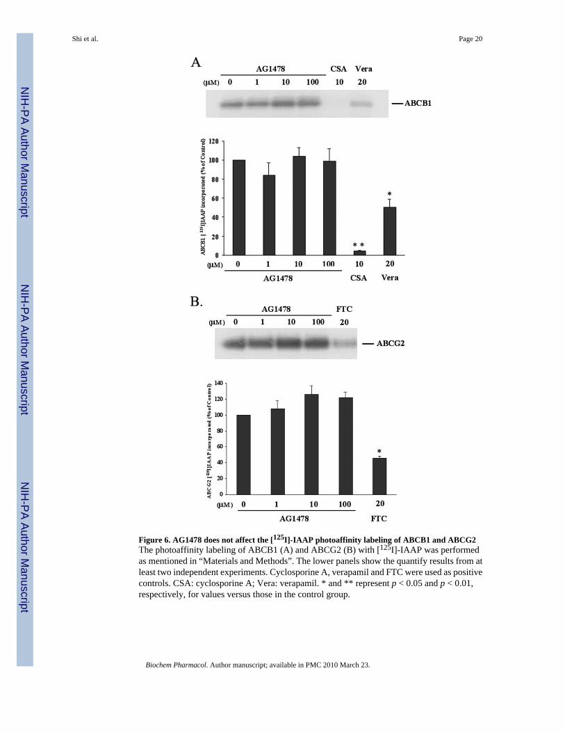
Figure 6. AG1478 does not affect the [125I]-IAAP photoaffinity labeling of ABCB1 and ABCG2The photoaffinity labeling of ABCB1 (A) and ABCG2 (B) with [125I]-IAAP was performedas mentioned in “Materials and Methods”. The lower panels show the quantify results from atleast two independent experiments. Cyclosporine A, verapamil and FTC were used as positivecontrols. CSA: cyclosporine A; Vera: verapamil. * and ** represent p < 0.05 and p < 0.01,respectively, for values versus those in the control group.
Shi et al. Page 20
Biochem Pharmacol. Author manuscript; available in PMC 2010 March 23.
NIH
-PA Author Manuscript
NIH
-PA Author Manuscript
NIH
-PA Author Manuscript

NIH
-PA Author Manuscript
NIH
-PA Author Manuscript
NIH
-PA Author Manuscript
Shi et al. Page 21
Table 1Effect of AG1478, verapamil and valspodar on ABCB1-mediated resistance to colchicine,vinblastine, paclitaxel and cisplatin.a
To examine the effects of AG1478, verapamil and valspodar on ABCB1-mediated resistance to colchicine,vinblastine, paclitaxel and cisplatin, cells were pre-incubated with or without AG1478, verapamil and valspodarfor 1 h and then incubated with various concentrations of colchicine, vinblastine, paclitaxel and cisplatin. Cellsurvival was determined by the MTT assay as described in “Materials and Methods”. Data are means ± SD of atleast three independent experiments performed in triplicate.
CompoundsIC50 ± SD (μM)
KB-3-1 KB-C2
Colchicine 0.0069±0.0010(1.0)a 6.9543±0.2437(947.8)
+ AG1478 10 μM 0.0069±0.0004(1.0) 3.6490±0.2268(505.8)
+ Verapamil 10 μM 0.0060±0.0003(0.9) 0.1077±0.0134(15.6)
0.0059±0.0007(0.9) 0.0097±0.0056(1.41)
+ Valspodar 5 μM
Vinblastine 0.0476±0.0043(1.0)a 0.3057±0.0152(6.4)
+ AG1478 10 μM 0.0428±0.0008(0.9) 0.1644±0.0245(3.5)
+ Verapamil 10 μM 0.0408±0.0062(0.9) 0.0419±0.0078(0.9)
0.0389±0.0045(0.8) 0.0399±0.0098(0.8)
+ Valspodar 5 μM
Paclitaxel 0.0084±0.0023(1.0)a 5.5643±1.1098(662.3)
+ AG1478 10 μM 0.0079±0.0004(1.0) 1.4461±0.2839(172.1)
+ Verapamil 10 μM 0.0072±0.0018(0.9) 0.0173±0.0077(2.0)
0.0068±0.0021(0.8) 0.0093±0.0064(1.1)
+ Valspodar 5 μM
Cisplatin 1.8867±0.0446(1.0)a 1.8235±0.0782(1.0)
+ AG1478 10 μM 1.8365±0.0578(1.0) 1.8293±0.0811(1.0)
+ Verapamil 10 μM 1.8116±0.0824(1.0) 1.7244±0.0387(0.9)
1.8233±0.0718(1.0) 1.8401±0.0702(1.0)
+ Valspodar 5 μM
aFold-resistance was determined by dividing the IC50 values of KB-C2 cells by the IC50 values for KB-3-1 cells in the absence or presence of
AG1478, verapamil or valspodar.
Biochem Pharmacol. Author manuscript; available in PMC 2010 March 23.

NIH
-PA Author Manuscript
NIH
-PA Author Manuscript
NIH
-PA Author Manuscript
Shi et al. Page 22
Table 2Effect of AG1478 and FTC on ABCG2-mediated resistance to flavopiridol, mitoxantrone,SN-38 and cisplatin.a
To examine the effects of AG1478 and FTC on ABCG2-mediated resistance to flavopiridol, mitoxantrone, SN-38and cisplatin, cells were pre-incubated with or without AG1478 and FTC for 1 h and then incubated with variousconcentrations of flavopiridol, mitoxantrone, SN-38 and cisplatin. Cell survival was determined by the MTTassay as described in “Materials and Methods”. Data are means ± SD of at least three independent experimentsperformed in triplicate.
CompoundsIC50 ± SD (μM)
HEK293/pcDNA3.1 ABCG2-482-G2 ABCG2-482-R5 ABCG2-482-T7
Flavopiridol 0.1668±0.0221(1.0)a 0.4132±0.0465(2.4) 0.3127±0.0265(1.9) 0.6968±0.0571(4.2)
+ AG14782.5 μM
0.1637±0.0134(1.0) 0.2339±0.0333(1.4) 0.1855±0.0293(1.1) 0.2797±0.0258(1.7)
+ AG147810 μM
0.1662±0.0161(1.0) 0.1959±0.0182(1.2) 0.1696±0.0046(1.0) 0.2415±0.0547(1.5)
+ FTC 2.5μM
0.1312±0.0143(0.8) 0.1164±0.0127(0.7) 0.1547±0.0348(0.9) 0.1718±0.0186(1.0)
Mitoxantrone 0.0323±0.0021(1.0)a 1.2467±0.0853(38.6) 0.4123±0.0263(12.8) 0.7854±0.1024(24.3)
+ AG14782.5 μM
0.0328±0.0038(1.0) 0.2559±0.0061(7.9) 0.0749±0.0136(2.4) 0.2745±0.1119(8.5)
+ AG147810 μM
0.0386±0.0006(1.1) 0.1213±0.0020(3.8) 0.0578±0.0149(1.8) 0.1317±0.0011(4.1)
+ FTC 2.5μM
0.0302±0.0034(0.9) 0.0611±0.0016(1.9) 0.0446±0.0087(1.4) 0.0867±0.0102(2.7)
SN-38 0.0052±0.0011(1.0)a 0.2234±0.0863(43.0) 0.1446±0.0562(22.0) 0.1004±0.0082(19.3)
+ AG14782.5 μM
0.0050±0.0004(1.0) 0.0336±0.0013(6.5) 0.0286±0.0035(5.5) 0.0217±0.0003(4.2)
+ AG147810 μM
0.0048±0.0002(0.9) 0.0137±0.0006(2.6) 0.0150±0.0032(2.9) 0.0091±0.0005(1.7)
+ FTC 2.5μM
0.0044±0.0014(0.8) 0.0078±0.0008(1.5) 0.0076±0.0012(1.5) 0.0074±0.0014(1.4)
Cisplatin 1.5466±0.2489(1.0)a 1.4457±0.1226(0.9) 1.4873±0.3117(0.9) 1.6118±0.1882(1.0)
+ AG14782.5 μM
1.6820±0.0693(1.1) 1.2079±0.2652(0.8) 1.1005±0.2058(0.7) 1.6790±0.2093(1.1)
+ AG147810 μM
1.3840±0.2390(0.8) 1.1531±0.3427(0.7) 1.1485±0.0771(0.7) 1.5520±0.0665(1.0)
+ FTC 2.5μM
1.3347±0.2586(0.8) 1.4632±0.4487(0.9) 1.2256±0.3251(0.8) 1.5671±0.2641(1.0)
aFold-resistance was determined by dividing the IC50 values of the ABCG2-trandfected cells by the IC50 values of HEK293/pcDNA3.1 cells in the
absence or presence of AG1478 or FTC.
Biochem Pharmacol. Author manuscript; available in PMC 2010 March 23.
Copyright © 2022 FDOKUMEN