Intensive meditation training, immune cell telomerase activity, and psychological mediators
Upload
independentCategory
view
1download
0
Magnetic Nanoparticles as Mediators of Ligand-FreeActivation of EGFR SignalingAtul A. Bharde1.¤a, Raghavendra Palankar1.¤b, Cornelia Fritsch1¤c, Arjen Klaver2, Johannes S. Kanger2,
Thomas M. Jovin1, Donna J. Arndt-Jovin1*
1 Laboratory of Cellular Dynamics, Max Planck Institute for Biophysical Chemistry, Gottingen, Germany, 2Nanobiophysics, Faculty of Science and Technology, University of
Twente, Enschede, The Netherlands
Abstract
Background: Magnetic nanoparticles (NPs) are of particular interest in biomedical research, and have been exploited formolecular separation, gene/drug delivery, magnetic resonance imaging, and hyperthermic cancer therapy. In the case ofcultured cells, magnetic manipulation of NPs provides the means for studying processes induced by mechanotransductionor by local clustering of targeted macromolecules, e.g. cell surface receptors. The latter are normally activated by binding oftheir natural ligands mediating key signaling pathways such as those associated with the epidermal growth factor (EGFR).However, it has been reported that EGFR may be dimerized and activated even in the absence of ligands. The present studyassessed whether receptor clustering induced by physical means alone suffices for activating EGFR in quiescent cells.
Methodology/Principal Findings: The EGFR on A431 cells was specifically targeted by superparamagnetic iron oxide NPs(SPIONs) carrying either a ligand-blocking monoclonal anti-EGFR antibody or a streptavidin molecule for targeting achimeric EGFR incorporating a biotinylated amino-terminal acyl carrier peptide moiety. Application of a magnetic field led toSPION magnetization and clustering, resulting in activation of the EGFR, a process manifested by auto andtransphosphorylation and downstream signaling. The magnetically-induced early signaling events were similar to thoseinherent to the ligand dependent EGFR pathways. Magnetization studies indicated that the NPs exerted magnetic dipolarforces in the sub-piconewton range with clustering dependent on Brownian motion of the receptor-SPION complex andmagnetic field strength.
Conclusions/Significance: We demonstrate that EGFR on the cell surface that have their ligand binding-pocket blocked byan antibody are still capable of transphosphorylation and initiation of signaling cascades if they are clustered by SPIONseither attached locally or targeted to another site of the receptor ectodomain. The results suggest that activation of growthfactor receptors may be triggered by ligand-independent molecular crowding resulting from overexpression and/orsequestration in membrane microdomains.
Citation: Bharde AA, Palankar R, Fritsch C, Klaver A, Kanger JS, et al. (2013) Magnetic Nanoparticles as Mediators of Ligand-Free Activation of EGFR Signaling. PLoSONE 8(7): e68879. doi:10.1371/journal.pone.0068879
Editor: Bing Xu, Brandeis University, United States of America
Received May 18, 2013; Accepted June 3, 2013; Published July 23, 2013
Copyright: � 2013 Bharde et al. This is an open-access article distributed under the terms of the Creative Commons Attribution License, which permitsunrestricted use, distribution, and reproduction in any medium, provided the original author and source are credited.
Funding: This work was supported by EU FP6 FLUOROMAG #037465 Project; German Research Council (DFG) Cluster of Excellence Microscopy at theNanometer Range and the Research Center Molecular Physiology of the Brain; and Hessen Agentur Projekt 193/09-23. The funders had no role in study design,data collection and analysis, decision to publish, or preparation of the manuscript.
Competing Interests: The authors have declared that no competing interests exist.
* E-mail: [email protected]
¤a Current address: Nanoscale Science and Engineering Center, The Ohio State University, Columbus, Ohio, United States of America¤b Current address: Nanostructure Group, Center for Innovation Competence, Ernst-Moritz-Arndt-University Greifswald, Greifswald, Germany¤c Current address: Department of Biology and Zoology, University of Fribourg, Fribourg, Switzerland
. These authors contributed equally to this work.
Introduction
Nanoparticles differing in composition, shape, size, and intrinsic
optical, electronic and magnetic properties have been used in
diverse biological applications such as imaging, sensing and
separation [1,2,3,4]. In particular, magnetic NPs [5] have been
exploited for molecular separation, gene/drug delivery, and
magnetic resonance imaging [6,7]. As sensors and actuators they
have been used to sense femtomolar concentrations of proteins,
mRNA or viruses [8], for focused heat-induced manipulation of
ion channels [9], or for mechanotransduction of ion channels in
neurons [10]. Some cell surface receptors are activated by
clustering, a prominent example being the FceR1 receptor on
basophils and mast cells that is aggregated upon recognition of
multivalent allergens by bound IgE [11]. Mannix et al. demon-
strated that monovalent antigen attached to SPIONs could induce
mast cell activation, manifested by Ca2+ waves arising after
clustering the FceR1 by a magnetic field [12]. Apoptosis of tumor
cells has been achieved by magnetic aggregation of SPIONs
coupled to a monoclonal antibody against DR4 receptors [13],
although it was necessary to apply the magnetic field for 2 hr in
order to observe caspase 3 activity. The same group achieved a
similar result in live zebrafish embryos by targeting the ovarian
TNF receptor with microinjected SPIONs and applying a field for
PLOS ONE | www.plosone.org 1 July 2013 | Volume 8 | Issue 7 | e68879

24 or 48 h. A number of recent studies have utilized large
magnetic NPs introduced by microinjection to redistribute
materials inside cells. Examples are cytoskeletal reorganization
induced by Raf1 NPs [14] and microtubule assembly in Xenopus
oocyte extracts by RANQ-GTP coupled NPs [15].
The epidermal growth factor receptor (EGFR, ErbB1, HER1),
a prototypic transmembrane tyrosine kinase receptor, is a member
of the ErbB (HER) family. Ligand binding results in dimerization
and subsequent trans-phosphorylation of several tyrosine residues
in the intracellular C-terminal tail of the receptor [16,17,18]. The
adaptor proteins Shc, Grb2 and Cbl recognize these phosphotyr-
osines, thereby propagating downstream signaling, effector func-
tions and receptor internalization [19,20]. These signaling
cascades orchestrate a wide range of cellular processes such as
cell differentiation, motility, and cell division [21,22].
It has not been firmly established whether receptor dimers or
oligomers can be activated and initiate downstream signaling in
the absence of physiological ligands. Yu et al. [23] reported that
EGFR dimerized and was activated merely by association with
a2b1 integrins in serum deprived cells while Takahashi et al. [24]
studied the effect of extracellular matrix glycans on ligand free
activation of ErbB3 mutants. However, another investigation of
integrin association by Alexi et al. [25] failed to demonstrate
EGFR activation without added ligand, and the authors concluded
that autocrine activation of the receptor was likely to have
occurred in some of the other studies. Monoclonal antibodies that
block ligand binding inhibit EGFR signaling and some cause down
regulation of the receptor [26,27,28], suggesting that ligand
binding is indeed required for EGFR activation. Some of these
antibodies have been humanized and used to treat cancers
expressing high levels of EGFR [29,30,31]. Certain mutations of
EGFR cause a constitutive ligand independent activation of the
receptor and such forms often arise under conditions favoring
cellular transformation [32,33].
We initiated the present study to determine whether clustering
of EGFR on living cells could bypass the ligand requirement for
activating the receptor. We exploited the biocompatibility, tunable
surface properties and ease of preparation of superparamagnetic
iron oxide NPs (SPIONs), employing the latter as magnetic
actuators (‘‘switches’’). Non-ligand mediated clustering activated
the EGFR and led to internalization of both the receptor and the
SPIONs.
Results
The properties of magnetic nanostructures depend on size.
Below a certain critical dimension they exhibit the phenomenon of
superparamagnetism, which is characterized by a high magnetic
susceptibility and the absence of a residual field [3,34,35]. SPIONs
were synthesized by alkaline co-precipitation using citrate as a
stabilizing agent [34,35]. Transmission electron microscopy
(TEM) images indicated the presence of well-defined, spherical
shaped particles 12–15 nm in diameter (Figure S1a–e). A more
homogenous SPION population was obtained by magnetic
fractionation followed by functionalization with streptavidin
(strv-SPION) (see Materials and Methods).
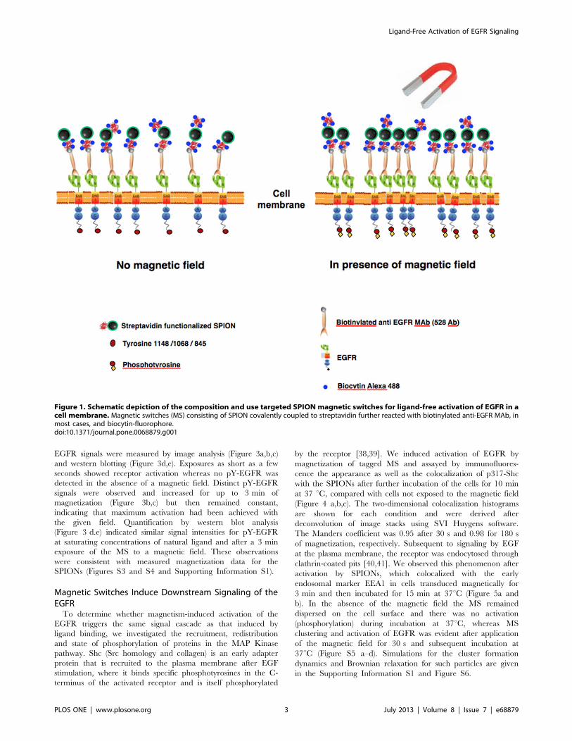
Magnetic Switches are Created by Covalently AttachingCell-targeting Molecules to SPION
Figure 1 shows pictorially the composition of ‘‘magnetic
switches’’ (MS) and the experimental approach for specific
targeting to EGFR. MS were created by coupling strv-SPION
to a biotinylated anti-EGFR (ectodomain) monoclonal antibody
(528 MAb). This antibody blocks binding of the normal ligands
for EGFR, and thus inhibits ligand-induced activation as well as
cell proliferation and orthotopic tumor growth [36,37]. We
adjusted the reagent concentrations to achieve #1 streptavidin
and ,1 MAb per nanoparticle. We also demonstrated that strv-
SPIONs by themselves can act as MS if targeted to cells
expressing a chimeric EGFR with a biotinylated acyl carrier
protein (ACP) tag on the amino terminus of the receptor (see
Materials and Methods). In order to visualize the MS on the
cell surface by fluorescence, unoccupied biotin binding sites on
strv-SPION were loaded with biocytin-Alexa 488 or biocytin-
Alexa 546.
SPIONS Aggregate and Cause Activation of theMembrane-bound EGFR Only in the Presence of aMagnetic Field
Application of the targeted MS to A431 cells and washing to
remove unbound particles resulted in MS distributed over the
entire cell surface (Figure S2a–d). Untargeted SPIONs (lacking
anti-EGFR) did not bind to the cells (Figure S2e). Exposure to
an external magnetic field led to aggregation of MS into small
clusters (Figure 2a) and activation of EGFR detected by an
EGFR-specific anti-phosphotyrosine (pY) antibody (Fig. 2a9). In
contrast, no EGFR-specific pY signal was observed in cells that
were not exposed to a magnetic field after binding of MS
(Figure 2b,b9). Figure 2c shows a 2-dimensional colocalization
histogram of EGFR phosphorylation and the MS signals from
the images in Figure 2a,a9, revealing a strong, positive
correlation (colocalization parameters: Manders coefficient
0.78, overlapping coefficient 0.82, and correlation coefficient
0.80).
In order to define the dipolar interactions and the anticipated
extent of magnetic field induced clustering of MS bound to EGFR
on the cell surface, we carried out a detailed theoretical analysis of
the experimental system. Finite element analysis calculations
indicated that the magnetic field experienced by MS on the cell
surface would be close to saturating strength (Figure S3). At the
separation distance of 10 nm the calculated force between
neighboring MS on cell surface was , 0.25 pN, decaying
exponentially with increasing separation distance. We conclude
that in our experimental system the strengths of the magnetic
dipole moment and of the external field sufficed for inducing the
MS to form stable clusters on the cell surface (Supporting
Information S1 and Figure S4).
Scanning electron microscopy (SEM) resolved the local
clustering of MS targeted to EGFR on A431 cell membranes. In
the absence of a magnetic field, MS were distributed densely on
the cell surface but were individually distinct with little or no
clustering (Figure 2d,d9). In contrast, cells treated with MS in the
presence of a magnetic field showed a dense population of particles
arranged in local clusters of , 100–200 nm (Figure 2e,e9).
MS did not exhibit magnetic hysteresis at room temperature,
indicating the existence of superparamagnetic behavior (Figure
S3). Based on the magnetic properties, we calculated the magnetic
diameter of MS to be , 10 nm (see Supporting Information S1), a
result which was corroborated by TEM and the estimation of the
hydrodynamic diameter as ,15 nm by dynamic light scattering
(Fig. S1).
EGFR Activation is Dependent on the Time of MagneticExposure
To determine the effect of magnetization as a function of
time, MS targeted to EGFR in A431 cells were exposed to a
magnetic field for various time periods and the resulting pY-
Ligand-Free Activation of EGFR Signaling
PLOS ONE | www.plosone.org 2 July 2013 | Volume 8 | Issue 7 | e68879
EGFR signals were measured by image analysis (Figure 3a,b,c)
and western blotting (Figure 3d,e). Exposures as short as a few
seconds showed receptor activation whereas no pY-EGFR was
detected in the absence of a magnetic field. Distinct pY-EGFR
signals were observed and increased for up to 3 min of
magnetization (Figure 3b,c) but then remained constant,
indicating that maximum activation had been achieved with
the given field. Quantification by western blot analysis
(Figure 3 d.e) indicated similar signal intensities for pY-EGFR
at saturating concentrations of natural ligand and after a 3 min
exposure of the MS to a magnetic field. These observations
were consistent with measured magnetization data for the
SPIONs (Figures S3 and S4 and Supporting Information S1).
Magnetic Switches Induce Downstream Signaling of theEGFR
To determine whether magnetism-induced activation of the
EGFR triggers the same signal cascade as that induced by
ligand binding, we investigated the recruitment, redistribution
and state of phosphorylation of proteins in the MAP Kinase
pathway. Shc (Src homology and collagen) is an early adapter
protein that is recruited to the plasma membrane after EGF
stimulation, where it binds specific phosphotyrosines in the C-
terminus of the activated receptor and is itself phosphorylated
by the receptor [38,39]. We induced activation of EGFR by
magnetization of tagged MS and assayed by immunofluores-
cence the appearance as well as the colocalization of p317-Shc
with the SPIONs after further incubation of the cells for 10 min
at 37 uC, compared with cells not exposed to the magnetic field
(Figure 4 a,b,c). The two-dimensional colocalization histograms
are shown for each condition and were derived after
deconvolution of image stacks using SVI Huygens software.
The Manders coefficient was 0.95 after 30 s and 0.98 for 180 s
of magnetization, respectively. Subsequent to signaling by EGF
at the plasma membrane, the receptor was endocytosed through
clathrin-coated pits [40,41]. We observed this phenomenon after
activation by SPIONs, which colocalized with the early
endosomal marker EEA1 in cells transduced magnetically for
3 min and then incubated for 15 min at 37uC (Figure 5a and
b). In the absence of the magnetic field the MS remained
dispersed on the cell surface and there was no activation
(phosphorylation) during incubation at 37uC, whereas MS
clustering and activation of EGFR was evident after application
of the magnetic field for 30 s and subsequent incubation at
37uC (Figure S5 a–d). Simulations for the cluster formation
dynamics and Brownian relaxation for such particles are given
in the Supporting Information S1 and Figure S6.
Figure 1. Schematic depiction of the composition and use targeted SPION magnetic switches for ligand-free activation of EGFR in acell membrane. Magnetic switches (MS) consisting of SPION covalently coupled to streptavidin further reacted with biotinylated anti-EGFR MAb, inmost cases, and biocytin-fluorophore.doi:10.1371/journal.pone.0068879.g001
Ligand-Free Activation of EGFR Signaling
PLOS ONE | www.plosone.org 3 July 2013 | Volume 8 | Issue 7 | e68879

High Order Clustering of the EGFR is Required forActivation by MS
Bivalent crosslinking of biotinylated 528 MAb bound to EGFR
mediated by streptavidin-Atto565 (Figure S7) did not result in a
detectable pY-EGFR signal, indicating that protein bridging
between the MAb bound to the receptor was incapable of
associating the EGFR with the kinase domainsoriented in an
active conformation. We conclude that higher order clustering of
the EGFR and/or a more intimate contact of the receptors are
required and can be achieved only by application of the magnetic
field.
Magnetic Activation of EGFR by Targeted SPIONs doesnot Depend on High Expression Levels of the Receptoron the Cell Membrane or on Targeting by a LargeMonoclonal Antibody
To determine whether MS were effective only on cells with very
high densities of receptor, we performed the same experiments on
Hela cells expressing 5?104 EGFR, i.e. much less than the 2?106
receptors characteristic of A431 cells. Figure S8 shows that EGFR
on Hela cells was activated by magnetization of bound MS,
demonstrating that activation by local clustering is a phenomenon
that can be generalized to other cell types with low receptor
densities.
A similar clustering and activation of the EGFR was induced by
strv-SPIONs targeted to the chimeric EGFR with the biotinylated
ACP tag (Figure 6). We conclude that higher-order clustering of
the receptor is sufficient for activation, i.e. does not require a
unique orientation of the extracellular domain or binding of a
large antibody molecule.
Discussion
Localized activation of EGFR induced by EGF-coated magnetic
microspheres (,1 mm) has been shown to occur exclusively at the
attachment loci on the cell membrane [42,43,44]. In the current
study, we explored the applicability of much smaller super-
paramagnetic NPs lacking a natural ligand for the receptor and
targeted to the EGFR with a monoclonal antibody that actually
Figure 2. Magnetic field induced activation of EGFR. Confocal image analysis-left column after 60 sec magnetization or right column withoutmagnetiztion. (a,b) MS- Alexa488-biocytin, green; (a9, b9) EGFR activation (pY-EGFR1068) red; (a99,b99) overlay of red, green and DAPI DNA staining,blue, images; and DIC image (bottom). Scale bar is 10 mm. (c) Two-dimensional colocalization histogram for the same confocal sections a and a9 afterbackground subtraction. SEM images of A431 cells reacted with MS after (d,d9) and without (e,e9) application of magnetic field. Scale bars, 1 mm for (d& e) and 500 nm for (d9 & e9), respectively.doi:10.1371/journal.pone.0068879.g002
Ligand-Free Activation of EGFR Signaling
PLOS ONE | www.plosone.org 4 July 2013 | Volume 8 | Issue 7 | e68879

blocks ligand binding. The aim was to assess whether local
clustering of the receptor by itself leads to activation and
downstream signaling.
We demonstrated that simple bivalent coupling by streptavidin
of the biotinylated antibody or targeting SPIONs to the cell
membrane did not result in EGFR activation. However, exposure
of the targeted SPIONs to a strong magnetic field resulted in a
time-dependent and ligand-independent EGFR activation. MS
consist of individual magnetic domains; a strong magnetic dipolar
interaction is induced by the external magnetic field, which
propagates and causes clustering of closely placed particles [45].
Exposure of cells to a magnetic field in the presence of non-
targeted SPIONs failed to activate the EGFR.
Cells respond to mechanical stimuli through integrin signaling
involving focal adhesion kinases (FAK) [46]. However, the forces
required for conventional cellular mechanotransduction through
integrins are on the order of pN to nN [46,47,48]. Surprisingly,
the magnitude of forces measured in this study for activation of
EGFR was estimated to be in the sub-pN range, (Supporting
Information S1), i.e. considerably less than the force required for
integrin mediated mechanotransduction.
Recently Rauch et al. [49] assayed the ability of dextran-coated
SPIONs with various surface charges to activate ERK, AKT and
EGFR in breast and colon cancer cells either expressing a
metastatic RAS mutation or without this mutation. They found
strong activation of the RAS pathway in the mutated cells in the
absence of a magnetic field. In the absence of a magnetic field the
epidermoid carcinoma cell line A431 did not show uptake or
activation of our targeted SPIONs after incubations of 15 min at
37uC (Figure S5.) However, a slow uptake of the SPIONs,
presumably by macropinocytosis, occurred upon prolonged
incubation. We did not investigate cells with RAS mutations and
thus cannot comment about whether non-dextran coated SPIONs
might also effect a similar response as that seen by Rauch et al in
Figure 3. Effect of magnetization time on the level of EGFR phosphorylation. (a, a9, a99) The time axis shown vertically. Confocal images ofMS 488Alexa biocytin signal (green, left panels) and of anti-pY-EGFR 1068 and GARIG-Cy5 (rainbow intensity scale, middle panels) on A431 cells as afunction of the applied magnetic field for time intervals of 30, 60 and 180 s. Overlay images are depicted with green/red LUTs, Alexa488-biocytin/GARIG-Cy5 respectively (right panels). Scale bar, 10 mm. (b) Fluorescence intensity ratio of pY-EGFR to MS signals as a function of magnetization. (c)Mean pixel intensity of the pY-EGFR signal from 5 images for each time point as a function of MS magnetization time. (e) Western blot analysis ofA431 cell extracts for pY-EGFR 1148. Lane 1, sample obtained from A431 cells incubated with MS in the absence of a magnetic field. Lanes 2–4, pY-EGFR signals for 30, 60 or 180 s of MS magnetization. Lane 5 and 6, pY-EGFR signals after treatment with 30 nM (saturating) or 100 pM EGF. Lane 7,extract of untreated cells. (e) Signal intensities of the positive pY-EGFR lanes in (d) relative to the lane from 30 nM EGF treated cells.doi:10.1371/journal.pone.0068879.g003
Ligand-Free Activation of EGFR Signaling
PLOS ONE | www.plosone.org 5 July 2013 | Volume 8 | Issue 7 | e68879

such cells. The surface charge and structure of NPs are important
determinants for their interaction with cells. This issue constitutes
an important area of investigation.
Another publication featured gold-coated SPIONs coated with
a saturating concentration of C225 monoclonal anti-EGFR
SPIONs to study the downregulation of the EGFR after prolonged
incubation (72 hours) in the absence of an external field whereby
the cells autophagosized the particles [50]. C225 was originally
characterized for its ability to block ligand binding to the EGFR,
to arrest cell proliferation and to promote tumor killing [28,37].
Coupling of the antibody to the NPs resulted in a two-fold increase
in cell killing.
The absolute magnetization of MS depends on the magnetiza-
tion distance and the strength of the applied magnetic field. We
achieved full activation of the EGFR on A431 cells with an
application of the magnetic field for 3 min at the employed field
strength. The same downstream signaling cascade was promoted
by magnetic activation with the SPIONs both using antibody
targeting or direct binding of streptavidin SPIONs to the
biotinylated chimeric EGFR.
The requirement for receptor-receptor interaction in the
activation of signal transduction is a signature event of many
receptor tyrosine kinases [52] and T cell receptors [53]. Cells with
both high and low expression levels of EGFR (A431cells with
2?106 compared to HeLa cells with 5?104) were activated by
SPIONs, leading to the conclusion that high local density (in the
absence of the natural ligand) suffices for activation of the EGFR
and its downstream signaling cascades. Thus, the data reinforce
the hypothesis that upregulation of EGFR, interactions with other
cell membrane components or sequestration in particular mem-
brane microdomains leading to functional receptor clusters can
potentiate cellular growth and migration of cells.
ConclusionsWe have demonstrated that physiologically important signaling
receptors can be mechanically activated using magnetic nano-
technology. SPION technology constitutes a valuable tool for
manipulating cellular processes in order to study and/or modulate
functional states. In some human cancers receptor tyrosine kinases
such as EGFR are in a constitutively active form due to mutations
that abrogate the quiescent, non-aggregated state of the receptor.
Our results provide direct evidence for a possible additional
mechanism, i.e clustering per se, for the ligand free activation of
EGFR in cancer cells expressing high levels of the wild-type
Figure 4. Shc activation as a result of magnetic activation of EGFR. Confocal immunofluorescence images of cells incubated for 15 min at37uC after 0 sec (a) or after 30 sec (b) or 180 sec (c) magnetic field activation. Image columns left to right: MS Alexa-488 biocytin (green); indirectimmunofluorescence of MAb against pY-317 Shc protein and GARIG-CY5 (red); overlay of the first 2 columns; two-dimensional colocalizationhistograms of MS and pY317-Shc fluorescence signals after deconvolution of 50 optical sections using SVI Huygens software.doi:10.1371/journal.pone.0068879.g004
Ligand-Free Activation of EGFR Signaling
PLOS ONE | www.plosone.org 6 July 2013 | Volume 8 | Issue 7 | e68879

receptor. Specific targeting of magnetized superparamagnetic NPs
to the EGFR actuates the normal signaling cascade via the
induction of a mechanical force of very low magnitude (sub-pN). It
follows that magnetic switches used in conjunction with an
external magnetic field may be used to control cellular functions
in vivo. In particular, it may be possible to specify the location and
strength of a signaling mechanism with magnetic control devices
or to specifically deliver SPIONs into cells by such techniques.
EGFR and other ErbB family members are upregulated in
approximately 50% of all human tumors [51]. A prominent
example is glioblastoma. Kantelhardt et al. [54] demonstrated that
glioma cells expressing highly upregulated EGFR, in contrast to
normal brain tissue lacking this receptor, were selectively labeled
by quantum dots specifically targeted to the EGFR. SPIONs can
be coupled to chemotherapeutic drugs, and animal studies show
that uptake into tumors can be promoted by focused magnetic
fields [55]. Thus, MS of the type described in this report may
constitute useful vehicles for specifically targeting chemotherapeu-
tic drugs to residual tumor cells after surgical resection.
Materials and Methods
Synthesis of Superparamagnetic Iron OxideNanoparticles and Surface Modification with Streptavidin
Superparamagnetic iron oxide NPs (SPIONs) were synthesized
by alkaline coprecipitation of magnetite [56] from a mixture of
Fe3+ and Fe2+ salts (FeCl3 and FeCl2 in 2:1 molar ratio) followed
by addition of concentrated NH4OH solution (25% w/v). The size
of fractionated SPIONs was monitored by the transmission
electron microscope (Jeol) operated at 120 kV and by dynamic
light scattering (Malvern instruments) (Figure S1 a–d and e).
SPIONS of 10–15 nm diameter were covalently coupled to
streptavidin such that most particles had one or fewer streptavidin
molecules (see Supporting Information S1 for details). Purified
strv-SPIONs were resuspended in 20 mM Na-PO4, pH 7.6, buffer
and further used for synthesis of the magnetic switches (MS).
Preparation of Magnetic SwitchesMagnetic switches (MS) capable of targeting EGFR were
prepared (except when cells expressed ACP-EGFR, see below) by
incubating a 5-fold molar excess of strv-SPIONs, in terms of biotin
binding capacity, with biotinylated anti-EGFR monoclonal
antibody 528 (Dianova) in 20 mM Na-PO4 buffer (pH 7.4) at
25uC for 15 min. Finally, the MS were incubated with a two-fold
molar excess of biocytin-Alexa fluorophore (Invitrogen) (in terms
of biotin binding capacity of strv-SPION) for 15 min and washed
by magnetic separation several times to remove unbound
fluorophore.
Covalent Labeling of Chimeric ACP-EGFR with Biotin-CoAon Expressing Cells
Biotin-CoA was synthesized and purified by HPLC C18
chromatography as described in the literature [57]. We construct-
ed a chimeric ErbB1 with an acyl carrier protein tag [58] between
the signal peptide and the ErbB1 mature protein sequence. The
serine that accepts a CoA derivative is located at position 36 in the
sequence of the mature expressed protein. The 78 amino acid
DNA sequence for the ACP tag was extracted from the AGA-
plasmid (kind gift of N. Johnsson) by PCR and inserted between
the signal sequence and the amino terminus by creating a new
Nhe1 restriction enzyme cleavage site using site-directed muta-
genesis of wild-type ErbB1 in pcDNA3 or a modified pcDNA3.1-
zeo2 plasmid. Stably-transfected Hela cell lines were selected by
antibiotic resistance and single cell cloning. The chimeric EGFR
behaves as wildtype EGFR as reported elsewhere [59].
Cells expressing the chimeric ACP-EGFR were labeled
covalently with 1 mM CoA-biotin by enzymatic reaction at room
temperature for 20 min with E. coli PPTase (phosphopantetheinyl
transferase) [57,60] which links biotin through a phosphodiester
bond to the serine hydroxyl in the ACP tag. For experiments
described in the text strv-SPION (10 mg/ml) NPs were added to
the cells at 15uC for 15 min and excess SPIONs removed by
washing before exposure of the cells to a magnetic field (Figure 6).
Figure 5. Localization of MS in endocytic vesicles after magnetic field pulse and 37uC incubation. (a) Confocal microscopy imagesshowing the endocytosis of MS after 3 min magnetization and subsequent incubation for 20 min at 37uC in the absence of magnetic field. Green, MSsignal; red, immunofluorescence staining of early endosomes by Mab EEA1 and GAMIG-CY3. Scale bar is 10 mm. (b) Two-dimensional colocalizationhistogram of MS and EEA1 from a z stack (0.7 mm sections) of 20 images.doi:10.1371/journal.pone.0068879.g005
Ligand-Free Activation of EGFR Signaling
PLOS ONE | www.plosone.org 7 July 2013 | Volume 8 | Issue 7 | e68879

Cell CultureThe following cell lines were used: A431, Human epidermoid
carcinoma (ATCC CRL 1555) Clone E3 from E. Helmreich,
Wurzburg; Hela SS6; Hela stably expressing actin-GFP; and Hela
stably expressing EGFR with an amino terminal ACP tag that can
be covalently labeled by phosphopantetheinyl transferase with
CoA–biotin. Cells were seeded on 24 mm square coverslips in
complete DMEM and grown to 70% confluency. Cells were
starved overnight and washed 3 times at 1 hr intervals prior to use
in order to achieve a quiescent state and thus minimize the serum-
or autocrine-induced activation of EGFR.
Binding to Cells and Magnetization of MSCells were incubated in Tyrode’s buffer containing 0.1% BSA
and 20 mM glucose at 15uC for 15 min with MS (5 mg/ml <6 nM Mab) and excess, unbound MS were removed by 3 washes
with the same buffer. MS were magnetized by placing 4
permanent Neodymium magnets in a quadrapole configuration
on the 4 sides of the glass coverslip at room temperture. After the
allotted magnetization time coverslips were removed from the
magnetic field and immediately fixed with 3.7% paraformalde-
hyde for 30 min on ice (unless otherwise indicated in the text) and
then stained for activated EGFR or other cellular proteins as
indicated in the figure legends.
Detection of Phosphorylation of EGFR (pY-EGFR) byImmunofluorescence
Cells were fixed in 3.7% PFA for 30 min at 4C, permeabilized
and blocked with 0.1% Tween 20, 1% BSA in PBS at room
temperature for 1 hr followed by incubation with rabbit anti-
phosphotyrosine EGFR (pY-EGFR 1068 or pY-EGFR 1148 (Cell
Signaling Technology) at 100–300 ng/ml. Primary antibodies
were detected using secondary F(ab)2 fragments of goat anti-rabbit
IgG (GARIG) labeled with Cy3 or Cy5 (Jackson Labs) at 1 mg/ml.
Coverslips were mounted on microscope slides in Mowiol
(Polysciences).
Western Blot AnalysisFor western blot analysis, A431 cells were grown in 24 well
plates and treated with MS in the presence or absence of a
magnetic field. Total cell lysates were obtained with Phosphosafe
Reagent (Novagen) and equal amounts of protein were separated
by a polyacrylamide gel electrophoresis, blotted to PVDF
membranes and probed with rabbit primary antibody (pY-EGFR
Figure 6. Magnetic activation is not dependent upon the targeting moiety. Stably transfected HeLa cells expressing chimeric ACP-EGFR,enzymatically modified by biotin CoA and carrying bound Strv-SPIONs. (a) 30 seconds, (b) 180 seconds of magnetization, (c) no magnetization. Greenchannel, Strv-SPION-biocytin-Alexa546; red channel, phosphorylated-EGFR (pY-EGFR 1068 and GARIG-CY5), overlay of green and red channels. Scalebar, 10 mm.doi:10.1371/journal.pone.0068879.g006
Ligand-Free Activation of EGFR Signaling
PLOS ONE | www.plosone.org 8 July 2013 | Volume 8 | Issue 7 | e68879

1148, Cell Signaling Technology) and HRP-conjugated goat anti-
rabbit IgG. Signal was developed by chemiluminescent (Super-
signal West Pico, Pierce) and imaged with Kodak Biomax Xray
film.
Detection of MS in Early EndosomesMagnetized cells were fixed after an additional incubation for
20 min at 37uC after removal from the magnetic field. Early
endosomes were visualized by indirect immunofluorescence using
250 ng/ml rabbit polyclonal antibody (Abcam) against early
endosomal antigen (EEA1) and goat anti-rabbit-Cy3 antibody
(Jackson Labs).
Shc Activation and Recruitment AssayAfter binding of MS and magnetization, A431 cells were
incubated for 10 min at 37 uC before fixation. Cells were stained
with rabbit anti-phosphotyrosine 317 Shc antibody (Cell Signaling
Technology) and GARIG-Cy5 (Jackson Labs). The distributions of
Alexa 488 MS signal and Shc signal were detected by GARIG-
Cy5. In the parallel control experiment the same incubation
conditions were used without the application of a magnetic field.
Confocal MicroscopyConfocal laser scanning fluorescence microscopy was carried
out with an LSM 510 Meta system (Carl Zeiss, Jena) using a 639,
1.4 NA Plan-Apochromat oil immersion objective. MS (Alexa 488)
were excited at 488 nm with the Argon ion laser and detected with
a 520/30 nm bandpass filter. GARIG-CY3 was excited with a
532 nm DSSP laser and its emission detected .585 nm. GARIG-
Cy5 was excited with a 633 nm HeNe laser and its emission
detected .650 nm. Images were recorded with 4 fold averaging in
the XY dimension with 2 s sampling time per frame. All
fluorescence images were recorded in multichannel mode in order
to avoid the crosstalk between channels. Transmission images
were acquired in DIC mode with either the 488 nm or the 532 nm
laser line.
Image ProcessingAntibody and MS fluorescence signals were quantified using
thresholding to segment images after background subtraction
using the public domain NIH image J programs (available on the
Internet at http://rsb.info.nih.gov/nih-image). Colocalization
analyses of confocal stacks of 0.3 m-separated images were
performed after deconvolution with Huygens image processing
software (Scientific Volume Imaging, Netherlands) and two-
dimensional histograms representing the distributions plotted from
these data. Mander’s coefficients were determined from decon-
volved stacks by an Image J plugin.
Scanning Electron MicroscopySEM imaging of MS targeted to EGFR on A431 cell surfaces
was carried out with a Hitachi S 5500 system operated at 30 kV.
Cells were grown on 1 cm2 Si wafers treated with collagen,
incubated with MS in the presence or absence of an external
magnetic field and fixed with 2.5% EM grade glutaraldehyde
followed by dehydration steps through a 30–100% ethanol
gradient. In view of the low electron density and insulating nature
of iron oxide, cells were treated with tannic acid and uranyl acetate
for conductivity and contrast enhancement. Images were acquired
over various magnifications in scanning mode with a field emission
electron gun.
Supporting Information
Figure S1 TEM images of as synthesized SPIONs before(a,c) and after (b,d) magnetic fractionation. SPIONs with
fairly uniform size and shape were obtained after magnetic
fractionation. (e) hydrodynamic size distribution from dynamic
light scattering of magnetically fractionated SPIONs with the
mean diameter of , 15 nm.
(TIF)
Figure S2 Targeted MS binding to A431 cells. (a) Single
confocal section near the cell attachment surface showing the
fluorescence signal from Mab528-biocytin488 labeled MS from a
confocal Z-stack. (b) DRAQ5 DNA fluorescence staining. (c)Overlay image from (a) and (b). (d) DIC image for (a and b). Scale
bar 20 m. (e) Image of cells incubated with Alexa488-biocytin
Strv-SPION lacking the targeting by anti-EGFR MAb, upper
panel, 488 image, (f) DIC of cells in e. Similar sensitivity for the
imaging of Alexa 488 fluorescence channel was used in a and e.
Scale bar, 10 mm.
(TIF)
Figure S3 Magnetization curve of SPIONs measured atroom temperature (open circles). Solid line, fit to the
Langevin equation weighted by a lognormal size distribution.
(TIF)
Figure S4 (a) Distribution of the magnetic diameter of the
population of SPIONs calculated based on magnetic properties.
(b) Dependence of dipolar interaction force between neighboring
SPIONs as a function of separation distance.
(TIF)
Figure S5 Magnetic switches induce receptor activationonly after exposure to a magnetic field. A431 cells bound
by MS and incubated for 15 min at 37uC either after exposure to a
magnetic field for 30 s (panels a and b) or without exposure to a
magnetic field (panels c and d). Galleries show every second
confocal section of an image stack of 34 sections, each subimage is
71 mm square, 1 mm = 7.17 pixels. a and c, fluorescence of MS
(stAv-SPIONs coupled with anti-EGFR 528 and loaded with 488
biocytin). b and d, immunofluorescence of pY-EGFR.
(TIF)
Figure S6 MS cluster formation and dissociation dy-namics. Simulation of MS clusters formation and dissociation
dynamics upon application and removal of a magnetic field (see
text for the equations used).
(TIF)
Figure S7 Lack of activation of EGFR induced by 20 minincubation after binding of streptavidin to cells saturat-ed by biotinylated Mab 528. Left, streptavidin signal; center,
lack of signal from antibody for activated pY-EGFR; right, DIC
image.
(TIF)
Figure S8 Confocal immunofluorescence images ofmagnetic field induced activation of EGFR in HeLacells. (a) Green channel, MS bound to EGFR on cell membrane;
(b) red channel, pY-EGFR; (c) overlay of green and red channels;
(d) DIC image.
(TIF)
Supporting Information S1 Magnetic characterizationand calculations on magnetic dipolar forces.
(PDF)
Ligand-Free Activation of EGFR Signaling
PLOS ONE | www.plosone.org 9 July 2013 | Volume 8 | Issue 7 | e68879

Acknowledgments
We thank Dr. V. Cordes and Dr. W. K. den Otter for the SEM
measurements and Brownian dynamics simulations respectively. A.A.B was
the recipient of a postdoctoral fellowship from the Alexander von
Humboldt Foundation.
Author Contributions
Conceived and designed the experiments: DJA-J TMJ JSK AAB RP.
Performed the experiments: AAB RP AK DJA-J. Analyzed the data: DJA-J
JSK AAB RP. Contributed reagents/materials/analysis tools: DJA-J CF
AK JSK. Wrote the paper: DJA-J TMJ AAB. Designed software used in
analysis: JSK.
References
1. Alivisatos AP, Gu W, Larabell C (2005) Quantum Dots as cellular probes. Annu
Rev Biomed Eng 7: 55–76.
2. Kim J, Lee JE, Lee SH, Lee JH, Park TG, et al. (2008) Designed fabrication of amultifunctional polymer nanomedical platform for simultaneous cancer-
targeted imaging and magnetically guided drug delivery. Adv Mater 20: 478–483.
3. Jun YW, Seo JW, Cheon J (2008) Nanoscaling Laws of magnetic nanoparticles
and their applicabilities in biomedical sciences. Acc Chem Res 41: 179–189.
4. Parak WJ, Pellegrino T, Plank C (2005) Labelling of cells with Quantum Dots.
Nanotechnology 16: R9–R25.
5. Colombo M, Carregal-Romero S, Casula MF, Gutierrez L, Morales MP, et al.(2012) Biological applications of magnetic nanoparticles. Chem Soc Rev 41:
4306–4334. 10.1039/c2cs15337h.
6. Pankhurst QA, Connolly J, Jones SK, Dobson J (2003) Applications of magnetic
nanoparticles in biomedicine. J Phy D: Appl Phys 36: R167–R181. 10.1088/0022–3727/36/13/201.
7. Pankhurst QA, Thanh NTK, Jones SK, Dobson J (2009) Progress in
applications of magnetic nanoparticles in biomedicine. J Phys D: Appl Phys42: 224001.
8. Perez JM, Josephson L, O’Loughlin T, Hogemann D, Weissleder R (2002)
Magnetic relaxation switches capable of sensing molecular interactions. Nat
Biotechnol 20: 816–820.
9. Huang H, Delikanli S, Zeng H, Ferkey DM, Pralle A (2010) Remote control ofion channels and neurons through magnetic-field heating of nanoparticles. Nat
Nanotechnol 5: 602–606.
10. Matthews BD, Thodeti CK, Tytell JD, Mammoto A, Overby DR, et al. (2010)Ultra-rapid activation of TRPV4 ion channels by mechanical forces applied to
cell surface beta1 integrins. Integr Biol (Camb) 2: 435–442.
11. Metzger H (1992) Transmembrane signaling: the joy of aggregation. Journal of
Immunology 149: 1477–1487.
12. Mannix RJ, Kumar S, Cassiola F, Montoya-Zavala M, Feinstein E, et al. (2008)Nanomagnetic actuation of receptor-mediated signal transduction. Nat Nano-
technol 3: 36–40.
13. Cho MH, Lee EJ, Son M, Lee JH, Yoo D, et al. (2012) A magnetic switch for thecontrol of cell death signalling in in vitro and in vivo systems. Nat Mater 11:
1038–1043. 10.1038/nmat3430.
14. Etoc F, Lisse D, Bellaiche Y, Piehler J, Coppey M, et al. (2013) Subcellular
control of Rac-GTPase signalling by magnetogenetic manipulation inside livingcells. Nat Nanotechnol 8: 193–198. 10.1038/nnano.2013.23.
15. Hoffmann C, Mazari E, Lallet S, Le Borgne R, Marchi V, et al. (2013)
Spatiotemporal control of microtubule nucleation and assembly using magneticnanoparticles. Nat Nanotechnol 8: 199–205. 10.1038/nnano.2012.246.
16. Lemmon MA (2009) Ligand-induced ErbB receptor dimerization. Exp Cell Res
315: 638–648.
17. Jura N, Zhang X, Endres NF, Seeliger MA, Schindler T, et al. (2011) Catalytic
control in the EGF receptor and its connection to general kinase regulatorymechanisms. Molecular Cell 42: 9–22.
18. Lu C, Mi LZ, Schurpf T, Walz T, Springer TA (2012) Mechanisms for kinase-
mediated dimerization of the epidermal growth factor receptor. J Biol Chem287: 38244–38253. 10.1074/jbc.M112.414391.
19. Oda K, Matsuoka Y, Funahashi A, Kitano H (2005) A Comprehensive pathwaymap of epidermal growth factor receptor signaling. Mol Syst Biol 1: 2005.0010.
20. Schulze WX, Deng L, Mann M (2005) Phosphotyrosine interactome of the
ErbB-receptor kinase family. Mol Syst Biol 1: 2005.0008.
21. Avraham R, Yarden Y (2011) Feedback regulation of egfr signalling: decisionmaking by early and delayed loops. Nat Rev Mol Cell Biol 12: 104–117.
22. Citri A, Yarden Y (2006) EGF-ErbB Signalling: Towards the systems level. Nat
Rev Mol Cell Biol 7: 505–516.
23. Yu X, Miyamoto S, Mekada E (2000) Integrin alpha 2 beta 1-dependent EGF
Receptor activation at cell-cell contact sites. J Cell Sci 113: 2139–2147.
24. Takahashi M, Yokoe S, Asahi M, Lee SH, Li W, et al. (2008) N-glycan of ErbBfamily plays a crucial role in dimer formation and tumor promotion. Biochim
Biophys Acta 1780: 520–524.
25. Alexi X, Berditchevski F, Odintsova E (2011) The effect of Cell-ECM adhesionon signalling via the ErbB family of growth factor receptors. Biochem Soc Trans
39: 568–573. 10.1042/BST0390568.
26. Lax I, Fischer R, Ng C, Segre J, Ullrich A, et al. (1991) Noncontiguous regions
in the extracellular domain of EGF receptor define ligand-binding specificity.Cell Regul 2: 337–345.
27. Li S, Schmitz KR, Jeffrey PD, Wiltzius JJ, Kussie P, et al. (2005) Structural basis
for inhibition of the epidermal growth factor receptor by cetuximab. Cancer Cell
7: 301–311.
28. Sato JD, Kawamoto T, Le AD, Mendelsohn J, Polikoff J, et al. (1983) Biological
effects in vitro of monoclonal antibodies to human epidermal growth factor
receptors. Molecular Biology and Medicine 1: 511–529.
29. Astsaturov I, Cohen RB, Harari P (2007) EGFR-targeting monoclonal
antibodies in head and neck cancer. Curr Cancer Drug Targets 7: 650–665.
30. Ng K, Zhu AX (2008) Targeting the epidermal growth factor receptor inmetastatic colorectal cancer. Crit Rev Oncol Hematol 65: 8–20.
31. Russell JS, Colevas AD (2012) The use of epidermal growth factor receptor
monoclonal antibodies in squamous cell carcinoma of the head and neck.Chemother Res Pract 2012: 761518. 10.1155/2012/761518.
32. Shih AJ, Telesco SE, Radhakrishnan R (2011) Analysis of somatic mutations in
cancer: molecular mechanisms of activation in the ErbB family of receptortyrosine kinases. Cancers (Basel) 3: 1195–1231.
33. Zeineldin R, Ning Y, Hudson LG (2010) the constitutive activity of epidermal
growth factor receptor vIII leads to activation and differential trafficking of wild-type epidermal growth factor receptor and ErbB2. J Histochem Cytochem 58:
529–541.
34. Roca AG, Costo R, Rebolledo AF, Veintemillas-Verdaguer S, Tartaj P, et al.(2009) Progress in the preparation of magnetic nanoparticles for applications in
biomedicine. J Phys D: Appl Phys 42: 224002.
35. Guardia P, Perez N, Labarta A, Batlle X (2009) Controlled synthesis of ironoxide nanoparticles over a wide size range. Langmuir 26: 5843–5847. 10.1021/
la903767e.
36. Gill GN, Kawamoto T, Cochet C, Le A, Sato JD, et al. (1984) Monoclonal anti-epidermal growth factor receptor antibodies which are inhibitors of epidermal
growth factor binding and antagonists of epidermal growth factor binding and
antagonists of epidermal growth factor-stimulated tyrosine protein kinaseactivity. J Biol Chem 259: 7755–7760.
37. Masui H, Kawamoto T, Sato JD, Wolf B, Sato G, et al. (1984) Growth
inhibition of human tumor cells in athymic mice by anti-epidermal growth factorreceptor monoclonal antibodies. Cancer Res 44: 1002–1007.
38. Sakaguchi K, Okabayashi Y, Kido Y, Kimura S, Matsumura Y, et al. (1998) Shc
phosphotyrosine-binding domain dominantly interacts with epidermal growthfactor receptors and mediates Ras activation in intact cells. Mol Endocrinol 12:
536–543.
39. Authier F, Chauvet G (1999) In vitro endosome-lysosome transfer ofdephosphorylated EGF receptor and Shc in rat liver. FEBS Lett 461: 25–31.
40. Goh LK, Huang F, Kim W, Gygi S, Sorkin A (2010) Multiple mechanisms
collectively regulate clathrin-mediated endocytosis of the epidermal growthfactor receptor. J Cell Biol 189: 871–883. 10.1083/jcb.201001008.
41. Sorkin A, Goh LK (2009) Endocytosis and intracellular trafficking of ErbBs. Exp
Cell Res 315: 683–696.
42. Lidke DS, Nagy P, Heintzmann R, Arndt-Jovin DJ, Post JN, et al. (2004)
Quantum Dot ligands provide new insights into ErbB/HER receptor-mediated
signal transduction. Nat Biotechnol 22: 198–203.
43. Brock R, Jovin TM (2001) Heterogeneity of signal transduction at the subcellular
level: microsphere-based focal EGF Receptor activation and stimulation of Shc
translocation. J Cell Sci 114: 2437–2447.
44. Friedlander E, Nagy P, Arndt-Jovin DJ, Jovin TM, Szollosi J, et al. (2005) Signal
transduction of ErbB receptors in Trastuzumab (Herceptin) sensitive and
resistant cell lines: local stimulation using magnetic microspheres as assessed byquantitative digital microscopy. Cytometry 67A: 161–171.
45. Lalatonne Y, Richardi J, Pileni MP (2004) Van der Waals versus dipolar forces
controlling mesoscopic organizations of magnetic nanocrystals. Nat Mater 3:121–125.
46. Overby DR, Matthews BD, Alsberg E, Ingber DE (2005) Novel dynamic
rheological behavior of individual focal adhesions measured within single cellsusing electromagnetic pulling cytometry. Acta Biomater 1: 295–303.
47. Wang N, Butler JP, Ingber DE (1993) Mechanotransduction across the cell
surface and through the cytoskeleton. Science 260: 1124–1127.
48. Chen CS (2008) Mechanotransduction - a field pulling together? J Cell Sci 121:3285–3292.
49. Rauch J, Kolch W, Mahmoudi M (2012) Cell type-specific activation of AKT
and ERK signaling pathways by small negatively-charged magnetic nanopar-ticles. Sci Rep 2: 868. 10.1038/srep00868.
50. Yokoyama T, Tam J, Kuroda S, Scott AW, Aaron J, et al. (2011) EGFR-
targeted hybrid plasmonic magnetic nanoparticles synergistically induceautophagy and apoptosis in non-small cell lung cancer cells. PLoS One 6:
e25507. 10.1371/journal.pone.0025507.
51. Hynes NE, Lane HA (2005) ErbB receptors and cancer: The complexity oftargeted inhibitors. Nat Rev Cancer 5: 341–354.
52. Schlessinger J (2002) Ligand-induced, receptor-mediated dimerization and
activation of EGF receptor. Cell 110: 669–672.
Ligand-Free Activation of EGFR Signaling
PLOS ONE | www.plosone.org 10 July 2013 | Volume 8 | Issue 7 | e68879

53. Balagopalan L, Barr VA, Samelson LE (2009) Endocytic events in TCR
signaling: focus on adapters in microclusters. Immunol Rev 232: 84–98.54. Kantelhardt SR, Caarls W, de Vries AHB, Hagen GM, Jovin TM, et al. (2010)
Specific visualization of glioma cells in living low-grade tumor tissue. PLoS One
5: e11323. 10.1371/journal.pone.0011323.55. Seeney C, Ojwang JO, Weiss RD, Klostergaard J (2012) Magnetically vectored
platforms for the targeted delivery of therapeutics to tumors: history and currentstatus. Nanomedicine (Lond) 7: 289–299. 10.2217/nnm.11.183 [doi]s.
56. Sahoo Y, Goodarzi A, Swihart MT, Ohulchanskyy TY, Kaur N, et al. (2005)
Aqueous ferrofluid of magnetite nanoparticles: Fluorescence labeling andmagnetophoretic control. J Phys Chem B 109: 3879–3885.
57. Vivero-Pol L, George N, Krumm H, Johnsson K, Johnsson N (2005) Multicolorimaging of cell surface proteins. J Am Chem Soc 127: 12770–12771.
58. Duncan RR, Bergmann A, Cousin MA, Apps DK, Shipston MJ (2004) Multi-
dimensional time-correlated single photon counting (TCSPC) fluorescence
lifetime imaging microscopy (FLIM) to detect FRET in cells. J Microsc 215: 1–
12. 10.1111/j.0022–2720.2004.01343.x.
59. Ziomkiewicz I, Loman A, Klement R, Fritsch C, Klymchenko A, et al. (2013)
Dynamic conformational transitions of the EGF receptor (EGFR) in living
mammalian cells determined by FRET and Fluorescence Lifetime Imaging
Microscopy. Cytometry A doi: 10.1002/cyto.a.22311.
60. Yin J, Straight PD, McLoughlin SM, Zhou Z, Lin AJ, et al. (2005) Genetically
encoded short peptide tag for versatile protein labeling by Sfp Phosphopan-
tetheinyl Transferase. Proc Natl Acad Sci U S A 102: 15815–15820.
Ligand-Free Activation of EGFR Signaling
PLOS ONE | www.plosone.org 11 July 2013 | Volume 8 | Issue 7 | e68879
Copyright © 2022 FDOKUMEN