Surface Lipids as Multifunctional Mediators of Skin Responses to Environmental Stimuli
12
Hindawi Publishing Corporation Mediators of Inflammation Volume 2010, Article ID 321494, 11 pages doi:10.1155/2010/321494 Review Article Surface Lipids as Multifunctional Mediators of Skin Responses to Environmental Stimuli Chiara De Luca 1 and Giuseppe Valacchi 2, 3 1 Laboratory of Tissue Engineering and Skin Pathophysiology, Istituto Dermopatico dell’Immacolata (IDI IRCCS), Via Monti di Creta 104, 00167 Rome, Italy 2 Department of Food and Nutrition, Research Institute of Human Ecology, Kyung Hee University, Seoul 130-701, Republic of Korea 3 Department of Biomedical Sciences, University of Siena, 53100 Siena, Italy Correspondence should be addressed to Chiara De Luca, [email protected] Received 6 September 2010; Accepted 9 September 2010 Academic Editor: Philip W. Wertz Copyright © 2010 C. De Luca and G. Valacchi. This is an open access article distributed under the Creative Commons Attribution License, which permits unrestricted use, distribution, and reproduction in any medium, provided the original work is properly cited. Skin surface lipid (SSL) film is a mixture of sebum and keratinocyte membrane lipids, protecting skin from environment. Its composition is unique for the high percentage of long chain fatty acids, and of the polyterpenoid squalene, absent in other human tissues, and in non-human Primates sebum. Here, the still incomplete body of information on SSL as mediators of external chemical, physical, and microbial signals and stressors is revised, focusing on the central event of the continuous oxidative modification induced by the metabolic activity of residential and pathological microbial flora, natural or iatrogenic UV irradiation, exposure to chemicals and cosmetics. Once alpha-tocopherol and ubiquinol-10 antioxidant defences of SSL are overcome, oxidation of squalene and cholesterol gives rise to reactive by-products penetrating deeper into skin layers, to mediate local defensive inflammatory, photo-protective, immune reactions or, at higher concentrations, inducing local but also systemic immune depression, ultimately implicating skin cancerogenesis. Qualitative modifications of SSL represent a pathogenetic sign of diagnostic value in dermatological disorders involving altered sebum production, like pytiriasis versicolor, acne, atopic or seborrheic dermatitis, as well as photo-aging. Achievements of nutriceutical interventions aimed at restoring normal SSL composition and homeostasis are discussed, as feasible therapeutic goals and major means of photo-protection. 1. Sources and Composition of Skin Surface Lipids A continuous hydrolipidic film, representing the actual interface between the epidermal viable layers and outer environment, covers human skin. Skin surface lipids (SSLs) are a mixture of sebaceous and epidermal lipids, displaying a very peculiar composition as compared to lipid fractions of serum or internal tissues. This peculiarity is originated by the unique contribution of sebum secreted from the sebaceous glands, unevenly distributed in all areas of the body with the exception of the palms and foot soles, and becoming extremely specialized in local districts, like the eye, where the meibomian glands exert highly efficient protective functions [1]. SSL composition in the skin areas with the highest concentration of sebaceous glands (forehead, upper chest, and dorsum) mainly reflects sebaceous secretion, flowing from those sites also to areas with lower concentration, where the contribution of cellular lipid components, rich in oleic and linoleic acid, becomes more relevant [2, 3]. The ker- atinocyte membrane lipid contribution and the continuous metabolic action of resident microbial flora hosted at skin surface in healthy conditions are key determinants of the uniqueness of this complex mixture. Major lipid components in human sebum include squalene (SQ—(2,6,10,15,19,23,- hexamethyl-2,6,10,14,18,22-tetracosahexaene), wax esters, and triglycerides. As a whole, the SSL fatty acids fraction is relatively poor in polyunsaturated fatty acids (PUFAs). Typically, sebum is rich in long-chain fatty acids (up to
Transcript of Surface Lipids as Multifunctional Mediators of Skin Responses to Environmental Stimuli

Hindawi Publishing CorporationMediators of InflammationVolume 2010, Article ID 321494, 11 pagesdoi:10.1155/2010/321494
Review Article
Surface Lipids as Multifunctional Mediators of Skin Responses toEnvironmental Stimuli
Chiara De Luca1 and Giuseppe Valacchi2, 3
1 Laboratory of Tissue Engineering and Skin Pathophysiology, Istituto Dermopatico dell’Immacolata (IDI IRCCS),Via Monti di Creta 104, 00167 Rome, Italy
2 Department of Food and Nutrition, Research Institute of Human Ecology, Kyung Hee University, Seoul 130-701, Republic of Korea3 Department of Biomedical Sciences, University of Siena, 53100 Siena, Italy
Correspondence should be addressed to Chiara De Luca, [email protected]
Received 6 September 2010; Accepted 9 September 2010
Academic Editor: Philip W. Wertz
Copyright © 2010 C. De Luca and G. Valacchi. This is an open access article distributed under the Creative Commons AttributionLicense, which permits unrestricted use, distribution, and reproduction in any medium, provided the original work is properlycited.
Skin surface lipid (SSL) film is a mixture of sebum and keratinocyte membrane lipids, protecting skin from environment. Itscomposition is unique for the high percentage of long chain fatty acids, and of the polyterpenoid squalene, absent in otherhuman tissues, and in non-human Primates sebum. Here, the still incomplete body of information on SSL as mediators ofexternal chemical, physical, and microbial signals and stressors is revised, focusing on the central event of the continuousoxidative modification induced by the metabolic activity of residential and pathological microbial flora, natural or iatrogenicUV irradiation, exposure to chemicals and cosmetics. Once alpha-tocopherol and ubiquinol-10 antioxidant defences of SSLare overcome, oxidation of squalene and cholesterol gives rise to reactive by-products penetrating deeper into skin layers, tomediate local defensive inflammatory, photo-protective, immune reactions or, at higher concentrations, inducing local but alsosystemic immune depression, ultimately implicating skin cancerogenesis. Qualitative modifications of SSL represent a pathogeneticsign of diagnostic value in dermatological disorders involving altered sebum production, like pytiriasis versicolor, acne, atopicor seborrheic dermatitis, as well as photo-aging. Achievements of nutriceutical interventions aimed at restoring normal SSLcomposition and homeostasis are discussed, as feasible therapeutic goals and major means of photo-protection.
1. Sources and Composition ofSkin Surface Lipids
A continuous hydrolipidic film, representing the actualinterface between the epidermal viable layers and outerenvironment, covers human skin. Skin surface lipids (SSLs)are a mixture of sebaceous and epidermal lipids, displaying avery peculiar composition as compared to lipid fractions ofserum or internal tissues. This peculiarity is originated by theunique contribution of sebum secreted from the sebaceousglands, unevenly distributed in all areas of the body withthe exception of the palms and foot soles, and becomingextremely specialized in local districts, like the eye, where themeibomian glands exert highly efficient protective functions[1].
SSL composition in the skin areas with the highestconcentration of sebaceous glands (forehead, upper chest,and dorsum) mainly reflects sebaceous secretion, flowingfrom those sites also to areas with lower concentration, wherethe contribution of cellular lipid components, rich in oleicand linoleic acid, becomes more relevant [2, 3]. The ker-atinocyte membrane lipid contribution and the continuousmetabolic action of resident microbial flora hosted at skinsurface in healthy conditions are key determinants of theuniqueness of this complex mixture. Major lipid componentsin human sebum include squalene (SQ—(2,6,10,15,19,23,-hexamethyl-2,6,10,14,18,22-tetracosahexaene), wax esters,and triglycerides. As a whole, the SSL fatty acids fractionis relatively poor in polyunsaturated fatty acids (PUFAs).Typically, sebum is rich in long-chain fatty acids (up to

2 Mediators of Inflammation
26 carbon atoms), linear or branched, mainly saturatedor monounsaturated [4, 5]. These are partly present inthe free form, secondary to the microbial and epitheliallipase activity on sebum triglycerides, and are responsiblefor antimycotic and antibacterial properties of the skin[5–7]. For the most part, these specialized fatty acids areesterified with cholesterol, or with fatty alcohols, to formthe fraction of wax mono- and diesters, crucial for skininsulation [8–10], available uniquely on the skin and hairshaft. This fraction has been extensively investigated byanalytical lipidomic approach in recent years, allowing, forexample, the identification of more than 160 different waxesters ranging from 24 to 42 total carbons, and 73 species ofceramides, in the human proximal hair [11, 12].
A most peculiar component of SSL is SQ [2], a keybiosynthetic precursor of cholesterol. In humans, about 60percent of dietary SQ is absorbed, transported in serum byvery low-density lipoproteins, and distributed ubiquitouslyin human tissues, with the greatest accumulation in theskin through sebocyte concentration [13]. SQ levels, beingnegligible in other organs, normally range about 12% oftotal SSL in adult life and can reach up to 20% [14, 15].In the liver and in other tissues, this linear 30-carbontriterpenoid compound is entirely metabolised to SQ 2,3-epoxide to be subsequently converted to lanosterol. SQoverproduction in sebocytes may be due to altered expressionand activity of two key oxygen-regulated enzymes involvedin SQ metabolism, squalene synthase, and squalene oxido-cyclase, in response to the anaerobic environment occurringlocally inside the sebaceous gland [16]. This biochemicalpeculiarity bears important biological implications, in thatthe peroxidable SQ molecule has been extensively provento be a key mediator of skin reactions to environmentalstressors [17].
In defence towards oxidative events occurring on theskin, vitamin E of nutritional origin, actively secreted fromsebaceous glands and probably cosecreted with SQ [18]and coenzyme Q10 of endogenous origin and partly co-synthesized with SQ by the sebaceous gland [19], providenecessary antioxidant protection to the skin lipid film.
The cellular-derived component of skin surface lipidsconsists of phospholipids derived from the plasma mem-brane of corneocytes, also contributing as the uniquecomponent of ceramides characterizing cell envelope [7, 20].Lipids from keratinizing cells account for the rather limitedcontribution of polyunsaturated fatty acids, namely linoleicacid [21], available in the surface hydrophobic film. Moredeeply into the cornified envelope, ceramides and proteinsconcur to form the unique intercellular matrix determiningmost of the functions of the skin barrier [22].
The overall composition of SSL differs therefore sen-sibly from lipids of the viable skin epithelial layers, andfrom systemic lipids, both in the relative percentage andtype of lipid fractions, and in the relative amount of thePUFA oxidable fraction [16]. Following the rather extensivestudies on the composition and role of SSL started bythe pioneer observations of Nicolaides [10], the researchon epidermal surface lipids and their prevalent sebaceouscomponent has been thereafter almost entirely dropped,
in favour of the studies on lipids of keratinocyte origin,thus unravelling the essential functions of ceramides in cellsignalling and skin disease [23, 24]. Nevertheless, the roleof SSL in mediating the biologic effects of UV irradiationand other environmental stimuli/stressors has been at leastpartly identified [25], and, most importantly, alterationsin sebum secretion have been recognised as a commonfeature of several inflammatory chronic skin diseases, findingtheir main localization in cutaneous areas where sebaceousglands are more concentrated. In addition, SSL chemicalcomposition can be severely altered in different skin diseases.This is the case of acne [26–28], atopic [29] and seborrheicdermatitis [30], pityriasis versicolor [31, 32], and androgenicalopecia [33]. Sebum must in fact also be regarded asvehicle transporting and transmitting several endogenousand exogenous molecules to the skin, including potentialregulatory factors of hair follicles [34].
All the above-mentioned dermatoses are so far lackingsatisfactory aetiopathogenic elucidation and consequentlyestablished and effective therapies. Interestingly, all of thembenefit from therapeutic UV irradiation protocols [35].
2. Effects on SSL of Natural UV Irradiation andPhototherapy
Among the different categories of environmental stressorsaffecting the skin, UV irradiation is the field of most intenseresearch targeting the increased risk of tumor developmentconnected with overexposure and the effects on cutaneouspigmentation of medical/aesthetic concern. Based on thecapability of UV rays to penetrate at different depths inthe epidermal and dermal layers [36], most of the studieshave been so far focused on the photoreceptors localisedin the skin viable compartments beneath corneum, suchas DNA, urocanic acid [37], and endogenous [38] orbacterial porphyrins [39]. These molecules are able toselectively absorb in the UV wavelenght range and thuscatalyse oxidative cytostatic, cytotoxic, or imunomodulatingphotoreactions, widely employed for medical applications inskin phototherapy.
Indeed, the very first target of UV and other environ-mental radiations during natural, professional, or thera-peutic exposure is represented by skin surface lipids [40].Nevertheless, their possible role as mediators of cutaneousand systemic biological effects has been so far not ade-quately highlighted. The UV-R absorbance spectrum forSSL has been assessed spectrometrically, showing significantabsorption, with a maximum at 215 nm. The hydrolipidlayer present on forehead skin in healthy conditions islikely to reduce transmission at 300 nm by about 10%[41]. Apart from representing a first line of defence bydirect UV absorption, SSL constitutes a suitable target andscavenger for all reactive species generated at skin levelby different molecular mechanisms, in the course of UVirradiation. This indirect photo-oxidation is definitely themost relevant biological effect of UV on the SSL mantel andis the main mechanism operating in ultraviolet A- (UVA-)and visible light-induced photodynamic stress induced on

Mediators of Inflammation 3
skin for therapeutical purposes [42]. Due to the scavengingaction of SSL under atmospheric oxidative chemical stress,photoirradiation, or microbial oxidative metabolism, thehydrolipid superficial mantel becomes a relevant source ofperoxidated intermediates on the skin, certain molecules ofthe resulting composition being clearly cytotoxic, irritant, orimmunogenic [34].
Among cutaneous SSL components, squalene is amost intriguing component. It represents indeed the mostabundant peroxidable fraction in SSL. It was previouslydemonstrated that when human sebum is subjected tohigh-dosage UVB irradiation in vitro, SQ is markedlydegraded as compared to cholesterol, sebum triglycerides,or mono-/diunsaturated free fatty acids (FFAs) [43, 44].UVR, alone or in combination with photosensitizers or otherphysicochemical stimuli including microbial peroxidizingmetabolism, induces oxidative degradation of SQ with thegeneration of a wide range of by-products of varying polarityand reactivity, that have been clearly characterised by ionmass spectrometry. These include SQ monohydroperoxide[43], different isomers of squalene epoxide, and shorterchain reactive aldehydes [45], in particular formaldehydeand malonyl dialdehyde (MDA) [46–48] ] (Figure 1). Morerecent studies have demonstrated that SQ is oxidized at amuch higher rate in physiological conditions by UVA eitherthan UVB, and that probably most previous results obtainedwith experimental UVB irradiation must be ascribed to theeffects of minimal doses of UVA, always contaminating UVBemission sources [49].
Following the initial studies concentrated on SQ oxi-dation dynamic, conclusive works have indicated that thevery first molecules undergoing oxidation in vivo under UVirradiation of skin are alpha-tocopherol and ubiquinol-10,both in sebum and viable keratinocyte membranes [17, 19,50–52]. These redox-cycling phenols have been undoubtedlyrecognised as the first targets of UV oxidation in boththree-dimensional skin experimental models [50, 51] orcell-free sets of sebum ex vivo irradiation [53, 54]. Onlyonce these borderline lipophilic antioxidant defences ofSSL are overcome, there occurs accelerated SQ oxidation;this leaves unchanged the remaining lipid fractions andprotects other unsaturated lipids such as cholesterol and,most importantly, the minimal amount of PUFA contributedby pure sebum, namely sebaleic acid (5,8-octadecadienoicacid). This special di-unsaturated fatty acid has been recentlyproven in vitro to be a feasible source of oxidized, biologicallyactive chemoattractant and proinflammatory species, whenmetabolized by neutrophils and keratinocytes in the skin[55]. In vivo instead, the protective effect of SQ on PUFAand the major physiological relevance of SQ peroxidesas compared to sebaleic acid oxometabolites were alreadyindirectly proven by the early observations of Morello et al.[56], documenting the absence of significant sebaleic aciddepletion in sebum of patients with severe acne, as comparedto healthy controls.
Based on the elevated proneness of SQ to photodecom-position, and the many biological effects of UV-induced SQperoxides it has been recently reproposed that these reac-tive by-products may be principal physiological molecular
mediators of the biological effects of UV irradiation andother pro-oxidants targeting skin [39].
As described above, the protective effects of SQ arecounterbalanced by the generation, by the same peroxidativemode, of low-molecular weight and relatively hydrosolu-ble reactive species [57, 58], chemically derived from theantioxidant action of the triterpene. These by-productsare able to diffuse from the skin outer corneum mantelinto viable layers and hence target keratinocyte plasmamembrane PUFA, thereby causing arachidonic acid decreaseand a sequel of consequent biological effects, includingthe production of a cascade of lipid oxidation reactivenucleophiles, such as 4-hydroxy-2 nonenal (4-HNE). SQperoxides display the typical bimodal biological behaviorof reactive species, exerting in vitro a paradoxical effect onkeratinocyte cells. At low concentrations and short times ofincubation, they stimulate DNA and protein synthesis, whilstinducing cellular damage and inhibiting mitotic activity athigher exposure times and dosages [44]. SQ peroxides alsostimulate keratinocytes to release increased amounts of pro-inflammatory cytokines in vitro [59] whereas they induceUV-like immunologic effects on the guinea pig ear model invivo. Topical application in concentrations likely producedunder physiological UV irradiation of the skin is in factable to suppress contact hypersensitivity to topically appliedhaptens like dinitrofluorobenzene (DNFB), as demonstratedby the reduction of lymphocyte infiltrate and the depletionof ATPase positive immunocompetent cells [53].
Among SSL fractions, SQ is the most efficient quencherof singlet oxygen [60], produced under UV irradiationof cutaneous photo-sensitizers or under conditions ofphagocyte-driven oxidative burst at skin inflammation sites[39]. SQ peroxide by-products generated by this scavengingaction are able to upregulate the release of PGE and cytokinesfrom keratinocytes [59], and through this mechanismthey possibly activate melanocyte dendricity and stimulatemelanin synthesis in the lower skin layers, as demonstratedon the guinea pig ear model [39]. In this direction, SQperoxide effects on melanogenesis can in part explain alsochanges in skin pigmentation in vivo.
Taken together, all these results confirm earlier classifi-cation of SQ as a sacrificial antioxidant, being extensivelydegraded under different types of peroxidant stimuli andvery prone to photodecomposition [61]. This way, thisunique molecule is able to afford protection of human skinsurface from further damage to the lipid mantel and tocellular peroxidable targets of the viable layers, induced bythe exposure to UV and other sources of ionizing radiation,both natural and iatrogenic. In view of the many ascertainedbiological effects of UV-induced SQ peroxides, it has beenrecently re-proposed that these reactive by-products may beprincipal physiological molecular mediators of the biologicaleffects of UV irradiation and other pro-oxidants targetingskin [39, 62].
The recognition of SQ as a principle target for oxidativestressors on the hydrolipid mantle has been acknowledgedin the last decade also for applicative purposes, in that thequantification of SQ oxidative rate, or of SQ hydroperoxideformation, has become a commonly used method to test

4 Mediators of Inflammation
Squalene
UV
Squalene epoxides
H3C
H3C
H3C
CH3
CH3CH3
CH3
O
O
O
O O
OH
Malonyl dialdehyde
H
H H
Other by-products
Squalene 2, 3-epoxide
Transfarnesal
Figure 1: Chemical structures of squalene and of its main photo-oxidation labile or stable by-products, generated in vitro under UVirradiation.
the protective/antioxidant efficacy of natural or artificialingredients of formulations directed towards skin protectionor sunscreening [48, 57, 61, 63].
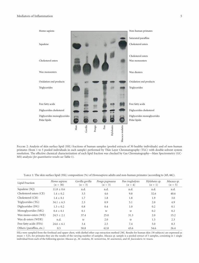
In conclusion, though, from the biological point of view,the reason why SQ molecule occurs in high concentrationin human, SSL is still considered an enigma. On the basisof all described biochemical properties, it can be sensiblyhypothesized that the natural deficiency of squalene oxido-cyclase activity in human sebaceous glands represents anevolutionary advantage, in that SQ is capable of neutralizingreactive oxygen species induced by UV irradiation on theskin, thus behaving as an antioxidant and, indirectly (SQdoes not absorb in the UV range), as a natural sunscreen[64]. The skin of monkeys, unlike that of humans, is coveredby a large quantity of hair, protecting from UV rays. In thefar less hairy human skin, the shield function is reasonablycarried out by SQ, in association with the physical defencesof stratum corneum and melanin. In contrast to humansebum in fact, SSL of other hominidae contains higher levelsof cholesterol, and surprisingly no SQ at all. Our group hasperformed a so far unique study on surface lipid compositionin the primate superfamily, proving that SQ is unique tohuman sebum, and completely missing in the main generaof non-human primates, including those closer to man, thehominoidea [65, 66]. Human sebum also contains higherlevels of triglycerides and their hydrolysis products and farlower levels of cholesterol. A synthesis of most relevant datais illustrated in Figure 2 and Table 1. In addition, SQ terpenetypical of human sebum is also a principal surface lipid of
different aquatic mammals, namely otter, beaver, kinkajou,and at least one species of mole [67–70]. In these species,SQ accounts for the essential properties of water repellenceand thermal insulation. Other nonaquatic mammals or birdshave evolved different cutaneous fats, such as wax esters andwax di-esters, ensuring the same vital properties.
The relevant distance of human sebum in compositionand function from the nearest primates, and the closesimilarity with semiaquatic mammals, bears interestingevolutionary implications and may offer some support to thediscussed hypothesis of the origin of man from some semi-aquatic hominids, feeding on fish. Marine food, especiallymicroalgae and seaweeds, as well edible seeds of plantsdwelling in Mangrovian habitats, like the genus Amarantus[71], display in fact an unusual rich SQ content. In the lightof the so-called Aquatic Ape Theory, that evidences featuresof convergence among different semi-aquatic species, suchas proboscis monkeys, beavers, sea-otters, hippopotamuses,seals, sea lions, and walruses [72], these new biochemicaldata on human sebum squalene offer indeed new space forspeculation.
3. Microbial Impact on SSL Composition andFunction
As previously mentioned, the epidermal surface hydrolipidlayer can also be viewed as the growing medium forresidential saprophytic microbial skin flora. The distribution
Mediators of Inflammation 5
Man
Gor
illa
Pon
go
Pan
Hyl
obat
es
Saturated paraffins
Cholesterol esters
Cholesterol esters
Polar lipids
Free fatty acids
Triglycerides
Wax diesters
Squalene
Wax monoestersCholesterol esters
Polar lipids
Free fatty acids
Triglycerides
Wax monoesters
Homo sapiens Non-human primates
Diglycerides monoglycerides
Diglycerides cholesterol
Diglycerides monoglycerides
Diglycerides cholesterol
Mac
aca
mu
latt
a
Oxidation end productsOxidation end products
Figure 2: Analysis of skin surface lipid (SSL) fractions of human samples (pooled extracts of 30 healthy individuals) and of non-humanprimates (from 1 to 5 pooled individuals in each sample) performed by Thin Layer Chromatography (TLC) with double-solvent systemresolution. The effective chemical characterization of each lipid fraction was checked by Gas Chromatography—Mass Spectrometry (GC-MS) analysis (for quantitative results see Table 1).
Table 1: The skin surface lipid (SSL) composition (%) of Homosapiens adults and non-human primates (according to [65, 66]).
Lipid Fraction Homo sapiens(n = 30)
Gorilla gorilla(n = 3)
Pongo pygmaeus(n = 3)
Pan troglodytes(n = 4)
Hylobates sp.(n = 1)
Macaca sp.(n = 5)
Squalene (SQ) 11.8 ± 0.6 n.d. n.d. n.d. n.d. n.d.
Cholesterol esters (CE) 1.4 ± 0.2 3.5 4.6 9.8 32.6 40.6
Cholesterol (CH) 1.4 ± 0.1 1.7 1.8 1.8 1.9 3.0
Triglycerides (TG) 34.1 ± 6.5 2.3 0.9 5.1 2.0 4.9
Diglycerides (DG) 1.3 ± 0.2 0.8 0.4 1.0 0.2 0.1
Monoglycerides (MG) 0.4 ± 0.1 0.1 tr tr 0.2 0.2
Wax mono-esters (WE) 24.5 ± 2.1 37.4 25.0 31.3 2.0 15.2
Wax di-esters (WDE) n.d. tr 2.0 tr 1.5 2.3
Free fatty acids (FFA) 24.6 ± 6.1 3.4 2.5 7.4 5.0 8.3
Others (paraffins, etc.) 0.5 50.8 62.8 43.6 54.6 26.4
SSLs were sampled from the forehead and upper chest, with diethyl ether cup extraction method [30]. Results for human skin (30 subjects) are expressed asmean ± S.D.; for primates they are the results of the varying number of samples. Macaca sp. sample is a pooled extract of 5 samples, consisting in 1 singleindividual from each of the following species: Macaca sp., M. mulatta, M. nemestrina, M. assemensis, and M. fascicularis. tr: traces.

6 Mediators of Inflammation
and density of bacterial and yeast population at cutaneoussurface is dependent on host age and on environmentalfactors such as sebum secretion, occlusion, temperature,and humidity [73, 74]. Sebum produced in the pilo-sebaceous gland is composed purely of squalene, waxes, andtriglycerides [34]. Once secreted, this rich medium is imme-diately colonized by various lipotrophic biologic agents, thedevelopment of which is controlled by several defensivehumoral mechanisms and by the contact with ambientoxygen. Bacterial/yeast lipase activity is the main responsibleof FFA presence on the skin. Oxygen and micro-organismstransform sebaceous moiety and hydrolyse triglycerides toFFA, with consequent relevant alterations of the SSL patternin cases of pathological microbial colonization of the skin.
This is the case of acne, where a role for Propionibac-terium infection is claimed among the main aetiopathogenictriggers [75]. Here, FFAs metabolically generated by thislipophilic bacterium account for the chronic inflammatoryreaction and the fibrogenetic action on infundibulum epithe-lium, thus sustaining comedones, pustules, and nodulesformation [27]. Similar mechanisms have been claimed toexplain at least partially the chronic inflammatory processcharacterizing atopic (AD) and seborrheic dermatitis (SD)[76, 77]. Pityrosporum ovale is a lipophilic saprophytebelonging to the normal skin flora, mainly localizing in thehorny layers, and in the upper tract of the sebaceous follicle.In the scalp, for instance, P. ovale constitutes up to 46%of the cutaneous flora in the healthy subject, while it mayincrease up to 82% in SD patients. The evidences of defectivecell-mediated response to P. ovale and to Candida albicansin SD patients [78], and the elevated incidence of SD inHIV+ and AIDS patients [79], support the hypothesis thata deficit of cell-mediated immunity may play an aetiologicalrole in the disease. A long-lasting pro-inflammatory actionof P. ovale is to be taken into account, the yeast beinghighly immunogenic, able to activate complement and toproduce pro-inflammatory reactive oxidized metabolites ofselected polyunsaturated SSL, including SQ [34]. Alterationsof SSL composition, along with oxidative by-products of SSLirradiation during antitumor PUVA or narrow-band UV-B phototherapy, induce similar immunologic impairmentlocally on skin. This causes an increased incidence ofparasite skin colonization, like, for example, the infectioncaused by the Demodex mite [80], an intrafollicular parasitefeeding on sebum, and also frequently causing blepharitis[34].
Antimicrobials may reduce the density of the residentpathogenic or saprophytic skin flora, but they do not com-pletely eliminate it. While antimicrobials may cause irritantand allergic contact dermatitis, no evidence exists that theiruse may change the ecology of resident bacteria on the skin,thereby leading to the overgrowth of pathogenic bacteria[74]. As a consequence, antibiotic/antimycotic treatment isnot the elective approach to most of these skin diseases.The antimicrobial function of FFA generated on sebum isan essential factor for the homeostasis of skin microbialcolonies, and changes in sebum fatty acid composition area main cause of microbial alterations on the pathologicalskin [81, 82]. In this respect, skin lipidomics are definitely
expected to offer important diagnostic and therapeuticsolutions in the near future.
Due to its high levels of unsaturation, SQ in particularhas been proposed as the precursor of highly toxic pro-inflammatory mediators, produced by bacterial or yeastlipoperoxidase activity [45]. A further well-characterizedexample of an aetiologic involvement of SSL in skin pathol-ogy is offered by pityriasis versicolor, a pigmentation diseasefeatured by large achromial spots occurring on skin areaswith highest concentration of sebum lipids, where Pityrospo-rum orbiculare finds optimal dwelling conditions. This skinsaprophyte in some individuals becomes pathogenic, dueto still unknown factors. In vitro cultures of P. orbicularehave documented a markedly increased peroxisomal lipidoxidation activity induced by the same unsaturated fattyacids present on the skin, namely linoleic acid [83], givingrise to hydrogen peroxide through a Fenton mechanism andthe subsequent generation of hydroxyl radicals. SQ, whichis not a substrate for lipoxygenase and is not metabolizedby Pityrosporum, may reasonably be peroxidized in vivo dueto the metabolic activities of yeast peroxisomes. In yeastcultures supplemented with linoleic acid plus SQ, a markedincrease in lipoperoxide formation was observed [83], withthe generation of the same toxic and unstable oxidationproducts (trans-trans farnesal and SQ epoxides) formedunder experimental high-dosage UV irradiation of sebumand also identified on skin in vivo [45].
This equivalence substantiates the role of SQ peroxidesin the development of the clinical features of pityriasis,possibly accounting for melanotoxicity and cutaneous depig-mentation [84]. In this connection, the early demonstrationthat vitamin E supplementation suppresses the inductionof peroxisomal beta-oxidation and catalase activity inducedby linoleic acid offers promising clues for new treatmentapproaches [85].
4. Skin Surface Lipids as Biomarkers of SkinDisease and Aging
All surface lipids examined so far play a role in the complexsignaling network originating at the epidermal level, so thatskin can no more be viewed as a specialized wrappingmaterial protecting internal organs from environment andguaranteeing the main function of permeability barrierhomeostasis, but more extensively as a complex organactively communicating with the external world [86]. Asa consequence, thorough revision has been made of thetraditional concept that only mere quantitative alterationsof sebum, without any concern for sebum quality andcomposition, accounted for skin diseases associated withseborrhea or sebostasis. The multifaceted functions of thesebaceous gland are hence gaining momentum [25, 87], asthe dysfunction of enzymes synthesizing or metabolizingSSL has been found in different disease states, along withaltered sebum antioxidant levels. As widely discussed above,SSLs are subjected to hydrolysis and oxidative processes,giving rise to biologically active by-products, which arecritically modulated by local lipid soluble antioxidant levels,

Mediators of Inflammation 7
Table 2: The skin surface lipid (SSL) composition (%) and total lipid content in adult healthy subjects and in adult patients suffering fromseborrheic dermatitis (HIV-negative and HIV-positive) and atopic dermatitis (according to [29, 30]).
Lipid FractionGroups
Healty subjects(n = 30)
Seborrheic dermatitis(n = 30)
Seborrheic dermatitis (HIV positive)(n = 30)
Atopic dermatitis(n = 20)
Squalene (SQ) 12.8 ± 0.6 11.7 ± 0.9∗ 11.5 ± 1.0∗ 10.8 ± 1.1∗
Cholesterol esters (CE) 1.3 ± 0.2 1.0 ± 0.4 1.8 ± 0.5∗ 2.4 ± 0.6∗
Wax esters (WE) 25.6 ± 3.2 23.9 ± 5.1 23.8 ± 4.5 21.7 ± 1.8∗
Triglycerides (TG) 36.1 ± 8.4 40.7 ± 10.3 42.3 ± 10.2 32.6 ± 10.6
Free fatty acids (FFA) 21.6 ± 8.8 18.9 ± 9.6 17.4 ± 10.5 28.8 ± 11.4
Cholesterol (CH) 1.2 ± 0.2 1.7 ± 0.5∗ 1.7 ± 0.5∗ 2.4 ± 0.4∗
Diglycerides (DG) 1.4 ± 0.2 1.2 ± 0.2 1.5 ± 0.2 1.3 ± 0.2
TOTAL LIPIDS (µg/cm2) 195.4 ± 20.6 171.2 ± 29.7∗ 167.2 ± 30.4∗ 172.6 ± 17.4∗
SSL sampled from the forehead, with diethyl ether cup extraction method [30]. Results are expressed as mean ± S.D.∗Significance level versus healthy subjects at P < .05.
actively transported to the skin surface at the level of thepilosebaceous unit. Indeed, SSL composition has been foundsignificantly altered in amount and quality in different skindiseases as well as in the aged skin.
In both atopic and seborrheic dermatitis, we and otherauthors reported a marked reduction of skin total surfacelipids [29, 30]. It was shown that SSL of children and adultswith atopic dermatitis present depleted levels of the lipidfractions of sebaceous origin, SQ, and wax esters and corre-spondingly increased levels of free and esterified cholesterol.Analogous alterations were found in seborrheic dermatitis,in HIV-negative and HIV-positive patients, these lattersuffering from SD with increased frequency as compared tohealthy population. These alterations of SSL composition,summarized in Table 2, were associated with significantsystemic depletion of the main lipophilic antioxidant levelsand detoxifying enzymatic activities, mainly vitamin E,ubiquinol-10, and erythrocyte glutathione peroxidase [88,89].
In the acne pathogenic process, SQ peroxides most likelyplay a role [90], in that SQ monohydroperoxide has beenproven to be highly comedogenic when topically applied onskin in the animal model [91]. Sebum SQ fraction was foundincreased by 2.2-fold in a group of patients affected withmoderate to severe acne, as compared to controls, reaching20% of the total sebaceous lipids [92]. The SSLs undergoimportant qualitative and quantitative modifications dueto photo-aging, where SQ peroxides seem again to play amajor role, mimicking the effects of chronic UV irradiationwhen applied experimentally on the skin [93, 94]. SSL mayhence represent a very efficient marker of the sun-protectingefficacy of chemical sunscreens. Experimental UV irradiationof a series of commercial chemical filters in the presenceof SQ in physiological concentrations led to the conclusionthat all most common UV filters protect SQ from UVB-driven oxidation at different extents (Benzophenone-3 >Octylsalicylate > Parsol MCX > Parsol5000 > Octyldimethylp-hydroxybenzoic acid (PABA) > Parsol 1789), depending
on filter dose and irradiation energy. Conversely, uponUVA irradiation, filters like Parsol 1789 and 5000 andoctyl-dimethyl PABA exerted a marked pro-oxidant effect,enhancing SQ peroxides formation possibly through theaction of their self-decomposition by-products induced byUVA, deserving careful attention for safety concerns [64,95].
The external lipid film hence represents a reliable invivo marker of skin disease, easy to monitor throughnoninvasive analytical techniques [30, 96]. Being the firsttarget of environmental stressors, SSLs also represent asuitable research tool for in vitro screening, to study drugdelivery through skin, as well as in vitro assessment of thechemical reactions of jewellery, textiles, cosmetics, drugs,industrial chemicals, and particles in direct and prolongedcontact with human skin [97]. To provide meaningful results,the composition of artificial SSL should accurately mimichuman sweat and sebum, and the conditions of the in vitrotest system should accurately reflect in vivo skin conditions.The wealth of results obtained employing artificial sebumformulations is characterized by the poor physiologicalvalue of most models, lacking essential components andpresenting unrealistic percent ratios of the single molecules.Variables like sweat composition, pH, temperature, and soforth need careful standardization, in order to guaranteereliability of the in vitro test system. The combination oftridimensional skin tissue models with SSL formulationsbears promising results for in vitro applications, providingboth corneum and sebum components to the skin barrierfunctional model.
5. The Influence of Diet andCosmeceutical Intervention
In view of the physiological relevance of the surface lipidcomposition for an optimal performance of the skin organ,the modulation of skin surface lipids composition may

8 Mediators of Inflammation
represent a powerful approach to enhance skin photo-protection, to prevent skin ageing, and to control microbialsymbiotic and pathogenic colonization and consequentskin diseases. This goal is attempted traditionally throughlocal application of lipid and/or lipophilic antioxidant-enriched cosmeceutical formulations, with immediate ben-eficial effects, albeit in most cases not durable after treatmentdiscontinuation.
The possibility to control SSL composition and skinfunction in general by diet or nutriceutical interventionis far more intriguing and promising a perspective. Theadministration of skin-tropic lipid-based oral supplementshas been claimed to be effective, though available reportsare sparse [98, 99]. The analysis of the existing literaturebrings to the conclusion that nutritional factors providebenefits to skin physiology, but information on the effectsof low-to-moderate doses of nutrients consumed in thelong term by healthy individuals is lacking, as well as arethe data on the direct effects on basal skin properties,including hydration, sebum production, and elasticity, up-to-date scant, and often contradictory. The largest part ofinformation refers to PUFA administration as a treatmentfor scaly skin, the observation that the formation of PUFAoxidation products on the skin can be suppressed by linoleicacid supplementation through vegetable oil sources [21]demonstrates that skin lipid film can be modulated throughthe diet more efficiently that through topical application.A low-glycaemic diet regimen also proved effective in thenormalization of sebum production and composition in acnepatients, confirming the feasibility of the control of diseaseconcurrent factors influencing sebaceous gland physiologythrough the systemic approach [100].
Concerning antioxidant supplementation, Passi et al.[54] reported no significant alteration of SSL after a daily oraldosage 200 mg of CoQ10 for 10 days. Oral supplementationof SQ to mice resulted in a marked dose-dependent upreg-ulation of cellular and nonspecific immune functions [13].In healthy volunteers, low- or high-dosage (13 or 27 g daily)SQ per os, per 90 days resulted in significant improvementof antiaging effects on photoaged skin, with facial wrinkledecrease, as confirmed by molecular markers of UV-inducedskin damage. Facial erythema decrease and pigmentationincrease were observed though high dose oral administrationof fat induced some side effects [101].
6. Conclusions
Data discussed so far allow us to consider skin surface lipids,and in particular its polyunsaturated component squalene,as a main target for pro-oxidant agents on the skin. SQperoxides generated locally upon natural or therapeutic UVirradiation are mediators of the immunological response andthe melanogenic process in the skin. The SSLs represent suit-able markers for the diagnosis of skin disease or aging, andfor treatment efficacy monitoring, provided that the resultsare combined with the analysis of the strictly interconnectedsystemic biomarkers of oxidative damage and antioxidantdefences.
Abbreviations
AD: Atopic dermatitisDNFB: DinitrofluorobenzeneFFA: Free fatty acids4-HNE: 4-hydroxy-2 nonenalMDA: Malonyl dialdehydePABA: Para-hydroxybenzoic acidPUFA: Polyunsaturated fatty acidsSD: Seborrheic dermatitisSQ: SqualeneSSL: Skin surface lipids.
Acknowledgments
Data related to sebum composition of non-human Primates,presented in this paper in original extended version, wereachieved with the invaluable technical collaboration of Dr.Mauro Grandinetti and Mr. Andrea Stancato.
References
[1] J. M. Tiffany, “The role of meibomian secretion in the tears,”Transactions of the Ophthalmological Societies of the UnitedKingdom, vol. 104, no. 4, pp. 396–401, 1985.
[2] D. T. Downing, J. S. Strauss, and P. E. Pochi, “Variabilityin the chemical composition of human skin surface lipids,”Journal of Investigative Dermatology, vol. 53, no. 5, pp. 322–327, 1969.
[3] W. J. Cunliffe and S. Shuster, “The rate of sebum excretion inman,” British Journal of Dermatology, vol. 81, no. 9, pp. 697–704, 1969.
[4] E. Haathi and E. C. Horning, “Isolation and characterizationof saturated and unsaturated fatty acids and alcohols ofhuman skin surface lipids,” Scandinavian journal of clinicaland laboratory investigation, vol. 15, part 4, no. 1, pp. 73–78,1963.
[5] R. Westerberg, P. Tvrdik, A.-B. Unden et al., “Role forELOVL3 and fatty acid chain length in development of hairand skin function,” Journal of Biological Chemistry, vol. 279,no. 7, pp. 5621–5629, 2004.
[6] S. J. Miller, R. Aly, H. R. Shinefeld, and P. M. Elias, “In vitroand in vivo antistaphylococcal activity of human stratumcorneum lipids,” Archives of Dermatology, vol. 124, no. 2, pp.209–215, 1988.
[7] S. H. Lee, S. K. Jeong, and S. K. Ahn, “An update of thedefensive barrier function of skin,” Yonsei Medical Journal,vol. 47, no. 3, pp. 293–306, 2006.
[8] N. Nicolaides and T. Ray, “Skin lipids. III. Fatty chainsin skin lipids. The use of vernix caseosa to differentiatebetween endogenous and exogenous components in humanskin surface lipid,” Journal of the American Oil Chemists’Society, vol. 42, no. 8, pp. 702–707, 1965.
[9] N. Nicolaides, “The monoene and other Wax alcohols ofhuman skin surface lipid and their relation to the fatty acidsof this lipid,” Lipids, vol. 2, no. 3, pp. 266–275, 1967.
[10] N. Nicolaides, “Skin lipids: their biochemical uniqueness,”Science, vol. 186, no. 4158, pp. 19–26, 1974.
[11] M. Fitzgerald and R. C. Murphy, “Electrospray mass spec-trometry of human hair wax esters,” Journal of Lipid Research,vol. 48, no. 5, pp. 1231–1246, 2007.

Mediators of Inflammation 9
[12] Y. Masukawa, H. Tsujimura, and H. Narita, “Liquidchromatography-mass spectrometry for comprehensive pro-filing of ceramide molecules in human hair,” Journal of LipidResearch, vol. 47, no. 7, pp. 1559–1571, 2006.
[13] G. S. Kelly, “Squalene and its potential clinical uses,”Alternative Medicine Review, vol. 4, no. 1, pp. 29–36, 1999.
[14] D. T. Downing, M. E. Stewart, and J. S. Strauss, “Estimationof sebum production rates in man by measurement of thesqualene content of skin biopsies,” Journal of InvestigativeDermatology, vol. 77, no. 4, pp. 358–360, 1981.
[15] D. Voet and J.G. Voet, Biochemistry. Lipid Metabolism, JohnWiley & Sons, New York, NY, USA, 1990.
[16] K. R. Smith and D. M. Thiboutot, “Thematic review series:skin Lipids. Sebaceous gland lipids: friend or foe?” Journal ofLipid Research, vol. 49, no. 2, pp. 271–281, 2008.
[17] Y. Yamamoto, “Role of active oxygen species and antioxidantsin photoaging,” Journal of Dermatological Science, vol. 27, no.1, pp. S1–S4, 2001.
[18] J. J. Thiele, S. U. Weber, and L. Packer, “Sebaceous glandsecretion is a major physiologic route of vitamin E deliveryto skin,” Journal of Investigative Dermatology, vol. 113, no. 6,pp. 1006–1010, 1999.
[19] L. Packer and G. Valacchi, “Antioxidants and the response ofskin to oxidative stress: vitamin E as a key indicator,” SkinPharmacology and Applied Skin Physiology, vol. 15, no. 5, pp.282–290, 2002.
[20] G. Micali, F. Lacarrubba, A. Bongu, and D. West, “The skinbarrier,” in The Biology of the Skin, R. K. Freinkel and D.T. Woodley, Eds., pp. 219–237, The Parthenon PublishingGroup, London, UK, 2001.
[21] V. A. Ziboh, C. C. Miller, and Y. Cho, “Metabolism of polyun-saturated fatty acids by skin epidermal enzymes: genera-tion of antiinflammatory and antiproliferative metabolites,”American Journal of Clinical Nutrition, vol. 71, supplement,pp. 316S–366S, 2000.
[22] P. M. Elias, “Stratum corneum defensive functions: anintegrated view,” Journal of Investigative Dermatology, vol.125, no. 2, pp. 183–200, 2005.
[23] C. C. Geilen, T. Wieder, and C. E. Orfanos, “Ceramidesignalling: regulatory role in cell proliferation, differentiationand apoptosis in human epidermis,” Archives of Dermatolog-ical Research, vol. 289, no. 10, pp. 559–566, 1997.
[24] Y. Uchida, A. Di Nardo, V. Collins, P. M. Elias, and W. M.Holleran, “De novo ceramide synthesis participates in theultraviolet B irradiation-induced apoptosis in undifferenti-ated cultured human keratinocytes,” Journal of InvestigativeDermatology, vol. 120, no. 4, pp. 662–669, 2003.
[25] C. C. Zouboulis, J. M. Baron, M. Bohm et al., “Frontiersin sebaceous gland biology and pathology,” ExperimentalDermatology, vol. 17, no. 6, pp. 542–551, 2008.
[26] D. Saint-Leger, A. Bague, E. Cohen, and M. Chivot, “Apossible role for squalene in the pathogenesis of acne. I.In vitro study of squalene oxidation,” British Journal ofDermatology, vol. 114, no. 5, pp. 535–542, 1986.
[27] D. Saint-Leger, A. Bague, E. Lefebvre, E. Cohen, and M.Chivot, “A possible role for squalene in the pathogenesis ofacne. II. In vivo study of squalene oxides in skin surfaceand intracomedonal lipids of acne patients,” British Journalof Dermatology, vol. 114, no. 5, pp. 543–552, 1986.
[28] A. C. Logan and L. Cordain, “Omega-3 fatty acids and acne,”Archives of Dermatology, vol. 139, no. 7, pp. 941–943, 2003.
[29] M. Picardo, S. Passi, C. De Luca, A. Morrone, F. Bartoli, and F.Ippolito, “Skin surface lipids in patients affected with atopicdermatitis,” in Immunological and Pharmacological Aspects of
Atopic and Contact Eczema, J. M. Czernielewski, Ed., vol. 4 ofPharmacology and the Skin, pp. 173–174, 1991.
[30] S. Passi, M. Picardo, A. Morrone, C. De Luca, and F. Ippolito,“Skin surface lipids in HIV sero-positive and HIV sero-negative patients affected with seborrheic dermatitis,” Journalof Dermatological Science, vol. 2, no. 2, pp. 84–91, 1991.
[31] M. Nazzaro Porro, F. Caprilli, and S. Passi, “Attivita lipoliticadel Pityrosporum orbiculare in vivo e in vitro. Studio del lipididella superficie cutanea nella Pitiriasi versicolore,” BollettinoDell Istituto Dermatologico San Gallicano, vol. 8, pp. 179–190,1973.
[32] M. Nazzaro Porro, S. Passi, F. Caprilli, L. Boniforti, and P.Loreti, “Growth requirements and lipid metabolism of Pity-rosporum orbiculare,” Journal of Investigative Dermatology,vol. 66, no. 3, pp. 178–182, 1976.
[33] J. T. Headington, “Cicatricial alopecia,” Dermatologic Clinics,vol. 14, no. 4, pp. 773–782, 1996.
[34] D. Saint-Leger, “Normal and pathologic sebaceous function.Research in a shallow milieu?” Pathologie Biologie, vol. 51, no.5, pp. 275–278, 2003.
[35] R. J. Sage and H. W. Lim, “UV-based therapy and vitamin D,”Dermatologic Therapy, vol. 23, no. 1, pp. 72–81, 2010.
[36] W. A. Bruls, H. Slaper, J. C. van der Leun, and L. Berrens,“Transmission of human epidermis and stratum corneumas a function of thickness in the ultraviolet and visiblewavelengths,” Photochemistry and Photobiology, vol. 40, no.4, pp. 485–494, 1984.
[37] J. Garssen, R. J. Vandebriel, and H. van Loveren, “Molecularaspects of UVB-induced immunosuppression,” Archives ofToxicology. Supplement, vol. 19, pp. 97–109, 1997.
[38] M. M. Mathews-Roth, “Erythropoietic protoporphyria,”British Journal of Dermatology, vol. 131, no. 6, pp. 751–766,1994.
[39] A. Ryu, K. Arakane, C. Koide, H. Arai, and T. Nagano,“Squalene as a target molecule in skin hyperpigmentationcaused by singlet oxygen,” Biological and PharmaceuticalBulletin, vol. 32, no. 9, pp. 1504–1509, 2009.
[40] K. Hayakawa and I. Matsuo, “Effects of PUVA therapy onskin surface lipids: skin surface lipid peroxidation in psoriasisvulgaris and its biological significance,” Tokai Journal ofExperimental and Clinical Medicine, vol. 11, no. 5, pp. 317–322, 1986.
[41] P. C. Beadle and J. L. Burton, “Absorption of ultraviolet radi-ation by skin surface lipid,” British Journal of Dermatology,vol. 104, no. 5, pp. 549–551, 1981.
[42] A. W. Girotti, “Photosensitized oxidation of membranelipids: reaction pathways, cytotoxic effects, and cytoprotec-tive mechanisms,” Journal of Photochemistry and PhotobiologyB: Biology, vol. 63, no. 1–3, pp. 103–113, 2001.
[43] K. Ohsawa, T. Watanabe, and R. Matsukawa, “The possiblerole of squalene and its peroxide of the sebum in theoccurrence of sunburn and protection from damage causedby UV irradiation,” Journal of Toxicological Sciences, vol. 9,no. 2, pp. 151–159, 1984.
[44] M. Picardo, C. Zompetta, C. De Luca et al., “Role ofskin surface lipids in UV-induced epidermal cell changes,”Archives of Dermatological Research, vol. 283, no. 3, pp. 191–197, 1991.
[45] C. De Luca, M. Picardo, A. Breathnach, and S. Passi,“Lipoperoxidase activity of Pityrosporum: characterisationof by-products and possible role in Pityriasis versicolor,”Experimental Dermatology, vol. 5, no. 1, pp. 49–56, 1996.
[46] K. J. Dennis and T. Shibamoto, “Production of malon-aldehyde from squalene, a major skin surface lipid, during

10 Mediators of Inflammation
UV-irradiation,” Photochemistry and Photobiology, vol. 49,no. 5, pp. 711–716, 1989.
[47] H. C. H. Yeo and T. Shibamoto, “Formation of formaldehydeand malonaldehyde by photooxidation of squalene,” Lipids,vol. 27, no. 1, pp. 50–53, 1992.
[48] A. Wei and T. Shibamoto, “Antioxidant activities of essentialoil mixtures toward skin lipid squalene oxidized by UVirradiation,” Cutaneous and Ocular Toxicology, vol. 26, no. 3,pp. 227–233, 2007.
[49] S. Ekanayake Mudiyanselage, M. Hamburger, P. Elsner, andJ. J. Thiele, “Ultraviolet A induces generation of squalenemonohydroperoxide isomers in human sebum and skinsurface lipids in vitro and in vivo,” Journal of InvestigativeDermatology, vol. 120, no. 6, pp. 915–922, 2003.
[50] M. Podda, M. G. Traber, C. Weber, L.-J. Yan, and L. Packer,“UV-irradiation depletes antioxidants and causes oxidativedamage in a model of human skin,” Free Radical Biology andMedicine, vol. 24, no. 1, pp. 55–65, 1998.
[51] J. J. Thiele, C. Schroeter, S. N. Hsieh, M. Podda, and L. Packer,“The antioxidant network of the stratum corneum,” CurrentProblems in Dermatology, vol. 29, pp. 26–42, 2001.
[52] A. A. Shvedova, Y. Y. Tyurina, V. A. Tyurina, Y. Kikuchi,V. E. Kagan, and P. J. Quinn, “Quantitative analysis ofphospholipid peroxidation and antioxidant protection in livehuman epidermal keratinocytes,” Bioscience Reports, vol. 21,no. 1, pp. 33–43, 2001.
[53] M. Picardo, C. Zompetta, C. De Luca et al., “Squa-lene peroxides may contribute to ultraviolet light-inducedimmunological effects,” Photodermatology Photoimmunologyand Photomedicine, vol. 8, no. 3, pp. 105–110, 1991.
[54] S. Passi, O. De Pita, P. Puddu, and G. P. Littarru, “Lipophilicantioxidants in human sebum and aging,” Free RadicalResearch, vol. 36, no. 4, pp. 471–477, 2002.
[55] C. Cossette, P. Patel, J. R. Anumolu et al., “Human neu-trophils convert the sebum-derived polyunsaturated fattyacid sebaleic acid to a potent granulocyte chemoattractant,”Journal of Biological Chemistry, vol. 283, no. 17, pp. 11234–11243, 2008.
[56] A. M. Morello, D. T. Downing, and J. S. Strauss, “Octadeca-dienoic acids in the skin surface lipids of acne patients andnormal subjects,” Journal of Investigative Dermatology, vol.66, no. 5, pp. 319–323, 1976.
[57] L. Declercq, I. Sente, L. Hellemans, H. Corstjens, and D.Maes, “Use of the synthetic superoxide dismutase/catalasemimetic EUK-134 to compensate for seasonal antioxidantdeficiency by reducing pre-existing lipid peroxides at thehuman skin surface,” International Journal of Cosmetic Sci-ence, vol. 26, no. 5, pp. 255–263, 2004.
[58] K. A. Mountfort, H. Bronstein, N. Archer, and S. M. Jickells,“Identification of oxidation products of squalene in solutionand in latent fingerprints by ESI-MS and LC/APCI-MS,”Analytical Chemistry, vol. 79, no. 7, pp. 2650–2657, 2007.
[59] M. Ottaviani, T. Alestas, E. Flori, A. Mastrofrancesco,C. C. Zouboulis, and M. Picardo, “Peroxidated squaleneinduces the production of inflammatory mediators in HaCaTkeratinocytes: a possible role in acne vulgaris,” Journal ofInvestigative Dermatology, vol. 126, no. 11, pp. 2430–2437,2006.
[60] Y. Kohno, Y. Egawa, S. Itoh, S.-I. Nagaoka, M. Takahashi, andK. Mukai, “Kinetic study of quenching reaction of singletoxygen and scavenging reaction of free radical by squalenein n-butanol,” Biochimica et Biophysica Acta, vol. 1256, no. 1,pp. 52–56, 1995.
[61] K. Someya, Y. Totsuka, M. Murakoshi, H. Kitano, and T.Miyazawa, “The antioxidant effect of palm fruit caroteneon skin lipid peroxidation in guinea pigs as estimated bychemiluminescence-HPLC method,” Journal of NutritionalScience and Vitaminology, vol. 40, no. 4, pp. 315–324, 1994.
[62] A. Pappas, “Epidermal surface lipids,” Dermatoendocrinology,vol. 1, no. 2, pp. 72–76, 2009.
[63] B. Auffray, “Protection against singlet oxygen, the main actorof sebum squalene peroxidation during sun exposure, usingCommiphora myrrha essential oil,” International Journal ofCosmetic Science, vol. 29, no. 1, pp. 23–29, 2007.
[64] C. De Luca, M. Grandinetti, A. Stancato, and S. Passi,“How antioxidant are sunscreen agents?” in Proceedings of the7th Congress European Society for Photobiology, Stresa, Italy,September 1997.
[65] C. De Luca, G. B. Fanfoni, M. Picardo, M. Nazzaro-Porro,and S. Passi, “The skin surface lipids of man compared withthose of other different primates,” The Journal of InvestigativeDermatology, vol. 92, no. 3, p. 473, 1989.
[66] C. De Luca, G. B. Fanfoni, A. Stancato, and S. Passi,“Significant differences in skin surface lipids between manand other primates,” Chemistry and Physics of Lipids, vol. 88,p. 125, 1997.
[67] N. Nicolaides, H. C. Fu, and G. R. Rice, “The skin surfacelipids of man compared with those of eighteen species ofanimals,” Journal of Investigative Dermatology, vol. 51, no. 2,pp. 83–89, 1968.
[68] J. S. Lindholm and D. T. Downing, “Occurrence of squalenein skin surface lipids of the otter, the beaver and thekinkajou,” Lipids, vol. 15, no. 12, pp. 1062–1063, 1980.
[69] J. S. Lindholm, J. M. McCormick, S. W. Colton, and D.T. Downing, “Variation in the chemical composition ofhuman surface skin lipids among mammals,” ComparativeBiochemistry and Physiology B, vol. 69, pp. 75–78, 1981.
[70] D. T. Downing and M. E. Stewart, “Skin surface lipids ofthe mole Scalopus aquaticus,” Comparative Biochemistry andPhysiology B, vol. 86, no. 4, pp. 667–670, 1987.
[71] H.-P. He and H. Corke, “Oil and squalene in amaranthusgrain and leaf,” Journal of Agricultural and Food Chemistry,vol. 51, no. 27, pp. 7913–7920, 2003.
[72] M. J. B. Verhaegen, “Aquatic Ape Theory and fossilhominids,” Medical Hypotheses, vol. 35, no. 2, pp. 108–114,1991.
[73] K. J. McGinley, G. F. Webster, M. R. Ruggieri, and J.J. Leyden, “Regional variations in density of cutaneouspropionibacteria: correlation of Propionibacterium acnespopulations with sebaceous secretion,” Journal of ClinicalMicrobiology, vol. 12, no. 5, pp. 672–675, 1980.
[74] P. Elsner, “Antimicrobials and the skin physiological andpathological flora,” Current Problems in Dermatology, vol. 33,pp. 35–41, 2006.
[75] S. Bhambri, J. Q. Del Rosso, and A. Bhambri, “Pathogenesisof acne vulgaris: recent advances,” Journal of Drugs inDermatology, vol. 8, no. 7, pp. 615–618, 2009.
[76] K. Neuber, W. Konig, and J. Ring, “Staphylococcus aureusand atopic eczema,” Hautarzt, vol. 44, no. 3, pp. 135–142,1993.
[77] T. L. Dawson Jr., “Malassezia globosa and restricta: break-through understanding of the etiology and treatment ofdandruff and seborrheic dermatitis through whole-genomeanalysis,” Journal of Investigative Dermatology SymposiumProceedings, vol. 12, no. 2, pp. 15–19, 2007.
[78] I. -M. Bergbrant, S. Johansson, D. Robbins, A. Scheynius,J. Faergemann, and T. Soderstrom, “An immunological

Mediators of Inflammation 11
study in patients with seborrhoeic dermatitis,” Clinical andExperimental Dermatology, vol. 16, no. 5, pp. 331–338, 1991.
[79] D. S. Goodman, E. D. Teplitz, A. Wishner, R. S. Klein, P. G.Burk, and E. Hershenbaum, “Prevalence of cutaneous dis-ease in patients with acquired immunodeficiency syndrome(AIDS) or AIDS-related complex,” Journal of the AmericanAcademy of Dermatology, vol. 17, no. 2, part 1, pp. 210–220,1987.
[80] L. G. Korkina, C. De Luca, V. A. Kostyuk, and S. Pastore,“Plant polyphenols and tumors: from mechanisms to ther-apies, prevention, and protection against toxicity of anti-cancer treatments,” Current Medicinal Chemistry, vol. 16, no.30, pp. 3943–3965, 2009.
[81] J. J. Wille and A. Kydonieus, “Palmitoleic acid isomer(C16:1Δ6) in human skin sebum is effective against gram-positive bacteria,” Skin Pharmacology and Applied SkinPhysiology, vol. 16, no. 3, pp. 176–187, 2003.
[82] H. Takigawa, H. Nakagawa, M. Kuzukawa, H. Mori, andG. Imokawa, “Deficient production of hexadecenoic acidin the skin is associated in part with the vulnerability ofatopic dermatitis patients to colonization by Staphylococcusaureus,” Dermatology, vol. 211, no. 3, pp. 240–248, 2005.
[83] M. Nazarro-Porro, S. Passi, M. Picardo, R. Mercantini, andA. S. Breatchnach, “Lipoxygenase activity of Pityrosporum invitro and in vivo,” The Journal of Investigative Dermatology,vol. 87, no. 1, pp. 108–112, 1986.
[84] M. Nazzaro Porro and S. Passi, “Identification of tyrosinaseinhibitors in cultures of Pityrosporum,” Journal of Investiga-tive Dermatology, vol. 71, no. 3, pp. 205–208, 1978.
[85] B. Hennig, G. A. Boissonneault, C. K. Chow, Y. Wang, D. H.Matulionis, and H. P. Glauert, “Effect of vitamin E on linoleicacid-mediated induction of peroxisomal enzymes in culturedporcine endothelial cells,” Journal of Nutrition, vol. 120, no. 4,pp. 331–337, 1990.
[86] C. M. Chuong, B. J. Nickoloff, P. M. Elias et al., “What is the’true’ function of skin?” Experimental Dermatology, vol. 11,no. 2, pp. 159–160, 2002.
[87] A. M. W. Porter, “Why do we have apocrine and sebaceousglands?” Journal of the Royal Society of Medicine, vol. 94, no.5, pp. 236–237, 2001.
[88] S. Passi, A. Morrone, C. De Luca, M. Picardo, and F. Ippolito,“Blood levels of vitamin E, polyunsaturated fatty acidsof phospholipids, lipoperoxides and glutathione peroxidasein patients affected with seborrheic dermatitis,” Journal ofDermatological Science, vol. 2, no. 3, pp. 171–178, 1991.
[89] M. Picardo, S. Passi, C. De Luca, A. Morrone, F. Bartoli,and F. Ippolito, “Blood levels of vitamin E, polyunsaturatedfatty acids of phospholipids, and glutathione peroxidasein patients with atopic dermatitis,” in Immunological andPharmacological Aspects of Atopic and Contact Eczema, J. M.Czernielewski, Ed., vol. 4 of Pharmacology and the Skin, pp.171–172, 1991.
[90] J. J. Leyden, “New understandings of the pathogenesis ofacne,” Journal of the American Academy of Dermatology, vol.32, no. 5, part 3, pp. S15–S25, 1995.
[91] K. Chiba, K. Yoshizawa, I. Makino, K. Kawakami, and M.Onoue, “Comedogenicity of squalene monohydroperoxidein the skin after topical application,” Journal of ToxicologicalSciences, vol. 25, no. 2, pp. 77–83, 2000.
[92] A. Pappas, S. Johnsen, J. C. Liu, and M. Eisinger, “Sebumanalysis of individuals with and without acne,” Dermatoen-docrinology, vol. 1, no. 3, pp. 157–161, 2009.
[93] K. Chiba, T. Sone, K. Kawakami, and M. Onoue, “Skinroughness and wrinkle formation induced by repeated
application of squalene-monohydroperoxide to the hairlessmouse,” Experimental Dermatology, vol. 8, no. 6, pp. 471–479, 1999.
[94] K. Chiba, K. Kawakami, T. Sone, and M. Onoue, “Char-acteristics of skin wrinkling and dermal changes inducedby repeated application of squalene monohydroperoxide tohairless mouse skin,” Skin Pharmacology and Applied SkinPhysiology, vol. 16, no. 4, pp. 242–251, 2003.
[95] S. Passi, C. De Luca, M. Grandinetti, A. Stancato, andM. Picardo, “Sunscreen agents inhibit squalene peroxida-tion following UV irradiation,” in Proceedings of the 2ndInternational Workshop on Photodermatology, New trends inPhotoprotection, Capri, Italy, April 1997.
[96] D. T. Downing and M. E. Stewart, “Analysis of sebaceouslipids,” in Methods of Skin Research, D. Skerrow and C. J.Skerrow, Eds., pp. 349–379, John Wiley & Sons, New York,NY, USA, 1985.
[97] A. B. Stefaniak and C. J. Harvey, “Dissolution of materials inartificial skin surface film liquids,” Toxicology in Vitro, vol. 20,no. 8, pp. 1265–1283, 2006.
[98] M. Richelle, M. Sabatier, H. Steiling, and G. Williamson,“Skin bioavailability of dietary vitamin E, carotenoids,polyphenols, vitamin C, zinc and selenium,” British Journalof Nutrition, vol. 96, no. 2, pp. 227–238, 2006.
[99] E. Boelsma, H. F. J. Hendriks, and L. Roza, “Nutritionalskin care: health effects of micronutrients and fatty acids,”American Journal of Clinical Nutrition, vol. 73, no. 5, pp. 853–864, 2001.
[100] R. N. Smith, A. Braue, G. A. Varigos, and N. J. Mann, “Theeffect of a low glycemic load diet on acne vulgaris and thefatty acid composition of skin surface triglycerides,” Journalof Dermatological Science, vol. 50, no. 1, pp. 41–52, 2008.
[101] S. Cho, C.-W. Choi, D. H. Lee et al., “High-dose squa-lene ingestion increases type I procollagen and decreasesultraviolet-induced DNA damage in human skin in vivobut is associated with transient adverse effects,” Clinical andExperimental Dermatology, vol. 34, no. 4, pp. 500–508, 2009.

Submit your manuscripts athttp://www.hindawi.com
Stem CellsInternational
Hindawi Publishing Corporationhttp://www.hindawi.com Volume 2014
Hindawi Publishing Corporationhttp://www.hindawi.com Volume 2014
MEDIATORSINFLAMMATION
of
Hindawi Publishing Corporationhttp://www.hindawi.com Volume 2014
Behavioural Neurology
EndocrinologyInternational Journal of
Hindawi Publishing Corporationhttp://www.hindawi.com Volume 2014
Hindawi Publishing Corporationhttp://www.hindawi.com Volume 2014
Disease Markers
Hindawi Publishing Corporationhttp://www.hindawi.com Volume 2014
BioMed Research International
OncologyJournal of
Hindawi Publishing Corporationhttp://www.hindawi.com Volume 2014
Hindawi Publishing Corporationhttp://www.hindawi.com Volume 2014
Oxidative Medicine and Cellular Longevity
Hindawi Publishing Corporationhttp://www.hindawi.com Volume 2014
PPAR Research
The Scientific World JournalHindawi Publishing Corporation http://www.hindawi.com Volume 2014
Immunology ResearchHindawi Publishing Corporationhttp://www.hindawi.com Volume 2014
Journal of
ObesityJournal of
Hindawi Publishing Corporationhttp://www.hindawi.com Volume 2014
Hindawi Publishing Corporationhttp://www.hindawi.com Volume 2014
Computational and Mathematical Methods in Medicine
OphthalmologyJournal of
Hindawi Publishing Corporationhttp://www.hindawi.com Volume 2014
Diabetes ResearchJournal of
Hindawi Publishing Corporationhttp://www.hindawi.com Volume 2014
Hindawi Publishing Corporationhttp://www.hindawi.com Volume 2014
Research and TreatmentAIDS
Hindawi Publishing Corporationhttp://www.hindawi.com Volume 2014
Gastroenterology Research and Practice
Hindawi Publishing Corporationhttp://www.hindawi.com Volume 2014
Parkinson’s Disease
Evidence-Based Complementary and Alternative Medicine
Volume 2014Hindawi Publishing Corporationhttp://www.hindawi.com


![[Cool] Gas Chromatography and Lipids](https://static.fdokumen.com/doc/165x107/6325a4b1852a7313b70e98e9/cool-gas-chromatography-and-lipids.jpg)