6. Analysis of plant lipids - BRIC
18
Research Signpost 37/661 (2), Fort P.O. Trivandrum-695 023 Kerala, India Plant Lipids Science, Technology, Nutritional Value and Benefits to Human Health, 2015: 221-238 ISBN: 978-81-308-0557-3 Editors: Grażyna Budryn and Dorota Żyżelewicz 6. Analysis of plant lipids Marianna Raczyk and Magdalena Rudzińska Institute of Food Technology of Plant Origin, Poznań University of Life Sciences, Wojska Polskiego 31, 60-624 Poznań, Poland Abstract. In this chapter selected methods used in plant lipid analyses are discussed. The first step of fat analysis consists in the isolation of lipid components from the plant matrix, thus in the first section the most popular methods of isolation are described. Afterwards, due to the complicated mixture of lipid components their separation is required where different types of chromatography techniques are applied. To detect and quantify them specific detectors are required as part of chromatographic systems, in which mass spectrometry, nuclear magnetic resonance and photometric spectroscopies are often applied. To assess specific chemical characteristic of lipids classical chemical methods using e.g. peroxide, iodine and acid values are very often applied. The last part selected oxidative stability tests such as the Schaal oven test, differential scanning calorimetry and Rancimat (OSI) tests were described. Finally sensory analysis as a quality determination technique was also discussed. Introduction Lipids are found in every plant, but their content and composition differ widely, depending on the type and part of the plant. The main lipids include Correspondence/Reprint request: Mgr. Marianna Raczyk, IInstitute of Food Technology of Plant Origin, Poznań University of Life Sciences, Wojska Polskiego 31, 60-624 Poznań, Poland. E-mail: [email protected]
-
Upload
khangminh22 -
Category
Documents
-
view
0 -
download
0
Transcript of 6. Analysis of plant lipids - BRIC

Research Signpost
37/661 (2), Fort P.O.
Trivandrum-695 023
Kerala, India
Plant Lipids Science, Technology, Nutritional Value and Benefits to Human Health, 2015: 221-238
ISBN: 978-81-308-0557-3 Editors: Grażyna Budryn and Dorota Żyżelewicz
6. Analysis of plant lipids
Marianna Raczyk and Magdalena Rudzińska Institute of Food Technology of Plant Origin, Poznań University of Life
Sciences, Wojska Polskiego 31, 60-624 Poznań, Poland
Abstract. In this chapter selected methods used in plant lipid analyses are discussed. The first step of fat analysis consists in the isolation of lipid components from the plant matrix, thus in the first section the most popular methods of isolation are described. Afterwards, due to the complicated mixture of lipid components their separation is required where different types of chromatography techniques are applied. To detect and quantify them specific detectors are required as part of chromatographic systems, in which mass spectrometry, nuclear magnetic resonance and photometric spectroscopies are often applied. To assess specific chemical characteristic of lipids classical chemical methods using e.g. peroxide, iodine and acid values are very often applied. The last part selected oxidative stability tests such as the Schaal oven test, differential scanning calorimetry and Rancimat (OSI) tests were described. Finally sensory analysis as a quality determination technique was also discussed.
Introduction
Lipids are found in every plant, but their content and composition differ
widely, depending on the type and part of the plant. The main lipids include
Correspondence/Reprint request: Mgr. Marianna Raczyk, IInstitute of Food Technology of Plant Origin, Poznań
University of Life Sciences, Wojska Polskiego 31, 60-624 Poznań, Poland. E-mail: [email protected]

Marianna Raczyk and Magdalena Rudzińska 222
triacylglycerols, diacylglycerols, monoacylglycerols, phospholipids,
glycolipids, sterols, free fatty acids, vitamins soluble in fat, waxes, carotene
and chlorophyll pigments. Storage lipids, such as oil in oilseeds contain the
highest amount of triacylglycerols (95-98%), in the form of fatty acid esters
with glycerol [1]. However, functional lipids in plant cells are composed
mainly of polar lipids such as phospho- and glycolipids with very low
amounts of triacylglycerols. Lipids are insoluble in water, but soluble in
organic solvents such as hexane, ether and chloroform, while polar lipids
require highly polar solvents such as butanol and propanol. The first and the
most important part of lipid analytical procedure consists in the selection of
an appropriate extraction/isolation technique to remove all lipids
representing their composition in the analysed material. A general rule for
the isolation of lipids is to utilize proper solvents such as neutral solvents to
isolate neutral lipids, e.g. tricyglycerides, and polar solvents to extract polar
lipids, for instance phospholipids and glycolipids. Isolation of polar lipids
often requires utilization of elevated temperature and some hydrolysing
agents. Utilization of polar solvents also causes extraction of non-lipid
components such as sugars and in the next step these compounds need to be
separated from lipids, which is usually performed by the application of water
and in the same step separation of the lipid phase from water soluble phase
components. The composition of fatty acids has a significant impact on
physical and biological properties of fat, therefore their assessment is very
often the first step of plant lipid analysis. There are many methods to
determine the composition and quality of fats. The contemporary, precise
and widely used analytical techniques are chromatographic methods.
Currently, also spectrometric methods are used on a large scale in lipid
analysis. The structure and features of analysed compounds have a
significant impact on the method used. Lipids are complex mixtures of many
different types of individual lipid classes, therefore often a single analysing
method would not be able to separate and analyse all compounds present in
fat. Most typically a combination of several techniques has to be applied
[2, 3].
Why are we interested in plant lipid analysis?
To better understand plant lipid metabolism and develop oils with
specific properties, we need to analyse lipids which take part in this process.
The further use of plants, as a source of oils, depends on the quantity and
quality of its plant lipids. Plants, which are good sources of oils, are
oftenpressed to obtain plant oils, but their further application greatly depends
on the required functional properties of the oils. Fatty acid composition has a
Analysis of plant lipids 223
significant influence on nutritional and healthy properties of oils however,
other oil compounds are also of significant importance in defining functional
properties of the oil. Amounts of saturated, unsaturated fats and fat-soluble
substances, such as antioxidants (tocopherols, carotenoids) or prooxidants
(chlorophylls, heavy metal ions), have a high impact on wholesomeness, but
also on oxidative stability and proper application in food processing.
Moreover, previously mentioned factors affect physical properties of oils
(colour, liquidity) and the quality (aroma, taste). The concentration and
quality of plant lipids have an impact in particularly on their application
(edible oils, biofuels and industrial oils) and processing conditions [4].
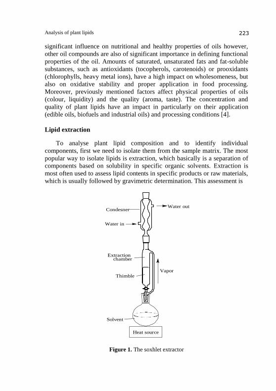
Lipid extraction
To analyse plant lipid composition and to identify individual
components, first we need to isolate them from the sample matrix. The most
popular way to isolate lipids is extraction, which basically is a separation of
components based on solubility in specific organic solvents. Extraction is
most often used to assess lipid contents in specific products or raw materials,
which is usually followed by gravimetric determination. This assessment is
Water outCondesner
Thimble
Heat source
Solvent
Vapor
Water in
Extraction chamber
Figure 1. The Soxhlet extractor
Figure 1. The soxhlet extractor

Marianna Raczyk and Magdalena Rudzińska 224
often inaccurate due to extraction of components other than lipids, which are
usually present in the sample matrix. The type of solvent or solvent mixture,
mainly polarity of solvents, define which types of lipids are extracted [5].
There are many methods of lipid extraction, choosing one of them is
mainly affected by the characteristics of the tested sample. However, in the
case of plant lipids it is defined by the type of lipids that we want to analyse,
neutral or polar, and accordingly solvents are selected. Utilization of polar
solvents for extraction, usually results in extraction of other components than
lipids, mainly carbohydrates and some protein. In this case to assess lipids
composition or identified compounds it is required to remove those non-lipid
components from the extract. Prior to lipid extraction alkaline, acid or
enzymatic hydrolysis is often used to release the lipid part of complex lipids
however, when the composition and identity of lipid components is the goal
of analysis, extraction of complex lipid components is required without
hydrolysis.
This is particularly important in plant lipids, which are usually combined
with sugars and sometimes with proteins. Prior to extraction hydrolysis is
not required for oilseeds, where lipids are found as storage lipids, not
metabolically involved lipids.
The Soxhlet extraction currently is a standard and reference method
used for food products and raw materials [8]. This method has many
advantages, such as continuous contact between the sample and fresh
solvent, so filtration and other steps are not needed. The main disadvantage
of this method is the required extraction time of 16-24 hours to achieve good
recovery of lipids. This procedure has been modified to improve efficiency
and shorten extraction time by 5-10 times utilizing pressure and elevated
temperature, or modifying the Soxhlet apparatus which makes it possible to
place the sample in boiling solvent (Soxtec Foss-Tecator) [9].
Lipids are found in tissues in a variety of physical forms, but complex
lipids are usually constituents of membranes, where they occur in close
association with such compounds as proteins and polysaccharides, with
which they interact through hydrophobic and van der Waals forces and
perhaps by ionic bonds. Various solvents or solvent combinations have been
suggested as extractants, but most lipid analysts use chloroform-methanol
(2:1 by volume) as suggested by Folch et al. [10]. Endogenous water in the
tissue is a ternary component of the system. The extract is shaken and
equilibrated with one fourth its volume of a saline solution, when the
mixture partitions into two layers, of which the lower is composed of
chloroform-methanol-water at 86:14:1 (by volume) and contains virtually
all the lipids, while the upper phase consists of the same solvents at 3:48:47
(by volume), respectively, and contains much of the non-lipid contaminants.

Analysis of plant lipids 225
It is not always recognised how important it is that the proportions of
chloroform, methanol and water in the combined phases should be as close
as possible to the 8:4:3 ratio (by volume), otherwise selective losses of lipids
may occur. If carried out with the correct protocol, this method can give
reliable results [11].
The Bligh-Dyer method is a modified Folch method, which was
applied in the interest of economy and ecology purpose. It is the most often
method used to isolate total lipid content in biological tissues, but is widely
used also in other structures. The advantage of this method is a reducted
amount of solvents used, but also the recovery of total lipids is higher when
compared to the Folch method [12].
AOAC 996.01 is an universally accepted method for nutritional food
labelling, set by the Association of Official Analytical Chemists (AOAC), to
determine total fatty acids in cereal-based products [13]. This procedure has
sufficient accuracy and repeatability and requires application of acid
hydrolysis prior to solvent extraction [14-17].
Most extraction methods mentioned above, are conventional but nowadays
more economic and environmentally friendly methods have been developed,
such as Supercritical Fluid Extraction [18], Pressurized Liquid Extraction,
Focused Microwave-Assisted Soxhlet Extraction [9, 18]. Dynamic
Ultrasound Assisted Extraction [19] and Microwave-Integrated Soxhlet [8,
20].
Microwave-assisted extraction utilizes microwave energy to
disintegrate samples, heat solvents and make extraction efficient and rapid.
According to the research in which this method was used [20], total fats and
oils were qualitatively similar to those obtained by conventional methods.
Fatty acid composition for each extract of tested oils did not show any
significant difference. The method should be consider as valuable, fast and
effective for extraction of fats and oils from food products. Another
technique using microwaves is called the focused microwave-assisted
Soxhlet method [21]. This is a modification of the Soxhlet method, where
microwaves are used in an extraction chamber, as auxiliary energy.
According to the research with the use of this method the best efficiency was
obtained for samples with the moisture content between 20% and 90%
(preferably around 30%). Microwave-assisted extraction was successfully
applied, as an alternative method for oil extraction from olive seeds [21].
Automated hydrolysis and extraction (AHE) is a commercial
procedure which combines acid hydrolysis and solvent extraction in a closed
system. The second part of the analytical process includes extraction of
lipids by the automated Soxhlet method. The advantage of AHE system is
that the whole process is automated and is run in a closed container under

Marianna Raczyk and Magdalena Rudzińska 226
pressure and elevated temperature, making the analysis shorter and more
efficient [16, 22].
Analysis of lipids
Lipids from biological samples, particularly of plant origin, are very
complex mixture samples and to analyse their identity it is required to
separate individual components or at least groups of compounds with
similar properties. For this purpose a variety of chromatographic
techniques are applied, and currently most often high pressure liquid
chromatography (HPLC) is used due to its high efficiency of separation
and high efficient detectors available. Spectrophotometric detectors can be
used in plant lipid analyses, due to the presence of chromophores and a
variety of pigmants. Although most often analysed part of lipid is a fatty
acid composition.
Chromatographic methods
Low-pressure chromatography is used to separate most simple lipids
by column chromatography. One of the effective and widely applied
adsorbents in this method is silicic acid. Substances are selectively eluted by
hexane and diethyl ether mixed at adequate proportions. The precise amount
of diethyl ether must be mixed with hexane to elute every lipid class, which
varies depending on the batch of the adsorbent and should be determined
experimentally. Complex lipids, as a single class, can be obtain with
methanol or chloroform [24].
Thin-layer chromatography (TLC) is a planar adsorption
chromatography, where individual lipids can be separated in multi-
dimension development with solvent. It is one of the oldest, but at the same
time most popular and widely used chromatographic techniques, in which a
thin layer of the stationary phase is placed on glass or aluminium plates. This
method has mainly two applications: purification of specific fractions of
lipids and quantitative analysis, the latter when densitometry with charring
or specific reagents is applied. Many advantages, as for example relatively
inexpensive equipment, easy to set up and operate in laboratories, make TLC
still a useful method. TLC is also widely applied for monitoring reactions
and identification of product formation in organic synthesis [14, 25].
Currently, TLC with pressurized chambers has significantly improved
separation and shorted time of analysis. Also many modification of
adsorbents are available to separate specific groups of lipid components
utilizing some chemical properties [27-29].

Analysis of plant lipids 227
Solvent
Time zero
Solvent
After time
Figure 2. Thin-layer chromatography.
The improvement of thin-layer chromatographic methods led to the
development of high performance thin-layer chromatography (HPTLC)
that provides much higher chromatographic resolution compared to TLC.
Against the traditional, HPTLC plates have significantly smaller size
particles and associated higher surface area, facilitating quicker and clearer
sample separation. A combination of HPTLC with new technologies, such as
the use of pressurized chambers, high resolution cameras, automated sample
applicators and matching software, provides more accurate analysis. These
factors make it an effective analytical technique for quality control [27-30].
Another currently widely used type of chromatographic method is high-
performance liquid chromatography (HPLC), that provides much higher chromatographic resolution compared to TLC. HPLC has been used for accurate separation and quantification of organic substances, including the separation of individual simple lipid classes from the lipid fraction. HPLC has been also used as a standard method to determine tocopherol content in oils [31]. Many different types of column packing materials, eluents and detectors have been used for lipid analysis. The first HPLC methods were elaborated in the 1980s for quantitative analysis of lipids, using mass detectors such as evaporative light-scattering detector (ELSD) and flame ionization detector (FID). The ELSD, depending on the method used, can be applied for analysis of both polar and non-polar lipids. Numerous lipid analyses have used an ELSD and columns with silica gel to separate phospholipid classes in lipid samples. Another very popular and the least expensive detection technique for HPLC is the UV-visible detector, which can be use for lipids that have chromophores (the part of a molecule responsible for its colour), but they have been limited to the analysis of lipid extracts that contain a mixture of saturated and unsaturated molecules. For HPLC analysis, FID was successfully used for several years, but since the sensitivity of ELSD has been improved, the latter method started to

Marianna Raczyk and Magdalena Rudzińska 228
predominate. The minimum detection limits with modern ELSD is 50-100ng [32, 33]. Another type of detector for lipid analysis which was reported in many researches is an aerosol-based detector, although the lowest limits of detection were found for the nucleation light scattering detector with limits below 1ng. Recently a new, commercially available mass detector is charged aerosol detector (CAD), which was evaluated with normal and reverse phase HPLC systems, widely used for the analysis of molecular species and quantitative analyses of lipid classes [34]. In spite of a huge application of HPLC methods for plant lipid analysis, this technique should be considered as a method that also has some disadvantages such as high costs of equipment and running, large amounts of solvents used during the process, high sensitivity of the column to very high (above 9.0) and low (below 3.0) pH of mobile phases [32-34].
Another widely used separation and identification method is gas
chromatography (GC). In this technique the stationary phase is a
microscopic layer of polymer (gas solid chromatography-GSC) or liquid (gas
liquid chromatography-GLC) on the inert solid support inside a column. The
GC column is a very important part of the system that provides separation,
therefore choosing an adequate one is crucial for the analysis. There are two
types of columns, one of them is a packed column mostly 1.5-10 m long and
2-4 mm wide, polarity of the stationary phase determines separation rate.
The analysis of gas samples is one of the areas where packed columns are
widely used. Another type of column is a capillary with an open cross
section, such columns are usually much longer (10-100 m) and narrower
(0.1-0.5 mm) than packed columns. Among capillary columns, the most
commonly used are fused silica columns: the wall coated open tubular
(WCOT) or porous layer open tubular (PLOT) columns. In the former a
liquid film coated to the deactivated wall of the column is the stationary
phase, but in PLOT it is a solid substance that is coated to the column wall.
Mostly capillary columns are used, because they provide better efficiency
(narrow peaks). The separation power is really high and it facilitates short
time runs, with the use of short columns. Another important part of GC is an
injector, also called an inlet, which provides the introduction of a sample
into a flow of carrier gas. An injector is attached to the column head. There
are three ways of introducing samples in GC: injection with split, splitless
and on-column modes. The following factors have a significant influence on
the separation, polarity of the stationary phase, temperature, flow rate of
carrier gas, length of column and amount of sample injected. Helium has
been applied most often as a carrier gas (mobile phase), but sometimes its
substitutes by hydrogen or nitrogen. Carrier gases are applied to promote
sample transport through the column. Diffusivity provides a measurement

Analysis of plant lipids 229
Table 1. Characteristics of carrier gases.
Carrier gas Viscosity at 50 °C
[kg/m s]
Diffusivity
(butane, 100 °C [m2s])
Helium 20.8 5.5 x 10⁻⁶
Hydrogen 9.4 6.0 x 10⁻⁶
Nitrogen 18.8 1.5 x 10⁻⁶
Table 2. Gas chromatography detectors.
Type of Detector Applicable Samples Detection Limit
Mass Spectrometer (MS) any sample 0.25 to 100 pg
Flame Ionization (FID) hydrocarbons 1 pg
Thermal Conductivity (TCD) universal 400 pg/ml
Electron-Capture (ECD) halogenated
hydrocarbons 50 fg
Atomic Emission (AED) element-selective 1 pg
Chemiluminescence (CS) oxidizing reagent Dark current of PMT
Photoionization (PID) vapour and
gaseous compounds 0.002 to 0.02 µg/L
for the diffusion speed of a solute vapour in a given gas and it is different for
each gas (table 1). Diffusion speed is one of the factors that determine the
speed of chromatography [35, 36].
The final, but essential part of the chromatograph is a detector. Different
types of detectors, their applications and detection limits are presented in
table 2. Currently, some of the most common detectors which have been
used with GC are flame ionization detectors (FID), thermal conductivity
detectors (TCD) and electron capture detectors (ECD) or a GC coupled with
a mass spectrometer (MS) [37].
Gas chromatography in the analysis of plant lipids
GC is often used to determine the fatty acid profile of lipids. The
procedure of fatty acid analysis in neutral lipids is as follows: dissolving
dewaxed and degummed oil in alcohol (usually methanol) and titrating by
1M ethanolic potassium standard solution to cover all free fatty acids into
their corresponding fatty acid potassium salts, which then are transformed
into free fatty acids (FFAs) using concentrated sulphuric acid (98%) at 60 °C.

Marianna Raczyk and Magdalena Rudzińska 230
Then, the obtained FFAs are transformed into corresponding fatty acid
methyl esters (FAMEs) using boron trifluoride in (14-15%) alcohol solution
at the same temperature [36, 37].
The fatty acid profile of fat can be obtained by gas chromatography
analysis of the corresponding FAME profiles. Most GC analyses are
performed on the equipment with a spit injector and a flame ionization
detector. Gas chromatography can be set at lower or higher temperatures and
depending on the analytical method, the temperature can be constant or
variable during the whole process. More accurate quantization of fatty acids
has been developed with internal or external standards and a FID response
correction factor the purpose of which is to correct the non-linearity of the
FID response to the mass of fatty acid methyl esters. Theoretical response
correction factors are calculated by a formula considering the detector
response proportional to the relative percentage of carbons in diverse
molecules. The application of this method also requires an optimization of
instrumental conditions [36, 37].
Another application of GC is to check purity of synthesized glycerol
ester (GE), but only a few research groups considered the use of GC-MS for
the analysis of intact GE in edible oils. Other applications where gas
chromatography have been successfully used is to determine the content of
sterols and tocopherols [38].
Short chain, volatile fatty acids are typically analyzed in the free form,
using specialized columns. This group of compounds may be referred to as
free fatty acids (FFAs), volatile fatty acids (VFA), or carboxylic acids. The
analysis of fatty acids in the free form instead of fatty acid methyl esters
results in an easier and quicker sample preparation. In addition, the
formation of undesirable derivatives is eliminated [23, 36].
GC-MS is widely used with other indirect methods for ester analysis, but
so far has not been applied in any direct method. GC-MS is commonly used
because of its advanced sensitivity, selectivity and a high separation power,
but for this reason samples have to be prepared carefully to obtain clear and
easy to interpret results [29, 37].
Mass spectrometry (MS) is a powerful analytical technique that can
provide quantitative and qualitative data, which cannot be obtained by other
methods. Some of the results providedwith the use of this method are
molecular weight, the empirical formula and even the complete structure of
an unknown factor. Using MS, chemical compounds have been ionized to
generate charged molecules or molecule fragments and their mass to charge
ratios are measured. Many examples of applying MS to lipid analysis can be
found in a book by Murphy[39].

Analysis of plant lipids 231
The MS principle, in its simplest form, is to bombard organic molecules
with electrons and positively charged ions. By this operation smaller ionized
entities are obtained, which are propelled throught an electrostatic or
magnetic field and are separated according to their mass to charge ratios.
Then, the ions are collected in sequence as the ratio increases. The largest
peak is usually given a value of 100 and the intensities of all other ions are
normalized to this. MS is usually charged with GC or LC. For lipid
applications GC-MS has been used most often, but LC-MS is applied for
structural analysis of lipids that cannot be volatilized by this technique. In
mass spectrometry, electrospray interface (ESI) is also used for lipid
analyses. ESI is a sensitive ionization process which found extensive
applications for structural information of complex phospholipids and
glycolipids. Many successful applications to analysis of non-polar lipids
such as triacyloglycerols with the use of matrix-assisted laser desorption
(MALDI) have been published. Another type of MS is a MSI-mass
spectrometry imaging that provides analysis of spatially localization of
compounds across surfaces in the case of 2D imaging, but volumes can be
analyzed also by 3D, or even 4D imaging. Two new software instruments
implement these new methods for rapid data exploration and visualization of
2D and 3D data sets in full spatial and spectral resolution. The next type of
mass spectrometry which is often used is a tandem mass spectrometry
(MS/MS). This method provides much better sensitivity of detection and
supplies more information about the structure of the molecules [39, 40].
Nuclear magnetic resonance (NMR) spectroscopy is a powerful
technique that is used to obtain quantitative and qualitative information
about the lipid composition of the extracted oil, avoiding invasive
manipulations of the sample. NMR has been applied to identify lipid
structures, and to detect double bonds and their position in fatty acid chains.
The potentiality of this technique in the study of the molecular components
of complex lipid systems has been successfully demonstrated. NMR
spectroscopy has been applied to analyses of the phospholipid profile of
plant extracts. This method has been widely used in conjunction with HPLC
and GC techniques to study the molecular composition of lipids extracted
[41, 42].
Depending on the atom which is used, there are several types of NMR.
Usually the method has been run with 1H,
13C,
15N,
31P. Hydrogen,
1H, is
usually used to analyze organic compounds, but the carbon isotope, 13
C, is
applied for non-polar lipids. 1
H NMR provides high sensitivity, but limited
resolution, whereas 13
C provides a more detailed analysis of various lipids
with higher resolution however, low natural abundance of isotope 13
C makes
this method more expensive [43, 44]. Recently the application of NMR in

Marianna Raczyk and Magdalena Rudzińska 232
the analysis of oils has strongly increased. NMR is a simple, non-destructive
and fast analytical method that enables simultaneous analysis of different
analytes in one sample. Through the comparison with reference spectra, the
determination of many components is possible. An NMR signal is directly
proportional to the amount of the respective nucleus, therefore a molar
concentration can be obtained simply from the NMR spectrum [44]. The
usefulness of the NMR technique in the study of the molecular components
of complex lipid has been presented in many researches [45-47].
Fat values
Despite a wide range of advanced analytical methods used in research
around the world, classical methods are still often applied and a lot of
currently used standard methods are based on them. One of the important
indexes for the determination of vegetable oil quality is acid value (AV),
according to the AOCS Official Method Cd 3d-63 [48], which determines
the amount of free fatty acids contained in oil, and it is used to determine the
degree of lipid hydrolysis. The principle of the method consists in
neutralizing the acid with potassium hydroxide solution versus
phenolphthalein, as an endpoint indicator. Although the AV method has
some disadvantages, such as titration error, mainly in case of strongly
coloured samples, requirements for large solvent and sample amounts
[4, 48].
Peroxide value (PV) is an index of the primary oxidation status of oils
that inform about the concentration of hydroperoxides (primary oxidation
products), which are unstable and easily can decompose into secondary
oxidation products such as ketons and aldehydes. An improved method to
determine PV uses changes in the electrical conductivity values induced by
the reaction of potassium iodide with the hydroperoxides form the sample.
PV can be determined based on the international standard method AOAC
Official Method 965.33 [49].
Iodine value (IV) is a commonly used method to determine the average
degree of lipid unsaturation. The higher IV, the more double bonds in the
fraction there. In general, the analysis consists of the following steps:
weighting lipid, dissolving in suitable organic solvent, adding an iodine
chloride (ICl), which reacts with unsaturated bonds, but the others remain in
the solution. Then, the remaining ICl is assayed by adding excess potassium
iodine, after that the concentration of iodine is determined by titrating with
sodium thiosulfate, in the presence of starch as an indicator. Based on the
result the amount of idodine chloride that reacted with unsaturated bonds can
be calculated [1].

Analysis of plant lipids 233
Previously mentioned methods such as AV, PV and IV, in spite of many disadvantages such as titration error, large amounts of solvents and sample used, play an important role in quality control and identification of lipids.
Oxidation stability of lipids
Lipid oxidation is one of the main reasons of quality deterioration of oils
and other fats and in the advanced stages it can lead to reduction of their
shelf life. Moreover, changes in oils caused by oxidative processes have a
significant influence on sensory and health characteristics of oils. Therefore,
the oxidation stability and the level of oxidation processes should be
determined.
Oxidative stability testing methods are based on quantity measurement of oxidation products or determination of antioxidant activity. Most oxidative stability testing methods focus on monitoring of the oxidation products. The change in the concentration of oxidation products can be analysed by many methods such as gas chromatography, liquid chromatograph, differential scanning calorimetry, the Rancimat test or the Schaal oven test. Some selected methods are described. The Schaal oven test is one of the oldest methods, where a peroxide value is determined daily in oil samples. Samples are kept in open glass and stored at an constant temperature, usually at 60°C. At the end induction time is calculated based on the plotted curve of PV against time [2]. The modification of the Shaal oven test is a reference method recommended by the AOCS and it is called the Oven Storage Test for Accelerated Aging of Oils. In this method oil oxidation level is analysed after storage period using more than one method, preferably one each for primary and secondary oxidation products [60]. Differential scanning calorimetry (DSC) is a method that is used to evaluate oxidation stability of oils. The same technique, but applying high pressure is called pressure differential scanning calorimetry (PDSC). In this method, the time (oxidation induction time-OTI), in which a rapid exothermic reaction takes place under certain conditions (temperature, pressure, oxygenation), is measured. In this technique high pressure decreases sample volatility by elevating its boiling point and it increases the concentration of the reacting gases. PDSC could potentially be used to study oxidative stability of plant lipids as the reaction of oil oxidation is exothermic, but literature reports on the measurement of the oxidative stability of oils by this technique are few. The technique has many advantages such as small sample required, short time and easy performance. In PDSC the experiment is carried out at lower temperatures and the heat of transition is more precisely defined [51-54].

Marianna Raczyk and Magdalena Rudzińska 234
The Rancimat test is a popular method for determining oil stability.
This technique was developed by Hadom and Zürcher, it consists in the
conductometric determination of volatile oxidation products. This method
has many advantages, such as easy procedure, relatively low cost and short
time of analyses [55]. The test is performed with the use of a Rancimat
apparatus (Figure 3) and it is an official method for determining oil stability
[56]. This test is based on automatic determination of time, known as the
oxidative stability index (OSI), before the maximum rate change of
oxidation, by measuring the increase in conductivity of deionised water,
caused by air that bubbled through a heated oil sample, that carries the
resulting volatile products into a separate chamber with deionised water
(Matthaus 1996). However, the technique has some limitations e.g. drastic
conditions (high temperature and a continuous supply of oxygen) [57].
Sensory analysis is a non-instrumental method of evaluating oil quality.
This method has a significant place in oil analysis but requires a trained
panel, it is time consuming and is rarely objective. Sensory analysis requires
a panel that is a group of trained human assessors. Procedures are strictly
specified, all tested products have to be specially prepared and need to be
monitored during the test. The method makes it possible to make inferences
and gain insight into tested products by applying statistical techniques. This
method is useful, because of irreplaceable sensitivity of the human senses.
Figure 3. A simple scheme of the Rancimat test.

Analysis of plant lipids 235
Sensory analysis is a successfully used method, in spite of some
disadvantages such as high costs. Strict procedures and guidelines provide
repeatable and reproducible results. Sensory quality plays an important role
in plant lipid analysis, because it directly reflects the potential consumer
interest, therefore despite the difficulties it should be carried out as the last
step of testing of food products [58, 59].
References
1. Kamal- Eldin A (2005) Minor components of fats and oils. In: Shahidi F, editor.
Bailey’s industrial oil and fat products. John Wiley and Sons, Inc., Hoboken,
New Jersey. pp. 320-359.
2. Gunstone F.D (2000) Vegetable oils in food technology, composition, properties
and uses. Blackwell Publishing. pp. 318-322.
3. Luque de Castro MD, Priego-Capote F (2010) Soxhlet extraction: Past and
present panacea. Journal of Chromatography A 1217: 2383-2389.
4. Sacchetti G, Maietti S, Muzzoli M, Scaglianti M, Manfredini S, Radice M, Bruni
R (2005) Comparative evaluation of 11 essential oils of different origin as
functional antioxidants, antiradicals and antimicrobials in foods. Food Chemistry
91: 621-632.
5. Becker W (1978) Solvent extraction of soybeans. Journal of the American Oil
Chemists’ Society 55: 754-761.
6. Garcia-Ayuso LE, Luque de Castro MD (1999) A comparative study between
chromatography and other techniques for quantification of triglycerides and fatty
acids in dairy products. Seminar in Food Analysis 4: 39–52.
7. Priego-Capote F, Luque de Castro M.D (2005) Focused microwave-assisted
Soxhlet extraction: a convincing alternative for total fat isolation from bakery
products. Talanta 65: 81-86.
8. Virot M, Tomao V, Colnagui G, Visinoni F, Chemat F (2007) New microwave-
integrated Soxhlet extraction: An advantageous tool for the extraction of lipids
from food products. Journal of Chromatography A 1174: 138-144.
9. Priego-Capote F, Ruiz-Mimenez J, Luque de Castro M.D (2007) Identification
and quantification of trans fatty acids in bakery products by gas
chromatography-mass spectrometry after focused microwave Soxhlet extraction.
Food Chemistry 100: 859–867.
10. Folch J, Less M, Sloane GH (1957) A simple method for the isolation and
purification of total lipids from animal tissues. Journal of Biological Chemistry
226: 497-509.
11. Bligh EG, Dyer W.J (1959) A rapid method of total lipid extraction and
purification. Can. J. Biochem. Physiol 37: 911-917.
12. Iverson SJ, Lang SLC, Cooper MH (2001) Comparison of the Bligh and Dyer
and Folch methods for total lipid determination in a broad range of marine
tissue. Lipids 36: 1283-1287.
Marianna Raczyk and Magdalena Rudzińska 236
13. AOAC (2000) Official methods of analysis. (996.01) Fat (total, saturated,
unsaturated, and monounsaturated) in cereal products (17th ed.). USA: AOAC
International.
14. Ratnayake WMN (2004) Overview of methods for the determination of trans
fatty acids by gas chromatography, silver-ion thin-layer chromatography, silver-
ion liquid chromatography and gas chromatography/mass spectrometry. Journal
of AOAC International 87: 523-539.
15. AOAC (2000) Official methods of analysis. (996.06) Fat (total, saturated,
unsaturated, and monounsaturated) in foods; hydrolytic extraction gas
chromatographic method (17th ed.). USA: AOAC International.
16. Robinson JE, Singh R, Kays SE (2008) Evaluation of an automated hydrolysis
and extraction method for quantification of total fat, lipid classes and trans fat in
cereal products. Food Chemistry 107: 1144-1150.
17. Zhao S, Zhang D (2013) Supercritical fluid extraction and characterisation of
Moringa oleifera leaves oil. Separation and Purification Technology 118:
497-502.
18. Camel V (2001) Recent extraction techniques for solid matrices-supercritical
fluid extraction, pressurized fluid extraction and microwave-assisted extraction:
their potential and pitfalls. Analyst 126: 1182-1193.
19. Ruiz-Jiménez J, Luque-Garcia J.L, Luque de Castro M.D (2003) Dynamic
ultrasound-assisted extraction of cadmium and lead from plants prior to
electrothermal atomic absorption spectrometry. Analytica Chimica Acta 480:
231-237.
20. Virot M, Tomao V, Ginies C, Visinoni F, Chemat F (2008) Microwave-
integrated extraction of total fats and oils. Journal of Chromatography A 1196-
1197: 57-64.
21. Luque de Castro MD, Garcia-Ayuso LE (1998) Soxhlet extraction of solid
materials: an outdated technique with a promising innovative future. Analytica
Chimica Acta 369: 1-10.
22. Helaleh MIH, Al-Omair A, Ahmed N, Gevao B (2005) Quantitative
determination of organochlorine pesticides in sewage sludges using soxtec,
soxhlet and pressurized liquid extractions and ion trap mass-mass spectrometric
detection. Analytical and Bioanalytical Chemistry 382: 1127-1134.
23. Passosa CP, Coimbra MA, Da Silvaa FA, Silvaa C.M (2011) Modelling the
supercritical fluid extraction of edible oils and analysis of the effect of
enzymatic pre-treatments of seed upon model parameters. Chemical Engineering
Research and Design 89: 1118-1125.
24. Claver JB, Valencia MC, Capitán-Vallvey LF (2009) Analysis of parabens in
cosmetics by low pressure liquid chromatography with monolithic column and
chemiluminescent detection. Talanta 79: 499-506.
25. Rouser G, Siakotos A.N, Fleischer S (1966) Quantitative analysis of
phospholipids by thin-layer chromatography and phosphorus analysis of spots.
Lipids 1: 85-86.
26. Momchilova S, Nikolova-Damyanova B (2003) Stationary phases for silver ion
chromatography of lipids: preparation and properties. J. Sep. Sci. 26: 261-270.

Analysis of plant lipids 237
27. Sherma J (2000) Thin-layer chromatography in food and agricultural analysis.
Journal of Chromatography A 880: 129-147.
28. Dillon JT, Aponte JC, Tsai Y, Huang Y (2011) Thin layer chromatography in
the separation of unsaturated organic compounds using silver-thiolate
chromatographic material. Journal of Chromatography A 1251: 240-243.
29. Deattu N, Suseela L, Narayanan N (2013) Chromatographic analysis of
polyherbal extract and formulation by HPTLC and GC-MS methods. Journal of
pharmacy research 6: 6-10. 30. Soler C, Manes J, Pico Y (2008) The Role of the Liquid Chromatography-Mass
Spectrometry in Pesticide Residue Determination in Food. Analytical Chemistry 38: 93-117.
31. ISO 9936: 1997. Animal and vegetable fats and oils - Determination of tocopherols and tocotrienols contents - Method using high-performance liquid chromatography.
32. Moreau RA, Asmann PT, Norman HA (1990) Analysis of Major Classes of Plant Lipids by High Performance Liquid Chromatography with Flame Ionization Detection. Phytochemistry 29: 2461-2466.
33. Donot F, Cazals G, Gunata Z, Egron D, Malinge J, Strub C, Fontan A, Schorr-
Galindo S (2013) Analysis of neutral lipids from microalgae by HPLC-ELSD
and APCI-MS/MS Journal of Chromatography B 942-943: 98-106.
34. Dixon RW, Peterson DS (2002) Development and testing of a detection method
for liquid chromatography based on aerosol charging. Analytical Chemistry 74:
2930-2937.
35. Lau B, Sun W, Ryan JJ (1985) Complication of using hydrogen as the GC
carrier gas in chlorinated dibenzofuran and dibenzodioxin GC/MS analysis.
Chemosphere 14: 799-802.
36. Aued-Pimentel S, Kus MM, Kumagai E.E, Ruvieri V, Zenebon O (2010)
Comparison of gas chromatographic and gravimetric methods for quantization of
total fat and fatty acids in foodstuffs. Quimica Nova. 33: 76-84.
37. Maree J, Kamatou G, Gibbons S, Viljoen A, Vuuren S (2014) The application of
GC-MS combined with chemometrics for the identification of antimicrobial
compounds from selected commercial essential oils. Chemometrics and
Intelligent Laboratory Systems 130: 172-181.
38. Du M, Ahn DU (2002) Simultaneous Analysis of Tocopherols, Cholesterol, and
Phytosterols Using Gas Chromatography. Journal of Food Science 67: 1696-1700.
39. Murphy R. C (1993) Mass spectrometry of lipids (Handbook of lipid research,
vol.7) Plenum Press, New York.
40. Ejsing CS, Duchoslav E, Sampaio J, Simons, K, Bonner R, Thiele C, Ekroos K,
Shevchenko A (2006) Automated identification and quantification of
glycerophospholipid molecular species by multiple precursor ion scanning.
Analytical Chemistry 78: 6202-6214.
41. Holzgrabe U (2010) Quantitative NMR spectroscopy in pharmaceutical
applications. Progress in Nuclear Magnetic Resonance Spectroscopy 57: 229-240.
42. Pollesello P, Eriksson O, Hockerstedt K (1996) Analysis of Total Lipid Extracts
from Human Liver by 13C and 1H Nuclear Magnetic Resonance Spectroscopy.
Analitical biochemistry 236: 41-48.

Marianna Raczyk and Magdalena Rudzińska 238
43. Gunstone FD (1993) High resolution 13C NMR spectroscopy of lipids.
Advanced Lipid Methodology 68: 1-68. 44. Beyer T, Diehl B, Holzgrabe U (2010). Quantitative NMR spectroscopy of
biologically active substances and excipients. Bioanal. Rev. 2: 1-22. 45. Casu M, Anderson GJ, Choi G, Gibbons WA (1991) NMR lipid profiles of cells,
tissues and body fluids-1D and 2D proton NMR of lipids from rat liver. Magn. Res. Chem. 29: 594-602.
46. Sacchi R, Medina I, Aubourg S.P, Giudicianni I, Paolillo L, Addeo F (1993) Quantitative high-resolution 13C-NMR analysis of lipids extracted from the white Muscle of Atlantic Tuna (Thunnus alalunga). J. Agric. Food Chem 41: 1247-1253.
47. Falch E, Storseth TR, Aursand M (2006) Multi-component analysis of marine lipids in fish gonads with emphasis on phospholipids using high resolution NMR spectroscopy. Chem. Phys. Lipids 144, 4-16.
48. Acid value. AOCS Official Method Cd 3d-63. 2000.
49. AOAC (1999). Official methods of analysis. (965.33) Peroxide value of oils and
fats. USA: AOAC International.
50. Abramovic H, Butinar B, Nikolic V (2007) Changes occurring in phenolic
content, tocopherol composition and oxidative stability of Camelina sativa oil
during storage. Food Chemistry 104: 903-909.
51. Gardette J, Baba M (2013) FTIR and DSC studies of the thermal and
photochemical stability of Balanites aegyptiaca oil (Toogga oil). Chemistry and
Physics of Lipids 170-171: 1-7.
52. Cross CK (1970) Oil stability: a DSC alternative for the Active Oxygen Method.
J. Am. Oil Chem. Soc. 666-672.
53. Kowalski B, Ratusz K, Kowalska D, Bekas W (2004). Determination of the
oxidative stability of vegetable oils by Differential Scanning Calorimetry and
Rancimat measurements. Eur. J. Lipid Sci. Technol. 106: 165-169.
54. Kowalski B, Gruczynska E, Maciaszek K (2000). Kinetics of rapeseed oil
oxidation by pressure differential scanning calorimetry measurements. Eur J
Lipid Sci Technol. 202: 337-41.
55. Hadorn H, Zürcher K (1974). Zur Bestimmung der Oxydationsstabiltät von Ölen
und Fetten. Deutsche Lebensmittel-Rundschau, 70: 57-65.
56. AOAC (1997). Official methods of analysis. (Cd 12-92) Oil Stability Index
(OSI). USA: AOAC International.
57. Matthaus B (1996) Determination of the oxidative stability of vegetable oils by
Rancimat and conductivity and chemiluminescence measurements. J. Am. Oil
Chem. Soc. 73: 1039-1043.
58. Kilcast D, Clegg S (2002) Sensory perception of creaminess and its relationship
with food structure. Food Quality and Preference 13: 609-623.
59. Escuderos M, Garcia M, Jiménez A, Horrillo M (2013) Edible and non-edible
olive oils discrimination by the application of a sensory olfactory system based
on tin dioxide sensors. Food Chemistry 136: 1154-1159. 60. AOCS Recommended Practice Cg5-97 (1997). Oven Storage Test for
Accelerated Ageing of Oils. Official Methods and Recommended Practices of the AOCS, American Oil Chemists Society, Champaign, IL.