Apotopes and the biliary specificity of primary biliary cirrhosis
Upload
khangminh22Category
view
0download
0
Gut, 1977, 18, 7-15
Speed of change in biliary lipids and bile acidswith chenodeoxycholic acid-is intermittenttherapy feasible?1J. H. ISER, G. M. MURPHY, AND R. HERMON DOWLING2
From the Gastroenterology Unit, Department of Medicine, Guy's Hospital and Medical School, London
SUmmARY To see whether intermittent chenodeoxycholic acid (CDCA) therapy is a potentialalternative to continous treatment for gallstone dissolution, the speed of change in bile lipid com-
position was studied after starting and stopping CDCA therapy. In addition, the relationship betweenbile lipid composition and the proportions of the bile acids was examined. Bile-rich duodenal fluidwas collected twice in the first week and then at approximately weekly intervals for four to six weeks,from six gallstone patients starting 13-15 mg CDCA.kg BW-1 day-' and from another group ofsix patients whose treatment was stopped after gallstone dissolution. After starting treatment, themean biliary cholesterol saturation index (based on criteria of Hegardt and Dam, 1971) decreasedfrom 149 ± SEM 0-17 to 0-92 ± 0-13 at three weeks and 0-88 ± 0 10 at four weeks, by which timebile lipid composition had become relatively constant. In patients whose treatment was stopped, bilereverted to its supersaturated state within one week, changing from an on-treatment mean saturationindex of 0 74 ± 0 10 to 115 ± 0O15 in six to eight days after withdrawing CDCA. The proportionof conjugated CDCA in the biliary bile acids increased from 27-9 ± 2-5 % to 60-5 ± 4-2% withinfour days and to 80-7 ± 6-2 % by four weeks after starting CDCA. When treatment was stopped,the proportion of CDCA reverted to pretreatment levels by two to three weeks. The saturationindex was significantly related (p < 0-001) to the percent of conjugated CDCA present, such thatwhen the proportion of CDCA exceeded 70%, bile was almost invariably unsaturated. Since themean time taken for bile to become unsaturated was not shorter than the time taken for bile torevert to its supersaturated state, it seems that intermittent treatment would not be adequate tomaintain an unsaturated bile and is, therefore, unlikely to be as effective as continuous treatmentin dissolving gallstones.
It is now known that, with adequate doses, oralchenodeoxycholic acid (CDCA) will dissolve mostradiolucent gallstones (Bell et al., 1972a; Danzingeret al., 1972; Hofmann and Paumgartner, 1975).
'Presented in part at the autumn meeting of the British Societyof Gastroenterology, Oxford, 1975 (Iser, J. H., Murphy, G.M., Dowling, R. H. Is intermittent chenodeoxycholic acidtherapy feasible for dissolving gallstones ? The speed ofchangein biliary lipids and bile acids after starting and stoppingtreatment. Gut, 16, 840 (Abstract)) and at the Second NATOAdvanced Study Institute on the Biliary System, Aalborg,1975 (Dowling, R. H., Murphy, G. M., Iser, J. H. Dis-solution of gallstones by chenodeoxycholic acid. In TheHepatobiliary System, pp. 485-501. Ed. W. Taylor, PlenumPublishing Co.)'Address for reprint requests: Professor R. H. Dowling,Gastroenterology Unit, Guy's Hospital, London Bridge, SEI.
Received for publication 6 October 19767
CDCA acts by reducing the secretion of cholesterolinto bile (Adler et al., 1975; Northfield et al., 1975)and we have recently shown that, before gallstonescan dissolve, the bile must become unsaturated incholesterol (Iser et al., 1975). To avoid prolongedtreatment with ineffective doses of CDCA, therefore,we believe that the bile lipid response to treatmentshould be monitored in all patients. However, it isnot known how soon after starting CDCA the bilebecomes unsaturated with cholesterol, nor when onecan expect to find representative changes in bile lipidcomposition.
Furthermore, when CDCA is stopped, the bilereverts to its supersaturated state (Mok et al., 1974a;Thistle et al., 1974) but again it is now known howquickly this happens. This reversion to the super-saturated state is not synonymous with the re-forma-
on May 31, 2022 by guest. P
rotected by copyright.http://gut.bm
j.com/
Gut: first published as 10.1136/gut.18.1.7 on 1 January 1977. D
ownloaded from

8
tion of gallstones, but in a few patients, whose treat-ment was stopped when the gallstones dissolved, thestones have recurred (Thistle et al., 1974; Iser et al.,1975). The frequency with which this happens isunknown, but, to prevent it, long-term treatmentmay be necessary and although there has been noevidence of short-term toxicity (in patients treatedfor up to four years) (Hofmann and Paumgartner,1975), the long-term side-effects of CDCA therapyare unknown. To minimise such possible risks andfor reasons of economy, intermittent treatment-forexample, treatment on alternate months-wouldseem preferable to continuous long-term therapy,but the efficacy of intermittent CDCA, both inchanging bile lipid composition and in dissolvinggallstones, is unknown.The aims of this study, therefore, were to examine
the time course of changes in bile lipid compositionafter starting and stopping CDCA, and, based onthese results, to see if intermittent treatment wouldeffectively maintain an unsaturated bile. We alsolooked at the proportions of the major biliary bileacids to see if the saturation of bile with cholesterolwas related to the proportion of CDCA or of theother bile acids in bile.
Methods
Bile lipid and biliary bile acid composition weremeasured at frequent intervals after starting andstopping CDCA therapy in two separate groups ofpatients.
STARTING CDCA TREATMENTSix patients (five women and one man) with radio-lucent gallstones in functioning gallbladders were
studied. Their mean age was 56 7 (± SEM6-6; range29-78) and their mean body weight was 69-2 kg(± 5-1; range 55-95).
After an overnight fast, the duodenum wasintubated under fluoroscopic control and bile-richfluid collected after intravenous cholecystokinin (1
unit per kg body weight of CCK-PZ, G. I. H.Laboratory, Karolinska Institute, Stockholm,Sweden). Bile samples were obtained at day 0 (pre-treatment baseline) and subsequently on days 2-4,6-8, 12-17, 18-24 and 26-28, and in three patients alsoon days 38-47 after starting treatment. At eachintubation, 4 ml of 'bile' was retained for analysis,the remainder being returned to the duodenum.Samples were stored immediately at - 20°C untilanalysed.To minimise the variable of different, doses of
CDCA, a factor known to influence bile lipidcomposition (Mok et al., 1974a), the patients weregiven 13-15 mg CDCA.kg BW-1 day-', a dose
J. H. Iser, G. M. Murphy, and R. Hermon Dowling
which we have previously shown results in both anunsaturated bile and gallstone dissolution (Iser et al.,1975). The last dose of CDCA was taken with theevening meal on the day before the duodenalintubation, so that the bile was sampled 13-17 hoursafter the last CDCA capsule.
STOPPING CDCA TREATMENT -
Six women (age 55 8 years + 6-8; range 28-74,weighing 66-7 kg ± 3-5; range 57-80 kg) who also hadhad radiolucent gallstones in functioning gallblad-ders and whose gallstones had dissolved after anaverage of 19 months' (range 15-26) treatment with amean dose of 14 2 mg CDCA kg BW-1 day-'(range 11 4-16-7) were studied when CDCA waswithdrawn. Bile-rich duodenal fluid was againaspirated just before treatment stopped (day 0) andon days 3-4, 6-8, 14-18, 20-22, and 28-31 after stop-ping therapy.
ANALYSIS OF 'BILE' SAMPLES
Bile lipidsAll samples were analysed for total bile acids,phospholipids, and cholesterol as previously de-scribed (Mok et al., 1974b). The degree of saturationof bile with cholesterol was then calculated as thesaturation (lithogenic) index (SI) (Thomas andHofmann, 1973) where an index of > 1 is super-saturated and <1 undersaturated, according to thelimits of cholesterol solubility as defined by Hegardtand Dam (1971).
Biliary bile acidsTo measure the proportion of the individual bileacids in bile, methanolic extracts of the bile sampleswere separated into the mono- (lithocholic acid), di-'(chenodeoxycholic and deoxycholic acids) and tri-hydroxy- (cholic acid) bile acid bands by thin layerchromatography, using 0-25 mm silica gel G (Merck,Darmstadt) plates and the solvent system, aceticacid :dichloro-ethane :water (10:10:1, by vol., Gregg,1966) for development. The proportions of theglycine and taurine conjugates of the major bileacids in bile were then determined by the 3ar-(Bruusgaard, 1970) and 7cx- (Haslewood et al., 1973)hydroxysteroid dehydrogenase enzyme assays. Thevalidity of this chromatographic-enzymatic technique
'Although the principal dihydroxy bile acids in bile arechenodeoxycholic acid (3a, 7a-dihydroxy-5fl-cholanic acid)and deoxycholic acid (3a- 12a-dihydroxy-5,-cholanic acid)the analytical methods used in the present study would notallow the quantitative resolution of deoxycholic acid from the7P epimer ofCDCA, ursodeoxycholic acid (3a, 7f,-dihydroxy-5fl-cholanic acid) which, according to Salen et al. (1974),Fromm et al. (1975) and Stiehl et al. (1975), may be found inthe bile of patients during treatment with CDCA.
on May 31, 2022 by guest. P
rotected by copyright.http://gut.bm
j.com/
Gut: first published as 10.1136/gut.18.1.7 on 1 January 1977. D
ownloaded from
Speed of change in biliary lipids and bile acids with intermittent chenodeoxycholic acid
was confirmed by comparing (1) the sum of theindividual bile acid concentrations as assayed fromthe TLC plates with the total bile acid concentrationas measured on non-chromatographed samples; and(2) the bile acid concentrations in the trihydroxy bileacid band when measured either with the 3ax- or withthe 7a-enzymatic assays (both ofwhich should mea-sure identical concentrations of cholic acid).
RELATIONSHIP BETWEEN BILE LIPIDS ANDBILIARY BILE ACIDSTo see if the saturation of bile with cholesterol wasrelated to the proportion ofCDCA in the biliary bileacids-or, indeed, to the proportion of the othermajor bile acids in bile-the saturation indices wereplotted against the percentages of each of the majorbile acids in turn. The statistical significance of thecorrelations between these variables was then calcu-lated separately for patients starting and stoppingtreatment, and also when the data for the two groupswere combined.
StatisticsThe statistical significance of differences between theresults was tested with Student's paired or unpairedtwo-tailed t tests where appropriate.
2.5 -
2.M
Satn.Index(H & D)
1.5
1.0
0.5
0
Results
BILE LIPID COMPOSITION
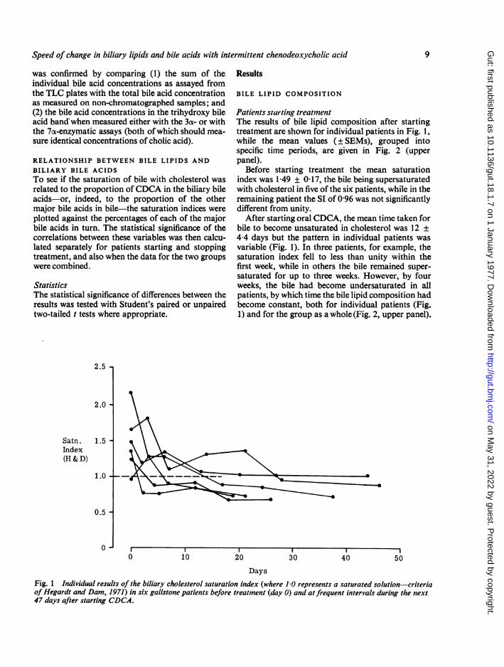
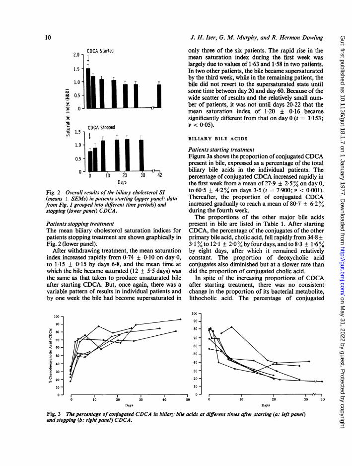
Patients starting treatmentThe results of bile lipid composition after startingtreatment are shown for individual patients in Fig. 1,while the mean values (± SEMs), grouped intospecific time periods, are given in Fig. 2 (upperpanel).
Before starting treatment the mean saturationindex was 149 ± 017, the bile being supersaturatedwith cholesterol in five of the six patients, while in theremaining patient the SI of 096 was not significantlydifferent from unity.
After starting oral CDCA, the mean time taken forbile to become unsaturated in cholesterol was 12 ±4-4 days but the pattern in individual patients wasvariable (Fig. 1). In three patients, for example, thesaturation index fell to less than unity within thefirst week, while in others the bile remained super-saturated for up to three weeks. However, by fourweeks, the bile had become undersaturated in allpatients, by which time the bile lipid composition hadbecome constant, both for individual patients (Fig.1) and for the group as a whole (Fig. 2, upper panel).
I *
0 10 20 30 40 50Days
Fig. 1 Individual results of the biliary cholesterol saturation index (where 1J0 represents a saturated solution-criteriaofHegardt and Dam, 1971) in six gallstone patients before treatment (day 0) and at frequent intervals during the next47 days after starting CDCA.
9
on May 31, 2022 by guest. P
rotected by copyright.http://gut.bm
j.com/
Gut: first published as 10.1136/gut.18.1.7 on 1 January 1977. D
ownloaded from
J. H. Iser, G. M. Murphy, and R. Hermon Dowling
2.0 CDCA Started
1.5 -
1.0
CDCA StoppedX1.5-
0 1O 20 30
Days
Fig. 2 Overall results of the biliary cholesterol SI(means i SEMs) in patients starting (upper panel: datafrom Fig. I grouped into different time periods) andstopping (lower panel) CDCA.
Patients stopping treatmentThe mean biliary cholesterol saturation indices forpatients stopping treatment are shown graphically inFig. 2 (lower panel).
After withdrawing treatment, the mean saturationindex increased rapidly from 0-74 ± 0-10 on day 0,to 1-15 ± 0-15 by days 6-8, and the mean time atwhich the bile became saturated (12 ± 5-5 days) wasthe same as that taken to produce unsaturated bileafter starting CDCA. But, once again, there was avariable pattern of results in individual patients andby one week the bile had become supersaturated in
only three of the six patients. The rapid rise in themean saturation index during the first week waslargely due to values of 1-63 and 1-58 in two patients.In two other patients, the bile became supersaturatedby the third week, while in the remaining patient, thebile did not revert to the supersaturated state untilsome time between day 20 and day 60. Because of thewide scatter of results and the relatively small num-ber of patients, it was not until days 20-22 that themean saturation index of 1P20 ± 0-16 becamesignificantly different from that on day 0 (t = 3 153;P < 005).
BILIARY BILE ACIDS
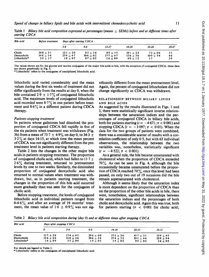
Patients starting treatmentFigure 3a shows the proportion of conjugated CDCApresent in bile, expressed as a percentage of the totalbiliary bile acids in the individual patients. Thepercentage of conjugated CDCA increased rapidly inthe first week from a mean of 27'9 ± 2 5% on day 0,to 60-5 ± 4-2% on days 3-5 (t = 7 900; p < 0 001).Thereafter, the proportion of conjugated CDCAincreased gradually to reach a mean of 80-7 ± 6-2%during the fourth week.The proportions of the other major bile acids
present in bile are listed in Table 1. After startingCDCA, the percentage of the conjugates of the otherprimary bile acid, cholic acid, fell rapidly from 34-8 +31 % to 12-1 ± 2-0 %by four days, and to 8&3 ± 1e6%by eight days, after which it remained relativelyconstant. The proportion of deoxycholic acidconjugates also diminished but at a slower rate thandid the proportion of conjugated cholic acid.
In spite of the increasing proportions of CDCAafter starting treatment, there was no consistentchange in the proportion of its bacterial metabolite,lithocholic acid. The percentage of conjugated
100 -
90 -
80 -
70 -
60 -
50 -
40 -
30 -
20 -
10 -
- -
0 10 20 30 40 50 0
Days
I I I1 1.. I
10 20 30 60
Days
Fig. 3 The percentage ofconfugated CDCA in biliary bile acids at different times after starting (a: left panel)and stopping (b: right panel) CDCA.
100 -
90 -
u 80-a
70 -
<: 60-
° 50-
0 40-
° 30-
a 20-
10 -
0- *~~~~~~~~~ *
10
on May 31, 2022 by guest. P
rotected by copyright.http://gut.bm
j.com/
Gut: first published as 10.1136/gut.18.1.7 on 1 January 1977. D
ownloaded from
Speed of change in biliary lipids and bile acids with intermittent chenodeoxycholic acid
Table 1 Biliary bile acid composition expressed as percentages (means ± SEMs) before and at different times afterstarting CDCA
Bile acid BeJore treatment Days after starting CDCA
2-4 6-8 12-17 18-24 26-28 38-47
Cholic 34-8 + 31 12-1 ± 2-0 8-3 ± 1-6 8-9 1-5 10-1 2-2 7-2 ± 0-6 5-1Deoxycholic 34-8 ± 1-6 25-9 4-7 30-6 6-2 17-7 ± 6-7 12-6 ± 3-6 10-9 ± 54 4-6Lithocholic* 2-9 ± 1-7 1-8 0-7 0-7 ± 0-5 1-6 09 4-0 ± 1-2 1-7 3 9
The values shown are for the glycine and taurine conjugates of the major bile acids in bile, with the exception of conjugated CDCA: these dataare shown graphically in Fig. 3.*'Lithocholic' refers to the conjugates of unsulphated lithocholic acid.
lithocholic acid varied considerably and the meanvalues during the first six weeks of treatment did notdiffer significantly from the results at day 0, when thebile contained 2-9 ± 1-7% of conjugated lithocholicacid. The maximum levels of conjugated lithocholicacid recorded were 8 7% in one patient before treat-ment and 8-6% in a different patient during CDCAtherapy.
Patients stopping treatmentIn patients whose gallstones had dissolved the pro-portion of conjugated CDCA fell rapidly in five ofthe six patients when treatment was withdrawn (Fig.3b) from a mean of 73-7 ± 4 9% on day 0, to 34-3 +3 2 %Y at days 14-15, at which time the mean percentofCDCA was not significantly different from the pre-treatment level in patients starting therapy.
Table 2 lists the changes in the other major bileacids in patients stopping treatment. The proportionof conjugated cholic acid, which had fallen to 11-7 ±
2-6%. during treatment, returned to pretreatmentlevels by one to two weeks. Similarly, the diminishedproportion of conjugated deoxycholic acid alsoreturned to normal values when treatment was with-drawn, but, as in patients starting treatment, thechanges in the proportion of this bile acid occurredmore gradually than was seen for the conjugates ofcholic acid.
Before stopping treatment, the levels of conjugatedlithocholic acid in individual patients ranged from0-4X6 %, and after an average of 19 months' treat-ment, the mean value of 1 6 ± 099% was not sig-
nificantly different from the mean pretreatment level.Again, the percent of conjugated lithocholate did notchange significantly as CDCA was withdrawn.
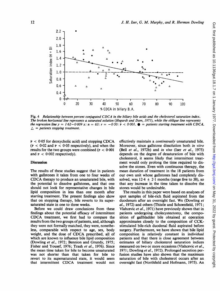
RELATIONSHIP BETWEEN BILIARY LIPIDSAND BILE ACIDSAs suggested by the results illustrated in Figs. 1 and3, there were statistically significant inverse relation-ships between the saturation indices and the per-centages of conjugated CDCA in biliary bile acids,both for patients starting (t = - 4-671; p < 0 001) andstopping CDCA (t = - 3 097; p < 0 01). When thedata for the two groups of patients were combined,there was a considerable scatter of results with a cor-relation coefficient of only 0 5, but with 63 individualobservations, the relationship between the twovariables was, nonetheless, statistically significant(t = -4-522;P <0-001).As a general rule, the bile became unsaturated with
cholesterol when the proportion of CDCA exceeded70%. As can be seen in Fig. 4, although the bileoccasionally became unsaturated before the propor-tion ofCDCA reached 70%, once this level had beenpassed, on only two out of 19 occasions did the bileremain supersaturated with cholesterol.Although it seems likely that the saturation index
is more dependent on the proportion of CDCA thanon the proportion of the other bile acids in bile, therewere, nonetheless, significant relationships betweenthe saturation indices and the percentages of bothcholic and deoxycholic acid. Again this was true, bothfor patients starting (p < 0'001 for cholic acid;
Table 2 Biliary bile acid composition during (day 0) and at different times after stopping CDCA
Bile acid Days after stopping CDCA
0 3-4 6-8 14-18 20-22 28-31
Cholic 11-7 ± 2-6 17-4 ± 4-5 29-6 + 6-0 37-2 ± 2-6 41-7 ± 3-1 33-9 ± 4-5Deoxycholic 12-6 ± 6-6 21-8 ± 2-6 20-6 ± 2-6 27-4 ± 3-2 29-1 ± 4-6 32-9 ± 9 4Lithocholic* 1-6 ± 0-9 1-7 + 0-9 1-5 ± 0-3 1-5 ± 0-2 0-5 ± 0-4 1-6 + 0-9
For details see legend to Table 1.*'Lithocholic' refers to the conjugates of unsulphated lithocholic acid.
11
on May 31, 2022 by guest. P
rotected by copyright.http://gut.bm
j.com/
Gut: first published as 10.1136/gut.18.1.7 on 1 January 1977. D
ownloaded from
J. H. Iser, G. M. Murphy, and R. Hermon Dowling
-mcn
+
0
(2L-a)
2.2 -
2.0 -
1.8 -
1.6 -
1.4 -
1.2-
1.0-
0.8 -
0.6
0.4
A* A A 0
A A0
A A i* A AA AA
I I I --- I----- r-20 30 40 50 60
% CDCA in biliary B.A.
I I I I
70 80 90 100
Fig. 4 Relationship between percent conjugated CDCA in the biliary bile acids and the cholesterol saturation index.The broken horizontal line represents a saturated solution (Hegardt and Dam, 1971), while the oblique line representsthe regression line y = 1 62-0 009 x: n = 63: r = -0S50: p < 0 001. 0 = patients starting treatment with CDCA.A = patients stopping treatment.
p < 0O05 for deoxycholic acid) and stopping CDCA(p < 002 and P < 0.05 respectively), and when theresults for the two groups were combined (p < 0 001and P < 0-O02 respectively).
Discussion
The results of these studies suggest that in patientswith gallstones it takes from one to four weeks ofCDCA therapy to produce an unsaturated bile, withthe potential to dissolve gallstones, and that oneshould not look for representative changes in bilelipid composition in less than one month afterstarting treatment. The present findings also showthat on stopping therapy, bile reverts to its super-saturated state in one to three weeks.
Before we could draw conclusions from thesefindings about the potential efficacy of intermittentCDCA treatment, we first had to compare theresults from the two groups of patients, and, althoughthey were not formally matched, they were, nonethe-less, comparable with respect to age, sex, bodyweight, and the dose of CDCA prescribed, all ofwhich are known to influence bile lipid composition(Dowling et al., 1972; Bennion and Grundy, 1975;Fisher and Yousef, 1976; Trash et al., 1976). Sincethe mean time taken for bile to become unsaturatedwas not shorter than that taken for bile torevert to its supersaturated state, it would seemthat intermittent CDCA treatment would not
effectively maintain a continuously unsaturated bile.Moreover, since gallstone dissolution both in vitro(Bell et al., 1972b) and in vivo (Iser et al., 1975)depends on the degree of desaturation of bile withcholesterol, it seems likely that intermittent treat-ment would only prolong the time required to dis-solve the stones. Even with continuous therapy, themean duration of treatment in the 18 patients fromour own unit whose gallstones had completely dis-solved, was 12-4 ± 2-1 months (range five to 38), sothat any increase in the time taken to dissolve thestones would be undesirable.The results in this paper were based on analyses of
spot samples of bile-rich fluid aspirated from theduodenum after an overnight fast. We (Dowling etal., 1972) and others (Thistle and Schoenfield, 1971;Vlahcevic et al., 1971) have previously shown that inpatients undergoing cholecystectomy, the compo-sition of gallbladder bile obtained at operationapproximates closely to the composition of CCK-stimulated bile-rich duodenal fluid aspirated beforesurgery. Furthermore, we have shown that bile lipidcomposition is relatively constant in individualpatients and that there is close agreement betweenestimates of biliary cholesterol saturation indicesmeasured on two or more occasions (Vlahcevic et al.,1971; Dowling et al., 1972). Prolonged secretion per-fusion studies have also shown that the maximumsaturation of bile with cholesterol occurs after an
overnight fast (Northfield and Hofmann, 1975). An
0O
12
W.
on May 31, 2022 by guest. P
rotected by copyright.http://gut.bm
j.com/
Gut: first published as 10.1136/gut.18.1.7 on 1 January 1977. D
ownloaded from
Speed of change in biliary lipids and bile acids with intermittent chenodeoxycholic acid
early morning spot sample, therefore, yields themost pessimistic estimate of the beneficial effects ofCDCA on bile lipid composition.There have been few previous studies either in
animals or in man on the speed of change in bilelipid and bile acid composition after oral CDCA.Earlier, we had shown that after feeding 30 mgCDCA.kg BW-1 day-' to the rhesus monkey, bilevolume, bile acid, and bile lipid secretion changedpromptly to reach a new steady state within 24-48hours (Dowling, 1973), although the speed of changein bile acid secretion did seem to be dose dependent,taking 10 days to reach a steady state with a smallerdose (5 mg . kg BW-1 day-'). In the hamster,Goldstein et al. (1975) found that oral CDCAreduced the molar ratio of cholesterol in bile and thatthis reduction persisted for up to 20 days after theCDCA was withdrawn. In man, there has been onlyone previous study on the kinetics of change in bilelipids induced by CDCA. Marks et a!. (1975) studiedthe effects of giving 125, 250, or 500 mg CDCA/dayto patients with gallstones before and at one and fourweeks after starting therapy. The only patients inwhom the bile became unsaturated were those given500 mg CDCA/day for one month. Even then the bilewas still saturated according to the limits defined byHegardt and Dam (1971) and was unsaturated onlyby Admirand and Small's (1968) original criteria-a level which in our experience is inadequate to dis-solve gallstones (Iser et a!., 1975).
In the present study there was a considerablevariation from patient to patient in the speed ofchange in bile lipid composition after starting andparticularly after stopping CDCA treatment. Thereason for this variation is unknown, but, theore-tically, there are several factors which could haveinfluenced the results in patients starting treatment.Apart from the dose ofCDCA prescribed, a variablewhich we controlled within reasonably narrow limits,patient compliance in taking the medication, bio-availability and intestinal absorption of the CDCAcapsules, and the biological variable of hepaticresponse to the ingested CDCA could all haveaffected the outcome. We have no reason to believethat the patients did not conscientiously take theircapsules, at least during this short period of intensivestudy, and to date there is little information aboutthe influence of the other factors on the bile lipidresponse to CDCA. Finally, the laboratory estima-tions of bile lipids could have modified the estimatesof cholesterol saturation in bile, but given anaccuracy of ± 5% in estimating bile acids, phospho-lipids, and cholesterol, we have calculated that,for bile samples with a saturation index of 10,the maximum variation due to the analyticaltechnique would change the saturation index by not
more than ± 0 1.The long-term effects of CDCA feeding on biliary
bile acid composition have been studied previously(Danzinger et al., 1973; Coyne et a!., 1975; Frommet al., 1975; Stiehl et al., 1975) and these investigatorsshowed that during treatment the percent of CDCAconjugates reaches from 73-99%. However, therehave been no previous studies on the speed of changein biliary bile acids in patients starting and stoppingCDCA therapy. As the percentage of CDCAconjugates increases in patients starting treatment,the reciprocal fall in the proportion of cholic acidconjugates is not unexpected since it is known thatoral CDCA depresses both cholate synthesisandpoolsize (Danzinger et al., 1973). In turn, since cholate isthe precursor of its bacterial metabolite deoxycholate,it is not surprising that the proportion of thissecondary bile acid should also fall progressively inpatients starting treatment.
It should be noted that the 7oc-hydroxysteroiddehydrogenase enzyme assay used in the presentstudy would not detect the presence of the 7p epimerof chenodeoxycholate, ursodeoxycholic acid, whichalso runs in the dihydroxy bile acid TLC band(Haslewood and Haslewood, 1974). (Indeed it hasrecently been implied that during 'chenotherapy', theCDCA may be partly converted to ursodeoxycholicacid and that the beneficial effects of CDCA oncholesterol solubility in bile may actually be mediatedby its conversion to ursodeoxycholate (Carey, 1975).)After four weeks' treatment, however, and inpatients whose gallstones had just been dissolved, thesum of the cholate, CDCA, and lithocholate levelsmeant that there was less than 13% 'deoxycholate'present in the biliary bile acids. It is unlikely, there-fore, that significant amounts of ursodeoxycholatehad been formed.
Just as the amount of deoxycholic acid present inbile is linked to the amount of its precursor, cholicacid, so the proportion of lithocholate in bile mightbe expected to reflect the increasing percentages ofCDCA in patients starting treatment and thedecreasing percentages of 'cheno' in the bile ofpatients whose treatment was withdrawn. It isfortunate that this was not the case, since lithocholateis potentially hepatotoxic (Palmer, 1976) and theabsence of lithocholate accumulation in the presentstudies is in accord with results from other units (LaRusso et a!., 1975; Bremmelgaard and Pedersen,1976). This failure to accumulate lithocholate during'chenotherapy' is probably due to the high rate oflithocholate sulphation by the human liver (Palmerand Bolt, 1971; Cowen et a!., 1975a) and the poorreabsorption of lithocholate sulphates from theintestine (Low-Beer et al., 1969; Cowen et al., 1975b).Thus, the formation of bile acid sulphates represents
13
on May 31, 2022 by guest. P
rotected by copyright.http://gut.bm
j.com/
Gut: first published as 10.1136/gut.18.1.7 on 1 January 1977. D
ownloaded from

14 J. H. Iser, G. M. Murphy, and R. Hermon Dowling
an efficient safety mechanism in man. Since the bilesamples were not solvolysed in the present study, theproportions of sulphated bile acids were notmeasured.The correlation between the saturation index and
the percentage of CDCA in biliary bile acidsshowed that in patients with gallstones, when the bilecontained more than 70% CDCA it was almostinvariably undersaturated with cholesterol and,while this suggests that the increased proportion ofCDCA was responsible for the improvement incholesterol solubility in bile, the present results cantell us nothing about the mechanisms for the changein bile lipid composition. Indeed, our results showthat the saturation index also correlates significantlywith the percentages of cholate and deoxycholateand since deoxycholate feeding actually increases thesaturation of bile with cholesterol, Low-Beer andPomare (1975) have postulated that the proportionof deoxycholate has an important role in controllingcholesterol solubility in bile.Although the results in this paper suggest that
intermittent CDCA will not continuously maintainbile in an unsaturated state, long-term intermittentCDCA could still be effective in reducing or prevent-ing gallstone recurrence once the stones have beendissolved. This hypothesis must await the proof ofclinical experience, but in our own series there havebeen six gallstone recurrences in five patients out of atotal of 17 patients whose gallstones had been com-pletely dissolved and who have been followed up fora mean period of 12 months. Our present policy is tostop CDCA treatment once two consecutivecholecystograms, three months apart, have con-firmed complete gallstone dissolution. Until we knowthe naturalhistory of the frequency and timingof gall-stone recurrence after CDCA withdrawal, we cannotlogically test whether long-term low or full dosemaintenance treatment, intermittent CDCA therapyor perhaps dietary measures (Pomare et al., 1974)will be necessary.
We thank Miss Elizabeth Sabin for technical assis-tance, and Charge Nurse John Howes for help withthe intubation procedures. We are grateful to WeddelPharmaceuticals Ltd, for supplies of chenodeoxy-cholic acid (Chendol) and financial support: J.H.T. isa Weddel Research Fellow. We are indebted to MrsHazel Creed for secretarial assistance.
References
Adler, R. D., Bennion, L. J., Duane, W. C., and Grundy, S.M. (1975). Effects of low dose chenodeoxycholic acid feed-ing on biliary lipid metabolism. Gastroenterology, 68, 326-334.
Admirand, W. H., and Small, D. M. (1968). The physico-
chemical basis of cholesterol gallstone formation in man.Journal of Clinical Investigation, 47, 1043-1052.
Bell, G. D., Whitney, B., and Dowling, R. H. (1972a). Gall-stone dissolution in man using chenodeoxycholic acid.Lancet, 2, 1213-1216.
Bell, G. D., Sutor, D. J., Whitney, B., and Dowling, R. H.(1972b). Factors influencing human gallstone dissolution inmonkey, dog and human bile. Gut, 13, 836. (Abstract).
Bennion, L. J., and Grundy, S. M. (1975). Effects of obesityand caloric intake in biliary lipid metabolism in man.Journal of Clinical Investigation, 56, 996-1011.
Bremmelgaard, A., and Pedersen, L. (1976). Bile acids in bileduring long-term chenodeoxycholic acid treatment.Scandinavian Journal of Gastroenterology, 11, 161-165.
Bruusgaard, A. (1970). Quantitative determination of themajor 3-hydroxy bile acids in biological material after thin-layer chromatographic separation. Clinica Chimica Acta,28,495-504.
Carey, M. C. (1975). Cheno and urso-what the goose andthe bear have in common. New EnglandJournal ofMedicine,293, 1255-1257. (Editorial).
Coyne, M. J., Bonorris, G. G., Chung, A., Goldstein, L. I.,Lahana, D., and Schoenfield, L. J. (1975). Treatment ofgallstones with chenodeoxycholic acid and phenobarbital.New England Journal of Medicine, 292, 604-607.
Cowen, A. E., Korman, M. G., Hofmann, A. F., and Cass,0. W. (1975a). Metabolism of lithocholate in healthy man.I. Biotransformation and biliary excretion of intravenouslyadministered lithocholate, lithocholylglycine, and theirsulfates. Gastroenterology, 69, 59-66.
Cowen, A. E., Korman, M. G., Hofmann, A. F., Cass, 0. W.,and Coffin, S. B. (1975b). Metabolism of lithocholate inhealthy man. II. Enterohepatic circulation. Gastro-enterology, 69, 67-76.
Danzinger, R. G. Hofmann, A. F., Schoenfield, L. J., andThistle, J. L. (1972). Dissolution of cholesterol gallstones bychenodeoxycholic acid. New England Journal of Medicine,286, 1-8.
Danzinger, R. G., Hofmann, A. F., Thistle, J. L., andSchoenfield, L. J. (1973). Effect of oral chenodeoxycholicacid on bile acid kinetics and biliary lipid composition inwomen with cholelithiasis. Journal of Clinical Investigation,52,2809-2821.
Dowling, R. H. (1973). Ninth Symposium on AdvancedMedicine, p. 379. Edited by G. Walker. Pitman Medical:London.
Dowling, R. H., Bell, G. D., and White, J. (1972). Lithogenicbile in patients with ileal dysfunction. Gut, 13,415-420.
Fisher, M. M., Price, V. M., and Yousef, 1. M. (1976).Biliary lipids in pregnancy. In The Hepatobiliary System,p. 555. Edited by W. Taylor. Plenum: New York. (In press).
Fromm, H., Holz-Slomczyk, M., Zobl, H., Schmidt, E., andSchmidt, F. W. (1975). Absence of hepatotoxicity inpatients treated with chenodeoxycholic acid (CDC) forgallstones. Gasiruenterology, 69, 822. (Abstract).
Goldstein, L. I., Bonorris, G. G., Coyne, M. J., and Schoen-field, L. J. (1975). Persistent effects of chenodeoxycholicacid on biliary lipids in the hamster. Journal ofLaboratoryand Clinical Medicine, 85, 1032-1041.
Gregg, J. A. (1966). New solvent systems for thin-layerchromatography of bile acids. Journal ofLipid Research, 7,579-581.
Halsewood, G. A. D., Murphy, G. M., and Richardson, J. M.(1973). A direct enzymatic assay for 7 a-hydroxy bile acidsand their conjugates. Clinical Science, 44, 95-98.
Haslewood, E. S., and Haslewood, G. A. D. (1974). Specifi-city and some characteristics of a 7 a-hydroxysteroiddehydrogenase from E. coli. In Advances in Bile AcidResearch, p. 105. Edited by S. Matern, J. Hackenschmidt.P. Back, and W. Gerok Schattauer: Stuttgart.
on May 31, 2022 by guest. P
rotected by copyright.http://gut.bm
j.com/
Gut: first published as 10.1136/gut.18.1.7 on 1 January 1977. D
ownloaded from
Speed of change in biliary lipids and bile acids with intermittent chenodeoxycholic acid 15
Hegardt, F. G., and Dam, H. (1971). The solubility ofcholesterol in aqueous solutions of bile salts and lecithin.Zeitschrift far Ernihrungswissenschaft, 10, 223-233.
Hofmann, A. F., and Paumgartner, G. (eds) (1975). Chenode-oxycholic Acid Therapy ofGallstones. Schattauer: Stuttgart.
Iser, J. H., Dowling, R. H., Mok, H. Y. I., and Bell, G. D.(1975). Chenodeoxycholic acid treatment of gallstones: Afollow-up report and analysis of factors influencingresponse to therapy. New EnglandJournal of Medicine, 293,378-383.
La Russo, N. F., Hoffman, N. E., Hofmann, A. F., North-field, T. C., and Thistle, J. L. (1975). Effect of primary bileacid ingestion on bile acid metabolism and biliary lipidsecretion in gallstone patients. Gastroenterology, 69, 1301-1314.
Low-Beer, T. S., and Pomare, E. W. (1975). Can colonicbacterial metabolites predispose to cholesterol gallstones?British MedicalJournal, 1, 438-440.
Low-Beer, T. S., Tyor, M. P., and Lack, L. (1969). Effectsof sulfation of taurolithocholic and glycolithocholic acidson their intestinal transport. Gastroenterology, 56,721-726.
Marks, J., Bonorris, G., Chung, A., Coyne, M., Goldstein,L., Okun, R., and Schoenfield, L. (1975). Feasibility of lowdose and intermittent chenodeoxycholic acid therapy ofgallstones. Gastroenterology, 68, 946. (Abstract).
Mok, H. Y. I., Bell, G. D., and Dowling, R. H. (1974a).Effect of different doses of chenodeoxycholic acid on bile-lipid composition and on frequency of side-effects inpatients with gallstones. Lancet, 2, 253-257.
Mok, H. Y. I., Perry, P. M., and Dowling, R. H. (1974b).The control of bile acid pool size: effect ofjejunal resectionand phenobarbitone on bile acid metabolism in the rat.Gut, 15, 247-253.
Northfield, T. C., and Hofmann, A. F. (1975). Biliary lipidoutput during three meals and an overnight fast. I.Relationship to bile acid pool size and cholesterol satura-tion of bile in gallstone and control subjects. Gut, 16, 1-11.
Northfield, T. C., La Russo, N. F., Hofmann, A. F., andThistle, J. L. (1975). Biliary lipid output during three meals
and an overnight fast. II. Effect of chenodeoxycholic acidtreatment in gallstone subjects. Gut, 16, 12-17.
Palmer, R. H. (1976). Toxic effects of lithocholate on the liverand biliary tree. In The Hepatobiliary System, p. 227. Editedby W. Taylor. Plenum: New York. (fn press).
Palmer, R. H., and Bolt, M. G. (1971). Bile acid sulfates. I.Synthesis of lithocholic acid sulfates and their identificationin human bile. Journal ofLipid Research, 12, 671-679.
Pomare, E. W., Heaton, K. W., Low-Beer, T. S., and White,C. (1974). Effect of wheat bran on bile salt metabolism andbile composition. Gut, 15, 824-825. (Abstract).
Salen, G., Tint, G. S., Eliav, B., Deering, N., and Mosbach,E. H. (1974). Increased formation of ursodeoxycholic acidin patients treated with chenodeoxycholic acid. Journal ofClinical Investigation, 53, 612-621.
Stiehl, A., Raedsch, R., and Kommerell, B. (1975). Increasedsulfation of lithocholate in patients with cholesterol gall-stones during chenodeoxycholate treatment. Digestion, 12,105-110.
Thistle, J. L., and Schoenfield, L. J. (1971). Induced altera-tions in composition of bile of persons having cholelithiasis.Gastroenterology, 61, 488-496.
Thistle, J. L., Yu, P. Y. S., Hofmann, A. F., and Ott, B. J.(1974). Prompt return of bile to supersaturated statefollowed by gallstone recurrence after discontinuance ofchenodeoxycholic acid therapy. Gastroenterology, 66,789.(Abstract).
Thomas, P. J., and Hofmann, A. F. (1973). A simple calcula-tion of the lithogenic index of bile: expressing biliary lipidcomposition on rectangular co-ordinates. Gastroenterology,65, 698-700 (Letter).
Trash, D. B., Ross, P. E., Murison, J., and Bouchier, I. A. D.(1976). The influence of age on cholesterol saturation ofbile. Gut, 17, 394. (Abstract).
Vlahcevic, Z. R., Bell, C. C., Juttijudata, P., and Swell, L.(1971). Bile-rich duodenal fluid as an indicator of biliarylipid composition and its applicability to detection oflithogenic bile. American Journal of Digestive Diseases, 16,797-802.
on May 31, 2022 by guest. P
rotected by copyright.http://gut.bm
j.com/
Gut: first published as 10.1136/gut.18.1.7 on 1 January 1977. D
ownloaded from
Copyright © 2022 FDOKUMEN