Importance of Plant Disease, Scope and Objective of Plant ...
198
Fundamentals of Plant Pathology Department of Plant Pathology, JNKVV, Jabalpur 0 Importance of Plant Disease, Scope and Objective of Plant Pathology Shraddha Karcho [email protected] JNKVV College of Agriculture ,Tikamgarh ------------------------------------------------------------------------------------------------------ Plant Pathology is a branch of agricultural science that deals with the study of fungi, bacteria, viruses, nematodes, and other microbes that cause diseases of plants. Plants diseases and disorders make plant to suffer, either kill or reduce their ability to survive/ reproduce. Any abnormal condition that alters the appearance or function of a plant is called plant disease. The term ‘Pathology’ is derived from two Greek words ‘pathos’ and ‘logos’, ‘Pathos’ means suffering and ‘logos’ Means to study/ knowledge. Therefore Pathology means “study of suffering”. Thus the Plant Pathology or Phytopathology (Gr. Phyton=plant) is the branch of biology that deals with the study of suffering plants. It is both science of learning and understanding the nature of disease and art of diagnosing and controlling the disease. Importance of Plant Diseases The study of plant diseases is important as they cause loss to the plant as well as plant produce. The various types of losses occur in the field, in storage or any time between sowing and consumption of produce. The diseases are responsible for direct monitory loss and material loss. Plant diseases still inflect suffering on untold millions of people worldwide causing an estimated annual yield loss of 14% globally with an estimated economic loss of 220 billion U. S. dollars. Fossil evidence indicates that plants were affected by different diseases 250 million year ago. The Plant disease has been associated with many important events in the history of mankind of the earth. The crop loss due to diseases is estimated to be approximately 30-50%. Cultivated plants are often more susceptible to diseases than are their wild relatives.
-
Upload
khangminh22 -
Category
Documents
-
view
0 -
download
0
Transcript of Importance of Plant Disease, Scope and Objective of Plant ...

Fundamentals of Plant PathologyDepartment of Plant Pathology, JNKVV, Jabalpur
0
Importance of Plant Disease, Scope and Objective of Plant PathologyShraddha Karcho
JNKVV College of Agriculture ,Tikamgarh
------------------------------------------------------------------------------------------------------
Plant Pathology is a branch of agricultural science that deals with the study
of fungi, bacteria, viruses, nematodes, and other microbes that cause diseases of
plants. Plants diseases and disorders make plant to suffer, either kill or reduce their
ability to survive/ reproduce. Any abnormal condition that alters the appearance or
function of a plant is called plant disease.
The term ‘Pathology’ is derived from two Greek words ‘pathos’ and ‘logos’, ‘Pathos’
means suffering and ‘logos’ Means to study/ knowledge. Therefore Pathology
means “study of suffering”. Thus the Plant Pathology or Phytopathology (Gr.
Phyton=plant) is the branch of biology that deals with the study of suffering plants. It
is both science of learning and understanding the nature of disease and art of
diagnosing and controlling the disease.
Importance of Plant DiseasesThe study of plant diseases is important as they cause loss to the plant as well as
plant produce. The various types of losses occur in the field, in storage or any time
between sowing and consumption of produce. The diseases are responsible for
direct monitory loss and material loss. Plant diseases still inflect suffering on untold
millions of people worldwide causing an estimated annual yield loss of 14% globally
with an estimated economic loss of 220 billion U. S. dollars. Fossil evidence
indicates that plants were affected by different diseases 250 million year ago. The
Plant disease has been associated with many important events in the history of
mankind of the earth.
The crop loss due to diseases is estimated to be approximately 30-50%.
Cultivated plants are often more susceptible to diseases than are their wild
relatives.
Fundamentals of Plant PathologyDepartment of Plant Pathology, JNKVV, Jabalpur
1
Important environmental factors that may affect development of plant diseases
are temperature, relative humidity, soil moisture, soil pH, soil type, and soil
fertility.
Each pathogen has an optimum temperature for growth. High soil-moisture
levels favors development of destructive water mold fungi, such as species of
Aphanomyces, Pythium, and Phytophthora.
High humidity favors development of the great majority of leaf and fruit diseases
caused by fungi and bacteria.
Soil pH, a measure of acidity or alkalinity, markedly influences a few diseases,
such as common scab of potato and club root (Plasmodiophora brassicae) of
crucifers.
Raising or lowering the levels of certain nutrients also influences the
development of some infectious diseases. Most control measures are directed
against inoculums of the pathogen and involve the principles of exclusion and
avoidance, eradication, protection, host resistance and selection, and therapy.
Important Famine in WorldLate blight of potato-1841-51 (Irish famine): The late blight of potato, a
diseased caused by the fungus, Phytophthorainfestans, is a famous example of
what a plant disease can do to change the course of history. In 1845, this
diseasedestroyed the potato crop of Ireland where potato constituted the staple diet
of the majority in rural areas. The disease had started in Ireland, England and part
of the continental Europe as early as 1830 and was causing some damage every
year, resulting in food shortage. It was reported that in 1840 the population of
Ireland was 8 million which was reduced to 4 million after the famine.
Coffee rust 1867-1870 (Sri Lanka):The coffee and tea were equally
consumed in England because these were available in plenty from such occupied
countries as India, Sri Lanka and Malaysia. Sri Lanka used to produce maximum
coffee in the world. In 1867, coffee rust attacked the plantations in Sri Lanka and by
1893, export of coffee from Sri Lanka had declined by 93%. The economic crisis

Fundamentals of Plant PathologyDepartment of Plant Pathology, JNKVV, Jabalpur
2
forced the planters to cut down coffee plants and take to tea planting. When coffee
rust was spreading in Sri Lanka the science of plant pathology was just developing
and control measure of the disease were not known. The system of monoculture in
coffee plantations of Sri Lanka was considered a contributoryfactor in devastations
caused by the coffee rust which was not prevalent in coffee growing countries of
South America.
Bengal Famine 1942 (India): In the last year of Second World War (1943)
Bengal had to face a serious famine. One of the reasons to which this famine has
been attributed was the loss in yield of the rice crop due to attack of
Helminthosporium leaf spot which had been affecting the crop for the last several
years. Situation was similar to the Irish potato famine but not so catastrophic.
In India wheat rust had been considered to cause a loss of over Rs. 40
million annually. In the year of epidemics there have been a losses amounting to
Rs. 500 million or more. Although introduction of dwarf high yielding varieties has
reduced the losses to a great extent even now thefarmers lose 8-10%of the
expected yield due to rusts. The loose smut of wheat is estimated to cause an
average loss of 3% every year. The ‘Molya’ disease, caused by a nematode is
another example. The disease of wheat and barley prevalent in most parts of
Rajasthan causes a loss of Rs. 30 million in barley and 40 million in wheat every
year. Different smuts of sorghum are responsible for an annual loss of Rs. 100
million. 5% to 75% loss in chickpea due to Ascochyta blight was reported from
Rajasthan during 1982. Wilt of pigeon pea causes 5-10% loss every year in U.P.
and Bihar.
Scope and Objectives of Plant PathologyScope and responsibilities of plant pathology is unlimited. Its ultimate goal is
to prevent and control plant diseases of economic importance. Responsibilities of
the science of plantpathology may be summarized as under.
Study of etiology, symptoms, predisposing factors and recurrence of such
diseases.
Fundamentals of Plant PathologyDepartment of Plant Pathology, JNKVV, Jabalpur
3
Plant pathology deals with different aspects of plant diseases and has wide
scope than human pathology which only deals with only one aspecti.e. plant
health.
The branch focuses on understanding how hosts, pathogens, and environments
interact to cause plant diseases and on understanding how to control plant
diseases.
In recent years plant pathologists have begun to specialize in particular
aspect. The fields in which notable advances have been made are:
Interaction between host and pathogen at chemical, molecular and genetic
level.
Plant virology, Mycology, chemistry of fungi toxicity.
Disease forecasting and Plant Quarantine.
On practical aspects much advances have been made in plant protection
chemicals; breeding for disease resistance. Increased population emphasizes the
application of all possible means to meet the food requirements
Expansion of crop area
Improved methods of cultivation
Increased use of fertilizers
Improved varieties
Increased irrigation
Crop protection
The science of plant pathology has four main objectives:1. To study the living, non-living and environmental causes of plant
diseases.(Etiology)
2. To study the mechanisms of disease development by pathogens.(Pathogenesis)
3. To study the interactions between the plants and the pathogen.(Epidemology)
4. To develop the methods of controlling the diseases and reducing the losses
caused by them.(Control/ Management)
Fundamentals of Plant PathologyDepartment of Plant Pathology, JNKVV, Jabalpur
4
History of Plant Pathology with special reference to Indian workPawan Amrite
[email protected], Jabalpur
-------------------------------------------------------------------------------------------------------------------
The development of science of Plant Pathology in the modern era in India as
in other countries followed the development of mycology. The study
of fungi in India was initiated by Europeans in the 19th century. They used to
collect fungi and send the specimens for identification to the laboratories in
Europe.
During 1850-1875, D.D. Cunningham and A. Barclay started identification
of fungi in India itself. Cunningham made a special study of rusts and smuts.
DD Cunningham identified the causal organism of red rust of tea in Assam
caused by Cephaleurous virescens.
K.R. Kirtikar Pusa (1885) was the first Indian scientist who collected and
identified the fungi in the country.
TS Ramakrishan Studies and contributed to genera Pythium, Phytophthora,
Colletotrichum, and the rusts. Wrote monograph on 'Diseases of Millets'
published by ICAR
E.J. Bulter who is also known as the ‘Father of Plant Pathology’ in India,
initiated an exhaustive study of fungi and diseases caused by them in 1901
at Imperial Agricultural Research Institute at Pusa (Bihar). During his stay of
20 years in this country, he made a scientific study of mostly fungal plant
diseases known in India at that time. The diseases studied by him for the first
time included wilt of cotton and pigeon pea, different diseases of rice, toddy
palm, sugarcane, potato and rusts of cereals. He wrote a monograph on
‘Pythiaceous and Allied Fungi’; and a classic text book, ‘Fungi and Diseases
in Plants’ in 1918.
J.F. Dastur (1886-1971), a colleague of Butler, was the first Indian Plant
Pathologist who is credited with detailed studies of fungi and diseases in
plants. He studied the genus Phytophthora and diseases caused by it in
L. 02

Fundamentals of Plant PathologyDepartment of Plant Pathology, JNKVV, Jabalpur
5
castor and potato. He is internationally known for the establishment of
Phytophthora parasitica from castor.
T.S. Sadasivan developed the concept of vivotoxins and worked out the
mechanism of wilting in cotton due to Fusarium oxysporum f.sp. vasinfectum.
Mitra in 1937 AD recorded Tilletia indica, a new bunt (Karnal bunt) on wheat.
The disease was thought, erroneously, to be soil borne. Later studies
showed that it was air borne, and the infection was not systemic (Mundkar,
1943 AD)
G.S. Kulkarni published exhaustive information on downy mildew and smuts
of sugarcane and pearl millet.
B.B. Mundkur started work on control of cotton wilt through
varietal resistance. He was also responsible for the identification
and classification of large number of Indian smut fungi. His most significant
contribution to plant pathology will be remembered through the ‘Indian
Phytopathological Society’ which he started almost single handedly in 1948
with its journal ‘Indian Phytopathology’. He also authored a text book entitled,
‘Fungi and Plant Diseases’.
M.K. Patel (Father of Indian plant bacteriology) established a school of plant
bacteriology at the College of Agriculture, Poona (Pune). He reported the first
new species, Xanthomonas uppalii on Ipomoea muricata in 1948. Advocated
the family Phytobacteriaceae to include all plant pathogenic bacteria.
M.K. Patel, V.P. Bhide and G. Rangaswami pioneered the work on bacterial
plant pathogens in India.
Yeshwant Laxman Nene reported “Khaira” disease of rice at Pantnagar due to
zinc deficiency (1965 AD) and authored the book "Fungicides in Plant Disease
Control"
Dr. K.C. Mehta (Father of Indian Rust) of Agra College, Agra investigated the
life cycle of cereal rusts in India during the first half of 20th century. Wrote
monograph on "Further studies on cereal rust in India''. Dr. R. Prasada

Fundamentals of Plant PathologyDepartment of Plant Pathology, JNKVV, Jabalpur
6
trained by Dr K.C. Mehta continued the work on rusts and added to the
knowledge of linseed rust.
Luthra and Sattar (1953) developed the solar heat treatment of wheat seed
for the control of loose smut.
S N Das Gupta carried out exhaustive studies on black tip of mango.
G. Rangaswami authored 'Diseases of Crop Plants in India' and 'Bacterial
Plant Diseases in India.
Jeevan Prakash Verma started the pioneering work on Xanthomonas
campestris pv. malvacearum causing bacterial blight of cotton. He laid a solid
foundation of Indian Plant Bacteriology with his students.
A. Mahadevan studied biochemical changes in diseased plants and enzymes.
He wrote the book ''Microorganism in Diseased Plants"
S. Nagarajan and H. singh (1975) Formulated 'Indian Stem Rust Rules' for
Puccinia graminis tritici
S. Nagarajan (1978) Using climatic and weather based informations to
identify Puccinia path in India.
CD Mayee contributed to the understanding of the ground nut rust, sunflower
downy mildew. Wrote 'Phytopathometery'.
RS Singh Wrote ''Plant diseases", a book known as 'Bible of Plant Pathology.

Fundamentals of Plant PathologyDepartment of Plant Pathology, JNKVV, Jabalpur
7
Concepts and terms in Plant PathologyVijay Yadav
JNKVV, College of Agriculture, Ganjbasoda----------------------------------------------------------------------------------------------------------
Plant Pathology (phytopathology) is defined as the study of the organisms
(infectious organisms) and environmental conditions (physiological factors) that
cause disease in plants, the mechanisms by which disease occurs, the interactions
between these causal agents and the plant (effects on plant growth, yield and
quality). Plant pathology also involves the study of pathogen identification, disease
etiology, disease cycles, economic impact, plant disease epidemiology, plant
disease resistance, how plant diseases affect humans and animals, pathosystem
genetics, and management of plant diseases. It also interfaces knowledge from
other scientific fields such as mycology, microbiology, virology, biochemistry, bio-
informatics, etc. Since plant pathology is directly relevant to man's need to grow
enough food and fiber to sustain civilization.
Causes of plant diseasesIn strict sense, the causes of plant diseases are grouped under following categories:
1. Animate or biotic causes: Pathogens of living nature are categorized into the
following groups.
(i) Fungi (ii) Bacteria (iii) Phytoplasma (iv) Rickettsia-like organisms (v) Algae (vi)
Phanerogams (vii) Protozoa (viii) Nematodes
2. Mesobiotic causes :These disease incitants are neither living or non-living, e.g. (i) Viruses (ii) Viroides
3. In animate or abiotic causes: In true sense these factors cause damages (any
reduction in the quality or quantity of yield or loss of revenue) to the plants rather
than causing disease. The causes are:
(i) Deficiencies or excess of nutrients (ii) Light (iii) Moisture (iv) Temperature (v) Air
pollutants (vi) Lack of oxygen (vii) Toxicity of pesticides (viii) Improper cultural
practices (ix) Abnormality in soil conditions (acidity, alkalinity)
L. 03

Fundamentals of Plant PathologyDepartment of Plant Pathology, JNKVV, Jabalpur
8
When a plant becomes diseased :
When the ability of the cells of a plant or plant part to carry out one or more of
essential functions is interfered with by either a pathogenic organism or an adverse
environmental factor, the activities of the cells are disrupted, altered, or inhibited,
the cells malfunction or die, and the plant becomes diseased. At first, the affliction is
localized to one or a few cells and is invisible. Soon, however, the reaction becomes
more widespread and affected plant parts develop changes visible to the naked
eye. These visible changes are the symptoms of the disease. The visible or
otherwise measurable adverse changes in a plant, produced in reaction to infection
by an organism or to an unfavorable environmental factor, are a measure of the
amount of disease in the plant.
Disease in plants, then, can be defined as the series of invisible and visible
responses of plant cells and tissues to a pathogenic organism or environmental
factor that result in adverse changes in the form, function, or integrity of the plant
and may lead to partial impairment or death of plant parts or of the entire plant.
Plant diseases are caused by pathogens. Hence a pathogen is always associated
with a disease. In other way, disease is a symptom caused by the invasion of a
pathogen that is able to survive, perpetuate and spread.
Plant disease diagnosisDiagnosis of plant diseases requires consideration of various biotic and
abiotic factors which may be involved in the causation of disease, as well as a
sound knowledge of the host plant symptoms and signs. Many variables may
influence each situation, including the state of the host, its cultural history, weather
conditions, soil and general site characteristics. A good understanding of the
"normal host" is essential, and experience is invaluable.
The complexity of the etiology (cause) of the disease usually determines the
difficulty of diagnosis. Many diseases in which a single pathogen is the principal
causal agent have distinguishing and characteristic associated symptoms and
signs. Diseases with more complex etiology may have symptoms which suggest
several possible causes, and may be difficult to diagnose precisely.

Fundamentals of Plant PathologyDepartment of Plant Pathology, JNKVV, Jabalpur
9
Steps in disease diagnosis
1. Identify the host
2. Determine the signs and symptoms of the disease
3. Identify the pathogen
4. Identify the disease
5. Prove pathogenicity - follow Koch's Postulates
Adverse effect of pathogens and Environmental factors on host :
Pathogenic microorganisms, i.e., the transmissible biotic agents that can cause
disease and are generally referred to as pathogens, usually cause disease in plants
by disturbing the metabolism of plant cells through enzymes, toxins, growth
regulators, and other substances they secrete and by absorbing foodstuffs from the
host cells for their own use. Some pathogens may also cause disease by growing
and multiplying in the xylem or phloem vessels of plants, thereby blocking the
upward transportation of water or the downward movement of sugars, respectively,
through these tissues. Environmental factors cause disease in plants when abiotic
factors, such as temperature, moisture, mineral nutrients, and pollutants, occur at
levels above or below a certain range tolerated by the plants.
Adverse effect of diseases on physiology of host :
Infection of roots may cause roots to rot and make them unable to absorb water and
nutrients from the soil; infection of xylem vessels, as happens in vascular wilts and
in some cankers, interferes with the translocation of water and minerals to the
crown of the plant; infection of the foliage, as happens in leaf spots, blights, rusts,
mildews, mosaics, and so on, interferes with photosynthesis; infection of phloem
cells in the veins of leaves and in the bark of stems and shoots, as happens in
cankers and in diseases caused by viruses, mollicutes, and protozoa, interferes with
the downward translocation of photosynthetic products; and infection of flowers and
fruits interferes with reproduction. Although infected cells in most diseases are
weakened or die, in some diseases, e.g., in crown gall, infected cells are induced to
divide much faster (hyperplasia) or to enlarge a great deal more (hypertrophy) than

Fundamentals of Plant PathologyDepartment of Plant Pathology, JNKVV, Jabalpur
10
normal cells and to produce abnormal amorphous overgrowths (tumors) or
abnormal organs.
Plant Disease Management:
The word ‘control’ is a complete term where permanent ‘control’ of a disease is
rarely achieved whereas, ‘management’ of a disease is a continuous process and is
more practical in influencing adverse affect caused by a disease. Disease
management requires a detail understanding of all aspects of crop production,
economics, environmental, cultural, genetics and epidemiological information upon
which the management decisions are made.
Importance of the Plant Diseases:
Globally, enormous losses of the crops are caused by the plant diseases. The loss
can occur from the time of seed sowing in the field to harvesting and storage.
Important historical evidences of plant disease epidemics are Irish Famine due to
late blight of potato (Ireland, 1845), Bengal famine due to brown spot of rice (India,
1942) and Coffee rust (Sri Lanka, 1967) etc. Such epidemics had left their effect on
the economy of the affected countries.

Fundamentals of Plant PathologyDepartment of Plant Pathology, JNKVV, Jabalpur
11
Important Plant Pathogenic Organisms
Usha [email protected]
Important plant pathogenic organism - Fungi, Bacteria, Fastidious Vascular Bacteria.
Plant diseases are caused by a number of organisms which ultimately lead to the lossin crop yield both quantitatively and qualitatively. The disease is defined asmalfunctioning process that is caused by continuous irritation which results in somesuffering producing symptoms.
On the other hand any agent that causes suffering of the plant is called as a pathogen.Various types of abiotic factors are also involved in the production of plant diseases.The disease or sufferings of the plant that is caused by the abiotic/environmental factoris called disorder. The major groups of (organism) pathogen/agent causing plantdiseases are:
A. Biotic Agents(i) Fungi (ii) Bacteria (iii) Fastidious vascular bacteria(iv) Protozoa (v) Phytoplasma (vi) Spiroplasma(vii) Algae (viii) Nematodes (ix) Phanerogamic higher plants
Biotic agents Usually causes diseases in plants by disturbing the metabolism ofplant cells(a) through the secretion of- enzymes
ToxinsGrowth regulatorsOther substances
(b) By growing and multiplying in- Xylem vessels,Phloem vessels
B. Mesobiotic agents - VirusesViroids
Biotic Factors of Plant Disease
L. 04
Fundamentals of Plant PathologyDepartment of Plant Pathology, JNKVV, Jabalpur
12
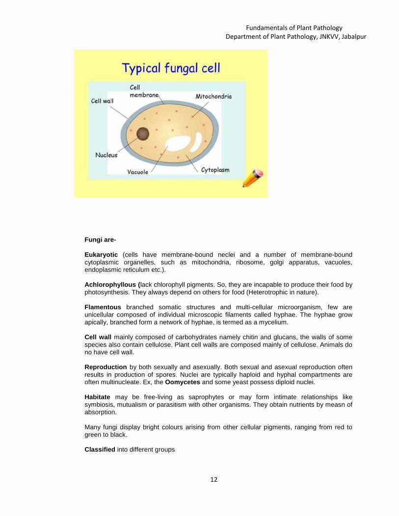
Fungi are-
Eukaryotic (cells have membrane-bound neclei and a number of membrane-boundcytoplasmic organelles, such as mitochondria, ribosome, golgi apparatus, vacuoles,endoplasmic reticulum etc.).
Achlorophyllous (lack chlorophyll pigments. So, they are incapable to produce their food byphotosynthesis. They always depend on others for food (Heterotrophic in nature).
Flamentous branched somatic structures and multi-cellular microorganism, few areunicellular composed of individual microscopic filaments called hyphae. The hyphae growapically, branched form a network of hyphae, is termed as a mycelium.
Cell wall mainly composed of carbohydrates namely chitin and glucans, the walls of somespecies also contain cellulose. Plant cell walls are composed mainly of cellulose. Animals dono have cell wall.
Reproduction by both sexually and asexually. Both sexual and asexual reproduction oftenresults in production of spores. Nuclei are typically haploid and hyphal compartments areoften multinucleate. Ex, the Oomycetes and some yeast possess diploid nuclei.
Habitate may be free-living as saprophytes or may form intimate relationships likesymbiosis, mutualism or parasitism with other organisms. They obtain nutrients by measn ofabsorption.
Many fungi display bright colours arising from other cellular pigments, ranging from red togreen to black.
Classified into different groups

Fundamentals of Plant PathologyDepartment of Plant Pathology, JNKVV, Jabalpur
13
Nutrition (Saprophytes, Obligate Saprophytes, Facultative Parasite, Parasite, Pathogen,Obligate Parasite, Facultative Saprophytes, Necrotroph) growth habit, ability to grow onartificial media,
Mode of reproduction- (Sexual, Asexual), presence or absence of different structures,mode of survival etc. Majority of the plant diseases we have seen are caused by the fungiand cause heavy loss to the growers.Plant diseases are grouped in many ways :-
(A) Symptoms they causes eg. Rots, wilts, spots, blights, rust, smut(B) Plant organs they affect eg. Stem diseases, Foliar diseases, Root diseases(C) Types of plants species affected eg. Field crops, vegetables, flowers, turf
Some of the important plant diseases caused by fungi are:
Name of the diseases Causal organismLate blight of potato Phytophthora infestansRice blast disease Pyricularia griseaBrown spot of rice Dreschlera oryzaeCoffee rust Haemilea vestarixDowny mildew of grapes Plasmopara viticolaRust of wheat Puccinia graminis tritici
Phytopathogenic Bacteria
Bacteria are prokaryotes. It consists of a single cell with a simple internal structure. Unlikeeukaryotic DNA, which is neatly packed into a cellular compartment called the nucleus,bacterial DNA floats free, in a twisted thread-like mass called the nucleoid. Bacterial cellsalso contain separate, circular pieces of DNA called plasmids.
Important characteristics of Phytopathogenic Bacteria:
Shape- Straight to curved rods with rigid cell walls (except filamentous bacteria)Some bacteria assume irregular shapes like “ V, Y, L”Example- Corynebacterium (clavibacters) – ‘V’ shape
Agrobacterium and Erwinia - ‘L’ shape
Carbohydrate - decomposition is mostly aerobic or oxidativeExample- Erwinia (facultative anaerobe)
Mostly Gram negative, rarely Gram positive
Fundamentals of Plant PathologyDepartment of Plant Pathology, JNKVV, Jabalpur
14
Example Gram +ve – Streptomyces, Corynebacterium, Clavibacter
PPB can be cultured on artificial media. However, pathogenic bacteria grow slowlycompared to saprophytes. Majority are flagellate
PPB can be identified based on flagellation, carbohydrate metabolism and pigmentproduction. These are passive invaders i.e. enter into the plants through wounds or naturalopenings.
Survival – The bacteria survive inside or outside of seed, and in plant debris and spreadsby means of water, rain, insects and agricultural implements.
All are susceptible to phages. (Phages are virus that kills bacteria)
All are non-spore forms except Bacillus Sp.
None of them cause human and animal diseases
Cell wall rigid and made up of chitin or cellulose
Incubation period – 36-48 hrs at 25 0C
Habitate – Saprophytic, symbionts
Distribution – About 1600 species of bacteria are present in air, water, soil and infood items all over world
Dispersal- depend on outside agents. They do not spread on the wind as many fungido. They disseminated by means of splashing water. Many bacterial canspread simply by touching an infected plant and then by touching ahealthy plant. They also transmitted by insect vectors, nematode andhuman.
Penetration- cannot penetrate the cuticle of plants but enter the plant through awound or natural opening.
Some of the important diseases caused by bacteria are :
Name of the diseases Causal organismFire blight of apple and pear Erwinia amylovoraBacterial Leaf Blight of rice Xanthomonas campestris pv. oryzaeBacterial wilt o solanaceous crops Ralstonia solanacearusCiturs canker Xanthomonas campestris pv. citriCommon scab of potato Streptomyces scabisCrown gall of apple Agrobacterium tumefaciens
Classification of Bacteria
Bacteria can be classified based on the following characterstics:-
1. Based on shape- Coccus, Bacillus, Spirillum, Vibrio2. Based on staining characters- Gram positive bacteria, Gram negative

Fundamentals of Plant PathologyDepartment of Plant Pathology, JNKVV, Jabalpur
15
bacteria
3. Based on arrangement of flagella- Atrichous bacteria, Monotrichous bacteria,Amphitrichous bacteria, Lopotrichousbacteria, Peritrichous bacteria.
4. Based on nutrition requirement- Autotrophs, Heterotrophs, Chemoautotrophs,Saprophytic bacteria, Symbiotic bacteria,Pathogenic bacteria
5. Based on oxygen requirement- Aerobes, Anaerobes6. Based on temperature dependence- Thermophilic, Mesothermic, Hypoctermic7. Based on DNA relatedness (phylogenic relationship)
Fastidious Vascular Bacteria
Fastidious vascular bacteria are similar to the phytopathogenic bacteria but they areintroduced directly into the sugar-rich phloem sieve tubes or into the water transportingxylem elements by vascular-feeding insects.
The fastidious vascular bacteria may be Fastidious phloem limited bacteria and FastidiousXylem Limited bacteria (XLB).
The fastidious Phloem limited bacteria are very small bacilli and generally possess Gram-negative prokaryotic cell morphology.
Fastidious xylem limited bacteria are Walled bacterial pathogens those inhabit the watertransporting cells of the xylem of their host plants and are transmitted by xylem feedingsharpshooters and spittlebugs, members of the leafhopper family.
Example- Citrus greening disease, Cucurbit yellow vine, Almond leaf scorch, Alfalfadwarf, Phony Peach, Ratoon stunting, Plum leaf scald.

Fundamentals of Plant PathologyDepartment of Plant Pathology, JNKVV, Jabalpur
16
Pathogenesis, Factors Affecting Disease Development, DiseaseTriangle and Tetrahedron.
K. S. Baghel & [email protected]
Krishi Vigyan Kendra, Rewa-------------------------------------------------------------------------------------------------------------------------
PATHOGENESIS :Pathogenesis is a series of events that happened in succession during a pathogenic
relationship of a pathogen and host which leads to disease, therefore in other terms
it also refers to the origin, development and resultant effects of disease from the
initial appearance of disease all the way to its end stages .The term pathogenesis
comes from Greek word for ‘disease’ and ‘beginning’. The study of pathogenesis is
important to diagnose and manage diseases.
FACTORS AFFECTING DISEASE DEVELOPMENT.
Disease development in brief involves a number of distinct events including the
dissemination of pathogen to the host, prepenetration, penetration of the pathogen
into the host, invasion and spread of the pathogen, reproduction of pathogen and
the survival of pathogen.
There are seven major events in disease cycle;
Inocultation
Penetration
Infection
Invasion
Reproduction
Dissemination
Survival
L. 05

Fundamentals of Plant PathologyDepartment of Plant Pathology, JNKVV, Jabalpur
17
Inoculation: It is the process by which the pathogen come in contact with its host.
Inoculation is the initial contact of a pathogen with a site of plant where infection is
possible. The pathogen that lands or brought into contact with the plants is called
the inoculum.
Types of Inoculum :
Primary Inoculum
Secondary Inoculum
Inoculum that survives dormant in off season and originate infection during crop
season is called primary inoculums, and the infection it caused are called primaryinfections. An inoculum produced from primary infection is called a secondaryinoculum and the infection it caused are called secondary infections.
Source of Inoculum:
Soil: Bacteria, fungus viz Rhizoctonia, Sclerotinia
Infected plant parts: Seed, cuttings, bulbs, corns, tubers etc.
Diseased debris: Alternaria , Pytophtohra
Alternate hosts: Rust
Collateral hosts: Viruses, Powdery mildews etc.
Penetration: The process by which pathogen enter its host. This process is divided
into
Pathogens penetrate plant surfaces by direct penetration of cell wall, through
natural openings, or through wounds. Most of the fungi penetrate through one of
these ways. Bacteria enter in plants mostly through wounds, whereas viruses and
viroids and protozoa enter through wounds made by vectors. Parasitic higher plants
enter into the host by direct penetration and nematodes enter plants directly or
sometimes through natural openings.
Infection: It is the process by which pathogens establish contact with susceptible
cells or tissues of the host and procure nutrients from them in other terms

Fundamentals of Plant PathologyDepartment of Plant Pathology, JNKVV, Jabalpur
18
establishment of organic relationship of the pathogen with susceptible cells of the
host called infection.
Types of infection : Successful infections result in the appearance of symptoms, i.e.
discolored , malformed, or necrotic areas on the host plants termed as local or
systemic infection. Some infection, remain latent, they do not produce symptoms
right away but at a later they appears during favorable environmental conditions and
suitable host.
Invasion: The spread of pathogens into the host are called invasion. Various
pathogens invade hosts in different ways and to different extent.
Ectoparasite : When pathogenic fungi produce mycelium only on the surface
of the plant but send haustoria into the epidermal cells. E.g. Powdery mildew
Endoparasites : Some fungi produce mycelium that grows only in the area
between cuticle and the epidermis. e.g. wilts, viruses etc. Endoparsites
further divides into sub-cuticular pathogens (Apple scab), sub-epidermal
pathogens (Rust) and vascular pathogens (Pseudomonas solanacearum)
Ecto-endo parasites : Most fungi spread into all the tissues of the plant
organs either by growing directly through the cells as an intracellular
mycelium or by growing between the cells as an intercellular mycelium. e.g.
Potato canker (Corticium solani).
Reproduction: Plant pathogens reproduce in a variety of ways.
Fungi reproduce by means of spores, which may be either asexual
(mitospores, i.e. products of mitosis) or sexual (meiospores, i.e. products of
meiosis). Parasitic higher plants reproduce just like plants, i.e. by seeds.
Bacteria and mollicutes reproduce by fission in which one mature individual
split into two. Viruses and viroids are replicated by the cell. Nematodes
reproduce by means of eggs.

Fundamentals of Plant PathologyDepartment of Plant Pathology, JNKVV, Jabalpur
19
Dissemination: It refers to transfer of inoculum either passively i.e. by wind, water,
insect, man, animals, machinery or actively move on own power some bacteria,
pythium (Zoospores) fungal spores expelled forcibly.
Survival :Survival of pathogens takes place through-
Infected crop debris
Seeds
In soil
On growing plants
Infected material on host plants
In propagating material
Alternate host
Collateral host
As dormant structures e.g. sclerotia, chlamydospores.
Disease Triangle
The disease triangle drawing most likely was first published by Stevens in 1960.
Although earlier plant pathologists certainly recognized the interaction among plant,
pathogen, and environment. They refers it as existence of a disease caused by a
biotic agent absolutely requires the interaction of a susceptible host, a virulent
pathogen, and an environment favorable for disease development.

Fundamentals of Plant PathologyDepartment of Plant Pathology, JNKVV, Jabalpur
20
Three factors must be present at the same time for a plant disease to occur. If any
one of the three is missing, plant disease does not occur.
The factors are:
A susceptible host
A virulent pathogen (one that can cause disease), and
A suitable environment
Tetrahedron: Plant pathologists have elaborated on the disease triangle by adding
one or more parameters .Additional parameters have included humans, vectors,
and time. Of these, only time is absolutely required so other elements represent
special case applications. A three-dimensional disease pyramid or tetrahedron has
been the most common figure drawn after addition of a single parameter. Adding
more than one parameter while retaining the pyramidal shape is possible by
drawing a base with four line segments.
Humans factor into the disease triangle because the influence of human activity on
disease is pervasive in agriculture and, perhaps to a lesser degree, in lower input
systems such as forestry and range management. Indeed, it is difficult to ignore

Fundamentals of Plant PathologyDepartment of Plant Pathology, JNKVV, Jabalpur
21
such elements as cultivation practices that affect a pathogen's life cycle, genetic
manipulation of plant hosts through breeding and genetic engineering, planting
large expanses of genetically similar plant populations, and various environmental
manipulations such. These factors can profoundly affect the occurrence and
severity of a particular disease.
Some important terminologies in Plant Pathology:
1. Alternate host One of two kinds of plants on which a parasitic fungus
(e.g., rust) must develop to complete its life cycle.2. Appressorium The swollen tip of a hypha or germ tube that facilitates
attachment and penetration of the host by a fungus.3. Collateral host The wild host of same families of pathogen in which
pathogen survive when primary host is not available.4. Concomitant
contaminationPropagules of pathogen present along with the seed.
5. Contamination Intermixed with a pathogen, (of spore) not pure.6. Cross protection The protection of host from disease by inoculating a
strain or isolate, closely related to the pathogen.
(protection maybe by competition or by induced
resistance)7. Disease Any malfunctioning of host cells and tissues that results
from continuous irritation by a pathogenic agent or
environmental factor and leads to development of
symptoms.8. Disease complex Plant disease where more than one pathogen is involved
in infection process.9. Disease cycle The chain of events involved in disease development,
including the stages of development of the pathogen and
the effect of the disease on the host.10. Disease The set of varying symptoms characterizing a disease
Fundamentals of Plant PathologyDepartment of Plant Pathology, JNKVV, Jabalpur
22
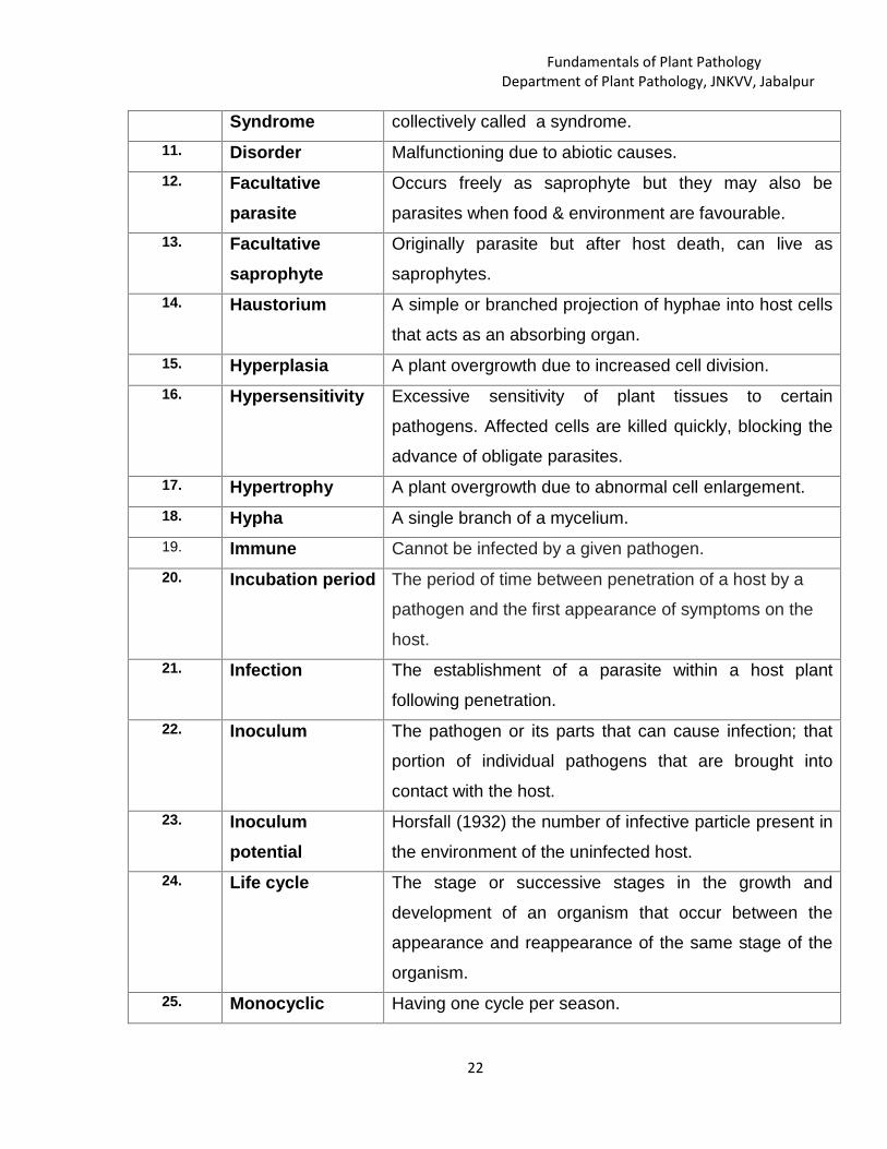
Syndrome collectively called a syndrome.11. Disorder Malfunctioning due to abiotic causes.12. Facultative
parasiteOccurs freely as saprophyte but they may also be
parasites when food & environment are favourable.13. Facultative
saprophyteOriginally parasite but after host death, can live as
saprophytes.14. Haustorium A simple or branched projection of hyphae into host cells
that acts as an absorbing organ.15. Hyperplasia A plant overgrowth due to increased cell division.16. Hypersensitivity Excessive sensitivity of plant tissues to certain
pathogens. Affected cells are killed quickly, blocking the
advance of obligate parasites.17. Hypertrophy A plant overgrowth due to abnormal cell enlargement.18. Hypha A single branch of a mycelium.19. Immune Cannot be infected by a given pathogen.20. Incubation period The period of time between penetration of a host by a
pathogen and the first appearance of symptoms on the
host.21. Infection The establishment of a parasite within a host plant
following penetration.22. Inoculum The pathogen or its parts that can cause infection; that
portion of individual pathogens that are brought into
contact with the host.23. Inoculum
potentialHorsfall (1932) the number of infective particle present in
the environment of the uninfected host.24. Life cycle The stage or successive stages in the growth and
development of an organism that occur between the
appearance and reappearance of the same stage of the
organism.25. Monocyclic Having one cycle per season.
Fundamentals of Plant PathologyDepartment of Plant Pathology, JNKVV, Jabalpur
23
26. Mosaic Symptom of certain viral diseases of plants characterized
by intermingled patches of normal and light green or
yellowish color.27. Mottle An irregular pattern of indistinct light and dark areas.28. Mycelium Mass of hyphae that make up the body of a fungus.29. Obligate parasite parasite which have not yet been grow apart from living
cells. Can not grow in culture eg. Rust, powdery mildew.30. Parasite An organism living on or in another living organism (host)
and obtaining its food from the latter.31. Pathogen An entity that can incite disease.32. Pathogenesis Chain of events that leads to development of disease.33. Pathogenicity The capability of a pathogen to cause disease.34. Polycyclic Completes many (life or disease) cycles in one year.35. Primary infection The first infection of a plant by the over wintering or over
summering pathogen.36. Primary
inoculumThe over wintering or over summering pathogen, or its
spores that cause primary infection.37. Resistance The ability of an organism to exclude or overcome,
completely or in some degree, the effect of a pathogen or
other damaging factor.38. Secondary
infectionAn infection caused by secondary inoculum.
39. Secondaryinoculum
Inoculum produced by infections that take place during
the same growing season.40. Sign The pathogen or its parts or products seen on a host
plant.41. Symptom The external and internal reactions or alterations of a
plant as a result of a disease.42. Virulence The degree of pathogenicity of a given pathogen.

Fundamentals of Plant PathologyDepartment of Plant Pathology, JNKVV, Jabalpur
24
Classification of Plant Diseases
S.P. [email protected]
JNKVV, College of Agriculture, Rewa--------------------------------------------------------------------------------------------------------------
A) On the basis of their mode of perpetuation and mode of primaryinfection:1) Soil borne diseases: In these diseases, the pathogens survive in soil or
on infested plant debris lying in soil either as their resting spores or as
mycelia strands and rhizomorphs. They all attack the root system of host
plants.
Eg: Damping off (Pythium sp.),Seedling blight (Phytophthora, Fusarium
sp.)
2) Air borne diseases: Some pathogens infects the host plant through air
and bring primary as well as secondary infection.
Eg: Rusts, Powdery mildews.Loose smuts bring about secondary
infection through air.
3) Seed borne diseases: Some pathogens survive as dormant mycelium in
the seeds or other propagative structures of host plants. Eg. Loose smut
of wheat (internally seed borne)
B) On the basis of their cause, diseases are classified as:1) Infectious plant diseases: These diseases are caused by living
agents,the pathogen. All pathogens are parasitic on plants. These are
characterised by the ability of the pathogen to grow and multiply rapidly.
Ex: Powdery mildews, Rusts.
2) Non-infectious diseases: These diseases do not spread from plant to
plant (non-infectious).These diseases are caused due to abiotic
factors(non parasitic or physiological). Eg: Black heart of potato.
L. 06

Fundamentals of Plant PathologyDepartment of Plant Pathology, JNKVV, Jabalpur
25
C) On the basis of production and spread of the inoculum:1) Single cycle disease or simple interest disease: In single cycle
disease the increase of disease is mathematically analogous to simple
interest disease.
2) Multiple cycle or compound interest disease: In multiple cycle
diseases, the increase in disease is mathematically analogous to
compound interest of money.
3) Polyetic diseases: These are also polycyclic diseases but they complete
their disease cycle in more than one year over years. Eg.: Cedar Apple
Rust
D) On the basis of plant parts affected:1) Localized: If they affect only specific organs or parts of the plants. Eg.:
Root
Rot, Leaf spot.
2) Systemic: If entire plant is affected. Eg.: Downy mildew, damping off.
E) On the basis of group of causal organisms:1) Fungal disease: Caused by plant pathogenic fungi. Eg. Anthracnose2) Bacterial disease: Caused by plant pathogenic bacteria. Example:
Citrus canker
3) Viral disease: Caused by plant viruses. Eg. Rice tungro disease
4) Phanerogamic phytopathogenic diseases: Caused by phanerogamic
plant parasites. Eg. Striga, Cuscutta.
5) Nematode Diseases: Diseases caused by plant pathogenic nematodes.
Eg. Ear cocle of wheat.
F) On the basis of occurrence and consequent effects:1) Endemic: When a diseases more or less constantly prevalent from year
to year in a moderate to severe form in a particular country. E.g., Wart
disease of potato is endemic to Darjeeling.
Fundamentals of Plant PathologyDepartment of Plant Pathology, JNKVV, Jabalpur
26
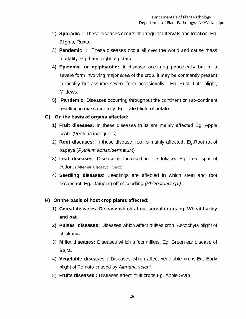
2) Sporadic : These diseases occurs at irregular intervals and location. Eg.
Blights, Rusts.
3) Pandemic : These diseases occur all over the world and cause mass
mortality. Eg. Late blight of potato.
4) Epidemic or epiphytotic: A disease occurring periodically but in a
severe form involving major area of the crop. it may be constantly present
in locality but assume severe form occasionally . Eg. Rust, Late blight,
Mildews.
5) Pandemic: Diseases occurring throughout the continent or sub-continent
resulting in mass mortality. Eg. Late blight of potato.
G) On the basis of organs affected:1) Fruit diseases: In these diseases fruits are mainly affected Eg. Apple
scab. (Venturia inaequalis)
2) Root diseases: In these disease, root is mainly affected. Eg.Root rot of
papaya.(Pythium aphanidermatum)
3) Leaf diseases: Disease is localised in the foliage. Eg. Leaf spot of
cotton. ( Alternaria gossypii (Jacz.)
4) Seedling diseases: Seedlings are affected in which stem and root
tissues rot. Eg. Damping off of seedling.(Rhizoctonia sp.)
H) On the basis of host crop plants affected:1) Cereal diseases: Disease which affect cereal crops eg. Wheat,barley
and oat.2) Pulses diseases: Diseases which affect pulses crop. Ascochyta blight of
chickpea.3) Millet diseases: Diseases which affect millets. Eg. Green ear disease of
Bajra.
4) Vegetable diseases : Diseases which affect vegetable crops.Eg. Early
blight of Tomato caused by Altrnaria solani.
5) Fruits diseases : Diseases affect fruit crops.Eg. Apple Scab

Fundamentals of Plant PathologyDepartment of Plant Pathology, JNKVV, Jabalpur
27
6) Ornamental plant diseases: Diseases affecting ornamental plants. Eg.
Chrysanthemum stunt.
7) Forest diseases: Diseases affecting forest trees and plantation. Eg.Sudden Oak Death (Phytophthora ramorum)
I) On the basis of symptoms produced on host plants:1) Rusts : Caused by Basidiomycetes of the order Uredinales. Eg. Stem
rust of wheat.2) Smuts : Caused by fungus of order Uredinales,mass of black powedery
spores and grains are not produced .Eg. Loose smut of wheat caused by
Ustilago nuda tritici.
3) Wilts :In this disease, the vascular system of plant is affected .Eg.
Bacterial wilt of cucurbits caused by Erwinia trachiephila.
4) Powdery mildews: It is a disease of foliage, stem, flower and
fruit.Eg.Powdery mildew of grapes.
5) Rots : In this disease, underground part of plant is infected.Eg.
Rhizoctonia and Phytophthora root rot.
6) Blight : It is complete chlorosis ,yellowing and browning which results in
death of the plant. Eg.Leaf blight of paddy.
7) Leaf spots :It may be caused by fungi or bacteria, spots are formed on
leaves which results in complete yellowing and dropping of leaf.
Eg.Septoria leaf spot in tomato
8) Canker : Canker is a dead area in bark or cortex of woody stem. Ex:
Citrus canker
10 Anthracnose: an ulcer-like lesion that can be necrotic and sunken. These
lesions can appear on the fruit, flowers and stems of the host - e.g. Apple
Anthracnose of stems and or leaves (Cryptosporiopsis sp. Formally Pezicula sp.), or
Dogwood Anthracnose (Discula distructiva)
11. Damping Off: it is a rapid collapse and death of very young seedling. Either the
seed rots before emergence or the seedling rots at the soil line and falls over and
dies. The most common genera involved are Fusarium, Rhizoctonia and Pythium.
Fundamentals of Plant PathologyDepartment of Plant Pathology, JNKVV, Jabalpur
28
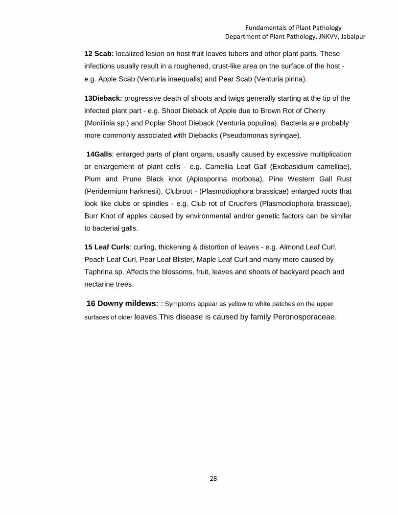
12 Scab: localized lesion on host fruit leaves tubers and other plant parts. These
infections usually result in a roughened, crust-like area on the surface of the host -
e.g. Apple Scab (Venturia inaequalis) and Pear Scab (Venturia pirina).
13Dieback: progressive death of shoots and twigs generally starting at the tip of the
infected plant part - e.g. Shoot Dieback of Apple due to Brown Rot of Cherry
(Monilinia sp.) and Poplar Shoot Dieback (Venturia populina). Bacteria are probably
more commonly associated with Diebacks (Pseudomonas syringae).
14Galls: enlarged parts of plant organs, usually caused by excessive multiplication
or enlargement of plant cells - e.g. Camellia Leaf Gall (Exobasidium camelliae),
Plum and Prune Black knot (Apiosporina morbosa), Pine Western Gall Rust
(Peridermium harknesii), Clubroot - (Plasmodiophora brassicae) enlarged roots that
look like clubs or spindles - e.g. Club rot of Crucifers (Plasmodiophora brassicae),
Burr Knot of apples caused by environmental and/or genetic factors can be similar
to bacterial galls.
15 Leaf Curls: curling, thickening & distortion of leaves - e.g. Almond Leaf Curl,
Peach Leaf Curl, Pear Leaf Blister, Maple Leaf Curl and many more caused by
Taphrina sp. Affects the blossoms, fruit, leaves and shoots of backyard peach and
nectarine trees.
16 Downy mildews: : Symptoms appear as yellow to white patches on the upper
surfaces of older leaves.This disease is caused by family Peronosporaceae.

Fundamentals of Plant PathologyDepartment of Plant Pathology, JNKVV, Jabalpur
29
Abiotic Diseases and their managementVibha Pandey
[email protected], College of Agriculture, Rewa
------------------------------------------------------------------------------------------------------------
Plants can be damaged by infectious microbes and; also by noninfectious factors
that are collectively termed as "a biotic diseases" or "a biotic disorders". A biotic
disorders are caused by nonliving factors, such as drought stress, sunscald, freeze
injury, wind injury, chemical injury, nutrient deficiency, or improper cultural practices,
such as overwatering or planting conditions. In contrast to biotic factors, a biotic
problems often affect several species or plants of various ages; typically, damage is
relatively uniform, doesn't spread and is often not progressive. A biotic problems are
not associated with pests. They are often caused by a single incident and are
related to environmental or physical factors or cultural practices. Once the
responsible factor has dissipated and is no longer affecting the plant, the plant may
grow out of the problem and develop new, normal appearing foliage. The common
abiotic disorders in plants are discussed below.
1. Nutrient deficiencies and toxicity.
1.1 Nutrient deficiencies
Nutrient deficiency reduces shoot growth and leaf size, cause leaf chlorosis,
necrosis and dieback of plant parts. However, nutrient deficiencies cannot be
reliably diagnosed on the basis of symptoms alone because numerous other
plant problems can produce similar symptoms. There are general symptoms
that can be expressed by deficiencies of nutrients but usually leaf and/or soil
samples are needed to confirm the problem. Plant nutrient deficiencies are best
diagnosed using plant tissue analysis. As opposed to soil nutrient analysis, plant
tissue analysis allows one to determine plant nutrient uptake rather than plant
nutrient availability. Because nutrient deficiencies lack visible signs, they are
often mistaken for virus diseases. One of the best ways to diagnose nutrient
disorders is the distribution of symptoms on the plant. Mobile nutrients are
readily transferred within the plant to the growing points so symptoms appear on
L. 07

Fundamentals of Plant PathologyDepartment of Plant Pathology, JNKVV, Jabalpur
30
the lower (older) leaves of the plant. Conversely, immobile nutrients display
symptoms on the meristem of the plant during nutrient deficiencies.
Nitrogen deficiencies may also result from infection by root pathogens such as
root-knot nematodes (Meloidogyne spp.). Nitrogen deficiencies can cause
increased susceptibility to certain leaf pathogens such as Alternaria solani, while
excessive plant N levels may result in increased susceptibility to other
pathogens such as Botrytis cinerea, or Rhizoctonia solani. However, phosphorus
deficiency can result in poor growth and stunting, a blue/green hue to the leaves,
and/or purple-colorations to stems and undersides of the leaves. Symptoms of
iron deficiency first develop in the new growth which appears as yellow-green
leaves, often with a striped appearance. Whereas, symptoms of K deficiency
include necrosis (tissue death) on leaf margins, leaf curling and browning, and
interveinal chlorosis. Plants that are deficient in K can be more prone to frost
damage as well as certain diseases. Blossom end rot is a common symptom of
Ca deficiency on fruits while other symptoms manifest as plant stunting,
localized tissue necrosis, and leaf marginal chlorosis. Blossom end rot and other
fruit disorders caused by Ca deficiency (e.g., bitter pit of apples) can often lead
to secondary colonization by fungi. Moreover, Ca is a component of host
response proteins to pathogen toxins, such as oxalic acid, which is utilized by
some fungi such as Sclerotium rolfsii during infection. Magnesium deficiency
results in chlorotic and necrotic tissues with an orange, red, or brownish color.
Yellowing of the leaf margin is also common on many plant species. Early leaf
senescence may also occur, particularly on older leaves as Mg is easily
translocated through the plant. Over-application of K and/or Ca, which competes
at soil cation exchange sites, can also lead to Mg deficiency. Similarly, over-
application of Mg can lead to Ca deficiency; it is important to maintain a suitable
Ca:Mg ratio in agricultural soils. Foliar and granular applications of Epsom salt
can be used to remedy Mg deficiency in crop plants
Micronutrient toxicities are common in many production systems. Symptoms
often include chlorosis or necrosis on leaf margins or tips, but leaf spotting,

Fundamentals of Plant PathologyDepartment of Plant Pathology, JNKVV, Jabalpur
31
flecking, and other symptoms can also occur. Nutrient toxicity are triggered by
excessively low or high soil pH. Micronutrient toxicities are particularly common
in greenhouse floriculture. For example, Fe and Mn toxicity often occurs in
greenhouse crops when the growing medium has a low pH. Excessive
micronutrients can also occur when irrigation water or soil has significant
concentrations of micronutrients.
1.2 Herbicide, insecticide and fungicide phytotoxicity.
Some herbicides cause root stunting or swelling, and could be confused with
damage from nematodes. Others herbicides cause necrotic/chlorotic spots or
blotches that resemble to foliar disease. Some herbicides cause mottled colors,
distortion or vein banding that has similarity to viral disease. For example, the
phenoxy herbicide 2-4D, a synthetic auxin, causes distortion in grape, cotton,
tomatoes, and many other plants, which could be confused with a virus disease.
Sometimes Diuron also causes discoloration along the veins in grapevine which
could be confused with a virus disease or a nutritional problem. Insecticides and
fungicides occasionally cause obvious plant damage and its symptoms vary
widely. Generally, flower petals are more susceptible to damage from pesticide
applications than leaves. The younger and more tender the leaves, the more
susceptible they are to injury from pesticide applications. Hot weather can
exacerbate the damage due to the chemicals cause. Pesticides that have
systemic action can have a more profound effect. Some active ingredients can
adversely affect the photosynthetic mechanism or other physiological processes
and can result in a general leaf chlorosis, interveinal chlorosis, leaf curling and
stunting. Emulsifiable concentrate (EC) formulations, soaps and oils can
adversely affect the waxy surface layer that protects the leaf from
desiccation. Applications with these products can result in the loss of the shiny
appearance of a leaf, leaf spotting and necrosis. Pesticides applied as soil
drenches can cause poor germination, seedling death, or distorted plant growth.
2. Physiological and Genetic Disorders

Fundamentals of Plant PathologyDepartment of Plant Pathology, JNKVV, Jabalpur
32
2.1 Physiological Disorders
There are numerous disorders that can occur because of environmental extremes
such as light, temperature, water, or wind.
2.1.1Sunburn is damage to foliage and other herbaceous plant parts caused by a
combination of too much light and heat and insufficient moisture. A yellow or brown
area develops on foliage, which then dies beginning in areas between the
veins. Sunscald is damage to bark caused by excessive light or heat. Damaged
bark becomes cracked and sunken. High temperatures coupled with low soil
moisture, plants may exhibit scorching on the margins of the leaves, premature leaf
drop, and in severe cases entire plant death. Sometimes physiological changes
result in abnormal color or growth habits. For example, geranium (Pelargonium
spp.) when subjected to temperatures above 95°F (35°C), newly forming leaves
may become "bleached" or white in color.
2.1.2 Frost damage causes shoots, buds and flowers to curl, turn brown or black
and die. Hailstones injure leaves, twigs, and in serious cases even the
bark. Chilling damage in sensitive plants can cause wilting of foliage and flowers
and development of dark water-soaked spots on leaves that can eventually turn
light brown or bleached, and die. Low-temperature injury can vary depending on the
time of year and plant species. Newly expanding shoots are more sensitive than
mature plant parts. If freezing temperatures are encountered after spring bud break,
shoots can be severely injured or even killed. Chilling temperatures (above 32°F;
0°C) can damage newly expanding plant parts, resulting in a purplish coloration of
foliage and possible necrosis. Woody parts of plants can also be injured by sub-
freezing temperatures. Bark can crack, thereby exposing underlying wood to attack
by pathogens or insects. Bark cracking from freezing injury greatly increases the
susceptibility to infection by pathogens such as Agrobacterium tumefaciens, which
causes crown gall on many ornamentals. Cold temperatures combined with freezing
rain can also result in ice accumulation on limbs of woody plants, which can cause
severe breakage.
Fundamentals of Plant PathologyDepartment of Plant Pathology, JNKVV, Jabalpur
33
2.1.3 In closed environments such as greenhouses and nursery storage areas,
plants can be exposed to toxic levels of ethylene gas. Toxic levels of ethylene gas
can cause premature abscission of flower buds, petals and leaves. Other
symptoms include wilted flowers, chlorosis, twisted growth or downward bending of
stems and leaves and undersized or narrow leaves.
2.1.4 In open environments, exposure of nursery plants to air pollutant gases such
as ozone, carbon monoxide, nitrous oxides and sulfur dioxide can cause
damage. Typical symptoms vary widely, but include slow growth and discolored,
dying, or prematurely dropping foliage. Damage is often found where plants are
located near sources of polluted air such as near freeways or industries or where
weather and topography concentrate the pollutants.
2.1.5 Chronic excess of water, plants appear stunted and have underdeveloped
shoots. In severe cases bleeding cankers on stems can occur. Adventitious roots
may form at the root crown. Bark can split and wood may become water-soaked
and discolored. Edema or corky, blister-like swelling can occur on the underside of
leaves on plants growing in waterlogged soils. Edema can be worse during cloudy,
overcast periods. In areas where waterlogged soils prevail for long periods of time,
an odor of rotten eggs may be noticeable, due to sulfur gas production in the
anaerobic soil.
2.2 Genetic Disorders
Sometimes plants or plant shoots exhibit an unusual and sudden change
of color producing discrete markings of variegation. For example, a
plant with entirely green leaves suddenly produces a shoot that has
leaves with edges lacking green pigment, stripes, or blotches. A new
shoot such as this is probably a chimera. It is produced when a
genetic mutation occurs in a specific region of the growing tip
resulting in a section with genetically different cells. The ostensible
result of the genetic change is dependent on the arrangement of the
genetically different cells in the shoot tip and their expression. This

Fundamentals of Plant PathologyDepartment of Plant Pathology, JNKVV, Jabalpur
34
can lead to sometimes bizarre variegation forms or sometimes forms that
are quite desirable. Sometimes variegation can be caused by viruses.
Viruses usually cause non-uniform chlorosis, such as mosaics, while
chimeras usually produce patterned forms such as variegation of color on
leaf margins, stripes, or complete loss of pigment. Some viroids may
also cause bleaching of pigments in leaves; such symptoms, however, are
generally produced throughout the plant and are not restricted to a
single shoot. Some nutrient disorders can cause variegation but these
disorders usually do not arise from a specific shoot as with chimeras.
3. Air Pollution and Damaging Gases
Several gaseous air pollutants can cause injury to plants, including ozone,
ethylene gas and sulfur dioxide.
3.1 Ozone
Ozone is produced when components of combustion/vehicle emissions such as
hydrocarbons and nitrogen oxides react with oxygen and sunlight to form ground
level ozone in the atmosphere. Its effects on plants can be mistaken for infectious
diseases. Ozone can cause flecking, which could be mistaken for mite injury. Ozone
can also cause bronzing, chlorosis and necrosis. Necrosis could be mistaken for a
leaf spot caused by an infectious agent. In conifers, injury can include needle-
banding and tip-burn.
3.2 Ethylene gas
Ethylene gas is another air pollutant, can build up in closed production areas such
as greenhouses. Ethylene is a plant hormone that is utilized to signal ripening and
senescing within plant tissues. It is colorless and odorless, making it difficult to
detect and diagnose. Symptoms are variable, depending on the host species, host
age, the ethylene concentration, and duration of exposure. Symptoms include
curled leaves, flower abortion, distorted/twisted stems, leaf and petal abscission,
and stunting. These symptoms can be mistaken for viral infection or herbicide injury.

Fundamentals of Plant PathologyDepartment of Plant Pathology, JNKVV, Jabalpur
35
Tomatoes, peppers, and vinca are particularly susceptible and may be the first
species to show symptoms in a greenhouse. In some plants, at least some damage
occurs with ethylene levels as low as 0.01 to 0.1 ppm. Concentrations of 1-10 ppm
cause major damage or even plant death.
3.3Sulfur dioxide
Contamination of air due to release of phytotoxic pollutants such as SO 2 may
influence the composition of environment and consequently the host parasite
relationship. SO2 enters in plants through open stomata, the gas reacts with
moisture and is converted into acid. The sulphite ions are about 30 times
more toxic than sulphate ions. Two general types of symptoms designated as
chronic and acute are produced by the plants due to the accumulation of
sulphate and sulphite ions in the leaf tissue. Chronic injury occurs on
exposure of plants to low concentration of SO2 (less than 100 ppb) at which
the rate of accumulation of the ions is slow the cells oxidize the sulphite ions
and injury occurs until sufficient sulphate ions accumulate. This type of
chronic injury is characterized by a general chlorotic appearance of the
leaves. Cells are not killed but the chlorophyll is bleached which appears as a
mild chlorosis or yellowing of the leaf or a silvering or bronzing of the lower
leaf surface without necrosis. Acute injury results from the absorption of lethal
quantities of SO2 appears as marginal or intercostal areas of dead tissues,
which are at full grayish green water soaked in appearance. In most plant
species, these areas become bleached in original colour, upon drying and
dead or necrotic areas may fall out leaving a ragged appearance to the leaf.
In case of severe injury abscission layer develops at the base of petiole and
the leaves fall down.
4. Equipment Injury
Human-inflicted damage can have a lasting effect on tree vigor. Lawn mowers,
weed trimmers, or improper pruning with hedge trimmers or chainsaws can gouge,

Fundamentals of Plant PathologyDepartment of Plant Pathology, JNKVV, Jabalpur
36
rip or split the trunk or limbs. Mower damage is a very common type of mechanical
injury to landscape trees. Wounded areas can serve as infection courts (opening
used by pathogens to enter a host) for microbial pathogens or insect pests that can
cause cankers or wood decay problems. Heavy equipment used in the area of trees
and landscape plants can result in compacted soils, reducing oxygen to the root
systems and leading to a slow decline of the tree. Excavation to install sidewalks,
irrigation systems or utility lines has a more direct impact by severing a portion of
the root system. Corresponding branch dieback may show up on the same side of
the tree on which the root system was damaged. Young trees and shrubs may die
or decline within a season or two, while older trees may struggle and decline over a
period of many years.
5. Damage due to Girdling
Girdling from guy wires, string, trunk wraps, vines or roots have a similar impact as
equipment injury. Pressure or constriction of the bark may collapse phloem tissue
and sometimes xylem tissue. The long-term impact of girdling is that fewer
photosynthates go to the root system. This leads to the development of a smaller
root system with less potential for water and nutrient uptake. Over time, affected
trees tend to be smaller than normal, have poor leaf emergence and are less
vigorous. Recovery from this type of injury is difficult.
6.Unusual Plant Growths
Other symptoms that can be mistaken for plant diseases but are actually abiotic in
origin are sloughing bark and odd growths. In trees, bark that is unusually rough or
that separates from the wood may be of concern. If this is a new event, then
concern is probably warranted. If the tree loses its bark every year then this is more
likely a normal occurrence. Sycamore trees tend to slough their bark off in early
summer following a period of rapid growth. Sometimes the bark gradually falls off
and sometimes appears to blow off within a couple of days. This process is normal
and does not affect the health of the tree. Bumpy bark, burls, and other odd plant
growths often look like symptoms of infectious diseases. However, burls,

Fundamentals of Plant PathologyDepartment of Plant Pathology, JNKVV, Jabalpur
37
lignotubers and other growths on trees are usually the result of an injury. Burls
frequently can be small or grow to a very large size. The bark covers the burl and
remains intact rather than appearing cracked or sunken. Lignotubers frequently hold
a cluster of buds that sprout rapidly in the spring. These gnarled growths look odd
but they don't seem to have a major impact on the health of the tree.

Fundamentals of Plant PathologyDepartment of Plant Pathology, JNKVV, Jabalpur
38
General Characters of fungi - Definition of fungus, somatic structures, typesof fungal thalli, fungal tissues, modifications of thallus, reproduction in fungi(asexual and sexual)
Sushma Nema & [email protected]
JNKVV, Jabalpur---------------------------------------------------------------------------------------------------------------
General characters of fungiFungi are the eukaryotic, achlorophyllous, and unicellular or multicellular
organisms, which may reproduce by asexual and sexual spores.
1. All are eukaryotic - Possess membrane-bound nuclei (containing chromosomes)
and a range of membrane-bound cytoplasmic organelles (e.g. mitochondria,
vacuoles, endoplasmic reticulum).
2. Most are filamentous - Composed of individual microscopic filaments called
hyphae, which exhibit apical growth and which branch to form a network of hyphae
called a mycelium.
3. Some are unicellular - e.g. yeasts.
4. Protoplasm of a hypha or cell is surrounded by a rigid wall - Composed
primarily of chitin and glucans, although the walls of some species contain cellulose.
5. Many reproduce both sexually and asexually - Both sexual and asexual
reproduction often result in the production of spores.
6. Their nuclei are typically haploid and hyphal compartments are often
multinucleate - Although the oomycota and some yeast possess diploid nuclei.
7. All are achlorophyllous - They lack chlorophyll pigments and are incapable of
photosynthesis.
8. All are chemoheterotrophic (chemo-organotrophic) - They utilise pre-existing
organic sources of carbon in their environment and the energy from chemical
reactions to synthesize the organic compounds they require for growth and energy.
9. Possess characteristic range of storage compounds - e.g. trehalose,
glycogen, sugar alcohols and lipids.
L. 08

Fundamentals of Plant PathologyDepartment of Plant Pathology, JNKVV, Jabalpur
39
10. May be free-living or may form intimate relationships with other organisms
i.e. may be free-living, parasitic or mutualistic (symbiotic).
ThallusThe body of the fungus is called as 'thallus'.
Eucarpic thallusThe thallus is differentiated into vegetative part, which absorbs nutrients, and
a reproductive part, which forms reproductive structure. Such thalli are called as
eucarpic. e.g. Pythium aphanidermatum.
Holocarpic thallusThe thallus does not show any differentiation on vegetative and reproductive
structure.
After a phase of vegetative growth, it gets converted into one or more reproductive
structures.
Such thalli are called as 'holocarpic' e.g. yeast, Synchytrium endobioticum
HyphaeHyphae is a tubular, transparent filament, usually branched, composed of an
outer cell wall and a cavity (lumen) lined or filled with protoplasm including
cytoplasm. Hyphae are divided into compartments or cells by cross walls called
septa and are generally called as septate (with cross wall) or coenocytic (aseptate -
without cross wall). Hyphae of most of the fungi measure 5-10 μm across.
Mycelium (pl. Mycelia)
The hyphal mass or network of hyphae constituting the body (thallus) of the
fungus is called as mycelium. The mycelium of parasitic fungi grows on the surface
of the host and spread between the cells and it is called intercellular mycelium. The
mycelium of parasitic fungi, which grows on the surface of the host and penetrates
into the host cells and is called intracellular mycelium. If the mycelium is
intercellular, food is absorbed through the host cell walls or membrane. If the
mycelium penetrates into the cells, the hyphal walls come into direct contact with
the host protoplasm. Intercellular hyphae of many fungi, especially of obligate

Fundamentals of Plant PathologyDepartment of Plant Pathology, JNKVV, Jabalpur
40
parasites of plants (fungi causing downy mildews, powdery mildews and rusts)
obtain nutrients through haustoria.
Monokaryotic mycelium (uninucleate)Mycelium contains single nucleus that usually forms part of haplophase in
the life cycle of fungi.
Dikaryotic mycelium (binucleate)Mycelium contains pair of nuclei (dikaryon), which denotes the diplophase in
the life cycle of fungi.
Homokaryotic myceliumThe mycelium contains genetically identical nuclei.
Heterokaryotic myceliumThe mycelium contains nuclei of different genetic constituents.
MultinucleateThe fungal cell contains more than 2 nuclei.
SeptaTransverse septa occur in the thallus of all filamentous fungi to cut off
reproductive cells from the rest of the hypha, to separate off the damaged parts or to
divide the hypha into regular or irregular compartments or cells. There are two
general types of septa in fungi viz., primary and adventitious. The primary septa are
formed in association with nuclear division and are laid down between daughter
nuclei. The adventitious septa are formed independently of nuclear division and are
especially associated with changes in the concentration of the protoplasm as it
moves from one part of the hypha to another.
Transverse septaSepta vary in their construction septa have biological importance in the
lifecycle of fungi. Some are simple whereas others are complex. All types of septa
are formed by centripetal growth from the hyphal wall inward. In some septa, the
growth continues until the septum is a solid plate. In others the septum remains
incomplete, leaving a pore in the centre that may often be plugged or occulted.
Some groups of Basidiomycetes like Auriculariaceae, Tremellaceae,
Aphyllophorales, Agaricales etc (except Ustilaginales and uredinales) have more

Fundamentals of Plant PathologyDepartment of Plant Pathology, JNKVV, Jabalpur
41
complex septa. Surrounding the central pore in the septum is a curved flange of wall
material, which is thickened to form a barrel-shaped or cylindrical structure
surrounding the pore. Septa of this type are termed dolipore septa (L. dolium = a
large jar or cask i . e., barrel).
These septa are often overlaid by perforated cap, which is an extension of the
endoplasmic reticulum. This cap is known as parenthosome or pore cap. Despite
these apparent barriers, there is a good cytoplasmic continuity between adjacent
cells. The septal pore may vary in width from 0.1 to 0.2 μm. Dolipore septa are
found in both monokaryotic and dikaryotic myceli
Fungal cell structure
Fungal cells are typically eukaryotic and have distinguished characteristics
than that of bacteria, and algae. The chief components of cell wall appears to be
various types of carbohydrate or their mixtures (upto 80-90%) such as cellulose,
pectose, callose etc., cellulose predominates in the cell wall of mastigomycotina
(lower fungi) while in higher fungi chitin is present. The living protoplast of the fungal
cell is enclosed in a cell membrane called as plasma membrane or plasmalemma.
Cytoplasm contains organelles such as nucleus, mitochondria, Golgi apparatus,
ribosomes, vacuoles, vesicles, microbodies, endoplasmic reticulum, lysosomes and
microtubules.
The fungal nucleus has nuclear envelope comprising of two typical unit
membrane and a central dense area known as nucleolus, which mainly consist of
RNA. In multinucleate hyphae, the nuclei may be interconnected by the
endoplasmic reticulum. Vacuoles present inside the cell provide turgor needed for
cell growth and maintenance of cell shape. Beside the osmotic function, they also
store reserve materials. The chief storage products of fungi are glycogen and lipid.
The apex of the hyphae are usually rich in vesicles and are called as apical
vesicular complex (AVC) which helps in the transportation of products formed by the
secretary action of golgi apparatus to the site where these products are utilized.
Specialized Somatic StructuresRhizoid

Fundamentals of Plant PathologyDepartment of Plant Pathology, JNKVV, Jabalpur
42
A rhizoid (Gr. rhiza = root + oeides = like) is a short, root-like filamentous
outgrowth of the thallus generally formed in tufts at the base of small unicellular thalli
or small porophores. Rhizoid serves as anchoring or attachment organ to the
substratum and also as an organ of absorption of nutrients from substratum. Rhizoids
are short, delicate filaments that contain protoplasm but no nuclei.
Rhizoids are common in lower fungi like Chytridiomycetes, Oomycetes and
Zygomycetes. Some species produce a many-branched rhizomycelium. This is an
extensive rhizoidal system that usually do not contains nuclei, but through which
nuclei migrate. e.g.
Cladochytrium sp. On rhizomycelium numerous sporangia develop. Such thalli are
polycentric, that is, they form several reproductive centres instead of a single one
where the thallus is termed monocentric.
AppressoriumAppressorium (p1. appressorium; L. apprimere = to press against) is a
simple or lobed structure of hyphal or germ tube and a pressing organ from which a
minute infection peg usually grow and enter the epidermal cell of the host. It helps
germ tube or hypha to attach to the surface of the host or substrates. In addition to
giving anchorage, appressoria help the penetrating hyphae, branches to pierce the
host cuticle.
HaustoriaHaustoria (sing. haustorium; L. haustor = drinker) are special hyphal
structures or outgrowths of somatic hyphae sent into the cell to absorb nutrients.
The hyphal branch said to function as haustorium becomes extremely thin and
pointed while piercing the host cell wall and expands in the cell cavity to form a
wider, simple or branched haustorium. Haustoria may be knob-like or balloon – like
in shape, elongated or branched like a miniature root system.
The hyphae of obligate parasites of plants like downy mildew, powdery mildew or
rust fungi late blight fungus etc ., produce haustoria. Hyphopodia: Hyphopodium (pl.
hyphopodia Gr. hyphe = web + pous = foot) is a small appendage with one or two
cells in length on an external hypha and function as absorbing structures.
Aggregations of hyphae and tissues

Fundamentals of Plant PathologyDepartment of Plant Pathology, JNKVV, Jabalpur
43
a. Mycelial strandMycelial strands are aggregates of parallel or interwoven undifferentiated
hyphae, which adhere closely and are frequently anastomosed or cemented
together. They are relatively loose (e.g. Sclerotium rolfsii growth on culture medium)
compared to rhizomorph. They have no well-defined apical meristem. Mycelial
strand formation is quite common in Basidiomycetes, Ascomycetes and
Deuteromycetes.
b. RhizomorphRhizomorph (Gr. rhiza=root + morphe = shape) is the aggregation of highly
differentiated hyphae with a well defined apical meristem, a central core of larger,
thin walled, cells which are often darkly pigmented. These root-like aggregation is
found in the honey fungus or honey agaric Armillariella mellea (=Armillaria mellea).
They grow faster than the mycelial strands. The growing tip of rhizomorph
resembles that of a root tip. The fungus may spread underground from one root
system to another by means of rhizomorph.
c. Fungal tissuesDuring certain stages of the life cycle of most fungi, the mycelium becomes
organized into loosely or compactly woven tissues. These organized fungal tissues
are called plectenchyma (Gr. plekein = to weave + encyma = infusion i.e., a woven
tissue). There are two types of plectenchyma viz., prosenchyma and
pseudoparenchyma. When the tissue is loosely woven and the hyphae lie parallel to
one another it is called prosenchyma (Gr. pros = toward + enchyma = infusion, i.e.,
approaching a tissue). These tissues have distinguishable and typical elongated
cells. Pseudoparenchyma (Gr. Pseudo = false) consists of closely packed, more or
less isodiametric or oval cells resembling the parenchyma cells of vascular plants.
In this type of tissues hyphae lose their individuality and are not distinguishable.
Cells in prosenchyma are thin-walled and cells in pseudoparenchyma.
Stroma and sclerotiumStromata and sclerotia are somatic structures of fungi.
i. Stroma (pl. stromata; Gr. stroma = mattress)

Fundamentals of Plant PathologyDepartment of Plant Pathology, JNKVV, Jabalpur
44
A stroma is a compact, somatic structure or hyphal aggregation similar to a
mattress or a cushion, on which or in which fructifications of fungi are usually
formed. They may be of various shapes and sizes. Hyphal masses like acervuli,
sporodochia, pionnotes etc. are the fertile stromata, which bear sporophores
producing spores.
ii. Sclerotium (pl. sclerotia; Gr. skeleros = hard)
A sclerotium is a resting body formed by aggregation of somatic hyphae into
dense, rounded, flattened, elongated or horn-shaped dark masses. They are thick-
walled resting structures, which contain food reserves. Sclerotia are hard structures
resistant to unfavourable physical and chemical conditions. They may remain
dormant for longer periods of time, sometimes for several years and germinate on
the return of favourable conditions. The sclerotia on germination may be
myceliogenous and produce directly the mycelium e.g. Sclerotium rolfsii,
Rhizoctonia solani and S. cepivorum (white rot of onion).
They may be sporogenous and bear mass of spores. e.g. Botrytis cinerea. They
may also be carpogenous where in they produce a spore fruit ( ascocarps or
basidiocarps) bearing stalk. e.g. Sclerotinia sp. Claviceps purpurea (ergot of rye).
Development of ascocarps is seen in Sclerotinia, where stalked cups or apothecia,
bearing asci, arise from sclerotia. In Claviceps purpurea, sclerotia germinate and
give rise to drumstick like structures called perithecial stromata, which contain
perithecia, flask-shaped cavities within which the asci are formed.
MycorrhizaeMycorrhiza (pl. mycorrhizae; Gr. mykes = mushroom + rhiza = root) is the
symbiotic association between higher plant roots and fungal mycelia. Many plants in
nature have mycorrhizal associations. Mycorrhizal plants increase the surface area
of the root system for better absorption of nutrients from soil especially when the
soils are deficient in phosphorus.
The nature of association is believed to be symbiotic (mutualism), non-
pathogenic or weakly pathogenic. There are three types of mycorrhizal fungal
associations with plant roots. They are ectotrophic or sheathing or ectomycorrhiza,.
endotrophic or endomycorrhiza and ectendotrophicmycorrhiza.

Fundamentals of Plant PathologyDepartment of Plant Pathology, JNKVV, Jabalpur
45
REPRODUCTIONReproduction is the formation of new individuals having all the characteristics
typical of a species. The fungi reproduce by means of asexual and sexual or
parasexual reproduction. Asexual reproduction is sometimes called somatic or
vegetative and it does not involve union of nuclei, sex cells or sex organs. The
union of two nuclei characterizes sexual reproduction.
ASEXUAL REPRODUCTIONIn fungi, asexual reproduction is more important for the propagation of
species. Asexual reproduction does not involve union of sex organs (gametangia) or
sex cells (gametes) or nuclei. In fungi the following are the common methods of
asexual reproduction.
1. Fragmentation of myceliumMycelial fragments from any part of the thallus may grow into new individuals
when suitable conditions are provided.
2. Fission of unicellular thalliIt is also known as transverse cell division. Reproduction by the method of
fission is are in fungi. Fission is simple splitting of cells into two daughter cells by
constriction and the formation of a cell wall. It is observed in Schizosaccharomyces
spp
3. BuddingBudding is the production of a small outgrowth (bud) from a parent cell. As the
bud is formed, the nucleus of the parent cell divides and one daughter nucleus
migrates into the bud. The bud increases in size, while still attached to the parent cell
and eventually breaks off and forms a new individual. It is common in
yeasts.(Saccharomyces sp. ).
4. Production of asexual sporesReproduction by the production of spores is very common in many fungi.
SPORESThe term 'spore'(Gr. spora=seed, spore) is applied to any small propagative,
reproductive or survival unit, which separates from a hypha or sporogenous cell and
can grow independently into a new individual. Spores may be unicellular or
Fundamentals of Plant PathologyDepartment of Plant Pathology, JNKVV, Jabalpur
46
multicellular. Multicellular spores are mostly with transverse septa and in some
genera like Alternaria a spore will have both transverse and longitudinal septa. Each
cell of a multicellular spore may be uninucleate, binucleate or multinucleate
depending on the fungal species. The spores may be in different shapes and sizes.
They may be spherical, oval or ovate, obovate, pyriform, obpyriform,
ellipsoid, cylindrical, oblong, allantoid, filiform or selecoid, falcate or fusion. The
spores may be with or without appendages. The spores may be motile or nonmotile.
If the spores are motile they are called planospores (Gr. Planets = wanderer) and
non-motile spores are called aplanospores. Spores may be thin or thick-walled,
hyaline or coloured, smooth or with ornamented walls.
Asexual sporesThe spores produced asexual means are:
a. Sporangiospores
b. Conidia
c. Chlamydospores
a. SporangiosporesSporangiospores may bemotile (planospores) or non-motile spores (aplanospores).
In simpler fungi sporangiospores are usually motile and are called zoospores.
These spores are produced in lower fungi, which inhabit aquatic or moist terrestrial
substrates. sporangiospores are formed in globose or sac-like structure called
sporangium (pl. sporangia; Gr. Spora = seed, spore + angeion = vessel). In the
zygomycetes and especially in the Mucorales, the non-motile asexual spores called
aplanospores are contained in globose sporangia surrounding a central core or
columella. Sporangia are also known in which there is no columella, or where the
spores (aplanospores) are arranged in a row inside a cylindrical sac termed a
Merosporangium (e.g. Syncephalastrum spp. Mucorales).
These aplanospores may be uni or multinucleate and are unicellular,
generally smooth-walled, globose or ellipsoid in shape. When aplanospores mature,
they may be surrounded by mucilage and rain splash or insects usually disperse
such spores. When aplanospores are dry then are dispersed by wind currents. The

Fundamentals of Plant PathologyDepartment of Plant Pathology, JNKVV, Jabalpur
47
sporangiospores for sporangium may vary from several thousands to only one. In
some fungi few-spores sporangia are called Sporangiola. Sporangiola are dispersed
as a unit. e.g. Choanephora sp. and Blakeslee sp. in Choanephoraceae of
Mucorales. In holocarpic thalli, the entire thallus (without differentiation of a
sporophore) becomes a sporangium. Its contents cleave into a number of segments
which round off and become zoospores. In eucarpic thalli, a part of the thallus, or
special branches from thallus, function as or produce sporangia.
In terrestrial and plant parasitic forms of lower fungi, the sporangium may
function as spore and no zoospores are formed. In others zoospores are formed
within the sporangium itself or the inner wall of the sporangium may grow out into a
short or long tube which swells to form a vesicle. The contents of the sporangium
move into a vesicle and the zoospores are differentiated. E.g. Pythium
aphanidermatum.
Zoospore (Gr. Zoon = animal + spora = seed, spore)
It is an asexually produced spore, which is motile by means of flagellum or
flagella. Zoospore is naked and its covering is only a hyaloplasm membrane.
Normally, zoospores are uninucleate and haploid. Zoospores may be spherical,
oval, pyriform, obpyriform, elongate or reniform in shape. The zoospores are
provided with one or two flagella (sing. flagellum, L. flagellum=whip) for its
movement in the surrounding film of water. Flagellum is a hair-or tinsel-like structure
that serves to propel a motile cell.
These flagella may be anterior, posterior or laterally attached to a groove in
the body. There are two types of flagella in zoospores. They are whiplash and tinsel
types. The whiplash flagellum has a long rigid base composed of all the eleven
fibrils and a short flexible end formed of the two central fibrils only. The tinsel
flagellum has a rachis, which is covered on all sides along its centre length with
short fibrils. In uniflagellate zoospores the flagellum may be anterior
or posterior. But in biflagellate zoospores one is whiplash and the other is tinsel type
and one points forward and the other backward. But in Plasmodiophorales fungi
flagella are of whiplash type and unequal.

Fundamentals of Plant PathologyDepartment of Plant Pathology, JNKVV, Jabalpur
48
Zoospores pass through the three phases viz., motility, encasement and
germination. The length of their motility depends on available moisture, temperature
and presence of stimulatory or inhibitory substances in the environment. Later the
zoospores become sluggish, spend or cast their flagella (except in chytridiacious
fungi and primary zoospores in Saprolegniales where flagella are shed but
withdrawn into its body become spherical and secrete thin wall around itself and
become encysted. The encysted zoospores germinate. The functions of zoospores
include initiation of new generation and acting as gametes.
b. ConidiosporesConidiospores or conidia (sing. Conidium) are asexual reproductive structures
borne on special spore bearing hyphae conidiophores. They are found in many
different groups of fungi, but especially in ascomycotina, Basidiomycotina and
Deuteromycotina. In Deuteromycotina conidia are the only means of reproduction.
Conidia may be borne singly or in chains or in cluster. They vary from unicellular (e.g.
Colletotrichum), bicellular, microconidia of Fusarium spp. And multicellular
(Pestalotiopsis, Cercospora). One-celled spores are called amerospores, two celled
spores are didymospores and multicellular spores are called phragmospores. The
multicellular conidia may be divided by the septa in one to three planes. In Alternaria
spp., conidia are with both transverse and longitudinal septa are called dictyospores.
The shape of the conidium may vary. They may be globose, elliptical, ovoid,
cylindrical, branched or spirally coiled or star-shaped (staurospores). The colour of
the conidia may be hyaline (hyalospore) or coloured (phaeospore) pink, green, or
dark. The dark pigments are probably melanins. The colour of the conidia and
conidiophores are important features used in classification. In the order
Entomophthorales (e.g. Basidiobolus, Pilobolus) asexual reproduction is by means
or forcibly discharged uninucleate or multinucleate primary conidia. On germination
primary conidia develops uninucleate or binucleate secondary conidia. In species of
Fusarium one or two-celled microconidia and many-celled macroconidia are
common.
Conidia may be formed in acropetal (oldest conidium at the base and the
youngest at the apex) or basipetal (oldest conidium at the apex and youngest at the

Fundamentals of Plant PathologyDepartment of Plant Pathology, JNKVV, Jabalpur
49
base) succession. Generally the term 'conidia'is used for any asexual spores other
than sporangia and spores formed directly by hyphal cells. When the spore is not
much differentiated from the cells of the conidiophore in shape the term oidium is
often used for conidia. A distinction between sporangiospores and conidia is that,
before germination of sporangiospores a new wall, eventually continuous with the
germ tube, is laid down within the original spore wall whilst in conidia there is no
new wall layer laid down. Conidiophores are also known as sporophores. They are
special hyphae bearing conidia.
They may be free, simple or branched. They may be distinct from each other
or may be aggregated to form compound sporophores or fruiting bodies such as
synnemata, sporodochia, acervuli and pycnidia. They may be provided with
sterigmata or specialized branches on which they bear conidia. Some conidial
spores are inflated at the tips (e.g. Aspergillus); others are inflated at intervals,
forming kneelike structures on which the conidia are grouped (Gonatobotrys); still
others have many branches, which are characteristically arranged, in whorls
(Verticillium) or in sympodium (Monopodium). They are generally produced on the
surface of the host. The sporogenous part of the conidiophore is commonly apical
but may be laterally placed. The apical zone of differentiation of conidiophore may
give rise to a single conidium or more often, to a succession of conidia in chains,
false heads.
c. ChlamydosporesChlamydospore (Gr. Chlamys = mantle + spora = seed, spore) is a thick-
walled thallic conidium that generally function as a resting spore. Terminal or
intercalary segments or mycelium may become packed with food reserves and
develop thick walls. The walls may be colourless or pigmented with dark melanin
pigment. These structures are known as chlamydospores. e.g. Fusarium, Mucor
racemosus, Saprolegnia. Generally there is no mechanism for detachment and
dispersal of chlamydospores. They become separated from each other by the
disintegration of intervening hyphae. They are the important organs or asexual
survival in soil fungi. When chlamydospores are found in between fungal cells they

Fundamentals of Plant PathologyDepartment of Plant Pathology, JNKVV, Jabalpur
50
are called 'intercalary chlamydospores'. Chlamydospores produced at the apex of
the hypha are called 'apical or terminal chlamydospores'.
SEXUAL REPRODUCTIONSexual reproduction in fungi involves union of two compatible nuclei. The
nuclei may be carried in motile or non-motile gametes, in gametangia or in somatic
cells of the thallus.
Phases of sexual reproductionThree typical phases occur in sequence during the sexual reproduction.
1. PlasmogamyIn plasmogamy (Gr. plasma=a molded object, i.e. a being + gamos =
marriage, union) anastomosis of two cells or gametes and fusion of their protoplasts
take place. In the process the two haploid nuclei of opposite sexes (compatible
nuclei) are brought together but eh nuclei will not fuse.
2. KaryogamyThe fusion of two haploid nuclei brought together as a result of plasmogamy
is called karyogamy (Gr. karyon = nut, nucleus + gamos = marriage). This stage
follows immediately after plasmogamy in many of the lower fungi or may be delayed
in higher fungi. In higher fungi plasmogamy results in a binucleate cell containing
one nucleus from each cell. Such a pair of nuclei is called dikaryon(NL. Di = two +
Gr. karyon = nut). These two nuclei may not fuse until later in the life history of the
fungus. Meanwhile, during growth and cell division of the binucleate cell, the
dikaryotic condition may be perpetuated from cell to cell by conjugate division of the
two closely associated nuclei and by the separation of the resulting sister nuclei with
two daughter cells. Nuclear fusion, which eventually takes place in all sexually
reproducing fungi, is followed by meiosis.
3. MeiosisKaryogamy results in the formation of a diploid (2n) nucleus. Meiosis (Gr.
meiosis=reduction) reduces the number of chromosomes to haploid and constitutes
the third phase of the sexual reproduction. This nucleus undergoes a reduction
division to form two haploid nuclei each with 'n'chromosomes. A mitotic division
follows and four nuclei are formed. In ascomycetes another nuclear division takes

Fundamentals of Plant PathologyDepartment of Plant Pathology, JNKVV, Jabalpur
51
place resulting in the formation eight nuclei. The nuclei get surrounded by a small
amount of cytoplasm and secrete a wall to become spores.
In a true sexual cycle, the above three phases occur in a regular sequence
and usually at specified points. If there is only one free living thallus, haploid or
diploid in the life cycle of a fungus is called haplobiontic (Gr. haplos = single + bios
= life). e.g. Oomycetes haploid gamete and diploid mycelium. If a haploid thallus
alternates with a diploid, the life cycle is called diplobiontic (Gr. diplos = double +
bios = life). e.g. Allomyces (water mold Coelomomyces, mosquito parasite) and in
some yeasts.
Organs involved in sexual reproduction
Fungi, which produce morphologically distinguishable male and female sex
organs in each thallus, are called hermaphroditic (Gr. hermes = the messenger of
the Gods, symbol of the male sex + aphrodite = the Goddess of love, symbol of
female sex) or monoecious or unisexual (Gr. monos = single, one + oikos =
dwelling, home). A single thallus of a monoecious fungi can reproduce sexually by
itself if it is self-compatible. In a fungus when the female and male organs are
produced on two different thalli it is said to be dioecious or bisexual. (Gr. dis = twice,
two + oikos = home; i.e.; the sexes separated into two different individuals).
Normally a single thallus of a dioecious fungus cannot reproduce sexually by itself
as the thallus is either male or female.
The sex organs of fungi are called gametangia (sing. gametangium; Gr.
gametes = husband + angeion = vessel, container). Sex cells are called gametes
and the mother cells (sex organs) are called gametangia. If the gametes and
gametangia produced are morphologically identical or similar they are called as
isogametes (Gr. ison = equal) and isogametangia respectively. When the gametes
and gametangia produced differ in size and structure (morphologically different)
they are called heterogametes (Gr. heteros = other, different) and
heterogametangia respectively. In the latter case, the male gametangium is called
antheridium (pl. antheridia; Gr. antheros = flowery + idion, dimin. suffix) and the
female gametangium is

Fundamentals of Plant PathologyDepartment of Plant Pathology, JNKVV, Jabalpur
52
called oogonium (pl. oogonia; Gr. oon = egg + gonos = offspring). The male gamete
is known as antherozoid or sperm and the female as an egg or oosphere.
Methods of sexual reproductionThe following are the five methods, which the fungi employ to bring the
compatible nuclei together for fusion.
1. Planogametic copulation
2.Gametangial contact (Gametangy)
3.Gametangial copulation (Gametangiogamy)
4.Spermatization
5.Somatogamy
1. Planogametic copulation or conjugationA planogamate is a motile gamete or sex cell. The fusion of two gametes, one or
both of which are motile is called planogametic copulation. This type of sexual
reproduction is common in aquatic fungi. There are three different types of
planogametic copulation.
a. Copulation of sogamous motile gametesIn this type morphologically similar butcompatible type of mating type of gametes
unite to form a motile zygote. e.g. Synchytrium.
b. Copulation in anisogamous motile gametesIt involves union of one larger gamete with another smaller gamete. The resultant
zygote is motile. The zygote resulting from isogamous or anisogamous
planogametic copulation forms a 'resting sporangium'. On further development it
functions as sporangium by differentiating zoospores internally.
c. Heterogamous planogametic copulationIn this type, a non-motile female gamete (oosphere) is fertilized by a motile male
gamete. This results in the formation of oospores, a resistant structure and resting
spore. Oospores germinate and produce mycelium directly.
2. Gametangial contactIn this method the male gamete (antheridium) and the female gamete
(oogonium) come in contact and one or more nuclei from the male gamete enter the
female gamete, oogonium dissolved in the intervening wall through a pore or through

Fundamentals of Plant PathologyDepartment of Plant Pathology, JNKVV, Jabalpur
53
a fertilization tube. In no case the gametangia actually fuse or lose their identify
during the sexual act. e.g. Fungi in Peronosporales. Gametangial contact is also
common in some Ascomycotina where antheridia and female organs (archigonia or
ascogonia) may or may not be well defined.
3. Gametangial copulation: This is a process of fusion of entire contents of
the two mating gametangia. There are two types.
a. Mixing of entire protoplasm of male and female gametangiaTwo gametangia meet and their entire contents fuse in the female gametangium
leading to formation of a zygote. The zygote forms a resting sporangium. e.g. Aquatic
fungi (Chytridiomycetes).
b. Isogamous copulation
Two morphologically similar gametangial hyphae come in contact, the wall at the
point of contact dissolves and the contents mix in the cell thus formed. This results
in the formation of zygospore. e.g. Mucor, Rhizopus, Phycomyces.
4. SpermatizationSome fungi like rusts bear numerous minute, non-motile uninucleate, male cells
called spermatia. (sing. spermatium; Gr. spermation=little seed) Spermatia are
produced in spiral receptacles called spermagonia (sing. spermagonium; Gr.
Sperma =seed, sperm+ gennao=I give birth) or pycnia (sing. pycnium; Gr.
pycnos=concentrated). Insects, wind or water carries them to the female
gametangium, which is usually a special receptive hypha (or trichogyne) to which
they become attached. A pore develops at the point of contact and the contents of
spermatium pass into the particular respective hyphae. This results in plasmogamy
and initiation of the dikaryotic stage of the cell.
5. SomatogamyIn somatogamy to sex organs are produced and somatic cells function as
gametes somatogamy (Gr. soma = body + gamos = marriage, union) hyphae
anastomose and the nuclei of opposite matting type are brought together in one
cell. Somatogamy is common in Ascomycotina and Basidiomycotina fungi.
Heterokaryosis
Fundamentals of Plant PathologyDepartment of Plant Pathology, JNKVV, Jabalpur
54
The phenomenon of existence of different kinds of nuclei in the same
individual is known as heterokaryosis. (Gr. heteros = other+ karyon=nut, nucleus).
The individual which exhibit heterokaryosis is called heterokaryon or heterokaryotic.
It has been demonstrated in numerous Ascomycetes, Basidiomycetes and Fungi
Imperfecti (Davis, 1966). In a heterokaryotic individual, each nucleus is independent
of all other nuclei, but the structure and behaviour of the
individual appear to be controlled by the kinds of genes it contains and the
proportion of each kind. Heterokaryosis may arise in a fungal thallus in four ways:
1. By the germination of a heterokaryotic spore, which will give rise to a
heterokaryotic soma.
2. By the introduction of genetically different nuclei into homokaryon (Gr.
homo=same + karyon = nut, nucleus), a soma in which all nuclei are similar.
3. By mutation, in a multinucleate homokaryon.The mutant nuclei subsequently
survive, multiply and spread among the wild-type nuclei.
4. By fusion of some nuclei in a haploid homokaryon to form diploid nuclei
which subsequently survive, multiply and spread among the haploid nuclei. Thus in
some fungi it is possible to have different kinds of haploid nuclei in the same soma
and a mixture of haploid and diploid nuclei. In most fungal individuals, the haploid
and diploid phases of the life cycle are clearly distinguishable.
Parasexuality or parasexual cycleSome fungi (Deuteromycetes) do not go through a sexual cycle but derive
many of the benefits of sexuality through parasexuality (Gr. para =beside+sex ).
This is a process in which plasmogamy, karyogamy and haploidization takes place,
but not at specified points in the thallus or the life cycle of an organism. Parasexual
cycle is very important in Deuteromycetes where sexual reproduction does not take
place. Some fungi, which reproduce sexually, also exhibit parasexuality.
Pontecorvo and Roper from the University of Glasgow in Aspergillus
nidulans, the imperfect stage of Emericella nidulans first discovered parasexuality in
1952. Since then it has been reported in number of fungi in Ascomycotina,
Cochliobolus sativus, (imperfect state: Bipolaris sorokiniana (syn .
Helminthosporium sativum) Leptosphaeria maculans etc.), Basidiomycotina

Fundamentals of Plant PathologyDepartment of Plant Pathology, JNKVV, Jabalpur
55
Puccinia graminis, Melampsora lini, Ustilago maydis, U. hordei, Schizophyllum
commune, Coprinus lagopus and Deuteromycotina. Ascochyta imperfecta,
Aspergillus amstelodami, A. fumigatus , A. rugulosus, A. oryzae, A. sojae, A. niger ,
Fusarium. oxysporum f.sp. cubense, F. oxysporum f.sp. callistephi, Phymatotrichum
omnivorum, Pyricularia oryzae, Penicillium italicum, Penicillium chrysogenum
Verticillium albo-atrum.
The sequence of events in a complete parasexual cycle is as follows.
1. Formation of heterokaryotic mycelium
2. Fusion between two nuclei
a. Fusion between like nuclei
b. Fusion between unlike nuclei
3. Multiplication of diploid nuclei side by side with the haploid nuclei
4. Occasional mitotic crossing-over during the multiplication of the diploid nuclei
5. Sorting out of diploid nuclei
6. Occasional haploidization of the diploid nuclei
7. Sorting out of new haploid strains
AnastomosisThe important cause for heterokaryosis is anastomosis. Anastomosis
involves fusion of hyphae of some species, movement of one or more nuclei into
one or the other of the fused cells, and the establishment of a compatible
heterokaryotic state. A similar nuclear displacement may occur in adjacent cells of
the same hypha by formation of clamp connections through which nucleus or nuclei
from one cell move into another.
Clamp connectionClamp connection is a mechanism found in Basidiomycetes. It is a bridge-like
hyphal connection characteristic of the secondary mycelium in many
Basidiomycetes. In ensure that sister nuclei arising from conjugate division of the
Fundamentals of Plant PathologyDepartment of Plant Pathology, JNKVV, Jabalpur
56
dikaryon become separated into two daughter cells. Clamp connections are found
during nuclear division and supposed to help in dikaryotization of adjacent cells.
Sexual sporesThe sexual spores are formed as a result of fusion between two opposite sex
gametes. They are resting spores, incapable of germination immediately after
formation. Sexual spores are
oospores, zygospores, ascospores and basidiospores desiring their names from the
class to which the fungi belong.
1. OosporesAn oospore (Gr.oon = egg + spora = seed, spore) is a sexually produced spore,
which develops from unequal gametangial copulation or markedly unequal gametic
fusion. It is the characteristic sexually produced spore of oomycetes. Oospores
develop from fertilized oospheres (Gr. oon = egg + sphaira = sphere). One or more
oospheres develop within ‘oogonia’, which are multinucleate, globose and female
gametangia
2. ZygosporesZygospores (Gr. zygos = yoke + spora = seed, spore) are sexually produced
resting spores or structures formed as a result of plasmogamy between two
gametangia, which are usually equal in size. They are resting structures. Zygospores
are the typical sexually produced spores of Zygomycetes e.g. Mucorales and
Entomophthorales. Zygospores are often large, thick-walled, warty structures with
large food reserves and are unsuitable for long distance dispersal.
3. AscosporesAscospores (Gr. askos = sac + spora = seed, spore) are the characteristic
spores of the large group of fungi known as Ascomycotina. They are formed as a
result of nuclear fusion immediately followed by meiosis. The four haploid daughter
nuclei then divide mitotically to give eight haploid nuclei around which the
ascospores are cut out. In most ascomycetes, the eight ascospores are contained
within a cylindrical sac or ascus from which they are forcible ejected by a squirting

Fundamentals of Plant PathologyDepartment of Plant Pathology, JNKVV, Jabalpur
57
process in which the ascus contents, consisting of ascospores and ascus sap, are
ejected by explosive breakdown of the tip of the turgid ascus whose elastic walls
contract.
Ascospores vary greatly in size, shape, colour and wall ornamentation. In
size, the range is from 4-5 x 1μm in small-spored forms such as minute cup fungus,
Dasyscyphus to 130 x 45μm in lichen, Pertusaria pertusa, which is a symbiotic
association between an ascomycetes and a green alga. Ascospore shape varies
from globose, oval, elliptical, lemon shaped sausage-
shaped, cylindrical or needle-shaped. Ascospores may be uninucleate or
multinucleate, unicellular or multicellular, divided up by transverse or by transverse
and longitudinal septa.
The wall may be thin or thick, hyaline or coloured, smooth or rough,
sometimes folded into reticulate folds and may have a mucilaginous outer layer
which may be extended to form simple or branched appendages. In general,
ascospores are resting structures, which survive adverse conditions. They may
have extensive food reserves in the form of lipid and sugars such as trehalose.
AscusAscus (pl. asci; Gr. askos = sac) is a sac-like cell generally containing a
definite number of ascospores (typically eight) formed by free cell formation usually
after karyogamy and meiosis. In the large majority of the Ascomycetes the asci are
elongated, either club-shaped or cylindrical. But globose, ovoid or rectangular asci
are also found.
Ordinarily the ascus represents a single cell in which the ascospores are formed.
Asci may be stalked or sessile, they may arise from a common fascicle and spread
out like a fan or they may arise simply at various levels within the fruiting body.
Development of ascusAscus develops from a specialized hypha called, ascogenous hypha, which
in turn develops from an ascogonium. The ascogenous hypha is multinucleate, and
its tip is recurved to form a crozier (Shepherd's crook). Within the ascogenous

Fundamentals of Plant PathologyDepartment of Plant Pathology, JNKVV, Jabalpur
58
hypha nuclear division occurs simultaneously. Two septa at the tip of the crozier cut
off a penultimate cell destined to become an ascus. The terminal cell of the crozier
curves round and fuses with the ascogenous hypha behind the penultimate cell, and
this region of the ascogenous hypha may grow on to form a new crozier in which
the same sequence of events is repeated. Repeated proliferation of the tip of the
crozier can result in a cluster of asci. In the ascus initial the two nuclei fuse and the
fusion nucleus undergoes meiosis to form four haploid daughter nuclei.
These nuclei then undergo a mitotic division so that eight haploid nuclei
result. During these nuclear divisions the ascus is elongating and the plane of the
division is parallel to the length of the ascus. Cytoplasm is cleared out around each
nuclei to form an ascospore. In some
forms the eight nuclei divide further so that each ascospore is binucleate. Where the
ascospores are multicellular there are repeated nuclear divisions. In some forms
more than eight ascospores are formed or the eight ascospores may break up into
part-spores. Double membranes form a cylindrical envelope lining the young ascus.
The lining layer is termed as ascus vesicle or ascospore membrane. Between the
two layers forming the membrane, the spore wall is secreted, and the inner
membrane forms the plasma membrane of the ascospore.
The forms of asci vary. The ascus with non-explosive ascospore release is
often a globose sac. But in the majority of the Ascomycetes, the ascus is cylindrical
and the ascospores are expelled from the ascus explosively. It is thought that the
explosive release follows increased turgour caused by water uptake. In many cases
the asci are surrounded by packing tissue in the form of paraphyses,
pseudoparaphyses and other asci, so that they can expand laterally but are forced
to elongate. In the cup fungi or Discomycetes the elongation of asci raises their tips
above the general level of the hymenium. The ascus tips are often phototropic and
when the increased pressure causes the ascus tip to burst, the spores are shot out
in a drop of liquid, the ascus sap. In this group, a large number of asci may be is
charged simultaneously, so that a cloud of ascospores is visible. This phenomenon
is known as puffing. In some Discomycetes (e.g. the Pezizales) the ascus tip is
surrounded by a cap or operculum, which is blown aside or actually blown off the tip
Fundamentals of Plant PathologyDepartment of Plant Pathology, JNKVV, Jabalpur
59
of the ascus by the force of explosion. However, in other Discomycetes (e.g. the
Helotiales) the ascus tip is perforated by a pore and there is no operculum. These
two types of asci are respectively termed operculate and inoperculate and the
presence or absence of an operculum is an important feature of classification.
In a flask-fungi (Pyrenomycetes) the asci are enclosed in a cavity, which
opens to the exterior through a narrow pore, the ostiole. As an ascus ripens, it
elongates and takes up a position inside the ostiole, often gripped in position by a
lining layer of hairs, periphyses. In this case the asci discharge their spores singly
and puffing does not happen.
Asci of Pyrenomycetes are never operculate in many groups, the ascus tip
has a distinctive apical apparatus. In many pyrenomycetes, there is an apical ring or
annulus when the ascus explodes, the apical ring is everted and is believed to grip
the ascospores as they are ejected. If the wall of the ascus is single it is called
unitunicate and if the wall of the ascus is double it is called bitunicate.
Loculoascomycetes have bitunicate asci. In bitunicate asci there are two wall layers,
which physically separate from each other. The outer layer is termed as ectoascus
or ectotunica and the inner layer is called the endoascus or endotunica. Both the
layers are made up of microfibrils embedded in an amorphous matrix. The two
layers differ only in the arrangement of microfibrils.
4. BasidiosporesBasidiospore (Gr. Basidion = small base + spora = seed, spore) is a spore
borne on the outside of a basidium, following karyogamy and meiosis.
Basidiospores are more uniform compared to ascospores. Typically they are
unicellular, but transversely septate spores are found in certain groups like
Dacrymycetaceae (Reid, 1974) In shape, they vary from globose, sausage-shaped,
fusoid, almond-shaped (i.e. flattened) and the wall may be smooth or ornamented
with spores, ridges or folds. The colour of basidiospores is an important criterion of
classification.
They may be colourless, white, cream, yellowish, brown, pink, purple or
black. The spore colour may be due to coloured substances in the cytoplasm of the
Fundamentals of Plant PathologyDepartment of Plant Pathology, JNKVV, Jabalpur
60
spore or in the spore wall. This explains the change of colour of a mushroom gill
from pink when immature, due to cytoplasmic spore pigments to purple when
mature due to wall pigments.
The spore is attached to the basidium at the tip of a ‘sterigmata’, a curve
horn -like prong projecting from the apex of the basidium. The spore is projected for
a short distance from a basidium. The point at which the spore is attached to the
sterigma is ‘hilum’. Hilum is usually found at the tip of a short conical projection, the
‘hilar appendix’ (Fig.31). The term ballistospores is used to describe basidiospores,
which are violently projected from their sterigmata. Most basidiospores are
ballistospores and in Gasteromycetes basidiospores are not projected violently.
Basidium (pl. basidia, Gr. basidion = a small base) is a spore bearing
structure bearing on its surface a definite number of basidiospores (typically four)
that are usually formed following karyogamy and meiosis. In contrast with the
endogenous spores of the ascus, basidia bear spores exogenously, usually on
projections called ‘sterigmata’. The number of spores per basidium is typically four,
but two spored basidia are quite common. There may be nine spores per basidium
in Phallus impudicus.
Basidia vary in structure and the form of the basidium is an important
criterion in classification. In the toadstools the basidium is a single cylindrical cell,
undivided by septa, typically bearing four basidiospores at its apex. Such basidia
are called ‘holobasidia’
In the Uredinales and Ustilaginales the basidium develops from a thick-
walled cell (teliospore or chlamydospore) and is usually divided into four cells by
three transverse septa. Transversely segmented basidia are also found in the
Auriculariaceae, but here the basidia do not arise from resting cells. In the
Tremellaceae, the basidia are longitudinally divided into four cells, while in the
Dacrymycetaceae the basidium is unsegmented but forked into two long arms, to
form the tuning type of basidium. Segmented basidia are sometimes termed
phragmobasidia (or heterobasidia).

Fundamentals of Plant PathologyDepartment of Plant Pathology, JNKVV, Jabalpur
61
Development of basidium
The development of basidium is well illustrated in Oudemansiella radicata
(Syn. Collybia radicata) (Fig.30). The basidium arises as a terminal cell of a hypha
making up the gill tissue on the underside of the cap of the fruit body. The basidia
are packed together to form a fertile layer or hymenium. A basidium is at first
densely packed with cytoplasm, but soon several small vacuoles appear. Later, a
single large vacuole develops at the base of the basidium and, by the enlargement
of this vacuole, cytoplasm is pushed towards the end of the basidium. A clear
cap is visible at the tip and it is here that the sterigmata develop. In the fully
developed basidium the spores are full of cytoplasm while the body of the basidium
contains only a thin lining of cytoplasm, surrounding an enlarged vacuole. Young
basidia are binucleate, and nuclear fusion occurs here. The resulting fusion nucleus
undergoes meiosis immediately, so that four haploid daughter nuclei result, and one
is distributed to each basidiospore. In some basidia a mitotic division follows
meiosis, so that some basidiospores are binucleate.
Reproductive structuresFungi reproduce by means of their propagules. In most fungi the propagules
are differentiated as spores. But in some fungi the stromatic aggregations of hyphae
like the sclerotia also perform the function of propagation. The simple or branched
spore-bearing hyphae are known as sporophores, but in some fungi the spores may
be formed directly by the hyphal cell e.g. chlamydospores. In general spore
formation starts when the vegetative growth has reached a certain development.
Types of sporophoresSpore bearing or sporogenous organs (sporophores) develop as special
branches from the vegetative hyphae. There are two types of sporophores viz.,
simple and compound. The spore bearing branches usually arise vertically and may
be distinctly branched. When these branches bear sporangia they are called
sporangiophores (as in Oomycetes of Mastigomycotina and Zygomycotina and
when the spore bearing branches bear conidia they are called conidiophores (as in
Ascomycotina and Deuteromycotina). Aggregation of hyphae from stromatic or
Fundamentals of Plant PathologyDepartment of Plant Pathology, JNKVV, Jabalpur
62
semistromatic structures and grows into compound sporophores. They bear layers
of sporogenous cells and spores and form the fructifications and fruit bodies. e.g.
pycnidia stipes formed by germination of sclerotia in Ascomycotina and by higher
basidiomycotina.
Fructifications and fruit bodiesThe sporophores bear fruiting bodies or form fructifications, which may be
asexual or sexual in nature. In lower fungi Plasmodiophoromycetes,
Chytridiomycetes, Oomycetes and Zygomycotina) asexual spores are usually
enclosed in simple sacs called sporangia or zoosporangia. In higher fungi
(Ascomycetes and Basidiomycetes) complex aggregates of spore bearing hyphae
are formed and supporting and protective tissues surround it. These complex
structures are called as spore fruits or fructifications (L.fructus = fruit).
Asexual fructificationsIn fungi conidiophores are grouped together to form specialized structures
such as synnemata (sing. synnema) and sporodochia (sing. sporodochium) or
produced in fructifications known as pycnidia (sing. pycnidium) and acervuli (sing.
acervulus).
a. Synnema or coremiumSynnema or Coremium (pl. coremia) Consists of a group of conidiophores
often united at the base and part way up the top. Conidia may be formed along the
length of the synnema or only at its apex. The conidiophores comprising a synnema
are often branched at the top with the conidia arising from the conidiogenous cells
at the tips of the numerous branches. e.g. Deuteromycotina (Arthrobotryum sp,
Penicillium claviforme, Doratomyces stemonitis, Ceratocystis ulmi.
b. SporodochiumSporodochium is a Superficial, cushion-shaped asexual fruiting body consisting of a
cluster of conidiophores. The conidiophores are packed tightly together and are
generally shorter than those composing a synnema. e.g. Epicoccum, Nectria.
sporodochium: (pl. sporodochia)

Fundamentals of Plant PathologyDepartment of Plant Pathology, JNKVV, Jabalpur
63
c. PycnidiumPycnidium is a globose or flask-shaped body, which is lined on the inside with
conidiophores. e.g. Septoria ,Phoma, Ascochyta, Leptosphaeria. Pycnidia may be
completely closed or may have an opening. The opening or mouth of pycnidium is
called ostiole (L. ostiolum = little door). They may be provided with a small papilla or
with a long neck leading to the opening. Pycnidia vary greatly in size, shape, colour
and consistency of the pseudoparenchymous wall.
d. AcervulusAcervulus (pl. acervuli) is a fruiting structure commonly found in the order
Melanconiales (Deuteromycotina). It is typically a flat or saucer-shaped mass of
aggregated hyphae bearing short conidiophores in a compact layer. Intermingled
with the conidiophores, setae (sing. seta; L. seta = bristle) are found. Setae are
long, pointed, dark coloured, sterile structures. In nature acervuli are produced on
plant tissues subepidermally or subcuticularly and becomes erumpent on maturity.
e.g. Colletotrichum.
SorusSorus (pl. sori; Gr. Soros = heap) is a little heap of sporangia or spores. It may be
naked or covered by a thin false membrane, as in smuts, or protected by the
epidermis as in rust diseases or white blister or white rust (Albugo spp.). The
structures break open at maturity and release the spores within, in the form of rust,
which is characteristic of these diseases.
Sexual fruiting bodiesa. Pycnium
Pycnium or spermagonium (pl. pycnia; Gr.pycnos = concentrated) is a fruit
body, which is similar to pycnidium and is formed in sexual cycle of rust fungi.
Pycnia are produced from primary uninucleate mycelium growing in the tissues of
the host. They may be determinate or indeterminate in growth and may form in a
subcuticular, subepidermal or subcortical fashion. Pycnia may be flask-
shaped,conical, flat and sprawling. The flask-shaped type is more typical, The
mouth of the flask (called ostiole) is lined by a bunch of unbranched, tapering,
pointed, orange coloured hairs called ‘periphyses’ (sing. periphysis; Gr. peri =

Fundamentals of Plant PathologyDepartment of Plant Pathology, JNKVV, Jabalpur
64
around + physis = a being, a growth). Periphyses develop from the upper edge of
spermagonial wall, converge toward a central point and curved upward. Among the
periphyses thinner-walled and branched hyphae called flexuous hyphae or
receptive hyphae are found. The pycnial wall cells send many closely -packed,
elongated, tapering, unbranched uninucleate sporogenous cells or spermatiophores
(Gr. spermation = little seed+ phoreus=bearer) in the cavity. These
spermatiophores give rise to a series of uninucleate spermatia (sing. spermatium
Gr. spermation=little seed) or pycnospores in a basipetal fashion.
Pycnospore is a non-motile, uninucleate, unicellular spore-like male structure
that empties its contents into a receptive female structure during plasmogamy.
Pycnospores are variously regarded as gametes or gametangia. The pycnospores
produced in large numbers are exuded up, out of the pycnial cavity through the
ostiole in a droplet of nectar (a thick, sticky, fragrant, sweet liquid). e.g. pycnium is
produced by Puccinia graminis tritici in the alternate host, barberry (Berberis
vulgaris).
b. AeciumAecium (pl. Aecia: Gr. aikia = injury) is also formed during sexual cycle.
Aecium is a shallow or deep cup-shaped structure produced in a leaf and located in
the lower portion and break through the lower epidermis. Aecia may be with or
without peridium (Fig.38). It is a group of typically dikaryotic hyphal cells within the
parasitized host that give rise to chains of dikaryotic aeciospores. Larger
aeciospores are alternated with small, sterile intercalary cells or disjunctor.
c. AscocarpsAscocarp (Gr. askos = sac+ karpos =fruit) is a fruiting body that contains asci
and ascospores. Ascomycetes fungi with few exceptions produce ascocarps. They
are in various forms like spherical, flask-shaped, cup-and saucer shaped and pod-
shaped. They may be closed in some, and provided with a narrow wide opening in
others. Ascocarps may formed singly or in groups. They may be superficial,
erumpent or deeply embedded in the substratum. The substratum may be
composed entirely of hose tissue, or it may be a hyphal stroma or in which the
ascocarps form. There are four categories ascocarps.

Fundamentals of Plant PathologyDepartment of Plant Pathology, JNKVV, Jabalpur
65
i. Cleistothecium: Asci are produced in completely closed ascocarp.
ii. Perithecium: It is more or less closed ascocarp; but at maturity it is provided
with ostiole through which the ascospores escape.
iii. Apothecium: Ascocarp produce asci in open
iv. Ascostroma or Pseudothecium: Stromatic ascocarp, which bears asci directly
in locules within the stroma.
i. Cleistothecium: (pl. cleistothecia; Gr. kleistos = closed + theke = case).
Cleistothecium or cleistocarp is a closed ascocarp and has no ostiole. It is
deep brown to black in colour, more or less spherical and often provided with
appendages on its body, which serve as organs of anchorage and help in
dissemination. They may contain one to several asci, which discharge their spores
violently. Cleistothecia crack open at maturity by swelling of the contents. They are
found in Eurotiales and Erysiphales (powdery mildews).
ii. PeritheciumPerithecium (pl. perithecia, Gr. peri = around + theke = a case) is a flask -
shaped ascocarp with a wall of its own. It is provided with a narrow ostiole and may
possess a short or a long neck through which the asci are released at maturity. The
asci are arranged in a regular manner and are lined the inside wall. The asci are
intermingled with sterile filaments called paraphyses, which help the asci in nutrition
and dispersal. The paraphyses, which are rigid and appear in the ostiole are called
periphyses. The perithecia may be borne singly or in groups.
In Sphaeriales and Hypocreales, the perithecia are borne on or embedded in
a mass of fungal tissue termed the 'perithecial stroma' and these are found in
Xylariaceae and by Cordyceps and Claviceps. In some cases, in addition to the
perithecial stroma, a fungus may develop a stromatic tissue on which or within
which asexual spores or conidia develop. e.g. Nectria cinnabarina (Coral -spot
fungus) forms pink conidial stromata. In perithecia, the ascus wall is
single and is called 'unitunicate'(L. unus=one+tunica =coat, mantle). Perithecia are
produced by fungi in Hypocreales (Hypocrea , Nectria, Ceratocystis, Podospora,
Chaetomium, Xylaria, Claviceps and Cordyceps) and Sphaeriales.

Fundamentals of Plant PathologyDepartment of Plant Pathology, JNKVV, Jabalpur
66
iii. ApotheciumApothecium (pl. Apothecia; Gr. apotheke=store house) is an open ascocarp.
It has a broad opening and is either cup or saucer shaped with asci arranged in a
palisade layer within. It is usually fleshy or leathery in nature. An apothecium
consists of three parts viz. hymenium, hypothecium and excipulum. The hymenium
is the layer of asci that lines the surface of hollow part of the disc, cup or saddle. It
is made up of club-shaped or cylindrical asci, usually with many or few paraphyses
among them. These paraphyses may be as long as the asci, longer or somewhat
shorter.
In some apothecia, the tips of paraphyses may be branched and the tips of
branches may unite above the asci and form a layer called the epithecium (pl.
epithecia; Gr. epi =upon+ theke =a case). The 'hypothecium'(pl. hypothecia; Gr.
Hypo=under+ theke =a case) is a thin layer of interwoven hyphae, which is found
immediately below the hymenium. The apothecium proper (i.e., the fleshy part of
the ascocarp that supports the hypothecium and hymenium) is called excipulum (pl.
excipula ; N.L. excipulum=receptacle), Excipulum consists of two parts viz., ectal
excipulum and medullary excipulum. Ectal excipulum is the outer layer of the
apothecium and the medullary excipulum is the inner portion. e.g. cup fungi
(Pyronema, Ascobolus, Peziza, Morchella etc) in Pezizales and Sclerotinia,
Trichoscyphella etc.) in elotiales.
iv. PseudotheciumPseudothecium or ascostroma (pl. ascostromata; Gr. askos = sac + stroma =
mattress, cushion) like perithecium is a flask-shaped ascocarp provided with an
ostiole through which the asci are discharged. In pseudothecium asci are directly
formed in a cavity (locule) within the stroma. The stroma itself thus forms the wall of
the ascocarp. In pseudothecium the ascus wall is double i.e. the ascus is bitunicate.
The walls are separable. The outer wall does not stretch readily but ruptures laterally
or its apex to allow the stretching of a inner layer. e.g. Cochliobolus, Pyrenophora,
Ophiobolus, Pleospora, Leptosphaeria of the class Loculoascomycetes.

Fundamentals of Plant PathologyDepartment of Plant Pathology, JNKVV, Jabalpur
67
d. BasidiocarpsBasidiocarp (Gr. basidion=small base + karps = fruit) is a fruiting body, which
bears basidia and basidiospores. Basidia are borne on the under surface of fruit
body. Basidia bear basidiospores exogenously usually on projections called
sterigmata. Basidia are typically formed in definite layers called hymenium (pl.
hymenia; Gr. hymen=membrane). Hymenium is composed of basidia and large
sterile structures called cystidia (sing. cystidium; Gr. kystis =bladder + -idion =
dimin. Suffix). They are highly developed and have compound structure.
Basidiocarps may be thin and crust-like, gelatinous, cartilaginous, papery, fleshy,
spongy, corky or woody. They may vary in size from microscopic to a metre or more
in dia. Most fungi in basidiomycotina except smuts (Ustilaginales) and rusts
(Uredinales) form basidiocarps. They include mushrooms, (Agaricus, Pleurotus,
Volvariella), shelf fungi, coral fungi (Clavariaceae) puff balls (Lycoperdaceae-
Lycoperdon sp.) earth stars, (Geastraceae-Geastrum stinkhorns sp. (Phallales -
phallus) and birds-nest fungi. (Nidulariales-Nidula sp.). The main body of the fungus
in each case is the extensive mycelium, which usually goes unnoticed. Basidiocarp
may be open from the beginning, exposing their basidia, or they may open at a later
stage, or even remain closed. In closed basidiocarps the spores are liberated only
on the disintegration of the basidiocarp or with its accidental fracture by external
forces (e.g. Lycoperdon).
Nomenclature-Binomial system of nomenclature, rules ofnomenclature, classification of fungi. Key to divisions and sub-divisionsTaxonomy and NomenclatureNomenclature is the naming of organisms. Both classification and nomenclature are
governed by International code of Botanical Nomenclature, in order to devise stable
methods of
naming various taxa, As per binomial nomenclature, genus and species represent
the name of an
organism. Binomials when written should be underlined or italicized when printed.
First letter of

Fundamentals of Plant PathologyDepartment of Plant Pathology, JNKVV, Jabalpur
68
the genus should be capital and is commonly a noun, while species is often an
adjective. An example for binomial can be cited as: Phytophthora infestans
Genus = Phytophthora
Species = infestans
Classification of FungiAn outline of classification (G.C. Ainsworth, F.K. Sparrow and A.S. Sussman,
The Fungi Vol. IV-B, 1973)
Key to divisions of Mycota
Plasmodium or pseudoplasmodium present. MYXOMYCOTA
Plasmodium or pseudoplasmodium absent, Assimilative phase filamentous.
EUMYCOTA
MYXOMYCOTA
Class: Plasmodiophoromycetes
1. Plasmodiophorales Plasmodiophoraceae Plasmodiophora,
Spongospora, Polymyxa Key to sub divisions of Eumycota
Motile cells (zoospores) present, … MASTIGOMYCOTINA
Sexual spores typically oospores Motile cells absent
Perfect (sexual) state present as
Zygospores… ZYGOMYCOTINA
Ascospores… ASCOMYCOTINA
Basidiospores… BASIDIOMYCOTINA
Perfect (sexual) state not seen … DEUTEROMYCOTINA

Fundamentals of Plant PathologyDepartment of Plant Pathology, JNKVV, Jabalpur
69
Classification of Fungi

Fundamentals of Plant PathologyDepartment of Plant Pathology, JNKVV, Jabalpur
70
Bacteria and mollicutes: General morphological characteristics, basicmethods of classification and reproduction.
Amita [email protected]
JNKVV, PAWARKHEDA---------------------------------------------------------------------------------------------------------------
Bacteria
“Bacteria” = Plural form, “Bacterium” = Singular form
Bacterium is single-celled microorganism which is single cell that do not have cell
organelles (mitochondria, endoplasmic reticulum, chloroplast etc.) or a true nucleus.
Bacteria are microscopic unicellular organism they are true living organism that
belongs to the kingdom prokaryotes.
Morphology of bacteria:
Bacterial morphology deals with size, shape, and arrangement of bacterial cells.
Size of Bacteria
Bacteria are microscopic organisms that are less than 3 micrometeres (μm) in size.
Size of cocci range from 0.5 to 3 μm, and the size of a rod shaped bacteria range
from 0.15 to 2 μm (width) to 0.5 to 20 μm (length).
Shape of BacteriaCoccus or Cocci are bacterial cells that are spherical, and resemble tiny balls.
Eg. Streptococcus.
Bacillus or Bacilli are bacterial cells that are rod shaped, and resemble a pill.
Eg. Bacillus anthracis
L. 09

Fundamentals of Plant PathologyDepartment of Plant Pathology, JNKVV, Jabalpur
71
Spiral bacteria have twisted or helical morphology that resembles little cork screws.
Eg.Gastritis,
Some Bacteria have Other Shapes Such as:Coccobacilli – Elongated spherical or ovoid form.
Filamentous – Bacilli that occur in long chains or threads.
Fusiform – Bacilli with tapered ends.
Arrangement of cellsBacteria are also characterized based on how cocci and bacilli aggregate
themselves.
Arrangement of cocci cellsSingly: Bacteria that appear as single cell, is just called as cocci
Diplococci: These cells are found in pairs and they are found attached to each
other
Streptococcus: These bacteria form long chains and remain attached to each
other
Staphylococcus: These bacteria are arranged irregularly in clusters like grapes
Arrangement of BacilliSingly: Bacteria that exists as single cell, called bacilli
Diplobacilli: These bacteria has two rod shaped cells which are attached to each
other
Streptobacilli: Cells are arranged as long chains in these bacteria

Fundamentals of Plant PathologyDepartment of Plant Pathology, JNKVV, Jabalpur
72
Morphological characteristics:
Bacteria are single-celled organisms.
They lack organelles such as chloroplasts and mitochondria.
Do not have the true nucleus.
Instead, their DNA, a double strand that is continuous and circular, is located
in a nucleoid.
The nucleoid is an irregularly shaped region that does not have a nuclear
membrane.
Bacteria also have a cell membrane and a cell wall that is often made
of peptidoglycan. Together, the cellmembrane and cell wall are referred to as
the cell envelope.
Reproduction occurs through binary fission.
Most numerous organisms on earth.
Earliest life forms (fossils date 2.5 billion years old).
Microscopic prokaryotes (no nucleus non membrane-bound organelles).
Contain ribosomes.
Infoldings of the cell membrane carry on photosynthesis and respiration.
Surrounded by protective cell wall containing peptidoglycan (protein-
carbohydrate).

Fundamentals of Plant PathologyDepartment of Plant Pathology, JNKVV, Jabalpur
73
Many are surrounded by a sticky, protective coating of sugars called the
capsule or glycocalyx (can attach to other bacteria or host).
Have small rings of DNA called plasmids.
May have short, hair like projections called pili on cell wall to attach to host or
another bacteria when transferring genetic material.
Most are unicellular.
Found in most habitats.
Most bacteria grow best at a pH of 6.5 to 7.0.
Main decomposers of dead organisms so recycle nutrients.
Some bacteria breakdown chemical and oil spills.
Some cause disease.
Move by flagella.
Some can form protective endospores around the DNA when conditions
become unfavorable; may stay inactive several years and then re-activate
when conditions favorable.
Once grouped together in the kingdom Monera.
Classified by their structure, motility (ability to move), molecular composition,
and reaction to stains (Gram stain)
Most are heterotrophic (can’t make their own food).
Can be aerobic (require oxygen) or anaerobic (don’t need oxygen).
Can be identified by Gram staining (gram positive or gram negative).

Fundamentals of Plant PathologyDepartment of Plant Pathology, JNKVV, Jabalpur
74
Reproduction: Most bacteria reproduce asexually by binary fission (chromosome replicates
and then the cell divides), budding, fragmentation.
Bacteria replicate (double in number) every 20 minutes under ideal
conditions.
Bacteria recombine genetic material in 3 ways –conjugation, transformation
and transduction.
Conjugation: Sexual reproductive method.
Two bacteria form a conjugation bridge or tube between them.
Pili hold the bacteria together.
DNA is transferred from one bacteria to the other.

Fundamentals of Plant PathologyDepartment of Plant Pathology, JNKVV, Jabalpur
75
Transformation: Bacteria pick up pieces of DNA from other dead bacterial cells.
New bacterium is genetically different from original.
Transduction: A bacteriophages (virus) carries a piece of DNA from one bacteria to
another.
Human insulin is produced in the lab by this method.

Fundamentals of Plant PathologyDepartment of Plant Pathology, JNKVV, Jabalpur
76

Fundamentals of Plant PathologyDepartment of Plant Pathology, JNKVV, Jabalpur
77

Fundamentals of Plant PathologyDepartment of Plant Pathology, JNKVV, Jabalpur
78
MollicutesWord is derived from the Latin
"Mollicutes" mollis = "soft" or "pliable", cutis = "skin".
Mollicutes is a class of bacteria distinguished by the absence of a cell wall
Individuals are very small
Typically only 0.2–0.3 μm (200-300 nm) in size
Have a very small genome size
They vary in form
Most have sterols that make the cell membrane somewhat more rigid
Many are able to move about through gliding, but members of the genus
Spiroplasma are helical and move by twisting
The best-known genus in the Mollicutes is Mycoplasma
Phytoplasma and Spiroplasma are plant pathogens associated with insect
vectors.
Fundamentals of Plant PathologyDepartment of Plant Pathology, JNKVV, Jabalpur
79
Classification
Kingdom:Prokaryotae
Bacteria:Have cell membrane and cell wall
Division I:Gracilicutes (Gram negative bacteria)
Class:Proteobacteria- Mostly single celled bacteria
Family:Enterobacteriaceae
Genus:Erwinia
Family:Pseudomonadaceae
Genus:Acidovorax, Pseudomonas, Rhizobacter, Rhizomonas,
Xanthomonas
Family:Rhizobiaceae
Genus:Agrobacterium, Rhizobium
Division-II:Firmicutes (Gram positive bacteria)
Class:Firmibacteria- Mostly single celled bacteria
Genus:Bacillus, Clostridium
Class:Thallobacteria (Branching bacteria)
Genus:Arthobacter, Clavibacter, Curtobacterium, Rhodococcus,
Streptomyces
Mollicutes: (Mycoplasma like organism- MLO/Phytoplasma/Spiroplasma) Have
only cell membrane and lake cell wall
Division-III: Tenericutes
Class:Mollicutes
Family:Spiroplasmataceae
Genus:Spiroplasma
Genus: Undefined, Known as Phytoplasma (MLO)

Fundamentals of Plant PathologyDepartment of Plant Pathology, JNKVV, Jabalpur
80
Phytoplasma ,Spiroplasma ,Nematode,Viroids and Algae
Amrita [email protected]
Krishi Vigyan Kendra, Sidhi-------------------------------------------------------------------------------------------------------------------------
Phytoplasmas are obligate bacterial parasites of plant phloem tissue and of
the insect vectors that are involved in their plant-to-plant transmission.
Phytoplasmas were discovered in 1967 by japans scientists Phytoplasmas are
characterized by the lack of a cell wall, a pleiomorphic or filamentous shape,
a diameter normally less than 1 μm, and a very small genome They are transmitted
from plant to plant by vectors (normally sap-sucking insects such as leafhoppers) in
which they both survive and replicate. Phytoplasmas are Mollicutes, which are
bound by a triple-layered membrane, rather than a cell wall.. A typical phytoplasma
is pleiomorphic or filamentous in shape and is less than 1 μm in diameter. Like
other prokaryotes, phytoplasmic . One characteristic symptom is abnormal floral
organ development including phyllody, Many phytoplasma-infected plants develop
a bushy or "witch's broom" appearance due to changes in their normal growth
patterns of leaf-like structu. Phytoplasmas are spread principally by
insect leafhoppers, planthoppers . phytoplasma disease in plants are generally
seen as “yellows,” a form of disease common in
Many lant species.
Example of phytoplasmasAster yellows ,Peach yellows , Grapevine yellows, Lime and peanut witches’
brooms ,Soybean purple stem, Blueberry stunt
Spiroplasma:
Spiroplasma is a genus of mollicutes, a group of small bacteria without cell
walls. Spiroplasma shares the simple metabolism, parasitic, lifestyle, fried- egg
L. 10
Fundamentals of Plant PathologyDepartment of Plant Pathology, JNKVV, Jabalpur
81
colony morphology and small genome of other mollicutes, but has a distinctive
helical morphology, unlike mycoplasma.
Spiroplasma are helical, prokaryotic microorganisms associated with a few diseases
in plants These organisms lack true cell wall the cell is surrounded by a triple-
layered unit membrane
They are culturable phytopathogens forming ‘fried egg’ type of colonies similar to
mycoplasmas the cell of Spiroplasma can vary in shape. They may be spherical.
Slightly ovoid or helical spiroplasma show rapid rotary or screw motion there are no
flagella present in these organisms spiroplasmas require sterols for their growth on
culture media spiroplasmas are resistant to penicillin they are sensitive to
tetracyclines spiroplasmas can be transmitted by insects, dodder and grafting the
cell contains both DNA and RNA
Example of Spiroplasma: corn stunt and citrus stubborn diseases
Nematodes : Nematodes are thread like round worms that live in a wide range of
environments including soil and fresh and salt water there are species of
nematodes that feed on fungi, bacteria, protozoa, other nematodes and plants
nematodes that feed on plant parts are called plant parasitic nematodes and are
ubiquitous in agricultural soil.
Plant parasitic nematodes on the basis of scientific and economic important top ten
order are determine plant parasitic nematodes
1. Root not nematodes (meloidogyna spices )
2. Cyst nematodes (heterodero species )
3. Root lesion nematodes (pratytenchus species )
4. The burrowing nematodes radopholus
5. Ditylenchus dipsaci
6. The pin wilt nematodes
7. The renifori nematodes
8. Xiphinema index (the only virus vactor nematodes)
9. Nacobbus abeerrans
10.Aphelen choides besseyi

Fundamentals of Plant PathologyDepartment of Plant Pathology, JNKVV, Jabalpur
82
Viroids:
Viroids Circular, uncapsulated, low molecular weight ssRNA molecules are
known as viroids. Contain onlysingle stranded RNA as nucleic acid. They have
much smaller size (the size of RNA, which consists of 147-375 nucleotides) and
lower (100000 to 130000) molecular weight. Thus viroids are smaller than viruses
i.e. less than one tenth the size of the genomes of the smallest known viruses.
Viroids are circular. They cause diseases in plants only. So far, no animal or human
disease has been shown to be caused by a viroid. They do not contain the protein
coat- capsid. Viroids are found in meristematic tissues.
Example of viroids: Potato spindle tuber viroid, coconut cadang cadang viroid,
citrus exocortis etc.
Viruses: Viruses are submicroscopic entities which replicate only in the living cell
and consist of template nucleic acid either DNA or RNA, and typically surrounded
by protein coat. Contain either DNA or RNA as nucleic acid. The complete fully
assembled virus is termed the virion. They vary from 20-200 nm in size (the size of
virus nucleic acid is 4-20 kb) They are generally bigger than viroids. They cause
diseases in plants. Animals, including men. They contain protein coat called-
‘capsid’ Many viruses in addition to having a capsid also contain a virus encoded
envelope. Viruses cause symptoms like-stunting mosaic phyllody discolouration
symptoms like chlorosis vein clearing vein banding etc.
Algae: The algae the term ‘algae’ inclues a broad diverse assemblage
encompassing all organism than perform organic photo synthesis other than
embryophyte lead plants it is now well established than algae are on unnatural (i.e.
non monophyletic) group formed by many evolutionary line ages with separate
origins. This heterogeneity is reflected in the huge diversity observed in their size
shape colour life history cell wall and chloroplast structure and secondary
metabolites it mostly in aquatic habitats, have a simple called a ‘thallus’ and in most
cases an autotrophic lifestyle, cephaleuros species are filametous green algae and
parasites of higher plants.C. virescens is the most frequently reported algal
pathogen of higher plants world wide and has the broadest host range anong

Fundamentals of Plant PathologyDepartment of Plant Pathology, JNKVV, Jabalpur
83
cephaleuras species. Frequent rains and worm weather are favourable condition for
these pathogens, The disease is called algal leaf spot, algal fruit spot and geen
scurf, cephaleuros infections on tea and coffee plant have been called ;red rust’

Fundamentals of Plant PathologyDepartment of Plant Pathology, JNKVV, Jabalpur
84
Virus: Nature, Structure, Replication and TransmissionAshish Kumar
[email protected], Jabalpur
----------------------------------------------------------------------------------------------------------
1. Introduction
Viruses are unique class of widespread and economically important plant
pathogens. Although, viruses are simple in structure and can’t be observed with
microscope, but can cause dangerous diseases in plants and animals. In plants
they can cause various diseases like mosaics, leaf roll/curl, yellows,
dwarfing/stunting, curly top/bunchy top etc. However, in animals they have been
found to cause Polio, AIDS, Rabies and Influenza. One pathogenic virus can attack
and produce disease in numerous plant genera, also many viruses can attack the
same plant species. Outside living cells, the viruses behave like chemical molecules
like TMV can survive in dried tobacco leaves for decades. Inside living cells, viruses
behave like parasite and utilize the cell systems and cell resources for their own
multiplication, thus exhaust the animal/plant cells. Viruses have characteristics
common to both living and non-living systems. Because of their inability to grow
independently, the virus cannot be considered as living. They are a cellular or non-
cellular. But they also cannot be reduced to the status of chemicals since they
reproduce inside the host cell. They are neither living ‘organism’ nor inert chemicals.
They are intermediate of living and non-living.
2. Characteristics of Plant Viruses
Viruses are composed of nucleic acid core (genetic material) enveloped by a
protein coat. The complete virus particle is known as virion. In viruses, only one
type of nucleic acid (RNA/DNA) is present and mostly they have one kind of protein.
Viruses do not have cell or any organization like that. Virus particles are mostly rod
shaped or polyhedral with some modifications.
L. 11

Fundamentals of Plant PathologyDepartment of Plant Pathology, JNKVV, Jabalpur
85
3. Composition and structure of Plant Viruses
Virus particles can be either rod shaped (rigid rods of flexuous threads) or
spherical in shape (isometric or polyhedral). Some have cylindrical shape also. The
protein coat, covering genetic material (DNA/RNA), makes the outer surface of virus
particles which is made up of definite number of protein subunits. The elongated
viruses may vary from 15x300nm (rigid rods) to 10x2000 nm (flexible threads). The
bacillus like, cylindrical rod shaped particles measure 52-75x300-380 nm. The
diameter of spherical viruses ranged from 17nm to 60nm. Some viruses consist of
more than one unit, i.e. two or more distinct nucleic acid strands enveloped in same
protein coat, even though, they may vary in size eg. Alfalfa mosaic virus has four
components of different sizes. Similarly, spherical (isometric) viruses may have two
or more components. In any multi-component viruses (rod/spherical) all nucleic acid
strands must be present in plant cell for biological activities. Some spherical virus
particles are made up of one type of nucleic acid (5-40%, RNA/DNA) and protein
(60-90%). The elongated viruses have lower level of nucleic acid as compared to
spherical viruses.
3.1 Viral Nucleic acid:
Viruses contain either RNA or DNA as genetic material. Most of plant viruses
contain RNA as genetic material. The RNA can be single or double stranded
(ssRNA or dsRNA). The RNA/DNA are long macromolecule made up of nucleotide
subunits. The sugar present in DNA and RNA is deoxyribose and ribose sugars
respectively, which are attached to phosphoric acid. The nucleotide unit holds a ring
(the base) attached to a five carbon sugar which is attached to phosphoric acid. The
RNA or DNA strands are formed as the sugar of one nucleotide reacts with
phosphate of another nucleotide. Two pyrimidine base (adenine and guanine)
interact with two purine bases (uracil and cytosine) in RNA strand. In DNA, the
oxygen of one sugar hydroxyl is lacking and the base uracil is replaced by the
thiamine base (methyl uracil).
Fundamentals of Plant PathologyDepartment of Plant Pathology, JNKVV, Jabalpur
86
3.2 Viral proteins:
Protein makes the coat or shell which cover the RNA/DNA (genetic material)
in a virus particle. The proteins are made up of amino acids. The genetic material
governs the amount and sequence of amino acids in protein, which differ for
different viruses. For example the TMV has protein subunits of 158 amino acids
arranged in a fixed sequence of s helix and makes the mass of 17,600 Daltons.
These units are arranged in a helix containing 161/3 subunits per turn (49 subunits
per three turns). The central hole of virus particle has a diameter of 4 nm while the
diameter of virus particle is 18 nm. Each TMV particle has approximately 130 helix
turns of protein subunits. The nucleic acid (RNA) is tightly packed between the
helices of protein subunits.
A virus structure can be one of the following: icosahedral, enveloped,
complex or helical.
3.2.1 Icosahedral:
These viruses appear spherical in shape, but a closer look actually reveals
their arrangement as icosahedral. In these structures, the subunits are arranged in
the form of a hollow, quasi spherical structure, with the genome within. An
icosahedron is defined as being made up of 20 equilateral triangular faces arranged
around the surface of a sphere.
The genetic material is fully enclosed inside of the capsid. Eg. Poliovirus,
rhinovirus, and adenovirus.
3.2.2 Envelope:
This virus structure is a conventionally icosahedral or helical structure that is
surrounded by a lipid bilayer membrane, meaning the virus is encased or
enveloped. Here the nucleocapsid is surrounded by a lipid bilayer membrane and is
comprised of host-cell lipids, also called an envelope. It also contains virally
encoded proteins, often glycoproteins which are trans-membrane proteins. These
viral proteins performs many purposes, such as binding to receptors on the host

Fundamentals of Plant PathologyDepartment of Plant Pathology, JNKVV, Jabalpur
87
cell, playing a role in membrane fusion and cell entry and can also form channels in
the viral membrane.
3.2.3 Complex:
These virus structures have a combination of icosahedral and helical shape
and may have a complex outer wall or head-tail morphology. The head-tail
morphology structure is unique to viruses that only infect bacteria and are known as
bacteriophages
3.2.4 Helical:
This virus structure has a capsid with a central cavity or hollow tube that is
made by proteins arranged in a circular fashion, creating a disc like shape. The disc
shapes are attached helically creating a tube with room for the nucleic acid in the
middle eg. Tobacco Mosaic Virus (TMV).
4. Replication of Plant Viruses:
Viruses act similar to organisms while they are inside living host cells and
during this parasitic mode they produce their replica (replicate), grow in number
(multiply), mutate and even recombine to give rise to new forms i.e. strains (create
variability). Cell nucleus, nucleolus and cytoplasm are the sites for viral replication.
The caulimoviruses and Gemini viruses, potex viruses and tobamoviruses replicate
inside nucles and viruses move out into cytoplasm. The bromoviruses, potyviruses,
nepoviruses and comoviruses replicate in cytoplasm. In terms of reproduction,
viruses differ from other cellular organism; prokaryotic (binary fusion) and eukaryotic
(sexual and/or asexual), viruses lose their identity due to disassembly of virion
inside host cell. The protein coat is removed and lost in plant cell followed by
replication of viral genome by host cell machinery. The behavior of virus particles
inside host cell has been conducted on isolated protoplasts (plant cells without cell
wall), callus tissues and the cells of vector. Hamilton (1974) described replication of
plant viruses in following steps:
Fundamentals of Plant PathologyDepartment of Plant Pathology, JNKVV, Jabalpur
88
4.1 Inoculation and Entry
Life activities of virus particles are confined only inside living host cells.
Viruses depend on some agency to get entry into host cell. Viruses placed on intact
plant surface fail to cause infection, as they are incapable of entering the host cell.
There is always a requirement of minor injury in plant cell in case of mechanically
transmitted viruses and this facilitate the entry of viruses into cell system. However,
some abrasive agents like carborandum powder are added to inoculums during
experimental inoculations which create minor entry points for the viruses. In nature
large number of viruses depend for their spreads (horizontal movement) and entry
(inoculation) on minor natural injuries or insect vectors, which ‘transmit’ them (place
directly inside/cell) while feeding on plants. When virus makes contact with with
protoplast they may get in through pinocytes, the small perforations in all
membrane.
4.2 Un coating of Viral Genome
The ability and process of un coating of viral genome largely depend on
structure of virus particles (coat-proteins). The protein coat of virus particle is
removed in early stages of infection process and thus, genome (nucleic acid)
becomes free (Eg. In TMV, genome become free within 10 minutes of inoculation).
This is prerequisite step of activity (replication/expression) of genome. It is believed
that removal of protein coat is both physical as well as enzymatic process; however
the exact place of uncoating is not completely known. The rod shaped viruses
attach to some sites on cell membrane.
4.3 Synthetic Phase
The synthetic phase (parasitic phase) starts when the virus genome/nucleic acid
(RNA/DNA) is free, from the protein envelop and here the genetic material
replicated and many replicas are produced. After replica formation synthesis of viral
protein takes place.
The replication of viral genomes for different types of nucleic acid takes place in
following manner:

Fundamentals of Plant PathologyDepartment of Plant Pathology, JNKVV, Jabalpur
89
i. Positive sense ssRNA virus: Replication in ssRNA i.e. mono-partite viruses
(eg. Tobacco Mosaic Virus) is a simple process. The single chain of nucleotides
(RNA) is called as +ve sense strand and functions directly as mRNA
(messenger RNA). The genetic information can be directly translated using
ribosomes, t-RNA and amino acids of host system. The viral nucleic acid can
be of two types mono-cistronic or poly-cistronic.
ii. Negative sense ssRNA virus: Some RNA viruses contain negative sense
strand. These viruses cannot directly act as mRNA for synthesis of enzyme
proteins which are required for replication. These viruses require specific
transcriptase/polymerase (from host cell), which is incorporated into the virus
particle. Now positive strand RNA, complementary to parent RNA, is
synthesized and infection proceeds in the same manner as described earlier in
case of +ve sense ssRNA virus. Eg. Lettuce necrotic, yellow mosaic virus and
potato yellow dwarf virus.
iii. Double stranded RNA virus: Certain viruses like wound tumor and rice dwarf
virus contain double stranded RNA in virion where both positive and negative
RNA strand are present. There are 10-12 segments of RNA, all contained in
same particle, each segment of genome is separately transmitted into a single
mRNA using replicase enzyme present in the core of particle. However, the
positive sense strand plays role in following two manners:
(a) Translated into proteins
(b) Act as template for negative strand RNA
iv. DNA viruses: Some plant viruses contain DNA as genetic material instead of
RNA like in Cauliflower Mosaic Virus. The caulimoviruses have dsDNA (double
stranded DNA) and Gemini viruses contain ssDNA (single stranded DNA). In
the process of replication of DNA viruses, once DNA becomes free from coat
protein, it utilizes replicase from host cell and can replicate. The caulimoviruses
contain dsDNA (double stranded DNA) and only one strand is transcribed into
mRNA using the host enzymes.
Fundamentals of Plant PathologyDepartment of Plant Pathology, JNKVV, Jabalpur
90
4.4 Protein Synthesis:
The viral RNA acts as mRNA and gets translated at ribosome for production
of proteins, which are specific to each virus species. The host amino acids are
arranged in a specific sequence, aided by tRNA and factors required for initiation
and termination of protein synthesis. In this way, protein coat of virus particles is
synthesized at the expense of host plant cells.
4.5 Assembly of Virions
The assembly of virus particles (virions) takes place at the end of synthetic
phase (production of viral nucleic acid and protein). This may takes place in
nucleus (complete virus particles-virions: come out) or in cytoplasm or in
viroplasm (electron-dense mass). The assembly of nucleic acid and viral
proteins seems to be a spontaneous process, but there must be some
information needed for their assembly. In this way, the nucleic acid is packed
inside protein coat and once the nucleic acid is packed inside protein coat, the
virion assembly is irreversible. The virus cannot enter another
reproductive/infective cycle inside same cell.
5. Transmission of Plant Viruses
Viruses are active (parasitic) only inside living host cells. Viruses cannot
move on their own and the movement of virions may be within plant tissues or
outside plants helped by various agencies. Depending upon nature of crop and
relationship with vector various agencies are employed during short and long
distance dispersal (transmission) of viruses. In viral epidemiology, the efficiency of
virus vectoring agency is the most important aspect. The other important means of
virus spread is via seed/vegetative parts i.e. propagating material and by contact in
mechanical transmission.
For causing new infections, viruses have to move out of infected host cells.
Cell to cell movement of viruses within plants takes places through. Plasmodesmata
are the connections between adjacent cells. Per day movement of viruses is
approximately 1.0 nm. For faster movement of viruses within plants, they enter and

Fundamentals of Plant PathologyDepartment of Plant Pathology, JNKVV, Jabalpur
91
move through phloem. Once the viruses are loaded in phloem, they become
systemic and move towards the sink tissues (growing apical tissues, fruits or
storage roots/stems). Then from phloem, the virus can move into adjacent
parenchymatous cells. In this way, they enter the new tissues for further spread.
Viruses causing mosaic type symptoms may be confined in certain cells/tissues.
5.1 Mechanical Transmission
One of the important means of field spread of viruses is mechanical or sap
transmission of plant viruses. The processes like physical contact/rubbing of
infected parts facilitate the transfer of virus in sap and in similar fashion mechanical
contact or rubbing with healthy tissue provide opportunity of inoculation, through
minor injuries. This is one of the easy ways to establish presence of virus is some
suspect plant specimens. Comparatively high concentration of virus particles of
1000 or more is required for each infection. Certain host plants, which are extra
sensitive or local lesion hosts are utilized as indicator plants. These are used to
verify presence of particular virus, based on known reaction.
5.2 Transmission through Propagative Material
Most plant viruses associated with diseased plants are systemic, thus
present in all plant parts/cells and in this way, they may spread with seed or
vegetative plant parts. There are fair chances of spread of plant viruses if grafts or
buds or plant cutting, are used as means of propagation. Important commercial
crops like potato, sugarcane and ornamentals face the issue of quality/disease free
seed/planting material.
5.3 Transmission with seeds:
Some plant viruses are carried on the seed coat. They may be present as
surface contaminants (TMV on tomato seeds) or inside the seed
(endosperm/embryo). Viruses considered under true seed transmission are carried
in embryo. For this the early infection of plant and invasion of embryonic tissue by
virus, prior to pollination and formation of female gametes is required. After
fertilizations, the embryonic sac (containing embryo and endosperm) is separated

Fundamentals of Plant PathologyDepartment of Plant Pathology, JNKVV, Jabalpur
92
from direct vascular connection of mother plants. Majority of unstable viruses are
lost. These reasons explain why majority of plant viruses are not transmitted
through seeds. Plant viruses like bean common mosaic, bean yellow mosaic,
lettuce mosaic, soybean mosaic etc. are transmitted by seed and pollen.
5.4 Transmission through Insects
In case of virus, the vectors help in their dispersal or transmission to ensure
the continuity of infection chain. In fact, epidemiology of virus disease depends on
behavior of vector. Hence, virus-vector relationship is biologically, ecologically and
economically important. Insects and mites play the role of vectoring viruses
efficiently or viruses have used the plant- insect /mites relationship for a ‘ride’ from
source (diseased plants) to targets (healthy tissue/plants). Aphid, leafhopper, psylla,
treehopper, planthopper, beetle, thrip, whitefly, grasshopper, mealy bug and plant
bug are various groups of insects/mites involved in transmission of various viruses.
Eg. Transmission of rice dwarf virus takes place by leaf topper (Inazuma dorsalis
and Nephotetix cicticeps), cucumber mosaic viruses is transmitted by aphid (Aphis
gossypi) and potato lea roll viruses is transmitted by aphid (Myzus persicae).
Virus-vector relationship: The process of viral disease development is
complicated by involvement of vectors (living organism) in host – pathogen
interaction. Virus-vector relationship plays important role in disease development
and influence the host range, and to some extent the evolutionary process in long-
term. The virus-vector relations is largely based on acquisition feeding period (The
time period for which virus free vector must feed on a virus infected plant to acquire
virus), acquisition access period (Period for which vector is allowed to feed on
source of virus), latent period (Duration after which a vector becomes able to
transmit virus i.e. viruliferous), inoculation access/feeding period (The time period
for which vector must feed before it is able to inoculate viruses into plants
(inoculation feeding period) and transmission threshold period (Minimum period
required by vector for acquisition and subsequent transfer to a virus free plant).
Fundamentals of Plant PathologyDepartment of Plant Pathology, JNKVV, Jabalpur
93
Depending upon the duration for which the vectors could transmit viruses and
the intimacy of their relationship, viruses have been categorized into three
categories:
(a) Non-persistent viruses: The acquisition and inoculation periods are few
seconds to a minute (very small). Vectors acquire viruses during probing or
feeding and without any latent period, vector can transmit virus. The period of
retention/persistence is also very brief. It is basically a mechanical
association and virus is lost during molting. Aphids are commonly involved in
non-persistent transmission and fasting before feeding enhances their
transmission efficiency.
(b) Persistent viruses: These viruses have more evolved or specific
relationship with their vectors. The up-take and passage of virus through
insect system (alimentary canal, gut wall, circulation in body fluid-
haemolymph and finally coming out with saliva) result in an intimate
biological relationship.Persistent viruses are also called circulative virus. .
Eg. Rice dwarf virus and wheat streak viruses are passed to next generation
of vectors through eggs. The passage of viruses to the progeny is called as
transovarial transfer.
(c) Semi-persistent viruses: This category has the plant viruses, which share
certain features with non-persistent and persistent viruses. They take a little
longer to acquire viruses (3-4 min), suggesting that virus may be neither
circulative nor persistent. In this type of viruses, pre-acquisition fasting does
not improve transmission efficiency and infectivity is lost after molting.
Phloem borne viruses are picked up and transmitted without latent period.
5.5 Transmission by Nematodes:
The nematodes are important carriers of plant viruses. Members of genera
Longidorus and Xiphinema transmit polyhedral nepoviruses such as grapevine
fanleaf virus, tomato ring spot, arabis mosaic. Trichodorus and Paratrichodorus
transmit tobacco rattle and pea early browning virus.
5.6 Transmission by mites:
Fundamentals of Plant PathologyDepartment of Plant Pathology, JNKVV, Jabalpur
94
The mites acquire viruses and wind may help in spread of disease to
distances. Eg. Aceria cajani transmit sterility mosaic virus in pigeon pea, Aceria
tulipae transmits wheat streak mosaic virus. The nymphs of mites acquire wheat
streak mosaic virus and transmit in persistent manner.
5.7 Transmission by fungi:
The fungal transmission is more prevalent in temperate climate (cool wet
conditions). Olpidium brassicae transmit the tobacco mosaic virus, tobacco stunt
virus and Lettuce big vein virus. Synchytrium endobioticum transmits potato virus X.
Spogospora subterranea transmits potato curly top virus.
Transmission by dodder:
Several plant viruses are passively transported from diseased plants to healthy
plants by thread like tubular structures of dodder. Certain viruses multiply in dodder.
Eg. Potato leaf roll, barley yellow dwarf, citrus tristeza are transmitted by different
species of Cuscuta.

Fundamentals of Plant PathologyDepartment of Plant Pathology, JNKVV, Jabalpur
95
Phanerogamic plant parasites
A.K.Jain
Jnkvv, College of Agriculture, Rewa
-------------------------------------------------------------------------------------------------------------------------
Phanerogamic parasites are flowering plants which lead a parasitic life on other
living plants and cause considerable loss in yield. About 2500 species of
phanerogamic parasites belonging to 11 families have been recorded throughout
the world. The phanerogamic plant parasites invade the stem and root of the host
plant to get food material. Some of the parasites are chlorophyll less and entirely
depends on the host plants for their food. They are called total or holo parasites.
Some parasites possess chlorophyll and manufacture carbohydrates. But depend
on the host for minerals, salts and water. These are called partial or semi parasites.
Classification of phanerogamic parasites : Broadly these parasites are classified
in two groups.
1) Stem parasites: These parasites attack on stem of the host plant and get
their nourishment. These are two types.
i. Total or holo stem parasites : These phanerogamic parasites are
devoid of chlorophyll and remain entirely depend on host plant for
their existence. Ex. Dodder or Amarbel (Cuscuta spp.)
ii. Partial or semi stem parasites : These phanerogamic parasites have
chlorophyll and can synthesize their food in the presence of sun light.
But depends on host plant for minerals and water. Ex. Banda
(Loranthus spp.)
2) Root parasites : These parasites attack on root of the host plant and get
their nourishment. These are two types.
L. 12

Fundamentals of Plant PathologyDepartment of Plant Pathology, JNKVV, Jabalpur
96
i. Total or holo root parasites : These phanerogamic parasites are
devoid of chlorophyll and remain entirely depend on host plant for
their existence. Ex. Broomrapes (Orobanche spp.)
ii. Partial or semi root parasites : These phanerogamic parasites have
chlorophyll and can synthesize their food in the presence of sun light.
But depends on host plant for minerals and water. Ex. Witch weed
(Striga spp.).
Important phanerogamic plant parasites
1) Dodder or Amarbel or Love-vine (Cuscuta spp.)Cuscuta is a very fast growing parasite which attack trees, shrubs, fruit plants
and vegetables but not cereal plants. These are non-chlorophyllous, leafless,
rootless having yellow orange or pink thread like twining stems, which attack to
the stems and other parts of the host plant. The stem of Cuscuta may be single
but generally tangled mass of interwining stems may be seen. This is a total or
holo stem parasite and is dependent on the host for their nourishment and
existence. When the stems of Cuscuta come in contact with the host, minute
root like organs known as haustoria penetrate the host cortex for getting food.
Thus host plant suffers from malnutrition. Plant mat be dwarf and vitality of the
host plant is reduced. Yellowing of the leaves of host plant with less flowers and
fruits may be seen. In case of severe attack, the affected parts or entire plant
may be die.
Cuscuta spp. produces tiny white, pink or yellowish flowers in clusters. The
ovary is biloculate, style may be distinct and fruit is a capsule in which abundant
tiny greyish reddish brown rough seeds are produced. A single plant may be
produce 3000 seeds in capsules.The Cuscuta seeds fall on the soils and remain
dormant for a long period. Under favourable conditions, the seeds germinate
and produce tiny colourless seedlings with a spiral growth at the upper most
portions. The seedlings survive only for short time in the absence of host. If host
is present, the contact between host and parasite is established and the portion
of the seedling embedded in the soil gradually shrivels and disappears. The

Fundamentals of Plant PathologyDepartment of Plant Pathology, JNKVV, Jabalpur
97
parasite may be spread by seeds or stem portion moved by irrigation water or
as dry straw carried by wind, birds or cattles.
Managementi. Use of dodder free seeds for planting.
ii. Crop rotation for 4 to 5 years should be adopted.
iii. Dodder plants should be manually removed to avoid the losses.
iv. Flow of irrigation water from infected field to healthy field should be
restricted.
v. Manures contaminated with dodder seeds should not be used.
vi. Use of clean agricultural implements and restricted movement of
domestic animals in affected areas.
vii. Foliar spray of herbicides like 2, 4-D, and pentachlorophenol @ 0.3-0.4%
is reported to control the Cuscuta.

Fundamentals of Plant PathologyDepartment of Plant Pathology, JNKVV, Jabalpur
98
Cuscuta on host plant
Association of Cuscuta with the host tissues through haustoria
2). Banda (Loranthus also known as Dendrophthae spp.)
Loranthus is a partial or semi stem parasite and attacks on fruits and forest trees.
The most common host is the mango tree and Madhuca latifolia. Loranthus has
green leaves, long tubular orange coloured flowers with small red berries and
devoid of root system, but develops root like absorbing organ known as haustoria.
The haustoria penetrate deep into the host tissues and drain water and minerals

Fundamentals of Plant PathologyDepartment of Plant Pathology, JNKVV, Jabalpur
99
from the host to parasite continuously resulting withering of branch. Sometimes,
parasite produces a creeping branch which grows closely along with the host stem
and forms haustoria at regular intervals. Approximate 20 to 30% losses have been
reported due to Loranthus.
The parasite is disseminated mainly through its seed carried by the birds.
The birds are attracted by the small red berries and seeds adhere to the branches
have sticky property. Thus easily carried by the birds. Droppings of the seeds get
deposited on the other trees mainly at the junction of a branch and main stem. The
natural fall of berries from higher to lower levels of the same host trees also ensure
continuous infestation. The parasite causes damage by preventing the production of
new growth by the host.
Managementi. The most common control is hand picking of parasite at early stages of its
growth or cut the infected branches.
ii. Injection of copper sulphate and 2,4-D into the affected branches have been
found effective.
iii. Foliar spray of diesel oil emulsion (30-40%) in soap water has been found
effective in eradication of parasite from mango trees.

Fundamentals of Plant PathologyDepartment of Plant Pathology, JNKVV, Jabalpur
100
Loranthus or Dendrophthae on host plant
3). Broomrapes (Orobanche spp.)
Broomrape is a total or holo root parasite affecting mainly dicots namely
tobacco, brinjal, tomato, cauliflower, turnip, sunflower, melons, broadbean and
other solaneceous and crucifer plants. In India, tobacco crop is badly affected by
the Orobanche. In northern India and Andhra Pradesh it is called Tokra and Malle,
respectively.
Broomrape is an annual fleshy flowering plant of about 10 to 15 inches with
cylindrical pale yellow or brownish red stem, thickened at the base and covered with
small thin scaly leaves. White and tubular flowers appear in the axis of leaves. The
fruits are capsular and contain abundant tiny black seeds. The seeds remain
dormant in the soil for many years. The seeds germinate in the soil due to
stimulants present in the root exudates of susceptible host. Several growth
regulators like ethylene, cytokinins, gibberellins and coumarins are also reported to
induce seed germination.
Broomrapes have no green tissues and the haustoria penetrate the roots of
host plant. It forms a nodule of tissues which fuses with the tissues of the host. A
large number of parasitic stems may be seen breaking the soil around the host plant
and parasitic roots may be seen interwined with the roots of host plant. As a result
food supply is diverted to the parasitic plant resulting in stunting of host plant,
chlorosis of leaves and even death of plant due to severe attack.
Management
i. Uprooting and destruction of parasitic plant before flowering.
ii. Long dry period during summer result in reducing the seed population in the
soil.
iii. Use of trap or catch crops is effective to control the broomrape. For example,
chilli should be grown after the affected crop. It induced the seed germination

Fundamentals of Plant PathologyDepartment of Plant Pathology, JNKVV, Jabalpur
101
of parasite and in absence of suitable host seeds may die. Carum ajown is a
trap crop for Orobanche cernua.
iv. Insects like Sciara spp. eat seedlings and stem of Orobanche. Agromyzid fly
and Phytophthora orobanchiae are reported to attack Orobanche.
v. Soil drenching with copper sulphate (25%) solution is reported to destroy the
Orobanche.
Orobanche attack on host plant
4). Witch weed (Striga spp.). Witch weed is a partial or semi root parasite but
obligate in nature. Numerous species of witch weed parasitize cereals, maize,
millets, sugarcane and tobacco. Most common species occurring in India are Striga
densiflora, S. asiatica, S. hermonthica , S. lutea and S. euphrasoides. Striga plants
are 9 inch to 2 feet with bright green, slightly hairy stem and long narrow leaves.
They usually developed in clusters in and around the host plant. Flowers are small,
brick red or yellowish or white in colour produced on aerial parts . A single plant can
produce 50000 to 500000 tiny brown seeds in capsules and can remain viable for
12 to 40 years. The life cycle from seed germination to seed setting needs 90-120
days. The roots of Striga are watery white without root hairs and obtain water as
well as nutrients from the host plant with the help of haustoria. Germination of
Striga seeds are stimulated in the presence of root exudates of the specific host.

Fundamentals of Plant PathologyDepartment of Plant Pathology, JNKVV, Jabalpur
102
After germination, the parasite grows below the soil surface for about 1-2 months
and produces underground stems and roots. The seeds are disseminated through
rain, irrigation water, flood and winds.
Striga is a pest of low fertile soil and usually the infection decreases if
nutrients especially nitrogen and phosphorus are applied in sufficient quantities.
Trap crops should be cultivated for at least three consecutive years in order to
reduce parasitic weed. These crops stimulate the suicidal germination of Striga and
reduces the Striga seed bank in the soil.
Management
i. Uprooting and destruction of parasitic plant before flowering.
ii. Flood irrigation is effective in eliminating the parasite.
iii. Intercropping with pigeon pea is recommended to reduce the Striga infection.
iv. Trap crops like cotton, soybean, lentil should be grown before growing main
crop. As soon as, the Striga plant emerges they should be destroyed by
ploughing or weedicides.
v. Catch crop like sudan grass may be planted to stimulate a high percentage
of the parasite seeds to germinate, but destroyed or harvested before the
parasite can reproduce.
vi. Application of nitrogenous fertilizers was found to reduce the seed
germination of Striga seeds.
vii. Soil drenching with 2-3% copper sulphate solution up to a depth of 4-6
inches has been found to kill the parasite.
viii. Foliar spray of 2,4D @ 0.5 to 1 kg ha-1 at 3-4 weeks intervals has been found
effective to control seed production.
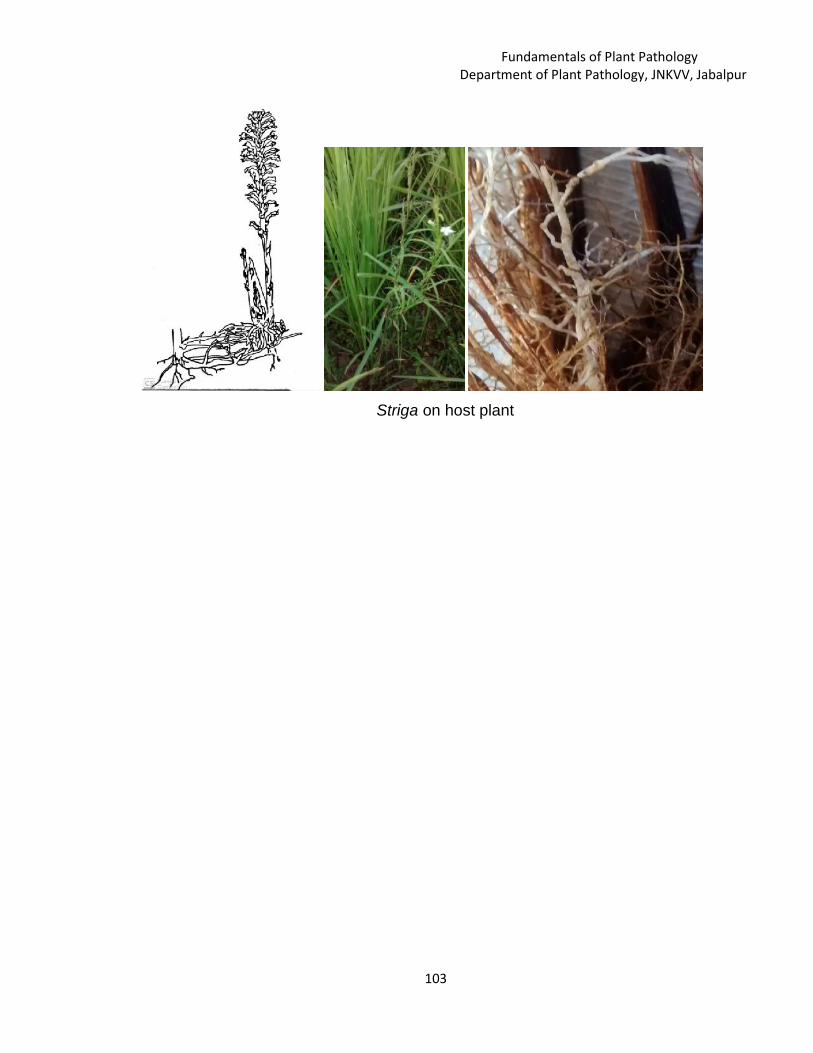
Fundamentals of Plant PathologyDepartment of Plant Pathology, JNKVV, Jabalpur
103
Striga on host plant

Fundamentals of Plant PathologyDepartment of Plant Pathology, JNKVV, Jabalpur
104
Classification, Morphology and Symptoms caused by Plant parasiticnematodes
Jayant [email protected]
JNKVV, Jabalpur--------------------------------------------------------------------------------------------------------
Classification of Nematode
The word taxonomy was coined by de Condolle (1875) and means
“arrangement by law” (taxis = arrangement, nomos = by law). It can be defined as
theory and practice of classifying organisms. The process of classifying
(=classification) is the grouping of organisms in a system or order. The organisms
are grouped to
gether and discriminated from other such groups on the basis of certain characters
known as taxonomic characters. They may be morphological, physiological,
ecological, genetical etc. The classification of plant parasitic nematodes is presently
based on the morphological characters. Besides qualitative morphological
characters certain quantitative characters (measurements) are also used for
classifying nematodes.
Nematodes are placed in the group invertebrate, Kingdon Animalia. Under
separate phylum, Nemata/ Nematoda.
The following are the characteristics of members of the phylum Nemata.
1. Inhabit marine, freshwater and terrestrial environments as free – livers and
parasites.
2. Bilateraly symmetrical, triploblastic, un segmented and pseudocoelomates.
3. Vermiform, round in cross – section, covered with a three – layered cuticle.
4. Growth accompanied by molting of juvenile stages, usually four juvenile
stages.
5. Oral opening surrounded by 6 lips and 16 sensory structures.
L. 13

Fundamentals of Plant PathologyDepartment of Plant Pathology, JNKVV, Jabalpur
105
6. Possess unique cephalic sense organs called amphids.
7. Body wall contains only longitudinal muscles connected to longitudinal nerve
chords by processes extending from each muscle.
8. Unique exscretory system containing gland cells or a set of collecting tubes.
9. Longitudinal nerve cords housed within the thickening of the hypodermis.
A. Order- TylenchidaI. Stoma has protrusible stomato stylet.
II. Three parts oesophagus.
III. The caudal alae is not provided by ribs.
IV. Cuticle has distinct annulations.
The order Tylenchida is redivided into two suborders namely- Tylenchina and
Criconematina.
I. Sub-order- Tylenchinai. Dorsal oesophageal gland opens into the lumen of oesophagus
anterior to median bulb near the stylet base.
ii. Males often have bursa.
iii. Median bulb is round and is not occupying the whole body diameter.
It comprises three super families as follows:
1. Super family- Tylenchoidea
2. Super family- Hoplolaimoidea
3. Super family- Heteroderoidea
A. Super family- Tylenchoidea
I. Procorpus and median bulb separated by more or less pronounced
constriction.
II. The terminal bulb well developed.
III. They are ecto or migratory parasites and both sexes are vermiform.
IV. Bursa generally present, often large.
V. The eggs are not deposited in gelatinous matrix.
It include following families:
1. Family- Tylenchidae : eg. Tylenchus
2. Family- Tylenchorhynchidae: eg. Tylenchorhynchus

Fundamentals of Plant PathologyDepartment of Plant Pathology, JNKVV, Jabalpur
106
3. Family- Neotylenchidae: eg. Neotylenchus
4. Family- Anguinidae: eg. Anguina tritici, Ditylenchus
B. Super family- Hoplolaimoidea
I. Body annulations strongly devloped.
II. The stylet is strong with prominent base knobs
III. Head sclerotization is distinct.
IV. The oesophageal glands overlap the intestine.
V. The caudal alae extend to the tail tip.
VI. The female may be vermiform or swollen while males are vermiform.
It include following families:
1. Family- Dolichodoridae : eg. Dolichodorus, Belanolaimus
2. Family- Haplolaimidae: eg. Haplolaimus, Rotylenchus,
Rotylenchulus
3. Family- Pratylenchidae: eg. Pratylenchus, Rhadopholus,
Hirschemanniella
4. Family- Nacobbidae: eg. Nacobbus
C. Super family- Heteroderoidea
I. The females are saccate, pyriform or lemon shaped, sessile and
males are vermiform.
II. The tail is short and without caudal alae in males.
III. Females with two ovaries and males have terminal spicules.
IV. The eggs may be retained in the female body which turns into cysts or
deposited in gelatinus matrix.
V. There is pronounced sexual dimorphism.
It include following families:
1. Family- Heterodoridae : eg. Heterodera, Globodera
2. Family- Melodogynidae: eg. Melodogyne
II. Sub-order- Criconematinai. The procorpus and median bulb confluent, large and terminal bulb is
small. The valve in median bulb is present.

Fundamentals of Plant PathologyDepartment of Plant Pathology, JNKVV, Jabalpur
107
ii. The males often have reduced stylet and oesophagus or even these
may be absent.
iii. Rectum and anus are poorly developed.
iv. The cuticle is generally strongly striated with annules.
v. The females have strongly striated with annules.
vi. The females have strongly developed and elongated stylet.
vii. The vulva is situated at posterior end and ovary is monodelphic.
viii. The bursa may be absent or poorly developed.
It comprises two super families as follows:
1. Super family- Criconematoidea
2. Super family- Tylenchuloidea
A. Super family- Criconematoidea
I. Coarse cuticle with annulations, large annules sometimes with spines
or scale like projections.
II. In some nematodes, the cuticle sheath is present.
III. The adult female is stout but vermiform.
IV. The length ranges from 0.1 to 0.2 mm.
It include following families:
1. Family- Criconematidae : eg. Criconema, Criconemaoides
2. Family- Hemicycliophoridae: eg. Hemicycliophora,
Hemicriconemoides
B. Super family- Tylenchuloidea
I. The cuticle annulations are fine or very thick.
II. The adult females are swollen, subspherical, saccate or elongate.
III. The males are with or without stylet.
IV. The tail is long and bursa is absent.
V. The excretory pore is located for backward or in normal position.
It include following families:
1. Family- Tylenchulidae : eg. Tylenchulus, Paratylenchus
2. Family- Sphaeronematidae: eg. Trophonema, Sphaeronema
3. Family- Meloidoderitidae: eg. meloidoderita

Fundamentals of Plant PathologyDepartment of Plant Pathology, JNKVV, Jabalpur
108
B. Order- AphlenchidaI. The dorsal oesophageal gland opens within the median bulb of
oesophagus.
II. The males are without caudal alae except in Aphlenchus and
Metaaphlenchus.
III. The median bulb is angular and occupying the entire body diameter.
The order Aphlenchida is redivided into one suborders namely- Aphelenchina
and two
super families as follows:
1. Super family- Aphelenchoidea
2. Super family- Aphelenchidoidea
A. Super family- Aphelenchoidea
I. The males are with bursa, spicules of normal shape and
gubernaculums is present.
II. The oesophageal glands may form long overlapping lobe or may not
overlap the intestine.
III. The lateral line with 6 to 14 striae at mid body.
It include following families:
1. Family- Aphelenchidae : eg. Aphelenchus
2. Family- Paraphelenchidae: eg. Paraphelenchus
B. Super family- Aphelenchidoidea
I. The spicules are thorne shaped and gubernaculums is present.
II. The stylet is with or without basal knobs.
III. The oesophageal glands forming long overlapping lobe on dorsal side
of intestine.
IV. The lateral lines are four or less in number.
It include following families:
1. Family- Aphelenchoididae : eg. Aphelenchoides,
Rhadinaphelenchus.
2. Family- Anomyctidae: eg. Anomyctus
3. Family- Entaphelenchidae: eg. Entaphelenchus

Fundamentals of Plant PathologyDepartment of Plant Pathology, JNKVV, Jabalpur
109
C. Order- DorylaimidaI. The stroma is with mural tooth or an axial spear called as odanto or
onchio stylet.
II. Two part oesophagus in which the anterior part is slender and the
posterior part expanded with 3 to 5 gland nuclei.
III. The amphids are of variable shape.
IV. The gland duct opens posterior to nerve ring.
The order Dorylaimida is redivided into two suborders namely- Dorylaimina
and Diphtherophorina.
Sub order- Dorylaiminai. The amphids are slit or pore like.
ii. The anterior part of oesophagus is a narrow tube encircled by nerve
ring while posterior part swollen and muscular.
iii. Stoma is provided with odontostylet.
iv. The excretory duct is absent or weakly developed.
v. The males are with paired testes, prominent spicules but
gubernaculums absent.
This suborder includes 5 super families out of which Longidoroidea contains
plant parasitic nematodes:
1. Dorylaimoidea
2. Actinolaimoidea
3. Leptochoidea
4. Belondiroidea
5. Longidoroidea
Super family- Longodoroidea
I. The males and females have similar tails.
II. The spear is greatly attenuated with long extensions.
III. The posterior enlarged part of the oesophagus is reduced in length.
IV. One pair of subventral gland nuclei in the posterior oesophagus.
It include following families:
1. Family- Longidoridae : eg. Longidorus

Fundamentals of Plant PathologyDepartment of Plant Pathology, JNKVV, Jabalpur
110
2. Family- Xiphinematidae: eg. Xiphinema
3. Family : Xiphidoridae : eg. Xiphidorus
Sub order- Diphtherophorinai. The amphids are postlabial and pouch like.
ii. Plump body and cuticle is thick and loose.
iii. Onchiostylet is composed of dorsal tooth and supporting structures.
iv. The anterior portion of oesophagus is a narrow while posterior part
swollen and pyriform to elongate.
v. The basal bulb provided with one dorsal and two subventral
uninucleate glands.
vi. The excretory pore is present.
vii. The females are with paired reflexed ovaries. The amles are with
single testis with spicules and gubernaculums.
This suborder includes 2 super families.
1. Trichodoroidea- Contains plant parasitic nematodes.
2. Diphterophoroidea
Super family- Trichodoroidea
I. Anterior portion of oesophagus is narrow and cylindrical while
posterior portion is expanded.
II. The anterior tip of a dorsal tooth formed by cell in anterior
oesophagus, is solid and strong, while posterior portion of stylet is
hollow.
III. The males are with single testis, spicules are simple and curved.
IV. The female is with one or two ovaries.
It include Familu Trichodoridae which contains genera Trichodorus
and Paratrichodorus.
General Morphology of Nematodes
Nematodes have adopted themselves to all the environment but they are
similar in morphology and life stages. Certain basic principles are common to all
nematodes. Nematodes are triploblastic, bilaterally symmetrical. Un segmented,
Pseudocoelomate, vermiform and clourless animals. The plant parasitic nematodes

Fundamentals of Plant PathologyDepartment of Plant Pathology, JNKVV, Jabalpur
111
are slender elongate, spindle shaped or fusiform, tapering towards both ends and
circular in cross section. The length of the nematode may vary from 0.2 mm to
about 11.0 mm (Paralongidorus maxximus) and the body width vary from 0.01 to
0.05 mm. In few genera, the females on maturity assume pear shape
(Meloidogyne), globular shape (Globodera), reniform (Rotylenchulus reniformis) or
saccate (Tylenchulus semipenetrans). Radially symmetric traits (triradiate,
tetraradiate and hexaradiate) exist in the anterior region. The regions of intestine,
excretory and reproductive systems show tendencies towards asymmetry. The
nematodes have one or two tubular gonads which open separately in the female
and into the rectum in the male which also have the copulatory structures.
The free living saprophytic nematodes are generally larger in size. The
animal and human parasitic helminthes may have length of few centimeters to even
a meteer or more.
The anterior end starts with the head, which consists of mouth and pharynx
bearing the cephalic papillae or setae. The portion between the head and the
oesophagus is known as the neck. Beginning at the anus and extending to the
posterior terminus is the tail.
Longitudinally the body is divided into four regions as dorsal, right lateral, left
and ventral. All the natural openings like vulva, excretory pore and anus are located
in the vental region. The Nematodes do not possess a specialized circulatory or
respiratory system.
The nematode body is divided into three regions. They are the outer body tube or
body wall, inner body tube (digestive tract) and body cavity or pseudococoelome.
General morphology of different parts of nematode is depicted in Fig. 1 and 2.

Fundamentals of Plant PathologyDepartment of Plant Pathology, JNKVV, Jabalpur
112
The-important-morphological-features-of-a-plant-parasitic-nematode
Female and Male nematode
Caudal alae
Longitudinal markings
Transverse marking or Striations
Fig. 1: General morphology of plant parasitic nematode

Fundamentals of Plant PathologyDepartment of Plant Pathology, JNKVV, Jabalpur
113
Cuticle
Bodywall of nematode
Fig. 2: Structure of bodywall and cuticle of plant parasitic nematode

Fundamentals of Plant PathologyDepartment of Plant Pathology, JNKVV, Jabalpur
114
The outer body tube
The outer body tube or body wall includes the cuticle, hypodermis, and
somatic muscles. The body wall protects the nematode from the harsh external
environment, serves as the exoskeleton and provides the mechanism for movement
of the organism through the soil and plant tissue.
The cuticle: The Cuticle is a non living, non cellular, triple – layered covering
secreted by the underlying hypodermis. The cuticle is flexible. It covers the entire
body and lines the oesophagus, vulva, anus, cloaca, excretory pore and sensory
organs. The feeding stylet and copulatory spicules are formed from cuticle.
The composition and from of the cuticle is highly variable. In general the
cuticle is composed of three primary zones viz., the cortical layer, median layer and
basal layer.
The cuticle of may nematodes have markings on the surface. They are
varied and complex and have been often used by taxonomists to assist in the
identification of various species. The cuticular makings are categorized into different
following types.
I. Punctuations
These are minute, round dots arranged in a pattern. They act as structures
for strengthening the cuticle.
II. Transverse marking or Striations
There are transverse lines present on the surface of the cuticle. These
markings exhibit distinct variations among the plant parasitic nematodes and often
used by the taxonomists for identification. The transverse markings cause a pattern
of ridges and furrows right from head to tail and theses markings gives the false
appearance as if the nematode is segmented.

Fundamentals of Plant PathologyDepartment of Plant Pathology, JNKVV, Jabalpur
115
III. Longitudinal markings
These markings are the lines on the cuticle, which runs longitudinally
throughout the length of the nematode body. These markings are divided into a)lateral lines or incisures and b) longitudinal ridges.
a) Lateral lines or Incisures
These are lines running longitudinal to the body axis of nematode but they
are confined to the lateral field in area just on top of lateral hypodermal chords on
either side of the nematode body running throughout the length.
b) Longitudinal ridges
Longitudinal ridges are raised lines present on cuticle running longitudinal to
nematode body axis but are confined in the area other than lateral field.
Alae are thickening or projections of the cuticle which occur in the lateral or
sublateral region. There are 3 types of alae I. Caudal alae II. Cervical alae and III.
Longitudinal alae.
I. Caudal alae
These are found in the posterior region and restricted to males as copulatory bursa.
II Cervical alae
These are confined to the anterior part of the nematode body and are found
in some species of marine nematodes.
III. Longitudinal alae
The longitudinal alae delimit the later fields and are known as lateral alae.
Their form varies in different species. They are transverse by striations or furrow
varying in number from 1 to 12. Functionally they probably assist in locomotion.
The functions of cuticle

Fundamentals of Plant PathologyDepartment of Plant Pathology, JNKVV, Jabalpur
116
Cuticle gives definite shape and size to the body, act as an exoskeleton,
helps in movement it functions as a semipermeable membrane.
Somatic musculature
Platymyrian
A flat type of cell with contractile elements limited in places to the base lying
close to the epidermis.
Coleomyarian
‘U’ shaped cells in which muscle fibre are adjacent and perpendicular to the
hypodermis and extend along the sides of the muscle cell of varying distances.
Circomyarina
This type of muscle cells are almost round and the muscle fibres completely
surround the cytoplasm.
The platymyarian muscle cell is considered primitive which might have
modified into coelomyarian type of narrowing and upward elongation of the fibrillar
zone. Muscle cells are connected to each other by means of cytoplasmic bridges
and have nerve connections.
Mode of Reproduction
Bisexual species: Bisexual species usually have equal numbers of females and
males. Insemination by a male and egg fertilization is necessary of the development
of the eggs in such forms.
Hermaphroditic species: In hermaphroditic species, reproduction through self
fertilization occurs. Hermaphroditic nematodes have the appearance of females.
Hermaphroditism is common among the free-living Rhabditida.

Fundamentals of Plant PathologyDepartment of Plant Pathology, JNKVV, Jabalpur
117
Syngonic: Syngonic sperm and eggs are produced in the same gonad. In most
cases, the gonad first produces sperm that are stored and later fertilize the eggs
subsequently developed by the same gonad (protandry).
Parthenogenesis: In parthenogenesis, the eggs develop without fertilization.
Parthenogenesis has been proved for a number of free-living nematodes. It is also
known to occur in plant parasites of the genera Meloidogyne and Heterodera.
Intersexes: intersex is an individual that exhibits a blending of male and female
characters. The possessed well-developed ovaries with uteri, vagina, and vulva. In
addition they had developed to some extent male sexual openings, spicules, brusal
muscles, and male genital papillae.
Above and Below Ground Symptoms Caused by Nematodes
A successful host parasite relationship results in appearance of symptoms on host
plant. Symptoms are generally non-specific like poor growth, stunting, discoloration
of foliage and patchiness of crop stand. Symptoms caused by plant parasitic
nematodes are shown in Figures 3 and 4.
Symptoms of injuries caused by plant parasitic nematodes can be classified as
I. Above ground symptoms
II. Below ground symptoms
I. Above ground symptomsLeaf discoloration: The leaf tip become white in rice due to rice white tip nematode
Aphelenchoides besseyi, yellowing of leaves in Chrysantyhemum due to
Chrysanthemus foliar nematodes, A. ritzemabosi.
Dead or devitalized buds: The nematodes affect the growing point and kill the
buds and stopping growth of affected tissues as in case of strawberry infected by A.
fragariae.
Seed galls: In wheat, Anguina tritici larva enter into the flower primordium and
develop into a gall.

Fundamentals of Plant PathologyDepartment of Plant Pathology, JNKVV, Jabalpur
118
Twisting of leaves and stem: The basal leaves become twisted due to nematode
infection as in onion infected with Ditylenchus. angustus.
Crinkled or distorted stem and foliage: The wheat seed gall nematode. A tritici
infests the growing point as a result distortions in stem and leaves take place.
Necrosis and discoloration: Discoloration of foliage and stem may range from
light to dark shades. Due to Rahadinaphelenchus cocophilus produces band of
necrotic tissue on coconut.
Lesions on leaves and stem: Small yellowish spots are produced on onion stem
and leaves due D.dipsaci, and the leaf lesion caused by A. ritzemabosi on
hrysanthemum.
A. ritzemabosi on Chrysanthemum
Chrysanthemum foliarnematode
Ditylenchus destructor cause root rot
Aphelenchoides fragariae-infected-strawberry
Rahadinaphelenchus cocophilus on coconutRoot galls or knots
Fig. 3: Symptoms caused by plant parasitic nematodes

Fundamentals of Plant PathologyDepartment of Plant Pathology, JNKVV, Jabalpur
119
II. Below ground symptoms
Nematode produces a wide range of symptom on underground parts of theplants which may be specific or non specific types.
Stunting: Plant growth is reduced and the plants are not able to withstand adverse
conditions. Patches of stunted plants appears in the field. (eg.) in potato due to
Globodera rostochiensis, in gingelly, due to Heterodera cajani and in wheat by
Heterodera avenae.
Wilting: Day wilting due to Meloidogyne spp. i.e. In hot weather the root – knot
infested plants tend to droop or wilt even in the presence of enough moisture in the
soil. Severe damage to the root system due to nematode infestation leads to day
wilting of plants.
Root galls or knots: The characteristic root galls are produced by root- knot
nematode, Meloidogyne spp. The size and number of galls may vary with host crop
and nematode sp.
Root lesion: The penetration and movement of nematodes in the root causes
typical root lesions eg. Necrotic lesions induced by Pratylenchus thornei on
chickpea; the burrowing nematode, Radopholus similes in banana. Helicotylenchus
multicinctus causes reddish brown lesion on banana roots. The rice root nematode
produces brown lesions on rice root.
Reduced root system: The root tip growth is arrested due to nematode feeding
and roots produce branches. This may be of various kinds such as coarse root,
infestations arrest the growth of lateral roots, and leads to a open root system with
only main root without lateral roots, stubby root. The lateral roots produce
excessive rootless and curly tip.

Fundamentals of Plant PathologyDepartment of Plant Pathology, JNKVV, Jabalpur
120
.
R a d op ho lus si m il es in ba n a na W h ea t see d g a l l nem a t od e fo lia g e
W hea t se ed ga ll ne m a tod e W i lt in g -a n d-d r yi ng - ou t-o f-B i tter -M el on -ca u sed -by -M e lo id og y ne -in co g ni ta
F ig . 4: S ym pto m s c a used b y pl a nt p a ra si tic nem at od es
Curly tip: The nematode retards the elongation of roots and cause curling of
roots known as “Fish book” symptom.
Root proliferation: Increase in the root growth or excessive branching due to
nematode infestation. The infested plant root produced excessive root hairs at
the point of nematode infestation. (eg.) Heterodera spp. Meloidogyne hapla
and Pratylenchus spp. etc.
Root – rot: The nematode feeds on the fleshly structure of roots and resulting
in rotting of tissues (eg.) Ditylenchus destructor cause root rot.
Root surface necrosis: Severe injury caused by nematodes lead to complete
decortication of roots and result in root necrosis.
Cluster of sprouts on tubers: Clusters of short and swollen sprouts are
formed in many tuber plant because of nematode infection. Eg. Ditylechus
dipsaci.

Fundamentals of Plant PathologyDepartment of Plant Pathology, JNKVV, Jabalpur
121
Dispersal and survival of the Plant pathogensShikha Sharma
JNKVV, College of Agriculture, Waraseoni----------------------------------------------------------------------------------------------------------------------
Definition of Dispersal: Transport of spores or infectious body acting as a inoculums
from one host to another host at various distance resulting in the spread of disease
or in other word we can say that displacement of a plant pathogen from its place of
origin or production to a suitable host or place where it can grow and futher
establish is called dispersal or dissemination of the pathogen.
It is of two type
1. Active/Direct/Autonomous: The dispersal of plant pathogen takes place
through soil, seed and planting material during normal agronomic operation.
Mechanism of Dispersal of plant pathogenActive Dispersal: spore require some structural mechanism or physiological
response which enable them to be thrown out with some force.
a. Liberation due to bursting of turgid cell: spores of ascomycota are liberated by
this mechanism as asci are turgid cell which burst in regular manner violently
liberated the spore.
b. Liberation due to round off of turgid cell: Separation of spore from the parent
hypae, then violent discharge of spore is due to change in shape of turgid
cell.eg occur in Sclerospora and acecia of rust fungi.
c. Ballistospore discharge: this type of discharge occur in basidiospore of rust and
smut fungi as point of contact of the spore with the sterigmata a gas filled
bubble appear and grow in few second to a definite size, then the spore is shot
away carrying the bubble.
d. Liberation due to hygroscopic movement: this movement is caused by a sudden
fall in humidity which results in drying of sporophore bearing the spore eg.
Sporangia of Phytophthora infestans and also in Peronospora ospora in which
L. 14

Fundamentals of Plant PathologyDepartment of Plant Pathology, JNKVV, Jabalpur
122
collapse of sap filled turgid hyphae and their bursting responsible for violent
discharge of spore.
2. Passive/Indirect discharge : The agency dislodging the spore is mainly the
force from some outside source such as strong air current, raindrops, falling of
spore mass, insect, workers etc.
1. Active/Direct/Autonomous:(A)soil: it is of two type(I)Dispersal in soil: soil borne pathogen viz., facultative saprophytes or facultative
parasite may be survive in soil or growth of pathogen in soil.The following are
different stages
a)Contamination of soil: Introduction of the pathogen infested soil, plant debris,
infested seed and planting material to a new area or contamination of the soil
takes place by the spread of the pathogen from an infested area to new area.
b)Growth and spread of the pathogen in soil: Growth and spread of the
pathogen depend on the virulent power of pathogen, high multiplication rate,
adaptiblity to the soil environment including saprophytic survival ability and
susceptibility of host and it is also depend on high growth rate, fast spore
germination, good enzymatic activity, production of antibiotic and tolerance to
antibiotic produce by soil microflora.The active saprophytic survival of Facultative
saprophyte and facultative parasite in soilis affected by soil structure, moisture
,organic matter, pH, antagonism , however specialized facultative parasite survive in
the ansence ofhost plant but they depend on the plant residue.the non specialized
facultative parasite can whole life in the soil.eg. Pythium sp., and Phytopthora sp.
Whereas., soil borne obligate parasite such as Plasmodiophora brassicae and
Synchytrichum endobioticum require the presence of active host.
c)Persistence of the Pathogen:The pathogen persit in the soil as dormant
structure like oospore as in Pythium, Phytophthora,Sclerospora and
chlamydospore in Fusarium .
II) Dispersal by the soil : The pathogen enter the soil, grow, multiply and further
spread in the soil. During the intercultural operation in the field,the propagule of

Fundamentals of Plant PathologyDepartment of Plant Pathology, JNKVV, Jabalpur
123
fungal and bacterial structure present in soil spread takes place from one place to
the other by tools, implements, feet of man and irrigation water.
(B)Seed and Seed Material: Dissemination of pathogen by seed and seed material
is of great importance as it introduce the pathogen to new area where they may
never existed before.The dormant structure of the pathogen viz., seed of Cuscuta,
sclerotia of ergot fungus, smut sori are found mixed with seed lots and they are
dispersed with seed lots and they are dispersed as seed contaminants. The fungal
spores and bacterial cells are present on the seed coat and are transported to long
distance and dormant mycelium of many fungi are also present in the seed is
transmitted to long distances. Propagating material from infested nursery spread
the pathogen to new area.
There are three types of dispersal by seeds:
i)Contaminated seed
ii)Externally seed borne
iii)Internally seed borne
i)Contaminated seed : Seed borne pathogens move in seed lot as separate
contaminate without intimate contact with the viable crop seeds. The identity of the
seeds of the host and parasite is difficult to separate eg smut of pearl millet
(Tolyposporium penicillariae),ergot of rye and pearlmillet (Claviceps purpurea and
C. fusiformis respectively. Smut sori and ergot mix easily with the seed lot during
harvesting and threshing. Also in smuts such as Kamal bunt of wheat (Neovossia
indica) and bunt of rice (Neovossia horrida) the infected kernels containing smut
sori are mixed with the seed. In leaf smut of rice (Entyloma oryzae) leaf pieces
containing smut sori are mixed with the seed.
ii)Externally seed borne:Close contact between structure of the pathogen and
seeds in established in diseases like covered smut and loose smut VI Daney, snoll
smut of sorghum, stinking smut of wheat and bacterial blight of cotton where the
pathogen gets lodged in the form of dormant spores or bacteria on the seed coat
during growth of the crop or at the time of harvest and threshing. In many
pathogens the externally seed-borne Structures such as smut spores can persist for
many years due to their inherent capacity for long survival. The spores of Tilletia

Fundamentals of Plant PathologyDepartment of Plant Pathology, JNKVV, Jabalpur
124
caries (stinking smut of wheat) remain viable even after 18 years and those of
Ustilago avenae (oat smut) for 13 years.
iii)Internally seed-borne: The Internally seed-borne pathogen may penetrate into
the ovary and cause infection of the embryo, while it is developing. Example of
internally seed borne pathogens like Ustilago nuda tritici are viable for more than 15
years. Other example are of Helminthosporium oryzae, Sclerospora graminico/a,
etc. The bacterial pathogens include Xanthomonas oryzae pv. oryzae, on rice,
Pseudomonas syringae pv. syringae in cucurbits, Xanthomonas campestris pv.
compestris on crufifers, etc.
Mainly man distributes seeds of cultivated crops. Sometimes animals and
birds also help in distribution of crop seeds. Man and animals are the main agencies
of dispersal of pathogen through seed. The pathogens thus mixed with the seed or
on the seed are transmitted.
Passive dispersal/IndirectPassive dispersal of plant pathogens occurs through
1. Animate agentsb. Insects
c. Mites
d. Fungi
e. Nematodes
f. Human beings
g. Farm and wild animals
h. Birds
i. Phanerogamic parasites
2. Inanimate agentsa. Wind
b. Water
Insect: plant pathogens are carried by insect either externally (epizoic) or
internally(endozoic). The external transmission of plant pathogens occur in those
fungi, which produce their conidia, oidia and spermatia in sweet or honey secretions
having attractive odours, the Sclerotinia brown rot of pear and apple, the honey dew

Fundamentals of Plant PathologyDepartment of Plant Pathology, JNKVV, Jabalpur
125
stage in the 'sugary disease' of sorghum and pearl millet in part of India and the
pycnial nectar in the cluster cup stage of rust. The spermatial oozing at the mouth of
spermagonia in the ascomycetes attract various types of insects, flies, pollinating
bees and wasps which play a dual role viz. pollination and transmission of
pathogens. The fire blight organism (Erwinia amylovara) pathogens and citrus
canker bacterium, (Xanthomonas axonopodis pv. cirri) are also carried in this
manner, the former by flies (bees) and ants and the latter by the leaf miner. The
black leg of potato caused by Erwinia carotovora in disseminated by maggots, wilt
of com caused by X. stewartii, gummosis of sugarcane caused by X vasculorum are
the other examples for bacterial diseases transmitted by insects However,
Ingenious transmission of pathogens, of an internal nature (endozoic) is provided by
the Dutch elm disease (Ceratostomella ulmi) and olive canker (Bacillus savastano
i). The former is transmitted by the elm bark beetles and the latter by the olive fly
(Olea. europaea). These insects, unlike the epizoic group, appear to have a close
biologic relationship with the pathogens, as they have not been reared without the
contaminating pathogens.
The cucumber wilt bacterium, Erwinia tracheiphila is spread by the striped
cucumber beetles (Acalymma vitata) and the spotted cucumber beetle (Diabrotica
undecimpunctata). Different types of insects spread more than 80 percent of the
viral and phytoplasmal diseases.
AphidsAphids are the most important insect vectors of plant viruses and transmit the
great majority of all stylet-borne viruses.. Aphids generally acquire the stylet-borne
virus after feeding on a diseased plant for only a few second (30 seconds or less)
and can transmit the virus after transfer to and feeding on a healthy plant for a
similarly short time of a few seconds. The length of time aphids remain viruliferous
after acquisition of a stylet-borne virus varies from a few minutes to several hours,
after which they can no longer transmit the virus.Eg Acyrthosiphon pisum non
persistently transmitted bean common mosaic virus and Toxoptera citricida also
transmitted nonpersistantly Citrus tristeza virus.

Fundamentals of Plant PathologyDepartment of Plant Pathology, JNKVV, Jabalpur
126
Leaf HoppersLeaf hoppers are phloem feeders and acquire the virus from the phloem
region. All leaf hoppers, transmitted viruses are circulatory. Several of these viruses
multiply in the vector (propagative) and some persists through the mouth and are
transmitted through the egg stage of the vector, Most leaf hopper vectors require a
feeding period of one to several days before they become viruliferous, but once they
have acquired the virus they may remain viruliferous for the rest of their lives.
MitesMites belonging to the families eriophyidae (eriophid mite) and tetranychidae
(spider mite) transmit plant viruses.
. FungiSome soil-borne fungal plant pathogens transmit plant viruses. Olpidium
brassicae, ploymyxa graminis and spongospora subterranea are the fungi involved
in transmission of virus disease. All these fungi are pathogens of the host which
carry of viruses. The Zoospores of the fungi are released from the host and the
zoospores carry the virus and transmit it to the susceptible hosts during their
infection process. In some cases plant viruses are carried on the outside of the
fungi. Examples are tobacco necrosis virus and cucumber mosaic virus.
The viruses like lettuce big vein virus are found inside the zoospores. They
persist for years in viable resting sporangia. The types of transmission by fungi can
be considered as non persistent and persistent transmission.
NematodesNematodes act as agents for dissemination of pathogenic fungi, bacteria and
viruses. Nematodes help these pathogenic fungi to enter into the host though
punctures for their own entry into host along with the nematodes. Plant nematodes
play a vital role as vector in transmitting certain virus diseases. Nematode vectors
transmit viruses by feeding on roots of infected plant and then moving on roots of
healthy plants. Larvae as well as adult nematodes can acquire and transmit virus,
but the virus is not carried through the larval molts or though the eggs. After
moulting the larvae or the resulting adults must feed on a virus source before they
can transmit again. Xiphinema, Longidorus and Trichodorus transmit both the
Fundamentals of Plant PathologyDepartment of Plant Pathology, JNKVV, Jabalpur
127
polyhedral and tubular type of viruses For example Corynebacterium tritici that
causes yellow ear rot of wheat is disseminated by ear cockle nematode. Similarly,
some pathogenic fungi such as, phytophthora, fusarium, Rhizoctonia, etc. are
carried on the body of nematodes. Paratrichodorous sp carry pea early browning
Tobacco rattle virus, Xiphinema index carry Grape vine chrome virus (NEPO virus)
Human beingMan is the most important factor responsible for short distance and long
distance dispersal of plant pathogens. He helps in dissemination unknowingly by his
usual agricultural practices.The means by which human beings help in dispersal are
as follows.
i. Transportation of seedsSeed trade is one of the different means of dispersal of plant pathogens in
which man plays an important role. The import and export of contaminated seeds
without proper precautions lead to movement of pathogens from one country to
another or from one continent to another. The disease which are amenable to such
transmission are mainly those that are carried in or on the propagative parts and
seed such as late blight of potato, the downy mildew of grapevine, citrus canker,
chestnut blight, Dutch elm disease, Fusarium wilt of banana, Katte disease of
cardamom and bunchy top of banana.
ii. Planting diseased seed materials (vegetatively propagated materials)Planting diseased bulbs, corms, tubers, rhizomes, cutting etc. of vegetatively
propagated plants such as potato, sweet potato, cassava, sugarcane, banana,
many ornamentals and fruit trees etc. help in dispersal of pathogens from field to
field, orchard to orchard, locality to locality or form one country to another.
iii. By adopting farming practicesHuman being (men and women) engaged in preparatory cultivation, planting,
irrigation, weeding, pruning etc. help in dispersal of plant pathogens. The spores
(oospores, chlamydospores), dormant structure like sclerotia are carried by worker's
clothing, shoes, hand etc. from field to field.
Fundamentals of Plant PathologyDepartment of Plant Pathology, JNKVV, Jabalpur
128
iv. Through clothingThrough clothing of workers plant pathogen are also disseminate. eg.Palm
workers engaged in cleaning coconut trees spread bud rot disease.
v. By use of contaminated implementsPathogens are transferred from one area to another by implements used in various
cultural operations (weeding, hoeing, thinning etc.) in the field. e.g. root rot of pulses
and cotton (Macrophomina phaseolina, bacterial angular leaf spot of cucumber
(Pseudomonas /achrymans)
and bacterial canker of tomato (Corynebacterium michiganensis). Cutting and
pruning knives help in dissemination from one plant to another e.g., Bunchy top of
banana.
vi. By use of diseased grafting and budding materialsGrafting and budding is the most effective method of distribution of
pathogens of horticultural crops (fruit trees, ornamental etc.) e.g. selection of
infected stocks and scions in propagation of h.orticultural helps in the dissemination
of pathogen.
f. Farm and wild animalsFarm animals (cattles) while feeding on diseased fodder ingest the viable
fungal propagules (spores of oospores or sclerotia) in to their digestive system.
Animals which feed on downy mildew affected pearl millet or sorghum take the
oospores along with the fodder. Oospores pass out as such in the dung. This dung
when used as manure spread in the field and act as source of incolum, Smut fungi
like grain smut of sorghum, loose smut and head smut of sorghum are carried from
field to field through the alimentary canals of farm animals.
Stem of dodder (cuscuta spp.) are carried by birds for building their nests.
Thus the phanerogamic parasites are getting transported to new location.
h.Phanerogamic parasitesThe virus is transmitted in the food stream of the dodder plant, being acquired from
the vascular bundles of the infected plant by the haustoria of dodder. After
translocation through the dodder phloem the virus is introduced in the next plant by

Fundamentals of Plant PathologyDepartment of Plant Pathology, JNKVV, Jabalpur
129
the new dodder haustoria produced in contact with the vascular bundles of the
inoculated plant. Cuscuta californica, C. campestris, C. subinclusa are usually
employed for dodder transmission of viruses and phytoplasmas. C. europaea, C.
epilinum and C. lupuliformis are also employed in transmission of viruses.
2. Inanimate agentsa. WindThe wind dispersal of plant pathogens is known as anemochory, It is one of the
most common methods of the dispersal of plant pathogens. It is the most dangerous
and potent mode of travel for plant pathogenic fungi. It acts as potent carrier of
propagules of fungi, bacteria and viruses. Usually the fungal pathogens are light in
weight and are well adapted to wind dispersal. Example Short distancedissemination occur iby sporangia of downy mildew fungi, conidia of powdery
mildew fungi and basidiospores of rust fungi. Where as long distance dessimination
in Uredospores of rust fungi, Chlamydospores of smut fungi conidia of Alternaria,
Helminthosporium and pyricularia. However, Damping-off pathogen (Pythium spp.),
wart disease pathogen of potato (synchytrium endobioticum); root rot pathogens
(Sclerotium and Rhizoctonia) and seeds of phanerogamic parasites witchweed
(Striga) are efficiently carried by wind. Viruses and phytoplasmas are not directly
transmitted by wind, but the insect and mite vectors that carry the viruses move to
different directions and distances depending upon the direction and speed of air.
b. WaterThe surface flow of water after heavy showers of rains or irrigation water from
canals and wells carries the pathogens to short distances. Soil inhabiting fungi like,
Fusarium, Ganoderma,Macrophomina Phytophthora, Plasmodiophora, Pythium,
Rhizoctonia, Sclerotium, Sclerotinia, Sporisorium, Ustilago, Verticillium etc., in the
form of mycelial fragments, spores or sclerotia, soil-bone bacteria and nematodes
carrying viruses are transmitted through the above process. They are transmitted
through rain or irrigation water that moves on the surface or through the soil.

Fundamentals of Plant PathologyDepartment of Plant Pathology, JNKVV, Jabalpur
130
Survival of plant pathogenThe means of survival are the first link in the infection chain or the disease cycle.
Any pathogen can cause disease under favorable conditions. The only important
factor is that the pathogen must come in contact with the host for the development
of the disease. Pathogen itself or its parts that are capable of causing disease when
come in contact to the host is called inoculum. The initial infection that occurs from
these sources in the crop are called primary infection and the propagules are
called primary inoculum.The pathogens find some alternates source of their
survival to maintain the infection chain in the absence of their cultivated host. After
initiation of the disease in the crop, the spores or cells or other structures of the
pathogen are sources of secondary inoculums and cause secondary infection. The
primary infection initiate the disease and secondary infection spread the disease
Fungal pathogens are highly diversified, where the hyphae, Dormant mycelium
either embedded in the embryo of seeds or other part of the plants, special
reproductive structures viz., rhizomorphs, sclerotia, chlamydospores, various types
of asexual spores like sporangia, soprangiospores, zoospores, conidia and sexual
spores as oospores, zygospores, resting spores, ascospores, basidiospores, etc. ,
serve as inoculum. In case of viruses and plant pathogenic bacteria, the individuals
are acting as inoculum, since they do not produce any special type of infective units
like resting spoes or endospores, etc. however in Streptomyces sp.
(Actinomycetes), fragments of filaments and spore-like cells serve as inoculum. In
phanerogamic parasites, seeds are the potential inoculum.The existence of the
pathogen between the two crop seasons is the vulnerable period in its life cycle.
Hence knowledge of the survival of the pathogen in the off-season is useful for the
plant pathologists to suggest suitable management practices.
These sources of survival of pathogens can be grouped as follows:
1. Survival by means infected plant parts.
2. Survival as saprophytes.
3. Survival by means of specialized resting structures
4. Survival in association with nematodes and fungi.
5. Survival in association with insects.

Fundamentals of Plant PathologyDepartment of Plant Pathology, JNKVV, Jabalpur
131
6. Survival in association with phanerogamic plant parasites
7. Survival on agricultural materials.
8. Survival on surface water.
1 Survival in Infected plant partsplant pathogens structure, mycelium survive in infected crop plants part viz, leaves,
stem, pod, seeds etc from few month to years as reservoir of primary inoculum eg.
Colletotrichum lindemuthinum, the causative organism of bean anthracnose.a. Alternate hosts: Pathogen having wide host-range and also tolerant to wide
range of weather conditions, the alternate hosts become very important source of
survival of the pathogen. Black rust of wheat caused by Puccinia graminis tritici
require alternate host Berberis vulgaris, the barberry bush, grows side by side with
wheat for the completion of the life cycle of heteroecious, rust pathogen in
temperate In such areas this wild host belonging to different family is important for
survival of the fungus. Where as Thalictrum flavum is alternate host for the brown
rust of wheat caused by Puccinia recondita .
b. Collateral hosts: Collateral hosts are the wild host of same family, which are
susceptible to the plant pathogens of crop plant and provide adequate facilities for
their growth and reproduction of these pathogenring off-seseaon and help in the
survival. For example, blast disease of paddy , Pyricularia oryzae can infect the
grass weeds like Bracguarua mutica, Dinebra retroflexa, Leersia hexandra,
Panicum repens, etc. and survive during off-season of rice-crop.
c. Self sown crops : Self-sown plants, voluntary crops and early sown crops are
reservoirs of many plant pathogens e.g. groundnut rust pathogen, Puccinia arahidis
and ring mosaic of groundnut caused by tomato spotted wilt virus. Self-sown rice
plants harbour the pathogen as well as vector. e.g., rice tungro virus and its vector,
Nephotettix virescens.
d. Perennial crop : also play a major role in the survival of the plant pathogens.
Pathogens, which infect perennial plants, can survive in them during low winter
temperature and/ or during the hot, dry weather of the summer. They survive in the
lesions on perennial host plants, which may be actively growing or are dominant.
Example bunchy top of banana survives continuously in the suckers produced by

Fundamentals of Plant PathologyDepartment of Plant Pathology, JNKVV, Jabalpur
132
the mother plants. In citrus canker the bacterium, Xanthomonas axonopodis pv. citri
survives in the cankers formed on the leaves and twigs of the standing tree.
e. Ratoon Crops: Ratoon crop also harbour the plant pathogens e.g., sugarcane
mosaic.
f. Survival by latent infection: Latent infection refers to the conditions in which the
plant pathogens may survive for a long time in plant tissue without development of
visible symptoms. Eg. Pseudomonas syringae pv. syringae and X axonopodis pv.
citri can survive in apparently healthy bark tissues of their tree hosts.
g. Survival as residents : Plant pathogenic bacteria have the capacity to grow on
the surface of host and non-host plant utilizing the small amount of nutrients that are
secreted on the plant surface. Eg. Fire blight of apple and pear caused by Erwinia
amylovora and bacterial blight of soybean caused by Pseudomonas syringae pv.
Glycinea.
2- Survival as saprophytes : In the absence of the living host ,the facultative
parasites are capable of surviving as saprophytes. Soil and plant debris serve as
media for this saprophytic survival. Spp. of Pythium, Sclerotium etc. resting
structure like oospores and sclerotia.In Pythium aphanidermatum (foot rot of
papaya), the fungus rapidly forming oospores. S. rolfsii, Sclerotia are formed and
these can germinate under decaying residues
3.Survival by means of specialized resting structuresPlant pathogens survive as dormant spores like conidia, perithesia etc and other
dormant structure such as chlamydospores, oospore and sclerotia. Apparently
ascospores or conidia derived from them, serve to carry the pathogen causing
peach-leaf curl (Taphrina deformans) over the winter. Conidia of Alternaria solani,
the pathogen of early blight of potato and tomato, survive for eighteen months in
dried diseased leaves. Specialized thick-walled chlamydospores of fusarium and
other Imperfect fungi, spores of many smut fungi and uredospores and teliospores
of certain rust fungi also are important enduring structures.
4. Survival in association with nematodes and fungiwheat mosaic, wheat spindle streak virus, lettuce big vein, tobacco necrosis,
tobacco rattle and tobacco ring spot viruses survive with nematodes or fungi found

Fundamentals of Plant PathologyDepartment of Plant Pathology, JNKVV, Jabalpur
133
in the soil between crop seasons. Tobacco ring spot virus is associated with the
nematode, Xiphinema americanum. Polymyxa graminis (Barley yellow mosaic, oat
yellow mosaic, wheat soil-borne mosaic, wheat spindle-streak mosaic) and
spongospora subterranea (potato mop top) carry the viruses internally and transmit
them through their resting spore.
5. Survival in association with insectsInsects are carriers of inoculum during growing season and plant pathogens
survive between growing seasons within insects. Some bacterial plant pathogens
may survive within the insect body and over winter therein. Example corn flea
beetle, Chaetocnema pulicaria Melsh carries inside its body, the corn wilt pathogen,
Xanthomonas stewartii and thus helps in its overwintering. The cucumber beetles,
Diabrotica vitata Fabr. and D.duodecimpunctata Oliv., which chew the plant parts
affected by Erwinia tracheiphila carry the pathogen inside their body, where it over
winters.
5.Survival in association with phanerogamic plant parasites : Orobanche
seeds survive for about 13 years. Seeds are abundantly produced for their
multiplication, which could attack the host plants. But dodder is an exception
because broken bits of shoot can attack host plant.
Loranthus and striga also help in survival of pathogen. phanerogamic parasite also
helps in the survival of plant pathogens during on and off-season.
6. Survival on agricultural materialsClavibacter michiganensis subsp. michiganensis has been shown to survive in air-
dried conditions for 7 to 8 months on the surface of wooden stakes and boxes or
wires or for 15months in air-dried tissues of diseased· tomato plants.
7. Survival on surface waterErwinia carotovora subsp. carotovora is detected from water from drains, ditches,
streams, rivers and lakes in mountainous upland and arable areas of Scotland and
Colorado throughout the year.

Fundamentals of Plant PathologyDepartment of Plant Pathology, JNKVV, Jabalpur
134
Parasitism and Variability in Plant PathogensAshish Kumar [email protected]
Krishi Vigyan Kendra, Panna------------------------------------------------------------------------------------------------------------------------
Parasitism is a type of symbiotic relationship, or long-term relationship
between two species, where one member, the parasite, gains benefits that come at
the expense of the host member. The word parasite comes from the Latin form of
the Greek word parasitos, meaning “one who eats at the table of another”.
An organism that lives on or in some other organism and obtains its food
from the latter is called a parasite. The removal of food by a parasite from its host is
called parasitism. A plant parasite is an organism that becomes intimately
associated with a plant and multiplies or grows at the expense of the plant. The
removal by the parasite of nutrients and water from the host plant usually reduces
efficiency in the normal growth of the plant and becomes detrimental to the further
development and reproduction of the plant.
In many cases, parasitism is intimately associated with pathogenicity, i.e., the
ability of a pathogen to cause disease, as the ability of the parasite to invade and
become established in the host generally results in the development of a diseased
condition in the host. In some cases of parasitism, as with the root nodule bacteria
of legume plants and the mycorrhizal infection of feeder roots of most flowering
plants, both the plant and the microorganism benefit from the association. This
phenomenon is known as symbiosis. In most plant diseases, however, the amount
of damage caused to plants is often much greater than would be expected from the
mere removal of nutrients by the parasite. This additional damage results from
substances secreted by the parasite or produced by the host in response to stimuli
originating in the parasite. Tissues affected by such substances may show
increased respiration, disintegration or collapse of cells, wilting, abscission,
abnormal cell division and enlargement, and degeneration of specific components
such as chlorophyll. These conditions in themselves do not seem directly to improve
the welfare of the parasite. It would appear, therefore, that the damage caused by a
L. 15
Fundamentals of Plant PathologyDepartment of Plant Pathology, JNKVV, Jabalpur
135
parasite is not always proportional to the nutrients removed by the parasite from its
host. Pathogenicity, then, is the ability of the parasite to interfere with one or more
of the essential functions of the plant, thereby causing disease. Parasitism
frequently plays an important, but not always the most important, role in
pathogenicity. Of the large number of groups of living organisms, only a few
members of a few groups can parasitize plants: fungi, bacteria, mollicutes, parasitic
higher plants, parasitic green algae, nematodes, protozoa, viruses, and viroids.
These parasites are successful because they can invade a host plant, feed and
proliferate in it, and withstand the conditions in which the host lives. Some
parasites, including viruses, viroids, mollicutes, some fastidious bacteria,
nematodes, protozoa, and fungi causing downy mildews, powdery mildews, and
rusts, are biotrophs, i.e., they can grow and reproduce in nature only in living hosts,
and they are called obligate parasites. Other parasites (most fungi and bacteria) can
live on either living or dead hosts and on various nutrient media, and they are
therefore called nonobligate parasites. Some nonobligate parasites live most of the
time or most of their life cycles as parasites, but, under certain conditions, may grow
saprophytically on dead organic matter; such parasites are semibiotrophs and are
called facultative saprophytes. Others live most of the time and thrive well on dead
organic matter (necrotrophs) but, under certain circumstances, may attack living
plants and become parasitic; these parasites are called facultative parasites. he
association of these pathogens with their host cells is an intimate one and results in
continuous absorption or diversion of nutrients, which would normally be utilized by
the host, into the body of the parasite. The depletion of nutrients, however, although
it restricts the growth of the host and causes symptoms, does not always kill the
host. In the case of obligate parasites, death of the host cells restricts the further
development of the parasite and may result in its death. Parasitism of cultivated
crops is a common phenomenon. About 2,500 species of higher plants are parasitic
on other plants, only a few of them are serious parasites of crop plants. A single
crop, e.g., the tomato, may be attacked by more than 40 species of fungi, 7
bacteria, 16 viruses, several mollicutes, and several nematodes. This number of
diseases is average as corn has 100, wheat 80, and apple and potato each are

Fundamentals of Plant PathologyDepartment of Plant Pathology, JNKVV, Jabalpur
136
susceptible to about 80–100 diseases. Fortunately, however, in any given location,
only a fraction of the diseases affecting a crop are present and, in any given year,
only a small number of these diseases become severe.
Types of Parasitism
There are many types of parasitism, and parasites can belong to multiple
classifications based on their size, characteristics, and relationship with the host.
1) Symbiosis : Tow living organisms may live in association with one aother and in a
sense, each may be Parasitic upon the other. However they are not only non-
pathogenic to one another, but are also essential to each other's development or at
least each is beneficial to the other's development. this phenomenon is known as
symbiosis. Lichens are classical examples of symbiosis in which a fungus and an
alga live in close association. the alga, through its photosynthetic activity, converts
the kinetic energy of the sun's rays into the potential energy of carbohydrates which
is useful for its own development and essential to the life of the fungus. The latter,
through its property of producing hydrolytic enzymes, breaks down material in the
substrate, and thus makes energy available and provides minerals and organic
nutrients essential to itself and the alga. another case of symbiosis is that of the
Rhizobium species forming root nodules in leguminous plants.
2) Obligate Parasites : There are various categories of parasitism. Parasites which
are restricted tc living tissues (also known as biotrophs) are called obligate
parasites. Obligate is also used to denote those parasites which have not been
grown in axenic cultures, that is, free from any other organism.
3) Facultative Saprophytes: The term faculties is both cumbersome and confusing
Comprehension of the phrase facultative saprophytic requires a mental pause for
translation to strongly parasitic. Facultative saprophytic requires a mental pause for
translation to strongly parasitic. Facultative saprophytes ordinarily have a life cycles
similar to that f obligate parasites, in that they pass most of the cycle in association
with the host and do not subsist as saprophytes. They may, however, subsist as

Fundamentals of Plant PathologyDepartment of Plant Pathology, JNKVV, Jabalpur
137
saprophytes for part of their life cycle, but do not complete their life cycle away from
the host.
4) Facultative parasites: This term implies that they are ordinarily saprophytic but
under certain conditions they may become parasitic. Many damping off fungi, such
as pythium and Rhizoctonia are good examples of this.
5) Saprophytes: Saprophytes are those organisms which have no relation with the
living cell and secure their nutrients from dead organic tissues or from available
inorganic materials.
In fact, a number of fungi, once considered to be obligate parasites, have
been cultured away from their hosts on artificial media. This makes the term
obligate inappropriate. Luttrell (1970) recognizes three categories of parasitism
which are as follows.
1) Biotrophs: There are those organisms which, regardless of the ease with which
they can be cultured in nature, obtain their food from the living tissues on which they
complete their life cycles. Some Typical examples are rusts, smuts and mildews.
2) Hemibiotrophs: These are organisms which attack living tissues in the same way
as biotrophs but continue to develop and sporulate after the tissue is dead. Typical
example of these is leaf-spotting fungi.
3) Perthotrophs: These are organisms which kill host tissues in advance of
penetration and then live saprophytically. Sclerotium rolfsii is a typical fungus
included in this category.
Variability in Plant PathogensVariability is the property of an organism to change its characters from one
generation to the others. In nature, variability in organism may be due to
hybridization and Mutation. In fungi, variability occurs through heterokaryosis,
recombination, paraxesuality, mutation and cytoplasmic adaptation.
Variation: when progeny of an individual show variation in characters from
parents such a progeny is called a variant.

Fundamentals of Plant PathologyDepartment of Plant Pathology, JNKVV, Jabalpur
138
Pathotype: A pathotype is a population of a parasite species in which all
individuals have a stated pathosystem character (pathogenicity or parasitic
ability) in common.
Biotype: progeny developed by variant having similar heredity is called a biotype
or a subgroup of individuals with in the species, usually characterized by the
possession of single or few characters in common.
Stages of Variation in pathogens• Certain common traits in organism make up- species e.g Puccina graminis. In
species some individual attack only some host genera e.g. wheat, barley, oats, this
give rise to group – f.sp. Puccina graminis f.sp. tritici, F. oxysporum f.sp. pisi. With
in species or f.sp. There are further subgroups of individual that infect different
varieties of the host– such subgroups are called races/ strains. These may further
have individuals that attack some new variety in severe form than others are
Biotypes
Variability in fungiIn fungi, variability occurs through parasexualism, Mutation, Herterokaryosis,
Recombination, and Heteroploidy
1) Parasexualism:It is a process by which genetic recombination can occur with fungal
heterokaryon. Parasexuality firstly demonstrated by Pontecorvo (1956) in
Aspergillus nidulans. In parasexualism, dissimilar nuclei to fuse and produce
diploids known as mitotic recombination. There is no fine coordination between
plasmogamy, karyogamy and haploidism. Parasexuality has produced new
races of Fusarium oxysporum f. sp. pisi, Ascochyta, Verticillium albo-atrum etc.
Sequence of events in Parasexuality is as follows:
i) Formation of dikaryotic mycelium
ii) Fusion between two nuclei
iii) Multiplication of diploid nuclei side by side the haploid nuclei
iv) Occasional mitotic crossing over during the multiplication of diploid nuclei
v) Sorting out of diploid nuclei

Fundamentals of Plant PathologyDepartment of Plant Pathology, JNKVV, Jabalpur
139
vi) Occasional haploidization of the diploid nuclei
vii) Sorting out of the new haploid strain.
2) Mutation:This refers sudden heritable change in genetic material of an organism.
Mutation is more or less an abrupt change in genetic material of an organism or a
virus, which is then transmitted in a hereditary fashion to the progeny. Mutations are
spontaneous and represent change in sequence of the bases of DNA either by-
substitution or by deletion or addition. Mutation may occur due to improper cell
division or abnormalities during division or physical radiations or Mutagenic
chemicals.
Mutations in nature are less frequent and are result of infrequent changes that
take place during cell division and result in irregularities in replication or
rearrangement of minute parts of genetic material of the cells.
Mutation can be induced artificially with increased frequency by physical
agents like ultra violet rays, X-rays, gamma-rays or by chemicals like
alkaloids, phenols etc.
Most mutations are recessive; therefore in diploid organisms mutation remains
unexpressed until they are brought together in a homozygous condition.
Although frequency of mutation is low, but given the great number of progeny
produced by pathogen, it is possible that large number of mutants differ in
virulence from their parents.
Mutation has been reported in Cladosporium fulvum causing tomato leaf
mould, Phytophthora infestans, Puccinia graminis and Melampsora lini, apart
from the appearance of highly destructive race T of Heliminthosporium
maydis.
3) Heterokaryosis:
Heterokaryosis is occurrence of dissimilar or genetically different nuclei in a
vegetative cell or spore or hypha as in basidiomycetes. Heterokaryosis is certainly a
way in which avirulent strains may acquire virulence, for example in Thanatephorus
cucumeris.

Fundamentals of Plant PathologyDepartment of Plant Pathology, JNKVV, Jabalpur
140
It provides haploid organisms the ability or somatic flexibility with changing
environment.
It increases diversity as genetic recombination is brought about by
interchanges of whole chromosome or through mitotic crossing-over.
It plays an important role in homothallic and imperfect fungi.
Heterokaryosis can arise by gene mutation or fusion of vegetative
mycelium or at the time of spore formation in many fungi such as
Neurospora tetrasperma, Podospora sp.
4) Recombination: Most changes in the characteristics of pathogens are the result
of recombination occurs during sexual processes. When two haploid nuclei (1N)
containing different genetic material unite to form diploid (2N) nucleus called a
Zygote, when undergo meiotic division produce new haploid . Recombination of
genetic factor occurs during meiotic division of zygote as a result of cross over in
which part of chromatid of one chromosome of a pair are expressed with that of the
other • Recombination can also occur during mitotic division of cell in the course of
growth of the individual and is important in fungi Puccinia graminis.
5) Heteroploidy : Heteroploidy is the existence of cells tissues or whole organisms
with numbers of chromosomes per nucleus. Heteroploids may be haploids, diploid,
triploid or tetraploids i.e. have one or more extra chromosomes from normal euploid
number e.g.N+1. This represents a normal situation in eukaryotes
Variability in Bacteria:Variability in Bacteria may be occurs due to conjugation or transformation or
transduction. In conjugation, transfer of DNA from one bacterial cell to another
donor cell (F+) transfers DNA to recipient cell (F-). In this process two compatible
bacteria come in contact and exchange the portion of plasmid or chromosomes
through Conjugation Bridge or pilus. In transformation, DNA taken up by the
bacterial cell from external environment by absorption. In the process of
transduction, bacterial genes transferred with a bacteriophage.
Variability in Viruses

Fundamentals of Plant PathologyDepartment of Plant Pathology, JNKVV, Jabalpur
141
Variability in viruses may be occurs due to recombination or results from
mixed infection of two strains of the virus or during replication of viruses. Mutation
also creates variability in viruses due to addition or deletion or replacement of
nucleotide changes in the coding regions which ultimately leads to functional
changes in the genes.

Fundamentals of Plant PathologyDepartment of Plant Pathology, JNKVV, Jabalpur
142
Pathogenesis-role of enzyme, toxin, growth regulators in diseasedevelopment
Pramod Kumar Gupta,[email protected]
JNKVV, Jabalpur------------------------------------------------------------------------------------------------------------------------
The term pathogenesis means step by step development of a disease and the
chain of events leading to that disease due to a series of changes in the structure
and function of a cell/tissue/organ being caused by a microbial, chemical or
physical agent. The pathogenesis of a disease is the mechanism by which an
etiological factor causes the disease. The term can also be used to describe the
development of the disease, such as acute, chronic and recurrent. The word
comes from the Greek pathos, "disease", and genesis, "creation". There are
several chemical weapons secreted by pathogens that are utilized as they carry
out their activities. These weapons include enzymes, toxins, growth regulators and
polysaccharides
Role of Enzymes in Disease developmentEnzymes are generally large protein molecules that catalyze organic reactions in
living cells and in solutions. Because most kinds of chemical reactions that occur in
a cell are enzymatic, there are almost as many kinds of enzymes as there are
chemical reactions. Each enzyme, being a protein, is coded for by a specific gene .
Studies have shown that in some organisms, cutinase production may be linked to
virulence. Pectic substances comprise the middle lamella and also form an
amorphous gel between the cellulose microfibrils in the primary cell wall.
Pectolytic EnzymesPectolytic enzymes are classified according to their catalytic activity and this is
complicated by the fact that the substrate is heterogeneous. There are two
mechanisms by which the linear chains of pectate or pectin are broken, hydrolysis
and b -elimination, the latter giving rise to oligomers which are 4,5 unsaturated at
the non-reducing end.
L. 16
Fundamentals of Plant PathologyDepartment of Plant Pathology, JNKVV, Jabalpur
143
Plant Substances and Their Enzymatic DegradationAerial plant part surfaces consist primarily of cuticle and/or cellulose, whereas root
cell wall surfaces consist only of cellulose. Cuticle consists of cutin, wax and
covering of layer of wax. The lower part of cutin contains pectin, cellulose lamellae
and a thin layer of pectic substances; below that there is a layer of cellulose
(Acharya 1989).
Cuticular waxPlant waxes are found as granular, blade, or rod-like projections or as continuous
layers outside or within the cuticle of many aerial plant parts. The presence and
condition of waxes at the leaf surface affect the degree of colonization of leaves
and the effect varies with the plant species.
CutinThe main component of the cuticle upper part is admixed with waxes. The lower
part is admixed with pectin and cellulose. Cutin is insoluble polyester of 16- and
18-carbon hydroxy fatty acids. Many fungi and a few bacteria produce cutinases
and/or nonspecific esterases, i.e., enzymes that can degrade cutin.
Pectic substancesMain components of the middle lamella, i.e., the intercellular cement that holds in
place the cells of plant tissues. Also make up a large portion of the primary cell
wall in which they form an amorphous gel filling the spaces between the cellulose
microfibrils.
Pectic enzymes are regulated by diverse factors like the presence of pectin,
growth phase, catabolite repression, plant extracts, temperature, anaerobiosis, iron
limitation and nitrogen starvation.
CelluloseCellulose is the major framework molecule of the plant cell wall existing as
microfibrils with matrix molecules (glycoproteins, hemicelluloses, pectins, lignins)
filling the spaces between the microfibrils and cellulose chains.
Cross-linking glycans (hemicelluloses)The enzymatic breakdown of hemicelluloses requires the activity of many
enzymes. Several hemicellulases seem to be produced by many plant

Fundamentals of Plant PathologyDepartment of Plant Pathology, JNKVV, Jabalpur
144
pathogenic fungi. Since hemicelluloses are such a diverse group of polymers
such as xyloglucans, glucomannans, glactomannans, arabinoglucans, etc.,
SuberinSuberin is found in certain tissues of various underground organs, such as roots,
tubers, and stolons, and in periderm layers, such as cork and bark tissues.
Suberins are also formed in response to wounding and to pathogen-induced
defenses of certain organs and cell types.
LigninLignin found in the middle lamella, as well as in the secondary cell wall of xylem
vessels and the fibres that strengthen plants. Also found in epidermal and
occasionally hypodermal cell walls of some plants
Cell wall flavonoidsFlavonoids are a large class of phenolic compounds that occur in most plant
tissues and, especially, in the vacuoles. Also occur as mixtures of single and
polymeric components in various barks and heartwoods.
Cell wall structural proteinsCell walls consist primarily of polysaccharides, i.e., cellulose fibres embedded in a
matrix of hemicelluloses and pectin, but structural proteins, in the form of
glycoproteins, may also form networks in the cell wall.
Enzymatic degradation of substances contained in plant cellsMost kinds of pathogens spend all or part of their lives in association with or inside
the living protoplast. Some of the nutrients, e.g., sugars and amino acids are
sufficiently small molecules to be absorbed by the pathogen directly.
ProteinsPlant cells contain innumerable different proteins, which play diverse roles as
catalysts of cellular reactions (enzymes) or as structural material (in membranes
and cell walls). The plant pathogenic enzymes involved in protein degradation are
similar to those present in higher plants and animals
StarchStarch is a glucose polymer and exists in two forms: amylose, an essentially linear
molecule amylopectin, a highly branched molecule of various chain lengths .Most

Fundamentals of Plant PathologyDepartment of Plant Pathology, JNKVV, Jabalpur
145
pathogens utilize starch and other reserve polysaccharides in their metabolic
activities.
LipidsVarious types of lipids occur in all plant cells, with the most important being
phospholipids and glycolipids, both of which, along with proteins, are the main
constituents of all plant cell membranes. Several fungi, bacteria, and nematodes
are known to be capable of degrading lipids. Lipolytic enzymes, called lipases,
phospholipases, and hydrolyze the lipid molecules with liberation of the fatty
acids.
Microbial Toxins in Plant Disease managementToxins are extremely poisonous substances and are effective in very low
concentrations. Fungi and bacteria may produce toxins in infected plants as well
as in culture medium. Toxins injure host cells either by affecting the permeability of
the cell membrane or by inactivating or inhibiting enzymes and subsequently
interrupting the corresponding enzymatic reactions.
Toxins may act directly on living host cells, damaging or even killing the host.
Some toxins are active on a wide range of plant species (non-host-specific) or in
some cases, as with the toxin victorin (host-specific). Non-Host Specific Toxins
Tabtoxin Pseudomonas syringae p.v. tabaci Phaseolotoxin-Pseudomonas
syringae p.v. phaseolicola Tentoxin-Alternaria alternata
PhaseolotoxinPhaseolotoxin is produced by the bacterium Pseudomonas syringae pv.
phaseolicola, the cause of halo blight of bean and some other legumes.
Phaseolotoxin plays a major role in the virulence of the pathogen by interfering
with or breaking the disease resistance of the host toward not only the halo blight
bacterium, but also several other fungal, bacterial, and viral pathogens.
TentoxinTentoxin is produced by the fungus Alternaria alternata (previously called A.
tenuis), which causes spots and chlorosis in plants of many species.
Cercosporin

Fundamentals of Plant PathologyDepartment of Plant Pathology, JNKVV, Jabalpur
146
Cercosporin is produced by the fungus Cercospora and by several other fungi. It
causes damaging leaf spot and blight diseases of many crop plants
Other non-host-specific toxinsFumaric acid- produced by Rhizopus spp. in almond hull rot disease
Oxalic acid -Sclerotium and Sclerotinia spp. in various plants they infect and
by Cryphonectria parasitica, the cause of chestnut blight
Alternaric acid, alternariol, and zinniol -Alternaria spp. in leaf spot diseases of
various plants
Ceratoulmin- Ophiostoma ulmi in Dutch elm disease
Fusicoccin- Fusicoccum amygdali in the twig blight disease of almond and peach
trees
Ophiobolin -several Cochliobolus spp. In diseases of grain crops
Pyricularin- Pyricularia grisea in rice blast disease
Fusaric acid and lycomarasmin -Fusarium oxysporum in tomato wilt
Victorin, or HV ToxinVictorin, or HV-toxin is produced by the fungus Cochliobolus (Helminthosporium)
victoriae, which causes Victoria blight of oats
PhytoalexinsPhytoalexins are toxic antimicrobial substances produced in appreciable amounts
in plants only after stimulation by various types of phytopathogenic
microorganisms or by chemical and mechanical injury. Phytoalexins are produced
by healthy cells adjacent to localized damaged and necrotic cells in response to
materials diffusing from the damaged cells.
Growth Regulators
The major groups of plant growth regulators are the auxins, gibberellins, kinins
and ethylene.Pathogens often cause an imbalance in the hormonal system by
causing the infected plant to produce more or less hormone, or in some cases the
pathogen itself elaborates hormone thus changing the hormone level. Some of the

Fundamentals of Plant PathologyDepartment of Plant Pathology, JNKVV, Jabalpur
147
commonly observed symptoms related to effects on plant growth regulation are:
stunting, overgrowths, galling, root branching, adventitious root formation,
defoliation, rosetting, leaf epinasty, etc.

Fundamentals of Plant PathologyDepartment of Plant Pathology, JNKVV, Jabalpur
148
Defense mechanism in plant
Dhananjay [email protected]
JNKVV, College of Aiculture, Waraseoni-------------------------------------------------------------------------------------------------------------------------
Defense is action that is taken to protect someone or something against attack.
How a pathogen fails to infect the host plant? In general plants defend
themselves either (i) By structural characteristics that act as physical barriers and
inhibit the pathogen from gaining entrance and spreading through the plant, or
(ii)by mean of biochemical reactions taking place in cell/ tissues to produce
chemical substance that inhibit pathogen. Two thinks are relevant here. one, both
structural and chemical defense may be used against pathogen at any stage of
infection i.e. during prepenetration, or penetration or post penetration. Second, each
of two lines of defense i.e. structural and chemical may be pre-existing (Passive)
existing before infection or active i.e. originating in response in response to
infection.
L. 17
Fundamentals of Plant PathologyDepartment of Plant Pathology, JNKVV, Jabalpur
149
(I) Structural defense1.) Pre- existing defense structures. The plant surface is the first line of defense.
Such structures include (i) amount and quality of wax over the cuticle, (ii)
thickness of the cuticle, (iii) structure of the epidermal cell walls,(IV) size,
location and shapes of stomata and lenticels and (v) the presence of thick walled
cells, trichomes and other such structures on plant surface that may hinder the
pathogen's advance.
2.) Post – inflectional structural defense . Some physical barriers may develop in
response to infection by the pathogen and these affect their entry into the host.
Some of these actively formed structures are as follows:
a) Histological barriers. These include the following
i) Cork layers – Some bacteria, fungi, viruses and nematodes stimulate
the host to form multilayered cork cells beyond the point of infection.
These develop as a result of stimulation of host cells by substance
secreted by the pathogen.
ii) Abscission layer – these develop on young active leaves of stone
fruits infected by any of several fungi, bacteria or viruses. such layers
developing at the point of infection ultimately help discarding the
infected area along with a few healthy cells from the host part.
iii) Tyloses- These outgrowths of the protoplasts of adjacent live
parenchyma cells protruding into xylem vessels through pits, are
known to develop under stress and in response to invasion by
vascular pathogen.
iv) Gum deposition – Plant produce variety of gummy substances
around lesions in response to injury or infection. These are common in
stone fruits. They are difficult to be invaded.
b) Cellular structures- These involve changes in the cell wall morphology, or
other structures derived from the cell wall of the cell being invaded. these
include (i) swelling of cell wall of epidermal and sub epidermal cells and (ii)
sheathing of hyphae penetrating a cell wall by enveloping them in a sheath
Fundamentals of Plant PathologyDepartment of Plant Pathology, JNKVV, Jabalpur
150
formed by extension of cell wall inward in a way surrounds and precedes the
invading hypha.
c) Cytoplasmic defense reaction - Some such reactions include the
appearance of some structures in the cytoplasm.
d) Hypersensitivity - In some host pathogen combinations, nucleus moves
towards the invading point and soon disintegrates. Resins like granule
develop in cytoplasm, resulting into death of plant cell. This leads to
degeneration of the pathogen structure- a fungal hypha.
(II) Biochemical defense
Although structure barriers may protect plants from attack by their pathogens,
the defense is less effective than the chemical substances produce in their cells
before or after infection. This become apparent from the fact that a particular
pathogen will not infect certain plant varieties even through no structural barriers
of kind seem to be present or to form in these varieties.
1. Pre- existing biochemical defense- The various pre- existing lines of
biochemical defense are as follows
a) Inhibitors released by the plant in its environment –Plant exudate a
variety of substances through the surfaces of their aboveground (aerial) part
in the phyllosphere as well as through the surfaces of their roots in the
rhizosphere. Some of the compounds released by some kinds of plants are
shown to have an inhibitory effect on certain pathogens (particularly during
penetration phase). for instance fungitoxic exudates on the leaves of tomato
and sugar beet inhibit the germination of the spores of their pathogen,
Botrytis and Cercospora respectively.
b) Defense through lack of essential factors- A range of the factors must be
available to the pathogen. Lack of some of these may result into protection of
plant surface.
(i) Lack of recognition between host and pathogen- Plant surfaces
lacking specific recognition factors(specific molecules/structures) that
can be recognized by the pathogen may not become infected.
Fundamentals of Plant PathologyDepartment of Plant Pathology, JNKVV, Jabalpur
151
Pathogen fails to recognize the plant as one of its hosts, and thus not
become attached to plant or may not produce structures/ chemicals
(appressoria, infection peg, enzymes, toxins) record for infection.
(ii) Lack of host receptors and sensitive site for toxins-This is
important particularly where a host-specific toxin is involved in disease
development. Only plants having sensitive receptors (binding sites) for
the toxin would become diseased. Plants lacking them would remain
resistant.
(iii) Lack of essential nutrients of the pathogen- Plant species or
varieties which do not produce one of the substances essential for the
pathogen would be resistant. For instance, Rhizoctonia needs a
substance necessary for the formation of hyphal cushion. Plants
lacking this substance would not become infected. Erwinacarotovora
var. atroseptica (bacterial soft rot of potatoes) is less severe on
potatoes with low reducing sugar content than in potatoes high in
reducing sugar.c) Inhibitors present in plant cell before infection – Presence of several
phenolics and tannins in high concentration in cells of young fruit or leaves
affords them resistance to pathogens. As the tissue age, their inhibitor
content decreases, lowering their resistance. Several other types of
performed compounds, For example the saponins tomatine in tomato and
avinacin in oats have antifungal activity. Plant cells may also contain
hydrolytic enzymes like glucanase and chitinases that may breakdown the
cell wall components of pathogens.
2. Post- inflectional or induced biochemical defense- These develop in
response to invading pathogen. A range of biochemical inhibitors and a
range of biochemical processes are induced in the plant by the attacking
pathogen. These are as follows.
a.) Defense through increased levels of phenolic compounds- Some of the
phenolics implicated in defense mechanisms are widespread in plant healthy
as well as diseased. However, their synthesis or accumulation is accelerated

Fundamentals of Plant PathologyDepartment of Plant Pathology, JNKVV, Jabalpur
152
following infection. Such compounds may be called common phenolics.
Certain other phenolics, not present in healthy plants but are produced upon
stimulation of plant by a pathogen or a mechanical or chemical injury. Such
compounds are known as phytoalexins.
(i) Common phenolics- These include chlorogenic acid, caffeic acid and
scopoletin etc.
(ii) Phytoalexins- These are general are not produced by healthy plants but
are produced by plant following infection, injury, or at least stimulation by
certain fungal but not bacterial secretions. Muller and Borger (1940)first
used the term phytoalexins for fungistatic compounds produced by the
plant in response to injury or infection.
Phytoalexins can be regarded as toxic substances which arise
only after stimulation by pathogenic microbes or by chemical and
mechanical injury and which are inhibitory to microorganisms attacking
plants. Phytoalexins have be demonstrated in a number of plants as
potato,pea, sweet potato, barely, carrot and other of more than 20
families
Phytoalexins
Phytoalexin Host Pathogen
Pisatin Pisumsativum Sclerotiniafructicola
Phaseollin, Kievitone Phaseolus vulgaris S. fructicola
Orchinol Orchismilitaris Rhizoctonia repens
Ipomeamarone Ipomoaeabatatas Ceratocystisfimbriata
Isocoumarin Daucascarota C. fimbriata
Gossypol Gossypiumhirsutum Verticilliumalbo-atrum
Rishitin Potato tuber Phytophthora infestans
Trifolirhizin Trifolium pretense Helmenthosporiumturcicum
Medicarpin Medicago sativa H. turcicum
Cicerin Cicerarietinum Ascochytarabiei

Fundamentals of Plant PathologyDepartment of Plant Pathology, JNKVV, Jabalpur
153
b) Hypersensitive reaction- This is one of the most important defense
mechanisms in plants. It occurs only in incompatible combinations of host
plants with fungi, bacteria, viruses and nematodes. The term Hypersensitivity
was first used by stakman (1915) to denote increased sensitivity as in the
rapid death of a host cell in immediate vicinity of the infection site of Puccinia
graminis. The invading pathogen may thus find itself surrounded by a zone of
dead host cells and if it is a biotroph its further growth may be prevented.
Here, the resistance is due to extreme sensitivity of the plant tissue and
characteristic necrotic lesions develop. This has got much significance in
obligate parasites as rust and powdery mildews. The concept of
hypersensitivity and resistance has been applied to a wide range of host –
parasite interaction.
The hypothesis for precise sequence of events is suggested by
Tomiyama(1963). A number of physiological changes occur in infected cells
and in adjacent cells. Such changes in hypersensitive reactions include (i)
loss of permeability of cell membranes. (ii) Increased respiration (iii)
accumulation and oxidation of phenolic compounds, production of
phytoalexins and others. The end result of all these is death and collapse of
the infected and perhaps a few surrounding cells. Pathogens are isolated by
necrotic tissue and quickly die. The reaction occurs in fungal, bacterial and
viral pathogen.
c) Fungitoxic phenolics released from nontoxic phenolic complexes –Many plant contain nontoxic glycosides (sugar joined to another, often
phenolic molecule). Several fungi and bacteria are known to produce or to
liberate from plant tissues an enzyme, glycosidase that can hydrolyse such
complex molecules and release the phenolic compound from the complex.
d) Phenol- oxidising enzymes (phenoloxidases)- The activity of several
polyphenoloxidases is generally higher in the infected tissues of resistant
varieties than in the infected susceptible ones or in uninfected plants. These

Fundamentals of Plant PathologyDepartment of Plant Pathology, JNKVV, Jabalpur
154
enzyme oxidise phenols to quinones, which are often more toxic to microbes
than original phenols. Another class of phenoloxidases, peroxidases, not only
oxidase phenols but also increase their polymerization rate into lignin like
substances.
e) Induced synthesis of enzymes-Pathogen attack may induce alterations in
enzyme synthesis in plant that may provide resistance around infection sites.
An important enzyme, whose activity increases or synthesis accelerated in
disease tissues is (PAL) phenylalanine ammonia lyase, a key enzyme in
producing basic molecules used for biosynthesis of most phenolics.
f) Formation of substrates resisting the enzymes – Some substance that
are not easily degraded by pathogen enzymes may appear in plant after
attack. These compounds are usually complexes between pectin's, proteins
and polyvalent cations such as calcium and magnesium.
g) Inactivation of pathogen enzymes –Several phenolsor their oxidation
products inhibit the pectolytic and other enzymes of the pathogen. The
resistance of many young immature fruit to fungal infection, as apple to
monilinia and grape to Botrytis seems to be due to their high content of such
enzyme – inhibitors.
h) Release of fungitoxic cyanides from nontoxic complexes–Some plants
like flax, sorghum, cassava etc. Contain cyanogenic glycosides or esters that
are nontoxic as long as in the cell (i.e. kept separate from hydrolytic
enzymes). When cells are physically damaged, or stressed to the point
where their membranes are disrupted and their cellular constituents are
decompartmentalized, the hydrolytic enzymes are mixed with the cyanogenic
complexes and release the toxic cyanide (HCN). Pathogenic fungi that can
infect cyanogenic plants appear capable of either detoxifying HCN by
converting it into a nontoxic metabolite or perhaps, of switching to an
alternate mitochondrial respiration that is insensitive to cyanide. One of the
enzymes that detoxifies HCN is formamide hydro-lyase which converts HCN
to nontoxic formamide (HCONH2).
Fundamentals of Plant PathologyDepartment of Plant Pathology, JNKVV, Jabalpur
155
i) Detoxification of pathogen toxins-In those cases where pathogen
produces a toxin, this line may provide resistance. Detoxification of same
toxins such as fusaric acid and pyricularin is known to occur in plants. Some
toxins may be metabolized more rapidly by resistant varieties of plant.

Fundamentals of Plant PathologyDepartment of Plant Pathology, JNKVV, Jabalpur
156
Epidemiology: Factor affecting disease development
Ashish Shrivastava
JNKVV, College of Agriculture, Ganjabasoda
----------------------------------------------------------------------------------------------------------
Epidemiology is primarily concerned with epidemics but the term has wide meaning
and has come to include most field aspects of plant diseases. It is the interaction of
crop, pathogen and environment. Epidemiology covers the effect of environmental
factor prevalence, incidence and severity. A proper understanding of epidemiology
is necessary for prediction of plant diseases and formulation of effective control
measures.
Concept: The word epidemic consists from the Greek words epi (on) and demons
(people). An epidemic has been defined as any increase of disease in a population.
A similar definition of an epidemic is the dynamics of change in plant disease in time
and space. The Term epidemiology has come to have broad meaning within plant
pathology. The term has been defined as the study of epidemics and of the factors
that influence them is called epidemiology. Epidemiology is concerned
simultaneously with population that occur is an environment.
Epidemiology is also concerned with population genetics of their host resistance
and with the evolutionary potential of the pathogen populations to produce pathogen
races that may be more virulent to host varieties or more resistant to pesticides.
Epidemiology, however must also take into account other biotic factors, such as
environment strongly influenced by human activity as it relates to disease
management.
Plant disease epidemic, sometimes called epiphytotics, most epidemics are more or
less localized and cause minor to moderate losses, some epidemics are kept in
check by chemical sprays and other control measures. Some plant disease
L. 18

Fundamentals of Plant PathologyDepartment of Plant Pathology, JNKVV, Jabalpur
157
epidemics e.g. Wheat rust, late blight of potato, downy mildew of grape and brown
spot of paddy have caused tremendous losses of produce over other large areas.
Element of an epidemic: Plant disease epidemics develop as a result of the timely
combination of the element in plant disease: Susceptible host plant, a virulent
pathogen and favourable environmental conditions over a relatively long period of
time. Humans may un willingly help initiate and develop epidemics. Thus, the
chance of an epidemic increases when the susceptibility of the host and virulence of
the pathogen are greater, as the environmental conditions approach the optimum
level for pathogen growth, reproduction and spread and as the duration of all
favourable conditions.
The interaction of the three components of disease has been visualized as triangle
generally referred to as the disease triangle. Each side of the triangle represents
one of three components. The length of each side is proportional to the sum total of
the characteristics of each component that favour the disease development. The
interaction of the components of plant disease, can be expressed to include time
and human. Indeed, the amount of each of the three components of plants disease
and their interactions in the development of disease are affected by a fourth
component time must be added to a disease triangle to give a disease pyramid. The
interaction of the four components can be visualized as a tetrahedron or pyramid.
Disease development in cultivated plants is also influenced greatly by a fifth
component human : Humans affect the kind of plants grown in a given area, the
degree of plant resistance and time of planting. By their cultural practices and by
the chemical and biological control they may use, human affect the amount of
primary and secondary inoculums available to attack the plants. The human
component has some time been used in place of the component “time” in the
disease tetrahedron but it should be considered a distinct fifth component that
influences the development of plant disease directly and indirectly.

Fundamentals of Plant PathologyDepartment of Plant Pathology, JNKVV, Jabalpur
158
Factor affecting the disease development:
Host: Several internal and external factors of particular host plants play an
important
role in the development of epidemics involving those hosts.
Level of genetic resistance or susceptibility of the host (Highly resistance,
moderately resistance and susceptible)
Degree of genetic uniformity of host plant
Type of crop (Annual & perennial)
Age of the host plants
Distance of susceptible host from the source of the primary inoculums
Abundance and distribution of susceptible host
Availability of alternate or collateral host etc
Are the factor which play important role in the development of epidemics
involving host.
The host plant having high level of resistance do not allow some races or
biotypes of a pathogen to become establishment and no epidemic unless
and until there is evolution of new races that can attack that resistance and
thus making the host susceptible. On the other hand susceptible host plant
lacking genes for resistance against existing races of the pathogen provide
suitable and adequate subtract of establishment and development of new
infections.
The disease in an area is initiated by primary inoculums surviving at some
sources. Longer the distance from the source of primary inoculums, longer
time will be required for build up of epidemic in a susceptible crop.
Continuous cultivation of a susceptible variety in a given area, large areas
under a similar susceptible variety and distribution of the variety over large
continuous areas help in build up of inoculums and improve the chance of
epidemic. Under these conditions, the pathogen is able to use maximum

Fundamentals of Plant PathologyDepartment of Plant Pathology, JNKVV, Jabalpur
159
number of its propagules effectively increase the rate of multiplication many
times and report the disease cycle quickly.
Plant change in their susceptibility to disease with age for example in root
rots, downy mildew, smuts and rusts etc. The hosts are susceptible only
during the growth period and become resistant during the adult period. In
some other diseases such as flower and fruit blight caused by fungal
pathogen is able to post harvest infections, plant parts are resistant during
growth and early adult period but become susceptible near ripening
presence of alternate host is necessary for providing primary inoculums. The
amount of inoculums thus available will determine the intensity of primary
infection and subsequent period. Presence of collateral hosts plays the
same role for pathogen of homogenous infection chain. In annual or
seasonal crops such as maize, wheat, vegetables, chickpea and cotton etc.,
the epidemic generally develops much faster than fruits and woody trees.
Pathogen:
Levels of virulence
Quantity of inoculums near the host
Type of reproduction of the pathogen (sexual : oospores, accospores) &
(asexual: conidia & zoospores)
Ecology of the pathogen (ectoparasites and endoparasites)
Mode of spread of the pathogen (active and passive dispersal) etc.
are the factor which play important role in the development of epidemics
involving pathogen :
virulent pathogens capable of infecting the host rapidly ensure a faster production
of larger amounts of inoculums and there by disease, than pathogen of lessor
virulence. The number of pathogen propgules (fungi, bacteria. Virus infected plants
etc.) within or near fields of host plants the more inoculums reaches the host and at
an earlier time thereby increasing the chance of an epidemic greatly. All pathogen
produce many offspring, while a few fungi, all nematodes and all parasitic plants
produce relatively small numbers of offspring. Some plant pathogenic fungi, bacteria

Fundamentals of Plant PathologyDepartment of Plant Pathology, JNKVV, Jabalpur
160
and viruses have short reproduction cycles and therefore polycyclic i.e. they can
produce many generations in a single growing season. Polycyclic pathogens
include fungi that cause rusts, mildews and leaf spots are responsible for most of
plant disease epidemics in the world. Several pathogens, such as smuts and
several short cycle rusts, require an entire year to complete a life cycle (Monocyclic
pathogen)and can therefore cause only one series of infection per year. In such
monocyclic diseases, the inoculums builds up from one year the next and the
epidemic is usually polyetic its develop over several years.
Most of the pathogens such as vascular fungi, bacteria, virus and protozoa
reproduce inside the plant and spared the spore with the help of vector. Therefore,
such pathogens can cause epidemics only when vector are active and plentiful, still
other pathogens as soil borne fungi, bacteria and nematodes, produce their
inoculums on infected plant parts in the soil within which the inoculums disperses
slowely and present little danger for sudden or widespread epidemics.
The spore of many plants pathogenic fungi, such as those causing rusts, mildews
and leaf spots, spores are released into the air can be dispersed by air over
distance varying from a few centimeters up to several kilometers. These kind of
fungi are responsible for the most frequent and widespread epidemic. Many of the
viruses are transmitted by aphids, whitefly and other insects, mollicutes and
fastidious bacteria are transmitted by leaf hopper and plant hopper, pathogens
carried with the seed or other vegetative propagetive organs (such as tuber and
bulbs) are often placed into midst of susceptible plants, but their ability to cause
epidemic depends on the effectiveness of their transmission to new plants.
Environmental factors: The majority of plant disease occurs wherever the host is
grown but usually do not develop into severe and widespread epidemics. This factor
dramatizes the controlling influence of the environment on the. Development of an
epidemic. The most important environmental factor that affects the development of
plant disease epidemic are moisture, temperature and light are necessary for
activities of biotic pathogens. A particular fungal pathogen meets all the conditions
for causing epidemic, high reproductive cycle, high aggressiveness, effective

Fundamentals of Plant PathologyDepartment of Plant Pathology, JNKVV, Jabalpur
161
dispersal and susceptible host even then development of epidemic may not occur if
weather is not favorable for germination of spore or in the absence of light stomata
have not opened to permit entry of the infection. The weather also affects the
activity of the pathogen on the host surface. It may not permit sporulation on the
host surface thus reducing amount of inoculums for secondary spread.
Activities of Human: many activities of human have direct or indirect effect on
plant disease epidemic such as selection of sites, propagetive materials and various
diseases management practices including cultural practices have a direct or indirect
effect of disease epidemics. Low-lying and poorly drained field, especially if near
other infected fields tend to favor the appearance and development of epidemic.
The use of infected seeds, nursery stocks and other propagetive materials, increase
the amount of primary inoculums within the crop and thus, greatly favor the
development of epidemics continuous monoculture, large acreage planted to the
same variety of the crop, high level of nitrogen fertilizers, dance planting and poor
sanitation all increase the possibility and severity of epidemics. Chemicals sprays,
cultural operations and biological control they may use human effects the amount of
primary and secondary inoculums available to attack plants.
New Tools in epidemiology:
The study of plant disease epidemiology has been facilitated greatly by new
methods and new equipment that make possible studies of aspect of plant diseases
that more possible. Some of the method and other equipment & that have been
used to great advantage in the plant disease epidemiology..
I Molecular tools : The most important tools of these are development and use of
genetic (DNA) probe that allow the definite detection and identification of a plant
pathogen within or on the surface of a plant tissue that include following tools 1.
Polymerase chain reaction (PCR), Random amplified polymorphic DNA (RAPD).
II Geographic Information system (GIS): GIS Is a computer system that can be
installed or any model desktop computer and is capable of assembling, Storing,
manipulating and displaying data that are referenced by Geographic coordinates.
Fundamentals of Plant PathologyDepartment of Plant Pathology, JNKVV, Jabalpur
162
GIS is adaptable to operation of any size and data can be used at any scale from
single field to an agriculture region.
III Global positioning system (GPS): It consists of a hand held device that is
coordinate with a global system of manmade satellites and depending on the
accuracy and co-ordination, provide quite accurate reading of the coordinate of the
position of the device.GPS enables a specific area of the field that are affected by
the pathogen, which can be visited and examined again periodically for incremental
advance of the symptom.
IV Geostatistics: Geostatistics consists of various “geostatical” techniques that are
applied in plant disease epidemiology to characterize quantitatively spited patterns
at disease development or the development of pathogen populations in space and
our time. These techniques have the capability to take into account the
characteristics of spastically distributed variables whether they are random or
systematic.
V Remote sensing: Remote sensing usually refers to the use of instruments for
measuring electromagnetic radiation reflected or emitted from an object. The
various remote sensing instruments store data obtained from field situations and
data are then printed out are analyzed.
VI Image Analysis: Image analysis refers to photography and electronic image
analysis usually of large areas of fields or of mountains. The image or photographs
are taken through aerial photography, ground based sensor data and satellite-
borne and air borne sensor. Air borne multispectral scanning is studied and used
widely for the surveillance of plant disease, pests and environmental stresses in
agriculture.
VII Information Technology : This technology involves primarily the use of
computers alone or in combination with other electronic devices. They help to
collect data on plant diseases at various levels and various locations in a
continuous manner electronic information technology can above all, describe and
display spatial pattern of characteristics of different pathogenic

Fundamentals of Plant PathologyDepartment of Plant Pathology, JNKVV, Jabalpur
163
PRINCIPLES AND METODS OF PLANT DISEASE MANAGEMENTD. S.Tomar
[email protected], College of Agriculture, Tikamgarh
---------------------------------------------------------------------------------------------------------------
The principles of plant disease management are generally based on identity
of the disease to minimizing the damage or loss below economic injury level.
Methods for the control of disease are classified as: Exclusion, Eradication,
Protection and Resistance, and later two more methods were included, Avoidance
and Therapy .
Principles of Plant disease management are as under:
1. Avoidance of the pathogen
2. Exclusion of the pathogen
3. Eradication of the pathogen
4. Protective measures
5. Development of resistance in host
6. Therapy of diseased plant
1. Avoidance of the pathogen
It involves tactics that prevent the contact of the pathogen on susceptible stage
of the plant and favorable conditions for the pathogen. This is achieved by
i). Selection of Geographic area
ii). Selection of field
iii). Choice of time of sowing
iv). Disease escaping varieties
v). Selection of seed
vi). Modification of cultural practice
i). Selection of Geographic area
Several bacterial and fungal diseases are more severely infect the crop in
wet areas than in dry areas. smut (Tolyposporium penicillariae) and ergot of pearl
L. 19

Fundamentals of Plant PathologyDepartment of Plant Pathology, JNKVV, Jabalpur
164
millet (Cleviceps microsphala) are serious in wet areas. Growing of pearl millet in
dry areas with irrigation facilities can be avoided from smut and ergot diseases.
ii). Selection of field
Selection of field will help in the management of several soil borne diseases.
Cultivation of same crop year after year in the same field then survivality of soil
borne pathogen is long and makes the soil sick., Ex: Red rot of sugar cane, , late
blight of potato, wilt diseases of arhar, green ear of bajra, root knot nematodes,
bacterial wilt of vegetables.
iii). Choice of time of sowing
Generally pathogens are able to infect the susceptible plants under certain
environmental conditions. Alteration of date of sowing can help in avoidance of
favourable conditions for pathogen. Ex. Pea and Gram planted soon after rain,
when soil temp and moisture level are high, shows high incidence of root rot, blight
and wilt. Delayed sowing will help in reducing the incidence of disease.
iv). Disease escaping varieties
The disease escaping quality of crop varieties due to characteristics of
growth and time of maturity e.g. early maturing varieties of Pea and wheat which
matures early (by January) usually escape damage from powdery mildew and rust.
v). Selection of seed
Selection of seed and planting material from healthy sources will effectively
manage the diseases such as loose smut of wheat caused by Ustilago nuda tritici,
bunchy top of banana (Banana virus-1), Fusarium wilt of banana (Panama wilt),
whip smut and red rot of sugarcane and virus free potato tubers through tuber
indexing.
vi). Modification of cultural practice

Fundamentals of Plant PathologyDepartment of Plant Pathology, JNKVV, Jabalpur
165
Modification in various cultural practices like method of sowing, choice of
crop, crop rotation, irrigation time/method, application of fertilizers and soil
amendment will help in management of plant diseases.
a). Sanitation: Field sanitation including removing the host debris is effective in
significantly reducing the incidence of late blight, ring rot and leaf roll diseases of
potato and phomopsis spp. in soybean. Eradication of host and pathogen or rouging
and elimination of over summering or over wintering host or control of weeds also
help for elimination of viral and soil borne pathogens.
b). Tillage: Tillage help in loosening of soil hence provide aeration which is effective
in preventing/ against soil borne pathogen like Fusarium root rot of beans and
Sclerotium rolfsii.
c). Crop rotation: Crop rotation refers to the practice of growing different type of
crops in a piece of land and this practice play an important role in increasing soil
fertility and crop production, maintaining the soil health and controlling seed borne
pathogens.
d). Mixed cropping: Fusarium wilt of pigeon pea is reduced when pigeon pea is
grown along with sorghum due to secretion of Hydrogen cyanide (HCN) by sorghum
root and this compound harm to wilt pathogen.
e) Inter cropping: Intercropping is a practice of growing two or more crops within a
field in order to derive the benefits of unutilized inter-row spaces, complementation,
and competition among the crops .Intercropping watermelon with upland rice
suppressed Fusarium wilt in rice.
e). Spacing: Sowing with optimum distance play an important role in disease
management. When sowing with close spacing results in high humidity among
plants which can be conducive for heavy seed borne infections. Disease caused by
Cercospora spp. on pulses spread very fast under crowdy conditions.
f). Method of sowing: Planting method is effective in increasing and reducing
disease severity, deep sowing of wheat seed increased bunt and flag smut

Fundamentals of Plant PathologyDepartment of Plant Pathology, JNKVV, Jabalpur
166
diseases. When ginger planted in raised bed, then incidence of Pythium myriotylum
infection is less.
g). Selection of healthy seed: Planting seeds should be free of the pathogen and
come from a carefully maintained, generally pure block and selected from
production fields and should be cleaned commercially and treated chemically or
non- chemically to avoid or eradication of seed borne inoculums .
h). Adjustment of planting time: Choice the planting time when requirements of
the host and pathogen do not correspond and so the plant may escape infection. If
winter wheat sown early in autumn may escape infection of Tilletia caries and T.
foetida due to plants pass the susceptible stage before optimum condition for bunt
spore germination. Similarly early sowing of oat seeds in spring reduces the disease
incidence of Ustilago avenae.
Exclusion of the pathogenIt means preventing the entrance and establishment of pathogens in
uninfected crops in a particular area. Objective of the exclusion is to
prevent spread of the disease and this can be achieved by:
i). Seed treatment
ii). Inspection and certification
iii). Plant quarantine
iv). Eradication of insect vector

Fundamentals of Plant PathologyDepartment of Plant Pathology, JNKVV, Jabalpur
167
i). Seed treatment
Eternal and external seed borne inoculums can be prevented through heat,
gas or chemical treatment of seed, tuber, bulbs and propagative plant part. The
seed treatments increase the seed germination and reduce disease development in
the field.
ii). Inspection and certification
Crops grown for seed purpose are inspected periodically for the presence of
diseases that are disseminated by seed. Necessary precautions are to be taken to
remove the diseased plants in early stages, and then the crop is certified as disease
free.
iii). Plant quarantine
Plant quarantine prevent the import and spread of plant pathogens into the
country or individual states by legal restriction, certain federal and state laws
regulate the conditions under which certain crops may be grown and distributed
between states and countries.
Types of Plant quarantine:
1. Domestic quarantine: When rules and regulations issued for prohibiting the
movement of insects, pest and diseases and their hosts from one state to another
state of India is called domestic quarantine. For example, Bunchy top of banana
present in Kerala, Assam, Bihar, West Bengal and Orissa, Banana mosaic present
in Maharashtra and Gujarat and Wart of potato present in Darjeeling area of West
Bengal.
2. Regulatory quarantine or foreign quarantine: In this type of quarantine rules
and regulations issued prohibiting the import of plants, plant materials, insects and
fungi into India from foreign countries by air, sea and land.
3. Exclusive quarantine or Embargo: Total restriction on import and export of
agricultural commodities.

Fundamentals of Plant PathologyDepartment of Plant Pathology, JNKVV, Jabalpur
168
2.Eradication of the pathogen
i). Biological control of plant pathogenIn the current intensive agricultural practices where the adverse side effects of
mismanaged chemical agriculture are weighing more than their benefits,
biological control is assuming an important component of the modern agriculture
for enhancing the productivity without affecting the ecosystem.
.
iii). Removal and destruction of diseased plants or plant organsThe presence of diseased plant, alternate and collateral host in the field is a source
for maintain of infection chain of pathogen. The removal of crop residue and
other survival substitute of inoculums will help in eradication of pathogen.
a). Rouging of diseased plants
b). Eradication of alternate and collateral host
c). Sanitation
iv). Heat and chemical treatment of diseased plant
Heat and chemical treatments of diseased plant eradicate the pathogen present in
the plant or on plant organs. Root dip of vegetables crop like bringal, tomato and
chilli in chemical fungicide or nematicide is a method of sanitizing the seedlings
before transplanting.
v). Soil treatmentIn this method, the soil treatment with chemicals, heat and some cultural practices
as summer deep ploughing, mulching, flooding and fallowing will help to
inactivate or eradicate the pathogens present in the soil.
a). Summer deep ploughing: Deep ploughing of field during summer months
expose soil to hot weather which will eradicate heat sensitive soil borne
pathogens.
b). Soil solarization: Soil solarization or slow soil pasteurization is the
hydro/thermal soil heating accomplished by covering moist soil with polyethylene
sheets as soil mulch during summer months for 4-6 weeks.
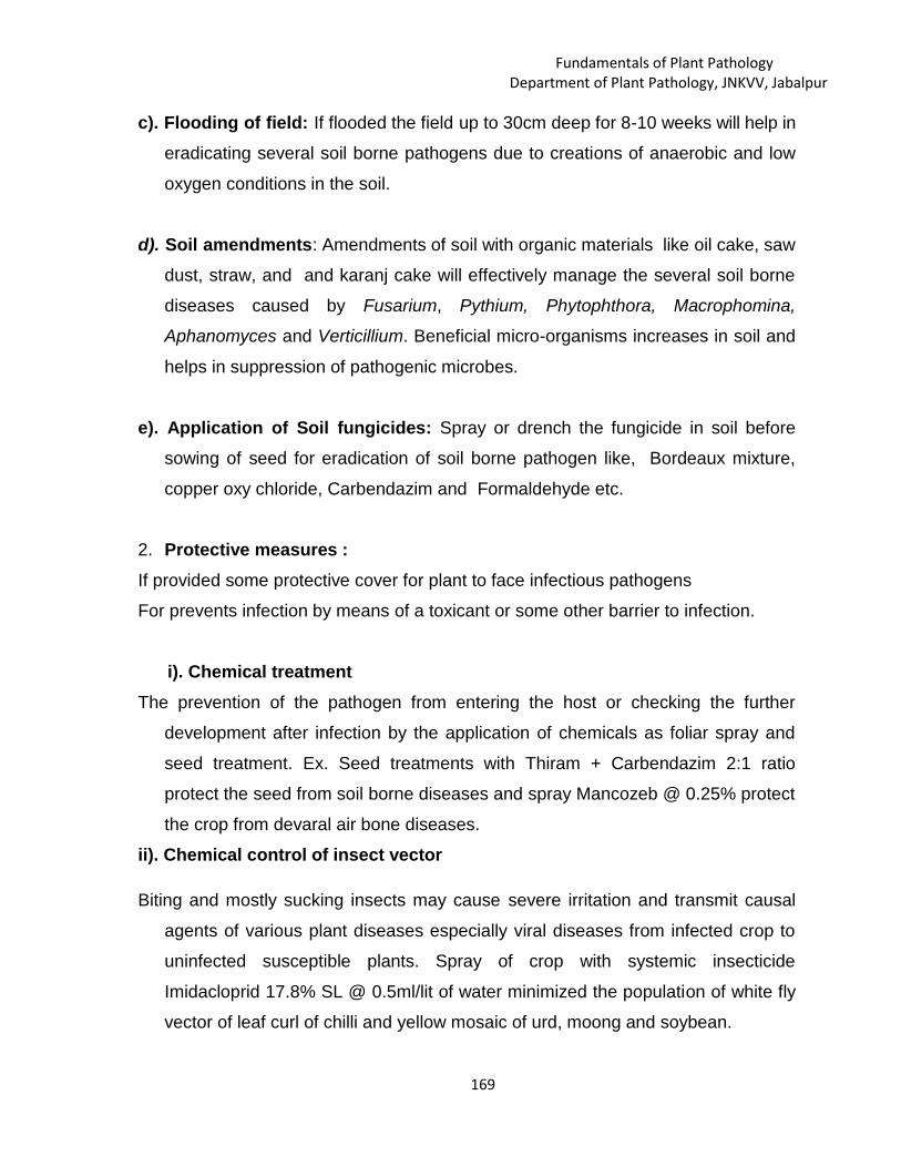
Fundamentals of Plant PathologyDepartment of Plant Pathology, JNKVV, Jabalpur
169
c). Flooding of field: If flooded the field up to 30cm deep for 8-10 weeks will help in
eradicating several soil borne pathogens due to creations of anaerobic and low
oxygen conditions in the soil.
d). Soil amendments: Amendments of soil with organic materials like oil cake, saw
dust, straw, and and karanj cake will effectively manage the several soil borne
diseases caused by Fusarium, Pythium, Phytophthora, Macrophomina,
Aphanomyces and Verticillium. Beneficial micro-organisms increases in soil and
helps in suppression of pathogenic microbes.
e). Application of Soil fungicides: Spray or drench the fungicide in soil before
sowing of seed for eradication of soil borne pathogen like, Bordeaux mixture,
copper oxy chloride, Carbendazim and Formaldehyde etc.
2. Protective measures :If provided some protective cover for plant to face infectious pathogens
For prevents infection by means of a toxicant or some other barrier to infection.
i). Chemical treatmentThe prevention of the pathogen from entering the host or checking the further
development after infection by the application of chemicals as foliar spray and
seed treatment. Ex. Seed treatments with Thiram + Carbendazim 2:1 ratio
protect the seed from soil borne diseases and spray Mancozeb @ 0.25% protect
the crop from devaral air bone diseases.
ii). Chemical control of insect vector
Biting and mostly sucking insects may cause severe irritation and transmit causal
agents of various plant diseases especially viral diseases from infected crop to
uninfected susceptible plants. Spray of crop with systemic insecticide
Imidacloprid 17.8% SL @ 0.5ml/lit of water minimized the population of white fly
vector of leaf curl of chilli and yellow mosaic of urd, moong and soybean.

Fundamentals of Plant PathologyDepartment of Plant Pathology, JNKVV, Jabalpur
170
iii). Modification of environments
The environment can be modified so that it is suitable for plant growth but not for
disease development. For example if aeration is increased in crop canopy then
humidity decrease on aerial plant parts, and become unfavourable for growth of
pathogen. Similarly improving soil drainage, changing the time of sowing, proper
irrigation practices can all produce conditions unfavourable to diseases
development.
iv). Modification of host nutritionNutrient application affects the development of plant diseases under field conditions
directly through the nutritional status of the plant and indirectly by affecting the
conditions which can influence the development of the disease . The root rot
disease of wheat and barley (take-all) caused by Gaeumannomyces graminis
can be effectively controlled by nutrition of the host plant
3. Development of resistance in hostDevelopment and utilization of host plant resistance remains the most viable,
environmentally safe, ecologically sound and also less expensive technique for
the management of diseases.
i). Selection and hybridization for disease resistance
ii). Resistance through chemotherapy
iii).Resistance through host nutrition
4. Therapy of diseased plantGenerally cure of diseased plant or plant parts in most crops is not possible, but
some crops and fruit trees have been successfully therapy by chemical and
physical treatment.
i). Chemotherapy
ii). Heat therapy
iii).Tree surgery
i). ChemotherapyIn chemotherapy, chemical treatments applied to eliminate the pathogen from the
infected part of the diseased plant. The using chemicals are mostly systemic

Fundamentals of Plant PathologyDepartment of Plant Pathology, JNKVV, Jabalpur
171
fungicides and antibiotics. These chemicals toxic to pathogen and provide
temporary resistance in host plant and they known as chemotherapeutants.
ii). Heat therapy
It is an important component of healthy seed and plant propagative material
production as it has proved most effective in eliminating the pathogen associated
with seed, tubers, bulbs and graft and also many virus diseases.
a). Hot water treatment: The seed and propagative plant part like cane set is
treated at 500 C for 10 to 15 minutes (grain seed) and 2 to 3 hours (can set). The
method is effective against loose smut of wheat, smut of barley, sorghum and smut,
wilt, leaf scald, ratoon stunting, grassy shoot, mosaic and chlorotic streak
pathogens of sugarcane.
b). Hot air treatment: The hot air treatment has been proved as eradication of plant
pathogen. If seed cane treated by hot air treatment at 540 C for 8 hours is effective
against grassy shoot and ratoon stunting causing pathogens.
c). Aerated stem therapy: Several fungal and viral pathogens eliminated through
specially designed aerated stem therapy (AST). The eradication of grassy shoot,
ratoon stunting and smut pathogens is recommended the treatment of sugarcane
set by aerated stem therapy at 500 C for 1-3 hours.
d). Moist hot air treatment: This method is successfully practices in management
of smut, grassy shoot, leaf scald and ratoon stunting diseases of sugarcane with the
seed can treated by moist hot air treatment at 540 C for 2.5 hours at 95-99 % RH.

Fundamentals of Plant PathologyDepartment of Plant Pathology, JNKVV, Jabalpur
172
iii).Tree surgery
After removal diseased parts and pruning and cutting practice covering the
wound with fungicidal paste is helping in avoid pathogen infection. Basically this
method employed on horticultural plants.
Biological methods for disease management:Practically using microorganisms against plant pathogens started with the
control of crown gall with Agrobacterium radiobacter and seedling blights caused
by Pythium and Rhizoctonia with Trichoderma harizanum, Gliocaladium virens and
Streptomycesgriseus.
Mechanisms of biological control (Antagonism)1. Antibiosis or Amensalism
Antibiosis or amensalism is microbial activities, in which one organism produce
antibiotics and toxins, which are volatile or nonvolatile in nature, and have a direct
effect on other organisms.
a). Antibiotics: Antibiotics are microbial toxins that can, at low concentrations,
poison or kill other microorganisms. Most microbes produce and secrete one or
more compounds with antibiotic activity. For example the strong
antagonists Trichoderma harzianum and T. viride secrete cell wall lysing enzymes,
6-1, 3-glucanase . Similarly Gliocladium virens produces gliotoxin which is
responsible for the death of Rhizoctonia solani on potato tubers. Some strains of
Pseudomonas fluorescens produce a range of compounds, viz., 2,4- diacetyl
phloroglucinol (DAPG), phenazines, pyocyanin, which have broad spectrum activity
against many plant pathogenic bacteria and fungi.
b). Bacteriocins: These are antibiotic like compounds with bactericidal specificity
closely related to the bacteriocin producer. Ex: crown gall (Agrobacterium
tumefaciens) can be control by the use of related Agrobacterium radiobacter
c). Volatile compounds: Antibiosis mediated by volatile compounds has been
observed in the management of soil borne pathogens, viz., Pythium ultimum,

Fundamentals of Plant PathologyDepartment of Plant Pathology, JNKVV, Jabalpur
173
Rhizoctonia solani and Verticillium dahlia, by Enterobacter cloacae. The volatile
fraction responsible for inhibition was identified as ammonia.
2. Competition: The majority of the biocontrol agents are fast growing and they
compete with plant pathogens for space, nutrients and minerals including oxygen.
During this process, the antagonist may suppress the growth of the pathogen
population in the rhizosphere and thus reduce disease development.The bacterial
bioagent Pseudomonas fluorescens produce extracellular, low molecular weight
iron transport agents, known as siderophores which chelate with iron in the soil,
and make it difficult for the pathogens to proliferate.
3. Mycoparasitism: Mycoparasitism or hyper parasitism occurs when the
antagonist invades the pathogens by secreting enzymes such as chitinases,
celluloses, glucanases and other lytic enzymes. The mechanism covers different
stages of interactions. First stage: Chemical stimulus of pathogenic fungi attracts
the antagonist fungi and induces a chemotropic response of the antagonist. Second
stage: Recognition between the pathogen and the antagonist is due to the lectins.
Third stage: It is followed by the interactions between hyphae of the pathogen and
the antagonist. Examples of such interactions are T. harzianum acting against
Fusarium oxyporum, F. roseum, F. solani, Phytophthara colocaciae and Sclerotium
rolfsii.
List some biocontrol agents for the management of plant diseases
S.No.
Name ofbioagent
Trade name Target pathoge
1. Trichoderma spp.
T. viride
Bio-fungus
Ecofit, Bioderma
soil borne diseases causing
pathogens viz., Sclerotinia,
phytopthora, Rhizoctonia
solani, Pythium spp.,
Fusarium
Verticillium,
2. Paecilomyces
lilacinus
Paecil or Bioact Egg parasite of Meloidogyne
incognita

Fundamentals of Plant PathologyDepartment of Plant Pathology, JNKVV, Jabalpur
174
3. Pythium
oligandrum
Polygandron Pythium altimum
4. Ampelomyces
quisqualis
Powdery mildew disease
5. Pseudomonas
florescence
Sclerotium rolfsii,
Xanthomonas axonopodis,
Fusarium moniliformae
6. Pseudomonas
syringae
Bio save 100 Botritis cinerea,Penicitllium
spp.
7. Bacillus subtilis Companion Ralstonia, Rhizoctonia,
Pythium,
Fusarium and Phytopthora
8. Agrobactertitum
radio bactor strain
Galltrol-A Agrobacterium tumefaciens
9. Pasteuria
penetrans
parasite on juvenile of root
knot nematode
Plant Growth Promoting Rhizobacteria (PGPR):Bio control agents also produce growth hormones like, auxins, cytokinin,
gibberellins etc. These hormones suppress the harmful pathogens and promote the
growth of plants and simultaneously increase the yield. A group of bacteria that
enhance the growth of plant through nitrogen fixation, phosphorus solubilization or
production of plant growth promoting metabolites are known as Plant Growth
Promoting Rhizobacteria (PGPR). Many PGPR strains specially Bacillus subtilis and
Pseudomonas fluorescens have a potential to be used as microbial inoculants to
enhance crop productivity due to their ability to produce phytohormones
Cross protection: The phenomenon in which plant tissues infected with mild strain
of a
pathogen are protected from infection by other severe strains of the pathogen. In
wilt disease of mint caused by Verticillium dahlia inoculation of roots at least 2

Fundamentals of Plant PathologyDepartment of Plant Pathology, JNKVV, Jabalpur
175
days earlier with Verticillium nigresces reduce the incidence of wilt. This strategy
also used in the management of severe strains of Citrus Tristeza virus.
Systemic Induced or Acquired Resistance Various biotic inducers (e.g., fungi,
bacteria, viruses, phytoplasma, insects, etc.) and abiotic stresses (e.g., chemical
and physical inducers) developed resistance due to the formation of structural
barriers such as lignification, induction of pathogenesis-related proteins, and
conditioning of the plants

Fundamentals of Plant PathologyDepartment of Plant Pathology, JNKVV, Jabalpur
176
Fungicides nature, chemical combinations and formulationsS. K .Tripathi
[email protected], College of Agriculture, Rewa
-------------------------------------------------------------------------------------------------------------------------FUNGICIDES
The word ‘fungicide’ originated from two latin words, viz., ‘fungus’and ‘caedo’. The word ‘caedo’ means ‘to kill.’ Thus ,the fungicide is achemical which has the ability to kill the fungus.Fungistat
The chemicals which may arrest the growth of the fungustemporarily. called fungistat and the phenomenon of temporarilyinhibiting the fungal growth is termed as fungistatis.
AntisporulantChemicals may inhibit the spore production without affecting the
growth of vegetative hyphas called as ‘Antisporulant’. The fungicide hasbeen defined as a chemical agent which has the ability to reduce orprevent the damage called Fungitoxicant’.
Fungicides can be broadly grouped based on their (i) mode ofaction (ii) general use and (iii) chemical composition.
ProtectantProtectant fungicides are prophylactic in their behavior. Fungicide
which is effective only if applied prior to fungal infection is called aprotectant, eg., Zineb, Sulphur.
TherapeutantFungicide which is capable of eradicating a fungus after it has
caused infection and there by curing the plant is called chemotherapeutant.eg. Carboxin,Oxycarboxin and antibiotics like Aureofungin.
EradicantEradicant are those which remove pathogenic fungi from an
infection court (area of the host around a propagating unit of a fungus inwhich infection could possibly occur). eg. Organic mercurials, limesulphur, dodine etc. These chemicals eradicate the dormant or activepathogen from the host.
L. 20

Fundamentals of Plant PathologyDepartment of Plant Pathology, JNKVV, Jabalpur
177
II. Based on general usesThe fungicides can also be classified based on the nature of their
use in managing the diseases.1. Seed protectants : Eg. Captan, thiram, organomercuries
carbendazim, carboxin etc.2. Soil fungicides (preplant) : Eg. Bordeaux mixture, copper oxy
chloride, Chloropicrin,Formaldehyde Vapam,
etc.,3. Soil fungicides : Eg. Bordeaux mixture, copper oxy chloride (for
growing plants), Capton, PCNB, thiram etc.4. Foliage and blossom : Eg. Capton, ferbam, zineb,
protectants mancozeb, chlorothalonil etc.5 Fruit protectants : Eg. Captan,
carbendazim,mancozeb
6 Eradicants : Eg. Organomercurials, lime sulphur, etc.7 Tree wound dressers : Eg. Boreaux paste, chaubattia paste, etc.8 Antibiotics : Eg. Actidione, Griseofulvin,
Streptomycin, Streptocycline,Based on Chemical CompositionThe chemical available for plant disease control runs into hundreds, however, all are notequally safe, effective and popular. Major group of fungicides used include salts of toxicmetals and organic acids, organic compounds of sulphur and mercury, quinones andheterocyclic nitrogenous compounds. Copper, mercury, zinc, tin and nickel are some of themetals used as base for inorganic and organic fungicides. The non metal substancesinclude, sulphur, chlorine, phosphorous etc
Fungicides formulations:
Fungicides are formulated in various ways and most commonly available formulationsare Emulsifiable Concentrates (EC) Wettable Powders (WP), Dusts (D), etc.
Commercially available fungicides usually consist of a mixture of active ingredient (a.i.)and other substances including diluents, wetting agents, stickers, emulsifiers, etc.Formulations containing mixtures of different active ingredients (especially mixtures ofprotectant and systemic fungicides) are also widely used nowadays. Differentformulations incorporating the same active ingredient may be used for distinct purposeslike seed treatment, foilar application etc.
Emulsifiable Concentrates (EC)
These are liquid formulations which can be diluted with water before application. Theactive ingredient is dissolved in a solvent. When these emulsifiable concentrate isadded to water, a milky mixture is formed which is a suspension of active ingredientand emulsified solvent in the water.
Wettable Powders (WP)

Fundamentals of Plant PathologyDepartment of Plant Pathology, JNKVV, Jabalpur
178
Wettable powder is a very common formulation for most of the fungicides, which isused for spray mixtures. The modern wettable powders are water-dispersible whichhave the quality to wet easily and disperse well in water. They are also called as Water-Dispersible Powders (WDP). The active ingredient is incorporated, usually at the rate of30-80%, with a finely ground inert dust (filler) such as Kaolin, a wetting agent and asuspending agent. The commonly used suspending agents are sodium ligninsulphonate (Sulphite dye), methyl celluloses, polyvinyl acetate and aluminium silicate.In addition, spreader-sticker is sometimes desirable, especially on plants with glossy orwaxy leaves.
Dusts (D)
Dust formulations usually contain 1-10% active ingredient for direct application in dryforms. They are manufactured in such a way that they are light enough to be carried bya slight breeze for a considerable distance. The finely divided particle of activeingredient is carried on a carrier particle. The commonly used carriers (diluents) arekaolin, talc, pyrophylite, bentonite, calcium silicate, hydrated silica, calcium carbonate,magnesium carbonate, gypsum, lime etc.
Granules
Granules are the formulations of the fungicide with inert materials formed into particlesabout the size of coarse sugar. The granules normally contain 3-10% of the activeingredient. Due to their size, the granules do not drift but have limited application beingconfined to soil and seed treatments.
Suspension or slurries
Formulation in which a dry form of the active ingredient is mixed with a liquid. Suchformulations usually contain a high percentage of active ingredient which is similar towettable powders. They are mixed with water for final use and require agitation. Theseare mostly used as seed dressers in seed processing companies.
Solutions True solutions are formulations in which active ingredient or a combination ofactive ingredients and a solvent is dissolved in water Solutions have the advantage ofrequiring no agitation after formulation is added in water. At present, the manufacturersare concentrating to develop new formulations to increase the efficacy of the chemicals.Some new formulations developed are: Soluble Liquid (SL), Soluble Powder (SP),Water Soluble Concentrate (WSC), Suspension Concentrate (SC) and Aqua Flow (AF).
Adjuvants
The fungicides can be commonly applied either as spraying or dusting. In sprayingmethod, the toxicant is made into a suspension in water. In order to increase theefficacy of the water mixed sprays, certain substances like wetting agents, dispersingagents, spreaders, stickers, etc. are added during the formulation of fungicides.Thevarious adjuvants are grouped as follows.
Dispersing agents (Deflocculating agents)
These are the substances which keep fine particles away from each other to preventdeflocculation. These materials, when added to formulations, ensure uniform

Fundamentals of Plant PathologyDepartment of Plant Pathology, JNKVV, Jabalpur
179
suspension and retard sedimentation of particles in the spray suspension. These arealso called as deflocculating agents. Eg. Gelatin, plant gums and milk products.
Emulsifying agentsPowders (WDP). The active ingredient is incorporated, usually at the rate of 30-
80%, with a finely ground inert dust (filler) such as Kaolin, a wetting agent and asuspending agent. The commonly used suspending agents are sodium ligninsulphonate (Sulphite dye), methyl celluloses, polyvinyl acetate and aluminium silicate.In addition, spreader-sticker is sometimes desirable, especially on plants with glossy orwaxy leaves. Agitation is generally necessary to keep uniform suspension.
Wetting agent (Wetters)
These are the materials which are added to ensure that there will be no layer of airbetween a solid and a liquid as they reduce the surface tension of the particles. Wettingagents, when added to aqueous fungicidal preparation, help in easy deposition onleaves. Eg. Polyethylene oxide condensat, esters of fatty acids and flour.
Spreading agent (Spreaders) Spreaders are the materials added to establishimproved contact between the spray materials and plant surface and thus ensuring agood coverage of fungicide. Wetting must precede spreading and this is the onlydistinction between wetting and spreading. Spreaders also reduce the surface tensionand thus improve contact. Eg. Soap, flour, sulphated amines, soapamines, mineral oils,glyceride oil, terpene oil, resinates and petroleum sulphonic acids.
Stickers (Adhesives)
The materials which are added to spray or dust to improve the adherence to plantsurfaces are called as stickers. They increase the tenacity of the fungicidalpreparations, thus increasing the residual action. Eg. Polyvinyl acetate, polybutanes,fish oil, linseed oil, milk casein,gelatin, dextrines, polyethylene polysulphide, starch,gum arabic, hydrocarbon oils and bentonite clays; Milk casein, gelatin also act as goodspreading and wetting agents besides acting as stickers.
Safeners
A Chemical which reduces the phytotoxicity of another chemical is called safener. Forexample copper sulphate is phytotoxic to plants, but with addition of lime its toxicity isreduced. Lime is, therefore, a safener. Lime is used universally with chemicals toprevent the formation of, or to neutralise arsenic, which is phytotoxic to plants.Glycerine oils are also used as safeners.

Fundamentals of Plant PathologyDepartment of Plant Pathology, JNKVV, Jabalpur
180
FUNGICIDES CLASSIFICATION AND THEIR MODE OF ACTIONA.R.WASNIKAR
JNKVV , Jabalpur--------------------------------------------------------------------------------------------------------Fungicides can be mainly classified based on their (i) mode of action (ii) general
use and (iii) chemical composition.Classification on the basis of Chemical Composition
. Major group of fungicides used include salts of toxic metals and organicacids, organic compounds of sulphur and mercury, quinines and heterocyclicnitrogenous compounds. Copper, mercury, zinc, tin and nickel are some of themetals used as base for inorganic and organic fungicides. The non metalsubstances include, sulphur, chlorine, phosphorous etc. The fungicides can bebroadly grouped as follows and discussed in detail.Copper Fungicides:
The fungicidal action of copper was mentioned as early as 1807 by Prevostagainst wheat bunt disease (Tilletia caries), but its large scale use as a fungicidestarted in 1885 after the discovery of Bordeaux mixture by Millardet in France.I. Copper sulphate preparationsBordeaux Mixture
The mixture of copper sulphate and lime was named as “BouillieBordelaise” (Bordeaux Mixture). The original formula developed by Millardetcontains 5 lbs of CuSO4 + 5lbs of lime + 50 gallons of water. The chemistry ofBordeaux mixture is complex and the suggested reaction is:
CuSO4 + Ca (OH)2 Cu(OH)2 + CaSO4The ratio of copper sulphate to lime solution determines the pH of the
mixture. The mixture prepared in the above said ratio gives neutral or alkalinemixture. If the quality of the used is inferior, the mixture may become acidic. If themixture is acidic, it contains free copper which is highly phytotoxic resulting inscorching of the plants. Therefore, it is highly essential to test the presence of freecopper in the mixture before applied. There are several methods to test theneutrality of the mixture, which are indicated below:(i) Field Test: Dip a well polished knife or a sickle in the mixture for few
minutes. If reddish deposit appears on the knife/sickle, it indicates the acidicnature of the mixture.
(ii) Litmus paper test: The colour of blue litmus paper must not change whendipped in the mixture.
(iii) pH paper test : If the paper is dipped in the mixture, it should show neutralpH.
L. 21

Fundamentals of Plant PathologyDepartment of Plant Pathology, JNKVV, Jabalpur
181
(iv) Chemical test: Acid a few drops of the mixture into a test tube containing 5ml of 10% potassium ferrocyanide. If red precipitate appears, it indicates theacidic nature of the mixture.
Bordeaux pasteBordeaux Paste consists of same constituents as that of Bordeaux mixture,
but it is in the form of a paste as the quantity of water used is too little. It is nothingbut 10 percent Bordeaux mixture and is prepared by mixing 1 kg of copper sulphateand 1 kg of lime in 10 litres of water. The method of mixing solution is similar to thatof Bordeaux mixture. It is a wound dresser and used to protect the woundedportions, cut ends of trees etc., against the infection by fungal pathogens.Burgundy mixture
It is prepared in the same way as Bordeaux mixture, except the lime issubstituted by sodium carbonate. So it is called as „Soda Bordeaux. It wasdeveloped Burgundy (France) in 1887 by Mason. The usual formula contains 1 kgof copper sulphate and 1 kg of sodium carbonate in 100 litres of water. It is a goodsubstitute for Bordeaux mixture and used in copper-sensitive crops.
Cheshunt compoundIt is compound usually prepared by mixing 2 parts of copper sulphate and 11
parts of ammonium carbonate. This formula was suggested by Bewley in the year1921. The two salts are well powdered, mixed thoroughly and stored in a air tightcontainer for 24 hours before being used. The ripened mixture is used by dissolvingit in water at the rate of 3 g/litre. The mixture is dissolved initially in a little hot waterand volume is made up with cold water and used for spraying.II. Copper carbonate preparationChaubattia Paste
Chaubattia paste is another wound dressing fungicide developed by Singh in1942 at Government Fruit Research Station, Chaubattia in the Almora district ofUttar Pradesh. It is usually prepared in glass containers or chinaware pot, by mixing800g of copper carbonate and 800g of red lead in litre of raw linseed oil or lanolin.This paste is usually applied to pruned parts of apple, pear and peaches to controlseveral diseases. The paste has the added advantage that it is not easily washedaway by rain water.III. Copper preparation
Cuprous oxidePreparation
Fungimar, Perenox, CopperSandoz, opper 4% dust,Perecot, Cuproxd, Kirt icopper.
Cuprous oxide is aprotective fungicide, usedmainly for seed treatmentand for foilage applicationagainst blight, downy

Fundamentals of Plant PathologyDepartment of Plant Pathology, JNKVV, Jabalpur
182
mildew and rusts.
IV. CopperoxychloridePreparation.
Blitox, Cupramar 50% WP,Fytolan, Bilmix 4%, Micop D-06, Micop w-50, Blue copper50, Cupravit, Cobox, Cuprax,Mycop.
It is a protective fungicide,controls Phytophthorainfestans on potatoes andseveral leaf spot and leafblight pathogens in field.
Sulphur fungicides:Use of sulphur in plant disease control is probably the oldest one and can be
classified as inorganic sulphur and organic sulphur. Inorganic sulphur is used in theform of elemental sulphur or as lime sulphur. Elemental sulphur can be either usedas dust or wettable sulphur, later being more widely used in plant disease control.Sulphur is best known for its effectiveness against powdery mildew of many plants,but also effective against certain rusts, leaf blights and fruit diseases.
Organic compounds of sulphur are now widely used in these days. All thesecompounds, called as 'carbamate fungicides', are derivatives of Dithiocarbamicacid, Dithiocarbamates are broadly grouped into two, based on the mechanism ofaction.Dithio –Carbamates:Monoalkyl Dithiocarbamates
Eg. Zineb, Maneb, Eg. Thiram, Ziram,
Mancozeb, Nabam, Vapam Ferbam
Dialkyl Dithiocarbamates
Trade Name Diseases Controlled
Inorganic Sulphur
1. Elemental Sulphur
(i) Sulphur dust
Sulphur dust Cosan,Wetsulf, Microsul
Sulphur is a contact andprotective fungicide, normallyapplied as sprays or as dust.It is generally used to controlpowdery mildews of fruits,vegetables, flowers andtobacco.
This is also effective againstapple scab (Venturiainaequallis)
and rusts of field crops.
2. Lime Sulphur(Calcium polysulphide)
It can be prepared byboiling 9 Kg or rock limeand 6.75Kg of sulphur in225 litres of
Lime Sulphur is effectiveagainst powdery mildews asa protective fungicide.
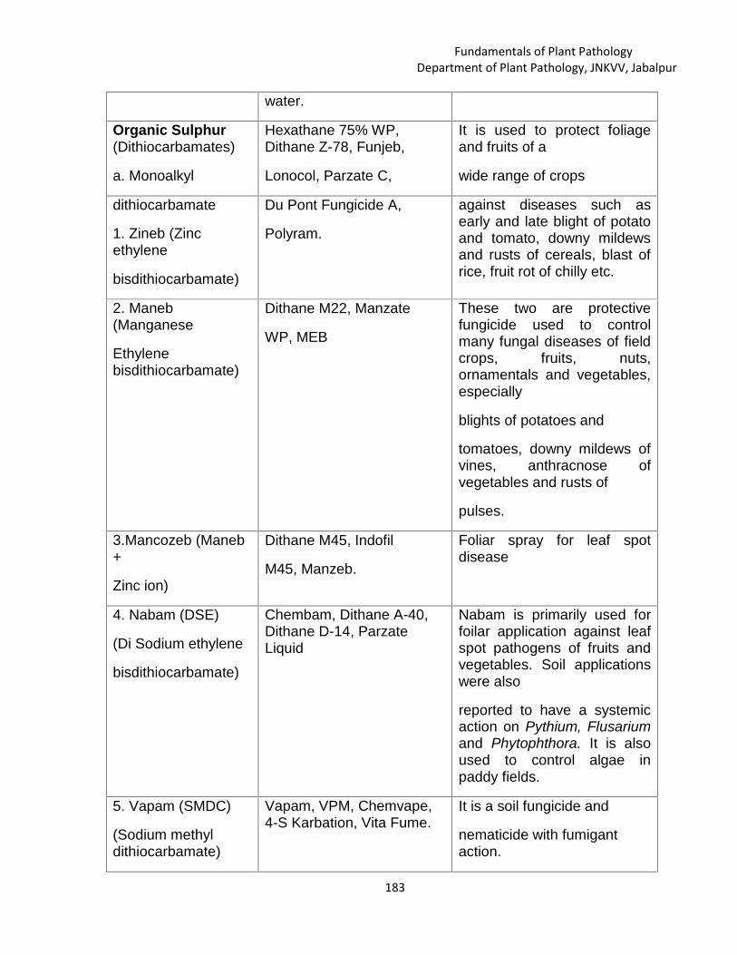
Fundamentals of Plant PathologyDepartment of Plant Pathology, JNKVV, Jabalpur
183
water.
Organic Sulphur(Dithiocarbamates)
a. Monoalkyl
Hexathane 75% WP,Dithane Z-78, Funjeb,
Lonocol, Parzate C,
It is used to protect foliageand fruits of a
wide range of crops
dithiocarbamate
1. Zineb (Zincethylene
bisdithiocarbamate)
Du Pont Fungicide A,
Polyram.
against diseases such asearly and late blight of potatoand tomato, downy mildewsand rusts of cereals, blast ofrice, fruit rot of chilly etc.
2. Maneb(Manganese
Ethylenebisdithiocarbamate)
Dithane M22, Manzate
WP, MEB
These two are protectivefungicide used to controlmany fungal diseases of fieldcrops, fruits, nuts,ornamentals and vegetables,especially
blights of potatoes and
tomatoes, downy mildews ofvines, anthracnose ofvegetables and rusts of
pulses.
3.Mancozeb (Maneb+
Zinc ion)
Dithane M45, Indofil
M45, Manzeb.
Foliar spray for leaf spotdisease
4. Nabam (DSE)
(Di Sodium ethylene
bisdithiocarbamate)
Chembam, Dithane A-40,Dithane D-14, ParzateLiquid
Nabam is primarily used forfoilar application against leafspot pathogens of fruits andvegetables. Soil applicationswere also
reported to have a systemicaction on Pythium, Flusariumand Phytophthora. It is alsoused to control algae inpaddy fields.
5. Vapam (SMDC)
(Sodium methyldithiocarbamate)
Vapam, VPM, Chemvape,4-S Karbation, Vita Fume.
It is a soil fungicide and
nematicide with fumigantaction.

Fundamentals of Plant PathologyDepartment of Plant Pathology, JNKVV, Jabalpur
184
. It is effective againstdamping off disease of
papaya and vegetables
and wilt of cotton. It is
also effective againstnematode infestation in
citrus, potato and root
knot nematodes invegetables.
b. Dialkyl
Dithiocarbamate
1. Ziram (Zincdimethyl
dithiocarbamate)
Cuman L. Ziram, Ziride
80 WDP, Hexaazir 80%WP, Corozate, Fukiazsin,Karbam white, Milbam,Vancide 51Z, Zerlate,Ziram, Ziberk, Zitox 80%WDP.
Ziram is a protective fungicidefor use on fruit andvegetables crops againstfungal pathogens includingapple scab. It is nonphytotoxic except to zincsensitive plants.
It is highly effective againstanthracnose of beans,pulses, tobacco & tomato,and also against rusts ofbeans etc.
2. Ferbam (Ferric
dimethyl
dithiocarbamate)
Coromat, Febam, Ferberk,Femate, Fermate D,Fermicide, Hexaferb 75%WP, Karbam Black,Ferradow.
Ferbam is mainly used
for the protection of foliageagainst fungal pathogens offruits and vegetablesincluding Taphrina deformansof
peaches, anthracnose ofcitrus, downy mildew oftobacco and apple scab.
3. Thiram (Tetramethyl
thiram disulphide)
Thiride 75 WDP, Thiride750, Thiram 75% WDP,Hexathir, Normerson,Panoram 75, Thiram,TMTD, Arasan, Tersan 75,Thylate, Pomarsol,
Thiuram.
It is used for seed treatmentboth as dry powder or as aslurry. It is a protectivefungicide also suitable forapplication to foliage tocontrol Botrytis spp. Onlettuces, ornamental, softfruits and vegetables, rust on

Fundamentals of Plant PathologyDepartment of Plant Pathology, JNKVV, Jabalpur
185
ornamentals and Venturiapirina on pears. It is alsoeffective against soil bornepathogens like Pythium,Rhizoctonia and Fusarium.
Mercury Fungicides:Mercury fungicides can be grouped as inorganic and organic mercury
compounds. Both the groups are highly fungitoxic and were extensively used asseed treatment chemicals against seed borne diseases.
Common Name Trade Name Diseases Controlled
I. Inorganic Mercury
1. Mercuric chloride
2. Mercurous chloride
II. Organomercurials
Methoxy ethylmercury Chloride
Phenyl mercurychloride
Ethyl MercuryChloride
Tolyl mercury acetate
Merfusan, Mersil
Cyclosan, M-C Turf
fungicide.
Agallol, Aretan,Emisan, Ceresanwet (India)
Ceresan Dry(India),
Ceresol, Leytosan.
Ceresan (USA)
Agrosan GN.
It is used for treating potato tubersand propagative materials of otherroot crops
Mercurous chloride is limited to soilapplication in crop protection usebecause of its phytotoxicity.Theseare used mainly for
treatment of seeds and plantingmaterials.
These fungicides are
used for seed treatment by dry,
wet or slurry method. For seed
treatment 1% metallic mercury isapplied at 0.25% concentration
Heterocyclic Nitrogen Compounds, Quinones and Miscellaneous FungicidesHeterocyclic Nitrogen Compounds:

Fundamentals of Plant PathologyDepartment of Plant Pathology, JNKVV, Jabalpur
186
Heterocyclic nitrogen compounds are mostly used as foliage and fruitsprotectants. Some compounds are very effectively used as seed dressers. Some ofthe commonly used fungicides are listed below.
Common Name Trade Name Diseases Controlled1.Captan (Kittleson's
Killer) (N-trichloromethyl
thio-4- cyclohexence-1,2- dicarboximide)
Captan 50W, Captan 75
W, Esso Fungicide 406,
Orthocide 406, Vancide
89, Deltan, Merpan,
Hexacap.
It is a seed dressing fungicideused to control
diseases of many fruits,
ornamental and vegetablecrops against rots anddamping off.
2. Captafol (Cis-N-
1,1,2,2-tetra chlorohexane
1,2- dicarboximide)
Foltaf, Difolaton,Difosan,
Captaspor, Foleid,
Sanspor.
It is a protective fungicide,widely used to
control foliage and fruit
diseases of tomatoes,
coffee potato.
3. Glyodin Glyoxaliadine, Glyoxide,Glyodin, Glyoxide Dry,
Glyodex 30% liquid and
70% WP.
It has a narrow spectrum ofactivity. As a spray, it controlsapple scab and cherry leafspot.
4.Folpet (Folpet) [N-
(trichloromethyl-thi)]
phthalimide
Phartan, Acryptan,Phaltan, Folpan,Orthophaltan.
It is also a protectivefungicide used mainly for
foliage application againstleaf spots, downy andpowdery mildews of manycrops.
Benzene compounds:Many aromatic compounds have important anti-microbial properties and
have been developed as fungicides. Some important benzene compoundscommonly used in plant disease control are listed below.
Common Name Trade Name Diseases Controlled
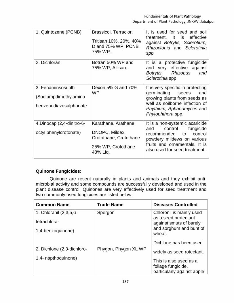
Fundamentals of Plant PathologyDepartment of Plant Pathology, JNKVV, Jabalpur
187
1. Quintozene (PCNB) Brassicol, Terraclor,
Tritisan 10%, 20%, 40%D and 75% WP, PCNB75% WP.
It is used for seed and soiltreatment. It is effectiveagainst Botrytis, Sclerotium,Rhizoctonia and Sclerotiniaspp.
2. Dichloran Botran 50% WP and75% WP, Allisan.
It is a protective fungicideand very effective againstBotrytis, Rhizopus andSclerotinia spp.
3. Fenaminsosuplh
(Sodiumpdimethylamino
benzenediazosulphonate
Dexon 5% G and 70%WP
It is very specific in protectinggerminating seeds andgrowing plants from seeds aswell as soilborne infection ofPhythium, Aphanomyces andPhytophthora spp.
4.Dinocap (2,4-dinitro-6-
octyl phenylcrotonate)
Karathane, Arathane,
DNOPC, Mildex,Crotothane, Crotothane
25% WP, Crotothane48% Liq.
It is a non-systemic acaricideand control fungiciderecommended to controlpowdery mildews on variousfruits and ornamentals. It isalso used for seed treatment.
Quinone Fungicides:Quinone are resent naturally in plants and animals and they exhibit anti-
microbial activity and some compounds are successfully developed and used in theplant disease control. Quinones are very effectively used for seed treatment andtwo commonly used fungicides are listed below:
Common Name Trade Name Diseases Controlled1. Chloranil (2,3,5,6-
tetrachlora-
1,4-benzoquinone)
2. Dichlone (2,3-dichloro-
1,4- napthoquinone)
Spergon
Phygon, Phygon XL WP.
Chloronil is mainly usedas a seed protectantagainst smuts of barelyand sorghum and bunt ofwheat.
Dichlone has been used
widely as seed rotectant.
This is also used as afoliage fungicide,particularly against apple
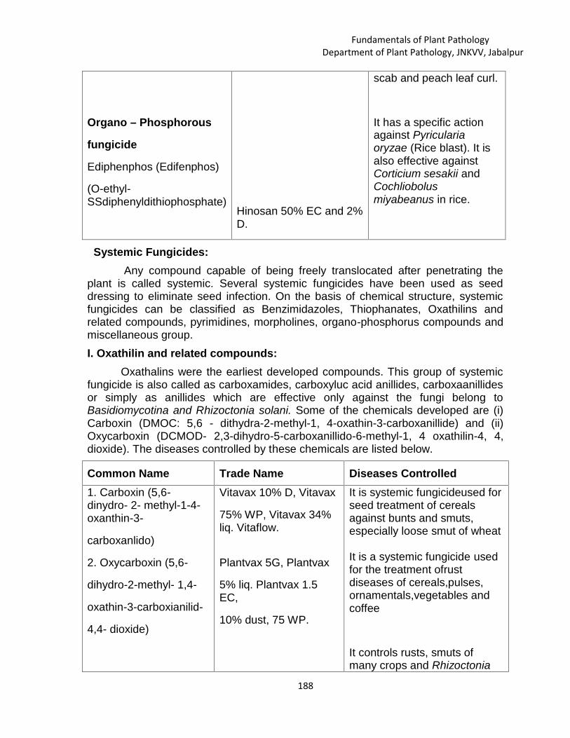
Fundamentals of Plant PathologyDepartment of Plant Pathology, JNKVV, Jabalpur
188
Organo – Phosphorous
fungicide
Ediphenphos (Edifenphos)
(O-ethyl-SSdiphenyldithiophosphate)
Hinosan 50% EC and 2%D.
scab and peach leaf curl.
It has a specific actionagainst Pyriculariaoryzae (Rice blast). It isalso effective againstCorticium sesakii andCochliobolusmiyabeanus in rice.
Systemic Fungicides:Any compound capable of being freely translocated after penetrating the
plant is called systemic. Several systemic fungicides have been used as seeddressing to eliminate seed infection. On the basis of chemical structure, systemicfungicides can be classified as Benzimidazoles, Thiophanates, Oxathilins andrelated compounds, pyrimidines, morpholines, organo-phosphorus compounds andmiscellaneous group.I. Oxathilin and related compounds:
Oxathalins were the earliest developed compounds. This group of systemicfungicide is also called as carboxamides, carboxyluc acid anillides, carboxaanillidesor simply as anillides which are effective only against the fungi belong toBasidiomycotina and Rhizoctonia solani. Some of the chemicals developed are (i)Carboxin (DMOC: 5,6 - dithydra-2-methyl-1, 4-oxathin-3-carboxanillide) and (ii)Oxycarboxin (DCMOD- 2,3-dihydro-5-carboxanillido-6-methyl-1, 4 oxathilin-4, 4,dioxide). The diseases controlled by these chemicals are listed below.
Common Name Trade Name Diseases Controlled1. Carboxin (5,6-dinydro- 2- methyl-1-4-oxanthin-3-
carboxanlido)
2. Oxycarboxin (5,6-
dihydro-2-methyl- 1,4-
oxathin-3-carboxianilid-
4,4- dioxide)
Vitavax 10% D, Vitavax
75% WP, Vitavax 34%liq. Vitaflow.
Plantvax 5G, Plantvax
5% liq. Plantvax 1.5EC,
10% dust, 75 WP.
It is systemic fungicideused forseed treatment of cerealsagainst bunts and smuts,especially loose smut of wheat
It is a systemic fungicide usedfor the treatment ofrustdiseases of cereals,pulses,ornamentals,vegetables andcoffee
It controls rusts, smuts ofmany crops and Rhizoctonia

Fundamentals of Plant PathologyDepartment of Plant Pathology, JNKVV, Jabalpur
189
3.Pyracarbolid (2-methyl-
5,6-dihydro- 4H-Pyran-3-
carboxylic acid anilide).
Sicarol.
solani, but is slightly moreeffective than carboxin
II. Benzimidazoles:The chemicals of this group show a very broad spectrum activity against a
variety of fungi. However, they are not effective against bacteria as well as fungibelongs to Mastigomycotina. Two types of fungicidal derivates of benzimidazolesare known. The first type of derivates includes fungicides such as thiabendazoleand fuberidazole. The fungicidal moiety of the second type is methyl-2-benzimidazole carbamate (MBC). The fungicides of this group may be simple MBCsuch as carbendazim or a complex from such as benomyl, which transforms intoMBC in plant system. Some of the important diseases controlled by thesecompounds are shown below:
Common Name Trade Name Diseases Controlled1.Benomyl (Methyl - 10
(butly carbomyl)-2benzimidazolecarbamate)
2. Carbendazim (MBC)
(Methyl -2-
Benzimidazolecarbamate)
Benlate 50 WP,Benomyl. Bavistin 50WP, MBC, Dersol,B.Sten 50, Zoom,Tagstin, Agrozim,Jkenstin.
It is a protective anderadicative fungicide withsystemic activity, effectiveagainst a wide range of fungiaffecting field crops, fruits andornamentals. It is veryeffective against rice blast,apple scab, powdery mildewof cereals, rose, curcurbitsand apple and Diseasescaused by Verticillium andRhizoctonia. It is also used aspre-and postharvest sprays ofdips for the control of storagerots of fruits and vegetables.
3. Thiabendazole (TBZ)
(2,4-thiazoylbenzimidazole)
Thiabendazole, Mertect,
Tecto, Storite.
It is a broad spectrumsystemic fungicide effectivelagainst many major fungaldiseases. Pathogenic fungalcontrol includes species ofBotrytis, Ceratocystis,
Cercospora, Colletotrichum,
Fusarium, Rhizoctonia,

Fundamentals of Plant PathologyDepartment of Plant Pathology, JNKVV, Jabalpur
190
4.Fuberidazole (2, (2-
buryl) - benzimidazole).
Voronit.
Sclerotinia, Septoria and
Verticillium. It is also effectivefor the post harvest treatmentof fruits and vegetables tocontrol storage diseases.
It is used for the treatment ofseeds against diseasescaused by Fusarium,Particularly F.nivale on ryeand F.culmorum of peas
III. Thiophanates:These compounds represent a new group of systemic fungicides based on
thiourea. They are the derivatives of thioallophanic acid. These fungicides containaromatic nucleus which is converted into benzimidazole ring for their activity.Hence, thiophanates are often classified under benzimidazole group and thebiological activity of thiophanates resembles of benomyl. Two compounds aredeveloped under this group are discussed.
Common Name Trade Name Diseases Controlled1. Thiophanate(1,2 - bis
(ethyl carbonyl-2-thioureido) benzene)
2. Thiophanate - methyl
(1,2 bis (3methoxycarbonyl- 2-thioureido) benzene.)
Topsin 50 WP,Cercobin
50 WP, Enovit.
Topsin-M70 WP,Cercobin-M 70 WP,Envovit-methyl,Mildothane.
It is a systemic fungicide witha broad range of action,effective against Venturia spp.,on apple and pear crops,powdery mildews, Botrytis andSclerotinia spp.
It is effective against a widerange of fungal pathogens,including Venturia spp. onapples and pears,Mycosphaerella musicola
on bananas, powdery mildewson apples, cucurbits, pearsand vines, Pyricularia oryzaeon rice, Botrytis andCerospora on various crops.
IV. Morpholines:

Fundamentals of Plant PathologyDepartment of Plant Pathology, JNKVV, Jabalpur
191
Common Name Trade Name Diseases ControlledTridemorph (2-6-dimethyl- 4-cyclo-tridecyl
morpholine)
Calixin 75 EC, Bardew,
Beacon
It is an eradicant fungicide withsystemic action, beingabsorbed through foliage androots to give some protectiveaction. It controls powderymildew diseases of cereals,vegetables and ornamentals. Itis highly effective againstMycosphaerella, Exobasidium
V. Pyrimidines, Pyridines, Piperidines and Imidazole:
Common Name Trade Name Diseases Controlled1. Triadimefon (1-(4-chlorophenoxy)-3, 3-dimethyl-1-(1-2-triazol-1yl) butan-2-one)
2. Triadimenol (1-(4-Chlorophenoxyl-3, 3-imethyl-1(1,2,4-triazol-1-
yl) butan-2-ol)
3. Bitertanal
(B-(1-1-biphenyl-4-yloxy-a- (1-1-dimethyl-ethyl-1-H-1,2-4- triazole-1-ethanol)
4. Etridiazole
(5-ethaoxy-3-
trichloromethyl, 1,2-
4-thiadiazole)
Bayleton, Amiral
Baytan
Baycor
Terrazole 30% WP,
Terrazole 95% WP,
It is very effective against
powdery mildews and rusts ofseveral crops.
It is also very effective againstpowdery mildews
and rusts of several crops.
It is highly effective againstrusts and powdery mildew of avariety of crops. It is also usedagainst Venturia and Moniliniaon fruits and Cereosporaleafspots of groundnut andbanana.
It is very effective againstPhytophthora and Pythiumspp. and seeding diseases ofcotton, groundnut, vegetables,fruits and ornamentals

Fundamentals of Plant PathologyDepartment of Plant Pathology, JNKVV, Jabalpur
192
Terrazole 25% EC,Koban, Pansol EG,Pansol 4% DP, TurbanWP, Terracoat Aaterra.
IX. Organo phosphorous compounds:
Common Name Trade Name Diseases Controlled1. Pyrazophos (2-0-0-Diethylthionophosphoryl)-5- methyl-6-carbethoxypyrazolo-(1- 5a)pyrimidine)
2. Iprobenphos (IBP)(S-benyzl-0-0-bisisopropylphosphorothiate)
X. Piperazine1.Triforine(N,N-bis-(1-foramido-2,2,2-trichloroethyl- piperazine)XI. Phenol derivative1. Choloroneb (1-4-dichloro-2,5-dimethoxy benzene)
Afugan, Curamil, WP,Missile EC.
Kitazin 48% EC,Kitazin17G, Kitazin 2% D.
Saprol-EG, Fungitex.
Demonsan 65 WP,TersanSP, Turffungicide
It is used to control powderymildews of cereals,vegetables, fruits andornamentals.
It is used to controlPyricularia oryzae andsheath blight of rice.
It is effective againstpowdery mildew, scab andother diseases of fruits andrust on ornamentals andcereals.
It is also active againststorage diseases of fruits. Itis highly fungistatic toRhizoctonia spp.,moderately so to ythiumspp. and poorly to Fusariumspp. It is used as asupplemental seedtreatment for beans andsoyabeans to controlseedling disease
XIII. Other systemic fungicides:Common Name Trade Name Diseases Controlled1. Metalaxyl (methyl-DLN-(2,6- dimethylphenyl-N-)2-methoxyacetyl
Apron 35 SD, RidomilRidomil MZ 72 WP(8% Metalaxyl + 64%Mancozeb)
It is a systemic fungicide andhighly effective for specificuse as seed dressing againstfungal pathogens of the orderPeronosporales.
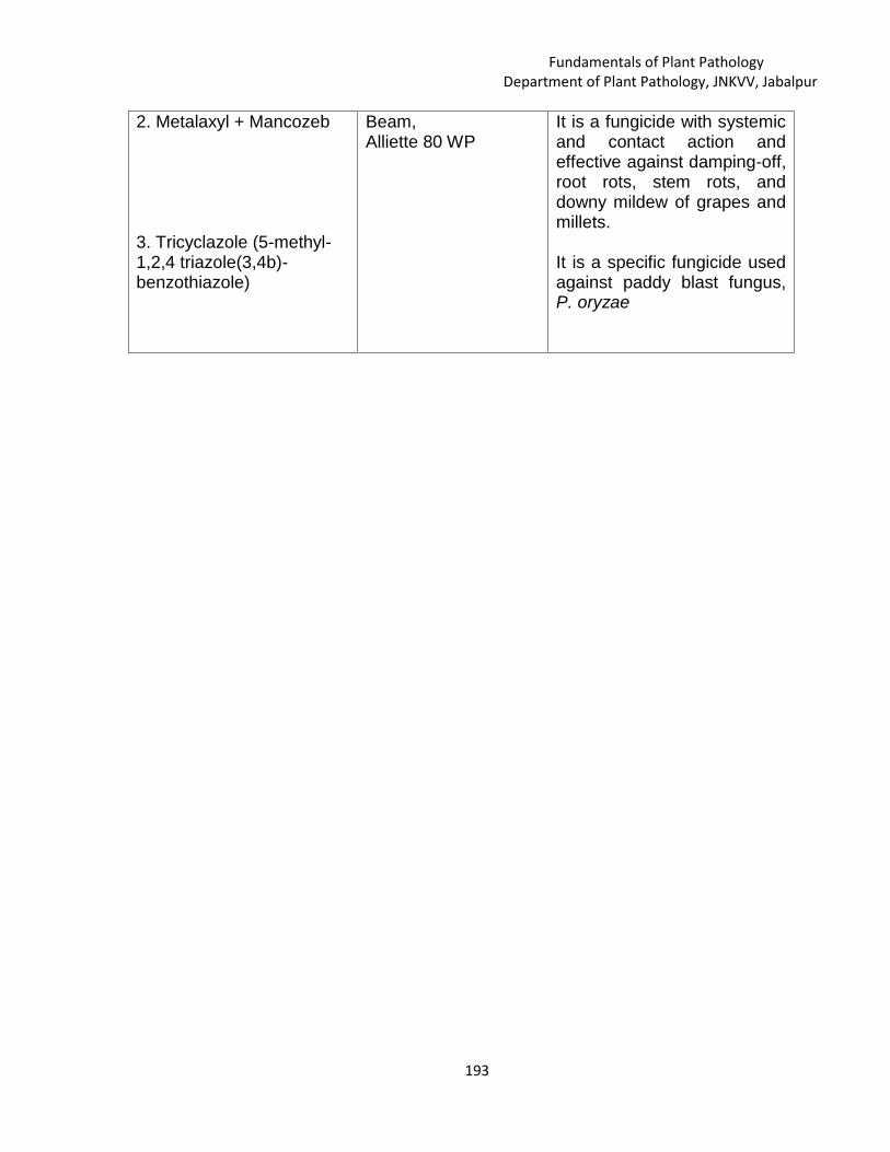
Fundamentals of Plant PathologyDepartment of Plant Pathology, JNKVV, Jabalpur
193
2. Metalaxyl + Mancozeb
3. Tricyclazole (5-methyl-1,2,4 triazole(3,4b)-benzothiazole)
Beam,Alliette 80 WP
It is a fungicide with systemicand contact action andeffective against damping-off,root rots, stem rots, anddowny mildew of grapes andmillets.
It is a specific fungicide usedagainst paddy blast fungus,P. oryzae

Fundamentals of Plant PathologyDepartment of Plant Pathology, JNKVV, Jabalpur
194
AntibioticsSanjeev Kumar
Department of Plant Pathology,JNKVV, Jabalpur-482004 (M.P.), India
---------------------------------------------------------------------------------------------------------------------------
Definition :“Antibiotic is defined as a chemical substance produced by one
microorganism, which in low concentration can inhibit or even kill othermicroorganism”.
Brief History
Sl.no Name of scientist Year Contribution1 Alexander Fleming 1928 Discovered first antibiotic ‘Penicillin’ in
Scotland from the fungus Penicilliumnotatum.
2 Windling 1932 Isolated first antibiotic ‘Gliotoxin’ fromGlioclodium fimbriatum to be used inplant protection
3 Dr. S. A.Waksman 1942 Coined term Antibiotics
4 W. Waksman 1944 Isolated non-toxic broad- spectrumantibiotic Streptomycin fromStreptomyces griseus
5 M.J. Thirumalacharand Gopalkrishnan
1962 Developed Aureofungin a antifungalantibiotic from Streptomycescinnamoneus var terricola in India
Character of good antibiotics It should be broad spectrum. It should be safe to plants It is fairly harmless to human beings and animals. It should be active on or inside of the plant. It tolerates oxidation, UV irradiation, rainfall, and high temperature.
Advantages in using antibiotics Easy to produce antibiotics at cheaper rate. Cure incurable diseases.
L. 22

Fundamentals of Plant PathologyDepartment of Plant Pathology, JNKVV, Jabalpur
195
Less number of sprays required. Less leaching effect. Acts as eradicant. Manage MLO’s and bacterial diseases.
Disadvantages in using antibiotics Shelf life very short. Less stable in storage. Narrow site of action. Risk of development of resistance.
Classification of antibioticsI. Based on Their Activity
a) Broad-spectrum antibiotic: Antibiotic which destroy or inhibit the growth ofa wide range of gram positive and gram negative bacteria.E. g. Tetracylines, Chloromycetin etc.
b) Narrow-spectrum antibiotic: Antibiotic which mainly attack gram positivebacteria or gram negative bacteria.E. g. Penicillin.
II.Based on Chemical Group1.β- Lacta antibiotics
Ex: a. penicillinsb. cephlosporins
2. TetracyclinsEx: a. Tetracyclins
b. Chlorotetracyclinec. Oxytetracyclins
3. RifamycinsEx: Rifamycin A to E
4. Amino glycosidic antibioticsEx: Streptomycin, Gentamycin
5. Macrolide antibioticsa. Erythromycinb. Spyramycinec. Olendomycine
6. Polypeptide group antibioticsBacitracine, Capriomycine, Polymixin, Viomycine
7. Miscellaneous groupVancomycine, Chloromphenical, Fusidic acid, Lincomycine
8. Synthetic antibioticsEx: Nitrofuron compoundsa. Furazolidin - Stomachb. Nitrofurantoin - Urinary track infection

Fundamentals of Plant PathologyDepartment of Plant Pathology, JNKVV, Jabalpur
196
c. Nitrofurozone - Ear infectionIII. Based on how they work on the stopping the infection of the bacteria
Bactericidal
They kill the bacteria present in the body which causes disease.Eg.:- Penicillin and polymyxin.
Bacteriostatic
The medicines which are used to inhibit the growth of microbes are known asbacteriostatic.Eg.:- Tetracycline, Chloramphenicol.
Mode of ActionSl.No mechanism Examples1 Cell wall synthesis inhibitors Penicillins, Cephalosporins,
Bacitracins
2 Inhibitors of protein synthesis Streptomycin, tetracyclins3 Inhibitors of nucleic acid synthesis Sulphanomides4 Disorganises the cytoplasmic
membraneAmphitericin- B
Antibiotics for plant disease managementAntibiotics are substances which are produced by micro- organisms and which actagainst micro-organisms. Most antibiotics known uptill now are products ofactinomycetes and some are from fungi and bacteria. Some of the importantantibiotics are mentioned here.
Sl.
no
Name ofAntibiotic
Source Disease managment
1 Streptomycin Streptomycesgriseus
Citrus canker, bacterial leaf spot of tomatoand pepper, hollow blight of french bean,fire blight of apples and pears.
Bacterial diseases of tubers, beans,cotton, crucifers, cereals, etc.
2 Cycloheximide Mildew of beans, covered smut of oats,brown rot of peach, yeasts but is inactiveagainst bacteria.
3 Griseofulvin Penicilliumgriseofulvwn
Downy mildew of cucurbits, Powderymildew of rose, Powdery mildew of beans,Early blight of tomato and Brown rot of

Fundamentals of Plant PathologyDepartment of Plant Pathology, JNKVV, Jabalpur
197
P. patulum, apple.
4 Blasticidins Streptomycesgriseochromogenes
Both bacteria and fungi
5 Aureofungin Streptomycescinnamomens.
Downy mildew, powdery mildew andanthrancnose of grapes, ricecontrols Helminthospriumoryzae. Pyricularia oryzae ofrice. Diplodia of mango, Alternaria rot oftomato, Sclerotinia rot ofpeach, Pythium rot of cucurbits etc
6 Tetra cyclones Streptomyces Crown gall, fire blight of apples etc.
7 Agrimycin-l00& 500
Halo blight, citrus canker, seedling blight,leaf spot, black arm disease of cotton, softrot and black leg of potato.
8 Antimycin Streptomyceskitasawensis
S.griseus
Early blight of tomato, seedling blight ofoat, rice blast etc.
9 Thiolutin Streptomycesalbus
Late blight of potato and downy mildew ofbroccoli
10 Mystatin Streptomycesnoursei
Anthracnose of beans and downy mildewof cucumber
11 Bulbifonnin Bacilus subtiles Wilt of pigeon pea.