GW583340 and GW2974, human EGFR and HER-2 inhibitors, reverse ABCG2-and ABCB1-mediated drug...
10
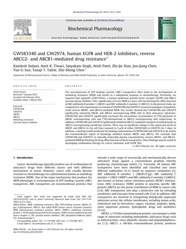
GW583340 and GW2974, human EGFR and HER-2 inhibitors, reverse ABCG2- and ABCB1-mediated drug resistance § Kamlesh Sodani, Amit K. Tiwari, Satyakam Singh, Atish Patel, Zhi-Jie Xiao, Jun-Jiang Chen, Yue-Li Sun, Tanaji T. Talele, Zhe-Sheng Chen * Department of Pharmaceutical Sciences, College of Pharmacy and Allied Health Professions, St. John’s University, Queens, NY 11439, USA 1. Introduction Cancer chemotherapy typically involves use of combinations of anticancer drugs from different classes and with different mechanisms of action. However, cancer cells usually become resistant to chemotherapy via a phenomenon known as multidrug resistance (MDR). One of the major mechanisms that produce the MDR phenotype is overexpression of ATP binding cassette (ABC) transporters. ABC transporters are transmembrane proteins that extrude a wide range of structurally and mechanistically diverse anticancer drugs against a concentration gradient, thereby producing chemotherapy failure [1]. Currently, 49 human ABC transporters have been identified and classified into seven different subfamilies (A–G) based on sequence similarities [2]. ABC subfamily B member 1 (ABCB1/P-gp), ABC subfamily C member 1 (ABCC1/MRP1) and ABC subfamily G member 2 [ABCG2, also known as breast cancer resistance protein (BCRP), mitoxan- trone (MX) resistance (MXR) protein or placenta specific ABC protein (ABCP)] are the prime contributors of MDR in cancer cells [3,4]. ABC transporters also play a protective role by extruding xenobiotics and toxicants from normal cells [3,4]. In addition, ABC transporters are involved in transporting biologically important substrates across the cellular membranes, including amino acids, cholesterol and its derivatives, sugars, vitamins, peptides, lipids, some important proteins, hydrophobic drugs and antibiotics [3,5,6]. ABCB1, a 170 kDa transmembrane protein, can transport a wide range of substrates including hydrophobic anticancer drugs such as anthracyclines, vinca alkaloids, taxanes and epipodophyllotox- ins [1,3]. ABCC1, a 190 kDa transmembrane protein, produces Biochemical Pharmacology 83 (2012) 1613–1622 A R T I C L E I N F O Article history: Received 7 January 2012 Accepted 28 February 2012 Available online 7 March 2012 Keywords: GW583340 GW2974 ABCB1 ABCG2 Multidrug resistance Tyrosine kinase inhibitor A B S T R A C T The overexpression of ATP binding cassette (ABC) transporters often leads to the development of multidrug resistance (MDR) and results in a suboptimal response to chemotherapy. Previously, we reported that lapatinib (GW572016), a human epidermal growth factor receptor (EGFR) and HER-2 tyrosine kinase inhibitor (TKI), significantly reverses MDR in cancer cells by blocking the efflux function of ABC subfamily B member 1 (ABCB1) and ABC subfamily G member 2 (ABCG2). In the present study, we conducted in vitro experiments to evaluate if GW583340 and GW2974, structural analogues of lapatinib, could reverse ABCB1- and ABCG2-mediated MDR. Our results showed that GW583340 and GW2974 significantly sensitized ABCB1 and ABCG2 overexpressing MDR cells to their anticancer substrates. GW583340 and GW2974 significantly increased the intracellular accumulation of [ 3 H]-paclitaxel in ABCB1 overexpressing cells and [ 3 H]-mitoxantrone in ABCG2 overexpressing cells respectively. In addition, GW583340 and GW2974 significantly inhibited ABCG2-mediated transport of methotrexate in ABCG2 overexpressing membrane vesicles. There was no significant change in the expression levels of ABCB1 and ABCG2 in the cell lines exposed to 5 mM of either GW583340 or GW2974 for 3 days. In addition, a docking model predicted the binding conformation of GW583340 and GW2974 to be within the transmembrane region of homology modeled human ABCB1 and ABCG2. We conclude that GW583340 and GW2974, at clinically achievable plasma concentrations, reverse ABCB1- and ABCG2- mediated MDR by blocking the drug efflux function of these transporters. These findings may be useful in developing combination therapy for cancer treatment with EGFR TKIs. ß 2012 Elsevier Inc. All rights reserved. § Grant support: This work was supported by funds from NIH (No. 1R15CA143701) and St. John’s University Research Seed Grant (No. 579-1110- 7002) to Z.S. Chen. Abbreviations: MDR, multidrug resistance; ABC, ATP-binding cassette; ABCB1 (P- gp), P-glycoprotein; ABCG2, also called BCRP (breast cancer resistance protein)/ MXR (mitoxantrone resistance protein); ABCC1 (MRP1), multidrug resistance protein 1; EGFR, epidermal growth factor receptor; HER2, human epidermal growth factor receptor 2; TKI, tyrosine kinase inhibitor; PBS, phosphate-buffered saline; FTC, Fumitremorgin C. * Corresponding author. Tel.: +1 718 990 1432; fax: +1 718 990 1877. E-mail address: [email protected] (Z.-S. Chen). Contents lists available at SciVerse ScienceDirect Biochemical Pharmacology jo u rn al h om epag e: ww w.els evier.c o m/lo cat e/bio c hem p har m 0006-2952/$ – see front matter ß 2012 Elsevier Inc. All rights reserved. doi:10.1016/j.bcp.2012.02.028
-
Upload
independent -
Category
Documents
-
view
0 -
download
0
Transcript of GW583340 and GW2974, human EGFR and HER-2 inhibitors, reverse ABCG2-and ABCB1-mediated drug...
Biochemical Pharmacology 83 (2012) 1613–1622
GW583340 and GW2974, human EGFR and HER-2 inhibitors, reverseABCG2- and ABCB1-mediated drug resistance§
Kamlesh Sodani, Amit K. Tiwari, Satyakam Singh, Atish Patel, Zhi-Jie Xiao, Jun-Jiang Chen,Yue-Li Sun, Tanaji T. Talele, Zhe-Sheng Chen *
Department of Pharmaceutical Sciences, College of Pharmacy and Allied Health Professions, St. John’s University, Queens, NY 11439, USA
A R T I C L E I N F O
Article history:
Received 7 January 2012
Accepted 28 February 2012
Available online 7 March 2012
Keywords:
GW583340
GW2974
ABCB1
ABCG2
Multidrug resistance
Tyrosine kinase inhibitor
A B S T R A C T
The overexpression of ATP binding cassette (ABC) transporters often leads to the development of
multidrug resistance (MDR) and results in a suboptimal response to chemotherapy. Previously, we
reported that lapatinib (GW572016), a human epidermal growth factor receptor (EGFR) and HER-2
tyrosine kinase inhibitor (TKI), significantly reverses MDR in cancer cells by blocking the efflux function
of ABC subfamily B member 1 (ABCB1) and ABC subfamily G member 2 (ABCG2). In the present study, we
conducted in vitro experiments to evaluate if GW583340 and GW2974, structural analogues of lapatinib,
could reverse ABCB1- and ABCG2-mediated MDR. Our results showed that GW583340 and GW2974
significantly sensitized ABCB1 and ABCG2 overexpressing MDR cells to their anticancer substrates.
GW583340 and GW2974 significantly increased the intracellular accumulation of [3H]-paclitaxel in
ABCB1 overexpressing cells and [3H]-mitoxantrone in ABCG2 overexpressing cells respectively. In
addition, GW583340 and GW2974 significantly inhibited ABCG2-mediated transport of methotrexate in
ABCG2 overexpressing membrane vesicles. There was no significant change in the expression levels of
ABCB1 and ABCG2 in the cell lines exposed to 5 mM of either GW583340 or GW2974 for 3 days. In
addition, a docking model predicted the binding conformation of GW583340 and GW2974 to be within
the transmembrane region of homology modeled human ABCB1 and ABCG2. We conclude that
GW583340 and GW2974, at clinically achievable plasma concentrations, reverse ABCB1- and ABCG2-
mediated MDR by blocking the drug efflux function of these transporters. These findings may be useful in
developing combination therapy for cancer treatment with EGFR TKIs.
� 2012 Elsevier Inc. All rights reserved.
Contents lists available at SciVerse ScienceDirect
Biochemical Pharmacology
jo u rn al h om epag e: ww w.els evier .c o m/lo cat e/b io c hem p har m
1. Introduction
Cancer chemotherapy typically involves use of combinations ofanticancer drugs from different classes and with differentmechanisms of action. However, cancer cells usually becomeresistant to chemotherapy via a phenomenon known as multidrugresistance (MDR). One of the major mechanisms that produce theMDR phenotype is overexpression of ATP binding cassette (ABC)transporters. ABC transporters are transmembrane proteins that
§ Grant support: This work was supported by funds from NIH (No.
1R15CA143701) and St. John’s University Research Seed Grant (No. 579-1110-
7002) to Z.S. Chen.
Abbreviations: MDR, multidrug resistance; ABC, ATP-binding cassette; ABCB1 (P-
gp), P-glycoprotein; ABCG2, also called BCRP (breast cancer resistance protein)/
MXR (mitoxantrone resistance protein); ABCC1 (MRP1), multidrug resistance
protein 1; EGFR, epidermal growth factor receptor; HER2, human epidermal growth
factor receptor 2; TKI, tyrosine kinase inhibitor; PBS, phosphate-buffered saline;
FTC, Fumitremorgin C.
* Corresponding author. Tel.: +1 718 990 1432; fax: +1 718 990 1877.
E-mail address: [email protected] (Z.-S. Chen).
0006-2952/$ – see front matter � 2012 Elsevier Inc. All rights reserved.
doi:10.1016/j.bcp.2012.02.028
extrude a wide range of structurally and mechanistically diverseanticancer drugs against a concentration gradient, therebyproducing chemotherapy failure [1]. Currently, 49 human ABCtransporters have been identified and classified into sevendifferent subfamilies (A–G) based on sequence similarities [2].ABC subfamily B member 1 (ABCB1/P-gp), ABC subfamily Cmember 1 (ABCC1/MRP1) and ABC subfamily G member 2 [ABCG2,also known as breast cancer resistance protein (BCRP), mitoxan-trone (MX) resistance (MXR) protein or placenta specific ABCprotein (ABCP)] are the prime contributors of MDR in cancer cells[3,4]. ABC transporters also play a protective role by extrudingxenobiotics and toxicants from normal cells [3,4]. In addition, ABCtransporters are involved in transporting biologically importantsubstrates across the cellular membranes, including amino acids,cholesterol and its derivatives, sugars, vitamins, peptides, lipids,some important proteins, hydrophobic drugs and antibiotics[3,5,6].
ABCB1, a 170 kDa transmembrane protein, can transport a widerange of substrates including hydrophobic anticancer drugs suchas anthracyclines, vinca alkaloids, taxanes and epipodophyllotox-ins [1,3]. ABCC1, a 190 kDa transmembrane protein, produces
K. Sodani et al. / Biochemical Pharmacology 83 (2012) 1613–16221614
resistance to anthracyclines, epipodophyllotoxins, vinca alkaloidsand camptothecins [7]. ABCG2 is a 72 kDa half transporter whichtransports important anticancer drugs such as MX, camptothecins,anthracyclines, methotrexate (MTX) and flavopiridol throughfunctional homodimer or heterodimer [8,9].
The human epidermal growth factor receptor (EGFR) is amember of HER/ErbB family of tyrosine kinase receptor [10]. TheEGFR family of receptors regulates differentiation and morpholo-gy of cells and plays a pivotal role in organ development andgrowth. However, overexpression, dysregulation or mutation ofEGFR leads to unrestricted and uncontrolled tumor growth andprogression through several pathways including Ras/mitogen-activated protein kinase (MAPK), phosphatidylinositol 3-kinase(PI3K) and protein kinase C signaling cascade [10,11]. Recently, anumber of EGFR tyrosine kinase inhibitors (TKIs) have beenapproved by FDA as therapy for different tumors. They exert theiractivity by competing with ATP for binding at the catalytic domainof the respective intracellular tyrosine kinases of EGFR receptors,thus blocking the intracellular signal transduction involved in thecancer development [12]. It is conceivable that TKI activity mightbe affected by the ATPase activity of ABC transporters. Interest-ingly, in vitro studies revealed that canertinib (CI-1033), an EGFRTKI, inhibited ABCG2 function and enhanced cytotoxicity oftopotecan and SN-38 in cancer cells [13]. Gefitinib, an EGFR TKI,was reported to inhibit ABCB1- and ABCG2-mediated effluxactivity in MCF-7/Adr and K562/BCRP cell line, respectively andthus reverse MDR [14,15]. Previously, we reported that erlotiniband AG1478, both are EGFR TKIs, and nilotinib, a BCR-ABL TKI, canantagonize ABCB1 and ABCG2 function in vitro [16–18]. Interest-ingly, lapatinib (Tykerb1; GW572016), an orally active, smallmolecule 4-anilinoquinazoline TKI, significantly augments thecytotoxic action of ABCB1 and ABCG2 substrates by reversingABCB1- and ABCG2-mediated MDR in both in vitro and in vivo [19].Lapatinib inhibits dual EGFR and HER-2 [20] and is currently usedin treatment of patients with advanced or metastatic breastcancer [21].
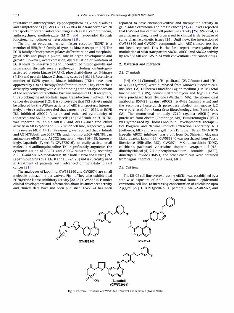
The analogues of lapatinib, GW583340 and GW2974, are smallmolecule quinazoline derivatives, Fig. 1. They also exhibit dualEGFR/ErbB2 kinase inhibitory activity [22,23]. GW583340 is underclinical development and information about its anticancer activityand clinical data have not been published. GW2974 has been
Fig. 1. Chemical structure of GW583340,
reported to have chemopreventive and therapeutic activity ingallbladder carcinoma and breast cancer [23,24]. It was reportedthat GW2974 has cardiac cell protective activity [25]. GW2974, asan anticancer drug, is not progressed to clinical trials because ofsome pharmacokinetic issues [24]. Until now, the interaction ofGW583340 and GW2974 compounds with ABC transporters hasnot been reported. This is the first report investigating themodulation of MDR transporters ABCB1, ABCC1 and ABCG2 activityby GW588340 and GW2974 with conventional anticancer drugs.
2. Materials and methods
2.1. Chemicals
[3H]-MX (4 Ci/mmol), [3H]-paclitaxel (23 Ci/mmol) and [3H]-MTX (24 Ci/mmol) were purchased from Moravek Biochemicals,Inc (Brea, CA). Dulbecco’s modified Eagle’s medium (DMEM), fetalbovine serum (FBS), penicillin/streptomycin and trypsin 0.25%were purchased from Hyclone (Waltham, MA). The monoclonalantibodies BXP-21 (against ABCG2), sc-8432 (against actin) andthe secondary horseradish peroxidase-labeled anti-mouse IgGwere purchased from Santa Cruz Biotechnology, Inc. (Santa Cruz,CA). The monoclonal antibody C219 (against ABCB1) waspurchased from Abcam (Cambridge, MA). Fumitremorgin C (FTC)was synthesized by Thomas McCloud, Developmental Therapeu-tics Program, and Natural Products Extraction Laboratory, NIH(Bethesda, MD) and was a gift from Dr. Susan Bates. ONO-1078(specific ABCC1 inhibitor) was a gift from Dr. Shin-ichi Akiyama(Sakuragaoka, Japan) [26]. GW583340 was purchased from TocrisBioscience (Ellisville, MO). GW2974, MX, doxorubicin (DOX),colchicine, paclitaxel, vincristine, cisplatin, verapamil, 3-(4,5-dimethylthiazol-yl)-2,5-diphenyltetrazolium bromide (MTT),dimethyl sulfoxide (DMSO) and other chemicals were obtainedfrom Sigma Chemical Co. (St. Louis, MO).
2.2. Cell lines
The KB-C2 cell line overexpressing ABCB1, was established by astep-wise exposure of KB-3-1, a parental human epidermoidcarcinoma cell line, to increasing concentration of colchicine upto2 mg/ml [27]. HEK293/pcDNA3.1 (parental), ABCG2-482-R2, and
GW2974 and lapatinib (GW572016).

K. Sodani et al. / Biochemical Pharmacology 83 (2012) 1613–1622 1615
ABCG2-482-T7 cell lines were established by selection with G418after transfecting HEK293 cell line with either an empty pcDNA3.1vector or pcDNA3.1 vector containing a full length ABCG2 witheither Arg or Thr at position 482, respectively, and were cultured inmedium with 2 mg/ml of G418 [28]. The lung cancer cell line NCI-H460 (parental) and ABCG2 overexpressing NCI-H460/MX20 cellswere kindly provided by Drs. Susan Bates and Robert Robey (NCI,NIH, Bethesda), ABCB1-transfected HEK/ABCB1 and ABCC1-trans-fected HEK/ABCC1 cell lines were kindly provided by Dr. SureshAmbudkar (NCI, NIH, Bethesda). All cells were grown as adherentmonolayer in drug-free culture media for more than 2 weeksbefore assay. All cell lines were cultured at 37 8C with 5% CO2 andDMEM containing 10% FBS and 1% penicillin/streptomycin.
2.3. Cytotoxicity determination by MTT assay
We used a modified MTT colorimetric assay to detect thesensitivity of cells to anticancer drugs in vitro [29]. Briefly, cellswere harvested and resuspended at a final concentration of6 � 103 cells/well for KB-3-1, HEK293/pcDNA3.1, HEK/ABCB1, NCI-H460, NCI-H460/MX20 and HEK/ABCC1 cells, and 6 � 103 cells/well for KB-C2, ABCG2-482-R2, and ABCG2-482-T7. Cells wereseeded evenly into (160 ml/well) 96-well plate. After seeding cellsin 160 ml medium in a 96-well plate and incubating for 24 h at37 8C, 20 ml of various concentrations of the appropriate anticancerdrug were added (20 ml of fixed concentration of the reversalcompounds were added one hour prior to the anticancer drugs).Subsequently the cells with anticancer drugs in DMEM supple-mented with 10% FBS were incubated at 37 8C for 72 h. After 72 h,20 ml MTT (4 mg/ml) was added to each well. The plates wereincubated at 37 8C for 4 h. After this, the MTT/medium wasremoved from each well without disturbing the cells, and 100 ml ofDMSO was added to each well. Finally, the absorbance was read at570 nm by Opsys microplate reader (Dynex Technologies, Chan-tilly, VA). The degree of resistance was calculated by dividing theIC50 for the MDR cells with or without inhibitors by that of theparental cells without inhibitor. The potential reversal agents usedin this study were GW583340 and GW2974 at 2.5 and 5 mM.Verapamil and FTC were used at nontoxic concentration of 5 mM asa positive control for ABCB1 and ABCG2 overexpressing cell lines,respectively.
2.4. Drug accumulation assay
2.4.1. [3H]-MX accumulation assay
The parental HEK293/pcDNA3.1, ABCG2-482-R2, and ABCG2-482-T7 cells were seeded in two T75 flasks and incubated withDMEM supplemented with 10% FBS at 37 8C. After the cells reached80% confluence, the cells were trypsinized and two aliquots(12 � 106 cells) from each cell line were suspended in the medium,pre-incubated with or without GW583340 or GW2974 (5 mM) at37 8C for 1 h. Subsequently, cells were suspended in the mediumcontaining 0.1 mM [3H]-MX with or without the reversal com-pound at 37 8C for 2 h. The cells were washed with PBS for threetimes. Finally, the cells were lysed by adding lysis buffer (pH 7.4,containing 1% Triton X-100 and 0.2% SDS) and transferred toscintillation vial. Each sample was placed in scintillation fluid andradioactivity was measured in a Packard TRI-CARB1 1900CA liquidscintillation analyzer from Packard Instrument Company, Inc(Downers Grove, IL).
2.4.2. [3H]-paclitaxel accumulation assay
The accumulation of [3H]-paclitaxel in KB-3-1 and KB-C2 cellswas measured in the presence or absence of GW583340, GW2974or verapamil at 5 mM. Confluent cells in 24-well plates werepreincubated with or without the reversal compounds for 1 h at
37 8C. To measure drug accumulation, cells were then incubatedwith 0.1 mM [3H]-paclitaxel for 2 h in the presence or absence ofthe reversal compounds at 37 8C. After washing three times withice cold PBS, the cells were trypsinized and placed in lysis buffer(pH 7.4, containing 1% Triton X-100 and 0.2% SDS). Each samplewas placed in a scintillation vial with 5 ml scintillation fluid andradioactivity was measured in a Packard TRI-CARB1 1900CA liquidscintillation analyzer from Packard Instrument Company, Inc(Downers Grove, IL).
2.5. Inside-out vesicle uptake assay
For the uptake assay, we used HEK293/pcDNA3.1 and ABCG2overexpressing ABCG2-482-R2 cell membrane vesicles. Theexperiment was carried out using a rapid filtration method, aspreviously described [30] in a medium containing membranevesicles (10 mg), 0.25 M sucrose, 10 mM Tris–HCl, pH 7.4, 10 mMMgCl2, 4 mM AMP/ATP, 10 mM phosphocreatine, 100 mg/mlcreatine phosphokinase, and radio labeled substrate ([3H]-MTX)with unlabeled substrate, in a total volume of 50 ml. Reactionswere carried out at 37 8C for 10 min and was stopped by adding3 ml of an ice-cold stop solution (0.25 M sucrose, 100 mM NaCl,10 mM Tris–HCl, pH 7.4). Samples were passed through 0.22 mMDura pore membrane filters (Millipore, Bedford, MA) undervacuum. The filters were washed three times with 3 ml of ice-cold stop solution and dried at room temperature for 30 min.Radioactivity was measured by the use of a liquid scintillationcounter. The rates of net ATP-dependent transport were deter-mined by subtracting the values obtained in the presence of 4 mMAMP from those obtained in the presence of 4 mM ATP.
2.6. Preparation of total cell lysates
Total cell lysates were prepared by harvesting the cells andrinsing twice with PBS. Cell extracts were prepared by incubatingcells for 30 min with lysis buffer (10 mM Tris HCl, pH 7.5, 1 mMEDTA, 0.1% SDS, 150 mM NaCl, 1% Triton X-100 and 0.01%leupeptin) followed by centrifugation at 12,000 � g at 4 8C for15 min. The supernatant containing total cell lysates was stored at�80 8C until the gel electrophoresis was run. Protein concentra-tions were determined by bicinchonic acid (BCATM) based proteinassay (Thermo Scientific, Rockford, IL).
2.7. Western blot analysis
Equal amounts of total cell lysates (80 mg protein) wereresolved by sodium dodecyl sulfate polyacrylamide gel electro-phoresis (SDS-PAGE) and electrophoretically transferred ontopolyvinylidene fluoride (PVDF) membranes. After incubation ina blocking solution in TBST buffer (10 mM Tris–HCl, pH 8.0,150 mM NaCl, and 0.1% Tween 20) for 1 h at room temperature, themembranes were immunoblotted overnight with primary mono-clonal antibodies against either actin at 1:200 dilution or ABCB1 orABCG2 at 1:200 dilution at 4 8C, and were then further incubatedfor 3 h at room temperature with horseradish peroxide (HRP)-conjugated secondary antibody (1:1000 dilution). The protein–antibody complex was detected by enhanced chemiluminescencedetection system (Amersham, NJ). The protein expression wasquantified by Scion Image Software (Scion Co., MD).
2.8. Molecular modeling
2.8.1. Ligand structure preparation
The structure of GW583340, GW2974 and the reference ligandlapatinib were built using the fragment dictionary of Maestro v9.0and energy minimized by Macromodel program v9.7 (Schrodinger,
K. Sodani et al. / Biochemical Pharmacology 83 (2012) 1613–16221616
Inc., New York, NY, 2009) using the OPLSAA force field with thesteepest descent followed by truncated Newton conjugate gradientprotocol. The low-energy 3D structure of GW583340, GW2974 andlapatinib were generated with the following parameters present inLigPrep v2.3: different protonation states at physiological pH � 2,and all possible tautomers and ring conformations. Ligand structuresobtained from the LigPrep run were further used for generating 100ligand conformations for each protonated structure using the defaultparameters of mixed torsional/low-mode sampling function. Theconformations were filtered to exclude redundant conformers with amaximum relative energy difference of 5 kcal/mol. The outputconformational search (Csearch) file containing 100 unique con-formers for each ligand were used as input for docking simulationsinto each binding site of human ABCB1 and ABCG2.
2.8.2. Protein structure preparation
The X-ray crystal structure of ABCB1 in apoprotein state (PDBID: 3G5U) and in complex with inhibitors QZ59-RRR (PDB ID:3G60), QZ59-SSS (PDB ID: 3G61) [31] and ATP bound (PDB ID:1MV5) obtained from the RCSB Protein Data Bank were used tobuild the homology model of human ABCB1. The protocol forhomology modeling is essentially the same as reported before [32].Refined human ABCB1 homology model was further used togenerate different receptor grids by selecting QZ59-RRR (site-1)and QZ59-SSS (site-2) bound ligands, all amino acid residuesknown to contribute to verapamil binding (site-3), two residues(Phe728 and Val982) known to be common to sites-1–3 (site-4)and ATP binding site. Homology model of ABCG2 based on themouse apoprotein (PDB ID: 3G5U) [31]; as template was generatedpreviously by Rosenberg and Bikadi [33,34]. The homology modelof ABCG2 was refined then the grid was generated using Arg482 asa centroid to dock the ligands. The choice of Arg482 as a centroidwas further supported by performing site map v2.3 (Schrodinger,LLC, New York, NY, 2009) calculations, which has identified highestscoring druggable site (data not shown) encompassing Arg482residue.
2.8.3. Docking protocol
Conformational libraries of GW583340, GW2974 and thereference ligand lapatinib were docked at each of the generatedgrids (sites-1–4 and ATP binding site of ABCB1, Arg482 residue forABCG2) using the ‘‘Extra Precision’’ (XP) mode of Glide programv5.0 (Schrodinger, Inc., New York, NY, 2009) with the defaultfunctions. The top scoring GW583340 and GW2974 conformationwith ABCB1 and ABCG2 was used for graphical analysis. Allcomputations were carried out on a Dell Precision 470n dualprocessor with the Linux OS (Red Hat Enterprise WS 4.0).
2.9. Statistical analysis
Differences of the parameters between two groups wereanalyzed by two tailed Student’s t test. P < 0.05 was consideredas statistically significant.
3. Results
3.1. GW583340 and GW2974 significantly potentiate the
antineoplastic substrates of ABCG2 and ABCB1 transporters, but not of
ABCC1 transporter
GW583340 and GW2974 were found to be nontoxic (with IC50
values of more than 100 mM) in ABCB1- and ABCG2-overexpres-sing cell lines when analyzed using the MTT assay (data notshown). Consequently, reversal concentration of 2.5 and 5 mM, atwhich no cytotoxicity was detected, were chosen for conductingreversal experiments. It has been established that mutations at
position 482 in ABCG2 can alter the substrate and antagonistspecificity of ABCG2 [35]. Therefore, we used both wild-type(R482) and mutant (R482T) forms of ABCG2 in the present study.The effect of GW583340 and GW2974 (2.5 and 5 mM) on ABCG2-mediated MDR in wild-type (ABCG2-482-R2) and mutant (ABCG2-482-T7) cell lines in combination with MX or DOX was determined.GW583340 and GW2974 significantly decreased the IC50 values ofMX in both ABCG2-482-R2 and ABCG2-482-T7 cell lines in aconcentration dependent manner (Table 1). In addition, thereversal effect produced by GW583340 at 5 mM was comparableto the effect produced by 5 mM of lapatinib and 5 mM of FTC (Table1). Similar concentration dependent decrease in IC50 values forDOX in ABCG2 overexpressing ABCG2-482-R2 and ABCG2-482-T7cell lines was obtained, when GW583340 or GW2974 (2.5 and5 mM) was combined with DOX (Table 1). However, no change inthe IC50 of cisplatin, a nonsubstrate of ABCG2, was seen with orwithout the combination of GW583340 and GW2974 at 5 mM(Table 1). In addition, the reversal effect was also analyzed inparental NCI-H460 and drug selected ABCG2 overexpressing NCI-H460/MX20 cells and a similar, concentration dependant reversaleffect with GW583340 and GW2974 was noticed in NCI-H460/MX20 cells (Table 1). However, there was no significant alterationin the IC50 values of MX, DOX and cisplatin in the presence orabsence of GW583340 and GW2974 in the HEK293/pcDNA3.1 andNCI-H460 cells (Table 1). Subsequently, the cell survival assay wasdone to determine whether both GW583340 and GW2974 couldreverse ABCB1-mediated MDR by potentiating the sensitivity tosubstrate anticancer agents. GW583340 and GW2974 significantlyincreased the sensitivity of ABCB1 overexpressing drug selectedKB-C2 and transfected HEK/ABCB1 cells to colchicine, vincristineand paclitaxel in a concentration-dependent fashion (Table 2).Lapatinib (GW572016), another TKI previously characterized byour laboratory [19] was used as a positive control. However,neither GW583340 nor GW2974 significantly altered the IC50
value of the aforementioned anticancer drugs in the parental KB-3-1 and HEK293/pcDNA3.1 cells (Table 2). In addition, GW583340and GW2974 did not alter the IC50 value of cisplatin in KB-3-1, KB-C2, HEK293/pcDNA3.1 and HEK/ABCB1 cell line at concentration of5 mM (Table 2). Verapamil, an ABCB1 inhibitor, at 5 mM,significantly decreased the IC50 values of colchicine, vincristineand paclitaxel in ABCB1 overexpressing KB-C2 and HEK/ABCB1 celllines (Table 2). These results suggest that GW583340 and GW2974significantly reverse the MDR mediated by ABCB1 or ABCG2.However, GW583340 and GW 2974, at 5 mM, did not significantlyaffect the ABCC1-mediated drug resistance in HEK/ABCC1 cell line(Table 3).
3.2. Effect of GW583340 or GW2974 on the cellular accumulation of
[3H]-MX and [3H]-paclitaxel in cells overexpressing ABCG2 and ABCB1
respectively
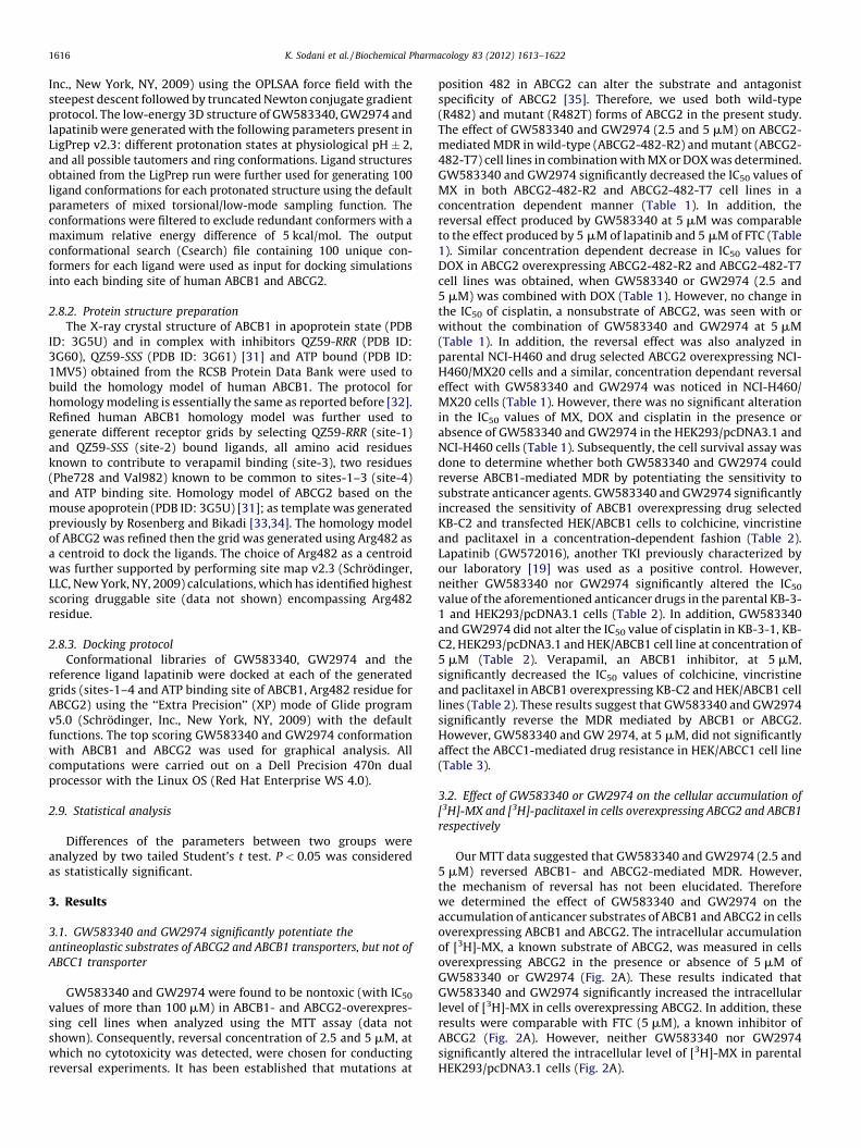
Our MTT data suggested that GW583340 and GW2974 (2.5 and5 mM) reversed ABCB1- and ABCG2-mediated MDR. However,the mechanism of reversal has not been elucidated. Thereforewe determined the effect of GW583340 and GW2974 on theaccumulation of anticancer substrates of ABCB1 and ABCG2 in cellsoverexpressing ABCB1 and ABCG2. The intracellular accumulationof [3H]-MX, a known substrate of ABCG2, was measured in cellsoverexpressing ABCG2 in the presence or absence of 5 mM ofGW583340 or GW2974 (Fig. 2A). These results indicated thatGW583340 and GW2974 significantly increased the intracellularlevel of [3H]-MX in cells overexpressing ABCG2. In addition, theseresults were comparable with FTC (5 mM), a known inhibitor ofABCG2 (Fig. 2A). However, neither GW583340 nor GW2974significantly altered the intracellular level of [3H]-MX in parentalHEK293/pcDNA3.1 cells (Fig. 2A).
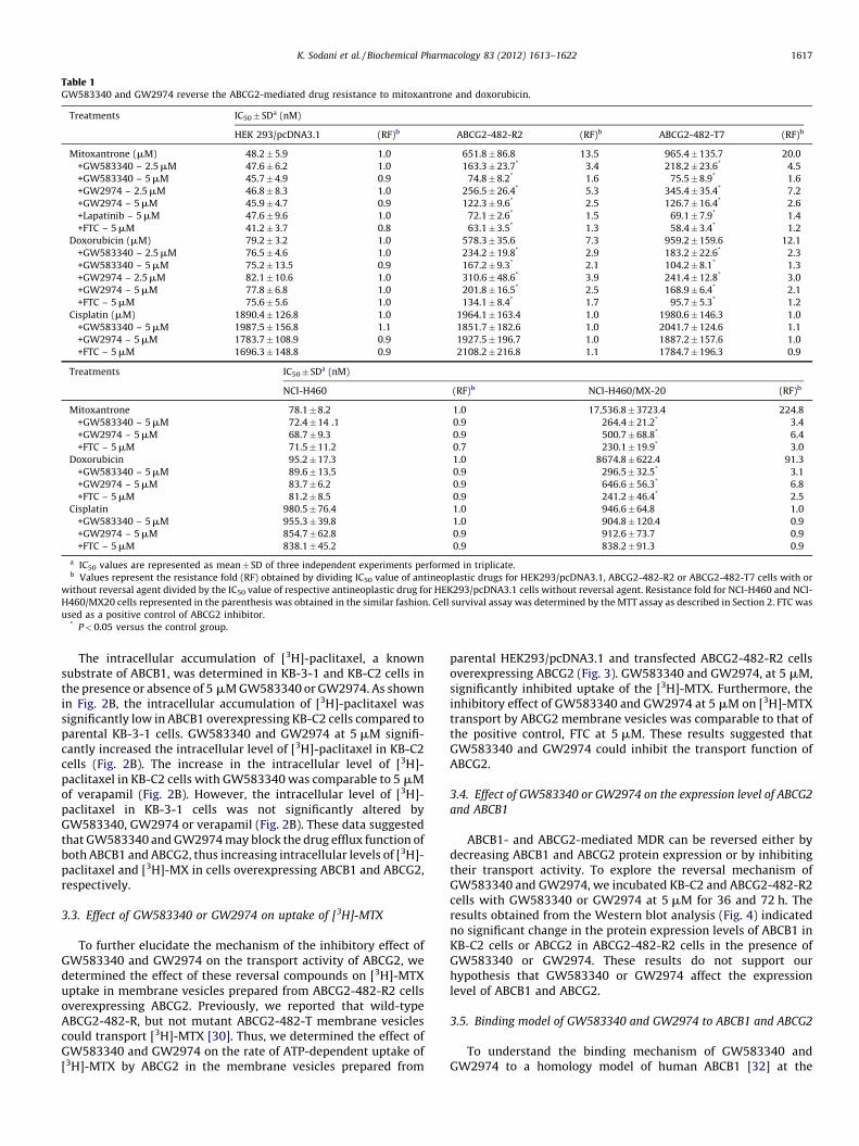
Table 1GW583340 and GW2974 reverse the ABCG2-mediated drug resistance to mitoxantrone and doxorubicin.
Treatments IC50� SDa (nM)
HEK 293/pcDNA3.1 (RF)b ABCG2-482-R2 (RF)b ABCG2-482-T7 (RF)b
Mitoxantrone (mM) 48.2 � 5.9 1.0 651.8 � 86.8 13.5 965.4 � 135.7 20.0
+GW583340 – 2.5 mM 47.6 � 6.2 1.0 163.3 � 23.7* 3.4 218.2 � 23.6* 4.5
+GW583340 – 5 mM 45.7 � 4.9 0.9 74.8 � 8.2* 1.6 75.5 � 8.9* 1.6
+GW2974 – 2.5 mM 46.8 � 8.3 1.0 256.5 � 26.4* 5.3 345.4 � 35.4* 7.2
+GW2974 – 5 mM 45.9 � 4.7 0.9 122.3 � 9.6* 2.5 126.7 � 16.4* 2.6
+Lapatinib – 5 mM 47.6 � 9.6 1.0 72.1 � 2.6* 1.5 69.1 � 7.9* 1.4
+FTC – 5 mM 41.2 � 3.7 0.8 63.1 � 3.5* 1.3 58.4 � 3.4* 1.2
Doxorubicin (mM) 79.2 � 3.2 1.0 578.3 � 35.6 7.3 959.2 � 159.6 12.1
+GW583340 – 2.5 mM 76.5 � 4.6 1.0 234.2 � 19.8* 2.9 183.2 � 22.6* 2.3
+GW583340 – 5 mM 75.2 � 13.5 0.9 167.2 � 9.3* 2.1 104.2 � 8.1* 1.3
+GW2974 – 2.5 mM 82.1 � 10.6 1.0 310.6 � 48.6* 3.9 241.4 � 12.8* 3.0
+GW2974 – 5 mM 77.8 � 6.8 1.0 201.8 � 16.5* 2.5 168.9 � 6.4* 2.1
+FTC – 5 mM 75.6 � 5.6 1.0 134.1 � 8.4* 1.7 95.7 � 5.3* 1.2
Cisplatin (mM) 1890.4 � 126.8 1.0 1964.1 � 163.4 1.0 1980.6 � 146.3 1.0
+GW583340 – 5 mM 1987.5 � 156.8 1.1 1851.7 � 182.6 1.0 2041.7 � 124.6 1.1
+GW2974 – 5 mM 1783.7 � 108.9 0.9 1927.5 � 196.7 1.0 1887.2 � 157.6 1.0
+FTC – 5 mM 1696.3 � 148.8 0.9 2108.2 � 216.8 1.1 1784.7 � 196.3 0.9
Treatments IC50� SDa (nM)
NCI-H460 (RF)b NCI-H460/MX-20 (RF)b
Mitoxantrone 78.1 � 8.2 1.0 17,536.8 � 3723.4 224.8
+GW583340 – 5 mM 72.4 � 14 .1 0.9 264.4 � 21.2* 3.4
+GW2974 – 5 mM 68.7 � 9.3 0.9 500.7 � 68.8* 6.4
+FTC – 5 mM 71.5 � 11.2 0.7 230.1 � 19.9* 3.0
Doxorubicin 95.2 � 17.3 1.0 8674.8 � 622.4 91.3
+GW583340 – 5 mM 89.6 � 13.5 0.9 296.5 � 32.5* 3.1
+GW2974 – 5 mM 83.7 � 6.2 0.9 646.6 � 56.3* 6.8
+FTC – 5 mM 81.2 � 8.5 0.9 241.2 � 46.4* 2.5
Cisplatin 980.5 � 76.4 1.0 946.6 � 64.8 1.0
+GW583340 – 5 mM 955.3 � 39.8 1.0 904.8 � 120.4 0.9
+GW2974 – 5 mM 854.7 � 62.8 0.9 912.6 � 73.7 0.9
+FTC – 5 mM 838.1 � 45.2 0.9 838.2 � 91.3 0.9
a IC50 values are represented as mean � SD of three independent experiments performed in triplicate.b Values represent the resistance fold (RF) obtained by dividing IC50 value of antineoplastic drugs for HEK293/pcDNA3.1, ABCG2-482-R2 or ABCG2-482-T7 cells with or
without reversal agent divided by the IC50 value of respective antineoplastic drug for HEK293/pcDNA3.1 cells without reversal agent. Resistance fold for NCI-H460 and NCI-
H460/MX20 cells represented in the parenthesis was obtained in the similar fashion. Cell survival assay was determined by the MTT assay as described in Section 2. FTC was
used as a positive control of ABCG2 inhibitor.* P < 0.05 versus the control group.
K. Sodani et al. / Biochemical Pharmacology 83 (2012) 1613–1622 1617
The intracellular accumulation of [3H]-paclitaxel, a knownsubstrate of ABCB1, was determined in KB-3-1 and KB-C2 cells inthe presence or absence of 5 mM GW583340 or GW2974. As shownin Fig. 2B, the intracellular accumulation of [3H]-paclitaxel wassignificantly low in ABCB1 overexpressing KB-C2 cells compared toparental KB-3-1 cells. GW583340 and GW2974 at 5 mM signifi-cantly increased the intracellular level of [3H]-paclitaxel in KB-C2cells (Fig. 2B). The increase in the intracellular level of [3H]-paclitaxel in KB-C2 cells with GW583340 was comparable to 5 mMof verapamil (Fig. 2B). However, the intracellular level of [3H]-paclitaxel in KB-3-1 cells was not significantly altered byGW583340, GW2974 or verapamil (Fig. 2B). These data suggestedthat GW583340 and GW2974 may block the drug efflux function ofboth ABCB1 and ABCG2, thus increasing intracellular levels of [3H]-paclitaxel and [3H]-MX in cells overexpressing ABCB1 and ABCG2,respectively.
3.3. Effect of GW583340 or GW2974 on uptake of [3H]-MTX
To further elucidate the mechanism of the inhibitory effect ofGW583340 and GW2974 on the transport activity of ABCG2, wedetermined the effect of these reversal compounds on [3H]-MTXuptake in membrane vesicles prepared from ABCG2-482-R2 cellsoverexpressing ABCG2. Previously, we reported that wild-typeABCG2-482-R, but not mutant ABCG2-482-T membrane vesiclescould transport [3H]-MTX [30]. Thus, we determined the effect ofGW583340 and GW2974 on the rate of ATP-dependent uptake of[3H]-MTX by ABCG2 in the membrane vesicles prepared from
parental HEK293/pcDNA3.1 and transfected ABCG2-482-R2 cellsoverexpressing ABCG2 (Fig. 3). GW583340 and GW2974, at 5 mM,significantly inhibited uptake of the [3H]-MTX. Furthermore, theinhibitory effect of GW583340 and GW2974 at 5 mM on [3H]-MTXtransport by ABCG2 membrane vesicles was comparable to that ofthe positive control, FTC at 5 mM. These results suggested thatGW583340 and GW2974 could inhibit the transport function ofABCG2.
3.4. Effect of GW583340 or GW2974 on the expression level of ABCG2
and ABCB1
ABCB1- and ABCG2-mediated MDR can be reversed either bydecreasing ABCB1 and ABCG2 protein expression or by inhibitingtheir transport activity. To explore the reversal mechanism ofGW583340 and GW2974, we incubated KB-C2 and ABCG2-482-R2cells with GW583340 or GW2974 at 5 mM for 36 and 72 h. Theresults obtained from the Western blot analysis (Fig. 4) indicatedno significant change in the protein expression levels of ABCB1 inKB-C2 cells or ABCG2 in ABCG2-482-R2 cells in the presence ofGW583340 or GW2974. These results do not support ourhypothesis that GW583340 or GW2974 affect the expressionlevel of ABCB1 and ABCG2.
3.5. Binding model of GW583340 and GW2974 to ABCB1 and ABCG2
To understand the binding mechanism of GW583340 andGW2974 to a homology model of human ABCB1 [32] at the
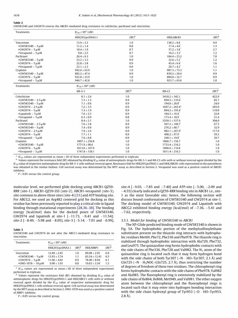
Table 2GW583340 and GW2974 reverse the ABCB1-mediated drug resistance to colchicine, paclitaxel and vincristine.
Treatments IC50� SDa (nM)
HEK293/pcDNA3.1 (RF)b HEK/ABCB1 (RF)b
Vincristine 13.9 � 2.3 1.0 138.2 � 8.8 9.9
+GW583340 – 5 mM 11.2 � 1.4 0.8 17.4 � 4.6* 1.3
+GW2974 – 5 mM 10.4 � 1.9 0.7 37.2 � 3.8* 2.7
+Verapamil – 5 mM 9.8 � 2.5 0.7 16.2 � 3.5* 1.2
Paclitaxel 26.4 � 6.3 1.0 184.4 � 23.2 7.0
+GW583340 – 5 mM 23.2 � 3.3 0.9 32.6 � 5.2* 1.2
+GW2974 – 5 mM 22.8 � 3.8 0.9 43.4 � 6.4* 1.6
+Verapamil – 5 mM 23.1 � 2.3 0.9 29.7 � 4.3* 1.1
Cisplatin 942.6 � 63.9 1.0 987.3 � 73.3 1.1
+GW583340 – 5 mM 882.2 � 47.4 0.9 839.2 � 20.4 0.9
+GW2974 – 5 mM 922.8 � 23.9 1.0 884.0 � 32.7 0.9
+Verapamil – 5 mM 946.7 � 42.8 1.0 923.7 � 63.6 1.0
Treatments IC50� SDa (nM)
KB-3-1 (RF)b KB-C2 (RF)b
Colochicine 8.1 � 2.6 1.0 3418.2 � 342.1 422.0
+GW583340 – 2.5 mM 7.3 � 1.5 0.9 694.5 � 135.6* 85.7
+GW583340 – 5 mM 7.1 � 0.6 0.9 194.8 � 26.8* 24.0
+GW2974 – 2.5 mM 7.2 � 3.5 0.9 1647.2 � 263.4* 203.0
+GW2974 – 5 mM 7.3 � 1.9 0.9 894.2 � 193.4* 110.0
+Lapatinib – 5 mM 7.6 � 2.5 0.9 184.3 � 16.4* 22.8
+Verapamil – 5 mM 6.3 � 0.9 0.8 173.4 � 18.9* 21.4
Paclitaxel 8.4 � 2.1 1.0 2559.1 � 537.9 304.0
+GW583340 – 2.5 mM 7.6 � 3.8 0.9 567.2 � 168.7* 67.5
+GW583340 – 5 mM 7.4 � 0.9 0.9 276.2 � 68.7* 32.9
+GW2974 – 2.5 mM 7.9 � 2.6 0.9 982.1 � 267.9* 117.0
+GW2974 – 5 mM 7.7 � 1.1 0.9 498.2 � 97.9* 59.3
+Verapamil – 5 mM 7.6 � 0.8 0.9 249.1 � 19.8* 29.7
Cisplatin 1807.1 � 256.8 1.0 1866.7 � 156.7 1.0
+GW583340 – 5 mM 1771.9 � 98.6 1.0 1733.4 � 216.2 1.0
+GW2974 – 5 mM 1813.6 � 167.9 1.0 1896.6 � 116.8 1.0
+Verapamil – 5 mM 1747.8 � 158.1 1.0 1811.4 � 216.3 1.0
a IC50 values are represented as mean � SD of three independent experiments performed in triplicate.b Values represent the resistance fold (RF) obtained by dividing IC50 value of antineoplastic drugs for KB-3-1 and KB-C2 cells with or without reversal agent divided by the
IC50 value of respective antineoplastic drug for KB-3-1 cells without reversal agent. Resistance fold for HEK293/pcDNA3.1 and HEK/ABCB1 cells represented in the parenthesis
was obtained in the similar fashion. Cell survival assay was determined by the MTT assay as described in Section 2. Verapamil was used as a positive control of ABCB1
inhibitor.* P < 0.05 versus the control group.
K. Sodani et al. / Biochemical Pharmacology 83 (2012) 1613–16221618
molecular level, we performed glide docking using ABCB1-QZ59-RRR (site-1), ABCB1-QZ59-SSS (site-2), ABCB1-verapamil (site-3),site common to above three sites (site-4) [31] and ATP binding site.For ABCG2, we used an Arg482 centered grid for docking as thisresidue has been previously reported to play a critical role in ligandbinding through mutational experiments [28,36–38]. The bindingenergy (kcal/mol) data for the docked poses of GW583340,GW2974 and lapatinib at site-1 [�11.73, �9.41 and �11.54];site-2 [�8.49, �5.98 and �8.05], site-3 [�5.14, �7.01 and �6.99],
Table 3GW583340 and GW2974 do not alter the ABCC1-mediated drug resistance to
vincristine.
Treatments IC50� SDa (nM)
HEK293/pcDNA3.1 (RF)b HEK/MRP1 (RF)b
Vincristine 12.89 � 2.83 1.0 89.08 � 2.92 6.9
+GW583340 – 5 mM 13.93 � 3.74 1.1 83.24 � 12.43 6.5
+GW2974 – 5 mM 11.66 � 4.82 0.9 78.40 � 8.94 6.1
+ONO-1078 – 10 mM 9.90 � 1.03 0.8 19.63 � 3.34* 1.5
a IC50 values are represented as mean � SD of three independent experiments
performed in triplicate.b Values represent the resistance fold (RF) obtained by dividing IC50 value of
antineoplastic drugs for HEK293/pcDNA3.1 and HEK/ABCC1 cells with or without
reversal agent divided by the IC50 value of respective antineoplastic drug for
HEK293/pcDNA3.1 cells without reversal agent. Cell survival assay was determined
by the MTT assay as described in Section 2. ONO-1078 was used as a positive control
of MRP1 inhibitor.* P < 0.05 versus the control group.
site-4 [�9.03, �7.85 and �7.40] and ATP-site [�5.00, �2.09 and�4.55] clearly indicated a QZ59-RRR binding site in ABCB1 i.e., site-1 as the most favorable site; hence, the following section willdiscuss bound conformation of GW583340 and GW2974 at site-1.The docking model of GW583340, GW2974 and Lapatinib withABCG2 exhibited binding energy (kcal/mol) of �7.36, �5.41 and�7.62, respectively.
3.5.1. Model for binding of GW583340 to ABCB1
The XP-Glide predicted binding mode of GW583340 is shown inFig. 5A. The hydrophobic portion of the methylsulfonylethanesubstituent present on the thiazole ring interacts with hydropho-bic residues Met69, Phe72, Phe336 and Phe978. The thiazole ring isstabilized through hydrophobic interaction with Ala729, Phe732,and Leu975. The quinazoline ring forms hydrophobic contacts withthe side chains of Phe336, Phe728 and Val982. The N1 atom of thequinazoline ring is located such that it may form hydrogen bondwith the side chains of both Tyr307 (–N� � �HO–Tyr307, 2.1 A) andGln725 (–N� � �H2NOC–Gln725, 2.7 A), thus restricting the numberof degrees of freedom of these two residues. The chlorophenyl ringforms hydrophobic contacts with the side chains of Phe978, Val982and Ala985. The fluorophenyl ring is extensively stabilized by theside chains of Ile864, Ile868, Met949, and Val981. The ether oxygenatom between the chlorophenyl and the fluorophenyl rings islocated such that it may enter into hydrogen bonding interactionwith the side chain hydroxyl group of Tyr953 (–O� � �HO–Tyr953,2.8 A).

Fig. 3. Effect of GW583340 or GW2974 on the transport of [3H]-MTX by ABCG2. The
rates of the uptake [3H]-MTX into membrane vesicles prepared from HEK293/
pcDNA3.1 and ABCG2-482-R2 cells were measured as described in Section 2.
Columns are the mean of triplicate determinations; bars, SD. *P < 0.01, versus the
control group. Experiments were done three independent times, and a
representative experiment is shown.
Fig. 2. Effect of GW583340 or GW2974 on the accumulation of [3H]-MX and [3H]-
paclitaxel. The accumulation [3H]-MX (A) in empty vector transfected HEK293/
pcDNA3.1, ABCG2 vector transfected wild-type ABCG2-482-R2, and mutant ABCG2-
482-T7 or [3H]-paclitaxel (B) in parental KB-3-1 and ABCB1 overexpressing KB-C2
cells was measured as described in Section 2. Columns are the mean of triplicate
determinations; bars, SD. *P < 0.05 versus the control group. Experiments were
done three independent times, and a representative experiment is shown.
K. Sodani et al. / Biochemical Pharmacology 83 (2012) 1613–1622 1619
3.5.2. Model for binding of GW583340 to ABCG2
The XP-Glide predicted binding model of GW583340 is shownin Fig. 5B. The methylsulfonylethane substituent present on thethiazole nucleus is stabilized through hydrophobic interactions
Fig. 4. Western blot analysis of ABCG2 and ABCB1, the effect of GW583340 or GW2974
ABCG2-482-R2 and ABCG2-482-T7 cells. (B) Expression of ABCB1 in KB-3-1 and KB-C2 c
ABCG2-482-R2 cells for 36 and 72 h. (D) Effect of GW583340 and GW2974 at 5 mM on th
cell lysate were used for each sample. Western blotting performed as described in Secti
other trials.
with the side chains of Phe489, Leu581, Trp627 and Val631. Theprotonated amino group connected to the 2-position ofthe thiazole ring is involved in electrostatic interactions withthe imidazole rings of each of the His630 residues protruding fromeach monomer. Through site-directed mutagenesis studies, it hasbeen reported that His630 from each monomer play a critical rolein function of ABCG2 [37]. The sulfur atom of the thiazole ring mayform electrostatic interaction with the side chain of another criticalresidue Asn629 (–S� � �H2NOC–Asn629, 3.2 A) implicated in ABCG2function [39]. The quinazoline ring forms hydrophobic contactswith the side chains of Phe507 and Met515. The fluorophenyl ringis extensively stabilized by the side chains of Phe489, Ile573and Pro574. The fluoro substituent is located such that it may enterinto hydrogen bonding interaction with the side chain of Tyr464(–F� � �HO–Tyr464, 3.0 A).
on ABCG2 and ABCB1 expression. (A) Expression of ABCG2 in HEK293/pcDNA3.1,
ells. (C) Effect of GW583340 and GW2974 at 5 mM on expression level of ABCG2 in
e expression level of ABCB1 in KB-C2 cells for 36 h and 72 h. Equal amounts of total
on 2. Representative result is shown here and similar results were obtained in two
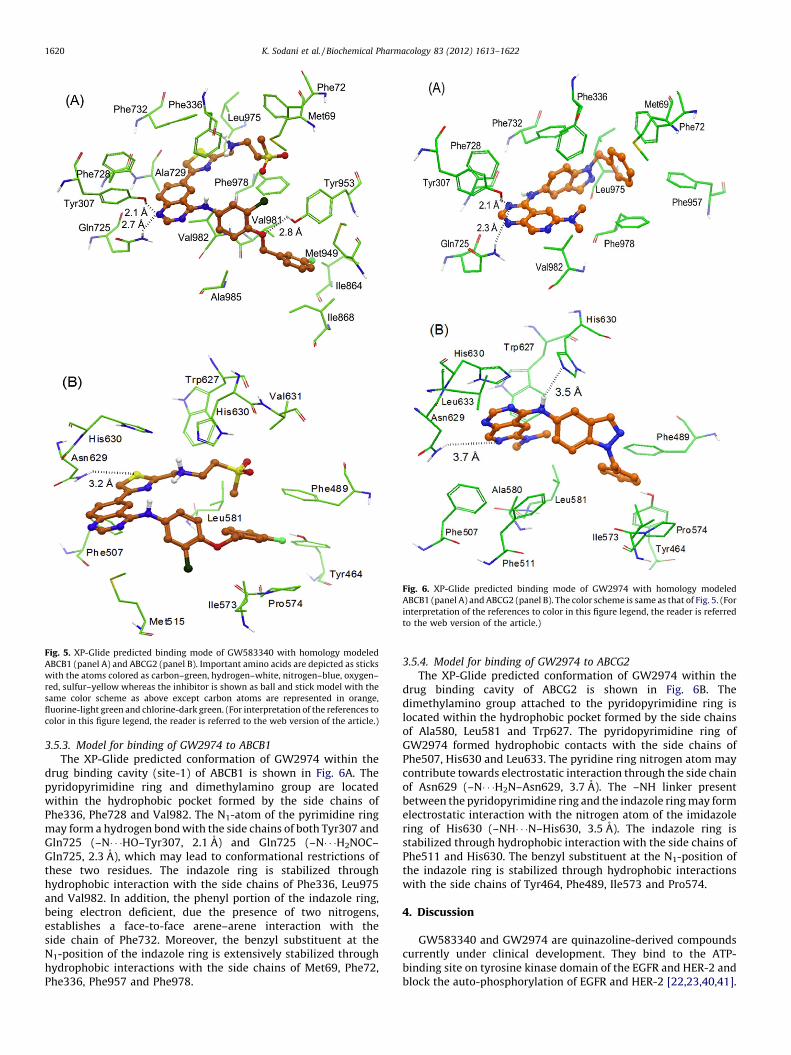
Fig. 5. XP-Glide predicted binding mode of GW583340 with homology modeled
ABCB1 (panel A) and ABCG2 (panel B). Important amino acids are depicted as sticks
with the atoms colored as carbon–green, hydrogen–white, nitrogen–blue, oxygen–
red, sulfur–yellow whereas the inhibitor is shown as ball and stick model with the
same color scheme as above except carbon atoms are represented in orange,
fluorine-light green and chlorine-dark green. (For interpretation of the references to
color in this figure legend, the reader is referred to the web version of the article.)
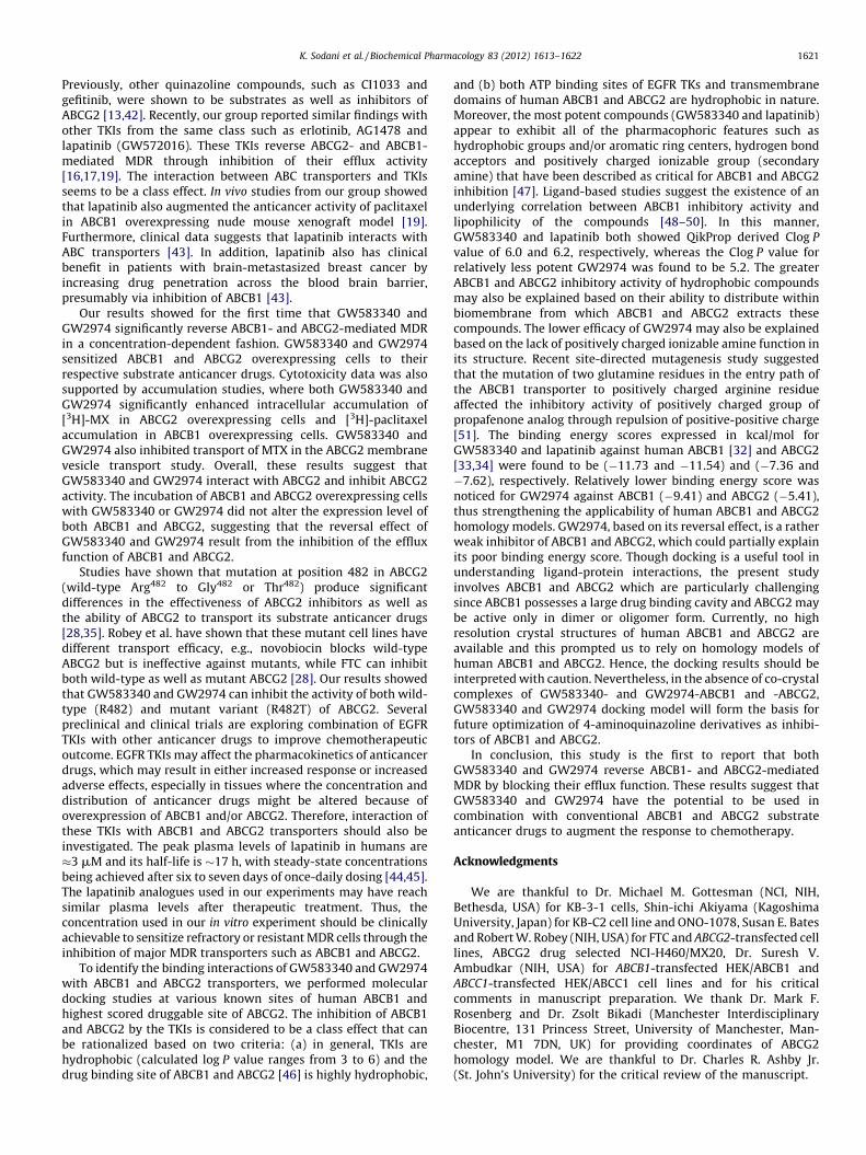
Fig. 6. XP-Glide predicted binding mode of GW2974 with homology modeled
ABCB1 (panel A) and ABCG2 (panel B). The color scheme is same as that of Fig. 5. (For
interpretation of the references to color in this figure legend, the reader is referred
to the web version of the article.)
K. Sodani et al. / Biochemical Pharmacology 83 (2012) 1613–16221620
3.5.3. Model for binding of GW2974 to ABCB1
The XP-Glide predicted conformation of GW2974 within thedrug binding cavity (site-1) of ABCB1 is shown in Fig. 6A. Thepyridopyrimidine ring and dimethylamino group are locatedwithin the hydrophobic pocket formed by the side chains ofPhe336, Phe728 and Val982. The N1-atom of the pyrimidine ringmay form a hydrogen bond with the side chains of both Tyr307 andGln725 (–N� � �HO–Tyr307, 2.1 A) and Gln725 (–N� � �H2NOC–Gln725, 2.3 A), which may lead to conformational restrictions ofthese two residues. The indazole ring is stabilized throughhydrophobic interaction with the side chains of Phe336, Leu975and Val982. In addition, the phenyl portion of the indazole ring,being electron deficient, due the presence of two nitrogens,establishes a face-to-face arene–arene interaction with theside chain of Phe732. Moreover, the benzyl substituent at theN1-position of the indazole ring is extensively stabilized throughhydrophobic interactions with the side chains of Met69, Phe72,Phe336, Phe957 and Phe978.
3.5.4. Model for binding of GW2974 to ABCG2
The XP-Glide predicted conformation of GW2974 within thedrug binding cavity of ABCG2 is shown in Fig. 6B. Thedimethylamino group attached to the pyridopyrimidine ring islocated within the hydrophobic pocket formed by the side chainsof Ala580, Leu581 and Trp627. The pyridopyrimidine ring ofGW2974 formed hydrophobic contacts with the side chains ofPhe507, His630 and Leu633. The pyridine ring nitrogen atom maycontribute towards electrostatic interaction through the side chainof Asn629 (–N� � �H2N–Asn629, 3.7 A). The –NH linker presentbetween the pyridopyrimidine ring and the indazole ring may formelectrostatic interaction with the nitrogen atom of the imidazolering of His630 (–NH� � �N–His630, 3.5 A). The indazole ring isstabilized through hydrophobic interaction with the side chains ofPhe511 and His630. The benzyl substituent at the N1-position ofthe indazole ring is stabilized through hydrophobic interactionswith the side chains of Tyr464, Phe489, Ile573 and Pro574.
4. Discussion
GW583340 and GW2974 are quinazoline-derived compoundscurrently under clinical development. They bind to the ATP-binding site on tyrosine kinase domain of the EGFR and HER-2 andblock the auto-phosphorylation of EGFR and HER-2 [22,23,40,41].
K. Sodani et al. / Biochemical Pharmacology 83 (2012) 1613–1622 1621
Previously, other quinazoline compounds, such as CI1033 andgefitinib, were shown to be substrates as well as inhibitors ofABCG2 [13,42]. Recently, our group reported similar findings withother TKIs from the same class such as erlotinib, AG1478 andlapatinib (GW572016). These TKIs reverse ABCG2- and ABCB1-mediated MDR through inhibition of their efflux activity[16,17,19]. The interaction between ABC transporters and TKIsseems to be a class effect. In vivo studies from our group showedthat lapatinib also augmented the anticancer activity of paclitaxelin ABCB1 overexpressing nude mouse xenograft model [19].Furthermore, clinical data suggests that lapatinib interacts withABC transporters [43]. In addition, lapatinib also has clinicalbenefit in patients with brain-metastasized breast cancer byincreasing drug penetration across the blood brain barrier,presumably via inhibition of ABCB1 [43].
Our results showed for the first time that GW583340 andGW2974 significantly reverse ABCB1- and ABCG2-mediated MDRin a concentration-dependent fashion. GW583340 and GW2974sensitized ABCB1 and ABCG2 overexpressing cells to theirrespective substrate anticancer drugs. Cytotoxicity data was alsosupported by accumulation studies, where both GW583340 andGW2974 significantly enhanced intracellular accumulation of[3H]-MX in ABCG2 overexpressing cells and [3H]-paclitaxelaccumulation in ABCB1 overexpressing cells. GW583340 andGW2974 also inhibited transport of MTX in the ABCG2 membranevesicle transport study. Overall, these results suggest thatGW583340 and GW2974 interact with ABCG2 and inhibit ABCG2activity. The incubation of ABCB1 and ABCG2 overexpressing cellswith GW583340 or GW2974 did not alter the expression level ofboth ABCB1 and ABCG2, suggesting that the reversal effect ofGW583340 and GW2974 result from the inhibition of the effluxfunction of ABCB1 and ABCG2.
Studies have shown that mutation at position 482 in ABCG2(wild-type Arg482 to Gly482 or Thr482) produce significantdifferences in the effectiveness of ABCG2 inhibitors as well asthe ability of ABCG2 to transport its substrate anticancer drugs[28,35]. Robey et al. have shown that these mutant cell lines havedifferent transport efficacy, e.g., novobiocin blocks wild-typeABCG2 but is ineffective against mutants, while FTC can inhibitboth wild-type as well as mutant ABCG2 [28]. Our results showedthat GW583340 and GW2974 can inhibit the activity of both wild-type (R482) and mutant variant (R482T) of ABCG2. Severalpreclinical and clinical trials are exploring combination of EGFRTKIs with other anticancer drugs to improve chemotherapeuticoutcome. EGFR TKIs may affect the pharmacokinetics of anticancerdrugs, which may result in either increased response or increasedadverse effects, especially in tissues where the concentration anddistribution of anticancer drugs might be altered because ofoverexpression of ABCB1 and/or ABCG2. Therefore, interaction ofthese TKIs with ABCB1 and ABCG2 transporters should also beinvestigated. The peak plasma levels of lapatinib in humans are�3 mM and its half-life is �17 h, with steady-state concentrationsbeing achieved after six to seven days of once-daily dosing [44,45].The lapatinib analogues used in our experiments may have reachsimilar plasma levels after therapeutic treatment. Thus, theconcentration used in our in vitro experiment should be clinicallyachievable to sensitize refractory or resistant MDR cells through theinhibition of major MDR transporters such as ABCB1 and ABCG2.
To identify the binding interactions of GW583340 and GW2974with ABCB1 and ABCG2 transporters, we performed moleculardocking studies at various known sites of human ABCB1 andhighest scored druggable site of ABCG2. The inhibition of ABCB1and ABCG2 by the TKIs is considered to be a class effect that canbe rationalized based on two criteria: (a) in general, TKIs arehydrophobic (calculated log P value ranges from 3 to 6) and thedrug binding site of ABCB1 and ABCG2 [46] is highly hydrophobic,
and (b) both ATP binding sites of EGFR TKs and transmembranedomains of human ABCB1 and ABCG2 are hydrophobic in nature.Moreover, the most potent compounds (GW583340 and lapatinib)appear to exhibit all of the pharmacophoric features such ashydrophobic groups and/or aromatic ring centers, hydrogen bondacceptors and positively charged ionizable group (secondaryamine) that have been described as critical for ABCB1 and ABCG2inhibition [47]. Ligand-based studies suggest the existence of anunderlying correlation between ABCB1 inhibitory activity andlipophilicity of the compounds [48–50]. In this manner,GW583340 and lapatinib both showed QikProp derived Clog P
value of 6.0 and 6.2, respectively, whereas the Clog P value forrelatively less potent GW2974 was found to be 5.2. The greaterABCB1 and ABCG2 inhibitory activity of hydrophobic compoundsmay also be explained based on their ability to distribute withinbiomembrane from which ABCB1 and ABCG2 extracts thesecompounds. The lower efficacy of GW2974 may also be explainedbased on the lack of positively charged ionizable amine function inits structure. Recent site-directed mutagenesis study suggestedthat the mutation of two glutamine residues in the entry path ofthe ABCB1 transporter to positively charged arginine residueaffected the inhibitory activity of positively charged group ofpropafenone analog through repulsion of positive-positive charge[51]. The binding energy scores expressed in kcal/mol forGW583340 and lapatinib against human ABCB1 [32] and ABCG2[33,34] were found to be (�11.73 and �11.54) and (�7.36 and�7.62), respectively. Relatively lower binding energy score wasnoticed for GW2974 against ABCB1 (�9.41) and ABCG2 (�5.41),thus strengthening the applicability of human ABCB1 and ABCG2homology models. GW2974, based on its reversal effect, is a ratherweak inhibitor of ABCB1 and ABCG2, which could partially explainits poor binding energy score. Though docking is a useful tool inunderstanding ligand-protein interactions, the present studyinvolves ABCB1 and ABCG2 which are particularly challengingsince ABCB1 possesses a large drug binding cavity and ABCG2 maybe active only in dimer or oligomer form. Currently, no highresolution crystal structures of human ABCB1 and ABCG2 areavailable and this prompted us to rely on homology models ofhuman ABCB1 and ABCG2. Hence, the docking results should beinterpreted with caution. Nevertheless, in the absence of co-crystalcomplexes of GW583340- and GW2974-ABCB1 and -ABCG2,GW583340 and GW2974 docking model will form the basis forfuture optimization of 4-aminoquinazoline derivatives as inhibi-tors of ABCB1 and ABCG2.
In conclusion, this study is the first to report that bothGW583340 and GW2974 reverse ABCB1- and ABCG2-mediatedMDR by blocking their efflux function. These results suggest thatGW583340 and GW2974 have the potential to be used incombination with conventional ABCB1 and ABCG2 substrateanticancer drugs to augment the response to chemotherapy.
Acknowledgments
We are thankful to Dr. Michael M. Gottesman (NCI, NIH,Bethesda, USA) for KB-3-1 cells, Shin-ichi Akiyama (KagoshimaUniversity, Japan) for KB-C2 cell line and ONO-1078, Susan E. Batesand Robert W. Robey (NIH, USA) for FTC and ABCG2-transfected celllines, ABCG2 drug selected NCI-H460/MX20, Dr. Suresh V.Ambudkar (NIH, USA) for ABCB1-transfected HEK/ABCB1 andABCC1-transfected HEK/ABCC1 cell lines and for his criticalcomments in manuscript preparation. We thank Dr. Mark F.Rosenberg and Dr. Zsolt Bikadi (Manchester InterdisciplinaryBiocentre, 131 Princess Street, University of Manchester, Man-chester, M1 7DN, UK) for providing coordinates of ABCG2homology model. We are thankful to Dr. Charles R. Ashby Jr.(St. John’s University) for the critical review of the manuscript.
K. Sodani et al. / Biochemical Pharmacology 83 (2012) 1613–16221622
References
[1] Gottesman MM, Fojo T, Bates SE. Multidrug resistance in cancer: role of ATP-dependent transporters. Nat Rev Cancer 2002;2:48–58.
[2] Vasiliou V, Vasiliou K, Nebert DW. Human ATP-binding cassette (ABC) trans-porter family. Hum Genomics 2009;3:281–90.
[3] Dean M, Annilo T. Evolution of the ATP-binding cassette (ABC) transportersuperfamily in vertebrates. Annu Rev Genomics Hum Genet 2005;6:123–42.
[4] Dean M, Rzhetsky A, Allikmets R. The human ATP-binding cassette (ABC)transporter superfamily. Genome Res 2001;11:1156–66.
[5] Gottesman MM, Ambudkar, Overview SV. ABC transporters and human dis-ease. J Bioenerg Biomembr 2001;33:453–8.
[6] Goldstein LJ, Gottesman MM, Pastan I. Expression of the MDR1 gene in humancancers. Cancer Treat Res 1991;57:101–19.
[7] Kruh GD, Belinsky MG. The MRP family of drug efflux pumps. Oncogene2003;22:7537–52.
[8] Doyle LA, Ross DD. Multidrug resistance mediated by the breast cancerresistance protein BCRP (ABCG2). Oncogene 2003;22:7340–58.
[9] Krishnamurthy P, Schuetz JD. Role of ABCG2/BCRP in biology and medicine.Annu Rev Pharmacol Toxicol 2006;46:381–410.
[10] Flynn JF, Wong C, Wu JM. Anti-EGFR therapy: mechanism and advances inclinical efficacy in breast cancer. J Oncol 2009;2009:526963.
[11] Normanno N, De LA, Bianco C, Strizzi L, Mancino M, Maiello MR, et al.Epidermal growth factor receptor (EGFR) signaling in cancer. Gene 2006;366:2–16.
[12] Lackey KE. Lessons from the drug discovery of lapatinib, a dual ErbB1/2tyrosine kinase inhibitor. Curr Top Med Chem 2006;6:435–60.
[13] Erlichman C, Boerner SA, Hallgren CG, Spieker R, Wang XY, James CD, et al. TheHER tyrosine kinase inhibitor CI1033 enhances cytotoxicity of 7-ethyl-10-hydroxycamptothecin and topotecan by inhibiting breast cancer resistanceprotein-mediated drug efflux. Cancer Res 2001;61:739–48.
[14] Kitazaki T, Oka M, Nakamura Y, Tsurutani J, Doi S, Yasunaga M, et al. Gefitinib,an EGFR tyrosine kinase inhibitor, directly inhibits the function of P-glycopro-tein in multidrug resistant cancer cells. Lung Cancer 2005;49:337–43.
[15] Yanase K, Tsukahara S, Asada S, Ishikawa E, Imai Y, Sugimoto Y. Gefitinibreverses breast cancer resistance protein-mediated drug resistance. Mol Can-cer Ther 2004;3:1119–25.
[16] Shi Z, Peng XX, Kim IW, Shukla S, Si QS, Robey RW, et al. Erlotinib (Tarceva, OSI-774) antagonizes ATP-binding cassette subfamily B member 1 and ATP-binding cassette subfamily G member 2-mediated drug resistance. CancerRes 2007;67:11012–20.
[17] Shi Z, Tiwari AK, Shukla S, Robey RW, Kim IW, Parmar S, et al. Inhibiting thefunction of ABCB1 and ABCG2 by the EGFR tyrosine kinase inhibitor AG1478.Biochem Pharmacol 2009;77:781–93.
[18] Tiwari AK, Sodani K, Wang SR, Kuang YH, Ashby Jr CR, Chen X, et al. Nilotinib(AMN107, Tasigna) reverses multidrug resistance by inhibiting the activity ofthe ABCB1/Pgp and ABCG2/BCRP/MXR transporters. Biochem Pharmacol2009;78:153–61.
[19] Dai CL, Tiwari AK, Wu CP, Su XD, Wang SR, Liu DG, et al. Lapatinib (Tykerb,GW572016) reverses multidrug resistance in cancer cells by inhibiting theactivity of ATP-binding cassette subfamily B member 1 and G member 2.Cancer Res 2008;68:7905–14.
[20] Johnston SR, Leary A. Lapatinib: a novel EGFR/HER2 tyrosine kinase inhibitorfor cancer. Drugs Today (Barc) 2006;42:441–53.
[21] Schwartzberg LS, Franco SX, Florance A, O’Rourke L, Maltzman J, Johnston S.Lapatinib plus letrozole as first-line therapy for HER-2+ hormone receptor-positive metastatic breast cancer. Oncologist 2010;15:122–9.
[22] Aird KM, Ding X, Baras A, Wei J, Morse MA, Clay T, et al. Trastuzumab signalingin ErbB2-overexpressing inflammatory breast cancer correlates with X-linkedinhibitor of apoptosis protein expression. Mol Cancer Ther 2008;7:38–47.
[23] Kiguchi K, Ruffino L, Kawamoto T, Ajiki T, Digiovanni J. Chemopreventiveand therapeutic efficacy of orally active tyrosine kinase inhibitors in atransgenic mouse model of gallbladder carcinoma. Clin Cancer Res 2005;11:5572–80.
[24] Witters LM, Witkoski A, Planas-Silva MD, Berger M, Viallet J, Lipton A.Synergistic inhibition of breast cancer cell lines with a dual inhibitor ofEGFR-HER-2/neu and a Bcl-2 inhibitor. Oncol Rep 2007;17:465–9.
[25] Spector NL, Yarden Y, Smith B, Lyass L, Trusk P, Pry K, et al. Activation of AMP-activated protein kinase by human EGF receptor 2/EGF receptor tyrosinekinase inhibitor protects cardiac cells. Proc Natl Acad Sci USA 2007;104:10607–12.
[26] Nagayama S, Chen ZS, Kitazono M, Takebayashi Y, Niwa K, Yamada K, et al.Increased sensitivity to vincristine of MDR cells by the leukotriene D4 receptorantagonist, ONO-1078. Cancer Lett 1998;130:175–82.
[27] Akiyama S, Fojo A, Hanover JA, Pastan I, Gottesman MM. Isolation and geneticcharacterization of human KB cell lines resistant to multiple drugs. Somat CellMol Genet 1985;11:117–26.
[28] Robey RW, Honjo Y, Morisaki K, Nadjem TA, Runge S, Risbood M, et al.Mutations at amino-acid 482 in the ABCG2 gene affect substrate and antago-nist specificity. Br J Cancer 2003;89:1971–8.
[29] Carmichael J, DeGraff WG, Gazdar AF, Minna JD, Mitchell JB. Evaluation of atetrazolium-based semiautomated colorimetric assay: assessment of chemo-sensitivity testing. Cancer Res 1987;47:936–42.
[30] Chen ZS, Robey RW, Belinsky MG, Shchaveleva I, Ren XQ, Sugimoto Y, et al.Transport of methotrexate, methotrexate polyglutamates, and 17beta-estra-diol 17-(beta-D-glucuronide) by ABCG2: effects of acquired mutations at R482on methotrexate transport. Cancer Res 2003;63:4048–54.
[31] Aller SG, Yu J, Ward A, Weng Y, Chittaboina S, Zhuo R, et al. Structure ofP-glycoprotein reveals a molecular basis for poly-specific drug binding. Sci-ence 2009;323:1718–22.
[32] Shi Z, Tiwari AK, Shukla S, Robey RW, Singh S, Kim IW, et al. Sildenafil reversesABCB1- and ABCG2-mediated chemotherapeutic drug resistance. Cancer Res2011;71:3029–41.
[33] Rosenberg MF, Bikadi Z, Chan J, Liu X, Ni Z, Cai X, et al. The human breast cancerresistance protein (BCRP/ABCG2) shows conformational changes with mitox-antrone. Structure 2010;18:482–93.
[34] Hazai E, Bikadi Z. Homology modeling of breast cancer resistance protein(ABCG2). J Struct Biol 2008;162:63–74.
[35] Mao Q, Unadkat JD. Role of the breast cancer resistance protein (ABCG2) indrug transport. AAPS J 2005;7:E118–33.
[36] Mitomo H, Kato R, Ito A, Kasamatsu S, Ikegami Y, Kii I, et al. A functional studyon polymorphism of the ATP-binding cassette transporter ABCG2: critical roleof arginine-482 in methotrexate transport. Biochem J 2003;373:767–74.
[37] Miwa M, Tsukahara S, Ishikawa E, Asada S, Imai Y, Sugimoto Y. Single aminoacid substitutions in the transmembrane domains of breast cancer resistanceprotein (BCRP) alter cross resistance patterns in transfectants. Int J Cancer2003;107:757–63.
[38] Alqawi O, Bates S, Georges E. Arginine482 to threonine mutation in the breastcancer resistance protein ABCG2 inhibits rhodamine 123 transport whileincreasing binding. Biochem J 2004;382:711–6.
[39] Ni Z, Bikadi Z, Cai X, Rosenberg MF, Mao Q. Transmembrane helices 1 and 6 ofthe human breast cancer resistance protein (BCRP/ABCG2): identification ofpolar residues important for drug transport. Am J Physiol Cell Physiol 2010;299:C1100–09.
[40] Rusnak DW, Affleck K, Cockerill SG, Stubberfield C, Harris R, Page M, et al. Thecharacterization of novel, dual ErbB-2/EGFR, tyrosine kinase inhibitors: po-tential therapy for cancer. Cancer Res 2001;61:7196–203.
[41] Cockerill S, Stubberfield C, Stables J, Carter M, Guntrip S, Smith K, et al.Indazolylamino quinazolines and pyridopyrimidines as inhibitors of the EGFrand C-erbB-2. Bioorg Med Chem Lett 2001;11:1401–5.
[42] Elkind NB, Szentpetery Z, Apati A, Ozvegy-Laczka C, Varady G, Ujhelly O, et al.Multidrug transporter ABCG2 prevents tumor cell death induced by theepidermal growth factor receptor inhibitor Iressa (ZD1839, Gefitinib). CancerRes 2005;65:1770–7.
[43] Midgley RS, Kerr DJ, Flaherty KT, Stevenson JP, Pratap SE, Koch KM, et al. Aphase I and pharmacokinetic study of lapatinib in combination with infusional5-fluorouracil, leucovorin and irinotecan. Ann Oncol 2007;18:2025–9.
[44] Burris III HA, Hurwitz HI, Dees EC, Dowlati A, Blackwell KL, O’Neil B, et al.Phase I safety, pharmacokinetics, and clinical activity study of lapatinib(GW572016), a reversible dual inhibitor of epidermal growth factor receptortyrosine kinases, in heavily pretreated patients with metastatic carcinomas.J Clin Oncol 2005;23:5305–13.
[45] Bence AK, Anderson EB, Halepota MA, Doukas MA, DeSimone PA, Davis GA,et al. Phase I pharmacokinetic studies evaluating single and multiple doses oforal GW572016, a dual EGFR-ErbB2 inhibitor, in healthy subjects. Invest NewDrugs 2005;23:39–49.
[46] Nicolle E, Boumendjel A, Macalou S, Genoux E, hmed-Belkacem A, Carrupt PA,et al. QSAR analysis and molecular modeling of ABCG2-specific inhibitors. AdvDrug Deliv Rev 2009;61:34–46.
[47] Ecker GF, Chiba P. Transporters as drug carriers. Wiley-VCH; 2009.[48] Klopman G, Shi LM, Ramu A. Quantitative structure-activity relationship of
multidrug resistance reversal agents. Mol Pharmacol 1997;52:323–34.[49] Crivori P, Reinach B, Pezzetta D, Poggesi I. Computational models for identifying
potential P-glycoprotein substrates and inhibitors. Mol Pharm 2006;3:33–44.[50] Pajeva IK, Globisch C, Wiese M. Combined pharmacophore modeling, docking,
and 3D QSAR studies of ABCB1 and ABCC1 transporter inhibitors. ChemMed-Chem 2009;4:1883–96.
[51] Parveen Z, Stockner T, Bentele C, Pferschy S, Kraupp M, Freissmuth M, et al.Molecular dissection of dual pseudosymmetric solute translocation pathwaysin human P-glycoprotein. Mol Pharmacol 2011;79:443–52.