Maintenance of retinal stem cells by Abcg2 is regulated by notch signaling
11
2652 Research Article Introduction One of the emerging universal characteristics of stem cells or progenitors is the preferential expression of ABCG2, a half transporter belonging to ATP-binding cassette (ABC) superfamily of transmembrane proteins (Doyle and Ross, 2003). These transporters mediate efflux of a broad spectrum of substrates including cytotoxic drugs, peptides, steroids, ions and phospholipids (Krishnamurthy and Schuetz, 2006). Their ability to preferentially exclude the DNA-intercalating dye, Hoechst 33342, is regarded to be the molecular basis for the enrichment of stem cells as side population (SP) cells by the Hoechst dye efflux assay (Goodell et al., 1996; Abbott, 2003; Doyle and Ross, 2003; Bunting, 2002). The assay involves incubation of cells with the Hoechst dye followed by dual- wavelength fluorescence-activated cell sorting (FACS), which identifies a small side population of cells with low dye accumulation, possessing stem cell properties and potential (Goodell et al., 1996). The notion that SP cell phenotype is mediated by ABCG2 was supported by observations that the perturbation of Abcg2 expression by retrovirus-mediated transduction or gene deletion affected the number of SP cells present in the hematopoietic compartment (Kim et al., 2002; Scharenberg et al., 2002; Zhou et al., 2002). The conservation of Abcg2 expression in SP cells, enriched from a wide range of tissues including blood (Zhou et al., 2003), muscle (Meeson et al., 2004), heart (Martin et al., 2004), gonad (Lassalle et al., 2004), lung (Summer et al., 2003), intestine (Staud and Pavek, 2005) and cornea (Budak et al., 2005) suggests that ABCG2 has an important role in stem cells. Molecular studies carried out on the hematopoietic compartment have begun to shed light on the involvement of Abcg2 in stem cells. For example, the highly regulated expression of Abcg2 during hematopoiesis, i.e. high levels of Abcg2 transcripts in stem cells and their sharp decline during lineage commitment, and the blocking of lineage commitment in response to ectopic expression of Abcg2 indicates a role for ABCG2 in the maintenance of hematopoietic stem cells (Zhou, S. et al., 2001; Scharenberg et al., 2002). The expression of Abcg2 and SP cell phenotype are also characteristics of neural stem cells, derived from different brain regions, including the retina (Hulspas and Quesenbury, 2000; Bhattacharya et al., 2003; Ahmad et al., 2004; Mouthon et al., 2006). However, little is known about the role of Abcg2 in neural stem cells and their lineage commitment. In addition, we do not know how Abcg2 expression is linked with the maintenance of stem cells. We have addressed these issues in developing retina, a suitable and accessible model of the central nervous system, where seven different types of cells are generated in an evolutionarily conserved temporal sequence by multipotential retinal stem cells (Rapaport et al., 2004). Our results demonstrate a regulated expression of Abcg2 during retinal histogenesis; levels of Abcg2 transcripts decrease with the onset of lineage commitment. This decrease in Abcg2 expression correlates with the progressive depletion of SP cell population as retinal histogenesis ensues. When Abcg2 is overexpressed in retinal progenitors, using retrovirus-mediated transduction, the differentiation is blocked, accompanied by an increase in the expression of stem cells markers and SP cell phenotype. By contrast, siRNA-mediated silencing of Abcg2 expression in retinal progenitors depletes SP cell population and promotes differentiation. In addition, we observed that Abcg2 expression and SP cell phenotype are influenced by Notch signaling, a key regulator of retinal stem cells. Our results demonstrate for the first time that ABCG2 is a ABCG2 belongs to the ATP-binding cassette superfamily of transmembrane proteins and is ubiquitously expressed in stem cells including those in the developing nervous system. The ability of ABCG2 to preferentially exclude DNA- intercalating dyes is regarded to be the basis for the enrichment of stem cells or progenitors as dye low side population (SP) cells. However, the role of ABCG2 in neural stem cells remains speculative and poorly understood. Here, we demonstrate using retinal stem cells, that ABCG2 is the molecular determinant of SP cell phenotype of neural stem cells and plays an important role in their maintenance. Overexpression of ABCG2 prevents the SP cell phenotype and adversely affects the lineage commitment of retinal stem cells. By contrast, targeted attenuation of ABCG2 depletes retinal SP cells and promotes their differentiation along pan neural and retinal lineages. In addition, we demonstrate for the first time that ABCG2 is a target of Notch signaling, and as such, constitutes one of the genes in the regulatory network of Notch signaling, involved in the maintenance of stem cells. Supplementary material available online at http://jcs.biologists.org/cgi/content/full/120/15/2652/DC1 Key words: Retina, Stem cells, Progenitors, Notch, ABCG2 Summary Maintenance of retinal stem cells by Abcg2 is regulated by notch signaling Sumitra Bhattacharya, Ani Das, Kavita Mallya and Iqbal Ahmad* Department of Ophthalmology and Visual Sciences, University of Nebraska Medical Center, Omaha, NE 68198-5840, USA *Author for correspondence (e-mail: [email protected]) Accepted 16 April 2007 Journal of Cell Science 120, 2652-2662 Published by The Company of Biologists 2007 doi:10.1242/jcs.008417 Journal of Cell Science
-
Upload
independent -
Category
Documents
-
view
4 -
download
0
Transcript of Maintenance of retinal stem cells by Abcg2 is regulated by notch signaling
2652 Research Article
IntroductionOne of the emerging universal characteristics of stem cells orprogenitors is the preferential expression of ABCG2, a halftransporter belonging to ATP-binding cassette (ABC)superfamily of transmembrane proteins (Doyle and Ross,2003). These transporters mediate efflux of a broad spectrumof substrates including cytotoxic drugs, peptides, steroids, ionsand phospholipids (Krishnamurthy and Schuetz, 2006). Theirability to preferentially exclude the DNA-intercalating dye,Hoechst 33342, is regarded to be the molecular basis for theenrichment of stem cells as side population (SP) cells by theHoechst dye efflux assay (Goodell et al., 1996; Abbott, 2003;Doyle and Ross, 2003; Bunting, 2002). The assay involvesincubation of cells with the Hoechst dye followed by dual-wavelength fluorescence-activated cell sorting (FACS), whichidentifies a small side population of cells with low dyeaccumulation, possessing stem cell properties and potential(Goodell et al., 1996). The notion that SP cell phenotype ismediated by ABCG2 was supported by observations that theperturbation of Abcg2 expression by retrovirus-mediatedtransduction or gene deletion affected the number of SP cellspresent in the hematopoietic compartment (Kim et al., 2002;Scharenberg et al., 2002; Zhou et al., 2002).
The conservation of Abcg2 expression in SP cells, enrichedfrom a wide range of tissues including blood (Zhou et al.,2003), muscle (Meeson et al., 2004), heart (Martin et al., 2004),gonad (Lassalle et al., 2004), lung (Summer et al., 2003),intestine (Staud and Pavek, 2005) and cornea (Budak et al.,2005) suggests that ABCG2 has an important role in stem cells.Molecular studies carried out on the hematopoieticcompartment have begun to shed light on the involvement ofAbcg2 in stem cells. For example, the highly regulated
expression of Abcg2 during hematopoiesis, i.e. high levels ofAbcg2 transcripts in stem cells and their sharp decline duringlineage commitment, and the blocking of lineage commitmentin response to ectopic expression of Abcg2 indicates a role forABCG2 in the maintenance of hematopoietic stem cells (Zhou,S. et al., 2001; Scharenberg et al., 2002).
The expression of Abcg2 and SP cell phenotype are alsocharacteristics of neural stem cells, derived from different brainregions, including the retina (Hulspas and Quesenbury, 2000;Bhattacharya et al., 2003; Ahmad et al., 2004; Mouthon et al.,2006). However, little is known about the role of Abcg2 inneural stem cells and their lineage commitment. In addition,we do not know how Abcg2 expression is linked with themaintenance of stem cells. We have addressed these issues indeveloping retina, a suitable and accessible model of thecentral nervous system, where seven different types of cells aregenerated in an evolutionarily conserved temporal sequence bymultipotential retinal stem cells (Rapaport et al., 2004). Ourresults demonstrate a regulated expression of Abcg2 duringretinal histogenesis; levels of Abcg2 transcripts decrease withthe onset of lineage commitment. This decrease in Abcg2expression correlates with the progressive depletion of SP cellpopulation as retinal histogenesis ensues. When Abcg2 isoverexpressed in retinal progenitors, using retrovirus-mediatedtransduction, the differentiation is blocked, accompanied by anincrease in the expression of stem cells markers and SP cellphenotype. By contrast, siRNA-mediated silencing of Abcg2expression in retinal progenitors depletes SP cell populationand promotes differentiation. In addition, we observed thatAbcg2 expression and SP cell phenotype are influenced byNotch signaling, a key regulator of retinal stem cells. Ourresults demonstrate for the first time that ABCG2 is a
ABCG2 belongs to the ATP-binding cassette superfamily oftransmembrane proteins and is ubiquitously expressed instem cells including those in the developing nervous system.The ability of ABCG2 to preferentially exclude DNA-intercalating dyes is regarded to be the basis for theenrichment of stem cells or progenitors as dyelow sidepopulation (SP) cells. However, the role of ABCG2 inneural stem cells remains speculative and poorlyunderstood. Here, we demonstrate using retinal stem cells,that ABCG2 is the molecular determinant of SP cellphenotype of neural stem cells and plays an important rolein their maintenance. Overexpression of ABCG2 preventsthe SP cell phenotype and adversely affects the lineage
commitment of retinal stem cells. By contrast, targetedattenuation of ABCG2 depletes retinal SP cells andpromotes their differentiation along pan neural and retinallineages. In addition, we demonstrate for the first time thatABCG2 is a target of Notch signaling, and as such,constitutes one of the genes in the regulatory network ofNotch signaling, involved in the maintenance of stem cells.
Supplementary material available online athttp://jcs.biologists.org/cgi/content/full/120/15/2652/DC1
Key words: Retina, Stem cells, Progenitors, Notch, ABCG2
Summary
Maintenance of retinal stem cells by Abcg2 isregulated by notch signalingSumitra Bhattacharya, Ani Das, Kavita Mallya and Iqbal Ahmad*Department of Ophthalmology and Visual Sciences, University of Nebraska Medical Center, Omaha, NE 68198-5840, USA*Author for correspondence (e-mail: [email protected])
Accepted 16 April 2007Journal of Cell Science 120, 2652-2662 Published by The Company of Biologists 2007doi:10.1242/jcs.008417
Jour
nal o
f Cel
l Sci
ence
2653Regulation of retinal stem cells
downstream target of Notch signaling. Together, theseobservations suggest that Abcg2 participates in themaintenance of retinal stem cells or progenitors, under theregulation of Notch signaling.
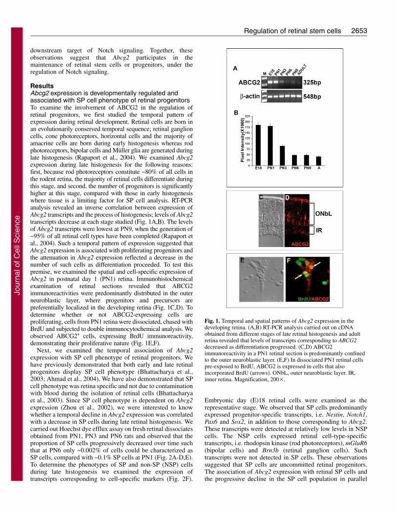
ResultsAbcg2 expression is developmentally regulated andassociated with SP cell phenotype of retinal progenitorsTo examine the involvement of ABCG2 in the regulation ofretinal progenitors, we first studied the temporal pattern ofexpression during retinal development. Retinal cells are born inan evolutionarily conserved temporal sequence; retinal ganglioncells, cone photoreceptors, horizontal cells and the majority ofamacrine cells are born during early histogenesis whereas rodphotoreceptors, bipolar cells and Müller glia are generated duringlate histogenesis (Rapaport et al., 2004). We examined Abcg2expression during late histogenesis for the following reasons:first, because rod photoreceptors constitute ~80% of all cells inthe rodent retina, the majority of retinal cells differentiate duringthis stage, and second, the number of progenitors is significantlyhigher at this stage, compared with those in early histogenesiswhere tissue is a limiting factor for SP cell analysis. RT-PCRanalysis revealed an inverse correlation between expression ofAbcg2 transcripts and the process of histogenesis; levels of Abcg2transcripts decrease at each stage studied (Fig. 1A,B). The levelsof Abcg2 transcripts were lowest at PN9, when the generation of~95% of all retinal cell types have been completed (Rapaport etal., 2004). Such a temporal pattern of expression suggested thatAbcg2 expression is associated with proliferating progenitors andthe attenuation in Abcg2 expression reflected a decrease in thenumber of such cells as differentiation proceeded. To test thispremise, we examined the spatial and cell-specific expression ofAbcg2 in postnatal day 1 (PN1) retina. Immunohistochemicalexamination of retinal sections revealed that ABCG2immunoreactivities were predominantly distributed in the outerneuroblastic layer, where progenitors and precursors arepreferentially localized in the developing retina (Fig. 1C,D). Todetermine whether or not ABCG2-expressing cells areproliferating, cells from PN1 retina were dissociated, chased withBrdU and subjected to double immunocytochemical analysis. Weobserved ABCG2+ cells, expressing BrdU immunoreactivity,demonstrating their proliferative nature (Fig. 1E,F).
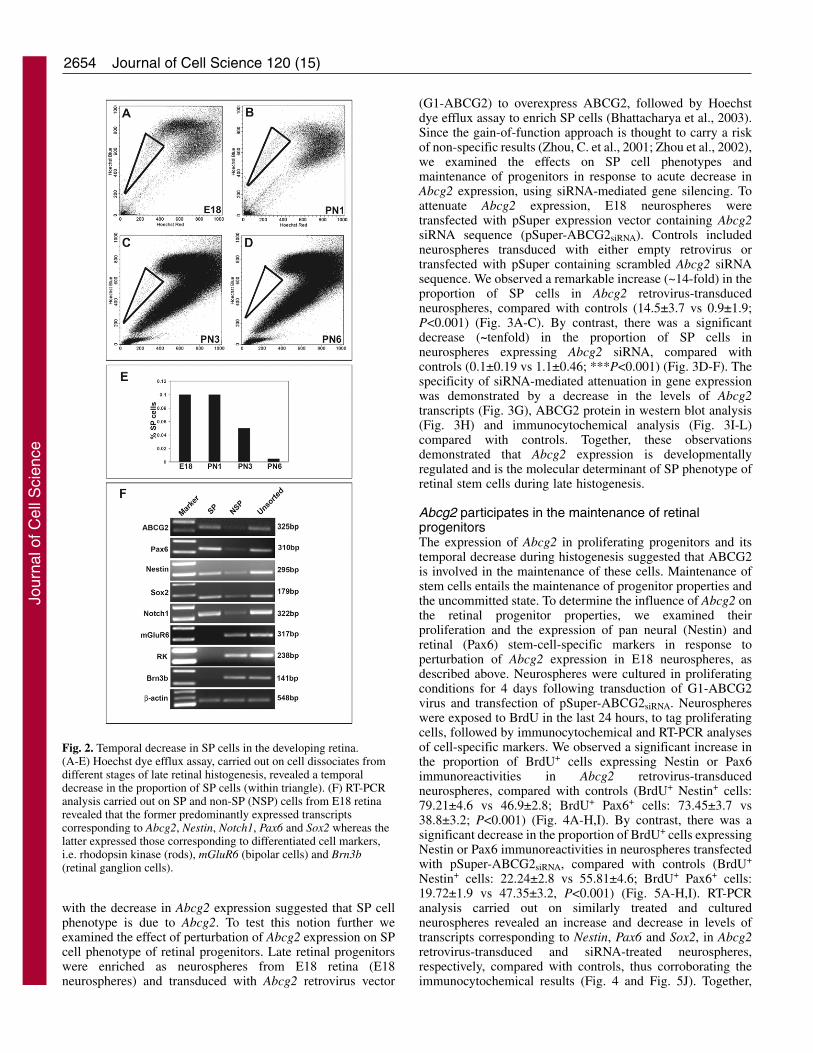
Next, we examined the temporal association of Abcg2expression with SP cell phenotype of retinal progenitors. Wehave previously demonstrated that both early and late retinalprogenitors display SP cell phenotype (Bhattacharya et al.,2003; Ahmad et al., 2004). We have also demonstrated that SPcell phenotype was retina specific and not due to contaminationwith blood during the isolation of retinal cells (Bhattacharyaet al., 2003). Since SP cell phenotype is dependent on Abcg2expression (Zhou et al., 2002), we were interested to knowwhether a temporal decline in Abcg2 expression was correlatedwith a decrease in SP cells during late retinal histogenesis. Wecarried out Hoechst dye efflux assay on fresh retinal dissociatesobtained from PN1, PN3 and PN6 rats and observed that theproportion of SP cells progressively decreased over time suchthat at PN6 only ~0.002% of cells could be characterized asSP cells, compared with ~0.1% SP cells at PN1 (Fig. 2A-D,E).To determine the phenotypes of SP and non-SP (NSP) cellsduring late histogenesis we examined the expression oftranscripts corresponding to cell-specific markers (Fig. 2F).
Embryonic day (E)18 retinal cells were examined as therepresentative stage. We observed that SP cells predominantlyexpressed progenitor-specific transcripts, i.e. Nestin, Notch1,Pax6 and Sox2, in addition to those corresponding to Abcg2.These transcripts were detected at relatively low levels in NSPcells. The NSP cells expressed retinal cell-type-specifictranscripts, i.e. rhodopsin kinase (rod photoreceptors), mGluR6(bipolar cells) and Brn3b (retinal ganglion cells). Suchtranscripts were not detected in SP cells. These observationssuggested that SP cells are uncommitted retinal progenitors.The association of Abcg2 expression with retinal SP cells andthe progressive decline in the SP cell population in parallel
Fig. 1. Temporal and spatial patterns of Abcg2 expression in thedeveloping retina. (A,B) RT-PCR analysis carried out on cDNAobtained from different stages of late retinal histogenesis and adultretina revealed that levels of transcripts corresponding to ABCG2decreased as differentiation progressed. (C,D) ABCG2immunoreactivity in a PN1 retinal section is predominantly confinedto the outer neuroblastic layer. (E,F) In dissociated PN1 retinal cellspre-exposed to BrdU, ABCG2 is expressed in cells that alsoincorporated BrdU (arrows). ONbL, outer neuroblastic layer. IR,inner retina. Magnification, 200�.
Jour
nal o
f Cel
l Sci
ence
2654
with the decrease in Abcg2 expression suggested that SP cellphenotype is due to Abcg2. To test this notion further weexamined the effect of perturbation of Abcg2 expression on SPcell phenotype of retinal progenitors. Late retinal progenitorswere enriched as neurospheres from E18 retina (E18neurospheres) and transduced with Abcg2 retrovirus vector
Journal of Cell Science 120 (15)
(G1-ABCG2) to overexpress ABCG2, followed by Hoechstdye efflux assay to enrich SP cells (Bhattacharya et al., 2003).Since the gain-of-function approach is thought to carry a riskof non-specific results (Zhou, C. et al., 2001; Zhou et al., 2002),we examined the effects on SP cell phenotypes andmaintenance of progenitors in response to acute decrease inAbcg2 expression, using siRNA-mediated gene silencing. Toattenuate Abcg2 expression, E18 neurospheres weretransfected with pSuper expression vector containing Abcg2siRNA sequence (pSuper-ABCG2siRNA). Controls includedneurospheres transduced with either empty retrovirus ortransfected with pSuper containing scrambled Abcg2 siRNAsequence. We observed a remarkable increase (~14-fold) in theproportion of SP cells in Abcg2 retrovirus-transducedneurospheres, compared with controls (14.5±3.7 vs 0.9±1.9;P<0.001) (Fig. 3A-C). By contrast, there was a significantdecrease (~tenfold) in the proportion of SP cells inneurospheres expressing Abcg2 siRNA, compared withcontrols (0.1±0.19 vs 1.1±0.46; ***P<0.001) (Fig. 3D-F). Thespecificity of siRNA-mediated attenuation in gene expressionwas demonstrated by a decrease in the levels of Abcg2transcripts (Fig. 3G), ABCG2 protein in western blot analysis(Fig. 3H) and immunocytochemical analysis (Fig. 3I-L)compared with controls. Together, these observationsdemonstrated that Abcg2 expression is developmentallyregulated and is the molecular determinant of SP phenotype ofretinal stem cells during late histogenesis.
Abcg2 participates in the maintenance of retinalprogenitorsThe expression of Abcg2 in proliferating progenitors and itstemporal decrease during histogenesis suggested that ABCG2is involved in the maintenance of these cells. Maintenance ofstem cells entails the maintenance of progenitor properties andthe uncommitted state. To determine the influence of Abcg2 onthe retinal progenitor properties, we examined theirproliferation and the expression of pan neural (Nestin) andretinal (Pax6) stem-cell-specific markers in response toperturbation of Abcg2 expression in E18 neurospheres, asdescribed above. Neurospheres were cultured in proliferatingconditions for 4 days following transduction of G1-ABCG2virus and transfection of pSuper-ABCG2siRNA. Neurosphereswere exposed to BrdU in the last 24 hours, to tag proliferatingcells, followed by immunocytochemical and RT-PCR analysesof cell-specific markers. We observed a significant increase inthe proportion of BrdU+ cells expressing Nestin or Pax6immunoreactivities in Abcg2 retrovirus-transducedneurospheres, compared with controls (BrdU+ Nestin+ cells:79.21±4.6 vs 46.9±2.8; BrdU+ Pax6+ cells: 73.45±3.7 vs38.8±3.2; P<0.001) (Fig. 4A-H,I). By contrast, there was asignificant decrease in the proportion of BrdU+ cells expressingNestin or Pax6 immunoreactivities in neurospheres transfectedwith pSuper-ABCG2siRNA, compared with controls (BrdU+
Nestin+ cells: 22.24±2.8 vs 55.81±4.6; BrdU+ Pax6+ cells:19.72±1.9 vs 47.35±3.2, P<0.001) (Fig. 5A-H,I). RT-PCRanalysis carried out on similarly treated and culturedneurospheres revealed an increase and decrease in levels oftranscripts corresponding to Nestin, Pax6 and Sox2, in Abcg2retrovirus-transduced and siRNA-treated neurospheres,respectively, compared with controls, thus corroborating theimmunocytochemical results (Fig. 4 and Fig. 5J). Together,
Fig. 2. Temporal decrease in SP cells in the developing retina.(A-E) Hoechst dye efflux assay, carried out on cell dissociates fromdifferent stages of late retinal histogenesis, revealed a temporaldecrease in the proportion of SP cells (within triangle). (F) RT-PCRanalysis carried out on SP and non-SP (NSP) cells from E18 retinarevealed that the former predominantly expressed transcriptscorresponding to Abcg2, Nestin, Notch1, Pax6 and Sox2 whereas thelatter expressed those corresponding to differentiated cell markers,i.e. rhodopsin kinase (rods), mGluR6 (bipolar cells) and Brn3b(retinal ganglion cells).
Jour
nal o
f Cel
l Sci
ence

2655Regulation of retinal stem cells
these results suggested that Abcg2 expression contributestowards the maintenance of retinal progenitor properties.
One of the mechanisms for the maintenance of retinalprogenitors is to keep them from lineage commitment.
Therefore, we were interested to determine whether Abcg2expression has a negative influence on the differentiation ofretinal progenitors. We addressed this question by examiningthe effects of perturbation of Abcg2 expression on pan neural
Fig. 3. Perturbation of Abcg2 expression affects SPcell phenotype of late retinal progenitors. (A-C) E18retinal progenitors, enriched as neurospheres, weretransduced with ABCG2 retrovirus (G1-ABCG2) orempty retrovirus (G1-Control), followed by Hoechstdye efflux assay. There was a significant increase inthe proportion of SP cells in G1-ABCG2 retrovirus-transduced neurospheres, compared with controls.(D-F) Neurospheres were transduced with retrovirusto express ABCG2 siRNA (pSuper-ABCG2siRNA) ormissense siRNA (pSuper-ABCG2missense) ascontrols, followed by Hoechst dye efflux assay.There was a significant decrease in the proportion ofSP cells in pSuper-ABCG2siRNA retrovirus-transduced neurospheres, compared with controls.(G-L) The specificity of siRNA-mediatedattenuation of Abcg2 expression is demonstrated bya decrease in levels of Abcg2 transcript (G) by RT-PCR analysis and ABCG2 protein by western blotanalysis (H) and by immunocytochemical analysis(I-L) in pSuper-ABCG2siRNA-transducedneurospheres, compared with controls. Data areexpressed as the mean ± s.e.m. from triplicatecultures of two different experiments. ***P<0.005compared with levels in the control. Magnification,200�.
Fig. 4. Ectopic Abcg2 expression promotes pan neuraland retinal progenitor properties in late retinalprogenitors. (A-I) E18 neurospheres, transduced withG1-ABCG2 retrovirus to overexpress ABCG2 or emptyretrovirus as controls. There was a significant increasein the proportion of BrdU+ cells expressing pan neuralprogenitor marker, Nestin (A-D,I) and retinal marker,Pax6 (E-H,I) in G1-ABCG2 retrovirus-transducedneurospheres, compared with controls. (J) RT-PCRanalysis revealed an increase in levels of transcriptscorresponding to Pax6, Nestin and Sox2 in G1-ABCG2-transduced cells, compared with controls, corroboratingthe immunocytochemical analysis. Data are expressedas the mean ± s.e.m. of triplicate cultures from twodifferent experiments. ***P<0.005 compared withlevels in the G1-control. Magnification, 200�.
Jour
nal o
f Cel
l Sci
ence

2656
differentiation of retinal progenitors. E18retinal progenitors, enriched as neurospheres,were transduced with G1-Abcg2 retrovirus ortransfected with pSuper-ABCG2siRNA asdescribed above. Instead of culturing them inproliferating conditions, neurospheres wereshifted to differentiating conditions (1%serum, minus mitogen) and cultured for 5days, followed by immunocytochemical andRT-PCR analyses of the expression ofneuronal (Map2) and glial (GFAP) specificmarkers. In both experimental and controlgroups a subset of cells was detectedexpressing Map2 or GFAPimmunoreactivities. However, there was asignificant decrease in the proportion ofMap2+ as well as GFAP+ cells in Abcg2retrovirus-transduced neurospheres,compared with controls (Map2+ cells:73.27±3.2 vs 41.23±2.8, P<0.001; GFAP+
cells: 41.05±2.3 vs 20.21±2.8; P<0.001)(Fig. 6A-H,M). By contrast, the proportion ofMap2+ and GFAP+ cells increasedsignificantly in siRNA vector-transfectedneurospheres, compared with controls(Map2+ cells: 63.1±3.7 vs 81.01±4.6,P<0.001; GFAP+ cells: 32.05±2.8 vs54.19±3.61; P<0.001) (Fig. 7A-H,M).Examination of levels of transcriptscorresponding to Map2 and Gfap revealedthat their levels decreased and increased inneurospheres transduced with Abcg2 andtransfected with siRNA, respectively,compared with controls, corroboratingimmunocytochemical results (Fig. 6 and Fig.7N). Together, these observations suggested
Journal of Cell Science 120 (15)
Fig. 5. Attenuation of Abcg2 expression adversely affectspan neural and retinal progenitor properties in late retinalprogenitors. (A-I) There was a significant decrease in theproportion of BrdU+ cells expressing the neural progenitormarker, Nestin (A-D,I) and retinal marker, Pax6 (E-H,I) inpSuper-ABCG2siRNA retrovirus-transduced neurospheres,compared with controls. (J) RT-PCR analysis revealed adecrease in levels of transcripts corresponding to Pax6,Nestin and Sox2 in pSuper-ABCG2siRNA-transducedneurospheres compared with controls, corroborating theimmunocytochemical analysis. Data are expressed as themean ± s.e.m. of triplicate cultures from two differentexperiments. Magnification, 200�.
Fig. 6. Ectopic Abcg2 expression adversely affects differentiation of late retinalprogenitors. (A-M). There was a significant decrease and increase in the proportion ofMap2+ (A-D,M)/GFAP+ (E-H,M) and Nestin+ (I-L,M) cells, respectively, in G1-ABCG2 retrovirus-transduced neurospheres, compared with controls. (N) RT-PCRanalysis revealed a decrease and increase in levels of transcripts corresponding toMap2/GFAP and Nestin, respectively, in G1-ABCG2-transduced neurospheres,compared with controls, corroborating the immunocytochemical analysis. (O) RT-PCRanalysis revealed a decrease in the expression of transcripts encoding the specificretinal cell markers, rhodopsin kinase (RK, rods), mGluR6 (bipolar cells), Syntaxin1(amacrine cells) and glutamine synthetase (GS, Müller cells) in G1-ABCG2 transducedneurospheres compared with controls, demonstrating the effects of ABCG2 on retinal-cell-specific differentiation. Data are expressed as the mean ± s.e.m. of triplicatecultures from two different experiments. ***P<0.005 compared with levels in thecontrol. Magnification, 200�.
Jour
nal o
f Cel
l Sci
ence

2657Regulation of retinal stem cells
that ABCG2 has a negative influence on pan neuraldifferentiation of retinal progenitors. Abcg2 expressioninfluenced the differentiation of retinal progenitors alongspecific retinal lineage as demonstrated by a decrease andincrease in levels of transcripts corresponding to rodphotoreceptors (Grk1, rhodopsin kinase), bipolar cells(mGluR6), amacrine cells (Stx1a, syntaxin1) and Müller glia(Glul, glutamine synthetase) in neurospheres overexpressingAbcg2 (Fig. 6O) and those treated with siRNA (Fig. 7O),respectively, compared with controls. To determine whetherthe effects of perturbation of ABCG2 expression ondifferentiation is reflected in changes in progenitorpopulations, we examined the proportion of cells expressingimmunoreactivities corresponding to Nestin in differentiatingconditions. In an inverse relationship to cells expressing panneural differentiation markers, the proportion of cellsexpressing Nestin and transcripts increased and decreased inneurospheres transduced with Abcg2 retrovirus (Fig. 6I-L,M,N) and treated with Abcg2 siRNA (Fig. 7I-L,M,N),respectively, suggesting that Abcg2 influenced differentiationat the levels of progenitors.
Next, to determine the mechanism underlying the expansionof SP cell populations in response to the ectopic expression ofAbcg2, we examined the expression of Mash1, a proneuralgene, whose transient expression characterizes the proliferatingintermediate retinal precursors during late retinal histogenesis(Ahmad et al., 1998). Levels of Mash1 transcripts along withthose corresponding to cell cycle regulators, cyclin D1, P27kip1
and Ki67, and the pan neuronal marker, Map2, weredetermined in SP and NSP cells in E18 neurospherestransduced with G1-Abcg2 or empty retrovirus. Mash1transcripts were detected in SP and NSP cells in bothconditions (Fig. 8). However, there was an increase in levelsof Mash1 transcripts in SP cells in G1-Abcg2-transducedneurospheres, compared with those in controls, and theincrease was at the expense of levels of transcripts in NSP cells.A similar change in the expression of cyclin D1, the major Dtype cyclin found in retinal progenitors (Sicinski et al., 1995;Fantl et al., 1995) was observed; levels of Ccnd1 transcriptsincreased in SP cells, enriched from G1-Abcg2-transducedneurospheres, compared with those in controls, at the expense
of those in NSP cells. Transcripts producing P27kip1, a cyclinkinase inhibitor that negatively regulates retinal progenitorproliferation (Ohnuma et al., 1999; Levine, 2000; Dyer andCepko, 2001) were detected only in NSP cells and their levelsdecreased in NSP cells enriched from Abcg2-transducedneurospheres, compared with those in controls. By contrast,transcript producing Ki67, a nuclear protein characteristic ofcells in S phase (Tirelli et al., 2002) was detected only in SPcells but their levels were higher in those overexpressing Abcg2than in controls. Together, these observations suggested thatAbcg2 favored the maintenance of proliferating progenitorsover committed or differentiating precursors.
Fig. 7. Attenuation of ABCG2 promotes differentiation of lateretinal progenitors. (A-M) There was a significant increase anddecrease in the proportion of Map2+ (A-D,M)/GFAP+ (E-H,M)and Nestin+ (I-L,M) cells, respectively, in pSuper-ABCG2siRNAretrovirus, compared with controls. RT-PCR analysis revealedan increase and decrease in levels of transcripts correspondingto Map2/GFAP and Nestin, respectively, in pSuper-ABCG2siRNA retrovirus-transduced neurospheres, comparedwith controls, corroborating the immunocytochemical analysis(N). RT-PCR analysis revealed an increase in the expression oftranscripts corresponding to specific retinal cell markers,rhodopsin kinase (RK, rods), mGluR6 (bipolar cells),Syntaxin1 (amacrine cells) and glutamine synthetase (GS,Müller cells) in pSuper-ABCG2siRNA retrovirus-transducedneurospheres as compared to controls, demonstrating theeffects of ABCG2 on retinal cell specific differentiation (O).Data are expressed as the mean ± s.e.m. of triplicate cultures oftwo different experiments. Magnification, 200�.
Fig. 8. Ectopic Abcg2 expression influences Mash1 and cell cycleregulators expression. SP and NSP cells were enriched by Hoechstdye efflux assay from neurospheres, transduced with G1-ABCG2retrovirus or empty retrovirus, followed by examination ofexpression of transcripts corresponding to a proneural gene (Mash1),cell cycle regulators (Ccnd1, Ki67, p27Kip1) and pan neural marker(Map2). Levels of Mash1, CyclinD and Ki67 transcripts wereincreased in SP cells obtained from ABCG2 retrovirus-transducedneurospheres compared with those from control neurosheres.Whereas levels of p27Kip1 and Map2 transcripts were detected onlyin NSP cells and their levels decreased in those transduced withABCG2 retrovirus, compared with those in controls.
Jour
nal o
f Cel
l Sci
ence
2658
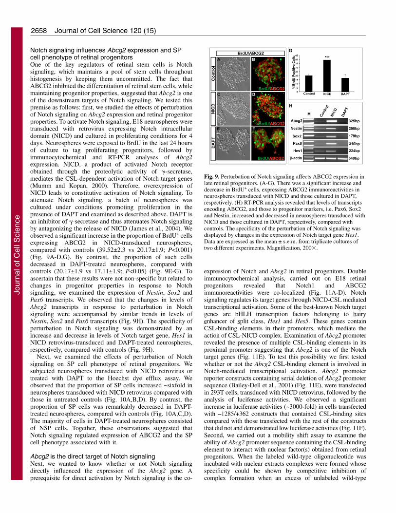
Notch signaling influences Abcg2 expression and SPcell phenotype of retinal progenitorsOne of the key regulators of retinal stem cells is Notchsignaling, which maintains a pool of stem cells throughouthistogenesis by keeping them uncommitted. The fact thatABCG2 inhibited the differentiation of retinal stem cells, whilemaintaining progenitor properties, suggested that Abcg2 is oneof the downstream targets of Notch signaling. We tested thispremise as follows: first, we studied the effects of perturbationof Notch signaling on Abcg2 expression and retinal progenitorproperties. To activate Notch signaling, E18 neurospheres weretransduced with retrovirus expressing Notch intracellulardomain (NICD) and cultured in proliferating conditions for 4days. Neurospheres were exposed to BrdU in the last 24 hoursof culture to tag proliferating progenitors, followed byimmunocytochemical and RT-PCR analyses of Abcg2expression. NICD, a product of activated Notch receptorobtained through the proteolytic activity of �-secretase,mediates the CSL-dependent activation of Notch target genes(Mumm and Kopan, 2000). Therefore, overexpression ofNICD leads to constitutive activation of Notch signaling. Toattenuate Notch signaling, a batch of neurospheres wascultured under conditions promoting proliferation in thepresence of DAPT and examined as described above. DAPT isan inhibitor of �-secretase and thus attenuates Notch signalingby antagonizing the release of NICD (James et al., 2004). Weobserved a significant increase in the proportion of BrdU+ cellsexpressing ABCG2 in NICD-transduced neurospheres,compared with controls (39.52±2.3 vs 20.17±1.9; P<0.001)(Fig. 9A-D,G). By contrast, the proportion of such cellsdecreased in DAPT-treated neurospheres, compared withcontrols (20.17±1.9 vs 17.11±1.9; P<0.05) (Fig. 9E-G). Toascertain that these results were not non-specific but related tochanges in progenitor properties in response to Notchsignaling, we examined the expression of Nestin, Sox2 andPax6 transcripts. We observed that the changes in levels ofAbcg2 transcripts in response to perturbation in Notchsignaling were accompanied by similar trends in levels ofNestin, Sox2 and Pax6 transcripts (Fig. 9H). The specificity ofperturbation in Notch signaling was demonstrated by anincrease and decrease in levels of Notch target gene, Hes1 inNICD retrovirus-transduced and DAPT-treated neurospheres,respectively, compared with controls (Fig. 9H).
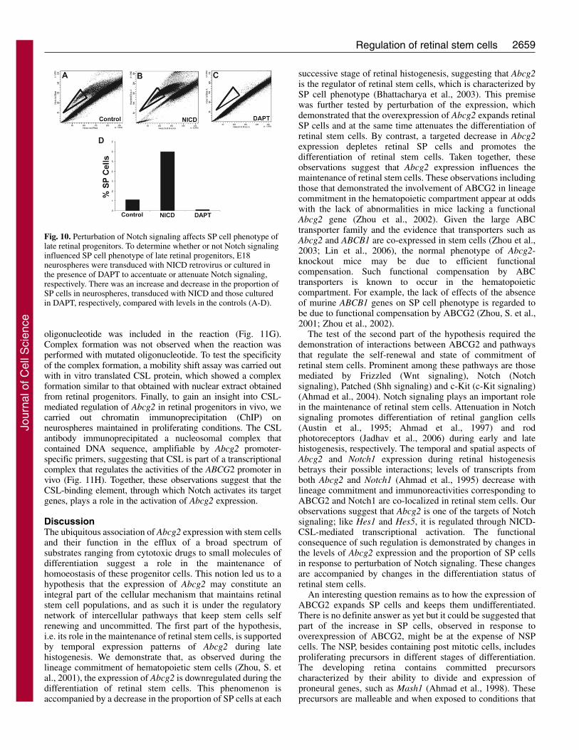
Next, we examined the effects of perturbation of Notchsignaling on SP cell phenotype of retinal progenitors. Wesubjected neurospheres transduced with NICD retrovirus ortreated with DAPT to the Hoechst dye efflux assay. Weobserved that the proportion of SP cells increased ~sixfold inneurospheres transduced with NICD retrovirus compared withthose in untreated controls (Fig. 10A,B,D). By contrast, theproportion of SP cells was remarkably decreased in DAPT-treated neurospheres, compared with controls (Fig. 10A,C,D).The majority of cells in DAPT-treated neurospheres consistedof NSP cells. Together, these observations suggested thatNotch signaling regulated expression of ABCG2 and the SPcell phenotype associated with it.
Abcg2 is the direct target of Notch signalingNext, we wanted to know whether or not Notch signalingdirectly influenced the expression of the Abcg2 gene. Aprerequisite for direct activation by Notch signaling is the co-
Journal of Cell Science 120 (15)
expression of Notch and Abcg2 in retinal progenitors. Doubleimmunocytochemical analysis, carried out on E18 retinalprogenitors revealed that Notch1 and ABCG2immunoreactivities were co-localized (Fig. 11A-D). Notchsignaling regulates its target genes through NICD-CSL mediatedtranscriptional activation. Some of the best-known Notch targetgenes are bHLH transcription factors belonging to hairyenhancer of split class, Hes1 and Hes5. These genes containCSL-binding elements in their promoters, which mediate theaction of CSL-NICD complex. Examination of Abcg2 promoterrevealed the presence of multiple CSL-binding elements in itsproximal promoter suggesting that Abcg2 is one of the Notchtarget genes (Fig. 11E). To test this possibility we first testedwhether or not the Abcg2 CSL-binding element is involved inNotch-mediated transcriptional activation. Abcg2 promoterreporter constructs containing serial deletion of Abcg2 promotersequence (Bailey-Dell et al., 2001) (Fig. 11E), were transfectedin 293T cells, transduced with NICD retrovirus, followed by theanalysis of luciferase activities. We observed a significantincrease in luciferase activities (~3000-fold) in cells transfectedwith –1285/+362 constructs that contained CSL-binding sitescompared with those transfected with the rest of the constructsthat did not and demonstrated low luciferase activities (Fig. 11F).Second, we carried out a mobility shift assay to examine theability of Abcg2 promoter sequence containing the CSL-bindingelement to interact with nuclear factor(s) obtained from retinalprogenitors. When the labeled wild-type oligonucleotide wasincubated with nuclear extracts complexes were formed whosespecificity could be shown by competitive inhibition ofcomplex formation when an excess of unlabeled wild-type
Fig. 9. Perturbation of Notch signaling affects ABCG2 expression inlate retinal progenitors. (A-G). There was a significant increase anddecrease in BrdU+ cells, expressing ABCG2 immunoreactivities inneurospheres transduced with NICD and those cultured in DAPT,respectively. (H) RT-PCR analysis revealed that levels of transcriptsencoding ABCG2, and those to progenitor markers, i.e. Pax6, Sox2and Nestin, increased and decreased in neurospheres transduced withNICD and those cultured in DAPT, respectively, compared withcontrols. The specificity of the perturbation of Notch signaling wasdisplayed by changes in the expression of Notch target gene Hes1.Data are expressed as the mean ± s.e.m. from triplicate cultures oftwo different experiments. Magnification, 200�.
Jour
nal o
f Cel
l Sci
ence
2659Regulation of retinal stem cells
oligonucleotide was included in the reaction (Fig. 11G).Complex formation was not observed when the reaction wasperformed with mutated oligonucleotide. To test the specificityof the complex formation, a mobility shift assay was carried outwith in vitro translated CSL protein, which showed a complexformation similar to that obtained with nuclear extract obtainedfrom retinal progenitors. Finally, to gain an insight into CSL-mediated regulation of Abcg2 in retinal progenitors in vivo, wecarried out chromatin immuno precipitation (ChIP) onneurospheres maintained in proliferating conditions. The CSLantibody immunoprecipitated a nucleosomal complex thatcontained DNA sequence, amplifiable by Abcg2 promoter-specific primers, suggesting that CSL is part of a transcriptionalcomplex that regulates the activities of the ABCG2 promoter invivo (Fig. 11H). Together, these observations suggest that theCSL-binding element, through which Notch activates its targetgenes, plays a role in the activation of Abcg2 expression.
DiscussionThe ubiquitous association of Abcg2 expression with stem cellsand their function in the efflux of a broad spectrum ofsubstrates ranging from cytotoxic drugs to small molecules ofdifferentiation suggest a role in the maintenance ofhomoeostasis of these progenitor cells. This notion led us to ahypothesis that the expression of Abcg2 may constitute anintegral part of the cellular mechanism that maintains retinalstem cell populations, and as such it is under the regulatorynetwork of intercellular pathways that keep stem cells selfrenewing and uncommitted. The first part of the hypothesis,i.e. its role in the maintenance of retinal stem cells, is supportedby temporal expression patterns of Abcg2 during latehistogenesis. We demonstrate that, as observed during thelineage commitment of hematopoietic stem cells (Zhou, S. etal., 2001), the expression of Abcg2 is downregulated during thedifferentiation of retinal stem cells. This phenomenon isaccompanied by a decrease in the proportion of SP cells at each
successive stage of retinal histogenesis, suggesting that Abcg2is the regulator of retinal stem cells, which is characterized bySP cell phenotype (Bhattacharya et al., 2003). This premisewas further tested by perturbation of the expression, whichdemonstrated that the overexpression of Abcg2 expands retinalSP cells and at the same time attenuates the differentiation ofretinal stem cells. By contrast, a targeted decrease in Abcg2expression depletes retinal SP cells and promotes thedifferentiation of retinal stem cells. Taken together, theseobservations suggest that Abcg2 expression influences themaintenance of retinal stem cells. These observations includingthose that demonstrated the involvement of ABCG2 in lineagecommitment in the hematopoietic compartment appear at oddswith the lack of abnormalities in mice lacking a functionalAbcg2 gene (Zhou et al., 2002). Given the large ABCtransporter family and the evidence that transporters such asAbcg2 and ABCB1 are co-expressed in stem cells (Zhou et al.,2003; Lin et al., 2006), the normal phenotype of Abcg2-knockout mice may be due to efficient functionalcompensation. Such functional compensation by ABCtransporters is known to occur in the hematopoieticcompartment. For example, the lack of effects of the absenceof murine ABCB1 genes on SP cell phenotype is regarded tobe due to functional compensation by ABCG2 (Zhou, S. et al.,2001; Zhou et al., 2002).
The test of the second part of the hypothesis required thedemonstration of interactions between ABCG2 and pathwaysthat regulate the self-renewal and state of commitment ofretinal stem cells. Prominent among these pathways are thosemediated by Frizzled (Wnt signaling), Notch (Notchsignaling), Patched (Shh signaling) and c-Kit (c-Kit signaling)(Ahmad et al., 2004). Notch signaling plays an important rolein the maintenance of retinal stem cells. Attenuation in Notchsignaling promotes differentiation of retinal ganglion cells(Austin et al., 1995; Ahmad et al., 1997) and rodphotoreceptors (Jadhav et al., 2006) during early and latehistogenesis, respectively. The temporal and spatial aspects ofAbcg2 and Notch1 expression during retinal histogenesisbetrays their possible interactions; levels of transcripts fromboth Abcg2 and Notch1 (Ahmad et al., 1995) decrease withlineage commitment and immunoreactivities corresponding toABCG2 and Notch1 are co-localized in retinal stem cells. Ourobservations suggest that Abcg2 is one of the targets of Notchsignaling; like Hes1 and Hes5, it is regulated through NICD-CSL-mediated transcriptional activation. The functionalconsequence of such regulation is demonstrated by changes inthe levels of Abcg2 expression and the proportion of SP cellsin response to perturbation of Notch signaling. These changesare accompanied by changes in the differentiation status ofretinal stem cells.
An interesting question remains as to how the expression ofABCG2 expands SP cells and keeps them undifferentiated.There is no definite answer as yet but it could be suggested thatpart of the increase in SP cells, observed in response tooverexpression of ABCG2, might be at the expense of NSPcells. The NSP, besides containing post mitotic cells, includesproliferating precursors in different stages of differentiation.The developing retina contains committed precursorscharacterized by their ability to divide and expression ofproneural genes, such as Mash1 (Ahmad et al., 1998). Theseprecursors are malleable and when exposed to conditions that
Fig. 10. Perturbation of Notch signaling affects SP cell phenotype oflate retinal progenitors. To determine whether or not Notch signalinginfluenced SP cell phenotype of late retinal progenitors, E18neurospheres were transduced with NICD retrovirus or cultured inthe presence of DAPT to accentuate or attenuate Notch signaling,respectively. There was an increase and decrease in the proportion ofSP cells in neurospheres, transduced with NICD and those culturedin DAPT, respectively, compared with levels in the controls (A-D).
Jour
nal o
f Cel
l Sci
ence

2660 Journal of Cell Science 120 (15)
promote the maintenance of retinal stem cells rather than theirdifferentiation they revert to a progenitor stage (Ahmad et al.,1998). The fact that the expression of Mash1 increases in SPcells in neurospheres transduced with ABCG2 retroviruscompared with those in controls suggests that the Mash1-positive precursors are recruited as SP cells in response tooverexpression of ABCG2 (Fig. 11). Indeed, the selectiveproliferation of ABCG2-expressing SP cells should also betaken into account for the expansion of retinal SP cells. Theclassic mechanism of the maintenance of stem cells involvesthe influence of the genome to promote cell proliferation, as inthe case of Wnt signaling, and repress cell commitment, as inthe case of Notch signaling (Ahmad et al., 2004). ABCG2,whose apparent function in stem cells is to protect them fromtoxins and maintain the homeostasis, might prevent cellcommitment by actively extruding extrinsic small moleculesthat promote differentiation. Additionally, it might preventdifferentiation by actively extruding key components of thedifferentiation-promoting signaling pathway, thus impairing it.Evidence has emerged that disparate signaling pathways arerecruited for the maintenance of stem cells or progenitors andit is quite likely that disparate mechanisms, one involving anABCG2 pump, are used to keep stem cells uncommitted tomaintain their pool.
Materials and MethodsCell cultureEmbryos were harvested from timed pregnant Sprague-Dawley rats (SASCO), andthe gestation day was confirmed by the morphological examination of embryos
(Christie, 1964). Embryos were harvested in Hank’s balanced salt solution (HBSS).Eyes were enucleated, and retinas were dissected and dissociated into single cellsas previously described (Bhattacharya et al., 2003). Retinal dissociates werecultured in retinal culture medium or RCM (DMEM-F12, 1� N2 supplement, 2mM L-glutamine, 100 U/ml penicillin, 100 �g/ml streptomycin) containing 20ng/ml EGF for 5 days to generate clonal neurospheres. The neurospheres werecollected, and plated on poly-D-lysine-coated and laminin-coated glass coverslipsand exposed to 5-bromo-2�-deoxyuridine (BrdU) (10 �M) for the final 24 hours.Neurospheres were frozen for RT-PCR or fixed in 4% ice-cold paraformaldehydefor immunocytochemical analysis. To induce differentiation, neurospheres werewashed extensively to remove BrdU and mitogen, and cultured in RCM and 1%FBS for a further 5 days, followed by collection for RT-PCR andimmunocytochemical analysis, as described above.
Hoechst dye efflux assayRetinal cells, from different stages of histogenesis were resuspended in HoechstIMDM (106 cells/ml) containing 2% FCS at 4°C overnight followed by stainingwith Hoechst 33342 (2.0 �g/ml) at 37°C for 30 minutes. Cells were sorted on aFACStar Plus (BDIS) cell sorter. The Hoechst dye was excited at 350 nm and itsfluorescence was measured at two wavelengths using a 485 BP22 (485/22 nm band-pass filter) and a 675 EFLP (675nm long-pass edge filter) optical filter (OmegaOptical, Brattleboro, VT). A 610 nm short-pass dichroic mirror was used to separatethese emission wavelengths (Omega Optical). The SP region was defined on thecytometer on the basis of its fluorescence emission in both blue and redwavelengths. Side population (SP) sorting gates were established after collecting5�105 events within this live gate.
Immunofluorescence analysisDetection of cell-specific markers and BrdU was performed as previously described(Bhattacharya et al., 2003). Briefly, paraformaldehyde-fixed cells were incubated in1� PBS containing 5% NGS and 0, 0.2 or 0.4% Triton X-100 followed by anovernight incubation in antibodies against Nestin (DSHB), Pax6 (DSHB), ABCG2(Chemicon), Map2 (Chemicon), GFAP (Sigma), Notch (Ahmad et al., 1995) andBrdU at 4°C. Cells were examined for epifluorescence after incubation in IgGconjugated to cyanin 3 (Cy3)/FITC. Images were captured using a cooled CCDcamera (Princeton Instruments, Trenton, NJ) and Openlab software (Improvision,Lexington, MA). To determine the percentage of specific cell types in a particular
Fig. 11. Notch signaling influences ABCG2expression directly.(A-D) Immunocytochemical analysis of E18neurospheres showed that immunoreactivitiescorresponding to ABCG2 (arrow) and Notch1(arrowhead) were co-localized in retinalprogenitors. Examination of ABCG2 promotersrevealed the presence four CSL binding sites(TGGGA) between –1285 and –657 (E). Todetermine the CSL-mediated effect on theactivities of ABCG2 promoter, ABCG2promoter deletion constructs were transfectedinto 293T cells, transduced with NICDretrovirus. (F) There was a significant increasein reporter activities with constructs containingCSL sites (b), compared to those without (a),which displayed base level reporter activities.(G) Mobility shift assay carried out withlabeled wild-type ABCG2 promoter sequencescontaining one of the CSL sites (sequence)using HEK293T cell nuclear extracts revealedthe formation of complexes, which were similarto those formed with in vitro translatedsuppressor of hairless (SuH) protein. Thesecomplexes could be competed with an excess ofunlabeled sequences and were not formed withsequences in which the CSL site was mutated.(H) Nucleosomal DNA immunoprecipitatedwith CSL antibody in a ChIP assay on E18retinal progenitors contained sequencescorresponding to the Abcg2 promoter,suggesting the presence of CSL on Abcg2regulatory complexes in vivo.
Jour
nal o
f Cel
l Sci
ence
2661Regulation of retinal stem cells
condition, the number of BrdU+ and cell-specific antigen-positive cells was countedin 10-15 randomly selected fields in two to three different coverslips. Eachexperiment was repeated at least three times. Values are expressed as means ± s.e.m.Data were analyzed using the Student’s t-test to determine the significance of thedifferences between various conditions.
RT-PCR analysisTotal RNA was isolated using a Qiagen RNA isolation kit. 2 �M cDNA wasamplified using gene-specific primers by using the following step cycle program ona Robocycler (Stratagene): denaturation at 94°C for 30 seconds, annealing atspecific temperature (see supplementary material Table S1) for 35 seconds,extension at 72°C for 40 seconds for 35 cycles followed by a final extension at 72°Cfor 5 minutes. PCR products were resolved on 2% agarose gels against 100bp DNAmarker (MBI Fermentas). Gene-specific primers were used as listed insupplementary material Table S1.
Transduction of neurospheresLate retinal progenitors were enriched as neurospheres from E18 retina (=E18neurospheres) and were transduced with ABCG2 retrovirus vector (G1-ABCG2) tooverexpress ABCG2. After transduction, the medium was replaced, andneurospheres were cultured in proliferating conditions for 4 days and exposed toBrdU in the last 24 hours, to tag proliferating cells, followed byimmunocytochemical and RT-PCR analyses of cell-specific markers.
siRNA-mediated gene silencingFor targeted silencing of Abcg2 expression, ABCG2 siRNA was cloned into pSupervector (pSuper-ABCG2siRNA) according to the manufacturer’s (Oligogene) protocol.Neurospheres in proliferating conditions were co-transfected with pSuper-ABCG2siRNA and pEGFP-C3 (to determine transfection efficiency) using Fugeneaccording to the manufacturer’s (Roche) protocol. A batch of neurospheres wassimilarly transfected with pSuper-ABCG2missense, containing scrambled sequence,as controls. The efficiency of gene silencing was determined byimmunocytochemical and RT-PCR analyses of ABCG2 protein and transcripts,respectively.
Reporter assayHEK293T cells were plated onto 100 mm Petri dishes at a density of 8-10�106
cells. Cells were co-transfected with a series of Abcg2 promoter constructs (Bailey-Dell et al., 2001) and NICD expression construct (James et al., 2004) using acalcium phosphate-based protocol. Transfection efficiency was examined by co-transfecting cells with pGFP-C3 (Clonetech). For luciferase assay, cells were lysedin 1� reporter lysis buffer (Promega) and 100 �l lysate was diluted five times usingassay reagent (Promega). Diluted samples (100 �l) were analyzed for luciferaseactivities using a luminometer (Pharmingen). Mean fold change for the respectivedeletion constructs in the experimental groups was calculated with respect tocontrols (pGL3-basic). Values are expressed as mean fold change ± s.e.m. from threedifferent experiments. Statistical analysis was performed using the Student’s t-testto determine the significance between different conditions.
Electrophoretic mobility shift assay (EMSA)EMSA was carried out as previously described (Ahmad et al., 1995). Briefly,oligonucleotides corresponding to Abcg2 promoter sequence containing the wild-type (GGTAGATGTTGGGATGGCTAC) or mutated (GGTAGATGT GAGAT -TGGCTAC) CSL binding site were end-labeled with [�-32P]ATP using T4polynucleotide kinase (New England Biolabs). The binding reaction was performedin 20 �l buffer containing 20 mM Tris-HCl (pH 8.0), 75 mM KCl, 5% glycerol,50 �g/ml bovine serum albumin (BSA), 0.025% nonidet NP-40, 1 mM EDTA, 5mM DTT and 1 �g of poly(dI/dC). The end-labeled probe (100 pmol (20,000 cpm)was incubated on ice for 30 minutes with 5 �g of nuclear extract. Samples wereloaded on a 5% polyacrylamide gel in 0.5 TBE buffer for 2 hours at 10 V/cm. Thegel was dried and autoradiographed. For the competition assay, a molar excess ofcold probe was added to the binding reactions.
Chromatin immunoprecipitation (ChIP) assayChIP assay was done using a modified procedure from Upstate Biotechnology.Briefly, E18 retinal progenitors were grown in proliferating conditions and histoneswere crosslinked to DNA by adding formaldehyde directly to culture medium to afinal concentration of 1% and incubating for 10 minutes at room temperature on arocking platform. Cells were washed three times with ice-cold PBS containingprotease inhibitors. Cell pellets were resuspended in pre-warmed SDS lysis buffer(1% SDS, 10 mM EDTA, 50 mM Tris-HCl, pH 8.1). To reduce the non-specificbackground, the samples were pre-cleared using 80 �l salmon-sperm DNA/proteinA agarose slurry at 4°C for 30 minutes. Samples were centrifuged at 100 r.p.m. for1 minute at 4°C. Supernatants were transferred to a new tube, theimmunoprecipitating antibody was added and incubation was carried out overnightat 4°C on a rocking platform. For a negative control we used no antibody or non-specific antibodies. The histone-antibody complex was precipitated using 60 �lsalmon sperm DNA and protein-A-Sepharose (Upstate) for 1 hour at 4°C.
Precipitates were washed sequentially at room temperature for 5 minutes, once withlow salt immune complex wash buffer (0.1% SDS, 1% Triton X-100, 2 mM EDTA,20 mM Tris-HCl, pH 8.1, 150 mM NaCl.), high salt immune complex wash buffer(0.1% SDS, 1% Triton X-100, 2 mM EDTA, 20 mM Tris-HCl, pH 8.1, 500 mMNaCl) and lithium salt immune complex wash buffer (2.25M LiCl, 1% IGEPAL-CA630, 1% deoxycholic acid sodium salt, 1 mM EDTA, 10 mM Tris-HCl, pH 8.1)and finally twice with TE buffer (10 mM Tris-HCl, 1 mM EDTA, pH 8.0). Aftercompletely removing the TE buffer, the precipitate was resuspended and extractedtwice in 250 �l freshly prepared elution buffer (1% SDS, 0.1M NaHCO3). Toreverse the histone-DNA crosslinks, samples were heated at 65°C for 4 hours. 200�l of initial sonicated sample was reverse crosslinked and used as input. Afterremoving the antibodies by protease digestion of the samples, DNA was recoveredand column purified. PCRs were performed using gene-specific primers (5�-GGTAGATGTTGGGATGGCTAC-3�, 5�-CCATCTACAACCCTACCGATG-3�).
We thank Douglas Ross for the ABCG2 promoter-reporterconstruct and Kenneth Cowen for ABCG2 retrovirus constructs. Thiswork was supported by the Nebraska Tobacco Settlement BiomedicalResearch Development, COBRE, the Lincy Foundation and thePearson Foundation.
ReferencesAbbott, B. L. (2003). ABCG2 (BCRP) expression in normal and malignant hematopoietic
cells. Hematol. Oncol. 21, 115-130.Ahmad, I., Zaqouras, P. and Artavanis-Tsakonas, S. (1995). Involvement of Notch-1
in mammalian retinal neurogenesis: association of Notch-1 activity with both immatureand terminally differentiated cells. Mech. Dev. 53, 73-85.
Ahmad, I., Dooley, C. M. and Polk, D. L. (1997). Delta-1 is a regulator of neurogenesisin the vertebrate retina. Dev. Biol. 185, 92-103.
Ahmad, I., Dooley, C. M. and Afiat, S. (1998). Involvement of Mash1 in EGF-mediatedregulation of differentiation in the vertebrate retina. Dev. Biol. 194, 86-98.
Ahmad, I., Das, A. V., James, J., Bhattacharya, S. and Zhao, X. (2004). Neural stemcells in the mammalian eye:types and regulation. Semin. Cell Biol. 15, 53-62.
Austin, C. P., Feldman, D. E., Ida, J. A., Jr and Cepko, C. L. (1995). Vertebrate retinalganglion cells are selected from competent progenitors by the action of Notch.Development 121, 3637-3650.
Bailey-Dell, K. J., Hassel, B., Doyle, L. A. and Ross, D. D. (2001). Promotercharacterization and genomic organization of the human breast cancer resistanceprotein (ATP-binding cassette transporter G2) gene. Biochim. Biophys. Acta 1520, 234-241.
Bhattacharya, S., Jackson, J. D., Das, A. V., Thoreson, W. B., Kuszynski, C., James,J., Joshi, S. and Ahmad, I. (2003). Direct identification and enrichment of retinal stemcells/progenitors using hoechst dye efflux assay. Invest. Ophthalmol. Vis. Sci. 44, 2764-2773.
Budak, M. T., Alpdogan, O. S., Zhou, M., Lavker, R. M., Akinci, M. A. and Wolosin,J. M. (2005). Ocular surface epithelia contain ABCG2-dependent side population cellsexhibiting features associated with stem cells. J. Cell Sci. 118, 1715-1724.
Bunting, K. D. (2002). ABC transporters as phenotypic markers and functional regulatorsof stem cells. Stem Cells 20, 11-20.
Christie, G. A. (1964). Developmental stages in somite and post-somite rat embryos,based on external appearance, and including some features of the macroscopicdevelopment of the oral cavity. J. Morphol. 114, 263-283.
Doyle, L. A. and Ross, D. D. (2003). Multidrug resistance mediated by the breast cancerresistance protein BCRP (ABCG2). Oncogene 22, 7340-7358.
Dyer, M. A. and Cepko, C. L. (2001). p27Kip1 and p57Kip2 regulate proliferation indistinct retinal progenitor cell populations. J. Neurosci. 21, 4259-4271.
Fantl, V., Stamp, G., Andrews, A., Rosewell, I. and Dickson, C. (1995). Mice lackingcyclin D1 are small and show defects in eye and mammary gland development. GenesDev. 9, 2364-2372.
Goodell, M. A., Brose, K., Paradis, G., Conner, A. S. and Mulligan, R. C. (1996).Isolation and functional properties of murine hematopoietic stem cells that arereplicating in vivo. J. Exp. Med. 183, 1797-1806.
Hulspas, R. and Quesenberry, P. J. (2000). Characterization of neurosphere cellphenotypes by flow cytometry. Cytometry 40, 245-250.
Jadhav, A. P., Mason, H. A. and Cepko, C. L. (2006). Notch 1 inhibits photoreceptorproduction in the developing mammalian retina. Development 133, 913-923.
James, J., Das, A. V., Rahnenfuhrer, J. and Ahmad, I. (2004). Cellular and molecularcharacterization of early and late retinal stem cells/progenitors: Differential regulationof proliferation and context dependent role of Notch signaling. J. Neurobiol. 61, 359-376.
Kim, M., Turnquist, H., Jackson, J., Sgagias, M., Yan, Y., Gong, M., Dean, M., Sharp,J. G. and Cowan, K. (2002). The multidrug resistance transporter ABCG2 (breastcancer resistance protein 1) effluxes Hoechst 33342 and is overexpressed inhematopoietic stem cells. Clin. Cancer Res. 8, 22-28.
Krishnamurthy, P. and Schuetz, J. D. (2006). Role of ABCG2/BCRP in biology andmedicine. Annu. Rev. Pharmacol. Toxicol. 46, 381-410.
Lassalle, B., Bastos, H., Louis, J. P., Riou, L., Testart, J., Dutrillaux, B., Fouchet, P.and Allemand, I. (2004). ‘Side Population’ cells in adult mouse testis express Bcrp1gene and are enriched in spermatogonia and germinal stem cells. Development 131,479-487.
Jour
nal o
f Cel
l Sci
ence
Levine, E. M., Close, J., Fero, M., Ostrovsky, A. and Reh, T. A. (2000). p27(Kip1)regulates cell cycle withdrawal of late multipotent progenitor cells in the mammalianretina. Dev. Biol. 219, 299-314.
Lin, T., Islam, O. and Heese, K. (2006). ABC transporters, neural stem cells, andneurogenesis-a different perspective. Cell Res. 16, 857-871.
Martin, C. M., Meeson, A. P., Robertson, S. M., Hawke, T. J., Richardson, J. A.,Bates, S., Goetsch, S. C., Gallardo, T. D. and Garry, D. J. (2004). Persistentexpression of the ATP-binding cassette transporter, Abcg2, identifies cardiac SP cellsin the developing and adult heart. Dev. Biol. 265, 262-275.
Meeson, A. P., Hawke, T. J., Graham, S., Jiang, N., Elterman, J., Hutcheson, K.,Dimaio, J. M., Gallardo, T. D. and Garry, D. J. (2004). Cellular and molecularregulation of skeletal muscle side population cells. Stem Cells 22, 1305-1320.
Mouthon, M. A., Fouchet, P., Mathieu, C., Sii-Felice, K., Etienne, O., Lages, C. S.and Boussin, F. D. (2006). Neural stem cells from mouse forebrain are contained in apopulation distinct from the ‘side population’. J. Neurochem. 99, 807-817.
Mumm, J. S. and Kopan, R. (2000). Notch signaling: from the outside in. Dev. Biol.228, 151-165.
Ohnuma, S., Philpott, A., Wang, K., Holt, C. E. and Harris, W. A. (1999). p27Xic1,a Cdk inhibitor, promotes the determination of glial cells in Xenopus retina. Cell 99,499-510.
Rapaport, D. H., Wong, L. L., Wood, E. D., Yasumura, D. and LaVail, M. M. (2004).Timing and topography of cell genesis in the rat retina. J. Comp. Neurol. 474, 304-324.
Scharenberg, C. W., Harkey, M. A. and Torok-Storb, B. (2002). The ABCG2transporter is an efficient Hoechst 33342 efflux pump and is preferentially expressedby immature human hematopoietic progenitors. Blood 99, 507-512.
Sicinski, P., Donaher, J. L., Parker, S. B., Li, T., Fazeli, A., Gardner, H., Haslam, S.
Z., Bronson, R. T., Elledge, S. J. and Weinberg, R. A. (1995). Cyclin D1 providesa link between development and oncogenesis in the retina and breast. Cell 82, 621-630.
Staud, F. and Pavek, P. (2005). Breast cancer resistance protein (BCRP/ABCG2). Int. J.Biochem. Cell Biol. 37, 720-725.
Summer, R., Kotton, D. N., Sun, X., Ma, B., Fitzsimmons, K. and Fine, A. (2003).Side population cells and Bcrp1 expression in lung. Am. J. Physiol. Lung Cell. Mol.Physiol. 285, L97-L104.
Tirelli, G., Sidari, L., Giacomarra, V., Papanikolla, L., Sasso, F., Russolo, M. andMelato, M. (2002). Do Ki67, S-phase, S + G2M and DNA ploidy, evaluated by flowcytometry, reveal locoregional metastasis in oral cavity and oropharynx carcinomas?Oncol. Rep. 9, 575-580.
Zhou, C., Zhao, L., Inagaki, N., Guan, J., Nakajo, S., Hirabayashi, T., Kikuyama, S.and Shioda, S. (2001). Atp-binding cassette transporter ABC2/ABCA2 in the rat brain:a novel mammalian lysosome-associated membrane protein and a specific marker foroligodendrocytes but not for myelin sheaths. J. Neurosci. 21, 849-857.
Zhou, S., Schuetz, J. D., Bunting, K. D., Colapietro, A. M., Sampath, J., Morris, J.J., Lagutina, I., Grosveld, G. C., Osawa, M., Nakauchi, H. et al. (2001). The ABCtransporter Bcrp1/ABCG2 is expressed in a wide variety of stem cells and is amolecular determinant of the side-population phenotype. Nat. Med. 7, 1028-1034.
Zhou, S., Morris, J. J., Barnes, Y., Lan, L., Schuetz, J. D. and Sorrentino, B. P. (2002).Bcrp1 gene expression is required for normal numbers of side population stem cells inmice, and confers relative protection to mitoxantrone in hematopoietic cells in vivo.Proc. Natl. Acad. Sci. USA 99, 12339-12344.
Zhou, S., Zong, Y., Lu, T. and Sorrentino, B. P. (2003). Hematopoietic cells from micethat are deficient in both Bcrp1/Abcg2 and Mdr1a/1b develop normally but aresensitized to mitoxantrone. Biotechniques 35, 1248-1252.
Journal of Cell Science 120 (15)2662
Jour
nal o
f Cel
l Sci
ence