Notch Signaling Regulates Motor Neuron Differentiation of Human Embryonic Stem Cells.
17
STEM CELLS 2014;00:00‐00 www.StemCells.com ©AlphaMed Press 2014 EMBRYONIC STEM CELLS/INDUCED PLURIPOTENT STEM CELLS 1 The Sidney and Judy Swartz Em‐ bryonic Stem Cell Research Center of The Goldyne Savad Institute of Gene Therapy and 3 The Depart‐ ment of Obstetrics and Gynecolo‐ gy, Hadassah University Medical Center, Jerusalem, Israel. ; 2 A. Al‐ fred Taubman Medical Research Institute, Department of Neurolo‐ gy, University of Michigan, USA. Correspondence: Benjamin E. Reu‐ binoff, MD, Ph.D., The Sidney and Judy Swartz Embryonic Stem Cell Research Center of The Goldyne Savad Institute of Gene Therapy, Hadassah University Medical Cen‐ ter, Ein Kerem P.O.B. 12000 Jerusa‐ lem 91120, Israel. Telephone: (972) 2‐677‐6424/5; Fax: (972) 2‐677‐ 6489; e‐mail:[email protected] Received November 14, 2013; ac‐ cepted for publication September 29, 2014; ©AlphaMed Press 1066‐5099/2014/$30.00/0 This article has been accepted for publication and undergone full peer review but has not been through the copyediting, typeset‐ ting, pagination and proofreading process which may lead to differ‐ ences between this version and the Version of Record. Please cite this article as doi: 10.1002/stem.1873 Notch Signaling Regulates Motor Neuron Diffe‐ rentiation of Human Embryonic Stem Cells. Etti Ben‐Shushan 1 , Eva Feldman 2 , Benjamin E. Reubinoff 1,3 Key words.Neural differentiation • Notch • Embryonic stem cells • Progeni‐ tor cells ABSTRACT In the pMN domain of the spinal cord, Notch signaling regulates the bal‐ ance between motor neuron differentiation and maintenance of the pro‐ genitor state for later oligodendrocyte differentiation. Here we sought to study the role of Notch signaling in regulation of the switch from the pMN progenitor state to differentiated motor neurons in a human model sys‐ tem. Human embryonic stem cells (hESCs) were directed to differentiate to pMN‐like progenitor cells by the inductive action of retinoic acid and a Shh agonist, purmorphamine. We found that the expression of the Notch sig‐ naling effector Hes5 was induced in hESC‐derived pMN‐like progenitors and remained highly expressed when they were cultured under conditions favoring motor neuron differentiation. Inhibition of Notch signaling by a ‐ secretase inhibitor in the differentiating pMN‐like progenitor cells de‐ creased Hes5 expression and enhanced the differentiation towards motor neurons. Conversely, over‐expression of Hes5 in pMN‐like progenitor cells during the differentiation interfered with retinoic acid‐ and purmorpha‐ mine‐ induced motor neuron differentiation and inhibited the emergence of motor neurons. Inhibition of Notch signaling had a permissive rather than an inductive effect on motor neuron differentiation. Our results indi‐ cate that Notch signaling has a regulatory role in the switch from the pMN progenitor to the differentiated motor neuron state. Inhibition of Notch signaling can be harnessed to enhance the differentiation of hESCs towards motor neurons. STEM CELLS 2014; 00:000–000 INTRODUCTION The Notch signaling pathway plays an essential role in maintenance of progenitor cell populations and in pre‐ venting their differentiation into mature progenies. Notch signaling is initiated when Notch receptor on one cell is activated by a ligand expressed on a neighboring cell. Upon activation, the Notch receptor intracellular domain (NICD) is cleaved by Presenilin proteases of the γ‐secretase complex, and translocates to the nucleus to form a complex with CSL and Master‐mind (Maml) pro‐ teins [1‐4]. This complex then activates expression of the Hes (Hes1 and Hes5) and Hey transcription factors, which repress the expression of proneural genes such as Neurogenin 1/2 and Ascl1, thereby inhibiting neu‐ ronal differentiation and maintaining neural progenitor cells [5]. In the developing spinal cord, Notch signaling has a prominent role both in maintenance of neural and glial progenitor cells and in regulation of specific neuronal fate decisions. Specific progenitor cells with distinct identities and fates are organized along the dorso‐ ventral axis of the neural tube in five domains, termed p0‐p3 and pMN. Recently it was shown that the tran‐
Transcript of Notch Signaling Regulates Motor Neuron Differentiation of Human Embryonic Stem Cells.

STEM CELLS 2014;00:00‐00 www.StemCells.com ©AlphaMed Press 2014
EMBRYONIC STEM CELLS/INDUCED PLURIPOTENT STEM CELLS 1The Sidney and Judy Swartz Em‐bryonic Stem Cell Research Center of The Goldyne Savad Institute of Gene Therapy and 3 The Depart‐ment of Obstetrics and Gynecolo‐gy, Hadassah University Medical Center, Jerusalem, Israel. ; 2 A. Al‐fred Taubman Medical Research Institute, Department of Neurolo‐gy, University of Michigan, USA. Correspondence: Benjamin E. Reu‐binoff, MD, Ph.D., The Sidney and Judy Swartz Embryonic Stem Cell Research Center of The Goldyne Savad Institute of Gene Therapy, Hadassah University Medical Cen‐ter, Ein Kerem P.O.B. 12000 Jerusa‐lem 91120, Israel. Telephone: (972) 2‐677‐6424/5; Fax: (972) 2‐677‐6489; e‐mail:[email protected] Received November 14, 2013; ac‐cepted for publication September 29, 2014; ©AlphaMed Press 1066‐5099/2014/$30.00/0 This article has been accepted for publication and undergone full peer review but has not been through the copyediting, typeset‐ting, pagination and proofreading process which may lead to differ‐ences between this version and the Version of Record. Please cite this article as doi: 10.1002/stem.1873
Notch Signaling Regulates Motor Neuron Diffe‐rentiation of Human Embryonic Stem Cells. Etti Ben‐Shushan1, Eva Feldman2, Benjamin E. Reubinoff 1, 3
Key words.Neural differentiation • Notch • Embryonic stem cells • Progeni‐
tor cells ABSTRACT
In the pMN domain of the spinal cord, Notch signaling regulates the bal‐ance between motor neuron differentiation and maintenance of the pro‐genitor state for later oligodendrocyte differentiation. Here we sought to study the role of Notch signaling in regulation of the switch from the pMN progenitor state to differentiated motor neurons in a human model sys‐tem. Human embryonic stem cells (hESCs) were directed to differentiate to pMN‐like progenitor cells by the inductive action of retinoic acid and a Shh agonist, purmorphamine. We found that the expression of the Notch sig‐naling effector Hes5 was induced in hESC‐derived pMN‐like progenitors and remained highly expressed when they were cultured under conditions favoring motor neuron differentiation. Inhibition of Notch signaling by a �‐secretase inhibitor in the differentiating pMN‐like progenitor cells de‐creased Hes5 expression and enhanced the differentiation towards motor neurons. Conversely, over‐expression of Hes5 in pMN‐like progenitor cells during the differentiation interfered with retinoic acid‐ and purmorpha‐mine‐ induced motor neuron differentiation and inhibited the emergence of motor neurons. Inhibition of Notch signaling had a permissive rather than an inductive effect on motor neuron differentiation. Our results indi‐cate that Notch signaling has a regulatory role in the switch from the pMN progenitor to the differentiated motor neuron state. Inhibition of Notch signaling can be harnessed to enhance the differentiation of hESCs towards motor neurons. STEM CELLS 2014; 00:000–000
INTRODUCTION
The Notch signaling pathway plays an essential role in maintenance of progenitor cell populations and in pre‐venting their differentiation into mature progenies. Notch signaling is initiated when Notch receptor on one cell is activated by a ligand expressed on a neighboring cell. Upon activation, the Notch receptor intracellular domain (NICD) is cleaved by Presenilin proteases of the γ‐secretase complex, and translocates to the nucleus to form a complex with CSL and Master‐mind (Maml) pro‐teins [1‐4]. This complex then activates expression of
the Hes (Hes1 and Hes5) and Hey transcription factors, which repress the expression of proneural genes such as Neurogenin 1/2 and Ascl1, thereby inhibiting neu‐ronal differentiation and maintaining neural progenitor cells [5]. In the developing spinal cord, Notch signaling has a prominent role both in maintenance of neural and glial progenitor cells and in regulation of specific neuronal fate decisions. Specific progenitor cells with distinct identities and fates are organized along the dorso‐ventral axis of the neural tube in five domains, termed p0‐p3 and pMN. Recently it was shown that the tran‐

www.StemCells.com ©AlphaMed Press 2014
2
scription factor Nkx6.1 plays an active role in inducing the expression of the Notch ligand Dll1 in both the pMN and p2 domains and the resulting Notch signaling main‐tains the progenitor state in the distinct domains. Ac‐cordingly, conditional knockout of Notch1 receptor re‐sults in a reduction of all neural progenitor subtypes in the ventral spinal cord [6]. Progenitor cells in the p0‐p3 domains generate different classes of ventral interneurons, named V0‐V3, respec‐tively, whereas pMN progenitor cells at early stages of development appear to be committed to generate mo‐tor neurons (MNs). Later in development, they switch to produce oligodendrocytes [7‐10]. pMN progenitor cells selectively express the bHLH protein Olig2, which is required to specify both motor neuron and oligoden‐drocyte cell identities [11, 12]. Olig2 primes pMN pro‐genitor cells to become motor neurons by triggering the expression of Neurogenin 2 (Ngn2) bHLH protein. Co‐expression of Olig2 and Ngn2 directs the pMN progeni‐tors to leave the cell cycle and to become motor neu‐rons, while progenitors in which Ngn2 expression is low remain as proliferative progenitors which are specified to an oligodendrocyte fate [12‐14]. Ngn2 is repressed by Notch signaling, as was shown by the upregulation of Ngn2 expression in the ventral spinal cord of Notch1 conditional null mice [6]. In the pMN domain, Notch signaling acts to maintain the balance between progenitors that differentiate into motor neurons during the neurogenic phase and those that are preserved as a pool of presumptive progenitors for the gliogenic phase [15, 16]. It was shown that loss of the Notch signaling in the pMN domain increased motor neuron differentiation and results in a progressive depletion of the pMN pro‐genitors over time. Conversely, activation of Notch sig‐naling resulted in a reduction in motor neurons [16, 17]. In light of the potential role of Notch signaling during motor neurons development in animal models, we sought to study its role in human motor neuron devel‐opment by using human embryonic stem cells (hESCs) as a model system. Human ESCs have been reported to generate spinal motor neurons in a pathway that recapitulates the steps of motor neuron differentiation in vivo. After ini‐tial neuralization, caudalization is induced by retinoic acid (RA) and ventralization by Shh morphogen [18‐21]. In response to the Shh, hESCs‐derived pMN progenitors express Pax6, Nkx6.1 and Olig2 transcription factors similar to their in vivo counterparts, and can be further differentiated into early Hb9 expressing and to mature ChAT‐producing spinal motor neurons. Using BAC transgenic reporter lines, the Notch compo‐nents Hes5 and Dll1 have been shown to be dynamically expressed during the differentiation of hESCs into mo‐tor neurons [22]. Hes5 is highly expressed in hESCs‐derived neural progenitors and is downregulated during their differentiation into motor neurons. Conversely, Dll1 is expressed at low levels in neural progenitors and
is upregulated upon their differentiation, being ex‐pressed in Hb9‐positive motor neurons. To better understand how the activity of Notch signal‐ing controls differentiation of hESCs into motor neu‐rons, the study we report here tested the functional relationship between the expression levels of the Notch downstream effector Hes5 and motor neuron differen‐tiation. We show that in response to RA and the Shh agonist purmorphamine (PUR), hESC‐derived neural progenitor cells are specified to generate pMN‐like pro‐genitor cells characterized by the expression of Olig2 and Ngn2. The neuralization and subsequent specifica‐tion to pMN‐like progenitors is concomitant with the induction of the expression of Hes5. However, further differentiation of the pMN‐like progenitor cells into motor neurons is low, raising the possibility that Notch signaling inhibits their differentiation. Using the �‐secretase inhibitor DAPT to inhibit Notch signaling in differentiating pMN‐like progenitor cells, we found that inhibition of Notch signaling downregulates Hes5 ex‐pression and enhances the differentiation of pMN‐like progenitors into motor neurons. Conversely, over‐expression of Hes5 in differentiating pMN‐like progeni‐tors inhibits subsequent differentiation into motor neu‐rons. Still, in the absence of RA and PUR, inhibition of Notch signaling was not sufficient to direct the differentiation of the pMN progenitors towards motor neural fate, in‐dicating a permissive rather than instructive role of Notch signaling in the process of differentiation towards spinal motor neurons. MATERIALS AND METHODS
Cell Culture Human ESCs (HES1 passages 21 to 33 with a normal karyotype) were cultured as described [23, 24]. For differentiation, hESC colonies were picked up by means of collagenase IV (1mg/ml; GIBCO‐BRL, Gaithers‐burg, MD), triturated into small, 50‐100 cell clumps, and placed into ultralow adherent culture dishes (Thermo Scientific Nunc HydroCell). For the first 4 days, cells were grown in NSC media, consisting of DMEM:nutrient mixture F‐12 (DMEM/F:12; Invitrogen, Carlsbad, CA) and 2% B27 supplement with 20 ng/ml FGF2 (Pepro‐Tech Inc, Rocky Hill, NJ) and 5 μM SB431542 (SB; Selleck Chemicals LLC, Houston, USA). At day 14, neural spheres were switched to medium consisting of DMEM/F12 and 1% N2 supplement with 1 μM all‐trans retinoic acid (RA; Sigma‐Aldrich) and 1 μM di‐butyril cAMP (Sigma). At day 21, the spheres were cultured in medium consisting of Neurobasal (Invitrogen) and N2 supplement with 1μM RA, 0.5 μM purmorphamine (PUR; Cayman Chemical, Ann Arbor, MI) and 1 μM di‐butyril cAMP for a 3‐week period. For differentiation, spheres were cut into small clusters and plated on poly‐lysine/laminin‐coated cover glasses for 1 week, in Neu‐robasal medium with 1% N2 supplement containing
www.StemCells.com ©AlphaMed Press 2014
3
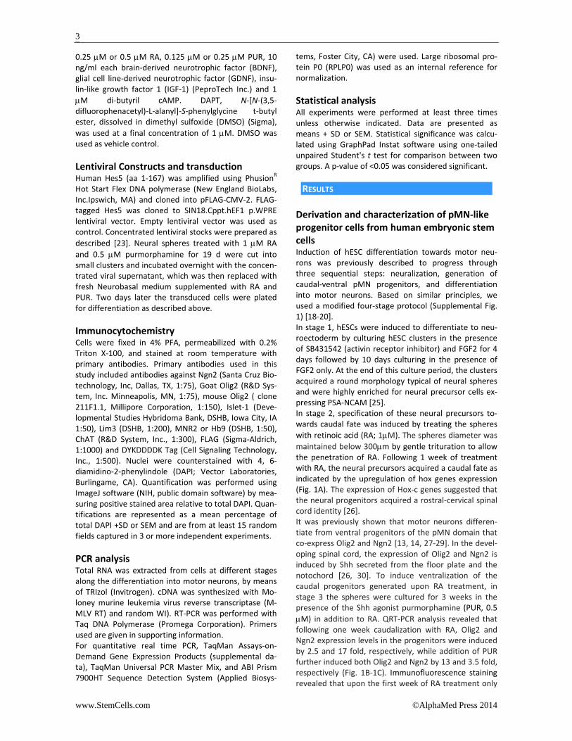
0.25 μM or 0.5 μM RA, 0.125 μM or 0.25 μM PUR, 10 ng/ml each brain‐derived neurotrophic factor (BDNF), glial cell line‐derived neurotrophic factor (GDNF), insu‐lin‐like growth factor 1 (IGF‐1) (PeproTech Inc.) and 1 μM di‐butyril cAMP. DAPT, N‐[N‐(3,5‐difluorophenacetyl)‐L‐alanyl]‐S‐phenylglycine t‐butyl ester, dissolved in dimethyl sulfoxide (DMSO) (Sigma), was used at a final concentration of 1 μM. DMSO was used as vehicle control. Lentiviral Constructs and transduction Human Hes5 (aa 1‐167) was amplified using PhusionR Hot Start Flex DNA polymerase (New England BioLabs, Inc.Ipswich, MA) and cloned into pFLAG‐CMV‐2. FLAG‐tagged Hes5 was cloned to SIN18.Cppt.hEF1�p.WPRE lentiviral vector. Empty lentiviral vector was used as control. Concentrated lentiviral stocks were prepared as described [23]. Neural spheres treated with 1 μM RA and 0.5 μM purmorphamine for 19 d were cut into small clusters and incubated overnight with the concen‐trated viral supernatant, which was then replaced with fresh Neurobasal medium supplemented with RA and PUR. Two days later the transduced cells were plated for differentiation as described above. Immunocytochemistry Cells were fixed in 4% PFA, permeabilized with 0.2% Triton X‐100, and stained at room temperature with primary antibodies. Primary antibodies used in this study included antibodies against Ngn2 (Santa Cruz Bio‐technology, Inc, Dallas, TX, 1:75), Goat Olig2 (R&D Sys‐tem, Inc. Minneapolis, MN, 1:75), mouse Olig2 ( clone 211F1.1, Millipore Corporation, 1:150), Islet‐1 (Deve‐lopmental Studies Hybridoma Bank, DSHB, Iowa City, IA 1:50), Lim3 (DSHB, 1:200), MNR2 or Hb9 (DSHB, 1:50), ChAT (R&D System, Inc., 1:300), FLAG (Sigma‐Aldrich, 1:1000) and DYKDDDDK Tag (Cell Signaling Technology, Inc., 1:500). Nuclei were counterstained with 4, 6‐diamidino‐2‐phenylindole (DAPI; Vector Laboratories, Burlingame, CA). Quantification was performed using ImageJ software (NIH, public domain software) by mea‐suring positive stained area relative to total DAPI. Quan‐tifications are represented as a mean percentage of total DAPI +SD or SEM and are from at least 15 random fields captured in 3 or more independent experiments. PCR analysis Total RNA was extracted from cells at different stages along the differentiation into motor neurons, by means of TRIzol (Invitrogen). cDNA was synthesized with Mo‐loney murine leukemia virus reverse transcriptase (M‐MLV RT) and random WI). RT‐PCR was performed with Taq DNA Polymerase (Promega Corporation). Primers used are given in supporting information. For quantitative real time PCR, TaqMan Assays‐on‐Demand Gene Expression Products (supplemental da‐ta), TaqMan Universal PCR Master Mix, and ABI Prism 7900HT Sequence Detection System (Applied Biosys‐
tems, Foster City, CA) were used. Large ribosomal pro‐tein P0 (RPLP0) was used as an internal reference for normalization. Statistical analysis All experiments were performed at least three times unless otherwise indicated. Data are presented as means + SD or SEM. Statistical significance was calcu‐lated using GraphPad Instat software using one‐tailed unpaired Student's t test for comparison between two groups. A p‐value of <0.05 was considered significant. RESULTS
Derivation and characterization of pMN‐like progenitor cells from human embryonic stem cells Induction of hESC differentiation towards motor neu‐rons was previously described to progress through three sequential steps: neuralization, generation of caudal‐ventral pMN progenitors, and differentiation into motor neurons. Based on similar principles, we used a modified four‐stage protocol (Supplemental Fig. 1) [18‐20]. In stage 1, hESCs were induced to differentiate to neu‐roectoderm by culturing hESC clusters in the presence of SB431542 (activin receptor inhibitor) and FGF2 for 4 days followed by 10 days culturing in the presence of FGF2 only. At the end of this culture period, the clusters acquired a round morphology typical of neural spheres and were highly enriched for neural precursor cells ex‐pressing PSA‐NCAM [25]. In stage 2, specification of these neural precursors to‐wards caudal fate was induced by treating the spheres with retinoic acid (RA; 1μM). The spheres diameter was maintained below 300μm by gentle trituration to allow the penetration of RA. Following 1 week of treatment with RA, the neural precursors acquired a caudal fate as indicated by the upregulation of hox genes expression (Fig. 1A). The expression of Hox‐c genes suggested that the neural progenitors acquired a rostral‐cervical spinal cord identity [26]. It was previously shown that motor neurons differen‐tiate from ventral progenitors of the pMN domain that co‐express Olig2 and Ngn2 [13, 14, 27‐29]. In the devel‐oping spinal cord, the expression of Olig2 and Ngn2 is induced by Shh secreted from the floor plate and the notochord [26, 30]. To induce ventralization of the caudal progenitors generated upon RA treatment, in stage 3 the spheres were cultured for 3 weeks in the presence of the Shh agonist purmorphamine (PUR, 0.5 μM) in addition to RA. QRT‐PCR analysis revealed that following one week caudalization with RA, Olig2 and Ngn2 expression levels in the progenitors were induced by 2.5 and 17 fold, respectively, while addition of PUR further induced both Olig2 and Ngn2 by 13 and 3.5 fold, respectively (Fig. 1B‐1C). Immunofluorescence staining revealed that upon the first week of RA treatment only

www.StemCells.com ©AlphaMed Press 2014
4
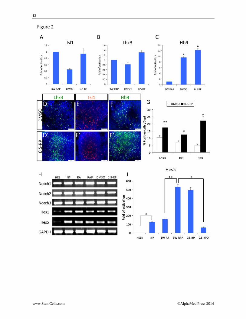
few cells were immunoreactive with either anti‐Olig2 (Fig. 1D) or ‐Ngn2 (Fig. 1E), while at the end of 3 weeks treatment with RA and PUR, 57% of the progenitors were stained positive for Olig2 (Fig. 1F) and 46% were stained positive for Ngn2 (Fig. 1G). 44% of the cells co‐expressed Olig2 and Ngn2 as demonstrated by co‐immunostaining for these markers (Supplemental Fig. 2). The expression of both Olig2 and Ngn2 is indicative of ventral pMN‐like progenitors. Analysis of motor neuron markers expression by qRT‐PCR revealed that Hb9 gene was upregulated by 17 fold upon RA treatment and its expression was slightly in‐creased (24 fold) in pMN‐like progenitor cells upon ad‐dition of purmorphamine (Fig. 1H). Isl1 gene expression was induced by 1.6 fold in the caudal progenitors, and its expression was mildly increased in the pMN‐like pro‐genitors (Fig. 1I). However, co‐immunostaining of the pMN‐like progenitors for Hb9 and either Olig2 or Ngn2 revealed that while the majority of the cells expressed Olig2 or Ngn2, only a few cells were immunoreactive with anti‐Hb9 (Fig. 1J, 1K). This observation suggested that at that stage the cells did not yet differentiate into motor neurons but remained as pMN‐like progenitors that have the potential to differentiate into motor neu‐rons. Differentiation of pMN‐like progenitor cells towards motor neurons In stage 4, the pMN‐like progenitors were induced to differentiate into early motor neurons [Hb9(+), ChAT(‐)] by plating on laminin for 1 week in differentiation me‐dium (0.5‐RP) that was supplemented with halved con‐centrations of RA and PUR (0.5μM and 0.25μM, respec‐tively). The medium was further supplemented with di‐butyril cAMP and the neurotrophic factors BDNF, GDNF and IGF‐1. As a control, the progenitors were differen‐tiated in the presence of DMSO vehicle combined with neurotrophic factors and di‐butyril cAMP. Following 1 week of differentiation, the cells were ana‐lyzed for the expression of motor neuron markers Isl1, Lhx3, and Hb9. The transcriptional levels of Isl1 and Lhx3 were not significantly changed compared with the levels in the pMN progenitors (Fig. 2A, 2B). The expres‐sion level of Hb9 was significantly (p=0.01) increased by 9 fold in control cells treated with DMSO and by 12 fold in cells treated with RA and PUR (Fig. 2C). It appears that plating control cells on laminin promoted MN diffe‐rentiation that was further mildly augmented by RA and PUR. The increase in Hb9 expression may be attributed to its activation by the upregulated expression of Ngn2 in pMN‐like progenitors [31]. Immunofluorescence staining of the differentiating pro‐genitors revealed 17% Lhx3 and 12% Isl1 positive cells in the presence of RA and PUR compared with 10% Lhx3 and 5% Isl1 positive cells in the control (p<0.01) (Fig. 2D‐2D', 2E‐2E' and 2G). Moreover, in the presence of RA and PUR, 22% of the cells were Hb9 positive compared with 5% positive cells in the control (p<0.001) (Fig. 2F‐2F' and 2G). Still at this stage of differentiation 36% and
18% of the cells expressed Olig2 and Ngn2, respectively, (Supplemental Fig. 3). Taken together, these data sug‐gest that RA and PUR promoted the differentiation of the pMN‐like progenitors towards MN fate, albeit a proportion of the cells remained in the pMN‐like proge‐nitor state. The Notch downstream effector Hes5 is upre‐gulated upon differentiation of human em‐bryonic stem cells into motor neurons It is well established that Notch signaling plays a key role in the maintenance of undifferentiated neural pro‐genitors while preventing premature differentiation. To test whether Notch signaling prevents pMN‐like proge‐nitors to differentiate into post‐mitotic MNs, we tested whether Notch signaling is active in the differentiating pMN‐like progenitors. We first assessed the expression of Notch receptors in hESCs and their differentiating progeny throughout the stages of the differentiation protocol. RT‐PCR analysis showed that Notch receptors (Notch1, Notch2, and Notch3) were expressed at similar levels in hESCs as well as in neural precursors, caudal, and pMN‐like progenitors and in the progenitors that were induced to differentiate (Fig. 2H). In contrast, low expression levels of the Notch downstream effectors, Hes1 and Hes5, were observed in hESCs and their ex‐pression was significantly induced upon differentiation into neural precursors, caudal and pMN‐like progeni‐tors, as well as differentiating progenitors (Fig. 2H). Both Hes1 and Hes5 were shown to be expressed in the ventricular zone of the spinal cord in complementary domains. However, in Notch1‐/‐ mice decreased ex‐pression of Hes5 but not Hes1 was observed in the ven‐tral spinal cord [6, 32], suggesting that in the ventral spinal cord Hes5 is a direct target of Notch signaling while Hes1 could be regulated by other signaling path‐ways. Thus, we focused our analysis on Hes5 and tested its expression throughout the four differentiation stages by qRT‐PCR. Hes5 was dramatically induced (100 fold) upon differen‐tiation of hESCs into neural precursors and a similar level of expression was maintained upon further diffe‐rentiation to caudal progenitors. Following RA and PUR treatment and differentiation into pMN‐like progeni‐tors, a four‐fold increase in Hes5 expression was ob‐served. This expression level was maintained after one week of plating the progenitors for differentiation into MNs, in line with the maintenance of pMN‐like progeni‐tor state by a proportion of the cells (Fig. 2I). Collective‐ly, these data suggest that Notch signaling is activated upon differentiation of hESCs towards pMN‐like proge‐nitors and that Hes5 may regulate the maintenance of pMN‐like progenitors and the prevention of their diffe‐rentiation.

www.StemCells.com ©AlphaMed Press 2014
5
Inhibition of Notch signaling enhances the differentiation of pMN‐like progenitor cells into motor neurons To study whether Notch signaling prevents the differen‐tiation of the pMN‐like progenitors into motor neurons we tested whether its inhibition promotes motor neu‐ron differentiation of the pMN‐like progenitors. Notch signaling can be blocked pharmacologically by DAPT, which inhibits �‐secretase activity and prevents Notch receptor cleavage [33, 34]. To test whether DAPT inhi‐bits Notch signaling during the differentiation of the pMN‐like progenitors towards motor neurons, the pro‐genitors were allowed to differentiate as before in the presence of RA and PUR combined DMSO or with DAPT (0.5‐RP or 0.5‐RPD, respectively). In the presence of DAPT, an 8‐fold decrease in Hes5 expression level was observed in the differentiating pMN‐like progenitors, indicating that Notch signaling was inhibited (Fig. 2I). Next, we tested whether the decrease in Hes5 expres‐sion was concomitant with an increased expression of motor neural markers. It was previously shown that specification of pMN progenitors into motor neuron fate is regulated by Ngn2 and Olig2, where progenitors that express both Olig2 and Ngn2 develop as motor neurons while those expressing only Olig2 become oli‐godendrocytes [13, 14, 27, 29]. Neither increase nor decrease in Olig2 expression was detected in DAPT‐treated cells, while a 1.5‐fold increase in Ngn2 expres‐sion was observed (Fig. 3A). Similarly, immunostaining showed comparable levels of Olig2 either in the ab‐sence or presence of DAPT (Fig. 3B‐3B') while the ex‐pression of Ngn2 was increased in the presence of DAPT (Fig. 3C‐3C'). These results suggest that Notch inhibition promoted the differentiation of the pMN‐like progeni‐tors towards the motor neuron fate. In DAPT‐treated cultures of differentiating pMN‐like progenitors, the expression levels of Isl1 and Lhx3 were increased by 3 and 1.7 fold, respectively (Fig. 3D), indi‐cating that inhibition of Notch signaling promoted the differentiation of the pMN‐like progenitors toward mo‐tor neurons. Ngn2 synergizes with Isl1 and Lhx3 to activate the ex‐pression of Hb9, which is specifically expressed in motor neurons [35‐39]. In accordance with the increased ex‐pression of Ngn2, Isl1, and Lhx3, a 3‐fold increase in Hb9 expression level was observed in DAPT‐treated cells (Fig. 3E). Immunostaining showed that in the presence of DAPT, Isl1 and Lhx3 expression was detected in 30% of the differentiating cells compared with 12% and 17%, re‐spectively, in the absence of DAPT (Fig. 3F‐3F', 3G‐3G' and 3J). Hb9 expression was detected in 31% of the cells differentiating in the presence of DAPT compared with 20% in its absence (Fig. 3H‐3H' and 3J). The early motor neurons could be further matured into motor neurons by continuous culturing in suspension. Following 4 to 5 weeks culturing period in the presence of DAPT, the differentiated cells no longer expressed
Hb9 while a substantial percent of the cells were stained positive to choline acetyl transferase (ChAT), which is exclusively expressed in motor neurons of the ventral spinal cord (Fig. 3I‐3I'). Moreover when treatment with DAPT was combined with further lowering the concentrations of RA and PUR (0.25‐RPD medium containing 0.25 μM RA, 0.125 μM PUR and 1 μM DAPT), 40% of the differentiating cells expressed Hb9 compared with 20% in the absence of DAPT (Fig. 3K‐3K'). Therefore, in subsequent experi‐ments we used the 0.25‐RPD medium for differentiation of the pMN‐like progenitors. Collectively, these data suggested that inhibition of Notch signaling in differentiating pMN‐like progenitors resulted in an increased expression of motor neuron markers. When the cells were allowed to differentiate in the presence of DAPT without RA and PUR, only 15% of the differentiating cells expressed Hb9 (data not shown). This finding suggested an instructive role of RA and PUR in directing the differentiation of the progenitors to‐wards motor neurons, which was augmented when Notch signaling was inhibited. Over‐expression of Hes5 inhibits the differen‐tiation of pMN‐like progenitor cells towards motor neurons. Previous studies have identified Hes5 as a Notch target in the developing spinal cord, where it represses pro‐neural genes such as Ngn2 [6, 40, 41]. To identify the downstream targets of Hes5 in the differentiating mo‐tor neurons, we used a lentiviral vector to over‐express Flag‐tagged Hes5 (FL‐Hes5) in pMN‐like progenitors just prior to their differentiation into motor neurons. Upon transduction of pMN‐like progenitors with FL‐Hes5 vec‐tor followed by differentiation in the presence of RA and PUR, an 8‐fold increase in Hes5 expression level was observed compared with control cells that were transduced with a vector that did not include Hes5 (Vector, Fig. 4A). As was expected, FL‐Hes5 transduced expression was not affected by DAPT (Fig. 4A), indicat‐ing a constitutive Hes5 expression in differentiating mo‐tor neurons, similar to constitutively active Notch sig‐naling. To test the effect of constitutive Hes5 expression on motor neuron differentiation we analyzed the expres‐sion levels of motor neuron markers in FL‐Hes5 over‐expressing cells compared with vector‐only expressing cells. Ngn2 was previously shown to be a direct target for Hes5 repression [41]; accordingly in FL‐Hes5 transduced cells, Ngn2 was no longer induced by RA and PUR com‐pared with a 1.9‐fold induction in vector‐only trans‐duced cells (Fig. 4B, 0.25‐RP). Moreover, in FL‐Hes5 transduced cells Ngn2 expression was not induced in the presence of DAPT (Fig. 4B, 0.25‐RPD), indicating a constitutive repression of Ngn2 expression in the diffe‐rentiating pMN‐like progenitors.

www.StemCells.com ©AlphaMed Press 2014
6
In vector‐only transduced cells, RA and PUR induced a 4‐fold increase in Hb9 expression level, while only a 2‐fold increase was observed in FL‐Hes5 transduced cells (Fig. 4C, 0.25‐RP). The expression level of Lhx3 in vector‐only transduced cells was induced by 7.9‐fold in the presence of RA and PUR, while only a 2 fold increase was observed in FL‐Hes5 transduced cells (Fig. 4D, 0.25‐RP). These data suggest that constitutively active Notch signaling interferes with RA and PUR to induce differen‐tiation of pMN‐like progenitors towards motor neuron fate. In the presence of DAPT, FL‐Hes5 over‐expression re‐sulted in a 2.5‐fold reduction in both Hb9 and Lhx3 ex‐pression levels compared with the expression levels in vector‐only transduced cells (Fig. 4C, 4D; 0.25‐RPD). In contrast, in the presence of RA and PUR, Isl1 expres‐sion was similarly induced in both FL‐Hes5 and vector‐only transduced cells (1.7 fold) (Fig. 4E, 0.25‐RP). In the presence of DAPT comparable expression levels of Isl1 (4 fold) were observed in either FL‐Hes5 or vector‐only expressing cells (Fig. 4E, 0.25‐RPD). These results sug‐gest that Hes5 does not directly regulate Isl1. In support with the RNA expression data, immunostain‐ing of FL‐Hes5 transduced cells for the motor neuron markers revealed a dramatic decrease in differentiation of the pMN‐like progenitors into motor neurons. In the absence of DAPT (Fig. 5A‐5D'), as low as 3‐5% of the FL‐Hes5 transduced cells were stained positive for Hb9, Isl1, or Lhx3 compared with 10‐15% positive cells in vector‐only transduced cells (Fig. 5B‐5D' and 5I‐5K, 0.25‐RP). In the presence of DAPT (Fig. 5E‐5H'), 4‐6% of the FL‐Hes5 transduced cells were stained positive compared with 20‐30% positive cells in vector‐only transduced cells (Fig. 5F‐5H' and 5I‐5K, 0.25‐RPD). Co‐immunostaining for FL‐Hes5 and Hb9 showed that Flag expressing cells did not express Hb9 (Supplemental Fig. 4). Interestingly, while the level of Isl1 mRNA transcripts was not reduced by FL‐Hes5 over expression (Fig. 4E), immunostaining analysis showed reduction in the per‐centage of Isl1 positive cells (Fig. 5D', 5H' and 5K) sug‐gesting post‐transcriptional regulation of Isl1 as pre‐viously reported [42, 43]. Our data suggest that constitutive expression of FL‐Hes5 in differentiating pMN‐like progenitors inhibited their differentiation into motor neurons. These results suggest that Notch signaling maintains pMN progenitors and prevents them from differentiation towards post‐mitotic motor neurons. Inhibition of Notch signaling enhances motor neuron fate specified by RA and PUR. Lhx3 expression is not exclusive to motor neurons but is also characteristic to progenitors in the p2 domain of the developing spinal cord. The p2 progenitors express the Chx10 transcription factor in addition to Lhx3 and give rise to V2 interneurons [39]. To assess whether the pMN‐like progenitors differentiate to a mixed popula‐tion of motor neurons and interneurons, the differen‐
tiating cells were co‐stained for Chx10, and either Lhx3 or Hb9. Strikingly, in the presence of RA and PUR, Chx10‐expressing cells were not demonstrated in the absence or presence of DAPT (Fig. 6A‐6A' and 6B‐6B'), while at the same time cells expressing Lhx3 (Fig. 6A‐6A') or Hb9 (Fig. 6B‐6B') were observed as expected. However, when the cells were allowed to differentiate in the presence of DAPT without RA and PUR, a sub‐stantial number of cells co‐expressed Chx10 and Lhx3 (Fig. 6C) while other cells expressed either Hb9 or Chx10 (Fig. 6D), indicating differentiation into a mixed popula‐tion of both inter‐ and motor neurons. In the presence of DMSO vehicle only a low percentage of the cells ex‐pressed Chx10, Lhx3, or Hb9 (Fig. 6C', 6D'). These results suggest that RA and PUR specify the pMN‐like progeni‐tors towards the motor neural fate while at the same time they prevent the differentiation towards the inter‐neural fate. Moreover, inhibition of Notch signaling en‐hances the motor neural‐directed differentiation in‐duced by RA and PUR. DISCUSSION
In this study, we show that Notch signaling has a role in motor neuron differentiation of hESCs. We demonstrate that Notch signaling is active during the differentiation of pMN‐like progenitors inhibiting their maturation into motor neurons. Inhibition of Notch signaling at the stage of pMN‐like progenitors differentiation by DAPT significantly enhances the emergence of motor neu‐rons, while forced expression of the Notch effector Hes5 inhibits motor neuron differentiation. Our data indicate that RA and Shh specify differentiating pMN‐like progenitors to become motor neurons, and that inhibition of Notch signaling provides a permissive sig‐nal for the differentiation of the specified progenitors towards motor neuron fate. In line with previous publications [2, 44] we show that Notch signaling is activated upon early neural differen‐tiation of hESCs as evidenced by induction of expression of its downstream effector, Hes5, concomitant with neuralization. Intriguingly, we demonstrate that the expression level of Hes5 is further increased following specification of early neural progenitors into pMN‐like progenitors by RA and the Shh agonist PUR. Moreover, it remains expressed at comparable levels in cultures of pMN‐like progenitors under conditions favoring diffe‐rentiation into motor neurons. We provide evidence that Notch signaling inhibits the differentiation of the pMN‐like progenitors towards motor neurons. Interfering with Notch signaling during the stage of their differentiation by DAPT leads to a marked decrease in Hes5 expression levels and en‐hances differentiation to motor neurons. Conversely, over‐expression of Hes5 in differentiating pMN‐like progenitors largely inhibits motor neuron differentia‐tion. The role of Notch signaling in the pMN domain of the developing spinal cord was previously studied in various

www.StemCells.com ©AlphaMed Press 2014
7
animal models. Notch signaling was shown to preserve the undifferentiated state of pMN progenitors during the period of motor neuron generation for later diffe‐rentiation to oligodendrocytes [15, 16]. Here we utilized hESCs to efficiently generate pMN‐like progenitors and study, for the first time, the role of Notch signaling dur‐ing their differentiation into motor neurons in a huma‐nized model system in vitro. We found that over‐expression of Hes5 in the differentiating progenitors led to reduced expression levels of Ngn2, while the inhibi‐tion of Notch signaling in these progenitors resulted in an increase in the expression of Ngn2. These observa‐tions are consistent with a previous report of Ngn2 as a direct target for repression by Notch signaling [41]. Our data suggest that the inhibition of Notch signaling upre‐gulates the expression of Ngn2 in differentiating pMN‐like progenitors and promotes their differentiation into motor neurons. Previously it has been reported that inhibition of Notch signaling in mouse embryoid bodies (EBs) derived from ptc1 null mutated ES cells, in which Shh is constitutively active, resulted in precocious loss of ventral neuronal precursors to enhanced neuronal differentiation [45]. However, in these EBs the inhibition of Notch signaling resulted in reduced expression of Olig2 and Nkx2.2, while in our pMN‐like progenitors DAPT had no signifi‐cant effect on Olig2 expression but rather increased the expression of Ngn2. Moreover, treating ptc1 null EBs with Shh and DAPT increased Isl1/2 expression but had no effect on Hb9 and Lhx3 expression. Hence, while precocious neuronal differentiation was observed in the absence of Notch signaling, motor neuron differentia‐tion was not specifically augmented as observed in our results. Species‐specific variation or differences in the methodology used may explain the dissimilar observa‐tions. In a previous study, it was reported that in conditional Notch1 receptor null mice, more V2 interneurons are generated at the expense of earlier‐born motor neurons [6]. Our data indicate that the inhibition of Notch signal‐ing per se in vitro is not sufficient to promote differen‐tiation into a specific neuronal subtype. Inhibition of Notch signaling in the absence of RA and PUR resulted in differentiation of the pMN‐like progenitors to a mixed population of motor neurons expressing Hb9 and V2 interneurons expressing Chx10 with no significantly enhanced differentiation to motor neurons or to V2 interneurons. This observation indicates that inhibition of Notch signaling provides pMN‐like progenitors with a permissive rather than instructive signal for neuronal differentiation, while RA and Shh specify the progeni‐tors towards motor neuron fate and block the differen‐tiation into V2 interneurons. Furthermore, over‐expression of Hes5 blocked the induction of Ngn2 ex‐pression and reduced the activation of motor neuron‐specific genes by RA and PUR. Thus, it is possible that Notch signaling inhibits the differentiation of the pMN‐like progenitors by interfering with RA and PUR in speci‐fication of the motor neuron fate.
We demonstrated that in addition to downregulating Ngn2 expression, over‐expression of Hes5 in the diffe‐rentiating pMN‐like progenitors also reduced the ex‐pression levels of Lhx3 and Hb9 genes. Previously it was shown that Ngn2 is repressed directly by Hes5 [41]. However, whether Hes5 regulates the expression of Hb9 or Lhx3 through direct binding to their regulatory regions is currently unknown. Previously it was shown that the MNE enhancer, which binds both b‐HLH proteins (Ngn2 and NeuroM) and Lim‐HD proteins (Isl1 and Lhx3), regulates the expression of Hb9 gene [35, 36]. The b‐HLH proteins bind the two E‐box consensus elements (CANNTG) included in MNE and synergize with the Lim‐HD proteins to activate Hb9 ex‐pression. Thus, it is possible that Hes5 affects the ex‐pression of Hb9 indirectly by downregulating the ex‐pression of Ngn2. Alternatively, it is possible that Hes5 targets the Hb9 enhancer directly. In support of this assumption, we identified within the MNE sequence an N‐box consensus element (CACNAG) which was reported as a specific binding site for Hes5 [46]. Future analysis may indicate whether Hes5 binds directly to the MNE enhancer and regulates Hb9 expression. Recently it was reported that the expression of human Lhx3 gene is regulated by multiple enhancers, but no N‐box element was identified in these regulatory ele‐ments, arguing against direct binding of Lhx3 regulatory regions by Hes5 [47]. The promotion of motor neuron differentiation by inhi‐bition of Notch signaling may be utilized to enhance the yield of motor neuron differentiation protocols. Based on the inductive effect of RA and Shh signaling, our yield of motor neurons was 20%. Inhibition of Notch signaling doubled the yield of motor neurons. Inhibition of Notch signaling and improving the efficiency of mo‐tor neuron generation in vitro may be highly valuable for disease modeling, and high throughput screening assays for molecules that may have therapeutic value in motor neuron diseases[48]. In conclusion, our data provide evidence for Notch sig‐naling as an important mechanism in controlling the differentiation of pMN progenitor cells into motor neu‐rons. By upregulating the expression levels of Hes5, Notch signaling reduces the expression of Ngn2 as well as Hb9 and Lhx3, which are induced by RA and PUR and are required for directing the pMN progenitor cells to‐wards motor neuron fate. The results argue for a model in which Notch signaling inhibits the differentiation of pMN progenitor cells by blocking the capacity of RA and PUR to induce the expression of key transcription fac‐tors that are required for specification and differentia‐tion of the progenitor cells to motor neurons. AUTHOR CONTRIBUTIONS
E.B.S.: conception and design, collection and assembly of data, data analysis and interpretation, manuscript writing, and final approval of manuscript; E.F.: financial
www.StemCells.com ©AlphaMed Press 2014
8
support, manuscript writing, and final approval of ma‐nuscript; B.E.F.: conception and design, data analysis and interpretation, manuscript writing, and final ap‐proval of manuscript. DISCLOSURE OF POTENTIAL CONFLICT OF INTEREST
Benjamin Reubinoff is a founder, holds shares and is the Chief Scientific Officer (CSO) of CellCure Neuroscience Ltd. The focus of the company is the development of human embryonic stem cells for transplantation thera‐py in neurological and retinal degeneration disorders. The company does not fund the study presented in this
manuscript and currently the company has no interest in the results of the study. ACKNOWLEDGMENTS
This study was funded by a generous donation from Mr. Alfred A. Taubman. It was also supported by The Sidney and Judy Swartz Human Embryonic Stem Cell Research Center fund, and a grant from the US‐Israel Binational Science Founda‐tion.
REFERENCES
1 Bray SJ. Notch signalling: a simple pathway
becomes complex. Nature reviews Molecular cell biology. 2006;7:678‐689.
2 Louvi A, Artavanis‐Tsakonas S. Notch signalling in vertebrate neural development. Nature reviews Neuroscience. 2006;7:93‐102.
3 Peng CY, Yajima H, Burns CE et al. Notch and MAML signaling drives Scl‐dependent interneuron diversity in the spinal cord. Neuron. 2007;53:813‐827.
4 Pierfelice T, Alberi L, Gaiano N. Notch in the vertebrate nervous system: an old dog with new tricks. Neuron. 2011;69:840‐855.
5 Ohtsuka T, Ishibashi M, Gradwohl G et al. Hes1 and Hes5 as notch effectors in mammalian neuronal differentiation. The EMBO journal. 1999;18:2196‐2207.
6 Yang X, Tomita T, Wines‐Samuelson M et al. Notch1 signaling influences v2 interneuron and motor neuron development in the spinal cord. Developmental neuroscience. 2006;28:102‐117.
7 Ericson J, Briscoe J, Rashbass P et al. Graded sonic hedgehog signaling and the specification of cell fate in the ventral neural tube. Cold Spring Harbor symposia on quantitative biology. 1997;62:451‐466.
8 Ericson J, Rashbass P, Schedl A et al. Pax6 controls progenitor cell identity and neuronal fate in response to graded Shh signaling. Cell. 1997;90:169‐180.
9 Ligon KL, Fancy SP, Franklin RJ et al. Olig gene function in CNS development and disease. Glia. 2006;54:1‐10.
10 Rowitch DH, Lu QR, Kessaris N et al. An 'oligarchy' rules neural development. Trends in neurosciences. 2002;25:417‐422.
11 Park HC, Mehta A, Richardson JS et al. olig2 is required for zebrafish primary motor neuron and oligodendrocyte development. Developmental biology. 2002;248:356‐368.
12 Zhou Q, Anderson DJ. The bHLH transcription factors OLIG2 and OLIG1 couple neuronal and glial subtype specification. Cell. 2002;109:61‐73.
13 Lee SK, Lee B, Ruiz EC et al. Olig2 and Ngn2 function in opposition to modulate gene expression in motor neuron progenitor cells. Genes & development. 2005;19:282‐294.
14 Novitch BG, Chen AI, Jessell TM. Coordinate regulation of motor neuron subtype identity and pan‐neuronal properties by the bHLH repressor Olig2. Neuron. 2001;31:773‐789.
15 Park HC, Appel B. Delta‐Notch signaling regulates oligodendrocyte specification. Development. 2003;130:3747‐3755.
16 Rabadan MA, Cayuso J, Le Dreau G et al. Jagged2 controls the generation of motor neuron and oligodendrocyte progenitors in the ventral spinal cord. Cell death and differentiation. 2012;19:209‐219.
17 Marklund U, Hansson EM, Sundstrom E et al. Domain‐specific control of neurogenesis achieved through patterned regulation of Notch ligand expression. Development. 2010;137:437‐445.
18 Lee H, Shamy GA, Elkabetz Y et al. Directed differentiation and transplantation of human embryonic stem cell‐derived motoneurons. Stem Cells. 2007;25:1931‐1939.
19 Li XJ, Du ZW, Zarnowska ED et al. Specification of motoneurons from human embryonic stem cells. Nature biotechnology. 2005;23:215‐221.
20 Li XJ, Hu BY, Jones SA et al. Directed differentiation of ventral spinal progenitors and motor neurons from human embryonic stem cells by small molecules. Stem Cells. 2008;26:886‐893.
21 Wichterle H, Lieberam I, Porter JA et al. Directed differentiation of embryonic stem cells into motor neurons. Cell. 2002;110:385‐397.
22 Placantonakis DG, Tomishima MJ, Lafaille F et al. BAC transgenesis in human embryonic stem cells as a novel tool to define the human neural lineage. Stem Cells. 2009;27:521‐532.
23 Gropp M, Reubinoff B. Lentiviral vector‐mediated gene delivery into human embryonic stem cells. Methods Enzymol. 2006;420:64‐81.
24 Ben‐Dor I, Itsykson P, Goldenberg D et al. Lentiviral vectors harboring a dual‐gene system allow high and homogeneous transgene expression in selected
polyclonal human embryonic stem cells. Mol Ther. 2006;14:255‐267.
25 Cohen MA, Itsykson P, Reubinoff BE. The role of FGF‐signaling in early neural specification of human embryonic stem cells. Developmental biology. 2010;340:450‐458.
26 Jessell TM. Neuronal specification in the spinal cord: inductive signals and transcriptional codes. Nature reviews Genetics. 2000;1:20‐29.
27 Guillemot F. Spatial and temporal specification of neural fates by transcription factor codes. Development. 2007;134:3771‐3780.
28 Ma YC, Song MR, Park JP et al. Regulation of motor neuron specification by phosphorylation of neurogenin 2. Neuron. 2008;58:65‐77.
29 Mizuguchi R, Sugimori M, Takebayashi H et al. Combinatorial roles of olig2 and neurogenin2 in the coordinated induction of pan‐neuronal and subtype‐specific properties of motoneurons. Neuron. 2001;31:757‐771.
30 Ericson J, Morton S, Kawakami A et al. Two critical periods of Sonic Hedgehog signaling required for the specification of motor neuron identity. Cell. 1996;87:661‐673.
31 Lee S, Lee B, Lee JW et al. Retinoid signaling and neurogenin2 function are coupled for the specification of spinal motor neurons through a chromatin modifier CBP. Neuron. 2009;62:641‐654.
32 de la Pompa JL, Wakeham A, Correia KM et al. Conservation of the Notch signalling pathway in mammalian neurogenesis. Development. 1997;124:1139‐1148.
33 Geling A, Steiner H, Willem M et al. A gamma‐secretase inhibitor blocks Notch signaling in vivo and causes a severe neurogenic phenotype in zebrafish. EMBO reports. 2002;3:688‐694.
34 Micchelli CA, Esler WP, Kimberly WT et al. Gamma‐secretase/presenilin inhibitors for Alzheimer's disease phenocopy Notch mutations in Drosophila. FASEB journal : official publication of the Federation of American Societies for Experimental Biology. 2003;17:79‐81.
35 Lee SK, Jurata LW, Funahashi J et al. Analysis of embryonic motoneuron gene regulation: derepression of general activators function in concert with

www.StemCells.com ©AlphaMed Press 2014
9
enhancer factors. Development. 2004;131:3295‐3306.
36 Lee SK, Pfaff SL. Synchronization of neurogenesis and motor neuron specification by direct coupling of bHLH and homeodomain transcription factors. Neuron. 2003;38:731‐745.
37 Jurata LW, Thomas JB, Pfaff SL. Transcriptional mechanisms in the development of motor control. Current opinion in neurobiology. 2000;10:72‐79.
38 Thaler J, Harrison K, Sharma K et al. Active suppression of interneuron programs within developing motor neurons revealed by analysis of homeodomain factor HB9. Neuron. 1999;23:675‐687.
39 Thaler JP, Lee SK, Jurata LW et al. LIM factor Lhx3 contributes to the specification of motor neuron and interneuron identity through cell‐type‐specific protein‐protein interactions. Cell. 2002;110:237‐249.
40 Genethliou N, Panayiotou E, Panayi H et al. SOX1 links the function of neural patterning and Notch signalling in the ventral spinal cord during the neuron‐glial fate switch. Biochemical and
biophysical research communications. 2009;390:1114‐1120.
41 Holmberg J, Hansson E, Malewicz M et al. SoxB1 transcription factors and Notch signaling use distinct mechanisms to regulate proneural gene function and neural progenitor differentiation. Development. 2008;135:1843‐1851.
42 Wang J, Greene SB, Bonilla‐Claudio M et al. Bmp signaling regulates myocardial differentiation from cardiac progenitors through a MicroRNA‐mediated mechanism. Developmental cell. 2010;19:903‐912.
43 Witman N, Heigwer J, Thaler B et al. miR‐128 regulates non‐myocyte hyperplasia, deposition of extracellular matrix and Islet1 expression during newt cardiac regeneration. Developmental biology. 2013;383:253‐263.
44 Yoon K, Gaiano N. Notch signaling in the mammalian central nervous system: insights from mouse mutants. Nature neuroscience. 2005;8:709‐715.
45 Crawford TQ, Roelink H. The notch response inhibitor DAPT enhances neuronal differentiation in embryonic
stem cell‐derived embryoid bodies independently of sonic hedgehog signaling. Developmental dynamics : an official publication of the American Association of Anatomists. 2007;236:886‐892.
46 Akazawa C, Sasai Y, Nakanishi S et al. Molecular characterization of a rat negative regulator with a basic helix‐loop‐helix structure predominantly expressed in the developing nervous system. The Journal of biological chemistry. 1992;267:21879‐21885.
47 Mullen RD, Park S, Rhodes SJ. A distal modular enhancer complex acts to control pituitary‐ and nervous system‐specific expression of the LHX3 regulatory gene. Mol Endocrinol. 2012;26:308‐319.
48 Yang YM, Gupta SK, Kim KJ et al. A small molecule screen in stem‐cell‐derived motor neurons identifies a kinase inhibitor as a candidate therapeutic for ALS. Cell stem cell. 2013;12:713‐726.
See www.StemCells.com for supporting information available online. STEM
CELLS ; 00:000–000

www.StemCells.com ©AlphaMed Press 2014
10
Figure 1. Caudalization and ventralization of neural progenitor cells. (A) Hox genes expression in caudal progenitors grown for 1 week in the presence of 1μM RA. RT‐PCR analysis indi‐cated induction of Hox genes in progenitors treated with RA (RA) compared with their non treated counterparts (NT). GAPDH levels were used as quantitative reference. (B‐C) Expression of Olig2 and Ngn2 in caudal and ventral progenitor cells. Neural progenitors were grown for 1 week in the presence of 1 μM RA followed by 3 weeks treatment with 1 μM RA and 0.5 μM PUR to generate first caudal progenitors (1W RA) and then ventral pMN‐like progenitors (3W RAP). QTR‐ PCR analysis of the expression Olig2 (B) and Ngn2 (C) genes in caudal (1W RA) and ventral (3W RAP) progenitor cells relative to the expression level in neural progenitor cells (NT). Expression level in neural progenitor cells was set as 1. Fold activation over control is derived from three experiments. Data are presented as mean + SD. * p < 0.01 by Student's t‐test. (D‐G) Immunofluoroscence images demonstrating the expression of Olig2 (D and F) and Ngn2 (E and G) in caudal (1W RA) and ventral (3W RAP) progenitor cells. Blue indicates DAPI stained nuclei. Scale bars = 100 μm. (H‐K) Expression of motor neurons markers in caudal and ventral pMN‐like progenitor cells. (H‐I) QRT‐PCR analysis of the expression of Hb9 (H) and Isl1 (I) genes in caudal (1W RA) and ventral (3W RAP) progeni‐tor cells relative to the expression level in neural progenitor cells (NT). Expression level in neural progenitor cells was set as 1. Fold activation over control is derived from three experiments. Data are presented as mean + SD. ** p < 0.05 by Student's t‐test. (J‐K) Immunofluoroscence analysis of ventral progenitor cells (3W RAP) stained with antibodies to Hb9 (green) and Olig2 (red, J) or Ngn2 (red, K). Blue indicates DAPI stained nuclei. Scale bars = 100 μm.

www.StemCells.com ©AlphaMed Press 2014
11
Figure 2. Expression of motor neurons markers upon differentiation of ventral pMN‐like progenitor cells. Ventral pMN‐like progenitor cells (3W RAP) were differentiated for 1 week in the presence of 0.5 μM RA and 0.25 μM PUR (0.5‐RP) or in the presence of DMSO vehicle control. (A‐C) QRT‐PCR analysis of the expression of Isl1 (A), Lhx3 (B) and Hb9 (C) genes in progenitor cells differentiating in the absence (DMSO) or presence of RA and PUR (0.5‐RP) relative to the expression levels in ventral pMN‐like progeni‐tor cells (3W RAP). Expression level in pMN‐like progenitor cells was set as 1. Fold activation over control is derived from three experiments. Data are presented as mean + SD. * p = 0.01 by Student's t‐test. (D‐G) Immunofluoroscence images demonstrating the expression of Lhx3 (D‐D'), Isl1 (E‐E'), and Hb9 (F‐F') in cells dif‐ferentiated in the presence of DMSO (D‐F) or RA and PUR (0.5‐RP, D'‐F'). Blue indicates DAPI counter stained nuclei. Scale bars = 100 μm. (G) Quantification of the percentage of Lhx3, Isl1 and Hb9 positive cells following differentiation in the presence of DMSO or 0.5‐RP. Data derived from three experiments are represented as mean + SD. * p < 0.01, ** p < 0.05 by Stu‐dent's t‐test. (H‐I) Notch signaling is active along the differentiation of neural progenitor cells into motor neurons. (H) RT‐PCR analysis of the expression of Notch receptors and Hes genes in undifferentiated hESC, neural progenitor cells (NP), caudal progenitor cells (RA), ventral pMN‐like progenitor cells (RAP), and progenitors differentiating in the absence (DMSO) or the presence of RA and PUR (0.5‐RP). GAPDH levels were used as quantitative reference. (I) QRT‐PCR analysis of the expression of Hes5 gene in undifferentiated hESCs, neural progenitor cells (NP), caudal progenitor cells (1W RA), ventral pMN‐like progenitor cells (3W RAP), progenitors differentiating in the presence of RA and PUR (0.5‐RP) and progenitors differentiating in the presence of RA, PUR and DAPT (0.5‐RPD). Expression level in undifferentiated hESCs was set as 1. Fold activation over undifferentiated hESCs is derived from four experiments. Data are presented as mean + SD. * p < 0.01, ** p < 0.05 by Student's t‐test.
www.StemCells.com ©AlphaMed Press 2014
12

www.StemCells.com ©AlphaMed Press 2014
13
Figure 3. Inhibition of Notch signaling enhances the differentiation of pMN‐like progenitor cells into motor neurons. pMN‐like progenitor cells were differentiated for 1 week in the presence of 0.5 μM RA and 0.25 μM PUR combined with DMSO vehicle (0.5‐RP) or with DAPT (0.5‐RPD). (A) QRT‐PCR analysis of the expression of Olig2 and Ngn2 genes in pMN‐like progenitor cells differentiating in the presence DAPT (0.5‐RPD; black bars) relative to the expression levels in progenitor cells differentiating in the absence of DAPT (0.5‐RP, white bars). Expression levels in progenitors differentiating without DAPT were set as 1. Fold activa‐tion is derived from four experiments. Data are presented as mean + SD. * p = 0.01, ** p < 0.05 by Student's t‐test. (B‐C) Immunofluorescence analysis of Olig2 (B) and Ngn2 (C) expression in progenitor cells differentiated in the ab‐sence (0.5‐RP, B‐C) or presence of DAPT (0.5‐RPD, B'‐C'). Blue indicates DAPI stained nuclei. Scale bars = 100 μm. (D‐E) QRT‐PCR analysis of the expression of Isl1, Lhx3 (D) and Hb9 (E) genes in progenitor cells differentiated in the presence of RA and PUR combined with DMSO vechile (0.5‐RP; white bars) or with DAPT (0.5‐RPD; black bars) rela‐tive to the expression in pMN‐like progenitor cells (3W RAP; blue bars). Expression level in pMN‐like progenitors was set as 1. Fold activation is derived from four experiments. Data are presented as mean + SD. * p < 0.01, ** p < 0.05 by Student's t‐test. (F‐H') Immunofluorescence images demonstrating the expression of Isl1 (F‐F'), Lhx3 (G‐G'), and Hb9 (H‐H') in cells differentiated in the absence (0.5‐RP, F‐H) or in the presence of DAPT (0.5‐RPD, F'‐H'). Blue indicates DAPI stained nuclei. Scale bars = 100 μm. (I‐I') Immunofluorescence images demonstrating the expression of ChAT in cells differentiating for 5 weeks in the presence of RA and PUR combined with DMSO vehicle (0.5‐RP, I) or with DAPT (0. 5‐RPD, I'). Blue indicates DAPI stained nuclei. Scale bars = 100 μm. (J) Quantification of the percentage of cells stained positive for Isl1, Lhx3, and Hb9 following differentiation in the presence of 0.5 μM RA and 0.25 μM PUR with DMSO vehicle (0.5‐RP; white bars) or with DAPT (0.5‐RPD; black bars). Data derived from three experiments are presented as mean + SD. * p < 0.01, ** p < 0.05 by Student's t‐test. (K‐K') Expression of Hb9 in cells differentiated in the presence of 0.25 μM RA and 0.125 μM PUR with DMSO vehicle (0.25‐RP, K) or with DAPT (0.25‐RPD, K'). Blue indicates DAPI stained nuclei. Scale bars = 100 μm.

www.StemCells.com ©AlphaMed Press 2014
14

www.StemCells.com ©AlphaMed Press 2014
15
Figure 4. Over‐expression of FL‐Hes5 reduces the differentiation of pMN‐like progenitor cells into motor neurons. pMN‐like progenitor cells infected with FL‐Hes5 lentiviral vector (FL‐Hes5) or with an empty lentiviral vector (Vector) were differentiated for 1 week in the presence of DMSO vehicle (DMSO) or 0.25 μM RA and 0.125 μM PUR combined with DMSO vehicle (0.25‐RP) or with DAPT (0.25‐RPD). (A‐E) QRT‐PCR analysis of the expression of Hes5 (A), Ngn2 (B), Hb9 (C), Lhx3 (D) and Isl1 (E) genes in vector‐only (Vector) and FL‐Hes5 (FL‐Hes5) infected progenitor cells differentiating in the presence of DMSO vehicle (DMSO; white bars) or in the presence of RA and PUR combined with DMSO vehicle (0.25‐RP; blue bars) or with DAPT (0.25‐RPD; black bars). Expression level in vector‐only infected cells differentiating in the presence DMSO vehicle (DMSO) was set as 1. Fold activation is derived from four experiments. Data are presented as mean + SEM. * p < 0.01, ** p < 0.05 by Student's t‐test.

www.StemCells.com ©AlphaMed Press 2014
16
Figure 5. Immunostaining showing the expression of FL‐Hes5 (A‐A' and E‐E'), Hb9 (B‐B' and F‐F'), Lhx3 (C‐C' and G‐G'), and Isl1 (D‐D' and H‐H') in vector‐only (Vector, A‐H) and FL‐Hes5 (FL‐Hes5, A'‐H') infected progenitor cells, differen‐tiating in the presence of RA and PUR combined with DMSO vehicle (0.25‐RP, A‐D') or with DAPT (0.25‐RPD, E‐H').Blue indicates DAPI stained nuclei. Scale bars = 200 μm. (I‐K) Quantification of the percentage of cells stained positive for Hb9 (I), Lhx3 (J), and Isl1 (K) in vector‐only (white bars) and FL‐Hes5 (black bars) infected progenitor cells following differentiation in the presence of RA and PUR com‐bined with DMSO (0.25‐RP) or with DAPT (0.25‐RPD). Data derived from three experiments are presented as mean + SD. * p < 0.01, ** p < 0.05 by Student's t‐test.

www.StemCells.com ©AlphaMed Press 2014
17
Figure 6. Retinoic acid and purmorphamine specify the differentiation of pMN‐like progenitor cells towards motor neurons rather than interneurons. Immunostaining showing the expression of Chx10 (red) and Lhx3 (green) (A‐A' and C‐C') or Chx10 (red) and Hb9 (green) (B‐B' and D‐D') in pMN‐like progenitor cells differentiating for 1 week in the presence of RA and PUR com‐bined with DMSO vehicle (0.25‐RP, A‐B) or with DAPT (0.25‐RPD, A'‐B') or in the presence of DAPT only (DAPT, C‐D) or DMSO vehicle (DMSO, C'‐D'). Blue indicates DAPI stained nuclei. Scale bars = 100 μm.