Changes in both neuron intrinsic properties and ...
38
Accepted manuscripts are peer-reviewed but have not been through the copyediting, formatting, or proofreading process. Copyright © 2019 the authors This Accepted Manuscript has not been copyedited and formatted. The final version may differ from this version. Research Articles: Systems/Circuits Changes in both neuron intrinsic properties and neurotransmission are needed to drive the increase in GnRH neuron firing rate during estradiol positive feedback Caroline E. Adams 1 , R. Anthony DeFazio 1 , Catherine A. Christian 2 , Lorin S. Milescu 3 , Santiago Schnell 1,4 and Suzanne M. Moenter 1,5,6 1 Department of Molecular and Integrative Physiology, University of Michigan, Ann Arbor, MI, 48109 2 Department of Molecular and Integrative Physiology, University of Illinois at Urbana-Champaign, Urbana, IL, 61801 3 Division of Biological Sciences, University of Missouri, Columbia, MO, 65211 4 Departments of Computational Medicine and Bioinformatics, University of Michigan, Ann Arbor, MI, 48109 5 Obstetrics and Gynecology, University of Michigan, Ann Arbor, MI, 48109 6 Internal Medicine, University of Michigan, Ann Arbor, MI, 48109 https://doi.org/10.1523/JNEUROSCI.2880-18.2019 Received: 7 November 2018 Revised: 27 December 2018 Accepted: 10 January 2019 Published: 17 January 2019 Author contributions: C.E.A., R.A.D., S.S., and S.M.M. designed research; C.E.A. performed research; C.E.A., C.A.C., and S.M.M. analyzed data; C.E.A. wrote the first draft of the paper; C.E.A. and S.M.M. wrote the paper; R.A.D., C.A.C., L.M., and S.S. edited the paper; L.M. contributed unpublished reagents/analytic tools. Conflict of Interest: The authors declare no competing financial interests. We thank Elizabeth Wagenmaker and Laura Burger for expert technical assistance and James L. Kenyon, University of Nevada, Reno, for the Excel spreadsheet used to calculate junction potentials. Corresponding author: Suzanne M. Moenter, Department of Molecular and Integrative Physiology, University of Michigan, 7725 Med Sci II, 1137 E. Catherine St, Ann Arbor, MI 48109-5622; 734-647-1750 (voice), 734-936-8813 (fax) [email protected] Cite as: J. Neurosci 2019; 10.1523/JNEUROSCI.2880-18.2019 Alerts: Sign up at www.jneurosci.org/alerts to receive customized email alerts when the fully formatted version of this article is published.
-
Upload
khangminh22 -
Category
Documents
-
view
0 -
download
0
Transcript of Changes in both neuron intrinsic properties and ...
Accepted manuscripts are peer-reviewed but have not been through the copyediting, formatting, or proofreadingprocess.
Copyright © 2019 the authors
This Accepted Manuscript has not been copyedited and formatted. The final version may differ from this version.
Research Articles: Systems/Circuits
Changes in both neuron intrinsic properties and neurotransmission areneeded to drive the increase in GnRH neuron firing rate during estradiolpositive feedback
Caroline E. Adams1, R. Anthony DeFazio1, Catherine A. Christian2, Lorin S. Milescu3, Santiago Schnell1,4
and Suzanne M. Moenter1,5,6
1Department of Molecular and Integrative Physiology, University of Michigan, Ann Arbor, MI, 481092Department of Molecular and Integrative Physiology, University of Illinois at Urbana-Champaign, Urbana, IL,618013Division of Biological Sciences, University of Missouri, Columbia, MO, 652114Departments of Computational Medicine and Bioinformatics, University of Michigan, Ann Arbor, MI, 481095Obstetrics and Gynecology, University of Michigan, Ann Arbor, MI, 481096Internal Medicine, University of Michigan, Ann Arbor, MI, 48109
https://doi.org/10.1523/JNEUROSCI.2880-18.2019
Received: 7 November 2018
Revised: 27 December 2018
Accepted: 10 January 2019
Published: 17 January 2019
Author contributions: C.E.A., R.A.D., S.S., and S.M.M. designed research; C.E.A. performed research; C.E.A.,C.A.C., and S.M.M. analyzed data; C.E.A. wrote the first draft of the paper; C.E.A. and S.M.M. wrote the paper;R.A.D., C.A.C., L.M., and S.S. edited the paper; L.M. contributed unpublished reagents/analytic tools.
Conflict of Interest: The authors declare no competing financial interests.
We thank Elizabeth Wagenmaker and Laura Burger for expert technical assistance and James L. Kenyon,University of Nevada, Reno, for the Excel spreadsheet used to calculate junction potentials.
Corresponding author: Suzanne M. Moenter, Department of Molecular and Integrative Physiology, Universityof Michigan, 7725 Med Sci II, 1137 E. Catherine St, Ann Arbor, MI 48109-5622; 734-647-1750 (voice),734-936-8813 (fax) [email protected]
Cite as: J. Neurosci 2019; 10.1523/JNEUROSCI.2880-18.2019
Alerts: Sign up at www.jneurosci.org/alerts to receive customized email alerts when the fully formatted versionof this article is published.

Journal section: Systems/Circuits 1
Changes in both neuron intrinsic properties and neurotransmission are needed to drive 2
the increase in GnRH neuron firing rate during estradiol positive feedback 3
Caroline E. Adams1, R. Anthony DeFazio1, Catherine A. Christian2, Lorin S. Milescu3, Santiago 4 Schnell1,4, and Suzanne M. Moenter1, 5, 6 5 Department of Molecular and Integrative Physiology, University of Michigan, Ann Arbor, MI, 6 481091, Department of Molecular and Integrative Physiology, University of Illinois at Urbana-7 Champaign, Urbana, IL, 618012, Division of Biological Sciences, University of Missouri, 8 Columbia, MO, 652113, Departments of Computational Medicine and Bioinformatics4, Obstetrics 9 and Gynecology5, and Internal Medicine6, University of Michigan, Ann Arbor, MI, 48109 10 Abbreviated title: Properties needed for estradiol positive feedback 11 Corresponding author: Suzanne M. Moenter, Department of Molecular and Integrative 12
Physiology, University of Michigan, 7725 Med Sci II, 1137 E. Catherine St, Ann Arbor, MI 13
48109-5622; 734-647-1750 (voice), 734-936-8813 (fax) [email protected] 14
Number of pages: 31 15 Number of figures: 6 16 Number of tables: 6 17 Number of words: abstract 246, introduction 495, discussion 1497. 18
Abbreviations: AHP, afterhyperpolarization potential; ERα, estrogen receptor alpha; GFP, 19
green-fluorescent protein; GnRH, gonadotropin-releasing hormone; KW, Kruskal-Wallis; LH, 20
luteinizing hormone; OVX, ovariectomized; OVX+E, ovariectomized and estradiol replaced; 21
FWHM, full width at half maximum; V1/2inact; membrane potential at which ½ of current is 22
inactivated; MCMC, Markov chain Monte Carlo 23
Acknowledgements: We thank Elizabeth Wagenmaker and Laura Burger for expert technical 24
assistance and James L. Kenyon, University of Nevada, Reno, for the Excel spreadsheet used 25
to calculate junction potentials. 26
Conflict of Interest: The authors have nothing to disclose. 27
Grant support: Supported by National Institute of Health/Eunice Kennedy Shriver National 28
Institute of Child Health and Human Development R01 HD41469 (SMM), R01 NS105825 and 29
R03 NS103029 (CAC). CEA was supported by T32 GM007863, T32 HD079342 and F30 30
HD085721. L.S.M was partially supported by American Heart Association grant 31
13SDG16990083 32

2
Abstract 33
Central output of gonadotropin-releasing hormone (GnRH) neurons controls fertility and is 34
sculpted by sex-steroid feedback. A switch of estradiol action from negative to positive feedback 35
initiates a surge of GnRH release, culminating in ovulation. In ovariectomized mice bearing 36
constant-release estradiol implants (OVX+E), GnRH neuron firing is suppressed in the morning 37
(AM) by negative feedback and activated in the afternoon (PM) by positive feedback; no time-of-38
day dependent changes occur in OVX mice. In this daily surge model, GnRH neuron intrinsic 39
properties are shifted to favor increased firing during positive feedback. It is unclear if this shift 40
and the observed concomitant increase in GABAergic transmission, which typically excites 41
GnRH neurons, are independently sufficient for increasing GnRH neuron firing rate during 42
positive feedback or if both are needed. To test this, we used dynamic clamp to inject selected 43
previously recorded trains of GABAergic postsynaptic conductances (PSgs) collected during the 44
different feedback states of the daily surge model into GnRH neurons from OVX, OVX+E AM, 45
and OVX+E PM mice. PSg trains mimicking positive feedback initiated more action potentials in 46
cells from OVX+E PM mice than negative feedback or OVX (open feedback loop) trains in all 47
three animal models, but the positive-feedback train was most effective when applied to cells 48
during positive feedback. In silico studies of model GnRH neurons in which >1000 PSg trains 49
were tested exhibited the same results. These observations support the hypothesis that GnRH 50
neurons integrate fast-synaptic and intrinsic changes to increase firing rates during positive 51
feedback. 52
Significance statement 53
Infertility affects 15-20% of couples; failure to ovulate is a common cause. Understanding how 54
the brain controls ovulation is critical for new developments in both infertility treatment and 55
contraception. Ovarian estradiol alters both the intrinsic properties of GnRH neurons and 56
synaptic inputs to these cells coincident with production of sustained GnRH release that 57

3
ultimately triggers ovulation. We demonstrate here using dynamic clamp and mathematical 58
modeling that estradiol-induced shifts in synaptic transmission alone can increase firing output, 59
but that the intrinsic properties of GnRH neurons during positive feedback further poise these 60
cells for increased response to higher frequency synaptic transmission. These data suggest that 61
GnRH neurons integrate fast-synaptic and intrinsic changes to increase firing rates during the 62
preovulatory GnRH surge. 63
Introduction 64
GnRH neurons form the final common pathway for the central control for reproduction. GnRH 65
initiates the release of luteinizing hormone (LH) and follicle-stimulating hormone from the 66
pituitary. Gonadotropins activate follicle maturation and steroidogenesis in the ovary. For most 67
of the estrous cycle, estradiol negative feedback suppresses GnRH/LH release. At the end of 68
the follicular phase (proestrus in rodents), rising levels of estradiol switch from suppressing 69
GnRH/LH to positive feedback action that induces a continuous surge of release that initiates an 70
LH surge that triggers ovulation (Sarkar et al., 1976; Moenter et al., 1991). 71
In ovariectomized rodents implanted with estradiol capsules (OVX+E), GnRH/LH surges can be 72
induced on a daily basis (Norman et al., 1973; Legan and Karsch, 1975; Christian et al., 2005). 73
Release of GnRH and LH, as well as GnRH neuron firing rate, are suppressed in the morning 74
(negative feedback) and elevated in the afternoon (positive feedback) (Christian et al., 2005; 75
Glanowska et al., 2012). No time-of-day dependent shift in GnRH neuron firing rate is observed 76
in OVX mice not treated with estradiol. Work using this daily surge paradigm, other estradiol-77
induced surge paradigms and studies during different stages of the natural estrous cycle have 78
identified multiple estradiol and time-of-day dependent changes to GnRH neuron intrinsic 79
properties and fast-synaptic transmission that may drive an increase in firing rate during positive 80
feedback (Chu and Moenter, 2006; Christian and Moenter, 2007; Zhang et al., 2007; Christian 81
et al., 2009; Zhang et al., 2009; Sun et al., 2010; Pielecka-Fortuna et al., 2011; Adams et al., 82
4
2018a). Changes to voltage-gated sodium, potassium, and calcium channel properties 83
culminate in an increase in GnRH neuron excitability during positive feedback and may increase 84
both spontaneous firing and responsiveness to fast-synaptic inputs to help drive an increase in 85
GnRH firing rate at this time (Adams et al., 2018b). In addition to these changes in intrinsic 86
properties, GABA postsynaptic current (PSC) frequency and amplitude are suppressed during 87
negative feedback and increased during positive feedback in both the daily surge model and 88
during the estrous cycle (Christian and Moenter, 2007; Adams et al., 2018a); activation of 89
GABAA receptors in these cells can be excitatory (DeFazio et al., 2002). 90
Whether changes in GABAergic fast-synaptic transmission, intrinsic properties or both are 91
necessary or sufficient for increasing GnRH neuron firing rate during positive feedback is not 92
known. We hypothesized that time-of-day and estradiol-dependent changes in intrinsic 93
properties renders GnRH neurons more responsive to GABA postsynaptic currents during 94
positive feedback compared to negative feedback and the open-loop OVX condition. To test our 95
hypothesis, we used the daily surge paradigm to examine GnRH neuron response to trains of 96
GABA postsynaptic conductances, and used dynamic clamp to introduce synaptic 97
conductances to evoke postsynaptic potentials and measure membrane response. Trains of 98
conductances were modeled from representative patterns from negative and positive feedback 99
and the open-loop OVX condition (Christian and Moenter, 2007). We also used GnRH neuron 100
models of two feedback conditions to test >1000 previously recorded trains of postsynaptic 101
currents in silico. 102
Materials and Methods 103
All chemicals were purchased from Sigma-Aldrich, unless noted. 104
Animals. Transgenic mice expressing green fluorescent protein (GFP) under the control of the 105
GnRH promoter (Tg(Gnrh1-EGFP)51Sumo MGI:6158457) (GnRH-GFP mice) were used (Suter 106

5
et al., 2000). Mice were housed in a vivarium kept at 21-23 ºC on a 14-h light:10-h dark cycle 107
with lights off at 6 P.M. (eastern standard time). Teklad 2916 chow (Envigo) and water were 108
available ad libitum. Adult females aged 58-170 days were randomly selected from our colony. 109
Ovariectomy was performed under isoflurane (VetOne) anesthesia. At the time of OVX, mice 110
were randomized to either receive a Silastic (Dow Corning) capsule containing 0.625 μg 17β-111
estradiol suspended in sesame oil (OVX+E) or not be treated further (OVX). Bupivacaine 112
(0.25%, APP Pharmaceuticals) was applied to surgical sites to reduce postoperative pain and 113
distress. Electrophysiologic experiments were performed 2-3 days after surgery and estradiol 114
status was confirmed by measurements of uterine mass of control mice (OVX, n=7, 49.1 ± 3.3 115
mg; OVX+E, n=12, 174.1 ± 4.8 mg; two-tailed unpaired Student’s t-test, t(17)=18.4, p<0.001). It is 116
important to point out that this daily surge model does not recapitulate the pattern of estradiol 117
during the cycle. It does, however, effectively induce both negative and positive feedback on LH 118
release in vivo and GnRH neuron activity in the brain slice relative to measurements in OVX 119
mice (Christian et al., 2005). This separates two variables, time of day and circulating estradiol 120
level, known to contribute to the generation of the LH surge in mice and other rodents and which 121
were the targets of the present investigations. Firing rates, GnRH neuron excitability and 122
GABAergic transmission during the daily estradiol-induced surge are similar to those during the 123
proestrous surge of the estrous cycle (Silveira et al., 2016; Adams et al., 2018a). 124
Brain Slice Preparation. All solutions were bubbled with 95% O2/5% CO2 throughout the 125
experiments and for at least 15 min before exposure to tissue. Brain slices were prepared either 126
from 7.5-8.5 h before lights out (AM recordings) or 1-2 h before lights out (PM recordings). The 127
brain was rapidly removed and placed in ice-cold sucrose saline solution containing the 128
following (in mM): 250 sucrose, 3.5 KCl, 26 NaHCO3, 10 D-glucose, 1.25 Na2HPO4, 1.2 MgSO4, 129
and 3.8 MgCl2, at pH 7.6 and 345 mOsm. Coronal (300 μm) slices were cut with a VT1200S 130
Microtome (Leica Biosystems). Slices were incubated in a 1:1 mixture of sucrose saline and 131

6
artificial CSF (ACSF) containing (in mM) 135 NaCl, 3.5 KCl, 26 NaHCO3, 10 D-glucose, 1.25 132
Na2HPO4, 1.2 MgSO4, and 2.5 CaCl2, at pH 7.4 and 305 mOsm, for 30 min at room temperature 133
(21 to 23°C). Slices were then transferred to 100% ACSF at room temperature for 0.5-5 h 134
before recording. 135
Data Acquisition. During recording, slices through the preoptic area and anterior hypothalamus, 136
which contain the majority of GnRH neuron somata, were placed into a chamber continuously 137
perfused with ACSF at a rate of 2 ml/min with oxygenated ACSF heated to 29.5-31.5°C with an 138
inline-heating unit (Warner Instruments). GFP-positive cells were visualized with a combination 139
of infrared differential interference contrast and fluorescence microscopy on an Olympus 140
BX51WI microscope. Recording pipettes were pulled from borosilicate glass capillaries (1.65-141
mm OD x 1.12-mm ID; World Precision Instruments) using a Flaming/Brown P-97 unit (Sutter 142
Instrument Company). Pipettes measured 2-4.5 MΩ when filled with (in mM): 125 K gluconate, 143
20 KCl, 10 HEPES, 5 EGTA, 0.1 CaCl2, 4 MgATP, and 0.4 NaGTP, 300 mOsm, pH 7.2 with 144
NaOH. Pipettes were wrapped with Parafilm (Bemis) to reduce capacitive transients; remaining 145
transients were electronically cancelled. Pipettes were placed in contact with a GFP-positive 146
neuron using an MP-285 micromanipulator (Sutter Instrument Company). All potentials reported 147
were corrected online for a liquid junction potential of −14.2 mV (Barry, 1994). Recordings were 148
made with an EPC-10 dual patch-clamp amplifier (HEKA Elektronik) and a Macintosh computer 149
running Patchmaster software (HEKA Elektronik). Experiments were analyzed offline using 150
custom software (DeFazio and Moenter, 2002; DeFazio et al., 2014) written in IgorPro 151
(Wavemetrics). 152
Whole-cell patch-clamp. After achieving a >1 GΩ seal and the whole-cell configuration, 153
membrane potential was held at -60 mV between protocols. Series resistance (Rs), input 154
resistance (Rin), membrane capacitance (Cm), and holding current (Ihold) were measured every 2-155
3 min using a 5 mV hyperpolarizing step from −60 mV (mean of 20 repeats, 20 ms duration, 156
7
sampled at 100 kHz). Only recordings with a Rin of >500 MΩ, Ihold of −30 to 25 pA, stable Rs of 157
<20 MΩ, and a stable Cm between 10 and 23 pF were used for analysis. 158
In current-clamp, direct current (<35 pA, -9.0±2.1 pA, n=34) was adjusted to keep cells within 3 159
mV of -59 mV. Membrane potential was sampled at 20 kHz and filtered at 6.7 kHz. Bridge 160
balance (95%) was used for most cells; for a few cells, bridge balance was not used but results 161
were similar. In all current-clamp and dynamic-clamp recordings, ACSF contained 100 μM 162
picrotoxin, 20 μM D-APV, and 20 μM CNQX to block ionotropic GABA and glutamate receptors. 163
To confirm estradiol and diurnal changes in GnRH neuron excitability, cells were injected with 164
current from 0-30 pA (500 ms, 10 pA steps). Confirming previous work (Adams et al., 2018b), 165
GnRH neurons from OVX+E PM mice fired more spikes in response to 500 ms current steps 166
from 20-30 pA compared to all other groups (p<0.05, two-way repeated-measures 167
ANOVA/Bonferroni, data not shown). 168
Dynamic clamp. Dynamic clamp was implemented using the freely available QuB software 169
(MLab edition, https://milesculabs.biology.missouri.edu/QuB.html) (Milescu et al., 2008) running 170
on a separate computer (Marquis C733-T with dual Intel Xeon E5-2667v2 Ivy Bridge-EP 171
3.3GHz, 8-core processors, 8x16Gb DDR3-1866 SDRAM, ASLabs) equipped for data 172
acquisition system (PCIe-6361 Multifunction DAQ card with a BNC interface, National 173
Instruments). The digital outputs of the EPC10 were used to trigger events in the dynamic clamp 174
system. The input to the dynamic clamp system is the membrane potential of the cell (Vm), read 175
from the EPC10 patch-clamp amplifier in current-clamp mode; the output of the dynamic clamp 176
is the computed command current (Idc), which drives the current command input of the EPC10. 177
This input-output cycle is repeated at ~50 kHz. 178
To implement the synaptic conductance model in dynamic clamp, Idc was calculated from the 179
postsynaptic conductance (gsim) and the linear driving force: Idc = gsim * (Vm – EGABA ), where 180
EGABA is the reversal potential for current through the GABAA receptor channel (EGABA = -181
8
36.5mV) (DeFazio et al., 2002). The time course of synaptic conductances was modeled using 182
a single exponential (gsim=gmaxe(-t/ τ)) where gmax is the maximum conductance, τ is the decay 183
time constant and t is the time after a trigger. The decay time constant of GABAergic PSCs 184
depends primarily on the intracellular chloride concentration (Houston et al., 2009) in the 185
membrane potential range used in these studies. It is thus critical to use a physiologic chloride 186
concentration to determine the endogenous conductance time course for use in dynamic clamp 187
studies. Whole-cell voltage-clamp recordings of endogenous GABA PSCs were obtained using 188
a physiologic 20 mM chloride pipette solution at a holding potential of -70 mV. Recordings were 189
made in the presence of 20 μM D-APV and 20 μM CNQX to block glutamatergic transmission. 190
The decay time constant was estimated using a single exponential fit from 90% of the peak to 191
10% of the peak of the averaged GABA PSC for each cell. Decay time constant was not 192
different among groups (OVX n=6, 8.8 ± 1.2 ms, OVX+E AM n=8, 11.0 ± 2.6 ms, OVX+E PM 193
n=5, 9.0 ± 1.5; p=0.94, Kruskal-Wallis/Dunn’s). The mean±SEM was 9.8 ± 1.0 ms, thus a value 194
of 10 ms was used in dynamic clamp experiments. 195
Mathematical Modeling 196
Summary. We use a published model GnRH neuron in which parameter distributions were 197
developed to reproduce GnRH neuron firing and action potential characteristics during positive 198
feedback, negative feedback, and the open-loop OVX condition (Adams et al., 2018b). Ten 199
representative parameter sets were chosen from each distribution to simulate a neuron for two 200
groups (negative/OVX, which were described by a single distribution of parameters, and positive 201
feedback). Using these model neurons, we measured GnRH neuron model response to PSg 202
trains constructed from over 300 cells and 1,000 PSC trains recorded from GnRH neurons 203
prepared from the daily surge model. Previous voltage-clamp experiments have isolated IA, IK, 204
IHVA, and ILVA in the daily surge model (Sun et al., 2010; Pielecka-Fortuna et al., 2011). We used 205
9
these experiments and experiments measuring ILVA from another model of estradiol feedback 206
(Zhang et al., 2009) to estimate parameters for IA, IK, IHVA, and ILVA. 207
Mathematical Equations 208
We previously modified a GnRH neuron model developed by Moran and Khadra (Moran et al., 209
2016) that was itself based upon the original Hodgkin and Huxley GnRH neuron model (LeBeau 210
et al., 2000). In this model, membrane potential is expressed in mV, time is in ms, currents are 211
in pA, and conductances are in nS. The governing equation for membrane potential is described 212
by: 213
(1) 214
where Cm= 20 pF is the cell capacitance (Pielecka-Fortuna et al., 2011), V is the cell membrane 215
potential, and t is the time. INaF and INaP are fast transient and persistent sodium currents, 216
respectively. IA is the A-type transient potassium current and IK is the delayed-rectifier or 217
sustained potassium current. IHVA and ILVA are high-voltage activated and low-voltage activated 218
calcium currents. IS describes a slow inward calcium current. Ih is the hyperpolarization-219
activated non-specific cation current. IKCa is a calcium-dependent potassium current, and IL is 220
the leak current. Iapp is the applied current, which was adjusted to hold the cell membrane at -60 221
mV. 222
Individual ionic currents were modeled using Ohm’s law I=G(V-E) where G is the conductance, 223
V is the membrane potential and E is the reversal potential for that ion. (V-E) describes the 224
driving force across the membrane and G is equal to the maximum conductance if all channels 225
are open (g) multiplied by the proportion of open channels. For the majority of currents, the 226
proportion of open channels was estimated using the Hodgkin-Huxley formalism: 227
(2) 228
Cm
dVdt
INaF INaP I A IK IHVA ILVA IS Ih IKCa IL Iapp ,
I gmph V E ,

10
where m and h represent activation and inactivation gating variables and p is the number of 229
independent activation gates. The Hodgkin-Huxley formalism was also used for the following 230
currents: 231
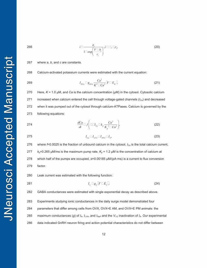
(3) 232
(4) 233
(5) 234
(6) 235
(7) 236
(8) 237
(9) 238
In the case of IA, IHVA, and Ih, the inactivation variable is the weighted sum of gating variables hi, 239
which have different voltage-dependent time constants, and represent two different populations 240
of inactivating gates present in the cell membrane: 241
(10) 242
where fA=0.8, fHVA = 0.2, fh = 0.384 for h1A, h1HVA, and h1h, respectively. 243
The activation and inactivation gating variables are governed by: 244
(11) 245
(12) 246
INaP gNaPmNaPhNaP V ENa ,
IA gAmA fAh1A (1 fA)h2A V EK
IK gkmK4 V EK ,
IHVA gHVAmHVA fHVAh1HVA 1 fHVA h2HVA V ECa
ILVA gLVAmLVA2 hLVA V ECa ,
IS gSmS V ECa ,
Ih gh fhh1h 1 fh h2h V Eh
dmdt
m V m
m V,
dhi
dth V hi
hiV
,

11
where m∞ and h∞ are steady-state activation and inactivation functions and τ is the time constant 247
(which can be voltage-dependent or independent) in ms. Steady-state activation and 248
inactivation functions are of the form: 249
(13) 250
V is the membrane potential, Vh is voltage at half activation or inactivation and k is the 251
steepness of the steady-state function. 252
Voltage-dependent time constants were estimated from one of two functions: 253
(14) 254
(15) 255
where V is the membrane potential and a, b, c, d, e, and f are constants. Parameter estimates 256
for f were close to zero or zero for mA, mK, and mLVA. 257
Fast transient sodium current is described using a Markov model with each of three subunits 258
having three states, open (O), closed (C), and inactivated (I) as in (Moran et al., 2016): 259
(16) 260
(17) 261
(18) 262
(19) 263
V is the membrane potential, r1, r2, r4 are voltage-independent constants and r3, α, and β are 264
voltage-dependent constants described by: 265
x V 1
1 expV Vh
k
,x m,h,
i
ei
expai V
bi
expci V
di
fi , i hNaP ,mA ,mK ,mHVA,mLVA,
i ci expV ai
bi
2
, i hh,1,hh,2 ,
INaF gNaFO3 V ENa ,
dCdt
r3 V I V O V r4 C,
dOdt
r2I V C V r1 O,
I 1 C O,
12
(20) 266
where a, b, and c are constants. 267
Calcium-activated potassium currents were estimated with the current equation: 268
(21) 269
Here, K = 1.0 μM, and Ca is the calcium concentration (μM) in the cytosol. Cytosolic calcium 270
increased when calcium entered the cell through voltage-gated channels (ICa) and decreased 271
when it was pumped out of the cytosol through calcium-ATPases. Calcium is governed by the 272
following equations: 273
(22) 274
(23) 275
where f=0.0025 is the fraction of unbound calcium in the cytosol, ICa is the total calcium current, 276
kp=0.265 μM/ms is the maximum pump rate, Kp = 1.2 μM is the concentration of calcium at 277
which half of the pumps are occupied, α=0.00185 μM/(pA·ms) is a current to flux conversion 278
factor. 279
Leak current was estimated with the following function: 280
(24) 281
GABA conductances were estimated with single exponential decay as described above. 282
Experiments studying ionic conductances in the daily surge model demonstrated four 283
parameters that differ among cells from OVX, OVX+E AM, and OVX+E PM animals: the 284
maximum conductances (g) of IA, ILVA, and INaP and the V1/2 inactivation of IA. Our experimental 285
data indicated GnRH neuron firing and action potential characteristics do not differ between 286
iai
1 expV bi
ci
,i , ,r3,
IKCa gKCaCa2
K 2 Ca2 V EK .
dCadt
f ICa kpCa2
K p2 Ca2 ,
ICa ILVA IHVA IS ,
IL gL V EL .
13
OVX and negative feedback despite having different ionic conductance characteristics. A 287
Markov Chain Monte Carlo method estimated probability distributions for the four estradiol and 288
time-of-day dependent parameters to predict intrinsic properties underlying excitability changes 289
(Adams et al., 2018b). A single identifiable distribution of solutions accounted for similar GnRH 290
neuron excitability in all groups other than positive feedback despite different underlying 291
conductance properties. This was attributable to the interdependence of voltage-gated 292
potassium channel properties: as gA increased V1/2 became more hyperpolarized, thus 293
maintaining the same action potential response to current injection. For the present work, we 294
thus picked ten parameter sets that traverse the entire distribution range in order to represent 295
both OVX and negative feedback parameter sets (which do not vary in excitability) in our 296
negative feedback model. Because the Markov Chain Monte Carlo method did not converge to 297
a single, identifiable distribution of solutions in the case of positive feedback (Adams et al., 298
2018b), ten representative parameter sets were chosen to produce ten separate positive 299
feedback neurons. 300
Maximum conductance values and reversal potentials are in Table 1. Parameter values for the 301
activation and inactivation variables are in Table 2. Parameter values for INaF are in Table 3. 302
Parameters that varied between the negative and positive feedback models are in Table 4. 303
All simulations were performed in MATLAB (MathWorks, https://www.mathworks.com/). 304
MATLAB scripts are available upon request. 305
Experimental Design 306
We measured GnRH neuron response to conductance trains modeled from previously recorded 307
PSC trains (example in Figure 1). Representative 30 second PSC trains for each group (OVX 308
AM, OVX+E AM, OVX+E PM) were chosen based upon mean isolated spontaneous GABA 309
PSC amplitude and frequency from a published data set containing voltage-clamp recordings of 310

14
>300 cells from OVX AM, OVX PM, OVX+E AM and OVX+E PM mice. (Christian and Moenter, 311
2007). Because OVX AM and OVX PM trains did not differ in mean frequency, interevent 312
interval distribution, or amplitude (Christian and Moenter, 2007), these groups were combined. 313
For each PSC train, conductance of each event was calculated (g=I/(V-EGABA)) (OVX AM mean 314
gmax=0.78±0.1 nS n=6 events, OVX+E AM gmax=0.73±0.2 n=4 events, OVX+E PM gmax=0.9±0.2 315
n=18 events; p>0.6, one-way ANOVA). Peak conductances and event times were used to 316
construct a new postsynaptic conductance (PSg) train using the physiologic decay time 317
constant determined above (10 ms). Each of the three trains was injected in random order 1-4 318
times into cells from OVX AM, OVX PM, OVX+E AM, and OVX+E PM animals. Direct current 319
(<30 pA, 1.7±2.5 pA, n=34) was adjusted to keep cells within 3 mV of -59 mV. A duration of 30 s 320
for PSg trains was chosen because it permitted data collection in response to all three train 321
types within a 7-minute period after recording stabilization, a time period during which GnRH 322
neurons do not experience a decrease in excitability (Adams et al., 2018b). Because cells from 323
OVX AM and PM mice did not differ in the number of action potentials fired in response to 324
conductance trains (n=8 and 6, respectively, p>0.59, two-tailed Mann-Whitney U-test), these 325
groups were combined for analysis. 326
Statistical Analyses 327
Data were analyzed using Prism 7 (GraphPad) or SPSS (IBM) and are reported as the mean ± 328
SEM unless otherwise noted. The number of cells per group is indicated by n. No more than 329
three cells were used per animal with at least five animals tested per group (OVX 14 cells from 330
7 mice; OVX+E AM 10 cells from 6 mice; OVX+E PM 10 cells from 6 mice). Data distribution 331
was tested with Shapiro-Wilk. Data requiring one-way analyses were compared using one-way 332
ANOVA with Fisher’s least square difference (LSD) post hoc analysis or Kruskal–Wallis test with 333
Dunn’s post hoc analysis as dictated by data distribution. All data requiring two-way or three-334
way analyses were compared using two-way or three-way ANOVA with Fisher’s LSD post 335

15
hoc analysis, respectively. ANOVA analyses did not assume equal subgroup sizes. Fisher’s 336
LSD was selected because of the large number (16-18) of multiple comparisons being 337
examined. Significance was set at p<0.05 but all p-values <0.1 are specified. Slopes and 338
intercepts were fit using linear regression, and ANCOVA analyses were performed to compare 339
slopes and intercepts among groups. 340
Results 341
Both PSg frequency and estradiol feedback contribute to increases in GnRH neuron firing rate. 342
In the daily surge model, estradiol and time-of-day interact to alter GnRH neuron intrinsic 343
properties, resulting in increased spontaneous firing rate and excitability during positive 344
feedback (Christian et al., 2005; Adams et al., 2018b). To investigate if observed concomitant 345
changes in GABAergic transmission (Christian and Moenter, 2007) play a critical role in 346
increasing GnRH neuron firing, we used dynamic clamp to deliver GABA PSg trains mimicking 347
the input measured during negative feedback, positive feedback and the open feedback loop 348
condition into cells from OVX+E AM, OVX+E PM, and OVX mice (AM and PM combined). 349
Figure 2 shows representative action potential firing from all three animal models in response to 350
all three conductance trains. Spikes were identified as induced by a postsynaptic conductance if 351
they occurred during the resulting postsynaptic potential and before the membrane potential 352
returned to baseline (typically near the PSP peak). In one cell from each of two animals in 353
positive feedback, a postsynaptic conductance initiated a burst of action potentials; all of these 354
events were counted as induced events if the time interval between subsequent events was ≤ 355
250 ms and if a continuous increase in Vm between spikes was observed. In some cells, 356
spontaneous spikes (that did not meet the above definition) occurred; these are marked with * in 357
Figure 2. The number of spontaneous action potentials was not different between post-synaptic 358
conductance trains or among groups (OVX+E PM: 3.2±0.8 spikes, OVX+E AM: 0.3±0.2 spikes, 359

16
OVX 2.4±0.6 spikes; p>0.1, two-way repeated-measures ANOVA). Data quantifying response to 360
trains (Figure 3) include only spikes that were induced by a postsynaptic conductance. 361
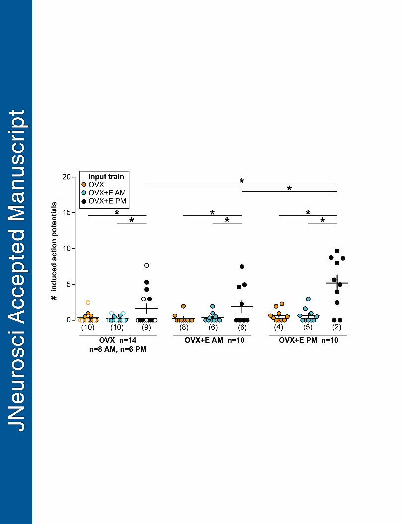
The positive feedback PSg train induced more action potentials in cells from all three animal 362
models than the negative feedback or open-loop OVX PSg trains (Figure 3,P<0.05, two-way 363
repeated-measures ANOVA/Fisher’s LSD). In addition to PSg train type, animal model of the 364
recorded cell influenced the GnRH response. Specifically, positive feedback trains initiated more 365
induced action potentials in cells during positive feedback (OVX+E PM) than in cells during 366
negative feedback (OVX+E AM) or cells from OVX mice. These changes were not attributable 367
to differences in input resistance, capacitance or holding current among groups (Table 5). There 368
was no difference in number of action potentials induced by PSg trains from negative feedback 369
vs the open-loop condition among any of the groups, despite a difference in frequency and 370
amplitude of GABA transmission between these states (Christian and Moenter, 2007). 371
Properties of PSgs associated with successful spike initiation 372
To assess if amplitude of the postsynaptic conductance and/or time between these events 373
affected the probability of inducing an action potential, postsynaptic conductances from all trains 374
were combined and sorted according to their peak conductance and preceding interevent 375
interval. Increased peak conductance was correlated with increased probability of initiating an 376
action potential in cells from all three animal models (Figure 4A-B OVX+E AM r2= 0.84 p<0.001, 377
OVX r2=0.62 p<0.0001, OVX+E PM r2=0.75 p<0.0001 by linear regression) and the slopes of 378
the linear regressions differed among these, being greatest in OVX+E PM cells and lowest in 379
OVX+E AM cells (OVX+E AM slope=0.41±0.05, OVX slope=0.13±0.02, OVX+E AM 380
slope=0.23±0.02, F=19.5 p<0.0001 by linear regression). In contrast, the likelihood of initiating a 381
spike was independent of interevent interval (Figure 4C-D, p>0.1, linear regression/ANCOVA). 382
The slopes of the linear regressions of interevent interval did not differ among cells from 383
different animal models, but the overall intercepts did (OVX+E PM = 0.27±0.05, OVX+E AM = 384
17
0.076±0.02, OVX = 0.072±0.02, F=7.6, p<0.0001) reflecting the increased response to 385
postsynaptic conductance trains during positive feedback relative to all other cell types. 386
Both PSg frequency and estradiol feedback contribute to increases in firing rate in models of 387
GnRH neurons. 388
We have demonstrated that both fast-synaptic input and GnRH neuron intrinsic properties 389
contribute to firing rate. Whole-cell patch-clamp recordings of the required stability are limited to 390
10 to 20 minutes, however, and our dynamic clamp data set was limited to the response to 391
representative 30-second patterns of postsynaptic currents selected from a much larger data set 392
of over >1,000 two-minute PSC trains from over 300 cells recorded by Christian and Moenter, 393
2007. To measure GnRH response to this entire data set, we used mathematical models of 394
GnRH neurons during multiple feedback states first developed to predict intrinsic properties 395
underlying excitability changes. One model identified a single, unimodal distribution of solutions 396
(parameter sets) to account for similar GnRH neuron excitability in OVX/negative feedback 397
neurons despite different underlying conductance properties; this was attributable to the 398
interdependence of voltage-gated potassium channel properties. In contrast, there was not a 399
single distribution of solutions to account for the positive feedback model, perhaps indicative of 400
redundant mechanisms for achieving the positive feedback state essential for reproduction. 401
402
Given this, we chose ten parameter sets for “negative feedback” cells that traversed the range 403
of parameters expected for both OVX and OVX+E AM cells while also reproducing OVX/OVX+E 404
AM excitability, and ten positive feedback cells chosen in a similar fashion (Table 4). Each 405
model neuron was exposed to >1000 two-minute PSg trains from 307 GnRH neurons (30 OVX 406
AM, 62 OVX PM, 48 OVX+E AM, and 167 OVX+E PM) and action potentials generated by the 407
model were counted. Recording duration varied in (Christian and Moenter, 2007), so different 408
numbers of PSg trains were available from each recorded cell. Model response to all the PSg 409

18
trains from a recorded GnRH neuron was averaged to account for variability in recording 410
duration. Firing response of a single model neuron did not differ among PSg train types (two-411
way ANOVA, Figure 5, Table 6). In contrast, when mean response to PSg conductance trains 412
was compared among all twenty model neurons, both train type and feedback model 413
contributed to firing frequency (Figure 6). The positive feedback (OVX+E PM) conductance 414
trains initiated more action potentials in both negative and positive feedback model neurons 415
(three-way repeated-measures ANOVA/Fisher’s LSD, p<0.001). Further, all four conductance 416
train types initiated more action potentials in positive feedback model neurons relative to 417
negative feedback model neurons (three-way repeated-measures ANOVA/Fisher’s LSD, 418
p<0.001). 419
420
Discussion 421
The brain regulates fertility via GnRH release. For most of the mammalian reproductive cycle, 422
GnRH release is pulsatile and estradiol feedback suppresses GnRH and LH release. At the end 423
of the follicular phase, high sustained estradiol levels initiate a surge of continuous GnRH 424
release culminating in ovulation (Sarkar et al., 1976; Moenter et al., 1991). This shift in estradiol 425
feedback is associated with changes in both intrinsic properties of and synaptic inputs to these 426
cells. Here we show that during positive feedback, GnRH neurons integrate changes in their 427
intrinsic properties with changes to fast-synaptic transmission to increase firing rate. 428
This work supports findings demonstrating increased GABAergic drive to GnRH neurons is 429
correlated with increased firing rate in several animal models (Sullivan and Moenter, 2004; 430
Christian et al., 2005; Sullivan and Moenter, 2005; Pielecka et al., 2006; Christian and Moenter, 431
2007; Roland and Moenter, 2011; Adams et al., 2018a). Past conclusions have been limited to 432
speculation regarding the relationship of these parameters measured in independent studies. 433
The present work extends those findings by demonstrating directly increased effectiveness of 434

19
GABAergic transmission mimicking positive feedback in driving firing of GnRH neurons from all 435
animal models both in dynamic clamp and modeling studies. It further reveals that the intrinsic 436
changes that occur in GnRH neurons during positive feedback (Chu and Moenter, 2006; Sun et 437
al., 2010; Pielecka-Fortuna et al., 2011; Adams et al., 2018b) are sufficiently robust to poise the 438
cell for increased response to synaptic input. 439
The dynamic clamp data were limited for practical reasons to one representative conductance 440
train per feedback state. These data were supported by results using mathematical models of 441
GnRH neurons developed to reproduce GnRH neuron firing response to current injection during 442
positive feedback, and during negative feedback and in the open-loop condition (which exhibit 443
similar excitability). Based on experimental data, five parameters were varied between these 444
feedback state models (those governing IA, INaP, and IHVA) (Chu and Moenter, 2006; Zhang et al., 445
2007; Zhang et al., 2009; Sun et al., 2010; Pielecka-Fortuna et al., 2011). Ten parameter sets 446
traversing the full parameter range were examined for each feedback state. For all model 447
neurons, conductance trains mimicking GABAergic transmission during positive feedback 448
invoked greater firing rates than conductance trains mimicking negative feedback or OVX 449
states. Further, regardless of conductance train type, positive feedback model neurons fired 450
more frequently than negative feedback/OVX neurons. This suggests that IA, INaP, and IHVA likely 451
contribute to the enhanced firing response to fast-synaptic inputs during positive feedback. Of 452
interest in this regard, blocking IHVA decreases GnRH neuron response rate to dynamic-clamp 453
induced GABA postsynaptic conductances (Hemond et al., 2012). 454
Spontaneous GnRH neuron firing rate monitored using extracellular recordings in cells from 455
OVX mice is intermediate to that of cells from OVX+E AM and OVX+E PM mice in the daily 456
surge model (Christian et al., 2005). In contrast, no differences were observed in the present 457
study between cells from brain slices prepared during negative feedback vs. the open-loop 458
condition in GnRH neuron action potential firing in response to any conductance train. Similarly, 459

20
there was no difference between the negative feedback and open-loop condition in GnRH 460
neuron excitability (Adams et al., 2018b). There are several possible explanations for this 461
difference. First, the present work was done in the whole-cell configuration, which dialyzes 462
many second messenger systems; extracellular recordings are not subject to this limitation. 463
Second, fast synaptic transmission was not inhibited during extracellular recordings; although of 464
very low frequency, glutamatergic inputs may also contribute to differences in firing rate. Third, 465
the GABAergic PSCs used to generate PSg trains were monitored with blockers of 466
glutamatergic transmission, thus may underestimate actual values. Fourth, it is possible that 30-467
second patterns are not sufficient for observing a difference in firing rate between these two cell 468
types, considering that most extracellular recordings determining firing rate were averaged over 469
30 minutes to one hour. This is supported by the finding that differences in firing rate between 470
positive and negative feedback model neurons exposed to longer PSg trains were detected for 471
every train type, and a difference in response to OVX PM and OVX+E AM conductance trains 472
could be detected in positive feedback model neurons. 473
In many neurons, postsynaptic potentials decay to baseline in a matter of milliseconds (<100 474
ms) and tens to hundreds of coordinated PSPs are necessary to initiate an action potential. 475
Some neurons in the hypothalamus appear unique in that their membrane potentials decay 476
more slowly, permitting fast-synaptic inputs to summate over prolonged timescales. For 477
example, in AGRP, POMC and PVH neurons, activation of a voltage-gated sodium current 478
slows postsynaptic potential decay times and consequently, relatively few successive 479
postsynaptic potentials can depolarize the membrane until threshold is reached and a spike is 480
initiated (Branco et al., 2016). Given these observations, we postulated that decreasing time 481
between successive postsynaptic conductances would increase spiking. We rejected this 482
hypothesis as interevent interval did not influence the probability of initiating a spike over the 483
intervals tested, perhaps because sufficiently short intervals did not occur in the data sets used. 484
21
In vivo, with all synaptic connections present, it is possible that shorter interevent intervals that 485
are more effective in initiating action potentials exist. In contrast to the lack of effect of interevent 486
interval, peak conductance amplitude was highly correlated with action potential firing. These 487
data suggest that postsynaptic conductance amplitudes and GnRH neuron responsiveness 488
appear to be more important than coordinated or successive GABA release from GnRH 489
afferents, at least within the limitations of brain slices. It is tempting to speculate that shifts in 490
conductance via the GABAA receptor also modulate GnRH neuron activity in vivo. Numerous 491
substances interact with this receptor in an allosteric manner to increase total conductance, 492
including benzodiazepines and the neurosteroid allopregnanolone (Defazio and Hablitz, 1998; 493
Sullivan and Moenter, 2003). It is also possible that the composition of GABAA receptor subunits 494
shifts among feedback conditions (Sim et al., 2000; Burger et al., 2018) 495
Afferent neurons and the postsynaptic responses they initiate (fast synaptic and 496
neuromodulatory) are likely to play a major role in estradiol feedback as GnRH neurons typically 497
do not express detectable estrogen receptor α (ERα) (Hrabovszky et al., 2000; Hrabovszky et 498
al., 2001; Couse et al., 2003; Wintermantel et al., 2006; Christian et al., 2008). Kisspeptin 499
neurons of anteroventral periventricular (AVPV) nucleus, which use GABA as a cotransmitter 500
and express ERα, are one likely relay (Cravo et al., 2011). The neuromodulator kisspeptin acts 501
directly on GnRH neurons to modulate multiple ionic currents, enhance GnRH neuron 502
excitability and stimulate action potential firing (Pielecka-Fortuna et al., 2008; Zhang et al., 503
2008; Pielecka-Fortuna et al., 2011; Zhang et al., 2013; Adams et al., 2018b). AVPV kisspeptin 504
expression is increased by estradiol treatment (Smith et al., 2005) and on the afternoon of 505
proestrus (positive feedback during the cycle) (Smith et al., 2006), and these cells fire higher 506
frequency action potentials and more bursts under these conditions (Zhang et al., 2015; Wang 507
et al., 2016). We postulate that kisspeptin release during positive feedback enhances GnRH 508
neuron responsiveness to the concurrent increase in GABAergic fast-synaptic inputs. In this 509
22
regard, ERα knock-out in GABAergic neurons blocked positive feedback and the LH surge in 510
mice (Cheong et al., 2015) and optical activation of GABAergic neurons in the AVPV initiated 511
LH surges (Kalil et al., 2016). Countering these observations, work from the same group 512
demonstrated knock-out of the gamma2 subunit of the GABAA receptor in GnRH neurons does 513
not appear to affect fertility (Lee et al., 2010). Because AVPV neurons can communicate using 514
GABA and/or kisspeptin, increased GABA transmission to GnRH neurons might be interpreted 515
as merely a marker for increased co-transmission of the excitatory neuromodulator (Piet et al., 516
2018). The present work, however, demonstrates that increasing GABA drive to GnRH neurons 517
likely contributes to their increased firing rate during estradiol positive feedback. 518
Our focus was on the interaction between GABAergic transmission and GnRH neuron 519
properties. Although estradiol increases glutamatergic transmission to GnRH neurons on 520
prorestrus in rats, glutamatergic transmission to GnRH neurons in mice is very low frequency, 521
with no AMPA or NMDA-mediated currents detected in about one-third of cells (Christian et al., 522
2009; Tada et al., 2013; Liu et al., 2017). The minor differences detected among groups at 523
these low frequencies are unlikely to have a substantial impact on firing, despite a more 524
depolarized reversal potential. Kisspeptin neurons may also have an additional role in driving 525
increased GnRH firing rate as kisspeptin also acts indirectly via unspecified afferents to 526
increase GABA PSC frequency and amplitude, and glutamate EPSC frequency (Pielecka-527
Fortuna et al., 2008). 528
Multiple factors can influence the switch from negative to positive feedback. In addition to time-529
of-day and estradiol-dependent signals, other internal and environmental cues may play a role. 530
It remains unclear if each of these signals are necessary for initiating the GnRH surge, or if they 531
act as redundant mechanisms for ensuring ovulation. The present studies suggest that GnRH 532
neurons do not merely relay upstream signals, but act to integrate multiple signals to modulate 533
firing rate and to initiate GnRH release. 534

23
References: 535
Adams C, Chen X, Moenter SM (2018a) Changes in GABAergic transmission to and intrinsic 536 excitability of gonadotropin-releasing hormone (GnRH) neuron during the estrous cycle 537 in mice. eNeuro. 538
Adams C, Stroberg W, DeFazio RA, Schnell S, Moenter SM (2018b) Gonadotropin-Releasing 539 Hormone (GnRH) Neuron Excitability Is Regulated by Estradiol Feedback and 540 Kisspeptin. J Neurosci 38:1249-1263. 541
Barry PH (1994) JPCalc, a software package for calculating liquid junction potential corrections 542 in patch-clamp, intracellular, epithelial and bilayer measurements and for correcting 543 junction potential measurements. J Neurosci Methods 51:107-116. 544
Branco T, Tozer A, Magnus CJ, Sugino K, Tanaka S, Lee AK, Wood JN, Sternson SM (2016) 545 Near-Perfect Synaptic Integration by Nav1.7 in Hypothalamic Neurons Regulates Body 546 Weight. Cell 165:1749-1761. 547
Burger LL, Vanacker C, Phumsatitispong C, Wagenmaker ER, Wang L, Olson DP, Moenter SM 548 (2018) Identification of genes enriched in GnRH neurons by translating ribosome affinity 549 purification and RNASeq in mice. Endocrinology, 159:1922-1940 550
Cheong RY, Czieselsky K, Porteous R, Herbison AE (2015) Expression of ESR1 in 551 Glutamatergic and GABAergic Neurons Is Essential for Normal Puberty Onset, Estrogen 552 Feedback, and Fertility in Female Mice. J Neurosci 35:14533-14543. 553
Christian CA, Moenter SM (2007) Estradiol induces diurnal shifts in GABA transmission to 554 gonadotropin-releasing hormone neurons to provide a neural signal for ovulation. J 555 Neurosci 27:1913-1921. 556
Christian CA, Mobley JL, Moenter SM (2005) Diurnal and estradiol-dependent changes in 557 gonadotropin-releasing hormone neuron firing activity. Proc Natl Acad Sci U S A 558 102:15682-15687. 559
Christian CA, Pielecka-Fortuna J, Moenter SM (2009) Estradiol suppresses glutamatergic 560 transmission to gonadotropin-releasing hormone neurons in a model of negative 561 feedback in mice. Biol Reprod 80:1128-1135. 562
Christian CA, Glidewell-Kenney C, Jameson JL, Moenter SM (2008) Classical estrogen receptor 563 alpha signaling mediates negative and positive feedback on gonadotropin-releasing 564 hormone neuron firing. Endocrinology 149:5328-5334. 565
Chu Z, Moenter SM (2006) Physiologic regulation of a tetrodotoxin-sensitive sodium influx that 566 mediates a slow afterdepolarization potential in gonadotropin-releasing hormone 567 neurons: possible implications for the central regulation of fertility. J Neurosci 26:11961-568 11973. 569
Couse JF, Yates MM, Walker VR, Korach KS (2003) Characterization of the hypothalamic-570 pituitary-gonadal axis in estrogen receptor (ER) Null mice reveals hypergonadism and 571 endocrine sex reversal in females lacking ERalpha but not ERbeta. Mol Endocrinol 572 17:1039-1053. 573
Cravo RM, Margatho LO, Osborne-Lawrence S, Donato J, Jr., Atkin S, Bookout AL, Rovinsky S, 574 Frazao R, Lee CE, Gautron L, Zigman JM, Elias CF (2011) Characterization of Kiss1 575 neurons using transgenic mouse models. Neuroscience 173:37-56. 576
DeFazio RA, Moenter SM (2002) Estradiol feedback alters potassium currents and firing 577 properties of gonadotropin-releasing hormone neurons. Mol Endocrinol 16:2255-2265. 578
DeFazio RA, Elias CF, Moenter SM (2014) GABAergic transmission to kisspeptin neurons is 579 differentially regulated by time of day and estradiol in female mice. J Neurosci 34:16296-580 16308. 581

24
DeFazio RA, Heger S, Ojeda SR, Moenter SM (2002) Activation of A-type gamma-aminobutyric 582 acid receptors excites gonadotropin-releasing hormone neurons. Mol Endocrinol 583 16:2872-2891. 584
Defazio T, Hablitz JJ (1998) Zinc and zolpidem modulate mIPSCs in rat neocortical pyramidal 585 neurons. J Neurophysiol 80:1670-1677. 586
Glanowska KM, Venton BJ, Moenter SM (2012) Fast scan cyclic voltammetry as a novel 587 method for detection of real-time gonadotropin-releasing hormone release in mouse 588 brain slices. J Neurosci 32:14664-14669. 589
Hemond PJ, O'Boyle MP, Roberts CB, Delgado-Reyes A, Hemond Z, Suter KJ (2012) 590 Simulated GABA synaptic input and L-type calcium channels form functional 591 microdomains in hypothalamic gonadotropin-releasing hormone neurons. J Neurosci 592 32:8756-8766. 593
Houston CM, Bright DP, Sivilotti LG, Beato M, Smart TG (2009) Intracellular chloride ions 594 regulate the time course of GABA-mediated inhibitory synaptic transmission. J Neurosci 595 29:10416-10423. 596
Hrabovszky E, Shughrue PJ, Merchenthaler I, Hajszan T, Carpenter CD, Liposits Z, Petersen 597 SL (2000) Detection of estrogen receptor-beta messenger ribonucleic acid and 125I-598 estrogen binding sites in luteinizing hormone-releasing hormone neurons of the rat brain. 599 Endocrinology 141:3506-3509. 600
Hrabovszky E, Steinhauser A, Barabas K, Shughrue PJ, Petersen SL, Merchenthaler I, Liposits 601 Z (2001) Estrogen receptor-beta immunoreactivity in luteinizing hormone-releasing 602 hormone neurons of the rat brain. Endocrinology 142:3261-3264. 603
Kalil B, Mcclennan T, Piet R, Herbison A (2016) Role of rostral periventricular area of the third 604 ventricle (RP3V) GABAergic neurons in generating the preovulatory luteinizing hormone 605 surge in female mouse. In: Society for Neuroscience. San Diego, CA: Neuroscience 606 Meeting Planner. Online. 607
LeBeau AP, Van Goor F, Stojilkovic SS, Sherman A (2000) Modeling of membrane excitability 608 in gonadotropin-releasing hormone-secreting hypothalamic neurons regulated by Ca2+-609 mobilizing and adenylyl cyclase-coupled receptors. J Neurosci 20:9290-9297. 610
Lee K, Porteous R, Campbell RE, Luscher B, Herbison AE (2010) Knockdown of GABA(A) 611 receptor signaling in GnRH neurons has minimal effects upon fertility. Endocrinology 612 151:4428-4436. 613
Legan SJ, Karsch FJ (1975) A daily signal for the LH surge in the rat. Endocrinology 96:57-62. 614 Liu X, Porteous R, Herbison AE (2017) Dynamics of GnRH Neuron Ionotropic GABA and 615
Glutamate Synaptic Receptors Are Unchanged during Estrogen Positive and Negative 616 Feedback in Female Mice. eNeuro 4. 617
Milescu LS, Yamanishi T, Ptak K, Mogri MZ, Smith JC (2008) Real-time kinetic modeling of 618 voltage-gated ion channels using dynamic clamp. Biophysical journal 95:66-87. 619
Moenter SM, Caraty A, Locatelli A, Karsch FJ (1991) Pattern of gonadotropin-releasing 620 hormone (GnRH) secretion leading up to ovulation in the ewe: existence of a 621 preovulatory GnRH surge. Endocrinology 129:1175-1182. 622
Moran S, Moenter SM, Khadra A (2016) A unified model for two modes of bursting in GnRH 623 neurons. J Comput Neurosci 40:297-315. 624
Norman RL, Blake CA, Sawyer CH (1973) Estrogen-dependent 24-hour periodicity in pituitary 625 LH release in the female hamster. Endocrinology 93:965-970. 626
Pielecka J, Quaynor SD, Moenter SM (2006) Androgens increase gonadotropin-releasing 627 hormone neuron firing activity in females and interfere with progesterone negative 628 feedback. Endocrinology 147:1474-1479. 629
Pielecka-Fortuna J, Chu Z, Moenter SM (2008) Kisspeptin acts directly and indirectly to 630 increase gonadotropin-releasing hormone neuron activity and its effects are modulated 631 by estradiol. Endocrinology 149:1979-1986. 632
25
Pielecka-Fortuna J, DeFazio RA, Moenter SM (2011) Voltage-gated potassium currents are 633 targets of diurnal changes in estradiol feedback regulation and kisspeptin action on 634 gonadotropin-releasing hormone neurons in mice. Biol Reprod 85:987-995. 635
Piet R, Kalil B, McLennan T, Porteous R, Czieselsky K, Herbison AE (2018) Dominant 636 Neuropeptide Cotransmission in Kisspeptin-GABA Regulation of GnRH Neuron Firing 637 Driving Ovulation. J Neurosci 38:6310-6322. 638
Roland AV, Moenter SM (2011) Prenatal androgenization of female mice programs an increase 639 in firing activity of gonadotropin-releasing hormone (GnRH) neurons that is reversed by 640 metformin treatment in adulthood. Endocrinology 152:618-628. 641
Sarkar DK, Chiappa SA, Fink G, Sherwood NM (1976) Gonadotropin-releasing hormone surge 642 in pro-oestrous rats. Nature 264:461-463. 643
Silveira M, Burger LL, DeFazio RA, Wagenmaker ER, Moenter SM (2016) GnRH neuron activity 644 and pituitary response in estradiol-induced vs proestrous luteinizing hormone surges in 645 female mice. Endocrinology:en20161771. 646
Sim JA, Skynner MJ, Pape JR, Herbison AE (2000) Late postnatal reorganization of GABA(A) 647 recpetor signalling in native GnRH neurons. Eur J Neurosci 12:3497-3504. 648
Smith JT, Cunningham MJ, Rissman EF, Clifton DK, Steiner RA (2005) Regulation of Kiss1 649 gene expression in the brain of the female mouse. Endocrinology 146:3686-3692. 650
Smith JT, Popa SM, Clifton DK, Hoffman GE, Steiner RA (2006) Kiss1 neurons in the forebrain 651 as central processors for generating the preovulatory luteinizing hormone surge. J 652 Neurosci 26:6687-6694. 653
Sullivan SD, Moenter SM (2003) Neurosteroids alter gamma-aminobutyric acid postsynaptic 654 currents in gonadotropin-releasing hormone neurons: a possible mechanism for direct 655 steroidal control. Endocrinology 144:4366-4375. 656
Sullivan SD, Moenter SM (2004) Prenatal androgens alter GABAergic drive to gonadotropin-657 releasing hormone neurons: implications for a common fertility disorder. Proc Natl Acad 658 Sci U S A 101:7129-7134. 659
Sullivan SD, Moenter SM (2005) GABAergic integration of progesterone and androgen 660 feedback to gonadotropin-releasing hormone neurons. Biol Reprod 72:33-41. 661
Sun J, Chu Z, Moenter SM (2010) Diurnal in vivo and rapid in vitro effects of estradiol on 662 voltage-gated calcium channels in gonadotropin-releasing hormone neurons. J Neurosci 663 30:3912-3923. 664
Suter KJ, Song WJ, Sampson TL, Wuarin JP, Saunders JT, Dudek FE, Moenter SM (2000) 665 Genetic targeting of green fluorescent protein to gonadotropin-releasing hormone 666 neurons: characterization of whole-cell electrophysiological properties and morphology. 667 Endocrinology 141:412-419. 668
Tada H, Kuroki Y, Funabashi T, Kamiya Y, Goto T, Suyama K, Sano A, Mitsushima D, Etgen 669 AM, Takahashi T (2013) Phasic synaptic incorporation of GluR2-lacking AMPA receptors 670 at gonadotropin-releasing hormone neurons is involved in the generation of the 671 luteinizing hormone surge in female rats. Neuroscience 248:664-669. 672
Wang L, DeFazio RA, Moenter SM (2016) Excitability and Burst Generation of AVPV Kisspeptin 673 Neurons Are Regulated by the Estrous Cycle Via Multiple Conductances Modulated by 674 Estradiol Action. eNeuro 3. 675
Wintermantel TM, Campbell RE, Porteous R, Bock D, Grone HJ, Todman MG, Korach KS, 676 Greiner E, Perez CA, Schutz G, Herbison AE (2006) Definition of estrogen receptor 677 pathway critical for estrogen positive feedback to gonadotropin-releasing hormone 678 neurons and fertility. Neuron 52:271-280. 679
Zhang C, Ronnekleiv OK, Kelly MJ (2013) Kisspeptin inhibits a slow afterhyperpolarization 680 current via protein kinase C and reduces spike frequency adaptation in GnRH neurons. 681 Am J Physiol Endocrinol Metab 304:E1237-1244. 682

26
Zhang C, Roepke TA, Kelly MJ, Ronnekleiv OK (2008) Kisspeptin depolarizes gonadotropin-683 releasing hormone neurons through activation of TRPC-like cationic channels. J 684 Neurosci 28:4423-4434. 685
Zhang C, Bosch MA, Levine JE, Ronnekleiv OK, Kelly MJ (2007) Gonadotropin-releasing 686 hormone neurons express K(ATP) channels that are regulated by estrogen and 687 responsive to glucose and metabolic inhibition. J Neurosci 27:10153-10164. 688
Zhang C, Bosch MA, Rick EA, Kelly MJ, Ronnekleiv OK (2009) 17Beta-estradiol regulation of T-689 type calcium channels in gonadotropin-releasing hormone neurons. J Neurosci 690 29:10552-10562. 691
Zhang C, Bosch MA, Qiu J, Ronnekleiv OK, Kelly MJ (2015) 17beta-Estradiol increases 692 persistent Na(+) current and excitability of AVPV/PeN Kiss1 neurons in female mice. Mol 693 Endocrinol 29:518-527. 694
695
696

27
Table 1: Parameter values for ionic currents appearing in Equations 2-9. E is the reversal 697
potential and g is the maximum conductance. Parameters are from (Zhang et al., 2009; Sun et 698
al., 2010; Pielecka-Fortuna et al., 2011). 699
700
E (mV) g (nS) INaF 54 758 INaP 54 Table 4IA -101 Table 4 IK -101 57 ILVA 82.5 0.0679 IHVA 82.5 Table 4IS 82.5 0.18 Ih -40 1 IKCa -101 1.18 IL -65 1 701
Table 2: Parameter values for the activation and inactivation variables appearing in Equations 702
13-15. Vh and k are the voltage at half activation or inactivation and the steepness of the steady-703
state function. is the time constant which is voltage independent for mNap, h1A, h2A, hLVA, mHVA, 704
h1HVA, h2HVA and ms. For hNaP, mA, mK, mLVA, h1h and h2h, is voltage-dependent and governed by 705
Equation 14 (a, b, c, and d are in mV, e and f are in ms) or Equation 15 (a and b are in mV, and 706
c is in ms). 707
708
INaP IA IK ILVA IHVA IS Ih
m h m h1 h2 m m h m h1 h2 m h1 h2 Vh (mV) -41.5 -47.4 -29.4 ** -19.7 -51.4 -80.1 -11 -36.6 -36.6 -45 -77.4 -77.4 k (mV) -3.0 8.2 -6.64 4.26 4.26 -12.3 -4.07 5.5 -7 14.6 14.6 -12 9.2 9.2 (ms) 0.4 Eq 14 Eq 14 7.67 100 Eq 14 Eq 14 250 0.816 53.4 728 1500 Eq 15 Eq 15
a - 67.3 -2.91 - - 23.8 31.3 - - - - - -89.8 -82.6 b - -27.5 25.6 - - 18 10.1 - - - - - 11.6 25.7 c - 67.3 65.3 - - 23.8 31.3 - - - - - 35.8 370.9 d - 27.5 -10.6 - - -18 -10.1 - - - - - 7.6 54.1 e - 574.5 1 - - 10.6 109 - - - - - - - f - 62.6 0.0527 - - 0 0.0391 - - - - - - - ** see Table 4. 709
710

28
Table 3: Parameters values for the Markov model of INaF (Equations 16 -19). (V) and (V) 711
describe the transition rates (ms-1) between the closed and open states, r1 and r2 describe the 712
transition rates between the open and inactivated states, and r3(V) and r4 describe the transition 713
rates between the inactivated and closed states. (V), (V), and r3(V) are voltage-dependent 714
while r1, r2, and r4 are voltage-independent. Maximum conductance was re-estimated to be 758 715
nS. 716
717
(V) (V) r1 r2 r3(V) r4
rate (ms-1) Eq. 20 Eq. 20 1.0 0.2 Eq. 20 0.05 a (ms-1) 55 60 - - 30 - b (mV) 6.4 32 - - 77.5 - c (mV) -15.9 10 - - 12 - 718 719 Table 4: Parameter set for negative feedback and positive feedback GnRH models 720 gNaP (nS) gA (nS) V1/2 inact (IA, mV) gHVA (nS) positive Feedback model 1 (Fig 5A) 1.006 391.953 -73.382 3.099 model 2 (Fig 5B) 0.741 473.829 -74.577 2.989 model 3 (Fig 5C) 0.929 467.298 -74.244 3.483 model 4 (Fig 5D) 1.068 444.406 -73.833 4.265 model 5 (Fig 5E) 1.598 447.050 -73.019 7.348 model 6 (Fig 5F) 1.167 394.336 -72.585 6.824 model 7 (Fig 5G) 1.974 411.111 -72.143 9.407 model 8 (Fig 5H) 0.781 244.552 -71.273 2.342 model 9 (Fig 5I) 0.804 230.366 -70.988 2.389 model 10 (Fig 5J) 0.713 202.316 -70.469 1.643 negative Feedback model 11 (Fig 5K) 0.389 313.792 -69.785 4.815 model 12 (Fig 5L) 0.515 291.525 -69.176 6.394 model 13 (Fig 5M) 0.391 338.008 -70.352 4.071 model 14 (Fig 5N) 0.284 329.019 -70.220 4.000 model 15 (Fig 5O) 0.350 328.879 -70.124 4.591 model 16 (Fig 5P) 0.351 320.634 -69.962 4.560 model 17 (Fig 5Q) 0.403 312.056 -69.693 5.608 model 18 (Fig 5R) 0.361 305.658 -69.591 5.592 model 19 (Fig 5S) 0.504 305.299 -69.464 6.206 model 20 (Fig 5T) 0.468 296.911 -69.333 6.022 721

29
Table 5: Whole-cell recording properties for Figures 2-4 722 Mean±SEM of GnRH whole-cell passive properties from Figures 2-4 OVX OVX+E AM OVX+E PM Input resistance (MΩ) 951 60 819 68 1040 96 Capacitance (pF) 16.9 0.9 15.4 1.0 14.9 0.8 Series resistance(MΩ) 11.6 0.9 11.4 0.8 11.6 0.8 Holding current (pA) 0.7 4.1 6.1 3.8 -8.3 4.5 One-way ANOVA for comparison of GnRH passive properties among groups: cells from diestrous and proestrous mice (Figures 2-4) group Input resistance (MΩ) F(2,31)=2.1 Capacitance (pF) KW statistic = 2.3 Series resistance (MΩ) KW statistic = 0.6 Holding current (pA) KW statistic = 4.8 (p=0.09) 723
Table 6: Two-way ANOVA parameters for comparison of model GnRH neuron response to OVX 724 AM, OVX PM, OVX+E AM, and OVX+E PM PSg trains from (Christian and Moenter, 2007). 725
parameter (figure) estradiol time-of-day interaction positive feedback model 1 (Fig 5A) F(1,303)=1.7 F(1,303)=2.1 F(1,303)=1.8 model 2 (Fig 5B) F(1,303)=1.6 F(1,303)=1.7 F(1,303)=1.7 model 3 (Fig 5C) F(1,303)=1.7 F(1,303)=2.0 F(1,303)=1.8 model 4 (Fig 5D) F(1,303)=1.7 F(1,303)=2.2 F(1,303)=1.9 model 5 (Fig 5E) F(1,303)=1.7 F(1,303)=3.2 (p=0.07) F(1,303)=2.3 model 6 (Fig 5F) F(1,303)=1.8 F(1,303)=2.6 F(1,303)=1.9 model 7 (Fig 5G) F(1,303)=1.6 F(1,303)=3.8 (p=0.05) F(1,303)=2.6 model 8 (Fig 5H) F(1,303)=1.6 F(1,303)=1.5 F(1,303)=1.6 model 9 (Fig 5I) F(1,303)=1.6 F(1,303)=1.5 F(1,303)=1.6 model 10 (Fig 5J) F(1,303)=1.5 F(1,303)=1.3 F(1,303)=1.5 negative feedback model 11 (Fig 5K) F(1,303)=1.4 F(1,303)=1.7 F(1,303)=1.4 model 12 (Fig 5L) F(1,303)=1.5 F(1,303)=2.0 F(1,303)=1.4 model 13 (Fig 5M) F(1,303)=1.4 F(1,303)=1.7 F(1,303)=1.4 model 14 (Fig 5N) F(1,303)=1.3 F(1,303)=1.5 F(1,303)=1.3 model 15 (Fig 5O) F(1,303)=1.4 F(1,303)=1.6 F(1,303)=1.4 model 16 (Fig 5P) F(1,303)=1.3 F(1,303)=1.6 F(1,303)=1.4 model 17 (Fig 5Q) F(1,303)=1.4 F(1,303)=1.8 F(1,303)=1.4 model 18 (Fig 5R) F(1,303)=1.4 F(1,303)=1.7 F(1,303)=1.4 model 19 (Fig 5S) F(1,303)=1.5 F(1,303)=2.0 F(1,303)=1.5 model 20 (Fig 5T) F(1,303)=1.5 F(1,303)=1.8 F(1,303)=1.4 726
727

30
Figure Legends 728
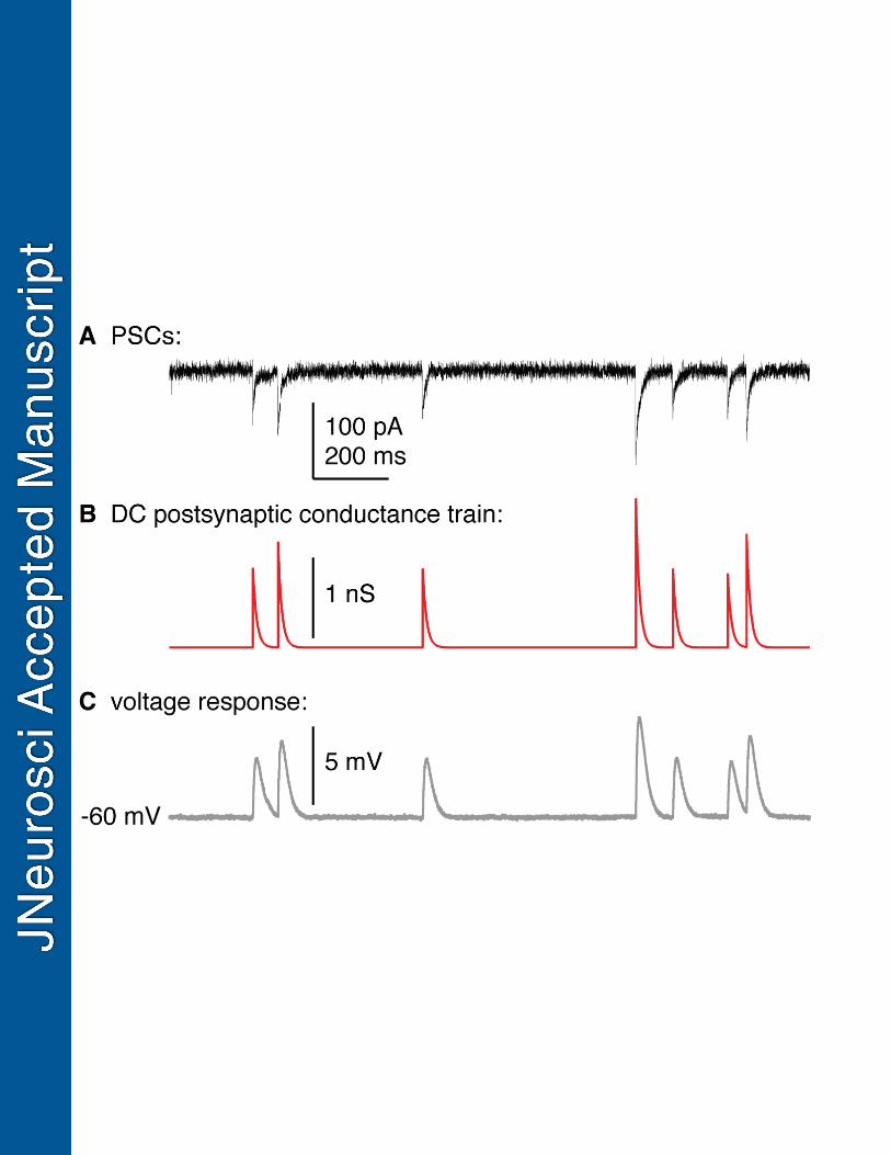
Figure 1. GABA postsynaptic currents in voltage-clamp were used to construct a 729
postsynaptic conductance train for dynamic clamp studies. A. GABA postsynaptic currents 730
recorded at -70 mV in voltage-clamp using a high-chloride pipette solution that slows decay time 731
B. Simulated dynamic clamp postsynaptic conductance train constructed from the peak times 732
and amplitudes in A, but using the decay time constant (10 ms) observed with physiological 733
pipette chloride. C. Response by an RC circuit to the postsynaptic conductance train in B. 734
735
Figure 2. Representative recordings of cells from OVX (top), OVX+E AM (middle), and OVX+E 736
PM (bottom) animals in response to conductance trains from OVX (orange), OVX+E AM (blue), 737
and OVX+E PM (black) conditions. * denotes spontaneous action potentials that were not 738
induced by a postsynaptic conductance. 739
740 Figure 3. Positive feedback conductance trains are most effective at initiating spikes and 741
positive feedback animal models are most responsive. Individual values and mean ± SEM 742
spikes induced during individual postsynaptic conductances in each train in all three cell types. 743
In the OVX group, open circles denote cells recorded in the PM and closed circles denote cells 744
recorded in the AM. Numbers in parentheses along x axis indicate number of cells not firing any 745
spikes. *p<0.05 two-way repeated-measures ANOVA/Fisher’s LSD test (cell type: F(2,31)=3.8 746
p=0.03, train: F(2,62)=33.4 p<0.001, interaction: F(4,62)=5.3 p=0.001) 747
748
Figure 4. Increasing maximum peak conductance increases the probability of initiating 749
an action potential. Mean likelihood of initiating an action potential for individual conductances 750
from OVX (orange), OVX+E AM (blue), and OVX+E PM (black) postsynaptic conductance trains 751
in cells from OVX (left column), OVX+E AM (middle column), and OVX+E PM (right column) 752
mice, considering: conductance amplitude (A linear scale, B log-log scale) or time since 753

31
previous event (C linear scale, D log-log scale). Different number of points between linear and 754
log-log scales is attributable to exclusion of zero values on the latter. Grey lines in left and 755
middle columns are a linear fit to the data (R-squared is indicated above each fit, grey asterisk 756
indicates that the slope of the linear regression is significantly non-zero suggesting the 757
correlation is significant (p<0.0001); and the three individual fits (from OVX, OVX+E AM, and 758
OVX+E PM cells) are shown in the right-most column. Red asterisks indicate p<0.05 ANCOVA 759
(A, slope: F(2,78)=22.4; B, slope: F(2,78)=0.5, intercept: F(2,80)=8.4). 760
761 Figure 5. Positive and negative feedback model response to OVX AM, OVX PM, OVX+E 762
AM, and OVX+E PM conductance trains from cells recorded by (Christian and Moenter, 763
2007). Each graph shows a single model neuron and each data point is that model neuron’s 764
response to PSg trains from a single GnRH neuron prepared from OVX AM (30 cells), OVX PM 765
(62 cells), OVX+E AM (48 cells) or OVX+E PM (167 cells) mice. In some cases, PSg trains from 766
a single GnRH neuron did not induce action potential firing (frequency = 0 Hz); the percentage 767
of cells that did not induce AP firing in the model neurons is indicated above the 0 Hz data 768
points for each group. 769
770
Figure 6. Conductance trains from OVX+E PM GnRH neurons are most effective at 771
initiating spikes in model neurons, but the feedback model also affects response. 772
Individual values and mean ± SEM frequency during postsynaptic conductance trains prepared 773
from OVX AM, OVX PM, OVX+E AM and OVX+E PM mice in negative feedback (neg FB) and 774
positive feedback (pos FB) model neurons. *p<0.05, **<0.01 and *** p<0.0001 three-way 775
repeated-measures ANOVA/Fisher’s LSD test (PSg time-of-day: F(1,18)=395 p<0.001, PSg 776
estradiol: F(1,18)=2989 p<0.001, PSg estradiol x time-of-day: F(1,18)=910 p<0.001, PSg time-of-777
day x feedback model: F(1,18)=13.8 p=0.002, PSg estradiol x feedback model: F(1,18)=85.6 778
p<0.001, PSg estradiol x time-of-day x feedback model: F(1,18)=37.5 p<0.001). 779