Spermatocyte apoptosis, which involves both intrinsic and extrinsic pathways, explains the sterility...
11
CSIRO PUBLISHING Reproduction, Fertility and Development, 2010, 22, 478–488 www.publish.csiro.au/journals/rfd Spermatocyte apoptosis, which involves both intrinsic and extrinsic pathways, explains the sterility of Graomys griseoflavus × Graomys centralis male hybrids Valeria Rodriguez A , Gabriela Diaz de Barboza A , Ruben Ponce B , Valeria Merico C , Silvia Garagna C,D and Nori Tolosa de Talamoni A,E A Laboratorio ‘Dr Cañas’, Bioquimica y Biologia Molecular, Facultad de Ciencias Medicas, Universidad Nacional de Cordoba, Cordoba, Argentina. B Quimica y Fisica Biologicas, Facultad de Odontología, Universidad Nacional de Cordoba, Cordoba, Argentina. C Dipartimento di BiologiaAnimale, Universitá degli Studi di Pavia, Pavia, Italy. D Centro di Ricerca Interdipartimentale di Ingegneria Tissutale e Centro di Eccellenza in BiologiaApplicata, Universitá degli Studi di Pavia, Pavia, Italy. E Corresponding author. Email: [email protected] Abstract. Spermatogenic impairment and the apoptotic pathways involved in establishing sterility of male hybrids obtained from crossing Graomys griseoflavus females with Graomys centralis males were studied.Testes from G. centralis, G. griseoflavus and hybrids were compared at different ages.Terminal transferase-mediated dUTP nick-end labelling assay (TUNEL), Fas, Bax and cytochrome c labelling were used for apoptosis evaluation, and calbindin D 28k staining as an anti-apoptotic molecule. In 1-month-old animals, spermatocytes were positive for all apoptotic markers, but moderate TUNEL (+) spermatocyte frequency was only found in G. centralis. At subsequent ages, the apoptotic markers were downregulated in testes from parental cytotypes, but not in hybrid testes. TUNEL (+) spermatocytes were present at 78% and 44% per tubule cross-section in 2- and 3-month-old hybrid animals, respectively. Pachytene spermatocyte death in adult hybrids occurs via apoptosis, as revealed by high caspase-3 expression. Calbindin was highly expressed in spermatocytes of adult hybrids, in which massive cell death occurs via apoptosis. Calbindin co-localisation with TUNEL or Fas, Bax and cytochrome c was very limited, suggesting an inverse regulation of calbindin and apoptotic markers. Hybrid sterility is due to breakdown of spermatogenesis at the pachytene spermatocyte stage. Both extrinsic and intrinsic pathways are involved in apoptosis of spermatocytes, which are the most sensitive cell type to apoptotic stimuli. Additional keywords: germ cells, mice, Robertsonian fusions. Introduction The genus Graomys comprises a South American group of rodents, which are distributed from Paraguay and Bolivia to southern Argentina, and probably southern Brazil (Musser and Carleton 2005; Lanzone et al. 2007). These animals exhibit a remarkable Robertsonian (Rb) autosomal polymorphism, which apparently occurs by Rb fusions in a non-random sequence (Zambelli et al. 2003). These chromosomal rearrangements resulted in different diploid numbers in individuals that are morphologically indistinguishable. Several species have been described in this genus, but certain confusion about the tax- onomic status, phylogenetic relationships and overall distri- bution was a matter of revision and debate. In Argentina, cytotypes 2n = 34–38 cohabit in the region known as ‘Monte’ (central-western area) and individuals 2n = 42 in the region designated ‘Espinal’ (central zone), but both distribution areas overlap in borderlands (Theiler and Blanco 1996). Cyto- genetic, molecular and reproductive data support the idea that 2n = 42 is the ancestral cytotype from which the other karyo- morphs derived through Rb fusions (Gardner and Patton 1976; Zambelli et al. 1994; Theiler et al. 1999a; Zambelli and Vidal- Rioja 1999). Phylogenetic studies based on cytochrome b and D-loop fragments of the mtDNA have shown that individuals with 2n = 41–42, pertaining to Graomys centralis (G. centralis), form a separate clade from individuals with 2n = 34–38, pertain- ing to Graomys griseoflavus (G. griseoflavus) (Catanesi et al. 2002). Reproductive studies have shown that crosses between 2n = 42 males and 2n = 36–38 females produced hybrids, while the reciprocal crosses were unproductive. All hybrid males and 80% of hybrid females were sterile. Histological obser- vations of testes from hybrids heterozygous for two or three Rb fusions indicated that spermatogenesis was blocked at the © CSIRO 2010 10.1071/RD09106 1031-3613/10/020478
Transcript of Spermatocyte apoptosis, which involves both intrinsic and extrinsic pathways, explains the sterility...
CSIRO PUBLISHING
Reproduction, Fertility and Development, 2010, 22, 478–488 www.publish.csiro.au/journals/rfd
Spermatocyte apoptosis, which involves both intrinsicand extrinsic pathways, explains the sterilityof Graomys griseoflavus × Graomys centralismale hybrids
Valeria RodriguezA, Gabriela Diaz de BarbozaA, Ruben PonceB,Valeria MericoC, Silvia GaragnaC,D and Nori Tolosa de TalamoniA,E
ALaboratorio ‘Dr Cañas’, Bioquimica y Biologia Molecular, Facultad de Ciencias Medicas,Universidad Nacional de Cordoba, Cordoba, Argentina.
BQuimica y Fisica Biologicas, Facultad de Odontología, Universidad Nacional de Cordoba,Cordoba, Argentina.
CDipartimento di Biologia Animale, Universitá degli Studi di Pavia, Pavia, Italy.DCentro di Ricerca Interdipartimentale di Ingegneria Tissutale e Centro di Eccellenzain Biologia Applicata, Universitá degli Studi di Pavia, Pavia, Italy.
ECorresponding author. Email: [email protected]
Abstract. Spermatogenic impairment and the apoptotic pathways involved in establishing sterility of male hybridsobtained from crossing Graomys griseoflavus females with Graomys centralis males were studied.Testes from G. centralis,G. griseoflavus and hybrids were compared at different ages.Terminal transferase-mediated dUTP nick-end labelling assay(TUNEL), Fas, Bax and cytochrome c labelling were used for apoptosis evaluation, and calbindin D28k staining as ananti-apoptotic molecule. In 1-month-old animals, spermatocytes were positive for all apoptotic markers, but moderateTUNEL (+) spermatocyte frequency was only found in G. centralis. At subsequent ages, the apoptotic markers weredownregulated in testes from parental cytotypes, but not in hybrid testes. TUNEL (+) spermatocytes were present at 78%and 44% per tubule cross-section in 2- and 3-month-old hybrid animals, respectively. Pachytene spermatocyte death in adulthybrids occurs via apoptosis, as revealed by high caspase-3 expression. Calbindin was highly expressed in spermatocytesof adult hybrids, in which massive cell death occurs via apoptosis. Calbindin co-localisation with TUNEL or Fas, Baxand cytochrome c was very limited, suggesting an inverse regulation of calbindin and apoptotic markers. Hybrid sterilityis due to breakdown of spermatogenesis at the pachytene spermatocyte stage. Both extrinsic and intrinsic pathways areinvolved in apoptosis of spermatocytes, which are the most sensitive cell type to apoptotic stimuli.
Additional keywords: germ cells, mice, Robertsonian fusions.
Introduction
The genus Graomys comprises a South American group ofrodents, which are distributed from Paraguay and Bolivia tosouthern Argentina, and probably southern Brazil (Musser andCarleton 2005; Lanzone et al. 2007). These animals exhibit aremarkable Robertsonian (Rb) autosomal polymorphism, whichapparently occurs by Rb fusions in a non-random sequence(Zambelli et al. 2003). These chromosomal rearrangementsresulted in different diploid numbers in individuals that aremorphologically indistinguishable. Several species have beendescribed in this genus, but certain confusion about the tax-onomic status, phylogenetic relationships and overall distri-bution was a matter of revision and debate. In Argentina,cytotypes 2n = 34–38 cohabit in the region known as ‘Monte’(central-western area) and individuals 2n = 42 in the regiondesignated ‘Espinal’ (central zone), but both distribution areas
overlap in borderlands (Theiler and Blanco 1996). Cyto-genetic, molecular and reproductive data support the idea that2n = 42 is the ancestral cytotype from which the other karyo-morphs derived through Rb fusions (Gardner and Patton 1976;Zambelli et al. 1994; Theiler et al. 1999a; Zambelli and Vidal-Rioja 1999). Phylogenetic studies based on cytochrome b andD-loop fragments of the mtDNA have shown that individualswith 2n = 41–42, pertaining to Graomys centralis (G. centralis),form a separate clade from individuals with 2n = 34–38, pertain-ing to Graomys griseoflavus (G. griseoflavus) (Catanesi et al.2002). Reproductive studies have shown that crosses between2n = 42 males and 2n = 36–38 females produced hybrids, whilethe reciprocal crosses were unproductive. All hybrid malesand 80% of hybrid females were sterile. Histological obser-vations of testes from hybrids heterozygous for two or threeRb fusions indicated that spermatogenesis was blocked at the
© CSIRO 2010 10.1071/RD09106 1031-3613/10/020478
Apoptosis in Graomys male germ cells Reproduction, Fertility and Development 479
level of primary spermatocytes (Theiler et al. 1999b). No fur-ther description about arrest of spermatogenesis of hybrid malesfrom the genus Graomys is present in the literature.
Sterility is commonly observed in inter- or intraspecifichybrids, which may result from chromosomal or genic incom-patibilities. However, there is little information available on themechanisms causing hybrid sterility as well as those involved indetermining the apoptotic pathways leading to germ cell death.
In a well known Rb heterozygous mouse model ofchromosomal-derived subfertility or sterility (Redi et al. 1985;Redi and Capanna 1988; Hauffe and Searle 1998; Castiglia andCapanna 2000; Wallace et al. 2002), Merico et al. (2003) havefound a high percentage of defective seminiferous tubules withmassive germ cell death. Using the same Rb mouse model withreduced fertility, we have recently demonstrated an intense apop-tosis of germ cells mediated, at least in part, by a mitochondrialapoptotic mechanism. We observed mitochondria relocationclose to the paranuclear region of spermatocytes, and Bax andcytochrome c redistribution in metaphase spermatocytes. Theredistribution of these apoptotic molecules could be responsiblefor a cascade of events leading to DNA fragmentation and, con-sequently, to germ cell death. The high expression of calbindinD28k (CB), a putative anti-apoptotic molecule, in metaphasespermatocytes negative for the apoptotic markers led us to pos-tulate that CB might protect germ cells from apoptotic death(Merico et al. 2008). The possibility that the process couldbe a caspase-dependent or -independent mechanism was notexplored, nor were other apoptotic pathways.
The apoptotic mechanisms in germ cells are multifactorialand are not completely elucidated. It is known that apoptosis is anormal process during spermatogenesis, which limits the num-ber of germ cells to ensure that Sertoli cells can provide nutrientsfor continuous generation of germ cells (Lee et al. 2006).The Fassystem, involving a set of caspases, has been reported to producespermatocyte apoptosis in the first round of rat spermatogenesis(Lizama et al. 2007). A caspase-independent mechanism trig-gered by calpain activation is another mechanism proposed tobe involved in germ cell apoptosis (Coureuil et al. 2006). Mem-bers of the p53 family and high [Ca2+]i seem to be regulatorsof apoptotic death in male germ cells (Petre-Lazar et al. 2007).NF-kappaB signals have shown to be increased in TUNEL (+)germ cells in cryptorchid testis, which suggest that NF-kappaBhas some role in germ cell apoptosis (Mizuno et al. 2009).
The aim of the present study was to use hybrid males obtainedfrom crossing Graomys griseoflavus females with Graomyscentralis males, which are known to be sterile, to address the fol-lowing questions: (1) to what extent do the hybrid males exhibitdeleterious changes in the seminiferous epithelium, (2) are bothintrinsic and extrinsic apoptotic pathways involved in germ celldeath and (3) is CB overexpressed in the spermatogenic cells asoccurs in the Rb heterozygous mice?
Materials and methodsAnimalsNine male animals from each parental species and their derivedhybrids were used to accomplish the study: (1) Graomys centralis(2n = 42 chromosomes), (2) Graomys griseoflavus (2n = 34)
and (3) hybrids (2n = 38) obtained by crossing Graomys griseo-flavus females with Graomys centralis males.The animals (threefor each group and each age) were 1, 2 and 3 months old.Graomys centralis were captured in the area of Montecristoand Capilla de los Remedios (province of Cordoba, Argentina),Graomys griseoflavus were captured in the area of Ñacuñan(province of Mendoza, Argentina) and hybrids were obtainedin the laboratory ‘Dr Cañas’ from the Facultad de CienciasMedicas, Universidad Nacional de Cordoba, Argentina. Thekaryotype of each animal was controlled from metaphase prepa-rations obtained from bone marrow (Ford and Hamerton 1956).Body and testis weight were determined in all animals. The sizeof the testes from hybrids was always very small, thus limit-ing the amount of biological material for hormonal assays. Theweight of each animal increased with age, but there were noweight differences among the three groups of animals at differ-ent ages (1-month-old animals, 48 ± 2 g; 2-month-old, 73 ± 2 g;3-month-old, 82 ± 3 g). Serum testosterone levels wereassayed by electrochemiluminescence immunoassay (RocheDiagnostics, Mannheim, Germany). The animals were main-tained according to the Guide for Care and Use of LaboratoryAnimals. All of them were killed by cervical dislocation; effortswere made to minimise their suffering.
ChemicalsAll chemicals were purchased from Sigma Aldrich Co (St Louis,MO, USA) unless otherwise stated.
HistologyThe right testis of each animal was fixed in Bouin’s fluidbefore processing for paraffin embedding. Five-micrometreserial transverse cross-sections were made for the subsequentco-localisation analyses. PAS reaction and haematoxylin coun-terstaining were used to visualise tissue sections and analyse cellmorphology.
DNA end labelling of tissue sections (TUNEL)DNA fragmentation was detected by the terminal transferase-mediated dUTP nick-end labelling assay (TUNEL) employ-ing ApopTag Plus peroxidase in situ Apoptosis Detection Kit(Chemicon International, Temecula, CA, USA). The detec-tion of peroxidase activity was performed by using 3,3′-diaminobenzidine (DAB; Zymed Laboratories Inc., Invitrogen,Carlsbad, CA, USA) as a chromogen and the sections werecounterstained with 0.5% (w/v) methyl green for 10 min atroom temperature. The apoptotic cells were counted at 400×magnification in at least three sections from three animals foreach treatment, which was accomplished by two independentresearchers in a blinded fashion. Positive and negative controlswere also performed. The positive controls were establishedusing the slides contained in the same kit following the manu-facturer’s instructions. Sections processed without TdT enzymein the labelling reaction mix were used as negative controls.
Immunohistochemical analysisSerial sections were processed according to the streptavidin–biotin peroxidase complex method. After deparaffinisation,sections were hydrated and incubated for 10 min in 0.5% (v/v)
480 Reproduction, Fertility and Development V. Rodriguez et al.
H2O2 diluted in methanol to reduce endogenous peroxidaseactivity. The slides were rinsed in PBS and incubated with nor-mal bovine serum at 10% (v/v) in PBS for 10 min to avoidnon-specific binding of the primary antibody. Later, the pri-mary antibodies were applied at different dilutions. Bax waslocalised using a rabbit anti-mouse Bax (P-19 polyclonal anti-body, 1 : 500; Santa Cruz Biotechnology, Santa Cruz, CA, USA).Cytochrome c localisation was carried out using a mouse anti-cytochrome c monoclonal antibody (1 : 1000; BD BiosciencesPharMingen, San Jose, CA, USA). Fas was detected by usinga purified mouse anti-CD95 monoclonal antibody (1 : 500; BDBiosciences PharMingen). CB was localised using a mouse anti-bovine CB monoclonal antibody (clone CB-955, 1 : 1000; SantaCruz Biotechnology). Bax and Fas antibodies were incubatedovernight at 4◦C, while cytochrome c and CB antibodies wereapplied at 37◦C for 1 h. Thereafter, the sections were washedin PBS and incubated with appropriate secondary biotinylatedantibodies diluted in PBS. Half an hour later the sectionswere incubated with peroxidase-conjugated streptavidin (ZymedLaboratories Inc.) and developed with DAB. The same pro-cedure but without the primary antibodies was used for thecontrol sections. Bax, cytochrome c and Fas immunoreactedsections were counterstained with haematoxylin. Sections werevisualised employing a Leica DM microscope (10×/0.25 and40×/0.65 N PLAN objectives) and images were obtained witha Leica DC 180 Camera (software Leica IM50 Image Manager;Leica, Cambridge, UK).
Western blot analysisThe expression of Fas, Bax, cytochrome c, CB, procaspase-3 andactive caspase-3 proteins was determined by protein extractionfollowed by western blotting analysis. The left testis of each ani-mal, freed from the albuginea membrane, was homogenised inPBS. The total protein suspension of each testis was centrifugedat 13 000g for 10 min at 4◦C and the protein concentrationof supernatants was determined using the method of Gornallet al. (1949). Supernatant suspensions (50 µg protein/sample)were mixed with RIPA lysis buffer (1% SDS, 1% Triton X-100,0.5% sodium deoxycholate in PBS, containing 1 mM PMSF and1 mM NaF). The mixture was denatured for 5 min at 95◦C andseparated in 12% (w/v) SDS-polyacrylamide minigels. Gels con-taining the separated proteins were immersed in the transferbuffer (25 mM TRIS-HCl, 192 mM glycine, 0.05% w/v SDSand 20% v/v methanol). Nitrocellulose membranes (0.45 µm)were blocked for 1.5 h with 2% (w/v) nonfat dry milk in 0.5 MTris-buffered saline solution (TBS) and incubated overnightat 4◦C with the primary antibodies at 1 : 1000 dilution (Fas,cytochrome c, caspase-3) and at 1 : 500 dilution (CB, Bax).Purified rabbit anti-active caspase-3 monoclonal antibody (BDBiosciences PharMingen) was used to detect not only the activeform of caspase-3, but also the pro-enzyme form of caspase-3.After three washings, appropriate biotinylated secondary anti-bodies were incubated at 37◦C for 1 h. Then the blots werewashed three times and streptavidin-biotin conjugate (ZymedLaboratories Inc.) was added. Detection was performed usingDAB as a chromogen. Monoclonal antibody anti-GAPDH (cloneGAPDH-71.1) was used to detect GAPDH as a marker to nor-malise the relative expression of the other proteins. The band
intensities were quantified using KS Lite version 2.0 software(Kontron Elektronik GmbH, Eching, Germany) in order to obtainthe relative expression of proteins.
Statistical analysisFive tissue sections were selected at random from each testis forboth the immunohistochemical studies and the TUNEL assay.A cross-section of a tubule was considered apoptotic (TUNEL(+) tubule) when two or more TUNEL (+) cells were found.The correction by Abercrombie (1946) was applied to all cellcounts. Two serial sections were treated with two different anti-bodies (CB and Fas, CB and Bax, CB and cytochrome c) or withCB antibody and TUNEL in order to compare and analyse theco-localisation of the different markers. Two independent oper-ators observed the serial sections and performed the countingunder the microscope. Also, the serial sections were capturedand properly aligned using Photoshop CS software for colocali-sation analysis. Statistical significance among the means of thedifferent groups was assessed by the one-way ANOVA followedby the Bonferroni’s post hoc test. P < 0.05 was considered to bestatistically significant.
ResultsHybrids exhibit decreased testis weight and serumtestosterone and high apoptosis of germ cellsThe three groups of animals (G. centralis, G. griseoflavus andhybrids) did not show differences in bodyweight, as describedunder the Materials and methods section. However, the testic-ular size was much smaller in hybrids as compared with thatof G. centralis or G. griseoflavus at the different ages. There-fore, the testicular weight/bodyweight ratio was lower in hybridsthan in the other two groups (Table 1). Serum testosteronelevels were also lower in hybrids in comparison with G. cen-tralis or G. griseoflavus. The hormone values increased from1 to 2 months of age in G. centralis and in G. griseoflavus, butremained unaltered in hybrids. These latter animals showed asignificant increase in the serum testosterone level one monthlater; however, the concentrations remained eight times lowerthan those from animals with parental cytotypes (Table 1). Thetesticular histology of G. centralis and G. griseoflavus had anormal appearance while that from 2- and 3-month-old hybridsshowed severe germ cell loss (Fig. 1). In 1-month-old G. cen-tralis, very few spermatids were visualised in the seminiferoustubule sections, whereas leptotenes were observed in G. griseo-flavus and early pachytene cells in hybrids (Fig. 1a, e, i). Theseminiferous epithelium showed all stages of male germ celldifferentiation in 2- and 3-month-old G. centralis and G. griseo-flavus (Fig. 1c, g). By contrast, spermatogenesis was arrestedin 2- and 3-month-old hybrids; almost only meiotic cells witha high degree of degeneration were found (Fig. 1k), most ofwhich appeared to be degenerating.To ensure that spermatogeniccell death was indeed apoptoptic, we examined DNA fragmen-tation by the TUNEL assay, as another apoptotic phenotype.TUNEL staining showed a significant number of apoptotic cellsin seminiferous tubules from 1-month-old G. centralis (Fig. 1b)and from hybrids at 2 and 3 months of age (Fig. 1l). Most ofthe TUNEL (+) cells were pachytene spermatocytes and only
Apoptosis in Graomys male germ cells Reproduction, Fertility and Development 481
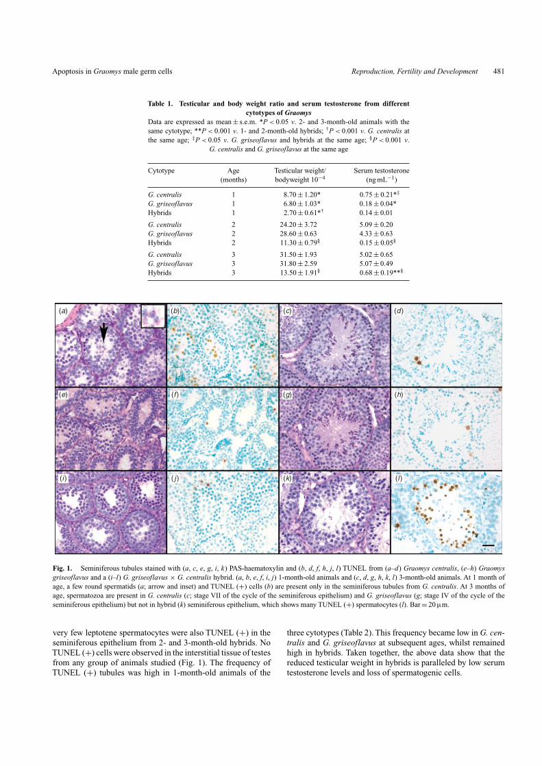
Table 1. Testicular and body weight ratio and serum testosterone from differentcytotypes of Graomys
Data are expressed as mean ± s.e.m. *P < 0.05 v. 2- and 3-month-old animals with thesame cytotype; **P < 0.001 v. 1- and 2-month-old hybrids; †P < 0.001 v. G. centralis atthe same age; ‡P < 0.05 v. G. griseoflavus and hybrids at the same age; §P < 0.001 v.
G. centralis and G. griseoflavus at the same age
Cytotype Age Testicular weight/ Serum testosterone(months) bodyweight 10−4 (ng mL−1)
G. centralis 1 8.70 ± 1.20* 0.75 ± 0.21*‡
G. griseoflavus 1 6.80 ± 1.03* 0.18 ± 0.04*Hybrids 1 2.70 ± 0.61*† 0.14 ± 0.01
G. centralis 2 24.20 ± 3.72 5.09 ± 0.20G. griseoflavus 2 28.60 ± 0.63 4.33 ± 0.63Hybrids 2 11.30 ± 0.79§ 0.15 ± 0.05§
G. centralis 3 31.50 ± 1.93 5.02 ± 0.65G. griseoflavus 3 31.80 ± 2.59 5.07 ± 0.49Hybrids 3 13.50 ± 1.91§ 0.68 ± 0.19**§
(a) (b) (c) (d )
(e) (f ) (g) (h)
(i ) ( j ) (k) (l)
Fig. 1. Seminiferous tubules stained with (a, c, e, g, i, k) PAS-haematoxylin and (b, d, f, h, j, l) TUNEL from (a–d) Graomys centralis, (e–h) Graomysgriseoflavus and a (i–l) G. griseoflavus × G. centralis hybrid. (a, b, e, f, i, j) 1-month-old animals and (c, d, g, h, k, l) 3-month-old animals. At 1 month ofage, a few round spermatids (a; arrow and inset) and TUNEL (+) cells (b) are present only in the seminiferous tubules from G. centralis. At 3 months ofage, spermatozoa are present in G. centralis (c; stage VII of the cycle of the seminiferous epithelium) and G. griseoflavus (g; stage IV of the cycle of theseminiferous epithelium) but not in hybrid (k) seminiferous epithelium, which shows many TUNEL (+) spermatocytes (l). Bar = 20 µm.
very few leptotene spermatocytes were also TUNEL (+) in theseminiferous epithelium from 2- and 3-month-old hybrids. NoTUNEL (+) cells were observed in the interstitial tissue of testesfrom any group of animals studied (Fig. 1). The frequency ofTUNEL (+) tubules was high in 1-month-old animals of the
three cytotypes (Table 2). This frequency became low in G. cen-tralis and G. griseoflavus at subsequent ages, whilst remainedhigh in hybrids. Taken together, the above data show that thereduced testicular weight in hybrids is paralleled by low serumtestosterone levels and loss of spermatogenic cells.
482 Reproduction, Fertility and Development V. Rodriguez et al.
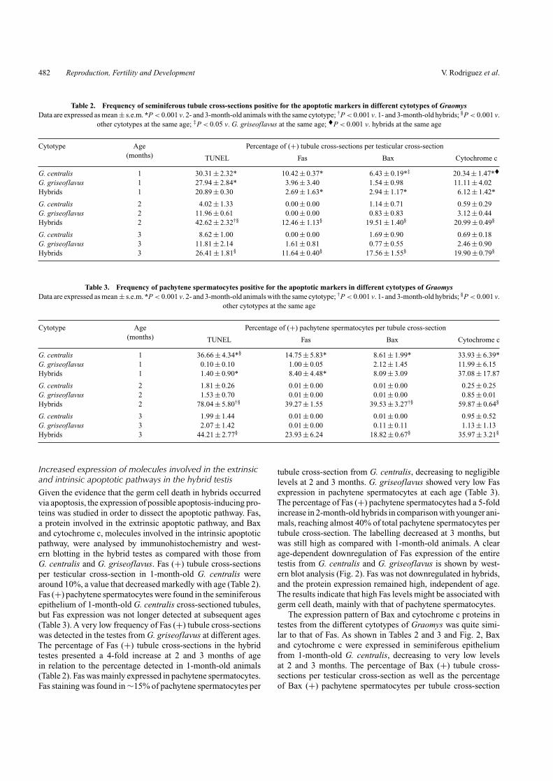
Table 2. Frequency of seminiferous tubule cross-sections positive for the apoptotic markers in different cytotypes of GraomysData are expressed as mean ± s.e.m. *P < 0.001 v. 2- and 3-month-old animals with the same cytotype; †P < 0.001 v. 1- and 3-month-old hybrids; §P < 0.001 v.
other cytotypes at the same age; ‡P < 0.05 v. G. griseoflavus at the same age; �P < 0.001 v. hybrids at the same age
Cytotype Age Percentage of (+) tubule cross-sections per testicular cross-section(months) TUNEL Fas Bax Cytochrome c
G. centralis 1 30.31 ± 2.32* 10.42 ± 0.37* 6.43 ± 0.19*‡ 20.34 ± 1.47*�G. griseoflavus 1 27.94 ± 2.84* 3.96 ± 3.40 1.54 ± 0.98 11.11 ± 4.02Hybrids 1 20.89 ± 0.30 2.69 ± 1.63* 2.94 ± 1.17* 6.12 ± 1.42*
G. centralis 2 4.02 ± 1.33 0.00 ± 0.00 1.14 ± 0.71 0.59 ± 0.29G. griseoflavus 2 11.96 ± 0.61 0.00 ± 0.00 0.83 ± 0.83 3.12 ± 0.44Hybrids 2 42.62 ± 2.32†§ 12.46 ± 1.13§ 19.51 ± 1.40§ 20.99 ± 0.49§
G. centralis 3 8.62 ± 1.00 0.00 ± 0.00 1.69 ± 0.90 0.69 ± 0.18G. griseoflavus 3 11.81 ± 2.14 1.61 ± 0.81 0.77 ± 0.55 2.46 ± 0.90Hybrids 3 26.41 ± 1.81§ 11.64 ± 0.40§ 17.56 ± 1.55§ 19.90 ± 0.79§
Table 3. Frequency of pachytene spermatocytes positive for the apoptotic markers in different cytotypes of GraomysData are expressed as mean ± s.e.m. *P < 0.001 v. 2- and 3-month-old animals with the same cytotype; †P < 0.001 v. 1- and 3-month-old hybrids; §P < 0.001 v.
other cytotypes at the same age
Cytotype Age Percentage of (+) pachytene spermatocytes per tubule cross-section(months) TUNEL Fas Bax Cytochrome c
G. centralis 1 36.66 ± 4.34*§ 14.75 ± 5.83* 8.61 ± 1.99* 33.93 ± 6.39*G. griseoflavus 1 0.10 ± 0.10 1.00 ± 0.05 2.12 ± 1.45 11.99 ± 6.15Hybrids 1 1.40 ± 0.90* 8.40 ± 4.48* 8.09 ± 3.09 37.08 ± 17.87
G. centralis 2 1.81 ± 0.26 0.01 ± 0.00 0.01 ± 0.00 0.25 ± 0.25G. griseoflavus 2 1.53 ± 0.70 0.01 ± 0.00 0.01 ± 0.00 0.85 ± 0.01Hybrids 2 78.04 ± 5.80†§ 39.27 ± 1.55 39.53 ± 3.27†§ 59.87 ± 0.64§
G. centralis 3 1.99 ± 1.44 0.01 ± 0.00 0.01 ± 0.00 0.95 ± 0.52G. griseoflavus 3 2.07 ± 1.42 0.01 ± 0.00 0.11 ± 0.11 1.13 ± 1.13Hybrids 3 44.21 ± 2.77§ 23.93 ± 6.24 18.82 ± 0.67§ 35.97 ± 3.21§
Increased expression of molecules involved in the extrinsicand intrinsic apoptotic pathways in the hybrid testisGiven the evidence that the germ cell death in hybrids occurredvia apoptosis, the expression of possible apoptosis-inducing pro-teins was studied in order to dissect the apoptotic pathway. Fas,a protein involved in the extrinsic apoptotic pathway, and Baxand cytochrome c, molecules involved in the intrinsic apoptoticpathway, were analysed by immunohistochemistry and west-ern blotting in the hybrid testes as compared with those fromG. centralis and G. griseoflavus. Fas (+) tubule cross-sectionsper testicular cross-section in 1-month-old G. centralis werearound 10%, a value that decreased markedly with age (Table 2).Fas (+) pachytene spermatocytes were found in the seminiferousepithelium of 1-month-old G. centralis cross-sectioned tubules,but Fas expression was not longer detected at subsequent ages(Table 3). A very low frequency of Fas (+) tubule cross-sectionswas detected in the testes from G. griseoflavus at different ages.The percentage of Fas (+) tubule cross-sections in the hybridtestes presented a 4-fold increase at 2 and 3 months of agein relation to the percentage detected in 1-month-old animals(Table 2). Fas was mainly expressed in pachytene spermatocytes.Fas staining was found in ∼15% of pachytene spermatocytes per
tubule cross-section from G. centralis, decreasing to negligiblelevels at 2 and 3 months. G. griseoflavus showed very low Fasexpression in pachytene spermatocytes at each age (Table 3).The percentage of Fas (+) pachytene spermatocytes had a 5-foldincrease in 2-month-old hybrids in comparison with younger ani-mals, reaching almost 40% of total pachytene spermatocytes pertubule cross-section. The labelling decreased at 3 months, butwas still high as compared with 1-month-old animals. A clearage-dependent downregulation of Fas expression of the entiretestis from G. centralis and G. griseoflavus is shown by west-ern blot analysis (Fig. 2). Fas was not downregulated in hybrids,and the protein expression remained high, independent of age.The results indicate that high Fas levels might be associated withgerm cell death, mainly with that of pachytene spermatocytes.
The expression pattern of Bax and cytochrome c proteins intestes from the different cytotypes of Graomys was quite simi-lar to that of Fas. As shown in Tables 2 and 3 and Fig. 2, Baxand cytochrome c were expressed in seminiferous epitheliumfrom 1-month-old G. centralis, decreasing to very low levelsat 2 and 3 months. The percentage of Bax (+) tubule cross-sections per testicular cross-section as well as the percentageof Bax (+) pachytene spermatocytes per tubule cross-section
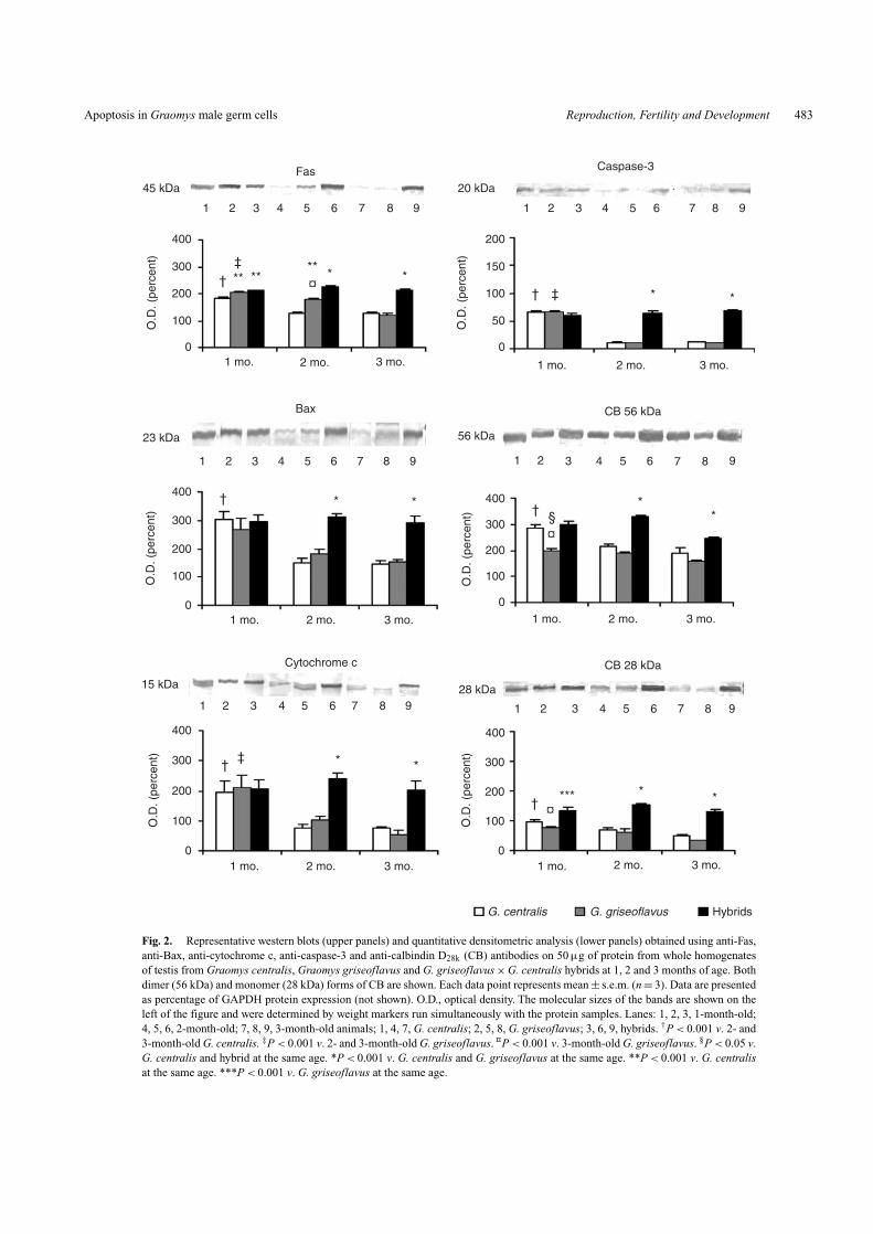
Apoptosis in Graomys male germ cells Reproduction, Fertility and Development 483
3 mo.
1 2 3 4 5 6 7 8 9
2 mo.
*†
†
†
‡
‡
‡†
†
*
*
*
* *¤
¤
¤
******
† *
*
1 mo. 1 mo. 3 mo.2 mo.
G. centralis G. griseoflavus Hybrids
3 mo.2 mo.1 mo.3 mo.2 mo.1 mo.
3 mo.2 mo.1 mo. 3 mo.2 mo.1 mo.
1 2 3 4 5 6 7 8 9
1 2 3 4 5 6 7 8 9
0
100
200
300
400
400
400
300
200
100
0
300
200
O.D
. (pe
rcen
t)O
.D. (
perc
ent)
200
150
100
50
0
O.D
. (pe
rcen
t)
100
0
15 kDa 28 kDa
23 kDa
1 2 3 4 5 6 7 8 9 1 2 3 4 5 6 7 8 9
1 2 3 4 5 6 7 8 9
**
§
*** * *
45 kDa 20 kDa
56 kDa
Caspase-3
CB 56 kDa
CB 28 kDaCytochrome c
Bax
Fas
O.D
. (pe
rcen
t)
0
100
200
300
400
O.D
. (pe
rcen
t)
0
100
200
300
400
O.D
. (pe
rcen
t)
Fig. 2. Representative western blots (upper panels) and quantitative densitometric analysis (lower panels) obtained using anti-Fas,anti-Bax, anti-cytochrome c, anti-caspase-3 and anti-calbindin D28k (CB) antibodies on 50 µg of protein from whole homogenatesof testis from Graomys centralis, Graomys griseoflavus and G. griseoflavus × G. centralis hybrids at 1, 2 and 3 months of age. Bothdimer (56 kDa) and monomer (28 kDa) forms of CB are shown. Each data point represents mean ± s.e.m. (n = 3). Data are presentedas percentage of GAPDH protein expression (not shown). O.D., optical density. The molecular sizes of the bands are shown on theleft of the figure and were determined by weight markers run simultaneously with the protein samples. Lanes: 1, 2, 3, 1-month-old;4, 5, 6, 2-month-old; 7, 8, 9, 3-month-old animals; 1, 4, 7, G. centralis; 2, 5, 8, G. griseoflavus; 3, 6, 9, hybrids. †P < 0.001 v. 2- and3-month-old G. centralis. ‡P < 0.001 v. 2- and 3-month-old G. griseoflavus. ¤P < 0.001 v. 3-month-old G. griseoflavus. §P < 0.05 v.G. centralis and hybrid at the same age. *P < 0.001 v. G. centralis and G. griseoflavus at the same age. **P < 0.001 v. G. centralisat the same age. ***P < 0.001 v. G. griseoflavus at the same age.

484 Reproduction, Fertility and Development V. Rodriguez et al.
(a) (b) (c)
(d ) (e) (f )
(g) (h) (i )
Fig. 3. Seminiferous tubules showing (a, b, c) Fas, (d, e, f ) Bax and (g, h, i) cytochrome c labelling from (a, d, g) 1-month-old, (b, e, h) 2-month-old and(c, f, i) 3-month-old hybrids. Cells positive for the apoptotic markers are spermatocytes (arrows) as shown in the insets. Bar = 20 µm.
was low in G. griseoflavus at different ages (Tables 2 and 3).Cytochrome c expression was much higher in 1-month-oldG. griseoflavus than in 2- and 3-month-old animals. A 6-foldincrease of Bax expression in tubule cross-sections per tes-ticular cross-section was found in 2-month-old hybrids ascompared with younger animals. The magnitude of the enhance-ment was significant considering that almost 40% of pachytenespermatocytes per tubule cross-section were positive for Baxstaining (Table 3). The percentage of cytochrome c (+) sem-iniferous tubule cross-sections per testicular cross-section wasincreased 3-fold in 2-month-old hybrids in comparison with thatof younger hybrids, reaching almost 60% of pachytene sper-matocytes per tubule cross-section. This expression declinedmoderately one month later. In hybrids, the main expressionof these apoptotic molecules occurred in the nucleus and thecytoplasm of pachytene spermatocytes, which means a redis-tribution of these molecules (Fig. 3). A positive signal for Fasbut negative for all the other apoptotic markers analysed wasalso revealed in Leydig cells from testes of the three cyto-types (data not shown, Fig. 3), indicating absence of cell death.
As occurs with Fas expression, western blot analysis revealsthat Bax and cytochrome c expression is downregulated inan age-dependent manner in the entire testis of both G. cen-tralis and G. griseoflavus. Similarly to Fas expression, hybridspresented lack of this regulation of Bax and cytochrome c expres-sion in the total testis (Fig. 2). High expression of Bax andcytochrome c occurring concomitant with the increased appear-ance of apoptotic germ cells suggests that the intrinsic pathwaywould be involved in the mechanism of germ cell death. Giventhe observation that cytochrome c was redistributed, indicatingcytochrome c release from mitochondria, we then examinedthe expression of the executioner caspase-3. The expressionof the pro-enzyme form of caspase-3 was similar in the totaltestis from all 1-month-old animals, regardless of the Graomyscytotype. Pro-caspase expression decreased in 2-month-oldG. centralis and G. griseoflavus, whereas in the hybrids expres-sion remained high and became significantly higher one monthlater (data not shown). Expression of the active form of caspase-3decreased markedly with age, both in G. centralis and inG. griseoflavus. By contrast, expression of the active caspase-3
Apoptosis in Graomys male germ cells Reproduction, Fertility and Development 485
was high in hybrids, irrespective of age (Fig. 2). Therefore, theresults indicate that apoptosis of the pachytene spermatocytes inhybrids involves both the extrinsic and the intrinsic pathways ina caspase-dependent manner.
Most of CB overexpression occurred in pachytenespermatocytes negative for apoptotic markersCB is a high-affinity calcium-binding protein proposed to be ananti-apoptotic molecule that can prevent or block apoptotic celldeath in testis (Merico et al. 2008). Its expression was analysedby immunohistochemistry and western blot in testicular tissuefrom Graomys with different cytotypes. As previously foundin mice (Merico et al. 2008), this protein dimerises in testisshowing two bands (28 kDa and 56 kDa). CB expression wasdetected in pachytene spermatocytes from 1-month-old G. cen-tralis, decreasing to negligible levels with age (data not shown).Very low levels of CB protein were observed in pachytene sper-matocytes from G. griseoflavus at all ages (data not shown).In hybrids, the percentage of CB (+) seminiferous tubules pertesticular cross-section had a 4-fold increase in 2- or 3-month-old hybrids as compared with the younger animals, reachingalmost 62% of pachytene spermatocytes per tubule cross-sectionat 2 months (Table 4, Fig. 4b). Both the CB monomer and thedimer expression was higher in hybrids as compared with theparental cytotypes (Fig. 2), with its most intense expression con-comitant with the highest levels of apoptosis in 2-month-oldanimals. However, its co-localisation with the apoptotic markersin pachytene spermatocytes was low, indicating that CB localisesin different pachytene spermatocytes (Table 4, Fig. 4a, b).
Discussion
The aim of the present study was to determine the extent ofspermatogenic impairment and the cellular and apoptotic mech-anisms involved in establishing the sterility of male hybrids(2n = 38) obtained from crossing G. griseoflavus (2n = 34)females with G. centralis (2n = 42) males. Histological analysisof the seminiferous epithelium of the male hybrids shows that thespermatogenic process is arrested at the meiotic stage, with onlyrare spermatids present in very few seminiferous tubules. Ourfindings show that disruption of spermatogenesis at meiosis I,leading to the absence of mature spermatozoa, is the main causeof sterility in hybrids between G. griseoflavus × G. centralis.
The massive death of pachytene spermatocytes detected inthe seminiferous tubules of hybrid males occurs via apoptosis,as revealed by high expression of the executioner caspase-3,which is triggered by both extrinsic and intrinsic pathways. Infact, proteins of both pathways (i.e. Fas, Bax and cytochrome c),as detected by western blot, were maintained highly expressedin the testes of hybrids whereas they were downregulated inparental males of the same age. This lack of downregulation wasreflected by a high frequency of Fas, Bax and cytochrome c (+)spermatocytes, as revealed by immunohistochemical analysis,on testicular sections from 2- and 3-month-old hybrids.
Moderate apoptosis of germ cells was detected in spermato-cytes from 1-month-old G. centralis, but not in G. griseoflavus orG. griseoflavus × G. centralis testes. However, markers of apop-tosis were found to be expressed in pachytene spermatocytes inall three cytotypes. Apoptosis of pachytene spermatocytes has
been described as a physiological process during the first wave ofspermatogenesis (Jahnukainen et al. 2004; Lizama et al. 2007).The negligible presence of TUNEL (+) spermatocytes in bothG. griseoflavus and G. griseoflavus × G. centralis seminiferoustubule cross-sections might be due to a difference in the onset ofthe first wave of spermatogenesis among cytotypes. In fact, inG. centralis, in which post-meiotic cells were already presentin the seminiferous epithelium of some tubule cross-sections,a slight, although significant, higher level of testosterone wasdetected, compared with the other two cytotypes, in whichmainly leptotene (in G. griseoflavus) and early-pachytene (inhybrids) cells were present in the seminiferous tubules (com-pare Fig. 1a with Fig. 1e and i). At subsequent ages, thetestosterone levels were very similar in both G. centralis andG. griseoflavus, whose seminiferous epithelia presented all thedifferentiative steps of the male germ cells, without prominentsigns of cell death. In fact, the apoptotic markers were down-regulated and the frequencies of germ cells expressing Fas, Bax,cytochrome c or positive for TUNEL staining were extremelylow, probably reflecting the physiological apoptosis that occursin normal testes of adult rodents (Huckins 1978; Allan et al.1992; Kerr 1992; Brinkworth et al. 1995; Blanco-Rodríguez andMartinez-Garcia 1996).
On the contrary, apoptosis remained relevant in the seminifer-ous epithelium of 2- and 3-month-old hybrid males in which 78%and 44% of pachytene spermatocytes were found to be positivefor TUNEL staining, respectively. In these animals, spermatoge-nesis is interrupted at the meiotic pachytene stage, which makesit difficult to determine the stage of the cycle of the seminiferousepithelium at which meiotic cell death occurs. The breakdown ofspermatogenesis in Graomys hybrids is very severe, with mas-sive germ cell depletion; in fact, only very few spermatids weredetected in the seminiferous epithelium. In 2- and 3-month-oldhybrids, both the intrinsic and extrinsic markers of apoptosisremained highly expressed, as evidenced by western blots and thefrequency of Fas, Bax and cytochrome c (+) spermatocytes. Inprevious papers, we reported germ cell depletion of both meioticand postmeiotic stages of differentiation in adult Rb heterozy-gous mice (Merico et al. 2003, 2008). We also showed that theintrinsic apoptotic pathway was involved in germ cell death,although other apoptotic mechanisms were not excluded (Mericoet al. 2008). Adult hybrids have low levels of testosterone,although we did not find anyTUNEL (+) Leydig cells.These lowlevels of testosterone might contribute to the detrimental effectsobserved in hybrid spermatogenesis, since testosterone has beenshown to be a critical germ cell survival factor (reviewed inSofikitis et al. 2008). The lowering of intratesticular testosteronein a rat animal model resulted in arrest of spermiogenesis and inapoptosis of germ cells (O’Donnell et al. 1996; Sofikitis et al.1999; Kim et al. 2001). On the contrary, it has been shown thattestosterone is able to inhibit in vitro-induced apoptosis of humanspermatocytes and spermatids (Erkkilä et al. 1997). The mecha-nism by which testosterone withdrawal induces germ cell deathhas not yet been elucidated. Altered expression of anti-apoptoticBcl-xl and Bcl-2 molecules was found after intratesticular with-drawal of testosterone in the rat (Show et al. 2004). In the presentwork we did not study the expression of the Bcl-2 anti-apoptoticproteins, but we found high expression of Bax, a Bcl-2 family

486 Reproduction, Fertility and Development V. Rodriguez et al.
Table 4. CB expression and co-localisation with the apoptotic markers studied in hybrid males betweenGraomys griseoflavus × Graomys centralis
Data are expressed as mean ± s.e.m. *P < 0.001 v. 2- and 3-month-old; **P < 0.05 v. 1- and 3-month-old
Age Percentage of CB (+) seminiferous Percentage of CB (+) pachytene(months) tubules per testicular cross-section spermatocytes per tubule cross-section
1 5.26 ± 1.66* 23.47 ± 11.562 23.40 ± 0.54 61.93 ± 5.26**3 23.15 ± 1.70 33.59 ± 1.39
Age Percentage of CB (+) pachytene spermatocytes per tubule cross-section showing co-localisation(months) CB/TUNEL CB/Fas CB/Bax CB/cytochrome c
1 0.00 ± 0.00 1.66 ± 1.66 1.94 ± 1.05 10.86 ± 5.452 14.28 ± 2.30** 4.71 ± 1.28 9.44 ± 1.25** 15.20 ± 5.463 6.44 ± 0.05 0.18 ± 0.18 2.59 ± 0.68 4.20 ± 0.45
(a) (b)
Fig. 4. Seminiferous tubules with (a) TUNEL and (b) calbindin D28k labelling from 2-month-old G. griseo-flavus × G. centralis hybrid. Most cells positive for TUNEL and calbindin D28k (CB) are spermatocytes (arrows)as shown in the insets. The asterisk marks co-localisation of CB and TUNEL labelling of a spermatocyte. MostTUNEL (+) cells are CB (−). Bar = 20 µm.
member known to block the ability of Bcl-2 to inhibit apopto-sis (Oltval et al. 1993; Chittenden et al. 1995), in hybrids of allages. Conversely, in adult parental cytotypes Bax expression isdownregulated.
Fas was highly expressed in adult hybrids. When it is acti-vated, it promotes processing and stabilisation of caspase-8(Scaffidi et al. 1998; Sánchez-Gómez et al. 2003; Henkler et al.2005). Active caspase-8 can proteolitically activate caspase-3,-6 or -7 leading to cell death due to the degradation of manycellular proteins (Riedl and Shi 2004). In mouse, rat and human,the Fas system has been implicated as a possible key regulatorof germ cell apoptosis in physiological and injured testes (Leeet al. 1997; Pentikäinen et al. 1999; Koji et al. 2001; Celik-Ozenci et al. 2006; Lizama et al. 2007). In particular, Fas hasbeen proposed to play a central role in determining pachytenespermatocyte apoptosis during the first wave of spermatogenesisin the rat (Lizama et al. 2007). We also found Fas expression inG. centralis and to a lesser extent in hybrid spermatocytes of onemonth of age, confirming the extrinsic pathway to be involvedin apoptosis at the onset of the process.
Both intrinsic and extrinsic pathways converge on caspase-3,which was found to be highly expressed in testes from
1-month-old males from any cytotype and in adult hybrids,in which a high frequency of TUNEL (+) spermatocytes waspresent. We describe for the first time the involvement of boththe intrinsic and extrinsic pathways in determining apoptosis inthe testes of adult hybrid animals derived from the mating oftwo different cytotypes of the genus Graomys and not inducedby testis injury, either chemical or physical.
To maintain testicular homeostasis, pro-survival and pro-apoptotic molecules work together, regulating the extent of apop-tosis to produce gametes of high quality (Mishra et al. 2006). Wehave studied the expression of CB, an anti-apoptotic molecule,which has been suggested to protect different cell types againstapoptotic cell death induced by both calcium-independent (bythe inhibition of caspase activity) and calcium-dependent path-ways (Christakos and Liu 2004; Choi et al. 2008). Here weshow that CB is overexpressed in pachytene spermatocytes fromhybrid males, confirming that CB is present in the same cell typethat undergoes DNA fragmentation. However, not all pachytenespermatocytes overexpress CB; it appears that a subgroup ofthese cells are CB (+). Similar results were reported in Rb het-erozygous mice, in which CB was found to be expressed inmetaphase spermatocytes, the cell type in which apoptosis takes
Apoptosis in Graomys male germ cells Reproduction, Fertility and Development 487
place (Merico et al. 2008). Thus, CB overexpression seems notto be dependent on the cell type. However, in both Graomyshybrids and Rb heterozygous mice, the frequency of cells thatshow simultaneous apoptosis and CB expression is very low.In Graomys hybrids, 62% or 34% of cells were CB positive,whereas only 14% and 6% showed co-localisation with TUNELin 2- and 3-month-old hybrids, respectively.Thus, the expressionof CB and that of the apoptotic markers seems to be inversely reg-ulated. The same pattern of expression has been found by LemaTomé et al. (2006) in a model of induced neuronal apoptosisin rats. These authors described immunoreactivity for activatedcaspase-3 in layers IV/V, between areas of high CB or calretininexpression. Also, in the caudate putamen, activated caspase-3did not invade zones of intense CB immunoreativity. However,it remains to be elucidated if the expression of CB represents aresponse to a surveillance mechanism for controlling cell deathbefore the onset of or during the apoptotic process.
This is the first study that describes (1) the timing of theonset of spermatogenesis in G. centralis and G. griseoflavus,useful knowledge in conservation biology studies and (2) thecellular and apoptotic mechanisms that are involved in deter-mining the spermatogenetic breakdown in hybrids between theancestral 2n = 42 cytotype and 2n = 34 cytotype derived throughRb fusions. We found that pachytene spermatocytes are themost sensitive cell type to apoptotic stimuli driven by boththe extrinsic and intrinsic pathways. Here we show that inG. griseoflavus × G. centralis hybrids a breakdown of spermato-genesis occurs at the meiotic stage of germ cell development,thus accounting for their sterility.
The present study contributes to the knowledge of the cellularbasis of spermatogenesis breakdown and the molecular mecha-nisms involved in germ cell apoptosis, which are important forunderstanding some aspects of male infertility.
AcknowledgementsThis work was supported by a Bilateral Grant (2005–2007) from SECYT-MAE (Argentina-Italia), and funds from FONCYT (PICT2005–32464),CONICET (PIP 2005–2006) and SECYT (UNC) (Dr Nori Tolosa de Talam-oni) and from FIRB 2005 (Project n. RBIP06FH7J), UNIPV-Regione Lom-bardia Project on Material Science and Biomedicine (Dr Silvia Garagna). DrNoriTolosa deTalamoni is a Member of Career from CONICET (Argentina).Valeria Rodriguez is a recipient of a fellowship from CONICET (Argentina).Special thanks are giving to Dr GerardoTheiler and investigators from Labo-ratory ‘Dr F. Cañas’ (Argentina) for helping with the capture of G. centralis,Dr Ricardo Ojeda and Daniela Rodriguez for helping with the capture ofG. griseoflavus and Dr Liliana Muñoz for testosterone determinations. Theauthors declare that there is no conflict of interest that would prejudice theimpartiality of this scientific work.
ReferencesAbercrombie, M. (1946). Estimation of nuclear population from microtome
sections. Anat. Rec. 94, 239–247. doi:10.1002/AR.1090940210Allan, D. J., Harmon, B. V., and Roberts, S. A. (1992). Spermatogonial
apoptosis has three morphologically recognizable phases and shows nocircadian rhythm during normal spermatogenesis in the rat. Cell Prolif.25, 241–250. doi:10.1111/J.1365-2184.1992.TB01399.X
Blanco-Rodríguez, J., and Martinez-Garcia, C. (1996). Spontaneous germcell death in the testis of the adult rat takes the form of apoptosis: re-evaluation of cell types that exhibit the ability to die during spermatogen-esis. Cell Prolif. 29, 13–31. doi:10.1111/J.1365-2184.1996.TB00091.X
Brinkworth, M. H., Weinbauer, G. F., Schlatt, S., and Nieschlag, E. (1995).Identification of male germ cells undergoing apoptosis in adult rats.J. Reprod. Fertil. 105, 25–33. doi:10.1530/JRF.0.1050025
Castiglia, R., and Capanna, E. (2000). Contact zone between chromoso-mal races of Mus musculus domesticus. 2. Fertility and segregationin laboratory-reared and wild mice heterozygous for multiple Robert-sonian rearrangements. Heredity 85, 147–156. doi:10.1046/J.1365-2540.2000.00743.X
Catanesi, C. I., Vidal-Rioja, L., Crisci, J. V., and Zambelli, A. (2002). Phy-logenetic relationships among Robertsonian karyomorpha of Graomysgriseoflavus (Rodentia, Muridae) by mitochondrial cytochrome b DNAsequencing. Hereditas 136, 130–136. doi:10.1034/J.1601-5223.2002.1360207.X
Celik-Ozenci, C., Sahin, Z., Ustunel, I., Akkoyunlu, G., Erdogru, T., Korgun,E. T., Baykara, M., and Demir, R. (2006). The Fas system may havea role in male reproduction. Fertil. Steril. 85(Suppl. 1), 1168–1178.doi:10.1016/J.FERTNSTERT.2005.08.058
Chittenden, T., Harrington, E. A., O’Connor, R., Flemington, C., Lutz, R. J.,Evan, G. I., and Guild, B. C. (1995). Induction of apoptosisby the Bcl-2 homologue Bak. Nature 374, 733–736. doi:10.1038/374733A0
Choi, W. S., Lee, E., Lim, J., and Oh, Y. J. (2008). Calbindin–D28k pre-vents drug-induced dopaminergic neuronal death by inhibiting caspaseand calpain activity. Biochem. Biophys. Res. Commun. 371, 127–131.doi:10.1016/J.BBRC.2008.04.020
Christakos, S., and Liu, Y. (2004). Biological actions and mechanism ofaction of calbindin in the process of apoptosis. J. Steroid Biochem. Mol.Biol. 89–90, 401–404. doi:10.1016/J.JSBMB.2004.03.007
Coureuil, M., Fouchet, P., Prat, M., Letallec, B., Barroca, V., DosSantos, C., Racine, C., and Allemand, I. (2006). Caspase-independentdeath of meiotic and postmeiotic cells overexpressing p53: calpaininvolvement. Cell Death Differ. 13, 1927–1937. doi:10.1038/SJ.CDD.4401887
Erkkilä, K., Henriksen, K., Hirvonen,V., Rannikko, S., Salo, J., Parvinen, M.,and Dunkel, L. (1997). Testosterone regulates apoptosis in adult humanseminiferous tubule in vitro. J. Clin. Endocrinol. Metab. 82, 2314–2321.doi:10.1210/JC.82.7.2314
Ford, C. E., and Hamerton, J. L. (1956). A colchicine, hypotonic cit-rate, squash sequence for mammalian chromosomes. Stain Technol. 31,247–251.
Gardner, A. L., and Patton, J. L. (1976). Karyotypic variation in oryzominerodents (Cricetidae) with comments on chromosomal evolution in theNeotropical cricetinae complex. Occas. Pap. Mus. Zool. La. State Univ.49, 1–48.
Gornall, A. G., Bardawill, C. J., and David, M. M. (1949). Determinationof serum proteins by means of the Biuret reaction. J. Biol. Chem. 177,751–766.
Hauffe, H. C., and Searle, J. B. (1998). Chromosomal heterozygosity andfertility in house mice (Mus musculus domesticus) from Northern Italy.Genetics 150, 1143–1154.
Henkler, F., Behrle, E., Dennehy, K. M., Wicovsky, A., Peters, N., Warnke,C., Pfizenmaier, K., and Wajant, H. (2005). The extracellular domainsof FasL and Fas are sufficient for the formation of supramolecu-lar FasL–Fas clusters of high stability. J. Cell Biol. 168, 1087–1098.doi:10.1083/JCB.200501048
Huckins, C. (1978). The morphology and kinetics of spermatogonialdegeneration in normal adult rats: an analysis using a simplifiedclassification of the germinal epithelium. Anat. Rec. 190, 905–926.doi:10.1002/AR.1091900410
Jahnukainen, K., Crysis, D., Hou, M., Parvinen, M., Eksborg, S., andSoder, O. (2004). Increased apoptosis occurring during the firstwave of spermatogenesis is stage-specific and primarily affects midpachytene spermatocytes in the rat testis. Biol. Reprod. 70, 290–296.doi:10.1095/BIOLREPROD.103.018390

488 Reproduction, Fertility and Development V. Rodriguez et al.
Kerr, J. B. (1992). Spontaneous degeneration of germ cells in normal rattestis: assessment of cell types and frequency during the spermatogeniccycle. J. Reprod. Fertil. 95, 825–830. doi:10.1530/JRF.0.0950825
Kim, J. M., Ghosh, S. R., Weil, A. C., and Zirkin, B. R. (2001). Caspase-3and caspase-activated deoxyribonuclease are associated with testiculargerm cell apoptosis resulting from reduced intratesticular testosterone.Endocrinology 142, 3809–3816. doi:10.1210/EN.142.9.3809
Koji, T., Hishikawa, Y., Ando, H., Nakanishi, Y., and Kobayashi, N. (2001).Expression of Fas and Fas ligand in normal and ischemia-reperfusiontestes: involvement of the Fas system in the induction of germ cellapoptosis in the damaged mouse testis. Biol. Reprod. 64, 946–954.doi:10.1095/BIOLREPROD64.3.946
Lanzone, C., Novillo, A., Suarez, N. S., and Ojeda, R. A. (2007). Cyto-genetics and redescription of Graomys (Rodentia, Sigmodontinae)from Chumbicha, Catamarca,Argentina. Mastozoologia Neotropical 14,249–255.
Lee, J., Richburg, J. H., Younkin, S. C., and Boekelheide, K. (1997). TheFas system is a key regulator of germ cell apoptosis in the testis.Endocrinology 138, 2081–2088. doi:10.1210/EN.138.5.2081
Lee, N. P. Y., Leung, K. W., Wo, J. Y., Tam, P. C., Yeung, W. S. B., andLuk, J. M. (2006). Blockage of testicular connexins induced apoptosisin rat seminiferous epithelium. Apoptosis 11, 1215–1229. doi:10.1007/S10495-006-6981-2
Lema Tomé, C. M., Bauer, C., Nottingham, C., Smith, C., Blackstone,K., et al. (2006). Mk801-induced caspase-3 in the postnatal brain:inverse relationship with calcium binding proteins. Neuroscience 141,1351–1363. doi:10.1016/J.NEUROSCIENCE.2006.05.019
Lizama, C., Alfaro, I., Reyes, J. G., and Moreno, R. D. (2007). Up-regulationof CD95 (Apo-1/Fas) is associated with spermatocyte apoptosis duringthe first round of spermatogenesis in the rat. Apoptosis 12, 499–512.doi:10.1007/S10495-006-0012-1
Merico, V., Pigozzi, M. I., Esposito, A., Merani, M. S., and Garagna, S.(2003). Meiotic recombination and spermatogenic impairment in Musmusculus domesticus carrying multiple simple Robertsonian transloca-tions. Cytogenet. Genome Res. 103, 321–329. doi:10.1159/000076820
Merico, V., Diaz de Barboza, G., Vasco, C., Ponce, R., Rodriguez, V.,Garagna, S., and Tolosa de Talamoni, N. (2008). A mitochondrial mech-anism is involved in apoptosis of Robertsonian mouse male germ cells.Reproduction 135, 797–804. doi:10.1530/REP-07-0466
Mishra, D. P., Pal, R., and Shaha, C. (2006). Changes in cytosolicCa2+ levels regulate Bcl2-xS and Bcl2-xL expression in spermato-genetic cells during apoptotic death. J. Biol. Chem. 281, 2133–2143.doi:10.1074/JBC.M508648200
Mizuno, K., Hayashi, Y., Kojima, Y., Nakane, A., Tozawa, K., and Kohri, K.(2009). Activation of NF-kappa B associated with germ cell apoptosisin testes of experimentally inducer cryptorchid rat model. Urology 73,389–393. doi:10.1016/J.UROLOGY.2008.09.019
Musser, G. G., and Carleton, M. D. (2005). Superfamily Muroidea. In ‘Mam-mal Species of the World. A Taxonomic and Geographic Reference’. 2ndedn. (Eds D. E. Wilson and D. M. Reeder.) pp. 687–752. (SmithsonianInstitution Press: Washington DC and London.)
O’Donnell, L., McLachlan, R. I., Wreford, N. G., de Kretser, D. M., andRobertson, D. M. (1996). Testosterone withdrawal promotes stage-specific detachment of round spermatids from the rat seminiferousepithelium. Biol. Reprod. 55, 895–901. doi:10.1095/BIOLREPROD55.4.895
Oltval, Z. N., Milliman, C. L., and Korsmeyer, S. J. (1993). Bcl-2 het-erodimerizes in vivo with a conserved homolog, Bax, that acceleratesprogrammed cell death. Cell 74, 609–619. doi:10.1016/0092-8674(93)90509-O
Pentikäinen, V., Erkkilä, K., and Dunkel, L. (1999). Fas regulates germ cellapoptosis in the human testis in vitro. Am. J. Physiol. Endocrinol. Metab.276, E310–E316.
http://www.publish.csiro.au/journals/rfd
Petre-Lazar, B., Livera, G., Moreno, S. G., Trautmann, E., Duquenne, C.,Hanoux, V., Habert, R., and Coffigny, H. (2007). The role of p63 ingerm cell apoptosis in the developing testis. J. Cell. Physiol. 210, 87–98.doi:10.1002/JCP.20829
Redi, C. A., and Capanna, E. (1988). Robertsonian heterozygotes in thehouse mouse and the fate of their germ cells. In ‘Cytogenetics of Mam-malianAutosomal Rearrangements’. (Ed.A. Daniel.) pp. 315–359. (AlanR Liss: New York.)
Redi, C. A., Garagna, S., Hilscher, B., and Winking, H. (1985). The effectsof some Robertsonian chromosome combinations on the seminiferousepithelium of the mouse. J. Embryol. Exp. Morphol. 85, 1–19.
Riedl, S. J., and Shi, Y. (2004). Molecular mechanisms of caspase regula-tion during apoptosis. Nat. Rev. Mol. Cell Biol. 5, 897–907. doi:10.1038/NRM1496
Sánchez-Gómez, M. V., Alberdi, E., Ibarretxe, G., Torre, I., and Matute,C. (2003). Caspase-dependent and caspase-independent oligodendro-cyte death mediated by AMPA and kainate receptors. J. Neurosci. 23,9519–9528.
Scaffidi, C., Fulda, S., Srinivasan, A., Friesen, C., Li, F., Tomaselli, K. J.,Debatin, K. M., Krammer, P. H., and Peter, M. E. (1998). TwoCD95 (APO-1/Fas) signalling pathways. EMBO J. 17, 1675–1687.doi:10.1093/EMBOJ/17.6.1675
Show, M. D., Folmer, J. S., Anway, M. D., and Zirkin, B. R. (2004). Testicularexpression and distribution of the rat bcl2 modifying factor in responseto reduced intratesticular testosterone. Biol. Reprod. 70, 1153–1161.doi:10.1095/BIOLREPROD.103.023200
Sofikitis, N., Ono, K., Yamamoto, Y., Papadopoulos, H., and Miyagawa, I.(1999). Influence of the male reproductive tract on the reproductivepotential of round spermatids abnormally released from the seminifer-ous epithelium. Hum. Reprod. 14, 1998–2006. doi:10.1093/HUMREP/14.8.1998
Sofikitis, N., Giotitsas, N., Tsounapi, P., Baltoqiannis, D., Giannakis, D.,and Pardalidis, N. (2008). Hormonal regulation of spermatogenesisand spermiogenesis. J. Steroid Biochem. Mol. Biol. 109, 323–330.doi:10.1016/J.JSBMB.2008.03.004
Theiler, G. R., and Blanco, A. (1996). Patterns of evolution in Graomysgriseoflavus (Rodentia, Muridae): II. Reproductive isolation betweencytotypes. J. Mammal. 77, 776–784. doi:10.2307/1382683
Theiler, G. R., Gardenal, C. N., and Blanco,A. (1999a). Patterns of evolutionin Graomys griseoflavus (Rodentia, Muridae). IV.A case of rapid specia-tion. J. Evol. Biol. 12, 970–979. doi:10.1046/J.1420-9101.1999.00097.X
Theiler, G. R., Ponce, R. H., Fretes, R. E., and Blanco,A. (1999b). Reproduc-tive barriers between the 2n = 42 and 2n = 36–38 cytotype of Graomys(Rodentia, Muridae). Mastozoologia Neotropical 6, 129–133.
Wallace, B. M., Searle, J. B., and Everett, C. A. (2002). The effect of multiplesimple Robertsonian heterozygosity on chromosome pairing and fertil-ity of wild-stock house mice (Mus musculus domesticus). Cytogenet.Genome Res. 96, 276–286. doi:10.1159/000063054
Zambelli, A., and Vidal-Rioja, L. (1999). Molecular events during chromo-somal divergence of the South American rodent Graomys griseoflavus.Acta Theriol. 44, 345–352.
Zambelli, A., Vidal-Rioja, L., and Wainberg, R. (1994). Cytogenetic anal-ysis of autosomal polymorphism in Graomys griseoflavus (Rodentia,Cricetidae). Z. Saugetierkd. 59, 14–20.
Zambelli, A., Catanesi, C. I., and Vidal-Rioja, L. (2003). Autosomal rear-rangements in Graomys griseoflavus (Rodentia): a model of non-randomRobertsonian divergence. Hereditas 139, 167–173. doi:10.1111/J.1601-5223.2003.01791.X
Manuscript received 24 April 2009, accepted 3 September 2009