Biologics, Skin Substitutes, Biomembranes and Scaffolds - MDPI
Upload
khangminh22Category
view
4download
0
Full PaPer
1700251 (1 of 12) © 2017 WILEY-VCH Verlag GmbH & Co. KGaA, Weinheim
www.mbs-journal.de
Novel 3D Neuron Regeneration Scaffolds Based on Synthetic Polypeptide Containing Neuron Cue
Zhen-Hua Wang, Yen-Yu Chang, Jhih-Guang Wu, Chia-Yu Lin, Hsiao-Lung An, Shyh-Chyang Luo, Tang K. Tang, and Wei-Fang Su*
Z.-H. Wang, Y.-Y. Chang, J.-G. Wu, C.-Y. Lin, Prof. S.-C. Luo, Prof. W.-F. SuDepartment of Materials Science and EngineeringNational Taiwan UniversityTaipei 10617, TaiwanE-mail: [email protected]. An, Dr. T. K. TangInstitute of Biomedical SciencesAcademia Sinica, Taipei 11529, TaiwanProf. S.-C. Luo, Prof. W.-F. SuMolecular Image CenterNational Taiwan UniversityTaipei 10617, Taiwan
The ORCID identification number(s) for the author(s) of this article can be found under https://doi.org/10.1002/mabi.201700251.
DOI: 10.1002/mabi.201700251
of affected people. The neural tissue dys-function has been observed in millions of people who suffer from stroke, trau-matic injury of brain, eye, spinal cord, and neurodegenerative diseases such as Alzheimer’s disease, Parkinson’s disease, and Huntington’s disease.[1,2] Recently, neural tissue engineering has become a potential technology to restore the func-tionality of damaged neural tissue with the hope to cure the patients with neural disorder and to improve their quality of life.[3,4] The tissue engineering involves the development of biological compat-ible scaffold that serves as substrate for cell growth, differentiation, and biological function.[5,6] The ideal material for scaf-folds should meet the following criteria: (1) biodegradable, (2) no cytotoxicity, (3) capable to promote cell–substrate inter-actions, (4) chemically compatible with aqueous solutions and physiological con-ditions, and (5) easy to scale up.[7,8]
Ideal 3D structured scaffold should function as biomimetic and hierarchical structures of extracellular matrix so that the cells are in 3D environment to recon-
struct the innate structure–function relationship through cell adhesion and cellular interactions.[9,10] Many synthetic polymers are biocompatible and biodegradable. They have been studied for the application in nerve regeneration. Examples are polyg-lycolic acid (PGA),[11] poly(d,l-lactide-co-ε-caprolactone),[12] poly-l-lactic acid,[13] poly lactic glycolic acid,[14] poly(3-hydroxybu-tyrate),[15] polycaprolactone,[16] poly(trimethylene carbonate-co-caprolactone),[17] and hydrogel.[18] However, these polymers do not contain biochemical cue to stimulate the neurite growth and they need to be modified with protein, laminin, collagen, hyaluronic acid, polysaccharides,[19–21] and others to enhance cell adhesion and tracking of neurites along underlying polymer substrates.[22] The synthetic polymers not derived from biological sources may elicit immune responses and inflam-mation. Self-assembling oligo-peptide scaffolds have been reported[23–25] to support extensive neurite outgrowth without negative immune response. The peptide was synthesized by linking amino acid stepwise using solid state phase peptide synthesizer.[26] The synthetic process of peptide is time con-suming and low yield. The fiber orientation of self-assembled peptide fiber cannot be controlled in this process.
Neuron Regeneration
Neural tissue engineering has become a potential technology to restore the functionality of damaged neural tissue with the hope to cure the patients with neural disorder and to improve their quality of life. This paper reports the design and synthesis of polypeptides containing neuron stimulate, glutamic acid, for the fabrication of biomimetic 3D scaffold in neural tissue engi-neering application. The polypeptides are synthesized by efficient chemical reactions. Monomer γ-benzyl glutamate-N-carboxyanhydride undergoes ring-opening polymerization to form poly(γ-benzyl-l-glutamate), then hydrolyzes into poly(γ-benzyl-l-glutamate)-r-poly(glutamic acid) random copolymer. The glutamic acid amount is controlled by hydrolysis time. The obtained polymer molecular weight is in the range of 200 kDa for good quality of fibers. The fibrous 3D scaffolds of polypeptides are fabricated using electrospinning techniques. The scaffolds are biodegradable and biocompatible. The biocom-patibility and length of neurite growth are improved with increasing amount of glutamic acid in scaffold. The 3D scaffold fabricated from aligned fibers can guide anisotropic growth of neurite along the fiber and into 3D domain. Furthermore, the length of neurite outgrowth is longer for scaffold made from aligned fibers as compared with that of isotropic fibers. This new polypep-tide has potential for the application in the tissue engineering for neural regeneration.
1. Introduction
Damage to neural tissue is one of the leading causes of dis-ability and death of human in the world. This damage also presents great challenge to the medical treatments and cares
Macromol. Biosci. 2018, 18, 1700251
© 2017 WILEY-VCH Verlag GmbH & Co. KGaA, Weinheim1700251 (2 of 12)
www.advancedsciencenews.com www.mbs-journal.de
In this paper, we report the development of a new neuron regeneration biomaterial: copolymers of poly(γ-benzyl-l-glutamate) and poly(l-glutamic acid). They are peptide-based materials and can be synthesized to contain neuron stimulant, glutamic acid.[27] They can be prepared through conventional chemical synthesis in large quantity and fabricated into 3D scaffold through electrospinning technique. This technique can control the fiber in either isotropic or aligned orientation. In this study, we select PC-12 cells as model cells because they can be induced with nerve growth factor (NGF) to undergo neural differentiation and become similar to sympathetic neu-rons.[28,29] Our results show that the 3D scaffold is biocompat-ible, biodegradable and promotes excellent neurite outgrowth. This research suggests that the new neuron regeneration bio-material, random copolymers of poly(γ-benzyl-l-glutamate) and poly(l-glutamic acid), is suitable in the application of nerve regeneration.
2. Experimental Section
The materials used and the experimental procedures performed in this study are described in the following.
2.1. Chemicals and Instrument
Majority of experimental chemicals were purchased and used as received without further purification. They were: l-glutamic acid γ-benzyl ester (99% purity; Sigma), triphosgene (98% purity; Sigma), sodium (99% purity; Sigma), dichloroacetic acid (99% purity; Acros), 33 wt% HBr in acetic acid (99% purity; Acros), tetrahydrofuran (THF; 99% purity; Sigma), benzene (99.7% purity; Sigma), methanol (99.8% purity; Sigma), dimethylacetamide (DMAc; 99.8% purity; Sigma), d-chloroform (99.8% purity; Merck), d-trifluoroacetic acid (99.8% purity; Acros), ether (98% purity; Macron), hexane (95% purity; Uni-onward Corp. Taiwan), sodium bicarbonate (99.5% purity; Sigma), streptomycin (S6501; Sigma), penicillin (P3032; Sigma), 0.01% poly-l-lysine (PLL) solution (P4707; Sigma), phosphate buffer saline (PBS; P4417; Sigma), thiazolyl tetrazo-lium bromide (MTT; 97.5% purity; Sigma), monoclonal Anti-β-Tubulin III antibody produced in mouse (T8660; Sigma), Alexa Fluor 488 dye (A11001; Invitrogen), Dulbecco’s modi-fied Eagle’s medium high glucose (DMEM-HG; 12100-046; Gibco), fetal bovine serum (10437-010; Gibco), horse serum (16050122; Gibco), NGF-7S (N0513; Sigma), 0.25% trypsin pro-tease (J130039; HyClone), dimethyl sulfoxide (DMSO; D2650; Sigma), fluorescein diacetate (FDA; F7378; Sigma), propidium iodide (PI; P4170; Sigma), and bovine serum albumin (BSA; A9418; Sigma).
Instruments used for material characterization and cell cul-ture observation included nuclear magnetic resonance spec-troscopy (NMR; Bruker; DPX400), Fourier transform infra-red spectroscopy (FTIR; Perkin Elmer; Spectrum 100), scanning electron microscope (SEM; JEOL; JSM-6700F), gel permeation chromatography (GPC; Waters; Breeze 2), differential scan-ning calorimetry (DSC, TA; Q200), centrifuge (Kubota; 2420), incubator (ESCO; 81022), ELISA reader (Perkin Elmer; Victor3
1420), Laminar Flow Hood (ESCO; class II type A2), optical microscope (DMI3000 B; Leica), laser scanning fluorescence confocal microscope (LSM510; Zeiss), confocal microscope system (LSM780; Zeiss), and Bio-AFM (BioScope Resolve, Bruker, tapping mode).
The apparatus of electrospinning process used to fabricate scaffolds consisted of a syringe pump (KDS-100; KD Scientific; USA), a power supply (You Shang Technical Corp.; Taiwan), and a metal collector (Chuan Chi Trading Co. Ltd; Taiwan).
2.2. Nomenclature of Polyglutamate Scaffold
In this study, three new novel polypeptide materials are used to fabricate scaffolds. The polypeptide materials belong to a class of polyglutamate. The abbreviation and full names of the materials are shown in the following. PBG: poly(γ-benzyl-l-glutamate); PGA: poly(l-glutamic acid); PBGA: poly(γ-benzyl-l-glutamate)-r-poly(l-glutamic acid); PBGA20: copolymer that contains 20 mol% poly(l-glutamic acid) and 80 mol% poly(γ-benzyl-l-glutamate); PBGA30: copolymer that contains 30 mol% poly(l-glutamic acid and 70 mol% poly(γ-benzyl-l-glutamate).The abbreviation names of polymers are used throughout the manuscript to indicate different chemical com-positions of polymer for scaffolds (substrates). The –I and –A added after the abbreviation indicated that fiber organization of the scaffold was isotropic and aligned, respectively.
Thus, the denotation of the scaffold samples was defined as (polymer type) (composition)–(fiber orientation). For instance, the sample of PBGA20-A represents the scaf-fold made from aligned fiber of copolymer of PBG and PGA where the amount of PGA was 20 mol%. The sample of PBG-I means that the scaffold was made from isotropic fibers of poly(γ-benzyl-l-glutamate).
2.3. Synthesis and Characterization of Poly(γ-benzyl-l-glutamate) and Poly(γ-benzyl-l-glutamate)-r-poly(l-glutamic acid)
The monomer of PBGA, γ-benzyl glutamate-N-carboxy anhy-dride (benzyl-glu-NCA), was synthesized by reacting l-glutamic acid γ-benzyl ester (5.00 g) and triphosgene (3.13 g) in 2:1 molar ratio in dry tetrahydrofuran (THF) at 50 °C for 2 h. The product was recrystallized in hexane and dried in vacuum overnight at 40 °C. The chemical structure of the monomer was confirmed by NMR and FTIR. The observed characteristic IR peaks were 1864 and 1788 cm−1 (Figure S1, Supporting Information) and the characteristic NMR chemical shifts (δ) were 7.37 ppm (s ArH), 6.4 ppm (s NH), 5.15 ppm (sCH2benzylic), 4.39 ppm (t CH), 2.61 ppm (m γCH2), and 2.14 ppm (m βCH2) (Figure S2, Supporting Information). The monomer melting point was 94 °C as determined by DSC (Figure S3, Sup-porting Information). Both NMR and DSC showed the mono mer purity of greater than 95%. The monomer yield was 63.0%.
The PBG was synthesized by dissolving 5 g of the mono mer, benzyl-glu-NCA, in 500 mL of anhydrous benzene and adding sodium methoxide initiator to the monomer solution in a molar ratio of 100:1. The reaction was carried out under dry nitrogen atmosphere at room temperature for 3 d. The fibrous
Macromol. Biosci. 2018, 18, 1700251

© 2017 WILEY-VCH Verlag GmbH & Co. KGaA, Weinheim1700251 (3 of 12)
www.advancedsciencenews.com www.mbs-journal.de
PBG was precipitated out from the solution by adding meth-anol. The PBG was dried in vacuum at 40 °C. The yield of PBG was 82.7%.
The PBGA was synthesized first by dissolving PBG (1 g) in dichloroacetic acid (25 mL) and then hydrolyzing with 33 wt% HBr in acetic acid (0.78 mL) at room temperature. The copoly-mers, PBGA20 and PBGA30, were obtained after hydrolyzing the PBG for 15 and 35 min, respectively. The molar percentage of polyglutamic acid in the PBGA20 and PBGA30 was 20% and 30%, respectively (Figure S4, Supporting Information) as deter-mined by NMR. The polymers were dried in vacuum at 30 °C overnight.
2.4. Fabrication and Characterization of 3D Scaffold by Electrospinning Process
A 20 wt% polymer solution was prepared in a mixed solvent of THF and DMAc (wt ratio of 9:1) for electrospinning. A rate of 5 mL h−1 at 20 kV was used to spin the fiber. The random fiber was collected on the Al-foil covered metal plate running at a speed of 280 rpm for 12 min and 5 s for the samples of MTT cytotoxicity test and immunocytochemistry, respectively. The aligned fiber was collected on Al-foil covered on the metal drum running at a speed of 3200 rpm for 20 min and 5 s for the samples of MTT cytotoxicity test and immunocy-tochemistry, respectively. The fibrous structure of electrospun scaffolds was characterized by scanning electron microscope (SEM). The samples of fibrous electrospun scaffolds were directly put on the holder with carbon conductive tape and were observed under SEM. The scaffold thickness was meas-ured by micrometer for ten samples, then the average value and standard deviation of thickness were determined. The thickness of scaffold was determined to be 33.7 ± 12.4 µm for aligned PBG scaffold and 37.5 ± 17.7 µm for isotropic PBG scaffold.
For density measurements, the scaffold samples with random fibers or aligned fibers were prepared by electrospin-ning for 20 min on a cover glass placed on the Al collecting plate. The sample size was ≈12 mm in diameter, 150 µm in thickness with an average weight of about 40 mg. Ten or more samples were used for density measurements, then average data were calculated and reported. The calculation of density was based on the following equation
Density of the scaffoldAverage weight of the scaffolds mg
Average thickness of the scaffolds mm Area of the coverglass mm
( )( ) ( )
=×
(1)
2.5. Biodegradability of 3D Scaffold
The biodegradability of PBG or PBGA scaffold was measured by the weight loss after the sample was immersed in a saline solu-tion at 37 °C for a certain period of one to 49 d. In the weight loss assay, electrospun scaffold taken from Al foil was cut into circular shape and the initial weight W0 was measured. Then, 24 scaffolds were placed inside a 24-well plate and the plate was soaked in 1 mL PBS solution and put into the incubator
of 37 °C. At predefined time (7, 14, 21, 28, 37, 42, and 49 d), the scaffolds were taken out from the PBS and washed with distilled water to dissolve and remove the salts remaining on the scaffolds. Then the scaffolds were dried in vacuum oven at 40 °C overnight and their final weights Wf were measured. Therefore, weight remaining was calculated by the formula: (Wf/W0) × 100. To determine the degradation effect on pH over time, the samples were prepared the same as the samples for the test of biodegradability. They were placed inside the 24-well plate with PBS and incubated at 37 °C in 5% CO2. The control was made using neat PBS buffer solution without scaffold that was aged at the same condition for up to 21 d. At predefined time (7, 14, 21, 28, 37, 42, and 49 d), the pH of the soaking solution was measured by a pH meter. Three samples were used for each measurement then average data were calculated and reported.
2.6. Biocompatibility of 3D Scaffold
Two tests were performed for the biocompatibility of scaf-folds: MTT cytotoxicity test and cell viability (live and dead cell staining) test. Scaffolds were placed inside the 24-well culture plates from Corning, soaked with 70% ethanol for 3 h and ster-ilized under UV radiation for 12 h before MTT test, live and dead cell viability test, and neural differentiation.
For the MTT cytotoxicity test, the scaffolds were first coated with 0.01% poly-l-lysine (PLL) solution for 10 min and washed with PBS. The purpose of PLL coating on the scaffold was to help the spread of cells on the scaffold smoothly and evenly.
The PC12 cell suspension was added on top of the scaffolds at a density of 50 000/well in 1 mL proliferation medium. After 24 h, the proliferation medium was removed and replaced with 100 µL of MTT solution (5 mg MTT/1 mL PBS). The sam-ples were kept in an incubator for another 2 h. Then the MTT solution was removed and 200 µL of DMSO was applied onto the purple formazan crystals for 20 min (see Section 3.4 for details). The soluble formazan was measured by ELISA at λ = 570 nm absorbance. The same MTT cytotoxicity test was also applied to the cells cultured for 4 and 7 d.
For the cell viability test, the scaffolds were first coated with 0.01% poly-l-lysine solution for 10 min and washed with PBS. Then PC12 cell suspension was added on top of the scaffolds at a density of 50 000/well in 1 mL proliferation medium. After 24 h, the proliferation medium was removed and replaced with a staining solution of 10 µL of FDA solution (5 mg FDA/1 mL PBS) and 10 µL of PI solution (2 mg PI/1 mL PBS). The sam-ples were kept in an incubator for 5 min. Then, the staining solution was removed, and samples were washed with PBS. The number of live and dead cells can be assessed from fluo-rescent microscope. The same live and dead cell staining and measurement were also applied to cells cultured for 4 and 7 d.
Statistical analysis of biocompatibility of 3D scaffolds made from isotropic fibers and aligned fibers was performed using Student’s two-tailed t-test for significance, and there were at least 4 samples for each group tested in MTT test (optical density at 570 nm wavelength). *P < 0.05; **P < 0.005; ***P < 0.0005 by Student’s two-tailed t-test stands for signifi-cance and P < 0.05 was considered statistically significant.
Macromol. Biosci. 2018, 18, 1700251

© 2017 WILEY-VCH Verlag GmbH & Co. KGaA, Weinheim1700251 (4 of 12)
www.advancedsciencenews.com www.mbs-journal.de
2.7. Cell Culture on 3D Scaffold for Neural Differentiation
The scaffolds were first coated with 0.01% poly-l-lysine solution for 10 min and were washed with PBS. The PC12 cell suspen-sion was added on top of the scaffolds at a density of 15 000 per well in 1 mL proliferation medium containing DMEM high glucose medium, 1.5 g L−1 sodium bicarbonate, 10% horse serum, 5% fetal bovine serum, and antibiotic solution (0.1 g L−1 streptomycin and 0.06 g L−1 penicillin) and allowed to rest for 24 h for initial attachment. The proliferation medium was later replaced with a differentiation medium containing DMEM high glucose, 1.5 g L−1 sodium bicarbonate, 1% horse serum, antibi-otic solution (0.1 g L−1 streptomycin and 0.06 g L−1 penicillin), and 100 ng mL−1 NGF to induce neural differentiation. The dif-ferentiation medium was replaced with new one every 2 d.
After induced to undergo neural differentiation, PC12 cells expressing β3 tubulin were immune stained with β3 tubulin antibody for neurite outgrowth observation. The PC12 cells cul-tured in differentiation medium with NGF for 24 h were washed with PBS and fixed with 4% formaldehyde solution for 15 min at room temperature. Samples were washed with PBS, and the cells were permeabilized with 0.1% triton X-100 solution and 0.1% Tween-20 solution for 15 min at room temperature and washed with PBS again. The samples were treated with a 2% of BSA blocking buffer for 1 h at room temperature and then washed with 0.1% Tween-20 solution. Then the samples were incubated with β3 tubulin primary antibody diluted (1:400) in a blocking buffer overnight at 4 °C. Primary antibody solution was removed next day. The samples were washed with 0.1% Tween-20 solution and incubated with Goat anti-Mouse IgG (H+L) Cross-Adsorbed Secondary Antibody diluted (1:400) in blocking buffer for 40 min at room temperature. Finally, the samples were washed with 0.1% Tween-20 solution and were observed under fluorescence microscope. The same immunocytochemistry was also applied to cells differentiated for 4 and 10 d.
Neurite length was determined by measuring at least ten neurite pieces as shown in the photo of optical microscopy study for each sample, then average value and standard devia-tion were calculated and reported.
3. Results and Discussion
The results of this study are summarized and discussed below.
3.1. Polypeptide Synthesis and Characterization
The polypeptides containing neuron cue of glutamic acid have been synthesized successfully with good yield according to Scheme 1. The homopolymer of poly(γ-benzyl-l-glutamate) (PBG) was synthesized first by ring-opening polymerization of amino acid anhydride monomer with a yield of 82.7%. The poly-peptides of random copolymer of poly(γ-benzyl-l-glutamate)-r-poly(l-glutamic acid) (PBGA) with different amount of glutamic acid were obtained by hydrolyzing PBG in acid condition for different time. The chemical structures and purity of polypep-tides were studied by FTIR, NMR, and DSC (Figures S1–S3, Supporting Information). The extent of hydrolysis can be
monitored by the NMR analysis. Two random copolymers of PBGA20 and PBGA30 were prepared, which contained 20 and 30 mol% of glutamic acid, respectively. Spectroscopy data showed that these synthesized material had high purity, >95%, which avoided any toxic effect on the PC-12 cells. The mole-cular weights (Mn and Mw) of the polypeptides were deter-mined by GPC. We were able to control the molecular weight of polypeptides in the range of 200 kDa with low molecular weight dispersity (PDI ≈ 1.2) for good quality of fibers using electro-spinning process. The hydrolysis process selectively cleavages the ester side chain without affecting the amide linkage in the main chain. Thus, the molecular weight of PBGA remained similar to that of PBG after hydrolysis.
3.2. Fabrication of 3D Scaffold by Electrospinning Process
The electrospinning process is a very versatile technique to pre-pare 3D fibrous structure by spinning the droplet of polymer solution into fibers under high voltage. The orientation of fibers is isotropic when they are collected on a metal plate. Whereas aligned orientation of fibers is obtained by using a rotating drum. By varying the concentration of polymer solution, flow rate, and voltage of the applied electric field, fibers of different diameters can be obtained.[30] We control the diameters of fibers within 1−2 µm, so we can rule out the size effect on the growth and differentiation of cells. Figure 1 shows the SEM photos of 3D scaffolds made from different polypeptide fibers with iso-tropic orientation (upper panels) and aligned orientation (lower panels). The 3D structure of the scaffold can be clearly observed by bioAFM as shown in Figure 2. The densities of 3D scaffolds fabricated from isotropic and aligned PBG fibers are 0.21 and 0.24 mg mm−3, respectively, when the scaffolds are prepared at the same spinning condition and time. The effects of fiber ori-entation on the biodegradability, biocompatibility, cell growth, and neurite outgrowth of 3D scaffold are investigated in details and discussed below.
3.3. Biodegradability of 3D Scaffold
The biodegradability of scaffolds is determined by monitoring the changes of weights and pH by immersing the samples in a phosphate buffer solution in an incubator of 5% CO2 at 37 °C for up to 49 d. Figure 3a shows the scaffolds loose weights with time indicating that the polypeptide can be biodegraded. The highest weight loss of 25% in 49 d is observed for poly-peptide containing the highest amount of glutamic acid with 30 mol%. The hydrophilic glutamic acid promotes the intake of buffer solution so that the degradation rate is increased with increasing amount of glutamic acid. The scaffolds made from aligned fibers show lower degradation rates, because the matrix of 3D scaffold with aligned fibers is denser, and the solution permeation rate is reduced when compared with the isotropic fibers (density: 0.24 vs 0.21 mg mm−3, see also Figure 1). The pH of all buffer solutions containing scaffolds dropped from ini-tial value of 7.4 to 6.8 after placing them in the incubator for 7 d (Figure 3b). This is due to the presence of CO2 in the envi-ronment. To prove this hypothesis, we run a control experiment
Macromol. Biosci. 2018, 18, 1700251

© 2017 WILEY-VCH Verlag GmbH & Co. KGaA, Weinheim1700251 (5 of 12)
www.advancedsciencenews.com www.mbs-journal.de
using neat buffer solution without scaffold. The pH of PBS decreased to 6.5 after 7 d and remained at that value for 21 d (Figure S5, Supporting Information). For the biodegradability test, after 7 d aging, the pH of all samples do not vary much and remain in the range of 6.8 ± 0.3 up to 49 d. It is interested to note that the polymer degradation does not change the buffer solu-tion pH after the initial 7 d. Our results meet the requirement of pH stability of PBS solution in tissue engineering application.[5]
3.4. Biocompatibility and PC-12 Cell Culture on 3D Scaffold
The MTT test is used to evaluate the biocompatibility of a scaf-fold by measuring the amount of live PC-12 cells in the presence of the scaffold. The live cells react with MTT to form purple formazan crystals. The amount of purple crystals can be deter-mined by their optical absorbance using ELISA plate reader. Figure 4 shows that all the samples have better biocompatibility
than the control TCPS (cells cultured in the wells of cell cul-ture dish without scaffold). The scaffold containing the highest amount of glutamic acid exhibits the best cell viability, i.e., biocompatibility. For example, Figure 4a shows the 1 d MTT absorbance of cell viability of 0.38–0.58 for PBG scaffolds with 0–30 mol% glutamic acid promoter. In comparison, the control TCPS has only 0.25 cell viability. Even after 7 d of incubation, the PBG scaffolds show better cell viability than control. The biomimic polypeptide structure of PBG provides better biocom-patibility than that of polystyrene structure of TCP control, thus higher cell viability of PBG is observed.
The biocompatibility of scaffolds is further evaluated by staining live and dead cells after the cell culture. The cells live better in the scaffold than in the control. No dead cells (red stain) are observed using optical microscopy for cells cultured on PBG-based scaffold. The population of cells on the scaffold made from aligned fibers is apparently higher than that of iso-tropic fibers (Figure S6a vs b, Supporting Information).
Macromol. Biosci. 2018, 18, 1700251
Scheme 1. Synthetic scheme of polyglutamates of PBG and PBGA.

© 2017 WILEY-VCH Verlag GmbH & Co. KGaA, Weinheim1700251 (6 of 12)
www.advancedsciencenews.com www.mbs-journal.de
Macromol. Biosci. 2018, 18, 1700251
Figure 2. BioAFM image of 3D scaffolds of poly(γ-benzyl-l-glutamate).
Figure 3. Comparison of biodegradability of various 3D scaffolds fabricated from isotropic fibers and aligned fibers of polyglutamates. The biodegra-dability was measured by a) weight change with time and b) pH change with time. (Samples were immersed in phosphate buffer solution for 49 d.)
Figure 1. SEM photos of 3D scaffolds of polyglutamates fabricated from isotropic fibers (top) and aligned fibers (bottom).

© 2017 WILEY-VCH Verlag GmbH & Co. KGaA, Weinheim1700251 (7 of 12)
www.advancedsciencenews.com www.mbs-journal.de
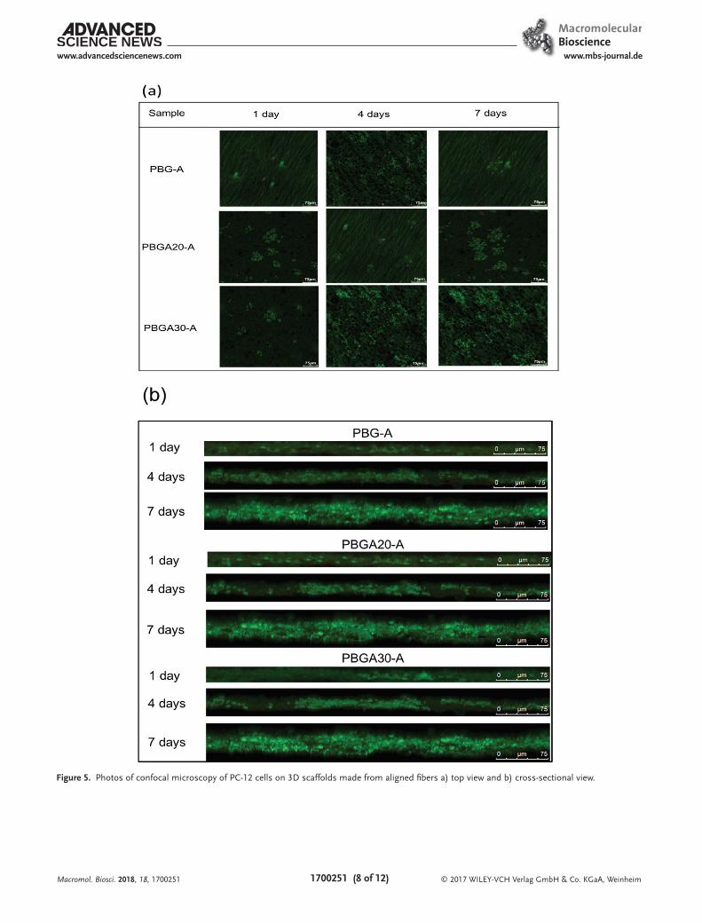
We examined the growth of cells further on the 3D scaf-fold made from aligned fibers using confocal microscopy. Figure 5a shows the top view of the cells on the scaffolds. From this figure, we observed the highest cell population for the cultured scaffold containing the highest amount of glutamic acid. Figure 5b shows the cross-sectional view of the cells on the scaffolds. Figure 5b shows that the cell growth with culture time progresses from the surface along the z-axis into the scaf-fold interior. The amount of z-axis cell growth is the highest for the scaffold containing the highest amount of glutamic acid. For instance, the sample thickness after 1 d culture is about the same at 30 µm, which is the thickness of the neat scaffold. However, the average thickness increases after 7 d culture into an average of 36 µm for PBG, 41 µm for PBGA20, and 45 µm for PBGA30. The scaffolds do not degrade after 7 d culture as shown in Figure 3a. The increase in thickness is apparently from the cell growth. These results clearly demonstrate that the molecular cue of glutamic acid is indeed to stimulate the rate of cell growth.
The morphology of PC-12 cells on scaffold after culture is carefully studied by SEM. The well-populated cells on scaffold are clearly observed (Figure 6). The cells, highlighted by white circles in Figure 6, grow not only on the scaffold surface but also into the interior through the pores of the 3D structure. This interior cell growth is more pronounced with the scaffold made from aligned fibers as shown in Figure 6b, which is con-sistent with the results of confocal microscopy study (Figure 5).
3.5. Neurite Outgrowth on 3D Scaffold
The PC-12 cells grown on the 3D scaffolds were further dif-ferentiated into neurite by nerve growth factor (NGF). The changes of neurite growth morphology with time and with dif-ferent glutamic content investigated by confocal microscopy are shown in Figure 7a,b for scaffolds made from isotropic fibers and aligned fibers, respectively. Figure 7 shows that both scaf-fold morphology and glutamic acid have critical effect on neu-rite growth. Scaffolds with aligned fibers promote the growth of elongated cell aligned with the fiber direction (Figure 7a). In contrast, scaffolds with isotropic fibers promote branched cell growth across the fibers (Figure 7b). The scaffold containing glutamic acid shows more active neurite outgrowth.
We further observe that the length of neurite outgrowth is increased with increasing glutamic acid concentration from 0% to 30%. This observation indicates that the glutamic acid is crit-ical in promoting the neurite outgrowth. The length of neurite outgrowth is longer for scaffold made from aligned fibers as compared with that of isotropic fibers. The aligned fibers appar-ently guide and promote neurite outgrowth. We speculate that the aligned fibers provide a favorable growth environment for directional neurite growth.[31] The average lengths of neurite outgrowth for different scaffolds are summarized in Table 1. After 10 d of culture, the lengths of neurite reach more than 100 µm, which is an average of 29% increase as compared with the control (TCPS).
The morphology of the neurite outgrowth on the scaffold made from aligned PBGA fibers was further investigated by the SEM as shown in Figure 8. The neurite outgrowth is more aligned with the direction of fibers with increasing the concen-tration of glutamic acid (PBGA20-A vs PBG-A; white particu-lates are PBS salt). These results further confirm the biological cue of glutamic acid for neurite outgrowth and the neurite growth along the direction of aligned fibers in the scaffold. This enhanced neutrite growth is specially desirable in tissue engi-neering for nerve regeneration.[31]
4. Conclusions
A new class of polypeptides containing different amount of neuron stimulate, glutamic acid, and with the molec-ular weight in the range of 200 kDa has been synthesized successfully using efficient chemical reactions. The poly-peptides can be fabricated into fibrous 3D scaffolds using electrospinning technique. The arrangement of fibers in the scaffold can be either isotropic or aligned. The scaf-folds degrade slowly and their pH did not vary much when they were immersed in the phosphate buffer solution for 49 d. The biocompatibility of 3D fibrous scaffolds and the length of neurite growth on the scaffolds are increased with increasing the amount of glutamic acid in the fibers. The morphology studies of neurite growth indicate the 3D scaffold of polypeptide fabricated from aligned fibers can guide anisotropic growth of neurite along the fiber into 3D domain better than unaligned fibers. This new polypeptide
Macromol. Biosci. 2018, 18, 1700251
Figure 4. Comparison of biocompatibility of various 3D scaffolds made from a) isotropic fibers (left) and b) aligned fibers (right).
© 2017 WILEY-VCH Verlag GmbH & Co. KGaA, Weinheim1700251 (8 of 12)
www.advancedsciencenews.com www.mbs-journal.de
Macromol. Biosci. 2018, 18, 1700251
Figure 5. Photos of confocal microscopy of PC-12 cells on 3D scaffolds made from aligned fibers a) top view and b) cross-sectional view.

© 2017 WILEY-VCH Verlag GmbH & Co. KGaA, Weinheim1700251 (9 of 12)
www.advancedsciencenews.com www.mbs-journal.de
Macromol. Biosci. 2018, 18, 1700251
Figure 6. SEM photos of undifferentiated cell growth of PC-12 on various 3D scaffolds a) isotropic fibers and b) aligned fibers.

© 2017 WILEY-VCH Verlag GmbH & Co. KGaA, Weinheim1700251 (10 of 12)
www.advancedsciencenews.com www.mbs-journal.de
Macromol. Biosci. 2018, 18, 1700251
Figure 7. Optical microscopic photos of neurite outgrowth on various 3D scaffolds a) isotropic fibers and b) aligned fibers.

© 2017 WILEY-VCH Verlag GmbH & Co. KGaA, Weinheim1700251 (11 of 12)
www.advancedsciencenews.com www.mbs-journal.de
Macromol. Biosci. 2018, 18, 1700251
has potential for the application in the tissue engineering of neural regeneration.
Supporting InformationSupporting Information is available from the Wiley Online Library or from the author.
AcknowledgementsThe authors express their appreciation to the Ministry of Science and Technology of Taiwan (Grant MOST 105-2221-E-002-228 and 106-2221-E-002-192) for financial support and to Dr. Margaret M. S. Wu, Emeritus Senior Scientific Advisor of ExxonMobil Research and Engineering Co., for editing this manuscript.
Conflict of InterestThe authors declare no conflict of interest.
Keywordsneurite outgrowth, neuron, polypeptide, regeneration, scaffold
Received: July 20, 2017Revised: October 4, 2017
Published online: December 12, 2017
[1] L. V. Kalia, A. E. Lang, Lancet 2015, 386, 896.[2] A. Kumar, A. Singh, Ekavali, Pharmacol. Rep. 2015, 67, 195.[3] Z. Álvareza, O. Castañoa, A. A. Castellsb, M. A. Mateos-Timonedaa,
J. A. Planella, E. Engela, S. Alcántarab, Biomaterials 2014, 35, 4769.[4] G. Orive, E. Anitua, J. L. Pedraz, D. F. Emerich, Nat. Rev. Neurosci.
2009, 10, 682.[5] L. L. Hench, J. M. Polak, Science 2002, 295, 1014.[6] C. Simitzi, A. Ranella, E. Stratakis, Acta Biomater. 2017, 51, 21.[7] R. J. McMurtrey, J. Neural Eng. 2014, 11, 066009.[8] X. Gu, F. Ding, D. F. Williams, Biomaterials 2014, 35, 6143.[9] P. X. Ma, Adv. Drug Delivery Rev. 2008, 60, 184.
[10] M. Maleki, A. Natalello, R. Pugliese, F. Gelain, Acta Biomater. 2017, 51, 268.
Table 1. Summary of neurite length of PC-12 on aligned and isotropic 3D Scaffolds.
Sample Neurite length of 1 d culture [µm] Neurite length of 4 d culture [µm] Neurite length of 10 d culture [µm]
TCPS 5.1 ± 1.2 33.2 ± 15.1 73.1 ± 33.0
PBG-I 5.0 ± 1.9 32.1 ± 22.6 81.4 ± 33.7
PBGA20-I 5.2 ± 2.2 39.9 ± 29.9 96.0 ± 46.3
PBGA30-I 5.5 ± 1.2 37.6 ± 22.9 90.4 ± 45.8
PBG-A 5.1 ± 2.1 34.1 ± 23.6 84.4 ± 32.4
PBGA20-A 5.3 ± 1.8 42.4 ± 23.5 98.0 ± 42.6
PBGA30-A 7.8 ± 1.9 39.6 ± 27.3 94.6 ± 43.9
Figure 8. SEM photos of differentiated PC-12 on various 3D scaffolds made from aligned fibers of PBG and PBGA.

© 2017 WILEY-VCH Verlag GmbH & Co. KGaA, Weinheim1700251 (12 of 12)
www.advancedsciencenews.com www.mbs-journal.de
[11] K. E. Kador, S. P. Grogan, E. W. Dorthé, P. Venugopalan, M. F. Malek, J. L. Goldberg, D. D. D’lima, Tissue Eng., Part A 2016, 22, 286.
[12] F. J. Rodríguez, E. Verdú, D. Ceballos, X. Navarro, Exp. Neurol. 2000, 161, 571.
[13] H. S. Koha, T. Yong, C. K. Chan, S. Ramakrishna, Biomaterials 2008, 29, 3574.
[14] G. E. Rooney, C. Moran, S. S. McMahon, T. Ritter, M. Maenz, A. Flugel, P. Dockery, T. O’Brien, L. Howard, A. J. Windebank, F. P. Barry, Tissue Eng., Part A 2008, 14, 681.
[15] G. G. Genchi, G. Ciofani, A. Polini, I. Liakos, D. Iandolo, A. Athanassiou, D. Pisignano, V. Mattoli, A. Menciassi, J. Tissue Eng. Regener. Med. 2015, 9, 151.
[16] J. Xie, M. R. MacEwan, X. Li, S. E. Sakiyama-Elbert, Y. Xia, ACS Nano 2009, 3, 1151.
[17] D. N. Rocha, P. Brites, C. Fonseca, A. P. Pêgo, PLoS One 2014, 9, e88593.
[18] S. Yao, X. Liu, S. Yu, X. Wang, S. Zhang, Q. Wu, X. Suna, H. Maod, Nanoscale 2016, 8, 10252.
[19] Z. Z. Khaing, C. E. Schmidta, Neurosci. Lett. 2012, 519, 103.[20] H. S. Koha, T. Yong, C. K. Chan, S. Ramakrishna, Biomaterials 2008,
29, 3574.
[21] T. W. Wang, M. Spector, Acta Biomater. 2009, 5, 2371.[22] A. Subramanian, U. M. Krishnan, S. Sethuraman, J. Biomed. Sci.
2009, 16, 108.[23] R. G. Ellis-Behnke, Y. X. Liang, S. W. You, D. K. C. Tay, S. Zhang,
K. F. So, G. E. Schneider, Proc. Natl. Acad. Sci. USA 2006, 103, 5054.[24] Y. Sun, W. Li, X. Wu, N. Zhang, Y. Zhang, S. Ouyang, X. Song,
X. Fang, R. Seeram, W. Xue, L. He, W. Wu, ACS Appl. Mater. Inter-faces 2016, 8, 2348.
[25] G. A. Silva, C. Czeisler, K. L. Niece, E. Beniash, D. A. Harrington, J. A. Kessler, S. Stupp, Science 2004, 303, 1352.
[26] T. C. Holmes, S. D. Lacalle, X. Su, G. Liu, A. Rich, S. Zhang, Proc. Natl. Acad. Sci. USA 2000, 97, 6728.
[27] M. Takeda, A. Takamiya, J. W. Jiao, K. S. Cho, S. G. Trevino, T. Matsuda, D. F. Chen, Invest. Ophthalmol. Visual Sci. 2008, 49, 1142.
[28] E. M. Adler, N. R. Gough, J. A. Blundon, Sci. Signaling 2006, 351, tr9.
[29] L. A. Greene, A. S. Tischler, Adv. Cell. Neurobiol. 1982, 3, 373.[30] P. H. Chen, H. C. Liao, S. H. Hsu, R. S. Chen, M. C. Wu, Y. F. Yang,
C. C. Wu, M. H. Chen, W. F. Su, RSC Adv. 2015, 5, 6932.[31] B. Zhu, S. C. Luo, H. Zhao, H. A. Lin, J. Sekine, A. Nakao, C. Chen,
Y. Yamashita, H. H. Yu, Nature Commun. 2014, 5,4523.
Macromol. Biosci. 2018, 18, 1700251
Copyright © 2022 FDOKUMEN