In vivo biostability of polyurethane–organosilicate nanocomposites
Upload
independentCategory
view
3download
0
Acta Biomaterialia 6 (2010) 3471–3481
Contents lists available at ScienceDirect
Acta Biomaterialia
journal homepage: www.elsevier .com/locate /actabiomat
Biodegradable and injectable cure-on-demand polyurethane scaffoldsfor regeneration of articular cartilage
J.A. Werkmeister a,*, R. Adhikari a,b, J.F. White a, T.A. Tebb a, T.P.T. Le a, H.C. Taing a, R. Mayadunne a,b,P.A. Gunatillake a,b, S.J. Danon c, J.A.M. Ramshaw a
a CSIRO Molecular and Health Technologies, Clayton South, Victoria 3169, Australiab PolyNovo Biomaterials, 2/320 Lorimer Street, Port Melbourne, Victoria 3207, Australiac CSIRO Molecular and Health Technologies, North Ryde, New South Wales 2113, Australia
a r t i c l e i n f o a b s t r a c t
Article history:Received 25 August 2009Received in revised form 16 February 2010Accepted 23 February 2010Available online 7 March 2010
Keywords:PolyurethaneHydrogelDegradationTissue formation
1742-7061/$ - see front matter Crown Copyright � 2doi:10.1016/j.actbio.2010.02.040
* Corresponding author. Tel.: +6 13 95458112; fax:E-mail address: [email protected] (J.A.
This paper describes the synthesis and characterization of an injectable methacrylate functionalizedurethane-based photopolymerizable prepolymer to form biodegradable hydrogels. The tetramethacrylateprepolymer was based on the reaction between two synthesized compounds, diisocyanato poly(ethyleneglycol) and monohydroxy dimethacrylate poly(e-caprolactone) triol. The final prepolymer was hydratedwith phosphate-buffered saline (pH 7.4) to yield a biocompatible hydrogel containing up to 86% water.The methacrylate functionalized prepolymer was polymerized using blue light (450 nm) with an initia-tor, camphorquinone and a photosensitizer, N,N-dimethylaminoethyl methacrylate. The polymer wasstable in vitro in culture media over the 28 days tested (1.9% mass loss); in the presence of lipase, around56% mass loss occurred over the 28 days in vitro. Very little degradation occurred in vivo in rats over thesame time period. The polymer was well tolerated with very little capsule formation and a moderate hosttissue response. Human chondrocytes, seeded onto Cultispher-S beads, were viable in the tetramethac-rylate prepolymer and remained viable during and after polymerization. Chondrocyte–bead–polymerconstructs were maintained in static and spinner culture for 8 weeks. During this time, cells remainedviable, proliferated and migrated from the beads through the polymer towards the edge of the polymer.New extracellular matrix (ECM) was visualized with Masson’s trichrome (collagen) and Alcian blue (gly-cosaminoglycan) staining. Further, the composition of the ECM was typical for articular cartilage withprominent collagen type II and type VI and moderate keratin sulphate, particularly for tissue constructscultured under dynamic conditions.Crown Copyright � 2010 Published by Elsevier Ltd. on behalf of Acta Materialia Inc. All rights reserved.
1. Introduction
It is well known that the regenerative capacity of the cartilagi-nous tissues in the knee is minimal and treatment often requiressurgical intervention [1]. Conventional surgical approaches likedebridement, microfracture and mosaicplasty are relatively simpleand common, but can be mechanically inferior with fibrocartilagerepair and limited long-term performance [1,2]. Autologous chon-drocyte implantation (ACI) offers a complex alternative strategy,but again is compromised by the nature of the reparative tissue [3].
Strategies based on implantation of exogenous scaffolds, bothnatural and synthetic, have been investigated for cartilage repair[4–6]. Natural biopolymers include collagens, alginate, fibrin, andhyaluronan, and can be used in conjunction with chondrocytes[4–6]. Synthetic polymers have largely included those based onpoly(glycolic acid), poly(lactic acid), poly(e-caprolactone) and
010 Published by Elsevier Ltd. on b
+6 13 95458101.Werkmeister).
other polyester co-polymers, usually without the incorporation ofcells [4–6]. We have recently shown the advantages of augmentingthe ACI approach with an appropriate scaffold delivery system[7–9]. In these instances we have used collagen as a delivery gelto implant seeded chondrocytes expanded on extracellular matrixbeads [7,8] or with synthetic polymer beads [9] and demonstratedimproved cartilage regeneration in pig chondral defects [10]. Whilebiodegradable natural polymers have clear advantages with re-spect to their capacity to integrate with cells, there are still issueswith the mechanical integrity of the implant.
To date, very few synthetic in situ polymerizable injectable sys-tems have been developed that possess suitable mechanical anddegradation properties [4,11]. These polymer systems, based onpoly(propylene fumarate) (PPF) and polyanhydrides, can be tai-lored for specific applications and are cell compatible. Nonetheless,they do not permit delivery of cells within the prepolymerizationmixture and as such are not ideal for cell-based therapies for car-tilage repair [12,13]. The PPF polymer systems require encapsula-tion of cells within gelatin as well as delayed addition of these
ehalf of Acta Materialia Inc. All rights reserved.

3472 J.A. Werkmeister et al. / Acta Biomaterialia 6 (2010) 3471–3481
cells after initiation of polymerization, making it difficult then todeliver in situ into the damaged tissue [12].
To enhance water uptake and subsequent cell compatibility,hydrogel scaffolds based on poly(ethylene glycol) (PEG) or poly(vi-nyl alcohol) (PVA) with chondroitin sulphate have been investi-gated with mixed results [14,15]. In both instances theprepolymers have been endcapped with methacrylate groups to al-low rapid and controllable polymerization. While the evidence forcell proliferation and ECM production in the PVA-based scaffoldswas minimal [15], the PEG-based hydrogels are cell compatiblebut often do not permit easy cell migration due to long lastingpolymer components and are mechanically compromised. Othercross-linking agents like PEG-diacrylate (PEG-DA) have also beenused with oligo (PEG fumarate) monomers [16] but again the cellviability is very dependent on the concentration of the initialmonomers and molecular weight of the PEG.
This paper reports on the design and synthesis of a new func-tionalized prepolymer with degradable arms, assessment of cellviability before and after photopolymerization, and new tissue for-mation, as well as degradation of the hydrogels in vitro and in vivo.The prepolymer is designed to allow simple injection to the dam-aged site in the presence of cells expanded on degradable collagenbeads, rapid photo-cross-linking to the adjacent tissue, and degra-dation with time into non-toxic components that resorb and do notinterfere with the regeneration of tissue. The prepolymer was pre-pared from diisocyanates as a backbone which were attached witha urethane-linkage to poly(e-caprolactone) with methacrylate
Fig. 1. Synthesis of tetrame
end-functional groups to achieve the desired cross-linking to formthe hydrogel. The design endeavoured to combine the knownmechanical properties of poorly hydrated PU with the hydrophilic-ity of PEG to achieve a water-miscible cell compatible system.
2. Materials and methods
2.1. Materials and reagents
Poly(ethylene glycol) Mn 1500 (PEG) and poly(e-caprolactone)triol Mn 900 (PCL) were purchased from Aldrich and the hydroxyland acid numbers were determined prior to use as described pre-viously (1). Stannous 2-ethylhexanoate and 3,5-di-tert-butyl-4-hydroxytoluene (BHT) were purchased from Aldrich and used asreceived. 2-Isocyanatoethyl methacrylate (IEM) was purchasedfrom Showa Denko (Switzerland) and purified by distillation underreduced pressure. Ethyl lysine diisocyanate (ELDI) was purchasedfrom Kyowa Hakko Kogyo (Japan) and purified by distillation underreduced pressure.
2.2. Polymer synthesis
The synthesis of the tetramethacrylate prepolymer is outlinedin Fig. 1 and was prepared in three different steps. The molecularweights used in all syntheses were calculated based on the hydro-xyl and acid titrations. Gel permeation chromatography (GPC) anddifferential scanning calorimetry (DSC) were measured to charac-
thacrylate prepolymer.

J.A. Werkmeister et al. / Acta Biomaterialia 6 (2010) 3471–3481 3473
terize the intermediates and final polymers. The polyols and inter-mediate products were degassed to remove any trace of moisturebefore the reaction and the reactions were carried out undernitrogen.
2.2.1. Step 1: synthesis of monohydroxy dimethacrylate poly(e-capro-lactone) triol (HDPCL)
Poly(e-caprolactone) triol (PCL) (59.17 g, 63.2 mmol) wasplaced in a reaction flask and dried at 80 �C in vacuo (0.1 mm Hg)for 1 h. BHT inhibitor (0.1 wt.% of total weight, 78.7 mg) and 2-iso-cyanatoethyl methacrylate (IEM) (19.63 g, 126.5 mmol) wereadded to the polyol and stirred at 80 �C. Stannous 2-ethylhexano-ate catalyst (0.1 wt.% of the polyol, 59.1 mg) was then added tothe mixture and the reaction continued with stirring overnight.The completion of the reaction was monitored by FT-IR for the dis-appearance of the isocyanate peak at 2272 cm�1. The product wasfound to be a viscous colourless liquid. The molecular weight of theviscous product was calculated based on hydroxyl and acid num-bers which were 51.62 and 0.42, respectively.
2.2.2. Step 2: synthesis of diisocyanato poly(ethylene glycol)PEG (102.87 g, 61.2 mmol) was placed in a reaction flask and
dried at 80 �C in vacuo (0.1 mm Hg) for 1 h. ELDI (27.69 g,122.5 mmol) was then added to the PEG and the reaction contin-ued with stirring overnight at 80 �C. The completion of the reactionwas monitored by the stabilization of NCO peak in the FT-IR anddisappearance of ELDI peak in the GPC. The isocyanate end groupcontent was determined by titration in accordance with the Amer-ican Society for Testing and Materials (ASTM) International Stan-dard D5155-01 and was found to be 3.29% NCO.
2.2.3. Step 3: synthesis of tetramethacrylate prepolymerThe monohydroxy dimethacrylate poly(e-caprolactone)
(HDPCL, 28.60 g, 26.3 mmol) synthesized in Step 1 was placed ina reaction flask and dried at 80 �C in vacuo (0.1 mm Hg) for 1 h.BHT (0.1 wt.% of the total weight, 33.65 mg) and poly(ethylene gly-col) prepolymer with isocyanate end groups (33.65 g, 13.2 mmol)from Step 2 was added to the HDPCL. The reaction flask was fittedwith an overhead mechanical stirrer equipped with a Teflon stirrerblade and the mixture stirred overnight at 80 �C. The reaction flaskwas wrapped with aluminium foil to exclude light during stirring.The completion of the reaction was monitored by FT-IR for the dis-appearance of the isocyanate peak at 2272 cm�1. The reactionyielded a viscous liquid as the final prepolymer.
2.3. Physico-chemical analyses
2.3.1. Hydroxyl number and molecular weight of polyolThe number average molecular weight was based on hydroxyl
numbers of the polyol. The hydroxyl numbers were determinedby the p-toluene isocyanate method in accordance with the Amer-ican Society for Testing and Materials (ASTM) International Stan-dard E1899-02. The viscosity of the prepolymers was measuredusing a Bohlin Rheometer (CSR 10) at room temperature.
2.3.2. Gel permeation chromatography (GPC)GPC was performed on a Waters Associates Liquid Chromato-
graph system (Waters 717, Rydalmere, Australia) instrument withfour Polymer Laboratories PLGel columns (3 � 5 lm Mixed-C and1 � 3 lm Mixed-E). Tetrahydrofuran was used as the eluent at aflow rate of 1.0 ml min�1 and the system was calibrated with nar-row disperse polystyrene standards (Polymer Laboratories EasiCal,Amherst, MA, USA) and molecular weights are reported as polysty-rene equivalents as measured by a change in refractive index withelution time.
2.3.3. Fourier transform infrared (FT-IR) spectroscopyFT-IR spectra were obtained using Perkin-Elmer 2000 FT-IR
spectrophotometer. FT-IR spectra of samples were obtained usingKBr discs.
2.3.4. Differential scanning calorimetry (DSC)DSC analysis was carried out over the temperature range �50 to
200 �C using Mettler DSC-028. The experiments were carried out ata heating rate 10 �C min�1 under nitrogen. Sample weights rangedbetween 20 and 25 mg. The samples were dried at 40 �C for 48 hunder a vacuum (0.1 torr) prior to analysis.
2.4. Preparation of polymer scaffold
2.4.1. Sterilization and hydration of tetramethacrylate prepolymerThe level and uniformity of hydration in the prepolymer was
critical to cell viability. Either a slow passive hydration or rapid ap-proaches were attempted. For slow passive hydration, the prepoly-mer (0.67 g) was first sterilized by steam autoclaving at 121 �C for15 min, then hydrated overnight in the dark at room temperaturewith 20 ml of sterile phosphate-buffered saline (PBS). PBS waschanged daily and cell viability of the hydrating prepolymer wasassessed with time over 4 weeks. Prepolymers were similarly ster-ilized and rapidly hydrated either by manual stirring or bymechanical stirring using an IKA Disperser Ultra Turrax Type T8(Staufen, Germany).
2.4.2. Polymer preparationFor every 1 g of the prepolymer, 10 ll of a 1:5 (w/w) CQ:DMA-
EMA (camphorquinone:N,N,-dimethylaminoethyl methacrylate;0.16%:0.83% final concentrations) was added. The preparationwas thoroughly mixed with a spatula and placed in Teflon moulds.The sample was photo-cured using an Elipar™ Freelight 2LED cur-ing light (430–480 nm, 1000 mW cm�2, 3 M ESPE) for 1 min. Thecured disks were 4.5 mm diameter � 2 mm deep.
2.5. Mechanical studies
Rheometric testing was performed using a Rheometric ScientificDMTA Mark IV (TA Instruments, USA). Cylindrical hydrated sam-ples 4.5 mm in diameter and 2 mm in height were carefully en-closed between parallel plates. A dynamic frequency sweep from0.1 to 10 Hz was applied to the samples in compression at roomtemperature using a final fixed strain of 0.03%. At all times the sta-tic force remained 20% greater than the dynamic force while theinitial static force was set at 0.1 N. Eight samples were tested.The average compressive modulus over the applied frequencywas obtained for each sample and the average modulus for theeight samples is expressed as the mean ± standard deviation. Sta-tistical significance was assessed by analysis of variance (ANOVA)followed by unpaired Student’s t-test using p < 0.05 as the criterionfor significance.
2.6. In vitro degradation studies
Degradation experiments were performed in the absence orpresence of 0.2 mg ml�1 lipase (EC 3.1.1.3 Type XIII from Pseudo-monas species, Sigma, St. Louis, MA) in PBS pH 7.4 on cured(CQ:DMAEMA) tetramethacrylate polymer described in Section2.4.2. For comparative purposes, a partially cured tetramethacry-late polymer, polymerized with 0.0125% riboflavin, was used as acontrol for rapid degradation. The extent of degradation was calcu-lated by measuring the degree of mass loss at days 7, 14, 22 and28 days compared to day 0. Three samples were used for each ofthe polymers at each time point with and without lipase. Resultsare expressed as % mean mass loss ± standard deviation. Statistical

3474 J.A. Werkmeister et al. / Acta Biomaterialia 6 (2010) 3471–3481
significance was assessed by analysis of variance (ANOVA) fol-lowed by unpaired Student’s t-test using p < 0.05 as the criterionfor significance.
2.7. In vivo subcutaneous implantation studies in rats
A 1 month study was conducted to assess any rapid degradationand the degree of inflammation around the polymers. The in vivodegradation of the CQ:DMAEMA cross-linked tetramethacrylatepolymer was assessed by inserting small plugs, 4.5 mm diame-ter � 2 mm deep (3 implants per rat, 3 rats) subcutaneously alongthe dorsal midline of rats, according to ISO 10993: InternationalStandard Biological evaluation of medical devices, Part 6: Testsfor local effects after implantation 2007, as described in detail pre-viously [17]. As a positive control to assess the effects of thedegrading polymer, a riboflavin partially cross-linked tetramethac-rylate polymer was implanted. Eight week old, Specific PathogenFree, female Sprague Dawley (Crj:CD(SD)IGS) rats were obtainedfrom the Animal Resource Centre (Canning Vale, WA, Australia).This study was approved by the CSIRO Animal Care and EthicsCommittee (Ethics number AEC 02-03). At 1 month after implanta-tion, animals were sacrificed and the tissue around the implantsremoved and fixed in 10% neutral buffered formalin (NBF) for his-tology and sections stained with haematoxylin and eosin (H&E).
2.8. Cell viability
Human articular chondrocytes (Edward Keller, Melbourne)were used to assess cell viability in the presence of the prepolymer,including the initiator and sensitizer, prior to polymerization, aswell as during and after polymerization. Cells were routinely cul-tured on CultiSpher-S beads (Percell Biolytica, Sweden) inDMEM:F12 (1:1) media with 10% FCS (complete chondrocyte med-ia, CCM). Seeding was as described previously [7,8] with intermit-tent stirring for 2 min every 30 min, initially at 25 rpm for 3 h, then45 rpm for 1.5 h. This was followed by 15 min continuous stirringat 45, 50, 55 to a final 60 rpm. Using this protocol, we and othershave previously found greater than 95% cell seeding onto beads(7–10). The degree of cell seeding was confirmed by trypsinizationand cell counting of aliquots of cell beads. Cell viability was as-sessed using the Live/Dead� cell viability assay (Molecular Probes,Invitrogen, USA), where live cells are stained green with CalceinAM and dead cells are stained red with ethidium homodimer-1.Prepolymer solutions were mixed with cells on beads (5:1, 0.6 gprepolymer mixture: 0.15 ml of CCM containing 10 � 106 cells on0.09 ml wet beads). For assessment of cell viability after polymer-ization, thin slices were cut, washed in warm PBS and then placedin the Live/Dead� stain for 30 min at 37 �C. Viability was assessedusing an Optiscan F900e confocal system with a KrAr laser and anOlympus BX61 microscope.
2.9. Tissue engineered constructs and assessment
Human articular chondrocytes were seeded onto beads andmixed with prepolymer. Human chondrocytes were passage threederived cells from Edward Keller (Melbourne). Seeding on beadswas performed 2 days prior to mixing and polymerizing in tissueconstructs. Cell density was 10.4 � 106 cells/72 mg Cultispher-Sdry beads in 50 ml DMEM:F12 (1:1) media with 10% FCS in125 ml spinner bottles. Using this cell seeding, we have shown thatat the time of use, the chondrocyte cell concentration is around14 � 106 cells ml�1 in the final cell–bead–polymer construct. Thisconcentration is comparable to the cell density in normal cartilage.Constructs (4.5 mm diameter � 2 mm deep) were cultured eitherunder static conditions in 6-well tissue culture plates or underdynamic conditions (60 rpm continuous stirring) in spinner flasks
in CCM as described previously [7–10]. Constructs were main-tained for 8 weeks under standard culture conditions with dailysupplementation of 50 lg ml�1 ascorbic acid (L-ascorbic acid phos-phate, Wako-Novachem). Six constructs were terminated at4 weeks (3 static, 3 spinner), and six were terminated at 8 weeksin culture. Samples at 4 and 8 weeks were fixed in formalin andprocessed for haematoxylin and eosin (H&E) staining, Masson’s tri-chrome (MT) staining for collagen and Alcian blue staining for gly-cosaminoglycans (GAG). At 8 weeks, cultured samples were firstdissected into two parts, and the second part was embedded andfrozen in Optimal Cutting Temperature Compound (Tissue-Tek�
O.C.T. Compound, Sakura Finetek, The Netherlands), and 4 lm thinsections were cut from frozen tissue samples with a freezingmicrotome and processed for immunohistochemical evaluationusing a peroxidase UltraVision Detection System (Thermo Scien-tific, DKSH, Australia). Sections (4 lm) were fixed in ice-cold meth-anol, hydrated and blocked for endogenous peroxidase withhydrogen peroxide provided in the kit. Collagen type I, collagentype II, collagen type VI and keratin sulphate were examined usingmonoclonal antibodies (MAb) to collagen type I (COL 1-5D8/G9,Chemicon, USA), collagen type II (Neomarkers 2B1.5), collagen typeVI (COL 6-2C9/E7) [18], and keratin sulphate (5D4, Seikagaku Corp.,Tokyo, Japan). Sections stained with anti-collagen type I and II MAbwere pre-treated with 1 mg ml�1 pepsin (Worthington, NewJersey) in Tris–HCl, pH 2.0, for 15 min at room temperature. Sec-tions stained for keratin sulphate were pre-treated with 0.5 U ml�1
chondroitinase AC II from Arthrobacter aurescens (Seikagaku Corp.,Tokyo, Japan) in Tris–acetate buffer, pH 7.4. Primary antibodieswere incubated for 50 min at room temperature, washed threetimes in PBS and incubated with biotinylated goat anti-mouse for10 min at room temperature. After further washing, streptavidin-peroxidase (provided in the kit) was added for 10 min at roomtemperature, washed and the chromogen (AEC; 3-amino-9-ethylc-arbazole) added as per the instructions in the kit. Sections werecounterstained with Harris’ haematoxylin (Sigma, St. Louis, MA),mounted and viewed using an Optiscan F900e confocal systemwith a KrAr laser and an Olympus BX61 microscope.
3. Results
3.1. Synthesis and properties of the tetramethacrylate prepolymer
The reaction scheme shown in Fig. 1 illustrates only the struc-tures of the major compounds formed. However, as shown byGPC the reaction product in Step 1 is a mixture of predominantlymono and dihydroxy compounds with a viscosity of 100,000 cent-istokes (cSt). The average molecular weight of the product wasdetermined by titration and gel permeation chromatography(GPC) and was consistent with the statistically predicted additionof, on average, 2 IEM molecules per PCL molecule (Mn, 1679). Inthe second step, the molecular weight of the resulting diisocyanatoPEG prepolymer was determined from isocyanate end group titra-tion and found to be Mn 2557.
The final step to produce the tetramethacrylate prepolymer in-volved reacting the hydroxyl group of the dimethacrylate function-alized poly(e-caprolactone) with the diisocyanato PEG prepolymer.The available isocyanate groups in the PEG prepolymer werematched stoichiometrically to the available hydroxyl groups inmethacrylate functionalized to ensure all segments are covalentlylinked in the cross-linked polymer network formed after curing.The viscosity increased significantly in the final step of the reac-tion, as expected, due to increase in molecular weight. GPC showeda bimodal distribution of the prepolymer (Mn 5125) attributed tothe presence of species functionalized to different degrees in theprevious step (Fig. 2A). The curing reaction of the final tetrameth-acrylate prepolymer was monitored by the disappearance of the

Fig. 2. (A) GPC showing MW distribution of the tetramethacrylate PCL. Peaks after40 min elution time are associated with air, water, and solvent only. (B) FT-IRspectra of final tetramethacrylate prepolymer before (red) and after (blue) curingshowing almost complete disappearance of methacrylate double bond at1631 cm�1 (", X-axis) post curing.
J.A. Werkmeister et al. / Acta Biomaterialia 6 (2010) 3471–3481 3475
methacrylate double bond at 1631 cm�1 by FT-IR (Fig. 2B). Thermaltransitions of the final prepolymer and the monohydroxy diacry-late PCL (HDPCL) were evaluated using DSC. The synthesized tetra-methacrylate polymer showed a Tg onset around �41 �C with avery low melting endotherm at 27.2 �C, indicative of an amorphousmorphology as expected from an hyperbranched structure withshort arm lengths.
Fig. 3. Cell viability of human chondrocytes using Live/Dead� assay. Cells on Cultispher-for 1 h. Cells on Cultispher-S beads in absence of polymer were used as control (C). Viab
3.2. Hydration of the tetramethacrylate prepolymer and cell viability
Consistent with the literature, initial experiments showed thatthe degree of hydration of the prepolymer was one of the criticalfactors for cell survival during polymerization. Cell viability wasmonitored over the hydration period described in Section 2.4.1.Polymers were allowed to hydrate for 10–12 days to ensure com-plete equilibrium. At this stage, cells cultured on beads were addedto the hydrated polymers and cell viability evaluated in the ab-sence of CQ/DMAEMA. Cells remained viable in these uncuredpolymers at 10 min, 1 h (Fig. 3A), 1 day and 3 days. The hydratedprepolymer contained 82–86% PBS and remained stable up to3 months with respect to consistency and cell viability. In addition,at this point, chondrocytes on CultiSpher-S beads could be mixedwith the prepolymer and polymerized without any loss of cell via-bility (Fig. 3B), similar to the viability with control cells on beads inthe absence of polymer (Fig. 3C). The concentration of CQ andDMAEMA chosen for polymerization was non-toxic to the cells.Prepolymers that were rapidly hydrated, either manually or usinga mechanical stirrer, gave variable results with respect to unifor-mity and extent of hydration, and more importantly with respectto cell viability. With manually stirred prepolymers, 80–86%hydration could also be achieved, but with time, the prepolymerseparated out of the PBS. While uncured polymers, hydrated bymanual stirring, were toxic to cells after 10 min, immediatepolymerization of these hydrated prepolymers with cells wasnon-toxic. The mechanically hydrated prepolymers were alsoproblematic and cell viability was compromised after polymeriza-tion and the polymers were fragile compared with the slow hydra-tion method.
3.3. Mechanical assessment of scaffold hydrogel
The viscoelastic behaviour of the hydrogels was also examinedunder periodic strain using dynamic mechanical testing over arange from 0.1 to 10 Hz. Eight samples of polymers were tested.The polymer in each of the eight samples had been hydrated toan average 82.0% (±SD 1.4%). The average compressive modulusfor the eight samples over the frequency range was 107.9 ±11.0 kPa. With cells (20 wt.% chondrocytes on beads), the averagecompressive modulus of 4 further samples was 103.3 ± 11.4. Nosignificant difference in modulus was found between any of thesamples. The modulus was independent of the imposed frequencyin the range of 0.1 to 10 Hz, indicating that the hydrogel containeda viscoelastic network structure. No significant differences wereobserved between the samples with and without cells and beads(p > 0.05, unpaired two-tailed Student’s t-test).
S beads in presence of uncured (A) and polymerized tetramethacrylate polymer (B)le cells stain green, dead cells stain red. Bars = 25 lm.

3476 J.A. Werkmeister et al. / Acta Biomaterialia 6 (2010) 3471–3481
3.4. In vitro degradation of polymer scaffold
The tetramethacrylate polymer polymerized with CQ:DMAEMAwas stable over the 28 days in culture medium at 37 �C with nosignificant loss of mass compared to day 0 (p > 0.05; unpairedtwo-tailed Student’s t-test) (Fig. 4, j). For comparative purposes,the same prepolymer polymerized with riboflavin appeared onlypartially polymerized and was partially degraded over the sameculture period (Fig. 4, N) – this degradation was significant com-pared to day 0 and compared with the CQ:DMAEMA polymer atdays 7, 14, 22 and 28 (p < 0.05, p < 0.05, p < 0.01 and p < 0.05, un-paired two-tailed Student’s t-test). In the presence of Pseudomonaslipase, the partially polymerized polymer completely degradedwithin 7 days (Fig. 4, 4) and was significantly different to theno-enzyme treated polymer at all time points (p < 0.005, unpaired
Fig. 4. Polymer hydrogel degradation in vitro in the absence (closed symbols) orpresence (open symbols) of lipase. Cross-linked polymerized tetramethacrylatepolymer using CQ:DMAEMA (j, h) and a control lightly cross-linked tetrameth-acrylate polymer using riboflavin (N, 4).
Fig. 5. One month rat explants of tetramethacrylate polymer fully polymerized with CQ:D(A and C) and 100 lm (B and D).
two-tailed Student’s t-test). The CQ:DMAEMA polymerized poly-mer partially degraded, with a mass loss of 42% at day 7 and 56%at day 28 (Fig. 4, h). The extent of degradation with lipase was sig-nificant compared to the untreated (no enzyme) polymer at the 4time points (p < 0.01, p < 0.05, p < 0.005 and p < 0.05, unpairedtwo-tailed Student’s t-test), and is indicative that in the presenceof cells secreting enzymes, these polymers have the potential todegrade over time.
3.5. In vivo animal implantation studies
A 1 month study was undertaken to evaluate any rapid degra-dation as well as visible signs of toxicity or intense inflammation.For cell-based therapies like cartilage repair, it is preferable thatthe carrier scaffold persist for at least 1 month during the criticaltime for rehabilitation. One month explants from the CQ:DMAEMApolymerized tetramethacrylate polymer showed negligible degra-dation and minimal inflammation with no signs of toxicity. Thepolymer was surrounded by a thin capsule around 2–4 cells thicklargely comprising epithelial-like cells (Fig. 5A). There were nosigns of cellular infiltration into the polymer. There was the occa-sional detection of small polymer fragments entrapped within thecapsular wall (Fig. 5B). This could have been the first signs of deg-radation or could have been caused by the roughened surfaceedges of the implants. To evaluate the host response to degrada-tion, a control partially cross-linked polymer was used as describedin Section 2. Again, these polymers were surrounded by a similarlythin capsule of 2–4 cells, largely epithelial-like. In this case, how-ever, there were marked signs of polymer degradation (Fig. 5C)as well as cellular infiltration and new tissue formation (Fig. 5D).The polymer appeared to be a non-uniform scaffold with unde-graded solid polymer as well as a more loose porous structure.Even when degradation had occurred, there were no signs ofmarked inflammation. Overall, these polymers seem to persist to
MAEMA (A and B) or partially polymerized with riboflavin (C and D). Bars = 200 lm

J.A. Werkmeister et al. / Acta Biomaterialia 6 (2010) 3471–3481 3477
allow new tissue formation and are well tolerated by the host withno adverse reactions.
3.6. Tissue engineered constructs
At 4 weeks, chondrocytes on beads and embedded in the hydro-gels were viable in both static and spinner culture systems. In sta-tic culture, the hydrogel was largely intact and cells hadproliferated and migrated from the beads into the surroundingpolymer and had begun to produce new matrix (Fig. 6A). GAGswere present in the surrounding matrix (Fig. 6C). In spinner cul-ture, the hydrogel varied with some evidence of fragmentationcaused by the mechanical agitation. Nonetheless, the seeded chon-drocytes had formed dense clusters of cells and new matrix adja-cent to the beads and also within the beads themselves (Fig. 6B)with indications of new GAG formation (Fig. 6D).
At 8 weeks, cell density and matrix formation had increased sig-nificantly. In static cultures, the polymer was infiltrated with pock-ets of cells as well as discrete trails of cells that had migratedthrough the polymer from the beads towards the edge of the poly-mer (Fig. 7A). GAGs were clearly present in the new extracellularmatrix within the porous beads as well as through the polymerand were more pronounced on the surface edge of the polymer(Fig. 7C). In the spinner cultures the histological picture was largelythe same, with perhaps more cells and matrix through the polymerand towards the surface (Fig. 7B and D).
The new matrix formed at 8 weeks contained large amounts ofcollagen type II, indicative of articular cartilage, in both static andparticularly the spinner cultured constructs (Fig. 8A and B).Collagen type VI was also prominent in the new matrix, again morepronounced in the tissue constructs maintained in spinner culture(Fig. 8C and D). Keratin sulphate staining appeared as a patchy pat-tern throughout the polymer and particular towards the surface
Fig. 6. Four week tissue construct of human chondrocytes cultured in vitro on Cultisphespinner culture (B and D). MT staining for cells and collagen (A and B) and Alcian bluestaining intense blue. Bars = 50 lm.
edge of the tissue, associated with a subset of cells (Fig. 8E andF). Collagen type I was present but at a significantly reduced level(Fig. 8G and H).
4. Discussion
Biodegradable polymers which can be injected as a viscousaqueous solution, then cured on demand within the tissue defect,offer advantages in the design of injectable scaffolds for tissueengineering applications. Synthetic biodegradable polymers offeran alternative gel delivery system for cartilage repair and can the-oretically be formulated to control mechanical and degradativebehaviour to match the application [5,6,19].
We have recently reported an injectable two componenturethane–acrylate based system based on biodegradable lysine-diisocyante (LDI)-based polyurethanes (PUs) for bone tissue engi-neering [20,21]. The LDI-based system has been suggested byZhang [22] as a non-toxic, fully biodegradable system to generatePU foams. In these studies, cells were able to grow on the scaffoldsbut could not be delivered in the polymer during cross-linking.
In the current paper we have adapted this system to allow cellsurvival during the cross-linking stage to align the process with acell-based injectable system for cartilage repair. The hydrogelwas prepared from diisocyanates as a backbone which wereattached with a urethane-linkage to poly(e-caprolactone) withmethacrylate end-functional groups to achieve the desired cross-linking. The design endeavoured to combine the good mechanicalproperties of linear PU with the hydrophilicity of PEG to achievea water-miscible cell compatible system. For cell survival variousparameters need to be addressed including the toxicity of the pre-polymer, initiator and sensitizer used for polymerization into thehydrogel. Previous studies have shown the importance of screen-ing appropriate systems for cell encapsulation [23]. In the current
r-S beads and polymerized within tetramethacrylate polymer in static (A and C) orstaining for GAG seen as light blue staining (C and D), with the Cultispher-S beads
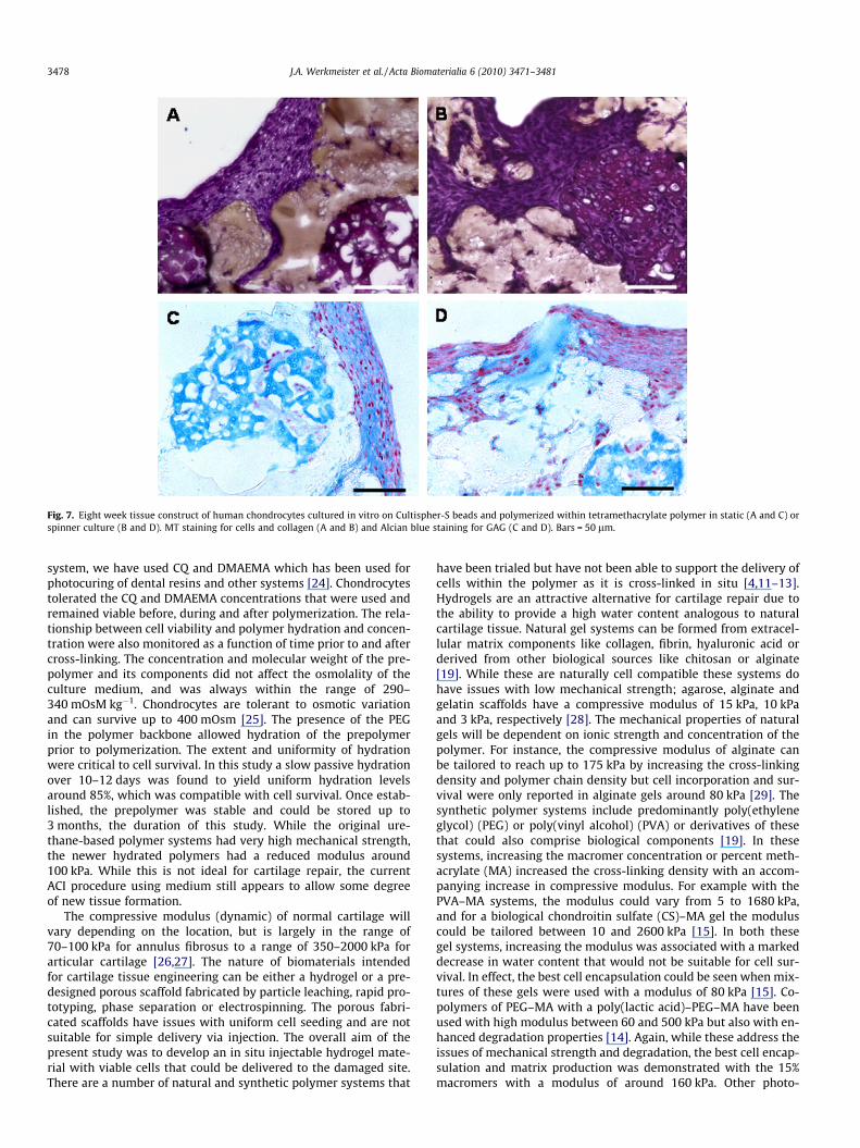
Fig. 7. Eight week tissue construct of human chondrocytes cultured in vitro on Cultispher-S beads and polymerized within tetramethacrylate polymer in static (A and C) orspinner culture (B and D). MT staining for cells and collagen (A and B) and Alcian blue staining for GAG (C and D). Bars = 50 lm.
3478 J.A. Werkmeister et al. / Acta Biomaterialia 6 (2010) 3471–3481
system, we have used CQ and DMAEMA which has been used forphotocuring of dental resins and other systems [24]. Chondrocytestolerated the CQ and DMAEMA concentrations that were used andremained viable before, during and after polymerization. The rela-tionship between cell viability and polymer hydration and concen-tration were also monitored as a function of time prior to and aftercross-linking. The concentration and molecular weight of the pre-polymer and its components did not affect the osmolality of theculture medium, and was always within the range of 290–340 mOsM kg�1. Chondrocytes are tolerant to osmotic variationand can survive up to 400 mOsm [25]. The presence of the PEGin the polymer backbone allowed hydration of the prepolymerprior to polymerization. The extent and uniformity of hydrationwere critical to cell survival. In this study a slow passive hydrationover 10–12 days was found to yield uniform hydration levelsaround 85%, which was compatible with cell survival. Once estab-lished, the prepolymer was stable and could be stored up to3 months, the duration of this study. While the original ure-thane-based polymer systems had very high mechanical strength,the newer hydrated polymers had a reduced modulus around100 kPa. While this is not ideal for cartilage repair, the currentACI procedure using medium still appears to allow some degreeof new tissue formation.
The compressive modulus (dynamic) of normal cartilage willvary depending on the location, but is largely in the range of70–100 kPa for annulus fibrosus to a range of 350–2000 kPa forarticular cartilage [26,27]. The nature of biomaterials intendedfor cartilage tissue engineering can be either a hydrogel or a pre-designed porous scaffold fabricated by particle leaching, rapid pro-totyping, phase separation or electrospinning. The porous fabri-cated scaffolds have issues with uniform cell seeding and are notsuitable for simple delivery via injection. The overall aim of thepresent study was to develop an in situ injectable hydrogel mate-rial with viable cells that could be delivered to the damaged site.There are a number of natural and synthetic polymer systems that
have been trialed but have not been able to support the delivery ofcells within the polymer as it is cross-linked in situ [4,11–13].Hydrogels are an attractive alternative for cartilage repair due tothe ability to provide a high water content analogous to naturalcartilage tissue. Natural gel systems can be formed from extracel-lular matrix components like collagen, fibrin, hyaluronic acid orderived from other biological sources like chitosan or alginate[19]. While these are naturally cell compatible these systems dohave issues with low mechanical strength; agarose, alginate andgelatin scaffolds have a compressive modulus of 15 kPa, 10 kPaand 3 kPa, respectively [28]. The mechanical properties of naturalgels will be dependent on ionic strength and concentration of thepolymer. For instance, the compressive modulus of alginate canbe tailored to reach up to 175 kPa by increasing the cross-linkingdensity and polymer chain density but cell incorporation and sur-vival were only reported in alginate gels around 80 kPa [29]. Thesynthetic polymer systems include predominantly poly(ethyleneglycol) (PEG) or poly(vinyl alcohol) (PVA) or derivatives of thesethat could also comprise biological components [19]. In thesesystems, increasing the macromer concentration or percent meth-acrylate (MA) increased the cross-linking density with an accom-panying increase in compressive modulus. For example with thePVA–MA systems, the modulus could vary from 5 to 1680 kPa,and for a biological chondroitin sulfate (CS)–MA gel the moduluscould be tailored between 10 and 2600 kPa [15]. In both thesegel systems, increasing the modulus was associated with a markeddecrease in water content that would not be suitable for cell sur-vival. In effect, the best cell encapsulation could be seen when mix-tures of these gels were used with a modulus of 80 kPa [15]. Co-polymers of PEG–MA with a poly(lactic acid)–PEG–MA have beenused with high modulus between 60 and 500 kPa but also with en-hanced degradation properties [14]. Again, while these address theissues of mechanical strength and degradation, the best cell encap-sulation and matrix production was demonstrated with the 15%macromers with a modulus of around 160 kPa. Other photo-

Fig. 8. Eight week tissue construct of human chondrocytes cultured in vitro on Cultispher-S beads and polymerized within tetramethacrylate polymer in static (A, C, E, and G)or spinner culture (B, D, F, and H). Collagen type II (A and B), collagen type VI (C and D), keratin sulphate (E and F) and collagen type I (G and H). Bars = 100 lm. Positivestaining is visualized by brown staining.
J.A. Werkmeister et al. / Acta Biomaterialia 6 (2010) 3471–3481 3479
cross-linkable hydrogels have also been reported to address theissues of modulus and degradation but again the final blends comeat a cost to cell viability. The use of branched dendrimer polymers,for example based on glycerol and succinic acid, can be added tothe PEG–MA gels to enhance degradation but the modulusreported was low, in the range of 4–34 kPa [26]. Alternatively, deg-radation can be improved using porogens, phase separation orfoaming [20,21], enzyme-cleavable sites [19], degradable micro-particles [8,20] or co-polymers that include biodegradable inter-penetrating polymers, either synthetic like poly(ethylene oxide)(PEO) [26] or natural like gelatin [30]. Again in these cases, the
variation in modulus needs to be addressed; for example increas-ing the degradation with PEO into the cross-linkable backbone ofPEG–MA changed the modulus from 320 to 20 kPa [27].
Overall, the mechanical properties of our current polymerhydrogel are largely comparable to the huge number of polymergel systems that have been described. The hydration requirementsof the polymer system, necessary for a cell compatible in situ deliv-erable gel system, come at a cost to the compressive modulus ofthe scaffold. Certainly there are possibilities to use the cell advan-tages of hydrogels with the mechanical advantages associated withporous scaffolds. A poor gel strength agarose can be used to fill a

3480 J.A. Werkmeister et al. / Acta Biomaterialia 6 (2010) 3471–3481
mechanical superior PLLA scaffold that supports cells with a mod-ulus of 3.38 MPa [31], but again the ability to deliver these cellseeded scaffolds non-invasively by injection is lost.
The polymer was stable in culture medium over 28 days but sig-nificant degradation occurred in the presence of exogenous lipase.For soft tissue applications, the ideal scaffold should hold itsmechanical strength up to 3 months and gradually degrade overthe next 3–4 months [1,10]. The polymer system described in thisstudy consists of relatively high proportion of segments linked byester groups and urethane groups which are susceptible to hydro-lytic degradation. The complete hydrolysis of the cross-linkednetwork will produce poly(methacrylic acid), caproic acid,poly(ethylene glycol), lysine and ethanolamine as major degrada-tion products. Hydrolytic degradation of polymers can be en-hanced in the presence of selective enzymes. PLLA can bedegraded with proteinase K [32] and PCL, used in our current poly-mer, can be hydrolytically degraded using bacterial lipases [33]. Inaddition, polymer degradation could be accelerated by decreasingthe effectiveness of photo-cross-linking by using riboflavin. Underthese conditions, the polymer degraded completely. These sameformulations were also investigated in short-term subcutaneousimplants in rats. The CQ:DMAEMA polymer remained largely intactand was associated with a minimal capsule formation and inflam-matory response. As a control, the riboflavin-based polymers diddegrade but again there was very little inflammatory responseand largely fibroblast infiltration. Overall the in vivo degradationstudies were aimed to show no adverse biological effects and sta-bility over the first 1 month of implantation that is important fortissue integration and stability. The potential of the polymer sys-tem to degrade was confirmed from the use of a partially polymer-ized polymer system as well as the use of lipase. In the presence oflipase there was significant degradation over the 28 days in bothcross-linked polymer systems. It is expected that in the presenceof cells that would contain lipases and other enzymes the polymerwould degrade over 6 months. This is supported by the immuno-histochemical analyses at 8 weeks that demonstrates new extra-cellular matrix production but also indications of some polymerdegradation. Tissue constructs comprising chondrocytes culturedon collagen beads and embedded within the polymer were moni-tored under static and dynamic culture conditions. With time,the polymer did degrade in the presence of cells seeded onto bio-degradable beads and allowed proliferation and migration of chon-drocytes through the polymer. At 8 weeks, there was clearchondrocyte expansion which appeared to be more pronouncedin the spinner culture system. Static culture of cartilage constructshas been shown to be less effective compared with constructsmaintained under intermittent hydrostatic pressure [34]. In thepresent study, the matrix was not quantitated, but the Alcian bluestaining typically demonstrated more GAGs present in the spinnerculture system. In both static and spinner systems, cells and matrixwere scattered in and around the collagen beads, throughout thepolymer but mostly towards the peripheral edge. One of the issueswith current cell-based cartilage therapies is tissue integration ofthe repairing wound with the adjacent normal tissue [5,6]. In thecurrent polymer system, seeded chondrocytes had no difficulty inmigrating towards the edge of the polymer network where normalcartilage would reside. The main extracellular protein of hyalinecartilage, collagen type II, was detected in all constructs in culture,along with keratin sulphate – again these ECM components weremore prominent in spinner culture systems. Collagen type I wasdetected but was markedly less pronounced compared with colla-gen type II. De-differentiation can occur with prolonged culturewith a change from collagen type II to collagen type I. However,compared with culture on two-dimensional tissue culture flasks,it has been known for some time that chondrocytes can maintaintheir correct phenotype in microcarrier suspension cultures [35].
We have previously shown that articular chondrocytes can main-tain their phenotype when cultured in spinner flasks with dynamicmotion on a variety of bead types including synthetic PLGA beads[9], alginate and collagen beads [7,8], and gelatin beads [10]. Inaddition, chondrocytes cultured on a variety of bead types in dy-namic suspension culture have a greater capacity to re-differenti-ate to the differentiated phenotype [36]. Collagen type VI wasalso detected in all tissue constructs, particularly localized aroundindividual cells and clusters of cells. Again there appeared to bemore collagen type VI in cultures maintained under dynamic mo-tion. We have previously shown in other wound healing responsesthat collagen type VI is indicative of early tissue regeneration [18].In cartilage it is well known that the functional unit of the tissue isthe chondron [37], and the pericellular environment in and aroundthis chondron is quite complex with abundant collagen type VI andother sulphated GAGs [38]. The presence of type VI collagen in ourtissue constructs, along with the other ECM components, is a goodindication that the essential cell-matrix signalling pathways areoperative [39] and the repairing tissue is structurally sound.
5. Conclusions
This study demonstrated that dimethacrylate PCL- and PEG-based prepolymers could be formulated to produce an injectablecure on demand multifunctional polymer network that can be hy-drated to allow effective cell delivery for cartilage repair. The ure-thane-based polymers were tailored to balance the need todegrade safely to allow cell migration and extracellular matrixdeposition. The formulation was non-toxic to human chondrocytesduring and after polymerization and was stable over an initial4 week period in vitro and in vivo with minimal cell inflammatoryresponses. Longer term cell-based tissue constructs showed theformation of appropriate extracellular matrix comprising collagentype II and type VI with glycosaminoglycans required for articularcartilage formation. Nonetheless, like other natural and synthetichydrogel systems, the requirement for sufficient hydration of thepolymer to support cell survival resulted in a relatively reducedcompressive modulus.
Acknowledgment
The authors wish to thank Russell Varley and Shadi Houshyar(CSIRO) for assistance with the mechanical evaluation of thepolymers.
Appendix A. Figures with essential colour discrimination
Certain figures in this article, particularly Figures 2, 3, 5–8, aredifficult to interpret in black and white. The full colour images canbe found in the on-line version, at doi:10.1016/j.actbio.2010.02.040.
References
[1] Hunziker EB. Articular cartilage repair: basic science and clinical progress. Areview of the current status and prospects. Osteoarthritis Cartilage2001;10:432–63.
[2] Gilbert J. Current treatment options for the restoration of articular cartilage.Am J Knee Surg 1998;11:42–6.
[3] Henderson I, Francisco R, Oakes B, Cameron J. Autologous chondrocyteimplantation for treatment of focal chondral defects of the knee – a clinical,arthroscopic, MRI and histologic evaluation at 2 years. Knee 2005;12:209–16.
[4] Temenoff JS, Mikos AG. Review: tissue engineering for regeneration of articularcartilage. Biomaterials 2000;21:431–40.
[5] Frenkel SR, Di Cesare PE. Scaffolds for articular cartilage. Ann Biomed Eng2004;32:26–34.
[6] Klein TJ, Malda J, Sah RL, Hutmacher DW. Tissue engineering of articularcartilage with biomimetic zones. Tissue Eng 2009;15:143–57.

J.A. Werkmeister et al. / Acta Biomaterialia 6 (2010) 3471–3481 3481
[7] Tebb TA, Tsai SW, Glattauer V, White JF, Ramshaw JAM, Werkmeister JA.Development of porous collagen beads for chondrocyte culture.Cytotechnology 2007;52:99–106.
[8] Glattauer V, White JF, Tsai WB, Tsai CC, Tebb TA, Danon SJ, et al. Preparation ofresorbable collagen-based beads for direst use in tissue engineering and celltherapy applications. J Biomed Mater Res 2010;92:1301–9.
[9] Thissen H, Chang KY, Tebb TA, Tsai WB, Glattauer V, Ramshaw JAM, et al.Synthetic biodegradable microparticles for articular cartilage tissueengineering. J Biomed Mater Res 2006;77:590–8.
[10] Chiang H, Kuo TF, Tsai CC, She BR, Huang YY, Lee HS, et al. Repair of porcinearticular cartilage defect with autologous chondrocyte transplantation. JOrthop Res 2005;23:584–93.
[11] Burkoth AK, Anseth KS. A review of photocrosslinked polyanhydrides in situforming degradable networks. Biomaterials 2000;21(23):2395–404.
[12] Payne RG, McGonigle JS, Yaszemski MJ, Yasko AW, Mikos AG. Development ofan injectable, in situ crosslinkable, degradable polymeric carrier for osteogeniccell populations. Part 2. Viability of encapsulated marrow stromal osteoblastscultured on crosslinking poly(propylene fumarate). Biomaterials2002;23:4373–80.
[13] Muggli DS, Burkoth AK, Anseth KS. Crosslinked polyanhydrides for use inorthopaedic applications: degradation behaviour and mechanics. J BiomedMater Res 1999;46:271–8.
[14] Bryant SJ, Bender J, Durand KL, Anseth KS. Encapsulating chondrocytes indegrading PEG hydrogels with high modulus: engineering gel structuralchanges to facilitate cartilaginous tissue production. Biotechnol Bioeng2004;86:747–55.
[15] Bryant SJ, Davis-Arehart KA, Luo N, Shoemaker RK, Arthur JA, Anseth KS.Synthesis and characterization of photopolymerized multifunctionalhydrogels: water-soluble poly(vinyl alcohol) and chondroitin sulfatemacromers for chondrocyte encapsulation. Macromolecules2004;37:6726–33.
[16] Shin H, Temenoff JS, Mikos AG. In vitro cytotoxicity of unsaturatedoligo[poly(ethylene glycol) fumarate] macromers and their crosslinkedhydrogels. Biomacromolecules 2003;4:552–60.
[17] Elvin CM, Danon SJ, White JF, Hickey M, Liyou NE, Brownlee AG, et al.Evaluation of photo-crosslinked fibrinogen as a rapid and strong tissueadhesive. J Biomed Mater Res 2009. [Epub ahead of print].
[18] Werkmeister JA, Tebb TA, White JF, Ramshaw JAM. Monoclonal antibody totype VI collagen demonstrates new tissue augmentation of a collagen-basedbiomaterial implant. J Histochem Cytochem 1993;41:1701–6.
[19] Tibbitt MW, Anseth KS. Hydrogels as extracellular matrix mimics for 3Dculture. Biotechnol Bioeng 2009;103:655–63.
[20] Bonzani IC, Adhikari R, Houshyar S, Mayadunne R, Gunatillake P, Stevens MM.Synthesis of two-component injectable polyurethanes for bone tissueengineering. Biomaterials 2007;28:423–33.
[21] Adhikari R, Gunatillake PA, Griffiths I, Tatai L, Wickramaratna M, Houshyar S,et al. Biodegradable injectable polyurethanes: synthesis and evaluation fororthopaedic applications. Biomaterials 2008;29:3762–70.
[22] Zhang JY, Beckman EJ, Piesco NP, Agarwal S. A new peptide-based urethanepolymer: synthesis, biodegradation, and potential to support cell growthin vitro. Biomaterials 2000;21:1247–58.
[23] Williams CG, Malik AN, Kim TK, Manson PN, Elisseeff JH. Variablecytocompatibility of six cell lines with photoinitiators used for polymerizinghydrogels and cell encapsulation. Biomaterials 2005;26:1211–8.
[24] Mizutani M, Matsuda T. Liquid acrylate-endcapped biodegradable poly(e-caprolatone-co-trimethylene carbonate). I. Preparation and visible light-induced photocuring characteristics. J Biomed Mater Res 2002;62:387–94.
[25] Urban JPG, Hall AC, Gehl KA. Regulation of matrix synthesis rates by the ionicand osmotic environment of articular chondrocytes. J Cell Physiol1993;154:262–70.
[26] S}ontjens SHM, Nettles DL, Carnahan MA, Setton LA, Grinstaff MW.Biodendrimer-based hydrogel scaffolds for cartilage tissue repair.Biomacromolecules 2006;7:310–6.
[27] Riley SL, Dutt S, De La Torre R, Chen AC, Sah RL, Ratcliffe A. Formulation of PEG-based hydrogels affects tissue-engineered cartilage construct characteristics. JMater Sci Mater Med 2001;12:983–90.
[28] Awad HA, Wickham MQ, Leddy HA, Gimble JM, Guilak F. Chondrogenicdifferentiation of adipose-derived adult stem cells in agarose, alginate, andgelatin scaffolds. Biomaterials 2004;25:3211–22.
[29] Kuo CK, Ma PX. Ionically crosslinked alginate hydrogels as scaffolds for tissueengineering: Part 1. Structure, gelation rate and mechanical properties.Biomaterials 2001;22:511–21.
[30] Lopes CMA, Felisberti MI. Mechanical behaviour and biocompatibility ofpoly(1-vinyl-2-pyrrolidinone)-gelatin IPN hydrogels. Biomaterials2003;24:1279–84.
[31] Gong Y, He L, Li J, Zhou Q, Ma Z, Gao C, et al. Hydrogel-filled polylactide porousscaffolds for cartilage tissue engineering. J Biomed Mater Res Part B ApplBiomater 2007;82B:192–204.
[32] Zeng J, Chen X, Liang Q, Xu X, Jing X. Enzymatic degradation of poly(L-lactide)and poly(e-caprolactone) electrospun fibers. Macromol Biosci2004;4:1118–25.
[33] Li S, Liu L, Garreau H, Vert M. Lipase-catalysed biodegradation of poly(epsilon-caprolactone) blended with various polylactide-based polymers.Biomacromolecules 2003;4:372–7.
[34] Hu JC, Athanasiou KA. The effects of intermittent hydrostatic pressure on self-assembled articular cartilage constructs. Tissue Eng 2006;12:1337–44.
[35] Frondoza C, Sohrabi A, Hungerford D. Human chondrocytes proliferate andproduce matrix components in microcarrier suspension culture. Biomaterials1996;17:879–88.
[36] Malda J, Van Blitterswijk CA, Grojec M, Martens DE, Tramper J, Riesle J.Expansion of bovine chondrocytes on microcarriers enhancesredifferentiation. Tissue Eng 2003;9:939–48.
[37] Poole CA, Flint MH, Beaumont BW. Chondrons in articular cartilage:ultrastructural analysis of the pericellular microenvironment in adult humanarticular cartilage. J Orthop Res 1987;5:509–22.
[38] Poole CA, Ayad S, Gilbert RT. Chondrons from articular cartilage (V):immunohistochemical and evaluation of type VI collagen organization inisolated chondrons by light, confocal and electron microscopy. J Cell Sci1992;103:1101–10.
[39] Lee GM, Poole CA, Kelley SS, Chang J, Caterson B. Isolated chondrons: a viablealternative for studies of chondrocyte metabolism in vitro. OsteoarthritisCartilage 1997;5:261–74.
Copyright © 2022 FDOKUMEN